EP4057002A1 - Cell imaging composition and cellular material imaging method using same - Google Patents
Cell imaging composition and cellular material imaging method using same Download PDFInfo
- Publication number
- EP4057002A1 EP4057002A1 EP20887834.8A EP20887834A EP4057002A1 EP 4057002 A1 EP4057002 A1 EP 4057002A1 EP 20887834 A EP20887834 A EP 20887834A EP 4057002 A1 EP4057002 A1 EP 4057002A1
- Authority
- EP
- European Patent Office
- Prior art keywords
- group
- compound
- cell
- composition
- present disclosure
- Prior art date
- Legal status (The legal status is an assumption and is not a legal conclusion. Google has not performed a legal analysis and makes no representation as to the accuracy of the status listed.)
- Granted
Links
Images













Classifications
-
- G—PHYSICS
- G01—MEASURING; TESTING
- G01N—INVESTIGATING OR ANALYSING MATERIALS BY DETERMINING THEIR CHEMICAL OR PHYSICAL PROPERTIES
- G01N33/00—Investigating or analysing materials by specific methods not covered by groups G01N1/00 - G01N31/00
- G01N33/48—Biological material, e.g. blood, urine; Haemocytometers
- G01N33/50—Chemical analysis of biological material, e.g. blood, urine; Testing involving biospecific ligand binding methods; Immunological testing
- G01N33/52—Use of compounds or compositions for colorimetric, spectrophotometric or fluorometric investigation, e.g. use of reagent paper and including single- and multilayer analytical elements
-
- C—CHEMISTRY; METALLURGY
- C09—DYES; PAINTS; POLISHES; NATURAL RESINS; ADHESIVES; COMPOSITIONS NOT OTHERWISE PROVIDED FOR; APPLICATIONS OF MATERIALS NOT OTHERWISE PROVIDED FOR
- C09K—MATERIALS FOR MISCELLANEOUS APPLICATIONS, NOT PROVIDED FOR ELSEWHERE
- C09K11/00—Luminescent materials, e.g. electroluminescent or chemiluminescent
- C09K11/06—Luminescent materials, e.g. electroluminescent or chemiluminescent containing organic luminescent materials
-
- C—CHEMISTRY; METALLURGY
- C07—ORGANIC CHEMISTRY
- C07F—ACYCLIC, CARBOCYCLIC OR HETEROCYCLIC COMPOUNDS CONTAINING ELEMENTS OTHER THAN CARBON, HYDROGEN, HALOGEN, OXYGEN, NITROGEN, SULFUR, SELENIUM OR TELLURIUM
- C07F5/00—Compounds containing elements of Groups 3 or 13 of the Periodic Table
- C07F5/02—Boron compounds
- C07F5/025—Boronic and borinic acid compounds
-
- C—CHEMISTRY; METALLURGY
- C12—BIOCHEMISTRY; BEER; SPIRITS; WINE; VINEGAR; MICROBIOLOGY; ENZYMOLOGY; MUTATION OR GENETIC ENGINEERING
- C12Q—MEASURING OR TESTING PROCESSES INVOLVING ENZYMES, NUCLEIC ACIDS OR MICROORGANISMS; COMPOSITIONS OR TEST PAPERS THEREFOR; PROCESSES OF PREPARING SUCH COMPOSITIONS; CONDITION-RESPONSIVE CONTROL IN MICROBIOLOGICAL OR ENZYMOLOGICAL PROCESSES
- C12Q1/00—Measuring or testing processes involving enzymes, nucleic acids or microorganisms; Compositions therefor; Processes of preparing such compositions
- C12Q1/02—Measuring or testing processes involving enzymes, nucleic acids or microorganisms; Compositions therefor; Processes of preparing such compositions involving viable microorganisms
- C12Q1/04—Determining presence or kind of microorganism; Use of selective media for testing antibiotics or bacteriocides; Compositions containing a chemical indicator therefor
-
- G—PHYSICS
- G01—MEASURING; TESTING
- G01N—INVESTIGATING OR ANALYSING MATERIALS BY DETERMINING THEIR CHEMICAL OR PHYSICAL PROPERTIES
- G01N15/00—Investigating characteristics of particles; Investigating permeability, pore-volume or surface-area of porous materials
- G01N15/10—Investigating individual particles
- G01N15/14—Optical investigation techniques, e.g. flow cytometry
- G01N15/1429—Signal processing
- G01N15/1433—Signal processing using image recognition
-
- G—PHYSICS
- G01—MEASURING; TESTING
- G01N—INVESTIGATING OR ANALYSING MATERIALS BY DETERMINING THEIR CHEMICAL OR PHYSICAL PROPERTIES
- G01N21/00—Investigating or analysing materials by the use of optical means, i.e. using sub-millimetre waves, infrared, visible or ultraviolet light
- G01N21/62—Systems in which the material investigated is excited whereby it emits light or causes a change in wavelength of the incident light
- G01N21/63—Systems in which the material investigated is excited whereby it emits light or causes a change in wavelength of the incident light optically excited
- G01N21/64—Fluorescence; Phosphorescence
- G01N21/6428—Measuring fluorescence of fluorescent products of reactions or of fluorochrome labelled reactive substances, e.g. measuring quenching effects, using measuring "optrodes"
-
- G—PHYSICS
- G01—MEASURING; TESTING
- G01N—INVESTIGATING OR ANALYSING MATERIALS BY DETERMINING THEIR CHEMICAL OR PHYSICAL PROPERTIES
- G01N21/00—Investigating or analysing materials by the use of optical means, i.e. using sub-millimetre waves, infrared, visible or ultraviolet light
- G01N21/75—Systems in which material is subjected to a chemical reaction, the progress or the result of the reaction being investigated
- G01N21/77—Systems in which material is subjected to a chemical reaction, the progress or the result of the reaction being investigated by observing the effect on a chemical indicator
-
- G—PHYSICS
- G01—MEASURING; TESTING
- G01N—INVESTIGATING OR ANALYSING MATERIALS BY DETERMINING THEIR CHEMICAL OR PHYSICAL PROPERTIES
- G01N31/00—Investigating or analysing non-biological materials by the use of the chemical methods specified in the subgroup; Apparatus specially adapted for such methods
- G01N31/22—Investigating or analysing non-biological materials by the use of the chemical methods specified in the subgroup; Apparatus specially adapted for such methods using chemical indicators
-
- G—PHYSICS
- G01—MEASURING; TESTING
- G01N—INVESTIGATING OR ANALYSING MATERIALS BY DETERMINING THEIR CHEMICAL OR PHYSICAL PROPERTIES
- G01N33/00—Investigating or analysing materials by specific methods not covered by groups G01N1/00 - G01N31/00
- G01N33/48—Biological material, e.g. blood, urine; Haemocytometers
- G01N33/50—Chemical analysis of biological material, e.g. blood, urine; Testing involving biospecific ligand binding methods; Immunological testing
- G01N33/5005—Chemical analysis of biological material, e.g. blood, urine; Testing involving biospecific ligand binding methods; Immunological testing involving human or animal cells
-
- G—PHYSICS
- G01—MEASURING; TESTING
- G01N—INVESTIGATING OR ANALYSING MATERIALS BY DETERMINING THEIR CHEMICAL OR PHYSICAL PROPERTIES
- G01N33/00—Investigating or analysing materials by specific methods not covered by groups G01N1/00 - G01N31/00
- G01N33/48—Biological material, e.g. blood, urine; Haemocytometers
- G01N33/50—Chemical analysis of biological material, e.g. blood, urine; Testing involving biospecific ligand binding methods; Immunological testing
- G01N33/58—Chemical analysis of biological material, e.g. blood, urine; Testing involving biospecific ligand binding methods; Immunological testing involving labelled substances
- G01N33/582—Chemical analysis of biological material, e.g. blood, urine; Testing involving biospecific ligand binding methods; Immunological testing involving labelled substances with fluorescent label
-
- C—CHEMISTRY; METALLURGY
- C09—DYES; PAINTS; POLISHES; NATURAL RESINS; ADHESIVES; COMPOSITIONS NOT OTHERWISE PROVIDED FOR; APPLICATIONS OF MATERIALS NOT OTHERWISE PROVIDED FOR
- C09K—MATERIALS FOR MISCELLANEOUS APPLICATIONS, NOT PROVIDED FOR ELSEWHERE
- C09K2211/00—Chemical nature of organic luminescent or tenebrescent compounds
- C09K2211/10—Non-macromolecular compounds
- C09K2211/1003—Carbocyclic compounds
- C09K2211/1007—Non-condensed systems
-
- C—CHEMISTRY; METALLURGY
- C09—DYES; PAINTS; POLISHES; NATURAL RESINS; ADHESIVES; COMPOSITIONS NOT OTHERWISE PROVIDED FOR; APPLICATIONS OF MATERIALS NOT OTHERWISE PROVIDED FOR
- C09K—MATERIALS FOR MISCELLANEOUS APPLICATIONS, NOT PROVIDED FOR ELSEWHERE
- C09K2211/00—Chemical nature of organic luminescent or tenebrescent compounds
- C09K2211/10—Non-macromolecular compounds
- C09K2211/1018—Heterocyclic compounds
- C09K2211/1025—Heterocyclic compounds characterised by ligands
- C09K2211/1029—Heterocyclic compounds characterised by ligands containing one nitrogen atom as the heteroatom
- C09K2211/104—Heterocyclic compounds characterised by ligands containing one nitrogen atom as the heteroatom with other heteroatoms
-
- C—CHEMISTRY; METALLURGY
- C09—DYES; PAINTS; POLISHES; NATURAL RESINS; ADHESIVES; COMPOSITIONS NOT OTHERWISE PROVIDED FOR; APPLICATIONS OF MATERIALS NOT OTHERWISE PROVIDED FOR
- C09K—MATERIALS FOR MISCELLANEOUS APPLICATIONS, NOT PROVIDED FOR ELSEWHERE
- C09K2211/00—Chemical nature of organic luminescent or tenebrescent compounds
- C09K2211/10—Non-macromolecular compounds
- C09K2211/1018—Heterocyclic compounds
- C09K2211/1025—Heterocyclic compounds characterised by ligands
- C09K2211/1044—Heterocyclic compounds characterised by ligands containing two nitrogen atoms as heteroatoms
- C09K2211/1055—Heterocyclic compounds characterised by ligands containing two nitrogen atoms as heteroatoms with other heteroatoms
-
- C—CHEMISTRY; METALLURGY
- C09—DYES; PAINTS; POLISHES; NATURAL RESINS; ADHESIVES; COMPOSITIONS NOT OTHERWISE PROVIDED FOR; APPLICATIONS OF MATERIALS NOT OTHERWISE PROVIDED FOR
- C09K—MATERIALS FOR MISCELLANEOUS APPLICATIONS, NOT PROVIDED FOR ELSEWHERE
- C09K2211/00—Chemical nature of organic luminescent or tenebrescent compounds
- C09K2211/18—Metal complexes
- C09K2211/188—Metal complexes of other metals not provided for in one of the previous groups
-
- G—PHYSICS
- G01—MEASURING; TESTING
- G01N—INVESTIGATING OR ANALYSING MATERIALS BY DETERMINING THEIR CHEMICAL OR PHYSICAL PROPERTIES
- G01N21/00—Investigating or analysing materials by the use of optical means, i.e. using sub-millimetre waves, infrared, visible or ultraviolet light
- G01N21/62—Systems in which the material investigated is excited whereby it emits light or causes a change in wavelength of the incident light
- G01N21/63—Systems in which the material investigated is excited whereby it emits light or causes a change in wavelength of the incident light optically excited
- G01N21/64—Fluorescence; Phosphorescence
- G01N21/6428—Measuring fluorescence of fluorescent products of reactions or of fluorochrome labelled reactive substances, e.g. measuring quenching effects, using measuring "optrodes"
- G01N2021/6439—Measuring fluorescence of fluorescent products of reactions or of fluorochrome labelled reactive substances, e.g. measuring quenching effects, using measuring "optrodes" with indicators, stains, dyes, tags, labels, marks
-
- G—PHYSICS
- G01—MEASURING; TESTING
- G01N—INVESTIGATING OR ANALYSING MATERIALS BY DETERMINING THEIR CHEMICAL OR PHYSICAL PROPERTIES
- G01N21/00—Investigating or analysing materials by the use of optical means, i.e. using sub-millimetre waves, infrared, visible or ultraviolet light
- G01N21/75—Systems in which material is subjected to a chemical reaction, the progress or the result of the reaction being investigated
- G01N21/77—Systems in which material is subjected to a chemical reaction, the progress or the result of the reaction being investigated by observing the effect on a chemical indicator
- G01N2021/7769—Measurement method of reaction-produced change in sensor
- G01N2021/7786—Fluorescence
-
- G—PHYSICS
- G01—MEASURING; TESTING
- G01N—INVESTIGATING OR ANALYSING MATERIALS BY DETERMINING THEIR CHEMICAL OR PHYSICAL PROPERTIES
- G01N21/00—Investigating or analysing materials by the use of optical means, i.e. using sub-millimetre waves, infrared, visible or ultraviolet light
- G01N21/17—Systems in which incident light is modified in accordance with the properties of the material investigated
- G01N21/19—Dichroism
-
- Y—GENERAL TAGGING OF NEW TECHNOLOGICAL DEVELOPMENTS; GENERAL TAGGING OF CROSS-SECTIONAL TECHNOLOGIES SPANNING OVER SEVERAL SECTIONS OF THE IPC; TECHNICAL SUBJECTS COVERED BY FORMER USPC CROSS-REFERENCE ART COLLECTIONS [XRACs] AND DIGESTS
- Y10—TECHNICAL SUBJECTS COVERED BY FORMER USPC
- Y10T—TECHNICAL SUBJECTS COVERED BY FORMER US CLASSIFICATION
- Y10T436/00—Chemistry: analytical and immunological testing
- Y10T436/17—Nitrogen containing
- Y10T436/173845—Amine and quaternary ammonium
Definitions
- the present disclosure relates to a composition for cell imaging, including 1-(ortho-benzophenylaminoalkyl)-phenylboric acid or its derivative as a fluorescent probe compound, and a method of cell-material imaging using the same.
- organelles such as mitochondria, endoplasmic reticulum and reticulum; and biomolecules such as lipids, amino acid derivatives and proteins exist. Fluorescent imaging of their intracellular locations in real time plays a big role in diagnosing related diseases and aging and researching treatment and prevention in addition to clarifying their functions.
- GFP green fluorescent protein
- an antibody with a fluorescent chromophore attached thereto is injected.
- Using a fluorescent protein makes it difficult to control the fluorescence intensity and color, requires significant time and cost as well as high-level technology.
- bioorthogonal conjugation techniques have been developed that use a relatively simple organic compound to generate fluorescence specific to a protein or specific biomolecule in a living body.
- bioorthogonal conjugation was first used by Carolyn R. Bertozzi, and generally requires two steps of manipulation ( Sletten, Ellen M., Bertozzi, Carolyn R., "Bioorthogonal Chemistry: Fishing for Selectivity in a Sea of Functionality” Angewandte Chemie International Edition. 2009, 48(38), 6974-6998 ). Copper-free click chemistry is known as a representative bioorthogonal conjugation.
- the present disclosure relates to a composition for cell imaging, including 1-(ortho-benzophenylaminoalkyl)-phenylboric acid or its derivative as a fluorescent probe compound, and a method of cell-material imaging using the same.
- a first aspect of the present disclosure provides a composition for cell imaging, including 1-(ortho-benzophenylaminoalkyl)-phenylboric acid or its derivative, represented by the following Chemical Formula 1 as a fluorescent probe compound: in the above Chemical Formula 1,
- a second aspect of the present disclosure provides a method of cell-material imaging, including measuring fluorescence generated by reacting a fluorescent probe compound, 1-(ortho-benzophenylaminoalkyl)-phenylboric acid or its derivative, represented by the following Chemical Formula 1, and a material in a target cell:
- the composition for cell imaging and the method of cell-material imaging can be used to obtain a fluorescent image of a material in a living cell, for example, a cell-material or organelle of a living cell.
- the composition for cell imaging and the method of cell-material imaging are provided to image the material in the cell by measuring fluorescence generated by reacting the probe and a material in a cell
- the composition for cell imaging and the method of cell-material imaging can be used for fluorescent imaging of material in a cell including mitochondria, DNA, golgi body, reticulum, lysosome, protein, Hg 2+ , Cu 2+ , ATP, amino acid or ROS (reactive oxygen species).
- the composition for cell imaging and the method of cell-material imaging can be used for fluorescent imaging of stem cells, endoplasmic reticulum or cancer cells.
- the composition for cell imaging and the method of cell-material imaging can be used for fluorescent imaging of stem cells, endoplasmic reticulum or cancer cells by imaging mitochondria and/or proteins in cells. Therefore, the composition for cell imaging and the method of cell-material imaging can be used for diagnosis and/or treatment of diseases (particularly, neoplastic disease or cancer).
- the composition for cell imaging and the method of cell-material imaging can selectively sense various proteins depending on environmental conditions such as pH.
- the composition for cell imaging and the method of cell-material imaging have a superior selectivity to human serum albumin (HAS) among various proteins and thus can selectively sense a cell containing HSA.
- HSA human serum albumin
- connection to or “coupled to” that is used to designate a connection or coupling of one element to another element includes both a case that an element is “directly connected or coupled to” another element and a case that an element is “electronically connected or coupled to” another element via still another element.
- the term "on” that is used to designate a position of one element with respect to another element includes both a case that the one element is adjacent to the other element and a case that any other element exists between these two elements.
- the term “comprises or includes” and/or “comprising or including” used in the document means that one or more other components, steps, operation and/or existence or addition of elements are not excluded in addition to the described components, steps, operation and/or elements unless context dictates otherwise.
- the term “about or approximately” or “substantially” is intended to have meanings close to numerical values or ranges specified with an allowable error and intended to prevent accurate or absolute numerical values disclosed for understanding of the present disclosure from being illegally or unfairly used by any unconscionable third party.
- the term “step of” does not mean “step for”.
- alkyl (group) includes a linear or branched saturated or unsaturated C 1-20 alkyl group, and may include, for example, methyl, ethyl, propyl, butyl, pentyl, hexyl, heptyl, octyl, nonyl, decyl, undecyl, dodecyl, tridecyl, tetradecyl, pentadecyl, hexadecyl, heptadecyl, octadecyl, nonadecyl, eicodecyl, or all the possible isomers thereof, but may not be limited thereto.
- alkenyl (group) refers to a monovalent hydrocarbon group including at least one carbon-carbon double bond in an alkyl (group) having two or more carbon atoms among the above-described alkyl (groups), and may include a linear or branched C 2-20 alkenyl (group), but may not be limited thereto.
- alkynyl (group) refers to a monovalent hydrocarbon group including at least one carbon-carbon double bond in an alkyl (group) having two or more carbon atoms among the above-described alkyl (groups), and may include a linear or branched C 2-20 alkynyl (group), but may not be limited thereto.
- aryl (group) refers to a monovalent functional group formed by the removal of one hydrogen atom from one or more rings of arene, and may include, for example, phenyl, biphenyl, terphenyl, naphthyl, anthryl, phenanthryl, pyrenyl, or all the possible isomers thereof, but may not be limited thereto.
- the arene may refer to a hydrocarbon group having an aromatic ring, and includes monocyclic and polycyclic hydrocarbon groups.
- the polycyclic hydrocarbon group includes one or more aromatic rings and includes an aromatic or nonaromatic ring as an additional ring, but may not be limited thereto.
- cycloalkyl (group) refers to a monovalent functional group having a saturated hydrocarbon ring, and may include a C 3-8 cycloalkyl (group), for example, cyclopropyl, cylcobutyl, cyclopentyl, cyclohexyl, cycloheptyl, cyclooctyl or all the possible isomers thereof, but may not be limited thereto.
- alkoxy refers to the above-defined alkyl groups connected to an oxygen atom, and may include a C 1-20 alkoxy (group), for example, methoxy, ethoxy, propoxy, butoxy, pentoxy, hexyloxy, heptyloxy, octyloxy, nonyloxy, decyloxy, undecyloxy, dodecyloxy, tridecyloxy, tetradecyloxy, pentadecyloxy, hexadecyloxy, heptadecyloxy, octadecyloxy, nonadecyloxy, acosanyloxy or all the possible isomers thereof, but may not be limited thereto.
- group for example, methoxy, ethoxy, propoxy, butoxy, pentoxy, hexyloxy, heptyloxy, octyloxy, nonyloxy, decyloxy, undecyloxy
- halo group refers to a halogen element from Group 17 of the periodic table included as a functional group in a compound, and the halogen element may include, for example, F, Cl, Br or I, but may not be limited thereto.
- the probe compound may include at least one of the following compound:
- the composition for cell imaging is used to image a material in a living cell, but is not limited thereto.
- the composition for cell imaging is provided to image the material in the cell by measuring fluorescence generated by reacting the probe and a material in a cell, but may not be limited thereto.
- the material in the cell includes a cell-material or an organelle, but may not be limited thereto.
- the material in the cell includes cytoplasm, mitochondria, proteins or biomolecules, but may not be limited thereto.
- the material in the cell includes mitochondria, DNA, golgi body, reticulum, lysosome, protein, Hg 2+ , Cu 2+ , ATP, amino acid or ROS (reactive oxygen species), but may not be limited thereto.
- the organelle includes mitochondria and/or proteins so that stem cells, endoplasmic reticulum or cancer cells are imaged, but may not be limited thereto.
- the composition for cell imaging and the method of cell-material imaging can be used for fluorescent imaging of stem cells, endoplasmic reticulum or cancer cells by imaging mitochondria and/or proteins in cells. Therefore, the composition for cell imaging and the method of cell-material imaging can be used for diagnosis and/or treatment of diseases (particularly, neoplastic disease or cancer). According to an embodiment of the present disclosure, the composition for cell imaging and the method of cell-material imaging can selectively sense various proteins depending on environmental conditions such as pH.
- composition for cell imaging and the method of cell-material imaging When the composition for cell imaging and the method of cell-material imaging are applied, the types of protein that can be sensed depending on the experiment and/or incubation environment can be expanded, and, thus, the composition for cell imaging and the method of cell-material imaging are not particularly limited to the types of protein.
- the compound of Chemical Formula 1 exhibits an excellent effect on fluorescent imaging of HSA among the proteins, and exhibits superior fluorescence intensity and selectivity for HAS. Therefore, the compound of Chemical Formula 1 can selectively sense a cell containing HSA.
- an appropriate pH range for selective fluorescent imaging of the HSA with the composition for cell imaging may be from about pH 7 to about pH 11, but may not be limited thereto.
- the pH range may be from about pH 7 to about pH 11, from about pH 7 to about pH 10, from about pH 7 to about pH 9, from about pH 7 to about pH 8.5, from about pH 7.5 to about pH 11, from about pH 7.5 to about pH 10, from about pH 7.5 to about pH 9, or from about pH 7.5 to about pH 8.5, but may not be limited thereto.
- an appropriate pH range for selective fluorescent imaging of the HSA with the composition for cell imaging may be from about pH 7.5 to about pH 8.5.
- the mechanism by which 1-(ortho-benzophenylaminoalkyl)-phenylboronic acid or its derivative performs fluorescent imaging of the mitochondria is presumed to be due to a fluorescent chromophore produced by reaction with phosphatidyl-ethanolamine present in the mitochondrial membrane.
- the composition for cell imaging when a cell is treated with the composition for cell imaging, the composition for cell imaging generates a specific fluorescence in a mitochondrial organelle. Therefore, the composition for cell imaging can be used as a probe for determining the shape and activity of mitochondria.
- the material in the cell includes a biomaterial selected from the group consisting of amino acid, nucleotide, amino acid ester, amino acid amide and combinations thereof containing an amine compound including aminoalcohol, but may not be limited thereto.
- the probe compound forms an imine bond with an amino group contained in the amine compound included in the intracellular material.
- Boron (B) contained in the probe compound connects a nitrogen (N) atom contained in the probe compound and a nitrogen (N) atom contained in the amine compound to form a fluorescent chromophore including an N-B-N bond-containing hetero ring in-situ.
- the fluorescent chromophore may be used to individually or simultaneously analyze fluorescence and circular dichroism (CD).
- CD circular dichroism
- the Compound 1 may be synthesized using a method known in the art.
- the Compound 1 may be synthesized by the method of the following Reaction Scheme 1, but may not be limited thereto:
- the amine compound including aminoalcohol includes an amine compound represented by the following Chemical Formula 2, but may not be limited thereto: in the above Chemical Formula 2,
- the amine compound may include at least one of the following compounds, but may not be limited thereto:
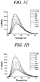
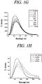
- the Compound 1 when the Compound 1 forms an imine bond with the amine compound including aminoalcohol, the Compound 1 can form an N-B-N bond-containing hetero ring and thus generate a fluorescence signal ( FIGS. 1A to 1H ).
- the Compound 1 forms an imine bond with aminoalcohol as shown in the Reaction Scheme 2 and boron connects N and N to form a fluorescent chromophore including an N-B-N bond-containing hetero ring, and, thus, the Compound 1 can generate a fluorescence signal.
- An uryl group of the Comparative Compound 2 has properties as a Lewis acid like a boric acid but cannot react with aminoalcohol. Therefore, it can be seen that the boric acid of the Compound 1 plays a special role in forming imine and an N-B-N hetero ring.
- Comparative Compound 3 has a fluorescent chromophore of a naphthylamine group, but fluorescence is quenched by an unshared electron pair present in N under normal conditions, and even in the presence of aminoalcohol, the Comparative Compound 3 cannot make a significant change in fluorescence or CD. Therefore, it can be seen that the generation of fluorescence or CD by the Compound 1 in the presence of aminoalcohol is closely related to the formation of an N-B-N hetero ring.
- the Compound 1 does not form imine with amine that does not have an additional functional group, such as -OH or -NH, which can be bonded to boron.
- the Compound 1 does not react with methylamine, ethylamine, phenylethylamine and the like under normal room temperature conditions.
- the Compound 1 can distinguish an amine compound that has an additional functional group, such as -OH or -NH, from an amine compound that does not have an additional functional group (Reaction Scheme 3).
- fluorescence is somewhat different for each kind of amine compound including aminoalcohol. This shows that if the Compound 1 is used as a probe compound, it is possible to identify even the type of a substrate (Reaction Scheme 4).
- the compound of the Chemical Formula 1 does not contain a fluorescent chromophore, but reacts with an amine compound including aminoalcohol to form a fluorescent chromophore.
- an amine compound including aminoalcohol to form a fluorescent chromophore.
- a substrate that can generate a fluorescence signal by reacting with the 1-(ortho-benzophenylaminoalkyl)-phenylboronic acid or its derivative includes aminoalcohol represented by the Chemical Formula 2 above and means a biomaterial selected from the group consisting of amino acid, nucleotide, amino acid ester, amino acid amide and combinations thereof.
- a second aspect of the present disclosure provides a method of cell-material imaging, including measuring fluorescence generated by reacting a fluorescent probe compound, 1-(ortho-benzophenylaminoalkyl)-phenylboric acid or its derivative, represented by the following Chemical Formula 1, and a material in a target cell:
- the target cell is a living cell, but is not limited thereto.
- the method of cell-material imaging further includes obtaining an image of the material in the target cell by measuring fluorescence generated by reacting the probe compound and the material in the target cell, but is not limited thereto.
- the material in the target cell includes a cell-material or an organelle, but is not limited thereto.
- the material in the target cell includes cytoplasm, mitochondria, proteins or biomolecules, but is not limited thereto.
- the material in the target cell includes mitochondria, DNA, golgi body, reticulum, lysosome, protein, Hg 2+ , Cu 2+ , ATP, amino acid or ROS (reactive oxygen species), but is not limited thereto.
- the organelle includes mitochondria and/or proteins so that stem cells, endoplasmic reticulum or cancer cells are imaged, but is not limited thereto.
- the composition for cell imaging and the method of cell-material imaging can be used for fluorescent imaging of stem cells, endoplasmic reticulum or cancer cells by imaging mitochondria and/or proteins in cells. Therefore, the cell imaging composition and the cell-material imaging method can be used for diagnosis and/or treatment of diseases (particularly, neoplastic disease or cancer). According to an embodiment of the present disclosure, the composition for cell imaging and the method of cell-material imaging can selectively sense various proteins depending on environmental conditions such as pH. When the composition for cell imaging and the method of cell-material imaging are applied, the types of protein that can be sensed depending on the experiment and/or incubation environment can be expanded, and, thus, the composition for cell imaging and the method of cell-material imaging are not particularly limited to the types of protein.
- the compound of Chemical Formula 1 exhibits an excellent effect on fluorescent imaging of HSA among the proteins, and exhibits superior fluorescence intensity and selectivity for HAS. Therefore, the compound of Chemical Formula 1 can selectively sense a cell containing HSA.
- an appropriate pH range for selective fluorescent imaging of the HSA with the composition for cell imaging may be from about pH 7 to about pH 11, but may not be limited thereto.
- the pH range may be from about pH 7 to about pH 11, from about pH 7 to about pH 10, from about pH 7 to about pH 9, from about pH 7 to about pH 8.5, from about pH 7.5 to about pH 11, from about pH 7.5 to about pH 10, from about pH 7.5 to about pH 9, or from about pH 7.5 to about pH 8.5, but may not be limited thereto.
- an appropriate pH range for selective fluorescent imaging of the HSA with the composition for cell imaging may be from about pH 7.5 to about pH 8.5.
- the mechanism by which 1-(ortho-benzophenylaminoalkyl)-phenylboronic acid or its derivative performs fluorescent imaging of the mitochondria is presumed to be due to a fluorescent chromophore produced by reaction with phosphatidyl-ethanolamine present in the mitochondrial membrane.
- the composition of Chemical formula 1 when a cell is treated with the composition of Chemical formula 1, the composition of Chemical formula 1 generates a specific fluorescence in a mitochondrial organelle. Therefore, the composition of Chemical formula 1 can be used as a probe for determining the shape and activity of mitochondria.
- the material in the target cell includes a biomaterial selected from the group consisting of amino acid, nucleotide, amino acid ester, amino acid amide and combinations thereof containing an amine compound including aminoalcohol, but may not be limited thereto.
- the probe compound forms an imine bond with an amino group contained in the amine compound included in the intracellular material.
- Boron (B) contained in the probe compound connects a nitrogen (N) atom contained in the probe compound and a nitrogen (N) atom contained in the amine compound to form a fluorescent chromophore including an N-B-N bond-containing hetero ring in-situ.
- the fluorescent chromophore may be used to individually or simultaneously analyze fluorescence and circular dichroism (CD).
- CD circular dichroism
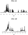
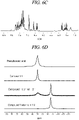
- 1 H NMR spectrum of 2:1 adduct formation (6A), 1 H NMR spectrum of 1:1 adduct formation (6B), 1 H NMR spectrum of 1:5 adduct formation (6C), by reaction of Compound 1 and alaninol( amino propanol), 11 B NMR spectra by reaction of Compound 1 and alaninol with changing equivalent (6D), and 1 H NMR spectra of 2:1 adduct formation by reaction of Compound 1 and other aminoalcohol (6E) can be seen.
- 1 eq of alaninol was added, 1 H NMR shows a very complex pattern unlike ethanolamine.
- Compound 1 shows a different reactivity depending on steric properties of carbon connected to an amine group. Ethanolamine does not have a steric hindrance and thus forms a 1:1 adduct with Compound 1 in a very short time ( FIGS. 2A and 2B , FIGS. 3A and 3B , and FIGS. 4A and 4B ). Meanwhile, alaninol has a steric hindrance and thus forms a 2:1 (Compound 1: alaninol) adduct instead of a 1:1 adduct, and when the equivalent of alaninol increases, a 1:1 to 1:2 adduct is formed ( FIGS. 6A to 6C , FIG. 6D , and FIGS. 6E( i ) to 6E(iv) ).
- Fluorescence is also slightly different for each kind of amine compound including aminoalcohol.
- a CD spectrum is remarkably different for each kind of aminoalcohol. This shows that if Compound 1 is used as a probe, it is possible to identify even the type of a substrate (Reaction Scheme 4).
- FIGS. 5A to 5D 1 H NMR spectrum of 1:5 adduct formation by reaction of Comparative Compound 1 and ethanolamine (5A), 1 H NMR spectrum of Comparative Compound 1 (5B), 1 H NMR spectrum of 1:5 adduct formation by reaction of Comparative Compound 2 and ethanolamine (5C), and 1 H NMR spectrum of Comparative Compound 2 (5D) can be seen.
- FIGS. 5A to 5D show the reactivity of Comparative Compound 1 which is similar to the Compound 1 but does not contain boron, Comparative Compound 2 substituted with an uryl group instead of boron, and aminoalcohol. It can be seen that in the absence of boron, no reaction occurs with aminoalcohol. It can be seen that the uryl group has properties as a Lewis acid like boron but does not contribute to the formation of imine.
- HeLa cells were incubated with 10 ⁇ M probes for 30 minutes and the residual probes were removed. Thereafter, (a) 0 ⁇ M, (b) 10 ⁇ M, (c) 100 ⁇ M and (d) 1,000 ⁇ M of ethanolamine were added for 30 minutes, followed by washing with DPBS to show a fluorescent image of the mitochondria in the cells through confocal microscopy (excitation wavelength: 405 nm/emission wavelength: 490 nm to 590 nm).
- FIGS. 7A to 7D it can be seen that HeLa cells were incubated with 10 ⁇ M probes for 30 minutes in an acetonitrile solution and the residual probes were removed and then, 0 ⁇ M (7A), 10 Mm (7B), 100 ⁇ M (7C) and 1,000 ⁇ M (7D) of ethanolamine were added for 30 minutes, followed by washing with DPBS to show a fluorescent image of the mitochondria in the cells observed by confocal microscopy.
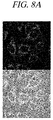
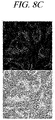
- FIGS. 8A to 8D it can be seen that HeLa cells were incubated with 10 ⁇ M probes for 30 minutes in a DMSO solution and the residual probes were removed and then, 0 Mm (8A), 10 Mm (8B), 100 ⁇ M (8C) and 1,000 ⁇ M (8D) of ethanolamine were added for 30 minutes, followed by washing with DPBS to show a fluorescent image of the mitochondria in the cells observed by confocal microscopy.
- HeLa cells were incubated with (a) 10 ⁇ M probes and (b) 10 ⁇ M probes + 40 ⁇ M CCCP for 30 minutes and washed with DPBS to show a fluorescent image of the mitochondria in the cells through confocal microscopy (excitation wavelength: 405 nm/emission wavelength: 490 nm to 590 nm).
- HeLa cells were incubated with 10 ⁇ M Compound 1 and 1 ⁇ M ER-Tracker Red for 30 minutes and a fluorescent image of the mitochondria in the cells was obtained by confocal microscopy.
- the fluorescent image of the mitochondria obtained using Compound 1 and ER-Tracker Red showed a degree of agreement of about 85% (Pearson correlation coefficient: 0.85 ⁇ 0.01).
- FIGS. 12A( i ) to 12A(iv) and FIG. 12B in order to compare photostability of mitochondrial fluorescence using Compound 1 and MitoTracker Deep Red, HeLa cells were co-stained with each of 10 ⁇ m Compound 1 and 50 nM MitoTracker Deep Red for 30 minutes, and fluorescence images and fluorescence intensity were obtained by scanning 90 times (20 sec/scan). The fluorescence intensity was analyzed by Olympus Fluoview Ver.4.0b software. The photostability of Compound 1 was checked based on the fluorescent images and fluorescence intensity depending on the number of scans and time. Referring to FIG. 12B , the fluorescence intensity of Compound 1 was higher than that of MitoTracker Deep Red.
- HeLa cells were treated with various concentrations of Compound 1 for 24 hours, cultured in a medium containing MTT for 4 hours, and then the viability of the cells depending on the concentration of Compound 1 was checked. When the absorbance of a sample not treated with Compound 1 was set to 100%, it was confirmed that the cells treated with 30 ⁇ M Compound 1 survived about 80%.
- Compound 1 exhibits fluorescent imaging performance and selectivity specific to HSA.
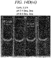
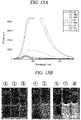
- Compound 1 at pH 7.4, pH 7.5 and pH 8.5 showed fluorescent imaging performance specific to HSA protein.
Landscapes
- Health & Medical Sciences (AREA)
- Chemical & Material Sciences (AREA)
- Life Sciences & Earth Sciences (AREA)
- Immunology (AREA)
- Engineering & Computer Science (AREA)
- Physics & Mathematics (AREA)
- General Health & Medical Sciences (AREA)
- Biochemistry (AREA)
- Analytical Chemistry (AREA)
- Molecular Biology (AREA)
- Pathology (AREA)
- General Physics & Mathematics (AREA)
- Hematology (AREA)
- Organic Chemistry (AREA)
- Biomedical Technology (AREA)
- Urology & Nephrology (AREA)
- Biotechnology (AREA)
- Microbiology (AREA)
- Cell Biology (AREA)
- Medicinal Chemistry (AREA)
- Food Science & Technology (AREA)
- Chemical Kinetics & Catalysis (AREA)
- Wood Science & Technology (AREA)
- Zoology (AREA)
- Biophysics (AREA)
- Proteomics, Peptides & Aminoacids (AREA)
- Materials Engineering (AREA)
- Optics & Photonics (AREA)
- Nuclear Medicine, Radiotherapy & Molecular Imaging (AREA)
- Genetics & Genomics (AREA)
- Toxicology (AREA)
- Tropical Medicine & Parasitology (AREA)
- General Engineering & Computer Science (AREA)
- Bioinformatics & Cheminformatics (AREA)
- Plasma & Fusion (AREA)
- Signal Processing (AREA)
- Dispersion Chemistry (AREA)
- Investigating Or Analysing Materials By The Use Of Chemical Reactions (AREA)
- Medicines Containing Antibodies Or Antigens For Use As Internal Diagnostic Agents (AREA)
- Investigating, Analyzing Materials By Fluorescence Or Luminescence (AREA)
Abstract
Description
- The present disclosure relates to a composition for cell imaging, including 1-(ortho-benzophenylaminoalkyl)-phenylboric acid or its derivative as a fluorescent probe compound, and a method of cell-material imaging using the same.
- The present inventors developed a new compound that selectively reacts with aminoalcohol to form a structure similar to Bodipy and thus exhibits strong circular dichroism (CD) and fluorescence.

- In cells, which are considered the basic units of life, organelles such as mitochondria, endoplasmic reticulum and reticulum; and biomolecules such as lipids, amino acid derivatives and proteins exist. Fluorescent imaging of their intracellular locations in real time plays a big role in diagnosing related diseases and aging and researching treatment and prevention in addition to clarifying their functions. Currently, in order to image a specific protein, the protein bonded to a green fluorescent protein (GFP) by genetic manipulation is expressed or an antibody with a fluorescent chromophore attached thereto is injected. Using a fluorescent protein makes it difficult to control the fluorescence intensity and color, requires significant time and cost as well as high-level technology. Also, since the fluorescent protein cannot pass through the cell membrane, it is necessary to squeeze the cell and then dissolve the cell membrane. For this reason, there is a need for a small molecular weight synthetic compound that can be bonded to a protein and can produce fluorescence. Such compounds can substitute for antibodies and thus can be widely used in research on protein functions as well as diagnosis and treatment of diseases targeting proteins.
- Although many chemical-tags that induce fluorescence in proteins have been developed so far, such a chemical tag requires complex manipulations to introduce an adapter, such as a peptide, to a target protein and modify the structure thereof (J. Biophotonics 4, No. 6, 391-402 (2011)).
- Recently, bioorthogonal conjugation techniques have been developed that use a relatively simple organic compound to generate fluorescence specific to a protein or specific biomolecule in a living body. The term "bioorthogonal conjugation" was first used by Carolyn R. Bertozzi, and generally requires two steps of manipulation (Sletten, Ellen M., Bertozzi, Carolyn R., "Bioorthogonal Chemistry: Fishing for Selectivity in a Sea of Functionality" Angewandte Chemie International Edition. 2009, 48(38), 6974-6998). Copper-free click chemistry is known as a representative bioorthogonal conjugation. This enables a strain-promoted alkyne-azide cycloaddition reaction by primarily binding azide (-N3 compound) to a target protein or lipid and then adding a probe to which cyclooctyne is attached (Carpenter, Richard D., Hausner, Sven H., Sutcliffe, Julie L., "Copper-Free Click for PET: Rapid 1,3-Dipolar Cycloadditions with a Fluorine-18 Cyclooctyne", ACS Medicinal Chemistry Letters. 2011, 2 (12): 885-889)). In addition to this, various bioorthogonal conjugation techniques have been developed (Kolb, Hartmuth C., Finn, M. G., Sharpless, K. Barry, Angewandte Chemie, International Edition (2001), 40(11), 2004-2021; Ning, X. H., Guo, J., Wolfert, M. A., and Boons, G.-J. Angew. Chem., Int. Ed. 2008, 47, 2253; Song, W., Wang, Y., Qu, J., Madden, M. M., and Lin, Q. Angew. Chem., Int. Ed. 2008, 47, 2832).
- The present disclosure relates to a composition for cell imaging, including 1-(ortho-benzophenylaminoalkyl)-phenylboric acid or its derivative as a fluorescent probe compound, and a method of cell-material imaging using the same.
- However, problems to be solved by the present disclosure are not limited to the above-described problems. Although not described herein, other problems to be solved by the present disclosure can be clearly understood by those skilled in the art from the following descriptions.
- A first aspect of the present disclosure provides a composition for cell imaging, including 1-(ortho-benzophenylaminoalkyl)-phenylboric acid or its derivative, represented by the following Chemical Formula 1 as a fluorescent probe compound:

- each of R1, R2, R3, R4 and R5 is independently selected from the group consisting of hydrogen, a halogen group, amino group, nitro group, cyano group, formyl group, carboxyl group, a C1-10 alkyl group, a C1-10 alkylcarbonyl group, a C6-10aryl group and a C1-10 alkoxy group,
- when the C1-10 alkyl group, the C1-10 alkylcarbonyl group, the C6-10aryl group or the C1-10 alkoxy group is substituted, it is substituted with at least one substituent selected from the group consisting of a halogen group, hydroxy group, amino group, cyano group, nitro group and a C6-10aryl group,
- Z is -N-, -O- or -CH, and
- n is an integer of from 0 to 5.
- A second aspect of the present disclosure provides a method of cell-material imaging, including measuring fluorescence generated by reacting a fluorescent probe compound, 1-(ortho-benzophenylaminoalkyl)-phenylboric acid or its derivative, represented by the following Chemical Formula 1, and a material in a target cell:

- In the above Chemical Formula 1,
- each of R1, R2, R3, R4 and R5 is independently selected from the group consisting of hydrogen, a halogen group, amino group, nitro group, cyano group, formyl group, carboxyl group, a C1-10 alkyl group, a C1-10 alkylcarbonyl group, a C6-10 aryl group and a C1-10 alkoxy group,
- when the C1-10 alkyl group, the C1-10 alkylcarbonyl group, the C6-10 aryl group or the C1-10 alkoxy group is substituted, it is substituted with at least one substituent selected from the group consisting of a halogen group, hydroxy group, amino group, cyano group, nitro group and a C6-10 aryl group,
- Z is -N-, -O- or -CH, and
- n is an integer of from 0 to 5.
- According to embodiments of the present disclosure, the composition for cell imaging and the method of cell-material imaging can be used to obtain a fluorescent image of a material in a living cell, for example, a cell-material or organelle of a living cell.
- According to embodiments of the present disclosure, the composition for cell imaging and the method of cell-material imaging are provided to image the material in the cell by measuring fluorescence generated by reacting the probe and a material in a cell, and the composition for cell imaging and the method of cell-material imaging can be used for fluorescent imaging of material in a cell including mitochondria, DNA, golgi body, reticulum, lysosome, protein, Hg2+, Cu2+, ATP, amino acid or ROS (reactive oxygen species).
- According to embodiments of the present disclosure, the composition for cell imaging and the method of cell-material imaging can be used for fluorescent imaging of stem cells, endoplasmic reticulum or cancer cells.
- According to embodiments of the present disclosure, the composition for cell imaging and the method of cell-material imaging can be used for fluorescent imaging of stem cells, endoplasmic reticulum or cancer cells by imaging mitochondria and/or proteins in cells. Therefore, the composition for cell imaging and the method of cell-material imaging can be used for diagnosis and/or treatment of diseases (particularly, neoplastic disease or cancer).
- According to the embodiments of the present disclosure, the composition for cell imaging and the method of cell-material imaging can selectively sense various proteins depending on environmental conditions such as pH. For example, the composition for cell imaging and the method of cell-material imaging have a superior selectivity to human serum albumin (HAS) among various proteins and thus can selectively sense a cell containing HSA.
-
-
FIGS. 1A to 1H show fluorescence spectra trend forCompound 1 + ethanolamine (1A), Compound 1 + alaninol (1B), Compound 1 + phenylalaninol (1C), Compound 1 + aminobutanol (1D), Compound 1 + valinol (1E), Compound 1 + leucinol (IF), Compound 1 + tryptophanol (1G), and comparison (Compound 1 + alaninol, leucinol, aminobutanol, phenylalaninol) (may be omitted) (1H), respectively, when used Compound 1 as a probe, according to an example of the present disclosure. -
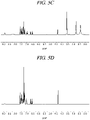
FIGS. 2A and 2B show 1H NMR spectra of 1:1 adduct formation by reaction ofCompound 1 and ethanolamine (2A), and Compound 1 (2B), respectively, according to an example of the present disclosure. -
FIGS. 3A and 3B show 13C NMR spectra of 1:1 adduct formation by reaction ofCompound 1 and ethanolamine (3A), and Compound 1 (3B), respectively, according to an example of the present disclosure. -
FIGS. 4A and 4B show 11B NMR spectra of 1:1 adduct formation by reaction ofCompound 1 and ethanolamine (4A), and Compound 1 (4B), respectively, according to an example of the present disclosure. -
FIGS. 5A to 5D show 1H NMR spectra of 1:5 adduct formation by reaction ofComparative Compound 1 and ethanolamine (5A), Comparative Compound 1 (5B), 1:5 adduct formation by reaction ofComparative Compound 2 and ethanolamine (5C), and Comparative Compound 2 (5D), respectively, according to an comparative example of the present disclosure. -
FIGS. 6A to 6C show 1H NMR spectra of 2:1 adduct formation (6A), 1:1 adduct formation (6B), and 1:5 adduct formation (6C), respectively, by reaction ofCompound 1 and alaninol(=amino propanol), according to an example of the present disclosure.FIG. 6D shows 11B NMR spectra by reaction ofCompound 1 and alaninol with changing equivalent, according to an example of the present disclosure.FIGS. 6E( i ) to 6E(iv) show 1H NMR spectra of 2:1 adduct formation by reaction ofCompound 1 and other aminoalcohol, respectively, according to an example of the present disclosure. -
FIGS. 7A to 7D show fluorescence images of mitochondria in a cell acquired by confocal microscopy after HeLa cells were incubated with 10 µM probe in acetonitrile solution and then added 0 µM ethanolamine (7A), 10 µM ethanolamine (7B), 100 µM ethanolamine (7C) and 1000 µM ethanolamine (7D), respectively, according to an example of the present disclosure. -
FIGS. 8A to 8D show fluorescence images of mitochondria in a cell acquired by confocal microscopy after HeLa cells were incubated with 10 µM probe in dimethyl sulfoxide solution and then added 0 µM ethanolamine (8A), 10 µM ethanolamine (8B), 100 µM ethanolamine (8C), and 1000 µM ethanolamine (8D), respectively, according to an example of the present disclosure. -
FIGS. 9A and9B show fluorescence images of mitochondria in a cell acquired by confocal microscopy after HeLa cells were incubated with 10 µM probe (9A), and 10 µM probe+40 µM CCCP (9B), respectively, for 30 min, according to an example of the present disclosure. -
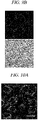
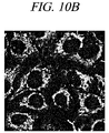
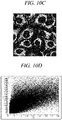
FIGS. 10A to 10C show fluorescence images of mitochondria in a cell by Compound 1 (excitation at 405 nm/ emission at 490 nm to 590 nm) (10A), Mitotracker Deep Red (excitation at 635 nm/emission at 655 nm to 755 nm) (10B), andCompound 1 and Mitotracker Deep Red merged (10C), respectively, acquired by confocal microscopy after HeLa cells were incubated with 10µM Compound FIG. 10D shows colocalization analysis graph ofCompound 1 and Mitotracker Deep Red (Ch 1 :Compound 1, Ch 2 : Mitotracker Deep Red), according to an example of the present disclosure.FIG. 10E shows fluorescence intensity profile of ROI 1 (region of interest) in theFIG. 10A (dark gray line :Compound 1, light gray line : Mitotracker Deep Red, Scale bar : 20 µm, Pearson's coefficient : 0.81±0.02), according to an example of the present disclosure. -
FIGS. 11A to 11C show fluorescence images of mitochondria in a cell by Compound 1 (excitation at 405 nm/ emission at 490 nm to 590 nm) (11A), ER(endoplasmic reticulum)-tracker Red (excitation at 559 nm/emission at 576 nm to 675 nm) (11B), andCompound 1 and ER-tracker Red merged (11C), respectively, acquired by confocal microscopy after HeLa cells were incubated with 10µM Compound FIG. 11D shows colocalization analysis graph ofCompound 1 and ER-tracker Red (Ch 1 :Compound 1, Ch 2 : ER-tracker Red), according to an example of the present disclosure. -
FIG. 11E shows fluorescence intensity profile ofROI 1 in theFIG. 11A (dark gray line :Compound 1, light gray line : ER-tracker Red, Scale bar : 20 µm, Pearson's coefficient : 0.85±0.01), according to an example of the present disclosure. -
FIGS. 12A( i ) to 12A(iv) show fluorescence images of mitochondria in a cell by Compound 1 (top) and Mitotracker Deep Red (bottom), acquired by confocal microscopy after HeLa cells were co-stained with 10µM Compound FIG. 12B shows fluorescence intensity profile of mitochondria by scan numbers (dark gray line :Compound 1, light gray line : Mitotracker Deep Red), acquired by confocal microscopy after HeLa cells were co-stained with 10µM Compound -
FIG. 13 shows cell viability according concentration ofCompound 1 obtained after culturing HeLa cells treated with various concentrations ofCompound 1 for 24 hours in a medium containing MTT for 4 hours, according to an example of the present disclosure. -
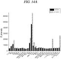
FIG. 14A shows fluorescence (FL) intensity profile measured after incubating 3µM Compound 1 with various proteins in DMSO for 0.5 hours and 1.5 hours, respectively (slit : 10 nm x 20 nm, λex= 410 nm), according to an example of the present disclosure. -
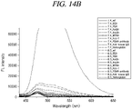
FIG. 14B shows FL intensity profile by protein and condition (pH) measured after incubating 3µM Compound 1 with various proteins in DMSO for 0.5 hours (slit: 10 nm x 20 nm, λex= 410 nm), according to an example of the present disclosure. -
FIG. 14C shows FL intensity profile by protein and condition (pH) measured after incubating 3µM Compound 1 with various proteins in DMSO for 1.5 hours (slit: 10 nm x 20 nm, λex= 410 nm), according to an example of the present disclosure. -
FIGS. 14D( i ) to 14D(vi) show fluorescence imaging of proteins measured after incubating 3µM Compound 1 with various proteins in DMSO for 0.5 hours and 1.5 hours, respectively (λex= 410 nm), according to an example of the present disclosure. -
FIG. 15A shows fluorescence intensity profile measured after eachincubating Compound 1 with HSA protein in buffer solution of various pH, according to an example of the present disclosure. -
FIG. 15B shows fluorescence imaging measured after eachincubating Compound 1 with HSA protein in buffer solution of various pH (from left ① ref, MES buffer, pH 5.5, ② HSA in MES buffer, pH 5.5, ③ HSA in PBS buffer pH 5.5, ④ ref, PBS buffer, pH 7.5, ⑤ HSA in PBS buffer, pH 7.5, ⑥ ref, Tris buffer, pH 8.5, ⑦ HSA in Tris buffer, pH 8.5, ⑧ HSA in PBS buffer, pH 8.5), according to an example of the present disclosure. - Hereinafter, embodiments and examples of the present disclosure will be described in detail with reference to the accompanying drawings so that the present disclosure may be readily implemented by those skilled in the art. However, it is to be noted that the present disclosure is not limited to the examples but can be embodied in various other ways. In drawings, parts irrelevant to the description are omitted for the simplicity of explanation, and like reference numerals denote like parts through the whole document.
- Through the whole document, the term "connected to" or "coupled to" that is used to designate a connection or coupling of one element to another element includes both a case that an element is "directly connected or coupled to" another element and a case that an element is "electronically connected or coupled to" another element via still another element.
- Through the whole document, the term "on" that is used to designate a position of one element with respect to another element includes both a case that the one element is adjacent to the other element and a case that any other element exists between these two elements.
- Further, through the whole document, the term "comprises or includes" and/or "comprising or including" used in the document means that one or more other components, steps, operation and/or existence or addition of elements are not excluded in addition to the described components, steps, operation and/or elements unless context dictates otherwise. Through the whole document, the term "about or approximately" or "substantially" is intended to have meanings close to numerical values or ranges specified with an allowable error and intended to prevent accurate or absolute numerical values disclosed for understanding of the present disclosure from being illegally or unfairly used by any unconscionable third party. Through the whole document, the term "step of" does not mean "step for".
- Through the whole document, the term "combination of" included in Markush type description means mixture or combination of one or more components, steps, operations and/or elements selected from a group consisting of components, steps, operation and/or elements described in Markush type and thereby means that the disclosure includes one or more components, steps, operations and/or elements selected from the Markush group.
- Through the whole document, a phrase in the form "A and/or B" means "A or B, or A and B".
- Through the whole document, the term "alkyl (group)" includes a linear or branched saturated or unsaturated C1-20 alkyl group, and may include, for example, methyl, ethyl, propyl, butyl, pentyl, hexyl, heptyl, octyl, nonyl, decyl, undecyl, dodecyl, tridecyl, tetradecyl, pentadecyl, hexadecyl, heptadecyl, octadecyl, nonadecyl, eicodecyl, or all the possible isomers thereof, but may not be limited thereto.
- Through the whole document, the term "alkenyl (group)" refers to a monovalent hydrocarbon group including at least one carbon-carbon double bond in an alkyl (group) having two or more carbon atoms among the above-described alkyl (groups), and may include a linear or branched C2-20 alkenyl (group), but may not be limited thereto.
- Through the whole document, the term "alkynyl (group)" refers to a monovalent hydrocarbon group including at least one carbon-carbon double bond in an alkyl (group) having two or more carbon atoms among the above-described alkyl (groups), and may include a linear or branched C2-20 alkynyl (group), but may not be limited thereto.
- Through the whole document, the term "aryl (group)" refers to a monovalent functional group formed by the removal of one hydrogen atom from one or more rings of arene, and may include, for example, phenyl, biphenyl, terphenyl, naphthyl, anthryl, phenanthryl, pyrenyl, or all the possible isomers thereof, but may not be limited thereto. The arene may refer to a hydrocarbon group having an aromatic ring, and includes monocyclic and polycyclic hydrocarbon groups. The polycyclic hydrocarbon group includes one or more aromatic rings and includes an aromatic or nonaromatic ring as an additional ring, but may not be limited thereto.
- Through the whole document, the term "cycloalkyl (group)" refers to a monovalent functional group having a saturated hydrocarbon ring, and may include a C3-8 cycloalkyl (group), for example, cyclopropyl, cylcobutyl, cyclopentyl, cyclohexyl, cycloheptyl, cyclooctyl or all the possible isomers thereof, but may not be limited thereto.
- Through the whole document, the term "alkoxy" refers to the above-defined alkyl groups connected to an oxygen atom, and may include a C1-20 alkoxy (group), for example, methoxy, ethoxy, propoxy, butoxy, pentoxy, hexyloxy, heptyloxy, octyloxy, nonyloxy, decyloxy, undecyloxy, dodecyloxy, tridecyloxy, tetradecyloxy, pentadecyloxy, hexadecyloxy, heptadecyloxy, octadecyloxy, nonadecyloxy, acosanyloxy or all the possible isomers thereof, but may not be limited thereto.
- Through the whole document, the term "halo group" refers to a halogen element from Group 17 of the periodic table included as a functional group in a compound, and the halogen element may include, for example, F, Cl, Br or I, but may not be limited thereto.
- Hereinafter, embodiments and examples of the present disclosure will be described in detail with reference to the accompanying drawings. However, the present disclosure may not be limited to the following embodiments, examples, and drawings.
- A first aspect of the present disclosure provides a composition for cell imaging, including 1-(ortho-benzophenylaminoalkyl)-phenylboric acid or its derivative, represented by the following
Chemical Formula 1 as a fluorescent probe compound:

Chemical Formula 1, - each of R1, R2, R3, R4 and R5 is independently selected from the group consisting of hydrogen, a halogen group, amino group, nitro group, cyano group, formyl group, carboxyl group, a C1-10 alkyl group, a C1-10 alkylcarbonyl group, a C6-10 aryl group and a C1-10 alkoxy group,
- when the C1-10 alkyl group, the C1-10 alkylcarbonyl group, the C6-10 aryl group or the C1-10 alkoxy group is substituted, it is substituted with at least one substituent selected from the group consisting of a halogen group, hydroxy group, amino group, cyano group, nitro group and a C6-10aryl group,
- Z is -N-, -O- or -CH, and
- n is an integer of from 0 to 5.
- In an embodiment of the present disclosure, the probe compound may include at least one of the following compound:


- In an embodiment of the present disclosure, the composition for cell imaging is used to image a material in a living cell, but is not limited thereto.
- In an embodiment of the present disclosure, the composition for cell imaging is provided to image the material in the cell by measuring fluorescence generated by reacting the probe and a material in a cell, but may not be limited thereto.
- In an embodiment of the present disclosure, the material in the cell includes a cell-material or an organelle, but may not be limited thereto.
- In an embodiment of the present disclosure, the material in the cell includes cytoplasm, mitochondria, proteins or biomolecules, but may not be limited thereto.
- In an embodiment of the present disclosure, the material in the cell includes mitochondria, DNA, golgi body, reticulum, lysosome, protein, Hg2+, Cu2+, ATP, amino acid or ROS (reactive oxygen species), but may not be limited thereto.
- In an embodiment of the present disclosure, the organelle includes mitochondria and/or proteins so that stem cells, endoplasmic reticulum or cancer cells are imaged, but may not be limited thereto.
- In an embodiment of the present disclosure, the composition for cell imaging and the method of cell-material imaging can be used for fluorescent imaging of stem cells, endoplasmic reticulum or cancer cells by imaging mitochondria and/or proteins in cells. Therefore, the composition for cell imaging and the method of cell-material imaging can be used for diagnosis and/or treatment of diseases (particularly, neoplastic disease or cancer). According to an embodiment of the present disclosure, the composition for cell imaging and the method of cell-material imaging can selectively sense various proteins depending on environmental conditions such as pH. When the composition for cell imaging and the method of cell-material imaging are applied, the types of protein that can be sensed depending on the experiment and/or incubation environment can be expanded, and, thus, the composition for cell imaging and the method of cell-material imaging are not particularly limited to the types of protein.
- In an embodiment of the present disclosure, the compound of
Chemical Formula 1 exhibits an excellent effect on fluorescent imaging of HSA among the proteins, and exhibits superior fluorescence intensity and selectivity for HAS. Therefore, the compound ofChemical Formula 1 can selectively sense a cell containing HSA. - In an embodiment of the present disclosure, an appropriate pH range for selective fluorescent imaging of the HSA with the composition for cell imaging may be from about pH 7 to about pH 11, but may not be limited thereto. For example, the pH range may be from about pH 7 to about pH 11, from about pH 7 to about
pH 10, from about pH 7 to about pH 9, from about pH 7 to about pH 8.5, from about pH 7.5 to about pH 11, from about pH 7.5 to aboutpH 10, from about pH 7.5 to about pH 9, or from about pH 7.5 to about pH 8.5, but may not be limited thereto. In an embodiment of the present disclosure, an appropriate pH range for selective fluorescent imaging of the HSA with the composition for cell imaging may be from about pH 7.5 to about pH 8.5. - In an embodiment of the present disclosure, in a method of fluorescent imaging of the cell-material or organelle, the mechanism by which 1-(ortho-benzophenylaminoalkyl)-phenylboronic acid or its derivative performs fluorescent imaging of the mitochondria is presumed to be due to a fluorescent chromophore produced by reaction with phosphatidyl-ethanolamine present in the mitochondrial membrane.
- In an embodiment of the present disclosure, when a cell is treated with the composition for cell imaging, the composition for cell imaging generates a specific fluorescence in a mitochondrial organelle. Therefore, the composition for cell imaging can be used as a probe for determining the shape and activity of mitochondria.
- In an embodiment of the present disclosure, the material in the cell includes a biomaterial selected from the group consisting of amino acid, nucleotide, amino acid ester, amino acid amide and combinations thereof containing an amine compound including aminoalcohol, but may not be limited thereto.
- In an embodiment of the present disclosure, the probe compound forms an imine bond with an amino group contained in the amine compound included in the intracellular material. Boron (B) contained in the probe compound connects a nitrogen (N) atom contained in the probe compound and a nitrogen (N) atom contained in the amine compound to form a fluorescent chromophore including an N-B-N bond-containing hetero ring in-situ. The fluorescent chromophore may be used to individually or simultaneously analyze fluorescence and circular dichroism (CD). However, the present disclosure may not be limited thereto.
- In an embodiment of the present disclosure, the
Compound 1 may be synthesized using a method known in the art. For example, theCompound 1 may be synthesized by the method of the followingReaction Scheme 1, but may not be limited thereto:
- In an embodiment of the present disclosure, the amine compound including aminoalcohol includes an amine compound represented by the following
Chemical Formula 2, but may not be limited thereto:

Chemical Formula 2, - each of Ra and Rb is independently selected from the group consisting of hydrogen, a halogen group, amino group, nitro group, cyano group, formyl group, carboxyl group, a C1-10 alkyl group, a C1-10 alkylcarbonyl group, a C6-10 aryl group and a C1-10 alkoxy group,
- when the C1-10 alkyl group, the C1-10 alkylcarbonyl group, the C6-10 aryl group or the C1-10 alkoxy group is substituted, it is substituted with at least one substituent selected from the group consisting of a halogen group, hydroxy group, amino group, cyano group, nitro group and a C6-10 aryl group, and
- m is an integer of from 0 to 5.
- In an embodiment of the present disclosure, the amine compound may include at least one of the following compounds, but may not be limited thereto:

- In an embodiment of the present disclosure, the compound of
Chemical Formula 1 having a carbonyl group (C=O), a borate group (-B(OH)2) and an amine group does not contain a fluorescent chromophore and thus cannot generate a fluorescence signal. However, as shown in the followingReaction Scheme 2 below, when the compound ofChemical Formula 1 forms an imine bond with the amine compound including aminoalcohol, the compound ofChemical Formula 1 can form an N-B-N bond-containing hetero ring and thus generate a fluorescence signal. For example, theCompound 1 ofChemical Formula 1 having a carbonyl group (C=O), a borate group (-B(OH)2) and an amine group does not contain a fluorescent chromophore and thus cannot generate a fluorescence signal. However, as shown in the followingReaction Scheme 2 below, when theCompound 1 forms an imine bond with the amine compound including aminoalcohol, theCompound 1 can form an N-B-N bond-containing hetero ring and thus generate a fluorescence signal (FIGS. 1A to 1H ).

- That is, the
Compound 1 forms an imine bond with aminoalcohol as shown in theReaction Scheme 2 and boron connects N and N to form a fluorescent chromophore including an N-B-N bond-containing hetero ring, and, thus, theCompound 1 can generate a fluorescence signal. The generation of fluorescence from aminoalcohol in this way is novel and has not been reported before. That is, 1-(ortho-benzophenylaminoalkyl)-phenylboronic acid or its derivative represented by theChemical Formula 1 has a carbonyl group (C=O) as a functional group that can react with various amine groups to form imine and also has a boric acid group (-B(OH)2) for forming the imine. - In an embodiment of the present disclosure, a carbonyl group (C=O) and a boric acid group (-B(OH)2) in the compound of
Chemical Formula 1 and aminoalcohol form an adduct having an N-B-N bond-containing hetero ring as shown in theReaction Scheme 2, which can be predicted from HRMS data, 11B NMR data, 1H NMR data and 13C NMR data obtained from the reaction between the compound ofChemical Formula 1, such as theCompound 1 and ethanol amine (FIGS. 2A and 2B ,FIGS. 3A and 3B , andFIGS. 4A and 4B ). - Meanwhile, the following
Comparative Compound 1 and the followingComparative Compound 2, which are similar to theCompound 1 but do not have a boric acid group (-B(OH)2), have a carbonyl group (C=O) but cannot react to form imine with aminoalcohol under normal room temperature conditions (FIGS. 5A to 5D ), and thus cannot generate a fluorescence signal or a CD signal. An uryl group of theComparative Compound 2 has properties as a Lewis acid like a boric acid but cannot react with aminoalcohol. Therefore, it can be seen that the boric acid of theCompound 1 plays a special role in forming imine and an N-B-N hetero ring.
- Also, the following
Comparative Compound 3 has a fluorescent chromophore of a naphthylamine group, but fluorescence is quenched by an unshared electron pair present in N under normal conditions, and even in the presence of aminoalcohol, theComparative Compound 3 cannot make a significant change in fluorescence or CD. Therefore, it can be seen that the generation of fluorescence or CD by theCompound 1 in the presence of aminoalcohol is closely related to the formation of an N-B-N hetero ring.
- In an embodiment of the present disclosure, the
Compound 1 does not form imine with amine that does not have an additional functional group, such as -OH or -NH, which can be bonded to boron. For example, theCompound 1 does not react with methylamine, ethylamine, phenylethylamine and the like under normal room temperature conditions. As a result, theCompound 1 can distinguish an amine compound that has an additional functional group, such as -OH or -NH, from an amine compound that does not have an additional functional group (Reaction Scheme 3).

- In an example of the present disclosure, fluorescence is somewhat different for each kind of amine compound including aminoalcohol. This shows that if the
Compound 1 is used as a probe compound, it is possible to identify even the type of a substrate (Reaction Scheme 4).

- In an embodiment of the present disclosure, the compound of the
Chemical Formula 1 does not contain a fluorescent chromophore, but reacts with an amine compound including aminoalcohol to form a fluorescent chromophore. As a result, there is a great difference in the intensity of fluorescence between in the presence and absence of aminoalcohol within the amine compound. Therefore, this result shows that the compound ofChemical Formula 1 is very useful as a fluorescence sensor. - In an embodiment of the present disclosure, a substrate that can generate a fluorescence signal by reacting with the 1-(ortho-benzophenylaminoalkyl)-phenylboronic acid or its derivative includes aminoalcohol represented by the
Chemical Formula 2 above and means a biomaterial selected from the group consisting of amino acid, nucleotide, amino acid ester, amino acid amide and combinations thereof. - A second aspect of the present disclosure provides a method of cell-material imaging, including measuring fluorescence generated by reacting a fluorescent probe compound, 1-(ortho-benzophenylaminoalkyl)-phenylboric acid or its derivative, represented by the following
Chemical Formula 1, and a material in a target cell:

- In the
above Chemical Formula 1, - each of R1, R2, R3, R4 and R5 is independently selected from the group consisting of hydrogen, a halogen group, amino group, nitro group, cyano group, formyl group, carboxyl group, a C1-10 alkyl group, a C1-10 alkylcarbonyl group, a C6-10 aryl group and a C1-10 alkoxy group,
- when the C1-10 alkyl group, the C1-10 alkylcarbonyl group, the C6-10 aryl group or the C1-10 alkoxy group is substituted, it is substituted with at least one substituent selected from the group consisting of a halogen group, hydroxy group, amino group, cyano group, nitro group and a C6-10 aryl group,
- Z is -N-, -O- or -CH, and
- n is an integer of from 0 to 5.
- Detailed descriptions on the second aspect of the present disclosure, which overlap with those on the first aspect of the present disclosure, are omitted hereinafter, but the descriptions of the first aspect of the present disclosure may be identically applied to the second aspect of the present disclosure, even though they are omitted hereinafter.
- In an embodiment of the present disclosure, the target cell is a living cell, but is not limited thereto.
- In an embodiment of the present disclosure, the method of cell-material imaging further includes obtaining an image of the material in the target cell by measuring fluorescence generated by reacting the probe compound and the material in the target cell, but is not limited thereto.
- In an embodiment of the present disclosure, the material in the target cell includes a cell-material or an organelle, but is not limited thereto.
- In an embodiment of the present disclosure, the material in the target cell includes cytoplasm, mitochondria, proteins or biomolecules, but is not limited thereto.
- In an embodiment of the present disclosure, the material in the target cell includes mitochondria, DNA, golgi body, reticulum, lysosome, protein, Hg2+, Cu2+, ATP, amino acid or ROS (reactive oxygen species), but is not limited thereto.
- In an embodiment of the present disclosure, the organelle includes mitochondria and/or proteins so that stem cells, endoplasmic reticulum or cancer cells are imaged, but is not limited thereto.
- In an embodiment of the present disclosure, the composition for cell imaging and the method of cell-material imaging can be used for fluorescent imaging of stem cells, endoplasmic reticulum or cancer cells by imaging mitochondria and/or proteins in cells. Therefore, the cell imaging composition and the cell-material imaging method can be used for diagnosis and/or treatment of diseases (particularly, neoplastic disease or cancer). According to an embodiment of the present disclosure, the composition for cell imaging and the method of cell-material imaging can selectively sense various proteins depending on environmental conditions such as pH. When the composition for cell imaging and the method of cell-material imaging are applied, the types of protein that can be sensed depending on the experiment and/or incubation environment can be expanded, and, thus, the composition for cell imaging and the method of cell-material imaging are not particularly limited to the types of protein.
- In an embodiment of the present disclosure, the compound of
Chemical Formula 1 exhibits an excellent effect on fluorescent imaging of HSA among the proteins, and exhibits superior fluorescence intensity and selectivity for HAS. Therefore, the compound ofChemical Formula 1 can selectively sense a cell containing HSA. - In an embodiment of the present disclosure, an appropriate pH range for selective fluorescent imaging of the HSA with the composition for cell imaging may be from about pH 7 to about pH 11, but may not be limited thereto. For example, the pH range may be from about pH 7 to about pH 11, from about pH 7 to about
pH 10, from about pH 7 to about pH 9, from about pH 7 to about pH 8.5, from about pH 7.5 to about pH 11, from about pH 7.5 to aboutpH 10, from about pH 7.5 to about pH 9, or from about pH 7.5 to about pH 8.5, but may not be limited thereto. In an embodiment of the present disclosure, an appropriate pH range for selective fluorescent imaging of the HSA with the composition for cell imaging may be from about pH 7.5 to about pH 8.5. - In an embodiment of the present disclosure, in a method of fluorescent imaging of the cell-material or organelle, the mechanism by which 1-(ortho-benzophenylaminoalkyl)-phenylboronic acid or its derivative performs fluorescent imaging of the mitochondria is presumed to be due to a fluorescent chromophore produced by reaction with phosphatidyl-ethanolamine present in the mitochondrial membrane.
- In an embodiment of the present disclosure, when a cell is treated with the composition of
Chemical formula 1, the composition ofChemical formula 1 generates a specific fluorescence in a mitochondrial organelle. Therefore, the composition ofChemical formula 1 can be used as a probe for determining the shape and activity of mitochondria. - In an embodiment of the present disclosure, the material in the target cell includes a biomaterial selected from the group consisting of amino acid, nucleotide, amino acid ester, amino acid amide and combinations thereof containing an amine compound including aminoalcohol, but may not be limited thereto.
- In an embodiment of the present disclosure, the probe compound forms an imine bond with an amino group contained in the amine compound included in the intracellular material. Boron (B) contained in the probe compound connects a nitrogen (N) atom contained in the probe compound and a nitrogen (N) atom contained in the amine compound to form a fluorescent chromophore including an N-B-N bond-containing hetero ring in-situ. The fluorescent chromophore may be used to individually or simultaneously analyze fluorescence and circular dichroism (CD). However, the present disclosure may not be limited thereto.
- Hereinafter, example embodiments are described in more detail by using Examples, but the present disclosure may not limited to the Examples.
-

- 2-aminobenzophenone (2.00 g, 10.1 mmol), 2-bromomethylphenyl boronic acid (2.59 g, 12.1 mmol) and potassium carbonate (1.39 g, 10.1 mmol) were added into an acetonitrile solvent (15 mL) and refluxed for 8 hours. Immediately after the reaction solution was dried using an evaporator, Compound 1 (2.5 g, 7.6 mmol) was obtained by silica gel column chromatography using EA/hexane (1/4) as a developer.
- Yield : 76%. 1H NMR (300 MHz, CD3CN): δ 7.64-7.61(m, 1H), 7.60-7.59(m, 1H), 7.58-7.55(m, 2H), 7.53-7.52(m, 1H), 7.51-7.47(m, 1H), 7.43-7.39(m, 2H), 7.38-7.33(m, 2H), 7.30-7.25(m, 1H), 6.88(d, 1H, 9Hz), 6.59-6.54(m, 1H), 4.65(s, 2H). 13C NMR (125 MHz, CDCl3): 199.58, 151.38, 143.75, 140.62, 135.42, 135.20, 134.56, 131.24, 129.91, 129.11, 128.39, 128.01, 126.77, 114.63, 112.68, 46.99. HRMS (EI): calcd for C20H19N1O1B1 [M+H]+: 332.1850; found: 332.2168.
-
- Instrument: Scinco FS-2
- Concentration of Compound 1: 1.5 mM
- Result:
FIGS. 1A to 1H - Referring to the fluorescence spectrum in
FIGS. 1A to 1H , changes in the fluorescence spectrum whenCompound 1 is used as a probe can be seen:Compound 1 + ethanolamine (1A),Compound 1 + alaninol (1B),Compound 1 + phenylalaninol (1C),Compound 1 + aminobutanol (1D),Compound 1 + valinol (1E),Compound 1 + leucinol (IF),Compound 1 + tryptophanol (1G), and comparison (Compound 1 + alaninol, leucinol, aminobutanol, phenylalaninol) (may be omitted) (1H). - The instrument, concentration and parameters were the same as those for ethanolamine and alaninol.
- Result:
FIGS. 1A to 1H - After Compound 1 (30 mg) was dissolved in 1 mL of CDCl3, 0.5 eq, 1 eq, 2 eq, 5 eq and 10 eq of aminoalcohol were sequentially added and stirred for 30 minutes each time. As a result, 1H NMR spectra were obtained (
FIG. 2A ). The aminoalcohol used in the experiment is as follows:
- No reaction occurred even when up to 10 eq of aminopropanol was added to vials in which the Comparative Compounds 1, 2 and 3 as comparative examples were dissolved, respectively. CD and fluorescence were measured for the mixture of each of Comparative Compounds 1, 2 and 3 and aminopropanol, but no change occurred.
- Referring to
FIGS. 2A and 2B , 1H NMR spectrum of 1:1 adduct formation by reaction ofCompound 1 and ethanolamine (2A), and 1H NMR spectrum of Compound 1 (2B) can be seen. - Referring to
FIGS. 3A and 3B , 13C NMR spectrum of 1:1 adduct formation by reaction ofCompound 1 and ethanolamine (3A), and 13C NMR spectrum of Compound 1 (3B) can be seen. - Referring to
FIGS. 4A and 4B , 11B NMR spectrum of 1:1 adduct formation by reaction ofCompound 1 and ethanolamine (4A), and 11B NMR spectrum of Compound 1 (4B) can be seen. - Referring to
FIGS. 6A to 6C ,FIG. 6D , andFIGS. 6E( i ) to 6E(iv) , 1H NMR spectrum of 2:1 adduct formation (6A), 1H NMR spectrum of 1:1 adduct formation (6B), 1H NMR spectrum of 1:5 adduct formation (6C), by reaction ofCompound 1 and alaninol(=amino propanol), 11B NMR spectra by reaction ofCompound 1 and alaninol with changing equivalent (6D), and 1H NMR spectra of 2:1 adduct formation by reaction ofCompound 1 and other aminoalcohol (6E) can be seen. When 1 eq of alaninol was added, 1H NMR shows a very complex pattern unlike ethanolamine. -
Compound 1 shows a different reactivity depending on steric properties of carbon connected to an amine group. Ethanolamine does not have a steric hindrance and thus forms a 1:1 adduct withCompound 1 in a very short time (FIGS. 2A and 2B ,FIGS. 3A and 3B , andFIGS. 4A and 4B ). Meanwhile, alaninol has a steric hindrance and thus forms a 2:1 (Compound 1: alaninol) adduct instead of a 1:1 adduct, and when the equivalent of alaninol increases, a 1:1 to 1:2 adduct is formed (FIGS. 6A to 6C ,FIG. 6D , andFIGS. 6E( i ) to 6E(iv) ). - Fluorescence is also slightly different for each kind of amine compound including aminoalcohol. In particular, a CD spectrum is remarkably different for each kind of aminoalcohol. This shows that if
Compound 1 is used as a probe, it is possible to identify even the type of a substrate (Reaction Scheme 4). -

- 2-aminobenzophenone (0.10 g, 0.51 mmol), benzylbromide (0.24 g, 1.4 mmol) and potassium carbonate (0.17 g, 1.2 mmol) were added into acetonitrile (1.5 mL) and refluxed for 8 hours. Compound 2 (0.083 g, 0.29 mmol) was obtained by silica gel column chromatography using EA/hexane (1/50) as a developer.
- Yield: 57%. 1H NMR (300 MHz, CDCl3): δ 9.11(t, J=3Hz, 1H), δ 7.72-6.59(m, 14H), δ 4.58(d, J=6Hz, 2H).
- As a comparative example, referring to
FIGS. 5A to 5D , 1H NMR spectrum of 1:5 adduct formation by reaction ofComparative Compound 1 and ethanolamine (5A), 1H NMR spectrum of Comparative Compound 1 (5B), 1H NMR spectrum of 1:5 adduct formation by reaction ofComparative Compound 2 and ethanolamine (5C), and 1H NMR spectrum of Comparative Compound 2 (5D) can be seen.FIGS. 5A to 5D show the reactivity ofComparative Compound 1 which is similar to theCompound 1 but does not contain boron,Comparative Compound 2 substituted with an uryl group instead of boron, and aminoalcohol. It can be seen that in the absence of boron, no reaction occurs with aminoalcohol. It can be seen that the uryl group has properties as a Lewis acid like boron but does not contribute to the formation of imine. -

- 1-bromomethyl-2-phenylurylbenzene (0.92 g, 3.0 mmol) compound and potassium carbonate (0.41 g, 3.0 mmol) in ACN (3 mL) were put into 2-aminobenzophenone (0.50 g, 2.5 mmol) and stirred at 85°C for 8 hours. The progress of the reaction was monitored by TLC test, and Comparative Compound 2 (0.60 mg, 1.4 mmol) was obtained by silica gel column chromatography using EA/hexane (1:9) as a developer.
- Yield: 56%. 1H NMR (300 MHz, CDCl3): δ 8.91(br, 1H), δ 7.62-6.57(m, 20H), δ 4.34(s, 2H).
-

- 2- (bromomethyl) phenyl boronic acid (0.150 g, 0.70 mmol) and potassium carbonate (0.19 g, 1.37 mmol) in ACN (1.5 mL) were put into 1-naphthylamine (0.10 mg, 0.70 mmol) and stirred at room temperature for 8 hours. Comparative compound 3 (0.04g, 0.14mmol) was obtained by silica gel column chromatography using EA/hexane (1:9) as a developer.
- Yield: 20%. 1H NMR (300 MHz, CD3CN): δ 7.98-6.79(m, 11H), δ 4.53(s, 2H).
- HeLa cells were incubated with 10 µM probes for 30 minutes and the residual probes were removed. Thereafter, (a) 0 µM, (b) 10 µM, (c) 100 µM and (d) 1,000 µM of ethanolamine were added for 30 minutes, followed by washing with DPBS to show a fluorescent image of the mitochondria in the cells through confocal microscopy (excitation wavelength: 405 nm/emission wavelength: 490 nm to 590 nm).
- Referring to
FIGS. 7A to 7D , it can be seen that HeLa cells were incubated with 10 µM probes for 30 minutes in an acetonitrile solution and the residual probes were removed and then, 0 µM (7A), 10 Mm (7B), 100 µM (7C) and 1,000 µM (7D) of ethanolamine were added for 30 minutes, followed by washing with DPBS to show a fluorescent image of the mitochondria in the cells observed by confocal microscopy. - Referring to
FIGS. 8A to 8D , it can be seen that HeLa cells were incubated with 10 µM probes for 30 minutes in a DMSO solution and the residual probes were removed and then, 0 Mm (8A), 10 Mm (8B), 100 µM (8C) and 1,000 µM (8D) of ethanolamine were added for 30 minutes, followed by washing with DPBS to show a fluorescent image of the mitochondria in the cells observed by confocal microscopy. - <Example 6: Fluorescent Imaging Test of Mitochondria using Compound 1-Uncoupler Process>
- HeLa cells were incubated with (a) 10 µM probes and (b) 10 µM probes + 40 µM CCCP for 30 minutes and washed with DPBS to show a fluorescent image of the mitochondria in the cells through confocal microscopy (excitation wavelength: 405 nm/emission wavelength: 490 nm to 590 nm).
-
FIGS. 9A and9B show fluorescence images of mitochondria in a cell acquired by confocal microscopy after HeLa cells were incubated with 10 µM probe (9A), and 10 µM probe+40 µM CCCP (9B), respectively, for 30 min, washed with DPBS. It can be seen that the experimental results of treating the mitochondria in the cells with cccp, which is an uncoupler, and imaging the cells using the probe ofCompound 1 are shown. - <Example 7: Comparison of Fluorescent Image of
Mitochondria using Compound 1 and MitoTracker Deep Red> - Referring to
FIGS. 10A to 10E , HeLa cells were incubated with 10µM Compound Compound 1 and MitoTracker Deep Red showed a degree of agreement of about 81% (Pearson correlation coefficient: 0.81±0.02). - <Example 8: Comparative Experiment of Fluorescent Imaging of
Mitochondria using Compound 1 and Endoplasmic Reticulum (ER)-Tracker Red> - Referring to
FIGS. 11A to 11C , HeLa cells were incubated with 10µM Compound Compound 1 and ER-Tracker Red showed a degree of agreement of about 85% (Pearson correlation coefficient: 0.85±0.01). - Referring to
FIGS. 12A( i ) to 12A(iv) andFIG. 12B , in order to compare photostability of mitochondrialfluorescence using Compound 1 and MitoTracker Deep Red, HeLa cells were co-stained with each of 10µm Compound Compound 1 was checked based on the fluorescent images and fluorescence intensity depending on the number of scans and time. Referring toFIG. 12B , the fluorescence intensity ofCompound 1 was higher than that of MitoTracker Deep Red. - Referring to
FIG. 13 , HeLa cells were treated with various concentrations ofCompound 1 for 24 hours, cultured in a medium containing MTT for 4 hours, and then the viability of the cells depending on the concentration ofCompound 1 was checked. When the absorbance of a sample not treated withCompound 1 was set to 100%, it was confirmed that the cells treated with 30µM Compound 1 survived about 80%. - <Example 11: Comparative Experiment of Fluorescent Imaging of
Protein using Compound 1> - After 3
µM Compound 1 and various types of proteins in DMSO were incubated for 0.5 hours and 1.5 hours, fluorescence intensity profiles and fluorescent images depending on the types of proteins were compared. - Referring to
FIGS. 14A to FIG. 14C andFIGS. 14D( i ) to 14D(vi) , it was confirmed thatCompound 1 exhibits fluorescent imaging performance and selectivity specific to HSA. - <Example 12: Comparison of Fluorescent Imaging of HAS
Protein using Compound 1 at Various pH Values> - After
Compound 1 and HSA protein were incubated in buffer solutions at various pH values, fluorescence intensity profiles and fluorescent images depending on pH were compared. - Referring to
FIG. 15A and FIG. 15B ,Compound 1 at pH 7.4, pH 7.5 and pH 8.5 showed fluorescent imaging performance specific to HSA protein. - The above description of the present disclosure is provided for the purpose of illustration, and it would be understood by a person with ordinary skill in the art that various changes and modifications may be made without changing technical conception and essential features of the present disclosure. Thus, it is clear that the above-described examples are illustrative in all aspects and do not limit the present disclosure. For example, each component described to be of a single type can be implemented in a distributed manner. Likewise, components described to be distributed can be implemented in a combined manner.
- The scope of the present disclosure is defined by the following claims rather than by the detailed description of the embodiment. It shall be understood that all modifications and embodiments conceived from the meaning and scope of the claims and their equivalents are included in the scope of the present disclosure.
Claims (16)
- A composition for cell imaging, comprising 1-(ortho-benzophenylaminoalkyl)-phenylboric acid or its derivative, represented by the following Chemical Formula 1 as a fluorescent probe compound:
 each of R1, R2, R3, R4 and R5 is independently selected from the group consisting of hydrogen, a halogen group, amino group, nitro group, cyano group, formyl group, carboxyl group, a C1-10 alkyl group, a C1-10 alkylcarbonyl group, a C6-10 aryl group and a C1-10 alkoxy group,when the C1-10 alkyl group, the C1-10 alkylcarbonyl group, the C6-10 aryl group or the C1-10 alkoxy group is substituted, it is substituted with at least one substituent selected from the group consisting of a halogen group, hydroxy group, amino group, cyano group, nitro group and a C6-10 aryl group,Z is -N-, -O- or -CH, andn is an integer of from 0 to 5.
 - The composition of Claim 1,
wherein the probe compound comprises at least one of the following compound:


- The composition of Claim 1,
wherein the composition for cell imaging is used to image a material in a living cell. - The composition of Claim 3,
wherein the material in the cell comprises a cell-material or an organelle. - The composition of Claim 3,
wherein the material in the cell includes cytoplasm, mitochondria, proteins or biomolecules. - The composition of Claim 4,
wherein the organelle includes mitochondria and/or proteins so that stem cells, endoplasmic reticulum or cancer cells are imaged. - The composition of Claim 3,
wherein the material in the cell includes a biomaterial selected from the group consisting of amino acid, nucleotide, amino acid ester, amino acid amide and combinations thereof containing an amine compound including aminoalcohol. - The composition of Claim 7,
wherein the amine compound includes an amine compound represented by Chemical Formula 2: each of Ra and Rb is independently selected from the group consisting of hydrogen, a halogen group, amino group, nitro group, cyano group, formyl group, carboxyl group, a C1-10 alkyl group, a C1-10 alkylcarbonyl group, a C6-10 aryl group and a C1-10 alkoxy group,when the C1-10 alkyl group, the C1-10 alkylcarbonyl group, the C6-10 aryl group or the C1-10 alkoxy group is substituted, it is substituted with at least one substituent selected from the group consisting of a halogen group, hydroxy group, amino group, cyano group, nitro group and a C6-10 aryl group, and m is an integer of from 0 to 5.
 - A method of cell-material imaging, comprising:
measuring fluorescence generated by reacting a fluorescent probe compound, 1-(ortho-benzophenylaminoalkyl)-phenylboric acid or its derivative, represented by the following Chemical Formula 1, and a material in a target cell: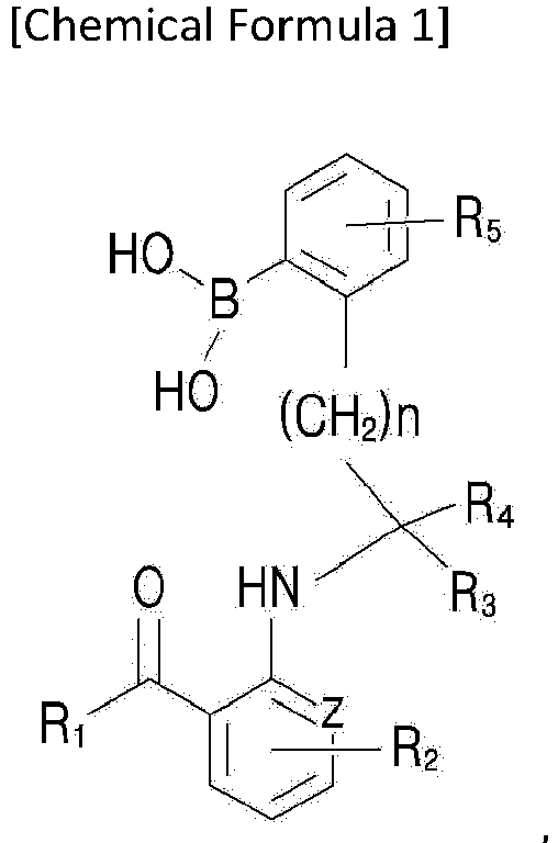 each of R1, R2, R3, R4 and R5 is independently selected from the group consisting of hydrogen, a halogen group, amino group, nitro group, cyano group, formyl group, carboxyl group, a C1-10 alkyl group, a C1-10 alkylcarbonyl group, a C6-10 aryl group and a C1-10 alkoxy group,when the C1-10 alkyl group, the C1-10 alkylcarbonyl group, the C6-10 aryl group or the C1-10 alkoxy group is substituted, it is substituted with at least one substituent selected from the group consisting of a halogen group, hydroxy group, amino group, cyano group, nitro group and a C6-10 aryl group,Z is -N-, -O- or -CH, andn is an integer of from 0 to 5.
 - The method of Claim 9,
wherein the target cell is a living cell. - The method of Claim 9, further comprising:
obtaining an image of the material in the target cell by measuring fluorescence generated by reacting the probe compound and the material in the target cell. - The method of Claim 9,
wherein the material in the target cell comprises a cell-material or an organelle. - The method of Claim 9,
wherein the material in the target cell includes cytoplasm, mitochondria, proteins or biomolecules. - The method of Claim 12,
wherein the organelle includes mitochondria and/or proteins so that stem cells, endoplasmic reticulum or cancer cells are imaged. - The method of Claim 9,
wherein the material in the target cell includes a biomaterial selected from the group consisting of amino acid, nucleotide, amino acid ester, amino acid amide and combinations thereof containing an amine compound including aminoalcohol. - The method of Claim 15,
wherein the amine compound includes an amine compound represented by Chemical Formula 2:
 each of Ra and Rb is independently selected from the group consisting of hydrogen, a halogen group, amino group, nitro group, cyano group, formyl group, carboxyl group, a C1-10 alkyl group, a C1-10 alkylcarbonyl group, a C6-10 aryl group and a C1-10 alkoxy group,when the C1-10 alkyl group, the C1-10 alkylcarbonyl group, the C6-10 aryl group or the C1-10 alkoxy group is substituted, it is substituted with at least one substituent selected from the group consisting of a halogen group, hydroxy group, amino group, cyano group, nitro group and a C6-10 aryl group, andm is an integer of from 0 to 5.
Applications Claiming Priority (2)
| Application Number | Priority Date | Filing Date | Title |
|---|---|---|---|
| KR20190144581 | 2019-11-12 | ||
| PCT/KR2020/015922 WO2021096263A1 (en) | 2019-11-12 | 2020-11-12 | Cell imaging composition and cellular material imaging method using same |
Publications (4)
| Publication Number | Publication Date |
|---|---|
| EP4057002A1 true EP4057002A1 (en) | 2022-09-14 |
| EP4057002A4 EP4057002A4 (en) | 2023-11-22 |
| EP4057002C0 EP4057002C0 (en) | 2024-08-07 |
| EP4057002B1 EP4057002B1 (en) | 2024-08-07 |
Family
ID=75912125
Family Applications (1)
| Application Number | Title | Priority Date | Filing Date |
|---|---|---|---|
| EP20887834.8A Active EP4057002B1 (en) | 2019-11-12 | 2020-11-12 | Cell imaging composition and cellular material imaging method using same |
Country Status (5)
| Country | Link |
|---|---|
| US (1) | US12181409B2 (en) |
| EP (1) | EP4057002B1 (en) |
| KR (1) | KR102413908B1 (en) |
| CN (1) | CN114729927A (en) |
| WO (1) | WO2021096263A1 (en) |
Families Citing this family (2)
| Publication number | Priority date | Publication date | Assignee | Title |
|---|---|---|---|---|
| KR20240131196A (en) * | 2023-02-23 | 2024-08-30 | 주식회사 키텍바이오 | albumin conjugate, method for producing the same and composition for diagnosis or treatment of cancer including the same |
| CN119264072B (en) * | 2024-12-06 | 2025-05-02 | 国科大杭州高等研究院 | Fluorescent probe for cell imaging and its synthesis method and application |
Family Cites Families (8)
| Publication number | Priority date | Publication date | Assignee | Title |
|---|---|---|---|---|
| JP2883808B2 (en) * | 1994-03-09 | 1999-04-19 | 科学技術振興事業団 | Color-forming compound |
| GB201016206D0 (en) * | 2010-09-27 | 2010-11-10 | Ge Healthcare Ltd | Apoptosis imaging agents |
| WO2012154213A1 (en) * | 2011-04-07 | 2012-11-15 | Cornell University | Cofluorons and methods of making and using them |
| KR101665046B1 (en) * | 2013-01-31 | 2016-10-11 | 인하대학교 산학협력단 | Ratiometric fluorescent chemosensor including boronic acid binding to mercury ion selectively, preparation method thereof and detection method of mercury ion using the same |
| KR101752473B1 (en) * | 2013-10-31 | 2017-07-03 | 한국과학기술연구원 | Method for synthesis of novel fluorescence probe library of dansyl scaffold and method for imaging live cells using thereof |
| CN110055054B (en) * | 2019-04-09 | 2021-04-06 | 中国科学院化学研究所 | Fluorescent probe for targeting living cell mitochondria G-quadruplex and application thereof |
| US11780857B2 (en) * | 2019-04-17 | 2023-10-10 | EWHA University—Industry Collaboration Foundation | Probe compounds for amino alcohols, and simultaneous fluorescence and circular dichroism analysis method |
| CN110894201B (en) | 2019-12-13 | 2023-02-03 | 安徽大学 | Single-molecule fluorescent probe for simultaneous super-resolution imaging of mitochondrial hydrogen peroxide, protein and nucleic acid and preparation and application thereof |
-
2020
- 2020-11-12 WO PCT/KR2020/015922 patent/WO2021096263A1/en not_active Ceased
- 2020-11-12 KR KR1020200151285A patent/KR102413908B1/en active Active
- 2020-11-12 EP EP20887834.8A patent/EP4057002B1/en active Active
- 2020-11-12 CN CN202080077691.9A patent/CN114729927A/en active Pending
-
2022
- 2022-05-11 US US17/741,512 patent/US12181409B2/en active Active
Also Published As
| Publication number | Publication date |
|---|---|
| EP4057002A4 (en) | 2023-11-22 |
| KR20210057700A (en) | 2021-05-21 |
| CN114729927A (en) | 2022-07-08 |
| WO2021096263A1 (en) | 2021-05-20 |
| EP4057002C0 (en) | 2024-08-07 |
| EP4057002B1 (en) | 2024-08-07 |
| KR102413908B1 (en) | 2022-06-29 |
| US12181409B2 (en) | 2024-12-31 |
| US20220267670A1 (en) | 2022-08-25 |
Similar Documents
| Publication | Publication Date | Title |
|---|---|---|
| EP2022794B1 (en) | Fluorescent compound and labeling agent comprising the same | |
| Gonçalves | Fluorescent labeling of biomolecules with organic probes | |
| US20150353585A1 (en) | Asymmetrical si rhodamine and rhodol synthesis | |
| Key et al. | Photophysical characterization of triazole-substituted coumarin fluorophores | |
| US12181409B2 (en) | Cell imaging composition and cellular material imaging method using same | |
| US9701667B2 (en) | Coumarin-based fluorogenic agents and uses thereof for specific protein labelling | |
| US8026110B2 (en) | Combinatorial rosamine library and uses thereof | |
| Rosenberg et al. | Fluorescence pH probes based on photoinduced electron transfer quenching of long fluorescence lifetime triangulenium dyes | |
| CA2336904A1 (en) | Novel fluorescent lanthanide chelates | |
| Bora et al. | Azadioxatriangulenium: synthesis and photophysical properties of reactive dyes for bioconjugation | |
| US6740756B1 (en) | Fluorescent lanthanide chelates | |
| Duan et al. | The synthesis of polarity-sensitive fluorescent dyes based on the BODIPY chromophore | |
| Raghava et al. | Green Fluorescent Diamino‐Terephthalonitriles with Reactive Terminal Groups Towards Synthesis of Value‐Added Fluorophores | |
| EP4103652A1 (en) | Fluorescent rhodamine dyes with enhanced cell permeability | |
| EP2395055A1 (en) | Fluorescent solvatochromic pigment | |
| Reddy et al. | Luminescent Trimethoprim–Polyaminocarboxylate Lanthanide Complex Conjugates for Selective Protein Labeling and Time-Resolved Bioassays | |
| US7977120B2 (en) | Fluorescent sensors for cellular amines | |
| JP4392502B2 (en) | Fluorescent amino acid derivatives | |
| KR20200122259A (en) | Novel probe compounds for fluorescence and/or circular dichroism sensor for amine compounds including amino alcohols, and simultaneous analysis method of fluorescence and circular dichroism | |
| da Frota et al. | Late‐stage Fluorination of Tyrosine Residues in Antiviral Protein Cyanovirin‐N | |
| Sinha et al. | Amino‐Terephthalonitrile Based Single Benzene Fluorophores Bearing Amino Esters and Lipids for Selective Biothiol Sensing and Bioimaging | |
| KR101125058B1 (en) | Compound for labeling material, intermediate therefor and process for producing the same | |
| US9958452B2 (en) | Highly fluorogenic protein labelling agents | |
| KR102950752B1 (en) | Probe compound, manufacturing method for the same and sensor comprising probe compound | |
| JP5044779B2 (en) | Phenophthalein derivatives and test agents for detecting polyamines in vivo |
Legal Events
| Date | Code | Title | Description |
|---|---|---|---|
| STAA | Information on the status of an ep patent application or granted ep patent |
Free format text: STATUS: THE INTERNATIONAL PUBLICATION HAS BEEN MADE |
|
| PUAI | Public reference made under article 153(3) epc to a published international application that has entered the european phase |
Free format text: ORIGINAL CODE: 0009012 |
|
| STAA | Information on the status of an ep patent application or granted ep patent |
Free format text: STATUS: REQUEST FOR EXAMINATION WAS MADE |
|
| 17P | Request for examination filed |
Effective date: 20220610 |
|
| AK | Designated contracting states |
Kind code of ref document: A1 Designated state(s): AL AT BE BG CH CY CZ DE DK EE ES FI FR GB GR HR HU IE IS IT LI LT LU LV MC MK MT NL NO PL PT RO RS SE SI SK SM TR |
|
| DAV | Request for validation of the european patent (deleted) | ||
| DAX | Request for extension of the european patent (deleted) | ||
| REG | Reference to a national code |
Ref country code: DE Ref legal event code: R079 Free format text: PREVIOUS MAIN CLASS: G01N0033520000 Ipc: C09K0011060000 Ref country code: DE Ref legal event code: R079 Ref document number: 602020035541 Country of ref document: DE Free format text: PREVIOUS MAIN CLASS: G01N0033520000 Ipc: C09K0011060000 |
|
| A4 | Supplementary search report drawn up and despatched |
Effective date: 20231025 |
|
| RIC1 | Information provided on ipc code assigned before grant |
Ipc: G01N 21/19 20060101ALN20231019BHEP Ipc: G01N 33/58 20060101ALI20231019BHEP Ipc: G01N 15/14 20060101ALI20231019BHEP Ipc: C09K 11/06 20060101AFI20231019BHEP |
|
| GRAP | Despatch of communication of intention to grant a patent |
Free format text: ORIGINAL CODE: EPIDOSNIGR1 |
|
| STAA | Information on the status of an ep patent application or granted ep patent |
Free format text: STATUS: GRANT OF PATENT IS INTENDED |
|
| RIC1 | Information provided on ipc code assigned before grant |
Ipc: G01N 21/19 20060101ALN20240214BHEP Ipc: G01N 33/58 20060101ALI20240214BHEP Ipc: G01N 15/1433 20240101ALI20240214BHEP Ipc: C09K 11/06 20060101AFI20240214BHEP |
|
| INTG | Intention to grant announced |
Effective date: 20240229 |
|
| GRAS | Grant fee paid |
Free format text: ORIGINAL CODE: EPIDOSNIGR3 |
|
| GRAA | (expected) grant |
Free format text: ORIGINAL CODE: 0009210 |
|
| STAA | Information on the status of an ep patent application or granted ep patent |
Free format text: STATUS: THE PATENT HAS BEEN GRANTED |
|
| RAP1 | Party data changed (applicant data changed or rights of an application transferred) |
Owner name: KYTECBIO CO., LTD. Owner name: EWHA UNIVERSITY-INDUSTRY COLLABORATION FOUNDATION |
|
| AK | Designated contracting states |
Kind code of ref document: B1 Designated state(s): AL AT BE BG CH CY CZ DE DK EE ES FI FR GB GR HR HU IE IS IT LI LT LU LV MC MK MT NL NO PL PT RO RS SE SI SK SM TR |
|
| REG | Reference to a national code |
Ref country code: GB Ref legal event code: FG4D |
|
| REG | Reference to a national code |
Ref country code: CH Ref legal event code: EP |
|
| REG | Reference to a national code |
Ref country code: IE Ref legal event code: FG4D |
|
| REG | Reference to a national code |
Ref country code: DE Ref legal event code: R096 Ref document number: 602020035541 Country of ref document: DE |
|
| U01 | Request for unitary effect filed |
Effective date: 20240807 |
|
| U07 | Unitary effect registered |
Designated state(s): AT BE BG DE DK EE FI FR IT LT LU LV MT NL PT RO SE SI Effective date: 20240902 |
|
| U20 | Renewal fee for the european patent with unitary effect paid |
Year of fee payment: 5 Effective date: 20241127 |
|
| PG25 | Lapsed in a contracting state [announced via postgrant information from national office to epo] |
Ref country code: NO Free format text: LAPSE BECAUSE OF FAILURE TO SUBMIT A TRANSLATION OF THE DESCRIPTION OR TO PAY THE FEE WITHIN THE PRESCRIBED TIME-LIMIT Effective date: 20241107 |
|
| PG25 | Lapsed in a contracting state [announced via postgrant information from national office to epo] |
Ref country code: PL Free format text: LAPSE BECAUSE OF FAILURE TO SUBMIT A TRANSLATION OF THE DESCRIPTION OR TO PAY THE FEE WITHIN THE PRESCRIBED TIME-LIMIT Effective date: 20240807 Ref country code: GR Free format text: LAPSE BECAUSE OF FAILURE TO SUBMIT A TRANSLATION OF THE DESCRIPTION OR TO PAY THE FEE WITHIN THE PRESCRIBED TIME-LIMIT Effective date: 20241108 |
|
| PG25 | Lapsed in a contracting state [announced via postgrant information from national office to epo] |
Ref country code: IS Free format text: LAPSE BECAUSE OF FAILURE TO SUBMIT A TRANSLATION OF THE DESCRIPTION OR TO PAY THE FEE WITHIN THE PRESCRIBED TIME-LIMIT Effective date: 20241207 |
|
| PG25 | Lapsed in a contracting state [announced via postgrant information from national office to epo] |
Ref country code: HR Free format text: LAPSE BECAUSE OF FAILURE TO SUBMIT A TRANSLATION OF THE DESCRIPTION OR TO PAY THE FEE WITHIN THE PRESCRIBED TIME-LIMIT Effective date: 20240807 |
|
| PG25 | Lapsed in a contracting state [announced via postgrant information from national office to epo] |
Ref country code: ES Free format text: LAPSE BECAUSE OF FAILURE TO SUBMIT A TRANSLATION OF THE DESCRIPTION OR TO PAY THE FEE WITHIN THE PRESCRIBED TIME-LIMIT Effective date: 20240807 Ref country code: RS Free format text: LAPSE BECAUSE OF FAILURE TO SUBMIT A TRANSLATION OF THE DESCRIPTION OR TO PAY THE FEE WITHIN THE PRESCRIBED TIME-LIMIT Effective date: 20241107 |
|
| PG25 | Lapsed in a contracting state [announced via postgrant information from national office to epo] |
Ref country code: RS Free format text: LAPSE BECAUSE OF FAILURE TO SUBMIT A TRANSLATION OF THE DESCRIPTION OR TO PAY THE FEE WITHIN THE PRESCRIBED TIME-LIMIT Effective date: 20241107 Ref country code: PL Free format text: LAPSE BECAUSE OF FAILURE TO SUBMIT A TRANSLATION OF THE DESCRIPTION OR TO PAY THE FEE WITHIN THE PRESCRIBED TIME-LIMIT Effective date: 20240807 Ref country code: NO Free format text: LAPSE BECAUSE OF FAILURE TO SUBMIT A TRANSLATION OF THE DESCRIPTION OR TO PAY THE FEE WITHIN THE PRESCRIBED TIME-LIMIT Effective date: 20241107 Ref country code: IS Free format text: LAPSE BECAUSE OF FAILURE TO SUBMIT A TRANSLATION OF THE DESCRIPTION OR TO PAY THE FEE WITHIN THE PRESCRIBED TIME-LIMIT Effective date: 20241207 Ref country code: HR Free format text: LAPSE BECAUSE OF FAILURE TO SUBMIT A TRANSLATION OF THE DESCRIPTION OR TO PAY THE FEE WITHIN THE PRESCRIBED TIME-LIMIT Effective date: 20240807 Ref country code: GR Free format text: LAPSE BECAUSE OF FAILURE TO SUBMIT A TRANSLATION OF THE DESCRIPTION OR TO PAY THE FEE WITHIN THE PRESCRIBED TIME-LIMIT Effective date: 20241108 Ref country code: ES Free format text: LAPSE BECAUSE OF FAILURE TO SUBMIT A TRANSLATION OF THE DESCRIPTION OR TO PAY THE FEE WITHIN THE PRESCRIBED TIME-LIMIT Effective date: 20240807 |
|
| PG25 | Lapsed in a contracting state [announced via postgrant information from national office to epo] |
Ref country code: SM Free format text: LAPSE BECAUSE OF FAILURE TO SUBMIT A TRANSLATION OF THE DESCRIPTION OR TO PAY THE FEE WITHIN THE PRESCRIBED TIME-LIMIT Effective date: 20240807 |
|
| PG25 | Lapsed in a contracting state [announced via postgrant information from national office to epo] |
Ref country code: CZ Free format text: LAPSE BECAUSE OF FAILURE TO SUBMIT A TRANSLATION OF THE DESCRIPTION OR TO PAY THE FEE WITHIN THE PRESCRIBED TIME-LIMIT Effective date: 20240807 |
|
| PG25 | Lapsed in a contracting state [announced via postgrant information from national office to epo] |
Ref country code: SK Free format text: LAPSE BECAUSE OF FAILURE TO SUBMIT A TRANSLATION OF THE DESCRIPTION OR TO PAY THE FEE WITHIN THE PRESCRIBED TIME-LIMIT Effective date: 20240807 |
|
| PLBE | No opposition filed within time limit |
Free format text: ORIGINAL CODE: 0009261 |
|
| STAA | Information on the status of an ep patent application or granted ep patent |
Free format text: STATUS: NO OPPOSITION FILED WITHIN TIME LIMIT |
|
| REG | Reference to a national code |
Ref country code: CH Ref legal event code: PL |
|
| PG25 | Lapsed in a contracting state [announced via postgrant information from national office to epo] |
Ref country code: MC Free format text: LAPSE BECAUSE OF FAILURE TO SUBMIT A TRANSLATION OF THE DESCRIPTION OR TO PAY THE FEE WITHIN THE PRESCRIBED TIME-LIMIT Effective date: 20240807 |
|
| REG | Reference to a national code |
Ref country code: CH Ref legal event code: PL |
|
| 26N | No opposition filed |
Effective date: 20250508 |
|
| PG25 | Lapsed in a contracting state [announced via postgrant information from national office to epo] |
Ref country code: CH Free format text: LAPSE BECAUSE OF NON-PAYMENT OF DUE FEES Effective date: 20241130 |
|
| PG25 | Lapsed in a contracting state [announced via postgrant information from national office to epo] |
Ref country code: IE Free format text: LAPSE BECAUSE OF NON-PAYMENT OF DUE FEES Effective date: 20241112 |
|
| U20 | Renewal fee for the european patent with unitary effect paid |
Year of fee payment: 6 Effective date: 20251126 |
|
| PGFP | Annual fee paid to national office [announced via postgrant information from national office to epo] |
Ref country code: GB Payment date: 20251121 Year of fee payment: 6 |
|
| PG25 | Lapsed in a contracting state [announced via postgrant information from national office to epo] |
Ref country code: HU Free format text: LAPSE BECAUSE OF FAILURE TO SUBMIT A TRANSLATION OF THE DESCRIPTION OR TO PAY THE FEE WITHIN THE PRESCRIBED TIME-LIMIT; INVALID AB INITIO Effective date: 20201112 |
|
| PG25 | Lapsed in a contracting state [announced via postgrant information from national office to epo] |
Ref country code: CY Free format text: LAPSE BECAUSE OF FAILURE TO SUBMIT A TRANSLATION OF THE DESCRIPTION OR TO PAY THE FEE WITHIN THE PRESCRIBED TIME-LIMIT; INVALID AB INITIO Effective date: 20201112 |