EP4044194B1 - Method for analysing potential ablation therapies - Google Patents
Method for analysing potential ablation therapies Download PDFInfo
- Publication number
- EP4044194B1 EP4044194B1 EP21156619.5A EP21156619A EP4044194B1 EP 4044194 B1 EP4044194 B1 EP 4044194B1 EP 21156619 A EP21156619 A EP 21156619A EP 4044194 B1 EP4044194 B1 EP 4044194B1
- Authority
- EP
- European Patent Office
- Prior art keywords
- ablation
- biomarkers
- data
- control system
- electrical
- Prior art date
- Legal status (The legal status is an assumption and is not a legal conclusion. Google has not performed a legal analysis and makes no representation as to the accuracy of the status listed.)
- Active
Links
Images


Classifications
-
- G—PHYSICS
- G16—INFORMATION AND COMMUNICATION TECHNOLOGY [ICT] SPECIALLY ADAPTED FOR SPECIFIC APPLICATION FIELDS
- G16H—HEALTHCARE INFORMATICS, i.e. INFORMATION AND COMMUNICATION TECHNOLOGY [ICT] SPECIALLY ADAPTED FOR THE HANDLING OR PROCESSING OF MEDICAL OR HEALTHCARE DATA
- G16H50/00—ICT specially adapted for medical diagnosis, medical simulation or medical data mining; ICT specially adapted for detecting, monitoring or modelling epidemics or pandemics
- G16H50/50—ICT specially adapted for medical diagnosis, medical simulation or medical data mining; ICT specially adapted for detecting, monitoring or modelling epidemics or pandemics for simulation or modelling of medical disorders
-
- G—PHYSICS
- G16—INFORMATION AND COMMUNICATION TECHNOLOGY [ICT] SPECIALLY ADAPTED FOR SPECIFIC APPLICATION FIELDS
- G16H—HEALTHCARE INFORMATICS, i.e. INFORMATION AND COMMUNICATION TECHNOLOGY [ICT] SPECIALLY ADAPTED FOR THE HANDLING OR PROCESSING OF MEDICAL OR HEALTHCARE DATA
- G16H50/00—ICT specially adapted for medical diagnosis, medical simulation or medical data mining; ICT specially adapted for detecting, monitoring or modelling epidemics or pandemics
- G16H50/70—ICT specially adapted for medical diagnosis, medical simulation or medical data mining; ICT specially adapted for detecting, monitoring or modelling epidemics or pandemics for mining of medical data, e.g. analysing previous cases of other patients
-
- A—HUMAN NECESSITIES
- A61—MEDICAL OR VETERINARY SCIENCE; HYGIENE
- A61B—DIAGNOSIS; SURGERY; IDENTIFICATION
- A61B34/00—Computer-aided surgery; Manipulators or robots specially adapted for use in surgery
- A61B34/10—Computer-aided planning, simulation or modelling of surgical operations
-
- A—HUMAN NECESSITIES
- A61—MEDICAL OR VETERINARY SCIENCE; HYGIENE
- A61B—DIAGNOSIS; SURGERY; IDENTIFICATION
- A61B5/00—Measuring for diagnostic purposes; Identification of persons
- A61B5/24—Detecting, measuring or recording bioelectric or biomagnetic signals of the body or parts thereof
- A61B5/316—Modalities, i.e. specific diagnostic methods
- A61B5/318—Heart-related electrical modalities, e.g. electrocardiography [ECG]
- A61B5/346—Analysis of electrocardiograms
-
- A—HUMAN NECESSITIES
- A61—MEDICAL OR VETERINARY SCIENCE; HYGIENE
- A61B—DIAGNOSIS; SURGERY; IDENTIFICATION
- A61B5/00—Measuring for diagnostic purposes; Identification of persons
- A61B5/72—Signal processing specially adapted for physiological signals or for diagnostic purposes
- A61B5/7235—Details of waveform analysis
- A61B5/7264—Classification of physiological signals or data, e.g. using neural networks, statistical classifiers, expert systems or fuzzy systems
- A61B5/7267—Classification of physiological signals or data, e.g. using neural networks, statistical classifiers, expert systems or fuzzy systems involving training the classification device
-
- G—PHYSICS
- G16—INFORMATION AND COMMUNICATION TECHNOLOGY [ICT] SPECIALLY ADAPTED FOR SPECIFIC APPLICATION FIELDS
- G16H—HEALTHCARE INFORMATICS, i.e. INFORMATION AND COMMUNICATION TECHNOLOGY [ICT] SPECIALLY ADAPTED FOR THE HANDLING OR PROCESSING OF MEDICAL OR HEALTHCARE DATA
- G16H10/00—ICT specially adapted for the handling or processing of patient-related medical or healthcare data
- G16H10/60—ICT specially adapted for the handling or processing of patient-related medical or healthcare data for patient-specific data, e.g. for electronic patient records
-
- G—PHYSICS
- G16—INFORMATION AND COMMUNICATION TECHNOLOGY [ICT] SPECIALLY ADAPTED FOR SPECIFIC APPLICATION FIELDS
- G16H—HEALTHCARE INFORMATICS, i.e. INFORMATION AND COMMUNICATION TECHNOLOGY [ICT] SPECIALLY ADAPTED FOR THE HANDLING OR PROCESSING OF MEDICAL OR HEALTHCARE DATA
- G16H20/00—ICT specially adapted for therapies or health-improving plans, e.g. for handling prescriptions, for steering therapy or for monitoring patient compliance
- G16H20/40—ICT specially adapted for therapies or health-improving plans, e.g. for handling prescriptions, for steering therapy or for monitoring patient compliance relating to mechanical, radiation or invasive therapies, e.g. surgery, laser therapy, dialysis or acupuncture
-
- G—PHYSICS
- G16—INFORMATION AND COMMUNICATION TECHNOLOGY [ICT] SPECIALLY ADAPTED FOR SPECIFIC APPLICATION FIELDS
- G16H—HEALTHCARE INFORMATICS, i.e. INFORMATION AND COMMUNICATION TECHNOLOGY [ICT] SPECIALLY ADAPTED FOR THE HANDLING OR PROCESSING OF MEDICAL OR HEALTHCARE DATA
- G16H50/00—ICT specially adapted for medical diagnosis, medical simulation or medical data mining; ICT specially adapted for detecting, monitoring or modelling epidemics or pandemics
- G16H50/20—ICT specially adapted for medical diagnosis, medical simulation or medical data mining; ICT specially adapted for detecting, monitoring or modelling epidemics or pandemics for computer-aided diagnosis, e.g. based on medical expert systems
Definitions
- the invention relates to a method for analysing potential ablation therapies according to claim 1, to a computer readable medium with a trained machine learning model stored on it according to the general part of claim 13, to a control system configured to perform said method according to claim 14 and to a surgery system according to claim 15.
- the present method is particularly concerned with ablation therapy for atrial fibrillation.
- atrial fibrillation is the chaotic activation of muscle cells of the atria. During atrial fibrillation, the atria only minimally contribute to the function of the heart. Atrial fibrillation, therefore, reduces the output of the heart but is not imminently dangerous. However, when becoming chronical, atrial fibrillation is correlated with increased morbidity and mortality.
- ablation therapy is the destruction of the cells that allow electrical wave re-entry to reduce chaotic activation of the atrial muscle cells.
- the success rate of ablation therapy depends on the location of the ablation. In many cases of atrial fibrillation, atria muscle cells around the pulmonary veins entry are ablated. While this standard therapy has shown good results for many patients, it is not always successful. For patients with complex atrial fibrillation or recurrent atrial fibrillation in particular, atrial mapping of the electrical wave fronts and re-entry points is available. However, such mapping is complex and hard to interpret. Further methods to identify and evaluate potential targets for ablation are therefore needed.
- TANG L. Y. W. et al. (2020), "Predicting Catheter Ablation Outcomes with Pre-ablation Heart Rhythm Data: Less Is More", ADVANCES IN INTELLIGENT DATA ANALYSIS XIX; pages 563 - 571 , discloses a method for predicting categorical outcome information for ablation therapies based on a machine learning model.
- the main realisation of the present invention is that pathologies of the heart that are treated with ablation are generally related to electrical misfunctions. As such, they are well described by the results of electrical measurements. It is therefore possible to develop a machine learning algorithm based on electrical data, namely ECG data.
- ECG data electrical data
- the definition of a cost function for the training of the machine learning model becomes more easily manageable in the present invention. Being able to choose a good cost function is a key to successfully applying a machine learning algorithm.
- a further advantage is that electrical measurements prior and posterior to ablation therapies are already available for a considerable number of patients. Deriving a predicted change of the electrical biomarkers also allows presenting the predicted change to a physician. This allows the physician to design an appropriate overall treatment for the patient based on the predicted change, paired with the physician's knowledge and experience. This can further enhance the quality of the overall treatment and the acceptance of the proposed method by physicians and patients.
- a method for analyzing potential ablation therapies is proposed, in particular for patients with atrial fibrillation, via a control system, wherein the method comprises an analysis step in which the control system applies a trained machine learning model to input data thereby generating output data, wherein the input data comprise electrical biomarkers derived from ECG data from a patient of at least one potential ablation therapy, the potential ablation therapy including at least one potential ablation event with a potential ablation location, wherein the output data comprise a predicted change of the electrical biomarkers after applying the potential ablation therapy to the patient, wherein the predicted change is derived from the input data via the trained machine learning model.
- the input data may be mainly constituted by ECG-derived data or ECG-derived data and ECG-data, further enhancing the above-mentioned advantages.
- Claim 2 also concerns the possibility of analysing the output data to identify potential ablation targets for the patient. A predicted change in electrical biomarkers can be a good indicator for identifying potential ablation targets and planning an ablation therapy.
- a training step for the machine learning model is proposed in claim 3.
- the training data set may be derived from a training database which can be retrospectively labelled.
- This database may be based on an open or commercial database thereby greatly increasing the usability of the proposed method.
- the electrical biomarkers may be derived by the control system from the ECG data of the training database.
- the biomarker response to single ablation events and the whole ablation therapy is of particular interest.
- a first model for deriving a change of the electrical biomarkers after applying one of the potential ablation events and/or a second model to derive a change of the electrical biomarkers may be trained in the training step.
- single ablation events may have a well-defined biomarker response while in other cases the biomarker response of the ablation therapy is composed by more than one ablation event. For the latter, the single ablation events may have a synergetic effect which can be seen by considering the whole ablation therapy.
- Claim 6 concerns the possibility that the first and the second model may be applied separately or jointly and in particular may be applied to different electrical biomarkers.
- the training data set may comprise focal source ablation events.
- Focal source ablations as a result of mapping are more specific, provide a wider range of ablation locations and enable identifying potential focal sources by the proposed method.
- Claim 8 concerns a derival step for deriving electrical biomarkers from the ECG data automatically.
- the electrical biomarkers may be derived from the ECG data by non-machine learning algorithms.
- known algorithms may be used to derive a number of electrical biomarkers from ECG data.
- those named in claim 9 are applied in the proposed method.
- This machine learning model could be part of the machine learning model for deriving the predicted change of the electrical biomarkers, thereby enabling the predictions based on ECG data directly.
- the possibilities of using mainly the last ablation event of the ablation therapies of the training database or using ablation events throughout the ablation therapies is considered.
- the embodiment of claim 11 relates to determining a classification of the potential ablation therapy by using the output data, which can also be based on a machine learning algorithm. Further, the analysis of electrical biomarkers based on non-machine learning algorithms can also be utilized in the classification.
- the method may provide the output of a classification of potential ablation therapies, in particular from online ECG data measurements.
- the output can be used to identify or verify potential ablation locations even during surgery.
- the proposed method is used for analysing potential ablation therapies 1.
- the proposed method is used for patients P with atrial fibrillation.
- the potential ablation therapies 1 are analysed via a control system 2.
- the control system 2 is shown schematically as a dedicated control system 2 comprising hardware connected to the patient P.
- the control system 2 may also be a general-purpose computer, a cloud-based computer system or the like and configured to perform the proposed method.
- the method comprises an analysis step 3 in which the control system 2 applies a trained machine learning model 4 to input data 5 thereby generating output data 6.
- the term "trained machine learning model” describes at least the minimum amount of data necessary to apply the result of training of a machine learning algorithm.
- the trained machine learning model 4 may be present in a compressed representation and does not have to be usable by any general-purpose computer.
- the trained machine learning model 4 may, for example, comprise weights of an otherwise predefined artificial neural network.
- the necessary operations to apply the trained machine learning model 4 may be stored in the control system 2 and/or be part of the trained machine learning model 4.
- the trained machine learning model 4 comprises program code and is applicable to the input data 5 by a general-purpose operating system.
- the input data 5 comprise electrical biomarkers 7 derived from ECG data 8 from a patient P of at least one potential ablation therapy 1.
- the potential ablation therapy 1 includes at least one potential ablation event 9 with a potential ablation location 10.
- the input data 5 are schematically shown on the left side in Fig. 1 .
- the electrical biomarkers 7 will be explained further below in more detail.
- the potential ablation therapy 1 may comprise only one potential ablation event 9 or more than one potential ablation event 9.
- the potential ablation therapy 1, the potential ablation event 9 and the potential ablation location 10 may be stored in any suitable data structure. This data structure is preferably standardised. In Fig. 1 , the values x, y, z represent the respective potential ablation therapy 1, e.g.
- Fig. 1 depicts three potential ablation therapies 1, one of them comprising two potential ablation events 9.
- the term "potential" means that this ablation therapy 1 does not necessarily have to be applied to a patient P.
- the proposed method rather supports making a prediction if applying this potential ablation therapy 1 to a patient P is promising.
- An ablation event 9 is formed by one or more regions in an area targeting a common focal source or by one or more regions that otherwise share a common area, for example, the entrance of a pulmonary vein.
- An ablation event may for example be one application of a cryocatheter.
- the output data 6 comprise a predicted change of the electrical biomarkers 7 after applying the potential ablation therapy 1 to the patient P.
- the predicted change is derived from the input data 5 via the trained machine learning model 4.
- Fig. 1 the change is shown in a set of changed electrical biomarkers 11. Further, a change in the ECG data 8 is illustrated for explanatory purposes.
- the ECG data 8 are preferably surface ECG data or intracardiac ECG data and may be measured by electrodes 12 connected to the control system 2.
- the electrical biomarkers 7, or some of the electrical biomarkers 7, may be time dependent and/or time independent.
- electrical biomarkers 7 as the focus of the machine learning mechanism and looking at the predicted change of the electrical biomarkers 7, a number of machine learning architectures become particularly suitable for the proposed method.
- the following list of preferred architectures is to be understood as not being comprehensive.
- the trained machine learning model 4 is derived by training in a generally known manner.
- the trained machine learning model 4 is part of a machine learning mechanism comprising training and application of the trained machine learning model 4.
- the underlying architecture may be a fixed-input architecture.
- the fixed-input architecture is a convolutional neural network, a feed forward network, a deep residual network or a classification architecture such as a support vector machine or a Bayes classifier.
- the fixed input architecture may be coupled to an autoencoder.
- the ECG data 8 may be split into time windows of a fixed length comprising one or more QRS-complexes. Additionally or alternatively, the electrical biomarker 7 may be time independent. Convolutional networks may be used for 2D input data 5. However, 1D implementations may also be applied, being well-suited for time series. Depending on the input data 5, a feed forward network is preferable, which may form a particularly simple approach. In particular, if the input data 5 comprise long time series, solutions for the vanishing gradient problem like deep residual networks can be used. In general, the ECG data 8 prior to an ablation therapy 1 may have a time dependence with early and late events being equally important. The named classification architectures may in particular be advantageous for a larger number of correlated electrical biomarkers 7.
- recurrent architectures in particular recurrent neural networks, more preferably, architectures based on long short-term memory or gated recurrent units, which can for example be used depending on the length of the ECG data 8 and/or the electrical biomarkers 7.
- attention architecture in particular a transformer architecture and/or a non-recurrent architecture with an attention mechanism.
- attention architectures are well-suited.
- the cost function for training may be based on the known response to the ablation therapy 1, as will be further described.
- the response can be compensated by time distance to the ablation therapy 1 and/or ablation event 9. It is preferred to predict a long time change of the electrical biomarkers 7 and/or a direct change of the electrical biomarkers 7 shortly after the ablation therapy 1.
- the input data 5 are mainly constituted by ECG-derived data, in particular by the electrical biomarkers 7.
- the input data 5 may be mainly constituted by ECG-derived data and ECG data 8.
- the term "mainly” here and in the following means that the named data form the core of the respective data or the algorithm using the respective data but other data may also be present. This takes into account that it is possible to include additional data into a machine learning algorithm without greatly influencing the result. The definition of the term "mainly” is therefore to be understood as a functional definition.
- the targeted use of non-electrical data 8 may be advantageous for the proposed method.
- the proposed method may comprise analysing output data 6 to identify potential ablation targets for the patient P.
- the trained machine learning model 4 has been or is trained in a training step 13 on a training data set 14 by the control system 2.
- the training data set 14 may be derived from a training database 15 that comprises ablation therapy data 16 including ablation events 9 and ECG data 8 determined prior and posterior to the ablation therapies 1 and/or one or more of the ablation events 9.
- Predicting a change of electrical biomarkers 7 has the advantage that databases with a variety of ablation therapies 1 can be used for training. Therefore, preferably the training step 13 comprises retrospectively labelling the training database 15 thereby generating at least part of the training data set 14.
- the labelling may in principle be performed at least partially manually. It is however preferred that the control system 2 at least partially, in particular mainly or completely, derives the electrical biomarkers 7 from the ECG data 8 of the training database 15 prior and posterior to the ablation therapies 1 and/or one or more of the ablation events 9. Automatically deriving the electrical biomarkers 7 from a database already containing ECG data 8 changes after an ablation therapy 1 enables an efficient approach for unsupervised learning.
- the potential ablation therapy 1 includes at least two potential ablation events 9 with different ablation locations 10.
- the control system 2 trains a first model 17 to derive a predicted change of the electrical biomarkers 7 after applying one of the potential ablation events 9 and/or a second model 18 to derive a predicted change of the electrical biomarkers 7 of the applying potential ablation therapy.
- the result of a complete ablation therapy 1 may vary from the sum of the results of single ablation events 9.
- a single ablation event 9 may be the source of success of the ablation therapy 1. Looking at individual ablation events 9 and the ablation therapy 1 may therefore lead to an improved analysis.
- the training does not have to be completely automatic.
- the training step 13 and therefore the trained machine learning model 4 may comprise a step of automatically training and a step of manual fine-tuning.
- the trained machine learning model 4 may also be part of a model comprising the trained machine learning model 4 and further manually derived models.
- the proposed method allows for an effective definition of a cost-function for training the trained machine learning model 4. This is advantageous as the cost function may be used with many different architectures.
- the architecture behind the machine learning itself is derived and/or changed during the training step 13.
- the training step 13 may include a neural architecture search and/or use guided or automated hyperparameter tuning.
- a general model may be trained and the first and the second model 17, 18 may be trained by transfer learning from the general model.
- control system 2 applies the first and the second model 17, 18 to the input data 5 to derive the predicted change of the electrical biomarkers 7.
- control system 2 applies the first and the second model 17, 18 to a different subset of the electrical biomarkers 7 to derive a predicted subchange of the electrical biomarkers 7 after one or more of the potential ablation events 9 and after the potential ablation therapy 1.
- the control system 2 can then derive the predicted change from the predicted subchanges of the electrical biomarkers 7.
- the ablation therapies 1 in the training data set 14 may mainly comprise at least one focal source ablation event.
- the training data set 14 may comprise ablation therapies 1 including mainly focal source ablation events.
- a focal source ablation event is an ablation event 9 for which a mapping or other determination of a focal source has been done previously to ablation therapy 1, to identify focal sources and where at least one of the focal sources is ablated. Nevertheless, an advantage of the proposed method is that a mapping is not always or even mostly not necessary. It is also not necessary to use the actual information on the mapping in the training data set 14, even if the training data set 14 comprises focal source ablation events derived from mapping.
- the method may comprise a derival step in which the control system 2 applies an algorithm to the ECG data 8 to derive at least part of the electrical biomarkers 7 from the ECG data 8 automatically.
- the control system 2 applies one or more non-machine learning algorithms to the ECG data 8 to derive one or more of the electrical biomarkers 7.
- the control system 2 may apply one or more secondary machine learning models to the ECG data 8 to derive one or more of the electrical biomarkers 7.
- the same algorithm or the same architecture is used to derive the electrical biomarkers 7 for training and in the derival step.
- the secondary machine learning models may be based on any suitable architecture, preferably on convolutional networks or an autoencoder. It is also possible to include the secondary machine learning model or models into the machine learning model 4 and apply and/or train these models together. For example, feature extraction layers are known.
- the electrical biomarkers 7 may comprise at least one of: a cardiac cycle-length, an ECG morphology classification, a signal amplitude, a cardiac rhythm classification, a peak timing value, in particular R-peak timing value, a peak variability, in particular R-peak variability or a quantitative change between beats, in particular consecutive beats, for example a mean square error value.
- the input data 5 may comprise patient data, preferably at least one of age, sex, risk factors, and/or biological biomarkers, preferably biomarkers representative for blood pressure data and/or blood-measured biomarkers.
- the cardiac cycle-length can be derived from an R-R-distance-measurement and/or a frequency analysis.
- the morphology can be identified by a score, a value like T-T-voltage increase, a comparison with QRS templates, or the like. Relevant is the standardised learning mechanism. Amplitude and power can be represented by mean values, peak values or the like.
- the rhythm classification may include a regularity score and/or a standard deviation of cycle-length.
- Non-electrical biomarkers may be included into the machine learning mechanism to interpret the result of the prediction; they may be mixed with the electrical biomarkers 7 or included only in an output layer for example. Non-electrical biomarkers may also be used in the derival step.
- the trained machine learning model 4, in particular the first model 17, is trained by using mainly the last ablation event 9 of the ablation therapies 1 of the training database 15 as the training data set 14 or by using ablation events 9 throughout the ablation therapies 1.
- the term "mainly” it would be possible to include a number of ablation events 9 that were not the last ablation events 9 in the training without creating notable adverse effects for the training data set 14.
- Fig. 3 shows a possible application of the first and the second model 17, 18.
- the first model 17 is used to predict the change of the biomarkers 7 for the ablation event 9 and afterwards the first and the second model 17, 18 are used to predict the change of the biomarkers 7 after a second ablation event 9 concluding the ablation therapy 1.
- the proposed method may comprise an ablation therapy classification step in which the control system 2 determines classification of one or more potential ablation events 9 and/or the potential ablation therapy 1, in particular by determining from the output data 6 a success score relating to the probability of successful treatment of the patient P by applying the potential ablation therapy 1.
- the method comprises an ablation therapy determination step in which the control system 2 determines a classification for at least two potential ablation events (9) and/or at least two potential ablation therapies 1 based on the output data 6 and/or in which the control system 2 determines an optimized ablation therapy 1 based on the output data 6.
- the success of a potential ablation event 9 may be classified different from the success of the potential ablation therapy 1.
- the control system 2 or a physician may design the potential ablation therapy by adding potential ablation events 9 targeting different electrical biomarkers.
- the proposed method may comprise a step of classifying potential ablation events 9 into predefined classes of potential ablation events 9.
- the predefined classes can then be defined as classes having one of a group of predefined results on one or more of the electrical biomarkers 7.
- the possibility of judging the result of a single potential ablation event 9, possibly even without the context of the complete potential ablation therapy 1, is one of the advantages of the proposed method.
- the control system 2 may receive ECG data 8, preferably from an online measurement of a patient P, in particular during surgery, and output the classification of potential ablation therapies 1.
- the potential ablation therapies 1 are at least partly defined by measuring ECG data 8 at or near potential ablation locations 10 during surgery. In this way, it becomes possible to identify and/or verify an ablation therapy 1 during surgery. In particular, it becomes possible to include or exclude at least one potential ablation event 9 in the ablation therapy 1.
- the control system 2 may receive ECG data 8 from a surgery system 19.
- a computer readable medium with a trained machine model 4 stored on it is proposed. Reference is made to all explanations given above.
- the trained machine learning model 4 on the computer readable medium has been derived in the training step 13.
- control system 2 configured to perform the proposed method is proposed. Reference is made to all explanations given above.
- a surgery system 19 connected to or forming part of a control system 2 is proposed.
- the control system 2 is configured to perform the proposed method.
- the surgery system 19 is adapted to be used during surgery, adapted to receive online ECG data 8 during surgery and adapted to display a result of the therapy classification step and/or the ablation therapy determination step.
- the surgery system may be connected to electrodes 12 for measuring the online ECG data 8. It may include hardware for processing ECG data 8, a display and/or an input unit.
Landscapes
- Health & Medical Sciences (AREA)
- Engineering & Computer Science (AREA)
- Medical Informatics (AREA)
- Public Health (AREA)
- Life Sciences & Earth Sciences (AREA)
- General Health & Medical Sciences (AREA)
- Biomedical Technology (AREA)
- Surgery (AREA)
- Pathology (AREA)
- Epidemiology (AREA)
- Primary Health Care (AREA)
- Data Mining & Analysis (AREA)
- Heart & Thoracic Surgery (AREA)
- Artificial Intelligence (AREA)
- Physics & Mathematics (AREA)
- Veterinary Medicine (AREA)
- Animal Behavior & Ethology (AREA)
- Molecular Biology (AREA)
- Databases & Information Systems (AREA)
- Biophysics (AREA)
- Cardiology (AREA)
- Nuclear Medicine, Radiotherapy & Molecular Imaging (AREA)
- Mathematical Physics (AREA)
- Evolutionary Computation (AREA)
- Fuzzy Systems (AREA)
- Robotics (AREA)
- Computer Vision & Pattern Recognition (AREA)
- Physiology (AREA)
- Psychiatry (AREA)
- Signal Processing (AREA)
- Urology & Nephrology (AREA)
- Measurement And Recording Of Electrical Phenomena And Electrical Characteristics Of The Living Body (AREA)
- Surgical Instruments (AREA)
- Other Investigation Or Analysis Of Materials By Electrical Means (AREA)
Description
- The invention relates to a method for analysing potential ablation therapies according to
claim 1, to a computer readable medium with a trained machine learning model stored on it according to the general part ofclaim 13, to a control system configured to perform said method according toclaim 14 and to a surgery system according toclaim 15. - The present method is particularly concerned with ablation therapy for atrial fibrillation. Electrically, atrial fibrillation is the chaotic activation of muscle cells of the atria. During atrial fibrillation, the atria only minimally contribute to the function of the heart. Atrial fibrillation, therefore, reduces the output of the heart but is not imminently dangerous. However, when becoming chronical, atrial fibrillation is correlated with increased morbidity and mortality. One treatment option for atrial fibrillation is ablation therapy. Ablation therapy is the destruction of the cells that allow electrical wave re-entry to reduce chaotic activation of the atrial muscle cells.
- The success rate of ablation therapy depends on the location of the ablation. In many cases of atrial fibrillation, atria muscle cells around the pulmonary veins entry are ablated. While this standard therapy has shown good results for many patients, it is not always successful. For patients with complex atrial fibrillation or recurrent atrial fibrillation in particular, atrial mapping of the electrical wave fronts and re-entry points is available. However, such mapping is complex and hard to interpret. Further methods to identify and evaluate potential targets for ablation are therefore needed.
- Known methods like
EP 3 744 282 A2 - Other known methods (
WO 2019/217430 A1 ) are concerned with learning activation pathways and treatment locations based on a measurement and functional models of the heart. A functional model is a good way to reduce the complexity of a machine learning algorithm and the required training data set. However, the functional understanding of atrial fibrillation is still progressing and combining functional models with machine learning algorithms is a complex task by itself. - TANG L. Y. W. et al. (2020), "Predicting Catheter Ablation Outcomes with Pre-ablation Heart Rhythm Data: Less Is More", ADVANCES IN INTELLIGENT DATA ANALYSIS XIX; pages 563 - 571, discloses a method for predicting categorical outcome information for ablation therapies based on a machine learning model.
- It is therefore an object of the present invention to provide a method for analysing potential ablation therapies with a manageable complexity that is realistically trainable and usable.
- The above-noted problem is solved by the method of
claim 1. - The main realisation of the present invention is that pathologies of the heart that are treated with ablation are generally related to electrical misfunctions. As such, they are well described by the results of electrical measurements. It is therefore possible to develop a machine learning algorithm based on electrical data, namely ECG data. By deriving electrical biomarkers from ECG data and using a trained machine learning model to predict a change of the electrical biomarkers after applying a potential ablation therapy, the complexity of the machine learning algorithm is significantly reduced. In particular, by shifting the focus away from trying to learn a broadly defined overall success of a treatment using a multitude of data, the definition of a cost function for the training of the machine learning model becomes more easily manageable in the present invention. Being able to choose a good cost function is a key to successfully applying a machine learning algorithm.
- A further advantage is that electrical measurements prior and posterior to ablation therapies are already available for a considerable number of patients. Deriving a predicted change of the electrical biomarkers also allows presenting the predicted change to a physician. This allows the physician to design an appropriate overall treatment for the patient based on the predicted change, paired with the physician's knowledge and experience. This can further enhance the quality of the overall treatment and the acceptance of the proposed method by physicians and patients.
- In detail, a method for analyzing potential ablation therapies is proposed, in particular for patients with atrial fibrillation, via a control system, wherein the method comprises an analysis step in which the control system applies a trained machine learning model to input data thereby generating output data, wherein the input data comprise electrical biomarkers derived from ECG data from a patient of at least one potential ablation therapy, the potential ablation therapy including at least one potential ablation event with a potential ablation location, wherein the output data comprise a predicted change of the electrical biomarkers after applying the potential ablation therapy to the patient, wherein the predicted change is derived from the input data via the trained machine learning model.
- According to claim 2 the input data may be mainly constituted by ECG-derived data or ECG-derived data and ECG-data, further enhancing the above-mentioned advantages. Claim 2 also concerns the possibility of analysing the output data to identify potential ablation targets for the patient. A predicted change in electrical biomarkers can be a good indicator for identifying potential ablation targets and planning an ablation therapy.
- A training step for the machine learning model is proposed in
claim 3. The training data set may be derived from a training database which can be retrospectively labelled. This database may be based on an open or commercial database thereby greatly increasing the usability of the proposed method. - In an embodiment according to claim 4 the electrical biomarkers may be derived by the control system from the ECG data of the training database.
- Presently, the biomarker response to single ablation events and the whole ablation therapy is of particular interest. In the embodiment according to
claim 5, a first model for deriving a change of the electrical biomarkers after applying one of the potential ablation events and/or a second model to derive a change of the electrical biomarkers may be trained in the training step. In some cases, single ablation events may have a well-defined biomarker response while in other cases the biomarker response of the ablation therapy is composed by more than one ablation event. For the latter, the single ablation events may have a synergetic effect which can be seen by considering the whole ablation therapy. -
Claim 6 concerns the possibility that the first and the second model may be applied separately or jointly and in particular may be applied to different electrical biomarkers. - In an embodiment according to
claim 7, the training data set may comprise focal source ablation events. Focal source ablations as a result of mapping are more specific, provide a wider range of ablation locations and enable identifying potential focal sources by the proposed method. -
Claim 8 concerns a derival step for deriving electrical biomarkers from the ECG data automatically. The electrical biomarkers may be derived from the ECG data by non-machine learning algorithms. Here, known algorithms may be used to derive a number of electrical biomarkers from ECG data. In particular, those named inclaim 9 are applied in the proposed method. However, it is equally preferred to use a machine learning model to derive one or more of the electrical biomarkers. This machine learning model could be part of the machine learning model for deriving the predicted change of the electrical biomarkers, thereby enabling the predictions based on ECG data directly. - In the embodiment according to
claim 10, in particular depending on the available training data set, the possibilities of using mainly the last ablation event of the ablation therapies of the training database or using ablation events throughout the ablation therapies is considered. - The embodiment of
claim 11 relates to determining a classification of the potential ablation therapy by using the output data, which can also be based on a machine learning algorithm. Further, the analysis of electrical biomarkers based on non-machine learning algorithms can also be utilized in the classification. - According to
claim 12, the method may provide the output of a classification of potential ablation therapies, in particular from online ECG data measurements. The output can be used to identify or verify potential ablation locations even during surgery. - Another teaching according to
claim 13, which is of equal importance, is directed to a computer readable medium with a trained machine learning model stored on it. All explanations given with respect to the proposed method and in particular with respect to the training step are fully applicable. - Another teaching according to
claim 14, which is also of equal importance, is directed to a control system configured to perform the proposed method. All explanations given with respect to the proposed method are fully applicable. - Another teaching according to
claim 15, which is also of equal importance, is directed to a surgery system connected to or forming part of the proposed control system. All explanations given with respect to the proposed method are fully applicable. - In the following, an embodiment of the invention is explained with respect to the drawings. The drawings show
- Fig. 1
- the proposed method schematically,
- Fig. 2
- the training of the machine learning model and
- Fig. 3
- the application of the trained machine learning model.
- The proposed method is used for analysing
potential ablation therapies 1. In the preferred case shown in the Figures, the proposed method is used for patients P with atrial fibrillation. Thepotential ablation therapies 1 are analysed via a control system 2. InFig. 1 , the control system 2 is shown schematically as a dedicated control system 2 comprising hardware connected to the patient P. The control system 2 may also be a general-purpose computer, a cloud-based computer system or the like and configured to perform the proposed method. - The method comprises an
analysis step 3 in which the control system 2 applies a trainedmachine learning model 4 to inputdata 5 thereby generatingoutput data 6. - Presently the term "trained machine learning model" describes at least the minimum amount of data necessary to apply the result of training of a machine learning algorithm. The trained
machine learning model 4 may be present in a compressed representation and does not have to be usable by any general-purpose computer. The trainedmachine learning model 4 may, for example, comprise weights of an otherwise predefined artificial neural network. The necessary operations to apply the trainedmachine learning model 4 may be stored in the control system 2 and/or be part of the trainedmachine learning model 4. In a preferred embodiment, however, the trainedmachine learning model 4 comprises program code and is applicable to theinput data 5 by a general-purpose operating system. - The
input data 5 compriseelectrical biomarkers 7 derived fromECG data 8 from a patient P of at least onepotential ablation therapy 1. Thepotential ablation therapy 1 includes at least onepotential ablation event 9 with apotential ablation location 10. Theinput data 5 are schematically shown on the left side inFig. 1 . Theelectrical biomarkers 7 will be explained further below in more detail. Thepotential ablation therapy 1 may comprise only onepotential ablation event 9 or more than onepotential ablation event 9. Thepotential ablation therapy 1, thepotential ablation event 9 and thepotential ablation location 10 may be stored in any suitable data structure. This data structure is preferably standardised. InFig. 1 , the values x, y, z represent the respectivepotential ablation therapy 1, e.g. the coordinates of apotential ablation location 10 in thepotential ablation therapy 1, and are shown as an example for illustrative purposes.Fig. 1 depicts threepotential ablation therapies 1, one of them comprising twopotential ablation events 9. The term "potential" means that thisablation therapy 1 does not necessarily have to be applied to a patient P. Presently, the proposed method rather supports making a prediction if applying thispotential ablation therapy 1 to a patient P is promising. - An
ablation event 9 is formed by one or more regions in an area targeting a common focal source or by one or more regions that otherwise share a common area, for example, the entrance of a pulmonary vein. An ablation event may for example be one application of a cryocatheter. - The
output data 6 comprise a predicted change of theelectrical biomarkers 7 after applying thepotential ablation therapy 1 to the patient P. The predicted change is derived from theinput data 5 via the trainedmachine learning model 4. InFig. 1 , the change is shown in a set of changedelectrical biomarkers 11. Further, a change in theECG data 8 is illustrated for explanatory purposes. - The
ECG data 8 are preferably surface ECG data or intracardiac ECG data and may be measured byelectrodes 12 connected to the control system 2. - The
electrical biomarkers 7, or some of theelectrical biomarkers 7, may be time dependent and/or time independent. By choosingelectrical biomarkers 7 as the focus of the machine learning mechanism and looking at the predicted change of theelectrical biomarkers 7, a number of machine learning architectures become particularly suitable for the proposed method. The following list of preferred architectures is to be understood as not being comprehensive. - The trained
machine learning model 4 is derived by training in a generally known manner. The trainedmachine learning model 4 is part of a machine learning mechanism comprising training and application of the trainedmachine learning model 4. - The underlying architecture may be a fixed-input architecture. Preferably the fixed-input architecture is a convolutional neural network, a feed forward network, a deep residual network or a classification architecture such as a support vector machine or a Bayes classifier. The fixed input architecture may be coupled to an autoencoder.
- To generate the fixed input, the
ECG data 8 may be split into time windows of a fixed length comprising one or more QRS-complexes. Additionally or alternatively, theelectrical biomarker 7 may be time independent. Convolutional networks may be used for2D input data 5. However, 1D implementations may also be applied, being well-suited for time series. Depending on theinput data 5, a feed forward network is preferable, which may form a particularly simple approach. In particular, if theinput data 5 comprise long time series, solutions for the vanishing gradient problem like deep residual networks can be used. In general, theECG data 8 prior to anablation therapy 1 may have a time dependence with early and late events being equally important. The named classification architectures may in particular be advantageous for a larger number of correlatedelectrical biomarkers 7. - Also preferred are recurrent architectures, in particular recurrent neural networks, more preferably, architectures based on long short-term memory or gated recurrent units, which can for example be used depending on the length of the
ECG data 8 and/or theelectrical biomarkers 7. Another preferred architecture is an attention architecture, in particular a transformer architecture and/or a non-recurrent architecture with an attention mechanism. In case it is not known in which part ofECG data 8 the relevant information for predicting the success of apotential ablation therapy 1 may be located, it can be advantageous to use low frequencies ofECG data 8 and derive long sequences ofelectrical biomarkers 7. In particular, if relevant information is sparsely distributed, attention architectures are well-suited. - The cost function for training may be based on the known response to the
ablation therapy 1, as will be further described. The response can be compensated by time distance to theablation therapy 1 and/orablation event 9. It is preferred to predict a long time change of theelectrical biomarkers 7 and/or a direct change of theelectrical biomarkers 7 shortly after theablation therapy 1. - Here and preferably, the
input data 5 are mainly constituted by ECG-derived data, in particular by theelectrical biomarkers 7. Alternatively, theinput data 5 may be mainly constituted by ECG-derived data andECG data 8. The term "mainly" here and in the following means that the named data form the core of the respective data or the algorithm using the respective data but other data may also be present. This takes into account that it is possible to include additional data into a machine learning algorithm without greatly influencing the result. The definition of the term "mainly" is therefore to be understood as a functional definition. - The targeted use of
non-electrical data 8 may be advantageous for the proposed method. The proposed method may comprise analysingoutput data 6 to identify potential ablation targets for the patient P. - With reference to
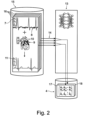
Fig. 2 , the training of themachine learning model 4 will be described. Here and preferably, the trainedmachine learning model 4 has been or is trained in atraining step 13 on atraining data set 14 by the control system 2. Thetraining data set 14 may be derived from atraining database 15 that comprisesablation therapy data 16 includingablation events 9 andECG data 8 determined prior and posterior to theablation therapies 1 and/or one or more of theablation events 9. - Predicting a change of
electrical biomarkers 7 has the advantage that databases with a variety ofablation therapies 1 can be used for training. Therefore, preferably thetraining step 13 comprises retrospectively labelling thetraining database 15 thereby generating at least part of thetraining data set 14. - The labelling may in principle be performed at least partially manually. It is however preferred that the control system 2 at least partially, in particular mainly or completely, derives the
electrical biomarkers 7 from theECG data 8 of thetraining database 15 prior and posterior to theablation therapies 1 and/or one or more of theablation events 9. Automatically deriving theelectrical biomarkers 7 from a database already containingECG data 8 changes after anablation therapy 1 enables an efficient approach for unsupervised learning. - In a preferred embodiment, the
potential ablation therapy 1 includes at least twopotential ablation events 9 withdifferent ablation locations 10. In thetraining step 13, the control system 2 then trains afirst model 17 to derive a predicted change of theelectrical biomarkers 7 after applying one of thepotential ablation events 9 and/or asecond model 18 to derive a predicted change of theelectrical biomarkers 7 of the applying potential ablation therapy. In some cases, the result of acomplete ablation therapy 1 may vary from the sum of the results ofsingle ablation events 9. In other cases, asingle ablation event 9 may be the source of success of theablation therapy 1. Looking atindividual ablation events 9 and theablation therapy 1 may therefore lead to an improved analysis. - It is noted that the training does not have to be completely automatic. The
training step 13 and therefore the trainedmachine learning model 4 may comprise a step of automatically training and a step of manual fine-tuning. Generally, the trainedmachine learning model 4 may also be part of a model comprising the trainedmachine learning model 4 and further manually derived models. - The proposed method allows for an effective definition of a cost-function for training the trained
machine learning model 4. This is advantageous as the cost function may be used with many different architectures. In one preferred embodiment, the architecture behind the machine learning itself is derived and/or changed during thetraining step 13. Thetraining step 13 may include a neural architecture search and/or use guided or automated hyperparameter tuning. - For faster training of the first and the
second model second model - Here and preferably, in the
analysis step 3 the control system 2 applies the first and thesecond model input data 5 to derive the predicted change of theelectrical biomarkers 7. Preferably, in theanalysis step 3 the control system 2 applies the first and thesecond model electrical biomarkers 7 to derive a predicted subchange of theelectrical biomarkers 7 after one or more of thepotential ablation events 9 and after thepotential ablation therapy 1. The control system 2 can then derive the predicted change from the predicted subchanges of theelectrical biomarkers 7. - Regarding the
training data set 14, theablation therapies 1 in thetraining data set 14 may mainly comprise at least one focal source ablation event. Preferably, thetraining data set 14 may compriseablation therapies 1 including mainly focal source ablation events. A focal source ablation event is anablation event 9 for which a mapping or other determination of a focal source has been done previously toablation therapy 1, to identify focal sources and where at least one of the focal sources is ablated. Nevertheless, an advantage of the proposed method is that a mapping is not always or even mostly not necessary. It is also not necessary to use the actual information on the mapping in thetraining data set 14, even if thetraining data set 14 comprises focal source ablation events derived from mapping. - The method may comprise a derival step in which the control system 2 applies an algorithm to the
ECG data 8 to derive at least part of theelectrical biomarkers 7 from theECG data 8 automatically. Preferably, in the derival step the control system 2 applies one or more non-machine learning algorithms to theECG data 8 to derive one or more of theelectrical biomarkers 7. Additionally or alternatively, in the derival step, the control system 2 may apply one or more secondary machine learning models to theECG data 8 to derive one or more of theelectrical biomarkers 7. - Here and preferably, the same algorithm or the same architecture is used to derive the
electrical biomarkers 7 for training and in the derival step. - The secondary machine learning models may be based on any suitable architecture, preferably on convolutional networks or an autoencoder. It is also possible to include the secondary machine learning model or models into the
machine learning model 4 and apply and/or train these models together. For example, feature extraction layers are known. - The
electrical biomarkers 7 may comprise at least one of: a cardiac cycle-length, an ECG morphology classification, a signal amplitude, a cardiac rhythm classification, a peak timing value, in particular R-peak timing value, a peak variability, in particular R-peak variability or a quantitative change between beats, in particular consecutive beats, for example a mean square error value. Additionally or alternatively, theinput data 5 may comprise patient data, preferably at least one of age, sex, risk factors, and/or biological biomarkers, preferably biomarkers representative for blood pressure data and/or blood-measured biomarkers. - The cardiac cycle-length can be derived from an R-R-distance-measurement and/or a frequency analysis. The morphology can be identified by a score, a value like T-T-voltage increase, a comparison with QRS templates, or the like. Relevant is the standardised learning mechanism. Amplitude and power can be represented by mean values, peak values or the like. The rhythm classification may include a regularity score and/or a standard deviation of cycle-length.
- Non-electrical biomarkers may be included into the machine learning mechanism to interpret the result of the prediction; they may be mixed with the
electrical biomarkers 7 or included only in an output layer for example. Non-electrical biomarkers may also be used in the derival step. - Here and preferably, the trained
machine learning model 4, in particular thefirst model 17, is trained by using mainly thelast ablation event 9 of theablation therapies 1 of thetraining database 15 as thetraining data set 14 or by usingablation events 9 throughout theablation therapies 1. Here, as mentioned above with respect to the definition of the term "mainly", it would be possible to include a number ofablation events 9 that were not thelast ablation events 9 in the training without creating notable adverse effects for thetraining data set 14. -
Fig. 3 shows a possible application of the first and thesecond model first model 17 is used to predict the change of thebiomarkers 7 for theablation event 9 and afterwards the first and thesecond model biomarkers 7 after asecond ablation event 9 concluding theablation therapy 1. - The proposed method may comprise an ablation therapy classification step in which the control system 2 determines classification of one or more
potential ablation events 9 and/or thepotential ablation therapy 1, in particular by determining from the output data 6 a success score relating to the probability of successful treatment of the patient P by applying thepotential ablation therapy 1. Preferably, the method comprises an ablation therapy determination step in which the control system 2 determines a classification for at least two potential ablation events (9) and/or at least twopotential ablation therapies 1 based on theoutput data 6 and/or in which the control system 2 determines an optimizedablation therapy 1 based on theoutput data 6. - The success of a
potential ablation event 9 may be classified different from the success of thepotential ablation therapy 1. In particular, the control system 2 or a physician may design the potential ablation therapy by addingpotential ablation events 9 targeting different electrical biomarkers. The proposed method may comprise a step of classifyingpotential ablation events 9 into predefined classes ofpotential ablation events 9. The predefined classes can then be defined as classes having one of a group of predefined results on one or more of theelectrical biomarkers 7. The possibility of judging the result of a singlepotential ablation event 9, possibly even without the context of the completepotential ablation therapy 1, is one of the advantages of the proposed method. - The control system 2 may receive
ECG data 8, preferably from an online measurement of a patient P, in particular during surgery, and output the classification ofpotential ablation therapies 1. Preferably thepotential ablation therapies 1 are at least partly defined by measuringECG data 8 at or nearpotential ablation locations 10 during surgery. In this way, it becomes possible to identify and/or verify anablation therapy 1 during surgery. In particular, it becomes possible to include or exclude at least onepotential ablation event 9 in theablation therapy 1. - The control system 2 may receive
ECG data 8 from asurgery system 19. - According to another teaching, a computer readable medium with a trained
machine model 4 stored on it is proposed. Reference is made to all explanations given above. The trainedmachine learning model 4 on the computer readable medium has been derived in thetraining step 13. - According to another teaching, a control system 2 configured to perform the proposed method is proposed. Reference is made to all explanations given above.
- According to another teaching, a
surgery system 19 connected to or forming part of a control system 2 is proposed. The control system 2 is configured to perform the proposed method. Reference is made to all explanations given above. Thesurgery system 19 is adapted to be used during surgery, adapted to receiveonline ECG data 8 during surgery and adapted to display a result of the therapy classification step and/or the ablation therapy determination step. The surgery system may be connected toelectrodes 12 for measuring theonline ECG data 8. It may include hardware for processingECG data 8, a display and/or an input unit.
Claims (15)
- Method for analyzing potential ablation therapies (1), in particular for patients (P) with atrial fibrillation, via a control system (2),wherein the method comprises an analysis step (3) in which the control system (2) applies a trained machine learning model (4) to input data (5) thereby generating output data (6),wherein the input data (5) comprise electrical biomarkers (7) derived from ECG data (8) from a patient (P) of at least one potential ablation therapy (1), the potential ablation therapy (1) including at least one potential ablation event (9) with a potential ablation location (10),wherein the output data (6) comprise a predicted change of the electrical biomarkers (7) after applying the potential ablation therapy (1) to the patient (P), wherein the predicted change is derived from the input data (5) via the trained machine learning model (4).
- Method according to claim 1, characterized in that the input data (5) is mainly constituted by ECG-derived data, in particular by the electrical biomarkers (7), or, that the input data (5) are mainly constituted by ECG-derived data and ECG data (8), and/or, that the method comprises analyzing the output data (6) to identify potential ablation targets for the patient (P).
- Method according to claim 1 or 2, characterized in that the trained machine learning model (4) has been or is trained in a training step (13) on a training data set (14) by the control system (2), preferably, that the training data set (14) is derived from a training database (15) that comprises ablation therapy data (16) including ablation events (9) and ECG data (8) determined prior and posterior to the ablation therapies (1) and/or one or more of the ablation events (9), more preferably, that the training step (13) comprises retrospectively labeling the training database (15) thereby generating at least part of the training data set (14).
- Method according to claim 3, characterized in that the control system (2) at least partially, in particular mainly or completely, derives the electrical biomarkers (7) from the ECG data (8) of the training database (14) prior and posterior to the ablation therapies (1) and/or one or more of the ablation events (9).
- Method according to claim 3 or 4, characterized in that the potential ablation therapy (1) includes at least two potential ablation events (9) with different ablation locations (10), that in the training step (13) the control system (2) trains a first model (17) to derive a predicted change of the electrical biomarkers (7) after applying one of the potential ablation events (9) and/or a second model (18) to derive a predicted change of the electrical biomarkers (7) after applying the potential ablation therapy (1).
- Method according to claim 5 characterized in that in the analysis step (3) the control system (2) applies the first and/or second model (17, 18) to the input data (5) to derive the predicted change of the electrical biomarkers (7), preferably, that in the analysis step (3) the control system (2) applies the first and the second model (17, 18) to a different subset of the electrical biomarkers (7) to derive a predicted subchange of the electrical biomarkers (7) after one or more of the potential ablation events (9) and after the potential ablation therapy (1), and that the control system (2) derives the predicted change from the predicted subchanges of the electrical biomarkers (7).
- Method according to one of claims 3 to 6, characterized in that the ablation therapies (1) in the training data set (14) mainly comprise at least one focal source ablation event, preferably that the training data set (14) comprises ablation therapies (1) including mainly focal source ablation events.
- Method according to one of the preceding claims, characterized in that the method comprises a derival step in which the control system (2) applies an algorithm to the ECG data (8) to derive at least part of the electrical biomarkers (7) from the ECG data (8) automatically, preferably, that in the derival step the control system (2) applies one or more non-machine learning algorithms to the ECG data (8) to derive one or more of the electrical biomarkers (7), and/or, that in the derival step the control system (2) applies one or more trained secondary machine learning models to the ECG data (8) to derive one or more of the electrical biomarkers (7).
- Method according to one of the preceding claims, characterized in that the electrical biomarkers (7) comprise at least one of: a cardiac cycle-length, an ECG morphology classification, a signal amplitude, a signal power, a cardiac rhythm classification, a peak timing value, in particular R-peak timing value, a peak variability, in particular R-peak variability or a quantitative change between beats, in particular consecutive beats, for example a mean square error value; and/or, that the input data (5) further comprises patient data, preferably at least one of: age, sex, risk factors, and/or biological biomarkers, preferably biomarkers representative for blood pressure data and/or blood-measured biomarkers.
- Method according to one of the preceding claims, characterized in that the trained machine learning model (4), in particular the first model (17), is trained by using mainly the last ablation event (9) of the ablation therapies (1) of the training database (15) as the training data set (14) or by using ablation events (9) throughout the ablation therapies (1).
- Method according to one of the preceding claims, characterized in that the method comprises an ablation therapy classification step in which the control system (2) determines a classification of one or more potential ablation events (9) and/or the potential ablation therapy (1), in particular by determining from the output data (6) a success score relating to the probability of successful treatment of the patient (P) by applying the potential ablation therapy (1), preferably, that the method comprises an ablation therapy determination step in which the control system (2) determines a classification for at least two potential ablation events (9) and/or at least two potential ablation therapies (1) from the output data (6), and/or, in which the control system (2) determines an optimized ablation therapy (1) from the output data (6).
- Method according to claim 11, characterized in that the control system (2) receives ECG data (8), preferably from an online measurement of a patient (P), in particular during surgery, and outputs the classification of potential ablation therapies (1), preferably, that the potential ablation therapies (1) are at least partly defined by measuring ECG data (8) at or near potential ablation locations (10) during surgery.
- Computer readable medium with a trained machine learning model (4) stored on it,
characterized in
that the trained machine learning model (4) has been derived in the training step (13) according to claim 3 and possibly one of claims 4 to 12. - Control system configured to perform the method according any of claims 1 to 12.
- Surgery system connected to or forming part of a control system (2), wherein the control system (2) is configured to perform the method according to claim 11 and possibly claim 12, wherein the surgery system (19) is adapted to be used during surgery, adapted to receive online ECG data (8) during surgery and adapted to display a result of the therapy classification step and/or the ablation therapy determination step.
Priority Applications (6)
| Application Number | Priority Date | Filing Date | Title |
|---|---|---|---|
| EP21156619.5A EP4044194B1 (en) | 2021-02-11 | 2021-02-11 | Method for analysing potential ablation therapies |
| ES21156619T ES2967327T3 (en) | 2021-02-11 | 2021-02-11 | Procedure to analyze potential ablation therapies |
| US18/276,193 US20240112819A1 (en) | 2021-02-11 | 2021-12-03 | Method for analyzing potential ablation therapies |
| CN202180093597.7A CN116918002A (en) | 2021-02-11 | 2021-12-03 | Methods for analyzing potential ablation therapies |
| PCT/EP2021/084257 WO2022171327A1 (en) | 2021-02-11 | 2021-12-03 | Method for analysing potential ablation therapies |
| JP2023548712A JP7686762B2 (en) | 2021-02-11 | 2021-12-03 | Methods for Analysing Potential Ablation Therapies - Patent application |
Applications Claiming Priority (1)
| Application Number | Priority Date | Filing Date | Title |
|---|---|---|---|
| EP21156619.5A EP4044194B1 (en) | 2021-02-11 | 2021-02-11 | Method for analysing potential ablation therapies |
Publications (3)
| Publication Number | Publication Date |
|---|---|
| EP4044194A1 EP4044194A1 (en) | 2022-08-17 |
| EP4044194C0 EP4044194C0 (en) | 2023-09-13 |
| EP4044194B1 true EP4044194B1 (en) | 2023-09-13 |
Family
ID=74591891
Family Applications (1)
| Application Number | Title | Priority Date | Filing Date |
|---|---|---|---|
| EP21156619.5A Active EP4044194B1 (en) | 2021-02-11 | 2021-02-11 | Method for analysing potential ablation therapies |
Country Status (6)
| Country | Link |
|---|---|
| US (1) | US20240112819A1 (en) |
| EP (1) | EP4044194B1 (en) |
| JP (1) | JP7686762B2 (en) |
| CN (1) | CN116918002A (en) |
| ES (1) | ES2967327T3 (en) |
| WO (1) | WO2022171327A1 (en) |
Families Citing this family (3)
| Publication number | Priority date | Publication date | Assignee | Title |
|---|---|---|---|---|
| US12165774B1 (en) * | 2024-04-26 | 2024-12-10 | Anumana, Inc. | Method and apparatus for predicting pulsed field ablation durability |
| US12374438B1 (en) | 2024-08-20 | 2025-07-29 | Anumana, Inc. | Apparatus and methods for prediction of repeat ablation efficacy |
| US12266107B1 (en) * | 2024-08-20 | 2025-04-01 | Anumana, Inc. | System and method for responding to a user input using an agent orchestrator |
Family Cites Families (17)
| Publication number | Priority date | Publication date | Assignee | Title |
|---|---|---|---|---|
| WO1996025094A1 (en) * | 1995-02-17 | 1996-08-22 | Ep Technologies, Inc. | Systems and methods for analyzing biopotential morphologies in body tissue |
| US9463072B2 (en) * | 2013-08-09 | 2016-10-11 | Siemens Aktiengesellschaft | System and method for patient specific planning and guidance of electrophysiology interventions |
| US20220248956A1 (en) * | 2015-09-07 | 2022-08-11 | Ablacon Inc. | Systems, Devices, Components and Methods for Detecting the Locations of Sources of Cardiac Rhythm Disorders in a Patient's Heart Using Body Surface Electrodes and/or Cardiac Monitoring Patches |
| AU2016379418A1 (en) | 2015-12-22 | 2018-06-14 | The Regents Of The University Of California | Computational localization of fibrillation sources |
| US10517496B2 (en) * | 2016-01-14 | 2019-12-31 | Biosense Webster (Israel) Ltd. | Region of interest focal source detection |
| US11622713B2 (en) * | 2016-11-16 | 2023-04-11 | Navix International Limited | Estimators for ablation effectiveness |
| US11259871B2 (en) | 2018-04-26 | 2022-03-01 | Vektor Medical, Inc. | Identify ablation pattern for use in an ablation |
| JP7488572B2 (en) * | 2018-04-30 | 2024-05-22 | ザ ボード オブ トラスティーズ オブ ザ レランド スタンフォード ジュニア ユニバーシティー | System and method for maintaining health using personal digital phenotypes |
| EP3790471A4 (en) | 2018-05-08 | 2022-01-12 | Acutus Medical, Inc. | CARDIAC INFORMATION PROCESSING SYSTEM |
| US12048487B2 (en) | 2019-05-06 | 2024-07-30 | Biosense Webster (Israel) Ltd. | Systems and methods for improving cardiac ablation procedures |
| US20210085387A1 (en) * | 2019-09-22 | 2021-03-25 | Biosense Webster (Israel) Ltd. | Guiding cardiac ablation using machine learning (ml) |
| US11587684B2 (en) * | 2019-09-24 | 2023-02-21 | Siemens Healthcare Gmbh | Prediction of target ablation locations for treating cardiac arrhythmias using deep learning |
| US12033051B2 (en) * | 2019-12-19 | 2024-07-09 | Lawrence Livermore National Security, Llc | Machine learning based reconstruction of intracardiac electrical behavior based on electrocardiograms |
| US20210369174A1 (en) * | 2020-05-27 | 2021-12-02 | Biosense Webster (Israel) Ltd. | Automatic detection of cardiac structures in cardiac mapping |
| US20220000410A1 (en) * | 2020-07-01 | 2022-01-06 | Biosense Webster (Israel) Ltd. | Mapping efficiency by suggesting map point's location |
| US20220008126A1 (en) * | 2020-07-07 | 2022-01-13 | Biosense Webster (Israel) Ltd. | Optimized ablation for persistent atrial fibrillation |
| US20220338939A1 (en) * | 2020-07-08 | 2022-10-27 | Biosense Webster (Israel) Ltd. | System and method to determine the location of a catheter |
-
2021
- 2021-02-11 EP EP21156619.5A patent/EP4044194B1/en active Active
- 2021-02-11 ES ES21156619T patent/ES2967327T3/en active Active
- 2021-12-03 JP JP2023548712A patent/JP7686762B2/en active Active
- 2021-12-03 US US18/276,193 patent/US20240112819A1/en active Pending
- 2021-12-03 WO PCT/EP2021/084257 patent/WO2022171327A1/en not_active Ceased
- 2021-12-03 CN CN202180093597.7A patent/CN116918002A/en active Pending
Also Published As
| Publication number | Publication date |
|---|---|
| EP4044194C0 (en) | 2023-09-13 |
| CN116918002A (en) | 2023-10-20 |
| JP2024507774A (en) | 2024-02-21 |
| JP7686762B2 (en) | 2025-06-02 |
| ES2967327T3 (en) | 2024-04-29 |
| US20240112819A1 (en) | 2024-04-04 |
| EP4044194A1 (en) | 2022-08-17 |
| WO2022171327A1 (en) | 2022-08-18 |
Similar Documents
| Publication | Publication Date | Title |
|---|---|---|
| US20240382263A1 (en) | Systems and methods for improving cardiac ablation procedures | |
| US20240112819A1 (en) | Method for analyzing potential ablation therapies | |
| US12097036B2 (en) | Method for constructing intracardiac abnormal activation point location model based on CNN and LSTM | |
| Cantwell et al. | Rethinking multiscale cardiac electrophysiology with machine learning and predictive modelling | |
| CN103116707A (en) | Heart disease intelligent diagnostic method based on case reasoning | |
| CN112989971B (en) | Electrocardiogram data fusion method and device for different data sources | |
| EP4341864A2 (en) | Analyzing and selecting predictive electrocardiogram features | |
| Chen et al. | A meta-transfer learning approach to ECG arrhythmia detection | |
| US20250000423A1 (en) | Device for diagnosing etiology, storage media and electronic device | |
| US12543996B2 (en) | Heart wall refinement of arrhythmia source locations | |
| Daliri et al. | A Survey of Artificial Neural Networks and Their Applications in Prediction of Cardiac Arrhythmia Via Optimization Models and Metaheuristic Algorithms | |
| Reznichenko et al. | Optimization of arrhythmia-based ecg-lead selection for computer-interpreted heart rhythm classification | |
| Sen et al. | Comparative analysis of machine learning techniques for arrhythmia detection | |
| KR20250096488A (en) | Method for analyzing ecg using multiple deep neural networks and recoding medium thereof | |
| Shahsavari et al. | Localizing the origin of idiopathic ventricular arrhythmias from ecg using a recurrent convolutional neural network with attention | |
| US20230022375A1 (en) | Survival decision tree graphs for personalized treatment planning | |
| Crusi et al. | Cardiac pulsed-field ablation: deep learning solutions for multi-parameter predictions | |
| Munandar | Data Mining for Heart Disease Prediction Based on Echocardiogram and Electrocardiogram Data | |
| Siekierski et al. | Heart beats classification method using a multi-signal ECG spectrogram and convolutional neural network with residual blocks | |
| ElRefai et al. | Correlation analysis of deep learning methods in S‐ICD screening | |
| Bai et al. | A Machine Learning Approach to the Personalization of Atrial Electrophysiological Models | |
| Green | Mathematical Modelling of Cardiac Rhythms in Health and Disease | |
| Gianjoppe Dos Santos et al. | Automatic computation of cardiovascular arrhythmic risk from ECG data of COVID-19 patients | |
| Goel et al. | CardioVascular Disease Detection uing Power Transforamtion and GradientBoosting Classifier | |
| Singh et al. | An AI-driven dual-phase model for heart disease diagnosis |
Legal Events
| Date | Code | Title | Description |
|---|---|---|---|
| PUAI | Public reference made under article 153(3) epc to a published international application that has entered the european phase |
Free format text: ORIGINAL CODE: 0009012 |
|
| STAA | Information on the status of an ep patent application or granted ep patent |
Free format text: STATUS: THE APPLICATION HAS BEEN PUBLISHED |
|
| AK | Designated contracting states |
Kind code of ref document: A1 Designated state(s): AL AT BE BG CH CY CZ DE DK EE ES FI FR GB GR HR HU IE IS IT LI LT LU LV MC MK MT NL NO PL PT RO RS SE SI SK SM TR |
|
| STAA | Information on the status of an ep patent application or granted ep patent |
Free format text: STATUS: REQUEST FOR EXAMINATION WAS MADE |
|
| 17P | Request for examination filed |
Effective date: 20221201 |
|
| RBV | Designated contracting states (corrected) |
Designated state(s): AL AT BE BG CH CY CZ DE DK EE ES FI FR GB GR HR HU IE IS IT LI LT LU LV MC MK MT NL NO PL PT RO RS SE SI SK SM TR |
|
| STAA | Information on the status of an ep patent application or granted ep patent |
Free format text: STATUS: EXAMINATION IS IN PROGRESS |
|
| 17Q | First examination report despatched |
Effective date: 20230313 |
|
| GRAP | Despatch of communication of intention to grant a patent |
Free format text: ORIGINAL CODE: EPIDOSNIGR1 |
|
| STAA | Information on the status of an ep patent application or granted ep patent |
Free format text: STATUS: GRANT OF PATENT IS INTENDED |
|
| RIC1 | Information provided on ipc code assigned before grant |
Ipc: G16H 20/40 20180101ALI20230418BHEP Ipc: G16H 50/20 20180101ALI20230418BHEP Ipc: G16H 50/50 20180101AFI20230418BHEP |
|
| INTG | Intention to grant announced |
Effective date: 20230508 |
|
| GRAS | Grant fee paid |
Free format text: ORIGINAL CODE: EPIDOSNIGR3 |
|
| GRAA | (expected) grant |
Free format text: ORIGINAL CODE: 0009210 |
|
| STAA | Information on the status of an ep patent application or granted ep patent |
Free format text: STATUS: THE PATENT HAS BEEN GRANTED |
|
| AK | Designated contracting states |
Kind code of ref document: B1 Designated state(s): AL AT BE BG CH CY CZ DE DK EE ES FI FR GB GR HR HU IE IS IT LI LT LU LV MC MK MT NL NO PL PT RO RS SE SI SK SM TR |
|
| REG | Reference to a national code |
Ref country code: CH Ref legal event code: EP |
|
| REG | Reference to a national code |
Ref country code: DE Ref legal event code: R096 Ref document number: 602021005027 Country of ref document: DE |
|
| REG | Reference to a national code |
Ref country code: IE Ref legal event code: FG4D |
|
| U01 | Request for unitary effect filed |
Effective date: 20231011 |
|
| U07 | Unitary effect registered |
Designated state(s): AT BE BG DE DK EE FI FR IT LT LU LV MT NL PT SE SI Effective date: 20231019 |
|
| PG25 | Lapsed in a contracting state [announced via postgrant information from national office to epo] |
Ref country code: GR Free format text: LAPSE BECAUSE OF FAILURE TO SUBMIT A TRANSLATION OF THE DESCRIPTION OR TO PAY THE FEE WITHIN THE PRESCRIBED TIME-LIMIT Effective date: 20231214 |
|
| PG25 | Lapsed in a contracting state [announced via postgrant information from national office to epo] |
Ref country code: RS Free format text: LAPSE BECAUSE OF FAILURE TO SUBMIT A TRANSLATION OF THE DESCRIPTION OR TO PAY THE FEE WITHIN THE PRESCRIBED TIME-LIMIT Effective date: 20230913 Ref country code: NO Free format text: LAPSE BECAUSE OF FAILURE TO SUBMIT A TRANSLATION OF THE DESCRIPTION OR TO PAY THE FEE WITHIN THE PRESCRIBED TIME-LIMIT Effective date: 20231213 Ref country code: HR Free format text: LAPSE BECAUSE OF FAILURE TO SUBMIT A TRANSLATION OF THE DESCRIPTION OR TO PAY THE FEE WITHIN THE PRESCRIBED TIME-LIMIT Effective date: 20230913 Ref country code: GR Free format text: LAPSE BECAUSE OF FAILURE TO SUBMIT A TRANSLATION OF THE DESCRIPTION OR TO PAY THE FEE WITHIN THE PRESCRIBED TIME-LIMIT Effective date: 20231214 |
|
| U20 | Renewal fee for the european patent with unitary effect paid |
Year of fee payment: 4 Effective date: 20240226 |
|
| PG25 | Lapsed in a contracting state [announced via postgrant information from national office to epo] |
Ref country code: IS Free format text: LAPSE BECAUSE OF FAILURE TO SUBMIT A TRANSLATION OF THE DESCRIPTION OR TO PAY THE FEE WITHIN THE PRESCRIBED TIME-LIMIT Effective date: 20240113 |
|
| REG | Reference to a national code |
Ref country code: ES Ref legal event code: FG2A Ref document number: 2967327 Country of ref document: ES Kind code of ref document: T3 Effective date: 20240429 |
|
| PG25 | Lapsed in a contracting state [announced via postgrant information from national office to epo] |
Ref country code: SM Free format text: LAPSE BECAUSE OF FAILURE TO SUBMIT A TRANSLATION OF THE DESCRIPTION OR TO PAY THE FEE WITHIN THE PRESCRIBED TIME-LIMIT Effective date: 20230913 Ref country code: RO Free format text: LAPSE BECAUSE OF FAILURE TO SUBMIT A TRANSLATION OF THE DESCRIPTION OR TO PAY THE FEE WITHIN THE PRESCRIBED TIME-LIMIT Effective date: 20230913 Ref country code: IS Free format text: LAPSE BECAUSE OF FAILURE TO SUBMIT A TRANSLATION OF THE DESCRIPTION OR TO PAY THE FEE WITHIN THE PRESCRIBED TIME-LIMIT Effective date: 20240113 Ref country code: CZ Free format text: LAPSE BECAUSE OF FAILURE TO SUBMIT A TRANSLATION OF THE DESCRIPTION OR TO PAY THE FEE WITHIN THE PRESCRIBED TIME-LIMIT Effective date: 20230913 Ref country code: SK Free format text: LAPSE BECAUSE OF FAILURE TO SUBMIT A TRANSLATION OF THE DESCRIPTION OR TO PAY THE FEE WITHIN THE PRESCRIBED TIME-LIMIT Effective date: 20230913 |
|
| PG25 | Lapsed in a contracting state [announced via postgrant information from national office to epo] |
Ref country code: PL Free format text: LAPSE BECAUSE OF FAILURE TO SUBMIT A TRANSLATION OF THE DESCRIPTION OR TO PAY THE FEE WITHIN THE PRESCRIBED TIME-LIMIT Effective date: 20230913 |
|
| REG | Reference to a national code |
Ref country code: DE Ref legal event code: R097 Ref document number: 602021005027 Country of ref document: DE |
|
| PLBE | No opposition filed within time limit |
Free format text: ORIGINAL CODE: 0009261 |
|
| STAA | Information on the status of an ep patent application or granted ep patent |
Free format text: STATUS: NO OPPOSITION FILED WITHIN TIME LIMIT |
|
| 26N | No opposition filed |
Effective date: 20240614 |
|
| PG25 | Lapsed in a contracting state [announced via postgrant information from national office to epo] |
Ref country code: MC Free format text: LAPSE BECAUSE OF FAILURE TO SUBMIT A TRANSLATION OF THE DESCRIPTION OR TO PAY THE FEE WITHIN THE PRESCRIBED TIME-LIMIT Effective date: 20230913 |
|
| REG | Reference to a national code |
Ref country code: CH Ref legal event code: PL |
|
| PG25 | Lapsed in a contracting state [announced via postgrant information from national office to epo] |
Ref country code: CH Free format text: LAPSE BECAUSE OF NON-PAYMENT OF DUE FEES Effective date: 20240229 |
|
| PG25 | Lapsed in a contracting state [announced via postgrant information from national office to epo] |
Ref country code: CH Free format text: LAPSE BECAUSE OF NON-PAYMENT OF DUE FEES Effective date: 20240229 |
|
| PG25 | Lapsed in a contracting state [announced via postgrant information from national office to epo] |
Ref country code: IE Free format text: LAPSE BECAUSE OF NON-PAYMENT OF DUE FEES Effective date: 20240211 |
|
| PG25 | Lapsed in a contracting state [announced via postgrant information from national office to epo] |
Ref country code: IE Free format text: LAPSE BECAUSE OF NON-PAYMENT OF DUE FEES Effective date: 20240211 |
|
| U20 | Renewal fee for the european patent with unitary effect paid |
Year of fee payment: 5 Effective date: 20250203 |
|
| PGFP | Annual fee paid to national office [announced via postgrant information from national office to epo] |
Ref country code: ES Payment date: 20250331 Year of fee payment: 5 |
|
| PG25 | Lapsed in a contracting state [announced via postgrant information from national office to epo] |
Ref country code: CY Free format text: LAPSE BECAUSE OF FAILURE TO SUBMIT A TRANSLATION OF THE DESCRIPTION OR TO PAY THE FEE WITHIN THE PRESCRIBED TIME-LIMIT; INVALID AB INITIO Effective date: 20210211 |
|
| PG25 | Lapsed in a contracting state [announced via postgrant information from national office to epo] |
Ref country code: HU Free format text: LAPSE BECAUSE OF FAILURE TO SUBMIT A TRANSLATION OF THE DESCRIPTION OR TO PAY THE FEE WITHIN THE PRESCRIBED TIME-LIMIT; INVALID AB INITIO Effective date: 20210211 |
|
| PG25 | Lapsed in a contracting state [announced via postgrant information from national office to epo] |
Ref country code: TR Free format text: LAPSE BECAUSE OF FAILURE TO SUBMIT A TRANSLATION OF THE DESCRIPTION OR TO PAY THE FEE WITHIN THE PRESCRIBED TIME-LIMIT Effective date: 20230913 |
|
| U20 | Renewal fee for the european patent with unitary effect paid |
Year of fee payment: 6 Effective date: 20260223 |
|
| PGFP | Annual fee paid to national office [announced via postgrant information from national office to epo] |
Ref country code: GB Payment date: 20260220 Year of fee payment: 6 |