EP4043903A1 - Magnetic resonance water-fat image separation method and apparatus, imaging system and storage medium - Google Patents
Magnetic resonance water-fat image separation method and apparatus, imaging system and storage medium Download PDFInfo
- Publication number
- EP4043903A1 EP4043903A1 EP22153131.2A EP22153131A EP4043903A1 EP 4043903 A1 EP4043903 A1 EP 4043903A1 EP 22153131 A EP22153131 A EP 22153131A EP 4043903 A1 EP4043903 A1 EP 4043903A1
- Authority
- EP
- European Patent Office
- Prior art keywords
- echo
- image
- water
- fat
- phase
- Prior art date
- Legal status (The legal status is an assumption and is not a legal conclusion. Google has not performed a legal analysis and makes no representation as to the accuracy of the status listed.)
- Granted
Links
Images



Classifications
-
- G—PHYSICS
- G01—MEASURING; TESTING
- G01R—MEASURING ELECTRIC VARIABLES; MEASURING MAGNETIC VARIABLES
- G01R33/00—Arrangements or instruments for measuring magnetic variables
- G01R33/20—Arrangements or instruments for measuring magnetic variables involving magnetic resonance
- G01R33/44—Arrangements or instruments for measuring magnetic variables involving magnetic resonance using nuclear magnetic resonance [NMR]
- G01R33/48—NMR imaging systems
- G01R33/4828—Resolving the MR signals of different chemical species, e.g. water-fat imaging
-
- G—PHYSICS
- G06—COMPUTING OR CALCULATING; COUNTING
- G06T—IMAGE DATA PROCESSING OR GENERATION, IN GENERAL
- G06T5/00—Image enhancement or restoration
- G06T5/50—Image enhancement or restoration using two or more images, e.g. averaging or subtraction
-
- G—PHYSICS
- G01—MEASURING; TESTING
- G01R—MEASURING ELECTRIC VARIABLES; MEASURING MAGNETIC VARIABLES
- G01R33/00—Arrangements or instruments for measuring magnetic variables
- G01R33/20—Arrangements or instruments for measuring magnetic variables involving magnetic resonance
- G01R33/44—Arrangements or instruments for measuring magnetic variables involving magnetic resonance using nuclear magnetic resonance [NMR]
- G01R33/48—NMR imaging systems
- G01R33/54—Signal processing systems, e.g. using pulse sequences ; Generation or control of pulse sequences; Operator console
- G01R33/56—Image enhancement or correction, e.g. subtraction or averaging techniques, e.g. improvement of signal-to-noise ratio and resolution
-
- G—PHYSICS
- G01—MEASURING; TESTING
- G01R—MEASURING ELECTRIC VARIABLES; MEASURING MAGNETIC VARIABLES
- G01R33/00—Arrangements or instruments for measuring magnetic variables
- G01R33/20—Arrangements or instruments for measuring magnetic variables involving magnetic resonance
- G01R33/44—Arrangements or instruments for measuring magnetic variables involving magnetic resonance using nuclear magnetic resonance [NMR]
- G01R33/48—NMR imaging systems
- G01R33/54—Signal processing systems, e.g. using pulse sequences ; Generation or control of pulse sequences; Operator console
- G01R33/56—Image enhancement or correction, e.g. subtraction or averaging techniques, e.g. improvement of signal-to-noise ratio and resolution
- G01R33/5608—Data processing and visualization specially adapted for MR, e.g. for feature analysis and pattern recognition on the basis of measured MR data, segmentation of measured MR data, edge contour detection on the basis of measured MR data, for enhancing measured MR data in terms of signal-to-noise ratio by means of noise filtering or apodization, for enhancing measured MR data in terms of resolution by means for deblurring, windowing, zero filling, or generation of gray-scaled images, colour-coded images or images displaying vectors instead of pixels
-
- G—PHYSICS
- G06—COMPUTING OR CALCULATING; COUNTING
- G06T—IMAGE DATA PROCESSING OR GENERATION, IN GENERAL
- G06T7/00—Image analysis
- G06T7/0002—Inspection of images, e.g. flaw detection
- G06T7/0012—Biomedical image inspection
-
- G—PHYSICS
- G06—COMPUTING OR CALCULATING; COUNTING
- G06T—IMAGE DATA PROCESSING OR GENERATION, IN GENERAL
- G06T7/00—Image analysis
- G06T7/20—Analysis of motion
- G06T7/262—Analysis of motion using transform domain methods, e.g. Fourier domain methods
-
- G—PHYSICS
- G16—INFORMATION AND COMMUNICATION TECHNOLOGY [ICT] SPECIALLY ADAPTED FOR SPECIFIC APPLICATION FIELDS
- G16H—HEALTHCARE INFORMATICS, i.e. INFORMATION AND COMMUNICATION TECHNOLOGY [ICT] SPECIALLY ADAPTED FOR THE HANDLING OR PROCESSING OF MEDICAL OR HEALTHCARE DATA
- G16H30/00—ICT specially adapted for the handling or processing of medical images
- G16H30/20—ICT specially adapted for the handling or processing of medical images for handling medical images, e.g. DICOM, HL7 or PACS
-
- G—PHYSICS
- G06—COMPUTING OR CALCULATING; COUNTING
- G06T—IMAGE DATA PROCESSING OR GENERATION, IN GENERAL
- G06T2207/00—Indexing scheme for image analysis or image enhancement
- G06T2207/10—Image acquisition modality
- G06T2207/10072—Tomographic images
- G06T2207/10088—Magnetic resonance imaging [MRI]
Definitions
- the present invention relates to the technical field of magnetic resonance imaging, in particular to a magnetic resonance imaging water-fat image separation method and apparatus, a magnetic resonance imaging system and a computer readable storage medium.
- MRI magnetic resonance imaging
- the Dixon method is a method used to produce a pure water proton image in MRI, the basic principle thereof being to separately acquire two types of echo signals, namely in-phase and out-of-phase echo signals, of water and fat protons; the two types of signals with different phases undergo arithmetic operations, each producing a pure water proton image and a pure fat proton image, thereby achieving the objective of fat suppression.
- Dixon water-fat image separation methods including the single-point Dixon method, the two-point Dixon method and the three-point Dixon method, etc.
- the radio frequency (RF) pulse sequence includes a 90° excitation pulse and a 180° refocusing pulse (also called a rephasing pulse), and multiple echoes can be acquired between two adjacent refocusing pulses, i.e. within one echo interval (also called an echo period).
- RF radio frequency
- Echoes at corresponding positions corresponding to different refocusing pulses form a set of echoes, for example: a first echo appearing after refocusing pulse 1, a first echo appearing after refocusing pulse 2, ..., a first echo appearing after refocusing pulse n can form one set of echoes, and a second echo appearing after refocusing pulse 1, a second echo appearing after refocusing pulse 2, ..., a second echo appearing after refocusing pulse n can form another set of echoes.
- An image can be independently reconstructed from each set of echoes. Because different echoes carry different amplitude and phase information, a pure water image and a pure fat image can be separately reconstructed by further calculation.
- embodiments of the present invention propose an MRI water-fat image separation method, and further propose an MRI water-fat image separation apparatus, an MRI system and a computer readable storage medium, in order to increase the acquisition efficiency and SNR.
- An MRI water-fat image separation method proposed in embodiments of the present invention comprises: within one echo period, acquiring a first echo set corresponding to Dixon water-fat separation calculation under a first readout gradient polarity, and acquiring a second echo set corresponding to Dixon water-fat separation calculation under a second readout gradient polarity, wherein the first readout gradient polarity and the second readout gradient polarity are opposite, and echoes in the second echo set are located at positions that are positionally symmetric to echoes in the first echo set with respect to the echo center of the echo period; obtaining a first echo image set based on first echo set data acquired in each echo period, and obtaining a second echo image set based on second echo set data acquired in each echo period; and using the first echo image set and the second echo image set to perform Dixon water-fat separation calculation, to obtain a water image and a fat image.
- the first echo set and the second echo set each comprise: one nearly-in-phase echo meeting a set in-phase requirement and one nearly-out-of-phase echo meeting a set out-of-phase requirement; and the Dixon water-fat separation calculation is two-point Dixon water-fat separation calculation; alternatively, the first echo set and the second echo set each comprise: one nearly-in-phase echo meeting a set in-phase requirement and two nearly-out-of-phase echoes meeting a set out-of-phase requirement; and the Dixon water-fat separation calculation is three-point Dixon water-fat separation calculation.
- the step of using the first echo image set and the second echo image set to perform Dixon water-fat separation calculation, to obtain a water image and a fat image comprises: separately subjecting each pair of echo images with symmetrical echo positions in the first echo image set and the second echo image set to phase difference correction, then separately subjecting each pair of echo images resulting from phase difference correction to complex averaging, to obtain a corresponding echo averaged image set; and using the images in the echo averaged image set obtained to perform Dixon water-fat separation calculation, to obtain a water image and a fat image.
- the step of separately subjecting each pair of echo images with symmetrical echo positions in the first echo image set and the second echo image set to phase difference correction comprises: subjecting either one of each pair of echo images with symmetrical echo positions in the first echo image set and the second echo image set to complex conjugate transformation, to obtain a first processing image; subtracting, from the first processing image or the other echo image that has not undergone complex conjugate transformation, a low-pass phase difference between the first processing image and the echo image that has not undergone complex conjugate transformation, to obtain a second processing image, wherein the second processing image and the echo image which has not had the low-pass phase difference subtracted therefrom together form two echo images resulting from phase difference correction.
- the step of using the first echo image set and the second echo image set to perform Dixon water-fat separation calculation, to obtain a water image and a fat image comprises: using the first echo image set to perform Dixon water-fat separation calculation, to obtain a first water image and a first fat image; using the second echo image set to perform Dixon water-fat separation calculation, to obtain a second water image and a second fat image; subjecting the first water image and the second water image to modulus averaging, to obtain a water image; and subjecting the first fat image and the second fat image to modulus averaging, to obtain a fat image.
- the first echo set and the second echo set each comprise: one nearly-in-phase echo meeting a set in-phase requirement and one nearly-out-of-phase echo meeting a set out-of-phase requirement; and the Dixon water-fat separation calculation is two-point Dixon water-fat separation calculation; alternatively, the first echo set and the second echo set each comprise: two nearly-in-phase echoes meeting a set in-phase requirement and one nearly-out-of-phase echo meeting a set out-of-phase requirement; and the Dixon water-fat separation calculation is three-point Dixon water-fat separation calculation.
- the water-fat separation module comprises: a first image integration module, for separately subjecting each pair of echo images with symmetrical echo positions in the first echo image set and the second echo image set to phase difference correction, then separately subjecting each pair of echo images resulting from phase difference correction to complex averaging, to obtain a corresponding echo averaged image set; and a first water-fat separation calculation module, for using the images in the echo averaged image set obtained to perform Dixon water-fat separation calculation, to obtain a water image and a fat image.
- the first image integration module comprises: a complex conjugate transformation module, for subjecting either one of each pair of echo images with symmetrical echo positions in the first echo image set and the second echo image set to complex conjugate transformation, to obtain a first processing image; a phase correction module, for subtracting, from the first processing image or the other echo image that has not undergone complex conjugate transformation, a low-pass phase difference between the first processing image and the echo image that has not undergone complex conjugate transformation, to obtain a second processing image; wherein the second processing image and the echo image which has not had the low-pass phase difference subtracted therefrom together form two echo images resulting from phase difference correction; and a complex averaging module, for separately subjecting each pair of echo images resulting from phase difference correction to complex averaging, to obtain a corresponding echo averaged image set.
- a complex conjugate transformation module for subjecting either one of each pair of echo images with symmetrical echo positions in the first echo image set and the second echo image set to complex conjugate transformation, to obtain a first processing image
- the water-fat separation module comprises: a second water-fat separation calculation module, for using the first echo image set to perform Dixon water-fat separation calculation, to obtain a first water image and a first fat image; and using the second echo image set to perform Dixon water-fat separation calculation, to obtain a second water image and a second fat image; and a second image integration module, for subjecting the first water image and the second water image to modulus averaging, to obtain a water image; and subjecting the first fat image and the second fat image to modulus averaging, to obtain a fat image.
- An MRI system proposed in embodiments of the present invention comprises the MRI water-fat image separation apparatus as described in any one of the embodiments above.
- a computer readable storage medium proposed in embodiments of the present invention has a computer program stored thereon, wherein the computer program is executable by a processor and realizes the MRI water-fat image separation method as described in any one of the embodiments above.
- the echo images corresponding to the two echo sets can undergo image merging to increase SNR followed by water-fat separation calculation, or undergo water-fat separation calculation followed by image merging to increase SNR; compared with a conventional TSE-Dixon water-fat separation calculation method, the number of echoes obtained in a very similar capture time is doubled, so the SNR is increased, and better water-fat imaging quality is achieved. Furthermore, by implementing the phase correction process before averaging in the merging process, it is possible to reduce redundancy and ensure consistency of local and global water-fat separation results.
- account is taken of the fact that at the present time, in order to increase the acquisition speed and reduce motion sensitivity, a fast-switching bipolar readout gradient can be used during data acquisition to acquire multiple echoes between each pair of refocusing pulses.
- account is taken of the fact that a recently developed Dixon reconstruction method allows sampling of nearly-out-of-phase (approximately out of phase, e.g. 120° - 180°) and nearly-in-phase (approximately in phase, e.g. 0° -30°) echoes, rather than precisely out-of-phase and in-phase echoes, as was the case with the original Dixon method.
- each echo set comprising nearly out-of-phase and nearly-in-phase echoes for corresponding Dixon water-fat separation calculation, in order to increase the data acquisition speed.
- the images corresponding to different echo sets may be subjected to processing consisting of merging followed by water-fat separation calculation, or water-fat separation calculation followed by merging, thereby increasing the SNR of the water image and the fat image.
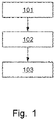
- Fig. 1 is a demonstrative flow chart of the MRI water-fat image separation method in an embodiment of the present invention.
- the method may comprise the following steps: Step 10 1: within one echo period, acquiring a first echo set corresponding to Dixon water-fat separation calculation under a first readout gradient polarity, and acquiring a second echo set corresponding to Dixon water-fat separation calculation under a second readout gradient polarity, wherein the first readout gradient polarity and the second readout gradient polarity are opposite, and echoes in the first echo set are positionally one-to-one symmetric to echoes in the second echo set with respect to the echo center of the echo period, i.e. the echoes in the second echo set are located at positions that are positionally symmetric to the echoes in the first echo set with respect to the echo center of the echo period.
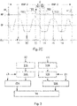
- Fig. 2A shows a schematic diagram of a data acquisition sequence of an existing two-point Dixon method based on a turbo spin echo (TSE) sequence.
- Fig. 2B shows a schematic diagram of a data acquisition sequence of a two-point Dixon method based on a turbo spin echo (TSE) sequence in one example of the present invention
- Fig. 2C is a schematic diagram of a data acquisition sequence of a three-point Dixon method based on a turbo spin echo (TSE) sequence in one example of the present invention.
- Only one spin echo period echo spacing, ESP
- One repetition time includes multiple spin echo periods as shown in Figs. 2A - 2C , i.e. multiple 180-degree rephasing RF pulses are included between two 90-degree RF pulses.
- RF, Gr and Ech correspond to the RF pulse, readout direction gradient and echo data, respectively.
- the MRI equipment applies a readout gradient in the readout gradient direction, and reads an out-of-phase echo Ea and an in-phase echo Eb.
- the MRI equipment applies two readout gradients in the readout gradient direction, reads four corresponding echoes E1, E2, E3, E4 based on the effect of the readout gradients, and puts the four echoes E1, E2, E3, E4 that are read into k-space.
- Echoes E1 and E3 are read out under the same readout gradient polarity and form an echo pair corresponding to a two-point Dixon water-fat separation algorithm; echoes E2 and E4 are read out under the same readout gradient polarity and form another echo pair corresponding to a two-point Dixon water-fat separation algorithm. These two echo pairs have the same dephasing factor, but opposite gradient polarities. Readout is repeated throughout the echo sequence. Each echo pair, E1 and E3, E2 and E4, is obtained with the same readout gradient polarity, and water-fat separation can be performed without performing phase correction.
- Echoes E2 and E3 are nearly-in-phase echoes which meet a set in-phase requirement and are positionally symmetric with respect to the echo center C of the echo period, with a complex conjugate relationship therebetween; echoes E1 and E4 are nearly-out-of-phase echoes which meet a set out-of-phase requirement and are positionally symmetric with respect to the echo center C of the echo period, with a complex conjugate relationship therebetween.
- the choice of TEs of echoes E2, E3 and echoes E1, E4 is flexible; as long as water-fat separation is stable, noise amplification is acceptable.
- echoes E2, E3 and echoes E1, E4 are nearly in phase and out of phase, respectively.
- they can set in another way, i.e. echoes E2, E3 are nearly out of phase, while echoes E1, E4 are nearly in phase.
- the first echo set and the second echo set are each an echo pair comprising one nearly-in-phase echo meeting a set in-phase requirement and one nearly-out-of-phase echo meeting a set out-of-phase requirement.
- the MRI equipment applies three readout gradients in the readout gradient direction, reads six corresponding echoes E1, E2, E3, E4, E5, E6 based on the effect of the readout gradients, and puts the six echoes E1, E2, E3, E4, E5, E6 that are read into k-space.
- Echoes E1, E3 and E5 are read out under the same readout gradient polarity and form an echo set corresponding to a three-point Dixon water-fat separation algorithm; echoes E2, E4 and E6 are read out under the same readout gradient polarity and form another echo set corresponding to a three-point Dixon water-fat separation algorithm. These two echo sets have the same dephasing factor, but opposite gradient polarities. Readout is repeated throughout the echo sequence. Each echo set, E1, E3 and E5, E2, E4 and E6, is obtained with the same readout gradient polarity, and water-fat separation can be performed without performing phase correction.
- Echoes E3 and E4 are nearly-in-phase echoes which meet a set in-phase requirement and are positionally symmetric with respect to the echo center C of the echo period, with a complex conjugate relationship therebetween;
- echoes E1 and E6 are nearly-out-of-phase echoes which meet a set out-of-phase requirement and are positionally symmetric with respect to the echo center C of the echo period, with a complex conjugate relationship therebetween;
- echoes E5 and E2 are also nearly-out-of-phase echoes which meet a set out-of-phase requirement and are positionally symmetric with respect to the echo center C of the echo period, with a complex conjugate relationship therebetween.
- the first echo set and the second echo set may each comprise: one nearly-in-phase echo meeting a set in-phase requirement and two nearly-out-of-phase echoes meeting a set out-of-phase requirement.
- the nearly-in-phase echo in the first echo set and the nearly-in-phase echo in the second echo set are positionally symmetric with respect to the echo center of the echo period
- the two nearly-out-of-phase echoes in the first echo set and the two nearly-out-of-phase echoes in the second echo set are positionally symmetric with respect to the echo center of the echo period.
- this embodiment is likewise suitable for other Dixon water-fat separation calculation methods. An item-by-item description is not repeated here.
- Step 102 obtaining a first echo image set based on first echo set data acquired in each echo period, and obtaining a second echo image set based on second echo set data acquired in each echo period.
- a first echo image set is obtained based on the first echo set data acquired in each echo period of the echo sequence in k-space
- a second echo image set is obtained based on the second echo set data acquired in each echo period of the echo sequence in k-space.
- an out-of-phase image corresponding to E1 can be obtained for data of echo E1 acquired in each echo period in k-space; an in-phase image corresponding to E3 can be obtained for data of echo E3 acquired in each echo period in k-space; and the out-of-phase image corresponding to E1 and the in-phase image corresponding to E3 together form the first echo image set.
- an in-phase image corresponding to E2 can be obtained for data of echo E2 acquired in each echo period in k-space; an out-of-phase image corresponding to E4 can be obtained for data of echo E4 acquired in each echo period in k-space; and the in-phase image corresponding to E2 and the out-of-phase image corresponding to E4 together form the second echo image set.
- Step 103 using the first echo image set and the second echo image set to perform Dixon water-fat separation calculation, to obtain a water image and a fat image.
- First implementation method image merging followed by water-fat separation calculation
- Each pair of echo images with symmetrical echo positions in the first echo image set and the second echo image set is separately subjected to phase difference correction, each pair of echo images that has undergone phase difference correction is then separately subjected to complex averaging, thereby obtaining a corresponding echo averaged image set by merging; then the images in the echo averaged image set obtained are used to perform Dixon water-fat separation calculation, to obtain a water image and a fat image.
- the process of subjecting the two echo images with symmetrical echo positions to phase difference correction may comprise: subjecting either one of the two echo images with symmetrical echo positions to complex conjugate transformation, to obtain a first processing image; subtracting, from the first processing image or the other echo image that has not undergone complex conjugate transformation, a low-pass phase difference between the first processing image and the echo image that has not undergone complex conjugate transformation, to obtain a second processing image; wherein the second processing image and the echo image which has not had the low-pass phase difference subtracted therefrom together form the two echo images resulting from phase difference correction.
- Fig. 3 shows a schematic diagram of the process of using acquired images to perform image reconstruction with water-fat separation in one example of the present invention.
- the two images with symmetrical echo positions can be subjected to phase difference correction first; when particular processing is performed, the two images with positional symmetry have equal status, and either one may be taken as the standard for performing phase difference correction
- Step 31A subjecting the in-phase image corresponding to E2 to complex conjugate transformation, to obtain a first in-phase processing image
- step 31B subjecting the out-of-phase image corresponding to E4 to complex conjugate transformation, to obtain a first out-of-phase processing image.
- step 31A may also comprise subjecting the in-phase image corresponding to E3 to complex conjugate transformation
- step 31B may also comprise subjecting the out-of-phase image corresponding to E1 to complex conjugate transformation
- Step 32A subtracting, from the first in-phase processing image, a low-pass phase difference between the first in-phase processing image and the in-phase image corresponding to E3, to obtain a second in-phase processing image;
- step 32B subtracting, from the first out-of-phase processing image, a low-pass phase difference between the first out-of-phase processing image and the out-of-phase image corresponding to E1, to obtain a second out-of-phase processing image.
- step 32A may also comprise subtracting, from the in-phase image corresponding to E3, the low-pass phase difference between the first in-phase processing image and the in-phase image corresponding to E3, and by the same principle, step 32B may also comprise subtracting, from the out-of-phase image corresponding to E1, the low-pass phase difference between the first out-of-phase processing image and the out-of-phase image corresponding to E1.
- Step 33A subjecting the second in-phase processing image and the in-phase image corresponding to E3 to complex averaging, and integrating the two images as an echo averaged in-phase image;
- step 33B subjecting the second out-of-phase processing image and the out-of-phase image corresponding to E1 to complex averaging, and integrating the two images as an echo averaged out-of-phase image.
- Step 34 using the echo averaged in-phase image and the echo averaged out-of-phase image to perform two-point Dixon water-fat separation calculation, to obtain a water image and a fat image.
- Second implementation method water-fat separation calculation followed by image merging
- the first echo image set is used to perform Dixon water-fat separation calculation, to obtain a first water image and a first fat image;
- the second echo image set is used to perform Dixon water-fat separation calculation, to obtain a second water image and a second fat image;
- the first water image and the second water image are subjected to modulus averaging, to obtain a water image;
- the first fat image and the second fat image are subjected to modulus averaging, to obtain a fat image.
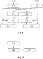
- Fig. 4 shows a schematic diagram of the process of using acquired images to perform image reconstruction with water-fat separation in another example of the present invention. As Fig. 4 shows, the process may comprise the following steps:
- step 42A may also comprise subjecting the second water image to complex conjugate transformation
- step 42B may also comprise subjecting the second fat image to complex conjugate transformation
- Step 43A subtracting, from the first processing water image, a low-pass phase difference between the first processing water image and the second water image, to obtain a second processing water image
- step 43B subtracting, from the first processing fat image, a low-pass phase difference between the first processing fat image and the second fat image, to obtain a second processing fat image.
- Step 44A subjecting the second processing water image and the second water image to complex averaging, to obtain a water image by integration
- step 44B subjecting the second processing fat image and the second fat image to complex averaging, to obtain a fat image by integration.
- the MRI water-fat image separation method in embodiments of the present invention has been described in detail above; the MRI water-fat image separation apparatus in embodiments of the present invention is described in detail below.
- the MRI water-fat image separation apparatus in embodiments of the present invention can be used to implement the MRI water-fat image separation method in embodiments of the present invention.
- Figs. 5A - 5C are demonstrative structural diagrams of the MRI water-fat image separation apparatus in embodiments of the present invention.
- the apparatus may comprise: a data acquisition module 510, an image extraction module 520 and a water-fat separation module 530.
- the data acquisition module 510 is used for: within one echo period, acquiring a first echo set corresponding to Dixon water-fat separation calculation under a first readout gradient polarity, and acquiring a second echo set corresponding to Dixon water-fat separation calculation under a second readout gradient polarity, wherein the first readout gradient polarity and the second readout gradient polarity are opposite, and echoes in the first echo set are positionally one-to-one symmetric to echoes in the second echo set with respect to the echo center of the echo period, i.e. the echoes in the second echo set are located at positions that are positionally symmetric to the echoes in the first echo set with respect to the echo center of the echo period.
- the image extraction module 520 is used for obtaining a first echo image set based on first echo set data acquired in each echo period, and obtaining a second echo image set based on second echo set data acquired in each echo period.
- the water-fat separation module 530 is used for using the first echo image set and the second echo image set to perform Dixon water-fat separation calculation, to obtain a water image and a fat image.
- the first echo set and the second echo set each comprise: one nearly-in-phase echo meeting a set in-phase requirement and one nearly-out-of-phase echo meeting a set out-of-phase requirement; and the Dixon water-fat separation calculation is two-point Dixon water-fat separation calculation; alternatively, the first echo set and the second echo set each comprise: two nearly-in-phase echoes meeting a set in-phase requirement and one nearly-out-of-phase echo meeting a set out-of-phase requirement; and the Dixon water-fat separation calculation is three-point Dixon water-fat separation calculation.
- the water-fat separation module 530 may be implemented in various ways; Figs. 5B and 5C each show one of these.
- the water-fat separation module 530 may comprise: a first image integration module 531 and a first water-fat separation calculation module 532.
- the first image integration module 531 is used for separately subjecting each pair of echo images with symmetrical echo positions in the first echo image set and the second echo image set to phase difference correction, then separately subjecting each pair of echo images that has undergone phase difference correction to complex averaging, to obtain a corresponding echo averaged image set.
- the first water-fat separation calculation module 532 is used for using the images in the echo averaged image set obtained to perform Dixon water-fat separation calculation, to obtain a water image and a fat image.
- the first image integration module 531 may comprise: a complex conjugate transformation module 5311, a phase correction module 5312 and a complex averaging module 5313.
- the complex conjugate transformation module 5311 is used for subjecting either one of each pair of echo images with symmetrical echo positions in the first echo image set and the second echo image set to complex conjugate transformation, to obtain a first processing image.
- the phase correction module 5312 is used for subtracting, from the first processing image or the other echo image that has not undergone complex conjugate transformation, a low-pass phase difference between the first processing image and the echo image that has not undergone complex conjugate transformation, to obtain a second processing image; wherein the second processing image and the echo image which has not had the low-pass phase difference subtracted therefrom together form the two echo images resulting from phase difference correction.
- the complex averaging module 5313 is used for separately subjecting each pair of echo images that has undergone phase difference correction to complex averaging, to obtain a corresponding echo averaged image set.
- the water-fat separation module 530 may comprise: a second water-fat separation calculation module 533 and a second image integration module 534.
- the second water-fat separation calculation module 533 is used for using the first echo image set to perform Dixon water-fat separation calculation, to obtain a first water image and a first fat image; and using the second echo image set to perform Dixon water-fat separation calculation, to obtain a second water image and a second fat image.
- the second image integration module 534 is used for subjecting the first water image and the second water image to modulus averaging, to obtain a water image; and subjecting the first fat image and the second fat image to modulus averaging, to obtain a fat image.
- Fig. 6 is a demonstrative structural diagram of another MRI water-fat image separation apparatus in embodiments of the present invention.
- the apparatus may comprise: at least one memory 61 and at least one processor 62.
- some other components may also be included, such as a communication port, etc. These components communicate via a bus 63.
- the at least one memory 61 is configured to store a computer program.
- the computer program may be understood to comprise all of the modules of the MRI water-fat image separation apparatus shown in Fig. 5 .
- the at least one memory 61 may also store an operating system, etc.
- Operating systems include but are not limited to: an Android operating system, a Symbian operating system, a Windows operating system and a Linux operating system, etc.
- the at least one processor 62 is configured to call the computer program stored in the at least one memory 61, to perform the MRI water-fat image separation method described in embodiments of the present invention.
- the processor 62 may be a CPU, a processing unit/module, an ASIC, a logic module or a programmable gate array, etc. It can receive and send data via the communication port.
- hardware modules in the embodiments above may be realized mechanically or electronically.
- one hardware module may comprise a specially designed permanent circuit or logic device (such as a dedicated processor, such as an FPGA or ASIC) for completing a specific operation.
- the hardware module may also comprise a programmable logic device or circuit that is temporarily configured by software (e.g. comprising a general processor or another programmable processor) for executing a specific operation.
- software e.g. comprising a general processor or another programmable processor
- the choice of whether to specifically use a mechanical method, or a dedicated permanent circuit, or a temporarily configured circuit (e.g. configured by software) to realize the hardware module can be decided according to considerations of cost and time.
- a computer readable storage medium having stored thereon a computer program that can be executed by a processor and realize the MRI water-fat image separation method described in embodiments of the present invention.
- a system or apparatus equipped with a storage medium may be provided; software program code realizing the function of any one of the embodiments above is stored on the storage medium, and a computer (or CPU or MPU) of the system or apparatus is caused to read and execute the program code stored in the storage medium.
- an operating system etc. operating on a computer to complete a portion of, or all, actual operations by means of an instruction based on program code.
- program code read out from the storage medium may be written into a memory installed in an expansion board inserted in the computer, or written into a memory installed in an expansion unit connected to the computer, and thereafter instructions based on the program code cause a CPU etc. installed on the expansion board or expansion unit to execute a portion of and all actual operations, so as to realize the function of any one of the embodiments above.
- Embodiments of storage media used for providing program code include floppy disks, hard disks, magneto-optical disks, optical disks (such as CD-ROM, CD-R, CD-RW, DVD-ROM, DVD-RAM, DVD-RW, DVD+RW), magnetic tapes, non-volatile memory cards and ROM.
- program code may be downloaded from a server computer via a communication network.
- Figs. 7A and 7B show image quality comparison figures of in vitro phantom water images obtained using a conventional two-point Dixon water-fat separation algorithm and the MRI water-fat image separation method in an embodiment of the present invention respectively, in one example, while Fig. 7C shows a schematic figure of ratio results for the two images in Figs. 7B and 7A .
- the Siemens Integrated Development Environment for Application (IDEA) is used to implement a sequence image reconstruction prototype system, and the in vitro phantom experiment is conducted on a Siemens instrument with a magnetic field strength of 0.55 T.
- the image obtained by the MRI water-fat image separation method in an embodiment of the present invention has a 30% - 40% higher SNR than the image obtained by the conventional two-point Dixon water-fat separation algorithm.
- the image quality is higher when the MRI water-fat image separation method in an embodiment of the present invention is used.
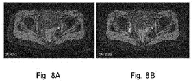
- Figs. 8A and 8B show image quality comparison figures of hip joint water images obtained using a combined-echo two-point Dixon water-fat separation algorithm with sampling performed twice and the MRI water-fat image separation method in an embodiment of the present invention, in one example.
- Figs. 8A is the hip joint water image obtained using the combined-echo two-point Dixon water-fat separation algorithm with sampling performed twice;
- Fig. 8B is the hip joint water image obtained using the MRI water-fat image separation method in an embodiment of the present invention.
- the image quality is similar in Figs. 8B and 8A , but the sampling time corresponding to Fig. 8B is shorter than the sampling time corresponding to Fig. 8A .
- the sampling efficiency is higher when the MRI water-fat image separation method in an embodiment of the present invention is used.
- the echo images corresponding to the two echo sets can undergo image merging to increase SNR followed by water-fat separation calculation, or undergo water-fat separation calculation followed by image merging to increase SNR; compared with a conventional TSE-Dixon water-fat separation calculation method, the number of echoes obtained in a very similar capture time is doubled, so the SNR is increased, and better water-fat imaging quality is achieved. Furthermore, by implementing the phase correction process before averaging in the merging process, it is possible to reduce redundancy and ensure consistency of local and global water-fat separation results.
- the technical solution in embodiments of the present invention applies a lower readout gradient strength, resulting in less image distortion and less misregistration between water and fat images.
Landscapes
- Physics & Mathematics (AREA)
- Engineering & Computer Science (AREA)
- General Physics & Mathematics (AREA)
- Health & Medical Sciences (AREA)
- Condensed Matter Physics & Semiconductors (AREA)
- High Energy & Nuclear Physics (AREA)
- General Health & Medical Sciences (AREA)
- Radiology & Medical Imaging (AREA)
- Nuclear Medicine, Radiotherapy & Molecular Imaging (AREA)
- Computer Vision & Pattern Recognition (AREA)
- Theoretical Computer Science (AREA)
- Signal Processing (AREA)
- Medical Informatics (AREA)
- Mathematical Physics (AREA)
- Multimedia (AREA)
- Artificial Intelligence (AREA)
- Epidemiology (AREA)
- Quality & Reliability (AREA)
- Primary Health Care (AREA)
- Public Health (AREA)
- Magnetic Resonance Imaging Apparatus (AREA)
Abstract
Description
- The present invention relates to the technical field of magnetic resonance imaging, in particular to a magnetic resonance imaging water-fat image separation method and apparatus, a magnetic resonance imaging system and a computer readable storage medium.
- In a magnetic resonance imaging (MRI) system, due to the fact that hydrogen atom nuclei in water and hydrogen atom nuclei in fat inside the human body are in different molecular environments, their resonant frequencies are different; when hydrogen protons in fat and other tissues are excited by an RF pulse at the same time, their relaxation times are also different. When signals are acquired at different echo times, fat tissue and water exhibit different phases and signal intensities.
- The Dixon method is a method used to produce a pure water proton image in MRI, the basic principle thereof being to separately acquire two types of echo signals, namely in-phase and out-of-phase echo signals, of water and fat protons; the two types of signals with different phases undergo arithmetic operations, each producing a pure water proton image and a pure fat proton image, thereby achieving the objective of fat suppression. At present, there are many types of Dixon water-fat image separation methods, including the single-point Dixon method, the two-point Dixon method and the three-point Dixon method, etc.
- In a Dixon-based turbo spin echo (TSE, also called fast spin echo, FSE) pulse sequence, the radio frequency (RF) pulse sequence includes a 90° excitation pulse and a 180° refocusing pulse (also called a rephasing pulse), and multiple echoes can be acquired between two adjacent refocusing pulses, i.e. within one echo interval (also called an echo period). Echoes at corresponding positions corresponding to different refocusing pulses form a set of echoes, for example: a first echo appearing after refocusing
pulse 1, a first echo appearing after refocusingpulse 2, ..., a first echo appearing after refocusing pulse n can form one set of echoes, and a second echo appearing after refocusingpulse 1, a second echo appearing after refocusingpulse 2, ..., a second echo appearing after refocusing pulse n can form another set of echoes. An image can be independently reconstructed from each set of echoes. Because different echoes carry different amplitude and phase information, a pure water image and a pure fat image can be separately reconstructed by further calculation. - In view of the fact that some of the existing water-fat image separation methods have a large number of dead zones and a low acquisition efficiency, while some have a low signal-to-noise ratio (SNR), those skilled in the art are still committed to searching for other solutions.
- In view of the above, embodiments of the present invention propose an MRI water-fat image separation method, and further propose an MRI water-fat image separation apparatus, an MRI system and a computer readable storage medium, in order to increase the acquisition efficiency and SNR.
- An MRI water-fat image separation method proposed in embodiments of the present invention comprises: within one echo period, acquiring a first echo set corresponding to Dixon water-fat separation calculation under a first readout gradient polarity, and acquiring a second echo set corresponding to Dixon water-fat separation calculation under a second readout gradient polarity, wherein the first readout gradient polarity and the second readout gradient polarity are opposite, and echoes in the second echo set are located at positions that are positionally symmetric to echoes in the first echo set with respect to the echo center of the echo period; obtaining a first echo image set based on first echo set data acquired in each echo period, and obtaining a second echo image set based on second echo set data acquired in each echo period; and using the first echo image set and the second echo image set to perform Dixon water-fat separation calculation, to obtain a water image and a fat image.
- In one embodiment, the first echo set and the second echo set each comprise: one nearly-in-phase echo meeting a set in-phase requirement and one nearly-out-of-phase echo meeting a set out-of-phase requirement; and the Dixon water-fat separation calculation is two-point Dixon water-fat separation calculation; alternatively, the first echo set and the second echo set each comprise: one nearly-in-phase echo meeting a set in-phase requirement and two nearly-out-of-phase echoes meeting a set out-of-phase requirement; and the Dixon water-fat separation calculation is three-point Dixon water-fat separation calculation.
- In one embodiment, the step of using the first echo image set and the second echo image set to perform Dixon water-fat separation calculation, to obtain a water image and a fat image, comprises: separately subjecting each pair of echo images with symmetrical echo positions in the first echo image set and the second echo image set to phase difference correction, then separately subjecting each pair of echo images resulting from phase difference correction to complex averaging, to obtain a corresponding echo averaged image set; and using the images in the echo averaged image set obtained to perform Dixon water-fat separation calculation, to obtain a water image and a fat image.
- In one embodiment, the step of separately subjecting each pair of echo images with symmetrical echo positions in the first echo image set and the second echo image set to phase difference correction, comprises: subjecting either one of each pair of echo images with symmetrical echo positions in the first echo image set and the second echo image set to complex conjugate transformation, to obtain a first processing image; subtracting, from the first processing image or the other echo image that has not undergone complex conjugate transformation, a low-pass phase difference between the first processing image and the echo image that has not undergone complex conjugate transformation, to obtain a second processing image, wherein the second processing image and the echo image which has not had the low-pass phase difference subtracted therefrom together form two echo images resulting from phase difference correction.
- In one embodiment, the step of using the first echo image set and the second echo image set to perform Dixon water-fat separation calculation, to obtain a water image and a fat image, comprises: using the first echo image set to perform Dixon water-fat separation calculation, to obtain a first water image and a first fat image; using the second echo image set to perform Dixon water-fat separation calculation, to obtain a second water image and a second fat image; subjecting the first water image and the second water image to modulus averaging, to obtain a water image; and subjecting the first fat image and the second fat image to modulus averaging, to obtain a fat image.
- An MRI water-fat image separation apparatus proposed in embodiments of the present invention comprises: a data acquisition module, for: within one echo period, acquiring a first echo set corresponding to Dixon water-fat separation calculation under a first readout gradient polarity, and acquiring a second echo set corresponding to Dixon water-fat separation calculation under a second readout gradient polarity, wherein the first readout gradient polarity and the second readout gradient polarity are opposite, and echoes in the second echo set are located at positions that are positionally symmetric to echoes in the first echo set with respect to the echo center of the echo period; an image extraction module, for obtaining a first echo image set based on first echo set data acquired in each echo period, and obtaining a second echo image set based on second echo set data acquired in each echo period; a water-fat separation module, for using the first echo image set and the second echo image set to perform Dixon water-fat separation calculation, to obtain a water image and a fat image.
- In one embodiment, the first echo set and the second echo set each comprise: one nearly-in-phase echo meeting a set in-phase requirement and one nearly-out-of-phase echo meeting a set out-of-phase requirement; and the Dixon water-fat separation calculation is two-point Dixon water-fat separation calculation; alternatively, the first echo set and the second echo set each comprise: two nearly-in-phase echoes meeting a set in-phase requirement and one nearly-out-of-phase echo meeting a set out-of-phase requirement; and the Dixon water-fat separation calculation is three-point Dixon water-fat separation calculation.
- In one embodiment, the water-fat separation module comprises: a first image integration module, for separately subjecting each pair of echo images with symmetrical echo positions in the first echo image set and the second echo image set to phase difference correction, then separately subjecting each pair of echo images resulting from phase difference correction to complex averaging, to obtain a corresponding echo averaged image set; and a first water-fat separation calculation module, for using the images in the echo averaged image set obtained to perform Dixon water-fat separation calculation, to obtain a water image and a fat image.
- In one embodiment, the first image integration module comprises: a complex conjugate transformation module, for subjecting either one of each pair of echo images with symmetrical echo positions in the first echo image set and the second echo image set to complex conjugate transformation, to obtain a first processing image; a phase correction module, for subtracting, from the first processing image or the other echo image that has not undergone complex conjugate transformation, a low-pass phase difference between the first processing image and the echo image that has not undergone complex conjugate transformation, to obtain a second processing image; wherein the second processing image and the echo image which has not had the low-pass phase difference subtracted therefrom together form two echo images resulting from phase difference correction; and a complex averaging module, for separately subjecting each pair of echo images resulting from phase difference correction to complex averaging, to obtain a corresponding echo averaged image set.
- In one embodiment, the water-fat separation module comprises: a second water-fat separation calculation module, for using the first echo image set to perform Dixon water-fat separation calculation, to obtain a first water image and a first fat image; and using the second echo image set to perform Dixon water-fat separation calculation, to obtain a second water image and a second fat image; and a second image integration module, for subjecting the first water image and the second water image to modulus averaging, to obtain a water image; and subjecting the first fat image and the second fat image to modulus averaging, to obtain a fat image.
- An MRI water-fat image separation apparatus proposed in embodiments of the present invention comprises: at least one memory and at least one processor, wherein: the at least one memory is configured to store a computer program; the at least one processor is configured to call the computer program stored in the at least one memory, and perform the MRI water-fat image separation method as described in any one of the embodiments above.
- An MRI system proposed in embodiments of the present invention comprises the MRI water-fat image separation apparatus as described in any one of the embodiments above.
- A computer readable storage medium proposed in embodiments of the present invention has a computer program stored thereon, wherein the computer program is executable by a processor and realizes the MRI water-fat image separation method as described in any one of the embodiments above.
- It can be seen from the solution above that because two echo sets corresponding to Dixon water-fat separation calculation are read in one echo period in embodiments of the present invention, nearly all of the time between two refocusing pulses is used for data acquisition, thus increasing the data acquisition efficiency and reducing dead zones. Moreover, each echo set is read out under the same readout gradient polarity, so there is no need to perform phase correction in each echo set.
- In addition, the echo images corresponding to the two echo sets can undergo image merging to increase SNR followed by water-fat separation calculation, or undergo water-fat separation calculation followed by image merging to increase SNR; compared with a conventional TSE-Dixon water-fat separation calculation method, the number of echoes obtained in a very similar capture time is doubled, so the SNR is increased, and better water-fat imaging quality is achieved. Furthermore, by implementing the phase correction process before averaging in the merging process, it is possible to reduce redundancy and ensure consistency of local and global water-fat separation results.
- Preferred embodiments of the present invention are described in detail below with reference to the accompanying figures, to give those skilled in the art a clearer understanding of the abovementioned and other features and advantages of the present invention. In the figures:
- FIG. 1
- is a demonstrative flow chart of the MRI water-fat image separation method in an embodiment of the present invention.
- FIG. 2A
- is a schematic diagram of a data acquisition sequence of an existing two-point Dixon method based on a turbo spin echo (TSE) sequence.
- FIG. 2B
- is a schematic diagram of a data acquisition sequence of a two-point Dixon method based on a turbo spin echo (TSE) sequence in one example of the present invention.
- FIG. 2C
- is a schematic diagram of a data acquisition sequence of a three-point Dixon method based on a turbo spin echo (TSE) sequence in one example of the present invention.
- FIG. 3
- is a schematic diagram of the process of using acquired images to perform image reconstruction with water-fat separation in one example of the present invention.
- FIG. 4
- is a schematic diagram of the process of using acquired images to perform image reconstruction with water-fat separation in another example of the present invention.
- FIGS. 5A - 5C
- are demonstrative structural diagrams of the MRI water-fat image separation apparatus in embodiments of the present invention.
- FIG. 6
- is a demonstrative structural diagram of another MRI water-fat image separation apparatus in embodiments of the present invention.
- FIGS. 7A and 7B
- are image quality comparison figures of in vitro phantom water images obtained using a conventional two-point Dixon water-fat separation algorithm and the MRI water-fat image separation method in an embodiment of the present invention respectively, in one example of the present invention.
- FIG. 7C
- is a schematic figure of ratio results for the two images in
Figs. 7B and 7A . - FIGS. 8A and 8B
- show image quality comparison figures of hip joint water images obtained using the MRI water-fat image separation method in an embodiment of the present invention and a combined-echo two-point Dixon water-fat separation algorithm with sampling performed twice, in one example.
-
Label Meaning 101 - 103, 31A - 34, 41A- 44B steps 510 data acquisition module 520 image extraction module 530 water- fat separation module 531 first image integration module 5311 complex conjugate transformation module 5312 phase correction module 5313 complex averaging module 532 first water-fat separation calculation module 533 second water-fat separation calculation module 534 second image integration module 61 memory 62 processor 63 bus - In embodiments of the present invention, account is taken of the fact that at the present time, in order to increase the acquisition speed and reduce motion sensitivity, a fast-switching bipolar readout gradient can be used during data acquisition to acquire multiple echoes between each pair of refocusing pulses. In addition, account is taken of the fact that a recently developed Dixon reconstruction method allows sampling of nearly-out-of-phase (approximately out of phase, e.g. 120° - 180°) and nearly-in-phase (approximately in phase, e.g. 0° -30°) echoes, rather than precisely out-of-phase and in-phase echoes, as was the case with the original Dixon method. Thus, in this embodiment, consideration is given to the following: acquiring two echo sets between each pair of refocusing pulses, each echo set comprising nearly out-of-phase and nearly-in-phase echoes for corresponding Dixon water-fat separation calculation, in order to increase the data acquisition speed. In addition, when using the acquired echo images to perform Dixon water-fat separation calculation, the images corresponding to different echo sets may be subjected to processing consisting of merging followed by water-fat separation calculation, or water-fat separation calculation followed by merging, thereby increasing the SNR of the water image and the fat image.
- In order to clarify the object, technical solution and advantages of the present invention, the present invention is explained in further detail below by way of embodiments.
-
Fig. 1 is a demonstrative flow chart of the MRI water-fat image separation method in an embodiment of the present invention. AsFig. 1 shows, the method may comprise the following steps:
Step 10 1: within one echo period, acquiring a first echo set corresponding to Dixon water-fat separation calculation under a first readout gradient polarity, and acquiring a second echo set corresponding to Dixon water-fat separation calculation under a second readout gradient polarity, wherein the first readout gradient polarity and the second readout gradient polarity are opposite, and echoes in the first echo set are positionally one-to-one symmetric to echoes in the second echo set with respect to the echo center of the echo period, i.e. the echoes in the second echo set are located at positions that are positionally symmetric to the echoes in the first echo set with respect to the echo center of the echo period. -
Fig. 2A shows a schematic diagram of a data acquisition sequence of an existing two-point Dixon method based on a turbo spin echo (TSE) sequence.Fig. 2B shows a schematic diagram of a data acquisition sequence of a two-point Dixon method based on a turbo spin echo (TSE) sequence in one example of the present invention, andFig. 2C is a schematic diagram of a data acquisition sequence of a three-point Dixon method based on a turbo spin echo (TSE) sequence in one example of the present invention. Only one spin echo period (echo spacing, ESP) is shown inFigs. 2A - 2C . One repetition time includes multiple spin echo periods as shown inFigs. 2A - 2C , i.e. multiple 180-degree rephasing RF pulses are included between two 90-degree RF pulses. RF, Gr and Ech correspond to the RF pulse, readout direction gradient and echo data, respectively. - As shown in
Fig. 2A , within one spin echo period of the echo sequence, i.e. between two 180-degree rephasing RF pulses RF_1 and RF_2, the MRI equipment applies a readout gradient in the readout gradient direction, and reads an out-of-phase echo Ea and an in-phase echo Eb. - As shown in
Fig. 2B , within one spin echo period of the echo sequence, i.e. between two 180-degree rephasing RF pulses RF_1 and RF_2, at first echo times (TE1) and second echo times (TE2) which are symmetrically distant from the echo center C, the MRI equipment applies two readout gradients in the readout gradient direction, reads four corresponding echoes E1, E2, E3, E4 based on the effect of the readout gradients, and puts the four echoes E1, E2, E3, E4 that are read into k-space. Echoes E1 and E3 are read out under the same readout gradient polarity and form an echo pair corresponding to a two-point Dixon water-fat separation algorithm; echoes E2 and E4 are read out under the same readout gradient polarity and form another echo pair corresponding to a two-point Dixon water-fat separation algorithm. These two echo pairs have the same dephasing factor, but opposite gradient polarities. Readout is repeated throughout the echo sequence. Each echo pair, E1 and E3, E2 and E4, is obtained with the same readout gradient polarity, and water-fat separation can be performed without performing phase correction. Echoes E2 and E3 are nearly-in-phase echoes which meet a set in-phase requirement and are positionally symmetric with respect to the echo center C of the echo period, with a complex conjugate relationship therebetween; echoes E1 and E4 are nearly-out-of-phase echoes which meet a set out-of-phase requirement and are positionally symmetric with respect to the echo center C of the echo period, with a complex conjugate relationship therebetween. - Of course, in other examples, the choice of TEs of echoes E2, E3 and echoes E1, E4 is flexible; as long as water-fat separation is stable, noise amplification is acceptable. For example, when using a 1.5 T system, echoes E2, E3 and echoes E1, E4 are nearly in phase and out of phase, respectively. When using a 3T system, they can set in another way, i.e. echoes E2, E3 are nearly out of phase, while echoes E1, E4 are nearly in phase.
- As can be seen, for the two-point Dixon water-fat separation calculation method shown in
Fig. 2 , the first echo set and the second echo set are each an echo pair comprising one nearly-in-phase echo meeting a set in-phase requirement and one nearly-out-of-phase echo meeting a set out-of-phase requirement. - As shown in
Fig. 2C , within one spin echo period of the echo sequence, i.e. between two 180-degree rephasing RF pulses RF_1 and RF_2, at first echo times (TE1), second echo times (TE2) and third echo times (TE3) which are symmetrically distant from the echo center C, the MRI equipment applies three readout gradients in the readout gradient direction, reads six corresponding echoes E1, E2, E3, E4, E5, E6 based on the effect of the readout gradients, and puts the six echoes E1, E2, E3, E4, E5, E6 that are read into k-space. Echoes E1, E3 and E5 are read out under the same readout gradient polarity and form an echo set corresponding to a three-point Dixon water-fat separation algorithm; echoes E2, E4 and E6 are read out under the same readout gradient polarity and form another echo set corresponding to a three-point Dixon water-fat separation algorithm. These two echo sets have the same dephasing factor, but opposite gradient polarities. Readout is repeated throughout the echo sequence. Each echo set, E1, E3 and E5, E2, E4 and E6, is obtained with the same readout gradient polarity, and water-fat separation can be performed without performing phase correction. Echoes E3 and E4 are nearly-in-phase echoes which meet a set in-phase requirement and are positionally symmetric with respect to the echo center C of the echo period, with a complex conjugate relationship therebetween; echoes E1 and E6 are nearly-out-of-phase echoes which meet a set out-of-phase requirement and are positionally symmetric with respect to the echo center C of the echo period, with a complex conjugate relationship therebetween; and echoes E5 and E2 are also nearly-out-of-phase echoes which meet a set out-of-phase requirement and are positionally symmetric with respect to the echo center C of the echo period, with a complex conjugate relationship therebetween. - As can be seen, for the three-point Dixon water-fat separation calculation method, the first echo set and the second echo set may each comprise: one nearly-in-phase echo meeting a set in-phase requirement and two nearly-out-of-phase echoes meeting a set out-of-phase requirement. Moreover, the nearly-in-phase echo in the first echo set and the nearly-in-phase echo in the second echo set are positionally symmetric with respect to the echo center of the echo period, and the two nearly-out-of-phase echoes in the first echo set and the two nearly-out-of-phase echoes in the second echo set are positionally symmetric with respect to the echo center of the echo period.
- In addition, this embodiment is likewise suitable for other Dixon water-fat separation calculation methods. An item-by-item description is not repeated here.
- Step 102: obtaining a first echo image set based on first echo set data acquired in each echo period, and obtaining a second echo image set based on second echo set data acquired in each echo period.
- In this step, a first echo image set is obtained based on the first echo set data acquired in each echo period of the echo sequence in k-space, and a second echo image set is obtained based on the second echo set data acquired in each echo period of the echo sequence in k-space.
- Taking the data acquisition sequence shown in
Fig. 2B as an example, an out-of-phase image corresponding to E1 can be obtained for data of echo E1 acquired in each echo period in k-space; an in-phase image corresponding to E3 can be obtained for data of echo E3 acquired in each echo period in k-space; and the out-of-phase image corresponding to E1 and the in-phase image corresponding to E3 together form the first echo image set. By the same principle, an in-phase image corresponding to E2 can be obtained for data of echo E2 acquired in each echo period in k-space; an out-of-phase image corresponding to E4 can be obtained for data of echo E4 acquired in each echo period in k-space; and the in-phase image corresponding to E2 and the out-of-phase image corresponding to E4 together form the second echo image set. - Step 103: using the first echo image set and the second echo image set to perform Dixon water-fat separation calculation, to obtain a water image and a fat image. In this step, when particular implementation is performed, two implementation methods are possible:
First implementation method: image merging followed by water-fat separation calculation - Each pair of echo images with symmetrical echo positions in the first echo image set and the second echo image set is separately subjected to phase difference correction, each pair of echo images that has undergone phase difference correction is then separately subjected to complex averaging, thereby obtaining a corresponding echo averaged image set by merging; then the images in the echo averaged image set obtained are used to perform Dixon water-fat separation calculation, to obtain a water image and a fat image. Specifically, the process of subjecting the two echo images with symmetrical echo positions to phase difference correction may comprise: subjecting either one of the two echo images with symmetrical echo positions to complex conjugate transformation, to obtain a first processing image; subtracting, from the first processing image or the other echo image that has not undergone complex conjugate transformation, a low-pass phase difference between the first processing image and the echo image that has not undergone complex conjugate transformation, to obtain a second processing image; wherein the second processing image and the echo image which has not had the low-pass phase difference subtracted therefrom together form the two echo images resulting from phase difference correction.
- Still taking the data acquisition sequence shown in
Fig. 2B as an example,Fig. 3 shows a schematic diagram of the process of using acquired images to perform image reconstruction with water-fat separation in one example of the present invention. As shown inFig. 3 , after obtaining the first echo image set formed of the out-of-phase image corresponding to E1 and the in-phase image corresponding to E3, and the second echo image set formed of the in-phase image corresponding to E2 and the out-of-phase image corresponding to E4, which were mentioned instep 102, due to the fact that the out-of-phase image corresponding to E1 and the out-of-phase image corresponding to E4 are two images with symmetrical echo positions, and the in-phase image corresponding to E3 and the in-phase image corresponding to E2 are two images with symmetrical echo positions, the two images with symmetrical echo positions can be subjected to phase difference correction first; when particular processing is performed, the two images with positional symmetry have equal status, and either one may be taken as the standard for performing phase difference correction calculation. Only one implementation situation is shown inFig. 3 , and this may for example comprise the following steps:
Step 31A: subjecting the in-phase image corresponding to E2 to complex conjugate transformation, to obtain a first in-phase processing image;step 31B: subjecting the out-of-phase image corresponding to E4 to complex conjugate transformation, to obtain a first out-of-phase processing image. - In other embodiments,
step 31A may also comprise subjecting the in-phase image corresponding to E3 to complex conjugate transformation, and by the same principle,step 31B may also comprise subjecting the out-of-phase image corresponding to E1 to complex conjugate transformation. -
Step 32A, subtracting, from the first in-phase processing image, a low-pass phase difference between the first in-phase processing image and the in-phase image corresponding to E3, to obtain a second in-phase processing image;step 32B, subtracting, from the first out-of-phase processing image, a low-pass phase difference between the first out-of-phase processing image and the out-of-phase image corresponding to E1, to obtain a second out-of-phase processing image. - In other embodiments,
step 32A may also comprise subtracting, from the in-phase image corresponding to E3, the low-pass phase difference between the first in-phase processing image and the in-phase image corresponding to E3, and by the same principle,step 32B may also comprise subtracting, from the out-of-phase image corresponding to E1, the low-pass phase difference between the first out-of-phase processing image and the out-of-phase image corresponding to E1. -
Step 33A: subjecting the second in-phase processing image and the in-phase image corresponding to E3 to complex averaging, and integrating the two images as an echo averaged in-phase image;step 33B, subjecting the second out-of-phase processing image and the out-of-phase image corresponding to E1 to complex averaging, and integrating the two images as an echo averaged out-of-phase image. Step 34: using the echo averaged in-phase image and the echo averaged out-of-phase image to perform two-point Dixon water-fat separation calculation, to obtain a water image and a fat image. - In addition, the three-point Dixon water-fat separation method is similar, so an item-by-item description is not repeated here.
- Second implementation method: water-fat separation calculation followed by image merging
- The first echo image set is used to perform Dixon water-fat separation calculation, to obtain a first water image and a first fat image; the second echo image set is used to perform Dixon water-fat separation calculation, to obtain a second water image and a second fat image; the first water image and the second water image are subjected to modulus averaging, to obtain a water image; and the first fat image and the second fat image are subjected to modulus averaging, to obtain a fat image.
- Still taking the data acquisition sequence shown in
Fig. 2B as an example,Fig. 4 shows a schematic diagram of the process of using acquired images to perform image reconstruction with water-fat separation in another example of the present invention. AsFig. 4 shows, the process may comprise the following steps: -
Step 41A: using the out-of-phase image corresponding to E1 and the in-phase image corresponding to E3 to perform two-point Dixon water-fat separation calculation, to obtain a first water image W1 and a first fat image F1;step 41B: using the in-phase image corresponding to E2 and the out-of-phase image corresponding to E4 to perform two-point Dixon water-fat separation calculation, to obtain a second water image W2 and a second fat image F2. -
Step 42A: subjecting the first water image W1 to complex conjugate transformation, to obtain a first processing water image W1*;step 42B: subjecting the first fat image F1 to complex conjugate transformation, to obtain a first processing fat image F1*. - In other embodiments,
step 42A may also comprise subjecting the second water image to complex conjugate transformation, and step 42B may also comprise subjecting the second fat image to complex conjugate transformation. -
Step 43A: subtracting, from the first processing water image, a low-pass phase difference between the first processing water image and the second water image, to obtain a second processing water image;step 43B: subtracting, from the first processing fat image, a low-pass phase difference between the first processing fat image and the second fat image, to obtain a second processing fat image. -
Step 44A: subjecting the second processing water image and the second water image to complex averaging, to obtain a water image by integration;step 44B: subjecting the second processing fat image and the second fat image to complex averaging, to obtain a fat image by integration. - The MRI water-fat image separation method in embodiments of the present invention has been described in detail above; the MRI water-fat image separation apparatus in embodiments of the present invention is described in detail below. The MRI water-fat image separation apparatus in embodiments of the present invention can be used to implement the MRI water-fat image separation method in embodiments of the present invention. For particulars not disclosed in detail in the apparatus embodiments of the present invention, reference can be made to the corresponding description in the method embodiments of the present invention; an item-by-item description is not repeated here.
-
Figs. 5A - 5C are demonstrative structural diagrams of the MRI water-fat image separation apparatus in embodiments of the present invention. As shown inFig. 5A , the apparatus may comprise: adata acquisition module 510, animage extraction module 520 and a water-fat separation module 530. - The
data acquisition module 510 is used for: within one echo period, acquiring a first echo set corresponding to Dixon water-fat separation calculation under a first readout gradient polarity, and acquiring a second echo set corresponding to Dixon water-fat separation calculation under a second readout gradient polarity, wherein the first readout gradient polarity and the second readout gradient polarity are opposite, and echoes in the first echo set are positionally one-to-one symmetric to echoes in the second echo set with respect to the echo center of the echo period, i.e. the echoes in the second echo set are located at positions that are positionally symmetric to the echoes in the first echo set with respect to the echo center of the echo period. - The
image extraction module 520 is used for obtaining a first echo image set based on first echo set data acquired in each echo period, and obtaining a second echo image set based on second echo set data acquired in each echo period. - The water-
fat separation module 530 is used for using the first echo image set and the second echo image set to perform Dixon water-fat separation calculation, to obtain a water image and a fat image. - The first echo set and the second echo set each comprise: one nearly-in-phase echo meeting a set in-phase requirement and one nearly-out-of-phase echo meeting a set out-of-phase requirement; and the Dixon water-fat separation calculation is two-point Dixon water-fat separation calculation; alternatively, the first echo set and the second echo set each comprise: two nearly-in-phase echoes meeting a set in-phase requirement and one nearly-out-of-phase echo meeting a set out-of-phase requirement; and the Dixon water-fat separation calculation is three-point Dixon water-fat separation calculation.
- In particular implementation, the water-
fat separation module 530 may be implemented in various ways;Figs. 5B and 5C each show one of these. - In one embodiment as shown in
Fig. 5B , the water-fat separation module 530 may comprise: a firstimage integration module 531 and a first water-fatseparation calculation module 532. The firstimage integration module 531 is used for separately subjecting each pair of echo images with symmetrical echo positions in the first echo image set and the second echo image set to phase difference correction, then separately subjecting each pair of echo images that has undergone phase difference correction to complex averaging, to obtain a corresponding echo averaged image set. The first water-fatseparation calculation module 532 is used for using the images in the echo averaged image set obtained to perform Dixon water-fat separation calculation, to obtain a water image and a fat image. - The first
image integration module 531 may comprise: a complexconjugate transformation module 5311, aphase correction module 5312 and acomplex averaging module 5313. - The complex
conjugate transformation module 5311 is used for subjecting either one of each pair of echo images with symmetrical echo positions in the first echo image set and the second echo image set to complex conjugate transformation, to obtain a first processing image. - The
phase correction module 5312 is used for subtracting, from the first processing image or the other echo image that has not undergone complex conjugate transformation, a low-pass phase difference between the first processing image and the echo image that has not undergone complex conjugate transformation, to obtain a second processing image; wherein the second processing image and the echo image which has not had the low-pass phase difference subtracted therefrom together form the two echo images resulting from phase difference correction. - The
complex averaging module 5313 is used for separately subjecting each pair of echo images that has undergone phase difference correction to complex averaging, to obtain a corresponding echo averaged image set. - In another embodiment as shown in
Fig. 5B , the water-fat separation module 530 may comprise: a second water-fatseparation calculation module 533 and a secondimage integration module 534. - The second water-fat
separation calculation module 533 is used for using the first echo image set to perform Dixon water-fat separation calculation, to obtain a first water image and a first fat image; and using the second echo image set to perform Dixon water-fat separation calculation, to obtain a second water image and a second fat image. - The second
image integration module 534 is used for subjecting the first water image and the second water image to modulus averaging, to obtain a water image; and subjecting the first fat image and the second fat image to modulus averaging, to obtain a fat image. -
Fig. 6 is a demonstrative structural diagram of another MRI water-fat image separation apparatus in embodiments of the present invention. As shown inFig. 6 , the apparatus may comprise: at least onememory 61 and at least oneprocessor 62. In addition, some other components may also be included, such as a communication port, etc. These components communicate via abus 63. - The at least one
memory 61 is configured to store a computer program. In one embodiment, the computer program may be understood to comprise all of the modules of the MRI water-fat image separation apparatus shown inFig. 5 . Furthermore, the at least onememory 61 may also store an operating system, etc. Operating systems include but are not limited to: an Android operating system, a Symbian operating system, a Windows operating system and a Linux operating system, etc. - The at least one
processor 62 is configured to call the computer program stored in the at least onememory 61, to perform the MRI water-fat image separation method described in embodiments of the present invention. Theprocessor 62 may be a CPU, a processing unit/module, an ASIC, a logic module or a programmable gate array, etc. It can receive and send data via the communication port. - It must be explained that not all of the steps and modules in the flows and structural diagrams above are necessary; certain steps or modules may be omitted according to actual requirements. The order in which steps are executed is not fixed, but may be adjusted as required. The partitioning of the modules is merely functional partitioning, employed for the purpose of facilitating description; during actual implementation, one module may be realized by multiple modules, and the functions of multiple modules may be realized by the same module; these modules may be located in the same device, or in different devices.
- It can be understood that hardware modules in the embodiments above may be realized mechanically or electronically. For example, one hardware module may comprise a specially designed permanent circuit or logic device (such as a dedicated processor, such as an FPGA or ASIC) for completing a specific operation. The hardware module may also comprise a programmable logic device or circuit that is temporarily configured by software (e.g. comprising a general processor or another programmable processor) for executing a specific operation. The choice of whether to specifically use a mechanical method, or a dedicated permanent circuit, or a temporarily configured circuit (e.g. configured by software) to realize the hardware module can be decided according to considerations of cost and time.
- In addition, in an embodiment of the present invention, a computer readable storage medium is further provided, having stored thereon a computer program that can be executed by a processor and realize the MRI water-fat image separation method described in embodiments of the present invention. Specifically, a system or apparatus equipped with a storage medium may be provided; software program code realizing the function of any one of the embodiments above is stored on the storage medium, and a computer (or CPU or MPU) of the system or apparatus is caused to read and execute the program code stored in the storage medium. Furthermore, it is also possible to cause an operating system etc. operating on a computer to complete a portion of, or all, actual operations by means of an instruction based on program code. It is also possible for program code read out from the storage medium to be written into a memory installed in an expansion board inserted in the computer, or written into a memory installed in an expansion unit connected to the computer, and thereafter instructions based on the program code cause a CPU etc. installed on the expansion board or expansion unit to execute a portion of and all actual operations, so as to realize the function of any one of the embodiments above. Embodiments of storage media used for providing program code include floppy disks, hard disks, magneto-optical disks, optical disks (such as CD-ROM, CD-R, CD-RW, DVD-ROM, DVD-RAM, DVD-RW, DVD+RW), magnetic tapes, non-volatile memory cards and ROM. Optionally, program code may be downloaded from a server computer via a communication network.
-
Figs. 7A and 7B show image quality comparison figures of in vitro phantom water images obtained using a conventional two-point Dixon water-fat separation algorithm and the MRI water-fat image separation method in an embodiment of the present invention respectively, in one example, whileFig. 7C shows a schematic figure of ratio results for the two images inFigs. 7B and 7A . In this example, the Siemens Integrated Development Environment for Application (IDEA) is used to implement a sequence image reconstruction prototype system, and the in vitro phantom experiment is conducted on a Siemens instrument with a magnetic field strength of 0.55 T. As shown inFig. 7C , the image obtained by the MRI water-fat image separation method in an embodiment of the present invention has a 30% - 40% higher SNR than the image obtained by the conventional two-point Dixon water-fat separation algorithm. As can be seen, for the same acquisition efficiency, the image quality is higher when the MRI water-fat image separation method in an embodiment of the present invention is used. -
Figs. 8A and 8B show image quality comparison figures of hip joint water images obtained using a combined-echo two-point Dixon water-fat separation algorithm with sampling performed twice and the MRI water-fat image separation method in an embodiment of the present invention, in one example.Figs. 8A is the hip joint water image obtained using the combined-echo two-point Dixon water-fat separation algorithm with sampling performed twice;Fig. 8B is the hip joint water image obtained using the MRI water-fat image separation method in an embodiment of the present invention. The protocol parameters used in both cases include: FOV (field of view) = 285 × 380 × 300 mm2, matrix size = 180 × 320, sampling bandwidth = 401 Hz/pixel, slice thickness = 6 mm, number of slices = 25, TE/TR for the two cases (echo time/repetition time) = 22/778 and 17/650, sampling time TA corresponding toFig. 8A = 2 minutes 18 seconds, sampling time TA corresponding toFig. 8B = 1 minute 55 seconds. - As can be seen, the image quality is similar in
Figs. 8B and 8A , but the sampling time corresponding toFig. 8B is shorter than the sampling time corresponding toFig. 8A . As can be seen, for the same imaging quality, the sampling efficiency is higher when the MRI water-fat image separation method in an embodiment of the present invention is used. - It can be seen from the solution above that because two echo sets corresponding to Dixon water-fat separation calculation are read in one echo period in embodiments of the present invention, nearly all of the time between two refocusing pulses is used for data acquisition, thus increasing the data acquisition efficiency and reducing dead zones. Moreover, each echo set is read out under the same readout gradient polarity, so there is no need to perform phase correction in each echo set.
- In addition, the echo images corresponding to the two echo sets can undergo image merging to increase SNR followed by water-fat separation calculation, or undergo water-fat separation calculation followed by image merging to increase SNR; compared with a conventional TSE-Dixon water-fat separation calculation method, the number of echoes obtained in a very similar capture time is doubled, so the SNR is increased, and better water-fat imaging quality is achieved. Furthermore, by implementing the phase correction process before averaging in the merging process, it is possible to reduce redundancy and ensure consistency of local and global water-fat separation results.
- Additionally, because less echo spacing time is needed to realize the same dephasing factor, there is less T2 blurring, so that a longer echo sequence can reduce the scanning time. Alternatively, with the same echo spacing time, the technical solution in embodiments of the present invention applies a lower readout gradient strength, resulting in less image distortion and less misregistration between water and fat images.
- The embodiments above are merely preferred embodiments of the present invention, which are not intended to limit it. Any amendments, equivalent substitutions or improvements etc. made within the spirit and principles of the present invention shall be included in the scope of protection thereof.
Claims (13)
- An MRI water-fat image separation method, comprising:within one echo period, acquiring a first echo set corresponding to Dixon water-fat separation calculation under a first readout gradient polarity, and acquiring a second echo set corresponding to Dixon water-fat separation calculation under a second readout gradient polarity (101), wherein the first readout gradient polarity and the second readout gradient polarity are opposite, and echoes in the second echo set are located at positions that are positionally symmetric to echoes in the first echo set with respect to the echo center of the echo period;obtaining a first echo image set based on first echo set data acquired in each echo period, and obtaining a second echo image set based on second echo set data acquired in each echo period (102),using the first echo image set and the second echo image set to perform Dixon water-fat separation calculation, to obtain a water image and a fat image (103).
- The MRI water-fat image separation method as claimed in claim 1, wherein the first echo set and the second echo set each comprise:one nearly-in-phase echo meeting a set in-phase requirement and one nearly-out-of-phase echo meeting a set out-of-phase requirement; andthe Dixon water-fat separation calculation is two-point Dixon water-fat separation calculation;alternatively, the first echo set and the second echo set each comprise:one nearly-in-phase echo meeting a set in-phase requirement and two nearly-out-of-phase echoes meeting a set out-of-phase requirement; andthe Dixon water-fat separation calculation is three-point Dixon water-fat separation calculation.
- The MRI water-fat image separation method as claimed in claim 1 or 2, wherein the step of using the first echo image set and the second echo image set to perform Dixon water-fat separation calculation, to obtain a water image and a fat image (103), comprises:separately subjecting each pair of echo images with symmetrical echo positions in the first echo image set and the second echo image set to phase difference correction, then separately subjecting each pair of echo images resulting from phase difference correction to complex averaging, to obtain a corresponding echo averaged image set;using the images in the echo averaged image set obtained to perform Dixon water-fat separation calculation, to obtain a water image and a fat image.
- The MRI water-fat image separation method as claimed in claim 3, wherein the step of separately subjecting each pair of echo images with symmetrical echo positions in the first echo image set and the second echo image set to phase difference correction, comprises:subjecting either one of each pair of echo images with symmetrical echo positions in the first echo image set and the second echo image set to complex conjugate transformation, to obtain a first processing image;subtracting, from the first processing image or the other echo image that has not undergone complex conjugate transformation, a low-pass phase difference between the first processing image and the echo image that has not undergone complex conjugate transformation, to obtain a second processing image, wherein the second processing image and the echo image which has not had the low-pass phase difference subtracted therefrom together form two echo images resulting from phase difference correction.
- The MRI water-fat image separation method as claimed in any one of claims 1 - 4, wherein the step of using the first echo image set and the second echo image set to perform Dixon water-fat separation calculation, to obtain a water image and a fat image (103), comprises:using the first echo image set to perform Dixon water-fat separation calculation, to obtain a first water image and a first fat image;using the second echo image set to perform Dixon water-fat separation calculation, to obtain a second water image and a second fat image;subjecting the first water image and the second water image to modulus averaging, to obtain a water image;subjecting the first fat image and the second fat image to modulus averaging, to obtain a fat image.
- An MRI water-fat image separation apparatus, comprising:a data acquisition module (510), for: within one echo period, acquiring a first echo set corresponding to Dixon water-fat separation calculation under a first readout gradient polarity, and acquiring a second echo set corresponding to Dixon water-fat separation calculation under a second readout gradient polarity, wherein the first readout gradient polarity and the second readout gradient polarity are opposite, and echoes in the second echo set are located at positions that are positionally symmetric to echoes in the first echo set with respect to the echo center of the echo period;an image extraction module (520), for obtaining a first echo image set based on first echo set data acquired in each echo period, and obtaining a second echo image set based on second echo set data acquired in each echo period;a water-fat separation module (530), for using the first echo image set and the second echo image set to perform Dixon water-fat separation calculation, to obtain a water image and a fat image.
- The MRI water-fat image separation apparatus as claimed in claim 6, wherein the first echo set and the second echo set each comprise: one nearly-in-phase echo meeting a set in-phase requirement and one nearly-out-of-phase echo meeting a set out-of-phase requirement; and the Dixon water-fat separation calculation is two-point Dixon water-fat separation calculation; alternatively,
the first echo set and the second echo set each comprise: two nearly-in-phase echoes meeting a set in-phase requirement and one nearly-out-of-phase echo meeting a set out-of-phase requirement; and the Dixon water-fat separation calculation is three-point Dixon water-fat separation calculation. - The MRI water-fat image separation apparatus as claimed in claim 6 or 7, wherein the water-fat separation module (530) comprises:a first image integration module (531), for separately subjecting each pair of echo images with symmetrical echo positions in the first echo image set and the second echo image set to phase difference correction, then separately subjecting each pair of echo images resulting from phase difference correction to complex averaging, to obtain a corresponding echo averaged image set;a first water-fat separation calculation module (532), for using the images in the echo averaged image set obtained to perform Dixon water-fat separation calculation, to obtain a water image and a fat image.
- The MRI water-fat image separation apparatus as claimed in claim 8, wherein the first image integration module (531) comprises:a complex conjugate transformation module (5311), for subjecting either one of each pair of echo images with symmetrical echo positions in the first echo image set and the second echo image set to complex conjugate transformation, to obtain a first processing image;a phase correction module (5312), for subtracting, from the first processing image or the other echo image that has not undergone complex conjugate transformation, a low-pass phase difference between the first processing image and the echo image that has not undergone complex conjugate transformation, to obtain a second processing image; wherein the second processing image and the echo image which has not had the low-pass phase difference subtracted therefrom together form two echo images resulting from phase difference correction; anda complex averaging module (5313), for separately subjecting each pair of echo images resulting from phase difference correction to complex averaging, to obtain a corresponding echo averaged image set.
- The MRI water-fat image separation apparatus as claimed in any one of claims 6 - 9, wherein the water-fat separation module (530) comprises:a second water-fat separation calculation module (533), for using the first echo image set to perform Dixon water-fat separation calculation, to obtain a first water image and a first fat image; and using the second echo image set to perform Dixon water-fat separation calculation, to obtain a second water image and a second fat image;a second image integration module (534), for subjecting the first water image and the second water image to modulus averaging, to obtain a water image; and subjecting the first fat image and the second fat image to modulus averaging, to obtain a fat image.
- An MRI water-fat image separation apparatus, comprising:
at least one memory (61) and at least one processor (62), wherein:the at least one memory (61) is configured to store a computer program;the at least one processor (62) is configured to call the computer program stored in the at least one memory (61), and perform the MRI water-fat image separation method as claimed in any one of claims 1 - 5. - An MRI system, comprising the MRI water-fat image separation apparatus as claimed in claims 6 - 11.
- A computer readable storage medium, with a computer program stored thereon, wherein the computer program is executable by a processor and realizes the MRI water-fat image separation method as claimed in any one of claims 1 - 5.
Applications Claiming Priority (1)
| Application Number | Priority Date | Filing Date | Title |
|---|---|---|---|
| CN202110113075.0A CN114820403B (en) | 2021-01-27 | 2021-01-27 | Magnetic resonance water-fat image separation method, device, imaging system and storage medium |
Publications (3)
| Publication Number | Publication Date |
|---|---|
| EP4043903A1 true EP4043903A1 (en) | 2022-08-17 |
| EP4043903C0 EP4043903C0 (en) | 2025-05-14 |
| EP4043903B1 EP4043903B1 (en) | 2025-05-14 |
Family
ID=80034831
Family Applications (1)
| Application Number | Title | Priority Date | Filing Date |
|---|---|---|---|
| EP22153131.2A Active EP4043903B1 (en) | 2021-01-27 | 2022-01-25 | Magnetic resonance water-fat image separation method and apparatus, imaging system and storage medium |
Country Status (3)
| Country | Link |
|---|---|
| US (1) | US11693072B2 (en) |
| EP (1) | EP4043903B1 (en) |
| CN (1) | CN114820403B (en) |
Families Citing this family (2)
| Publication number | Priority date | Publication date | Assignee | Title |
|---|---|---|---|---|
| CN117148244A (en) * | 2022-05-24 | 2023-12-01 | 上海联影医疗科技股份有限公司 | Image water and fat separation method, device, equipment and computer-readable storage medium |
| DE102023209597A1 (en) * | 2023-09-29 | 2025-04-03 | Siemens Healthineers Ag | Acquisition of magnetic resonance data of an object under investigation, which includes spins of at least two different spin species |
Citations (2)
| Publication number | Priority date | Publication date | Assignee | Title |
|---|---|---|---|---|
| WO2019157246A1 (en) * | 2018-02-08 | 2019-08-15 | Board Of Regents, The University Of Texas System | System and method for fast t2-weighted mr imaging with fat and fluid suppression |
| US10459059B2 (en) * | 2015-06-26 | 2019-10-29 | Koninklijke Philips N.V. | Phase corrected Dixon Magnetic Resonance Imaging |
Family Cites Families (6)
| Publication number | Priority date | Publication date | Assignee | Title |
|---|---|---|---|---|
| CN103257333B (en) * | 2012-02-17 | 2016-04-13 | 西门子(深圳)磁共振有限公司 | Method for separate imaging of water and fat in a kind of magnetic resonance imaging and device |
| US10274566B2 (en) | 2013-04-03 | 2019-04-30 | Koninklijke Philips N.V. | Dixon-type water/fat separation MRI using high-SNR in-phase image and lower-SNR at least partially out-of-phase image |
| CN105433944B (en) | 2014-07-31 | 2018-07-03 | 西门子公司 | For obtaining the method and device of the MR data of object |
| RU2739479C2 (en) | 2016-06-02 | 2020-12-24 | Конинклейке Филипс Н.В. | Magnetic resonance tomography with water and fat separation by dickson method |
| EP3413070A1 (en) * | 2017-06-09 | 2018-12-12 | Koninklijke Philips N.V. | Dual-echo dixon-type water/fat separation mr imaging |
| CN107153169B (en) * | 2017-07-04 | 2020-03-27 | 南京拓谱医疗科技有限公司 | Steady state precession gradient multi-echo water-fat separation imaging method |
-
2021
- 2021-01-27 CN CN202110113075.0A patent/CN114820403B/en active Active
-
2022
- 2022-01-25 EP EP22153131.2A patent/EP4043903B1/en active Active
- 2022-01-27 US US17/585,901 patent/US11693072B2/en active Active
Patent Citations (2)
| Publication number | Priority date | Publication date | Assignee | Title |
|---|---|---|---|---|
| US10459059B2 (en) * | 2015-06-26 | 2019-10-29 | Koninklijke Philips N.V. | Phase corrected Dixon Magnetic Resonance Imaging |
| WO2019157246A1 (en) * | 2018-02-08 | 2019-08-15 | Board Of Regents, The University Of Texas System | System and method for fast t2-weighted mr imaging with fat and fluid suppression |
Non-Patent Citations (3)
| Title |
|---|
| LI ZHIQIANG ET AL: "Rapid water and lipid imaging with T2 mapping using a radial IDEAL-GRASE technique", MAGNETIC RESONANCE IN MEDICINE, JOHN WILEY & SONS, INC, UNITED STATES, vol. 61, no. 6, 7 April 2009 (2009-04-07), pages 1415 - 1424, XP007913768, ISSN: 1522-2594, [retrieved on 20090407], DOI: 10.1002/MRM.21918 * |
| SHI CHENG ET AL: "Combined Echo Two-Point Dixon method for high efficiency water/fat separation", PROCEEDINGS OF THE 2021 ISMRM & SMRT ANNUAL MEETING & EXHIBITION, 15-20 MAY 2021, ISMRM, 2030 ADDISON STREET, 7TH FLOOR, BERKELEY, CA 94704 USA, no. 4172, 30 April 2021 (2021-04-30), XP040726189 * |
| ZHIQIANG LI ET AL: "Fast decomposition of water and lipid using a GRASE technique with the IDEAL algorithm", MAGNETIC RESONANCE IN MEDICINE, vol. 57, no. 6, 29 May 2007 (2007-05-29), pages 1047 - 1057, XP055097347, ISSN: 0740-3194, DOI: 10.1002/mrm.21232 * |
Also Published As
| Publication number | Publication date |
|---|---|
| CN114820403B (en) | 2025-04-29 |
| CN114820403A (en) | 2022-07-29 |
| EP4043903C0 (en) | 2025-05-14 |
| US20220236355A1 (en) | 2022-07-28 |
| US11693072B2 (en) | 2023-07-04 |
| EP4043903B1 (en) | 2025-05-14 |
Similar Documents
| Publication | Publication Date | Title |
|---|---|---|
| CN106569159B (en) | Fast pushforward motion correction for MR imaging | |
| JP4236320B2 (en) | Method for forming diffusion weighted images in an MRI system | |
| KR101084470B1 (en) | Magnetic resonance imaging device | |
| CN111133327B (en) | Dixon-type water/fat separation MR imaging | |
| US6177795B1 (en) | Spectral component imaging using phased array coils | |
| CN105829906B (en) | Phase-sensitive inversion recovery MRI with water/fat separation | |
| EP4043903B1 (en) | Magnetic resonance water-fat image separation method and apparatus, imaging system and storage medium | |
| RU2009116459A (en) | SYSTEMS AND METHODS FOR ASSESSING GAS TRANSFER IN LUNG USING MPT WITH A HYPERPOLARIZED 129Xe | |
| JPH0370965B2 (en) | ||
| US20080272781A1 (en) | Q-space sampling method and diffusion spectrum imaging method employing the same | |
| JP2001299720A (en) | Method for collecting data of correction of magnetic field drift, method for correcting magnetic field drift and mri device | |
| JPS635734A (en) | Suppression of undesired response signal | |
| WO2006134558A2 (en) | Low power decoupling for multi-nuclear spectroscopy | |
| US11226385B2 (en) | Dixon type water/fat separation MR imaging with improved fat shift correction | |
| US12392851B2 (en) | Dixon-type water/fat separation MR imaging | |
| Harvey et al. | Echo‐volumar imaging (EVI) at 0.5 T: first whole‐body volunteer studies | |
| JPH10305021A (en) | Pulse sequence generation method for nuclear spin tomography apparatus and nuclear spin tomography apparatus | |
| CN112285623B (en) | Magnetic resonance imaging water-fat image separation method and device and magnetic resonance imaging system | |
| CN115023622A (en) | Mr imaging with suppression of flow-induced leakage and/or exchange artifacts using dickson-type water/fat separation | |
| US4888552A (en) | Magnetic resonance imaging | |
| Larkman et al. | Water and fat separation using standard SENSE processing | |
| Yu et al. | Single acquisition water fat separation for SSFP cardiac CINE imaging: feasibility study | |
| Curatolo et al. | The usefulness of Dixon Method for Magnetic Resonance Imaging | |
| RU2773486C2 (en) | Magnetic resonance imaging with water/fat separation by dixon method | |
| WO2025162736A1 (en) | Dixon type water/fat separation mr imaging |
Legal Events
| Date | Code | Title | Description |
|---|---|---|---|
| PUAI | Public reference made under article 153(3) epc to a published international application that has entered the european phase |
Free format text: ORIGINAL CODE: 0009012 |
|
| STAA | Information on the status of an ep patent application or granted ep patent |
Free format text: STATUS: REQUEST FOR EXAMINATION WAS MADE |
|
| 17P | Request for examination filed |
Effective date: 20220125 |
|
| AK | Designated contracting states |
Kind code of ref document: A1 Designated state(s): AL AT BE BG CH CY CZ DE DK EE ES FI FR GB GR HR HU IE IS IT LI LT LU LV MC MK MT NL NO PL PT RO RS SE SI SK SM TR |
|
| RBV | Designated contracting states (corrected) |
Designated state(s): AL AT BE BG CH CY CZ DE DK EE ES FI FR GB GR HR HU IE IS IT LI LT LU LV MC MK MT NL NO PL PT RO RS SE SI SK SM TR |
|
| STAA | Information on the status of an ep patent application or granted ep patent |
Free format text: STATUS: EXAMINATION IS IN PROGRESS |
|
| 17Q | First examination report despatched |
Effective date: 20231208 |
|
| RAP1 | Party data changed (applicant data changed or rights of an application transferred) |
Owner name: SIEMENS HEALTHINEERS AG |
|
| GRAP | Despatch of communication of intention to grant a patent |
Free format text: ORIGINAL CODE: EPIDOSNIGR1 |
|
| STAA | Information on the status of an ep patent application or granted ep patent |
Free format text: STATUS: GRANT OF PATENT IS INTENDED |
|
| INTG | Intention to grant announced |
Effective date: 20241213 |
|
| GRAS | Grant fee paid |
Free format text: ORIGINAL CODE: EPIDOSNIGR3 |
|
| GRAA | (expected) grant |
Free format text: ORIGINAL CODE: 0009210 |
|
| STAA | Information on the status of an ep patent application or granted ep patent |
Free format text: STATUS: THE PATENT HAS BEEN GRANTED |
|
| AK | Designated contracting states |
Kind code of ref document: B1 Designated state(s): AL AT BE BG CH CY CZ DE DK EE ES FI FR GB GR HR HU IE IS IT LI LT LU LV MC MK MT NL NO PL PT RO RS SE SI SK SM TR |
|
| REG | Reference to a national code |
Ref country code: GB Ref legal event code: FG4D |
|
| REG | Reference to a national code |
Ref country code: CH Ref legal event code: EP |
|
| REG | Reference to a national code |
Ref country code: IE Ref legal event code: FG4D |
|
| U01 | Request for unitary effect filed |
Effective date: 20250514 |
|
| U07 | Unitary effect registered |
Designated state(s): AT BE BG DE DK EE FI FR IT LT LU LV MT NL PT RO SE SI Effective date: 20250520 |
|
| PG25 | Lapsed in a contracting state [announced via postgrant information from national office to epo] |
Ref country code: ES Free format text: LAPSE BECAUSE OF FAILURE TO SUBMIT A TRANSLATION OF THE DESCRIPTION OR TO PAY THE FEE WITHIN THE PRESCRIBED TIME-LIMIT Effective date: 20250514 |
|
| PG25 | Lapsed in a contracting state [announced via postgrant information from national office to epo] |
Ref country code: GR Free format text: LAPSE BECAUSE OF FAILURE TO SUBMIT A TRANSLATION OF THE DESCRIPTION OR TO PAY THE FEE WITHIN THE PRESCRIBED TIME-LIMIT Effective date: 20250815 Ref country code: NO Free format text: LAPSE BECAUSE OF FAILURE TO SUBMIT A TRANSLATION OF THE DESCRIPTION OR TO PAY THE FEE WITHIN THE PRESCRIBED TIME-LIMIT Effective date: 20250814 |
|
| PG25 | Lapsed in a contracting state [announced via postgrant information from national office to epo] |
Ref country code: PL Free format text: LAPSE BECAUSE OF FAILURE TO SUBMIT A TRANSLATION OF THE DESCRIPTION OR TO PAY THE FEE WITHIN THE PRESCRIBED TIME-LIMIT Effective date: 20250514 |
|
| PG25 | Lapsed in a contracting state [announced via postgrant information from national office to epo] |
Ref country code: HR Free format text: LAPSE BECAUSE OF FAILURE TO SUBMIT A TRANSLATION OF THE DESCRIPTION OR TO PAY THE FEE WITHIN THE PRESCRIBED TIME-LIMIT Effective date: 20250514 |
|
| PG25 | Lapsed in a contracting state [announced via postgrant information from national office to epo] |
Ref country code: RS Free format text: LAPSE BECAUSE OF FAILURE TO SUBMIT A TRANSLATION OF THE DESCRIPTION OR TO PAY THE FEE WITHIN THE PRESCRIBED TIME-LIMIT Effective date: 20250814 |
|
| PG25 | Lapsed in a contracting state [announced via postgrant information from national office to epo] |
Ref country code: IS Free format text: LAPSE BECAUSE OF FAILURE TO SUBMIT A TRANSLATION OF THE DESCRIPTION OR TO PAY THE FEE WITHIN THE PRESCRIBED TIME-LIMIT Effective date: 20250914 |
|
| PG25 | Lapsed in a contracting state [announced via postgrant information from national office to epo] |
Ref country code: SM Free format text: LAPSE BECAUSE OF FAILURE TO SUBMIT A TRANSLATION OF THE DESCRIPTION OR TO PAY THE FEE WITHIN THE PRESCRIBED TIME-LIMIT Effective date: 20250514 |
|
| PG25 | Lapsed in a contracting state [announced via postgrant information from national office to epo] |
Ref country code: CZ Free format text: LAPSE BECAUSE OF FAILURE TO SUBMIT A TRANSLATION OF THE DESCRIPTION OR TO PAY THE FEE WITHIN THE PRESCRIBED TIME-LIMIT Effective date: 20250514 |
|
| PG25 | Lapsed in a contracting state [announced via postgrant information from national office to epo] |
Ref country code: SK Free format text: LAPSE BECAUSE OF FAILURE TO SUBMIT A TRANSLATION OF THE DESCRIPTION OR TO PAY THE FEE WITHIN THE PRESCRIBED TIME-LIMIT Effective date: 20250514 |
|
| U20 | Renewal fee for the european patent with unitary effect paid |
Year of fee payment: 5 Effective date: 20260119 |
|
| PLBE | No opposition filed within time limit |
Free format text: ORIGINAL CODE: 0009261 |
|
| STAA | Information on the status of an ep patent application or granted ep patent |
Free format text: STATUS: NO OPPOSITION FILED WITHIN TIME LIMIT |
|
| REG | Reference to a national code |
Ref country code: CH Ref legal event code: L10 Free format text: ST27 STATUS EVENT CODE: U-0-0-L10-L00 (AS PROVIDED BY THE NATIONAL OFFICE) Effective date: 20260325 |
|
| PGFP | Annual fee paid to national office [announced via postgrant information from national office to epo] |
Ref country code: GB Payment date: 20260210 Year of fee payment: 5 |
|
| 26N | No opposition filed |
Effective date: 20260217 |