EP4043075B1 - Kn-93 for use in preventing and treating psoriasis - Google Patents
Kn-93 for use in preventing and treating psoriasis Download PDFInfo
- Publication number
- EP4043075B1 EP4043075B1 EP22152260.0A EP22152260A EP4043075B1 EP 4043075 B1 EP4043075 B1 EP 4043075B1 EP 22152260 A EP22152260 A EP 22152260A EP 4043075 B1 EP4043075 B1 EP 4043075B1
- Authority
- EP
- European Patent Office
- Prior art keywords
- camk4
- mice
- seq
- cells
- skin
- Prior art date
- Legal status (The legal status is an assumption and is not a legal conclusion. Google has not performed a legal analysis and makes no representation as to the accuracy of the status listed.)
- Active
Links
Images









Classifications
-
- A—HUMAN NECESSITIES
- A61—MEDICAL OR VETERINARY SCIENCE; HYGIENE
- A61K—PREPARATIONS FOR MEDICAL, DENTAL OR TOILETRY PURPOSES
- A61K31/00—Medicinal preparations containing organic active ingredients
- A61K31/16—Amides, e.g. hydroxamic acids
- A61K31/18—Sulfonamides
-
- A—HUMAN NECESSITIES
- A61—MEDICAL OR VETERINARY SCIENCE; HYGIENE
- A61P—SPECIFIC THERAPEUTIC ACTIVITY OF CHEMICAL COMPOUNDS OR MEDICINAL PREPARATIONS
- A61P17/00—Drugs for dermatological disorders
- A61P17/06—Antipsoriatics
-
- A—HUMAN NECESSITIES
- A61—MEDICAL OR VETERINARY SCIENCE; HYGIENE
- A61K—PREPARATIONS FOR MEDICAL, DENTAL OR TOILETRY PURPOSES
- A61K45/00—Medicinal preparations containing active ingredients not provided for in groups A61K31/00 - A61K41/00
-
- C—CHEMISTRY; METALLURGY
- C12—BIOCHEMISTRY; BEER; SPIRITS; WINE; VINEGAR; MICROBIOLOGY; ENZYMOLOGY; MUTATION OR GENETIC ENGINEERING
- C12Y—ENZYMES
- C12Y207/00—Transferases transferring phosphorus-containing groups (2.7)
- C12Y207/11—Protein-serine/threonine kinases (2.7.11)
- C12Y207/11017—Ca2+/Calmodulin-dependent protein kinase (2.7.11.17)
Definitions
- the invention relates to the field of biomedical technologies, in particular, to KN-93 for use in a method for preventing and treating psoriasis.
- T-lymphocytes also referred to as T cells
- macrophages a large number of immune cells
- dendritic cells a large number of immune cells, such as T cells
- keratinocytes a large number of immune cells, such as T-lymphocytes (also referred to as T cells), macrophages, dendritic cells and keratinocytes, play an important role.
- T cells T-lymphocytes
- macrophages also referred to as T cells
- dendritic cells dendritic cells
- keratinocytes a large number of immune cells, such as T cells, macrophages, dendritic cells and keratinocytes.
- cytokines When being activated, immune cells will release cytokines to stimulate the keratinocytes, resulting in abnormal proliferation of the keratinocytes and formation of dermatokeratinization and scaling.
- CaMK4 Calcium/calmodulin-dependent protein kinase IV encoded by CaMK4 gene, is expressed in a variety of cells, especially in T cells.
- CaMK4 mainly plays an important role in immune response, including activation and development of the T cells, and regulates gene expression by activating transcription factors adenosine cyclophosphate-response element binding protein (CREB), adenosine cyclophosphate-response element modulator alpha CREM ⁇ ), and retinoic acid receptor-related orphan nuclear receptor gamma t (ROR ⁇ t) in immune cells (including T cells and antigen-presenting cells).
- CREB adenosine cyclophosphate-response element binding protein
- CREM ⁇ adenosine cyclophosphate-response element modulator alpha CREM ⁇
- ROR ⁇ t retinoic acid receptor-related orphan nuclear receptor gamma t
- CaMK4 is an important signal molecule for the differentiation of naive CD4 + T cells into Th17 cells.
- CaMK4 can affect the differentiation of Th17 cells and the production of IL-17A, which not only indicates that CaMK4 is an important and necessary link in the differentiation and action of Th17 cells, but also indicates that CaMK4 may become a new therapeutic target for autoimmune inflammatory diseases. Because immune cells dominated by Th17 cells are involved in the occurrence and development of many autoimmune diseases, it is speculated that CaMK4 may play an important role in the pathogenesis of psoriasis and may become a new therapeutic target for psoriasis.
- KN-93 has also been reported to increase the levels of IL-10 in cardiotoxin damaged muscle in article "" Calcium/Calmodulin-Dependent Protein Kinase IV (CaMKIV) Mediates Acute Skeletal Muscle Inflammatory Response” SHI DANDAN ET AL, 2017 .
- CaMKIV Calcium/Calmodulin-Dependent Protein Kinase IV
- the invention is defined by the appended claims.
- the purpose of the disclosure is to provide an application of CaMK4 in preparation of a medicine for preventing and treating psoriasis, so as to solve the problems existing in the above prior art, and take CaMK4 as a medicine target to realize the effective prevention and treatment of psoriasis.
- the invention proposes KN-93 for use in a method for preventing and treating psoriasis
- said KN-93 is an inhibitor of CaMK4.
- said method restores interleukin-10 (IL-10) levels suppressed by CaMK4.
- the present disclosure discloses the following technical effects: The present disclosure used immunohistochemistry, real-time quantitative polymerase chain reaction (PCR), and Western blot to analyze the expression of CaMK4 in psoriasis patients' skin lesions and imiquimod (IMQ)-induced psoriasis skin lesions in mice, and the results show that CaMK4 is positively correlated with the development of psoriasis.
- PCR real-time quantitative polymerase chain reaction
- IMQ imiquimod
- a CaMK4-deficient (CaMK4 -/- ) mouse psoriasis model is constructed by IMQ induction, compared with wild-type (CaMK4 +/+ ) mice, which can significantly reduce skin thickness, scaling and epidermal thickness.
- mice through bone marrow-derived macrophages (BMDM) transfusion, exogenous administration of IL-10 and CaMK4 inhibitor KN-93, it was found that inhibiting the expression of CaMK4 in immune cells or keratinocytes can reduce skin thickness, scaling, epidermal thickness and expression of proinflammatory cytokines.
- the psoriasiform phenotype in mice with IMQ induced macrophage specific knockout CaMK4 (CaMK4 fl/fl Lyz2 Cre) was less than that of control (CaMK4 fl/fl ) mice. Therefore, the present disclosure proposes a new action target CaMK4 when preparing the medicine for preventing and treating psoriasis.
- the present disclosure finds that CaMK4 is significantly increased in skin lesions of psoriasis patients and imiquimod (IMQ)-induced psoriasis skin lesions in mice, suggesting that CaMK4 is positively correlated with psoriasis development, which will be further explained with specific examples below.
- IMQ imiquimod
- mice 6-8-week-old C57BU6 mice were given 5% IMQ applied to the shaved back skin of the mice at 62.5 mg per day for 5 days. The 6-8-week-old C57BU6 mice were used as controls.
- RNA of tissues or cells was extracted by Tritzol. Specifically, 1milliliter (ml) Trizol was added to tissues or cells, the tissues were homogenized with homogenizer, and the cells were fully lysed by repeated blowing; then 200 microliters ( ⁇ L) chloroform were added, vortexed shaken for 15 seconds (s), and stood at room temperature for 10min, thereby obtaining a first mixed solution.
- the first mixed solution was centrifuged at12000g (g is the gravitational acceleration) for 15min at 4 °C, and the supernatant of the first mixed solution was carefully aspirated about 500 ⁇ L in an eppendorf (EP) tube, then an equal volume of isopropanol precooled at 4 °C was added, gently inverted and mixed, and left to stand for 10min at room temperature, thereby obtaining a second mixed solution.
- the second mixed solution was centrifuged at 12000g for 15min at 4 °C and the supernatant was discarded, 1ml of 75% ethanol was added and washed twice, 20 ⁇ L of DEPC-treated water were added after dried, and the concentration and purity were determined by Nanodrop2000 for use.
- a reverse transcription Kit (TaKaRa) was applied to synthesize the complementary DNA (cDNA) of each sample, and then the cDNA was used as a template to amplify the gene fragment.
- the relative expression of target gene was calculated using 2- ⁇ Ct.
- All amplification primers used in the invention are as follows: Mouse primer sequences Gene Forward primer(5'-3') Reverse primer(5'-3') Gapdh gtgttcctacccccaatgtg (SEQ ID NO:1) ggtcctcagtgtagcccaag (SEQ ID NO:2) Camk4 gagaacctcgtcccggattac (SEQ ID NO:3) acacaatggatgtagcacccc (SEQ ID NO:4) S100a8 aaatcaccatgccctctacaag (SEQ ID NO:5) cccacttttatcaccatcgcaa (SEQ ID NO:6) S100a9 caccctgagcaagaaggaat (SEQ ID NO:7) tgtcatttatgagggcttcattt (SEQ ID NO:8) Lcn2 acatttgtccaagctccagggc (SEQ ID
- the cells were fixed and broken, and rabbit anti-human CaMK4 antibody and AF647-labeled goat anti-rabbit IgG second antibody were added sequentially. After tH&E staining was completed, the cells were rinsing once in PBS, resuspended by adding 200 ⁇ L PBS, and detected by flow cytometry.
- CaMK4 was increased significantly in psoriasis patients and induced skin lesions in mice. Compared with healthy control groups, the mRNA levels of CaMK4 in peripheral blood of patients with psoriasis were also elevated, with the highest expression intensity in CD14+ monocytes and a positive correlation with IL1B and IL12B expression. The protein levels and mRNA levels of CaMK4 were significantly increased in CD3 + T cells, F4/80 + cells, CD11c + cells and KCs after IMQ induction, as revealed by sorting of immune cell subsets in mouse skin and isolation of keratinocytes.
- mice model constructed in the example 1 skin samples were collected on the 5th day for experiment.
- the psoriasiform phenotype of CaMK4 -/- mice was less than that of CaMK4 +/+ mice, including skin thickness, scaling and epidermal thickness.
- the mRNA levels of proinflammatory cytokines, chemokines, antimicrobial peptides (AMPs) and epidermis development genes in the skin lesions of CaMK4 -/- mice were reduced compared with those of CaMK4 +/+ mice, and the mRNA levels of tissue repair-related genes and phagocytosis-related genes were increased compared with those of CaMK4 +/+ mice.
- IMQ triggers psoriasiform inflammation via TLR7, which is expressed on a variety of bone marrow-derived natural immune cells. Moreover, IMQ induces upregulation of CaMK4 in F4/80 + and CD11c + cells of mouse skin (with reference to the example 1). Therefore, the changes of specific skin myeloid cell subsets were further detected. Firstly, immune cells were isolated as follows.
- RPMI Roswell Park memorial institute
- the digested tissue was filtered through a 74 ⁇ m nylon mesh to remove tissue fragments, and additional RPMI 1640 was added to the suspension to inactivate the enzyme activity.
- Skin leukocytes were obtained by centrifugation with 30% and 70% percoll at 1260g for 20 min and aspirating the middle white cloudy layer. After rinsing with PBS, the cells (i.e., skin leukocytes) were counted.
- CD11b + DCs in the skin of IMQ induced CaMK4 -/- mice were significantly decreased, while MHC II + macrophages were significantly increased.
- IL-17A + CD45 + cells IL-17A + CD4 + cells
- IL-17A + ⁇ TCR + cells were reduced in the skin of IMQ-induced Camk4 -/- mice compared to Camk4+/+ mice. It was also found that after IMQ induction, IL-17A mainly came from ⁇ TCR + cells, while IFN- ⁇ mainly from CD4 + T cells.
- Macrophages play a regulatory role mainly by producing IL-10.
- IL-10 is a central anti-inflammatory mediator, which can reduce tissue damage caused by excessive and uncontrolled inflammatory stimulation and protect the host from excessive inflammatory response.
- a result of quantitative PCR showed that the mRNA level of IL-10 in the skin of IMQ induced CaMK4 -/- mice was higher than that of CaMK4 +/+ mice.
- a result of immunohistochemistry showed that the protein level of IL-10 in the skin of CaMK4 -/- mice was significantly higher than that of CaMK4 +/+ mice.
- Results of immunofluorescence and flow cytometry showed that IL-10 mainly came from dermal macrophages.
- Bone marrow cells were rinsed from the tibia and femur of mice and cultured in a RPMI 1640 medium containing 10% FBS, 1% of antibiotics and 10 nanograms per milliliter (ng/ml) M-CSF. The medium was changed every 2 days for 6 consecutive days, so that BMDMs can be obtained.
- BMDMs were treated for 6h with an inhibitor that is one selected from a group consisting of CaMK4 10 ⁇ M KN-93, ADCY1 inhibitor 20 ⁇ M ST034307, 2 ⁇ g/ml cAMP, MEK1/2 inhibitor 20 ⁇ M U0126 , and p38 inhibitor 20 ⁇ M SB203580; and then stimulated with 2 ⁇ g/ml IMQ for 12h to extract protein and RNA, or stimulated for 24h to determine cytokines.
- an inhibitor that is one selected from a group consisting of CaMK4 10 ⁇ M KN-93, ADCY1 inhibitor 20 ⁇ M ST034307, 2 ⁇ g/ml cAMP, MEK1/2 inhibitor 20 ⁇ M U0126 , and p38 inhibitor 20 ⁇ M SB203580; and then stimulated with 2 ⁇ g/ml IMQ for 12h to extract protein and RNA, or stimulated for 24h to determine cytokines.
- IMQ 62.5mg of IMQ were applied to the shaved back skin of wild-type (WT) mice for 5 days.
- IL-10 (1 ⁇ g per mouse) or PBS was injected into mice through tail vein on 1 h before IMQ application on day 0 and day 2. Samples were collected on day 5 for subsequent experiments.
- in vivo exogenous supplementation of IL-10 attenuated the psoriasiform phenotype in mice compared to control mice, including reduced skin thickness, scaling, erythema, epidermal thickness and proinflammatory cytokines.
- the psoriasiform phenotype was attenuated in mice with Camk4-/-BMDM adoptive transfer compared to control mice, including reduced skin thickness, scaling, erythema, epidermal thickness, and proinflammatory cytokines. It's indicated that lack of CaMK4 could restore IL-10 production in macrophages, and IL-10 could reduce IMQ-induced psoriasiform inflammation in mice.
- the construction method of a mouse psoriasis model was the same as that in the example 1.
- the psoriasiform phenotype of Camk4 fl/fl Lyz2-Cre mice was less than that of control mice, reduced skin thickness, scaling, erythema, epidermal thickness, and proinflammatory cytokines. Moreover, MHC II + macrophages were increased in the skin.
- TNF- ⁇ and IL-17A stimulated HaCaT cells to simulate the detection of psoriasis cell model.
- HaCaT cells were transfected with siCaMK4 or control for 24 hours and stimulated with TNF- ⁇ (50 ng/ml) and IL-17A (50 ng/ml) for 24h.
- TNF- ⁇ 50 ng/ml
- IL-17A 50 ng/ml
- CaMK4 inhibited KCs apoptosis and promoted KCs proliferation and proinflammatory gene expression through the AKT-NF- ⁇ B pathway.
- IMQ 62.5mg of IMQ were applied to the shaved back skin of wild-type mice for 5 days.
- KN-93 (0.24mg per mouse) or PBS was injected into mice through tail vein one hour before IMQ application on day 0 and day 2. Samples were collected on day 5 for subsequent experiments.
- the psoriasiform phenotype of KN-93 treated mice was less than that of control mice, including reduced skin thickness, scaling, epidermal thickness and proinflammatory cytokines. Moreover, the mRNA level of IL-10 in skin of KN-93 treated mice was higher than that of control mice.
Landscapes
- Health & Medical Sciences (AREA)
- Chemical & Material Sciences (AREA)
- Life Sciences & Earth Sciences (AREA)
- General Health & Medical Sciences (AREA)
- Animal Behavior & Ethology (AREA)
- Veterinary Medicine (AREA)
- Public Health (AREA)
- Medicinal Chemistry (AREA)
- Pharmacology & Pharmacy (AREA)
- Organic Chemistry (AREA)
- Engineering & Computer Science (AREA)
- Bioinformatics & Cheminformatics (AREA)
- General Chemical & Material Sciences (AREA)
- Epidemiology (AREA)
- Chemical Kinetics & Catalysis (AREA)
- Dermatology (AREA)
- Nuclear Medicine, Radiotherapy & Molecular Imaging (AREA)
- Molecular Biology (AREA)
- General Engineering & Computer Science (AREA)
- Genetics & Genomics (AREA)
- Wood Science & Technology (AREA)
- Zoology (AREA)
- Biochemistry (AREA)
- Measuring Or Testing Involving Enzymes Or Micro-Organisms (AREA)
- Medicines That Contain Protein Lipid Enzymes And Other Medicines (AREA)
- Peptides Or Proteins (AREA)
- Medicinal Preparation (AREA)
Description
- The invention relates to the field of biomedical technologies, in particular, to KN-93 for use in a method for preventing and treating psoriasis.
- Psoriasis is a chronic inflammatory immune skin disease that manifests clinically as red papules and plaques covered with thick silver-white scaling. A large number of immune cells, such as T-lymphocytes (also referred to as T cells), macrophages, dendritic cells and keratinocytes, play an important role. When being activated, immune cells will release cytokines to stimulate the keratinocytes, resulting in abnormal proliferation of the keratinocytes and formation of dermatokeratinization and scaling. Some examples of proposals of methods of treatment of psoriasis are given in documents
WO 01/08696 A2 WO 2014/175333 A1 , as well as in the article "IL-10is a key cytokine in psoriasis. Proof of principle by IL-10 therapy: a new therapeutic approach" K ASADU LLAH ET AL 1998. - Calcium/calmodulin-dependent protein kinase IV encoded by CaMK4 gene, is expressed in a variety of cells, especially in T cells. CaMK4 mainly plays an important role in immune response, including activation and development of the T cells, and regulates gene expression by activating transcription factors adenosine cyclophosphate-response element binding protein (CREB), adenosine cyclophosphate-response element modulator alpha CREMα), and retinoic acid receptor-related orphan nuclear receptor gamma t (ROR γt) in immune cells (including T cells and antigen-presenting cells).
- At present, the research of CaMK4 gene dysfunction mainly focuses on systemic lupus erythematosus and lupus nephritis, which leads to autoantibody production, immune complex formation and immune disorder, especially closely related to helper T 17 (Th17) cells. It has been reported that CaMK4 regulates differentiation of Th17 cells and expression of interleukin-17A (IL-17A) through protein kinase B (AKT)/ mammalian target of rapamycin (mTOR)/S6K/ROR γt and CREM/ROR γt pathways, indicating that CaMK4 is an important signal molecule for the differentiation of naive CD4+ T cells into Th17 cells.
- CaMK4 can affect the differentiation of Th17 cells and the production of IL-17A, which not only indicates that CaMK4 is an important and necessary link in the differentiation and action of Th17 cells, but also indicates that CaMK4 may become a new therapeutic target for autoimmune inflammatory diseases. Because immune cells dominated by Th17 cells are involved in the occurrence and development of many autoimmune diseases, it is speculated that CaMK4 may play an important role in the pathogenesis of psoriasis and may become a new therapeutic target for psoriasis.
- Other examples of the research of CaMK4 gene inhibition are given in documents
US 2014/161769 A1 suggesting a method of treating or reducing the likelihood of an inflammatory autoimmune disorder comprising administration of an inhibitor of CaMK4, which can be KN-93 KN-93 has also been proposed as a CaMK4 inhibitor in a model of systemic lupus erythematosus in the article "Suppression of autoimmunity and organ pathology in lupus-prone mice upon inhibition of calcium/calmodulin dependent protein kinase type IV "ICHINOSE KUNIHIRO ET AL, 2010". KN-93 has also been reported to increase the levels of IL-10 in cardiotoxin damaged muscle in article ""Calcium/Calmodulin-Dependent Protein Kinase IV (CaMKIV) Mediates Acute Skeletal Muscle Inflammatory Response" SHI DANDAN ET AL, 2017. - The invention is defined by the appended claims. The purpose of the disclosure is to provide an application of CaMK4 in preparation of a medicine for preventing and treating psoriasis, so as to solve the problems existing in the above prior art, and take CaMK4 as a medicine target to realize the effective prevention and treatment of psoriasis.
- In order to achieve the above purpose, the invention provides the following solutions:
The invention proposes KN-93 for use in a method for preventing and treating psoriasis Notably, said KN-93 is an inhibitor of CaMK4.. - Notably, said method restores interleukin-10 (IL-10) levels suppressed by CaMK4.
- The present disclosure discloses the following technical effects:
The present disclosure used immunohistochemistry, real-time quantitative polymerase chain reaction (PCR), and Western blot to analyze the expression of CaMK4 in psoriasis patients' skin lesions and imiquimod (IMQ)-induced psoriasis skin lesions in mice, and the results show that CaMK4 is positively correlated with the development of psoriasis. A CaMK4-deficient (CaMK4-/-) mouse psoriasis model is constructed by IMQ induction, compared with wild-type (CaMK4+/+) mice, which can significantly reduce skin thickness, scaling and epidermal thickness. Moreover, in vivo experiments in mice, through bone marrow-derived macrophages (BMDM) transfusion, exogenous administration of IL-10 and CaMK4 inhibitor KN-93, it was found that inhibiting the expression of CaMK4 in immune cells or keratinocytes can reduce skin thickness, scaling, epidermal thickness and expression of proinflammatory cytokines. In addition, the psoriasiform phenotype in mice with IMQ induced macrophage specific knockout CaMK4 (CaMK4 fl/fl Lyz2 Cre) was less than that of control (CaMK4fl/fl) mice. Therefore, the present disclosure proposes a new action target CaMK4 when preparing the medicine for preventing and treating psoriasis. -
-
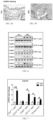
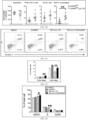
FIGS. 1a-1d show expressions of CaMK4 detected by immunohistochemistry in healthy skin and psoriasis patient's lesional skin (scale bar: 100 µm). -
FIG. 2 shows mRNA levels of CaMK4 detected by quantitative PCR in peripheral blood of healthy control group (n=24) and psoriasis patients (n=24). -
FIG. 3a shows proportion of CaMK4 indicated cells of T cell subsets (including CD4+, CD8+, DN cells) and CD14+ monocytes in peripheral blood of healthy control group (n=15) and psoriasis patients (n=12) detected by flow cytometry. -
FIG. 3b shows mean fluorescence intensity (MFI) of CaMK4 of T cell subsets (CD4+, CD8+, DN cells) and CD14+ monocytes in peripheral blood of healthy control group (n=15) and psoriasis patients (n=12) detected by flow cytometry. -
FIGS. 4a-4b show correlation between proinflammatory gene in peripheral blood and CaMK4 expression in psoriasis patients. -
FIGS. 5a-5b show expression detected by immunohistochemistry of CaMK4 in skin of a mouse model (scale bar: 50 µm). -
FIG. 6 shows protein levels of CaMK4 detected by Western blot in mouse keratinocytes (KCs) and CD3+ T cells, CD19+ B cells, F4/80+ cells and CD11c+ cells of mouse skin sorted by magnetic beads. -
FIG. 7 shows mRNA levels of CaMK4 detected by quantitative PCR in CD3+ T cells, CD19+ B cells, F4/80+ cells and CD11c+ cells of mouse skin sorted by magnetic beads; where 5-7 mouse skin tissues were used as a sample for magnetic bead sorting, data represents an independent experiment with 3 samples in each group, expressed as mean ± standard deviation, *p<0.05, **p<0.01. -
FIG. 8 is a representative picture showing mouse back skin of CaMK4-/- mice and CaMK4+/+ mice. -
FIG. 9a shows scoring curves of back skin thickness of CaMK4-/- mice and CaMK4+/+ mice. -
FIG. 9b shows scoring curves of scaling of CaMK4-/- mice and CaMK4+/+ mice. -
FIG. 9c shows scoring curves of erythema of CaMK4-/- mice and CaMK4+/+ mice. -
FIG. 10 shows hematoxylin-eosin (H&E) staining of skin sections of CaMK4-/- mice and CaMK4+/+ mice (scale bar: 200 µm). -
FIG. 11 shows a result of statistical analysis of epidermal thickness of IMQ induced CaMK4-/- mice and CaMK4+/+ mice. -
FIG. 12 shows results of RNA sequencing analysis of whole skin of IMQ induced CaMK4+/+ (n=3) mice and CaMK4-/- (n=3) mice. -
FIG. 13 shows mRNA levels detected by quantitative PCR of skin pathogenic factors induced by IMQ; where data represent twice independent experiments with 7-8 mice in each group, expressed as mean ± standard deviation, *p<0.05, **p<0.01. -
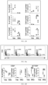
FIGS. 14a-14b show results of statistical analysis of proportion and number of neutrophils in mouse skin. -
FIGS. 15a-15b show results of statistical analysis of proportions and numbers of F4/80+ CD11c+ cells, CD11b+ DCs and MHC II+ macrophages in mouse skin. -
FIGS. 16a-16b show results of statistical analysis of proportions and numbers of CD4+ T cells and γδ TCR+ T cells in mouse skin. -
FIGS. 17a-17b show results of statistical analysis of proportions and numbers of IL-17A+ CD45+ cells, IFN-γ+ CD4+ cells, IL-17A+ CD4+ cells, IL-4+ CD4+ cells, IFN-γ+ γδ TCR+ cells, IL-17A+ γδ TCR+ cells in mouse skin; where data represent twice independent experiments with 7-8 mice in each group, expressed as mean ± standard deviation, *p<0.05, **p<0.01. -
FIG. 18 shows mRNA levels detected by quantitative PCR of IL10 in mouse skin of IMQ induced CaMK4-/- mice and CaMK4+/+ mice. -
FIG. 19 shows expression detected by immunohistochemistry of IL-10 in mouse skin of IMQ induced CaMK4-/- mice and CaMK4+/+ mice (scale bar: 200 µm). -
FIG. 20 shows a result detected by immunofluorescence of F4/80, IL-10 and DAPI in mouse skin of IMQ induced CaMK4-/- mice and CaMK4+/+ mice (scale bar: 50 µm); where a solid arrows show dermal F4/80+ IL-10+ cells. -
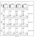
FIG. 21 shows expressions detected by flow cytometry of IL-10 in F4/80+ CD11c+ cells, CD11b+ DCs and MHC II+ macrophages of IMQ induced CaMK4-/- mice and CaMK4+/+ mice. -
FIG. 22 shows results of statistical analysis of MFI of IL-10; where data represent twice independent experiments with 7-8 mice in each group, expressed as mean ± standard deviation, *p<0.05, **p<0.01. -
FIG. 23 shows interaction between CaMK4 and AKT in BMDMs detected by co-immunocoprecipitation. -
FIG. 24 shows protein levels detected by Western blot of CaMK4, ADCY1, p-Erk1/2, Erk1/2, p-p38 and p38 in BMDMs respectively treated with IMQ, KN-93, ST034307 and cAMP. -
FIG. 25 shows mRNA levels detected by quantitative PCR of IL10 in BMDMs respectively treated with IMQ, KN-93, ST034307, cAMP, U0126 and SB203580. -
FIG. 26 shows contents detected by enzyme linked immunosorbent assay (ELISA) of IL-10 in BMDMs supernatant respectively treated with IMQ, KN-93, ST034307, cAMP, U0126 and SB203580. -
FIG. 27 shows mRNA levels of macrophage phenotypic markers (including M1 and M2) in IMQ stimulated CaMK4+/+ and CaMK4-/- BMDMs detected by quantitative PCR after 12 hours of RNA extraction with 2 µg/mL IMQ. -
FIG. 28 shows mRNA levels of phagocytosis-related genes in IMQ stimulated CaMK4+/+ and CaMK4-/- BMDMs detected by quantitative PCR after 12 hours of RNA extraction with 2 µg/mL IMQ. -
FIG. 29 shows protein levels of CaMK4, p-Erk1/2, Erk1/2, p-p38, p38 in monocytes detected by Western blot after treatment of peripheral blood CD14+ monocytes from magnetic bead-sorted psoriasis patients (n = 3) withCaMK4 inhibitor 10 µM KN-93, MEK1/2inhibitor 20 µM U0126, andp38 inhibitor 20 µM SB203580 for 12 h for protein extraction . -
FIG. 30 shows mRNA levels of IL10 in monocytes detected by quantitative PCR after treatment of peripheral blood CD14+ monocytes from magnetic bead-sorted psoriasis patients (n = 3) withCaMK4 inhibitor 10 µM KN-93, MEK1/2inhibitor 20 µM U0126, andp38 inhibitor 20 µM SB203580 for 12 h for RNA extraction. -
FIG. 31 shows content of IL-10 in monocyte supernatant detected by ELISA after treatment of peripheral blood CD14+ monocytes from magnetic bead-sorted psoriasis patients (n = 3) withCaMK4 inhibitor 10 µM KN-93, MEK1/2inhibitor 20 µM U0126, andp38 inhibitor 20 µM SB203580. -
FIG. 32 shows mRNA levels of monocyte cytokines detected by quantitative PCR after treatment of peripheral blood CD14+ monocytes from magnetic bead-sorted psoriasis patients (n = 3) withCaMK4 inhibitor 10 µM KN-93, MEK1/2inhibitor 20 µM U0126, andp38 inhibitor 20 µM SB203580 for 24 h for cytokine determination; where data represent three times independent experiments, expressed as mean ± standard deviation, *p<0.05, **p<0.01. -
FIG. 33 is a schematic diagram of therapeutic model of mice injected IL-10 or phosphate buffer saline (PBS) in vivo. -
FIG. 34 is a representative picture showing back skin of mice injected with PBS or IL-10. -
FIG. 35a shows scoring curves of back skin thickness of mice injected PBS orlL-10 in vivo. -
FIG. 35b shows scoring curves of scaling of mice injected PBS or IL-10 in vivo. -
FIG. 35c shows scoring curves of erythema of mice injected PBS or IL-10 in vivo. -
FIG. 36 shows H&E staining of skin sections of mice injected PBS or IL-10 in vivo (scale bar: 200 µm). -
FIG. 37 shows a result of statistical analysis of epidermal thickness of mice injected PBS or IL-10 in vivo. -
FIG. 38 shows mRNA levels detected by quantitative PCR of skin pathogenic factors of mice injected PBS or IL-10 in vivo induced by IMQ. -
FIG. 39 is a representative picture showing back skin of mice injected of 1×10⁶ representative pictures of mice with CaMK4+/+ BMDMs or CaMK4-/- BMDMs in vivo. -
FIG. 40a shows scoring curves of back skin thickness of mice injected of 1×10⁶ representative pictures of mice with CaMK4+/+ BMDMs or CaMK4-/- BMDMs in vivo. -
FIG. 40b shows scoring curves of scaling of mice injected of 1×10⁶ representative pictures of mice with CaMK4+/+ BMDMs or CaMK4-/- BMDMs in vivo. -
FIG. 40c shows scoring curves of erythema of mice injected of 1×10⁶ representative pictures of mice with CaMK4+/+ BMDMs or CaMK4-/- BMDMs in vivo. -
FIG. 41 shows H&E staining of skin sections of mice injected of 1×10⁶ representative pictures of mice with CaMK4+/+ BMDMs or CaMK4-/- BMDMs in vivo (scale bar: 200 µm). -
FIG. 42 shows a result of statistical analysis of epidermal thickness mice injected of 1×10⁶ representative pictures of mice with CaMK4+/+ BMDMs or CaMK4-/- BMDMs in vivo. -
FIG. 43 shows mRNA levels detected by quantitative PCR of skin pathogenic factors induced by IMQ; where data represent twice independent experiments with 5-6 mice in each group, expressed as mean ± standard deviation, *p<0.05, **p<0.01. -
FIG. 44 is a representative picture showing back skin of IMQ induced CaMK4fl/fl mice and CaMK4 fl/fl Lyz2-Cre mice. -
FIG. 45a show scoring curves of back skin thickness of IMQ induced CaMK4 fl/fl mice and CaMK4 fl/fl Lyz2 Cre mice. -
FIG. 45b show scoring curves of scaling of IMQ induced CaMK4 fl/fl mice and CaMK4 fl/fl Lyz2 Cre mice. -
FIG. 45c show scoring curves of erythema of IMQ induced CaMK4fl/fl mice and CaMK4fl/fl Lyz2 Cre mice. -
FIG. 46 shows H&E staining of skin sections of IMQ induced CaMK4fl/fl mice and CaMK4fl/fl Lyz2 Cre mice (scale bar: 200 µm). -
FIG. 47 shows a result of statistical analysis of epidermal thickness of IMQ induced CaMK4fl/fl mice and CaMK4fl/fl Lyz2 Cre mice. -
FIG. 48 shows mRNA levels detected by quantitative PCR of skin pathogenic factors of IMQ induced CaMK4fl/fl mice and CaMK4fl/fl Lyz2 Cre mice. -
FIG. 49 shows a result of statistical analysis of proportion of neutrophils, F4/80+ CD11c+ cells, CD11b+ DCS and MHC II+ macrophages in mouse skin of IMQ induced CaMK4fl/fl mice and CaMK4fl/fl Lyz2 Cre mice; where data represent twice independent experiments with 6 mice in each group, expressed as mean ± standard deviation, *p<0.05, **p<0.01. -
FIG. 50 shows a result of statistical analysis of number of neutrophils, F4/80+ CD11c+ cells, CD11b+ DCS and MHC II+ macrophages in mouse skin of IMQ induced CaMK4fl/fl mice and CaMK4fl/fl Lyz2 Cre mice; where data represent twice independent experiments with 6 mice in each group, expressed as mean ± standard deviation, *p<0.05, **p<0.01. -
FIG. 51 shows proportion of HaCaT cells with early and late apoptosis detected by flow cytometry after application of TNF-α and IL-17A stimulation to HaCaT cells. -
FIG. 52 shows proportion of HaCaT cells with early and late apoptosis detected by flow cytometry after application of TNF-α and IL-17A stimulation to HaCaT cells. -
FIG. 53 shows proportion of HaCaT cells in G0/G1 phase, S phase and M phase of cell cycle detected by flow cytometry after application of TNF-α and IL-17A stimulation to HaCaT cells. -
FIG. 54 shows expression of proinflammatory genes detected by quantitative PCR after application of TNF-α and IL-17A stimulation to HaCaT cells. -
FIG. 55 shows interaction between CaMK4 and AKT detected by co-immunocoprecipitation after application of TNF-α and IL-17A stimulation to HaCaT cells. -
FIG. 56 shows protein levels of CaMK4, p-AKT, AKT, p-IKK α/β, IKK α, p-NF-κ B p65 and NF- κ of B p65 detected by Western blot after application of TNF-α and IL-17A stimulation to HaCaT cells; where data represent three times independent experiments, expressed as mean ± standard deviation, *p<0.05, **p<0.01. -
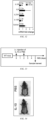
FIG. 57 is a schematic diagram of therapeutic model of mice injected CaMK4 inhibitor KN-93 or PBS in vivo. -
FIG. 58 shows a representative picture showing back skin mice injected CaMK4 inhibitor KN-93 or PBS in vivo. -
FIG. 59a shows scoring curves of back skin thickness mice injected CaMK4 inhibitor KN-93 or PBS in vivo. -
FIG. 59b shows scoring curves of scaling of mice injected CaMK4 inhibitor KN-93 or PBS in vivo. -
FIG. 59c shows scoring curves of erythema of mice injected CaMK4 inhibitor KN-93 or PBS in vivo. -
FIG. 60 shows H&E staining of skin sections of mice injected CaMK4 inhibitor KN-93 or PBS in vivo (scale bar: 200 µm). -
FIG. 61 shows a result of statistical analysis of epidermal thickness of mice injected CaMK4 inhibitor KN-93 or PBS in vivo. -
FIG. 62 shows mRNA levels detected by quantitative PCR of skin pathogenic factors induced by IMQ of mice injected CaMK4 inhibitor KN-93 or PBS in vivo; where data represent twice independent experiments with 5-7 mice in each group, expressed as mean ± standard deviation, *p<0.05, **p<0.01. - Various exemplary examples of the invention are described in detail. The detailed description should not be considered as a limitation of the invention, but should be understood as a more detailed description of some aspects, characteristics and examples of the invention.
- It should be understood that the terms described in the present disclosure are only for describing special examples and are not used to limit the invention. In addition, for the numerical range in the present disclosure it should be understood that each intermediate value between the upper and lower limits of the range is also specifically disclosed. Each smaller range between any stated value or intermediate value within the stated range and any other stated value or intermediate value within the range are also included in the present disclosure
- The upper and lower limits of these smaller ranges can be independently included or excluded from the range.
- Unless otherwise stated, all technical and scientific terms used herein have the same meanings generally understood by those skilled in the related art of the present disclosure Although the present disclosure only describes preferred methods and materials, any method and material similar or equivalent to that described herein may also be used in the implementation or testing of the present disclosure
- The terms "contain", "include", "have", "with" etc. used in this document are open-ended terms, i.e., meaning including but not limited to.
- The reagents or materials used in the following examples are commercially available if not otherwise specified.
- The present disclosure finds that CaMK4 is significantly increased in skin lesions of psoriasis patients and imiquimod (IMQ)-induced psoriasis skin lesions in mice, suggesting that CaMK4 is positively correlated with psoriasis development, which will be further explained with specific examples below.
- Patients with typical clinical manifestations of psoriasis (33 cases) were selected from the outpatients of the Department of Dermatology of the First Affiliated Hospital ofAnhui Medical University, China, of which 9 cases were taken from lesion sites and normal skin samples at the edges of the lesions in saline by conventional methods, and the samples were processed on the same day to prepare pathological sections; 24 cases were taken from blood by conventional methods, mixed 1:3 with Trizol and then lyophilized at -80°C for RNA extraction..
- An internationally used mouse model of psoriasis was used: 6-8-week-old C57BU6 mice were given 5% IMQ applied to the shaved back skin of the mice at 62.5 mg per day for 5 days. The 6-8-week-old C57BU6 mice were used as controls.
- The total RNA of tissues or cells was extracted by Tritzol. Specifically, 1milliliter (ml) Trizol was added to tissues or cells, the tissues were homogenized with homogenizer, and the cells were fully lysed by repeated blowing; then 200 microliters (µL) chloroform were added, vortexed shaken for 15 seconds (s), and stood at room temperature for 10min, thereby obtaining a first mixed solution. The first mixed solution was centrifuged at12000g (g is the gravitational acceleration) for 15min at 4 °C, and the supernatant of the first mixed solution was carefully aspirated about 500 µL in an eppendorf (EP) tube, then an equal volume of isopropanol precooled at 4 °C was added, gently inverted and mixed, and left to stand for 10min at room temperature, thereby obtaining a second mixed solution. The second mixed solution was centrifuged at 12000g for 15min at 4 °C and the supernatant was discarded, 1ml of 75% ethanol was added and washed twice, 20µL of DEPC-treated water were added after dried, and the concentration and purity were determined by Nanodrop2000 for use.
- A reverse transcription Kit (TaKaRa) was applied to synthesize the complementary DNA (cDNA) of each sample, and then the cDNA was used as a template to amplify the gene fragment. The relative expression of target gene was calculated using 2- ΔΔ Ct.
- All amplification primers used in the invention are as follows:
Mouse primer sequences Gene Forward primer(5'-3') Reverse primer(5'-3') Gapdh gtgttcctacccccaatgtg (SEQ ID NO:1) ggtcctcagtgtagcccaag (SEQ ID NO:2) Camk4 gagaacctcgtcccggattac (SEQ ID NO:3) acacaatggatgtagcacccc (SEQ ID NO:4) S100a8 aaatcaccatgccctctacaag (SEQ ID NO:5) cccacttttatcaccatcgcaa (SEQ ID NO:6) S100a9 caccctgagcaagaaggaat (SEQ ID NO:7) tgtcatttatgagggcttcattt (SEQ ID NO:8) Lcn2 acatttgttccaagctccagggc (SEQ ID NO:9) catggcgaactggttgtagtccg (SEQ ID NO:10) Camp gctgtggcggtcactatcac (SEQ ID NO:11) tgtctagggactgctggttga (SEQ ID NO:12) Tnfa actggcagaagaggcactc (SEQ ID NO:13) ctggcaccactagttggttg (SEQ ID NO:14) Tgfb1 acaattcctggcgttacctt (SEQ ID NO:15) agccctgtattccgtctcc (SEQ ID NO:16) Ifng atgaacgctacacactgcatc (SEQ ID NO:17) ccatccttttgccagttcctc (SEQ ID NO:18) II1b ctgaactcaactgtgaaatgc (SEQ ID NO:19) tgatgtgctgctgcgaga (SEQ ID NO:20) II10 gctcttactgactggcatgag (SEQ ID NO:21) cgcagctctaggagcatgtg (SEQ ID NO:22) II17a tttaactcccttggcgcaaaa (SEQ ID NO:23) ctttccctccgcattgacac (SEQ ID NO:24) II17f aaccagggcatttctgtcccac (SEQ ID NO:24) ggcattgatgcagcctgagtgt (SEQ ID NO:26) II22 atgagtttttcccttatggggac (SEQ ID NO:27) gctggaagttggacacctcaa (SEQ ID NO:28) II23a atgctggattgcagagcagta (SEQ ID NO:29) acggggcacattatttttagtct (SEQ ID NO:30) Ccl2 ccagcaagatgatcccaatg (SEQ ID NO:31) tacgggtcaacttcacattc (SEQ ID NO:32) Ccl20 aatctgtgtgcgctgatcca (SEQ ID NO:33) ccttgggctgtgtccaattc (SEQ ID NO:34) Cd36 atgggctgtgatcggaactg (SEQ ID NO:35) tttgccacgtcatctgggttt (SEQ ID NO:36) Macro acagagccgattttgaccaag (SEQ ID NO:37) cagcagtgcagtacctgcc (SEQ ID NO:38) Trem2 ctggaaccgtcaccatcactc (SEQ ID NO:39) cgaaactcgatgactcctcgg (SEQ ID NO:40) Axl atggccgacattgccagtg (SEQ ID NO:41) cggtagtaatccccgttgtaga (SEQ ID NO:42) Mertk cagggcctttaccagggaga (SEQ ID NO:43) tgtgtgctggatgtgatcttc (SEQ ID NO:44) Gpnmb cattcccatctcgaaggtgaaa (SEQ ID NO:45) aaatggcagagtcgttgagga (SEQ ID NO:46) Cd81 gtggagggctgcaccaaat (SEQ ID NO:47) gacgcaaccacagagctaca (SEQ ID NO:48) Cd5l gatcgtgtttttcagagtctcca (SEQ ID NO:49) tgcagtcaaccccttgaataag (SEQ ID NO:50) Fcrls acaggatctaagtggctgaatgt (SEQ ID NO:51) ctgggtcgttgccctatctg (SEQ ID NO:52) Cd80 acccccaacataactgagtct (SEQ ID NO:53) ttccaaccaagagaagcgagg (SEQ ID NO:54) Cd86 tgtttccgtggagacgcaag (SEQ ID NO:55) ttgagcctttgtaaatgggca (SEQ ID NO:56) Nos2 gttctcagcccaacaatacaag a (SEQ ID NO:57) gtggacgggtcgatgtcac (SEQ ID NO:58) Arg1 aacacggcagtggctttaacc (SEQ ID NO:59) ggttttcatgtggcgcattc (SEQ ID NO:60) Retnla ccaatccagctaactatccctcc (SEQ ID NO:61) ccagtcaacgagtaagcacag (SEQ ID NO:62) Chil3 tctgaaagacaagaacactga gc (SEQ ID NO:63) gcaggtccaaacttccatcc (SEQ ID NO:64) -
Gene Forward primer(5'-3') Reverse primer(5'-3') GAPDH gtctcctctgacttcaacagcg (SEQ ID NO:65) accaccctgttgctgtagccaa (SEQ ID NO:66) CAM K4 gttcttcttcgcctctcacatcc (SEQ ID NO:67) ctgtgacgagttctaggaccag (SEQ ID NO:68) S100A8 atgccgtctacagggatgacct (SEQ ID NO:69) agaatgaggaactcctggaagtta(SEQ ID NO:70) S100A9 gcacccagacaccctgaacca (SEQ ID NO:71) tgtgtccaggtcctccatgatg (SEQ ID NO:72) CAMP gacacagcagtcaccagaggat (SEQ ID NO:73) tcacaactgatgtcaaaggagcc (SEQ ID NO:74) DEFB4A ataggcgatcctgttacctgcc (SEQ ID NO:75) catcagccacagcagcttcttg (SEQ ID NO:76) SPRR2A gttccacagctccaccttca (SEQ ID NO:77) cacagcccaggacttccttt (SEQ ID NO:78) SPRR2B actggttaatcctgagactccagc (SEQ ID NO:79) aggaggatatttctgctggcac (SEQ ID NO:80) IL1B ccacagaccttccaggagaatg (SEQ ID NO:81) gtgcagttcagtgatcgtacagg (SEQ ID NO:82) IL6 aaattcggtacatcctcgacggc a(SEQ ID NO:83) agtgcctctttgctgctttcacac (SEQ ID NO:84) IL10 gactttaagggttacctgggttg (SEQ ID NO:85) tcacatgcgccttgatgtctg (SEQ ID NO:86) IL12B gacattctgcgttcaggtccag catttttgcggcagatgaccgtg (SEQ ID NO:88) (SEQ ID NO:87) IL17A agattactacaaccgatccacct (SEQ ID NO:89) ggggacagagttcatgtggta (SEQ ID NO:90) IL23A gagccttctctgctccctgata (SEQ ID NO:91) gactgaggcttggaatctgctg (SEQ ID NO:92) TNFA ctcttctgcctgctgcactttg (SEQ ID NO:93) atgggctacaggcttgtcactc (SEQ ID NO:94) TGFB1 caattcctggcgatacctcag (SEQ ID NO:95) gcacaactccggtgacatcaa (SEQ ID NO:96) CCL2 agaatcaccagcagcaagtgtc c (SEQ ID NO:97) tcctgaacccacttctgcttgg (SEQ ID NO:98) CCL20 aagttgtctgtgtgcgcaaatcc (SEQ ID NO:99) ccattccagaaaagccacagtttt(SEQ ID NO:100) -
Reagent Addition SYBR Premix Ex Taq II (2×) 10µL Forward Primer (10µM) 0.8µL Reverse Primer (10µM) 0.8µL cDNA 1µL dH2O 7.4µL Amplification program First step Second step Melting curve 95°C, 10s 95°C, 15s 95°C, 15s 60°C, 40s 60°C, 1min 40 cycles 95°C, 15s - 1ml of anticoagulation was taken into 15ml centrifuge tube, 5ml of red blood cell lysate was added, mixed and lysed on ice for 5min (appropriate mixing once in between), thereby obtaining lysed solution. The reaction was terminated by filling PBS, the lysed solution was centrifuged at 500g for 10min at 4 °C, cell precipitation was collected, rinsing once in PBS, and cells were collected. After cell counting, 1×10⁶ cells were taken, and after blocking Fc receptor, antibodies V500-CD45, PerCP/Cy5.5-CD3, FITC-CD4, PE/Cy7-CD8, and PE-CD14 were added for surface markers staining. Then, the cells were fixed and broken, and rabbit anti-human CaMK4 antibody and AF647-labeled goat anti-rabbit IgG second antibody were added sequentially. After tH&E staining was completed, the cells were rinsing once in PBS, resuspended by adding 200µL PBS, and detected by flow cytometry.
- After paraffin sections were dewaxed, antigen retrieval was performed with 0.01 molarities (M) (pH=6.0) sodium citrate. Endogenous peroxidase was blocked with 3% H2O2 and nonspecific proteins were blocked by confining liquid. First antibody (i.e., CaMK4 antibody) was incubated overnight at 4 °C and the second antibody was incubated at 37 °C for 30min, color development was performed with diaminobenzidine (DAB), nuclei were stained with hematoxylin, dried, and sealed.
- Referring to
FIGS .1-7 , CaMK4 was increased significantly in psoriasis patients and induced skin lesions in mice. Compared with healthy control groups, the mRNA levels of CaMK4 in peripheral blood of patients with psoriasis were also elevated, with the highest expression intensity in CD14+ monocytes and a positive correlation with IL1B and IL12B expression. The protein levels and mRNA levels of CaMK4 were significantly increased in CD3+ T cells, F4/80+ cells, CD11c+ cells and KCs after IMQ induction, as revealed by sorting of immune cell subsets in mouse skin and isolation of keratinocytes. - Using the mouse model constructed in the example 1, skin samples were collected on the 5th day for experiment.
- 1) The back skin thickness, scaling and erythema development of mice were observed on days 0 - 5 after applying IMQ to the shaved back skin of mice. According to the clinical psoriasis area and severity index, the severity of back skin was evaluated by thickness, scaling and erythema. The back skin thickness was measured with vernier calipers. Scaling and erythema were scored on a scale ranging from 0-4: 0 represents none; 1 represents mild; 2 represents moderate; 3 represents significant; and 4 represents very significant.
- 2) Skin sections were prepared for staining with H&E, and the skin sections were stained by using the conventional universal H&E staining method.
- 3) Statistical analysis of epidermal thickness. Specifically, H&E-stained sections were photographed in a double-blind manner and then measured for epidermal thickness.
- 4) RNA sequencing analysis of whole skin of CaMK4+/+ (n=3) and CaMK4-/- (n=3) mice induced by IMQ. Specifically, the back skin of mice was isolated, frozen at -80 °C in the size of soybean particles and sent to Lc-bio technologies (Hangzhou) Co., Ltd, China, for RNA sequencing.
- 5) The mRNA levels of IMQ induced mouse skin pathogenic factors were detected by quantitative PCR. The method was the same as that in the example 1.
- Referring to
FIGS. 8-13 , the psoriasiform phenotype of CaMK4-/- mice was less than that of CaMK4+/+ mice, including skin thickness, scaling and epidermal thickness. Moreover, the mRNA levels of proinflammatory cytokines, chemokines, antimicrobial peptides (AMPs) and epidermis development genes in the skin lesions of CaMK4-/- mice were reduced compared with those of CaMK4+/+ mice, and the mRNA levels of tissue repair-related genes and phagocytosis-related genes were increased compared with those of CaMK4+/+ mice. - IMQ triggers psoriasiform inflammation via TLR7, which is expressed on a variety of bone marrow-derived natural immune cells. Moreover, IMQ induces upregulation of CaMK4 in F4/80+ and CD11c+ cells of mouse skin (with reference to the example 1). Therefore, the changes of specific skin myeloid cell subsets were further detected. Firstly, immune cells were isolated as follows.
- On the 5th day after continuous application of IMQ, the back skin of mice was separated, then cut into small pieces and placed in a Roswell Park memorial institute (RPMI) 1640 medium containing 1 milligram per milliliter (mg/mL) of collagenase IV, 50 micrograms per milliliter (µg/mL) of deoxyribonuclease (DNase) I, 10 millimoles per liter (mM) of 2-[4-(2-hydroxyethyl)piperazin-1-yl]ethanesulfonic acid (HEPES) and 10% of FBS, and digested at 37°C for 90 min. The digested tissue was filtered through a 74 µm nylon mesh to remove tissue fragments, and additional RPMI 1640 was added to the suspension to inactivate the enzyme activity. Skin leukocytes were obtained by centrifugation with 30% and 70% percoll at 1260g for 20 min and aspirating the middle white cloudy layer. After rinsing with PBS, the cells (i.e., skin leukocytes) were counted. After cell counting, 1×10⁶ cells were taken, and after blocking the Fc receptor, antibodies PerCP/Cy5.5-CD45, V450-CD11b, FITC-Ly6C, PE-CD11c, PE/Cy7-F4/80, APC-Ly6G, APC/Cy7-MHC II were added for surface marker staining. After the staining was completed, the cells were rinsed once in PBS, 200 µL PBS was added to resuspend the cells and detected by flow cytometry.
- Referring to
FIGS. 14-17 , compared with CaMK4+/+ mice, CD11b+ DCs in the skin of IMQ induced CaMK4-/- mice were significantly decreased, while MHC II+ macrophages were significantly increased. - Detection of CD4+ T cell subsets and γδ T cell subsets showed that IL-17A+ CD45+ cells, IL-17A+ CD4+ cells and IL-17A+ γδTCR+ cells were reduced in the skin of IMQ-induced Camk4-/- mice compared to Camk4+/+ mice. It was also found that after IMQ induction, IL-17A mainly came from γδ TCR+ cells, while IFN- γ mainly from CD4+ T cells.
- Macrophages play a regulatory role mainly by producing IL-10. IL-10 is a central anti-inflammatory mediator, which can reduce tissue damage caused by excessive and uncontrolled inflammatory stimulation and protect the host from excessive inflammatory response. To determine whether macrophages produce IL-10 and participate in alleviating psoriasis symptoms of CaMK4 deficiency, the expressions of IL-10 in skin and macrophages were first detected.
- 1) The mRNA levels of IL-10 in mouse skin were detected by quantitative PCR, and the method was the same as that in the example 1.
- 2) The expression of IL-10 in mouse skin was detected by immunohistochemistry, and the method was the same as that in the example 1.
- 3) F4/80, IL-10 and DAPI in mouse skin were detected by immunofluorescence. After paraffin sections were dewaxed, antigen retrieval was performed with 0.01 molarities (M) (pH=6.0) sodium citrate. Endogenous peroxidase was blocked with 3% H2O2 and nonspecific proteins were blocked by confining liquid. First antibody (i.e., F4/80 and IL-10 antibody) was incubated overnight at 4 °C and the second antibody was incubated at 37 °C for 30min, nuclei were stained with DAPI, and sealed with anti-fluorescence quenching sealing solution.
- 4) The expressions of IL-10 in F4/80+ CD11c+ cells, CD11b+ DCs and MHC II+ macrophages were detected by flow cytometry. The method was the same as that in the example 3.
- Referring to
FIG. 18-22 , a result of quantitative PCR showed that the mRNA level of IL-10 in the skin of IMQ induced CaMK4-/- mice was higher than that of CaMK4+/+ mice. A result of immunohistochemistry showed that the protein level of IL-10 in the skin of CaMK4-/- mice was significantly higher than that of CaMK4+/+ mice. Results of immunofluorescence and flow cytometry showed that IL-10 mainly came from dermal macrophages. - Determination of cytokines in macrophages.
- Bone marrow cells were rinsed from the tibia and femur of mice and cultured in a RPMI 1640 medium containing 10% FBS, 1% of antibiotics and 10 nanograms per milliliter (ng/ml) M-CSF. The medium was changed every 2 days for 6 consecutive days, so that BMDMs can be obtained. BMDMs were treated for 6h with an inhibitor that is one selected from a group consisting of
CaMK4 10 µM KN-93,ADCY1 inhibitor 20 µM ST034307, 2 µg/ml cAMP, MEK1/2inhibitor 20 µM U0126 , andp38 inhibitor 20 µM SB203580; and then stimulated with 2 µg/ml IMQ for 12h to extract protein and RNA, or stimulated for 24h to determine cytokines. - 1) The protein levels of CaMK4, ADCY1, p-Erk1/2, Erk1/2, p-p38, p38 in BMDMs treated with different inhibitors were detected by Western blot. Protein samples were obtained by lysing the cells (i.e., bone marrow cells)with radio immunoprecipitation assay (RIPA) lysis buffer containing a mixture of phenylmethylsulfonyl fluoride (PMSF), protease inhibitor and phosphatase inhibitor. After quantification of proteins, 10% sodium dodecyl sulfate-polyacrylamide (SDS-PAGE) gel electrophoresis was performed. The proteins in the gel were transferred to the nitrocellulose membranes; fist antibody and second antibody were incubated, and color development was performed with an electrochemiluminescence (ECL) solution.
- 2) The mRNA levels of IL-10 in BMDMs treated with different inhibitors were detected by quantitative PCR. The method was the same as that in the example 1.
- 3) The contents of IL-10 in BMDMs supernatant treated with different inhibitors were detected by ELISA according to the standard method in the manufacturer's instructions.
- 4) The mRNA levels of macrophage phenotypic markers (including M1 and M2) and phagocytosis-related genes in IMQ stimulated CaMK4+/+ and CaMK4-/- BMDMs were detected by quantitative PCR. The method was the same as that in the example 1.
- 5) The protein expression levels, IL-10 mRNA levels, IL-10 contents and cytokine mRNA levels in peripheral blood CD14+ monocytes of patients with psoriasis (n=3) were determined by using the methods described above.
- Referring to
FIGS. 23-32 , in vitro experiments revealed that inhibition of CaMK4 BMDMs restored IL-10 production by upregulating ADCY1-cAMP-Erk1/2 and p38 pathways; inhibition of CaMK4 upregulated the expression of M2 genes and phagocytosis-related genes, and demonstrated that CaMK4 downregulation of ADCY1-cAMP-Erk1/2 and p38 pathways inhibited IL-10 production by macrophages and CaMK4 inhibited M2 macrophage polarization and phagocytosis. Similarly, inhibition of CaMK4 in peripheral monocytes from patients with psoriasis restored IL-10 production and downregulated the expression of proinflammatory cytokines. - Exogenous repletion of IL-10 and Camk4-/-BMDM in mice in vivo by adoptive transfer.
- 62.5mg of IMQ were applied to the shaved back skin of wild-type (WT) mice for 5 days. IL-10 (1µg per mouse) or PBS was injected into mice through tail vein on 1 h before IMQ application on
day 0 andday 2. Samples were collected onday 5 for subsequent experiments. - 62.5mg of IMQ were applied to the shaved back skin of wild-type mice for 5 days. 1× 10⁶ CaMK4+/+ BMDMs or CaMK4-/- BMDMs were injected into mice through tail vein on 1 h before IMQ application on
day 0. Samples were collected onday 5 for subsequent experiments. - 1) The back skin thickness, scaling and erythema development of mice were observed on days 0 - 5 after applying IMQ to the shaved back skin of mice, and scored. The method is the same as that in the example 2;
- 2) Statistical analysis of epidermal thickness was performed; the method is the same as that in the example 2.
- 3) The mRNA levels of IMQ induced mouse skin pathogenic factors were detected by quantitative PCR. The method was the same as that in the example 1.
- Referring to
FIGS. 33-43 , in vivo exogenous supplementation of IL-10 attenuated the psoriasiform phenotype in mice compared to control mice, including reduced skin thickness, scaling, erythema, epidermal thickness and proinflammatory cytokines. The psoriasiform phenotype was attenuated in mice with Camk4-/-BMDM adoptive transfer compared to control mice, including reduced skin thickness, scaling, erythema, epidermal thickness, and proinflammatory cytokines. It's indicated that lack of CaMK4 could restore IL-10 production in macrophages, and IL-10 could reduce IMQ-induced psoriasiform inflammation in mice. - The construction method of a mouse psoriasis model was the same as that in the example 1.
- 1) The back skin thickness, scaling and erythema development of mice were observed on days 0 - 5 after applying IMQ to the shaved back skin of mice, and scored. The method is the same as that in the example 2.
- 2) Statistical analysis of epidermal thickness was performed; the method is the same as that in the example 2.
- 3) The mRNA levels of skin pathogenic factors induced by IMQ were detected by quantitative PCR, and the method was the same as that in the example 1.
- 4) The proportion and number of F4/80+ CD11c+ cells, CD11b+ DCs and MHC II+ macrophages in mouse skin were detected by flow cytometry. The method was the same as that in the example 3.
- Referring to
FIGS. 44-50 , the psoriasiform phenotype of Camk4 fl/fl Lyz2-Cre mice was less than that of control mice, reduced skin thickness, scaling, erythema, epidermal thickness, and proinflammatory cytokines. Moreover, MHC II+ macrophages were increased in the skin. - Application of TNF-α and IL-17A stimulated HaCaT cells to simulate the detection of psoriasis cell model.
- HaCaT cells were transfected with siCaMK4 or control for 24 hours and stimulated with TNF-α (50 ng/ml) and IL-17A (50 ng/ml) for 24h. The proportion of early and late apoptosis and the proportion of HaCaT cells in G0/G1 phase, S phase and M phase of cell cycle were detected by flow cytometry.
- 1) The expression of proinflammatory genes was detected by quantitative PCR, and the method was the same as that in example 1;
- 2) The interaction between CaMK4 and AKT was detected by co-immunocoprecipitation. The prepared cell lysate was incubated with CaMK4 antibody or rabbit IgG at 4 °C overnight; incubated with protein A/G magnetic beads at 4 °C for 1h. The magnetic beads were rinsed three times, suspended in SDS-PAGE sample buffer, incubated at room temperature for 10 minutes to remove the magnetic beads, and the supernatant was collected and analyzed by Western blot with AKT antibody.
- 3) The protein levels of different cytokines were detected by Western blot, and the method was the same as that in the example 5.
- Referring to
FIGS. 51-56 , inhibition of CaMK4 in KCs elevated TNF-α and IL-17A stimulated apoptosis of HaCaT cells, decreased cell proliferation and proinflammatory gene expression. CaMK4 affects the downstream NF-κB pathway by interacting with AKT, and indicating that CaMK4 inhibited KCs apoptosis and promoted KCs proliferation and proinflammatory gene expression through the AKT-NF-κB pathway. - Evaluation of therapeutic effect by CaMK4 inhibitor KN-93 in mice.
- 62.5mg of IMQ were applied to the shaved back skin of wild-type mice for 5 days. KN-93 (0.24mg per mouse) or PBS was injected into mice through tail vein one hour before IMQ application on
day 0 andday 2. Samples were collected onday 5 for subsequent experiments. - Referring to
FIGS. 57-62 , the psoriasiform phenotype of KN-93 treated mice was less than that of control mice, including reduced skin thickness, scaling, epidermal thickness and proinflammatory cytokines. Moreover, the mRNA level of IL-10 in skin of KN-93 treated mice was higher than that of control mice.
Claims (3)
- KN-93 for use in a method for preventing and treating psoriasis
- KN-93 for the use according to claim 1, wherein said KN-93 is an inhibitor of CaM K4.
- KN-93 for the use according to claim 1, wherein said method restores interleukin-10 (IL-10) levels suppressed by CaMK4.
Applications Claiming Priority (1)
| Application Number | Priority Date | Filing Date | Title |
|---|---|---|---|
| CN202110075788.2A CN112755191A (en) | 2021-01-20 | 2021-01-20 | Application of CaMK4 in preparation of medicine for preventing and treating psoriasis |
Publications (3)
| Publication Number | Publication Date |
|---|---|
| EP4043075A1 EP4043075A1 (en) | 2022-08-17 |
| EP4043075C0 EP4043075C0 (en) | 2024-03-06 |
| EP4043075B1 true EP4043075B1 (en) | 2024-03-06 |
Family
ID=75703498
Family Applications (1)
| Application Number | Title | Priority Date | Filing Date |
|---|---|---|---|
| EP22152260.0A Active EP4043075B1 (en) | 2021-01-20 | 2022-01-19 | Kn-93 for use in preventing and treating psoriasis |
Country Status (5)
| Country | Link |
|---|---|
| US (1) | US12383519B2 (en) |
| EP (1) | EP4043075B1 (en) |
| JP (1) | JP7389151B2 (en) |
| KR (1) | KR102836476B1 (en) |
| CN (1) | CN112755191A (en) |
Families Citing this family (8)
| Publication number | Priority date | Publication date | Assignee | Title |
|---|---|---|---|---|
| CN114371135B (en) * | 2021-10-25 | 2024-01-30 | 孙良丹 | Evaluation system for evaluating psoriasis and application |
| CN113862348B (en) * | 2021-11-03 | 2023-08-11 | 中国医科大学附属第一医院 | Application of Prelp as a diagnostic marker and therapeutic target of psoriasis |
| CN114452392B (en) * | 2022-02-07 | 2024-06-18 | 孙良丹 | Use of agents for inhibiting CAMK2G expression in the preparation of a medicament for the treatment of psoriasis |
| CN114699406B (en) * | 2022-04-06 | 2023-07-28 | 孙良丹 | Use of compounds or pharmaceutically acceptable derivatives thereof for inhibiting AIM2 protein activity |
| CN115414483B (en) * | 2022-09-26 | 2024-10-29 | 孙良丹 | Use of ADRB1 activity inhibitor as or in the preparation of a preparation for treating psoriasis |
| CN116466095B (en) * | 2023-06-15 | 2023-09-01 | 朗肽生物制药股份有限公司 | A kind of polypeptide and its application |
| KR102674989B1 (en) | 2023-12-04 | 2024-06-18 | 대한민국 | Pharmaceutical composition for preventing or treating COVID-19 comprising of xanthatin as an active ingredient |
| KR102641011B1 (en) | 2023-12-04 | 2024-02-28 | 대한민국 | Pharmaceutical composition for preventing or treating COVID-19 comprising of ST034307 as an active ingredient |
Family Cites Families (4)
| Publication number | Priority date | Publication date | Assignee | Title |
|---|---|---|---|---|
| WO2001008696A2 (en) * | 1999-07-28 | 2001-02-08 | Schering Corporation | Combined use of interleukin 10 and methotrexate for immuno-modulatory therapy |
| AU2009324661A1 (en) * | 2008-12-10 | 2011-07-07 | Purdue Research Foundation | Cell-permeant peptide-based inhibitor of kinases |
| EP2575799A4 (en) | 2010-05-19 | 2013-11-13 | Beth Israel Hospital | METHODS FOR TREATING INFLAMMATORY AUTOIMMUNE DISORDERS |
| WO2014175333A1 (en) * | 2013-04-23 | 2014-10-30 | 株式会社プリベンテック | External skin preparation for treating and/or preventing psoriasis vulgaris |
-
2021
- 2021-01-20 CN CN202110075788.2A patent/CN112755191A/en active Pending
-
2022
- 2022-01-17 US US17/577,156 patent/US12383519B2/en active Active
- 2022-01-19 KR KR1020220008130A patent/KR102836476B1/en active Active
- 2022-01-19 EP EP22152260.0A patent/EP4043075B1/en active Active
- 2022-01-20 JP JP2022007405A patent/JP7389151B2/en active Active
Also Published As
| Publication number | Publication date |
|---|---|
| JP2022112030A (en) | 2022-08-01 |
| EP4043075C0 (en) | 2024-03-06 |
| KR20220105609A (en) | 2022-07-27 |
| EP4043075A1 (en) | 2022-08-17 |
| KR102836476B1 (en) | 2025-07-21 |
| US12383519B2 (en) | 2025-08-12 |
| JP7389151B2 (en) | 2023-11-29 |
| US20220226267A1 (en) | 2022-07-21 |
| CN112755191A (en) | 2021-05-07 |
Similar Documents
| Publication | Publication Date | Title |
|---|---|---|
| EP4043075A1 (en) | Camk4 as a target in the preparation of a medicine for preventing and treating psoriasis | |
| Hucke et al. | Sodium chloride promotes pro-inflammatory macrophage polarization thereby aggravating CNS autoimmunity | |
| US12280077B2 (en) | Bifidobacterium longum with the ability to relieve atopic dermatitis and its application | |
| Oh et al. | Age-related tumor growth in mice is related to integrin α 4 in CD8+ T cells | |
| Meephansan et al. | Regulation of IL-33 expression by IFN-γ and tumor necrosis factor-α in normal human epidermal keratinocytes | |
| Borghi et al. | Targeting the aryl hydrocarbon receptor with indole-3-aldehyde protects from vulvovaginal candidiasis via the IL-22-IL-18 cross-talk | |
| Wei et al. | Propionate promotes ferroptosis and apoptosis through mitophagy and ACSL4-mediated ferroptosis elicits anti-leukemia immunity | |
| Zhang et al. | Enhancement of human eosinophil apoptosis by fluticasone propionate, budesonide, and beclomethasone | |
| Wu et al. | IgA nephropathy benefits from compound K treatment by inhibiting NF-κB/NLRP3 inflammasome and enhancing autophagy and SIRT1 | |
| Shiner et al. | Paraoxonase 2 (PON2) expression is upregulated via a reduced-nicotinamide-adenine-dinucleotide-phosphate (NADPH)-oxidase-dependent mechanism during monocytes differentiation into macrophages | |
| Lin et al. | Accelerated and severe lupus nephritis benefits from M1, an active metabolite of ginsenoside, by regulating NLRP3 inflammasome and T cell functions in mice | |
| Seneschal et al. | Alopecia areata: Recent advances and emerging therapies | |
| Shen et al. | Melatonin attenuates imiquimod-induced psoriasis-like inflammation and restores the th17/treg immune balance | |
| Liu et al. | Maternal siRNA silencing of placental SAA2 mitigates preterm birth following intrauterine inflammation | |
| CN106687583B (en) | Methods of making dendritic cells and compositions comprising the same | |
| Joo et al. | Suppression of T cell activation by hirsutenone, isolated from the bark of Alnus japonica, and its therapeutic advantages for atopic dermatitis | |
| Yang et al. | Extracellular vesicles from adipose stem cells ameliorate allergic rhinitis in mice by immunomodulatory | |
| EP2974738B1 (en) | Reagents and methods for cancer treatment and prevention | |
| Jin et al. | A BET inhibitor, NHWD-870, can downregulate dendritic cells maturation via the IRF7-mediated signaling pathway to ameliorate imiquimod-induced psoriasis-like murine skin inflammation | |
| Park et al. | Therapeutic anti-psoriatic effects of myeloid-derived suppressor cells in combination with systemic tacrolimus (FK-506) in an imiquimod-induced mouse model of psoriasis | |
| EP3909591A1 (en) | Pharmaceutical composition for treating pancreatitis, comprising clonal stem cells | |
| WO2019053295A1 (en) | Methods for improving the cell therapy efficacy with mesenchymal stem cell populations | |
| Wang et al. | The fungal protein Lingzhi-8 ameliorates psoriasis-like dermatitis in mice through gut CD103+ tolerogenic dendritic cells, retinaldehyde dehydrogenase 2, and Dectin-1 | |
| US11413313B2 (en) | Pharmaceutical composition for preventing or treating atopic dermatitis comprising clonal stem cells | |
| Charó et al. | The Anti-Inflammatory Effect of Nanoarchaeosomes on Human Endothelial Cells. Pharmaceutics 2022, 14, 736 |
Legal Events
| Date | Code | Title | Description |
|---|---|---|---|
| PUAI | Public reference made under article 153(3) epc to a published international application that has entered the european phase |
Free format text: ORIGINAL CODE: 0009012 |
|
| STAA | Information on the status of an ep patent application or granted ep patent |
Free format text: STATUS: REQUEST FOR EXAMINATION WAS MADE |
|
| 17P | Request for examination filed |
Effective date: 20220119 |
|
| AK | Designated contracting states |
Kind code of ref document: A1 Designated state(s): AL AT BE BG CH CY CZ DE DK EE ES FI FR GB GR HR HU IE IS IT LI LT LU LV MC MK MT NL NO PL PT RO RS SE SI SK SM TR |
|
| STAA | Information on the status of an ep patent application or granted ep patent |
Free format text: STATUS: EXAMINATION IS IN PROGRESS |
|
| 17Q | First examination report despatched |
Effective date: 20230310 |
|
| GRAP | Despatch of communication of intention to grant a patent |
Free format text: ORIGINAL CODE: EPIDOSNIGR1 |
|
| STAA | Information on the status of an ep patent application or granted ep patent |
Free format text: STATUS: GRANT OF PATENT IS INTENDED |
|
| INTG | Intention to grant announced |
Effective date: 20230915 |
|
| GRAS | Grant fee paid |
Free format text: ORIGINAL CODE: EPIDOSNIGR3 |
|
| GRAA | (expected) grant |
Free format text: ORIGINAL CODE: 0009210 |
|
| STAA | Information on the status of an ep patent application or granted ep patent |
Free format text: STATUS: THE PATENT HAS BEEN GRANTED |
|
| AK | Designated contracting states |
Kind code of ref document: B1 Designated state(s): AL AT BE BG CH CY CZ DE DK EE ES FI FR GB GR HR HU IE IS IT LI LT LU LV MC MK MT NL NO PL PT RO RS SE SI SK SM TR |
|
| REG | Reference to a national code |
Ref country code: CH Ref legal event code: EP |
|
| REG | Reference to a national code |
Ref country code: DE Ref legal event code: R096 Ref document number: 602022002177 Country of ref document: DE |
|
| REG | Reference to a national code |
Ref country code: IE Ref legal event code: FG4D |
|
| U01 | Request for unitary effect filed |
Effective date: 20240322 |
|
| U07 | Unitary effect registered |
Designated state(s): AT BE BG DE DK EE FI FR IT LT LU LV MT NL PT SE SI Effective date: 20240405 |
|
| PG25 | Lapsed in a contracting state [announced via postgrant information from national office to epo] |
Ref country code: GR Free format text: LAPSE BECAUSE OF FAILURE TO SUBMIT A TRANSLATION OF THE DESCRIPTION OR TO PAY THE FEE WITHIN THE PRESCRIBED TIME-LIMIT Effective date: 20240607 |
|
| PG25 | Lapsed in a contracting state [announced via postgrant information from national office to epo] |
Ref country code: RS Free format text: LAPSE BECAUSE OF FAILURE TO SUBMIT A TRANSLATION OF THE DESCRIPTION OR TO PAY THE FEE WITHIN THE PRESCRIBED TIME-LIMIT Effective date: 20240606 Ref country code: HR Free format text: LAPSE BECAUSE OF FAILURE TO SUBMIT A TRANSLATION OF THE DESCRIPTION OR TO PAY THE FEE WITHIN THE PRESCRIBED TIME-LIMIT Effective date: 20240306 |
|
| PG25 | Lapsed in a contracting state [announced via postgrant information from national office to epo] |
Ref country code: ES Free format text: LAPSE BECAUSE OF FAILURE TO SUBMIT A TRANSLATION OF THE DESCRIPTION OR TO PAY THE FEE WITHIN THE PRESCRIBED TIME-LIMIT Effective date: 20240306 |
|
| PG25 | Lapsed in a contracting state [announced via postgrant information from national office to epo] |
Ref country code: RS Free format text: LAPSE BECAUSE OF FAILURE TO SUBMIT A TRANSLATION OF THE DESCRIPTION OR TO PAY THE FEE WITHIN THE PRESCRIBED TIME-LIMIT Effective date: 20240606 Ref country code: NO Free format text: LAPSE BECAUSE OF FAILURE TO SUBMIT A TRANSLATION OF THE DESCRIPTION OR TO PAY THE FEE WITHIN THE PRESCRIBED TIME-LIMIT Effective date: 20240606 Ref country code: HR Free format text: LAPSE BECAUSE OF FAILURE TO SUBMIT A TRANSLATION OF THE DESCRIPTION OR TO PAY THE FEE WITHIN THE PRESCRIBED TIME-LIMIT Effective date: 20240306 Ref country code: GR Free format text: LAPSE BECAUSE OF FAILURE TO SUBMIT A TRANSLATION OF THE DESCRIPTION OR TO PAY THE FEE WITHIN THE PRESCRIBED TIME-LIMIT Effective date: 20240607 Ref country code: ES Free format text: LAPSE BECAUSE OF FAILURE TO SUBMIT A TRANSLATION OF THE DESCRIPTION OR TO PAY THE FEE WITHIN THE PRESCRIBED TIME-LIMIT Effective date: 20240306 |
|
| PG25 | Lapsed in a contracting state [announced via postgrant information from national office to epo] |
Ref country code: IS Free format text: LAPSE BECAUSE OF FAILURE TO SUBMIT A TRANSLATION OF THE DESCRIPTION OR TO PAY THE FEE WITHIN THE PRESCRIBED TIME-LIMIT Effective date: 20240706 |
|
| PG25 | Lapsed in a contracting state [announced via postgrant information from national office to epo] |
Ref country code: SM Free format text: LAPSE BECAUSE OF FAILURE TO SUBMIT A TRANSLATION OF THE DESCRIPTION OR TO PAY THE FEE WITHIN THE PRESCRIBED TIME-LIMIT Effective date: 20240306 |
|
| PG25 | Lapsed in a contracting state [announced via postgrant information from national office to epo] |
Ref country code: CZ Free format text: LAPSE BECAUSE OF FAILURE TO SUBMIT A TRANSLATION OF THE DESCRIPTION OR TO PAY THE FEE WITHIN THE PRESCRIBED TIME-LIMIT Effective date: 20240306 |
|
| PG25 | Lapsed in a contracting state [announced via postgrant information from national office to epo] |
Ref country code: PL Free format text: LAPSE BECAUSE OF FAILURE TO SUBMIT A TRANSLATION OF THE DESCRIPTION OR TO PAY THE FEE WITHIN THE PRESCRIBED TIME-LIMIT Effective date: 20240306 |
|
| PG25 | Lapsed in a contracting state [announced via postgrant information from national office to epo] |
Ref country code: SK Free format text: LAPSE BECAUSE OF FAILURE TO SUBMIT A TRANSLATION OF THE DESCRIPTION OR TO PAY THE FEE WITHIN THE PRESCRIBED TIME-LIMIT Effective date: 20240306 |
|
| PG25 | Lapsed in a contracting state [announced via postgrant information from national office to epo] |
Ref country code: SM Free format text: LAPSE BECAUSE OF FAILURE TO SUBMIT A TRANSLATION OF THE DESCRIPTION OR TO PAY THE FEE WITHIN THE PRESCRIBED TIME-LIMIT Effective date: 20240306 Ref country code: SK Free format text: LAPSE BECAUSE OF FAILURE TO SUBMIT A TRANSLATION OF THE DESCRIPTION OR TO PAY THE FEE WITHIN THE PRESCRIBED TIME-LIMIT Effective date: 20240306 Ref country code: RO Free format text: LAPSE BECAUSE OF FAILURE TO SUBMIT A TRANSLATION OF THE DESCRIPTION OR TO PAY THE FEE WITHIN THE PRESCRIBED TIME-LIMIT Effective date: 20240306 Ref country code: PL Free format text: LAPSE BECAUSE OF FAILURE TO SUBMIT A TRANSLATION OF THE DESCRIPTION OR TO PAY THE FEE WITHIN THE PRESCRIBED TIME-LIMIT Effective date: 20240306 Ref country code: IS Free format text: LAPSE BECAUSE OF FAILURE TO SUBMIT A TRANSLATION OF THE DESCRIPTION OR TO PAY THE FEE WITHIN THE PRESCRIBED TIME-LIMIT Effective date: 20240706 Ref country code: CZ Free format text: LAPSE BECAUSE OF FAILURE TO SUBMIT A TRANSLATION OF THE DESCRIPTION OR TO PAY THE FEE WITHIN THE PRESCRIBED TIME-LIMIT Effective date: 20240306 |
|
| REG | Reference to a national code |
Ref country code: DE Ref legal event code: R097 Ref document number: 602022002177 Country of ref document: DE |
|
| PLBE | No opposition filed within time limit |
Free format text: ORIGINAL CODE: 0009261 |
|
| STAA | Information on the status of an ep patent application or granted ep patent |
Free format text: STATUS: NO OPPOSITION FILED WITHIN TIME LIMIT |
|
| U20 | Renewal fee for the european patent with unitary effect paid |
Year of fee payment: 4 Effective date: 20241224 |
|
| 26N | No opposition filed |
Effective date: 20241209 |
|
| REG | Reference to a national code |
Ref country code: CH Ref legal event code: PL |
|
| PG25 | Lapsed in a contracting state [announced via postgrant information from national office to epo] |
Ref country code: MC Free format text: LAPSE BECAUSE OF FAILURE TO SUBMIT A TRANSLATION OF THE DESCRIPTION OR TO PAY THE FEE WITHIN THE PRESCRIBED TIME-LIMIT Effective date: 20240306 |
|
| PG25 | Lapsed in a contracting state [announced via postgrant information from national office to epo] |
Ref country code: CH Free format text: LAPSE BECAUSE OF NON-PAYMENT OF DUE FEES Effective date: 20250131 |
|
| U1N | Appointed representative for the unitary patent procedure changed after the registration of the unitary effect |
Representative=s name: SANTARELLI; FR |
|
| PG25 | Lapsed in a contracting state [announced via postgrant information from national office to epo] |
Ref country code: IE Free format text: LAPSE BECAUSE OF NON-PAYMENT OF DUE FEES Effective date: 20250119 |