EP4042934A1 - Heat-sensitive plate for qualitatively recording the component temperature of living organisms using heat-sensitive silicone - Google Patents
Heat-sensitive plate for qualitatively recording the component temperature of living organisms using heat-sensitive silicone Download PDFInfo
- Publication number
- EP4042934A1 EP4042934A1 EP20874151.2A EP20874151A EP4042934A1 EP 4042934 A1 EP4042934 A1 EP 4042934A1 EP 20874151 A EP20874151 A EP 20874151A EP 4042934 A1 EP4042934 A1 EP 4042934A1
- Authority
- EP
- European Patent Office
- Prior art keywords
- temperature
- thermosensitive
- plate
- organisms
- qualitative measurements
- Prior art date
- Legal status (The legal status is an assumption and is not a legal conclusion. Google has not performed a legal analysis and makes no representation as to the accuracy of the status listed.)
- Granted
Links
Images





Classifications
-
- A—HUMAN NECESSITIES
- A61—MEDICAL OR VETERINARY SCIENCE; HYGIENE
- A61B—DIAGNOSIS; SURGERY; IDENTIFICATION
- A61B5/00—Measuring for diagnostic purposes; Identification of persons
- A61B5/01—Measuring temperature of body parts ; Diagnostic temperature sensing, e.g. for malignant or inflamed tissue
- A61B5/015—By temperature mapping of body part
-
- A—HUMAN NECESSITIES
- A61—MEDICAL OR VETERINARY SCIENCE; HYGIENE
- A61B—DIAGNOSIS; SURGERY; IDENTIFICATION
- A61B5/00—Measuring for diagnostic purposes; Identification of persons
- A61B5/68—Arrangements of detecting, measuring or recording means, e.g. sensors, in relation to patient
- A61B5/6846—Arrangements of detecting, measuring or recording means, e.g. sensors, in relation to patient specially adapted to be brought in contact with an internal body part, i.e. invasive
- A61B5/6867—Arrangements of detecting, measuring or recording means, e.g. sensors, in relation to patient specially adapted to be brought in contact with an internal body part, i.e. invasive specially adapted to be attached or implanted in a specific body part
- A61B5/6868—Brain
-
- A—HUMAN NECESSITIES
- A61—MEDICAL OR VETERINARY SCIENCE; HYGIENE
- A61B—DIAGNOSIS; SURGERY; IDENTIFICATION
- A61B17/00—Surgical instruments, devices or methods
- A61B2017/00017—Electrical control of surgical instruments
- A61B2017/00022—Sensing or detecting at the treatment site
- A61B2017/00039—Electric or electromagnetic phenomena other than conductivity, e.g. capacity, inductivity, Hall effect
-
- A—HUMAN NECESSITIES
- A61—MEDICAL OR VETERINARY SCIENCE; HYGIENE
- A61B—DIAGNOSIS; SURGERY; IDENTIFICATION
- A61B17/00—Surgical instruments, devices or methods
- A61B2017/00017—Electrical control of surgical instruments
- A61B2017/00022—Sensing or detecting at the treatment site
- A61B2017/00084—Temperature
-
- A—HUMAN NECESSITIES
- A61—MEDICAL OR VETERINARY SCIENCE; HYGIENE
- A61B—DIAGNOSIS; SURGERY; IDENTIFICATION
- A61B34/00—Computer-aided surgery; Manipulators or robots specially adapted for use in surgery
- A61B34/20—Surgical navigation systems; Devices for tracking or guiding surgical instruments, e.g. for frameless stereotaxis
- A61B2034/2046—Tracking techniques
-
- A—HUMAN NECESSITIES
- A61—MEDICAL OR VETERINARY SCIENCE; HYGIENE
- A61B—DIAGNOSIS; SURGERY; IDENTIFICATION
- A61B90/00—Instruments, implements or accessories specially adapted for surgery or diagnosis and not covered by any of the groups A61B1/00 - A61B50/00, e.g. for luxation treatment or for protecting wound edges
- A61B90/39—Markers, e.g. radio-opaque or breast lesions markers
- A61B2090/3983—Reference marker arrangements for use with image guided surgery
Definitions
- thermosensitive plate for qualitative measurements of the component temperature of living organisms by means of thermosensitive silica used in the field of medical surgery.
- thermosensitive plate that allows qualitative brain temperature measurements to be taken during surgery using a thermosensitive silica that changes color according to the temperature to which it is exposed, which will allow the neurosurgeon to visually identify different cerebral cortical temperature patterns.
- thermosensitive plate may also be used in other areas of medicine, both surgical and non-surgical, in which it is wished to observe the different temperature patterns of the surfaces with which it is in contact, and in non-medical areas.
- the same device described above also has incorporated electrodes to simultaneously perform a bimodal record that includes the electrocorticography and the qualitative thermography by means of the change in color of the thermosensitive silica.
- the same device described above also has materials incorporated to be registered and followed by navigation systems used in surgery.
- thermosensitive plate specifically designed to identify the thermogenic metabolic behavior of the brain during neurosurgical procedures.
- the thermosensitive plate of this invention could be used in different neurosurgical procedures, such as: epilepsy surgery, vascular neurosurgery, and oncological neurosurgery, among others, and in general surgery to monitor the temperature of organs during transplants, during cardiac bypass surgery, oncological surgery, etc.; furthermore, it may also be used in fields outside of medicine.
- this invention comprises a thermosensitive silica plate (1) that responds to the different temperatures to which it is exposed, exhibiting color changes within a few seconds.
- the basic plate may be phosphorescent, as well as thermosensitive and phosphorescent.
- the thermosensitive plate may be made of flexible and non-adhesive polymers, one of which may be silica. This raw material is combined with a colorant, commonly in powder form, but it may be made of any presentation that can be mixed with the base that will form the plate, and once mixed, a catalyst is added and it is poured into a mold. The speed of plate response and the color change contrast once it has come into contact with the thermogenic surface, depends on the quantity of the colorant and the thickness of the plate.
- thermosensitive plate More specifically, the chemical components of the thermosensitive plate are "RTV" (room temperature vulcanized) type liquid silica, to which 0.1 to 30% (range not limited) of the powdered thermochromic thermosensitive colorant is added, which responds with thermal changes within the range of the temperature of the live organism on which it will be placed, and once these two initial components have been mixed homogenously, 3% of the catalyst is added, which is calculated in relation to the quantity of silica used. It is then mixed homogenously and poured into the mold, which may be in any shape, but preferably flat so that the thermosensitive/thermochromic silica has the largest surface of contact with the target organ or tissue.
- RTV room temperature vulcanized
- the thickness range includes, but is not limited to, 1 to 5 mm.
- thermosensitive plate changes color within a range that can be easily seen by the naked eye, without requiring any type of special light or filter. Normally colors are chosen that contrast when they change color: white/black, purple/pink, yellow/green. The speed of response depends in large part on the amount of colorant that is incorporated into the base plate, and the thickness of the plate.
- thermosensitive/thermochromic silica when the thermosensitive plate comes into contact with the target surface, organ, or tissue, the thermosensitive/thermochromic silica will change color in a few seconds (depending on the thickness and the quality of the colorant that is included in the mixture; the more colorant there is (without saturating the mixture) and the thinner the plate, the response will have better spatial distribution and will occur in a shorter period of time).
- Preferably two colors will be chosen (although it is not limited to being two-color) that easily show contrast to the naked eye (including, but not limited to: purple to Mexican pink) as shown in the images. In living beings, higher metabolism usually generates higher temperatures, and vice versa, thus the thermosensitive/thermochromic silica plate may have several patterns, depending on the temperature to which it is exposed.
- thermosensitive/thermochromic plate (1) may be applied directly to the surface of the target tissue or organ to create functional maps, such as over a cerebral cut to identify the zones of metabolic activation of eloquent areas or areas with different metabolism, such as, for example, epileptogenic zones and the start of ictal zones due to their different metabolism. It may also be used to identify neoplastic or infectious lesions on any organ, without the need to use contrast dyes or media.
- This same thermosensitive/thermochromic plate (1) may have localizers for the navigation systems and/or electrodes for bimodal registration.
- thermosensitive silica plate (1) has incorporated electrodes (5) to simultaneously perform electrocorticography, and for qualitative determination of the cerebral temperature.
- thermosensitive plate for qualitative measurements of the component temperature of organisms of this invention comprises a thermosensitive silica plate (1) with a temperature irradiation surface (2), a conduction plane (3), and a temperature transfer surface (4); and at least one electrode to register electrical activity (5) placed on the temperature irradiation surface (2).
- the device of this invention also comprises compatible material locators (7) to be detected by navigation systems placed on the temperature irradiation surface (2), with the goal of the navigation system being able to make corrections in rotation, movement, and deformation of the target tissue or organ.
- the silica may also be transparent, with color, or it may be thermochromic, and it may also have "fiducials" incorporated into it, which are markers that can be recognized by the different navigation systems that will allow the navigation systems to make adjustments through anatomic changes caused by surgical manipulation of the tissue or organ that is manipulated, or by displacement by gravity, movement, rotation, or anesthetic effect, among others, thus increasing precision during navigation-guided surgeries.
- the thermosensitive/thermochromic silica may also be phosphorescent.
- thermosensitive/thermochromic silica plate of the thermosensitive plate of this invention functional maps can be made of the different components and organs of living beings, including functional cerebral mapping.
- thermosensitive/thermochromic silica plate of the thermosensitive plate may be flexible and moldable, and it may be placed directly on the surface that will be evaluated, or it may be isolated by a thin layer of material to make it biocompatible, if so required. Furthermore, with the thermosensitive plate of this invention, the blood flow can be detected by its temperature gradient.
- thermosensitive plate of this invention does not require electric parts, in its version to be used as the base for electrocorticography grids.
- the difference with the current electrocorticography grids is that in addition to the electrodes to register the electrical activity of the cerebral cortex, it may have "fiducials" to be detected by navigation systems, and it may be thermosensitive/thermochromic to take BIMODAL measurements of the activity of the organ being evaluated, specifically the nervous system, which includes the qualitative evaluation of the temperature and the electrical activity, SIMULTANEOUSLY.
- This invention has a method of application that consists of the steps of:
- An example of an embodiment of the invention that shows that the invention can be used is found in the following epilepsy surgery tests to define the epileptogenic zones, which consists of temperature measurement tables that were created in twelve epilepsy cases, where it can be clearly seen that the zones responsible for the epilepsy are between 2 and 7°C colder than the rest of the brain. That is to say, the temperature varies according to the region.
- the cortex is colder than the basal ganglia, and the basal ganglia are colder than the brain stem in resting state, and when an eloquent area of the brain is used, this increases temperature, as shown in the videos of the patient on whom I operated when he was awake.
- a trivariate graph was created in order to analyze the behavior between Age, Meningeal Temperature, and Age of Onset, where two trends are seen based on this analysis: when age increases, the Meningeal Temperature increases in the range of 13-28 years, and likewise it is seen that in the range of 22-56 years, when age increases the Meningeal Temperature decreases (see Graph 3).
- the proportion was calculated (age/time of epilepsy) and box plots were created with all temperatures recorded to analyze the behavior of the medians based on the second and third quartiles, where it was found that of the proportion in the interval (1(U) ⁇ x ⁇ 2.35(D35)) the trend is growing with an angle tending towards 45°, otherwise in the interval (2.44(D44) ⁇ x ⁇ 33) the trend is growing with an angle of inclination tending towards 90°, therefore there is a relationship and trend in this proportion, as shown in Figure 21 , where the difference can be seen in the trends in the medians of the proportions (U-D35) and (D44-33).
- thermosensitive silica plate (1) was placed in the zone responsible for the epilepsy it remained cold (purple). In that patient it corresponds with the abnormal electrical activity registered with electrodes (electrocorticography) during the surgery, while the rest of the silica heated up quickly and took on a brilliant pink color in the zone where the electrical activity is normal.
Landscapes
- Health & Medical Sciences (AREA)
- Life Sciences & Earth Sciences (AREA)
- Medical Informatics (AREA)
- Molecular Biology (AREA)
- Pathology (AREA)
- Engineering & Computer Science (AREA)
- Biomedical Technology (AREA)
- Heart & Thoracic Surgery (AREA)
- Physics & Mathematics (AREA)
- Biophysics (AREA)
- Surgery (AREA)
- Animal Behavior & Ethology (AREA)
- General Health & Medical Sciences (AREA)
- Public Health (AREA)
- Veterinary Medicine (AREA)
- Neurology (AREA)
- Measuring And Recording Apparatus For Diagnosis (AREA)
- Agricultural Chemicals And Associated Chemicals (AREA)
Abstract
Description
- This invention refers to a thermosensitive plate for qualitative measurements of the component temperature of living organisms by means of thermosensitive silica used in the field of medical surgery.
- In current medical treatments for refractory epilepsy that require surgery to control or cure epilepsy, it is essential to be very precise regarding which part of the brain should be removed during surgery to improve control of the crisis, and it is equally important to define which part of the brain it is not necessary to remove during epilepsy surgery. There are currently methods such as electroencephalogram and magnetic resonance imaging, brain tomography, Single Photon Emission Tomography (SPECT), magnetoencephalogram, and the methods used during epilepsy surgery to define the epileptogenic zones are electrocorticography and stereoelectroencephalography.
- Some of the aforementioned methods are invasive and can affect other areas of the patient's brain during surgery. This invention provides a thermosensitive plate that allows qualitative brain temperature measurements to be taken during surgery using a thermosensitive silica that changes color according to the temperature to which it is exposed, which will allow the neurosurgeon to visually identify different cerebral cortical temperature patterns.
- This thermosensitive plate may also be used in other areas of medicine, both surgical and non-surgical, in which it is wished to observe the different temperature patterns of the surfaces with which it is in contact, and in non-medical areas.
- The same device described above also has incorporated electrodes to simultaneously perform a bimodal record that includes the electrocorticography and the qualitative thermography by means of the change in color of the thermosensitive silica.
- The same device described above also has materials incorporated to be registered and followed by navigation systems used in surgery.
-
- Figure 1
- illustrates the parts of the thermosensitive plate, showing the basic version of the thermosensitive plate.
- Figure 2
- illustrates a thermosensitive/thermochromic plate with incorporated electrodes to obtain bimodal information (qualitative temperature and electrical activity of the organ or tissue).
- Figure 3
- illustrates a translucent or thermosensitive/thermochromic plate that is compatible with navigation systems.
- Figure 4
- is a photo of
case 1. - Figure 5
- is a photo of
case 2. - Figure 6
- is a photo of
case 3. - Figure 7
- is a photo of case 4.
- Figure 8
- is a photo of
case 5. - Figure 9
- is a photo of
case 6. - Figure 10
- is a photo of case 7.
- Figure 11
- is a photo of case 8.
- Figure 12
- is a photo of case 9.
- Figure 13
- is a photo of case 10.
- Figure 14
- is a photo of
case 11. - Figure 15
- is a photo of case 12.
- Figure 16
- is a photo of
case 13. - Figure 17
- illustrates a dispersion graph between "Irritative Zone Temperature" and "Maximal Brain Temperature."
- Figure 18
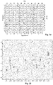
- illustrates a trivariate histogram between Age, Age of Onset, and Meningeal Temperature.
- Figure 19
- illustrates a trivariate analysis graph between Age, Star Age, and Delta T.
- Figure 20
- illustrates a graph showing the difference of medians between Registered Temperatures.
- Figure 21
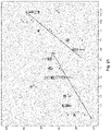
- illustrates a graph that shows the difference of medians between the Age Proportion/Time with Epilepsy.
- We have discovered that the epileptogenic zones are colder than the rest of the brain, as their metabolism is different. There is currently not a thermosensitive plate specifically designed to identify the thermogenic metabolic behavior of the brain during neurosurgical procedures. The thermosensitive plate of this invention could be used in different neurosurgical procedures, such as: epilepsy surgery, vascular neurosurgery, and oncological neurosurgery, among others, and in general surgery to monitor the temperature of organs during transplants, during cardiac bypass surgery, oncological surgery, etc.; furthermore, it may also be used in fields outside of medicine.
- As shown in
Figures 1-3 , this invention comprises a thermosensitive silica plate (1) that responds to the different temperatures to which it is exposed, exhibiting color changes within a few seconds. The basic plate may be phosphorescent, as well as thermosensitive and phosphorescent. - The thermosensitive plate may be made of flexible and non-adhesive polymers, one of which may be silica. This raw material is combined with a colorant, commonly in powder form, but it may be made of any presentation that can be mixed with the base that will form the plate, and once mixed, a catalyst is added and it is poured into a mold. The speed of plate response and the color change contrast once it has come into contact with the thermogenic surface, depends on the quantity of the colorant and the thickness of the plate.
- More specifically, the chemical components of the thermosensitive plate are "RTV" (room temperature vulcanized) type liquid silica, to which 0.1 to 30% (range not limited) of the powdered thermochromic thermosensitive colorant is added, which responds with thermal changes within the range of the temperature of the live organism on which it will be placed, and once these two initial components have been mixed homogenously, 3% of the catalyst is added, which is calculated in relation to the quantity of silica used. It is then mixed homogenously and poured into the mold, which may be in any shape, but preferably flat so that the thermosensitive/thermochromic silica has the largest surface of contact with the target organ or tissue. The thinner the transfer/thickness surface, the more quickly the thermosensitive/thermochromic silica will respond and with better spatial resolution. In the preferred embodiment of the invention, the thickness range includes, but is not limited to, 1 to 5 mm. Once the drying process at ambient temperature has finished over a maximum period of 48 hours, it may be removed from the mold and cut to the desired size to cover the organ or tissue with which it will be in contact.
- The thermosensitive plate changes color within a range that can be easily seen by the naked eye, without requiring any type of special light or filter. Normally colors are chosen that contrast when they change color: white/black, purple/pink, yellow/green. The speed of response depends in large part on the amount of colorant that is incorporated into the base plate, and the thickness of the plate.
- In the preferred embodiment of the invention, although not solely in the preferred embodiment, when the thermosensitive plate comes into contact with the target surface, organ, or tissue, the thermosensitive/thermochromic silica will change color in a few seconds (depending on the thickness and the quality of the colorant that is included in the mixture; the more colorant there is (without saturating the mixture) and the thinner the plate, the response will have better spatial distribution and will occur in a shorter period of time). Preferably two colors will be chosen (although it is not limited to being two-color) that easily show contrast to the naked eye (including, but not limited to: purple to Mexican pink) as shown in the images. In living beings, higher metabolism usually generates higher temperatures, and vice versa, thus the thermosensitive/thermochromic silica plate may have several patterns, depending on the temperature to which it is exposed.
- The thermosensitive/thermochromic plate (1) may be applied directly to the surface of the target tissue or organ to create functional maps, such as over a cerebral cut to identify the zones of metabolic activation of eloquent areas or areas with different metabolism, such as, for example, epileptogenic zones and the start of ictal zones due to their different metabolism. It may also be used to identify neoplastic or infectious lesions on any organ, without the need to use contrast dyes or media. This same thermosensitive/thermochromic plate (1) may have localizers for the navigation systems and/or electrodes for bimodal registration.
- The thermosensitive silica plate (1) has incorporated electrodes (5) to simultaneously perform electrocorticography, and for qualitative determination of the cerebral temperature.
- More specifically, the thermosensitive plate for qualitative measurements of the component temperature of organisms of this invention comprises a thermosensitive silica plate (1) with a temperature irradiation surface (2), a conduction plane (3), and a temperature transfer surface (4); and at least one electrode to register electrical activity (5) placed on the temperature irradiation surface (2).
- The device of this invention also comprises compatible material locators (7) to be detected by navigation systems placed on the temperature irradiation surface (2), with the goal of the navigation system being able to make corrections in rotation, movement, and deformation of the target tissue or organ.
- With the technical characteristics of this invention, the observer will be able to visually identify different temperature patterns with which the device is in contact. The silica may also be transparent, with color, or it may be thermochromic, and it may also have "fiducials" incorporated into it, which are markers that can be recognized by the different navigation systems that will allow the navigation systems to make adjustments through anatomic changes caused by surgical manipulation of the tissue or organ that is manipulated, or by displacement by gravity, movement, rotation, or anesthetic effect, among others, thus increasing precision during navigation-guided surgeries. The thermosensitive/thermochromic silica may also be phosphorescent.
- With the thermosensitive/thermochromic silica plate of the thermosensitive plate of this invention, functional maps can be made of the different components and organs of living beings, including functional cerebral mapping.
- The thermosensitive/thermochromic silica plate of the thermosensitive plate may be flexible and moldable, and it may be placed directly on the surface that will be evaluated, or it may be isolated by a thin layer of material to make it biocompatible, if so required. Furthermore, with the thermosensitive plate of this invention, the blood flow can be detected by its temperature gradient.
- The thermosensitive plate of this invention does not require electric parts, in its version to be used as the base for electrocorticography grids. The difference with the current electrocorticography grids is that in addition to the electrodes to register the electrical activity of the cerebral cortex, it may have "fiducials" to be detected by navigation systems, and it may be thermosensitive/thermochromic to take BIMODAL measurements of the activity of the organ being evaluated, specifically the nervous system, which includes the qualitative evaluation of the temperature and the electrical activity, SIMULTANEOUSLY.
- This invention has a method of application that consists of the steps of:
- a) Keeping the thermosensitive plate cold.
- b) Eliminating any substance that might interfere in the plate's contact with the target surface.
- c) Leaving the plate in contact with the target surface, without moving it or modifying its temperature with solutions or exposure to warm light, for example.
- d) Observing the visual response with the naked eye, which will depend on the temperature and adequate contact of the thermosensitive plate with the target surface.
- e) Removing the thermosensitive plate.
- f) Cooling the plate to a temperature that is lower than body temperature, ideally using a cold sterile saline solution.
- g) Repeating the process as required.
- An example of an embodiment of the invention that shows that the invention can be used is found in the following epilepsy surgery tests to define the epileptogenic zones, which consists of temperature measurement tables that were created in twelve epilepsy cases, where it can be clearly seen that the zones responsible for the epilepsy are between 2 and 7°C colder than the rest of the brain. That is to say, the temperature varies according to the region. The cortex is colder than the basal ganglia, and the basal ganglia are colder than the brain stem in resting state, and when an eloquent area of the brain is used, this increases temperature, as shown in the videos of the patient on whom I operated when he was awake.
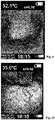
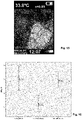
- These tables show the measurements from each case, with each table showing a range of colors that clearly illustrates how the irritative activity zone in patients with epilepsy is colder (blue), and around this colder zone there is a radial heating pattern towards the healthy brain tissue. It is from that assessment that a silica was designed, so that instead of all of these measurements we can qualitatively identify the coldest zone with the change in color of the silica (purple is cold, pink is hot).
-


- Twelve patients who met the study's inclusion criteria were studied, and of these patients, three were male (25%) and nine were female (75%). The average age was 32.5 ± 16.6596 years, the asymmetry index value was 0.795, therefore the distribution is skewed to the right, with a Kurtosis value of 0.047 being the flat distribution due to the range and size of the population. Note that the average and standard deviation of the temperatures recorded in the Irritative Zone Temperature and the Maximal Brain Temperature are almost equal, therefore the size of the existing sample is valid for this study (see Table 1).

- In
Figure 16 , referring to the graphs of recorded Temperature Errors, it was found that the averages and dispersions where the values with the lowest recorded temperatures were in the Irritative Zone. - In
Figure 17 , referring to the dispersion between the Irritative Zone Temperature and the Maximal Brain Temperature, it is seen that when the temperature in the Irritative Zone is increased, it increases the Maximal Brain Temperature. - A trivariate graph was created in order to analyze the behavior between Age, Meningeal Temperature, and Age of Onset, where two trends are seen based on this analysis: when age increases, the Meningeal Temperature increases in the range of 13-28 years, and likewise it is seen that in the range of 22-56 years, when age increases the Meningeal Temperature decreases (see Graph 3).
- In
Figure 18 , referring to the trivariate histogram between Age, Age of Onset, and Meningeal Temperature, it is seen that temperature tends to stabilize at higher ages. - As can be seen in
Figure 19 , a trivariate graph was created using Age, Age of Onset, and Delta T, in order to evaluate the behavior between these three variables. It was found that by increasing the age of the patients, the value of Delta decreases, noting that the Age of Onset does not influence this trend, and when the age of patients increases the value of Delta decreases. - In order to evaluate the behavior of all temperatures recorded by age and patient, a box plot was created in order to compare the medians based on the second and third quartiles, where it is seen that in the interval (26≤x≤56), the value of the medians decreases with respect to age, as shown in
Figure 20 , where it is seen that as of 26 years of age, the medians of temperature registered decreases. - In order to evaluate a possible association between age and time with epilepsy, the proportion was calculated (age/time of epilepsy) and box plots were created with all temperatures recorded to analyze the behavior of the medians based on the second and third quartiles, where it was found that of the proportion in the interval (1(U)≤x≤2.35(D35)) the trend is growing with an angle tending towards 45°, otherwise in the interval (2.44(D44)≤x≤33) the trend is growing with an angle of inclination tending towards 90°, therefore there is a relationship and trend in this proportion, as shown in
Figure 21 , where the difference can be seen in the trends in the medians of the proportions (U-D35) and (D44-33). - Another test was run during another surgery, in which a bimodal measurement was taken in which electrocorticography and cerebral temperature were registered simultaneously, finding the points of maximum abnormal electrical activity by electrocorticography, which were
electrodes 7 and 11, and subsequently the purple-colored thermosensitive plate was placed to determine if inpositions 7 and 11 the heat is dissipated and the purple-colored thermosensitive plate turns the color pink in the zone of normal electrical activity, and it remains purple in the zone of equal abnormal electrical activity. That is, it was seen that when the thermosensitive silica plate (1) was placed in the zone responsible for the epilepsy it remained cold (purple). In that patient it corresponds with the abnormal electrical activity registered with electrodes (electrocorticography) during the surgery, while the rest of the silica heated up quickly and took on a brilliant pink color in the zone where the electrical activity is normal.
Claims (9)
- A thermosensitive plate for qualitative measurements of the component temperature of organisms, which includes:
a thermosensitive silica plate (1) with a temperature irradiation surface (2), a conduction plane (3) and a temperature transfer surface (4). - The thermosensitive plate for qualitative measurements of the component temperature of organisms of claim 1, wherein it comprises locators of compatible material (7) to be detected by navigation systems placed on the temperature irradiation surface (2).
- The thermosensitive plate for qualitative measurements of the component temperature of organisms of claim 1, wherein it takes qualitative measurements of the cerebral temperature during surgery.
- The thermosensitive plate for qualitative measurements of the component temperature of organisms of claim 1, wherein it detects the blood flow by its temperature gradient.
- The thermosensitive plate for qualitative measurements of the component temperature of organisms of claim 1, wherein it has at least one electrode to record electrical activity (5) placed on the temperature irradiation surface (2).
- The thermosensitive plate for qualitative measurements of the component temperature of organisms of claim 1, wherein the range of plate thickness is 1 to 5 mm.
- A thermosensitive plate for qualitative measurements of the component temperature of organisms, which is comprised of: liquid silica; thermosensitive/thermochromic colorant in powder form of 0.1 to 30%; a catalyst in a quantity of 3%, which is calculated in relation to the quantity of silica used.
- The thermosensitive plate for qualitative measurements of the component temperature of organisms of claim 7, wherein the range of plate thickness is from 1 to 5 mm.
- A method for preparing a thermosensitive plate for qualitative measurements of the component temperature of organisms, which comprises the steps of:
Adding liquid silica in a recipient, to which 0.1 to 30% of the thermosensitive/thermochromic colorant in powder form is added, which responds with thermal changes within the temperature range of the living organism on which it will be placed, and once the homogenous mixture of these two initial components has been done, 3% of a catalyst is added, which is calculated in relation to the amount of silica used; then it is homogenously mixed and poured into a mold, which may be in any shape, but preferably flat so that the thermosensitive/thermochromic silica has the largest surface for contact with the target organ or tissue; and once the drying process at ambient temperature has finished within a maximum period of 48 hours, it may be removed from the mold and cut to the size desired to cover the organ or tissue with which it will be in contact.
Applications Claiming Priority (2)
| Application Number | Priority Date | Filing Date | Title |
|---|---|---|---|
| MX2019012160A MX2019012160A (en) | 2019-10-09 | 2019-10-09 | A thermosensible plate for qualitative records of the temperature of components of living organisms by means of a thermosensible silicone. |
| PCT/MX2020/050035 WO2021071352A1 (en) | 2019-10-09 | 2020-10-08 | Heat-sensitive plate for qualitatively recording the component temperature of living organisms using heat-sensitive silicone |
Publications (4)
| Publication Number | Publication Date |
|---|---|
| EP4042934A1 true EP4042934A1 (en) | 2022-08-17 |
| EP4042934A4 EP4042934A4 (en) | 2023-10-18 |
| EP4042934C0 EP4042934C0 (en) | 2025-05-14 |
| EP4042934B1 EP4042934B1 (en) | 2025-05-14 |
Family
ID=75437376
Family Applications (1)
| Application Number | Title | Priority Date | Filing Date |
|---|---|---|---|
| EP20874151.2A Active EP4042934B1 (en) | 2019-10-09 | 2020-10-08 | Heat-sensitive plate for qualitatively recording the component temperature of living organisms using heat-sensitive silicone |
Country Status (5)
| Country | Link |
|---|---|
| US (1) | US20230181045A1 (en) |
| EP (1) | EP4042934B1 (en) |
| CA (1) | CA3157348A1 (en) |
| MX (1) | MX2019012160A (en) |
| WO (1) | WO2021071352A1 (en) |
Family Cites Families (16)
| Publication number | Priority date | Publication date | Assignee | Title |
|---|---|---|---|---|
| US7097357B2 (en) * | 2004-06-02 | 2006-08-29 | General Electric Company | Method and system for improved correction of registration error in a fluoroscopic image |
| WO2007144795A1 (en) * | 2006-06-12 | 2007-12-21 | Koninklijke Philips Electronics N.V. | Body cover and a method of communicating a variation in temperature of the skin |
| US20080146913A1 (en) * | 2006-12-18 | 2008-06-19 | Medrad, Inc. | Thermochromic devices for vascular access procedures |
| WO2008133551A1 (en) * | 2007-04-27 | 2008-11-06 | St. Jude Medical Ab | Implantable concentration sensor and device |
| US8323221B2 (en) * | 2007-11-12 | 2012-12-04 | Koninklijke Philips Electronics N.V. | Tissue temperature indicating element for ultrasound therapy |
| US9095303B2 (en) * | 2009-03-23 | 2015-08-04 | Flint Hills Scientific, Llc | System and apparatus for early detection, prevention, containment or abatement of spread abnormal brain activity |
| EP2513953B1 (en) * | 2009-12-16 | 2017-10-18 | The Board of Trustees of the University of Illionis | Electrophysiology using conformal electronics |
| WO2013116235A1 (en) * | 2012-01-30 | 2013-08-08 | THE UNITED STATES OF AMERICA, as represented by THE SECRETARY, DEPT OF HEALTH & HUMAN SERVICES | Promoting transcranial direct current stimulation or transcranial magnetic stimulation using temperature-induced synaptic modulation |
| WO2013177112A1 (en) * | 2012-05-22 | 2013-11-28 | Hot Dot, Inc. | Thermocromatic patch for monitoring/detecting body temperature |
| US9819074B2 (en) * | 2013-03-11 | 2017-11-14 | The Regents Of The University Of California | Monolithically integrated implantable flexible antenna for electrocorticography and related biotelemetry devices |
| JP2017532079A (en) * | 2014-08-11 | 2017-11-02 | ザ ボード オブ トラスティーズ オブ ザ ユニヴァーシ | Skin device for analysis of temperature and heat transport properties |
| CN105220859B (en) * | 2015-08-31 | 2017-08-08 | 中山市优威斯特装饰材料有限公司 | A kind of dalle and preparation method thereof |
| JP6679377B2 (en) * | 2016-03-30 | 2020-04-15 | 日東電工株式会社 | Temperature sensor |
| US12144579B2 (en) * | 2018-03-30 | 2024-11-19 | Northwestern University | Wireless skin sensor with methods and uses |
| WO2019191749A1 (en) * | 2018-03-31 | 2019-10-03 | The Research Foundation For The State University Of New York | Pressure-regulating implant and methods of use thereof |
| US12121416B2 (en) * | 2019-02-25 | 2024-10-22 | William T. MCCLELLAN | Statically charged and rechargeable scar modulation dressing |
-
2019
- 2019-10-09 MX MX2019012160A patent/MX2019012160A/en unknown
-
2020
- 2020-10-08 EP EP20874151.2A patent/EP4042934B1/en active Active
- 2020-10-08 CA CA3157348A patent/CA3157348A1/en active Pending
- 2020-10-08 WO PCT/MX2020/050035 patent/WO2021071352A1/en not_active Ceased
- 2020-10-08 US US17/767,993 patent/US20230181045A1/en active Pending
Also Published As
| Publication number | Publication date |
|---|---|
| EP4042934A4 (en) | 2023-10-18 |
| EP4042934C0 (en) | 2025-05-14 |
| US20230181045A1 (en) | 2023-06-15 |
| CA3157348A1 (en) | 2021-04-15 |
| EP4042934B1 (en) | 2025-05-14 |
| MX2019012160A (en) | 2021-04-12 |
| WO2021071352A1 (en) | 2021-04-15 |
Similar Documents
| Publication | Publication Date | Title |
|---|---|---|
| Schrader et al. | Documentation of electrode localization | |
| Pouratian et al. | Utility of preoperative functional magnetic resonance imaging for identifying language cortices in patients with vascular malformations | |
| Cannestra et al. | Functional magnetic resonance imaging and optical imaging for dominant-hemisphere perisylvian arteriovenous malformations | |
| Maguire et al. | Topographical disorientation following unilateral temporal lobe lesions in humans | |
| MacKie | An aid to the preoperative assessment of pigmented lesions of the skin | |
| Ganslandt et al. | Magnetic source imaging combined with image-guided frameless stereotaxy: a new method in surgery around the motor strip | |
| Schiffbauer et al. | Preoperative magnetic source imaging for brain tumor surgery: a quantitative comparison with intraoperative sensory and motor mapping | |
| Tranel et al. | Neuroanatomical correlates of electrodermal skin conductance responses | |
| Pfisterer et al. | Fiducial versus nonfiducial neuronavigation registration assessment and considerations of accuracy | |
| Mellergard et al. | Epidural temperature and possible intracerebral temperature gradients in man | |
| Bot et al. | Analysis of stereotactic accuracy in patients undergoing deep brain stimulation using Nexframe and the Leksell frame | |
| VanSickle et al. | Electrode placement accuracy in robot-assisted asleep deep brain stimulation | |
| Southwell et al. | Comparison of deep brain stimulation lead targeting accuracy and procedure duration between 1.5-and 3-Tesla interventional magnetic resonance imaging systems: an initial 12-month experience | |
| Lee et al. | Is MRI a reliable tool to locate the electrode after deep brain stimulation surgery? Comparison study of CT and MRI for the localization of electrodes after DBS | |
| Alsing et al. | MR scanning, tattoo inks, and risk of thermal burn: an experimental study of iron oxide and organic pigments: effect on temperature and magnetic behavior referenced to chemical analysis | |
| McEvoy et al. | Electrophysiological validation of STN-SNr boundary depicted by susceptibility-weighted MRI | |
| D’Andrea et al. | Functional magnetic resonance imaging (fMRI), pre-intraoperative tractography in neurosurgery: the experience of Sant’Andrea Rome University Hospital | |
| EP4042934B1 (en) | Heat-sensitive plate for qualitatively recording the component temperature of living organisms using heat-sensitive silicone | |
| Bozzao et al. | Identification of the pyramidal tract by neuronavigation based on intraoperative magnetic resonance tractography: correlation with subcortical stimulation | |
| Melo et al. | Comparison of direct MRI guided versus atlas-based targeting for subthalamic nucleus and globus pallidus deep brain stimulation | |
| Schlaier et al. | Discrepancies between the MRI-and the electrophysiologically defined subthalamic nucleus | |
| Zarow et al. | A standardized method for brain-cutting suitable for both stereology and MRI-brain co-registration | |
| Giammattei et al. | Extreme lateral supracerebellar infratentorial approach: how I do it | |
| de Font-Réaulx et al. | Thermography mapping patterns in temporal lobe epilepsy surgery | |
| Fang et al. | Function-specific projections from V2 to V4 in macaques |
Legal Events
| Date | Code | Title | Description |
|---|---|---|---|
| STAA | Information on the status of an ep patent application or granted ep patent |
Free format text: STATUS: THE INTERNATIONAL PUBLICATION HAS BEEN MADE |
|
| PUAI | Public reference made under article 153(3) epc to a published international application that has entered the european phase |
Free format text: ORIGINAL CODE: 0009012 |
|
| STAA | Information on the status of an ep patent application or granted ep patent |
Free format text: STATUS: REQUEST FOR EXAMINATION WAS MADE |
|
| 17P | Request for examination filed |
Effective date: 20220509 |
|
| AK | Designated contracting states |
Kind code of ref document: A1 Designated state(s): AL AT BE BG CH CY CZ DE DK EE ES FI FR GB GR HR HU IE IS IT LI LT LU LV MC MK MT NL NO PL PT RO RS SE SI SK SM TR |
|
| DAV | Request for validation of the european patent (deleted) | ||
| DAX | Request for extension of the european patent (deleted) | ||
| A4 | Supplementary search report drawn up and despatched |
Effective date: 20230920 |
|
| RIC1 | Information provided on ipc code assigned before grant |
Ipc: A61B 90/00 20160101ALI20230914BHEP Ipc: A61B 17/00 20060101ALI20230914BHEP Ipc: A61B 5/01 20060101AFI20230914BHEP |
|
| GRAP | Despatch of communication of intention to grant a patent |
Free format text: ORIGINAL CODE: EPIDOSNIGR1 |
|
| STAA | Information on the status of an ep patent application or granted ep patent |
Free format text: STATUS: GRANT OF PATENT IS INTENDED |
|
| INTG | Intention to grant announced |
Effective date: 20250129 |
|
| GRAS | Grant fee paid |
Free format text: ORIGINAL CODE: EPIDOSNIGR3 |
|
| GRAA | (expected) grant |
Free format text: ORIGINAL CODE: 0009210 |
|
| STAA | Information on the status of an ep patent application or granted ep patent |
Free format text: STATUS: THE PATENT HAS BEEN GRANTED |
|
| AK | Designated contracting states |
Kind code of ref document: B1 Designated state(s): AL AT BE BG CH CY CZ DE DK EE ES FI FR GB GR HR HU IE IS IT LI LT LU LV MC MK MT NL NO PL PT RO RS SE SI SK SM TR |
|
| REG | Reference to a national code |
Ref country code: GB Ref legal event code: FG4D |
|
| REG | Reference to a national code |
Ref country code: CH Ref legal event code: EP |
|
| REG | Reference to a national code |
Ref country code: IE Ref legal event code: FG4D |
|
| REG | Reference to a national code |
Ref country code: DE Ref legal event code: R096 Ref document number: 602020051405 Country of ref document: DE |
|
| U01 | Request for unitary effect filed |
Effective date: 20250528 |
|
| U07 | Unitary effect registered |
Designated state(s): AT BE BG DE DK EE FI FR IT LT LU LV MT NL PT RO SE SI Effective date: 20250605 |
|
| U1N | Appointed representative for the unitary patent procedure changed after the registration of the unitary effect |
Representative=s name: BIRD & BIRD SOCIETA TRA AVVOCATI S.R.L.; IT |
|
| PG25 | Lapsed in a contracting state [announced via postgrant information from national office to epo] |
Ref country code: ES Free format text: LAPSE BECAUSE OF FAILURE TO SUBMIT A TRANSLATION OF THE DESCRIPTION OR TO PAY THE FEE WITHIN THE PRESCRIBED TIME-LIMIT Effective date: 20250514 |
|
| PG25 | Lapsed in a contracting state [announced via postgrant information from national office to epo] |
Ref country code: GR Free format text: LAPSE BECAUSE OF FAILURE TO SUBMIT A TRANSLATION OF THE DESCRIPTION OR TO PAY THE FEE WITHIN THE PRESCRIBED TIME-LIMIT Effective date: 20250815 Ref country code: NO Free format text: LAPSE BECAUSE OF FAILURE TO SUBMIT A TRANSLATION OF THE DESCRIPTION OR TO PAY THE FEE WITHIN THE PRESCRIBED TIME-LIMIT Effective date: 20250814 |
|
| PG25 | Lapsed in a contracting state [announced via postgrant information from national office to epo] |
Ref country code: PL Free format text: LAPSE BECAUSE OF FAILURE TO SUBMIT A TRANSLATION OF THE DESCRIPTION OR TO PAY THE FEE WITHIN THE PRESCRIBED TIME-LIMIT Effective date: 20250514 |
|
| U20 | Renewal fee for the european patent with unitary effect paid |
Year of fee payment: 6 Effective date: 20250909 |
|
| PGFP | Annual fee paid to national office [announced via postgrant information from national office to epo] |
Ref country code: GB Payment date: 20250922 Year of fee payment: 6 |
|
| PG25 | Lapsed in a contracting state [announced via postgrant information from national office to epo] |
Ref country code: HR Free format text: LAPSE BECAUSE OF FAILURE TO SUBMIT A TRANSLATION OF THE DESCRIPTION OR TO PAY THE FEE WITHIN THE PRESCRIBED TIME-LIMIT Effective date: 20250514 |
|
| PG25 | Lapsed in a contracting state [announced via postgrant information from national office to epo] |
Ref country code: RS Free format text: LAPSE BECAUSE OF FAILURE TO SUBMIT A TRANSLATION OF THE DESCRIPTION OR TO PAY THE FEE WITHIN THE PRESCRIBED TIME-LIMIT Effective date: 20250814 |
|
| PG25 | Lapsed in a contracting state [announced via postgrant information from national office to epo] |
Ref country code: IS Free format text: LAPSE BECAUSE OF FAILURE TO SUBMIT A TRANSLATION OF THE DESCRIPTION OR TO PAY THE FEE WITHIN THE PRESCRIBED TIME-LIMIT Effective date: 20250914 |
|
| PG25 | Lapsed in a contracting state [announced via postgrant information from national office to epo] |
Ref country code: SM Free format text: LAPSE BECAUSE OF FAILURE TO SUBMIT A TRANSLATION OF THE DESCRIPTION OR TO PAY THE FEE WITHIN THE PRESCRIBED TIME-LIMIT Effective date: 20250514 |
|
| PG25 | Lapsed in a contracting state [announced via postgrant information from national office to epo] |
Ref country code: CZ Free format text: LAPSE BECAUSE OF FAILURE TO SUBMIT A TRANSLATION OF THE DESCRIPTION OR TO PAY THE FEE WITHIN THE PRESCRIBED TIME-LIMIT Effective date: 20250514 |
|
| PG25 | Lapsed in a contracting state [announced via postgrant information from national office to epo] |
Ref country code: SK Free format text: LAPSE BECAUSE OF FAILURE TO SUBMIT A TRANSLATION OF THE DESCRIPTION OR TO PAY THE FEE WITHIN THE PRESCRIBED TIME-LIMIT Effective date: 20250514 |
|
| PLBE | No opposition filed within time limit |
Free format text: ORIGINAL CODE: 0009261 |
|
| STAA | Information on the status of an ep patent application or granted ep patent |
Free format text: STATUS: NO OPPOSITION FILED WITHIN TIME LIMIT |
|
| REG | Reference to a national code |
Ref country code: CH Ref legal event code: L10 Free format text: ST27 STATUS EVENT CODE: U-0-0-L10-L00 (AS PROVIDED BY THE NATIONAL OFFICE) Effective date: 20260325 |
|
| 26N | No opposition filed |
Effective date: 20260217 |