EP4039330A1 - Methods and compositions for the treatment of neuroblastoma malignancies - Google Patents
Methods and compositions for the treatment of neuroblastoma malignancies Download PDFInfo
- Publication number
- EP4039330A1 EP4039330A1 EP21382560.7A EP21382560A EP4039330A1 EP 4039330 A1 EP4039330 A1 EP 4039330A1 EP 21382560 A EP21382560 A EP 21382560A EP 4039330 A1 EP4039330 A1 EP 4039330A1
- Authority
- EP
- European Patent Office
- Prior art keywords
- human
- activator
- gene
- subject
- nxph1
- Prior art date
- Legal status (The legal status is an assumption and is not a legal conclusion. Google has not performed a legal analysis and makes no representation as to the accuracy of the status listed.)
- Withdrawn
Links
- 206010028980 Neoplasm Diseases 0.000 title claims abstract description 124
- 230000036210 malignancy Effects 0.000 title claims abstract description 62
- 238000000034 method Methods 0.000 title claims abstract description 40
- 238000011282 treatment Methods 0.000 title claims abstract description 30
- 206010029260 Neuroblastoma Diseases 0.000 title claims description 130
- 239000000203 mixture Substances 0.000 title claims description 19
- 108090000623 proteins and genes Proteins 0.000 claims abstract description 184
- 239000012190 activator Substances 0.000 claims abstract description 71
- 238000000338 in vitro Methods 0.000 claims abstract description 20
- 230000014509 gene expression Effects 0.000 claims description 137
- 108090000126 neurexophilin Proteins 0.000 claims description 77
- 102000003819 neurexophilin Human genes 0.000 claims description 77
- 201000011510 cancer Diseases 0.000 claims description 40
- 239000000523 sample Substances 0.000 claims description 26
- 241000282414 Homo sapiens Species 0.000 claims description 25
- 206010061289 metastatic neoplasm Diseases 0.000 claims description 23
- 102100040499 Contactin-associated protein-like 2 Human genes 0.000 claims description 20
- 108020003175 receptors Proteins 0.000 claims description 20
- 230000001965 increasing effect Effects 0.000 claims description 19
- 230000001394 metastastic effect Effects 0.000 claims description 19
- 102100039677 Adenylate cyclase type 1 Human genes 0.000 claims description 18
- 102100021606 Ephrin type-A receptor 7 Human genes 0.000 claims description 18
- 101000756805 Homo sapiens Repulsive guidance molecule B Proteins 0.000 claims description 18
- 102100027455 PH and SEC7 domain-containing protein 2 Human genes 0.000 claims description 18
- 102100034385 Plexin-A4 Human genes 0.000 claims description 18
- 102100022814 Repulsive guidance molecule B Human genes 0.000 claims description 18
- 102100023150 Sodium channel protein type 2 subunit alpha Human genes 0.000 claims description 18
- 102100023720 Sodium channel protein type 3 subunit alpha Human genes 0.000 claims description 18
- 102100031367 Sodium channel protein type 9 subunit alpha Human genes 0.000 claims description 18
- 102100036980 Talin-2 Human genes 0.000 claims description 18
- 102000004893 Transcription factor AP-2 Human genes 0.000 claims description 18
- 108090001039 Transcription factor AP-2 Proteins 0.000 claims description 18
- 102100035142 V-set and transmembrane domain-containing protein 2A Human genes 0.000 claims description 18
- 239000012472 biological sample Substances 0.000 claims description 17
- 230000003247 decreasing effect Effects 0.000 claims description 14
- 239000008194 pharmaceutical composition Substances 0.000 claims description 14
- 230000003321 amplification Effects 0.000 claims description 13
- 238000003199 nucleic acid amplification method Methods 0.000 claims description 13
- 229920001184 polypeptide Polymers 0.000 claims description 12
- 102000004196 processed proteins & peptides Human genes 0.000 claims description 12
- 108090000765 processed proteins & peptides Proteins 0.000 claims description 12
- 210000004100 adrenal gland Anatomy 0.000 claims description 11
- 108700026495 N-Myc Proto-Oncogene Proteins 0.000 claims description 10
- 230000001605 fetal effect Effects 0.000 claims description 9
- 238000002560 therapeutic procedure Methods 0.000 claims description 9
- 102100030053 Secreted frizzled-related protein 3 Human genes 0.000 claims description 8
- 108010020277 WD repeat containing planar cell polarity effector Proteins 0.000 claims description 8
- 150000001875 compounds Chemical class 0.000 claims description 8
- 239000012634 fragment Substances 0.000 claims description 8
- 230000002265 prevention Effects 0.000 claims description 8
- 150000003384 small molecules Chemical class 0.000 claims description 7
- 101710194155 Adenylate cyclase type 1 Proteins 0.000 claims description 5
- 101710168458 Contactin-associated protein-like 2 Proteins 0.000 claims description 5
- 101710116633 Ephrin type-A receptor 7 Proteins 0.000 claims description 5
- 108010008045 NAV1.2 Voltage-Gated Sodium Channel Proteins 0.000 claims description 5
- 101710126633 PH and SEC7 domain-containing protein 2 Proteins 0.000 claims description 5
- 108010003081 Peripherins Proteins 0.000 claims description 5
- 101710100264 Plexin-A4 Proteins 0.000 claims description 5
- 101710105097 Sodium channel protein type 3 subunit alpha Proteins 0.000 claims description 5
- 101710097939 Sodium channel protein type 9 subunit alpha Proteins 0.000 claims description 5
- 101710142305 Talin-2 Proteins 0.000 claims description 5
- 101710098339 V-set and transmembrane domain-containing protein 2A Proteins 0.000 claims description 5
- 210000005047 peripherin Anatomy 0.000 claims description 5
- 238000003745 diagnosis Methods 0.000 claims description 4
- 101150055666 sox6 gene Proteins 0.000 claims 5
- 102000004590 Peripherins Human genes 0.000 claims 4
- 238000011338 personalized therapy Methods 0.000 abstract description 2
- 210000004027 cell Anatomy 0.000 description 120
- 208000037265 diseases, disorders, signs and symptoms Diseases 0.000 description 32
- 102000004169 proteins and genes Human genes 0.000 description 31
- 230000002782 sympathoadrenal effect Effects 0.000 description 28
- 238000004458 analytical method Methods 0.000 description 20
- 102100036694 Transcription factor SOX-6 Human genes 0.000 description 19
- 238000004393 prognosis Methods 0.000 description 19
- 101000642517 Homo sapiens Transcription factor SOX-6 Proteins 0.000 description 18
- 230000015572 biosynthetic process Effects 0.000 description 18
- 201000010099 disease Diseases 0.000 description 18
- 102100028465 Peripherin Human genes 0.000 description 17
- 101000987578 Homo sapiens Peripherin Proteins 0.000 description 16
- ODLMAHJVESYWTB-UHFFFAOYSA-N propylbenzene Chemical compound CCCC1=CC=CC=C1 ODLMAHJVESYWTB-UHFFFAOYSA-N 0.000 description 16
- 102000005962 receptors Human genes 0.000 description 16
- 101000749877 Homo sapiens Contactin-associated protein-like 2 Proteins 0.000 description 15
- 238000003556 assay Methods 0.000 description 14
- 208000035475 disorder Diseases 0.000 description 14
- 101000959343 Homo sapiens Adenylate cyclase type 1 Proteins 0.000 description 13
- 101000898708 Homo sapiens Ephrin type-A receptor 7 Proteins 0.000 description 13
- 101001080808 Homo sapiens PH and SEC7 domain-containing protein 2 Proteins 0.000 description 13
- 101001067178 Homo sapiens Plexin-A4 Proteins 0.000 description 13
- 101000684826 Homo sapiens Sodium channel protein type 2 subunit alpha Proteins 0.000 description 13
- 101000684820 Homo sapiens Sodium channel protein type 3 subunit alpha Proteins 0.000 description 13
- 101000654386 Homo sapiens Sodium channel protein type 9 subunit alpha Proteins 0.000 description 13
- 101000598030 Homo sapiens Talin-2 Proteins 0.000 description 13
- 101000803714 Homo sapiens V-set and transmembrane domain-containing protein 2A Proteins 0.000 description 13
- 230000012010 growth Effects 0.000 description 13
- 230000002103 transcriptional effect Effects 0.000 description 13
- 239000004480 active ingredient Substances 0.000 description 12
- 230000000694 effects Effects 0.000 description 12
- 241001465754 Metazoa Species 0.000 description 11
- 241000699670 Mus sp. Species 0.000 description 11
- 210000003737 chromaffin cell Anatomy 0.000 description 11
- 239000003814 drug Substances 0.000 description 11
- 230000011664 signaling Effects 0.000 description 11
- 210000000130 stem cell Anatomy 0.000 description 11
- 101150022024 MYCN gene Proteins 0.000 description 10
- 229940079593 drug Drugs 0.000 description 10
- 238000001943 fluorescence-activated cell sorting Methods 0.000 description 10
- -1 troches Substances 0.000 description 10
- 206010027476 Metastases Diseases 0.000 description 9
- 238000002474 experimental method Methods 0.000 description 9
- 230000001537 neural effect Effects 0.000 description 9
- 238000012360 testing method Methods 0.000 description 9
- 241000699666 Mus <mouse, genus> Species 0.000 description 8
- 229920003356 PDX® Polymers 0.000 description 8
- 229960003722 doxycycline Drugs 0.000 description 8
- XQTWDDCIUJNLTR-CVHRZJFOSA-N doxycycline monohydrate Chemical compound O.O=C1C2=C(O)C=CC=C2[C@H](C)[C@@H]2C1=C(O)[C@]1(O)C(=O)C(C(N)=O)=C(O)[C@@H](N(C)C)[C@@H]1[C@H]2O XQTWDDCIUJNLTR-CVHRZJFOSA-N 0.000 description 8
- 238000001727 in vivo Methods 0.000 description 8
- 108020004999 messenger RNA Proteins 0.000 description 8
- 239000003921 oil Substances 0.000 description 8
- 235000019198 oils Nutrition 0.000 description 8
- 108700012912 MYCN Proteins 0.000 description 7
- 102100030124 N-myc proto-oncogene protein Human genes 0.000 description 7
- 239000000872 buffer Substances 0.000 description 7
- 230000002349 favourable effect Effects 0.000 description 7
- 210000001165 lymph node Anatomy 0.000 description 7
- 230000004044 response Effects 0.000 description 7
- 239000000243 solution Substances 0.000 description 7
- 239000003826 tablet Substances 0.000 description 7
- XLYOFNOQVPJJNP-UHFFFAOYSA-N water Substances O XLYOFNOQVPJJNP-UHFFFAOYSA-N 0.000 description 7
- 102100021772 Neurexin-2 Human genes 0.000 description 6
- 230000002596 correlated effect Effects 0.000 description 6
- 239000003446 ligand Substances 0.000 description 6
- 239000000463 material Substances 0.000 description 6
- 210000000653 nervous system Anatomy 0.000 description 6
- 230000036961 partial effect Effects 0.000 description 6
- 239000000546 pharmaceutical excipient Substances 0.000 description 6
- UCSJYZPVAKXKNQ-HZYVHMACSA-N streptomycin Chemical compound CN[C@H]1[C@H](O)[C@@H](O)[C@H](CO)O[C@H]1O[C@@H]1[C@](C=O)(O)[C@H](C)O[C@H]1O[C@@H]1[C@@H](NC(N)=N)[C@H](O)[C@@H](NC(N)=N)[C@H](O)[C@H]1O UCSJYZPVAKXKNQ-HZYVHMACSA-N 0.000 description 6
- 239000000725 suspension Substances 0.000 description 6
- 210000001519 tissue Anatomy 0.000 description 6
- WOVKYSAHUYNSMH-RRKCRQDMSA-N 5-bromodeoxyuridine Chemical compound C1[C@H](O)[C@@H](CO)O[C@H]1N1C(=O)NC(=O)C(Br)=C1 WOVKYSAHUYNSMH-RRKCRQDMSA-N 0.000 description 5
- 206010061818 Disease progression Diseases 0.000 description 5
- 101000969980 Homo sapiens Neurexin-2 Proteins 0.000 description 5
- 101000969975 Homo sapiens Neurexin-2-beta Proteins 0.000 description 5
- 241000124008 Mammalia Species 0.000 description 5
- 241000283973 Oryctolagus cuniculus Species 0.000 description 5
- 230000029918 bioluminescence Effects 0.000 description 5
- 238000005415 bioluminescence Methods 0.000 description 5
- 239000007859 condensation product Substances 0.000 description 5
- 230000000875 corresponding effect Effects 0.000 description 5
- 235000014113 dietary fatty acids Nutrition 0.000 description 5
- 230000005750 disease progression Effects 0.000 description 5
- 238000009826 distribution Methods 0.000 description 5
- 150000002148 esters Chemical class 0.000 description 5
- 239000000194 fatty acid Substances 0.000 description 5
- 229930195729 fatty acid Natural products 0.000 description 5
- 150000004665 fatty acids Chemical class 0.000 description 5
- 239000000796 flavoring agent Substances 0.000 description 5
- 230000006870 function Effects 0.000 description 5
- 230000001939 inductive effect Effects 0.000 description 5
- 239000003112 inhibitor Substances 0.000 description 5
- 230000002401 inhibitory effect Effects 0.000 description 5
- 238000004519 manufacturing process Methods 0.000 description 5
- 210000000933 neural crest Anatomy 0.000 description 5
- 210000001428 peripheral nervous system Anatomy 0.000 description 5
- 238000002360 preparation method Methods 0.000 description 5
- 230000004083 survival effect Effects 0.000 description 5
- 239000003765 sweetening agent Substances 0.000 description 5
- 208000024891 symptom Diseases 0.000 description 5
- 239000013598 vector Substances 0.000 description 5
- 102100033793 ALK tyrosine kinase receptor Human genes 0.000 description 4
- VTYYLEPIZMXCLO-UHFFFAOYSA-L Calcium carbonate Chemical compound [Ca+2].[O-]C([O-])=O VTYYLEPIZMXCLO-UHFFFAOYSA-L 0.000 description 4
- IAYPIBMASNFSPL-UHFFFAOYSA-N Ethylene oxide Chemical compound C1CO1 IAYPIBMASNFSPL-UHFFFAOYSA-N 0.000 description 4
- 108050001049 Extracellular proteins Proteins 0.000 description 4
- 241000287828 Gallus gallus Species 0.000 description 4
- 101000779641 Homo sapiens ALK tyrosine kinase receptor Proteins 0.000 description 4
- 229930006000 Sucrose Natural products 0.000 description 4
- 102000040945 Transcription factor Human genes 0.000 description 4
- 108091023040 Transcription factor Proteins 0.000 description 4
- 239000007900 aqueous suspension Substances 0.000 description 4
- 230000004071 biological effect Effects 0.000 description 4
- 210000001185 bone marrow Anatomy 0.000 description 4
- 235000013330 chicken meat Nutrition 0.000 description 4
- 238000001514 detection method Methods 0.000 description 4
- 239000002270 dispersing agent Substances 0.000 description 4
- 230000002708 enhancing effect Effects 0.000 description 4
- 235000013355 food flavoring agent Nutrition 0.000 description 4
- 235000003599 food sweetener Nutrition 0.000 description 4
- 210000000609 ganglia Anatomy 0.000 description 4
- 238000003197 gene knockdown Methods 0.000 description 4
- 230000002068 genetic effect Effects 0.000 description 4
- 238000010166 immunofluorescence Methods 0.000 description 4
- 238000002347 injection Methods 0.000 description 4
- 239000007924 injection Substances 0.000 description 4
- 210000004185 liver Anatomy 0.000 description 4
- 239000002609 medium Substances 0.000 description 4
- 230000009401 metastasis Effects 0.000 description 4
- 210000004940 nucleus Anatomy 0.000 description 4
- 239000000047 product Substances 0.000 description 4
- 238000011002 quantification Methods 0.000 description 4
- 239000007787 solid Substances 0.000 description 4
- 239000005720 sucrose Substances 0.000 description 4
- 239000000375 suspending agent Substances 0.000 description 4
- 230000000946 synaptic effect Effects 0.000 description 4
- 230000004614 tumor growth Effects 0.000 description 4
- 239000000080 wetting agent Substances 0.000 description 4
- 108091032973 (ribonucleotides)n+m Proteins 0.000 description 3
- FWBHETKCLVMNFS-UHFFFAOYSA-N 4',6-Diamino-2-phenylindol Chemical compound C1=CC(C(=N)N)=CC=C1C1=CC2=CC=C(C(N)=N)C=C2N1 FWBHETKCLVMNFS-UHFFFAOYSA-N 0.000 description 3
- 102100031118 Catenin delta-2 Human genes 0.000 description 3
- IAZDPXIOMUYVGZ-UHFFFAOYSA-N Dimethylsulphoxide Chemical compound CS(C)=O IAZDPXIOMUYVGZ-UHFFFAOYSA-N 0.000 description 3
- 102100024117 Disks large homolog 2 Human genes 0.000 description 3
- 239000006144 Dulbecco’s modified Eagle's medium Substances 0.000 description 3
- 108010009307 Forkhead Box Protein O3 Proteins 0.000 description 3
- 102100035421 Forkhead box protein O3 Human genes 0.000 description 3
- 101000922056 Homo sapiens Catenin delta-2 Proteins 0.000 description 3
- 101001053980 Homo sapiens Disks large homolog 2 Proteins 0.000 description 3
- 101001066265 Homo sapiens Endothelial transcription factor GATA-2 Proteins 0.000 description 3
- 101000590691 Homo sapiens MAGUK p55 subfamily member 2 Proteins 0.000 description 3
- 108060003951 Immunoglobulin Proteins 0.000 description 3
- 108060001084 Luciferase Proteins 0.000 description 3
- 239000005089 Luciferase Substances 0.000 description 3
- OKKJLVBELUTLKV-UHFFFAOYSA-N Methanol Chemical compound OC OKKJLVBELUTLKV-UHFFFAOYSA-N 0.000 description 3
- 102100021582 Neurexin-1-beta Human genes 0.000 description 3
- 229930182555 Penicillin Natural products 0.000 description 3
- JGSARLDLIJGVTE-MBNYWOFBSA-N Penicillin G Chemical compound N([C@H]1[C@H]2SC([C@@H](N2C1=O)C(O)=O)(C)C)C(=O)CC1=CC=CC=C1 JGSARLDLIJGVTE-MBNYWOFBSA-N 0.000 description 3
- 108010029485 Protein Isoforms Proteins 0.000 description 3
- 102000001708 Protein Isoforms Human genes 0.000 description 3
- 102100030680 SH3 and multiple ankyrin repeat domains protein 2 Human genes 0.000 description 3
- 101710067890 SHANK2 Proteins 0.000 description 3
- 239000000556 agonist Substances 0.000 description 3
- 230000004075 alteration Effects 0.000 description 3
- 210000004369 blood Anatomy 0.000 description 3
- 239000008280 blood Substances 0.000 description 3
- 210000004556 brain Anatomy 0.000 description 3
- 239000006285 cell suspension Substances 0.000 description 3
- 239000003153 chemical reaction reagent Substances 0.000 description 3
- 239000003795 chemical substances by application Substances 0.000 description 3
- 238000002512 chemotherapy Methods 0.000 description 3
- 239000003086 colorant Substances 0.000 description 3
- 239000002299 complementary DNA Substances 0.000 description 3
- 230000007423 decrease Effects 0.000 description 3
- 238000013461 design Methods 0.000 description 3
- 239000000839 emulsion Substances 0.000 description 3
- 238000009472 formulation Methods 0.000 description 3
- 102000018358 immunoglobulin Human genes 0.000 description 3
- 208000015181 infectious disease Diseases 0.000 description 3
- 239000004615 ingredient Substances 0.000 description 3
- 230000000670 limiting effect Effects 0.000 description 3
- 229940057995 liquid paraffin Drugs 0.000 description 3
- 238000001325 log-rank test Methods 0.000 description 3
- 230000003211 malignant effect Effects 0.000 description 3
- 108010082117 matrigel Proteins 0.000 description 3
- 230000035772 mutation Effects 0.000 description 3
- 210000002569 neuron Anatomy 0.000 description 3
- 231100000252 nontoxic Toxicity 0.000 description 3
- 230000003000 nontoxic effect Effects 0.000 description 3
- 239000004006 olive oil Substances 0.000 description 3
- 235000008390 olive oil Nutrition 0.000 description 3
- 230000003287 optical effect Effects 0.000 description 3
- 229940049954 penicillin Drugs 0.000 description 3
- 230000001242 postsynaptic effect Effects 0.000 description 3
- 239000003755 preservative agent Substances 0.000 description 3
- 230000008569 process Effects 0.000 description 3
- 239000013074 reference sample Substances 0.000 description 3
- 239000011435 rock Substances 0.000 description 3
- 210000004116 schwann cell Anatomy 0.000 description 3
- 210000002966 serum Anatomy 0.000 description 3
- 239000002904 solvent Substances 0.000 description 3
- 238000007619 statistical method Methods 0.000 description 3
- 229960005322 streptomycin Drugs 0.000 description 3
- 230000005062 synaptic transmission Effects 0.000 description 3
- 210000004881 tumor cell Anatomy 0.000 description 3
- PUPZLCDOIYMWBV-UHFFFAOYSA-N (+/-)-1,3-Butanediol Chemical compound CC(O)CCO PUPZLCDOIYMWBV-UHFFFAOYSA-N 0.000 description 2
- SGKRLCUYIXIAHR-NLJUDYQYSA-N (4r,4ar,5s,5ar,6r,12ar)-4-(dimethylamino)-1,5,10,11,12a-pentahydroxy-6-methyl-3,12-dioxo-4a,5,5a,6-tetrahydro-4h-tetracene-2-carboxamide Chemical compound C1=CC=C2[C@H](C)[C@@H]([C@H](O)[C@@H]3[C@](C(O)=C(C(N)=O)C(=O)[C@@H]3N(C)C)(O)C3=O)C3=C(O)C2=C1O SGKRLCUYIXIAHR-NLJUDYQYSA-N 0.000 description 2
- ZORQXIQZAOLNGE-UHFFFAOYSA-N 1,1-difluorocyclohexane Chemical compound FC1(F)CCCCC1 ZORQXIQZAOLNGE-UHFFFAOYSA-N 0.000 description 2
- VBICKXHEKHSIBG-UHFFFAOYSA-N 1-monostearoylglycerol Chemical compound CCCCCCCCCCCCCCCCCC(=O)OCC(O)CO VBICKXHEKHSIBG-UHFFFAOYSA-N 0.000 description 2
- IIZPXYDJLKNOIY-JXPKJXOSSA-N 1-palmitoyl-2-arachidonoyl-sn-glycero-3-phosphocholine Chemical compound CCCCCCCCCCCCCCCC(=O)OC[C@H](COP([O-])(=O)OCC[N+](C)(C)C)OC(=O)CCC\C=C/C\C=C/C\C=C/C\C=C/CCCCC IIZPXYDJLKNOIY-JXPKJXOSSA-N 0.000 description 2
- IZHVBANLECCAGF-UHFFFAOYSA-N 2-hydroxy-3-(octadecanoyloxy)propyl octadecanoate Chemical compound CCCCCCCCCCCCCCCCCC(=O)OCC(O)COC(=O)CCCCCCCCCCCCCCCCC IZHVBANLECCAGF-UHFFFAOYSA-N 0.000 description 2
- 244000215068 Acacia senegal Species 0.000 description 2
- 235000006491 Acacia senegal Nutrition 0.000 description 2
- 235000003911 Arachis Nutrition 0.000 description 2
- 244000105624 Arachis hypogaea Species 0.000 description 2
- CIWBSHSKHKDKBQ-JLAZNSOCSA-N Ascorbic acid Chemical compound OC[C@H](O)[C@H]1OC(=O)C(O)=C1O CIWBSHSKHKDKBQ-JLAZNSOCSA-N 0.000 description 2
- 241000416162 Astragalus gummifer Species 0.000 description 2
- 108091033409 CRISPR Proteins 0.000 description 2
- CURLTUGMZLYLDI-UHFFFAOYSA-N Carbon dioxide Chemical compound O=C=O CURLTUGMZLYLDI-UHFFFAOYSA-N 0.000 description 2
- 102100025064 Cellular tumor antigen p53 Human genes 0.000 description 2
- 108010008532 Deoxyribonuclease I Proteins 0.000 description 2
- 241000283073 Equus caballus Species 0.000 description 2
- DHMQDGOQFOQNFH-UHFFFAOYSA-N Glycine Chemical compound NCC(O)=O DHMQDGOQFOQNFH-UHFFFAOYSA-N 0.000 description 2
- 102000003886 Glycoproteins Human genes 0.000 description 2
- 108090000288 Glycoproteins Proteins 0.000 description 2
- 229920000084 Gum arabic Polymers 0.000 description 2
- 241000282412 Homo Species 0.000 description 2
- 101001108436 Homo sapiens Neurexin-1 Proteins 0.000 description 2
- 101001108433 Homo sapiens Neurexin-1-beta Proteins 0.000 description 2
- 101000692768 Homo sapiens Paired mesoderm homeobox protein 2B Proteins 0.000 description 2
- 101000757378 Homo sapiens Transcription factor AP-2-alpha Proteins 0.000 description 2
- 206010061598 Immunodeficiency Diseases 0.000 description 2
- 241000713666 Lentivirus Species 0.000 description 2
- 102100021310 Neurexin-3 Human genes 0.000 description 2
- 108700020796 Oncogene Proteins 0.000 description 2
- 230000018199 S phase Effects 0.000 description 2
- 238000011579 SCID mouse model Methods 0.000 description 2
- CDBYLPFSWZWCQE-UHFFFAOYSA-L Sodium Carbonate Chemical compound [Na+].[Na+].[O-]C([O-])=O CDBYLPFSWZWCQE-UHFFFAOYSA-L 0.000 description 2
- CZMRCDWAGMRECN-UGDNZRGBSA-N Sucrose Chemical compound O[C@H]1[C@H](O)[C@@H](CO)O[C@@]1(CO)O[C@@H]1[C@H](O)[C@@H](O)[C@H](O)[C@@H](CO)O1 CZMRCDWAGMRECN-UGDNZRGBSA-N 0.000 description 2
- 229920001615 Tragacanth Polymers 0.000 description 2
- 102100022972 Transcription factor AP-2-alpha Human genes 0.000 description 2
- 108010078814 Tumor Suppressor Protein p53 Proteins 0.000 description 2
- 102000056014 X-linked Nuclear Human genes 0.000 description 2
- 235000010489 acacia gum Nutrition 0.000 description 2
- 230000009471 action Effects 0.000 description 2
- 230000003213 activating effect Effects 0.000 description 2
- 230000007439 adrenal gland development Effects 0.000 description 2
- 210000001943 adrenal medulla Anatomy 0.000 description 2
- 208000029560 autism spectrum disease Diseases 0.000 description 2
- 230000000903 blocking effect Effects 0.000 description 2
- 230000037396 body weight Effects 0.000 description 2
- 210000004899 c-terminal region Anatomy 0.000 description 2
- 229910000019 calcium carbonate Inorganic materials 0.000 description 2
- 239000001506 calcium phosphate Substances 0.000 description 2
- 229910000389 calcium phosphate Inorganic materials 0.000 description 2
- 235000011010 calcium phosphates Nutrition 0.000 description 2
- 235000011089 carbon dioxide Nutrition 0.000 description 2
- 230000024245 cell differentiation Effects 0.000 description 2
- 210000001175 cerebrospinal fluid Anatomy 0.000 description 2
- 230000008859 change Effects 0.000 description 2
- 230000001276 controlling effect Effects 0.000 description 2
- 238000007796 conventional method Methods 0.000 description 2
- 239000013078 crystal Substances 0.000 description 2
- 238000011161 development Methods 0.000 description 2
- 230000018109 developmental process Effects 0.000 description 2
- 239000003085 diluting agent Substances 0.000 description 2
- 238000010790 dilution Methods 0.000 description 2
- 239000012895 dilution Substances 0.000 description 2
- 230000003292 diminished effect Effects 0.000 description 2
- 229960004497 dinutuximab Drugs 0.000 description 2
- 239000003651 drinking water Substances 0.000 description 2
- 235000020188 drinking water Nutrition 0.000 description 2
- 239000000975 dye Substances 0.000 description 2
- 235000013601 eggs Nutrition 0.000 description 2
- 210000002257 embryonic structure Anatomy 0.000 description 2
- 238000005516 engineering process Methods 0.000 description 2
- 239000007903 gelatin capsule Substances 0.000 description 2
- 230000004077 genetic alteration Effects 0.000 description 2
- 231100000118 genetic alteration Toxicity 0.000 description 2
- 239000008187 granular material Substances 0.000 description 2
- 239000001963 growth medium Substances 0.000 description 2
- 210000001308 heart ventricle Anatomy 0.000 description 2
- BXWNKGSJHAJOGX-UHFFFAOYSA-N hexadecan-1-ol Chemical compound CCCCCCCCCCCCCCCCO BXWNKGSJHAJOGX-UHFFFAOYSA-N 0.000 description 2
- 238000003384 imaging method Methods 0.000 description 2
- 238000011534 incubation Methods 0.000 description 2
- 230000005764 inhibitory process Effects 0.000 description 2
- NOESYZHRGYRDHS-UHFFFAOYSA-N insulin Chemical compound N1C(=O)C(NC(=O)C(CCC(N)=O)NC(=O)C(CCC(O)=O)NC(=O)C(C(C)C)NC(=O)C(NC(=O)CN)C(C)CC)CSSCC(C(NC(CO)C(=O)NC(CC(C)C)C(=O)NC(CC=2C=CC(O)=CC=2)C(=O)NC(CCC(N)=O)C(=O)NC(CC(C)C)C(=O)NC(CCC(O)=O)C(=O)NC(CC(N)=O)C(=O)NC(CC=2C=CC(O)=CC=2)C(=O)NC(CSSCC(NC(=O)C(C(C)C)NC(=O)C(CC(C)C)NC(=O)C(CC=2C=CC(O)=CC=2)NC(=O)C(CC(C)C)NC(=O)C(C)NC(=O)C(CCC(O)=O)NC(=O)C(C(C)C)NC(=O)C(CC(C)C)NC(=O)C(CC=2NC=NC=2)NC(=O)C(CO)NC(=O)CNC2=O)C(=O)NCC(=O)NC(CCC(O)=O)C(=O)NC(CCCNC(N)=N)C(=O)NCC(=O)NC(CC=3C=CC=CC=3)C(=O)NC(CC=3C=CC=CC=3)C(=O)NC(CC=3C=CC(O)=CC=3)C(=O)NC(C(C)O)C(=O)N3C(CCC3)C(=O)NC(CCCCN)C(=O)NC(C)C(O)=O)C(=O)NC(CC(N)=O)C(O)=O)=O)NC(=O)C(C(C)CC)NC(=O)C(CO)NC(=O)C(C(C)O)NC(=O)C1CSSCC2NC(=O)C(CC(C)C)NC(=O)C(NC(=O)C(CCC(N)=O)NC(=O)C(CC(N)=O)NC(=O)C(NC(=O)C(N)CC=1C=CC=CC=1)C(C)C)CC1=CN=CN1 NOESYZHRGYRDHS-UHFFFAOYSA-N 0.000 description 2
- 230000003993 interaction Effects 0.000 description 2
- 230000003447 ipsilateral effect Effects 0.000 description 2
- 235000010445 lecithin Nutrition 0.000 description 2
- 239000000787 lecithin Substances 0.000 description 2
- 229940067606 lecithin Drugs 0.000 description 2
- 239000007788 liquid Substances 0.000 description 2
- HQKMJHAJHXVSDF-UHFFFAOYSA-L magnesium stearate Chemical compound [Mg+2].CCCCCCCCCCCCCCCCCC([O-])=O.CCCCCCCCCCCCCCCCCC([O-])=O HQKMJHAJHXVSDF-UHFFFAOYSA-L 0.000 description 2
- 239000012528 membrane Substances 0.000 description 2
- 238000002493 microarray Methods 0.000 description 2
- 239000002480 mineral oil Substances 0.000 description 2
- 235000010446 mineral oil Nutrition 0.000 description 2
- 239000001788 mono and diglycerides of fatty acids Substances 0.000 description 2
- 210000001982 neural crest cell Anatomy 0.000 description 2
- 239000000346 nonvolatile oil Substances 0.000 description 2
- 102000039446 nucleic acids Human genes 0.000 description 2
- 108020004707 nucleic acids Proteins 0.000 description 2
- 150000007523 nucleic acids Chemical class 0.000 description 2
- 239000013610 patient sample Substances 0.000 description 2
- 239000008188 pellet Substances 0.000 description 2
- 239000013612 plasmid Substances 0.000 description 2
- 238000010837 poor prognosis Methods 0.000 description 2
- 239000000843 powder Substances 0.000 description 2
- 239000002243 precursor Substances 0.000 description 2
- 230000003518 presynaptic effect Effects 0.000 description 2
- 238000000513 principal component analysis Methods 0.000 description 2
- 238000000746 purification Methods 0.000 description 2
- RXWNCPJZOCPEPQ-NVWDDTSBSA-N puromycin Chemical compound C1=CC(OC)=CC=C1C[C@H](N)C(=O)N[C@H]1[C@@H](O)[C@H](N2C3=NC=NC(=C3N=C2)N(C)C)O[C@@H]1CO RXWNCPJZOCPEPQ-NVWDDTSBSA-N 0.000 description 2
- 238000003753 real-time PCR Methods 0.000 description 2
- 230000000306 recurrent effect Effects 0.000 description 2
- 230000009467 reduction Effects 0.000 description 2
- 238000002271 resection Methods 0.000 description 2
- 235000011069 sorbitan monooleate Nutrition 0.000 description 2
- 239000001593 sorbitan monooleate Substances 0.000 description 2
- 229940035049 sorbitan monooleate Drugs 0.000 description 2
- 239000008223 sterile water Substances 0.000 description 2
- 239000000126 substance Substances 0.000 description 2
- 125000000185 sucrose group Chemical group 0.000 description 2
- 210000000331 sympathetic ganglia Anatomy 0.000 description 2
- 230000004797 therapeutic response Effects 0.000 description 2
- 238000013518 transcription Methods 0.000 description 2
- 230000035897 transcription Effects 0.000 description 2
- 102000035160 transmembrane proteins Human genes 0.000 description 2
- 108091005703 transmembrane proteins Proteins 0.000 description 2
- 102000027257 transmembrane receptors Human genes 0.000 description 2
- 108091008578 transmembrane receptors Proteins 0.000 description 2
- QORWJWZARLRLPR-UHFFFAOYSA-H tricalcium bis(phosphate) Chemical compound [Ca+2].[Ca+2].[Ca+2].[O-]P([O-])([O-])=O.[O-]P([O-])([O-])=O QORWJWZARLRLPR-UHFFFAOYSA-H 0.000 description 2
- 230000001960 triggered effect Effects 0.000 description 2
- 235000015112 vegetable and seed oil Nutrition 0.000 description 2
- 239000008158 vegetable oil Substances 0.000 description 2
- 239000003981 vehicle Substances 0.000 description 2
- 238000012800 visualization Methods 0.000 description 2
- LNAZSHAWQACDHT-XIYTZBAFSA-N (2r,3r,4s,5r,6s)-4,5-dimethoxy-2-(methoxymethyl)-3-[(2s,3r,4s,5r,6r)-3,4,5-trimethoxy-6-(methoxymethyl)oxan-2-yl]oxy-6-[(2r,3r,4s,5r,6r)-4,5,6-trimethoxy-2-(methoxymethyl)oxan-3-yl]oxyoxane Chemical compound CO[C@@H]1[C@@H](OC)[C@H](OC)[C@@H](COC)O[C@H]1O[C@H]1[C@H](OC)[C@@H](OC)[C@H](O[C@H]2[C@@H]([C@@H](OC)[C@H](OC)O[C@@H]2COC)OC)O[C@@H]1COC LNAZSHAWQACDHT-XIYTZBAFSA-N 0.000 description 1
- WRIDQFICGBMAFQ-UHFFFAOYSA-N (E)-8-Octadecenoic acid Natural products CCCCCCCCCC=CCCCCCCC(O)=O WRIDQFICGBMAFQ-UHFFFAOYSA-N 0.000 description 1
- 102100030471 1-acyl-sn-glycerol-3-phosphate acyltransferase delta Human genes 0.000 description 1
- IXPNQXFRVYWDDI-UHFFFAOYSA-N 1-methyl-2,4-dioxo-1,3-diazinane-5-carboximidamide Chemical compound CN1CC(C(N)=N)C(=O)NC1=O IXPNQXFRVYWDDI-UHFFFAOYSA-N 0.000 description 1
- 102100024682 14-3-3 protein eta Human genes 0.000 description 1
- LQJBNNIYVWPHFW-UHFFFAOYSA-N 20:1omega9c fatty acid Natural products CCCCCCCCCCC=CCCCCCCCC(O)=O LQJBNNIYVWPHFW-UHFFFAOYSA-N 0.000 description 1
- FJKROLUGYXJWQN-UHFFFAOYSA-M 4-hydroxybenzoate Chemical compound OC1=CC=C(C([O-])=O)C=C1 FJKROLUGYXJWQN-UHFFFAOYSA-M 0.000 description 1
- QSBYPNXLFMSGKH-UHFFFAOYSA-N 9-Heptadecensaeure Natural products CCCCCCCC=CCCCCCCCC(O)=O QSBYPNXLFMSGKH-UHFFFAOYSA-N 0.000 description 1
- 102100040077 A-kinase anchor protein 6 Human genes 0.000 description 1
- 102100033935 AP-3 complex subunit beta-2 Human genes 0.000 description 1
- 102100024120 AP-4 complex accessory subunit RUSC1 Human genes 0.000 description 1
- 102100034571 AT-rich interactive domain-containing protein 1B Human genes 0.000 description 1
- 101150020330 ATRX gene Proteins 0.000 description 1
- 102100021886 Activin receptor type-2A Human genes 0.000 description 1
- 102100021879 Adenylyl cyclase-associated protein 2 Human genes 0.000 description 1
- 229920000936 Agarose Polymers 0.000 description 1
- HJCMDXDYPOUFDY-WHFBIAKZSA-N Ala-Gln Chemical compound C[C@H](N)C(=O)N[C@H](C(O)=O)CCC(N)=O HJCMDXDYPOUFDY-WHFBIAKZSA-N 0.000 description 1
- 239000012099 Alexa Fluor family Substances 0.000 description 1
- 102100029231 Alpha-2,8-sialyltransferase 8B Human genes 0.000 description 1
- GUBGYTABKSRVRQ-XLOQQCSPSA-N Alpha-Lactose Chemical compound O[C@@H]1[C@@H](O)[C@@H](O)[C@@H](CO)O[C@H]1O[C@@H]1[C@@H](CO)O[C@H](O)[C@H](O)[C@H]1O GUBGYTABKSRVRQ-XLOQQCSPSA-N 0.000 description 1
- 239000005995 Aluminium silicate Substances 0.000 description 1
- 102100036441 Amyloid-beta A4 precursor protein-binding family A member 2 Human genes 0.000 description 1
- 102100034611 Ankyrin repeat domain-containing protein 12 Human genes 0.000 description 1
- 101100125452 Arabidopsis thaliana ICR1 gene Proteins 0.000 description 1
- 102100036783 Arf-GAP with GTPase, ANK repeat and PH domain-containing protein 1 Human genes 0.000 description 1
- 102100023221 Arginine and glutamate-rich protein 1 Human genes 0.000 description 1
- 102100031464 Armadillo repeat protein deleted in velo-cardio-facial syndrome Human genes 0.000 description 1
- 102100033887 Arylsulfatase D Human genes 0.000 description 1
- 102100035553 Autism susceptibility gene 2 protein Human genes 0.000 description 1
- WOVKYSAHUYNSMH-UHFFFAOYSA-N BROMODEOXYURIDINE Natural products C1C(O)C(CO)OC1N1C(=O)NC(=O)C(Br)=C1 WOVKYSAHUYNSMH-UHFFFAOYSA-N 0.000 description 1
- 102100040539 BTB/POZ domain-containing protein KCTD1 Human genes 0.000 description 1
- 241000283690 Bos taurus Species 0.000 description 1
- 208000003174 Brain Neoplasms Diseases 0.000 description 1
- 206010006187 Breast cancer Diseases 0.000 description 1
- 208000026310 Breast neoplasm Diseases 0.000 description 1
- 102100022287 C-Jun-amino-terminal kinase-interacting protein 2 Human genes 0.000 description 1
- 102100025905 C-Jun-amino-terminal kinase-interacting protein 4 Human genes 0.000 description 1
- 102100031650 C-X-C chemokine receptor type 4 Human genes 0.000 description 1
- 102000014819 CACNA1B Human genes 0.000 description 1
- 102100028738 CAP-Gly domain-containing linker protein 3 Human genes 0.000 description 1
- 108010032389 CBFA2T2 myeloid-transforming gene-related protein Proteins 0.000 description 1
- 102100029395 CLIP-associating protein 2 Human genes 0.000 description 1
- 108010033531 CMP-N-acetylneuraminate-poly-alpha-2,8-sialosyl sialyltransferase Proteins 0.000 description 1
- 238000010354 CRISPR gene editing Methods 0.000 description 1
- 102100033210 CUGBP Elav-like family member 2 Human genes 0.000 description 1
- 102100025488 CUGBP Elav-like family member 4 Human genes 0.000 description 1
- 102100036293 Calcium-binding mitochondrial carrier protein SCaMC-3 Human genes 0.000 description 1
- 102100032582 Calcium-dependent secretion activator 1 Human genes 0.000 description 1
- 102100025232 Calcium/calmodulin-dependent protein kinase type II subunit beta Human genes 0.000 description 1
- 102100025227 Calcium/calmodulin-dependent protein kinase type II subunit gamma Human genes 0.000 description 1
- 102100033561 Calmodulin-binding transcription activator 1 Human genes 0.000 description 1
- 102100038710 Capping protein-inhibiting regulator of actin dynamics Human genes 0.000 description 1
- 102100037398 Casein kinase I isoform epsilon Human genes 0.000 description 1
- 102000003952 Caspase 3 Human genes 0.000 description 1
- 108090000397 Caspase 3 Proteins 0.000 description 1
- 102100031441 Cell cycle checkpoint protein RAD17 Human genes 0.000 description 1
- 102100024485 Cell division cycle-associated protein 7 Human genes 0.000 description 1
- 108091006146 Channels Proteins 0.000 description 1
- 102100038164 Chromodomain-helicase-DNA-binding protein 9 Human genes 0.000 description 1
- 102100031615 Ciliary neurotrophic factor receptor subunit alpha Human genes 0.000 description 1
- 102100034177 Clathrin coat assembly protein AP180 Human genes 0.000 description 1
- 102100032369 Coiled-coil domain-containing protein 112 Human genes 0.000 description 1
- 102100035217 Coiled-coil domain-containing protein 136 Human genes 0.000 description 1
- 102100025823 Coiled-coil domain-containing protein 82 Human genes 0.000 description 1
- 102100040453 Connector enhancer of kinase suppressor of ras 2 Human genes 0.000 description 1
- 102100024326 Contactin-1 Human genes 0.000 description 1
- 229920002261 Corn starch Polymers 0.000 description 1
- 102100041023 Coronin-2A Human genes 0.000 description 1
- 241000938605 Crocodylia Species 0.000 description 1
- 102100031685 Cyclin-dependent kinase-like 2 Human genes 0.000 description 1
- CMSMOCZEIVJLDB-UHFFFAOYSA-N Cyclophosphamide Chemical compound ClCCN(CCCl)P1(=O)NCCCO1 CMSMOCZEIVJLDB-UHFFFAOYSA-N 0.000 description 1
- 102100032620 Cytotoxic granule associated RNA binding protein TIA1 Human genes 0.000 description 1
- IGXWBGJHJZYPQS-SSDOTTSWSA-N D-Luciferin Chemical compound OC(=O)[C@H]1CSC(C=2SC3=CC=C(O)C=C3N=2)=N1 IGXWBGJHJZYPQS-SSDOTTSWSA-N 0.000 description 1
- 102100033462 DENN domain-containing protein 1B Human genes 0.000 description 1
- 108020004414 DNA Proteins 0.000 description 1
- 230000007067 DNA methylation Effects 0.000 description 1
- 230000033616 DNA repair Effects 0.000 description 1
- 102100035619 DNA-(apurinic or apyrimidinic site) lyase Human genes 0.000 description 1
- 102100038587 Death-associated protein kinase 1 Human genes 0.000 description 1
- 102000007260 Deoxyribonuclease I Human genes 0.000 description 1
- 102100030012 Deoxyribonuclease-1 Human genes 0.000 description 1
- 108010053770 Deoxyribonucleases Proteins 0.000 description 1
- 102000016911 Deoxyribonucleases Human genes 0.000 description 1
- BWGNESOTFCXPMA-UHFFFAOYSA-N Dihydrogen disulfide Chemical compound SS BWGNESOTFCXPMA-UHFFFAOYSA-N 0.000 description 1
- 102100024425 Dihydropyrimidinase-related protein 3 Human genes 0.000 description 1
- 102100036966 Dipeptidyl aminopeptidase-like protein 6 Human genes 0.000 description 1
- 102100028571 Disabled homolog 2-interacting protein Human genes 0.000 description 1
- 102100035347 DmX-like protein 2 Human genes 0.000 description 1
- 102100034127 Dual specificity protein phosphatase 26 Human genes 0.000 description 1
- 238000001061 Dunnett's test Methods 0.000 description 1
- 102100027325 E3 ubiquitin-protein ligase HACE1 Human genes 0.000 description 1
- 102100035102 E3 ubiquitin-protein ligase MYCBP2 Human genes 0.000 description 1
- 102100029652 EH domain-binding protein 1 Human genes 0.000 description 1
- 102000002322 Egg Proteins Human genes 0.000 description 1
- 108010000912 Egg Proteins Proteins 0.000 description 1
- 102100032053 Elongation of very long chain fatty acids protein 4 Human genes 0.000 description 1
- 102100031785 Endothelial transcription factor GATA-2 Human genes 0.000 description 1
- 102100031941 Enhancer of polycomb homolog 2 Human genes 0.000 description 1
- 108700039887 Essential Genes Proteins 0.000 description 1
- 102100027300 Extracellular serine/threonine protein kinase FAM20C Human genes 0.000 description 1
- 241000282326 Felis catus Species 0.000 description 1
- 102100028122 Forkhead box protein P1 Human genes 0.000 description 1
- 102100028924 Formin-2 Human genes 0.000 description 1
- 102100020976 G kinase-anchoring protein 1 Human genes 0.000 description 1
- 102100038904 GPI inositol-deacylase Human genes 0.000 description 1
- 102100037759 GRB2-associated-binding protein 2 Human genes 0.000 description 1
- 102100037949 GTP-binding protein Di-Ras2 Human genes 0.000 description 1
- 102100021260 Galactosylgalactosylxylosylprotein 3-beta-glucuronosyltransferase 1 Human genes 0.000 description 1
- 102100035212 Gamma-aminobutyric acid type B receptor subunit 1 Human genes 0.000 description 1
- 102100028652 Gamma-enolase Human genes 0.000 description 1
- 108010010803 Gelatin Proteins 0.000 description 1
- 102100022967 General transcription factor II-I repeat domain-containing protein 1 Human genes 0.000 description 1
- 102100036769 Girdin Human genes 0.000 description 1
- 102100030668 Glutamate receptor 4 Human genes 0.000 description 1
- 102100034722 Glutathione S-transferase LANCL1 Human genes 0.000 description 1
- 239000004471 Glycine Substances 0.000 description 1
- 244000068988 Glycine max Species 0.000 description 1
- 235000010469 Glycine max Nutrition 0.000 description 1
- 102100033958 Glycine receptor subunit beta Human genes 0.000 description 1
- 102100034228 Grainyhead-like protein 1 homolog Human genes 0.000 description 1
- 102100021383 Guanine nucleotide exchange factor DBS Human genes 0.000 description 1
- 102100035910 Guanine nucleotide-binding protein G(I)/G(S)/G(O) subunit gamma-2 Human genes 0.000 description 1
- 102100025296 Guanine nucleotide-binding protein G(o) subunit alpha Human genes 0.000 description 1
- 102100023281 Guanine nucleotide-binding protein subunit beta-5 Human genes 0.000 description 1
- 101001126450 Homo sapiens 1-acyl-sn-glycerol-3-phosphate acyltransferase delta Proteins 0.000 description 1
- 101000760084 Homo sapiens 14-3-3 protein eta Proteins 0.000 description 1
- 101000890611 Homo sapiens A-kinase anchor protein 6 Proteins 0.000 description 1
- 101000779237 Homo sapiens AP-3 complex subunit beta-2 Proteins 0.000 description 1
- 101000690135 Homo sapiens AP-4 complex accessory subunit RUSC1 Proteins 0.000 description 1
- 101000924255 Homo sapiens AT-rich interactive domain-containing protein 1B Proteins 0.000 description 1
- 101000970954 Homo sapiens Activin receptor type-2A Proteins 0.000 description 1
- 101000897856 Homo sapiens Adenylyl cyclase-associated protein 2 Proteins 0.000 description 1
- 101000928677 Homo sapiens Amyloid-beta A4 precursor protein-binding family A member 2 Proteins 0.000 description 1
- 101000924485 Homo sapiens Ankyrin repeat domain-containing protein 12 Proteins 0.000 description 1
- 101000928218 Homo sapiens Arf-GAP with GTPase, ANK repeat and PH domain-containing protein 1 Proteins 0.000 description 1
- 101000685364 Homo sapiens Arginine and glutamate-rich protein 1 Proteins 0.000 description 1
- 101000923072 Homo sapiens Armadillo repeat protein deleted in velo-cardio-facial syndrome Proteins 0.000 description 1
- 101000925559 Homo sapiens Arylsulfatase D Proteins 0.000 description 1
- 101000874361 Homo sapiens Autism susceptibility gene 2 protein Proteins 0.000 description 1
- 101000613885 Homo sapiens BTB/POZ domain-containing protein KCTD1 Proteins 0.000 description 1
- 101001046656 Homo sapiens C-Jun-amino-terminal kinase-interacting protein 2 Proteins 0.000 description 1
- 101001076862 Homo sapiens C-Jun-amino-terminal kinase-interacting protein 4 Proteins 0.000 description 1
- 101000922348 Homo sapiens C-X-C chemokine receptor type 4 Proteins 0.000 description 1
- 101000767055 Homo sapiens CAP-Gly domain-containing linker protein 3 Proteins 0.000 description 1
- 101000990005 Homo sapiens CLIP-associating protein 1 Proteins 0.000 description 1
- 101000989987 Homo sapiens CLIP-associating protein 2 Proteins 0.000 description 1
- 101000944442 Homo sapiens CUGBP Elav-like family member 2 Proteins 0.000 description 1
- 101000914306 Homo sapiens CUGBP Elav-like family member 4 Proteins 0.000 description 1
- 101000867747 Homo sapiens Calcium-dependent secretion activator 1 Proteins 0.000 description 1
- 101001077352 Homo sapiens Calcium/calmodulin-dependent protein kinase type II subunit beta Proteins 0.000 description 1
- 101001077334 Homo sapiens Calcium/calmodulin-dependent protein kinase type II subunit gamma Proteins 0.000 description 1
- 101000945309 Homo sapiens Calmodulin-binding transcription activator 1 Proteins 0.000 description 1
- 101000957909 Homo sapiens Capping protein-inhibiting regulator of actin dynamics Proteins 0.000 description 1
- 101001026376 Homo sapiens Casein kinase I isoform epsilon Proteins 0.000 description 1
- 101001130422 Homo sapiens Cell cycle checkpoint protein RAD17 Proteins 0.000 description 1
- 101000980893 Homo sapiens Cell division cycle-associated protein 7 Proteins 0.000 description 1
- 101000883548 Homo sapiens Chromodomain-helicase-DNA-binding protein 9 Proteins 0.000 description 1
- 101000993348 Homo sapiens Ciliary neurotrophic factor receptor subunit alpha Proteins 0.000 description 1
- 101000732333 Homo sapiens Clathrin coat assembly protein AP180 Proteins 0.000 description 1
- 101000868825 Homo sapiens Coiled-coil domain-containing protein 112 Proteins 0.000 description 1
- 101000737212 Homo sapiens Coiled-coil domain-containing protein 136 Proteins 0.000 description 1
- 101000932751 Homo sapiens Coiled-coil domain-containing protein 82 Proteins 0.000 description 1
- 101000749824 Homo sapiens Connector enhancer of kinase suppressor of ras 2 Proteins 0.000 description 1
- 101000909520 Homo sapiens Contactin-1 Proteins 0.000 description 1
- 101000748858 Homo sapiens Coronin-2A Proteins 0.000 description 1
- 101000777764 Homo sapiens Cyclin-dependent kinase-like 2 Proteins 0.000 description 1
- 101000654853 Homo sapiens Cytotoxic granule associated RNA binding protein TIA1 Proteins 0.000 description 1
- 101000870914 Homo sapiens DENN domain-containing protein 1B Proteins 0.000 description 1
- 101001137256 Homo sapiens DNA-(apurinic or apyrimidinic site) lyase Proteins 0.000 description 1
- 101000956145 Homo sapiens Death-associated protein kinase 1 Proteins 0.000 description 1
- 101001053501 Homo sapiens Dihydropyrimidinase-related protein 3 Proteins 0.000 description 1
- 101000804935 Homo sapiens Dipeptidyl aminopeptidase-like protein 6 Proteins 0.000 description 1
- 101000915396 Homo sapiens Disabled homolog 2-interacting protein Proteins 0.000 description 1
- 101000804534 Homo sapiens DmX-like protein 2 Proteins 0.000 description 1
- 101001017423 Homo sapiens Dual specificity phosphatase 28 Proteins 0.000 description 1
- 101001017415 Homo sapiens Dual specificity protein phosphatase 26 Proteins 0.000 description 1
- 101001009246 Homo sapiens E3 ubiquitin-protein ligase HACE1 Proteins 0.000 description 1
- 101001012951 Homo sapiens EH domain-binding protein 1 Proteins 0.000 description 1
- 101000921354 Homo sapiens Elongation of very long chain fatty acids protein 4 Proteins 0.000 description 1
- 101000920664 Homo sapiens Enhancer of polycomb homolog 2 Proteins 0.000 description 1
- 101000937709 Homo sapiens Extracellular serine/threonine protein kinase FAM20C Proteins 0.000 description 1
- 101001059893 Homo sapiens Forkhead box protein P1 Proteins 0.000 description 1
- 101001059398 Homo sapiens Formin-2 Proteins 0.000 description 1
- 101001075222 Homo sapiens G kinase-anchoring protein 1 Proteins 0.000 description 1
- 101001099051 Homo sapiens GPI inositol-deacylase Proteins 0.000 description 1
- 101001024902 Homo sapiens GRB2-associated-binding protein 2 Proteins 0.000 description 1
- 101000951231 Homo sapiens GTP-binding protein Di-Ras2 Proteins 0.000 description 1
- 101000894906 Homo sapiens Galactosylgalactosylxylosylprotein 3-beta-glucuronosyltransferase 1 Proteins 0.000 description 1
- 101001058231 Homo sapiens Gamma-enolase Proteins 0.000 description 1
- 101000903798 Homo sapiens General transcription factor II-I repeat domain-containing protein 1 Proteins 0.000 description 1
- 101001071367 Homo sapiens Girdin Proteins 0.000 description 1
- 101001010438 Homo sapiens Glutamate receptor 4 Proteins 0.000 description 1
- 101001090483 Homo sapiens Glutathione S-transferase LANCL1 Proteins 0.000 description 1
- 101000996225 Homo sapiens Glycine receptor subunit beta Proteins 0.000 description 1
- 101001069933 Homo sapiens Grainyhead-like protein 1 homolog Proteins 0.000 description 1
- 101000615232 Homo sapiens Guanine nucleotide exchange factor DBS Proteins 0.000 description 1
- 101001073272 Homo sapiens Guanine nucleotide-binding protein G(I)/G(S)/G(O) subunit gamma-2 Proteins 0.000 description 1
- 101000857837 Homo sapiens Guanine nucleotide-binding protein G(o) subunit alpha Proteins 0.000 description 1
- 101000829985 Homo sapiens Guanine nucleotide-binding protein subunit beta-5 Proteins 0.000 description 1
- 101001066074 Homo sapiens Integral membrane protein GPR137C Proteins 0.000 description 1
- 101001077835 Homo sapiens Interferon regulatory factor 2-binding protein 2 Proteins 0.000 description 1
- 101001081548 Homo sapiens Islet cell autoantigen 1-like protein Proteins 0.000 description 1
- 101000975105 Homo sapiens Katanin p60 ATPase-containing subunit A-like 1 Proteins 0.000 description 1
- 101001006878 Homo sapiens Kelch-like protein 24 Proteins 0.000 description 1
- 101000975939 Homo sapiens Kinase D-interacting substrate of 220 kDa Proteins 0.000 description 1
- 101001139062 Homo sapiens Kinesin heavy chain isoform 5A Proteins 0.000 description 1
- 101001006789 Homo sapiens Kinesin heavy chain isoform 5C Proteins 0.000 description 1
- 101001006782 Homo sapiens Kinesin-associated protein 3 Proteins 0.000 description 1
- 101001139162 Homo sapiens Kinesin-like protein KIF3C Proteins 0.000 description 1
- 101001139117 Homo sapiens Krueppel-like factor 7 Proteins 0.000 description 1
- 101001090484 Homo sapiens LanC-like protein 2 Proteins 0.000 description 1
- 101001065761 Homo sapiens Leucine-rich repeat transmembrane neuronal protein 2 Proteins 0.000 description 1
- 101000868279 Homo sapiens Leukocyte surface antigen CD47 Proteins 0.000 description 1
- 101000966782 Homo sapiens Lysophosphatidic acid receptor 1 Proteins 0.000 description 1
- 101000687968 Homo sapiens Membrane-associated tyrosine- and threonine-specific cdc2-inhibitory kinase Proteins 0.000 description 1
- 101000979001 Homo sapiens Methionine aminopeptidase 2 Proteins 0.000 description 1
- 101000969087 Homo sapiens Microtubule-associated protein 2 Proteins 0.000 description 1
- 101000891579 Homo sapiens Microtubule-associated protein tau Proteins 0.000 description 1
- 101001018259 Homo sapiens Microtubule-associated serine/threonine-protein kinase 1 Proteins 0.000 description 1
- 101001018147 Homo sapiens Mitogen-activated protein kinase kinase kinase 4 Proteins 0.000 description 1
- 101001059984 Homo sapiens Mitogen-activated protein kinase kinase kinase kinase 4 Proteins 0.000 description 1
- 101000981971 Homo sapiens NAC-alpha domain-containing protein 1 Proteins 0.000 description 1
- 101000581981 Homo sapiens Neural cell adhesion molecule 1 Proteins 0.000 description 1
- 101000979120 Homo sapiens Neurensin-1 Proteins 0.000 description 1
- 101000969961 Homo sapiens Neurexin-3 Proteins 0.000 description 1
- 101000969963 Homo sapiens Neurexin-3-beta Proteins 0.000 description 1
- 101001007743 Homo sapiens Neurexophilin-2 Proteins 0.000 description 1
- 101001007734 Homo sapiens Neurexophilin-3 Proteins 0.000 description 1
- 101000783526 Homo sapiens Neuroendocrine protein 7B2 Proteins 0.000 description 1
- 101000745163 Homo sapiens Neuronal acetylcholine receptor subunit alpha-3 Proteins 0.000 description 1
- 101000974345 Homo sapiens Nuclear receptor coactivator 7 Proteins 0.000 description 1
- 101001129096 Homo sapiens PI-PLC X domain-containing protein 2 Proteins 0.000 description 1
- 101001129803 Homo sapiens Paired mesoderm homeobox protein 2A Proteins 0.000 description 1
- 101001120056 Homo sapiens Phosphatidylinositol 3-kinase regulatory subunit alpha Proteins 0.000 description 1
- 101001001516 Homo sapiens Phosphatidylinositol 4-kinase alpha Proteins 0.000 description 1
- 101000701367 Homo sapiens Phospholipid-transporting ATPase IA Proteins 0.000 description 1
- 101001064282 Homo sapiens Platelet-activating factor acetylhydrolase IB subunit beta Proteins 0.000 description 1
- 101001096190 Homo sapiens Pleckstrin homology domain-containing family A member 1 Proteins 0.000 description 1
- 101001066701 Homo sapiens Pogo transposable element with ZNF domain Proteins 0.000 description 1
- 101000997292 Homo sapiens Potassium voltage-gated channel subfamily B member 1 Proteins 0.000 description 1
- 101000994656 Homo sapiens Potassium voltage-gated channel subfamily KQT member 5 Proteins 0.000 description 1
- 101000872514 Homo sapiens Probable E3 ubiquitin-protein ligase HERC1 Proteins 0.000 description 1
- 101001039297 Homo sapiens Probable G-protein coupled receptor 153 Proteins 0.000 description 1
- 101001095240 Homo sapiens Prolyl endopeptidase-like Proteins 0.000 description 1
- 101000892338 Homo sapiens Protein AF1q Proteins 0.000 description 1
- 101000882083 Homo sapiens Protein FAM135A Proteins 0.000 description 1
- 101001048843 Homo sapiens Protein FAM163A Proteins 0.000 description 1
- 101000823473 Homo sapiens Protein FAM171B Proteins 0.000 description 1
- 101000877832 Homo sapiens Protein FAM184A Proteins 0.000 description 1
- 101000713957 Homo sapiens Protein RUFY3 Proteins 0.000 description 1
- 101000873612 Homo sapiens Protein bicaudal D homolog 1 Proteins 0.000 description 1
- 101000930348 Homo sapiens Protein dispatched homolog 2 Proteins 0.000 description 1
- 101000863956 Homo sapiens Protein dopey-2 Proteins 0.000 description 1
- 101001026852 Homo sapiens Protein kinase C epsilon type Proteins 0.000 description 1
- 101001000061 Homo sapiens Protein phosphatase 1 regulatory subunit 12A Proteins 0.000 description 1
- 101000736906 Homo sapiens Protein prune homolog 2 Proteins 0.000 description 1
- 101000702132 Homo sapiens Protein spinster homolog 1 Proteins 0.000 description 1
- 101000841715 Homo sapiens Protein unc-80 homolog Proteins 0.000 description 1
- 101000768927 Homo sapiens Protein yippee-like 1 Proteins 0.000 description 1
- 101001134890 Homo sapiens Protein-L-isoaspartate O-methyltransferase domain-containing protein 2 Proteins 0.000 description 1
- 101001129833 Homo sapiens Protein-L-isoaspartate(D-aspartate) O-methyltransferase Proteins 0.000 description 1
- 101000613393 Homo sapiens Protocadherin beta-15 Proteins 0.000 description 1
- 101000610001 Homo sapiens Protocadherin beta-6 Proteins 0.000 description 1
- 101000798007 Homo sapiens RAC-gamma serine/threonine-protein kinase Proteins 0.000 description 1
- 101000755643 Homo sapiens RIMS-binding protein 2 Proteins 0.000 description 1
- 101000858600 Homo sapiens RING finger and SPRY domain-containing protein 1 Proteins 0.000 description 1
- 101001079872 Homo sapiens RING finger protein 112 Proteins 0.000 description 1
- 101000668165 Homo sapiens RNA-binding motif, single-stranded-interacting protein 1 Proteins 0.000 description 1
- 101000680858 Homo sapiens RPA-interacting protein Proteins 0.000 description 1
- 101000713960 Homo sapiens RUN and FYVE domain-containing protein 2 Proteins 0.000 description 1
- 101001106816 Homo sapiens Rab11 family-interacting protein 2 Proteins 0.000 description 1
- 101000580034 Homo sapiens Ras-specific guanine nucleotide-releasing factor RalGPS1 Proteins 0.000 description 1
- 101000665838 Homo sapiens Receptor expression-enhancing protein 1 Proteins 0.000 description 1
- 101001074528 Homo sapiens Regulating synaptic membrane exocytosis protein 1 Proteins 0.000 description 1
- 101001074555 Homo sapiens Regulating synaptic membrane exocytosis protein 3 Proteins 0.000 description 1
- 101000884234 Homo sapiens Renal cancer differentiation gene 1 protein Proteins 0.000 description 1
- 101000686903 Homo sapiens Reticulophagy regulator 1 Proteins 0.000 description 1
- 101000885382 Homo sapiens Rho guanine nucleotide exchange factor 10-like protein Proteins 0.000 description 1
- 101000687720 Homo sapiens SWI/SNF complex subunit SMARCC2 Proteins 0.000 description 1
- 101000617099 Homo sapiens Scrapie-responsive protein 1 Proteins 0.000 description 1
- 101000654335 Homo sapiens Secernin-1 Proteins 0.000 description 1
- 101000873676 Homo sapiens Secretogranin-2 Proteins 0.000 description 1
- 101000873629 Homo sapiens Secretory carrier-associated membrane protein 5 Proteins 0.000 description 1
- 101000708013 Homo sapiens Sentrin-specific protease 7 Proteins 0.000 description 1
- 101000829203 Homo sapiens Serine/arginine repetitive matrix protein 4 Proteins 0.000 description 1
- 101000794043 Homo sapiens Serine/threonine-protein kinase BRSK2 Proteins 0.000 description 1
- 101001059443 Homo sapiens Serine/threonine-protein kinase MARK1 Proteins 0.000 description 1
- 101000836079 Homo sapiens Serpin B8 Proteins 0.000 description 1
- 101000650857 Homo sapiens Small glutamine-rich tetratricopeptide repeat-containing protein beta Proteins 0.000 description 1
- 101000693995 Homo sapiens Sodium channel subunit beta-3 Proteins 0.000 description 1
- 101000962322 Homo sapiens Sodium leak channel NALCN Proteins 0.000 description 1
- 101000633186 Homo sapiens Sorting nexin-16 Proteins 0.000 description 1
- 101000704203 Homo sapiens Spectrin alpha chain, non-erythrocytic 1 Proteins 0.000 description 1
- 101000629319 Homo sapiens Spindlin-1 Proteins 0.000 description 1
- 101000697510 Homo sapiens Stathmin-2 Proteins 0.000 description 1
- 101000716926 Homo sapiens Sterile alpha motif domain-containing protein 14 Proteins 0.000 description 1
- 101000651178 Homo sapiens Striated muscle preferentially expressed protein kinase Proteins 0.000 description 1
- 101000628527 Homo sapiens Sulfotransferase 4A1 Proteins 0.000 description 1
- 101000832443 Homo sapiens Synaptic vesicle 2-related protein Proteins 0.000 description 1
- 101000714470 Homo sapiens Synaptotagmin-1 Proteins 0.000 description 1
- 101000714450 Homo sapiens Synaptotagmin-17 Proteins 0.000 description 1
- 101000839323 Homo sapiens Synaptotagmin-7 Proteins 0.000 description 1
- 101000585070 Homo sapiens Syntaxin-1A Proteins 0.000 description 1
- 101000648077 Homo sapiens Syntaxin-binding protein 1 Proteins 0.000 description 1
- 101000661570 Homo sapiens Syntaxin-binding protein 5-like Proteins 0.000 description 1
- 101000852214 Homo sapiens THO complex subunit 4 Proteins 0.000 description 1
- 101000800055 Homo sapiens Testican-1 Proteins 0.000 description 1
- 101000658624 Homo sapiens Testis-specific Y-encoded-like protein 4 Proteins 0.000 description 1
- 101000976959 Homo sapiens Transcription factor 4 Proteins 0.000 description 1
- 101000596771 Homo sapiens Transcription factor 7-like 2 Proteins 0.000 description 1
- 101000642514 Homo sapiens Transcription factor SOX-4 Proteins 0.000 description 1
- 101000798702 Homo sapiens Transmembrane protease serine 4 Proteins 0.000 description 1
- 101000801094 Homo sapiens Transmembrane protein 130 Proteins 0.000 description 1
- 101000798524 Homo sapiens Transmembrane protein 169 Proteins 0.000 description 1
- 101000597854 Homo sapiens Transmembrane protein 196 Proteins 0.000 description 1
- 101000597770 Homo sapiens Tropomodulin-1 Proteins 0.000 description 1
- 101000597772 Homo sapiens Tropomodulin-2 Proteins 0.000 description 1
- 101000772165 Homo sapiens Tubby-related protein 4 Proteins 0.000 description 1
- 101000788517 Homo sapiens Tubulin beta-2A chain Proteins 0.000 description 1
- 101000801254 Homo sapiens Tumor necrosis factor receptor superfamily member 16 Proteins 0.000 description 1
- 101000958729 Homo sapiens Unconventional myosin-IXa Proteins 0.000 description 1
- 101000583031 Homo sapiens Unconventional myosin-Va Proteins 0.000 description 1
- 101000939387 Homo sapiens Urocortin-3 Proteins 0.000 description 1
- 101000806419 Homo sapiens V-type proton ATPase subunit G 2 Proteins 0.000 description 1
- 101000622430 Homo sapiens Vang-like protein 2 Proteins 0.000 description 1
- 101000983936 Homo sapiens Voltage-dependent L-type calcium channel subunit beta-3 Proteins 0.000 description 1
- 101000935123 Homo sapiens Voltage-dependent N-type calcium channel subunit alpha-1B Proteins 0.000 description 1
- 101000650011 Homo sapiens WD repeat-containing protein 47 Proteins 0.000 description 1
- 101000916503 Homo sapiens Zinc finger CCHC domain-containing protein 12 Proteins 0.000 description 1
- 101000785626 Homo sapiens Zinc finger E-box-binding homeobox 1 Proteins 0.000 description 1
- 101000915477 Homo sapiens Zinc finger MIZ domain-containing protein 1 Proteins 0.000 description 1
- 101000802350 Homo sapiens Zinc finger SWIM domain-containing protein 6 Proteins 0.000 description 1
- 101000759236 Homo sapiens Zinc finger protein 142 Proteins 0.000 description 1
- 101000723758 Homo sapiens Zinc finger protein 25 Proteins 0.000 description 1
- 101000760214 Homo sapiens Zinc finger protein 33A Proteins 0.000 description 1
- 101001013509 Homo sapiens bMERB domain-containing protein 1 Proteins 0.000 description 1
- 101000944219 Homo sapiens cAMP-dependent protein kinase catalytic subunit beta Proteins 0.000 description 1
- 101150018316 Igsf3 gene Proteins 0.000 description 1
- 102000017727 Immunoglobulin Variable Region Human genes 0.000 description 1
- 108010067060 Immunoglobulin Variable Region Proteins 0.000 description 1
- 102100022519 Immunoglobulin superfamily member 3 Human genes 0.000 description 1
- 102100023915 Insulin Human genes 0.000 description 1
- 108090001061 Insulin Proteins 0.000 description 1
- 102100031110 Integral membrane protein GPR137C Human genes 0.000 description 1
- 102100025356 Interferon regulatory factor 2-binding protein 2 Human genes 0.000 description 1
- 102100027664 Islet cell autoantigen 1-like protein Human genes 0.000 description 1
- SHGAZHPCJJPHSC-NUEINMDLSA-N Isotretinoin Chemical compound OC(=O)C=C(C)/C=C/C=C(C)C=CC1=C(C)CCCC1(C)C SHGAZHPCJJPHSC-NUEINMDLSA-N 0.000 description 1
- 102100022968 Katanin p60 ATPase-containing subunit A-like 1 Human genes 0.000 description 1
- 102100027794 Kelch-like protein 24 Human genes 0.000 description 1
- 102100023924 Kinase D-interacting substrate of 220 kDa Human genes 0.000 description 1
- 102100020685 Kinesin heavy chain isoform 5A Human genes 0.000 description 1
- 102100027928 Kinesin heavy chain isoform 5C Human genes 0.000 description 1
- 102100027930 Kinesin-associated protein 3 Human genes 0.000 description 1
- 102100020733 Kinesin-like protein KIF3C Human genes 0.000 description 1
- 102100020692 Krueppel-like factor 7 Human genes 0.000 description 1
- 238000012313 Kruskal-Wallis test Methods 0.000 description 1
- GUBGYTABKSRVRQ-QKKXKWKRSA-N Lactose Natural products OC[C@H]1O[C@@H](O[C@H]2[C@H](O)[C@@H](O)C(O)O[C@@H]2CO)[C@H](O)[C@@H](O)[C@H]1O GUBGYTABKSRVRQ-QKKXKWKRSA-N 0.000 description 1
- 102100034723 LanC-like protein 2 Human genes 0.000 description 1
- 240000007472 Leucaena leucocephala Species 0.000 description 1
- 235000010643 Leucaena leucocephala Nutrition 0.000 description 1
- 102100031990 Leucine-rich repeat transmembrane neuronal protein 2 Human genes 0.000 description 1
- 102100032913 Leukocyte surface antigen CD47 Human genes 0.000 description 1
- 239000012097 Lipofectamine 2000 Substances 0.000 description 1
- 102100040607 Lysophosphatidic acid receptor 1 Human genes 0.000 description 1
- 101150066553 MDR1 gene Proteins 0.000 description 1
- 238000000134 MTT assay Methods 0.000 description 1
- 231100000002 MTT assay Toxicity 0.000 description 1
- 108091007877 MYCBP2 Proteins 0.000 description 1
- 102100024262 Membrane-associated tyrosine- and threonine-specific cdc2-inhibitory kinase Human genes 0.000 description 1
- 102100030550 Menin Human genes 0.000 description 1
- 102100023174 Methionine aminopeptidase 2 Human genes 0.000 description 1
- 238000012179 MicroRNA sequencing Methods 0.000 description 1
- 102100040243 Microtubule-associated protein tau Human genes 0.000 description 1
- 102100033268 Microtubule-associated serine/threonine-protein kinase 1 Human genes 0.000 description 1
- 102100033060 Mitogen-activated protein kinase kinase kinase 4 Human genes 0.000 description 1
- 102100028194 Mitogen-activated protein kinase kinase kinase kinase 4 Human genes 0.000 description 1
- 241001529936 Murinae Species 0.000 description 1
- 241000699660 Mus musculus Species 0.000 description 1
- 241000204031 Mycoplasma Species 0.000 description 1
- 102100031623 Myelin transcription factor 1-like protein Human genes 0.000 description 1
- 108010063737 Myristoylated Alanine-Rich C Kinase Substrate Proteins 0.000 description 1
- 102000015695 Myristoylated Alanine-Rich C Kinase Substrate Human genes 0.000 description 1
- 102100028903 Myristoylated alanine-rich C-kinase substrate Human genes 0.000 description 1
- 101150059596 Myt1l gene Proteins 0.000 description 1
- 230000004988 N-glycosylation Effects 0.000 description 1
- 102100026796 NAC-alpha domain-containing protein 1 Human genes 0.000 description 1
- 108060005251 Nectin Proteins 0.000 description 1
- 102000002356 Nectin Human genes 0.000 description 1
- 229930193140 Neomycin Natural products 0.000 description 1
- 102100027347 Neural cell adhesion molecule 1 Human genes 0.000 description 1
- 102100023233 Neurensin-1 Human genes 0.000 description 1
- 101710203761 Neurexin-1 Proteins 0.000 description 1
- 101710203764 Neurexin-2 Proteins 0.000 description 1
- 101710203763 Neurexin-3 Proteins 0.000 description 1
- 101710154380 Neurexin-3-beta Proteins 0.000 description 1
- 102100027527 Neurexophilin-1 Human genes 0.000 description 1
- 102100027526 Neurexophilin-2 Human genes 0.000 description 1
- 102100027532 Neurexophilin-3 Human genes 0.000 description 1
- 208000029726 Neurodevelopmental disease Diseases 0.000 description 1
- 102100036248 Neuroendocrine protein 7B2 Human genes 0.000 description 1
- 102100039908 Neuronal acetylcholine receptor subunit alpha-3 Human genes 0.000 description 1
- 102100035069 Neuronal vesicle trafficking-associated protein 2 Human genes 0.000 description 1
- 101710085178 Neuronal vesicle trafficking-associated protein 2 Proteins 0.000 description 1
- 238000000636 Northern blotting Methods 0.000 description 1
- 108010051791 Nuclear Antigens Proteins 0.000 description 1
- 102000019040 Nuclear Antigens Human genes 0.000 description 1
- 102100022930 Nuclear receptor coactivator 7 Human genes 0.000 description 1
- 101710163270 Nuclease Proteins 0.000 description 1
- 101150052547 Nxph1 gene Proteins 0.000 description 1
- 239000005642 Oleic acid Substances 0.000 description 1
- ZQPPMHVWECSIRJ-UHFFFAOYSA-N Oleic acid Natural products CCCCCCCCC=CCCCCCCCC(O)=O ZQPPMHVWECSIRJ-UHFFFAOYSA-N 0.000 description 1
- 102000043276 Oncogene Human genes 0.000 description 1
- 102000000470 PDZ domains Human genes 0.000 description 1
- 108050008994 PDZ domains Proteins 0.000 description 1
- 102100031210 PI-PLC X domain-containing protein 2 Human genes 0.000 description 1
- 102100031686 Paired mesoderm homeobox protein 2A Human genes 0.000 description 1
- 102100026354 Paired mesoderm homeobox protein 2B Human genes 0.000 description 1
- 235000019483 Peanut oil Nutrition 0.000 description 1
- 241001494479 Pecora Species 0.000 description 1
- 241000009328 Perro Species 0.000 description 1
- UQVKZNNCIHJZLS-UHFFFAOYSA-N PhIP Chemical compound C1=C2N(C)C(N)=NC2=NC=C1C1=CC=CC=C1 UQVKZNNCIHJZLS-UHFFFAOYSA-N 0.000 description 1
- 102100026169 Phosphatidylinositol 3-kinase regulatory subunit alpha Human genes 0.000 description 1
- 102100036161 Phosphatidylinositol 4-kinase alpha Human genes 0.000 description 1
- 102100030622 Phospholipid-transporting ATPase IA Human genes 0.000 description 1
- 102100030655 Platelet-activating factor acetylhydrolase IB subunit beta Human genes 0.000 description 1
- 102100037862 Pleckstrin homology domain-containing family A member 1 Human genes 0.000 description 1
- 102100034345 Pogo transposable element with ZNF domain Human genes 0.000 description 1
- 241000276498 Pollachius virens Species 0.000 description 1
- 229920003171 Poly (ethylene oxide) Polymers 0.000 description 1
- 239000004698 Polyethylene Substances 0.000 description 1
- 102100034310 Potassium voltage-gated channel subfamily B member 1 Human genes 0.000 description 1
- 102100034365 Potassium voltage-gated channel subfamily KQT member 5 Human genes 0.000 description 1
- 102100034747 Probable E3 ubiquitin-protein ligase HERC1 Human genes 0.000 description 1
- 102100041018 Probable G-protein coupled receptor 153 Human genes 0.000 description 1
- 206010036790 Productive cough Diseases 0.000 description 1
- 102100037822 Prolyl endopeptidase-like Human genes 0.000 description 1
- 102100040665 Protein AF1q Human genes 0.000 description 1
- 102100024949 Protein CBFA2T2 Human genes 0.000 description 1
- 102100039003 Protein FAM135A Human genes 0.000 description 1
- 102100023773 Protein FAM163A Human genes 0.000 description 1
- 102100022632 Protein FAM171B Human genes 0.000 description 1
- 102100035471 Protein FAM184A Human genes 0.000 description 1
- 102100036452 Protein RUFY3 Human genes 0.000 description 1
- 102100035898 Protein bicaudal D homolog 1 Human genes 0.000 description 1
- 102100035637 Protein dispatched homolog 2 Human genes 0.000 description 1
- 102100029929 Protein dopey-2 Human genes 0.000 description 1
- 102100037339 Protein kinase C epsilon type Human genes 0.000 description 1
- 102100036547 Protein phosphatase 1 regulatory subunit 12A Human genes 0.000 description 1
- 102100036040 Protein prune homolog 2 Human genes 0.000 description 1
- 102100030293 Protein spinster homolog 1 Human genes 0.000 description 1
- 102100029475 Protein unc-80 homolog Human genes 0.000 description 1
- 102100028420 Protein yippee-like 1 Human genes 0.000 description 1
- 102100033430 Protein-L-isoaspartate O-methyltransferase domain-containing protein 2 Human genes 0.000 description 1
- 102100031674 Protein-L-isoaspartate(D-aspartate) O-methyltransferase Human genes 0.000 description 1
- 102100040928 Protocadherin beta-15 Human genes 0.000 description 1
- 102100039152 Protocadherin beta-6 Human genes 0.000 description 1
- 238000001190 Q-PCR Methods 0.000 description 1
- 102100032314 RAC-gamma serine/threonine-protein kinase Human genes 0.000 description 1
- 102100022371 RIMS-binding protein 2 Human genes 0.000 description 1
- 102100028855 RING finger and SPRY domain-containing protein 1 Human genes 0.000 description 1
- 102100028089 RING finger protein 112 Human genes 0.000 description 1
- 238000002123 RNA extraction Methods 0.000 description 1
- 102100039692 RNA-binding motif, single-stranded-interacting protein 1 Human genes 0.000 description 1
- 238000003559 RNA-seq method Methods 0.000 description 1
- 102100022419 RPA-interacting protein Human genes 0.000 description 1
- 238000011529 RT qPCR Methods 0.000 description 1
- 102100036453 RUN and FYVE domain-containing protein 2 Human genes 0.000 description 1
- 102100021314 Rab11 family-interacting protein 2 Human genes 0.000 description 1
- 102100027536 Ras-specific guanine nucleotide-releasing factor RalGPS1 Human genes 0.000 description 1
- 102100038271 Receptor expression-enhancing protein 1 Human genes 0.000 description 1
- 102100036240 Regulating synaptic membrane exocytosis protein 1 Human genes 0.000 description 1
- 102100036261 Regulating synaptic membrane exocytosis protein 3 Human genes 0.000 description 1
- 102100020982 Regulator of G-protein signaling 17 Human genes 0.000 description 1
- 101710148109 Regulator of G-protein signaling 17 Proteins 0.000 description 1
- 102100030715 Regulator of G-protein signaling 7 Human genes 0.000 description 1
- 101710140396 Regulator of G-protein signaling 7 Proteins 0.000 description 1
- 102100038066 Renal cancer differentiation gene 1 protein Human genes 0.000 description 1
- 102100024734 Reticulophagy regulator 1 Human genes 0.000 description 1
- 102100039777 Rho guanine nucleotide exchange factor 10-like protein Human genes 0.000 description 1
- 239000006146 Roswell Park Memorial Institute medium Substances 0.000 description 1
- 108091006464 SLC25A23 Proteins 0.000 description 1
- 108091006263 SLC4A8 Proteins 0.000 description 1
- 108091006657 SLC9A6 Proteins 0.000 description 1
- 102100037375 SLIT-ROBO Rho GTPase-activating protein 3 Human genes 0.000 description 1
- 101150083405 SRGAP3 gene Proteins 0.000 description 1
- 102100024790 SWI/SNF complex subunit SMARCC2 Human genes 0.000 description 1
- 206010039424 Salivary hypersecretion Diseases 0.000 description 1
- 102100021675 Scrapie-responsive protein 1 Human genes 0.000 description 1
- 102100031312 Secernin-1 Human genes 0.000 description 1
- 102100035894 Secretory carrier-associated membrane protein 5 Human genes 0.000 description 1
- 102100031406 Sentrin-specific protease 7 Human genes 0.000 description 1
- 102100023663 Serine/arginine repetitive matrix protein 4 Human genes 0.000 description 1
- 102100029891 Serine/threonine-protein kinase BRSK2 Human genes 0.000 description 1
- 102100028921 Serine/threonine-protein kinase MARK1 Human genes 0.000 description 1
- 238000012311 Shapiro-Wilk normality test Methods 0.000 description 1
- 102100027721 Small glutamine-rich tetratricopeptide repeat-containing protein beta Human genes 0.000 description 1
- 102100027200 Sodium channel subunit beta-3 Human genes 0.000 description 1
- FAPWRFPIFSIZLT-UHFFFAOYSA-M Sodium chloride Chemical compound [Na+].[Cl-] FAPWRFPIFSIZLT-UHFFFAOYSA-M 0.000 description 1
- 102100039242 Sodium leak channel NALCN Human genes 0.000 description 1
- 102000006633 Sodium-Bicarbonate Symporters Human genes 0.000 description 1
- 102100029972 Sodium/hydrogen exchanger 6 Human genes 0.000 description 1
- 102100029594 Sorting nexin-16 Human genes 0.000 description 1
- 238000002105 Southern blotting Methods 0.000 description 1
- 102100031874 Spectrin alpha chain, non-erythrocytic 1 Human genes 0.000 description 1
- 102100026756 StAR-related lipid transfer protein 9 Human genes 0.000 description 1
- 229920002472 Starch Polymers 0.000 description 1
- 101150005754 Stard9 gene Proteins 0.000 description 1
- 102100028051 Stathmin-2 Human genes 0.000 description 1
- 235000021355 Stearic acid Nutrition 0.000 description 1
- 102100020930 Sterile alpha motif domain-containing protein 14 Human genes 0.000 description 1
- 102100027659 Striated muscle preferentially expressed protein kinase Human genes 0.000 description 1
- 102100026707 Sulfotransferase 4A1 Human genes 0.000 description 1
- 102100024514 Synaptic vesicle 2-related protein Human genes 0.000 description 1
- 108010057722 Synaptosomal-Associated Protein 25 Proteins 0.000 description 1
- 102100030552 Synaptosomal-associated protein 25 Human genes 0.000 description 1
- 102100036417 Synaptotagmin-1 Human genes 0.000 description 1
- 102100036415 Synaptotagmin-17 Human genes 0.000 description 1
- 102100028197 Synaptotagmin-7 Human genes 0.000 description 1
- 102100029932 Syntaxin-1A Human genes 0.000 description 1
- 102100025293 Syntaxin-binding protein 1 Human genes 0.000 description 1
- 102100038004 Syntaxin-binding protein 5-like Human genes 0.000 description 1
- 102100036434 THO complex subunit 4 Human genes 0.000 description 1
- 102100033390 Testican-1 Human genes 0.000 description 1
- 102100034918 Testis-specific Y-encoded-like protein 4 Human genes 0.000 description 1
- FOCVUCIESVLUNU-UHFFFAOYSA-N Thiotepa Chemical compound C1CN1P(N1CC1)(=S)N1CC1 FOCVUCIESVLUNU-UHFFFAOYSA-N 0.000 description 1
- 102100023489 Transcription factor 4 Human genes 0.000 description 1
- 102100036693 Transcription factor SOX-4 Human genes 0.000 description 1
- 101710198028 Transcription factor SOX-6 Proteins 0.000 description 1
- 102100033705 Transmembrane protein 130 Human genes 0.000 description 1
- 102100032477 Transmembrane protein 169 Human genes 0.000 description 1
- 102100035295 Transmembrane protein 196 Human genes 0.000 description 1
- 229920004890 Triton X-100 Polymers 0.000 description 1
- 102100035291 Tropomodulin-1 Human genes 0.000 description 1
- 102100035293 Tropomodulin-2 Human genes 0.000 description 1
- 102100029299 Tubby-related protein 4 Human genes 0.000 description 1
- 102100025225 Tubulin beta-2A chain Human genes 0.000 description 1
- 238000010162 Tukey test Methods 0.000 description 1
- 102100033725 Tumor necrosis factor receptor superfamily member 16 Human genes 0.000 description 1
- 102100038307 Unconventional myosin-IXa Human genes 0.000 description 1
- 102100030409 Unconventional myosin-Va Human genes 0.000 description 1
- 102100037430 V-type proton ATPase subunit G 2 Human genes 0.000 description 1
- 102100023520 Vang-like protein 2 Human genes 0.000 description 1
- 241000251539 Vertebrata <Metazoa> Species 0.000 description 1
- 102100025838 Voltage-dependent L-type calcium channel subunit beta-3 Human genes 0.000 description 1
- 102100028271 WD repeat-containing protein 47 Human genes 0.000 description 1
- 238000001793 Wilcoxon signed-rank test Methods 0.000 description 1
- 108700042462 X-linked Nuclear Proteins 0.000 description 1
- 102100028878 Zinc finger CCHC domain-containing protein 12 Human genes 0.000 description 1
- 102100026457 Zinc finger E-box-binding homeobox 1 Human genes 0.000 description 1
- 102100028535 Zinc finger MIZ domain-containing protein 1 Human genes 0.000 description 1
- 102100034685 Zinc finger SWIM domain-containing protein 6 Human genes 0.000 description 1
- 102100023392 Zinc finger protein 142 Human genes 0.000 description 1
- 102100028393 Zinc finger protein 25 Human genes 0.000 description 1
- 102100024658 Zinc finger protein 33A Human genes 0.000 description 1
- WERKSKAQRVDLDW-ANOHMWSOSA-N [(2s,3r,4r,5r)-2,3,4,5,6-pentahydroxyhexyl] (z)-octadec-9-enoate Chemical compound CCCCCCCC\C=C/CCCCCCCC(=O)OC[C@H](O)[C@@H](O)[C@H](O)[C@H](O)CO WERKSKAQRVDLDW-ANOHMWSOSA-N 0.000 description 1
- 210000001015 abdomen Anatomy 0.000 description 1
- 238000010521 absorption reaction Methods 0.000 description 1
- DPXJVFZANSGRMM-UHFFFAOYSA-N acetic acid;2,3,4,5,6-pentahydroxyhexanal;sodium Chemical compound [Na].CC(O)=O.OCC(O)C(O)C(O)C(O)C=O DPXJVFZANSGRMM-UHFFFAOYSA-N 0.000 description 1
- 239000011149 active material Substances 0.000 description 1
- 101150063416 add gene Proteins 0.000 description 1
- 239000000654 additive Substances 0.000 description 1
- 239000002671 adjuvant Substances 0.000 description 1
- 210000004404 adrenal cortex Anatomy 0.000 description 1
- 230000001919 adrenal effect Effects 0.000 description 1
- 235000010443 alginic acid Nutrition 0.000 description 1
- 239000000783 alginic acid Substances 0.000 description 1
- 229920000615 alginic acid Polymers 0.000 description 1
- 229960001126 alginic acid Drugs 0.000 description 1
- 150000004781 alginic acids Chemical class 0.000 description 1
- 125000002947 alkylene group Chemical group 0.000 description 1
- 235000012211 aluminium silicate Nutrition 0.000 description 1
- 238000010171 animal model Methods 0.000 description 1
- 239000000427 antigen Substances 0.000 description 1
- 108091007433 antigens Proteins 0.000 description 1
- 102000036639 antigens Human genes 0.000 description 1
- 239000003963 antioxidant agent Substances 0.000 description 1
- 230000003078 antioxidant effect Effects 0.000 description 1
- 235000006708 antioxidants Nutrition 0.000 description 1
- 230000006907 apoptotic process Effects 0.000 description 1
- 229960005070 ascorbic acid Drugs 0.000 description 1
- 235000010323 ascorbic acid Nutrition 0.000 description 1
- 239000011668 ascorbic acid Substances 0.000 description 1
- 239000012298 atmosphere Substances 0.000 description 1
- 102100031147 bMERB domain-containing protein 1 Human genes 0.000 description 1
- 235000013871 bee wax Nutrition 0.000 description 1
- 239000012166 beeswax Substances 0.000 description 1
- 230000008901 benefit Effects 0.000 description 1
- 230000002146 bilateral effect Effects 0.000 description 1
- 239000011230 binding agent Substances 0.000 description 1
- 230000003115 biocidal effect Effects 0.000 description 1
- 230000031018 biological processes and functions Effects 0.000 description 1
- 239000000090 biomarker Substances 0.000 description 1
- 238000001574 biopsy Methods 0.000 description 1
- 230000000740 bleeding effect Effects 0.000 description 1
- 230000036772 blood pressure Effects 0.000 description 1
- 210000000988 bone and bone Anatomy 0.000 description 1
- 210000005013 brain tissue Anatomy 0.000 description 1
- 235000019437 butane-1,3-diol Nutrition 0.000 description 1
- 102100033065 cAMP-dependent protein kinase catalytic subunit beta Human genes 0.000 description 1
- 235000010216 calcium carbonate Nutrition 0.000 description 1
- 229960004562 carboplatin Drugs 0.000 description 1
- 190000008236 carboplatin Chemical compound 0.000 description 1
- 239000001768 carboxy methyl cellulose Substances 0.000 description 1
- 101150038500 cas9 gene Proteins 0.000 description 1
- 230000022131 cell cycle Effects 0.000 description 1
- 239000002771 cell marker Substances 0.000 description 1
- 239000013553 cell monolayer Substances 0.000 description 1
- 210000003855 cell nucleus Anatomy 0.000 description 1
- 238000001516 cell proliferation assay Methods 0.000 description 1
- 230000003833 cell viability Effects 0.000 description 1
- 230000001413 cellular effect Effects 0.000 description 1
- 238000005119 centrifugation Methods 0.000 description 1
- 229960000541 cetyl alcohol Drugs 0.000 description 1
- 210000000038 chest Anatomy 0.000 description 1
- 210000000349 chromosome Anatomy 0.000 description 1
- DQLATGHUWYMOKM-UHFFFAOYSA-L cisplatin Chemical compound N[Pt](N)(Cl)Cl DQLATGHUWYMOKM-UHFFFAOYSA-L 0.000 description 1
- 229960004316 cisplatin Drugs 0.000 description 1
- 239000013599 cloning vector Substances 0.000 description 1
- 229940110456 cocoa butter Drugs 0.000 description 1
- 235000019868 cocoa butter Nutrition 0.000 description 1
- 239000003240 coconut oil Substances 0.000 description 1
- 235000019864 coconut oil Nutrition 0.000 description 1
- 238000002648 combination therapy Methods 0.000 description 1
- 230000000295 complement effect Effects 0.000 description 1
- 238000011109 contamination Methods 0.000 description 1
- 238000011443 conventional therapy Methods 0.000 description 1
- 239000008120 corn starch Substances 0.000 description 1
- 238000003235 crystal violet staining Methods 0.000 description 1
- 229960004397 cyclophosphamide Drugs 0.000 description 1
- 238000004163 cytometry Methods 0.000 description 1
- 230000001086 cytosolic effect Effects 0.000 description 1
- 230000034994 death Effects 0.000 description 1
- 231100000517 death Toxicity 0.000 description 1
- 230000006866 deterioration Effects 0.000 description 1
- 238000010586 diagram Methods 0.000 description 1
- 235000005911 diet Nutrition 0.000 description 1
- 230000037213 diet Effects 0.000 description 1
- 230000008034 disappearance Effects 0.000 description 1
- 238000010494 dissociation reaction Methods 0.000 description 1
- 230000005593 dissociations Effects 0.000 description 1
- 239000002552 dosage form Substances 0.000 description 1
- 239000003937 drug carrier Substances 0.000 description 1
- 239000000890 drug combination Substances 0.000 description 1
- 230000009977 dual effect Effects 0.000 description 1
- 210000003278 egg shell Anatomy 0.000 description 1
- 238000001962 electrophoresis Methods 0.000 description 1
- 230000008030 elimination Effects 0.000 description 1
- 238000003379 elimination reaction Methods 0.000 description 1
- 239000003995 emulsifying agent Substances 0.000 description 1
- 238000010201 enrichment analysis Methods 0.000 description 1
- 210000003743 erythrocyte Anatomy 0.000 description 1
- 239000003797 essential amino acid Substances 0.000 description 1
- 235000020776 essential amino acid Nutrition 0.000 description 1
- 125000001495 ethyl group Chemical group [H]C([H])([H])C([H])([H])* 0.000 description 1
- VJJPUSNTGOMMGY-MRVIYFEKSA-N etoposide Chemical compound COC1=C(O)C(OC)=CC([C@@H]2C3=CC=4OCOC=4C=C3[C@@H](O[C@H]3[C@@H]([C@@H](O)[C@@H]4O[C@H](C)OC[C@H]4O3)O)[C@@H]3[C@@H]2C(OC3)=O)=C1 VJJPUSNTGOMMGY-MRVIYFEKSA-N 0.000 description 1
- 229960005420 etoposide Drugs 0.000 description 1
- 230000002964 excitative effect Effects 0.000 description 1
- 230000007717 exclusion Effects 0.000 description 1
- 230000029142 excretion Effects 0.000 description 1
- 239000000284 extract Substances 0.000 description 1
- 238000001914 filtration Methods 0.000 description 1
- 238000000684 flow cytometry Methods 0.000 description 1
- 239000012530 fluid Substances 0.000 description 1
- 239000012909 foetal bovine serum Substances 0.000 description 1
- 238000002825 functional assay Methods 0.000 description 1
- 230000004927 fusion Effects 0.000 description 1
- 108020001507 fusion proteins Proteins 0.000 description 1
- 102000037865 fusion proteins Human genes 0.000 description 1
- 210000001035 gastrointestinal tract Anatomy 0.000 description 1
- 229920000159 gelatin Polymers 0.000 description 1
- 239000008273 gelatin Substances 0.000 description 1
- 235000019322 gelatine Nutrition 0.000 description 1
- 235000011852 gelatine desserts Nutrition 0.000 description 1
- 238000011331 genomic analysis Methods 0.000 description 1
- 229940074045 glyceryl distearate Drugs 0.000 description 1
- 229940075507 glyceryl monostearate Drugs 0.000 description 1
- 239000007902 hard capsule Substances 0.000 description 1
- 230000036541 health Effects 0.000 description 1
- FBPFZTCFMRRESA-UHFFFAOYSA-N hexane-1,2,3,4,5,6-hexol Chemical compound OCC(O)C(O)C(O)C(O)CO FBPFZTCFMRRESA-UHFFFAOYSA-N 0.000 description 1
- 238000010842 high-capacity cDNA reverse transcription kit Methods 0.000 description 1
- 230000002962 histologic effect Effects 0.000 description 1
- 102000056289 human UCN3 Human genes 0.000 description 1
- 238000009396 hybridization Methods 0.000 description 1
- 235000010979 hydroxypropyl methyl cellulose Nutrition 0.000 description 1
- 239000001866 hydroxypropyl methyl cellulose Substances 0.000 description 1
- 229920003088 hydroxypropyl methyl cellulose Polymers 0.000 description 1
- UFVKGYZPFZQRLF-UHFFFAOYSA-N hydroxypropyl methyl cellulose Chemical compound OC1C(O)C(OC)OC(CO)C1OC1C(O)C(O)C(OC2C(C(O)C(OC3C(C(O)C(O)C(CO)O3)O)C(CO)O2)O)C(CO)O1 UFVKGYZPFZQRLF-UHFFFAOYSA-N 0.000 description 1
- 239000012729 immediate-release (IR) formulation Substances 0.000 description 1
- 230000003053 immunization Effects 0.000 description 1
- 229940072221 immunoglobulins Drugs 0.000 description 1
- 239000007943 implant Substances 0.000 description 1
- 238000012606 in vitro cell culture Methods 0.000 description 1
- 238000010348 incorporation Methods 0.000 description 1
- 239000003701 inert diluent Substances 0.000 description 1
- 230000008595 infiltration Effects 0.000 description 1
- 238000001764 infiltration Methods 0.000 description 1
- 238000001802 infusion Methods 0.000 description 1
- 229940102223 injectable solution Drugs 0.000 description 1
- 229940102213 injectable suspension Drugs 0.000 description 1
- 229940125396 insulin Drugs 0.000 description 1
- 230000004068 intracellular signaling Effects 0.000 description 1
- 238000007918 intramuscular administration Methods 0.000 description 1
- 238000007912 intraperitoneal administration Methods 0.000 description 1
- 238000001990 intravenous administration Methods 0.000 description 1
- QXJSBBXBKPUZAA-UHFFFAOYSA-N isooleic acid Natural products CCCCCCCC=CCCCCCCCCC(O)=O QXJSBBXBKPUZAA-UHFFFAOYSA-N 0.000 description 1
- 229960005280 isotretinoin Drugs 0.000 description 1
- NLYAJNPCOHFWQQ-UHFFFAOYSA-N kaolin Chemical compound O.O.O=[Al]O[Si](=O)O[Si](=O)O[Al]=O NLYAJNPCOHFWQQ-UHFFFAOYSA-N 0.000 description 1
- 239000008101 lactose Substances 0.000 description 1
- 239000010410 layer Substances 0.000 description 1
- 230000003902 lesion Effects 0.000 description 1
- 125000005647 linker group Chemical group 0.000 description 1
- 239000007937 lozenge Substances 0.000 description 1
- 239000000314 lubricant Substances 0.000 description 1
- 235000019359 magnesium stearate Nutrition 0.000 description 1
- 238000002826 magnetic-activated cell sorting Methods 0.000 description 1
- 230000014759 maintenance of location Effects 0.000 description 1
- 238000013507 mapping Methods 0.000 description 1
- 239000003550 marker Substances 0.000 description 1
- 230000035800 maturation Effects 0.000 description 1
- 230000002503 metabolic effect Effects 0.000 description 1
- 230000004060 metabolic process Effects 0.000 description 1
- 238000003358 metastasis assay Methods 0.000 description 1
- 229920000609 methyl cellulose Polymers 0.000 description 1
- 235000010981 methylcellulose Nutrition 0.000 description 1
- 239000001923 methylcellulose Substances 0.000 description 1
- 230000001617 migratory effect Effects 0.000 description 1
- 238000002156 mixing Methods 0.000 description 1
- 238000002625 monoclonal antibody therapy Methods 0.000 description 1
- 210000001616 monocyte Anatomy 0.000 description 1
- 210000005087 mononuclear cell Anatomy 0.000 description 1
- 230000000877 morphologic effect Effects 0.000 description 1
- 210000003097 mucus Anatomy 0.000 description 1
- VMGAPWLDMVPYIA-HIDZBRGKSA-N n'-amino-n-iminomethanimidamide Chemical compound N\N=C\N=N VMGAPWLDMVPYIA-HIDZBRGKSA-N 0.000 description 1
- 125000004123 n-propyl group Chemical group [H]C([H])([H])C([H])([H])C([H])([H])* 0.000 description 1
- 210000004897 n-terminal region Anatomy 0.000 description 1
- 210000003739 neck Anatomy 0.000 description 1
- 229960004927 neomycin Drugs 0.000 description 1
- 230000009826 neoplastic cell growth Effects 0.000 description 1
- 210000000440 neutrophil Anatomy 0.000 description 1
- 231100000344 non-irritating Toxicity 0.000 description 1
- 238000010899 nucleation Methods 0.000 description 1
- 238000011580 nude mouse model Methods 0.000 description 1
- QIQXTHQIDYTFRH-UHFFFAOYSA-N octadecanoic acid Chemical compound CCCCCCCCCCCCCCCCCC(O)=O QIQXTHQIDYTFRH-UHFFFAOYSA-N 0.000 description 1
- OQCDKBAXFALNLD-UHFFFAOYSA-N octadecanoic acid Natural products CCCCCCCC(C)CCCCCCCCC(O)=O OQCDKBAXFALNLD-UHFFFAOYSA-N 0.000 description 1
- ZQPPMHVWECSIRJ-KTKRTIGZSA-N oleic acid Chemical compound CCCCCCCC\C=C/CCCCCCCC(O)=O ZQPPMHVWECSIRJ-KTKRTIGZSA-N 0.000 description 1
- 239000007935 oral tablet Substances 0.000 description 1
- 210000000056 organ Anatomy 0.000 description 1
- 230000008520 organization Effects 0.000 description 1
- 239000003973 paint Substances 0.000 description 1
- 201000008129 pancreatic ductal adenocarcinoma Diseases 0.000 description 1
- 239000012188 paraffin wax Substances 0.000 description 1
- 239000002245 particle Substances 0.000 description 1
- 230000008506 pathogenesis Effects 0.000 description 1
- 230000001575 pathological effect Effects 0.000 description 1
- 239000000312 peanut oil Substances 0.000 description 1
- 210000005259 peripheral blood Anatomy 0.000 description 1
- 239000011886 peripheral blood Substances 0.000 description 1
- 230000035790 physiological processes and functions Effects 0.000 description 1
- 229920000573 polyethylene Polymers 0.000 description 1
- 229920001223 polyethylene glycol Polymers 0.000 description 1
- 235000010482 polyoxyethylene sorbitan monooleate Nutrition 0.000 description 1
- 239000000244 polyoxyethylene sorbitan monooleate Substances 0.000 description 1
- 229920000053 polysorbate 80 Polymers 0.000 description 1
- 229920002451 polyvinyl alcohol Polymers 0.000 description 1
- 239000001267 polyvinylpyrrolidone Substances 0.000 description 1
- 235000013855 polyvinylpyrrolidone Nutrition 0.000 description 1
- 238000012847 principal component analysis method Methods 0.000 description 1
- 230000007425 progressive decline Effects 0.000 description 1
- 230000000750 progressive effect Effects 0.000 description 1
- 230000002062 proliferating effect Effects 0.000 description 1
- 230000035755 proliferation Effects 0.000 description 1
- XJMOSONTPMZWPB-UHFFFAOYSA-M propidium iodide Chemical compound [I-].[I-].C12=CC(N)=CC=C2C2=CC=C(N)C=C2[N+](CCC[N+](C)(CC)CC)=C1C1=CC=CC=C1 XJMOSONTPMZWPB-UHFFFAOYSA-M 0.000 description 1
- 230000006337 proteolytic cleavage Effects 0.000 description 1
- 229950010131 puromycin Drugs 0.000 description 1
- 238000003762 quantitative reverse transcription PCR Methods 0.000 description 1
- 238000001959 radiotherapy Methods 0.000 description 1
- 230000006798 recombination Effects 0.000 description 1
- 238000005215 recombination Methods 0.000 description 1
- 238000011084 recovery Methods 0.000 description 1
- 210000000664 rectum Anatomy 0.000 description 1
- 230000001105 regulatory effect Effects 0.000 description 1
- BOLDJAUMGUJJKM-LSDHHAIUSA-N renifolin D Natural products CC(=C)[C@@H]1Cc2c(O)c(O)ccc2[C@H]1CC(=O)c3ccc(O)cc3O BOLDJAUMGUJJKM-LSDHHAIUSA-N 0.000 description 1
- 230000000452 restraining effect Effects 0.000 description 1
- 230000033764 rhythmic process Effects 0.000 description 1
- CVHZOJJKTDOEJC-UHFFFAOYSA-N saccharin Chemical compound C1=CC=C2C(=O)NS(=O)(=O)C2=C1 CVHZOJJKTDOEJC-UHFFFAOYSA-N 0.000 description 1
- 229940081974 saccharin Drugs 0.000 description 1
- 235000019204 saccharin Nutrition 0.000 description 1
- 239000000901 saccharin and its Na,K and Ca salt Substances 0.000 description 1
- 210000003296 saliva Anatomy 0.000 description 1
- 208000026451 salivation Diseases 0.000 description 1
- 238000001507 sample dispersion Methods 0.000 description 1
- 229920006395 saturated elastomer Polymers 0.000 description 1
- 238000007790 scraping Methods 0.000 description 1
- 230000011218 segmentation Effects 0.000 description 1
- 210000000582 semen Anatomy 0.000 description 1
- 238000000926 separation method Methods 0.000 description 1
- 239000008159 sesame oil Substances 0.000 description 1
- 235000011803 sesame oil Nutrition 0.000 description 1
- 238000007493 shaping process Methods 0.000 description 1
- 230000019491 signal transduction Effects 0.000 description 1
- 239000004055 small Interfering RNA Substances 0.000 description 1
- 235000010413 sodium alginate Nutrition 0.000 description 1
- 239000000661 sodium alginate Substances 0.000 description 1
- 229940005550 sodium alginate Drugs 0.000 description 1
- 229910001467 sodium calcium phosphate Inorganic materials 0.000 description 1
- 229910000029 sodium carbonate Inorganic materials 0.000 description 1
- 235000019812 sodium carboxymethyl cellulose Nutrition 0.000 description 1
- 229920001027 sodium carboxymethylcellulose Polymers 0.000 description 1
- 239000001488 sodium phosphate Substances 0.000 description 1
- 239000007901 soft capsule Substances 0.000 description 1
- 241000894007 species Species 0.000 description 1
- 239000007921 spray Substances 0.000 description 1
- 210000003802 sputum Anatomy 0.000 description 1
- 208000024794 sputum Diseases 0.000 description 1
- 238000010186 staining Methods 0.000 description 1
- 239000008107 starch Substances 0.000 description 1
- 235000019698 starch Nutrition 0.000 description 1
- 239000008117 stearic acid Substances 0.000 description 1
- 230000004936 stimulating effect Effects 0.000 description 1
- 238000007920 subcutaneous administration Methods 0.000 description 1
- 238000010254 subcutaneous injection Methods 0.000 description 1
- 239000007929 subcutaneous injection Substances 0.000 description 1
- 108010090953 subunit 1 GABA type B receptor Proteins 0.000 description 1
- 239000000829 suppository Substances 0.000 description 1
- 238000001356 surgical procedure Methods 0.000 description 1
- 230000002459 sustained effect Effects 0.000 description 1
- 210000004243 sweat Anatomy 0.000 description 1
- 230000027912 synapse maturation Effects 0.000 description 1
- 239000006188 syrup Substances 0.000 description 1
- 235000020357 syrup Nutrition 0.000 description 1
- 239000000454 talc Substances 0.000 description 1
- 235000012222 talc Nutrition 0.000 description 1
- 229910052623 talc Inorganic materials 0.000 description 1
- 238000002626 targeted therapy Methods 0.000 description 1
- 230000008685 targeting Effects 0.000 description 1
- 210000001138 tear Anatomy 0.000 description 1
- 230000001225 therapeutic effect Effects 0.000 description 1
- 239000002562 thickening agent Substances 0.000 description 1
- 238000010361 transduction Methods 0.000 description 1
- 230000026683 transduction Effects 0.000 description 1
- 238000001890 transfection Methods 0.000 description 1
- 230000001052 transient effect Effects 0.000 description 1
- 238000013519 translation Methods 0.000 description 1
- 230000003363 transsynaptic effect Effects 0.000 description 1
- RYFMWSXOAZQYPI-UHFFFAOYSA-K trisodium phosphate Chemical compound [Na+].[Na+].[Na+].[O-]P([O-])([O-])=O RYFMWSXOAZQYPI-UHFFFAOYSA-K 0.000 description 1
- 230000000381 tumorigenic effect Effects 0.000 description 1
- 238000007492 two-way ANOVA Methods 0.000 description 1
- OGWKCGZFUXNPDA-XQKSVPLYSA-N vincristine Chemical compound C([N@]1C[C@@H](C[C@]2(C(=O)OC)C=3C(=CC4=C([C@]56[C@H]([C@@]([C@H](OC(C)=O)[C@]7(CC)C=CCN([C@H]67)CC5)(O)C(=O)OC)N4C=O)C=3)OC)C[C@@](C1)(O)CC)CC1=C2NC2=CC=CC=C12 OGWKCGZFUXNPDA-XQKSVPLYSA-N 0.000 description 1
- 229960004528 vincristine Drugs 0.000 description 1
- OGWKCGZFUXNPDA-UHFFFAOYSA-N vincristine Natural products C1C(CC)(O)CC(CC2(C(=O)OC)C=3C(=CC4=C(C56C(C(C(OC(C)=O)C7(CC)C=CCN(C67)CC5)(O)C(=O)OC)N4C=O)C=3)OC)CN1CCC1=C2NC2=CC=CC=C12 OGWKCGZFUXNPDA-UHFFFAOYSA-N 0.000 description 1
- 235000012431 wafers Nutrition 0.000 description 1
- 239000011534 wash buffer Substances 0.000 description 1
- DGVVWUTYPXICAM-UHFFFAOYSA-N β‐Mercaptoethanol Chemical compound OCCS DGVVWUTYPXICAM-UHFFFAOYSA-N 0.000 description 1
Images

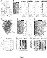
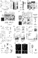
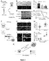


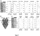

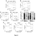
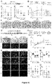

Classifications
-
- A—HUMAN NECESSITIES
- A61—MEDICAL OR VETERINARY SCIENCE; HYGIENE
- A61P—SPECIFIC THERAPEUTIC ACTIVITY OF CHEMICAL COMPOUNDS OR MEDICINAL PREPARATIONS
- A61P35/00—Antineoplastic agents
-
- A—HUMAN NECESSITIES
- A61—MEDICAL OR VETERINARY SCIENCE; HYGIENE
- A61P—SPECIFIC THERAPEUTIC ACTIVITY OF CHEMICAL COMPOUNDS OR MEDICINAL PREPARATIONS
- A61P25/00—Drugs for disorders of the nervous system
-
- A—HUMAN NECESSITIES
- A61—MEDICAL OR VETERINARY SCIENCE; HYGIENE
- A61P—SPECIFIC THERAPEUTIC ACTIVITY OF CHEMICAL COMPOUNDS OR MEDICINAL PREPARATIONS
- A61P35/00—Antineoplastic agents
- A61P35/04—Antineoplastic agents specific for metastasis
-
- C—CHEMISTRY; METALLURGY
- C12—BIOCHEMISTRY; BEER; SPIRITS; WINE; VINEGAR; MICROBIOLOGY; ENZYMOLOGY; MUTATION OR GENETIC ENGINEERING
- C12Q—MEASURING OR TESTING PROCESSES INVOLVING ENZYMES, NUCLEIC ACIDS OR MICROORGANISMS; COMPOSITIONS OR TEST PAPERS THEREFOR; PROCESSES OF PREPARING SUCH COMPOSITIONS; CONDITION-RESPONSIVE CONTROL IN MICROBIOLOGICAL OR ENZYMOLOGICAL PROCESSES
- C12Q1/00—Measuring or testing processes involving enzymes, nucleic acids or microorganisms; Compositions therefor; Processes of preparing such compositions
- C12Q1/68—Measuring or testing processes involving enzymes, nucleic acids or microorganisms; Compositions therefor; Processes of preparing such compositions involving nucleic acids
- C12Q1/6876—Nucleic acid products used in the analysis of nucleic acids, e.g. primers or probes
- C12Q1/6883—Nucleic acid products used in the analysis of nucleic acids, e.g. primers or probes for diseases caused by alterations of genetic material
- C12Q1/6886—Nucleic acid products used in the analysis of nucleic acids, e.g. primers or probes for diseases caused by alterations of genetic material for cancer
-
- C—CHEMISTRY; METALLURGY
- C12—BIOCHEMISTRY; BEER; SPIRITS; WINE; VINEGAR; MICROBIOLOGY; ENZYMOLOGY; MUTATION OR GENETIC ENGINEERING
- C12Q—MEASURING OR TESTING PROCESSES INVOLVING ENZYMES, NUCLEIC ACIDS OR MICROORGANISMS; COMPOSITIONS OR TEST PAPERS THEREFOR; PROCESSES OF PREPARING SUCH COMPOSITIONS; CONDITION-RESPONSIVE CONTROL IN MICROBIOLOGICAL OR ENZYMOLOGICAL PROCESSES
- C12Q2600/00—Oligonucleotides characterized by their use
- C12Q2600/106—Pharmacogenomics, i.e. genetic variability in individual responses to drugs and drug metabolism
-
- C—CHEMISTRY; METALLURGY
- C12—BIOCHEMISTRY; BEER; SPIRITS; WINE; VINEGAR; MICROBIOLOGY; ENZYMOLOGY; MUTATION OR GENETIC ENGINEERING
- C12Q—MEASURING OR TESTING PROCESSES INVOLVING ENZYMES, NUCLEIC ACIDS OR MICROORGANISMS; COMPOSITIONS OR TEST PAPERS THEREFOR; PROCESSES OF PREPARING SUCH COMPOSITIONS; CONDITION-RESPONSIVE CONTROL IN MICROBIOLOGICAL OR ENZYMOLOGICAL PROCESSES
- C12Q2600/00—Oligonucleotides characterized by their use
- C12Q2600/158—Expression markers
Definitions
- the present invention relates to methods and compositions for use in the treatment and/or prevention of neuroblastoma malignancies. More specifically, it concerns the use of an activator of a gene belonging to the SA-c1 module in the treatment and/or prevention of neuroblastoma malignancies in a subject.
- NB Neuroblastoma
- LR-NBs low-to-intermediate risk
- HR-NBs high-risk tumours
- NBs Treatment of NBs depends on their level of risk and includes for example surgery, radiotherapy, chemotherapy, combination of chemotherapy with monoclonal antibody therapy (dinutuximab) and targeted therapy.
- these genetic alterations are only found in up to 16% of all NB cases and are thus insufficient to explain the differences in the aetiology and malignancy of HR-NBs relative to LR-NBs.
- NBs originate from alterations to cells in the sympatho-adrenal (SA) lineage, one of the multiple cell fate branches of neural crest cells.
- SA lineage is composed of four cell identities: Schwann cell precursors, bridge cells, chromaffin cells and sympathoblasts.
- NB cell-of-origin There is some debate regarding the specific identity of the NB cell-of-origin.
- adrenal NBs derive from fetal chromaffin cells, and that LR-NBs and HR-NBs can be discriminated based on the degree of chromaffin cell differentation.
- Another study pinpointed instead fetal sympathoblasts as the NB cell-of-origin, yet could not discriminate NB subtypes based on this signature.
- NB is a devastating paediatric cancer originating in the sympatho-adrenal lineage derived from the neural crest.
- HR-NBs are metastatic and display a strong resistance to therapy and high rates of relapse, leading to poor prognosis. Identifying the molecular signatures that discriminate LR- from HR-NBs should thus facilitate the design of efficient therapies against aggressive NB tumours.
- the inventors By comparing the transcriptional landscapes of LR- and HR-NBs with that of the healthy human fetal adrenal gland (a major site of NB formation), the inventors have identified the transcriptional signatures associated to the formation of both LR- and HR-NBs. Among these genes relevant to NB formation, the inventors have further identified 503 genes, organised into four different clusters (c1-c4), which are differentially expressed between LR-NBs and HR-NBs (LR vs HR DEGs). The main cluster c1 is particularly interesting in that it identified 338 genes that are expressed at significantly lower levels in HR-NBs than in LR-NBs, so that these c1 genes might impede the formation of metastases specifically observed in HR-NBs.
- LR-NBs and HR-NBs can be discriminated by a transcriptional signature corresponding to the core SA lineage and consisting of 4,518 genes shared by at least 3 of the 4 cell types forming the human SA lineage.
- a transcriptional signature corresponding to the core SA lineage and consisting of 4,518 genes shared by at least 3 of the 4 cell types forming the human SA lineage.
- more than half of the 503 LR vs HR DEGs were retrieved in this core SA developmental signature.
- 92% of this core SA signature corresponded to genes belonging to the cluster c1 (242 genes out of 262, hereafter named "SA-c1" genes or SA-c1 module).
- the inventors demonstrate here for the first time that the transcriptional landscapes of LR-NB and HR-NBs can be discriminated based on the SA-c1 signature corresponding to the core SA lineage, and that this developmental program is unexpectedly associated with a favourable prognosis.
- said genes are strikingly predictive of NB malignancy, patient prognosis and disease progression.
- One of these genes, encoding the secreted Neurexophilin-1 (NXPH-1) protein, and its receptor ⁇ -NRXN1 have been further characterized in functional assays. The results obtained confirmed the dual role of these two molecules; the interaction between the ligand NXPH1 and its receptor ⁇ -NRXN1 and the subsequent signalling cascade promote NB growth while unexpectedly inhibiting the formation of metastases.
- the findings thereby uncover the genes comprising the SA-c1 module, and more specifically NXPH1/ ⁇ -NRXN1 signalling, as possible therapeutic targets for the treatment of HR-NBs. Additionally, the findings shed a new light on the complex contribution of the neural crest-derived developmental program to NB pathogenesis and reveal that the developmental SA program appears to facilitate NB formation, but also to concomitantly restrict its malignant potential.
- the present invention thus provides activators of the genes comprising the SA-c1 module for use in the treatment of NB malignancies, particularly HR-NBs. The activators of the invention are particularly helpful for NB patients showing at least one metastasis, and for those patients affected by NB tumours displaying a strong resistance to conventional therapy.
- the invention discloses an activator of a gene belonging to the SA-c1 module, or a pharmaceutical composition comprising said activator in a therapeutically effective amount, for use in the treatment of NB malignancies in a subject, wherein the gene is selected from the SA-c1 module listed in Table 1.
- Table 1 SA-c1 genes ranked by their fold change expression between LR-NBS and HR-NBs (highest #1 to lowest #242).
- the gene is selected from the group consisting of: Transcription factor SOX-6 (SOX6; GenelD 55553, updated 26.04.21); Neurexophilin-1 (NXPH1; GenelD 30010, updated 26.04.21); Peripherin (PRPH; GenelD 5630, updated 26.04.21); Transcription Factor AP-2 Beta (TFAP2B; GenelD 7021, updated 26.04.21 ); Ephrin type-A receptor 7 (EPHA7; GenelD 2045, updated 26.04.21 ); Sodium channel protein type 2 subunit alpha (SCN2A; GenelD 6326, updated 26.04.21); V-set and transmembrane domain-containing protein 2A (VSTM2A, Gene ID 222008, updated 26.04.21 ); Sodium channel protein type 9 subunit alpha (SCN9A; Gene ID 6335, updated 26.04.21 ); Plexin-A4 (PLXNA4; Gene ID 91584, updated 26.04.
- core SA signature refers to the transcriptional signature shared by at least 3 of the 4 cell sympatho-adrenal cell types involved the development of medulla of the human adrenal gland.
- the cluster c1 contains the 338 genes that are i) expressed at higher levels in both LR-NB and HR-NB samples compared to samples of the healthy fetal adrenal gland, and ii) expressed at higher levels in LR-NBs than in HR-NBs.
- the SA-c1 module is a sub-cluster of the cluster c1, comprising 242 genes characterized as relevant in NB formation.
- This sub-cluster comprises transcription factors (TFs) SOX6 and TFAP2, involved in the early stages of the NCC lineage; PRPH and the TFs GATA2 and FOXO3, which are associated with chromaffin cell differentiation; and various autism spectrum disorder genes (CNTNAP2, CTNND2, DLG2, NRXN2 and SHANK2) recently associated with NB aetiology.
- polypeptide(s) and protein(s) are used interchangeably.
- protein(s) of interest is understood as those proteins encoded by the corresponding genes of interest.
- gene(s) of interest is understood as any gene selected from the SA-c1 module (Table 1).
- NXPH1 One of the genes comprising the SA-c1 module is NXPH1.
- the protein NXPH1 encoded by the gene is also known as NPH1 (UniProt ID P58417 V131, last modified 12.08.20).
- NXPHs The ⁇ -NRXN-specific ligands (NXPHs) comprise a family of four glycoproteins (NXPH1-4). In contrast to other ⁇ -NRXN ligands expressed throughout the brain in virtually all excitatory and inhibitory neurons, the NXPH family is restricted to certain inhibitory neuronal subpopulations. While the function of NXPHs is poorly understood, data suggest that they block the interaction of ⁇ -NRXNs with post-synaptic proteins within the neural system.
- NXPH1-3 bind ⁇ -NRXN1.
- NXPH1-3 are small 29 kDa, secreted, neuropeptide-like proteins found in a variety of tissues. NXPHs are preferentially expressed in the brain but they are also present in non-neural tissues in a species-specific pattern. In the mouse, NXPH1-3 are expressed primarily in small subsets of inhibitory neurons of the nervous system. In humans, the strongest expression of NXPH1 is detected in the adrenal gland.
- NXPH1-3 are highly evolutionarily conserved in mammals. Whilst NXPH1 is the best-characterized member of the family, NXPH2 and NXPH3 are expected to undergo a similar maturation process due to their high degree of sequence conservation and to present comparable, even possibly overlapping, functions. Each gene codes for a variable N-terminal region and a highly conserved C-terminal region. In neural tissues, the immature form of NXPH1 undergoes N-glycosylation at its C-terminal domain and a proteolytic cleavage of the N-terminal domain, thus giving rise to a mature neuropeptide-like form that binds to the extracellular domain of the transmembrane receptors ⁇ -NRXNs.
- NXPH1 plays a role in synaptogenesis and synapse maturation by regulating the accessibility of other ligands to the extracellular domain of ⁇ -NRXNs. To date, there is no evidence that NXPH1 plays other functions and the intracellular signaling consequences of its binding to ⁇ -NRXNs remain unknown. However, it is worth noting that a loss of NXPH1 expression correlates with the malignant progression of both pancreatic ductal adenocarcinoma and breast cancer. NXPH1 has also been proposed as a DNA methylation biomarker associated with a good prognosis for NB patients.
- NRXN In mammals, the highly conserved NRXN family consists of three genes: neurexin 1, neurexin 2 and neurexin 3. Each NRXN gene possesses two independent promoters driving the expression of 2 different isoforms: alpha ( ⁇ )- and beta ( ⁇ )-NRXNs. All the NRXN isoforms are type-1 transmembrane proteins containing an isoform-specific extracellular domain followed by a single transmembrane region and a short cytoplasmic PDZ domain. The ⁇ -NRXNs represent an N-terminal truncated form of ⁇ -NRXNs and present a specific short N-terminal sequence.
- NRXNs are widely expressed in the nervous system, where all three genes (NRXN1-3) are transcribed at similar levels and play key roles in synaptic formation and neurotransmission. NRXNs are located at the synaptic cleft where they constitute trans-synaptic signaling complexes together with different pre- and post-synaptic ligands and transmembrane proteins. Pre-synaptic NRXNs send an instructive signal that induces the initial organization of the post-synaptic bouton. They moreover contribute to synaptic refinement in a neuronal subtype-specific manner. Consistent with their relevant role in synaptic neurotransmission, genetic alterations in the NXRNs genes are associated with various neuropsychiatric and neurodevelopmental disorders.
- the activator of the gene modulates the specification and early development of the neural crest cells and their derivatives.
- the sympatho-adrenal lineage will give rise to the peripheral nervous system (PNS), which comprises the chromaffin cells of the adrenal medulla, the paravertebral sympathetic ganglia chain and additional prevertebral ganglia such as the celiac ganglia, the suprarenal ganglia and the mesenteric ganglia
- PNS peripheral nervous system
- the PNS controls many physiological functions including heart rhythm, blood pressure, perspiration and salivation. Alterations in the formation or function of the PNS cause several pediatric disorders, including NB.
- the present invention comprises the use of a gene activator (hereinafter “activator of the invention”).
- activator of the invention refers to any molecule capable of enhancing the biological activity of the gene by any mode of action, including but not limited to inducing the expression of the gene and increasing the levels of the biological activity of the polypeptide encoded by the gene (hereinafter “protein of interest”).
- the activator of the invention might, for example, and among others, increase the biological activity of an extracellular polypeptide encoded by the gene i.e., NXPH1 or FRZB by binding to and activating its receptor.
- the activators of the invention may be identified using methods based on (i) the analysis of the migratory and invasive capacity of NB tumour cells in vitro, including but not restricted to scratch assays, transmembrane assays, microfluidic chamber assays or cell exclusion zone assays; as previously reported ( Puls et al., 2018 Scientific Reports 8, 13039 .; Evensen et al., 2013 PloS ONE 8(12): e82811 .; and Hulkower, K.I., et al., 2011 Pharmaceutics 3(1): 107-124 ); or (ii) the analysis of the metastatic potential of NB tumour cells in vivo using experimental models of metastasis in mouse and/or chick animal models, as reported by the inventors in their manuscript or by others ( Delloye-Bourgeois et al., 2017 Cancer Cell 32, 427-443.e8 ; Palmer et al., 2011 J Vis Exp 51: 2815
- any activator may be used in the use of the present invention.
- the activator may be selected from the group consisting of: (i) an antibody or fragment thereof; (ii) a small molecule; (iii) an expression activator of the gene of interest; and (iv) an expression activator of a gene encoding a receptor, wherein said receptor binds a polypeptide encoded by NXPH1 or FRZB.
- the activator is an antibody or fragment thereof.
- antibody is understood as a polypeptide including at least a light chain or heavy chain immunoglobulin variable region which specifically recognizes and binds an epitope of a polypeptide, such as a receptor or a fragment thereof.
- Antibodies are composed of a heavy and a light chain, each of which has a variable region, termed the variable heavy (VH) region and the variable light (VL) region. Together, the VH region and the VL region are responsible for binding the antigen recognized by the antibody.
- Antibodies of the present disclosure include those that are specific for the receptors of the extracellular protein of interest (NXPH or FRZB) and enhance the biological activity of the protein of interest.
- antibody includes intact immunoglobulins, as well the variants and portions thereof, such as Fab' fragments, F(ab)'2 fragments, single chain Fv proteins (“scFv”), and disulfide stabilized Fv proteins ("dsFv”).
- scFv protein is a fusion protein in which a light chain variable region of an immunoglobulin and a heavy chain variable region of an immunoglobulin are bound by a linker, while in dsFvs, the chains have been mutated to introduce a disulfide bond to stabilize the association of the chains.
- the term also includes genetically engineered forms such as chimeric antibodies (for example, humanized murine antibodies) or heteroconjugate antibodies (such as diabodies or bispecific antibodies).
- the antibodies can be prepared using any of the methods which are known by the person skilled in the art, some of which have been mentioned above.
- the polyclonal antibodies are prepared by means of immunizing an animal with the protein to be targeted.
- the monoclonal antibodies are prepared using the method described by Kohler and Milstein et al. 1975 (Nature 256: 495-497 ). Once antibodies with a specific receptor binding capacity are identified, those capable of enhancing the activity of the extracellular protein of interest will be selected using a relevant identification assay.
- the antibody may be an agonist.
- the term "agonist” is understood as any molecule capable of binding specifically to the same receptors as an extracellular protein of interest and activating one or more functions of said protein.
- the antibody may bind an inhibitor of a protein of interest.
- inhibitor refers to any chemical or biological substance that reduces or suppresses the activity of the protein of interest.
- the activator may also be a small molecule.
- the activator small molecules according to the invention are molecules, typically with a molecular weight less than about 1000 Daltons, or in some embodiments, less than about 500 Daltons, wherein the molecule is capable of modulating, to some measurable extent, the activity of a target molecule, such as a polypeptide encoded by one of the genes of interest in the present invention.
- the small molecule may be an agonist, as described above.
- (gene) expression activator or any variants thereof is understood as any activator which increases the levels of the expression of a gene, for example by controlling the transcription or the translation of the gene.
- the expression activator may increase the levels of expression of the genes/proteins of interest or the levels of expression of the gene encoding a receptor binding an extracellular protein of interest.
- Methods for inducing the expression of the gene encoding the protein include, without being limited to, editing technologies such as CRISPR/cas9 or Cas9 nickase technology.
- the present invention relates to a gene activator for use in the treatment and/or prevention of NB malignancies.
- treating refers to processes involving a slowing, interrupting, arresting, controlling, stopping, reducing, or reversing the progression or severity of an existing symptom, disorder, condition, or disease, but does not necessarily involve a total elimination of all disease-related symptoms, conditions, or disorders.
- the treatment of a disorder or disease may, for example, lead to a halt in the progression of the disorder or disease (e.g., no deterioration of symptoms) or a delay in the progression of the disorder or disease (in case the halt in progression is of a transient nature only).
- the "treatment" of a disorder or disease may also lead to a partial response (e.g., amelioration of symptoms) or complete response (e.g., disappearance of symptoms) of the subject/patient suffering from the disorder or disease. Accordingly, the "treatment” of a disorder or disease may also refer to an amelioration of the disorder or disease, which may lead to a halt in the progression of the disorder or disease or a delay in the progression of the disorder or disease. Such a partial or complete response may be followed by a relapse. It is to be understood that a subject/patient may experience a broad range of responses to a treatment (such as the exemplary responses as described herein above).
- preventing refers to the capacity of the gene activator to minimize or hinder the progression of a disease, condition or disorder in a subject.
- NB malignancies is used to refer to any malignancies that develop from certain very early forms of nerve cells and chromaffin cells, also known as immature sympatho-adrenal progenitor cells, found in several areas of the body.
- the stage of the disease may be determined according to the International Neuroblastoma Staging System (INSS), developed taking into account the degree of tumour resection, presence of ipsilateral or contralateral lymph nodes involvement, tumour infiltration across the midline of the body, and separation of patients with INSS stage 4S from other children with metastatic disease (INSS stage 4) to harmonize staging across groups.
- INSS stage 4S International Neuroblastoma Staging System
- Table 2 International Neuroblastoma Staging System
- the INRG Staging System is a more recent staging system which utilizes image-defined risk factors in place of degree of surgical resection.
- the different stages and their key characteristics are shown in Table 3.
- Table 3. International Neuroblastoma Risk Group Staging System (INRGSS).
- INRG Stage Description L1 Localized tumour with no image-defined risk factors
- M Distant metastatic disease MS Metastatic disease in children under 18 months with metastases limited to skin, liver, and/or bone marrow ( ⁇ 10% involvement)
- a combination of clinical, pathologic, and genetic markers are used to predict the clinical behavior of the tumour and how it will respond to treatment. These markers are used to define risk, and include age at the time of diagnosis, stage of the disease, histologic category, the grade or how cells of the tumour are differentiated, MYCN gene status, chromosome 11q status, and tumour cell ploidy.
- NB can be classified into one of four categories: very low-risk, low-risk, intermediate-risk, or high-risk.
- HR-NBs include stage L1 with MYCN amplification, stage L2 with MYCN amplification, stage M in children younger than 18 months of age with MYCN amplification, stage M in children with older than 18 months, stage MS in children younger than 18 months with 11q aberration and stage MS in children younger than 18 months of age with MYCN amplification.
- the NB malignancy to be treated is a HR-NB malignancy.
- the stage of the neuroblastoma malignancy is INSS4.
- the NB malignancy carries amplification of the oncogene MYCN.
- the activator inhibits the metastatic potential of the NB malignancy.
- the term 'metastatic potential' refers to the capacity of the NB cells to colonize and grow in organs or locations other than those forming the peripheral nervous system. This includes, but is not restricted to, the bone marrow, the bones, the lymph nodes and/or the liver.
- the term “subject” refers to human and non-human animals, such as veterinary subjects.
- non-human animal includes all vertebrates, e.g., mammals and non-mammals, such as non-human primates, mice, rabbits, sheep, dog, cat, horse, cow, chickens, amphibians, and reptiles.
- the subject is a human, man or woman of any age or race.
- the activator of the invention is comprised within a pharmaceutical composition in a therapeutically effective amount.
- therapeutically effective amount means the amount of activator of the invention that will elicit the biological or medical response of a tissue, system, animal or human that is being sought by the researcher, veterinarian, medical doctor or other clinician.
- the biological or medical response to elicit is the treatment and/or prevention of NB malignancies.
- the activators of the present invention can be incorporated into pharmaceutical compositions for its administration to the subject.
- the activators of the invention may conveniently be presented in dosage unit form and may be prepared by any of the methods well known in the art of pharmacy. All methods include the step of bringing the active ingredient into association with the carrier that constitutes one or more accessory ingredients.
- the pharmaceutical compositions are prepared by uniformly and intimately bringing the active ingredient into association with a liquid carrier or a finely divided solid carrier or both, and then, if necessary, shaping the product into the desired formulation.
- the active compound is included in an amount sufficient to produce the desired effect upon the process or condition of diseases.
- composition is intended to encompass a product comprising the specified ingredients in the specified amounts, as well as any product which results, directly or indirectly, from combination of the specified ingredients in the specified amounts.
- compositions containing the active ingredient may be in a form suitable for oral use, for example, as tablets, troches, lozenges, aqueous or oily suspensions, dispersible powders or granules, emulsions, solutions, hard or soft capsules, or syrups or elixirs.
- Compositions intended for oral use may be prepared according to any method known to the art for the manufacture of pharmaceutical compositions and such compositions may contain one or more agents selected from the group consisting of sweetening agents, flavoring agents, coloring agents and preserving agents in order to provide pharmaceutically elegant and palatable preparations. Tablets contain the active ingredient in admixture with non-toxic pharmaceutically acceptable excipients that are suitable for the manufacture of tablets.
- excipients may be for example, inert diluents, such as calcium carbonate, sodium carbonate, lactose, calcium phosphate or sodium phosphate; granulating and disintegrating agents, for example, corn starch, or alginic acid; binding agents, for example starch, gelatin or acacia; and lubricating agents, for example magnesium stearate, stearic acid or talc.
- the tablets may be uncoated or they may be coated by known techniques to delay disintegration and absorption in the gastrointestinal tract and thereby provide a sustained action over a longer period.
- a time delay material such as glyceryl monostearate or glyceryl distearate may be employed.
- Oral tablets may also be formulated for immediate release, such as fast melt tablets or wafers, rapid dissolve tablets or fast dissolve films.
- Formulations for oral use may also be presented as hard gelatin capsules wherein the active ingredient is mixed with an inert solid diluent, for example, calcium carbonate, calcium phosphate or kaolin, or as soft gelatin capsules wherein the active ingredient is mixed with water or an oil medium, for example peanut oil, liquid paraffin, or olive oil.
- an inert solid diluent for example, calcium carbonate, calcium phosphate or kaolin
- water or an oil medium for example peanut oil, liquid paraffin, or olive oil.
- Aqueous suspensions contain the active materials in admixture with excipients suitable for the manufacture of aqueous suspensions.
- excipients are suspending agents, for example sodium carboxymethylcellulose, methylcellulose, hydroxy-propylmethylcellulose, sodium alginate, polyvinyl-pyrrolidone, gum tragacanth and gum acacia; dispersing or wetting agents may be a naturally-occurring phosphatide, for example lecithin, or condensation products of an alkylene oxide with fatty acids, for example polyoxyethylene stearate, or condensation products of ethylene oxide with long chain aliphatic alcohols, for example heptadecaethyleneoxycetanol, or condensation products of ethylene oxide with partial esters derived from fatty acids and a hexitol such as polyoxyethylene sorbitol monooleate, or condensation products of ethylene oxide with partial esters derived from fatty acids and hexitol anhydrides, for example polyethylene sorbitan monoo
- the aqueous suspensions may also contain one or more preservatives, for example ethyl, or n-propyl, p-hydroxybenzoate, one or more coloring agents, one or more flavoring agents, and one or more sweetening agents, such as sucrose or saccharin.
- preservatives for example ethyl, or n-propyl, p-hydroxybenzoate
- coloring agents for example ethyl, or n-propyl, p-hydroxybenzoate
- coloring agents for example ethyl, or n-propyl, p-hydroxybenzoate
- flavoring agents for example ethyl, or n-propyl, p-hydroxybenzoate
- sweetening agents such as sucrose or saccharin.
- Oily suspensions may be formulated by suspending the active ingredient in a vegetable oil, for example arachis oil, olive oil, sesame oil or coconut oil, or in a mineral oil such as liquid paraffin.
- the oily suspensions may contain a thickening agent, for example beeswax, hard paraffin or cetyl alcohol. Sweetening agents such as those set forth above, and flavoring agents may be added to provide a palatable oral preparation. These compositions may be preserved by the addition of an anti-oxidant such as ascorbic acid.
- Dispersible powders and granules suitable for preparation of an aqueous suspension by the addition of water provide the active ingredient in admixture with a dispersing or wetting agent, suspending agent and one or more preservatives.
- a dispersing or wetting agent e.g., glycerol, glycerol, glycerol, glycerol, glycerol, glycerol, glycerin, glycerin, glycerin, glycerin, glycerin, sorbitol, sorbitol, sorbitol, sorbitol, sorbitol, sorbitol, sorbitol, sorbitol, sorbitol, sorbitol, glycerol, glycerol, glycerol, glycerol, glycerol, glycerol, glycerol, glycerol, glycerol
- the pharmaceutical compositions of the invention may also be in the form of oil-in-water emulsions.
- the oily phase may be a vegetable oil, for example olive oil or arachis oil, or a mineral oil, for example liquid paraffin or mixtures of these.
- Suitable emulsifying agents may be naturally-occurring gums, for example gum acacia or gum tragacanth, naturally-occurring phosphatides, for example soy bean, lecithin, and esters or partial esters derived from fatty acids and hexitol anhydrides, for example sorbitan monooleate, and condensation products of the said partial esters with ethylene oxide, for example polyoxyethylene sorbitan monooleate.
- the emulsions may also contain sweetening and flavoring agents.
- the pharmaceutical compositions may be in the form of a sterile injectable aqueous or oleagenous suspension.
- This suspension may be formulated according to the known art using those suitable dispersing or wetting agents and suspending agents that have been mentioned above.
- the sterile injectable preparation may also be a sterile injectable solution or suspension in a non-toxic parenterally acceptable diluent or solvent, for example as a solution in 1,3-butane diol.
- the acceptable vehicles and solvents that may be employed are water, Ringer's solution and isotonic sodium chloride solution.
- sterile, fixed oils are conventionally employed as a solvent or suspending medium.
- any bland fixed oil may be employed, including synthetic mono- or diglycerides.
- fatty acids such as oleic acid find use in the preparation of injectables.
- the activator of the invention may also be administered in the form of suppositories for rectal administration of the drug.
- suppositories for rectal administration of the drug.
- These compositions can be prepared by mixing the drug with a suitable non-irritating excipient that is solid at ordinary temperatures but liquid at the rectal temperature and will therefore melt in the rectum to release the drug.
- suitable non-irritating excipient that is solid at ordinary temperatures but liquid at the rectal temperature and will therefore melt in the rectum to release the drug.
- Such materials are cocoa butter and polyethylene glycols.
- an appropriate dosage level of the activator of the invention will generally be about 0.01 to 500 mg per kg patient body weight per day, which can be administered in single or multiple doses.
- the compositions may be provided in the form of tablets containing 1.0 to 1000 milligrams of the active ingredient, particularly 1.0, 5.0, 10.0, 15.0, 20.0, 25.0, 50.0, 75.0, 100.0, 150.0, 200.0, 250.0, 300.0, 400.0, 500.0, 600.0, 750.0, 800.0, 900.0, and 1,000.0 milligrams of the activator for the symptomatic adjustment of the dosage to the patient to be treated.
- the specific dose level and frequency of dosage for any particular patient may vary and will depend upon a variety of factors including the activity of the specific compound employed, the metabolic stability and length of action of that compound, the age, body weight, general health, sex, diet, mode and time of administration, rate of excretion, drug combination, the severity of the particular condition, and the host undergoing therapy.
- the activator of the present invention may be used in combination with one or more other drugs in the treatment, prevention, control, amelioration, or reduction of risk of NB malignancies, where the combination of the drugs together are safer or more effective than either drug alone.
- the pharmaceutical composition further comprises a compound for the treatment of NB malignancies in a subject.
- Examples of compounds useful for treating NB malignancies which may be used in conjunction with the activator of the invention include, without being limited to, cisplatin, vincristine, carboplatin, etoposide, cyclophosphamide, dinutuximab and isotretinoin.
- the activators of the present invention may be administered by oral, parenteral (e.g., intramuscular, intraperitoneal, intravenous, ICV, intracisternal injection or infusion, subcutaneous injection, or implant), by inhalation spray, nasal, vaginal, rectal, sublingual or buccal routes of administration and may be formulated, alone or together, in suitable dosage unit formulations containing conventional non-toxic pharmaceutically acceptable carriers, adjuvants and vehicles appropriate for each route of administration.
- parenteral e.g., intramuscular, intraperitoneal, intravenous, ICV, intracisternal injection or infusion, subcutaneous injection, or implant
- inhalation spray nasal, vaginal, rectal, sublingual or buccal routes of administration
- nasal, vaginal, rectal, sublingual or buccal routes of administration may be formulated, alone or together, in suitable dosage unit formulations containing conventional non-toxic pharmaceutically acceptable carriers, adjuvants and vehicles appropriate for each route of administration.
- the activators of the invention are effective for use
- Such other compound(s)/drug(s) may be administered by a route and in an amount commonly used therefore, simultaneously or sequentially with the activator of the invention.
- a pharmaceutical composition in unit dosage form containing such other drugs and the activator is preferred.
- the combination therapy may also include therapies in which the activator of the invention and one or more other drugs are administered on different overlapping schedules.
- the activator of the invention and the other active ingredients may be used in lower doses than when each is used singly.
- the pharmaceutical compositions of the present invention include those that contain one or more other active ingredients, in addition to an activator.
- the activator of the invention is an activator of the NXPH1 gene. In some particular embodiments, the activator of the invention is an activator of the ⁇ -NRXN1 gene.
- the present invention relates to an in vitro method for diagnosing NB malignancies in a subject, comprising: (i) determining the expression levels of at least one gene selected from the group consisting of the genes listed in Table 1 in a biological sample from the subject, and (ii) comparing the expression levels obtained in step (i) with a first reference expression level obtained in a fetal adrenal gland sample from a subject not suffering from NB malignancies, wherein if the expression levels of the at least one gene listed in (i) are increased with respect to the first reference expression level, then the subject is suffering from an NB malignancy.
- the in vitro method for diagnosing NB malignancies in a subject comprises determining the expression levels of at least one gene selected from the group consisting of: SOX6; NXPH1; PRPH; TFAP2B; EPHA7; SCN2A; VSTM2A; SCN9A; PLXNA4; SCN3A; TLN2; CNTNAP2; ADCY1; RGMB; and PSD2 in a biological sample from the subject.
- NXPH1; PRPH; TFAP2B; EPHA7; SCN2A; VSTM2A; SCN9A; PLXNA4; SCN3A; TLN2; CNTNAP2; ADCY1; RGMB; and PSD2 may be associated to both LR-NB and HR-NB malignancies in a subject, increased expression levels of SOX6 are generally associated to LR-NB malignancies, and decreased expression levels of SOX6 are generally associated to HR-NB malignancies.
- the in vitro method for diagnosing NB malignancies in a subject comprises:
- an expression level increased with respect to a reference expression level refers to an expression level that is at least: 5% higher, 8% higher, 10% higher, 15% higher, 20% higher, 25% higher, 30% higher, 35% higher, 40% higher, 45% higher, 50% higher, 55% higher, 60% higher, 65% higher, 70% higher, 75% higher, 80% higher, 85% higher, 90% higher, 95% higher, 96% higher, 97% higher, 98% higher, 99% higher, 100% higher, 1-fold higher, 2-fold higher, 5-fold higher, 10 fold higher, 20 fold higher, 30 fold higher, 40 fold higher, 50 fold higher, 60 fold higher, 70 fold higher, 80 fold higher, 90 fold higher, 100 fold higher, 1000-fold higher, or more, than the first reference expression level.
- the expression levels of at least one gene selected from the group consisting of: NXPH1; PRPH; TFAP2B; EPHA7; SCN2A; VSTM2A; SCN9A; PLXNA4; SCN3A; TLN2; CNTNAP2; ADCY1; RGMB; PSD2; and/or SOX6 are determined in the biological sample from the subject.
- the expression levels of two or more genes selected from the group consisting of: NXPH1; PRPH; TFAP2B; EPHA7; SCN2A; VSTM2A; SCN9A; PLXNA4; SCN3A; TLN2; CNTNAP2; ADCY1; RGMB; PSD2; and/or SOX6 are determined in the biological sample from the subject.
- the expression levels of NXPH1; PRPH; TFAP2B; EPHA7; SCN2A; VSTM2A; SCN9A; PLXNA4; SCN3A; TLN2; CNTNAP2; ADCY1; RGMB; PSD2; and/or SOX6 are determined in the biological sample from the subject.
- the expression levels of NXPH1; PRPH; TFAP2B; EPHA7; SCN2A; VSTM2A; SCN9A; PLXNA4; SCN3A; TLN2; CNTNAP2; ADCY1; RGMB; or PSD2 in the sample are at least 4-fold higher than the first reference expression level.
- the expression levels of NXPH1; PRPH; TFAP2B; EPHA7; SCN2A; VSTM2A; SCN9A; PLXNA4; SCN3A; TLN2; CNTNAP2; ADCY1; RGMB; or PSD2 in the sample are at least 10-fold higher than the first reference expression level.
- the expression levels of SOX6 in the sample are at least 3-fold higher than the first reference expression level.
- an expression level decreased with respect to the first reference expression level refers to an expression level that is at least: 5% lower, 8% lower, 10% lower, 15% lower, 20% lower, 25% lower, 30% lower, 35% lower, 40% lower, 45% lower, 50% lower, 55% lower, 60% lower, 65% lower, 70% lower, 75% lower, 80% lower, 85% lower, 90% lower, 95% lower, 96% lower, 97% lower, 98% lower, 99% lower, 100% lower, 1-fold lower, 2-fold lower, 5-fold lower, 10 fold lower, 20 fold lower, 30 fold lower, 40 fold lower, 50 fold lower, 60 fold lower, 70 fold lower, 80 fold lower, 90 fold lower, 100 fold lower, 1000-fold lower, or more, than the second reference expression level.
- the expression levels of SOX6 in the sample are at least 3-fold lower than the first reference expression level.
- the in vitro method may further comprise: (iii) comparing the expression levels obtained in step (i) with a second reference expression level obtained in a biological sample from a subject diagnosed with a LR-NB malignancy; and (iv) comparing the expression levels obtained in step (i) with a third reference expression level obtained in a biological sample from a subject diagnosed with a HR-NB malignancy, wherein (a) if the expression levels of the genes listed in (i) are decreased with respect to the second reference expression level and comparable to the third reference expression level, then the subject is susceptible to receiving treatment based on an activator of the invention.
- an expression level decreased with respect to the second reference expression level refers to an expression level that is at least: 5% lower, 8% lower, 10% lower, 15% lower, 20% lower, 25% lower, 30% lower, 35% lower, 40% lower, 45% lower, 50% lower, 55% lower , 60% lower, 65% lower, 70% lower, 75% lower, 80% lower, 85% lower, 90% lower, 95% lower, 96% lower, 97% lower, 98% lower, 99% lower, 100% lower, 1-fold lower, 2-fold lower, 5-fold lower, 10 fold lower, 20 fold lower, 30 fold lower, 40 fold lower, 50 fold lower, 60 fold lower, 70 fold lower, 80 fold lower, 90 fold lower, 100 fold lower, 1000-fold lower, or more, than the second reference expression level.
- the expression levels of NXPH1; PRPH; TFAP2B; EPHA7; SCN2A; VSTM2A; SCN9A; PLXNA4; SCN3A; TLN2; CNTNAP2; ADCY1; RGMB; or PSD2 in the sample are at least 2.5-fold lower than the second reference expression level.
- the expression levels of NXPH1; PRPH; TFAP2B; EPHA7; SCN2A; VSTM2A; SCN9A; PLXNA4; SCN3A; TLN2; CNTNAP2; ADCY1; RGMB or PSD2 in the sample are at least 5-fold lower than the second reference expression level.
- the expression levels of SOX6 in the sample are at least 1-fold lower than the second reference expression level.
- the expression levels of SOX6 in the sample are at least 2-fold lower than the second reference expression level.
- An expression level comparable to a reference expression level is any expression level which is not increased or decreased with respect the reference level, as defined herein.
- the determination of the expression levels of the genes of interest may comprise measuring the level of cDNA, the level of mRNA, and/or the level of the protein encoded by said gene.
- the levels of mRNA of said gene can be quantified by means of using conventional methods, for example, methods comprising the amplification of mRNA and the quantification of the product of the amplification of said mRNA, such as electrophoresis and staining, or alternatively, by means of Southern blot and the use of suitable probes, Northern blot and the use of specific probes of mRNA of the gene of interest (gene encoding the protein) or of the corresponding cDNA thereof, S1 nuclease mapping, hybridisation, RT-Q-PCR, micro-array and RNA-sequencing, etc.
- the quantification of the expression of the gene encoding the protein is going to be carried out from the protein of interest, then the isolated biological sample of the subject must be treated to extract the protein of interest.
- Methods for extracting or isolating proteins are known by the person skilled in the art and they are commercially available.
- the levels of gene encoding the protein of interest can be quantified by means of any conventional method that enables said protein to be detected and quantified in a sample of a subject.
- the levels of said protein can be quantified, for example, by means of the use of antibodies with the capacity to bind specifically to the protein of interest and the subsequent quantification of the complexes formed.
- the decreased or increased expression as mentioned above may lead to a decrease or increase in the transcription rate of a gene of about: 5%, 8%, 10%, 15%, 20%, 25%, 30%, 35%, 40%, 45%, 50%, 55%, 60%, 65%, 70%, 75%, 80%, 85%, 90%, 95%, 96%, 97%, 98%, 99%, 100%, 1-fold, 2-fold, 5-fold, 10 fold, 20 fold, 30 fold, 40 fold, 50 fold, 60 fold, 70 fold, 80 fold, 90 fold, 100 fold, 1000-fold, or more, or any value in between these values, when compared to the reference sample.
- the decreased or increased expression may lead to a decrease or increase in the amount of mRNA of a gene of about 5%, 8%, 10%, 15%, 20%, 25%, 30%, 35%, 40%, 45%, 50%, 55%, 60%, 65%, 70%, 75%, 80%, 85%, 90%, 95%, 96%, 97%, 98%, 99% or 100%, 1-fold, 2-fold, 5-fold, 10 fold, 20 fold, 30 fold, 40 fold, 50 fold, 60 fold, 70 fold, 80 fold, 90 fold, 100 fold, 1000-fold, or more, or any value in between these values, when compared to the reference sample.
- the decreased or increased expression may lead to a decrease or increase in the amount of the polypeptide encoded by the gene of about 5%, 8%, 10%, 15%, 20%, 25%, 30%, 35%, 40%, 45%, 50%, 55%, 60%, 65%, 70%, 75%, 80%, 85%, 90%, 95%, 96%, 97%, 98%, 99% or 100%, 1-fold, 2-fold, 5-fold, 10 fold, 20 fold, 30 fold, 40 fold, 50 fold, 60 fold, 70 fold, 80 fold, 90 fold, 100 fold, 1000-fold, or more, or any value in between these values, when compared to the reference sample.
- reference level refers to the expression levels of the gene encoding the protein of interest in a sample of a subject other than the subject being diagnosed with the method of the invention.
- first reference level refers to the expression levels of the gene encoding the protein of interest in a fetal adrenal gland sample from a subject not suffering from NB malignancies ("control individual/s").
- second reference level refers to the expression levels of the gene encoding the protein of interest in a sample of a subject diagnosed with a LR-NB malignancy.
- third reference level refers to the expression levels of the gene encoding the protein of interest in a sample of a subject diagnosed with a HR-NB malignancy.
- biological sample refers to any material comprising a nucleic acid and/or a polypeptide of interest.
- biological samples include, but without limiting to, a biopsy sample, a tissue (e.g., brain tissue), cell or fluid (e.g., serum, saliva, semen, sputum, cerebral spinal fluid (CSF), tears, mucus, sweat, brain extracts and the like) and blood (e.g., PBMCs (peripheral blood-derived mononuclear cells) such as neutrophils, monocytes).
- tissue e.g., brain tissue
- cell or fluid e.g., serum, saliva, semen, sputum, cerebral spinal fluid (CSF), tears, mucus, sweat, brain extracts and the like
- blood e.g., PBMCs (peripheral blood-derived mononuclear cells) such as neutrophils, monocytes).
- PBMCs peripheral blood-derived mononuclear cells
- the levels of expression of the gene of interest are determined in a blood sample from the subject.
- the levels of expression of NXPH1 and FRZB are determined in a blood sample from the subject.
- the subject is susceptible to receive a therapy based on an activator of the invention.
- the activator is selected from the group consisting of (i) an antibody or fragment thereof; (ii) a small molecule; (iii) an activator of the gene expression; and (iv) an expression activator of a gene encoding a receptor, wherein the receptor binds a polypeptide encoded by NXPH1 or FRZB.
- the subject is a human subject.
- the NB malignancy of the in vitro method is a HR-NB malignancy, as defined above.
- the gene of interest is NXPH1.
- the present invention also encompasses a method of treating or preventing NB malignancies in a subject, comprising enhancing the activity of the gene of interest, as described herein, and the use of a gene activator as described herein in the manufacture of a medicament for the treatment of NB malignancies in a subject. All the preferred embodiments and explanations of the terms used in these inventive aspects have been explained above.
- Fertilized white Leghorn chicken eggs were provided by Granja Gibert, rambla Regueral, S/N, 43850 Cambrils, Spain. Eggs were incubated in a humidified atmosphere at 38oC in a Javier Masalles 240N incubator, manipulated at embryonic day 10 (E10) and sacrificed at E17. Sex was not identified.
- mice 5 weeks-old female NOD/SCID mice (NOD.CB17-Prkdcscid /NCrHsd; RRID: IMSR_ARC: NODSCID) and 7 weeks-old male NSG mice (NOD.Cg-Prkdcscidll2rgtm1Wjl/SzJ; RRID: BCBC_4142) provided by Envigo were used for subcutaneous xenografting and metastasis analyses, respectively. Mice were housed under a regimen of 12 hours light/12 hours dark cycles in specific pathogen-free conditions. Sterile dry pellets and water was administered ad libitum. Animals were sacrificed by cervical dislocation or CO2 administration at the end of the experiment or when required for ethical reasons.
- NB-PDX models (HSJD-NB-012, HSJD-NB-011 and HSJD-NB-007) were established, maintained and provided by A.M.C under a local animal care and use committee-approved protocol (135/11). All NB-PDX models were derived from patients with progressive stage INSS4 disease, refractory to all treatments.
- HSJD-NB-011 was established from a 2.5-year-old male, with amplification of MYCN gene, TP53 wild type and ALK-mutated (I1171N).
- HSJD-NB-007 was established from a 5-year-old male with amplification of MYCN gene and no mutations in TP53 and ALK.
- HSJD-NB-012 was established from a 4-year-old male and its genetic profile is unknown.
- HSJD-NB-011 and HSJD-NB-007 have been previously used for publication ( Monterrubio et al, J Control Release. 2017;255:108-19 ; Boeva et al, Nat Genet. 2017;49(9):1408-13 ).
- Experimental manipulation of NB-PDXs was performed in HSJD under the supervision of A.M.C.
- tumours were dissected off the flank of immunocompromised nude mice, minced using razor blades in a sterile dish and transferred to a 50ml falcon tube.
- Cells were dissociated following manufacturer instructions using the Brain Tumour Dissociation Kit and a gentle MACS dissociator (Milteny Biotec #130-095-942 and #130-093-235). Red blood cells were removed by using ACK buffer.
- a single cell suspension weas prepared in FACS buffer (ice-cold DMEM-5%FBS) containing 10 ⁇ M of Rock inhibitor (SIGMA #Y0503) to a final concentration of 1 ⁇ 107 cells/ml and stained for flow cytometry analysis as explained below
- HEK-293 (RRID: CVCL_0045), LAN-1 (RRID: CVCL_1827), IMR-5 (RRID: CVCL_1306), SH-SY5Y (RRID: CVCL_0019), SK-N-AS (RRID: CVCL_1700), SK-N-JD (RRID: CVCL_WH12), SK-N-LP (RRID: CVCL_WH13), LA1-5s (RRID: CVCL_2549), SK-N-SH (RRID: CVCL_0531), SL-N-Be(2)c (RRID: CVCL_0529) and IMR-32 (RRID: CVCL_0346) cell lines were obtained from Marian Mart ⁇ nez-Balbás (IBMB-CSIC), Cinzia Lavarino (HSJD) and Joan Xavier Comella (VHIR).
- NB cell lines were grown in either RPMI 1640-Glutamax (ThermoFisher Scientific cat# 61870010) supplemented with 10% (LAN-1, IMR-5, SH-SY5Y, SK-N-AS, SK-N-JD, SK-N-LP, LA1-5s) or 20% (SK-N-SH, SK-N-Be(2)c, IMR-32) foetal bovine serum (FBS; Cultek cat# 16SV30160.03RYB35908) plus 1% penicillin/streptomycin antibiotic cocktail (ThermoFisher Scientific cat# 15140-122).
- the HEK-293 cell line was grown in DMEM-Glutamax (Lifetechnologies cat#61965-026) supplemented 10% FBS and 1% penicillin/streptomycin cocktail. Unless indicated, cells were grown in standard culture conditions. The presence of mycoplasma contamination was routinely checked by immunofluorescence. Cell lines have not been re-authenticated for the present paper.
- Genome-wide transcriptomic analyses were performed using the web-based Phantasus software (https://artyomovlab.wustl.edu/phantasus; version 1.5.1) (Zenkova et al, 2018; Available from: https://genome.ifmo.ru/phantasus). Normalized log expression values were obtained using the Quantile Normalize Adjustment tool. The Maximun Median Probe method was then selected to collapse the different probes of a single gene. Finally, genes were filtered based on raw expression levels and the 12,000 most-expressed genes from each array were selected for subsequent analysis. Sample dispersion was then assessed using the Principal Component Analysis method and detected outliers were removed from further analysis.
- DEGs Differentially-expressed genes between groups were identified using the Limma R package.
- the genes were considered as differentially expressed when the false discovery rate (FDR)-adjusted P-value (with Benjamini-Hochberg procedure) was ⁇ 0.05.
- FDR false discovery rate
- P-value Benjamini-Hochberg procedure
- GSEA Gene set enrichment analyses
- Gene ontology analysis Gene ontology (GO) term enrichment analyses (biological process) were performed with the PANTHER classification system (http://pantherdb.org) ( Mi et al, Nucleic Acids Res. 2019;47(D1):D419-D426 ; Su et al, Genome Biol. 2014;15((12)):523 ). The 50 most-enriched GO terms were used to assess GO term distribution and were annotated with the Ancestor Chart tool of QuickGO (https://www.ebi.ac.uk/QuickGO).
- NB patient microarray SEQC 498 (GSE62564) was obtained from the R2 genome analysis and visualization platform (https://hgserver1.amc.nl/cgi-bin/r2/main.cgi).
- the R2-web based application was used to generate Kaplan-Meier event-free survival curves. Data were grouped based on the expression levels of either NXPH1, NRXN1-3 or the full SA-c1 gene module. In the latter case, the SEQC cohort was subdivided into quartiles based on the combined expression of the 242 SA-c1 genes, considering as "high” or "low” the expression of each SA-c1 gene with respect to its own average expression level.
- the quartiles Q1-Q4 thereby consisted of 123-126 samples showing numbers of SA-c1 genes expressed at high levels as follows: 487 ⁇ Q1 ⁇ 360; 359 ⁇ Q2 ⁇ 250; 249 ⁇ Q3 ⁇ 135 and 134 ⁇ Q4 ⁇ 4.
- Statistical significance was automatically assessed by the R2-server using a log-rank test.
- the R2-web based application was also used to compare the distribution of patients among different NB prognosis groups according to expression levels of the SA-c1 gene module or NXPH1.
- the category "progression" included tumors that did not respond to therapy plus those that were recurrent despite an initial therapeutic response.
- Statistical significance was automatically assessed by R2-server using the Chi-square + Fisher's test.
- Plasmids and lentiviral infection Inhibition of NXPH1 and ⁇ -NRXN1 expression was triggered by lentiviral infection of short-hairpin constructs inserted into in pLKO.1 or doxycycline-inducible pSLIK-Neo vectors ( Moffat et al, Cell. 2006;124(6):1283-98 ; Shin et al, Proc Natl Acad Sci U S A. 2006;103(37):13759-64 ).
- the pLKO.1-TRC cloning vector (RRID: Addgene_10878) and the pLKO.1-sh-Ctl (RRID: Addgene_10879) were kindly provided by Marian Mart ⁇ nez-Balbás.
- Doxycycline-inducible miRshRNA-eGFP-expressing pSLIK-Neo lentiviral vectors were generated by gateway recombination between the TTRE-eGFP-miR-shRNA entry vector and the pSLIK destination vector ( Shin et al, Proc Natl Acad Sci U S A. 2006;103(37):13759-64 ).
- pEN_TTGmiRc2 and pSLIK-Neo were purchased from Addgene (Cat#25753, RRID: Addgene_25752 and Cat#25735, RRID: Addgene_25735; gifts from Ian Fraser).
- NXPH1 and ⁇ -NRXN1-specific sh-RNA sequences were designed using the web-based Genetic Perturbation Platform of the Broad Institute (https://portals.broadinstitute.org/gpp/public/). Expression of luciferase was triggered by lentiviral infection of pLEX-hFL2iG vector.
- Lentiviral particles were generated using the psPAX2 (RRID: Addgene_12260) and pMD2.G plasmids (RRID: Addgene_12259) kindly provided by Marian Mart ⁇ nez-Balbás (IBMB-CSIC). Briefly, HEK293T cell line was transfected using the Lipofectamine 2000 Tranfection Reagent (ThermoFisher Scientific #11668019) following manufacturer's instruction and lentiviruses were harvested 48h post-transfection. Transduction of target cells was usually performed readily after lentivirus recovery. 2 ⁇ g/ml of puromycin (SIGMA #p8833) and 600 ⁇ g/ml of neomycin/G418 (SIGMA-ALDRICH #A1720) were used for selection of transduced cells.
- RNA purification and RT-qPCR Total RNA was extracted with TRIzol Reagent (ThermoFisher Scientific #15596-018) and Pellet Paint Co-precipitant (Merck Millipore #69049-3). Genomic DNA traces were removed with DNA-free DNase Treatment and Removal Reagents (FisherScientific #AM1906) following manufacturer's instructions. DNA-free RNA was retro-transcribed using the High-Capacity cDNA Reverse Transcription Kit (LifeTechnlogies #4368814). Semi-quantitative PCR was performed using the Lightcycler 480 SYBR Green 2x Master (Roche #04887352001) on a Roche Lightcycler 480 Real-Time PCR System.
- primers were design using the UCSC Genome browser and Primer3Plus online softwares. 18SrRNA or TBP were used as housekeeping genes. Relative cDNA levels were calculated using the efficiency-corrected ⁇ Ct method ( Bookout et al, Curr Protoc Mol Biol. 2006;15:15.8 ). Heatmap representations were performed using GraphPad Prism v6 (RRID: SCR_002798).
- Fluorescence-activated cell sorting All experiments were conducted using a BD FACSAria Fusion at the Cytometry Core Facility (Centres Cient ⁇ fics i Tecnol ⁇ gics) of Universitat de Barcelona.
- NB cells were grown in sphere-forming media (SFM) containing DMEM/F12 (ThermoFisher Scientific #11330-032), 10% KnockOut serum replacement (ThermoFisher Scientific #10828012), 2mM Glutamax (ThermoFisher Scientific #35050038), 1% MEM non-essential amino acids (ThermoFisher Scientific #11140035), 0.1mM ⁇ -mercaptoethanol (SIGMA-ALDRICH #M3148), 1% penicillin/streptomycin cocktail (ThermoFisher Scientific #15140-122) and 0.1ng/ml basic FGF (ThermoFisher Scientific #PHG0021).
- SFM sphere-forming media
- DMEM/F12 ThermoFisher Scientific #11330-032
- 10% KnockOut serum replacement ThermoFisher Scientific #10828012
- 2mM Glutamax ThermoFisher Scientific #35050038
- RNA extraction To assess mRNA expression in stem cell-enrichment conditions, 3 ⁇ 106 cells were grown on uncoated 60mm plates in 2ml SFM. Spheres were allowed to grow for 5 weeks and were photographed using an inverted bright-field microscope (Leica DMIRBE). Viable spheres were recovered by centrifugation at 100g for 5min at 4oC) and processed for RNA extraction as described below.
- rat monoclonal anti-BrdU [clone BU1/75 (ICR1)] (BioRad Cat #MCA 2060, RRID: AB_323427), rabbit monoclonal anti-active Caspase-3 (clone C92-605) (BD PharMingen Cat#559596, RRID: AB_397274), rabbit polyclonal anti-phospho-Histone H3 (Ser10) (Upstate Cat#06-570, RRID: AB_310177), rat monoclonal anti-phospho-Histone H3 (pSer28) (clone HTA28) (Sigma Cat#H9908, RRID: AB_260096), mouse monoclonal anti-Human Nuclear Antigen (clone 235-1) (Abcam Cat#ab191181, RRID: AB_2492189), rabbit polyclonal anti-Ki67 (Abcam Cat#ab66155; RRID: AB:1140752), mouse monoclonal anti
- Optical sections of NB tumour graft samples were acquired at RT with the Leica LAS software (RRID: SCR_013673), in a Leica TCS SP5 confocal microscope using 20x (dry/HC PL APO 20x/0.70 CS ⁇ /0.17/C) or 40x (oil /HCX PL APO 40X/1.25-0.75 OIL CS ⁇ /0.17/D) objective lenses or with the LSM Software ZEN 2.1 (RRID: SCR_018163), in a ZEISS Lsm780 confocal microscope using 25x (oil,w,Glic, NA 0.8, Plan-Apochromat/ImmKorr) or 40x (oil/NA 1.3/Plan-Apochromat/(UV)VIS-IR).
- Leica LAS software RRID: SCR_013673
- 20x dry/HC PL APO 20x/0.70 CS ⁇ /0.17/C
- 40x oil /HCX PL
- NB tumour grafts were acquired as tile scans using the motorized XY stage. Tiles were directly stitched into a single mosaic by the microscope software. Maximal projections obtained from 3-6 ⁇ m Z-stack images were processed with ImageJ for image merging, resizing and cell counting. Cell counting was performed in a minimum of two sections per tumour.
- Two Fiji macros were developed for cell counting. The first one uses the DAPI channel to create a binary mask containing all the nuclei; segmentation is based on local contrast enhancing, filtering and a manual pipeline for fine tuning. The second one uses the so-produced binary mask to extract the nuclei selections and collate them against the original images, in order to intensity threshold the nuclei for the three markers used: GFP, Ki67 and pH3.
- the macro delivers a result table and a processed compound image with all the nuclei labelled according to their content. Codes and the details on the procedure can be downloaded at https://github.com/MolecularlmagingPlatformlBMB/CellProliferationAssay.
- SK-N-SH clones carrying a conditional knockdown of NXPH1 (sh-NXPH1), ⁇ -NRXN1 (sh- ⁇ NRXN1) or a scramble control sequence (sh-Ctrl) were first activated in vitro in presence of 1mg/ml doxycycline (ACEFESA #PAA29510025) for 96 hours.
- Pre-activated cells were then sorted by FACS based on eGFP expression, and resuspended in grafting media consisting of DMEM/F12, 7mg/ml Matrigel (Fisher Scientific #10365602, 1mg/ml doxycycline and 10 ⁇ M of Rock inhibitor (SIGMA #Y0503) at a concentration of 0.5 ⁇ 107 cells/ml. Then, 100 ⁇ l cell suspensions were grafted subcutaneously using a 0.5ml BD Micro-Fine 29G insulin syringe. Each animal received 2 grafts, one on each flank.
- a fresh solution containing 2mg/ml of doxycycline dissolved in 7.5% sucrose-bearing sterile water was administered ad libitum in drinking water, its administration starting 2 days prior to xenografting.
- Mouse weight and tumour growth were monitored twice a week until tumour detection. From this point, the tumour volume was measured every two days with a digital calliper and mice were sacrificed once the tumour volume reached ethical permission limits. Tumours were then resected, weighted and photographed.
- SK-N-SH clones carrying a luciferase cassette and a conditional knockdown of NXPH1 (sh-NXPH1), ⁇ -NRXN1 (sh- ⁇ NRXN1) or a scramble control sequence (sh-Ctrl) were first activated in vitro in presence of 1mg/ml doxycycline for 96 hours.
- NB cells To test the metastatic potential of NB cells, 2 ⁇ 105 cells resuspended in 100 ⁇ l of PBS were injected into the left cardiac ventricle of 7 weeks-old male NSG mice (NOD.Cg-Prkdcscid ll2rgtm1Wjl/SzJ) with a 26G 1 ⁇ 2 13mm needle, using a total of 7 injected mice per experimental condition. 2mg/ml of doxycycline dissolved in 7.5% sucrose-bearing sterile water was administered ad libitum in drinking water as explained before.
- Chick CAM assay At E10, a small window was created on the egg shell using a sterile scalpel. Cells resuspended in grafting media consisting of DMEM/F12, 7mg/ml Matrigel and 10 ⁇ M of Rock inhibitor were implanted onto the surface of the chorio-allantoid membrane (CAM) by gently scraping the upper CAM layer (avoiding bleeding or visible rupture of the capillaries).
- CAM chorio-allantoid membrane
- tumour grafts were removed from the CAM and tumours were photographed, weighted and measured using a digital calliper (VWR).
- Tumours were then fixed in 4% PFA for 1h30min at 4°C, washed in PBS1x and sequentially cryo-protected with PBS solutions containing 15% and 30% sucrose. Small cubical tumour pieces (5mm ⁇ 5mm ⁇ 5 mm approx.) were then embedded in Tissue-tek (Sakura Finetek #4583) and frozen on dry ice. Tumour pieces were sectioned on a cryostat (Leica) at 16 ⁇ m, collected serially on home-made TESPA pre-coated slides and slides stored at -20°C. Immunofluorescence was performed as described above.
- mice were carried out in accordance with the European Union guidelines (Directive 2010/63/EU) and according to the guidelines from the Animal Care Committee at the Generalitat de Catalunya. The studies were approved by the ethics committee of the Pare Cient ⁇ fic de Barcelona and by the Pare de Recerca Biomèdica de Barcelona (PRBB) animal facility policy. According to Spanish animal care guidelines, no approval was required to perform the experiments involving chicken embryos herein.
- Human NB-PDX models (HSJD-NB-012, HSJD-NB-011 and HSJD-NB-007) were established, maintained and provided by A.M.C under a local animal care and use committee-approved protocol (135/11).
- Example 1 NB formation is correlated with a neural signature accentuated in LR-NBs.
- PCA principal component analysis
- the DEGs found upregulated in these two comparisons were both characterized by a marked enrichment in GO terms related to the nervous system, or to the cell cycle and DNA repair ( Fig. 1D, E ).
- the GO terms related to the nervous system represented 68% of the top 50 GOs for the LR vs fAG comparison but only 18% for the HR vs fAG comparison ( Fig. 1D, E ), indicating that the formation of both LR- and HR-NBs is associated to a neural signature, and that this signature is more pronounced in LR-NBs.
- This comparison identified a list of 503 LR vs HR DEGs that an unbiased hierarchical clustering subdivided into 4 clusters of genes presenting distinct expression profiles ( Fig. 1F ).
- the clusters c1 and c2 consisted of genes more strongly expressed in both LR-NBs and HR-NBs than in fAG samples.
- the best represented cluster (c1) contained 338 genes that were more strongly expressed in LR-NBs than in HR-NBs and were associated with a marked enrichment in GO terms related to the nervous system ( Fig. 1F, G ).
- the expression levels of the top 30 genes found in cluster c1 were all correlated with a favorable patient prognosis, as shown by Kaplan-Meyer analyses done with an independent cohort of 498 NB patients ( Fig. 6F ).
- the 33 genes found in cluster c2 were more strongly expressed in HR-NBs than in LR-NBs and presented the features of oncogenes, as shown by their GO term enrichment profile and Kaplan-Meyer analyses ( Fig. 1F, G , Fig. 6F ).
- the clusters c3 and c4 consisted of genes expressed more weakly in both LR-NBs and HR-NBs than in fAG samples.
- the 32 genes found in cluster c3 presented the features of tumor suppressors, while the cluster c4 contained 100 genes that were more expressed in HR-NBs than in LR-NBs and were mainly correlated with a bad prognosis ( Fig. 1F, G , Fig. 6F ).
- Our analysis thus identified a complex transcriptional signature relevant to NB formation that furthermore distinguishes LR-NBs from HR-NBs.
- 67% of the 503 genes found in this signature, which formed the cluster c1 are both associated with a neural identity and a better patient outcome.
- Example 2 The core sympatho-adrenal signature is enriched in LR-NBs and associates with better patient prognosis.
- SA sympatho-adrenal
- the signatures of the non-adrenal medulla cell types identified in the developing human adrenal gland did not show any enrichment, except the adrenal cortex signature which was enriched in genes expressed at higher levels in HR-NBs ( Fig. 7A ).
- the full SCP, bridge cell, chromaffin cell and sympathoblast signatures were largely overlapping ( Fig. 2C ).
- SA-c1 genes common to the core SA signature and to the LR vs HR DEGs of the cluster c1 (thereafter named "SA-c1" genes) were found for instance the transcription factors (TFs) SOX6 and TFAP2 involved in the early stages of the NCC lineage ( Dong et al, Cancer Cell. 2020;38(5):716-733.e6 ; Rothstein et al, Genome Res. 2020;30(1):35-48 ), PRPH and the TFs GATA2 and FOXO3, which are associated with chromaffin cell differentiation ( Dong et al, Cancer Cell.
- TFs transcription factors
- the expression levels of the SA-c1 module correlated to patient prognosis more strongly than the INSS classification.
- the group of INSS4 samples from the quartile Q1 presented a higher EFS probability than the groups of INSS1-3 samples from the quartiles Q3 and Q4 ( Fig. 9A, B ).
- the SEQC cohort based on the progression of the disease, including within the category "progression" the tumors that did not respond to therapy plus those that were recurrent despite an initial therapeutic response.
- We thereby observed an inverse correlation between disease progression and the combined expression of the SA-c1 genes Fig. 2H ).
- the module of SAc1 genes related to the core sympatho-adrenal program is thus strikingly predictive of NB malignancy, patient prognosis and disease progression.
- the SA-c1 genes might thus play a crucial role in restraining NB malignancy.
- Example 3 The expression of NXPH1 and its receptors ⁇ -NRXN1 / 2 associates with favorable patient prognosis and identifies NB cells with a neural crest stem cell identity.
- NXPH1 Neurexophilin-1
- NXPH1 is mostly expressed in the nervous system and encodes a secreted glycoprotein that specifically binds to and modulates the activity of the ⁇ -Neurexin transmembrane receptors ( ⁇ -NRXN1, 2 and 3; Fig. 3C ), which are known to play key roles in synaptogenesis and neurotransmission ( Missler et al. 1998 J. Neurosci. 18, 3630-8 ; Petrenko et al. 1996 J Neurosci. 16, 4360-4369 ; Reissner et al. 2014 J Biol Chem. 289, 27585-27603 ; Südhof et al. 2017 Cell 171(4):745-769 ).
- NRXN2 also came out in the list of SA-c1 genes ( Fig. 2E ) and both NRXN1 and NRXN2 expression levels associated with a favorable prognosis, whereas NRXN3 levels did not ( Fig. 3D and Fig. 9F ).
- NXPH1 and ⁇ -NRXN1 / 2 levels increased in all the NB cell lines harbouring a sphere-forming capacity ( Fig. 3F ).
- This finding reveals a strong positive correlation between the expression of NXPH1 and ⁇ -NRXN1 / 2 in NB cells, and the acquisition of a NCC stem cell identity.
- ⁇ -NRXN1 high cells harboured an enhanced proliferative ability when seeded under conditions of extreme dilution, relative to purified ⁇ -NRXN1 - cells or unsorted SK-N-SH cells ( Fig. 3I ).
- ⁇ -NRXN1 + cells we compared the growth potential of SK-N-SH cells deprived of their ⁇ -NRXN1 + cell subpopulation with that of non-deprived cells using a chick chorio-allantoid membrane (CAM) assay ( Fig. 3J ).
- CAM chick chorio-allantoid membrane
- NXPH1/ ⁇ -NRXN1 signaling regulates NB growth we generated stable clones of SK-N-SH cells that constitutively express sh-RNAs targeting either NXPH1 or ⁇ -NRXN1 ( Fig. 10A-D ). Both NXPH1 and ⁇ -NRXN1 knockdowns severely decreased cell viability over a 3 days-period in vitro and caused a complete growth arrest after one week ( Fig. 10E, F ). To circumvent this effect, we generated clones whose sh-RNA and eGFP production was only induced upon doxycycline treatment ( Fig. 10G ).
- NXPH1/ ⁇ -NRXN1 signaling would be sufficient to increase the growth of NB tumours.
- parental SK-N-SH cells were embedded in Matrigel in the presence or absence of recombinant human NXPH1 (rNXPH1, 10 ⁇ g/ml) and seeded onto the richly vascularised CAM of 10 days-old chicken embryos ( Fig. 4F ). After 7 days, we found that rNXPH1 treatment increased the number of NB cells per tumour section by 62% ( Fig. 4G , H and 11A-D).
- Example 5 NXPH1 / ⁇ -NRXN signaling inhibits the metastatic potential of NBs.
- NXPH1/ ⁇ -NRXN1 signaling regulates the metastatic potential of NB cells in vivo we generated clones of SK-N-SH cells carrying a constitutively-expressed luciferase cassette in addition to the doxycycline-inducible sh-RNAs and eGFP. FACS-purified, doxycycline-treated cells from the sh-NXPH1, sh- ⁇ NRXN1 and sh-Ctrl clones were injected into the left cardiac ventricle of immunodeficient NOD-SCID gamma (NSG) mice and their dissemination and metastasis growth was monitored non-invasively by bioluminescence (BLI) over 9 weeks ( Fig.
- BBI bioluminescence
Landscapes
- Health & Medical Sciences (AREA)
- Chemical & Material Sciences (AREA)
- Life Sciences & Earth Sciences (AREA)
- Organic Chemistry (AREA)
- General Health & Medical Sciences (AREA)
- Proteomics, Peptides & Aminoacids (AREA)
- Nuclear Medicine, Radiotherapy & Molecular Imaging (AREA)
- Pharmacology & Pharmacy (AREA)
- Medicinal Chemistry (AREA)
- Animal Behavior & Ethology (AREA)
- General Chemical & Material Sciences (AREA)
- Public Health (AREA)
- Veterinary Medicine (AREA)
- Engineering & Computer Science (AREA)
- Chemical Kinetics & Catalysis (AREA)
- Immunology (AREA)
- Analytical Chemistry (AREA)
- Zoology (AREA)
- Oncology (AREA)
- Wood Science & Technology (AREA)
- Bioinformatics & Cheminformatics (AREA)
- Pathology (AREA)
- Genetics & Genomics (AREA)
- Biomedical Technology (AREA)
- Neurology (AREA)
- Physics & Mathematics (AREA)
- Biophysics (AREA)
- Biotechnology (AREA)
- Microbiology (AREA)
- Molecular Biology (AREA)
- Hospice & Palliative Care (AREA)
- Neurosurgery (AREA)
- Biochemistry (AREA)
- General Engineering & Computer Science (AREA)
- Medicines That Contain Protein Lipid Enzymes And Other Medicines (AREA)
Abstract
The present invention relates to an activator of a gene belonging to the SA-c1 module for use in the treatment of NB malignancies in a subject, as well as to an in vitro method for designing a personalized therapy for a subject with NB malignancies.
Description
- The present invention relates to methods and compositions for use in the treatment and/or prevention of neuroblastoma malignancies. More specifically, it concerns the use of an activator of a gene belonging to the SA-c1 module in the treatment and/or prevention of neuroblastoma malignancies in a subject.
- Neuroblastoma (NB) represents the most common cancer in infants and the most frequent solid extracranial tumour in childhood, accounting for 10-15% of cancer-related child deaths. NBs are nearly all sporadic (98%) and present a high degree of heterogeneity, which is reflected by the diversity of primary NB locations, with half of the tumours being detected in para-spinal ganglia in the abdomen, chest and neck, while the other half are found in the adrenal gland medulla. About 60% of NB tumours are classified as low-to-intermediate risk (LR-NBs) and correspond mostly to loco-regional tumours that present a good prognosis and can even regress spontaneously. By contrast, the remaining 40% of NB cases are classified as high-risk (HR-NBs) tumours, and they are often already metastatic at the time of diagnosis, displaying a strong resistance to therapy and a high probability of relapse that is reflected in poor patient outcome.
- Treatment of NBs depends on their level of risk and includes for example surgery, radiotherapy, chemotherapy, combination of chemotherapy with monoclonal antibody therapy (dinutuximab) and targeted therapy. The most predictive events of a poor prognosis for NB patients appear to be amplification of the proto-oncogene MYCN, and mutations in the ALK, ATRX and PHOX2B genes. However, these genetic alterations are only found in up to 16% of all NB cases and are thus insufficient to explain the differences in the aetiology and malignancy of HR-NBs relative to LR-NBs.
- NBs originate from alterations to cells in the sympatho-adrenal (SA) lineage, one of the multiple cell fate branches of neural crest cells. This SA lineage is composed of four cell identities: Schwann cell precursors, bridge cells, chromaffin cells and sympathoblasts. There is some debate regarding the specific identity of the NB cell-of-origin. One study argued that adrenal NBs derive from fetal chromaffin cells, and that LR-NBs and HR-NBs can be discriminated based on the degree of chromaffin cell differentation. Another study pinpointed instead fetal sympathoblasts as the NB cell-of-origin, yet could not discriminate NB subtypes based on this signature. Another study found that the signatures of sympathoblasts and norepinephrine-chromaffin cells were highly enriched in samples from NB patients, but suggested that the signature of Schwann cell precursors is the one distinguishing LR-NBs from HR-NBs.
- Thus, more work is needed to elucidate whether LR-NBs and HR-NBs differ in the way they exploit the transcriptional program underlying their developmental SA origin, and more importantly, whether this SA program might be exploited to identify novel factors capable of impeding the metastatic dissemination and malignancy of HR-NBs, and to subsequently design novel, personalized therapies to impede said metastatic dissemination.
- NB is a devastating paediatric cancer originating in the sympatho-adrenal lineage derived from the neural crest. Unlike LR-NBs, HR-NBs are metastatic and display a strong resistance to therapy and high rates of relapse, leading to poor prognosis. Identifying the molecular signatures that discriminate LR- from HR-NBs should thus facilitate the design of efficient therapies against aggressive NB tumours.
- By comparing the transcriptional landscapes of LR- and HR-NBs with that of the healthy human fetal adrenal gland (a major site of NB formation), the inventors have identified the transcriptional signatures associated to the formation of both LR- and HR-NBs. Among these genes relevant to NB formation, the inventors have further identified 503 genes, organised into four different clusters (c1-c4), which are differentially expressed between LR-NBs and HR-NBs (LR vs HR DEGs). The main cluster c1 is particularly interesting in that it identified 338 genes that are expressed at significantly lower levels in HR-NBs than in LR-NBs, so that these c1 genes might impede the formation of metastases specifically observed in HR-NBs.
- Furthermore, the authors have demonstrated that LR-NBs and HR-NBs can be discriminated by a transcriptional signature corresponding to the core SA lineage and consisting of 4,518 genes shared by at least 3 of the 4 cell types forming the human SA lineage. Remarkably, more than half of the 503 LR vs HR DEGs were retrieved in this core SA developmental signature. Even more strikingly, 92% of this core SA signature corresponded to genes belonging to the cluster c1 (242 genes out of 262, hereafter named "SA-c1" genes or SA-c1 module).
- Thus, the inventors demonstrate here for the first time that the transcriptional landscapes of LR-NB and HR-NBs can be discriminated based on the SA-c1 signature corresponding to the core SA lineage, and that this developmental program is unexpectedly associated with a favourable prognosis. Indeed, said genes are strikingly predictive of NB malignancy, patient prognosis and disease progression. One of these genes, encoding the secreted Neurexophilin-1 (NXPH-1) protein, and its receptor α-NRXN1, have been further characterized in functional assays. The results obtained confirmed the dual role of these two molecules; the interaction between the ligand NXPH1 and its receptor α-NRXN1 and the subsequent signalling cascade promote NB growth while unexpectedly inhibiting the formation of metastases.
- The findings thereby uncover the genes comprising the SA-c1 module, and more specifically NXPH1/α-NRXN1 signalling, as possible therapeutic targets for the treatment of HR-NBs. Additionally, the findings shed a new light on the complex contribution of the neural crest-derived developmental program to NB pathogenesis and reveal that the developmental SA program appears to facilitate NB formation, but also to concomitantly restrict its malignant potential. The present invention thus provides activators of the genes comprising the SA-c1 module for use in the treatment of NB malignancies, particularly HR-NBs. The activators of the invention are particularly helpful for NB patients showing at least one metastasis, and for those patients affected by NB tumours displaying a strong resistance to conventional therapy.
- In one aspect, the invention discloses an activator of a gene belonging to the SA-c1 module, or a pharmaceutical composition comprising said activator in a therapeutically effective amount, for use in the treatment of NB malignancies in a subject, wherein the gene is selected from the SA-c1 module listed in Table 1.
Table 1. SA-c1 genes ranked by their fold change expression between LR-NBS and HR-NBs ( highest # 1 to lowest #242).Rank (risk disease) Gene name (primary) Cross-reference (GeneID) UniProtKB Entry name # 01 SOX6 55553 P35712 SOX6_HUMAN #02 NXPH1 30010 P58417 NXPH1_HUMAN #03 PRPH 5630 P41219 PRPH_HUMAN #04 TFAP2B 7021 Q92481 AP2B_HUMAN #05 EPHA7 2026 P09104 ENOG_HUMAN #06 SCN2A 6326 Q99250 SCN2A_HUMAN #07 VSTM2A 222008 Q8TAG5 VTM2A_HUMAN #08 SCN9A 6335 Q15858 SCN9A_HUMAN #09 PLXNA4 91584 Q9HCM2 PLXA4_HUMAN #10 SCN3A 6328 Q9NY46 SCN3A_HUMAN #11 TLN2 83660 Q9Y4G6 TLN2_HUMAN #12 CNTNAP2 1272 Q12860 CNTN1_HUMAN #13 ADCY1 107 Q08828 ADCY1_HUMAN #14 RGMB 285704 Q6NW40 RGMB_HUMAN #15 PSD2 84249 Q9BQ17 PSD2_HUMAN # 16 KIF5A 3800 O60282 KIF5C_HUMAN #17 SCG2 7857 P13521 SCG2_HUMAN #18 ST8SIA2 8128 Q92186 SIA8B_HUMAN #19 CNTN1 1271 P26992 CNTFR_HUMAN #20 PRUNE2 158471 Q8WUY3 PRUN2_HUMAN #21 DLG2 54769 Q96HU8 DIRA2_HUMAN #22 CAMK2B 816 Q13554 KCC2B_HUMAN #23 TMEM196 256130 Q5HYL7 TM196_HUMAN #24 PCDHB6 56130 Q9Y5E3 PCDB6_HUMAN #25 CAMTA1 23261 Q9Y6Y1 CMTA1_HUMAN #26 FAM134B 26122 Q52LR7 EPC2_HUMAN #27 GRIA4 2893 P48058 GRIA4_HUMAN #28 AGPAT4 56895 Q9NRZ5 PLCD_HUMAN #29 PVRL1 22841 Q7L804 RFIP2_HUMAN #30 FRZB 27086 Q9H334 FOXP1_HUMAN #31 PIK3R1 5295 P27986 P85A_HUMAN #32 NALCN 4684 P13591 NCAM1_HUMAN #33 CORO2A 26047 Q9UHC6 CNTP2_HUMAN #34 DPP6 23312 Q8TDJ6 DMXL2_HUMAN #35 SYT17 51760 Q9BSW7 SYT17_HUMAN #36 PTN 5764 P21246 PTN_HUMAN #37 RIMS1 22999 Q86UR5 RIMS1_HUMAN #38 ZCCHC12 170261 Q6PEW1 ZCH12_HUMAN #39 GNAO1 2775 P09471 GNAO_HUMAN #40 SHANK2 22941 Q9UPX8 SHAN2_HUMAN #41 RGS7 6000 P49802 RGS7_HUMAN #42 TMOD1 7111 P28289 TMOD1_HUMAN #43 SCN3B 55800 Q9NY72 SCN3B_HUMAN #44 UNC80 285175 Q8N2C7 UNC80_HUMAN #45 GABBR1 9846 Q9UQC2 GAB2_HUMAN #46 NCOA7 5818 Q15223 NECT1_HUMAN #47 GATA2 2550 Q9UBS5 GABR1_HUMAN #48 SRGAP3 9901 O43295 SRGP3_HUMAN #49 ICA1L 130026 Q8NDH6 ICA1L_HUMAN #50 MYO5A 4649 B2RTY4 MYO9A_HUMAN #51 LPAR1 26045 O43300 LRRT2_HUMAN #52 KCTD1 284252 Q719H9 KCTD1_HUMAN #53 SYT7 9066 043581 SYT7_HUMAN #54 CHRNA3 80205 Q3L8U1 CHD9_HUMAN #55 RGS17 26575 Q9UGC6 RGS17_HUMAN #56 DENND1B 153090 Q5VWQ8 DAB2P_HUMAN #57 FOXO3 56776 Q9NZ56 FMN2_HUMAN #58 RNF112 7732 Q9ULX5 RN112_HUMAN #59 ATP6V1G2 534 O95670 VATG2_HUMAN #60 ARHGEF10L 55160 Q9HCE6 ARGAL_HUMAN #61 HRK 8739 000198 HRK_HUMAN #62 RIMS3 9783 Q9UJD0 RIMS3_HUMAN #63 APBA2 321 Q99767 APBA2_HUMAN #64 DIRAS2 1612 P53355 DAPK1_HUMAN #65 CAMK2G 818 Q13555 KCC2G_HUMAN #66 REEP1 54463 Q9H6L5 RETR1_HUMAN #67 PRKCE 5581 Q02156 KPCE_HUMAN #68 SPEG 10290 Q15772 SPEG_HUMAN #69 KCNQ5 56479 Q9NR82 KCNQ5_HUMAN #70 CELF4 10659 095319 CELF2_HUMAN #71 SPOCK1 6695 Q08629 TICN1_HUMAN #72 LRRTM2 4133 P11137 MTAP2_HUMAN #73 AKAP6 9472 Q13023 AKAP6_HUMAN #74 CADPS 8618 Q9ULU8 CAPS1_HUMAN #75 ATP8A1 10396 Q9Y2Q0 AT8A1_HUMAN #76 SLC4A8 9498 Q2Y0W8 S4A8_HUMAN #77 FYN 2487 Q92765 SFRP3_HUMAN #78 SYT1 6857 P21579 SYT1_HUMAN #79 SVOP 55530 Q8N4V2 SVOP_HUMAN #80 CTNND2 57482 Q6ZU35 CRACD_HUMAN #81 PRKACB 5567 P22694 KAPCB_HUMAN #82 PKD1 5310 P98161 PKD1_HUMAN #83 CAP2 10486 P40123 CAP2_HUMAN #84 BICD1 636 Q96G01 BICD1_HUMAN #85 TMOD2 29767 Q9NZR1 TMOD2_HUMAN #86 CBFA2T2 352954 Q8NAP1 CAST3_HUMAN #87 GAB2 2534 P06241 FYN_HUMAN #88 MYT1L 23148 015069 NACAD_HUMAN #89 GNB5 10681 014775 GNB5_HUMAN #90 SPTAN1 6709 Q13813 SPTN1_HUMAN #91 NRSN1 140767 Q8IZ57 NRSN1_HUMAN #92 ARVCF 421 000192 ARVC_HUMAN #93 PLCXD2 257068 Q0VAA5 PLCX2_HUMAN #94 HMP19 51617 Q6IAL2 Q61AL2_HUMAN #95 CDKL2 961 Q08722 CD47_HUMAN #96 SRRM4 84530 A7MD48 SRRM4_HUMAN #97 YPEL1 29799 O60688 YPEL1_HUMAN #98 GATS 2624 P23769 GATA2_HUMAN #99 GRHL1 29841 Q9NZI5 GRHL1_HUMAN #100 CNKSR2 22866 Q8WXI2 CNKR2_HUMAN #101 KIFAP3 22920 Q92845 KIFA3_HUMAN #102 B3GAT1 27087 Q9P2W7 B3GA1_HUMAN #103 SCG5 6447 P05408 7B2_HUMAN #104 SNAP25 6616 P60880 SNP25_HUMAN #105 SOX4 6659 Q06945 SOX4_HUMAN #106 DPYSL3 1809 Q14195 DPYL3_HUMAN #107 MAPT 4137 P10636 TAU_HUMAN #108 ARSD 414 P51689 ARSD_HUMAN #109 SULT4A1 25830 Q9BR01 ST4A1_HUMAN #110 KCNB1 3745 Q14721 KCNB1_HUMAN #111 SNAP91 9892 060641 AP180_HUMAN #112 MYT1 23040 Q9UL68 MYT1L_HUMAN #113 CCDC136 153733 Q8NEF3 CC112_HUMAN #114 STXBP5L 9515 Q9Y2K9 STB5L_HUMAN #115 DAPK1 1501 Q9UQB3 CTND2_HUMAN #116 RALGPS1 5937 P29558 RBMS1_HUMAN #117 MCF2L 10962 Q13015 AF1Q_HUMAN #118 RIMBP2 23504 015034 RIMB2_HUMAN #119 SCRG1 11341 075711 SCRG1_HUMAN #120 CCDC88A 79780 Q8N4S0 CCD82_HUMAN #121 ZNF142 7701 P52746 ZN142_HUMAN #122 ELOVL4 78986 Q9BV47 DUS26_HUMAN #123 RUFY2 55680 Q8WXA3 RUFY2_HUMAN #124 PAFAH1B1 5048 P43034 LIS1_HUMAN #125 KIAA1211 57498 Q9ULH0 KDIS_HUMAN #126 TMEM130 222865 Q8N3G9 TM130_HUMAN #127 KIF5C 22920 Q92845 KIFA3_HUMAN #128 FAM184A 165215 Q6P995 F171B_HUMAN #129 SAMD14 201191 Q8IZD0 SAM14_HUMAN #130 NACAD 259232 Q8IZF0 NALCN_HUMAN #131 NCAM1 135112 Q8NI08 NCOA7_HUMAN #132 KATNAL1 84056 Q9BW62 KATL1_HUMAN #133 GPR153 387509 Q6NV75 GP153_HUMAN #134 CLIP3 23122 075122 CLAP2_HUMAN #135 CELF2 8999 Q92772 CDKL2_HUMAN #136 MAPK8IP2 4137 P10636 TAU_HUMAN #137 DISP2 163486 Q6P3S1 DEN1B_HUMAN #138 MYO9A 4661 Q01538 MYT1_HUMAN #139 TMEM169 92691 Q96HH4 TM169_HUMAN #140 FOXP1 2309 O43524 FOXO3_HUMAN #141 SNX16 64089 P57768 SNX16_HUMAN #142 IGSF3 3321 O75054 IGSF3_HUMAN #143 STXBP1 6812 P61764 STXB1_HUMAN #144 STX1A 6804 Q16623 STX1A_HUMAN #145 GNG2 54331 P59768 GBG2_HUMAN #146 NRXN2 9379 Q9P2S2 NRX2A_HUMAN #147 FAM163A 57579 Q9P2D6 F135A_HUMAN #148 CLASP2 1136 P32297 ACHA3_HUMAN #149 DUSP26 1804 P42658 DPP6_HUMAN #150 PPP1R12A 4659 014974 MYPT1_HUMAN #151 MAP2 4216 Q9Y6R4 M3K4_HUMAN #152 GPR137C 283554 Q8N3F9 G137C_HUMAN #153 BRSK2 89927 Q96MC5 MERB1_HUMAN #154 KIF3C 3798 Q12840 KIF5A_HUMAN #155 ACVR2A 92 P27037 AVR2A_HUMAN #156 MAST1 23263 015068 MCF2L_HUMAN #157 ANKRD12 23253 Q6UB98 ANR12_HUMAN #158 TIA1 7072 P31483 TIA1_HUMAN #159 STMN2 11075 Q93045 STMN2_HUMAN #160 TULP4 56995 Q9NRJ4 TULP4_HUMAN #161 TUBB2A 7280 Q13885 TBB2A_HUMAN #162 CACNA1B 774 Q00975 CAC1B_HUMAN #163 GLRB 2743 P48167 GLRB_HUMAN #164 KLF7 54800 Q6TFL4 KLH24_HUMAN #165 SCRN1 9805 Q12765 SCRN1_HUMAN #166 ZNF25 219749 P17030 ZNF25_HUMAN #167 RUSC1 23623 Q9BVN2 RUSC1_HUMAN #168 PCDHB15 56121 Q9Y5E8 PCDBF_HUMAN #169 GKAP1 80318 Q5VSY0 GKAP1_HUMAN #170 RSPRY1 89970 Q96DX4 RSPRY_HUMAN #171 SPAG9 9043 060271 JIP4_HUMAN #172 PI4KA 5297 P42356 PI4KA_HUMAN #173 FMN2 56975 Q8IXL6 FA20C_HUMAN #174 VANGL2 57216 Q9ULK5 VANG2_HUMAN #175 ZSWIM6 57688 Q9HCJ5 ZSWM6_HUMAN #176 AP3B2 8120 Q13367 AP3B2_HUMAN #177 STARD9 57519 Q9P2P6 STAR9_HUMAN #178 MYCBP2 4644 Q9Y4I1 MYO5A_HUMAN #179 HAND2 9464 P61296 HAND2_HUMAN #180 RAB11FIP2 5884 O75943 RAD17_HUMAN #181 KLHL24 10314 043813 LANC1_HUMAN #182 APC2 10297 O95996 APCL_HUMAN #183 DAB2IP 1,028E+12 P49674 KC1E_HUMAN #184 CACNB3 784 P54284 CACB3_HUMAN #185 KIDINS220 3797 014782 KIF3C_HUMAN #186 CD47 55704 Q3V6T2 GRDN_HUMAN #187 C16orf45 9024 Q8IWQ3 BRSK2_HUMAN #188 GTF2IRD1 9569 Q9UHL9 GT2D1_HUMAN #189 SP4 6671 Q02446 SP4_HUMAN #190 CCDC82 64753 Q96JN2 CC136_HUMAN #191 PREPL 9581 Q4J6C6 PPCEL_HUMAN #192 EHBP1 1809 Q14195 DPYL3_HUMAN #193 AKT3 10000 Q9Y243 AKT3_HUMAN #194 OGG1 4968 015527 OGG1_HUMAN #195 CCDC112 9139 O43439 MTG8R_HUMAN #196 ARID1B 57492 Q8NFD5 ARI1B_HUMAN #197 MARCKS 4139 Q9P0L2 MARK1_HUMAN #198 PCMT1 5110 P22061 PIMT_HUMAN #199 DMXL2 85455 A7MBM2 DISP2_HUMAN #200 RBMS1 65055 Q9H902 REEP1_HUMAN #201 ENO2 23301 Q8NDI1 EHBP1_HUMAN #202 FAM20C 79632 Q8NB25 F184A_HUMAN #203 AUTS2 26053 Q8WXX7 AUTS2_HUMAN #204 CHD9 56853 Q9BZC1 CELF4_HUMAN #205 CNTFR 22866 Q8WXI2 CNKR2_HUMAN #206 RPAIN 84268 Q86UA6 RIP_HUMAN #207 WDR47 22911 O94967 WDR47_HUMAN #208 PGAP1 80055 Q75T13 PGAP1_HUMAN #209 SLC9A6 10479 Q92581 SL9A6_HUMAN #210 TSPYL4 23270 Q9UJ04 TSYL4_HUMAN #211 PCMTD2 55251 Q9NV79 PCMD2_HUMAN #212 DOPEY2 1740 Q15700 DLG2_HUMAN #213 HACE1 57531 Q8IYU2 HACE1_HUMAN #214 MAP4K4 23542 Q13387 JIP2_HUMAN #215 TCF4 6925 P15884 ITF2_HUMAN #216 IRF2BP2 359948 Q7Z5L9 I2BP2_HUMAN #217 SENP7 57337 Q9BQF6 SENP7_HUMAN #218 ZNF33A 7581 Q06730 ZN33A_HUMAN #219 HERC1 8925 Q15751 HERC1_HUMAN #220 EPC2 6785 Q9GZR5 ELOV4_HUMAN #221 PHIP 55023 Q8WWQ0 PHIP_HUMAN #222 SMARCC2 6601 Q8TAQ2 SMRC2_HUMAN #223 SCAMP5 192683 Q8TAC9 SCAM5_HUMAN #224 LANCL2 1902 Q92633 LPAR1_HUMAN #225 FAM135A 2045 Q15375 EPHA7_HUMAN #226 PLEKHA1 59338 Q9HB21 PKHA1_HUMAN #227 MAP3K4 9448 095819 M4K4_HUMAN #228 ARGLU1 55082 Q9NWB6 ARGL1_HUMAN #229 AGAP1 116987 Q9UPQ3 AGAP1_HUMAN #230 RUFY3 22902 Q7L099 RUFY3_HUMAN #231 FAM171B 148753 Q96GL9 F163A_HUMAN #232 ZEB1 6935 P37275 ZEB1_HUMAN #233 CSNK1E 7464 Q92828 COR2A_HUMAN #234 MARK1 22983 Q9Y2H9 MAST1_HUMAN #235 ZMIZ1 57178 Q9ULJ6 ZMIZ1_HUMAN #236 SPIN1 10927 Q9Y657 SPIN1_HUMAN #237 RAD17 9649 Q5JS13 RGPS1_HUMAN #238 LANCL1 55915 Q9NS86 LANC2_HUMAN #239 SGTB 54557 Q96EQ0 SGTB_HUMAN #240 MLLT11 23077 O75592 MYCB2_HUMAN #241 POGZ 23126 Q7Z3K3 POGZ_HUMAN #242 YWHAH 7533 Q04917 1433F_HUMAN - In some embodiments of the invention, the gene is selected from the group consisting of: Transcription factor SOX-6 (SOX6; GenelD 55553, updated 26.04.21); Neurexophilin-1 (NXPH1; GenelD 30010, updated 26.04.21); Peripherin (PRPH; GenelD 5630, updated 26.04.21); Transcription Factor AP-2 Beta (TFAP2B; GenelD 7021, updated 26.04.21); Ephrin type-A receptor 7 (EPHA7; GenelD 2045, updated 26.04.21); Sodium
channel protein type 2 subunit alpha (SCN2A; GenelD 6326, updated 26.04.21); V-set and transmembrane domain-containing protein 2A (VSTM2A, Gene ID 222008, updated 26.04.21); Sodiumchannel protein type 9 subunit alpha (SCN9A; Gene ID 6335, updated 26.04.21); Plexin-A4 (PLXNA4; Gene ID 91584, updated 26.04.21); Sodiumchannel protein type 3 subunit alpha (SCN3A; Gene ID 6328, updated 26.04.21); Talin-2 (TLN2; Gene ID 83660, updated 26.04.21); Contactin-associated protein-like 2 (CNTNAP2; Gene ID 26047, updated 26.04.21); Adenylate cyclase type 1 (ADCY1; GeneID 107, updated 26.04.21); RGM domain family member B (RGMB, Gene ID 285704, updated 26.04.21); PH and SEC7 domain-containing protein 2 (PSD2; Gene ID 84249, updated 26.04.21). - The term "core SA signature" as used herein refers to the transcriptional signature shared by at least 3 of the 4 cell sympatho-adrenal cell types involved the development of medulla of the human adrenal gland.
- The cluster c1 contains the 338 genes that are i) expressed at higher levels in both LR-NB and HR-NB samples compared to samples of the healthy fetal adrenal gland, and ii) expressed at higher levels in LR-NBs than in HR-NBs.
- The SA-c1 module is a sub-cluster of the cluster c1, comprising 242 genes characterized as relevant in NB formation. This sub-cluster comprises transcription factors (TFs) SOX6 and TFAP2, involved in the early stages of the NCC lineage; PRPH and the TFs GATA2 and FOXO3, which are associated with chromaffin cell differentiation; and various autism spectrum disorder genes (CNTNAP2, CTNND2, DLG2, NRXN2 and SHANK2) recently associated with NB aetiology.
- In the present invention, the terms polypeptide(s) and protein(s) are used interchangeably. The term "protein(s) of interest" is understood as those proteins encoded by the corresponding genes of interest. The term "gene(s) of interest" is understood as any gene selected from the SA-c1 module (Table 1).
- One of the genes comprising the SA-c1 module is NXPH1. The protein NXPH1 encoded by the gene is also known as NPH1 (UniProt ID P58417 V131, last modified 12.08.20).
- The α-NRXN-specific ligands (NXPHs) comprise a family of four glycoproteins (NXPH1-4). In contrast to other α-NRXN ligands expressed throughout the brain in virtually all excitatory and inhibitory neurons, the NXPH family is restricted to certain inhibitory neuronal subpopulations. While the function of NXPHs is poorly understood, data suggest that they block the interaction of α-NRXNs with post-synaptic proteins within the neural system.
- In higher mammals, NXPH1-3 bind α-NRXN1. NXPH1-3 are small 29 kDa, secreted, neuropeptide-like proteins found in a variety of tissues. NXPHs are preferentially expressed in the brain but they are also present in non-neural tissues in a species-specific pattern. In the mouse, NXPH1-3 are expressed primarily in small subsets of inhibitory neurons of the nervous system. In humans, the strongest expression of NXPH1 is detected in the adrenal gland.
- NXPH1-3 are highly evolutionarily conserved in mammals. Whilst NXPH1 is the best-characterized member of the family, NXPH2 and NXPH3 are expected to undergo a similar maturation process due to their high degree of sequence conservation and to present comparable, even possibly overlapping, functions. Each gene codes for a variable N-terminal region and a highly conserved C-terminal region. In neural tissues, the immature form of NXPH1 undergoes N-glycosylation at its C-terminal domain and a proteolytic cleavage of the N-terminal domain, thus giving rise to a mature neuropeptide-like form that binds to the extracellular domain of the transmembrane receptors α-NRXNs. NXPH1 plays a role in synaptogenesis and synapse maturation by regulating the accessibility of other ligands to the extracellular domain of α-NRXNs. To date, there is no evidence that NXPH1 plays other functions and the intracellular signaling consequences of its binding to α-NRXNs remain unknown. However, it is worth noting that a loss of NXPH1 expression correlates with the malignant progression of both pancreatic ductal adenocarcinoma and breast cancer. NXPH1 has also been proposed as a DNA methylation biomarker associated with a good prognosis for NB patients.
- In mammals, the highly conserved NRXN family consists of three genes:
neurexin 1,neurexin 2 andneurexin 3. Each NRXN gene possesses two independent promoters driving the expression of 2 different isoforms: alpha (α)- and beta (β)-NRXNs. All the NRXN isoforms are type-1 transmembrane proteins containing an isoform-specific extracellular domain followed by a single transmembrane region and a short cytoplasmic PDZ domain. The β-NRXNs represent an N-terminal truncated form of α-NRXNs and present a specific short N-terminal sequence. Thus, most α-NRXN ligands (including NXPHs) are not able to bind to β-NRXNs. NRXNs are widely expressed in the nervous system, where all three genes (NRXN1-3) are transcribed at similar levels and play key roles in synaptic formation and neurotransmission. NRXNs are located at the synaptic cleft where they constitute trans-synaptic signaling complexes together with different pre- and post-synaptic ligands and transmembrane proteins. Pre-synaptic NRXNs send an instructive signal that induces the initial organization of the post-synaptic bouton. They moreover contribute to synaptic refinement in a neuronal subtype-specific manner. Consistent with their relevant role in synaptic neurotransmission, genetic alterations in the NXRNs genes are associated with various neuropsychiatric and neurodevelopmental disorders. - In some embodiments, the activator of the gene modulates the specification and early development of the neural crest cells and their derivatives. Among the multiple derivatives of this embryonic population, the sympatho-adrenal lineage will give rise to the peripheral nervous system (PNS), which comprises the chromaffin cells of the adrenal medulla, the paravertebral sympathetic ganglia chain and additional prevertebral ganglia such as the celiac ganglia, the suprarenal ganglia and the mesenteric ganglia
- The PNS controls many physiological functions including heart rhythm, blood pressure, perspiration and salivation. Alterations in the formation or function of the PNS cause several pediatric disorders, including NB.
- The present invention comprises the use of a gene activator (hereinafter "activator of the invention"). In the present invention, the term "gene activator" or variants thereof refer to any molecule capable of enhancing the biological activity of the gene by any mode of action, including but not limited to inducing the expression of the gene and increasing the levels of the biological activity of the polypeptide encoded by the gene (hereinafter "protein of interest").
- In some embodiments of the invention, the activator of the invention might, for example, and among others, increase the biological activity of an extracellular polypeptide encoded by the gene i.e., NXPH1 or FRZB by binding to and activating its receptor.
- The activators of the invention may be identified using methods based on (i) the analysis of the migratory and invasive capacity of NB tumour cells in vitro, including but not restricted to scratch assays, transmembrane assays, microfluidic chamber assays or cell exclusion zone assays; as previously reported (Puls et al., 2018 Scientific Reports 8, 13039.; Evensen et al., 2013 PloS ONE 8(12): e82811.; and Hulkower, K.I., et al., 2011 Pharmaceutics 3(1): 107-124); or (ii) the analysis of the metastatic potential of NB tumour cells in vivo using experimental models of metastasis in mouse and/or chick animal models, as reported by the inventors in their manuscript or by others (Delloye-Bourgeois et al., 2017 ; Palmer et al., 2011 J Vis Exp 51: 2815; Sethi et al., 2011 Cancer Cell 19(2): 192-205 and Zijlstra et al., 2002 Cancer Cell 13(3): 221-234).
- Any activator may be used in the use of the present invention. However, in a particular embodiment of the use of the invention, the activator may be selected from the group consisting of: (i) an antibody or fragment thereof; (ii) a small molecule; (iii) an expression activator of the gene of interest; and (iv) an expression activator of a gene encoding a receptor, wherein said receptor binds a polypeptide encoded by NXPH1 or FRZB.
- In some embodiments, the activator is an antibody or fragment thereof.
- The term "antibody" is understood as a polypeptide including at least a light chain or heavy chain immunoglobulin variable region which specifically recognizes and binds an epitope of a polypeptide, such as a receptor or a fragment thereof. Antibodies are composed of a heavy and a light chain, each of which has a variable region, termed the variable heavy (VH) region and the variable light (VL) region. Together, the VH region and the VL region are responsible for binding the antigen recognized by the antibody. Antibodies of the present disclosure include those that are specific for the receptors of the extracellular protein of interest (NXPH or FRZB) and enhance the biological activity of the protein of interest.
- The term "antibody" includes intact immunoglobulins, as well the variants and portions thereof, such as Fab' fragments, F(ab)'2 fragments, single chain Fv proteins ("scFv"), and disulfide stabilized Fv proteins ("dsFv"). A scFv protein is a fusion protein in which a light chain variable region of an immunoglobulin and a heavy chain variable region of an immunoglobulin are bound by a linker, while in dsFvs, the chains have been mutated to introduce a disulfide bond to stabilize the association of the chains. The term also includes genetically engineered forms such as chimeric antibodies (for example, humanized murine antibodies) or heteroconjugate antibodies (such as diabodies or bispecific antibodies).
- The antibodies can be prepared using any of the methods which are known by the person skilled in the art, some of which have been mentioned above. Thus, the polyclonal antibodies are prepared by means of immunizing an animal with the protein to be targeted. The monoclonal antibodies are prepared using the method described by Kohler and Milstein et al. 1975 (Nature 256: 495-497). Once antibodies with a specific receptor binding capacity are identified, those capable of enhancing the activity of the extracellular protein of interest will be selected using a relevant identification assay.
- The antibody may be an agonist. In the context of the present invention, the term "agonist" is understood as any molecule capable of binding specifically to the same receptors as an extracellular protein of interest and activating one or more functions of said protein. Alternatively, the antibody may bind an inhibitor of a protein of interest. The term "inhibitor" refers to any chemical or biological substance that reduces or suppresses the activity of the protein of interest.
- The activator may also be a small molecule. The activator small molecules according to the invention are molecules, typically with a molecular weight less than about 1000 Daltons, or in some embodiments, less than about 500 Daltons, wherein the molecule is capable of modulating, to some measurable extent, the activity of a target molecule, such as a polypeptide encoded by one of the genes of interest in the present invention.
- The small molecule may be an agonist, as described above.
- The term "(gene) expression activator" or any variants thereof is understood as any activator which increases the levels of the expression of a gene, for example by controlling the transcription or the translation of the gene.
- Within the present invention, the expression activator may increase the levels of expression of the genes/proteins of interest or the levels of expression of the gene encoding a receptor binding an extracellular protein of interest. Methods for inducing the expression of the gene encoding the protein include, without being limited to, editing technologies such as CRISPR/cas9 or Cas9 nickase technology.
- The present invention relates to a gene activator for use in the treatment and/or prevention of NB malignancies.
- As used herein, the term "treating" (or "treat" or "treatment") refers to processes involving a slowing, interrupting, arresting, controlling, stopping, reducing, or reversing the progression or severity of an existing symptom, disorder, condition, or disease, but does not necessarily involve a total elimination of all disease-related symptoms, conditions, or disorders. The treatment of a disorder or disease may, for example, lead to a halt in the progression of the disorder or disease (e.g., no deterioration of symptoms) or a delay in the progression of the disorder or disease (in case the halt in progression is of a transient nature only). The "treatment" of a disorder or disease may also lead to a partial response (e.g., amelioration of symptoms) or complete response (e.g., disappearance of symptoms) of the subject/patient suffering from the disorder or disease. Accordingly, the "treatment" of a disorder or disease may also refer to an amelioration of the disorder or disease, which may lead to a halt in the progression of the disorder or disease or a delay in the progression of the disorder or disease. Such a partial or complete response may be followed by a relapse. It is to be understood that a subject/patient may experience a broad range of responses to a treatment (such as the exemplary responses as described herein above).
- As used herein, the term "preventing" (or "prevent" or "prevention") refers to the capacity of the gene activator to minimize or hinder the progression of a disease, condition or disorder in a subject.
- In the present invention, the condition or disorder to be treated or prevented is a NB malignancy. As used herein, the term "NB malignancies" is used to refer to any malignancies that develop from certain very early forms of nerve cells and chromaffin cells, also known as immature sympatho-adrenal progenitor cells, found in several areas of the body.
- The stage of the disease may be determined according to the International Neuroblastoma Staging System (INSS), developed taking into account the degree of tumour resection, presence of ipsilateral or contralateral lymph nodes involvement, tumour infiltration across the midline of the body, and separation of patients with INSS stage 4S from other children with metastatic disease (INSS stage 4) to harmonize staging across groups. The different stages and their key characteristics are shown in Table 2.
Table 2. International Neuroblastoma Staging System (INSS). INSS Stage Description 1 Localized tumour, grossly resected, no lymph node involvement 2A Unilateral tumour, incomplete gross excision, negative lymph nodes 2B Unilateral tumour, with or without incomplete gross excision, with positive ipsilateral lymph nodes 3 Unresectable tumour infiltrating across midline or unilateral tumour with contralateral lymph nodes or midline tumour with bilateral lymph nodes 4 Distant metastatic disease 4S Localized primary tumour as defined by stage - The INRG Staging System, or INRGSS, is a more recent staging system which utilizes image-defined risk factors in place of degree of surgical resection. The different stages and their key characteristics are shown in Table 3.
Table 3. International Neuroblastoma Risk Group Staging System (INRGSS). INRG Stage Description L1 Localized tumour with no image-defined risk factors L2 Localized tumour with one or more image-defined risk factors M Distant metastatic disease MS Metastatic disease in children under 18 months with metastases limited to skin, liver, and/or bone marrow (<10% involvement) - In the INRG classification system, a combination of clinical, pathologic, and genetic markers are used to predict the clinical behavior of the tumour and how it will respond to treatment. These markers are used to define risk, and include age at the time of diagnosis, stage of the disease, histologic category, the grade or how cells of the tumour are differentiated, MYCN gene status, chromosome 11q status, and tumour cell ploidy.
- According to these markers, NB can be classified into one of four categories: very low-risk, low-risk, intermediate-risk, or high-risk. HR-NBs include stage L1 with MYCN amplification, stage L2 with MYCN amplification, stage M in children younger than 18 months of age with MYCN amplification, stage M in children with older than 18 months, stage MS in children younger than 18 months with 11q aberration and stage MS in children younger than 18 months of age with MYCN amplification.
- In some embodiments of the invention the NB malignancy to be treated is a HR-NB malignancy.
- In some embodiments of the invention, the stage of the neuroblastoma malignancy is INSS4. In some embodiments of the invention, the NB malignancy carries amplification of the oncogene MYCN.
- In particular embodiments of the invention, the activator inhibits the metastatic potential of the NB malignancy. In the context of NB, the term 'metastatic potential' refers to the capacity of the NB cells to colonize and grow in organs or locations other than those forming the peripheral nervous system. This includes, but is not restricted to, the bone marrow, the bones, the lymph nodes and/or the liver.
- As used herein, the term "subject" refers to human and non-human animals, such as veterinary subjects. The term "non-human animal" includes all vertebrates, e.g., mammals and non-mammals, such as non-human primates, mice, rabbits, sheep, dog, cat, horse, cow, chickens, amphibians, and reptiles. In a preferred embodiment, the subject is a human, man or woman of any age or race.
- In some embodiments of the invention, the activator of the invention is comprised within a pharmaceutical composition in a therapeutically effective amount. The term "therapeutically effective amount" means the amount of activator of the invention that will elicit the biological or medical response of a tissue, system, animal or human that is being sought by the researcher, veterinarian, medical doctor or other clinician. In the context of the present invention, the biological or medical response to elicit is the treatment and/or prevention of NB malignancies.
- The activators of the present invention can be incorporated into pharmaceutical compositions for its administration to the subject. The activators of the invention may conveniently be presented in dosage unit form and may be prepared by any of the methods well known in the art of pharmacy. All methods include the step of bringing the active ingredient into association with the carrier that constitutes one or more accessory ingredients. In general, the pharmaceutical compositions are prepared by uniformly and intimately bringing the active ingredient into association with a liquid carrier or a finely divided solid carrier or both, and then, if necessary, shaping the product into the desired formulation. In the pharmaceutical composition the active compound is included in an amount sufficient to produce the desired effect upon the process or condition of diseases. As used herein, the term "composition" is intended to encompass a product comprising the specified ingredients in the specified amounts, as well as any product which results, directly or indirectly, from combination of the specified ingredients in the specified amounts.
- The pharmaceutical compositions containing the active ingredient may be in a form suitable for oral use, for example, as tablets, troches, lozenges, aqueous or oily suspensions, dispersible powders or granules, emulsions, solutions, hard or soft capsules, or syrups or elixirs. Compositions intended for oral use may be prepared according to any method known to the art for the manufacture of pharmaceutical compositions and such compositions may contain one or more agents selected from the group consisting of sweetening agents, flavoring agents, coloring agents and preserving agents in order to provide pharmaceutically elegant and palatable preparations. Tablets contain the active ingredient in admixture with non-toxic pharmaceutically acceptable excipients that are suitable for the manufacture of tablets. These excipients may be for example, inert diluents, such as calcium carbonate, sodium carbonate, lactose, calcium phosphate or sodium phosphate; granulating and disintegrating agents, for example, corn starch, or alginic acid; binding agents, for example starch, gelatin or acacia; and lubricating agents, for example magnesium stearate, stearic acid or talc. The tablets may be uncoated or they may be coated by known techniques to delay disintegration and absorption in the gastrointestinal tract and thereby provide a sustained action over a longer period. For example, a time delay material such as glyceryl monostearate or glyceryl distearate may be employed. Oral tablets may also be formulated for immediate release, such as fast melt tablets or wafers, rapid dissolve tablets or fast dissolve films.
- Formulations for oral use may also be presented as hard gelatin capsules wherein the active ingredient is mixed with an inert solid diluent, for example, calcium carbonate, calcium phosphate or kaolin, or as soft gelatin capsules wherein the active ingredient is mixed with water or an oil medium, for example peanut oil, liquid paraffin, or olive oil.
- Aqueous suspensions contain the active materials in admixture with excipients suitable for the manufacture of aqueous suspensions. Such excipients are suspending agents, for example sodium carboxymethylcellulose, methylcellulose, hydroxy-propylmethylcellulose, sodium alginate, polyvinyl-pyrrolidone, gum tragacanth and gum acacia; dispersing or wetting agents may be a naturally-occurring phosphatide, for example lecithin, or condensation products of an alkylene oxide with fatty acids, for example polyoxyethylene stearate, or condensation products of ethylene oxide with long chain aliphatic alcohols, for example heptadecaethyleneoxycetanol, or condensation products of ethylene oxide with partial esters derived from fatty acids and a hexitol such as polyoxyethylene sorbitol monooleate, or condensation products of ethylene oxide with partial esters derived from fatty acids and hexitol anhydrides, for example polyethylene sorbitan monooleate. The aqueous suspensions may also contain one or more preservatives, for example ethyl, or n-propyl, p-hydroxybenzoate, one or more coloring agents, one or more flavoring agents, and one or more sweetening agents, such as sucrose or saccharin.
- Oily suspensions may be formulated by suspending the active ingredient in a vegetable oil, for example arachis oil, olive oil, sesame oil or coconut oil, or in a mineral oil such as liquid paraffin. The oily suspensions may contain a thickening agent, for example beeswax, hard paraffin or cetyl alcohol. Sweetening agents such as those set forth above, and flavoring agents may be added to provide a palatable oral preparation. These compositions may be preserved by the addition of an anti-oxidant such as ascorbic acid.
- Dispersible powders and granules suitable for preparation of an aqueous suspension by the addition of water provide the active ingredient in admixture with a dispersing or wetting agent, suspending agent and one or more preservatives. Suitable dispersing or wetting agents and suspending agents are exemplified by those already mentioned above. Additional excipients, for example sweetening, flavoring and coloring agents, may also be present.
- The pharmaceutical compositions of the invention may also be in the form of oil-in-water emulsions. The oily phase may be a vegetable oil, for example olive oil or arachis oil, or a mineral oil, for example liquid paraffin or mixtures of these. Suitable emulsifying agents may be naturally-occurring gums, for example gum acacia or gum tragacanth, naturally-occurring phosphatides, for example soy bean, lecithin, and esters or partial esters derived from fatty acids and hexitol anhydrides, for example sorbitan monooleate, and condensation products of the said partial esters with ethylene oxide, for example polyoxyethylene sorbitan monooleate. The emulsions may also contain sweetening and flavoring agents.
- The pharmaceutical compositions may be in the form of a sterile injectable aqueous or oleagenous suspension. This suspension may be formulated according to the known art using those suitable dispersing or wetting agents and suspending agents that have been mentioned above. The sterile injectable preparation may also be a sterile injectable solution or suspension in a non-toxic parenterally acceptable diluent or solvent, for example as a solution in 1,3-butane diol. Among the acceptable vehicles and solvents that may be employed are water, Ringer's solution and isotonic sodium chloride solution. In addition, sterile, fixed oils are conventionally employed as a solvent or suspending medium. For this purpose, any bland fixed oil may be employed, including synthetic mono- or diglycerides. In addition, fatty acids such as oleic acid find use in the preparation of injectables.
- The activator of the invention may also be administered in the form of suppositories for rectal administration of the drug. These compositions can be prepared by mixing the drug with a suitable non-irritating excipient that is solid at ordinary temperatures but liquid at the rectal temperature and will therefore melt in the rectum to release the drug. Such materials are cocoa butter and polyethylene glycols.
- In the treatment, prevention, control, amelioration, or reduction of risk of NB malignancies, an appropriate dosage level of the activator of the invention will generally be about 0.01 to 500 mg per kg patient body weight per day, which can be administered in single or multiple doses. For oral administration, the compositions may be provided in the form of tablets containing 1.0 to 1000 milligrams of the active ingredient, particularly 1.0, 5.0, 10.0, 15.0, 20.0, 25.0, 50.0, 75.0, 100.0, 150.0, 200.0, 250.0, 300.0, 400.0, 500.0, 600.0, 750.0, 800.0, 900.0, and 1,000.0 milligrams of the activator for the symptomatic adjustment of the dosage to the patient to be treated.
- It will be understood, however, that the specific dose level and frequency of dosage for any particular patient may vary and will depend upon a variety of factors including the activity of the specific compound employed, the metabolic stability and length of action of that compound, the age, body weight, general health, sex, diet, mode and time of administration, rate of excretion, drug combination, the severity of the particular condition, and the host undergoing therapy.
- The activator of the present invention may be used in combination with one or more other drugs in the treatment, prevention, control, amelioration, or reduction of risk of NB malignancies, where the combination of the drugs together are safer or more effective than either drug alone. Thus, in particular embodiments of the use of the invention, the pharmaceutical composition further comprises a compound for the treatment of NB malignancies in a subject.
- Examples of compounds useful for treating NB malignancies which may be used in conjunction with the activator of the invention include, without being limited to, cisplatin, vincristine, carboplatin, etoposide, cyclophosphamide, dinutuximab and isotretinoin.
- The activators of the present invention may be administered by oral, parenteral (e.g., intramuscular, intraperitoneal, intravenous, ICV, intracisternal injection or infusion, subcutaneous injection, or implant), by inhalation spray, nasal, vaginal, rectal, sublingual or buccal routes of administration and may be formulated, alone or together, in suitable dosage unit formulations containing conventional non-toxic pharmaceutically acceptable carriers, adjuvants and vehicles appropriate for each route of administration. In addition to the treatment of warm-blooded animals the activators of the invention are effective for use in humans.
- Such other compound(s)/drug(s) may be administered by a route and in an amount commonly used therefore, simultaneously or sequentially with the activator of the invention. When the activator is used simultaneously with one or more other drugs, a pharmaceutical composition in unit dosage form containing such other drugs and the activator is preferred. However, the combination therapy may also include therapies in which the activator of the invention and one or more other drugs are administered on different overlapping schedules. When used in combination with one or more other active ingredients, the activator of the invention and the other active ingredients may be used in lower doses than when each is used singly. Accordingly, the pharmaceutical compositions of the present invention include those that contain one or more other active ingredients, in addition to an activator.
- In some embodiments, the activator of the invention is an activator of the NXPH1 gene. In some particular embodiments, the activator of the invention is an activator of the α-NRXN1 gene.
- In another aspect, the present invention relates to an in vitro method for diagnosing NB malignancies in a subject, comprising: (i) determining the expression levels of at least one gene selected from the group consisting of the genes listed in Table 1 in a biological sample from the subject, and (ii) comparing the expression levels obtained in step (i) with a first reference expression level obtained in a fetal adrenal gland sample from a subject not suffering from NB malignancies, wherein if the expression levels of the at least one gene listed in (i) are increased with respect to the first reference expression level, then the subject is suffering from an NB malignancy.
- In some embodiments, the in vitro method for diagnosing NB malignancies in a subject comprises determining the expression levels of at least one gene selected from the group consisting of: SOX6; NXPH1; PRPH; TFAP2B; EPHA7; SCN2A; VSTM2A; SCN9A; PLXNA4; SCN3A; TLN2; CNTNAP2; ADCY1; RGMB; and PSD2 in a biological sample from the subject.
- Whilst increased expression levels of NXPH1; PRPH; TFAP2B; EPHA7; SCN2A; VSTM2A; SCN9A; PLXNA4; SCN3A; TLN2; CNTNAP2; ADCY1; RGMB; and PSD2 may be associated to both LR-NB and HR-NB malignancies in a subject, increased expression levels of SOX6 are generally associated to LR-NB malignancies, and decreased expression levels of SOX6 are generally associated to HR-NB malignancies.
- Therefore, in some specific embodiments, the in vitro method for diagnosing NB malignancies in a subject comprises:
- (i) determining the expression levels of at least one gene selected from the group consisting of: NXPH1; PRPH; TFAP2B; EPHA7; SCN2A; VSTM2A; SCN9A; PLXNA4; SCN3A; TLN2; CNTNAP2; ADCY1; RGMB; and PSD2 in a biological sample from the subject; and/or
- (ii) determining the expression levels of SOX6 in a biological sample from the subject; and
- (iii) comparing the expression levels obtained in step (i) with a first reference expression level obtained in a fetal adrenal gland sample from a subject not suffering from NB malignancies,
- As used herein, an expression level increased with respect to a reference expression level refers to an expression level that is at least: 5% higher, 8% higher, 10% higher, 15% higher, 20% higher, 25% higher, 30% higher, 35% higher, 40% higher, 45% higher, 50% higher, 55% higher, 60% higher, 65% higher, 70% higher, 75% higher, 80% higher, 85% higher, 90% higher, 95% higher, 96% higher, 97% higher, 98% higher, 99% higher, 100% higher, 1-fold higher, 2-fold higher, 5-fold higher, 10 fold higher, 20 fold higher, 30 fold higher, 40 fold higher, 50 fold higher, 60 fold higher, 70 fold higher, 80 fold higher, 90 fold higher, 100 fold higher, 1000-fold higher, or more, than the first reference expression level.
- In some embodiments, the expression levels of at least one gene selected from the group consisting of: NXPH1; PRPH; TFAP2B; EPHA7; SCN2A; VSTM2A; SCN9A; PLXNA4; SCN3A; TLN2; CNTNAP2; ADCY1; RGMB; PSD2; and/or SOX6 are determined in the biological sample from the subject.
- In some embodiments of the invention, the expression levels of two or more genes selected from the group consisting of: NXPH1; PRPH; TFAP2B; EPHA7; SCN2A; VSTM2A; SCN9A; PLXNA4; SCN3A; TLN2; CNTNAP2; ADCY1; RGMB; PSD2; and/or SOX6 are determined in the biological sample from the subject.
- In some specific embodiments of the invention, the expression levels of NXPH1; PRPH; TFAP2B; EPHA7; SCN2A; VSTM2A; SCN9A; PLXNA4; SCN3A; TLN2; CNTNAP2; ADCY1; RGMB; PSD2; and/or SOX6 are determined in the biological sample from the subject.
- In some embodiments, the expression levels of NXPH1; PRPH; TFAP2B; EPHA7; SCN2A; VSTM2A; SCN9A; PLXNA4; SCN3A; TLN2; CNTNAP2; ADCY1; RGMB; or PSD2 in the sample are at least 4-fold higher than the first reference expression level.
- In some embodiments, the expression levels of NXPH1; PRPH; TFAP2B; EPHA7; SCN2A; VSTM2A; SCN9A; PLXNA4; SCN3A; TLN2; CNTNAP2; ADCY1; RGMB; or PSD2 in the sample are at least 10-fold higher than the first reference expression level.
- In some embodiments, the expression levels of SOX6 in the sample are at least 3-fold higher than the first reference expression level.
- The term "an expression level decreased with respect to the first reference expression level" refers to an expression level that is at least: 5% lower, 8% lower, 10% lower, 15% lower, 20% lower, 25% lower, 30% lower, 35% lower, 40% lower, 45% lower, 50% lower, 55% lower, 60% lower, 65% lower, 70% lower, 75% lower, 80% lower, 85% lower, 90% lower, 95% lower, 96% lower, 97% lower, 98% lower, 99% lower, 100% lower, 1-fold lower, 2-fold lower, 5-fold lower, 10 fold lower, 20 fold lower, 30 fold lower, 40 fold lower, 50 fold lower, 60 fold lower, 70 fold lower, 80 fold lower, 90 fold lower, 100 fold lower, 1000-fold lower, or more, than the second reference expression level.
- In some specific embodiments, the expression levels of SOX6 in the sample are at least 3-fold lower than the first reference expression level.
- In some embodiments of the invention, the in vitro method may further comprise: (iii) comparing the expression levels obtained in step (i) with a second reference expression level obtained in a biological sample from a subject diagnosed with a LR-NB malignancy; and (iv) comparing the expression levels obtained in step (i) with a third reference expression level obtained in a biological sample from a subject diagnosed with a HR-NB malignancy, wherein (a) if the expression levels of the genes listed in (i) are decreased with respect to the second reference expression level and comparable to the third reference expression level, then the subject is susceptible to receiving treatment based on an activator of the invention.
- As used herein, an expression level decreased with respect to the second reference expression level refers to an expression level that is at least: 5% lower, 8% lower, 10% lower, 15% lower, 20% lower, 25% lower, 30% lower, 35% lower, 40% lower, 45% lower, 50% lower, 55% lower , 60% lower, 65% lower, 70% lower, 75% lower, 80% lower, 85% lower, 90% lower, 95% lower, 96% lower, 97% lower, 98% lower, 99% lower, 100% lower, 1-fold lower, 2-fold lower, 5-fold lower, 10 fold lower, 20 fold lower, 30 fold lower, 40 fold lower, 50 fold lower, 60 fold lower, 70 fold lower, 80 fold lower, 90 fold lower, 100 fold lower, 1000-fold lower, or more, than the second reference expression level.
- In some embodiments, the expression levels of NXPH1; PRPH; TFAP2B; EPHA7; SCN2A; VSTM2A; SCN9A; PLXNA4; SCN3A; TLN2; CNTNAP2; ADCY1; RGMB; or PSD2 in the sample are at least 2.5-fold lower than the second reference expression level.
- In some embodiments, the expression levels of NXPH1; PRPH; TFAP2B; EPHA7; SCN2A; VSTM2A; SCN9A; PLXNA4; SCN3A; TLN2; CNTNAP2; ADCY1; RGMB or PSD2 in the sample are at least 5-fold lower than the second reference expression level.
- In some embodiments, the expression levels of SOX6 in the sample are at least 1-fold lower than the second reference expression level.
- In some embodiments, the expression levels of SOX6 in the sample are at least 2-fold lower than the second reference expression level.
- An expression level comparable to a reference expression level is any expression level which is not increased or decreased with respect the reference level, as defined herein.
- All the relevant terms have been explained above in the present description, and these explanations, as well as its particular embodiments, are applicable to the present inventive aspect.
- Methods for determining the expression levels of a gene are widely known in the state of the art, and any of them can be used in the context of the present invention. The determination of the expression levels of the genes of interest may comprise measuring the level of cDNA, the level of mRNA, and/or the level of the protein encoded by said gene. By way of illustration and not limitation, the levels of mRNA of said gene can be quantified by means of using conventional methods, for example, methods comprising the amplification of mRNA and the quantification of the product of the amplification of said mRNA, such as electrophoresis and staining, or alternatively, by means of Southern blot and the use of suitable probes, Northern blot and the use of specific probes of mRNA of the gene of interest (gene encoding the protein) or of the corresponding cDNA thereof, S1 nuclease mapping, hybridisation, RT-Q-PCR, micro-array and RNA-sequencing, etc. If the quantification of the expression of the gene encoding the protein is going to be carried out from the protein of interest, then the isolated biological sample of the subject must be treated to extract the protein of interest. Methods for extracting or isolating proteins are known by the person skilled in the art and they are commercially available. The levels of gene encoding the protein of interest can be quantified by means of any conventional method that enables said protein to be detected and quantified in a sample of a subject. By way of illustration and not limitation, the levels of said protein can be quantified, for example, by means of the use of antibodies with the capacity to bind specifically to the protein of interest and the subsequent quantification of the complexes formed.
- Accordingly, in some embodiments of the invention, the decreased or increased expression as mentioned above may lead to a decrease or increase in the transcription rate of a gene of about: 5%, 8%, 10%, 15%, 20%, 25%, 30%, 35%, 40%, 45%, 50%, 55%, 60%, 65%, 70%, 75%, 80%, 85%, 90%, 95%, 96%, 97%, 98%, 99%, 100%, 1-fold, 2-fold, 5-fold, 10 fold, 20 fold, 30 fold, 40 fold, 50 fold, 60 fold, 70 fold, 80 fold, 90 fold, 100 fold, 1000-fold, or more, or any value in between these values, when compared to the reference sample.
- Similarly, in some embodiments, the decreased or increased expression may lead to a decrease or increase in the amount of mRNA of a gene of about 5%, 8%, 10%, 15%, 20%, 25%, 30%, 35%, 40%, 45%, 50%, 55%, 60%, 65%, 70%, 75%, 80%, 85%, 90%, 95%, 96%, 97%, 98%, 99% or 100%, 1-fold, 2-fold, 5-fold, 10 fold, 20 fold, 30 fold, 40 fold, 50 fold, 60 fold, 70 fold, 80 fold, 90 fold, 100 fold, 1000-fold, or more, or any value in between these values, when compared to the reference sample.
- In some embodiments of the invention, the decreased or increased expression may lead to a decrease or increase in the amount of the polypeptide encoded by the gene of about 5%, 8%, 10%, 15%, 20%, 25%, 30%, 35%, 40%, 45%, 50%, 55%, 60%, 65%, 70%, 75%, 80%, 85%, 90%, 95%, 96%, 97%, 98%, 99% or 100%, 1-fold, 2-fold, 5-fold, 10 fold, 20 fold, 30 fold, 40 fold, 50 fold, 60 fold, 70 fold, 80 fold, 90 fold, 100 fold, 1000-fold, or more, or any value in between these values, when compared to the reference sample.
- The term "reference level" refers to the expression levels of the gene encoding the protein of interest in a sample of a subject other than the subject being diagnosed with the method of the invention. Specifically, the term "first reference level" refers to the expression levels of the gene encoding the protein of interest in a fetal adrenal gland sample from a subject not suffering from NB malignancies ("control individual/s"). The term "second reference level" refers to the expression levels of the gene encoding the protein of interest in a sample of a subject diagnosed with a LR-NB malignancy. The term "third reference level" refers to the expression levels of the gene encoding the protein of interest in a sample of a subject diagnosed with a HR-NB malignancy.
- The levels of expression may be determined using any type of biological sample from the subject. As used herein, the term "biological sample" refers to any material comprising a nucleic acid and/or a polypeptide of interest. Examples of biological samples include, but without limiting to, a biopsy sample, a tissue (e.g., brain tissue), cell or fluid (e.g., serum, saliva, semen, sputum, cerebral spinal fluid (CSF), tears, mucus, sweat, brain extracts and the like) and blood (e.g., PBMCs (peripheral blood-derived mononuclear cells) such as neutrophils, monocytes).
- In some embodiments of the invention, the levels of expression of the gene of interest are determined in a blood sample from the subject. In preferred embodiments of the invention, the levels of expression of NXPH1 and FRZB are determined in a blood sample from the subject.
- As described above, if the expression levels of the at least one gene listed in (i) are decreased with respect to the second reference expression level and comparable with respect to the third reference expression level, then the subject is susceptible to receive a therapy based on an activator of the invention.
- Thus, in another preferred embodiment of the method of the invention, the activator is selected from the group consisting of (i) an antibody or fragment thereof; (ii) a small molecule; (iii) an activator of the gene expression; and (iv) an expression activator of a gene encoding a receptor, wherein the receptor binds a polypeptide encoded by NXPH1 or FRZB.
- All these activators have been defined and explained for the use of the invention, and, as understood by the skilled person, all of them are applicable to the present method of the invention.
- In certain embodiments of the invention, the subject is a human subject.
- In certain embodiments of the invention, the NB malignancy of the in vitro method is a HR-NB malignancy, as defined above.
- In certain embodiments of the invention, the gene of interest is NXPH1.
- In further inventive aspects, the present invention also encompasses a method of treating or preventing NB malignancies in a subject, comprising enhancing the activity of the gene of interest, as described herein, and the use of a gene activator as described herein in the manufacture of a medicament for the treatment of NB malignancies in a subject. All the preferred embodiments and explanations of the terms used in these inventive aspects have been explained above.
- Unless otherwise defined, all technical and scientific terms used herein have the same meaning as commonly understood by one of ordinary skilled in the art to which this invention belongs. Methods and materials similar or equivalent to those described herein can be used in the practice of the present invention. Throughout the description and claims the word "comprise" and its variations are not intended to exclude other technical features, additives, components, or steps. Additional objects, advantages and features of the invention will become apparent to those skilled in the art upon examination of the description or may be learned by practice of the invention. The following examples, drawings and sequence listing are provided by way of illustration and are not intended to be limiting of the present invention.
-
-
Figure 1 . NB formation is correlated with a neural signature accentuated in LR-NBs. -
Figure 2 . The core sympatho-adrenal signature is enriched in LR-NBs and associates with better patient prognosis. -
Figure 3 . The expression of NXPH1 and its receptors α-NRXN1/2 associates with favorable patient prognosis and identifies NB cells with a neural crest stem cell identity. -
Figure 4 . The activity of NXPH1 and α-NRXN1 is required for NB growth. -
Figure 5 . The inhibition of NXPH1 or α-NRXN1 activity unleashes the metastatic potential of NB cells. -
Figure 6 . Genome-wide transcriptomic analysis of neuroblastoma formation and malignancy and event-free survival probability of the top 3 candidate genes of clusters c1-c4. -
Figure 7 . Gene set enrichment analyses of distinct signatures related to human adrenal gland development. -
Figure 8 . Comparison of the transcript levels of the top 15 SA-c1 genes in fAG, LR-NB and HR-NB samples. -
Figure 9 . Event-free survival probabilities of NB patient samples based on the expression levels of SA-c1 genes, NXPH1 or NRXN1-3. -
Figure 10 . Cellular behaviour of constitutive and inducible sh-NXPH1 and sh-αNRXN1 clones in vitro. -
Figure 11 . The addition of rNXPH1 stimulates growth and increases the proportion of P75/NTR+ NB cells in the CAM assay. - Animal Studies. Fertilized white Leghorn chicken eggs were provided by Granja Gibert, rambla Regueral, S/N, 43850 Cambrils, Spain. Eggs were incubated in a humidified atmosphere at 38ºC in a Javier Masalles 240N incubator, manipulated at embryonic day 10 (E10) and sacrificed at E17. Sex was not identified.
- 5 weeks-old female NOD/SCID mice (NOD.CB17-Prkdcscid /NCrHsd; RRID: IMSR_ARC: NODSCID) and 7 weeks-old male NSG mice (NOD.Cg-Prkdcscidll2rgtm1Wjl/SzJ; RRID: BCBC_4142) provided by Envigo were used for subcutaneous xenografting and metastasis analyses, respectively. Mice were housed under a regimen of 12 hours light/12 hours dark cycles in specific pathogen-free conditions. Sterile dry pellets and water was administered ad libitum. Animals were sacrificed by cervical dislocation or CO2 administration at the end of the experiment or when required for ethical reasons.
- Patient-derived xenografts. Human NB-PDX models (HSJD-NB-012, HSJD-NB-011 and HSJD-NB-007) were established, maintained and provided by A.M.C under a local animal care and use committee-approved protocol (135/11). All NB-PDX models were derived from patients with progressive stage INSS4 disease, refractory to all treatments. HSJD-NB-011 was established from a 2.5-year-old male, with amplification of MYCN gene, TP53 wild type and ALK-mutated (I1171N). HSJD-NB-007 was established from a 5-year-old male with amplification of MYCN gene and no mutations in TP53 and ALK. HSJD-NB-012 was established from a 4-year-old male and its genetic profile is unknown. HSJD-NB-011 and HSJD-NB-007 have been previously used for publication (Monterrubio et al, J Control Release. 2017;255:108-19; Boeva et al, Nat Genet. 2017;49(9):1408-13). Experimental manipulation of NB-PDXs was performed in HSJD under the supervision of A.M.C.
- To quantify α-NRXN1 expression in PDXs, tumours were dissected off the flank of immunocompromised nude mice, minced using razor blades in a sterile dish and transferred to a 50ml falcon tube. Cells were dissociated following manufacturer instructions using the Brain Tumour Dissociation Kit and a gentle MACS dissociator (Milteny Biotec #130-095-942 and #130-093-235). Red blood cells were removed by using ACK buffer. A single cell suspension weas prepared in FACS buffer (ice-cold DMEM-5%FBS) containing 10µM of Rock inhibitor (SIGMA #Y0503) to a final concentration of 1·107 cells/ml and stained for flow cytometry analysis as explained below
- Cell lines. HEK-293 (RRID: CVCL_0045), LAN-1 (RRID: CVCL_1827), IMR-5 (RRID: CVCL_1306), SH-SY5Y (RRID: CVCL_0019), SK-N-AS (RRID: CVCL_1700), SK-N-JD (RRID: CVCL_WH12), SK-N-LP (RRID: CVCL_WH13), LA1-5s (RRID: CVCL_2549), SK-N-SH (RRID: CVCL_0531), SL-N-Be(2)c (RRID: CVCL_0529) and IMR-32 (RRID: CVCL_0346) cell lines were obtained from Marian Martínez-Balbás (IBMB-CSIC), Cinzia Lavarino (HSJD) and Joan Xavier Comella (VHIR). NB cell lines were grown in either RPMI 1640-Glutamax (ThermoFisher Scientific cat# 61870010) supplemented with 10% (LAN-1, IMR-5, SH-SY5Y, SK-N-AS, SK-N-JD, SK-N-LP, LA1-5s) or 20% (SK-N-SH, SK-N-Be(2)c, IMR-32) foetal bovine serum (FBS; Cultek cat# 16SV30160.03RYB35908) plus 1% penicillin/streptomycin antibiotic cocktail (ThermoFisher Scientific cat# 15140-122). The HEK-293 cell line was grown in DMEM-Glutamax (Lifetechnologies cat#61965-026) supplemented 10% FBS and 1% penicillin/streptomycin cocktail. Unless indicated, cells were grown in standard culture conditions. The presence of mycoplasma contamination was routinely checked by immunofluorescence. Cell lines have not been re-authenticated for the present paper.
- Genome-wide transcriptomic analysis. The HSJD-NB dataset used in this study has been published previously (Gomez S et al, Epigenomics. 2015;7(7):1137-53) and is available at GEO repository (GSE54720). The full human SCP, bridge cell, chromaffin cell and sympathoblast signatures were obtained from a published study (Kildisiute et al, Sci Adv. 2021 ), and the complementary signatures were obtained from a Venn diagram analysis performed with a freely available software developed by the group of Dr Y. Van de Peer (VIB, Brussels, Belgium, http://bioinformatics.psb.ugent.be/webtools/Venn/).
- Genome-wide transcriptomic analyses were performed using the web-based Phantasus software (https://artyomovlab.wustl.edu/phantasus; version 1.5.1) (Zenkova et al, 2018; Available from: https://genome.ifmo.ru/phantasus). Normalized log expression values were obtained using the Quantile Normalize Adjustment tool. The Maximun Median Probe method was then selected to collapse the different probes of a single gene. Finally, genes were filtered based on raw expression levels and the 12,000 most-expressed genes from each array were selected for subsequent analysis. Sample dispersion was then assessed using the Principal Component Analysis method and detected outliers were removed from further analysis. Differentially-expressed genes (DEGs) between groups were identified using the Limma R package. The genes were considered as differentially expressed when the false discovery rate (FDR)-adjusted P-value (with Benjamini-Hochberg procedure) was <0.05. Gene set enrichment analyses (GSEA) were performed using the FGSEA tool from Phantasus.
- Gene ontology analysis. Gene ontology (GO) term enrichment analyses (biological process) were performed with the PANTHER classification system (http://pantherdb.org) (Mi et al, Nucleic Acids Res. 2019;47(D1):D419-D426; Su et al, Genome Biol. 2014;15((12)):523). The 50 most-enriched GO terms were used to assess GO term distribution and were annotated with the Ancestor Chart tool of QuickGO (https://www.ebi.ac.uk/QuickGO).
- R2 genomic analysis and visualization platform. Publically available NB patient microarray SEQC 498 (GSE62564) was obtained from the R2 genome analysis and visualization platform (https://hgserver1.amc.nl/cgi-bin/r2/main.cgi). The R2-web based application was used to generate Kaplan-Meier event-free survival curves. Data were grouped based on the expression levels of either NXPH1, NRXN1-3 or the full SA-c1 gene module. In the latter case, the SEQC cohort was subdivided into quartiles based on the combined expression of the 242 SA-c1 genes, considering as "high" or "low" the expression of each SA-c1 gene with respect to its own average expression level. The quartiles Q1-Q4 thereby consisted of 123-126 samples showing numbers of SA-c1 genes expressed at high levels as follows: 487≤Q1≤360; 359≤Q2≤250; 249≤Q3≤135 and 134≤Q4≤4. Statistical significance was automatically assessed by the R2-server using a log-rank test. The R2-web based application was also used to compare the distribution of patients among different NB prognosis groups according to expression levels of the SA-c1 gene module or NXPH1. For the analysis of disease progression, the category "progression" included tumors that did not respond to therapy plus those that were recurrent despite an initial therapeutic response. Statistical significance was automatically assessed by R2-server using the Chi-square + Fisher's test.
- Plasmids and lentiviral infection. Inhibition of NXPH1 and α-NRXN1 expression was triggered by lentiviral infection of short-hairpin constructs inserted into in pLKO.1 or doxycycline-inducible pSLIK-Neo vectors (Moffat et al, Cell. 2006;124(6):1283-98; Shin et al, Proc Natl Acad Sci U S A. 2006;103(37):13759-64). The pLKO.1-TRC cloning vector (RRID: Addgene_10878) and the pLKO.1-sh-Ctl (RRID: Addgene_10879) were kindly provided by Marian Martínez-Balbás. Doxycycline-inducible miRshRNA-eGFP-expressing pSLIK-Neo lentiviral vectors were generated by gateway recombination between the TTRE-eGFP-miR-shRNA entry vector and the pSLIK destination vector (Shin et al, Proc Natl Acad Sci U S A. 2006;103(37):13759-64). pEN_TTGmiRc2 and pSLIK-Neo were purchased from Addgene (Cat#25753, RRID: Addgene_25752 and Cat#25735, RRID: Addgene_25735; gifts from Ian Fraser). NXPH1 and α-NRXN1-specific sh-RNA sequences were designed using the web-based Genetic Perturbation Platform of the Broad Institute (https://portals.broadinstitute.org/gpp/public/). Expression of luciferase was triggered by lentiviral infection of pLEX-hFL2iG vector.
- Lentiviral particles were generated using the psPAX2 (RRID: Addgene_12260) and pMD2.G plasmids (RRID: Addgene_12259) kindly provided by Marian Martínez-Balbás (IBMB-CSIC). Briefly, HEK293T cell line was transfected using the Lipofectamine 2000 Tranfection Reagent (ThermoFisher Scientific #11668019) following manufacturer's instruction and lentiviruses were harvested 48h post-transfection. Transduction of target cells was usually performed readily after lentivirus recovery. 2µg/ml of puromycin (SIGMA #p8833) and 600µg/ml of neomycin/G418 (SIGMA-ALDRICH #A1720) were used for selection of transduced cells.
- RNA purification and RT-qPCR. Total RNA was extracted with TRIzol Reagent (ThermoFisher Scientific #15596-018) and Pellet Paint Co-precipitant (Merck Millipore #69049-3). Genomic DNA traces were removed with DNA-free DNase Treatment and Removal Reagents (FisherScientific #AM1906) following manufacturer's instructions. DNA-free RNA was retro-transcribed using the High-Capacity cDNA Reverse Transcription Kit (LifeTechnlogies #4368814). Semi-quantitative PCR was performed using the Lightcycler 480 SYBR Green 2x Master (Roche #04887352001) on a Roche Lightcycler 480 Real-Time PCR System. Unless indicated otherwise, primers were design using the UCSC Genome browser and Primer3Plus online softwares. 18SrRNA or TBP were used as housekeeping genes. Relative cDNA levels were calculated using the efficiency-corrected ΔCt method (Bookout et al, Curr Protoc Mol Biol. 2006;15:15.8). Heatmap representations were performed using GraphPad Prism v6 (RRID: SCR_002798).
- Fluorescence-activated cell sorting (FACS). All experiments were conducted using a BD FACSAria Fusion at the Cytometry Core Facility (Centres Científics i Tecnològics) of Universitat de Barcelona. Briefly, single cell suspensions coming from in vitro cell cultures or dissociated PDXs were prepared in FACS buffer (ice-cold DMEM-5%FBS) at 107 cells/ml and incubated for 60min at 4°C with the following fluorescent-conjugated antibodies diluted in FACS buffer: rat monoclonal anti-CD31 (clone 390), PE-Cy™7 (BD Pharmingen Cat#561410, RRID: AB_10612003), rat monoclonal anti-CD11b (clone M1/70), PE-Cy™7 (BD PharMingen Cat#561098, RRID: AB_2033994), mouse monoclonal anti-Disialoganglioside GD2 (clone 14.G2a), Alexa Fluor® 647 (BD PharMingen Cat#562096, RRID: AB_1154051) and a rabbit polyclonal anti-Neurexin 1α, ATTO488 (Alomone Labs Cat#ANR-031-AG, RRID: AB_2756687). Finally, cells were washed twice with ice-cold FACS washing buffer and resuspended at 5·106 cells/ml in ice-cold FACS buffer. Dox-induced cells from sh-Ctrl, sh-NXPH1 and sh-αNRXN1 clones were purified based on eGFP sorting. DAPI (0.1µg/ml) or propidium iodide (2µg/ml) were used as vital dyes
- The gating strategy for detection and purification of α-NRXN1 subpopulations was adapted from a previous study (Merlos-Suarez et al, Cell Stem Cell. 2011;8(5):511-24). The 10-12% of NB cells showing the strongest mean fluorescent intensity were used as α-NRXN1 + and the 20-40% with the lowest intensity were used as α-NRXN1. Among α-NRXN1+ cells, the 0.5-1% of cells showing the highest intensity were considered as α-NRXN1 High and the 10-20% with an
intensity 3 to 4 times lower as α-NRXN1Low. For depletion experiments, the 10-15% of cells showing the brightest NRXN1-A488 intensity was discarded. FACS data was analyzed with FlowJo (RRID: SCR_000410). - MTT assay. Approximately 8,000 cells were seeded in quadruplicates. At desired timepoints, 10µl of 5mg/ml MTT solution (SIGMA #M2128) was added to each well and incubated at 37°C for 4 hours. Formazan crystals were dissolved with 100µl DMSO (SIGMA #D2650) and the optical density at 570nm (OD570) was measured. This assay was performed on n=2-4 biological replicates.
- S-phase index and BrdU-retention assay. A solution containing 10 µM of 5-bromo-2'-deoxyuridine (BrdU; SIGMA #858811) was added to the growth medium of SK-N-SH cell monolayers and cells were incubated at 37ºC for 2 hours to estimate the S-phase index, or for 24 hours prior to the CAM assay to assess BrdU retention, using n=4-9 biological replicates. Cells were then fixed, permebilized and incubated with 5U/ml DNase I type II (SIGMA #D4527) for 15 min at RT. BrdU incorporation was then assessed by immunofluorescence as described below.
- Tumour spheroid assay. NB cells were grown in sphere-forming media (SFM) containing DMEM/F12 (ThermoFisher Scientific #11330-032), 10% KnockOut serum replacement (ThermoFisher Scientific #10828012), 2mM Glutamax (ThermoFisher Scientific #35050038), 1% MEM non-essential amino acids (ThermoFisher Scientific #11140035), 0.1mM β-mercaptoethanol (SIGMA-ALDRICH #M3148), 1% penicillin/streptomycin cocktail (ThermoFisher Scientific #15140-122) and 0.1ng/ml basic FGF (ThermoFisher Scientific #PHG0021). Medium was renewed every week. The sphere-forming capacity of the different human NB cell lines was performed on n=2 biological replicates.
- To assess mRNA expression in stem cell-enrichment conditions, 3·106 cells were grown on uncoated 60mm plates in 2ml SFM. Spheres were allowed to grow for 5 weeks and were photographed using an inverted bright-field microscope (Leica DMIRBE). Viable spheres were recovered by centrifugation at 100g for 5min at 4ºC) and processed for RNA extraction as described below.
- To assess the sphere-forming capacity of NB cells, 2,000 dissociated cells from each population of interest were sorted in quadruplicates and seeded directly onto 12-well plates coated with 0.5% agarose (Condisa Laboratory #8014). Cells were allowed to grow for 1 week and the presence of spheroids was monitored every two days. To detect the presence of viable spheroids after 1 week, plates were incubated with 0.5mg/ml MTT (SIGMA #M2128) for 3 hours at 37°C. Viable spheres (black coloured) were manually counted using an inverted bright-field microscope (Leica DMIRBE). The sphere-forming capacity was assessed for n=1-4 biological replicates per experimental condition.
- 2D Extreme limiting dilution assay (ELDA). Cell subpopulations were FACS-sorted, seeded in triplicates into 96-well plates and grown until one experimental condition fully covered the well surface. Half of the growth medium was renewed every week. Cells were then fixed with 4% PFA (SIGMA #16005) for 15min at RT, washed with PBS and stained with 0.5% crystal violet (SIGMA #V5365) for 20 min at RT. The dye excess was washed out and plates were air-dried for at least 2 hours at RT. Crystal violet staining was dissolved by adding 200µl methanol to each well, followed by 20min at RT. Finally, the OD570 was measured. This assay was performed on n=3 biological replicates per experimental condition.
- Immunofluorescence. Fixed samples (either cells growing in vitro or tumor sections) were permeabilized with 0.1% Triton X100 in PBS (0.1% PBT) for 15 minutes at RT, saturated for 30 min in a blocking buffer (10% horse serum, 1% BSA, 0,3% Glycine in 0.1% PBT), and incubated overnight at 4°C with primary antibodies diluted in blocking buffer. After 3 washes in PBS, samples were incubated with fluorescence-conjugated secondary antibodies for 2 hours at RT. Samples were stained with rat monoclonal anti-BrdU [clone BU1/75 (ICR1)] (BioRad Cat #MCA 2060, RRID: AB_323427), rabbit monoclonal anti-active Caspase-3 (clone C92-605) (BD PharMingen Cat#559596, RRID: AB_397274), rabbit polyclonal anti-phospho-Histone H3 (Ser10) (Upstate Cat#06-570, RRID: AB_310177), rat monoclonal anti-phospho-Histone H3 (pSer28) (clone HTA28) (Sigma Cat#H9908, RRID: AB_260096), mouse monoclonal anti-Human Nuclear Antigen (clone 235-1) (Abcam Cat#ab191181, RRID: AB_2492189), rabbit polyclonal anti-Ki67 (Abcam Cat#ab66155; RRID: AB:1140752), mouse monoclonal anti-p75NTR (clone ME20.4) (Millipore Cat#05-446, RRID: AB_309737) and mouse monoclonal anti-PSA-NCAM (clone 2-2B) (AbCys Cat#AbC0019, RRID: AB_2313692). Cell nuclei were stained with 1 µg/ml DAPI and samples mounted in home-made Mowiol. At least n=4 independent samples were assessed for each experimental condition.
- Image acquisition and treatment. Optical sections of human NB cell samples were acquired at RT with the Leica LAS_X (2016) software on an Automated Inverted Leica AF7000 wide-field microscope using 20x (Dry/NA 0.5/HC PL APO/0.70 CS∞/0.17/C) or 40x (oil/NA 1.25-0.75/HCX PL APO Lbd Blue ∞/0.17/D). Cell counting was performed in minimum of 4 fields per well using the ImageJ software (https://imagej.nih.gov/ij/; RRID: SCR_003070) (Schindelin et al, Nature Methods. 2012. p. 9(7):676-82; Rueden et al, BMC Bioinformatics. 2017;18(1):529).
- Optical sections of NB tumour graft samples were acquired at RT with the Leica LAS software (RRID: SCR_013673), in a Leica TCS SP5 confocal microscope using 20x (dry/HC PL APO 20x/0.70 CS ∞/0.17/C) or 40x (oil /HCX PL APO 40X/1.25-0.75 OIL CS ∞/0.17/D) objective lenses or with the LSM Software ZEN 2.1 (RRID: SCR_018163), in a ZEISS Lsm780 confocal microscope using 25x (oil,w,Glic, NA 0.8, Plan-Apochromat/ImmKorr) or 40x (oil/NA 1.3/Plan-Apochromat/(UV)VIS-IR). Whole sections of NB tumour grafts were acquired as tile scans using the motorized XY stage. Tiles were directly stitched into a single mosaic by the microscope software. Maximal projections obtained from 3-6µm Z-stack images were processed with ImageJ for image merging, resizing and cell counting. Cell counting was performed in a minimum of two sections per tumour.
- Cell counting. Two Fiji macros were developed for cell counting. The first one uses the DAPI channel to create a binary mask containing all the nuclei; segmentation is based on local contrast enhancing, filtering and a manual pipeline for fine tuning. The second one uses the so-produced binary mask to extract the nuclei selections and collate them against the original images, in order to intensity threshold the nuclei for the three markers used: GFP, Ki67 and pH3. The macro delivers a result table and a processed compound image with all the nuclei labelled according to their content. Codes and the details on the procedure can be downloaded at https://github.com/MolecularlmagingPlatformlBMB/CellProliferationAssay.
- Tumour Xenografts. SK-N-SH clones carrying a conditional knockdown of NXPH1 (sh-NXPH1), α-NRXN1 (sh-αNRXN1) or a scramble control sequence (sh-Ctrl) were first activated in vitro in presence of 1mg/ml doxycycline (ACEFESA #PAA29510025) for 96 hours. Pre-activated cells were then sorted by FACS based on eGFP expression, and resuspended in grafting media consisting of DMEM/F12, 7mg/ml Matrigel (Fisher Scientific #10365602, 1mg/ml doxycycline and 10µM of Rock inhibitor (SIGMA #Y0503) at a concentration of 0.5·107 cells/ml. Then, 100µl cell suspensions were grafted subcutaneously using a 0.5ml BD Micro-Fine 29G insulin syringe. Each animal received 2 grafts, one on each flank. A fresh solution containing 2mg/ml of doxycycline dissolved in 7.5% sucrose-bearing sterile water was administered ad libitum in drinking water, its administration starting 2 days prior to xenografting. Mouse weight and tumour growth were monitored twice a week until tumour detection. From this point, the tumour volume was measured every two days with a digital calliper and mice were sacrificed once the tumour volume reached ethical permission limits. Tumours were then resected, weighted and photographed.
- Experimental metastasis assay. SK-N-SH clones carrying a luciferase cassette and a conditional knockdown of NXPH1 (sh-NXPH1), α-NRXN1 (sh-αNRXN1) or a scramble control sequence (sh-Ctrl) were first activated in vitro in presence of 1mg/ml doxycycline for 96 hours. To test the metastatic potential of NB cells, 2·105 cells resuspended in 100µl of PBS were injected into the left cardiac ventricle of 7 weeks-old male NSG mice (NOD.Cg-Prkdcscid ll2rgtm1Wjl/SzJ) with a 26G ½ 13mm needle, using a total of 7 injected mice per experimental condition. 2mg/ml of doxycycline dissolved in 7.5% sucrose-bearing sterile water was administered ad libitum in drinking water as explained before. The generation of metastases was monitored twice per week by bioluminescence imaging (BLI) with the IVIS Imaging System (Perkin Elmer) after retro-orbital administration of 15µg/µl D-Luciferin (GoldBio #LUCK-10G). Metastatic lesions were quantified by measuring the photon count of the region of interest, using an exposure time of 2 minutes and a medium binning value. Images taken at time "2 min" post-injection are presented with a higher threshold (10^7) than images taken at later time points to show the even cell distribution throughout the body right after injection.
- Chick CAM assay. At E10, a small window was created on the egg shell using a sterile scalpel. Cells resuspended in grafting media consisting of DMEM/F12, 7mg/ml Matrigel and 10µM of Rock inhibitor were implanted onto the surface of the chorio-allantoid membrane (CAM) by gently scraping the upper CAM layer (avoiding bleeding or visible rupture of the capillaries). To assess the effects of NXPH1 on tumour growth, parental SK-N-SH cells were trypsinized and resuspended in grafting media in presence of 10µg/ml recombinant NXPH1 (rNXPH1) or 100µg/ml BSA (Ctrl; SIGMA #A7906). To test the growth of NB cells depleted from their α-NRXN1+ subpopulation, parental SK-N-SH depleted or not from their α-NRXN1+ subpopulation were FACS-purified and resuspended in grafting media. In both experiments, a concentration of 0.5·106 cells/10µl was used and grafted onto the CAM, using n=4-13 biological replicates. At E17 NB tumour grafts were removed from the CAM and tumours were photographed, weighted and measured using a digital calliper (VWR). The final tumour volume (V) was calculated using the following formula: V= 4/3·π·length·depth·width. Tumours were then fixed in 4% PFA for 1h30min at 4°C, washed in PBS1x and sequentially cryo-protected with PBS solutions containing 15% and 30% sucrose. Small cubical tumour pieces (5mm·5mm·5 mm approx.) were then embedded in Tissue-tek (Sakura Finetek #4583) and frozen on dry ice. Tumour pieces were sectioned on a cryostat (Leica) at 16µm, collected serially on home-made TESPA pre-coated slides and slides stored at -20°C. Immunofluorescence was performed as described above.
- Statistics. No statistical method was used to predetermine sample size. The experiments were not randomized. The investigators were not blinded to allocation during experiments. Statistical analyses were performed using the GraphPad Prism 6 software (RRID: SCR_002798). Unless noted otherwise (see quantifications), cell counts were typically performed on 3 images per sample and n values correspond to different tumors or biological replicates. The normal distribution of the values was assessed by the Shapiro-Wilk normality test. Significance was then assessed with a two-way ANOVA + Dunnett's or Tukey's test for data presenting a normal distribution, or alternatively with the non-parametric Mann-Whitney test, the Mantel-Cox log-rank test or the Kruskal-Wallis test + Dunn's test for non-normally distributed data. For survival analysis, significance was assessed with the Gehan-Breslow-Wilcoxon test. Statistical analysis performed on the GSE62564 dataset was automatically assessed by the R2 server (https://hgserver1.amc.nl/cgi-bin/r2/main.cgi) using the Mantel-Cox log-rank test or the Chi-square + Fisher's test. The following convention was used: n.s: P>0.05; *P<0.05, **P<0.01, ***P<0.001. Statistical details of experiments can be found in the Fig. legends and main text above.
- Study approval. All the experimental procedures involving mice were carried out in accordance with the European Union guidelines (Directive 2010/63/EU) and according to the guidelines from the Animal Care Committee at the Generalitat de Catalunya. The studies were approved by the ethics committee of the Pare Científic de Barcelona and by the Pare de Recerca Biomèdica de Barcelona (PRBB) animal facility policy. According to Spanish animal care guidelines, no approval was required to perform the experiments involving chicken embryos herein. Human NB-PDX models (HSJD-NB-012, HSJD-NB-011 and HSJD-NB-007) were established, maintained and provided by A.M.C under a local animal care and use committee-approved protocol (135/11).
- We set out to define the transcriptional signatures associated to the aetiology of LR- and HR-NBs and to the malignant behavior of HR-NBs. As such, we analyzed the transcriptome of primary tumor samples from 19 NB patients. These samples were classified as LR (n=9) or HR (n=10) based on their tumor stage (International Neuroblastoma Staging System INSS 1-3, 4s and 4) (Brodeur et al, J Clin Oncol. 1993;11(8):1466-77), their MYCN amplification status and their age at diagnosis (
Fig. 1A ) (Gomez S et al, Epigenomics. 2015;7(7):1137-53). Samples of the human fetal adrenal gland (fAG) were used as a normal reference tissue (Fig. 1A ). A principal component analysis (PCA) confirmed the overall homogeneity of these groups, identifying one outlier sample in the LR-NB group that was removed from subsequent analyses (Fig. 6A and B ). - To identify the transcriptional signatures associated to NB aetiology, we first independently compared the LR- and HR-NB groups to the fAG samples and thereby identified 4,932 and 3,782 differentially expressed genes (DEGs), respectively (
Fig. 1B, C ). A gene ontology (GO) enrichment analysis revealed that the downregulated DEGs retrieved from the LR vs fAG and HR vs fAG comparisons were associated mainly with GO terms related to metabolism and biosynthesis (Fig. 1D, E ), in agreement with previous findings (De Preter et al, Genome Biol. 7 (2006) doi:10.1186/gb-2006-7-9-r84.). By contrast, the DEGs found upregulated in these two comparisons were both characterized by a marked enrichment in GO terms related to the nervous system, or to the cell cycle and DNA repair (Fig. 1D, E ). The GO terms related to the nervous system represented 68% of the top 50 GOs for the LR vs fAG comparison but only 18% for the HR vs fAG comparison (Fig. 1D, E ), indicating that the formation of both LR- and HR-NBs is associated to a neural signature, and that this signature is more pronounced in LR-NBs. - We next searched for a transcriptional signature relevant to NB formation that furthermore distinguishes LR-NBs from HR-NBs. To this aim, we first crossed the two independent comparisons LR vs fAG and HR vs fAG and thereby retrieved 3,096 common DEGs, which included the well-established markers of NB malignancy: ALK, ATRX, MYCN, PHOX2A and PHOX2B (
Fig. 6C-E ). We then searched among these 3,096 common DEGs for the genes that were further differentially expressed between the LR-NB and HR-NB groups. This comparison identified a list of 503 LR vs HR DEGs that an unbiased hierarchical clustering subdivided into 4 clusters of genes presenting distinct expression profiles (Fig. 1F ). The clusters c1 and c2 consisted of genes more strongly expressed in both LR-NBs and HR-NBs than in fAG samples. The best represented cluster (c1) contained 338 genes that were more strongly expressed in LR-NBs than in HR-NBs and were associated with a marked enrichment in GO terms related to the nervous system (Fig. 1F, G ). The expression levels of the top 30 genes found in cluster c1 (such as SOX6, NXPH1, PRPH) were all correlated with a favorable patient prognosis, as shown by Kaplan-Meyer analyses done with an independent cohort of 498 NB patients (Fig. 6F ). Conversely, the 33 genes found in cluster c2 (such as CDCA7, C4orf46, THOC4) were more strongly expressed in HR-NBs than in LR-NBs and presented the features of oncogenes, as shown by their GO term enrichment profile and Kaplan-Meyer analyses (Fig. 1F, G ,Fig. 6F ). On the other hand, the clusters c3 and c4 consisted of genes expressed more weakly in both LR-NBs and HR-NBs than in fAG samples. The 32 genes found in cluster c3 presented the features of tumor suppressors, while the cluster c4 contained 100 genes that were more expressed in HR-NBs than in LR-NBs and were mainly correlated with a bad prognosis (Fig. 1F, G ,Fig. 6F ). Our analysis thus identified a complex transcriptional signature relevant to NB formation that furthermore distinguishes LR-NBs from HR-NBs. Remarkably, 67% of the 503 genes found in this signature, which formed the cluster c1, are both associated with a neural identity and a better patient outcome. - We next assessed whether the transcriptional signature distinguishing LR-NBs and HR-NBs was related to their sympatho-adrenal (SA) origin. Both the mouse and human SA lineages can be subdivided into 4 cell identities: Schwann cell precurors (SCPs), bridge cells, chromaffin cells and sympathoblasts (
Fig. 2A ) [Furlan et al, Science (80-). 2017;357(6346):eaal3753 ; Dong et al, Cancer Cell. 2020;38(5):716-733.e6; Kildisiute et al, Sci Adv. 2021 ; Hanemaaijer et al, Proc Natl Acad Sci U S A. 2021;118(5)]. We searched for their possible enrichment within our list of LR vs HR DEGs using transcriptional signatures recently identified for each of these 4 cell identities by single-cell RNA-seq during human adrenal gland development (Kildisiute et al, Sci Adv. 2021 ). We thereby observed that the 4 SA cell signatures all showed a strong enrichment for genes expressed at higher levels in LR-NBs than in HR-NBs, retrieving as many as 342 of our 503 DEGs (Fig. 2B ). By contrast, the signatures of the non-adrenal medulla cell types identified in the developing human adrenal gland did not show any enrichment, except the adrenal cortex signature which was enriched in genes expressed at higher levels in HR-NBs (Fig. 7A ). We noticed that the full SCP, bridge cell, chromaffin cell and sympathoblast signatures were largely overlapping (Fig. 2C ). We retrieved a strong enrichment for genes expressed at higher levels in LR-NBs when we used a core SA signature consisting of the 4518 genes shared by at least 3 of the 4 SA cell signatures (Fig. 2D ). By contrast, this enrichment was lost when using the signatures specifically consisting of the genes unique to SCPs, bridge cells or sympathoblasts, and the "unique" chromaffin cell signature showed a converse and weak enrichment towards genes expressed at higher levels in HR-NBs (Fig. 7B and C ). Using the core SA signature, we retrieved more than half of the 503 LR vs HR DEGs (Fig. 2D and Table S6). Even more strikingly, 92% of this list corresponded to genes belonging to the cluster c1 (242 genes out of 262;Fig. 2E , Tables 1 and S6), with the top 15 SA-c1 genes (SOX6; NXPH1; PRPH; TFAP2B; EPHA7; SCN2A; VSTM2A; SCN9A; PLXNA4; SCN3A; TLN2; CNTNAP2;ADCY1; RGMB; PSD2) presenting mRNA levels increased by at least 4 folds between LR-NB and HR-NB samples (Fig.8 and Table 1). The transcriptional landscapes of LR-NB and HR-NBs can thus be discriminated based on a signature corresponding to the core SA lineage, and this developmental program is unexpectedly associated with a favorable prognosis. - Among these 242 genes common to the core SA signature and to the LR vs HR DEGs of the cluster c1 (thereafter named "SA-c1" genes) were found for instance the transcription factors (TFs) SOX6 and TFAP2 involved in the early stages of the NCC lineage (Dong et al, Cancer Cell. 2020;38(5):716-733.e6; Rothstein et al, Genome Res. 2020;30(1):35-48), PRPH and the TFs GATA2 and FOXO3, which are associated with chromaffin cell differentiation (Dong et al, Cancer Cell. 2020;38(5):716-733.e6), and various autism spectrum disorder genes (CNTNAP2, CTNND2, DLG2, NRXN2 and SHANK2) recently associated with NB aetiology (Lopez et al, Genome Res. 2020 Sep;30(9):1228-42) (
Fig. 2E , Tables 1). To test the predictive potential of the SA-c1 gene module on NB prognosis, we subdivided the SEQC NB cohort into quartiles based on the combined expression of the 242 SA-c1 genes, whereby the quartiles Q1 and Q4 consisted of the patient samples presenting the highest and lowest numbers of SA-c1 genes expressed above average levels, respectively (see Materials and Methods). These quartiles showed a progressive decrease in event-free survival probability (8-years EFS probability of 0.88, 0.80, 0.49 and 0.27 for Q1, Q2, Q3 and Q4, respectively,Fig. 2F ). They also consisted of increasing proportions of INSS4 tumors, which are metastatic and aggressive (Brodeur et al, J Clin Oncol. 1993;11(8):1466-77), at the expense of INSS1-3 tumors usually associated with a good prognosis (Fig. 2G ). Importantly, the positive correlation between SA-c1 gene expression and better prognosis was also observed when focusing on either INSS1-3 or INSS4 tumors (Fig. 9A, B ). Remarkably, the expression levels of the SA-c1 module correlated to patient prognosis more strongly than the INSS classification. For instance, the group of INSS4 samples from the quartile Q1 presented a higher EFS probability than the groups of INSS1-3 samples from the quartiles Q3 and Q4 (Fig. 9A, B ). We further sub-divided the SEQC cohort based on the progression of the disease, including within the category "progression" the tumors that did not respond to therapy plus those that were recurrent despite an initial therapeutic response. We thereby observed an inverse correlation between disease progression and the combined expression of the SA-c1 genes (Fig. 2H ). The module of SAc1 genes related to the core sympatho-adrenal program is thus strikingly predictive of NB malignancy, patient prognosis and disease progression. The SA-c1 genes might thus play a crucial role in restraining NB malignancy. - To functionally test the impact of SA-c1 genes on NB malignancy, we selected Neurexophilin-1 (NXPH1). NXPH1 was the SA-c1 gene with the second highest fold change enrichment in LR-NBs relative to HR-NBs (fc LR/HR=7.46;
Fig. 2E ) and its expression levels strongly correlated with favorable patient prognosis and with diminished disease progression in both the INSS1-3 and INSS4 NBs sub-groups (Fig. 3A , B andFig.9 C-E) . NXPH1 is mostly expressed in the nervous system and encodes a secreted glycoprotein that specifically binds to and modulates the activity of the α-Neurexin transmembrane receptors (α-NRXN1, 2 and 3;Fig. 3C ), which are known to play key roles in synaptogenesis and neurotransmission (Missler et al. 1998 J. Neurosci. 18, 3630-8; Petrenko et al. 1996 J Neurosci. 16, 4360-4369; Reissner et al. 2014 J Biol Chem. 289, 27585-27603; Südhof et al. 2017 Cell 171(4):745-769). Interestingly, NRXN2 also came out in the list of SA-c1 genes (Fig. 2E ) and both NRXN1 and NRXN2 expression levels associated with a favorable prognosis, whereas NRXN3 levels did not (Fig. 3D andFig. 9F ). - To this aim, we first characterized the expression of NXPH1 and its α-NRXN1/2/3 receptors in a panel of 10 human NB cell lines with diverse genetic profiles and morphological properties (
Fig. 3E ; Boeva et al., 2017 Nat. Genet. 49, 1408-1413; Walton et al., 2004 Neoplasia 6, 838-845). In basal culture conditions NXPH1 and α-NRXN1/2 were expressed weakly, while the expression of α-NRXN3 transcripts was below the threshold of detection (Fig. 3E ). No obvious correlation was evident between these transcripts and the MYCN amplification status or ALK mutation (Fig. 3E ). Growing NB cell lines in restrictive culture conditions for 5 weeks in vitro can lead to the formation of spheroids that are progressively enriched in cells presenting stem cell-like characteristics and a more specific NCC identity (Hansford et al., 2007 Cancer Res 67, 11234-11243; Ikegaki et al., 2013 Proc Natl Acad Sci U S A 110, 6097-6102; Takenobu et al., 2010 ). Following this protocol, most of the NB cell lines (8/10) showed the capacity to grow as stem cell-enriched spheroids and presented with an increase in the transcripts encoding the stereotypic stem cell markers CXCR4, NANOG and KIT, the NCC stem cell-specific marker p75NTR and the MDR1 gene associated with chemotherapy resistance (Fig. 3F ; Foster et al., 2018 Biomedicines 6 (1); Hirschmann-Jax et al., 2004 Proc Natl Acad Sci U S A 101, 14228-14233; Jiang et al., 2009 Stem Cells Dev 18(7):1059-70). Remarkably, NXPH1 and α-NRXN1/2 levels increased in all the NB cell lines harbouring a sphere-forming capacity (Fig. 3F ). This finding reveals a strong positive correlation between the expression of NXPH1 and α-NRXN1/2 in NB cells, and the acquisition of a NCC stem cell identity. - Using a fluorescence-conjugated antibody recognizing the extracellular region of human α-NRXN1, we identified a small subpopulation of α-NRXN1+ cells (less than 1.5%) within the 3 human NB cell lines that showed the highest sphere-forming capacity and in cells dissociated from 3 different NB patient-derived xenografts (PDX;
Fig. 3G ). Purifying α-NRXN1high cells from the SK-N-SH cell line or from the PDX NB-012 by FACS and growing them in restrictive culture conditions for a week revealed their increased spheroid-forming capacity compared to both α-NRXN1low and α-NRXN1- cells (Fig. 3H ). In addition, α-NRXN1high cells harboured an enhanced proliferative ability when seeded under conditions of extreme dilution, relative to purified α-NRXN1- cells or unsorted SK-N-SH cells (Fig. 3I ). To assess the importance of α-NRXN1+ cells in vivo, we compared the growth potential of SK-N-SH cells deprived of their α-NRXN1 + cell subpopulation with that of non-deprived cells using a chick chorio-allantoid membrane (CAM) assay (Fig. 3J ). After 7 days of incubation in ovo, the number of cells quantified per tumour section was decreased by ∼50% for the α-NRXN1+-deprived cells relative to their control (Fig. 3K, L ), thus revealing the requirement of α-NRXN1+ cells to support NB tumour growth in vivo. Together, our findings establish a correlation between NXPH1/α-NRXN1 expression and the tumourigenic potential of NB cells, arguing that NXPH1/α-NRXN1 signaling could control NB growth and/or aggressiveness. - To study whether NXPH1/α-NRXN1 signaling regulates NB growth, we generated stable clones of SK-N-SH cells that constitutively express sh-RNAs targeting either NXPH1 or α-NRXN1 (
Fig. 10A-D ). Both NXPH1 and α-NRXN1 knockdowns severely decreased cell viability over a 3 days-period in vitro and caused a complete growth arrest after one week (Fig. 10E, F ). To circumvent this effect, we generated clones whose sh-RNA and eGFP production was only induced upon doxycycline treatment (Fig. 10G ). Using eGFP+ FACS-purified cells pre-treated with doxycyline for 96 hours, we observed that the growth arrest caused by both NXPH1 and NRXN1 knockdown was minimized in the inducible sh-NXPH1 and sh-αNRXN1 clones when grown in basal culture conditions (Fig. 10H-O ). These sh-NXPH1 and sh-αNRXN1 cells had however a diminished sphere-forming capacity when grown in restrictive culture conditions (Fig. 4A-C ). We next tested the effect of these inducible sh-NXPH1 and sh-αNRXN1 clones on NB growth in vivo, by xenografting FACS-purified, doxycyline-treated cells into the flanks of immuno-compromised NOD/SCID mice that received doxycycline ad libitum (Fig. 4A ). The sh-NXPH1 and sh-αNRXN1 cells produced fewer and smaller tumours than their control and caused a marked delay in the time required for their detection (Fig. 4D, E ). Thus, inhibiting NXPH1/α-NRXN1 signaling strongly impairs NB tumour formation in vivo. - We next wondered whether stimulating NXPH1/α-NRXN1 signaling would be sufficient to increase the growth of NB tumours. To test this idea, parental SK-N-SH cells were embedded in Matrigel in the presence or absence of recombinant human NXPH1 (rNXPH1, 10 µg/ml) and seeded onto the richly vascularised CAM of 10 days-old chicken embryos (
Fig. 4F ). After 7 days, we found that rNXPH1 treatment increased the number of NB cells per tumour section by 62% (Fig. 4G , H and 11A-D). As SK-N-SH cells were incubated with BrdU for 24 hours in vitro before seeding onto the CAM, we could moreover determine that rNPHX1 addition increased the fraction of NB cells that actively cycled during the incubation in ovo (BrdU-ir<100%;Fig. 4I, J ). By contrast, rNPHX1 treatment did not affect the proportion of NB cells that became quiescent or cycled very slowly during that period (BrdU-ir=100;Fig. 4I, J ), nor apoptosis (Fig. 11E, F ). Interestingly, the enhanced proliferation rate caused by rNPHX1 was associated with a 2-fold increase in the proportion of NB cells expressing the neural crest stem cell marker p75/NTR (Fig. 11G, H ). Together, these data reveal that NXPH1/α-NRXN1 signaling is necessary and sufficient for NB tumour growth in vivo (Fig. 4K ). - Finally, we set out to define whether NXPH1/α-NRXN1 signaling regulates the metastatic potential of NB cells in vivo. To this end, we generated clones of SK-N-SH cells carrying a constitutively-expressed luciferase cassette in addition to the doxycycline-inducible sh-RNAs and eGFP. FACS-purified, doxycycline-treated cells from the sh-NXPH1, sh-αNRXN1 and sh-Ctrl clones were injected into the left cardiac ventricle of immunodeficient NOD-SCID gamma (NSG) mice and their dissemination and metastasis growth was monitored non-invasively by bioluminescence (BLI) over 9 weeks (
Fig. 5A ). A few minutes after injection, cells from the sh-Ctrl, sh-NXPH1 and sh-αNRXN1 clones were seen disseminating within the body of the mice (Fig. 5B ). Their BLI then regressed to background levels during the first 3-4 weeks, indicating that the vast majority of the cells disappeared during that period (Fig. 5B ). After 9 weeks, 86% and 57% of the mice injected with sh-NXPH1 and sh-αNRXN1 cells had developed detectable metastatic tumour masses, whereas only 29% of the mice injected with the sh-Ctrl cells had (Fig. 5B, C ). Moreover, the metastatic tumours developed by the sh-NXPH1 and sh-αNRXN1 cells were detected earlier and produced a photon count much higher than the sh-Ctrl cells (Fig. 5B, D ). Metastases were particularly evident in the liver and bone marrow, in agreement with previous reports on the organotropism of metastatic NBs (DuBois et al., 1999 J Pediatr Hematol Oncol 21(3):181-9; Vo et al., 2014 J. Clin. Oncol. 32, 3169-3176). Thus, reducing the expression of NXPH1 or that of its α-NRXN1 receptor stimulates the metastatic potential of NB cells in vivo. Our latter findings therefore reveal that NXPH1/α-NRXN1 signaling represses the dissemination and metastatic potential of NBs (Fig. 5E ).
Claims (15)
- An activator of a gene belonging to the SA-c1 module, or a pharmaceutical composition comprising said activator in a therapeutically effective amount, for use in the treatment and/or prevention of neuroblastoma (NB) malignancies in a subject, wherein the gene is selected from the group listed in Table 1.
- The activator or composition according to claim 1, wherein the gene is selected from the group consisting of: SOX-6 (SOX6); Neurexophilin-1 (NXPH1); Peripherin (PRPH); Transcription Factor AP-2 Beta (TFAP2B); Ephrin type-A receptor 7 (EPHA7); Sodium channel protein type 2 subunit alpha (SCN2A); V-set and transmembrane domain-containing protein 2A (VSTM2A); Sodium channel protein type 9 subunit alpha (SCN9A); Plexin-A4 (PLXNA4); Sodium channel protein type 3 subunit alpha (SCN3A); Talin-2 (TLN2); Contactin-associated protein-like 2 (CNTNAP2); Adenylate cyclase type 1 (ADCY1); RGM domain family member B (RGMB); PH and SEC7 domain-containing protein 2 (PSD2).
- The activator or composition for use according to any one of claims 1 or 2, wherein the activator is selected from the group consisting of:(i) an antibody or fragment thereof;(ii) a small molecule;(iii) an expression activator of the gene; and(iv) an expression activator of a gene encoding a receptor, wherein the receptor binds a polypeptide encoded by NXPH1 or FRZB.
- The activator or composition for use according to any one of claims 1 to 3, wherein the pharmaceutical composition further comprises a compound for the treatment of NB malignancies in a subject.
- The activator or composition for use according to any one of claims 1 to 4, wherein the subject is a human subject.
- The activator or composition for use according to any one of claims 1 to 5, wherein the NB malignancy is a high-risk NB malignancy (HR-NB).
- The activator or the composition for use according to any one of claims 1 to 6, wherein the stage of the NB malignancy is INSS4 or wherein the NB malignancy carries amplification of the oncogene MYCN.
- The activator or composition for use according to any one of claims 1 to 7, wherein the activator inhibits the metastatic potential of the NB malignancy.
- The activator for use according to any one of claims 1 to 8, wherein the gene is NXPH1.
- An in vitro method for the diagnosis of NB malignancies in a subject, comprising:(i) determining the expression levels of at least one gene selected from the group consisting of: NXPH1; PRPH; TFAP2B; EPHA7; SCN2A; VSTM2A; SCN9A; PLXNA4; SCN3A; TLN2; CNTNAP2; ADCY1; RGMB; and PSD2 in a biological sample from the subject; and/or(ii) determining the expression levels of SOX6 in a biological sample from the subject; and(ii) comparing the expression levels obtained in step (i) with a first reference expression level obtained in a fetal adrenal gland sample from a subject not suffering from NB malignancies,wherein if the expression levels of the at least one gene listed in (i) are increased with respect to the first reference expression level, and/or the expression levels of SOX6 are increased or decreased with respect to the first reference expression level, then the subject is suffering from an NB malignancy.
- An in vitro method according to claim 10, further comprising:(iii) comparing the expression levels obtained in step (i) with a second reference expression level obtained in a biological sample from a subject diagnosed with a LR-NB malignancy; and(iv) comparing the expression levels obtained in step (i) with a third reference expression level obtained in a biological sample from a subject diagnosed with a HR-NB malignancy;wherein if the expression levels of the at least one gene listed in (i) are decreased with respect to the second reference expression level and comparable to the third reference expression level, then the subject is susceptible to receive a therapy based on a gene activator according to any one of claims 1 to 9.
- An in vitro method according to any one of claims 10 to 11, wherein the expression levels of SOX6; NXPH1; PRPH; TFAP2B; EPHA7; SCN2A; VSTM2A; SCN9A; PLXNA4; SCN3A; TLN2; CNTNAP2; ADCY1; RGMB and PSD2 are determined.
- An in vitro method according to any one of claims 11 to 12, wherein the activator is selected from the group consisting of:(i) an antibody or fragment thereof;(ii) a small molecule;(iii) an expression activator of the gene; and(iv) an expression activator of a gene encoding a receptor, wherein the receptor binds a polypeptide encoded by NXPH1 or FRZB.
- An in vitro method according to any one of claims 10 to 13, wherein the subject is a human subject; and wherein the NB malignancy is a HR-NB malignancy.
- An in vitro method according to any one of claims 10 to 14, wherein the gene is NXPH1.
Priority Applications (1)
| Application Number | Priority Date | Filing Date | Title |
|---|---|---|---|
| PCT/EP2022/052700 WO2022167578A1 (en) | 2021-02-04 | 2022-02-04 | Methods and compositions for the treatment of neuroblastoma malignancies |
Applications Claiming Priority (1)
| Application Number | Priority Date | Filing Date | Title |
|---|---|---|---|
| EP21382092 | 2021-02-04 |
Publications (1)
| Publication Number | Publication Date |
|---|---|
| EP4039330A1 true EP4039330A1 (en) | 2022-08-10 |
Family
ID=74732825
Family Applications (1)
| Application Number | Title | Priority Date | Filing Date |
|---|---|---|---|
| EP21382560.7A Withdrawn EP4039330A1 (en) | 2021-02-04 | 2021-06-25 | Methods and compositions for the treatment of neuroblastoma malignancies |
Country Status (1)
| Country | Link |
|---|---|
| EP (1) | EP4039330A1 (en) |
Citations (1)
| Publication number | Priority date | Publication date | Assignee | Title |
|---|---|---|---|---|
| JP2006349658A (en) * | 2005-02-21 | 2006-12-28 | Hisamitsu Pharmaceut Co Inc | A reagent for detecting a nervous system cancer stem cell, a method for isolating a nervous system cancer stem cell, a nervous system cancer stem cell, and a prognostic agent for neuroblastoma. |
-
2021
- 2021-06-25 EP EP21382560.7A patent/EP4039330A1/en not_active Withdrawn
Patent Citations (1)
| Publication number | Priority date | Publication date | Assignee | Title |
|---|---|---|---|---|
| JP2006349658A (en) * | 2005-02-21 | 2006-12-28 | Hisamitsu Pharmaceut Co Inc | A reagent for detecting a nervous system cancer stem cell, a method for isolating a nervous system cancer stem cell, a nervous system cancer stem cell, and a prognostic agent for neuroblastoma. |
Non-Patent Citations (52)
| Title |
|---|
| "UniProt", Database accession no. P58417 |
| BOEVA ET AL., NAT GENET, vol. 49, no. 9, 2017, pages 1408 - 13 |
| BOEVA ET AL., NAT. GENET., vol. 49, 2017, pages 1408 - 1413 |
| BOOKOUT ET AL., CURR PROTOC MOL BIOL., vol. 15, no. 15, 2006, pages 8 |
| BRODEUR ET AL., J CLIN ONCOL, vol. 11, no. 8, 1993, pages 1466 - 77 |
| DATABASE WPI Week 200718, Derwent World Patents Index; AN 2007-179974, XP002804978 * |
| DE PRETER ET AL., GENOME BIOL, vol. 7, 2006 |
| DELLOYE-BOURGEOIS ET AL., CANCER CELL, vol. 32, 2017, pages 427 - 443 |
| DONG ET AL., CANCER CELL, vol. 38, no. 5, 2020, pages 716 - 733 |
| DUBOIS ET AL., J PEDIATR HEMATOL ONCOL, vol. 21, no. 3, 1999, pages 181 - 9 |
| EVENSEN ET AL., PLOS ONE, vol. 8, no. 12, 2013, pages e82811 |
| FANLO-ESCUDERO LUCÍA ET AL: "Neural crest-related NXPH1/[alpha]-NRXN signaling opposes neuroblastoma malignancy by inhibiting metastasis", BIORXIV, 26 November 2021 (2021-11-26), XP055869321, Retrieved from the Internet <URL:https://www.biorxiv.org/content/10.1101/2021.11.26.470092v1.full.pdf> [retrieved on 20211204], DOI: 10.1101/2021.11.26.470092 * |
| FOSTER ET AL., BIOMEDICINES, vol. 6, no. 1, 2018 |
| FURLAN ET AL., SCIENCE, vol. 357, no. 6346, 2017, pages eaal3753 |
| GEIGER KATHRIN ET AL: "FOXO3/FKHRL1 is activated by 5-aza-2-deoxycytidine and induces silenced caspase-8 in neuroblastoma", MOLECULAR BIOLOGY OF THE CELL, 1 June 2012 (2012-06-01), United States, pages 2226 - 2234, XP055870072, Retrieved from the Internet <URL:https://www.ncbi.nlm.nih.gov/pmc/articles/PMC3364184/pdf/2226.pdf> [retrieved on 20211207], DOI: 10.1091/mbc.e11-06-0535 * |
| GOMEZ S ET AL., EPIGENOMICS, vol. 7, no. 7, 2015, pages 1137 - 53 |
| HANEMAAIJER ET AL., PROC NATL ACAD SCI USA., vol. 118, no. 5, 2021 |
| HANSFORD ET AL., CANCER RES, vol. 67, 2007, pages 11234 - 11243 |
| HIRSCHMANN-JAX ET AL., PROC NATL ACAD SCI U S A, vol. 101, 2004, pages 14228 - 14233 |
| HULKOWER, K.I. ET AL., PHARMACEUTICS, vol. 3, no. 1, 2011, pages 107 - 124 |
| IKEGAKI ET AL., PROC NATL ACAD SCI U S A, vol. 110, 2013, pages 6097 - 6102 |
| IKRAM FAKHERA ET AL: "Transcription factor activating protein 2 beta (TFAP2B) mediates noradrenergic neuronal differentiation in neuroblastoma", MOLECULAR ONCOLOGY, ELSEVIER, vol. 10, no. 2, 7 November 2015 (2015-11-07), pages 344 - 359, XP029403161, ISSN: 1574-7891, DOI: 10.1016/J.MOLONC.2015.10.020 * |
| JANSKY SELINA ET AL: "Single-cell transcriptomic analyses provide insights into the developmental origins of neuroblastoma", NATURE GENETICS, NATURE PUBLISHING GROUP US, NEW YORK, vol. 53, no. 5, 25 March 2021 (2021-03-25), pages 683 - 693, XP037448435, ISSN: 1061-4036, [retrieved on 20210325], DOI: 10.1038/S41588-021-00806-1 * |
| JIANG ET AL., STEM CELLS DEV, vol. 18, no. 7, 2009, pages 1059 - 70 |
| KILDISIUTE ET AL., SCI ADV, vol. 7, no. 6, 5 February 2021 (2021-02-05), pages eabd3311 |
| KILDISIUTE ET AL., SCI ADV., vol. 7, no. 6, 5 February 2021 (2021-02-05), pages eabd3311 |
| KOHLERMILSTEIN ET AL., NATURE, vol. 256, 1975, pages 495 - 497 |
| LOPEZ ET AL., GENOME RES, vol. 30, no. 9, September 2020 (2020-09-01), pages 1228 - 42 |
| MERLOS-SUAREZ ET AL., CELL STEM CELL, vol. 8, no. 5, 2011, pages 511 - 24 |
| MI ET AL., NUCLEIC ACIDS RES., vol. 47, no. D1, 2019, pages D419 - D426 |
| MISSLER ET AL., J. NEUROSCI., vol. 18, 1998, pages 3630 - 8 |
| MOFFAT ET AL., CELL, vol. 124, no. 6, 2006, pages 1283 - 98 |
| MONTERRUBIO ET AL., J CONTROL RELEASE, vol. 255, 2017, pages 108 - 19 |
| PALMER ET AL., J VIS EXP, vol. 51, 2011, pages 2815 |
| PANDEY GAURAV KUMAR ET AL: "The Risk-Associated Long Noncoding RNANBAT-1Controls Neuroblastoma Progression by Regulating Cell Proliferation and Neuronal Differentiation", CANCER CELL, CELL PRESS, US, vol. 26, no. 5, 10 November 2014 (2014-11-10), pages 722 - 737, XP029094869, ISSN: 1535-6108, DOI: 10.1016/J.CCELL.2014.09.014 * |
| PETRENKO ET AL., J NEUROSCI, vol. 16, 1996, pages 4360 - 4369 |
| PUIS ET AL., SCIENTIFIC REPORTS, vol. 8, 2018, pages 13039 |
| REISSNER ET AL., J BIOL CHEM, vol. 289, 2014, pages 27585 - 27603 |
| ROTHSTEIN ET AL., GENOME RES, vol. 30, no. 1, 2020, pages 35 - 48 |
| RUEDEN ET AL., BMC BIOINFORMATICS, vol. 18, no. 1, 2017, pages 529 |
| SANDRA ACKERMANN ET AL: "FOXP1 inhibits cell growth and attenuates tumorigenicity of neuroblastoma", BMC CANCER, BIOMED CENTRAL, LONDON, GB, vol. 14, no. 1, 18 November 2014 (2014-11-18), pages 840, XP021203200, ISSN: 1471-2407, DOI: 10.1186/1471-2407-14-840 * |
| SCHINDELIN ET AL., NATURE METHODS, vol. 9, no. 7, 2012, pages 676 - 82 |
| SETHI ET AL., CANCER CELL, vol. 19, no. 2, 2011, pages 192 - 205 |
| SHARIF SAMANEH ET AL: "Induction of morphological and functional differentiation of human neuroblastoma cells by miR-124", JOURNAL OF BIOSCIENCES, SPRINGER INDIA, NEW DELHI, vol. 42, no. 4, 31 October 2017 (2017-10-31), pages 555 - 563, XP036372819, ISSN: 0250-5991, [retrieved on 20171031], DOI: 10.1007/S12038-017-9714-5 * |
| SHIN ET AL., PROC NATL ACAD SCI USA., vol. 103, no. 37, 2006, pages 13759 - 64 |
| SU ET AL., GENOME BIOL, vol. 15, no. 12, 2014, pages 523 |
| SUDHOF ET AL., CELL, vol. 171, no. 4, 2017, pages 745 - 769 |
| TAKENOBU ET AL., ONCOGENE, vol. 30, 2010, pages 97 - 105 |
| TAN FEI ET AL: "Dihydropyrimidinase-like protein 3 expression is negatively regulated by MYCN and associated with clinical outcome in neuroblastoma", CANCER SCIENCE, vol. 104, no. 12, 1 December 2013 (2013-12-01), JP, pages 1586 - 1592, XP055869102, ISSN: 1347-9032, DOI: 10.1111/cas.12278 * |
| VO ET AL., J. CLIN. ONCOL., vol. 32, 2014, pages 3169 - 3176 |
| WALTON ET AL., NEOPLASIA, vol. 6, 2004, pages 838 - 845 |
| ZIJLSTRA ET AL., CANCER CELL, vol. 13, no. 3, 2002, pages 221 - 234 |
Similar Documents
| Publication | Publication Date | Title |
|---|---|---|
| Ikezu et al. | Inhibition of colony stimulating factor 1 receptor corrects maternal inflammation-induced microglial and synaptic dysfunction and behavioral abnormalities | |
| Phan et al. | A myelin-related transcriptomic profile is shared by Pitt–Hopkins syndrome models and human autism spectrum disorder | |
| CN111733231A (en) | micro-RNAs for therapy and diagnosis and compositions comprising same | |
| US10772887B2 (en) | Anti-HTLV-1 drug and therapeutic agent for HTLV-1-associated myelopathy/tropical spastic paraparesis (HAM/TSP) | |
| KR20190095074A (en) | Animal model of brain tumor and manufacturing method of animal model | |
| Bedeir et al. | Multiplex immunohistochemistry reveals cochlear macrophage heterogeneity and local auditory nerve inflammation in cisplatin-induced hearing loss | |
| WO2021042066A1 (en) | Kmt2a-maml2 fusion molecules and uses thereof | |
| Schmitt et al. | Functional synapses between small cell lung cancer and glutamatergic neurons | |
| Poon et al. | Mesenchymal stem cells and fibroblasts contribute to microvascular proliferation in glioblastoma and are correlated with immunosuppression and poor outcome | |
| Huang et al. | Cell type-and region-specific translatomes in an MPTP mouse model of Parkinson's disease | |
| Sussman et al. | A longitudinal single-cell and spatial multiomic atlas of pediatric high-grade glioma | |
| Fanlo et al. | Neural crest-related NXPH1/α-NRXN signaling opposes neuroblastoma malignancy by inhibiting organotropic metastasis | |
| Audesse et al. | FOXO3 regulates a common genomic program in aging and glioblastoma stem cells | |
| EP4281783A2 (en) | Methods to quantify rate of clonal expansion and methods for treating clonal hematopoiesis and hematologic malignancies | |
| Verhagen et al. | Paneth cells as the origin of intestinal cancer in the context of inflammation | |
| CN109554469A (en) | The tumour cell and its molecular marker of T cell acute lymphatic leukemia | |
| WO2022167578A1 (en) | Methods and compositions for the treatment of neuroblastoma malignancies | |
| WO2014026010A1 (en) | Recurrent gene fusions in hemangiopericytoma | |
| US20230184774A1 (en) | Diagnosis and treatment of cancers showing high expression of piwi and/or nmd complex protein | |
| Wang et al. | Transcriptomic profiling of lung fibroblasts in silicosis: Regulatory roles of Nrf2 agonists in a mouse model | |
| Garcia-Peterson et al. | Paneth cell SIRT1 deficiency increases intestinal stress resistance by modulating the gut microbiota | |
| Wu et al. | Integrated machine learning–based RNA sequencing and single-cell analysis reveal RNA methylation regulation patterns in the immune microenvironment of Alzheimer’s disease | |
| JP6338872B2 (en) | Method for determining the risk of developing optic neuropathy | |
| Verhagen et al. | The origin of intestinal cancer in the context of inflammation | |
| Fanlo-Escudero et al. | Neural crest-related NXPH1/α-NRXN signaling opposes neuroblastoma malignancy by inhibiting metastasis |
Legal Events
| Date | Code | Title | Description |
|---|---|---|---|
| PUAI | Public reference made under article 153(3) epc to a published international application that has entered the european phase |
Free format text: ORIGINAL CODE: 0009012 |
|
| STAA | Information on the status of an ep patent application or granted ep patent |
Free format text: STATUS: THE APPLICATION HAS BEEN PUBLISHED |
|
| AK | Designated contracting states |
Kind code of ref document: A1 Designated state(s): AL AT BE BG CH CY CZ DE DK EE ES FI FR GB GR HR HU IE IS IT LI LT LU LV MC MK MT NL NO PL PT RO RS SE SI SK SM TR |
|
| STAA | Information on the status of an ep patent application or granted ep patent |
Free format text: STATUS: THE APPLICATION IS DEEMED TO BE WITHDRAWN |
|
| 18D | Application deemed to be withdrawn |
Effective date: 20230211 |