EP4026498B1 - Method and apparatus for predicting region-specific cerebral cortical contraction rate on basis of ct image - Google Patents
Method and apparatus for predicting region-specific cerebral cortical contraction rate on basis of ct image Download PDFInfo
- Publication number
- EP4026498B1 EP4026498B1 EP20861207.7A EP20861207A EP4026498B1 EP 4026498 B1 EP4026498 B1 EP 4026498B1 EP 20861207 A EP20861207 A EP 20861207A EP 4026498 B1 EP4026498 B1 EP 4026498B1
- Authority
- EP
- European Patent Office
- Prior art keywords
- image
- region
- contraction rate
- cerebral cortical
- information
- Prior art date
- Legal status (The legal status is an assumption and is not a legal conclusion. Google has not performed a legal analysis and makes no representation as to the accuracy of the status listed.)
- Active
Links
Images


Classifications
-
- G—PHYSICS
- G06—COMPUTING OR CALCULATING; COUNTING
- G06T—IMAGE DATA PROCESSING OR GENERATION, IN GENERAL
- G06T7/00—Image analysis
- G06T7/0002—Inspection of images, e.g. flaw detection
- G06T7/0012—Biomedical image inspection
- G06T7/0014—Biomedical image inspection using an image reference approach
-
- A—HUMAN NECESSITIES
- A61—MEDICAL OR VETERINARY SCIENCE; HYGIENE
- A61B—DIAGNOSIS; SURGERY; IDENTIFICATION
- A61B6/00—Apparatus or devices for radiation diagnosis; Apparatus or devices for radiation diagnosis combined with radiation therapy equipment
- A61B6/50—Apparatus or devices for radiation diagnosis; Apparatus or devices for radiation diagnosis combined with radiation therapy equipment specially adapted for specific body parts; specially adapted for specific clinical applications
- A61B6/501—Apparatus or devices for radiation diagnosis; Apparatus or devices for radiation diagnosis combined with radiation therapy equipment specially adapted for specific body parts; specially adapted for specific clinical applications for diagnosis of the head, e.g. neuroimaging or craniography
-
- A—HUMAN NECESSITIES
- A61—MEDICAL OR VETERINARY SCIENCE; HYGIENE
- A61B—DIAGNOSIS; SURGERY; IDENTIFICATION
- A61B6/00—Apparatus or devices for radiation diagnosis; Apparatus or devices for radiation diagnosis combined with radiation therapy equipment
- A61B6/02—Arrangements for diagnosis sequentially in different planes; Stereoscopic radiation diagnosis
- A61B6/03—Computed tomography [CT]
- A61B6/032—Transmission computed tomography [CT]
-
- A—HUMAN NECESSITIES
- A61—MEDICAL OR VETERINARY SCIENCE; HYGIENE
- A61B—DIAGNOSIS; SURGERY; IDENTIFICATION
- A61B5/00—Measuring for diagnostic purposes; Identification of persons
- A61B5/05—Detecting, measuring or recording for diagnosis by means of electric currents or magnetic fields; Measuring using microwaves or radio waves
- A61B5/055—Detecting, measuring or recording for diagnosis by means of electric currents or magnetic fields; Measuring using microwaves or radio waves involving electronic [EMR] or nuclear [NMR] magnetic resonance, e.g. magnetic resonance imaging
-
- A—HUMAN NECESSITIES
- A61—MEDICAL OR VETERINARY SCIENCE; HYGIENE
- A61B—DIAGNOSIS; SURGERY; IDENTIFICATION
- A61B5/00—Measuring for diagnostic purposes; Identification of persons
- A61B5/40—Detecting, measuring or recording for evaluating the nervous system
- A61B5/4076—Diagnosing or monitoring particular conditions of the nervous system
- A61B5/4088—Diagnosing of monitoring cognitive diseases, e.g. Alzheimer, prion diseases or dementia
-
- A—HUMAN NECESSITIES
- A61—MEDICAL OR VETERINARY SCIENCE; HYGIENE
- A61B—DIAGNOSIS; SURGERY; IDENTIFICATION
- A61B5/00—Measuring for diagnostic purposes; Identification of persons
- A61B5/72—Signal processing specially adapted for physiological signals or for diagnostic purposes
- A61B5/7235—Details of waveform analysis
- A61B5/7264—Classification of physiological signals or data, e.g. using neural networks, statistical classifiers, expert systems or fuzzy systems
- A61B5/7267—Classification of physiological signals or data, e.g. using neural networks, statistical classifiers, expert systems or fuzzy systems involving training the classification device
-
- A—HUMAN NECESSITIES
- A61—MEDICAL OR VETERINARY SCIENCE; HYGIENE
- A61B—DIAGNOSIS; SURGERY; IDENTIFICATION
- A61B6/00—Apparatus or devices for radiation diagnosis; Apparatus or devices for radiation diagnosis combined with radiation therapy equipment
- A61B6/52—Devices using data or image processing specially adapted for radiation diagnosis
- A61B6/5211—Devices using data or image processing specially adapted for radiation diagnosis involving processing of medical diagnostic data
- A61B6/5217—Devices using data or image processing specially adapted for radiation diagnosis involving processing of medical diagnostic data extracting a diagnostic or physiological parameter from medical diagnostic data
-
- A—HUMAN NECESSITIES
- A61—MEDICAL OR VETERINARY SCIENCE; HYGIENE
- A61B—DIAGNOSIS; SURGERY; IDENTIFICATION
- A61B6/00—Apparatus or devices for radiation diagnosis; Apparatus or devices for radiation diagnosis combined with radiation therapy equipment
- A61B6/54—Control of apparatus or devices for radiation diagnosis
-
- G—PHYSICS
- G06—COMPUTING OR CALCULATING; COUNTING
- G06N—COMPUTING ARRANGEMENTS BASED ON SPECIFIC COMPUTATIONAL MODELS
- G06N3/00—Computing arrangements based on biological models
- G06N3/02—Neural networks
- G06N3/04—Architecture, e.g. interconnection topology
- G06N3/042—Knowledge-based neural networks; Logical representations of neural networks
-
- G—PHYSICS
- G06—COMPUTING OR CALCULATING; COUNTING
- G06N—COMPUTING ARRANGEMENTS BASED ON SPECIFIC COMPUTATIONAL MODELS
- G06N3/00—Computing arrangements based on biological models
- G06N3/02—Neural networks
- G06N3/04—Architecture, e.g. interconnection topology
- G06N3/045—Combinations of networks
-
- G—PHYSICS
- G06—COMPUTING OR CALCULATING; COUNTING
- G06N—COMPUTING ARRANGEMENTS BASED ON SPECIFIC COMPUTATIONAL MODELS
- G06N3/00—Computing arrangements based on biological models
- G06N3/02—Neural networks
- G06N3/04—Architecture, e.g. interconnection topology
- G06N3/0464—Convolutional networks [CNN, ConvNet]
-
- G—PHYSICS
- G06—COMPUTING OR CALCULATING; COUNTING
- G06N—COMPUTING ARRANGEMENTS BASED ON SPECIFIC COMPUTATIONAL MODELS
- G06N3/00—Computing arrangements based on biological models
- G06N3/02—Neural networks
- G06N3/08—Learning methods
-
- G—PHYSICS
- G06—COMPUTING OR CALCULATING; COUNTING
- G06N—COMPUTING ARRANGEMENTS BASED ON SPECIFIC COMPUTATIONAL MODELS
- G06N3/00—Computing arrangements based on biological models
- G06N3/02—Neural networks
- G06N3/08—Learning methods
- G06N3/09—Supervised learning
-
- G—PHYSICS
- G06—COMPUTING OR CALCULATING; COUNTING
- G06T—IMAGE DATA PROCESSING OR GENERATION, IN GENERAL
- G06T7/00—Image analysis
- G06T7/0002—Inspection of images, e.g. flaw detection
- G06T7/0012—Biomedical image inspection
-
- G—PHYSICS
- G06—COMPUTING OR CALCULATING; COUNTING
- G06T—IMAGE DATA PROCESSING OR GENERATION, IN GENERAL
- G06T7/00—Image analysis
- G06T7/10—Segmentation; Edge detection
- G06T7/11—Region-based segmentation
-
- G—PHYSICS
- G06—COMPUTING OR CALCULATING; COUNTING
- G06T—IMAGE DATA PROCESSING OR GENERATION, IN GENERAL
- G06T7/00—Image analysis
- G06T7/30—Determination of transform parameters for the alignment of images, i.e. image registration
- G06T7/32—Determination of transform parameters for the alignment of images, i.e. image registration using correlation-based methods
-
- G—PHYSICS
- G06—COMPUTING OR CALCULATING; COUNTING
- G06V—IMAGE OR VIDEO RECOGNITION OR UNDERSTANDING
- G06V10/00—Arrangements for image or video recognition or understanding
- G06V10/40—Extraction of image or video features
-
- G—PHYSICS
- G06—COMPUTING OR CALCULATING; COUNTING
- G06V—IMAGE OR VIDEO RECOGNITION OR UNDERSTANDING
- G06V20/00—Scenes; Scene-specific elements
- G06V20/70—Labelling scene content, e.g. deriving syntactic or semantic representations
-
- G—PHYSICS
- G16—INFORMATION AND COMMUNICATION TECHNOLOGY [ICT] SPECIALLY ADAPTED FOR SPECIFIC APPLICATION FIELDS
- G16H—HEALTHCARE INFORMATICS, i.e. INFORMATION AND COMMUNICATION TECHNOLOGY [ICT] SPECIALLY ADAPTED FOR THE HANDLING OR PROCESSING OF MEDICAL OR HEALTHCARE DATA
- G16H30/00—ICT specially adapted for the handling or processing of medical images
- G16H30/40—ICT specially adapted for the handling or processing of medical images for processing medical images, e.g. editing
-
- G—PHYSICS
- G16—INFORMATION AND COMMUNICATION TECHNOLOGY [ICT] SPECIALLY ADAPTED FOR SPECIFIC APPLICATION FIELDS
- G16H—HEALTHCARE INFORMATICS, i.e. INFORMATION AND COMMUNICATION TECHNOLOGY [ICT] SPECIALLY ADAPTED FOR THE HANDLING OR PROCESSING OF MEDICAL OR HEALTHCARE DATA
- G16H50/00—ICT specially adapted for medical diagnosis, medical simulation or medical data mining; ICT specially adapted for detecting, monitoring or modelling epidemics or pandemics
- G16H50/20—ICT specially adapted for medical diagnosis, medical simulation or medical data mining; ICT specially adapted for detecting, monitoring or modelling epidemics or pandemics for computer-aided diagnosis, e.g. based on medical expert systems
-
- G—PHYSICS
- G16—INFORMATION AND COMMUNICATION TECHNOLOGY [ICT] SPECIALLY ADAPTED FOR SPECIFIC APPLICATION FIELDS
- G16H—HEALTHCARE INFORMATICS, i.e. INFORMATION AND COMMUNICATION TECHNOLOGY [ICT] SPECIALLY ADAPTED FOR THE HANDLING OR PROCESSING OF MEDICAL OR HEALTHCARE DATA
- G16H50/00—ICT specially adapted for medical diagnosis, medical simulation or medical data mining; ICT specially adapted for detecting, monitoring or modelling epidemics or pandemics
- G16H50/70—ICT specially adapted for medical diagnosis, medical simulation or medical data mining; ICT specially adapted for detecting, monitoring or modelling epidemics or pandemics for mining of medical data, e.g. analysing previous cases of other patients
-
- A—HUMAN NECESSITIES
- A61—MEDICAL OR VETERINARY SCIENCE; HYGIENE
- A61B—DIAGNOSIS; SURGERY; IDENTIFICATION
- A61B6/00—Apparatus or devices for radiation diagnosis; Apparatus or devices for radiation diagnosis combined with radiation therapy equipment
- A61B6/52—Devices using data or image processing specially adapted for radiation diagnosis
- A61B6/5211—Devices using data or image processing specially adapted for radiation diagnosis involving processing of medical diagnostic data
- A61B6/5229—Devices using data or image processing specially adapted for radiation diagnosis involving processing of medical diagnostic data combining image data of a patient, e.g. combining a functional image with an anatomical image
- A61B6/5247—Devices using data or image processing specially adapted for radiation diagnosis involving processing of medical diagnostic data combining image data of a patient, e.g. combining a functional image with an anatomical image combining images from an ionising-radiation diagnostic technique and a non-ionising radiation diagnostic technique, e.g. X-ray and ultrasound
-
- G—PHYSICS
- G06—COMPUTING OR CALCULATING; COUNTING
- G06N—COMPUTING ARRANGEMENTS BASED ON SPECIFIC COMPUTATIONAL MODELS
- G06N7/00—Computing arrangements based on specific mathematical models
- G06N7/01—Probabilistic graphical models, e.g. probabilistic networks
-
- G—PHYSICS
- G06—COMPUTING OR CALCULATING; COUNTING
- G06T—IMAGE DATA PROCESSING OR GENERATION, IN GENERAL
- G06T2207/00—Indexing scheme for image analysis or image enhancement
- G06T2207/10—Image acquisition modality
- G06T2207/10072—Tomographic images
- G06T2207/10081—Computed x-ray tomography [CT]
-
- G—PHYSICS
- G06—COMPUTING OR CALCULATING; COUNTING
- G06T—IMAGE DATA PROCESSING OR GENERATION, IN GENERAL
- G06T2207/00—Indexing scheme for image analysis or image enhancement
- G06T2207/10—Image acquisition modality
- G06T2207/10072—Tomographic images
- G06T2207/10088—Magnetic resonance imaging [MRI]
-
- G—PHYSICS
- G06—COMPUTING OR CALCULATING; COUNTING
- G06T—IMAGE DATA PROCESSING OR GENERATION, IN GENERAL
- G06T2207/00—Indexing scheme for image analysis or image enhancement
- G06T2207/10—Image acquisition modality
- G06T2207/10072—Tomographic images
- G06T2207/10104—Positron emission tomography [PET]
-
- G—PHYSICS
- G06—COMPUTING OR CALCULATING; COUNTING
- G06T—IMAGE DATA PROCESSING OR GENERATION, IN GENERAL
- G06T2207/00—Indexing scheme for image analysis or image enhancement
- G06T2207/20—Special algorithmic details
- G06T2207/20076—Probabilistic image processing
-
- G—PHYSICS
- G06—COMPUTING OR CALCULATING; COUNTING
- G06T—IMAGE DATA PROCESSING OR GENERATION, IN GENERAL
- G06T2207/00—Indexing scheme for image analysis or image enhancement
- G06T2207/20—Special algorithmic details
- G06T2207/20081—Training; Learning
-
- G—PHYSICS
- G06—COMPUTING OR CALCULATING; COUNTING
- G06T—IMAGE DATA PROCESSING OR GENERATION, IN GENERAL
- G06T2207/00—Indexing scheme for image analysis or image enhancement
- G06T2207/20—Special algorithmic details
- G06T2207/20084—Artificial neural networks [ANN]
-
- G—PHYSICS
- G06—COMPUTING OR CALCULATING; COUNTING
- G06T—IMAGE DATA PROCESSING OR GENERATION, IN GENERAL
- G06T2207/00—Indexing scheme for image analysis or image enhancement
- G06T2207/30—Subject of image; Context of image processing
- G06T2207/30004—Biomedical image processing
- G06T2207/30016—Brain
Definitions
- the present disclosure relates to an apparatus for predicting a region-specific cerebral cortical contraction rate on the basis of a computed tomography (CT) image, which may predict a region-specific cerebral cortical contraction rate by using a CT image, instead of a magnetic resonance imaging (MRI) image, by using deep learning and machine learning technology.
- CT computed tomography
- MRI magnetic resonance imaging
- Dementia is a chronic and progressive brain syndrome, and is a typical organic mental disorder of old age which is accompanied by disorders of cognitive functions including memory and executive ability, emotional disorders, and behavioral control disorders due to the occurrence and progression of brain lesions, and negatively impacts the ability to independently carry out everyday activities. Dementia may be caused by various brain injuries, but the most representative cause is Alzheimer's dementia, which accounts for 71% of all dementia types.
- Alzheimer's disease Although the exact cause of Alzheimer's disease is not yet known, it is known that due to increased production of beta-amyloid proteins and decreased emission, beta-amyloid proteins are deposited and amyloid plaques are formed, resulting in extensive neuronal destruction through interaction with other factors.
- PET positron emission tomography
- CT PET-computed tomography
- the method has a disadvantage in that the PET-CT device additionally provides a CT image in addition to the PET image, but the CT image is not used at all to diagnose Alzheimer's disease. In particular, when diagnosing Alzheimer's disease, it is necessary to additionally check a region-specific cerebral cortical contraction rate. Because this information may not be provided through the CT image, the information should be obtained and provided by obtaining and analyzing a magnetic resonance imaging (MRI) image through a separate MRI device.
- MRI magnetic resonance imaging
- AHMED MD RISHAD ET AL "Neuroimaging and Machine Learning for Dementia Diagnosis: Recent Advancements and Future Prospects", IEEE REVIEWS IN BIOMEDICAL ENGINEERING, vol. 12,pages 19-33 , teaches to use classical machine learning and deep learning (page or dementia diagnosis.
- ATTORI MASUMI ET AL "Automated measurement of medial temporal lobe atrophy by computed tomography",INTERNATIONAL JOURNAL OF COMPUTER ASSISTED RADIOLOGY AND SURGERY, SPRINGER, DE, vol. 1, no. 6, 17 February 2007, pages 321-330 , attempts, as the self-explanatory title indicates, to diagnose dementia of Alzheimer by volumetrically measuring the medial temporal lobe atrophy in CT scans.
- SMALL ET AL "Current and future uses of neuroimaging for cognitively impaired patients",THE LANCET NEUROLOGY, ELSEVIER, AMSTERDAM, NL, vol. 7, no. 2, 18 January 2008, pages 161-172 , mentions the use of CT as structural technique for the assessment of stroke.
- the present disclosure provides an apparatus for predicting a region-specific cerebral cortical contraction rate on the basis of a computed tomography (CT) image, which predicts a region-specific cerebral cortical contraction rate by using a CT image, instead of a magnetic resonance imaging (MRI) image, by using deep learning and machine learning technology.
- CT computed tomography
- MRI magnetic resonance imaging
- a method of predicting a region-specific cerebral cortical contraction rate on basis of a computed tomography (CT) image includes: a deep learning step of training a deep learning network, by selecting and using CT images of a plurality of patients and segmentation information, about a correlation between the CT images and the segmentation information; a feature extraction step of extracting, on basis of each segmentation information, semantic feature information corresponding to each of the CT images; a machine learning step of additionally obtaining a region-specific cerebral cortical contraction rate corresponding to each semantic feature information and then training a machine learning model about a correlation between the semantic feature information and the region-specific cerebral cortical contract rate; a segmentation step of, when an image to be analyzed is input, obtaining segmentation information corresponding to the image to be analyzed through the deep learning network; and a prediction step of extracting semantic feature information corresponding to the image to be analyzed on basis of the segmentation information and then predicting and notifying a region-specific cerebral cor
- the deep learning network may be implemented as a U-net model.
- the segmentation information may include white matter region information, gray matter region information, and ventricular region information.
- the semantic feature information may include a three-dimensional (3D) volume ratio of white matter, a 3D volume ratio of gray matter, a sum of 3D volume ratios of white matter and gray matter, a 3D volume of ventricle, a two-dimensional (2D) area ratio of white matter, a 2D area ratio of gray matter, a sum of 2D area ratios of white matter and gray matter, and a 2D area of ventricle.
- 3D three-dimensional
- 2D two-dimensional
- the machine learning model may be implemented as a soft majority voting model using at least one of a regularized logistic regression model, a linear discriminant analysis model, and a Gaussian Naive Bayes model.
- the method may further include, when CT images of a plurality of patients or an image to be analyzed are input, performing an image pre-processing operation by performing image registration through rigid body transformation and then removing a skull image.
- an apparatus for predicting a region-specific cerebral cortical contraction rate on basis of a computed tomography (CT) image includes: a CT image pre-processing unit configured to, when CT images of a plurality of patients or an image to be analyzed are input, perform image registration through rigid body transformation and then remove a skull image; a deep learning unit configured to additionally obtain each segmentation information corresponding to each of the CT images and then train a deep learning network about a correlation between the CT image and the segmentation information; a segmentation unit configured to obtain and output segmentation information corresponding to the image to be analyzed through the deep learning network; a feature extraction unit configured to extract semantic feature information corresponding to each CT image or the image to be analyzed based on each segmentation information; a machine learning unit configured to additionally obtain a region-specific cerebral cortical contraction rate corresponding to each semantic feature information of the CT image and then train a machine learning model about a correlation between the semantic feature information and the region-specific cerebral cortical contraction rate
- the deep learning network may be implemented as a U-net model.
- the segmentation information may include white matter region information, gray matter region information, and ventricular region information.
- the semantic feature information may include a three-dimensional (3D) volume ratio of white matter, a 3D volume ratio of gray matter, a sum of 3D volume ratios of white matter and gray matter, a 3D volume of ventricle, a two-dimensional (2D) area ratio of white matter, a 2D area ratio of gray matter, a sum of 2D area ratios of white matter and gray matter, and a 2D area of ventricle.
- 3D three-dimensional
- 2D two-dimensional
- the machine learning model may be implemented as a soft majority voting model using at least one of a regularized logistic regression model, a linear discriminant analysis model, and a Gaussian Naive Bayes model.
- a segmented computed tomography (CT) image may be obtained through a deep learning network, and a region-specific cerebral cortical contraction rate may be more rapidly and accurately predicted through a machine learning module.
- CT computed tomography
- both a PET image and a CT image obtained through a positron emission tomography (PET)-CT device may be used to diagnose Alzheimer's disease, and particularly, even a region-specific cerebral cortical contraction rate may be obtained and provided without using a separate magnetic resonance imaging (MRI) device.
- PET positron emission tomography
- MRI magnetic resonance imaging
- a block diagram of the present specification shows an illustrative conceptual aspect for embodying a principle of the present disclosure.
- all flowcharts, state transition diagrams, pseudo-code, and the like show various processes that may be tangibly embodied in a computer-readable medium and that are executed by computers or processors regardless of whether or not the computers or the processors are clearly shown.
- processors or functional blocks represented as concepts similar to the processors and shown in the accompanying drawings may be provided by hardware having capability to execute appropriate software as well as dedicated hardware.
- the functions may be provided by the processors, they may be provided by a single dedicated processor, a single shared processor, or a plurality of individual processors and some of them may be shared.
- DSP digital signal processor
- ROM read-only memory
- RAM random-access memory
- non-volatile memory for storing software without being limited thereto.
- DSP digital signal processor
- ROM read-only memory
- RAM random-access memory
- non-volatile memory for storing software without being limited thereto.
- DSP digital signal processor
- ROM read-only memory
- RAM random-access memory
- non-volatile memory for storing software without being limited thereto.
- the above-mentioned terms may also include well-known other hardware.
- components represented as means for performing functions mentioned in the detailed description are intended to include all methods for performing functions including all types of software including, for example, a combination of circuit devices performing these functions, firmware/micro code, or the like, and are coupled to appropriate circuits for executing the software so as to execute these functions. It is to be understood that because functions provided by variously mentioned means are combined with each other and are combined with a scheme demanded by the claims in the present disclosures defined by the claims, any means capable of providing these functions are equivalent to means recognized from the present specification.
- FIG. 1 is a view illustrating an apparatus for predicting a region-specific cerebral cortical contraction rate on the basis of a computed tomography (CT) image, according to an embodiment of the present disclosure.
- CT computed tomography
- an apparatus for predicting a region-specific cerebral cortical contraction rate of the present disclosure includes a CT image pre-processing unit 110, a deep learning unit 121, a segmentation unit 123, a feature extraction unit 130, a machine learning unit 141 and a prediction unit 143.
- the CT image pre-processing unit 110 receives and pre-processes each pre-obtained CT image or CT image of a subject to be analyzed, and converts the image into an image capable of deep learning.
- a CT image is a plurality of cross-sectional images scanned in axial, sagittal, and coronal directions of the brain and each of the cross-sectional images has a different coordinate system according to a scanning direction
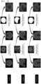
- the plurality of cross-sectional images are registered through rigid body transformation as shown in FIG 2 and a correlation between the cross-sectional images is identified.
- a skull image included in the CT image is removed to leave only a brain tissue image (skull stripping).
- intensity normalization and histogram equalization may be additionally performed to improve image quality.
- the deep learning unit 121 generates a plurality of training data including a pre-processed CT image and segmentation information corresponding to the pre-processed CT image, and trains a deep learning network through the training data.
- the deep learning network may be implemented as a convolutional neural network (CNN) or a U-net, and particularly, it is more preferable that the deep learning network is implemented as a U-net having strengths in image segmentation and labeling.
- the U-net is an artificial neural network including multiple convolutional layers having a U shape as shown in FIG. 3 , and has strengths in image segmentation and labeling.
- the segmentation information includes white matter region information, gray matter region information, and ventricular region information, and may be obtained as a result of analyzing a magnetic resonance imaging (MRI) image corresponding to the CT image through an image segmentation program such as a Freesurfer or by training and verifying segmentation information manually input by a user through the deep learning network.
- MRI magnetic resonance imaging
- the segmentation unit 123 When an image to be analyzed (i.e., a CT image of a subject to be analyzed) is input, the segmentation unit 123 automatically obtains segmentation information corresponding to the image to be analyzed through the deep learning network whose training is completed, and generates and outputs a CT image including the segmentation information, that is, a segmented CT image.
- the feature extraction unit 130 extracts semantic feature information from the segmented CT image.
- the semantic feature information may include at least one of (1) a three-dimensional (3D) volume ratio of white matter, (2) a 3D volume ratio of gray matter, (3) a sum of 3D volume ratios of white matter and gray matter, (4) a 3D volume of ventricle, (5) a two-dimensional (2D) area ratio of white matter, (6) a 2D area ratio of gray matter, (7) a sum of 2D area ratios of white matter and gray matter, and (8) a 2D area of ventricle.
- 3D three-dimensional
- the feature extraction unit 130 converts the CT image into a 3D volume image, divides a region of the 3D volume image into a white matter region, a gray matter region, and a ventricular region according to the segmentation information included in the CT image, and extracts 3D volume ratios of white matter, gray matter, and ventricle (1), (2), (4) through their volume values.
- the feature extraction unit 130 selects a CT image just before the ventricle is observed while axially scanning the 3D volume image from top to bottom, and then extracts 2D area ratios of white matter and gray matter (5), (6) based on the CT image.
- the feature extraction unit 130 selects a CT image in which the largest ventricle is observed while axially scanning the 3D volume image from top to bottom, and then extracts a 2D area of ventricle (8) based on the CT image.
- the machine learning unit 141 generates a plurality of training data including semantic feature information and a region-specific cerebral cortical contraction rate corresponding to the semantic feature information, and trains a machine learning model through the training data.
- the region-specific cerebral cortical contraction rate may be a value directly measured by medical staff by referring to the CT image, and may be, for example, a cerebral cortical contraction rate in four regions of the brain related to Alzheimer's disease (frontal, parietal, and medial temporal lobes of the left and right).
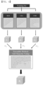
- the machine learning model of the present disclosure may be implemented as a soft majority voting (SMV) model using at least one of a regularized logistic regression (RLR) algorithm, a linear discriminant analysis (LDA) algorithm, and a Gaussian Naive Bayes (GNB) algorithm, as shown in FIG. 4 .
- SMV soft majority voting
- RLR regularized logistic regression
- LDA linear discriminant analysis
- GNB Gaussian Naive Bayes
- the RLR algorithm is an algorithm to which regularization is applied to prevent a model in a general regression algorithm from being underfitted or overfitted, and particularly, L2 regularization from among various regularization methods is used in the present disclosure.
- the LDA algorithm is a probabilistic generative model among machine learning algorithms, and is an algorithm that classifies data into a group when the squared distance between observed data and central (mean) data is the minimum.
- the GNB algorithm is an algorithm that uses a Gaussian function as a kernel function in a naive Bayes algorithm that creates a classifier using Bayes' theorem that assumes independence between features among machine learning algorithms.
- the SMV model is a model that corresponds to an aggregation method among ensemble models of machine learning, and is an algorithm that determines a final class by giving weights based on a sum of conditional probabilities of individual classifiers and then voting by majority vote.
- the SMV model has improved performance when compared to a simple direct majority voting model.
- the prediction unit 143 automatically obtains and outputs a region-specific cerebral cortical contraction rate corresponding to the semantic feature information of the image to be analyzed through the machine learning model whose training is completed.
- a method of predicting a region-specific cerebral cortical contraction rate on the basis of a CT image of the present disclosure will be described with reference to FIG. 5 .
- a method of predicting a region-specific cerebral cortical contraction rate of the present disclosure roughly includes a training step S10 and a prediction step S20.
- CT images (train) of a plurality of patients are obtained and pre-processed by accessing a medical information database in which medical information, medical treatment result information, and diagnostic information of a plurality of patients are stored.
- segmentation information seg corresponding to each of the CT images is additionally obtained.
- segmentation information of an MRI image corresponding to a CT image is stored in the medical information database, the segmentation information is used as it is.
- segmentation information including white matter region information, gray matter region information, and ventricular region information is extracted and used from the MRI image through a known image segmentation program.
- the information is manually received from a user.
- a deep learning network training step S12 a plurality of training data having the CT image as an input condition and the segmentation information as an output condition are generated, and then a deep learning network is trained about a correlation between the CT image and the segmentation information through the training data.
- deep learning is performed by using a stochastic gradient descent algorithm, and a stopping point is selected by applying a training early stopping method.
- a machine learning model training step S13 semantic feature information corresponding to each CT image is extracted based on each segmentation information, and a region-specific cerebral cortical contraction rate corresponding to each CT image is obtained.
- the cerebral cortical contract rate may be a value measured by medical staff by referring to the CT image.
- a plurality of training data having the semantic feature information as an input condition and the region-specific cerebral cortical contraction rate as an output condition are generated, and then a machine learning model is trained through the training data.
- the prediction step S20 may be performed.
- an image to be analyzed i.e., a CT image of a subject to be analyzed
- a PET-CT device i.e., a PET image of a subject to be analyzed
- segmentation information corresponding to the image to be analyzed is obtained through the deep learning network.
- a feature information extraction step S23 semantic feature information corresponding to the image to be analyzed is extracted based on the segmentation information obtained in operation S22.
- a region-specific cerebral cortical contraction rate corresponding to the semantic feature information is obtained and output through the machine learning model.
- the method according to the present embodiment may be embodied as a program executed in a computer and may be stored in a computer-readable recording medium, and examples of the computer-readable recording medium include read-only memories (ROMs), random-access memories (RAMs), compact disc (CD)-ROMs, magnetic tapes, floppy disks, optical data storage devices, and carrier waves (e.g., data transmission through the Internet).
- ROMs read-only memories
- RAMs random-access memories
- CD-ROMs compact disc
- magnetic tapes floppy disks
- optical data storage devices e.g., data transmission through the Internet.
- the computer-readable recording medium may be distributed among computer systems that are interconnected through a network so that computer-readable code is stored and executed in a distributed fashion.
- Functional programs, code, and code segments for embodying the method may be easily derived by programmers in the technical field to which the present disclosure pertains.
Landscapes
- Engineering & Computer Science (AREA)
- Health & Medical Sciences (AREA)
- Life Sciences & Earth Sciences (AREA)
- Physics & Mathematics (AREA)
- Medical Informatics (AREA)
- General Health & Medical Sciences (AREA)
- Theoretical Computer Science (AREA)
- Biomedical Technology (AREA)
- Biophysics (AREA)
- Molecular Biology (AREA)
- Public Health (AREA)
- Nuclear Medicine, Radiotherapy & Molecular Imaging (AREA)
- Pathology (AREA)
- General Physics & Mathematics (AREA)
- Radiology & Medical Imaging (AREA)
- Surgery (AREA)
- Animal Behavior & Ethology (AREA)
- Heart & Thoracic Surgery (AREA)
- Veterinary Medicine (AREA)
- High Energy & Nuclear Physics (AREA)
- Data Mining & Analysis (AREA)
- Artificial Intelligence (AREA)
- Optics & Photonics (AREA)
- Computer Vision & Pattern Recognition (AREA)
- Computational Linguistics (AREA)
- Evolutionary Computation (AREA)
- Mathematical Physics (AREA)
- Neurology (AREA)
- Software Systems (AREA)
- Computing Systems (AREA)
- General Engineering & Computer Science (AREA)
- Neurosurgery (AREA)
- Oral & Maxillofacial Surgery (AREA)
- Dentistry (AREA)
- Physiology (AREA)
- Epidemiology (AREA)
- Primary Health Care (AREA)
- Multimedia (AREA)
- Quality & Reliability (AREA)
- Pulmonology (AREA)
Description
- The present disclosure relates to an apparatus for predicting a region-specific cerebral cortical contraction rate on the basis of a computed tomography (CT) image, which may predict a region-specific cerebral cortical contraction rate by using a CT image, instead of a magnetic resonance imaging (MRI) image, by using deep learning and machine learning technology.
- Dementia is a chronic and progressive brain syndrome, and is a typical organic mental disorder of old age which is accompanied by disorders of cognitive functions including memory and executive ability, emotional disorders, and behavioral control disorders due to the occurrence and progression of brain lesions, and negatively impacts the ability to independently carry out everyday activities. Dementia may be caused by various brain injuries, but the most representative cause is Alzheimer's dementia, which accounts for 71% of all dementia types.
- Although the exact cause of Alzheimer's disease is not yet known, it is known that due to increased production of beta-amyloid proteins and decreased emission, beta-amyloid proteins are deposited and amyloid plaques are formed, resulting in extensive neuronal destruction through interaction with other factors.
- Accordingly, a method of diagnosing Alzheimer's disease by obtaining a positron emission tomography (PET) image through a PET-computed tomography (CT) device and analyzing and evaluating the accumulation of beta-amyloid based on the PET image has been proposed.
- However, the method has a disadvantage in that the PET-CT device additionally provides a CT image in addition to the PET image, but the CT image is not used at all to diagnose Alzheimer's disease. In particular, when diagnosing Alzheimer's disease, it is necessary to additionally check a region-specific cerebral cortical contraction rate. Because this information may not be provided through the CT image, the information should be obtained and provided by obtaining and analyzing a magnetic resonance imaging (MRI) image through a separate MRI device.
- AHMED MD RISHAD ET AL: "Neuroimaging and Machine Learning for Dementia Diagnosis: Recent Advancements and Future Prospects", IEEE REVIEWS IN BIOMEDICAL ENGINEERING, vol. 12,pages 19-33, teaches to use classical machine learning and deep learning (page or dementia diagnosis.
- ATTORI MASUMI ET AL: "Automated measurement of medial temporal lobe atrophy by computed tomography",INTERNATIONAL JOURNAL OF COMPUTER ASSISTED RADIOLOGY AND SURGERY, SPRINGER, DE, vol. 1, no. 6, 17 February 2007, pages 321-330, attempts, as the self-explanatory title indicates, to diagnose dementia of Alzheimer by volumetrically measuring the medial temporal lobe atrophy in CT scans. SMALL ET AL: "Current and future uses of neuroimaging for cognitively impaired patients",THE LANCET NEUROLOGY, ELSEVIER, AMSTERDAM, NL, vol. 7, no. 2, 18 January 2008, pages 161-172, mentions the use of CT as structural technique for the assessment of stroke.
- To solve the above problems, the present disclosure provides an apparatus for predicting a region-specific cerebral cortical contraction rate on the basis of a computed tomography (CT) image, which predicts a region-specific cerebral cortical contraction rate by using a CT image, instead of a magnetic resonance imaging (MRI) image, by using deep learning and machine learning technology.
- Objectives of the present disclosure are not limited thereto, and other unmentioned objectives will be clearly understood by one of ordinary skill in the art to which the present disclosure pertains from the following description.
- To solve the problems, according to an embodiment of the present disclosure, a method of predicting a region-specific cerebral cortical contraction rate on basis of a computed tomography (CT) image includes: a deep learning step of training a deep learning network, by selecting and using CT images of a plurality of patients and segmentation information, about a correlation between the CT images and the segmentation information; a feature extraction step of extracting, on basis of each segmentation information, semantic feature information corresponding to each of the CT images; a machine learning step of additionally obtaining a region-specific cerebral cortical contraction rate corresponding to each semantic feature information and then training a machine learning model about a correlation between the semantic feature information and the region-specific cerebral cortical contract rate; a segmentation step of, when an image to be analyzed is input, obtaining segmentation information corresponding to the image to be analyzed through the deep learning network; and a prediction step of extracting semantic feature information corresponding to the image to be analyzed on basis of the segmentation information and then predicting and notifying a region-specific cerebral cortical contraction rate corresponding to the semantic feature information through the machine learning model.
- The deep learning network may be implemented as a U-net model.
- The segmentation information may include white matter region information, gray matter region information, and ventricular region information.
- The semantic feature information may include a three-dimensional (3D) volume ratio of white matter, a 3D volume ratio of gray matter, a sum of 3D volume ratios of white matter and gray matter, a 3D volume of ventricle, a two-dimensional (2D) area ratio of white matter, a 2D area ratio of gray matter, a sum of 2D area ratios of white matter and gray matter, and a 2D area of ventricle.
- The machine learning model may be implemented as a soft majority voting model using at least one of a regularized logistic regression model, a linear discriminant analysis model, and a Gaussian Naive Bayes model.
- The method may further include, when CT images of a plurality of patients or an image to be analyzed are input, performing an image pre-processing operation by performing image registration through rigid body transformation and then removing a skull image.
- To solve the problems, according to another embodiment of the present disclosure, an apparatus for predicting a region-specific cerebral cortical contraction rate on basis of a computed tomography (CT) image includes: a CT image pre-processing unit configured to, when CT images of a plurality of patients or an image to be analyzed are input, perform image registration through rigid body transformation and then remove a skull image; a deep learning unit configured to additionally obtain each segmentation information corresponding to each of the CT images and then train a deep learning network about a correlation between the CT image and the segmentation information; a segmentation unit configured to obtain and output segmentation information corresponding to the image to be analyzed through the deep learning network; a feature extraction unit configured to extract semantic feature information corresponding to each CT image or the image to be analyzed based on each segmentation information; a machine learning unit configured to additionally obtain a region-specific cerebral cortical contraction rate corresponding to each semantic feature information of the CT image and then train a machine learning model about a correlation between the semantic feature information and the region-specific cerebral cortical contraction rate; and a prediction unit configured to predict and notify a region-specific cerebral cortical contraction rate corresponding to the semantic feature information of the image to be analyzed through the machine learning model.
- The deep learning network may be implemented as a U-net model.
- The segmentation information may include white matter region information, gray matter region information, and ventricular region information.
- The semantic feature information may include a three-dimensional (3D) volume ratio of white matter, a 3D volume ratio of gray matter, a sum of 3D volume ratios of white matter and gray matter, a 3D volume of ventricle, a two-dimensional (2D) area ratio of white matter, a 2D area ratio of gray matter, a sum of 2D area ratios of white matter and gray matter, and a 2D area of ventricle.
- The machine learning model may be implemented as a soft majority voting model using at least one of a regularized logistic regression model, a linear discriminant analysis model, and a Gaussian Naive Bayes model.
- According to the present disclosure, a segmented computed tomography (CT) image may be obtained through a deep learning network, and a region-specific cerebral cortical contraction rate may be more rapidly and accurately predicted through a machine learning module.
- As a result, both a PET image and a CT image obtained through a positron emission tomography (PET)-CT device may be used to diagnose Alzheimer's disease, and particularly, even a region-specific cerebral cortical contraction rate may be obtained and provided without using a separate magnetic resonance imaging (MRI) device. The invention is set out in the appended set of claims.
-
-
FIG. 1 is a view illustrating an apparatus for predicting a region-specific cerebral cortical contraction rate on the basis of a computed tomography (CT) image, according to an embodiment of the present disclosure. -
FIG. 2 is a view for describing a CT image pre-processing method, according to an embodiment of the present disclosure. -
FIG. 3 is a view illustrating a deep learning network, according to an embodiment of the present disclosure. -
FIG. 4 is a view illustrating a machine learning module, according to an embodiment of the present disclosure. -
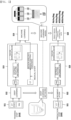
FIG. 5 is a view for describing a method of predicting a region-specific cerebral cortical contraction rate on the basis of a CT image, according to an embodiment of the present disclosure. - The following description illustrates only a principle of the present disclosure. In addition, it is to be understood that all conditional terms and embodiments mentioned in the present specification are basically intended only to allow one of ordinary skill in the art to understand a concept of the present disclosure, and the present disclosure is not limited to embodiments and states particularly mentioned as such.
- Further, it is to be understood that all detailed descriptions mentioning a specific embodiment of the present disclosure as well as principles, aspects, and embodiments of the present disclosure are intended to include structural and functional equivalences thereof. Further, it is to be understood that these equivalences include an equivalence that will be developed in the future as well as an equivalence that is current well-known, that is, all devices invented so as to perform the same function regardless of a structure.
- Therefore, it is to be understood that, for example, a block diagram of the present specification shows an illustrative conceptual aspect for embodying a principle of the present disclosure. Similarly, it is to be understood that all flowcharts, state transition diagrams, pseudo-code, and the like, show various processes that may be tangibly embodied in a computer-readable medium and that are executed by computers or processors regardless of whether or not the computers or the processors are clearly shown.
- Functions of various devices including processors or functional blocks represented as concepts similar to the processors and shown in the accompanying drawings may be provided by hardware having capability to execute appropriate software as well as dedicated hardware. When the functions are provided by the processors, they may be provided by a single dedicated processor, a single shared processor, or a plurality of individual processors and some of them may be shared.
- In addition, terms mentioned as a processor, a control, or a concept similar to the processor or the control should not be interpreted to exclusively cite hardware having capability to execute software, but should be interpreted to implicitly include digital signal processor (DSP) hardware and a read-only memory (ROM), a random-access memory (RAM), and a non-volatile memory for storing software without being limited thereto. The above-mentioned terms may also include well-known other hardware.
- In the claims of the present specification, components represented as means for performing functions mentioned in the detailed description are intended to include all methods for performing functions including all types of software including, for example, a combination of circuit devices performing these functions, firmware/micro code, or the like, and are coupled to appropriate circuits for executing the software so as to execute these functions. It is to be understood that because functions provided by variously mentioned means are combined with each other and are combined with a scheme demanded by the claims in the present disclosures defined by the claims, any means capable of providing these functions are equivalent to means recognized from the present specification.
- The above-mentioned objectives, features, and advantages will become more obvious from the following detailed description provided in relation to the accompanying drawings. Therefore, one of ordinary skill in the art to which the present disclosure pertains may easily practice a technical idea of the present disclosure. Further, in describing the present disclosure, in the case in which it is judged that a detailed description of a well-known technology associated with the present disclosure may unnecessarily make the gist of the present disclosure unclear, it will be omitted.
-
FIG. 1 is a view illustrating an apparatus for predicting a region-specific cerebral cortical contraction rate on the basis of a computed tomography (CT) image, according to an embodiment of the present disclosure. - Referring to
FIG. 1 , an apparatus for predicting a region-specific cerebral cortical contraction rate of the present disclosure includes a CT image pre-processingunit 110, adeep learning unit 121, asegmentation unit 123, afeature extraction unit 130, amachine learning unit 141 and aprediction unit 143. - The CT image pre-processing
unit 110 receives and pre-processes each pre-obtained CT image or CT image of a subject to be analyzed, and converts the image into an image capable of deep learning. - That is, by considering that a CT image is a plurality of cross-sectional images scanned in axial, sagittal, and coronal directions of the brain and each of the cross-sectional images has a different coordinate system according to a scanning direction, the plurality of cross-sectional images are registered through rigid body transformation as shown in
FIG 2 and a correlation between the cross-sectional images is identified. Next, a skull image included in the CT image is removed to leave only a brain tissue image (skull stripping). - Also, when necessary, intensity normalization and histogram equalization may be additionally performed to improve image quality.
- The
deep learning unit 121 generates a plurality of training data including a pre-processed CT image and segmentation information corresponding to the pre-processed CT image, and trains a deep learning network through the training data. - In this case, the deep learning network may be implemented as a convolutional neural network (CNN) or a U-net, and particularly, it is more preferable that the deep learning network is implemented as a U-net having strengths in image segmentation and labeling. The U-net is an artificial neural network including multiple convolutional layers having a U shape as shown in
FIG. 3 , and has strengths in image segmentation and labeling. - The segmentation information includes white matter region information, gray matter region information, and ventricular region information, and may be obtained as a result of analyzing a magnetic resonance imaging (MRI) image corresponding to the CT image through an image segmentation program such as a Freesurfer or by training and verifying segmentation information manually input by a user through the deep learning network.
- When an image to be analyzed (i.e., a CT image of a subject to be analyzed) is input, the
segmentation unit 123 automatically obtains segmentation information corresponding to the image to be analyzed through the deep learning network whose training is completed, and generates and outputs a CT image including the segmentation information, that is, a segmented CT image. - The
feature extraction unit 130 extracts semantic feature information from the segmented CT image. - The semantic feature information may include at least one of (1) a three-dimensional (3D) volume ratio of white matter, (2) a 3D volume ratio of gray matter, (3) a sum of 3D volume ratios of white matter and gray matter, (4) a 3D volume of ventricle, (5) a two-dimensional (2D) area ratio of white matter, (6) a 2D area ratio of gray matter, (7) a sum of 2D area ratios of white matter and gray matter, and (8) a 2D area of ventricle.
- In more detail, the
feature extraction unit 130 converts the CT image into a 3D volume image, divides a region of the 3D volume image into a white matter region, a gray matter region, and a ventricular region according to the segmentation information included in the CT image, and extracts 3D volume ratios of white matter, gray matter, and ventricle (1), (2), (4) through their volume values. - The
feature extraction unit 130 selects a CT image just before the ventricle is observed while axially scanning the 3D volume image from top to bottom, and then extracts 2D area ratios of white matter and gray matter (5), (6) based on the CT image. - Finally, the
feature extraction unit 130 selects a CT image in which the largest ventricle is observed while axially scanning the 3D volume image from top to bottom, and then extracts a 2D area of ventricle (8) based on the CT image. - The
machine learning unit 141 generates a plurality of training data including semantic feature information and a region-specific cerebral cortical contraction rate corresponding to the semantic feature information, and trains a machine learning model through the training data. In this case, the region-specific cerebral cortical contraction rate may be a value directly measured by medical staff by referring to the CT image, and may be, for example, a cerebral cortical contraction rate in four regions of the brain related to Alzheimer's disease (frontal, parietal, and medial temporal lobes of the left and right). - The machine learning model of the present disclosure may be implemented as a soft majority voting (SMV) model using at least one of a regularized logistic regression (RLR) algorithm, a linear discriminant analysis (LDA) algorithm, and a Gaussian Naive Bayes (GNB) algorithm, as shown in
FIG. 4 . - For reference, the RLR algorithm is an algorithm to which regularization is applied to prevent a model in a general regression algorithm from being underfitted or overfitted, and particularly, L2 regularization from among various regularization methods is used in the present disclosure.
- The LDA algorithm is a probabilistic generative model among machine learning algorithms, and is an algorithm that classifies data into a group when the squared distance between observed data and central (mean) data is the minimum.
- The GNB algorithm is an algorithm that uses a Gaussian function as a kernel function in a naive Bayes algorithm that creates a classifier using Bayes' theorem that assumes independence between features among machine learning algorithms.
- The SMV model is a model that corresponds to an aggregation method among ensemble models of machine learning, and is an algorithm that determines a final class by giving weights based on a sum of conditional probabilities of individual classifiers and then voting by majority vote. The SMV model has improved performance when compared to a simple direct majority voting model.
- When the semantic feature information of the image to be analyzed is input, the
prediction unit 143 automatically obtains and outputs a region-specific cerebral cortical contraction rate corresponding to the semantic feature information of the image to be analyzed through the machine learning model whose training is completed. - A method of predicting a region-specific cerebral cortical contraction rate on the basis of a CT image of the present disclosure will be described with reference to
FIG. 5 . - As shown in
FIG. 5 , a method of predicting a region-specific cerebral cortical contraction rate of the present disclosure roughly includes a training step S10 and a prediction step S20. - First, in a training data generation step S11, CT images (train) of a plurality of patients are obtained and pre-processed by accessing a medical information database in which medical information, medical treatment result information, and diagnostic information of a plurality of patients are stored.
- Also, segmentation information seg corresponding to each of the CT images is additionally obtained. When segmentation information of an MRI image corresponding to a CT image is stored in the medical information database, the segmentation information is used as it is. When only an MRI image is stored, segmentation information including white matter region information, gray matter region information, and ventricular region information is extracted and used from the MRI image through a known image segmentation program. In contrast, when there is no MRI image and no segmentation information of the MRI image, the information is manually received from a user.
- In a deep learning network training step S12, a plurality of training data having the CT image as an input condition and the segmentation information as an output condition are generated, and then a deep learning network is trained about a correlation between the CT image and the segmentation information through the training data. In the case of a U-net, deep learning is performed by using a stochastic gradient descent algorithm, and a stopping point is selected by applying a training early stopping method.
- In a machine learning model training step S13, semantic feature information corresponding to each CT image is extracted based on each segmentation information, and a region-specific cerebral cortical contraction rate corresponding to each CT image is obtained. In this case, the cerebral cortical contract rate may be a value measured by medical staff by referring to the CT image.
- A plurality of training data having the semantic feature information as an input condition and the region-specific cerebral cortical contraction rate as an output condition are generated, and then a machine learning model is trained through the training data.
- When the training step S10 is completed, the prediction step S20 may be performed.
- In an analysis target image input step S21 of the prediction step S20, an image to be analyzed (i.e., a CT image of a subject to be analyzed) provided from a PET-CT device is received and stored.
- In a segmentation step S22, segmentation information corresponding to the image to be analyzed is obtained through the deep learning network.
- In a feature information extraction step S23, semantic feature information corresponding to the image to be analyzed is extracted based on the segmentation information obtained in operation S22.
- Finally, in a prediction step S24, a region-specific cerebral cortical contraction rate corresponding to the semantic feature information is obtained and output through the machine learning model.
- The method according to the present embodiment may be embodied as a program executed in a computer and may be stored in a computer-readable recording medium, and examples of the computer-readable recording medium include read-only memories (ROMs), random-access memories (RAMs), compact disc (CD)-ROMs, magnetic tapes, floppy disks, optical data storage devices, and carrier waves (e.g., data transmission through the Internet).
- The computer-readable recording medium may be distributed among computer systems that are interconnected through a network so that computer-readable code is stored and executed in a distributed fashion. Functional programs, code, and code segments for embodying the method may be easily derived by programmers in the technical field to which the present disclosure pertains.
- In addition, although preferred embodiments of the present disclosure have been illustrated and described above, the present disclosure is not limited to the above-described specific embodiments. Various modified embodiments may be made by one of ordinary skill in the art without departing from the scope of the present disclosure as claimed in the claims.
Claims (11)
- A method of predicting a region-specific cerebral cortical contraction rate on basis of a computed tomography (CT) image, the method comprising: a deep learning step of training a deep learning network, by selecting and using CT images of a plurality of patients and segmentation information, about a correlation between the CT images and the segmentation information; a feature extraction step of extracting, on basis of each segmentation information, semantic feature information corresponding to each of the CT images; a machine learning step of additionally obtaining a region-specific cerebral cortical contraction rate corresponding to each semantic feature information and then training a machine learning model about a correlation between the semantic feature information and the region-specific cerebral cortical contract rate; a segmentation step of, when an image to be analyzed is input, obtaining segmentation information corresponding to the image to be analyzed through the deep learning network; and a prediction step of extracting semantic feature information corresponding to the image to be analyzed on basis of the segmentation information and then predicting and notifying a region-specific cerebral cortical contraction rate corresponding to the semantic feature information through the machine learning model.
- The method of predicting a region-specific cerebral cortical contraction rate on basis of a CT image of claim 1, wherein the deep learning network is implemented as a U-net model.
- The method of predicting a region-specific cerebral cortical contraction rate on basis of a CT image of claim 1, wherein the segmentation information is extracted based on a magnetic resonance imaging (MRI) image, and comprises white matter region information, gray matter region information, and ventricular region information.
- The method of predicting a region-specific cerebral cortical contraction rate on basis of a CT image of claim 1, wherein the semantic feature information comprises a three-dimensional (3D) volume ratio of white matter, a 3D volume ratio of gray matter, a sum of 3D volume ratios of white matter and gray matter, a 3D volume of ventricle, a two-dimensional (2D) area ratio of white matter, a 2D area ratio of gray matter, a sum of 2D area ratios of white matter and gray matter, and a 2D area of ventricle.
- The method of predicting a region-specific cerebral cortical contraction rate on basis of a CT image of claim 1, wherein the machine learning model is implemented as a soft majority voting model using at least one of a regularized logistic regression model, a linear discriminant analysis model, and a Gaussian Naive Bayes model.
- The method of predicting a region-specific cerebral cortical contraction rate on basis of a CT image of claim 1, further comprising, when CT images of a plurality of patients or an image to be analyzed are input, performing an image pre-processing operation by performing image registration through rigid body transformation and then removing a skull image.
- An apparatus for predicting a region-specific cerebral cortical contraction rate on basis of a computed tomography (CT) image, the apparatus comprising: a CT image pre-processing unit configured to, when CT images of a plurality of patients or an image to be analyzed are input, perform image registration through rigid body transformation and then remove a skull image; a deep learning unit configured to additionally obtain each segmentation information corresponding to each of the CT images and then train a deep learning network about a correlation between the CT image and the segmentation information; a segmentation unit configured to obtain and output segmentation information corresponding to the image to be analyzed through the deep learning network; a feature extraction unit configured to extract semantic feature information corresponding to each CT image or the image to be analyzed based on each segmentation information; a machine learning unit configured to additionally obtain a region-specific cerebral cortical contraction rate corresponding to each semantic feature information of the CT image and then train a machine learning model about a correlation between the semantic feature information and the region-specific cerebral cortical contraction rate; and a prediction unit configured to predict and notify a region-specific cerebral cortical contraction rate corresponding to the semantic feature information of the image to be analyzed through the machine learning model.
- The apparatus for predicting a region-specific cerebral cortical contraction rate on basis of a CT image of claim 7, wherein the deep learning network is implemented as a U-net model.
- The apparatus for predicting a region-specific cerebral cortical contraction rate on basis of a CT image of claim 7, wherein the segmentation information comprises white matter region information, gray matter region information, and ventricular region information.
- The apparatus for predicting a region-specific cerebral cortical contraction rate on basis of a CT image of claim 7, wherein the semantic feature information comprises a three-dimensional (3D) volume ratio of white matter, a 3D volume ratio of gray matter, a sum of 3D volume ratios of white matter and gray matter, a 3D volume of ventricle, a two-dimensional (2D) area ratio of white matter, a 2D area ratio of gray matter, a sum of 2D area ratios of white matter and gray matter, and a 2D area of ventricle.
- The apparatus for predicting a region-specific cerebral cortical contraction rate on basis of a CT image of claim 7, wherein the machine learning model is implemented as a soft majority voting model using at least one of a regularized logistic regression model, a linear discriminant analysis model, and a Gaussian Naive Bayes model.
Applications Claiming Priority (2)
| Application Number | Priority Date | Filing Date | Title |
|---|---|---|---|
| KR1020190109863A KR102276545B1 (en) | 2019-09-05 | 2019-09-05 | Method and apparatus for cortical neurodegeneration prediction in alzheimer's disease using CT images |
| PCT/KR2020/011790 WO2021045507A2 (en) | 2019-09-05 | 2020-09-02 | Method and apparatus for predicting region-specific cerebral cortical contraction rate on basis of ct image |
Publications (3)
| Publication Number | Publication Date |
|---|---|
| EP4026498A2 EP4026498A2 (en) | 2022-07-13 |
| EP4026498A4 EP4026498A4 (en) | 2023-03-29 |
| EP4026498B1 true EP4026498B1 (en) | 2024-05-01 |
Family
ID=74852120
Family Applications (1)
| Application Number | Title | Priority Date | Filing Date |
|---|---|---|---|
| EP20861207.7A Active EP4026498B1 (en) | 2019-09-05 | 2020-09-02 | Method and apparatus for predicting region-specific cerebral cortical contraction rate on basis of ct image |
Country Status (5)
| Country | Link |
|---|---|
| US (1) | US12266110B2 (en) |
| EP (1) | EP4026498B1 (en) |
| JP (1) | JP7317306B2 (en) |
| KR (1) | KR102276545B1 (en) |
| WO (1) | WO2021045507A2 (en) |
Families Citing this family (9)
| Publication number | Priority date | Publication date | Assignee | Title |
|---|---|---|---|---|
| WO2020163539A1 (en) * | 2019-02-05 | 2020-08-13 | University Of Virginia Patent Foundation | System and method for fully automatic lv segmentation of myocardial first-pass perfusion images |
| US11640552B2 (en) * | 2019-10-01 | 2023-05-02 | International Business Machines Corporation | Two stage training to obtain a best deep learning model with efficient use of computing resources |
| KR102608203B1 (en) * | 2022-02-09 | 2023-11-30 | 사회복지법인 삼성생명공익재단 | Dementia prediction method based on 2-dimensional magnetic resonance imaging and analysis apparatus |
| WO2023153839A1 (en) * | 2022-02-09 | 2023-08-17 | 사회복지법인 삼성생명공익재단 | Dementia information calculation method and analysis device using two-dimensional mri |
| JP7834513B2 (en) * | 2022-03-09 | 2026-03-24 | キヤノン株式会社 | Information processing device and learning method |
| CN115908451B (en) * | 2022-11-04 | 2025-12-23 | 北京航空航天大学 | Cardiac CT image segmentation method combining multi-view geometry and migration learning |
| KR102800156B1 (en) * | 2022-12-13 | 2025-04-23 | 사회복지법인 삼성생명공익재단 | Region of interest extracting method for dementia prediction in tau-pet, stage of dementia prediction method using ct and tau-pet, and analysis apparatus |
| CN116894812A (en) * | 2023-06-14 | 2023-10-17 | 齐鲁理工学院 | Individualized disease prediction methods, devices, media and equipment based on sequence learning |
| KR102712064B1 (en) * | 2023-06-27 | 2024-09-27 | 사회복지법인 삼성생명공익재단 | Dementia related information generation method using volume predicted from computed tomography and analysis apparatus |
Family Cites Families (10)
| Publication number | Priority date | Publication date | Assignee | Title |
|---|---|---|---|---|
| WO2007114238A1 (en) | 2006-03-30 | 2007-10-11 | National University Corporation Shizuoka University | Apparatus for determining brain atrophy, method of determining brain atrophy and program for determining brain atrophy |
| EP2451348A4 (en) | 2010-04-13 | 2013-07-31 | Baxter Int | USE OF VENTRICULAR ENLARGEMENT RATE IN INTRAVENOUS IMMUNOGLOBULIN TREATMENT OF ALZHEIMER'S DISEASE |
| JP5878125B2 (en) | 2010-09-07 | 2016-03-08 | 株式会社日立メディコ | Dementia diagnosis support apparatus and dementia diagnosis support method |
| LT2994160T (en) * | 2013-05-06 | 2019-08-26 | Baxalta Incorporated | Treatment of alzheimer`s disease subpopulations with pooled immunoglobulin g |
| KR101944069B1 (en) * | 2016-12-29 | 2019-01-30 | 주식회사 마이다스아이티 | Method and apparatus for deciding Alzheimer's disease based on machine learning |
| EP3677180B1 (en) | 2017-08-29 | 2026-02-25 | FUJIFILM Corporation | Medical information display device and program |
| KR101916347B1 (en) | 2017-10-13 | 2018-11-08 | 주식회사 수아랩 | Deep learning based image comparison device, method and computer program stored in computer readable medium |
| KR102064811B1 (en) * | 2018-01-19 | 2020-01-13 | 사회복지법인 삼성생명공익재단 | Method and apparatus for predicting and classifying dementia progression of amyloid positive mild cognitive impairment |
| KR102001398B1 (en) | 2018-01-25 | 2019-07-18 | 재단법인 아산사회복지재단 | Method and apparatus for predicting brain desease change though machine learning and program for the same |
| KR101936302B1 (en) | 2018-06-29 | 2019-01-08 | 이채영 | Diagnosis method and apparatus for neurodegenerative diseases based on deep learning network |
-
2019
- 2019-09-05 KR KR1020190109863A patent/KR102276545B1/en active Active
-
2020
- 2020-09-02 WO PCT/KR2020/011790 patent/WO2021045507A2/en not_active Ceased
- 2020-09-02 US US17/640,593 patent/US12266110B2/en active Active
- 2020-09-02 EP EP20861207.7A patent/EP4026498B1/en active Active
- 2020-09-02 JP JP2022514861A patent/JP7317306B2/en active Active
Also Published As
| Publication number | Publication date |
|---|---|
| KR102276545B1 (en) | 2021-07-15 |
| JP2022547909A (en) | 2022-11-16 |
| JP7317306B2 (en) | 2023-07-31 |
| US12266110B2 (en) | 2025-04-01 |
| KR20210029318A (en) | 2021-03-16 |
| US20220335611A1 (en) | 2022-10-20 |
| WO2021045507A2 (en) | 2021-03-11 |
| EP4026498A4 (en) | 2023-03-29 |
| EP4026498A2 (en) | 2022-07-13 |
| WO2021045507A3 (en) | 2021-04-29 |
Similar Documents
| Publication | Publication Date | Title |
|---|---|---|
| US12266110B2 (en) | Method and apparatus for predicting region-specific cerebral cortical contraction rate on basis of CT image | |
| US12400321B2 (en) | Classifying neurological disease status using deep learning | |
| CN109447183B (en) | Prediction model training method, device, equipment and medium | |
| Yiğit et al. | Applying deep learning models to structural MRI for stage prediction of Alzheimer's disease | |
| Mahanand et al. | Identification of brain regions responsible for Alzheimer’s disease using a self-adaptive resource allocation network | |
| CN110363760B (en) | Computer system for recognizing medical images | |
| US20170109878A1 (en) | Segmentation of cardiac magnetic resonance (cmr) images using a memory persistence approach | |
| JPWO2019172181A1 (en) | Diagnostic support devices, programs, trained models, and learning devices | |
| Cigdem et al. | The performance of local-learning based clustering feature selection method on the diagnosis of parkinson’s disease using structural MRI | |
| Pallawi et al. | Study of Alzheimer’s disease brain impairment and methods for its early diagnosis: a comprehensive survey | |
| Qazi et al. | Automated Heart Wall Motion Abnormality Detection from Ultrasound Images Using Bayesian Networks. | |
| JP7852212B2 (en) | Methods for operating diagnostic support devices, computer programs, and medical devices | |
| Arabi et al. | High accuracy diagnosis for mri imaging of alzheimer’s disease using xgboost | |
| KR20230033769A (en) | Machine learning based classification method for cognitive ability of elder using magnetic resonance imaging and analysis apparatus | |
| KR102257295B1 (en) | Diagnostic system for diagnosing vulnerable atheromatous plaque through ultrasound image-based machine learning and the diagnostic method thereof | |
| Oommen et al. | A comprehensive study on early detection of Alzheimer disease using convolutional neural network | |
| KR101948701B1 (en) | Method for determining brain disorder of subject based on latent variables which describe brain structure thereof and apparatus using the same | |
| Khan | A Novel Deep Learning-Based Framework for Context-Aware Semantic Segmentation in Medical Imaging | |
| JP2025510130A (en) | Methods for the refinement of functional magnetic resonance images | |
| Sreelakshmi et al. | Prediction of neurological disorders using visual saliency: Current trends and future directions | |
| KR102809490B1 (en) | Machine learning training method and apparatus using same | |
| Tamilarasi et al. | Multimodal MRI analysis for schizophrenia classification using machine learning algorithm | |
| Yi̇ği̇t | Çok Modlu Görüntüleme Ve Klinik Verilerle Derin Sinir Ağları Kullanarak Demans Tanısı | |
| Rao et al. | A comparative analysis of Alzheimer’s Disease by Pre-Trained CNN Classifiers using Two Palnes of MRI Images | |
| Modi et al. | 3D Mri-Based Classification of Alzheimer's Disease, Mild Cognitive Impairment and Cognitively Normal Subjects Using Deep Learning |
Legal Events
| Date | Code | Title | Description |
|---|---|---|---|
| STAA | Information on the status of an ep patent application or granted ep patent |
Free format text: STATUS: THE INTERNATIONAL PUBLICATION HAS BEEN MADE |
|
| PUAI | Public reference made under article 153(3) epc to a published international application that has entered the european phase |
Free format text: ORIGINAL CODE: 0009012 |
|
| STAA | Information on the status of an ep patent application or granted ep patent |
Free format text: STATUS: REQUEST FOR EXAMINATION WAS MADE |
|
| 17P | Request for examination filed |
Effective date: 20220321 |
|
| AK | Designated contracting states |
Kind code of ref document: A2 Designated state(s): AL AT BE BG CH CY CZ DE DK EE ES FI FR GB GR HR HU IE IS IT LI LT LU LV MC MK MT NL NO PL PT RO RS SE SI SK SM TR |
|
| DAV | Request for validation of the european patent (deleted) | ||
| DAX | Request for extension of the european patent (deleted) | ||
| A4 | Supplementary search report drawn up and despatched |
Effective date: 20230224 |
|
| RIC1 | Information provided on ipc code assigned before grant |
Ipc: G06N 3/04 20060101ALI20230221BHEP Ipc: G06N 3/08 20060101ALI20230221BHEP Ipc: G16H 30/40 20180101ALI20230221BHEP Ipc: G16H 50/20 20180101ALI20230221BHEP Ipc: A61B 5/055 20060101ALI20230221BHEP Ipc: A61B 6/03 20060101ALI20230221BHEP Ipc: A61B 6/00 20060101AFI20230221BHEP |
|
| GRAP | Despatch of communication of intention to grant a patent |
Free format text: ORIGINAL CODE: EPIDOSNIGR1 |
|
| STAA | Information on the status of an ep patent application or granted ep patent |
Free format text: STATUS: GRANT OF PATENT IS INTENDED |
|
| INTG | Intention to grant announced |
Effective date: 20240116 |
|
| GRAS | Grant fee paid |
Free format text: ORIGINAL CODE: EPIDOSNIGR3 |
|
| GRAA | (expected) grant |
Free format text: ORIGINAL CODE: 0009210 |
|
| STAA | Information on the status of an ep patent application or granted ep patent |
Free format text: STATUS: THE PATENT HAS BEEN GRANTED |
|
| AK | Designated contracting states |
Kind code of ref document: B1 Designated state(s): AL AT BE BG CH CY CZ DE DK EE ES FI FR GB GR HR HU IE IS IT LI LT LU LV MC MK MT NL NO PL PT RO RS SE SI SK SM TR |
|
| REG | Reference to a national code |
Ref country code: GB Ref legal event code: FG4D |
|
| REG | Reference to a national code |
Ref country code: CH Ref legal event code: EP |
|
| REG | Reference to a national code |
Ref country code: IE Ref legal event code: FG4D |
|
| REG | Reference to a national code |
Ref country code: DE Ref legal event code: R096 Ref document number: 602020030405 Country of ref document: DE |
|
| REG | Reference to a national code |
Ref country code: LT Ref legal event code: MG9D |
|
| REG | Reference to a national code |
Ref country code: NL Ref legal event code: MP Effective date: 20240501 |
|
| PG25 | Lapsed in a contracting state [announced via postgrant information from national office to epo] |
Ref country code: IS Free format text: LAPSE BECAUSE OF FAILURE TO SUBMIT A TRANSLATION OF THE DESCRIPTION OR TO PAY THE FEE WITHIN THE PRESCRIBED TIME-LIMIT Effective date: 20240901 |
|
| PG25 | Lapsed in a contracting state [announced via postgrant information from national office to epo] |
Ref country code: BG Free format text: LAPSE BECAUSE OF FAILURE TO SUBMIT A TRANSLATION OF THE DESCRIPTION OR TO PAY THE FEE WITHIN THE PRESCRIBED TIME-LIMIT Effective date: 20240501 |
|
| PG25 | Lapsed in a contracting state [announced via postgrant information from national office to epo] |
Ref country code: FI Free format text: LAPSE BECAUSE OF FAILURE TO SUBMIT A TRANSLATION OF THE DESCRIPTION OR TO PAY THE FEE WITHIN THE PRESCRIBED TIME-LIMIT Effective date: 20240501 Ref country code: HR Free format text: LAPSE BECAUSE OF FAILURE TO SUBMIT A TRANSLATION OF THE DESCRIPTION OR TO PAY THE FEE WITHIN THE PRESCRIBED TIME-LIMIT Effective date: 20240501 |
|
| PG25 | Lapsed in a contracting state [announced via postgrant information from national office to epo] |
Ref country code: GR Free format text: LAPSE BECAUSE OF FAILURE TO SUBMIT A TRANSLATION OF THE DESCRIPTION OR TO PAY THE FEE WITHIN THE PRESCRIBED TIME-LIMIT Effective date: 20240802 |
|
| PG25 | Lapsed in a contracting state [announced via postgrant information from national office to epo] |
Ref country code: PT Free format text: LAPSE BECAUSE OF FAILURE TO SUBMIT A TRANSLATION OF THE DESCRIPTION OR TO PAY THE FEE WITHIN THE PRESCRIBED TIME-LIMIT Effective date: 20240902 |
|
| REG | Reference to a national code |
Ref country code: AT Ref legal event code: MK05 Ref document number: 1681256 Country of ref document: AT Kind code of ref document: T Effective date: 20240501 |
|
| PG25 | Lapsed in a contracting state [announced via postgrant information from national office to epo] |
Ref country code: NL Free format text: LAPSE BECAUSE OF FAILURE TO SUBMIT A TRANSLATION OF THE DESCRIPTION OR TO PAY THE FEE WITHIN THE PRESCRIBED TIME-LIMIT Effective date: 20240501 |
|
| PG25 | Lapsed in a contracting state [announced via postgrant information from national office to epo] |
Ref country code: ES Free format text: LAPSE BECAUSE OF FAILURE TO SUBMIT A TRANSLATION OF THE DESCRIPTION OR TO PAY THE FEE WITHIN THE PRESCRIBED TIME-LIMIT Effective date: 20240501 |
|
| PG25 | Lapsed in a contracting state [announced via postgrant information from national office to epo] |
Ref country code: AT Free format text: LAPSE BECAUSE OF FAILURE TO SUBMIT A TRANSLATION OF THE DESCRIPTION OR TO PAY THE FEE WITHIN THE PRESCRIBED TIME-LIMIT Effective date: 20240501 |
|
| PG25 | Lapsed in a contracting state [announced via postgrant information from national office to epo] |
Ref country code: PL Free format text: LAPSE BECAUSE OF FAILURE TO SUBMIT A TRANSLATION OF THE DESCRIPTION OR TO PAY THE FEE WITHIN THE PRESCRIBED TIME-LIMIT Effective date: 20240501 |
|
| PG25 | Lapsed in a contracting state [announced via postgrant information from national office to epo] |
Ref country code: LV Free format text: LAPSE BECAUSE OF FAILURE TO SUBMIT A TRANSLATION OF THE DESCRIPTION OR TO PAY THE FEE WITHIN THE PRESCRIBED TIME-LIMIT Effective date: 20240501 |
|
| PG25 | Lapsed in a contracting state [announced via postgrant information from national office to epo] |
Ref country code: PT Free format text: LAPSE BECAUSE OF FAILURE TO SUBMIT A TRANSLATION OF THE DESCRIPTION OR TO PAY THE FEE WITHIN THE PRESCRIBED TIME-LIMIT Effective date: 20240902 Ref country code: PL Free format text: LAPSE BECAUSE OF FAILURE TO SUBMIT A TRANSLATION OF THE DESCRIPTION OR TO PAY THE FEE WITHIN THE PRESCRIBED TIME-LIMIT Effective date: 20240501 Ref country code: NO Free format text: LAPSE BECAUSE OF FAILURE TO SUBMIT A TRANSLATION OF THE DESCRIPTION OR TO PAY THE FEE WITHIN THE PRESCRIBED TIME-LIMIT Effective date: 20240801 Ref country code: NL Free format text: LAPSE BECAUSE OF FAILURE TO SUBMIT A TRANSLATION OF THE DESCRIPTION OR TO PAY THE FEE WITHIN THE PRESCRIBED TIME-LIMIT Effective date: 20240501 Ref country code: LV Free format text: LAPSE BECAUSE OF FAILURE TO SUBMIT A TRANSLATION OF THE DESCRIPTION OR TO PAY THE FEE WITHIN THE PRESCRIBED TIME-LIMIT Effective date: 20240501 Ref country code: IS Free format text: LAPSE BECAUSE OF FAILURE TO SUBMIT A TRANSLATION OF THE DESCRIPTION OR TO PAY THE FEE WITHIN THE PRESCRIBED TIME-LIMIT Effective date: 20240901 Ref country code: HR Free format text: LAPSE BECAUSE OF FAILURE TO SUBMIT A TRANSLATION OF THE DESCRIPTION OR TO PAY THE FEE WITHIN THE PRESCRIBED TIME-LIMIT Effective date: 20240501 Ref country code: GR Free format text: LAPSE BECAUSE OF FAILURE TO SUBMIT A TRANSLATION OF THE DESCRIPTION OR TO PAY THE FEE WITHIN THE PRESCRIBED TIME-LIMIT Effective date: 20240802 Ref country code: FI Free format text: LAPSE BECAUSE OF FAILURE TO SUBMIT A TRANSLATION OF THE DESCRIPTION OR TO PAY THE FEE WITHIN THE PRESCRIBED TIME-LIMIT Effective date: 20240501 Ref country code: ES Free format text: LAPSE BECAUSE OF FAILURE TO SUBMIT A TRANSLATION OF THE DESCRIPTION OR TO PAY THE FEE WITHIN THE PRESCRIBED TIME-LIMIT Effective date: 20240501 Ref country code: BG Free format text: LAPSE BECAUSE OF FAILURE TO SUBMIT A TRANSLATION OF THE DESCRIPTION OR TO PAY THE FEE WITHIN THE PRESCRIBED TIME-LIMIT Effective date: 20240501 Ref country code: AT Free format text: LAPSE BECAUSE OF FAILURE TO SUBMIT A TRANSLATION OF THE DESCRIPTION OR TO PAY THE FEE WITHIN THE PRESCRIBED TIME-LIMIT Effective date: 20240501 Ref country code: RS Free format text: LAPSE BECAUSE OF FAILURE TO SUBMIT A TRANSLATION OF THE DESCRIPTION OR TO PAY THE FEE WITHIN THE PRESCRIBED TIME-LIMIT Effective date: 20240801 |
|
| PG25 | Lapsed in a contracting state [announced via postgrant information from national office to epo] |
Ref country code: DK Free format text: LAPSE BECAUSE OF FAILURE TO SUBMIT A TRANSLATION OF THE DESCRIPTION OR TO PAY THE FEE WITHIN THE PRESCRIBED TIME-LIMIT Effective date: 20240501 |
|
| PG25 | Lapsed in a contracting state [announced via postgrant information from national office to epo] |
Ref country code: EE Free format text: LAPSE BECAUSE OF FAILURE TO SUBMIT A TRANSLATION OF THE DESCRIPTION OR TO PAY THE FEE WITHIN THE PRESCRIBED TIME-LIMIT Effective date: 20240501 |
|
| PG25 | Lapsed in a contracting state [announced via postgrant information from national office to epo] |
Ref country code: CZ Free format text: LAPSE BECAUSE OF FAILURE TO SUBMIT A TRANSLATION OF THE DESCRIPTION OR TO PAY THE FEE WITHIN THE PRESCRIBED TIME-LIMIT Effective date: 20240501 |
|
| PG25 | Lapsed in a contracting state [announced via postgrant information from national office to epo] |
Ref country code: SK Free format text: LAPSE BECAUSE OF FAILURE TO SUBMIT A TRANSLATION OF THE DESCRIPTION OR TO PAY THE FEE WITHIN THE PRESCRIBED TIME-LIMIT Effective date: 20240501 Ref country code: RO Free format text: LAPSE BECAUSE OF FAILURE TO SUBMIT A TRANSLATION OF THE DESCRIPTION OR TO PAY THE FEE WITHIN THE PRESCRIBED TIME-LIMIT Effective date: 20240501 |
|
| PG25 | Lapsed in a contracting state [announced via postgrant information from national office to epo] |
Ref country code: SM Free format text: LAPSE BECAUSE OF FAILURE TO SUBMIT A TRANSLATION OF THE DESCRIPTION OR TO PAY THE FEE WITHIN THE PRESCRIBED TIME-LIMIT Effective date: 20240501 |
|
| PG25 | Lapsed in a contracting state [announced via postgrant information from national office to epo] |
Ref country code: SM Free format text: LAPSE BECAUSE OF FAILURE TO SUBMIT A TRANSLATION OF THE DESCRIPTION OR TO PAY THE FEE WITHIN THE PRESCRIBED TIME-LIMIT Effective date: 20240501 Ref country code: SK Free format text: LAPSE BECAUSE OF FAILURE TO SUBMIT A TRANSLATION OF THE DESCRIPTION OR TO PAY THE FEE WITHIN THE PRESCRIBED TIME-LIMIT Effective date: 20240501 Ref country code: RO Free format text: LAPSE BECAUSE OF FAILURE TO SUBMIT A TRANSLATION OF THE DESCRIPTION OR TO PAY THE FEE WITHIN THE PRESCRIBED TIME-LIMIT Effective date: 20240501 Ref country code: EE Free format text: LAPSE BECAUSE OF FAILURE TO SUBMIT A TRANSLATION OF THE DESCRIPTION OR TO PAY THE FEE WITHIN THE PRESCRIBED TIME-LIMIT Effective date: 20240501 Ref country code: DK Free format text: LAPSE BECAUSE OF FAILURE TO SUBMIT A TRANSLATION OF THE DESCRIPTION OR TO PAY THE FEE WITHIN THE PRESCRIBED TIME-LIMIT Effective date: 20240501 Ref country code: CZ Free format text: LAPSE BECAUSE OF FAILURE TO SUBMIT A TRANSLATION OF THE DESCRIPTION OR TO PAY THE FEE WITHIN THE PRESCRIBED TIME-LIMIT Effective date: 20240501 |
|
| REG | Reference to a national code |
Ref country code: DE Ref legal event code: R097 Ref document number: 602020030405 Country of ref document: DE |
|
| PG25 | Lapsed in a contracting state [announced via postgrant information from national office to epo] |
Ref country code: IT Free format text: LAPSE BECAUSE OF FAILURE TO SUBMIT A TRANSLATION OF THE DESCRIPTION OR TO PAY THE FEE WITHIN THE PRESCRIBED TIME-LIMIT Effective date: 20240501 |
|
| PLBE | No opposition filed within time limit |
Free format text: ORIGINAL CODE: 0009261 |
|
| STAA | Information on the status of an ep patent application or granted ep patent |
Free format text: STATUS: NO OPPOSITION FILED WITHIN TIME LIMIT |
|
| 26N | No opposition filed |
Effective date: 20250204 |
|
| PG25 | Lapsed in a contracting state [announced via postgrant information from national office to epo] |
Ref country code: SI Free format text: LAPSE BECAUSE OF FAILURE TO SUBMIT A TRANSLATION OF THE DESCRIPTION OR TO PAY THE FEE WITHIN THE PRESCRIBED TIME-LIMIT Effective date: 20240501 Ref country code: MC Free format text: LAPSE BECAUSE OF FAILURE TO SUBMIT A TRANSLATION OF THE DESCRIPTION OR TO PAY THE FEE WITHIN THE PRESCRIBED TIME-LIMIT Effective date: 20240501 |
|
| REG | Reference to a national code |
Ref country code: CH Ref legal event code: PL |
|
| PG25 | Lapsed in a contracting state [announced via postgrant information from national office to epo] |
Ref country code: LU Free format text: LAPSE BECAUSE OF NON-PAYMENT OF DUE FEES Effective date: 20240902 |
|
| REG | Reference to a national code |
Ref country code: BE Ref legal event code: MM Effective date: 20240930 |
|
| PG25 | Lapsed in a contracting state [announced via postgrant information from national office to epo] |
Ref country code: BE Free format text: LAPSE BECAUSE OF NON-PAYMENT OF DUE FEES Effective date: 20240930 |
|
| PG25 | Lapsed in a contracting state [announced via postgrant information from national office to epo] |
Ref country code: FR Free format text: LAPSE BECAUSE OF NON-PAYMENT OF DUE FEES Effective date: 20240930 |
|
| PG25 | Lapsed in a contracting state [announced via postgrant information from national office to epo] |
Ref country code: CH Free format text: LAPSE BECAUSE OF NON-PAYMENT OF DUE FEES Effective date: 20240930 |
|
| PG25 | Lapsed in a contracting state [announced via postgrant information from national office to epo] |
Ref country code: IE Free format text: LAPSE BECAUSE OF NON-PAYMENT OF DUE FEES Effective date: 20240902 |
|
| PG25 | Lapsed in a contracting state [announced via postgrant information from national office to epo] |
Ref country code: SE Free format text: LAPSE BECAUSE OF FAILURE TO SUBMIT A TRANSLATION OF THE DESCRIPTION OR TO PAY THE FEE WITHIN THE PRESCRIBED TIME-LIMIT Effective date: 20240501 |
|
| PGFP | Annual fee paid to national office [announced via postgrant information from national office to epo] |
Ref country code: DE Payment date: 20250916 Year of fee payment: 6 |
|
| PGFP | Annual fee paid to national office [announced via postgrant information from national office to epo] |
Ref country code: GB Payment date: 20250916 Year of fee payment: 6 |
|
| PG25 | Lapsed in a contracting state [announced via postgrant information from national office to epo] |
Ref country code: CY Free format text: LAPSE BECAUSE OF FAILURE TO SUBMIT A TRANSLATION OF THE DESCRIPTION OR TO PAY THE FEE WITHIN THE PRESCRIBED TIME-LIMIT; INVALID AB INITIO Effective date: 20200902 |
|
| PG25 | Lapsed in a contracting state [announced via postgrant information from national office to epo] |
Ref country code: HU Free format text: LAPSE BECAUSE OF FAILURE TO SUBMIT A TRANSLATION OF THE DESCRIPTION OR TO PAY THE FEE WITHIN THE PRESCRIBED TIME-LIMIT; INVALID AB INITIO Effective date: 20200902 |