EP4026482A1 - Apparatus for hierarchically diagnosing brain atrophy disease on basis of brain thickness information - Google Patents
Apparatus for hierarchically diagnosing brain atrophy disease on basis of brain thickness information Download PDFInfo
- Publication number
- EP4026482A1 EP4026482A1 EP20859769.0A EP20859769A EP4026482A1 EP 4026482 A1 EP4026482 A1 EP 4026482A1 EP 20859769 A EP20859769 A EP 20859769A EP 4026482 A1 EP4026482 A1 EP 4026482A1
- Authority
- EP
- European Patent Office
- Prior art keywords
- brain
- thickness information
- hierarchical
- diagnosis
- information
- Prior art date
- Legal status (The legal status is an assumption and is not a legal conclusion. Google has not performed a legal analysis and makes no representation as to the accuracy of the status listed.)
- Pending
Links
Images



Classifications
-
- A—HUMAN NECESSITIES
- A61—MEDICAL OR VETERINARY SCIENCE; HYGIENE
- A61B—DIAGNOSIS; SURGERY; IDENTIFICATION
- A61B5/00—Measuring for diagnostic purposes; Identification of persons
- A61B5/72—Signal processing specially adapted for physiological signals or for diagnostic purposes
- A61B5/7271—Specific aspects of physiological measurement analysis
- A61B5/7275—Determining trends in physiological measurement data; Predicting development of a medical condition based on physiological measurements, e.g. determining a risk factor
-
- G—PHYSICS
- G16—INFORMATION AND COMMUNICATION TECHNOLOGY [ICT] SPECIALLY ADAPTED FOR SPECIFIC APPLICATION FIELDS
- G16H—HEALTHCARE INFORMATICS, i.e. INFORMATION AND COMMUNICATION TECHNOLOGY [ICT] SPECIALLY ADAPTED FOR THE HANDLING OR PROCESSING OF MEDICAL OR HEALTHCARE DATA
- G16H30/00—ICT specially adapted for the handling or processing of medical images
- G16H30/40—ICT specially adapted for the handling or processing of medical images for processing medical images, e.g. editing
-
- A—HUMAN NECESSITIES
- A61—MEDICAL OR VETERINARY SCIENCE; HYGIENE
- A61B—DIAGNOSIS; SURGERY; IDENTIFICATION
- A61B5/00—Measuring for diagnostic purposes; Identification of persons
- A61B5/05—Detecting, measuring or recording for diagnosis by means of electric currents or magnetic fields; Measuring using microwaves or radio waves
- A61B5/055—Detecting, measuring or recording for diagnosis by means of electric currents or magnetic fields; Measuring using microwaves or radio waves involving electronic [EMR] or nuclear [NMR] magnetic resonance, e.g. magnetic resonance imaging
-
- A—HUMAN NECESSITIES
- A61—MEDICAL OR VETERINARY SCIENCE; HYGIENE
- A61B—DIAGNOSIS; SURGERY; IDENTIFICATION
- A61B5/00—Measuring for diagnostic purposes; Identification of persons
- A61B5/40—Detecting, measuring or recording for evaluating the nervous system
- A61B5/4076—Diagnosing or monitoring particular conditions of the nervous system
- A61B5/4088—Diagnosing of monitoring cognitive diseases, e.g. Alzheimer, prion diseases or dementia
-
- A—HUMAN NECESSITIES
- A61—MEDICAL OR VETERINARY SCIENCE; HYGIENE
- A61B—DIAGNOSIS; SURGERY; IDENTIFICATION
- A61B5/00—Measuring for diagnostic purposes; Identification of persons
- A61B5/72—Signal processing specially adapted for physiological signals or for diagnostic purposes
- A61B5/7235—Details of waveform analysis
- A61B5/7264—Classification of physiological signals or data, e.g. using neural networks, statistical classifiers, expert systems or fuzzy systems
- A61B5/7267—Classification of physiological signals or data, e.g. using neural networks, statistical classifiers, expert systems or fuzzy systems involving training the classification device
-
- G—PHYSICS
- G06—COMPUTING OR CALCULATING; COUNTING
- G06N—COMPUTING ARRANGEMENTS BASED ON SPECIFIC COMPUTATIONAL MODELS
- G06N20/00—Machine learning
-
- G—PHYSICS
- G16—INFORMATION AND COMMUNICATION TECHNOLOGY [ICT] SPECIALLY ADAPTED FOR SPECIFIC APPLICATION FIELDS
- G16H—HEALTHCARE INFORMATICS, i.e. INFORMATION AND COMMUNICATION TECHNOLOGY [ICT] SPECIALLY ADAPTED FOR THE HANDLING OR PROCESSING OF MEDICAL OR HEALTHCARE DATA
- G16H50/00—ICT specially adapted for medical diagnosis, medical simulation or medical data mining; ICT specially adapted for detecting, monitoring or modelling epidemics or pandemics
- G16H50/50—ICT specially adapted for medical diagnosis, medical simulation or medical data mining; ICT specially adapted for detecting, monitoring or modelling epidemics or pandemics for simulation or modelling of medical disorders
-
- G—PHYSICS
- G16—INFORMATION AND COMMUNICATION TECHNOLOGY [ICT] SPECIALLY ADAPTED FOR SPECIFIC APPLICATION FIELDS
- G16H—HEALTHCARE INFORMATICS, i.e. INFORMATION AND COMMUNICATION TECHNOLOGY [ICT] SPECIALLY ADAPTED FOR THE HANDLING OR PROCESSING OF MEDICAL OR HEALTHCARE DATA
- G16H50/00—ICT specially adapted for medical diagnosis, medical simulation or medical data mining; ICT specially adapted for detecting, monitoring or modelling epidemics or pandemics
- G16H50/70—ICT specially adapted for medical diagnosis, medical simulation or medical data mining; ICT specially adapted for detecting, monitoring or modelling epidemics or pandemics for mining of medical data, e.g. analysing previous cases of other patients
-
- G—PHYSICS
- G16—INFORMATION AND COMMUNICATION TECHNOLOGY [ICT] SPECIALLY ADAPTED FOR SPECIFIC APPLICATION FIELDS
- G16H—HEALTHCARE INFORMATICS, i.e. INFORMATION AND COMMUNICATION TECHNOLOGY [ICT] SPECIALLY ADAPTED FOR THE HANDLING OR PROCESSING OF MEDICAL OR HEALTHCARE DATA
- G16H50/00—ICT specially adapted for medical diagnosis, medical simulation or medical data mining; ICT specially adapted for detecting, monitoring or modelling epidemics or pandemics
- G16H50/20—ICT specially adapted for medical diagnosis, medical simulation or medical data mining; ICT specially adapted for detecting, monitoring or modelling epidemics or pandemics for computer-aided diagnosis, e.g. based on medical expert systems
Definitions
- the present disclosure relates to a hierarchical diagnosis device of brain atrophy based on brain thickness information for hierarchically diagnosing brain atrophy, in particular, the subtypes of brain atrophy, by acquiring and learning brain thickness information.
- neurodegenerative diseases such as Alzheimer's disease are increasing. Since neurodegenerative diseases are brain related diseases, noninvasive diagnosis methods are necessary.
- the noninvasive diagnosis methods of brain related diseases chiefly include diagnosis methods using Magnetic Resonance Imaging (MRI) of the brain, and they are the best way to diagnose the condition of brain tissues. Since there is no influence of artifact by the skull compared to Computed Tomography (CT) of the brain, it is possible to precisely diagnose lesions in the brain stem, cerebellum and temporal lobe, detect cerebral infarction early, and precisely diagnose brain perfusion and the condition of blood vessels in the brain.
- CT Computed Tomography
- MRI is an imaging technique that acquires a cross-sectional image of a human body using a magnetic field generated by the magnetism.
- T1 weighted imaging imaging which allows tissues with short relaxation time to become bright
- T2 weighted imaging imaging which allows tissues with long T2 relaxation time to become bright
- the most suitable T2 weighted images for molecule images and cell images may be acquired by controlling the image parameters so that tissues with long T2 relaxation time appear bright in the images.
- the existing brain disease analysis device using the noninvasive diagnosis method only can accurately determine whether a person has a brain disease, and derives the significance of lesions observed in MRI images in reliance on a doctor's personal experience to determine the type of disorder, and further, predict the prognosis of the disorder.
- AD Alzheimer's Disease
- FDD Frontotemporal Dementia
- PPA Primary Progressive Aphasia
- nfvPPA Nonfluent/agrammatic Variant PPA
- svPPA Semantic Variant PPA
- the present disclosure is designed to solve the above-described problem, and therefore the present disclosure is directed to providing a hierarchical diagnosis device of brain atrophy based on brain thickness information for diagnosing brain atrophy, and further, hierarchically diagnosing the subtypes of brain atrophy by acquiring and learning brain thickness information using Magnetic Resonance Imaging (MRI) images.
- MRI Magnetic Resonance Imaging
- a hierarchical diagnosis device of brain atrophy based on brain thickness information including a brain structure modeling unit to generate a grey matter surface mesh and a white matter surface mesh by mesh modeling of multiple Magnetic Resonance Imaging (MRI) images; a brain thickness extraction unit to acquire brain thickness information by collecting and analyzing a distance between corresponding points of the grey matter surface mesh and the white matter surface mesh; a training data generation unit to generate and store multiple training data including the brain thickness information and diagnosis information when diagnosis model training is requested; a diagnosis model training unit to classify and analyze the brain thickness information into groups of a hierarchical structure according to the diagnosis information to acquire feature information for each group, and generate and train hierarchical classifiers based on the hierarchical structure of the groups and the feature information for each group; and a brain atrophy diagnosis unit to acquire new brain thickness information upon receiving a new input of an MRI image of a subject, and hierarchically identify and notify a type of brain atrophy
- MRI Magnetic Resonance Imaging
- the diagnosis model training unit generates and trains a first classifier for classifying the brain atrophy as normal cognition or dementia; a second classifier for classifying as Alzheimer's Disease (AD) or Frontotemporal Dementia (FTD) in case of the dementia; a third classifier for classifying as Behavior Variables FTD (bvFTD) or Primary Progressive Aphasia (PPA) in case of the Frontotemporal Dementia (FTD); and a fourth classifier for classifying as Nonfluent/agrammatic Variant PPA (nfvPPA) or Semantic Variant PPA (svPPA) in case of the Primary Progressive Aphasia (PPA).
- nfvPPA Nonfluent/agrammatic Variant PPA
- svPPA Semantic Variant PPA
- the diagnosis model training unit generates and trains a first classifier for classifying the brain atrophy as normal cognition or dementia; a second classifier for classifying as Alzheimer's Disease (AD) or Frontotemporal Dementia (FTD) in case of the dementia; a third classifier for classifying as Behavior Variables FTD (bvFTD) or Nonfluent/agrammatic Variant PPA (nfvPPA) in case of the Frontotemporal Dementia (FTD); a fourth classifier for classifying as Behavior Variables FTD (bvFTD) or Semantic Variant PPA (svPPA) in case of the Frontotemporal Dementia (FTD); and a fifth classifier for classifying as Nonfluent/agrammatic Variant PPA (nfvPPA) or Semantic Variant PPA (svPPA) in case of the Frontotemporal Dementia (FTD).
- a first classifier for classifying the brain atrophy as normal cognition or dementia
- AD Alzheimer's
- the brain structure modeling unit further includes a function of matching locations of each vertex on a brain surface by re-sampling a grey matter surface and a white matter surface according to a preset reference template.
- the hierarchical diagnosis device of brain atrophy further includes a denoising unit to remove noise included in each thickness information.
- the diagnosis model training unit includes a dimensionality reduction unit to reduce a data dimension of the brain thickness information; and a hierarchical classifier training unit to classify the brain thickness information into groups of a hierarchical structure according to the diagnosis information, acquire feature information for each group through linear determinant analysis, and generate and train hierarchical classifiers based on the hierarchical structure of the groups and the feature information for each group.
- the present disclosure extracts brain thickness information for classifying the subtypes of brain atrophy from Magnetic Resonance Imaging (MRI) images, and finally hierarchically diagnoses normal cognition (NC) and the subtypes of brain atrophy (AD, bvFTD, nfvPPA, svPPA) using curvelet-based denoising, linear dimensionality reduction and machine learning algorithms for the extracted brain thickness information, thereby identifying the type of brain atrophy more accurately and precisely.
- MRI Magnetic Resonance Imaging
- any block diagrams herein represent conceptual views of illustrative systems embodying the principles of the present disclosure.
- any flow charts, state transition diagrams, pseudo code and the like represent various processes which may be substantially represented in computer-readable medium and so executed by a computer or processor, whether or not such computer or processor is explicitly shown.
- processor control or the term presented in a similar concept should not be construed to refer exclusively to hardware capable of running software, and it should be understood that such terms implicitly include, without limitation, digital signal processor (DSP) hardware and read only memory (ROM), random access memory (RAM) and nonvolatile memory for storing software. Other well-known hardware may also be included.
- DSP digital signal processor
- ROM read only memory
- RAM random access memory
- nonvolatile memory for storing software.
- Other well-known hardware may also be included.
- any element expressed as a means for performing the function described in the detailed description is intended to encompass any way of performing the function including, for example, a combination of circuit elements that perform the function or software in any form including firmware, microcode or the like, combined with appropriate circuitry for running the software to perform the function.
- the present disclosure defined by the appended claims should be understood that the functions provided by the various recited means are combined and brought together in the manner which the appended claims call for, and thus any means that can provide the functions is equivalent to those shown herein.
- FIG. 1 is a diagram showing a hierarchical diagnosis device of brain atrophy based on brain thickness information according to an embodiment of the present disclosure.
- the hierarchical diagnosis device 100 of brain atrophy of the present disclosure includes a brain structure modeling unit 110, a brain thickness extraction unit 120, a denoising unit 130, a training data generation unit 140, a diagnosis model training unit 150 and a brain atrophy diagnosis unit 160.
- the brain structure modeling unit 110 generates a grey matter surface and a white matter surface having a polygonal mesh data structure by mesh modeling of multiple 3-dimensional volumetric images such as Magnetic Resonance Imaging (MRI) images.
- MRI Magnetic Resonance Imaging
- the MRI image is preferably a T1 weighted image.
- the polygonal mesh is represented as a mesh including faces and vertices, but the number of faces and vertices of the brain surface mesh extracted for each individual may differ, and in this case, comparative analysis between individuals is difficult.
- the present disclosure pre-acquires and stores a reference template corresponding to the standard brain structure, and extracts a new vertex of which locations on the brain surface almost match for each vertex ID by re-sampling the grey matter surface and the white matter surface through the reference template, thereby allowing feature comparison between individuals.
- the brain thickness extraction unit 120 extracts a pair of vertices (i.e., a pair of corresponding points) matched to each other based on the grey matter surface mesh and the white matter surface mesh acquired through the brain structure modeling unit 110, and acquires brain thickness information by collecting and analyzing the distance between the vertices. That is, a change in grey matter thickness by the brain shrinkage is identified through the brain thickness information.
- the brain surface generated and re-sampled through the brain structure modeling unit 110 has a large amount of noise, and due to the large amount of noise, brain thickness information extracted for each individual also has a large amount of noise.
- the noise has a locally nonlinear projective property.
- the denoising unit 130 of the present disclosure converts the brain thickness information into frequency domain, and removes all high frequency components (i.e., locally nonlinear projective noise components) included in the brain thickness information through a denoising method such as Laplace Beltrami Operator (LBO).
- LBO Laplace Beltrami Operator
- the training data generation unit 140 is selectively activated to acquire brain thickness information and diagnosis information corresponding to each of a plurality of patients and combine them to generate multiple training data.
- the diagnosis model training unit 150 includes a dimensionality reduction unit 151 and a hierarchical classifier training unit 152, and generates a diagnosis model for brain atrophy diagnosis by machine learning of the multiple training data.
- the dimensionality reduction unit 151 reduces the data dimension of the brain thickness information to lower dimension so that shrinkage can be actually compared through a dimensionality reduction method such as Principal Component Analysis (PCA). That is, as regions of the brain in which the brain shrinks vary depending on the type of brain atrophy, the present disclosure selects each region of the brain in which the brain shrinks as a principal component by the percentage of Table 1 (exemplary percentage) and brings out only the features of the locations, and an example of actual application (an example of application in percentage) may be as shown in Table 2.
- PCA Principal Component Analysis
- the hierarchical classifier training unit 152 classifies the brain thickness information included in the training data into brain disease groups of hierarchical structure as shown in FIG. 2 according to the diagnosis information, and extracts one-dimensional feature information for each group by calculating and applying a matrix including features with low correlation between groups and features with high correlation within groups through a feature extraction method such as Linear Discriminant Analysis (LDA) and support vector machine (SVM). Additionally, hierarchical classifiers are generated and trained based on the hierarchical structure of the groups and the feature information for each group, and a diagnosis model including the hierarchical classifiers is acquired and stored.
- LDA Linear Discriminant Analysis
- SVM support vector machine
- the brain atrophy diagnosis unit 160 is activated when disease diagnosis is requested.
- the brain atrophy diagnosis unit 160 When brain thickness information corresponding to an MRI image of a subject is newly acquired, the brain atrophy diagnosis unit 160 finally identifies and notifies the type of brain atrophy corresponding to the brain thickness information of the subject by hierarchically analyzing the similarity with the brain disease groups through the hierarchical classifiers in the diagnosis model.
- the present disclosure extracts the brain thickness information for classifying the subtypes of brain atrophy from the MRI image, and hierarchically diagnoses brain atrophy as shown in FIG. 2 using curvelet-based denoising, linear dimensionality reduction and machine learning algorithms for the extracted brain thickness information.
- the type of brain atrophy of the subject is diagnosed through the procedure of identifying whether the subject has normal cognition (NC) or dementia, and in the case of dementia, identifying whether the subject has Alzheimer's Disease (AD) or Frontotemporal Dementia (FTD), and in the case of FTD, identifying whether the subject has Behavior Variables FTD (bvFTD) or Primary Progressive Aphasia (PPA), and in the case of PPA, identifying whether the subject has Nonfluent/agrammatic Variant PPA (nfvPPA) or Semantic Variant PPA (svPPA).
- the type of brain atrophy may be variously adjusted as needed. For example, instead of nfvPPA and svPPA, Progressive Nonfluent Aphasia (PNFA) and Semantic Dementia (SD) may be applied.
- PNFA Progressive Nonfluent Aphasia
- SD Semantic Dementia
- the hierarchical diagnosis device of brain atrophy may be implemented as an independent hardware device, but where necessary, may be implemented in the form of a plugin into the existing medical equipment, or a software program which is installed and executed in a medical device.
- FIG. 3 is a diagram illustrating a brain atrophy diagnosis method according to an embodiment of the present disclosure, and the brain atrophy diagnosis method of the present disclosure largely includes a training step and a diagnosis step.
- MRI images and diagnosis information of a plurality of patients with brain atrophy of NC, AD, bvFTD, nfvPPA and svPPA are acquired by accessing a database server in which the medical images and the diagnosis information of the patients are stored (S11).
- a grey matter surface mesh and a white matter surface mesh are generated by mesh modeling of each MRI image, and correspondence between the patients is achieved by re-sampling according to a preset reference template (S12).
- pairs of corresponding points of the re-sampled grey matter surface mesh and white matter surface mesh are all extracted, and brain thickness information of each patient is extracted by collecting and analyzing the distance between the corresponding points (S13), and noise (i.e., harmonics having a locally nonlinear projective property) included in the brain thickness information is removed (S14).
- patient diagnosis information is added to the brain thickness information acquired through S14 to generate multiple training data (S15).
- the generation of the multiple training data is completed through the step S15, and the data dimension of the brain thickness information is reduced through Principal Component Analysis (PCA) (S16).
- PCA Principal Component Analysis
- the dimension reduced brain thickness information is classified into hierarchical groups according to the diagnosis information, and feature information for each group is extracted through linear discriminant analysis. Additionally, hierarchical classifiers for hierarchically diagnosing brain atrophy based on the hierarchical structure of the groups and the feature information for each group are generated and trained, and a diagnosis model including the hierarchical classifiers is generated and stored (S17).
- the diagnosis step may be performed.
- brain thickness information of the subject is acquired by the same process as the above-described steps S12 to S15 (S22 to S25).
- a group having the highest similarity with the brain thickness information of the subject is hierarchically searched through the hierarchical classifiers of the diagnosis model, and the diagnosis information corresponding to the group having the highest similarity is acquired and outputted as brain atrophy diagnosis information (S26).
- FIG. 4 is a diagram showing an example of diagnostic modeling according to an embodiment of the present disclosure
- the diagnosis model of the present disclosure may include a first classifier for classifying an NC group and a Dementia group, a second classifier for classifying an AD group and an FTD group, a third classifier for classifying a bvFTD group and a PPA group, and a fourth classifier for classifying an nfvPPA group and an svPPA group.
- brain atrophy may be hierarchically diagnosed based on the brain thickness information using the first to fourth classifiers in a sequential order as follows.
- FIG. 5 is a diagram showing an example of diagnostic modeling according to another embodiment of the present disclosure, and referring to FIG. 5 , the diagnosis model of the present disclosure may include a first classifier for classifying an NC group and a Dementia group, a second classifier for classifying an AD group and an FTD group, a third classifier for classifying a bvFTD group and an nfvPPA group, a fourth classifier for classifying a bvFTD group and an svPPA group, and a fifth classifier for classifying an nfvPPA group and an svPPA group.
- the present disclosure builds the hierarchical classifiers to hierarchically identify the type of brain atrophy, it is possible to diagnose and notify the type of brain atrophy of the subject more accurately and precisely.
- the above-described method according to the present disclosure may be implemented as a program that is executed in a computer and stored in a computer-readable recording medium
- examples of the computer-readable recording medium include ROM, RAM, CD-ROM, magnetic tape, floppy disk and optical data storage devices, as well as those embodied in the form of a carrier wave (for example, transmission via Internet).
- the computer-readable recording medium is distributed over computer systems connected via a network, and stores and executes the computer-readable code in a distributed manner. Additionally, functional programs, code and code segments for implementing the method may be easily inferred by programmers in the technical field pertaining to the present disclosure.
Landscapes
- Health & Medical Sciences (AREA)
- Engineering & Computer Science (AREA)
- Life Sciences & Earth Sciences (AREA)
- Medical Informatics (AREA)
- Public Health (AREA)
- Physics & Mathematics (AREA)
- Biomedical Technology (AREA)
- General Health & Medical Sciences (AREA)
- Pathology (AREA)
- Neurology (AREA)
- Animal Behavior & Ethology (AREA)
- Molecular Biology (AREA)
- Surgery (AREA)
- Biophysics (AREA)
- Veterinary Medicine (AREA)
- Heart & Thoracic Surgery (AREA)
- Nuclear Medicine, Radiotherapy & Molecular Imaging (AREA)
- Data Mining & Analysis (AREA)
- Artificial Intelligence (AREA)
- Physiology (AREA)
- Psychiatry (AREA)
- Epidemiology (AREA)
- Primary Health Care (AREA)
- Computer Vision & Pattern Recognition (AREA)
- Theoretical Computer Science (AREA)
- Software Systems (AREA)
- Radiology & Medical Imaging (AREA)
- Databases & Information Systems (AREA)
- Psychology (AREA)
- Developmental Disabilities (AREA)
- Neurosurgery (AREA)
- Child & Adolescent Psychology (AREA)
- High Energy & Nuclear Physics (AREA)
- Hospice & Palliative Care (AREA)
- Mathematical Physics (AREA)
- Evolutionary Computation (AREA)
- General Engineering & Computer Science (AREA)
- General Physics & Mathematics (AREA)
- Computing Systems (AREA)
- Signal Processing (AREA)
Abstract
Description
- The present disclosure relates to a hierarchical diagnosis device of brain atrophy based on brain thickness information for hierarchically diagnosing brain atrophy, in particular, the subtypes of brain atrophy, by acquiring and learning brain thickness information.
- With aging population, neurodegenerative diseases such as Alzheimer's disease are increasing. Since neurodegenerative diseases are brain related diseases, noninvasive diagnosis methods are necessary.
- The noninvasive diagnosis methods of brain related diseases chiefly include diagnosis methods using Magnetic Resonance Imaging (MRI) of the brain, and they are the best way to diagnose the condition of brain tissues. Since there is no influence of artifact by the skull compared to Computed Tomography (CT) of the brain, it is possible to precisely diagnose lesions in the brain stem, cerebellum and temporal lobe, detect cerebral infarction early, and precisely diagnose brain perfusion and the condition of blood vessels in the brain. MRI is an imaging technique that acquires a cross-sectional image of a human body using a magnetic field generated by the magnetism. In MRI, a variety of images may be acquired by controlling the image parameters, and imaging which allows tissues with short relaxation time to become bright is referred to as T1 weighted imaging, and imaging which allows tissues with long T2 relaxation time to become bright is referred to as T2 weighted imaging. Additionally, the most suitable T2 weighted images for molecule images and cell images may be acquired by controlling the image parameters so that tissues with long T2 relaxation time appear bright in the images.
- However, noninvasive brain disease detection has low accuracy.
- The existing brain disease analysis device using the noninvasive diagnosis method only can accurately determine whether a person has a brain disease, and derives the significance of lesions observed in MRI images in reliance on a doctor's personal experience to determine the type of disorder, and further, predict the prognosis of the disorder.
- Accordingly, there is a growing need for not only brain disease diagnosis but also hierarchical diagnosis of the subtypes of brain atrophy (AD, bvFTD, SD, PNFA), Alzheimer's Disease (AD), Frontotemporal Dementia (FTD), Primary Progressive Aphasia (PPA), Nonfluent/agrammatic Variant PPA (nfvPPA) and Semantic Variant PPA (svPPA).
- The present disclosure is designed to solve the above-described problem, and therefore the present disclosure is directed to providing a hierarchical diagnosis device of brain atrophy based on brain thickness information for diagnosing brain atrophy, and further, hierarchically diagnosing the subtypes of brain atrophy by acquiring and learning brain thickness information using Magnetic Resonance Imaging (MRI) images.
- The object of the present disclosure is not limited to the above-mentioned object, and other objects not mentioned herein will be clearly understood by those having ordinary skill in the technical field pertaining to the present disclosure from the following description.
- As a means for solving the above-described problem, according to an embodiment of the present disclosure, there is provided a hierarchical diagnosis device of brain atrophy based on brain thickness information, including a brain structure modeling unit to generate a grey matter surface mesh and a white matter surface mesh by mesh modeling of multiple Magnetic Resonance Imaging (MRI) images; a brain thickness extraction unit to acquire brain thickness information by collecting and analyzing a distance between corresponding points of the grey matter surface mesh and the white matter surface mesh; a training data generation unit to generate and store multiple training data including the brain thickness information and diagnosis information when diagnosis model training is requested; a diagnosis model training unit to classify and analyze the brain thickness information into groups of a hierarchical structure according to the diagnosis information to acquire feature information for each group, and generate and train hierarchical classifiers based on the hierarchical structure of the groups and the feature information for each group; and a brain atrophy diagnosis unit to acquire new brain thickness information upon receiving a new input of an MRI image of a subject, and hierarchically identify and notify a type of brain atrophy corresponding to the new thickness information through the hierarchical classifiers.
- The diagnosis model training unit generates and trains a first classifier for classifying the brain atrophy as normal cognition or dementia; a second classifier for classifying as Alzheimer's Disease (AD) or Frontotemporal Dementia (FTD) in case of the dementia; a third classifier for classifying as Behavior Variables FTD (bvFTD) or Primary Progressive Aphasia (PPA) in case of the Frontotemporal Dementia (FTD); and a fourth classifier for classifying as Nonfluent/agrammatic Variant PPA (nfvPPA) or Semantic Variant PPA (svPPA) in case of the Primary Progressive Aphasia (PPA).
- Alternatively, the diagnosis model training unit generates and trains a first classifier for classifying the brain atrophy as normal cognition or dementia; a second classifier for classifying as Alzheimer's Disease (AD) or Frontotemporal Dementia (FTD) in case of the dementia; a third classifier for classifying as Behavior Variables FTD (bvFTD) or Nonfluent/agrammatic Variant PPA (nfvPPA) in case of the Frontotemporal Dementia (FTD); a fourth classifier for classifying as Behavior Variables FTD (bvFTD) or Semantic Variant PPA (svPPA) in case of the Frontotemporal Dementia (FTD); and a fifth classifier for classifying as Nonfluent/agrammatic Variant PPA (nfvPPA) or Semantic Variant PPA (svPPA) in case of the Frontotemporal Dementia (FTD).
- The brain structure modeling unit further includes a function of matching locations of each vertex on a brain surface by re-sampling a grey matter surface and a white matter surface according to a preset reference template.
- In addition, the hierarchical diagnosis device of brain atrophy further includes a denoising unit to remove noise included in each thickness information.
- Furthermore, the diagnosis model training unit includes a dimensionality reduction unit to reduce a data dimension of the brain thickness information; and a hierarchical classifier training unit to classify the brain thickness information into groups of a hierarchical structure according to the diagnosis information, acquire feature information for each group through linear determinant analysis, and generate and train hierarchical classifiers based on the hierarchical structure of the groups and the feature information for each group.
- The present disclosure extracts brain thickness information for classifying the subtypes of brain atrophy from Magnetic Resonance Imaging (MRI) images, and finally hierarchically diagnoses normal cognition (NC) and the subtypes of brain atrophy (AD, bvFTD, nfvPPA, svPPA) using curvelet-based denoising, linear dimensionality reduction and machine learning algorithms for the extracted brain thickness information, thereby identifying the type of brain atrophy more accurately and precisely.
-
-
FIG. 1 is a diagram showing a hierarchical diagnosis device of brain atrophy based on brain thickness information according to an embodiment of the present disclosure. -
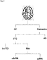
FIG. 2 is a diagram showing a hierarchical structure of brain disease groups. -
FIG. 3 is a diagram illustrating a brain atrophy diagnosis method according to an embodiment of the present disclosure. -
FIG. 4 is a diagram showing an example of diagnostic modeling according to an embodiment of the present disclosure. -
FIG. 5 is a diagram showing an example of diagnostic modeling according to another embodiment of the present disclosure. - The following disclosure describes the principle of the present disclosure. Accordingly, although not explicitly described or illustrated herein, those skilled in the art will realize the principle of the present disclosure and invent a variety of devices included in the concept and scope of the present disclosure. In addition, all the conditional terms and embodiments as used herein are, in principle, obviously intended to help understanding of the concept of the present disclosure, and should not be understood as limited to the particular embodiments and aspects described herein.
- In addition, it should be understood that the detailed description of not only the principle, perspective and embodiments of the present disclosure but also particular embodiments is intended to include structural and functional equivalents thereto. Furthermore, these equivalents should be understood as including equivalents that are now well known as well as equivalents that will be developed in the future, i.e., all devices invented to perform the same function irrespective of structure.
- Accordingly, for example, it should be understood that any block diagrams herein represent conceptual views of illustrative systems embodying the principles of the present disclosure. Likewise, it should be understood that any flow charts, state transition diagrams, pseudo code and the like represent various processes which may be substantially represented in computer-readable medium and so executed by a computer or processor, whether or not such computer or processor is explicitly shown.
- The functions of various devices shown in the drawings including the processor or function blocks indicated in a similar concept may be provided by the use of dedicated hardware as well as hardware capable of running software in association with appropriate software. When provided by the processor, the functions may be provided by a single dedicated processor, a single shared processor or a plurality of individual processors, and some of them may be shared.
- In addition, the explicit use of the term processor, control or the term presented in a similar concept should not be construed to refer exclusively to hardware capable of running software, and it should be understood that such terms implicitly include, without limitation, digital signal processor (DSP) hardware and read only memory (ROM), random access memory (RAM) and nonvolatile memory for storing software. Other well-known hardware may also be included.
- In the appended claims of the present disclosure, any element expressed as a means for performing the function described in the detailed description is intended to encompass any way of performing the function including, for example, a combination of circuit elements that perform the function or software in any form including firmware, microcode or the like, combined with appropriate circuitry for running the software to perform the function. The present disclosure defined by the appended claims should be understood that the functions provided by the various recited means are combined and brought together in the manner which the appended claims call for, and thus any means that can provide the functions is equivalent to those shown herein.
- The above-described objects, features and advantages will be apparent through the following detailed description related to the accompanying drawings, and accordingly those having ordinary skill in the technical field pertaining to the present disclosure will easily carry out the technical spirit of the present disclosure. In addition, in describing the present disclosure, when it is determined that a certain detailed description of known technology related to the present disclosure may unnecessarily obscure the subject matter of the present disclosure, the detailed description is omitted.
-
FIG. 1 is a diagram showing a hierarchical diagnosis device of brain atrophy based on brain thickness information according to an embodiment of the present disclosure. - Referring to
FIG. 1 , the hierarchical diagnosis device 100 of brain atrophy of the present disclosure includes a brainstructure modeling unit 110, a brainthickness extraction unit 120, adenoising unit 130, a trainingdata generation unit 140, a diagnosismodel training unit 150 and a brainatrophy diagnosis unit 160. - The brain
structure modeling unit 110 generates a grey matter surface and a white matter surface having a polygonal mesh data structure by mesh modeling of multiple 3-dimensional volumetric images such as Magnetic Resonance Imaging (MRI) images. In this instance, in particular, the MRI image is preferably a T1 weighted image. - The polygonal mesh is represented as a mesh including faces and vertices, but the number of faces and vertices of the brain surface mesh extracted for each individual may differ, and in this case, comparative analysis between individuals is difficult.
- The present disclosure pre-acquires and stores a reference template corresponding to the standard brain structure, and extracts a new vertex of which locations on the brain surface almost match for each vertex ID by re-sampling the grey matter surface and the white matter surface through the reference template, thereby allowing feature comparison between individuals.
- The brain
thickness extraction unit 120 extracts a pair of vertices (i.e., a pair of corresponding points) matched to each other based on the grey matter surface mesh and the white matter surface mesh acquired through the brainstructure modeling unit 110, and acquires brain thickness information by collecting and analyzing the distance between the vertices. That is, a change in grey matter thickness by the brain shrinkage is identified through the brain thickness information. - For reference, the brain surface generated and re-sampled through the brain
structure modeling unit 110 has a large amount of noise, and due to the large amount of noise, brain thickness information extracted for each individual also has a large amount of noise. The noise has a locally nonlinear projective property. - The
denoising unit 130 of the present disclosure converts the brain thickness information into frequency domain, and removes all high frequency components (i.e., locally nonlinear projective noise components) included in the brain thickness information through a denoising method such as Laplace Beltrami Operator (LBO). - When machine learning is requested, the training
data generation unit 140 is selectively activated to acquire brain thickness information and diagnosis information corresponding to each of a plurality of patients and combine them to generate multiple training data. - The diagnosis
model training unit 150 includes adimensionality reduction unit 151 and a hierarchicalclassifier training unit 152, and generates a diagnosis model for brain atrophy diagnosis by machine learning of the multiple training data. - The
dimensionality reduction unit 151 reduces the data dimension of the brain thickness information to lower dimension so that shrinkage can be actually compared through a dimensionality reduction method such as Principal Component Analysis (PCA). That is, as regions of the brain in which the brain shrinks vary depending on the type of brain atrophy, the present disclosure selects each region of the brain in which the brain shrinks as a principal component by the percentage of Table 1 (exemplary percentage) and brings out only the features of the locations, and an example of actual application (an example of application in percentage) may be as shown in Table 2.[Table 1] Percentage of brain thickness information dimension Percentage of frequency dimension (LB operator) Percentage of PCA dimension CN vs Dementia 100% about 0.2% about 0.1% AD vs FTD 100% about 0.2% about 0.1% bvFTD vs PPA 100% about 0.2% about 0.1% nfvPPA vs svPPA 100% about 0.2% about 0.1% [Table 2] Brain thickness information dimension Frequency dimension (LB operator) PCA dimension CN vs Dementia 81,924 150-170 75-85 AD vs FTD 81,924 220-260 55-65 bvFTD vs PPA 81,924 250-290 90-100 nfvPPA vs svPPA 81,924 200-240 75-85 - The hierarchical
classifier training unit 152 classifies the brain thickness information included in the training data into brain disease groups of hierarchical structure as shown inFIG. 2 according to the diagnosis information, and extracts one-dimensional feature information for each group by calculating and applying a matrix including features with low correlation between groups and features with high correlation within groups through a feature extraction method such as Linear Discriminant Analysis (LDA) and support vector machine (SVM). Additionally, hierarchical classifiers are generated and trained based on the hierarchical structure of the groups and the feature information for each group, and a diagnosis model including the hierarchical classifiers is acquired and stored. - Finally, the brain
atrophy diagnosis unit 160 is activated when disease diagnosis is requested. When brain thickness information corresponding to an MRI image of a subject is newly acquired, the brainatrophy diagnosis unit 160 finally identifies and notifies the type of brain atrophy corresponding to the brain thickness information of the subject by hierarchically analyzing the similarity with the brain disease groups through the hierarchical classifiers in the diagnosis model. - As described above, the present disclosure extracts the brain thickness information for classifying the subtypes of brain atrophy from the MRI image, and hierarchically diagnoses brain atrophy as shown in
FIG. 2 using curvelet-based denoising, linear dimensionality reduction and machine learning algorithms for the extracted brain thickness information. - That is, the type of brain atrophy of the subject is diagnosed through the procedure of identifying whether the subject has normal cognition (NC) or dementia, and in the case of dementia, identifying whether the subject has Alzheimer's Disease (AD) or Frontotemporal Dementia (FTD), and in the case of FTD, identifying whether the subject has Behavior Variables FTD (bvFTD) or Primary Progressive Aphasia (PPA), and in the case of PPA, identifying whether the subject has Nonfluent/agrammatic Variant PPA (nfvPPA) or Semantic Variant PPA (svPPA). The type of brain atrophy may be variously adjusted as needed. For example, instead of nfvPPA and svPPA, Progressive Nonfluent Aphasia (PNFA) and Semantic Dementia (SD) may be applied.
- In addition, the hierarchical diagnosis device of brain atrophy may be implemented as an independent hardware device, but where necessary, may be implemented in the form of a plugin into the existing medical equipment, or a software program which is installed and executed in a medical device.
- Hereinafter, an operation method of the hierarchical diagnosis device of brain atrophy according to an embodiment of the present disclosure will be described in more detail with reference to
FIGS. 3 and4 . -
FIG. 3 is a diagram illustrating a brain atrophy diagnosis method according to an embodiment of the present disclosure, and the brain atrophy diagnosis method of the present disclosure largely includes a training step and a diagnosis step. - First, in the training step, MRI images and diagnosis information of a plurality of patients with brain atrophy of NC, AD, bvFTD, nfvPPA and svPPA are acquired by accessing a database server in which the medical images and the diagnosis information of the patients are stored (S11).
- Additionally, a grey matter surface mesh and a white matter surface mesh are generated by mesh modeling of each MRI image, and correspondence between the patients is achieved by re-sampling according to a preset reference template (S12).
- Additionally, pairs of corresponding points of the re-sampled grey matter surface mesh and white matter surface mesh are all extracted, and brain thickness information of each patient is extracted by collecting and analyzing the distance between the corresponding points (S13), and noise (i.e., harmonics having a locally nonlinear projective property) included in the brain thickness information is removed (S14).
- Additionally, patient diagnosis information is added to the brain thickness information acquired through S14 to generate multiple training data (S15).
- The generation of the multiple training data is completed through the step S15, and the data dimension of the brain thickness information is reduced through Principal Component Analysis (PCA) (S16).
- Additionally, the dimension reduced brain thickness information is classified into hierarchical groups according to the diagnosis information, and feature information for each group is extracted through linear discriminant analysis. Additionally, hierarchical classifiers for hierarchically diagnosing brain atrophy based on the hierarchical structure of the groups and the feature information for each group are generated and trained, and a diagnosis model including the hierarchical classifiers is generated and stored (S17).
- When the training step is completed, the diagnosis step may be performed.
- When an MRI image of a subject is newly acquired through MRI equipment (S21), brain thickness information of the subject is acquired by the same process as the above-described steps S12 to S15 (S22 to S25).
- Then, a group having the highest similarity with the brain thickness information of the subject is hierarchically searched through the hierarchical classifiers of the diagnosis model, and the diagnosis information corresponding to the group having the highest similarity is acquired and outputted as brain atrophy diagnosis information (S26).
-
FIG. 4 is a diagram showing an example of diagnostic modeling according to an embodiment of the present disclosure, and referring toFIG. 4 , the diagnosis model of the present disclosure may include a first classifier for classifying an NC group and a Dementia group, a second classifier for classifying an AD group and an FTD group, a third classifier for classifying a bvFTD group and a PPA group, and a fourth classifier for classifying an nfvPPA group and an svPPA group. - In this circumstance, when brain thickness information of a subject is acquired, brain atrophy may be hierarchically diagnosed based on the brain thickness information using the first to fourth classifiers in a sequential order as follows.
- First, (1) one of the NC group and the Dementia group to which the brain thickness information of the subject is more similar is identified through the first classifier.
- (2) When the similarity with the NC group is higher, it is determined that the subject has NC, and when the similarity with the Dementia group is higher, similarity with the FTD group and similarity with the AD group are additionally calculated through the second classifier.
- (3) When the similarity with the AD group is higher, it is determined that the subject has AD, and when the similarity with the FTD group is higher, similarity with the bvFTD group and similarity with the PPA group are additionally calculated through the third classifier.
- (4) When the similarity with the bvFTD group is higher, it is determined that the subject has bvFTD, and when the similarity with the PPA group is higher, similarity with the nfvPPA group and similarity with the svPPA group are additionally calculated through the fourth classifier.
- Additionally, (5) when the similarity with the nfvPPA group is higher, it is determined that the subject has nfvPPA, and when the similarity with the svPPA group is higher, it is determined that the subject has svPPA.
-
FIG. 5 is a diagram showing an example of diagnostic modeling according to another embodiment of the present disclosure, and referring toFIG. 5 , the diagnosis model of the present disclosure may include a first classifier for classifying an NC group and a Dementia group, a second classifier for classifying an AD group and an FTD group, a third classifier for classifying a bvFTD group and an nfvPPA group, a fourth classifier for classifying a bvFTD group and an svPPA group, and a fifth classifier for classifying an nfvPPA group and an svPPA group. - In this case, first, (1) one of the NC group and the Dementia group to which brain thickness information of a subject is more similar is identified through the first classifier.
- (2) When the similarity with the NC group is higher, it is determined that the subject has NC, and when the similarity with the Dementia group is higher, similarity with the FTD group and similarity with the AD group are additionally calculated through the second classifier.
- (3) When the similarity with AD group is higher, it is determined that the subject has AD, and when the similarity with FTD group is higher, similarity with bvFTD group and nfvPPA group, similarity with bvFTD group and svPPA group and similarity with nfvPPA group and svPPA group are respectively calculated using the third classifier, the fourth classifier and the fifth classifier at the same time.
- (4) As a result of cross-checking of the third to fifth classifiers, when there is a group having received two votes among the total of three groups (bvFTD, nfvPPA, svPPA), it is determined that the subject has brain atrophy corresponding to the corresponding group, and when each of the total of three groups receives each one vote, brain atrophy is determined on the basis of a group having the greatest distance from the classifier.
- As described above, since the present disclosure builds the hierarchical classifiers to hierarchically identify the type of brain atrophy, it is possible to diagnose and notify the type of brain atrophy of the subject more accurately and precisely.
- The above-described method according to the present disclosure may be implemented as a program that is executed in a computer and stored in a computer-readable recording medium, and examples of the computer-readable recording medium include ROM, RAM, CD-ROM, magnetic tape, floppy disk and optical data storage devices, as well as those embodied in the form of a carrier wave (for example, transmission via Internet).
- The computer-readable recording medium is distributed over computer systems connected via a network, and stores and executes the computer-readable code in a distributed manner. Additionally, functional programs, code and code segments for implementing the method may be easily inferred by programmers in the technical field pertaining to the present disclosure.
- Although the preferred embodiments of the present disclosure have been hereinabove illustrated and described, the present disclosure is not limited to the above-described particular embodiments, and a variety of modifications may be made thereto by those having ordinary skill in the technical field pertaining to the present disclosure without departing from the subject matter of the present disclosure claimed in the appended claims, and such modifications should not be individually understood from the technical spirit or scope of the present disclosure.
Claims (6)
- A hierarchical diagnosis device of brain atrophy based on brain thickness information, comprising:a brain structure modeling unit to generate a grey matter surface mesh and a white matter surface mesh by mesh modeling of multiple Magnetic Resonance Imaging (MRI) images;a brain thickness extraction unit to acquire brain thickness information by collecting and analyzing a distance between corresponding points of the grey matter surface mesh and the white matter surface mesh;a training data generation unit to generate and store multiple training data including the brain thickness information and diagnosis information when diagnosis model training is requested;a diagnosis model training unit to classify and analyze the brain thickness information into groups of a hierarchical structure according to the diagnosis information to acquire feature information for each group, and generate and train hierarchical classifiers based on the hierarchical structure of the groups and the feature information for each group; anda brain atrophy diagnosis unit to acquire new brain thickness information upon receiving a new input of an MRI image of a subject, hierarchically search for a group having highest similarity with the new thickness information through the hierarchical classifiers, and identify and notify a type of brain atrophy based on the group search results.
- The hierarchical diagnosis device of brain atrophy based on brain thickness information according to claim 1, wherein the diagnosis model training unit generates and trains a first classifier for classifying the brain atrophy as normal cognition or dementia; a second classifier for classifying as Alzheimer's Disease (AD) or Frontotemporal Dementia (FTD) in case of the dementia; a third classifier for classifying as Behavior Variables FTD (bvFTD) or Primary Progressive Aphasia (PPA) in case of the Frontotemporal Dementia (FTD); and a fourth classifier for classifying as Nonfluent/agrammatic Variant PPA (nfvPPA) or Semantic Variant PPA (svPPA) in case of the Primary Progressive Aphasia (PPA).
- The hierarchical diagnosis device of brain atrophy based on brain thickness information according to claim 1, wherein the diagnosis model training unit generates and trains a first classifier for classifying the brain atrophy as normal cognition or dementia; a second classifier for classifying as Alzheimer's Disease (AD) or Frontotemporal Dementia (FTD) in case of the dementia; a third classifier for classifying as Behavior Variables FTD (bvFTD) or Nonfluent/agrammatic Variant PPA (nfvPPA) in case of the Frontotemporal Dementia (FTD); a fourth classifier for classifying as Behavior Variables FTD (bvFTD) or Semantic Variant PPA (svPPA) in case of the Frontotemporal Dementia (FTD); and a fifth classifier for classifying as Nonfluent/agrammatic Variant PPA (nfvPPA) or Semantic Variant PPA (svPPA) in case of the Frontotemporal Dementia (FTD).
- The hierarchical diagnosis device of brain atrophy based on brain thickness information according to claim 1, wherein the brain structure modeling unit further includes a function of matching locations of each vertex on a brain surface by re-sampling a grey matter surface and a white matter surface according to a preset reference template.
- The hierarchical diagnosis device of brain atrophy based on brain thickness information according to claim 1, further comprising:
a denoising unit to remove noise included in each thickness information. - The hierarchical diagnosis device of brain atrophy based on brain thickness information according to claim 1, wherein the diagnosis model training unit includes a dimensionality reduction unit to reduce a data dimension of the brain thickness information; and a hierarchical classifier training unit to classify the brain thickness information into groups of a hierarchical structure according to the diagnosis information, acquire feature information for each group through linear determinant analysis, and generate and train hierarchical classifiers based on the hierarchical structure of the groups and the feature information for each group.
Applications Claiming Priority (2)
| Application Number | Priority Date | Filing Date | Title |
|---|---|---|---|
| KR1020190109012A KR102328521B1 (en) | 2019-09-03 | 2019-09-03 | Apparatus for cortical atrophy disease hierarchical diagnosis based on brain thickness information |
| PCT/KR2020/011787 WO2021045505A1 (en) | 2019-09-03 | 2020-09-02 | Apparatus for hierarchically diagnosing brain atrophy disease on basis of brain thickness information |
Publications (2)
| Publication Number | Publication Date |
|---|---|
| EP4026482A1 true EP4026482A1 (en) | 2022-07-13 |
| EP4026482A4 EP4026482A4 (en) | 2023-09-27 |
Family
ID=74852066
Family Applications (1)
| Application Number | Title | Priority Date | Filing Date |
|---|---|---|---|
| EP20859769.0A Pending EP4026482A4 (en) | 2019-09-03 | 2020-09-02 | Apparatus for hierarchically diagnosing brain atrophy disease on basis of brain thickness information |
Country Status (5)
| Country | Link |
|---|---|
| US (1) | US20220313103A1 (en) |
| EP (1) | EP4026482A4 (en) |
| JP (1) | JP7291332B2 (en) |
| KR (1) | KR102328521B1 (en) |
| WO (1) | WO2021045505A1 (en) |
Families Citing this family (3)
| Publication number | Priority date | Publication date | Assignee | Title |
|---|---|---|---|---|
| KR102684775B1 (en) * | 2021-04-07 | 2024-07-16 | 의료법인 성광의료재단 | Diagnosis method and method for psychopathy using radiomics feature of medical image |
| CN114298234B (en) * | 2021-12-31 | 2022-10-04 | 深圳市铱硙医疗科技有限公司 | Brain medical image classification method and device, computer equipment and storage medium |
| CN115568828A (en) * | 2022-10-26 | 2023-01-06 | 深圳博脑医疗科技有限公司 | Detection method, device, terminal equipment and computer-readable storage medium |
Family Cites Families (14)
| Publication number | Priority date | Publication date | Assignee | Title |
|---|---|---|---|---|
| JP4799251B2 (en) | 2006-04-05 | 2011-10-26 | 富士フイルム株式会社 | Similar case search device, similar case search method and program thereof |
| KR100900258B1 (en) | 2006-08-09 | 2009-05-29 | 재단법인서울대학교산학협력재단 | Neurobiological model to measure human intelligence |
| US8099299B2 (en) * | 2008-05-20 | 2012-01-17 | General Electric Company | System and method for mapping structural and functional deviations in an anatomical region |
| US9008394B2 (en) | 2008-11-26 | 2015-04-14 | General Electric Company | Methods and apparatus for determining brain cortical thickness |
| KR101797501B1 (en) * | 2010-11-10 | 2017-11-16 | 삼성전자주식회사 | Apparatus and method for analyzing brain disease using magnetic resonance imaging and functional magnetic resonance imaging |
| KR20150072463A (en) * | 2013-12-11 | 2015-06-30 | 한국 한의학 연구원 | Health state determining method and health state determining apparatus using image of face |
| KR101789425B1 (en) | 2016-05-30 | 2017-10-25 | 연세대학교 산학협력단 | Methods for diagnosing early onset dementia by using 13C magnetic resonance imaging |
| FR3055998B1 (en) * | 2016-09-09 | 2018-10-12 | Olea Medical | SYSTEM AND METHOD FOR RECONSTRUCTING A PHYSIOLOGICAL SIGNAL OF AN ARTERY / FABRIC / VEIN DYNAMIC SYSTEM OF AN ORGAN IN A SURFACE SPACE |
| KR101944069B1 (en) * | 2016-12-29 | 2019-01-30 | 주식회사 마이다스아이티 | Method and apparatus for deciding Alzheimer's disease based on machine learning |
| KR101754291B1 (en) | 2017-04-04 | 2017-07-06 | 이현섭 | Medical image processing system and method for personalized brain disease diagnosis and status determination |
| KR101886000B1 (en) * | 2017-06-21 | 2018-08-06 | 조선대학교산학협력단 | Brain structural abnormality index based on brain magnetic resonance images and the method for diagnosing neurodegenerative disease using thereof |
| WO2019036749A1 (en) * | 2017-08-24 | 2019-02-28 | Adams Warwick Russell | A system for detecting early parkinson's disease and other neurological diseases and movement disorders |
| WO2020010320A1 (en) * | 2018-07-05 | 2020-01-09 | The Platypus Institute, Inc. | Identifying and strengthening physiological/neurophysiological states predictive of superior performance |
| EP3844780A4 (en) * | 2018-08-31 | 2022-06-22 | Blackthorn Therapeutics, Inc. | SYSTEM AND METHOD FOR IDENTIFYING TRANSDIAGNOSTIC CHARACTERISTICS COMMON TO MENTAL DISORDERS |
-
2019
- 2019-09-03 KR KR1020190109012A patent/KR102328521B1/en active Active
-
2020
- 2020-09-02 US US17/639,972 patent/US20220313103A1/en active Pending
- 2020-09-02 EP EP20859769.0A patent/EP4026482A4/en active Pending
- 2020-09-02 WO PCT/KR2020/011787 patent/WO2021045505A1/en not_active Ceased
- 2020-09-02 JP JP2022514616A patent/JP7291332B2/en active Active
Also Published As
| Publication number | Publication date |
|---|---|
| WO2021045505A1 (en) | 2021-03-11 |
| US20220313103A1 (en) | 2022-10-06 |
| EP4026482A4 (en) | 2023-09-27 |
| JP2022547063A (en) | 2022-11-10 |
| JP7291332B2 (en) | 2023-06-15 |
| KR20210028321A (en) | 2021-03-12 |
| KR102328521B1 (en) | 2021-11-19 |
Similar Documents
| Publication | Publication Date | Title |
|---|---|---|
| Eijlers et al. | Reduced network dynamics on functional MRI signals cognitive impairment in multiple sclerosis | |
| Liu et al. | Anatomical landmark based deep feature representation for MR images in brain disease diagnosis | |
| Gay et al. | Abnormal resting-state functional connectivity in patients with chronic fatigue syndrome: results of seed and data-driven analyses | |
| Jie et al. | Topological graph kernel on multiple thresholded functional connectivity networks for mild cognitive impairment classification | |
| Chen et al. | Classification of Alzheimer disease, mild cognitive impairment, and normal cognitive status with large-scale network analysis based on resting-state functional MR imaging | |
| WO2022256063A1 (en) | Predictive prognosis based on multimodal analysis | |
| Anderson et al. | Network anticorrelations, global regression, and phase‐shifted soft tissue correction | |
| Xu et al. | Denoising the speaking brain: toward a robust technique for correcting artifact-contaminated fMRI data under severe motion | |
| Huerta et al. | Quality assessment of very long-term ECG recordings using a convolutional neural network | |
| US20220313103A1 (en) | Hierarchical diagnosis device of brain atrophy based on brain thickness information | |
| Vaghari et al. | A multi-site, multi-participant magnetoencephalography resting-state dataset to study dementia: The BioFIND dataset | |
| US20110052024A1 (en) | method of analysing stroke images | |
| EP4231311A1 (en) | Method and apparatus for providing information needed for dementia diagnosis | |
| US20230263455A1 (en) | Network-based functional imaging output for evaluating multiple sclerosis | |
| Morgan et al. | MRI network progression in mesial temporal lobe epilepsy related to healthy brain architecture | |
| Adriaanse et al. | The association of glucose metabolism and eigenvector centrality in Alzheimer's disease | |
| Arroyo-Palacios et al. | Characterization of shape differences among ICP pulses predicts outcome of external ventricular drainage weaning trial | |
| Singh et al. | Alzheimer's Dementia and its Various Biomarker Details with Several Detection Methodologies-A Comprehensive Review | |
| Sree Ranjani | Machine learning applications for a real-time monitoring of arrhythmia patients using IoT | |
| Fan et al. | Hemispheric coupling between structural and functional asymmetries in clinically asymptomatic carotid stenosis with cognitive impairment | |
| CN117838124A (en) | A driving intention detection method, system and device based on EEG traceability network | |
| FEM | CARS 2023—Computer Assisted Radiology and Surgery | |
| Carrera-Pinzón et al. | Characterizing the left ventricular ultrasound dynamics in the frequency domain to estimate the cardiac function | |
| Cathala et al. | WHITE-Net: White matter HyperIntensities Tissue Extraction using deep learning Network | |
| Washimkar et al. | Application of FCM clustering on AM-FM to detect MRI disease progression for multiple sclerosis |
Legal Events
| Date | Code | Title | Description |
|---|---|---|---|
| STAA | Information on the status of an ep patent application or granted ep patent |
Free format text: STATUS: THE INTERNATIONAL PUBLICATION HAS BEEN MADE |
|
| PUAI | Public reference made under article 153(3) epc to a published international application that has entered the european phase |
Free format text: ORIGINAL CODE: 0009012 |
|
| STAA | Information on the status of an ep patent application or granted ep patent |
Free format text: STATUS: REQUEST FOR EXAMINATION WAS MADE |
|
| 17P | Request for examination filed |
Effective date: 20220317 |
|
| AK | Designated contracting states |
Kind code of ref document: A1 Designated state(s): AL AT BE BG CH CY CZ DE DK EE ES FI FR GB GR HR HU IE IS IT LI LT LU LV MC MK MT NL NO PL PT RO RS SE SI SK SM TR |
|
| DAV | Request for validation of the european patent (deleted) | ||
| DAX | Request for extension of the european patent (deleted) | ||
| A4 | Supplementary search report drawn up and despatched |
Effective date: 20230829 |
|
| RIC1 | Information provided on ipc code assigned before grant |
Ipc: G16H 50/70 20180101ALI20230823BHEP Ipc: G16H 50/50 20180101ALI20230823BHEP Ipc: G16H 30/40 20180101ALI20230823BHEP Ipc: G06N 20/00 20190101ALI20230823BHEP Ipc: G16H 50/20 20180101ALI20230823BHEP Ipc: A61B 5/055 20060101ALI20230823BHEP Ipc: A61B 5/00 20060101AFI20230823BHEP |