EP4019973A1 - Detection method of multiple analytes - Google Patents
Detection method of multiple analytes Download PDFInfo
- Publication number
- EP4019973A1 EP4019973A1 EP21217229.0A EP21217229A EP4019973A1 EP 4019973 A1 EP4019973 A1 EP 4019973A1 EP 21217229 A EP21217229 A EP 21217229A EP 4019973 A1 EP4019973 A1 EP 4019973A1
- Authority
- EP
- European Patent Office
- Prior art keywords
- variety
- human
- knobby
- protrusions
- microparticle
- Prior art date
- Legal status (The legal status is an assumption and is not a legal conclusion. Google has not performed a legal analysis and makes no representation as to the accuracy of the status listed.)
- Granted
Links
Images








Classifications
-
- G—PHYSICS
- G01—MEASURING; TESTING
- G01N—INVESTIGATING OR ANALYSING MATERIALS BY DETERMINING THEIR CHEMICAL OR PHYSICAL PROPERTIES
- G01N33/00—Investigating or analysing materials by specific methods not covered by groups G01N1/00 - G01N31/00
- G01N33/48—Biological material, e.g. blood, urine; Haemocytometers
- G01N33/50—Chemical analysis of biological material, e.g. blood, urine; Testing involving biospecific ligand binding methods; Immunological testing
- G01N33/53—Immunoassay; Biospecific binding assay; Materials therefor
- G01N33/543—Immunoassay; Biospecific binding assay; Materials therefor with an insoluble carrier for immobilising immunochemicals
- G01N33/54313—Immunoassay; Biospecific binding assay; Materials therefor with an insoluble carrier for immobilising immunochemicals the carrier being characterised by its particulate form
- G01N33/54326—Magnetic particles
- G01N33/54333—Modification of conditions of immunological binding reaction, e.g. use of more than one type of particle, use of chemical agents to improve binding, choice of incubation time or application of magnetic field during binding reaction
-
- G—PHYSICS
- G01—MEASURING; TESTING
- G01N—INVESTIGATING OR ANALYSING MATERIALS BY DETERMINING THEIR CHEMICAL OR PHYSICAL PROPERTIES
- G01N33/00—Investigating or analysing materials by specific methods not covered by groups G01N1/00 - G01N31/00
- G01N33/48—Biological material, e.g. blood, urine; Haemocytometers
- G01N33/50—Chemical analysis of biological material, e.g. blood, urine; Testing involving biospecific ligand binding methods; Immunological testing
- G01N33/53—Immunoassay; Biospecific binding assay; Materials therefor
- G01N33/543—Immunoassay; Biospecific binding assay; Materials therefor with an insoluble carrier for immobilising immunochemicals
- G01N33/54313—Immunoassay; Biospecific binding assay; Materials therefor with an insoluble carrier for immobilising immunochemicals the carrier being characterised by its particulate form
-
- C—CHEMISTRY; METALLURGY
- C12—BIOCHEMISTRY; BEER; SPIRITS; WINE; VINEGAR; MICROBIOLOGY; ENZYMOLOGY; MUTATION OR GENETIC ENGINEERING
- C12Q—MEASURING OR TESTING PROCESSES INVOLVING ENZYMES, NUCLEIC ACIDS OR MICROORGANISMS; COMPOSITIONS OR TEST PAPERS THEREFOR; PROCESSES OF PREPARING SUCH COMPOSITIONS; CONDITION-RESPONSIVE CONTROL IN MICROBIOLOGICAL OR ENZYMOLOGICAL PROCESSES
- C12Q1/00—Measuring or testing processes involving enzymes, nucleic acids or microorganisms; Compositions therefor; Processes of preparing such compositions
- C12Q1/68—Measuring or testing processes involving enzymes, nucleic acids or microorganisms; Compositions therefor; Processes of preparing such compositions involving nucleic acids
- C12Q1/6813—Hybridisation assays
- C12Q1/6834—Enzymatic or biochemical coupling of nucleic acids to a solid phase
-
- G—PHYSICS
- G01—MEASURING; TESTING
- G01N—INVESTIGATING OR ANALYSING MATERIALS BY DETERMINING THEIR CHEMICAL OR PHYSICAL PROPERTIES
- G01N33/00—Investigating or analysing materials by specific methods not covered by groups G01N1/00 - G01N31/00
- G01N33/48—Biological material, e.g. blood, urine; Haemocytometers
- G01N33/50—Chemical analysis of biological material, e.g. blood, urine; Testing involving biospecific ligand binding methods; Immunological testing
- G01N33/5005—Chemical analysis of biological material, e.g. blood, urine; Testing involving biospecific ligand binding methods; Immunological testing involving human or animal cells
- G01N33/5008—Chemical analysis of biological material, e.g. blood, urine; Testing involving biospecific ligand binding methods; Immunological testing involving human or animal cells for testing or evaluating the effect of chemical or biological compounds, e.g. drugs, cosmetics
- G01N33/5076—Chemical analysis of biological material, e.g. blood, urine; Testing involving biospecific ligand binding methods; Immunological testing involving human or animal cells for testing or evaluating the effect of chemical or biological compounds, e.g. drugs, cosmetics involving cell organelles, e.g. Golgi complex, endoplasmic reticulum
-
- G—PHYSICS
- G01—MEASURING; TESTING
- G01N—INVESTIGATING OR ANALYSING MATERIALS BY DETERMINING THEIR CHEMICAL OR PHYSICAL PROPERTIES
- G01N33/00—Investigating or analysing materials by specific methods not covered by groups G01N1/00 - G01N31/00
- G01N33/48—Biological material, e.g. blood, urine; Haemocytometers
- G01N33/50—Chemical analysis of biological material, e.g. blood, urine; Testing involving biospecific ligand binding methods; Immunological testing
- G01N33/53—Immunoassay; Biospecific binding assay; Materials therefor
- G01N33/5308—Immunoassay; Biospecific binding assay; Materials therefor for analytes not provided for elsewhere, e.g. nucleic acids, uric acid, worms, mites
-
- G—PHYSICS
- G01—MEASURING; TESTING
- G01N—INVESTIGATING OR ANALYSING MATERIALS BY DETERMINING THEIR CHEMICAL OR PHYSICAL PROPERTIES
- G01N33/00—Investigating or analysing materials by specific methods not covered by groups G01N1/00 - G01N31/00
- G01N33/48—Biological material, e.g. blood, urine; Haemocytometers
- G01N33/50—Chemical analysis of biological material, e.g. blood, urine; Testing involving biospecific ligand binding methods; Immunological testing
- G01N33/53—Immunoassay; Biospecific binding assay; Materials therefor
- G01N33/543—Immunoassay; Biospecific binding assay; Materials therefor with an insoluble carrier for immobilising immunochemicals
- G01N33/54306—Solid-phase reaction mechanisms
-
- G—PHYSICS
- G01—MEASURING; TESTING
- G01N—INVESTIGATING OR ANALYSING MATERIALS BY DETERMINING THEIR CHEMICAL OR PHYSICAL PROPERTIES
- G01N33/00—Investigating or analysing materials by specific methods not covered by groups G01N1/00 - G01N31/00
- G01N33/48—Biological material, e.g. blood, urine; Haemocytometers
- G01N33/50—Chemical analysis of biological material, e.g. blood, urine; Testing involving biospecific ligand binding methods; Immunological testing
- G01N33/53—Immunoassay; Biospecific binding assay; Materials therefor
- G01N33/543—Immunoassay; Biospecific binding assay; Materials therefor with an insoluble carrier for immobilising immunochemicals
- G01N33/54313—Immunoassay; Biospecific binding assay; Materials therefor with an insoluble carrier for immobilising immunochemicals the carrier being characterised by its particulate form
- G01N33/54326—Magnetic particles
-
- G—PHYSICS
- G01—MEASURING; TESTING
- G01N—INVESTIGATING OR ANALYSING MATERIALS BY DETERMINING THEIR CHEMICAL OR PHYSICAL PROPERTIES
- G01N33/00—Investigating or analysing materials by specific methods not covered by groups G01N1/00 - G01N31/00
- G01N33/48—Biological material, e.g. blood, urine; Haemocytometers
- G01N33/50—Chemical analysis of biological material, e.g. blood, urine; Testing involving biospecific ligand binding methods; Immunological testing
- G01N33/58—Chemical analysis of biological material, e.g. blood, urine; Testing involving biospecific ligand binding methods; Immunological testing involving labelled substances
- G01N33/582—Chemical analysis of biological material, e.g. blood, urine; Testing involving biospecific ligand binding methods; Immunological testing involving labelled substances with fluorescent label
-
- G—PHYSICS
- G01—MEASURING; TESTING
- G01N—INVESTIGATING OR ANALYSING MATERIALS BY DETERMINING THEIR CHEMICAL OR PHYSICAL PROPERTIES
- G01N33/00—Investigating or analysing materials by specific methods not covered by groups G01N1/00 - G01N31/00
- G01N33/48—Biological material, e.g. blood, urine; Haemocytometers
- G01N33/50—Chemical analysis of biological material, e.g. blood, urine; Testing involving biospecific ligand binding methods; Immunological testing
- G01N33/68—Chemical analysis of biological material, e.g. blood, urine; Testing involving biospecific ligand binding methods; Immunological testing involving proteins, peptides or amino acids
Definitions
- the disclosure relates to a detection method, and more particularly, to a detection method of multiple analytes.
- Ligand binding assay is a detection method that relies on affinity binding between ligand molecules and analytes
- enzyme-linked immunosorbent assay ELISA
- detection is performed by utilizing the property of binding specificity between an antibody and an antigen in combination with enzyme reaction.
- the technology has been developing toward maturity.
- long detection time is consumed and only one analyte can be detected in one reaction.
- the disclosure provides a detection method of multiple analytes, which can have the effect of improving detection sensitivity.
- the detection method of multiple analytes in the disclosure includes the following.
- a microparticle is provided.
- the microparticle is coupled with at least one first ligand, and includes a body and a plurality of first protrusions formed on a surface of the body.
- the microparticle is mixed with a specimen including a variety of analytes to form a first complex.
- the first complex is mixed with a variety of second ligands carrying a variety of first labels, such that the variety of second ligands bind to the variety of analytes in the first complex and form a second complex.
- the variety of first labels in the second complex are detected.
- the microparticle is a knobby particle.
- the body of the knobby particle includes a copolymer core, a polymer layer, and a silicon-based layer from the inside to the outside.
- a plurality of second protrusions are formed on a surface of the copolymer core.
- An average height of the second protrusions is 100 nanometers to 5000 nanometers.
- the microparticle is a knobby magnetic particle.
- the body of the knobby magnetic particle includes a copolymer core, a polymer layer, a magnetic substance layer, and a silicon-based layer from the inside to the outside.
- a plurality of second protrusions are formed on a surface of the copolymer core. An average height of the second protrusions is 100 nanometers to 5000 nanometers.
- a ratio of an average height of the first protrusions to an average diameter of the body is 0.005 to 0.25.
- a ratio of an average volume of the first protrusions to an overall volume of the body is 1 ⁇ 10 -7 to 2 ⁇ 10 -2
- a total volume of the first protrusions to an average volume of the microparticle is 1 ⁇ 10 -1 to 6 ⁇ 10 -1 .
- an average number of the first protrusions is 5 to 500.
- an average diameter of the microparticle is 1 ⁇ m to 20 ⁇ m.
- the microparticle is non-spherical.
- the manner in which the at least one first ligand is coupled to the microparticle comprises non-covalent bonding, covalent bonding, avidin-biotin interaction, electrostatic adsorption, hydrophobic adsorption, or a combination of the above.
- the variety of analytes are located on a surface of the specimen.
- the step of forming the first complex includes performing the at least one first ligand to recognize and directly bind to a target located on the surface of the specimen.
- the at least one first ligand includes a first specific antibody.
- the first specific antibody includes an antibody against a surface antigen on the human exosome, an antibody against a surface antigen on the human blood cell, an antibody against a surface antigen on the human immune cell, an antibody against a surface antigen on the human tumor cell, or a combination thereof.
- the specimen includes a human exosome, a human blood cell, a human immune cell, a human tumor cell, or a combination thereof.
- the variety of analytes include a surface antigen on the human exosome, a surface antigen on the human blood cell, a surface antigen on the human immune cell, a surface antigen on the human tumor cell, or a combination thereof.
- the variety of second ligands include a variety of second specific antibodies.
- the variety of second specific antibodies include antibody against a surface antigen on the human exosome, an antibody against a surface antigen on the human blood cell, an antibody against a surface antigen on the human immune cell, an antibody against a surface antigen on the human tumor cell, or a combination thereof.
- the at least one first ligand includes a variety of first ligands.
- the step of forming the first complex includes performing the variety of first ligands to recognize and directly bind to the variety of analytes.
- the variety of first ligands include a variety of nucleic acid probes.
- the variety of nucleic acid probes include a variety of primers or aptamers.
- the variety of analytes include a variety of nucleic acid sequences carrying a variety of second labels.
- the variety of second labels include biotin, a variety of antigenic epitopes, or a combination thereof.
- the variety of second ligands include a variety of specific proteins.
- the variety of specific proteins include an anti-biotin antibody, avidin, streptavidin, neutravidin, a third specific antibody, or a combination thereof.
- the variety of first labels include a variety of fluorescent labels or a variety of luminescent labels.
- the variety of fluorescent labels include FITC, Alexa, PE, PerCP, BV, APC, Pacific Blue, or a combination thereof.
- the variety of luminescent labels include luciferase.
- the detection method of multiple analytes in the disclosure includes the following.
- a microparticle is provided.
- the microparticle is coupled with a variety of ligands, and includes a body and a plurality of protrusions formed on a surface of the body.
- the microparticle is mixed with a specimen including a variety of analytes to form a complex.
- the variety of analytes carry a variety of labels.
- the variety of labels in the complex are detected.
- the variety of ligands include a variety of nucleic acid probes.
- the variety of labels include a variety of fluorescent labels, a variety of luminescent labels, or a combination thereof.
- the variety of analytes include a variety of nucleic acid sequences.
- the microparticles of the disclosure can be arranged on the surface of the body by disposing a plurality of first protrusions with irregular shapes, thereby increasing the surface area of the microparticles (ie, the sum of the surface area of the body and the surface area of the first protrusions).
- the microparticles can be coupled with more first ligands to identify and bind more targets, thereby enhancing the detection signal and improving the detection sensitivity.
- FIG. 1 is a flowchart of a detection method of multiple analytes according to an exemplary embodiment.
- FIG. 2 is a schematic view illustrating flows of the detection method of multiple analytes according to an exemplary embodiment.
- FIG. 3A is a schematic cross-sectional view of a knobby particle according to an exemplary embodiment.
- FIG. 3B is a schematic cross-sectional view of a knobby magnetic particle according to an exemplary embodiment.
- step S10 is firstly performed, in which a microparticle 110 is provided.
- the microparticle 110 includes a body 111 and a plurality of first protrusions 112 formed on a surface 111a of the body 111.
- the microparticle 100 is non-spherical.
- the first protrusions 112 have irregular shapes, and may be evenly or unevenly distributed on the surface 111a of the body 111.
- the first protrusions 112 are integrally formed with the body 111, and the first protrusions 112 are seamlessly connected to the body 111.
- the first protrusions 112 have an average height H1 (i.e., the average vertical distance from the top of the first protrusions 112 to the surface 111a of the body 111).
- the microparticle 110 may be a knobby particle 110a or a knobby magnetic particle 110b.
- the knobby particle 110a is non-magnetic, and the knobby magnetic particle 110b is magnetic.
- the body 111 of the knobby particle 110a includes a copolymer core 113, a polymer layer 114, and a silicon-based layer 115 from the inside to the outside.
- a plurality of second protrusions 113b are formed on a surface 113a of the copolymer core 113.
- the second protrusions 113b have irregular shapes, and may be evenly or unevenly distributed on the surface 113a of the copolymer core 113. Substantially, the second protrusions 113b are integrally formed with the copolymer core 113, and the second protrusions 113b are seamlessly connected to the copolymer core 113.
- an average height H2 of the second protrusions 113b may be 100 nanometers (nm) to 5000 nm, for example but not limited to, about 200 nm to 4500 nm, about 500 nm to 4000 nm, about 800 nm to 3500 nm, about 300 nm to 1000 nm, about 600 nm to 1800 nm, about 750 nm to 2500 nm, about 900 nm to 3000 nm, about 1000 nm to 3600 nm, and about 1500 nm to 4800 nm.
- the polymer layer 114 and the silicon-based layer 115 are sequentially formed on the copolymer core 113 in a substantially conformal manner. Therefore, the first protrusions 112 of the knobby particle 110a substantially correspond to the second protrusions 113b of the copolymer core 113, but not limited thereto.
- the body 111 of the knobby magnetic particle 110b includes the copolymer core 113, the polymer layer 114, a magnetic substance layer 116, and the silicon-based layer 115 from the inside to the outside.
- the second protrusions 113b is formed on the surface 113a of the copolymer core 113.
- the second protrusions 113b have irregular shapes, and may be evenly or unevenly distributed on the surface 113a of the copolymer core 113. Substantially, the second protrusions 113b are integrally formed with the copolymer core 113, and the second protrusions 113b are seamlessly connected to the copolymer core 113.
- the average height H2 of the second protrusions may be 100 nm to 5000 nm, for example but not limited to, about 200 nm to 4500 nm, about 500 nm to 4000 nm, about 800 nm to 3500 nm, about 300 nm to 1000 nm, about 600 nm to 1800 nm, about 750 nm to 2500 nm, about 900 nm to 3000 nm, about 1000 nm to 3600 nm, and about 1500 nm to 4800 nm.
- the polymer layer 114, the magnetic substance layer 116, and the silicon-based layer 115 are sequentially formed on the copolymer core 113 in a substantially conformal manner.
- the first protrusions 112 of the knobby magnetic particle 110b substantially corresponds to the second protrusions 113b of the copolymer core 113, but not limited thereto.
- the copolymer core 113 is, for example but not limited to, styrene/divinylbenzene copolymer, methyl methacrylate/triethylene glycol dimethacrylate copolymer, methyl methacrylate/ethylene glycol dimethacrylate copolymer, styrene/triethylene glycol dimethacrylate copolymer, styrene/ethylene glycol dimethacrylate copolymer, or methyl methacrylate/divinylbenzene copolymer.
- the polymer layer 114 may be a surface of the copolymer core 113 modified with functional groups, and includes at least one functional group (for example but not limited to, a carboxyl group, an amino group, or a combination thereof).
- the material of the silicon-based layer 115 may include but is not limited to tetramethoxysilane (TMOS), tetraethoxysilane (TEOS), 3-Aminopropyltriethoxysilane (APTES), and 3-Glycidoxypropyltrimethoxysilane (GOPTS).
- the material of the magnetic substance layer 116 may include but is not limited to paramagnetic materials, superparamagnetic materials, ferromagnetic materials, ferrimagnetic materials, or a combination thereof.
- the average height H1 of the first protrusions 112 may be 0.1 micrometer ( ⁇ m) to 5 ⁇ m, for example but not limited to, about 0.2 ⁇ m to 4.5 ⁇ m, about 0.3 ⁇ m to 4 ⁇ m, about 0.4 ⁇ m to 3.5 ⁇ m, about 0.5 ⁇ m to 3 ⁇ m, and about 0.6 ⁇ m to 2.5 ⁇ m.
- An average volume V1 of the first protrusions 112 may be 0.00052 cubic micrometer ( ⁇ m 3 ) to 65 ⁇ m 3 , for example but not limited to, about 0.001 ⁇ m 3 to 60 ⁇ m 3 , about 0.01 ⁇ m 3 to 50 ⁇ m 3 , and about 0.1 ⁇ m 3 to 40 ⁇ m 3 .
- An average diameter D1 of the body 111 may be 1 ⁇ m to 20 ⁇ m, for example but not limited to, about 2 ⁇ m to 15 ⁇ m, about 3 ⁇ m to 18 ⁇ m, about 4 ⁇ m to 10 ⁇ m, and about 5 ⁇ m to 12 ⁇ m.
- An average volume V2 of the body 111 may be 0.52 ⁇ m 3 to 4200 ⁇ m 3 , for example but not limited to, about 1 ⁇ m 3 to 4000 ⁇ m 3 , about 10 ⁇ m 3 to 3800 ⁇ m 3 , about 20 ⁇ m 3 to 3600 ⁇ m 3 , about 30 ⁇ m 3 to 3300 ⁇ m 3 , about 40 ⁇ m 3 to 2900 ⁇ m 3 , about 50 ⁇ m 3 to 2500 ⁇ m 3 , about 60 ⁇ m 3 to 2300 ⁇ m 3 , about 80 ⁇ m 3 to 2000 ⁇ m 3 , and about 100 ⁇ m 3 to 1000 ⁇ m 3 .
- a ratio of the average height H1 of the first protrusions 112 to the average diameter D1 of the body 111 may be 0.005 to 0.25, for example but not limited to, about 0.001 to 0.23, about 0.05 to 0.20, about 0.01 to 0.18, and about 0.05 to 0.15.
- a ratio of the average volume V1 of the first protrusions 112 to the average volume V2 of the body 111 may be 1 ⁇ 10 -7 to 2 ⁇ 10 -2 , for example but not limited to, 1 ⁇ 10 -6 to 1.5 ⁇ 10 -2 , 1 ⁇ 10 -5 to 1.2 ⁇ 10 -2 , and 1 ⁇ 10 -5 to 1 ⁇ 10 -2 .
- An average diameter D of the microparticle 110 (i.e., the average of the shortest diameter and the longest diameter of the microparticle 110, for example, the average of a shortest diameter D2 and a longest diameter D3 of the knobby particle 110a or of the knobby magnetic particle 110b) may be 1 ⁇ m to 20 ⁇ m, for example but not limited to, about 2 ⁇ m to 18 ⁇ m, about 3 ⁇ m to 15 ⁇ m, and about 5 ⁇ m to 10 ⁇ m.
- the microparticle 110 is coupled with at least one first ligand 120 (one first ligand shown in FIG. 1 serving as an example, but not limited thereto).
- the first ligand 120 is coupled to the surface 111a of the body 111 of the microparticle 110 and the first protrusions 112.
- the manner in which the first ligand 120 is coupled to the microparticle 110 includes non-covalent bonding, covalent bonding, avidin-biotin interaction, electrostatic adsorption, hydrophobic adsorption, or a combination thereof.
- the first ligand 120 includes, for example but not limited to, a specific antibody.
- step S20 is performed, in which the microparticle 110 is mixed with a variety of analytes 130a, 130b, 130c to form a first complex 140.
- the variety of analytes 130a, 130b, 130c are located on a surface S1 of a specimen S.
- Forming the first complex 140 includes performing the at least one first ligand 120 to recognize and directly bind to a target T on the surface S1 of the specimen S.
- the first ligand 120 includes a first specific antibody, which is capable of recognizing and directly binding to the target T.
- the first specific antibody includes but is not limited to an antibody against a surface antigen on the human exosome (e.g., anti-human CD9, anti-human CD63, anti-human CD29, anti-human CD81, anti-human HER2, anti-human EGFR, and anti-human EpCAM), an antibody against a surface antigen on the human blood cell (e.g., anti-human CD45 and anti-human CD235a), an antibody against a surface antigen on the human immune cell (e.g., anti-human CD3 and anti-human CD19), an antibody against a surface antigen on the human tumor cell (e.g., anti-human CEA, anti-human PD-L1, and anti-human HER2) or a combination thereof.
- a surface antigen on the human exosome e.g., anti-human CD9, anti-human CD63, anti-human CD29, anti-human CD81
- the specimen S includes but is not limited to human exosomes, human blood cells, human immune cells, human tumor cells, or a combination thereof.
- the analytes 130a, 130b, 130c include but is not limited to a surface antigen on the human exosomes (e.g., CD9, CD63, CD81, HER2, EGFR, and EpCAM), a surface antigen on the human blood cells (e.g., CD45 and CD235a), a surface antigen on the human immune cells (e.g., CD3 and CD19), a surface antigen on the human tumor cells (e.g., CEA, PD-L1, and HER2), or a combination thereof.
- a surface antigen on the human exosomes e.g., CD9, CD63, CD81, HER2, EGFR, and EpCAM
- a surface antigen on the human blood cells e.g., CD45 and CD235a
- a surface antigen on the human immune cells e.g., CD
- the target T may include but is not limited to at least one of the surface antigens on the human exosome, at least one of the surface antigens on the human blood cell, at least one of the surface antigens on the human immune cell, at least one of the surface antigens on the human tumor cell, or a combination thereof.
- the target T may be identical or different to any one of the analytes 130a, 130b, 130c.
- the target T is, for example, the surface antigen CD9 on the human exosomes, and the analytes are, for example, the surface antigens CD9, CD63, and CD81 on the human exosomes.
- the target T is, for example, the surface antigen HER2 on the human exosomes
- the analytes are, for example, the surface antigens CD9, CD63, and CD81 on the human exosomes.
- the disclosure is not limited thereto.
- the target T and the analyte are the same kind of surface antigens, the target T and the analyte 130a are not the same surface antigen. For example, as shown in FIG.
- the target T and the analyte 130a are the same kind of surface antigen 13a, in which the part that is recognized and bound to by the first ligand 120 is namely the target T, while another part that is not bound to by the first ligand 120 may serve as the analyte 130a for subsequent detection.
- step S30 is performed, in which the first complex 140 is mixed with a variety of second ligands 150a, 150b, 150c carrying a variety of first labels 151a, 151b, 151c, such that the second ligands 150a, 150b, 150c bind to the analytes 130a, 130b, 130c in the first complex 140 and form a second complex 160.
- second ligands 150a, 150b, 150c bind to the analytes 130a, 130b, 130c in the first complex 140 and form a second complex 160.
- FIG. 2 only one specimen S binding to one second ligand (150a, 150b, or 150c) is shown in FIG. 2 .
- the specimen S may bind to the variety of (or the plurality of) second ligands 150a, 150b, 150c at the same time as long as the variety of (or the plurality of) analytes 130a, 130b, 130c are present on the surface of the specimen S.
- the second ligands 150a, 150b, 150c are, for example, a variety of second specific antibodies.
- the second specific antibodies include an antibody against a surface antigen on the human exosome, an antibody against a surface antigen on the human blood cell, an antibody against a surface antigen on the human immune cell, an antibody against a surface antigen on the human tumor cell, or a combination thereof.
- the second specific antibodies may be identical or different to the first specific antibodies.
- the first labels 151a, 151b, 151c include but are not limited to a variety of different fluorescent labels or luminescent labels.
- the fluorescent labels may include but are not limited to FITC, Alexa (e.g., AF-488, AF-594, AF-647, and AF-700), PE (e.g., PE, PE-Cyanine5, PE-Cyanine7, and PE-Dazzle594), PerCP (e.g., PerCP and PerCP-Cyanine5.5), BV (Brilliant Violet, e.g., BV421, BV450, BV510, BV570, BV605, BV650, BV711, BV750, and BV785), APC (e.g., APC and APC-Cyanine7), Pacific Blue, or a combination thereof.
- Alexa e.g., AF-488, AF-594, AF-647, and AF-700
- PE e.g., PE, PE-Cyanine5, PE-Cyanine7, and PE-Dazzle594
- PerCP e.g., PerCP
- the luminescent labels include but are not limited to luciferase. Those having common knowledge in the technical field may select the first ligand, the first label, and the second ligand depending on actual needs (e.g., the source of specimens or the number of analytes), which is not limited by the disclosure.
- step S40 is performed, in which the first labels 151a, 151b, 151c of the second ligands 150a, 150b, 150c in the second complex 160 are detected.
- the detection result may represent the relative contents (or expressions) of the analytes 130a, 130b, 130c on the surface S1 of the specimen S.
- the detection is performed by using, for example, a flow cytometry analyzer, but is not limited thereto. So far, the performing of the detection method of multiple analytes of this embodiment has been substantially completed.
- the microparticle 110 of this embodiment compared to a general spherical microparticle, in the microparticle 110 of this embodiment, by disposing the first protrusions having irregular shapes on the surface of the body, the surface area of the microparticle 110 (i.e., the sum of the surface area of the body and the surface area of the first protrusions) is thus increased. Accordingly, the microparticle 110 of this embodiment may be coupled with more first ligands 120 to recognize and bind to more targets T, thereby increasing the strength of detection signals to improve the detection sensitivity.
- the surface of the magnetic substance layer 140 of the knobby magnetic particle 110b is a rough surface (or has small protrusions)
- the surface of the knobby magnetic particle 110b is also a rough surface.
- the knobby magnetic particle 110b is of a greater surface area. Accordingly, the knobby magnetic particle 110b is coupled with more first ligands 120, and recognizes and binds to more targets T.
- FIG. 4 is a schematic view illustrating flows of a detection method of multiple analytes according to another embodiment of the disclosure.
- the method of this embodiment is similar to the method in FIG. 2 , while the main difference lies in that, the at least one first ligand 120 includes a variety of first ligands 120a, 120b, 120c, and the first ligands 120a, 120b, 120c are, for example, a variety of nucleic acid probes (e.g., a variety of primers or aptamers).
- the analytes 130a, 130b, 130c include a variety of nucleic acid sequences carrying a variety of second labels 132a, 132b, 132c (for example but not limited to biotin, antigenic epitopes, or a combination thereof).
- the second ligands 150a, 150b, 150c include specific proteins (for example but not limited to, an anti-biotin antibody, avidin, streptavidin, neutravidin, third specific antibody, or a combination thereof).
- the microparticle 110 coupled with the first ligand 120a, 120b, 120c is first mixed with the analytes 130a, 130b, 130c, such that the first ligands 120a, 120b, 120c recognize and directly bind to the corresponding (or complementary) parts of sequence 131a, 131b, 131c of the analytes 130a, 130b, 130c and form the first complex 140.
- the first complex 140 is mixed with the second ligands 150a, 150b, 150c carrying the first labels 151a, 151b, 151c, such that the second ligands 150a, 150b, 150c bind to the second labels 132a, 132b, 132c of the analytes 130a, 130b, 130c in the first complex 140 and form the second complex 160.
- the first labels 151a, 151b, 151c of the second ligands 150a, 150b, 150c in the second complex 160 are detected.
- the second labels 132a, 132b, 132c may also be the first labels 150a, 150b, 150c. Accordingly, after the first ligands 120a, 120b, 120c bind to the parts of sequence 131a, 131b, and 131c of the analytes 130a, 130b, 130c, the formed first complex 140 carries a variety of fluorescent labels or a variety of luminescent labels. Therefore, the detection may be performed directly without adding the second ligand to form the second complex. So far, the performing of the detection method of multiple analytes of this embodiment has been substantially completed.
- specifications of general spherical microparticles or knobby particles are analyzed by using a scanning electron microscope (SEM) and a multisizer.
- SEM scanning electron microscope
- FIG. 5A , FIG. 5C and FIG. 5E are the analysis results of the spherical microparticles
- FIG. 5B , FIG. 5D and FIG. 5F are the analysis results of the knobby particles.
- the average diameter of the spherical microparticles in FIG. 5A is about 2.421 ⁇ m
- the average diameter of the knobby particles in FIG. 5B is about 3.280 ⁇ m
- the average diameter of the spherical microparticles in FIG. 5C is about 4.541 ⁇ m
- the average diameter of the knobby particles in FIG. 5D is about 4.153 ⁇ m
- the average diameter of the spherical microparticles in FIG. 5E is about 7.568 ⁇ m
- the average diameter of the knobby particles in FIG. 5F is about 7.189 ⁇ m.
- the average volume of the knobby particles is about 13.86 ⁇ m 3
- the average diameter of the bodies is about 2.5 ⁇ m
- the average volume of the bodies is about 6.14 ⁇ m 3
- the average height of the first protrusions is about 1.2 ⁇ m
- the average volume of the first protrusions is about 0.679 ⁇ m 3
- the total volume of the first protrusions is about 7.72 ⁇ m 3 . That is, in the knobby particles of FIG.
- the ratio of the average height of the first protrusions to the average diameter of the bodies is about 0.48
- the ratio of the average volume of the first protrusions to the average volume of the bodies is about 0.111
- each knobby particle has about 11 first protrusions
- the total volume of the first protrusions is 55.7% of the average volume of the knobby particles.
- the average volume of the knobby particles is about 49.09 ⁇ m 3
- the average diameter of the bodies is about 4.5 ⁇ m
- the average volume of the bodies is about 35.78 ⁇ m 3
- the average height of the first protrusions is about 0.8 ⁇ m
- the average volume of the first protrusions is about 0.268 ⁇ m 3
- the total volume of the first protrusions is about 13.31 ⁇ m 3 . That is, in the knobby particles of FIG.
- the ratio of the average height of the first protrusions to the average diameter of the bodies is about 0.18
- the ratio of the average volume of the first protrusions to the average volume of the bodies is about 0.007
- each knobby particle has about 49 first protrusions
- the total volume of the first protrusions is 27.1% of the average volume of the knobby particles.
- the average volume of the knobby particles is about 201 ⁇ m 3
- the average diameter of the bodies is about 7.5 ⁇ m
- the average volume of the bodies is about 166 ⁇ m 3
- the average height of the first protrusions is about 1 ⁇ m
- the average volume of the first protrusions is about 0.392 ⁇ m 3
- the total volume of the first protrusions is about 35 ⁇ m 3 . That is, in the knobby particles of FIG.
- the ratio of the average height of the first protrusions to the average diameter of the bodies is about 0.133
- the ratio of the average volume of the first protrusions to the average volume of the bodies is about 0.0024
- each knobby particle has about 89 first protrusions
- the total volume of the first protrusions is 17.4% of the average volume of the knobby particles.
- the ratio of the average height (or average volume) of the first protrusions to the average diameter (or average volume) of the bodies may be related to the surface morphology (ie, overall appearance).
- the ratio of the average height of the first protrusions to the average diameter of the body is greater than 0.25
- the ratio of the average volume of the first protrusions to the average volume of the bodies is greater than 2 ⁇ 10 -2 . Therefore, the surface morphology of the knobby particles in FIG. 5B are irregular. In other words, it is difficult to clearly identify the positions of the bodies and the first protrusions.
- the ratios of the average height of the first protrusions to the average diameter of the bodies are between 0.005 and 0.25, and the ratios of the average volume of the first protrusions to the average volume of the bodies are between 1 ⁇ 10 -7 and 2 ⁇ 10 -2 . Therefore, the positions of the bodies and the first protrusions may be identified in the knobby particles of FIG. 5D and FIG. 5F .
- the first specific antibody (anti-human CD9, anti-human CD63, or anti-human HER2) was firstly coupled to a general spherical microparticle and a microparticle (a knobby particle or a knobby magnetic particle) of this disclosure.
- a microparticle a knobby particle or a knobby magnetic particle
- assays and comparisons of the number of grafted specific antibodies were performed on the spherical microparticle coupled with the first specific antibody, the knobby particle coupled with the first specific antibody, and the knobby magnetic particle coupled with the first specific antibody.
- the classes and diameters of the microparticles used in this embodiment are shown in Table 1.
- Table 1 the classes and diameters of the microparticles Class of the microparticle Diameter of the microparticles ( ⁇ m)
- Example 1 knobby particle 8.5
- Example 2 knobby magnetic particle 8.5
- Example 3 knobby particle 2.5
- Example 4 knobby particle 4.5
- the step in which anti-human CD9 (or anti-human CD63 or anti-human HER2) was coupled to the spherical microparticle, the knobby particle, and the knobby magnetic particle is generally as follows: (1) 1 ⁇ 10 6 particles (i.e., the spherical microparticles of Comparative Examples 1 to 3, the knobby particles of Examples 1 and 3 to 4, or the knobby magnetic particles of Examples 2) surface-modified with amine groups were washed with 200 ⁇ L of a MES buffer solution three times.
- EDC Ethyl-3-(3-dimethylaminopropyl) carbodiimide hydrochloride
- NHS N-Hydroxysulfosuccinimide sodium salt
- PAA 15 kDa poly acrylic acid
- the spherical microparticles and the knobby particles were collected by centrifugation at a rotation speed of 10000 rpm for 3 minutes, and the knobby magnetic particles were collected with a magnet, in which the magnet was performed to stay thereon for at least 1 minute.
- the particles were washed with 200 ⁇ L of a MES buffer solution three times and added with 20 ⁇ L of anti-human CD9 (or anti-human CD63 or anti-human HER2) into a pH3 citric acid-PBS solution (0.625M citric acid dissolved in PBS solution, pH3.0) for reaction overnight at 4°C, such that anti-human CD9 (or anti-human CD63 or anti-human HER2) was coupled on the surface of the particles.
- the mixture was added into 200 ⁇ L of a bovine serum albumin solution (10 mg/mL BSA dissolved in a MES solution) for perform reaction overnight at 4°C to cover the rest of the surface of the particles that has not been coupled with anti-human CD9 (or anti-human CD63 or anti-human HER2).
- a bovine serum albumin solution (10 mg/mL BSA dissolved in a MES solution) for perform reaction overnight at 4°C to cover the rest of the surface of the particles that has not been coupled with anti-human CD9 (or anti-human CD63 or anti-human HER2).
- the particles were washed with a PBS solution (0.1% BSA and 0.01% sodium azide dissolved in PBS) three times. Lastly, the particles coupled with anti-human CD9 (or anti-human CD63 or anti-human HER2) were dispersed in 100 ⁇ L of a PBS solution. (3) The concentration of particles was calculated by using an automated cell counter, and particles coupled with anti-human CD9 (or anti-human CD63 or anti-human HER2) on the surface with a concentration of about 3 to 8 ⁇ 10 6 /mL were obtained.
- the step in which assays of the number of grafted specific antibodies were performed on the particles coupled with anti-human CD9 (or anti-human CD63 or anti-human HER2) is generally as follows: (1) 50 ⁇ L (1250 counts) of the particles coupled with anti-human CD9 (or anti-human CD63 or anti-human HER2) on the surface were added into 5 ⁇ L of Antimouse IgG-FITC, and then were added into a PBS solution until the total reaction volume was 200 ⁇ L. At room temperature, the mixing was performed with a vortex mixer at a rotation speed of 1500 rpm for reaction for 30 minutes. After the reaction was completed, the particles were collected.
- the spherical microparticles and knobby particles were collected by centrifugation at a rotation speed of 10000 rpm for 3 minutes.
- the knobby magnetic particles were collected with a magnet and stayed on the magnet for at least 1 minute.
- the reaction solution was removed, and the collected particles were washed with 200 ⁇ Lof a PBS solution two times.
- the particles were added into 100 ⁇ L of a PBS solution, and fluorescence signal analysis was performed on the particles with a flow cytometry analyzer to measure the number of grafted anti-human CD9 (or anti-human CD63 or anti-human HER2) (represented by average fluorescence intensity, i.e., as the average fluorescence intensity increases, the number of grafts increases).
- Table 4 shows comparisons of the grafting number of spherical microparticles (Comparative Examples 1 to 3) and knobby particles (Examples 1, 3 to 4) with the same diameter (i.e., 2.5, 4.5, 8.5 ⁇ m).
- Table 2 comparison of the number of grafted first specific antibodies (anti-human CD9, anti-human CD63 or Anti-Human Her2) in Comparative Example 1, Example 1 and Example 2 Multiples of fluorescence intensity First specific antibody Example 1/Comparative Example 1 (diameter: 8.5 ⁇ m) Example 2/Example 1 (diameter: 8.5 ⁇ m) Anti-Human CD9 ⁇ 1.71 ⁇ 2.24 Anti-Human CD63 ⁇ 4.55 - Anti-Human Her2 - ⁇ 3.81
- the knobby particle of this disclosure is significantly coupled with more first specific antibodies.
- the number of grafts in Example 2 is about 2.24 times that in Example 1.
- the number of grafts in Example 2 is about 3.81 times that in Example 1. That is, whether the first specific antibody is anti-human CD9 or anti-human HER2, the number of grafts in Example 2 is greater than the number of grafts in Example 1. Therefore, compared with the knobby particle of this disclosure, the knobby magnetic particle of this disclosure is significantly coupled with more first specific antibodies.
- Table 3 comparison of the number of grafted first specific antibodies (anti-human CD9 or Anti-Human CD63) in Comparative Examples 1 to 3 and Examples 1 and 3 to 4 Multiples of fluorescence intensity First specific antibody Comparative Example 3 / Comparative Example 2 Comparative Example 1 / Comparative Example 2 Example 1 / Comparative Example 2 Example 4 / Comparative Example 2 Example 1 / Comparative Example 2 Anti-Human CD9 ⁇ 4.71 ⁇ 5.62 ⁇ 0.90 ⁇ 6.05 ⁇ 9.59 Anti-Human CD63 ⁇ 1.96 ⁇ 1.98 ⁇ 0.63 ⁇ 3.16 ⁇ 9.00 Table 4: comparison of the number of grafted first specific antibodies (anti-human CD9 or Anti-Human CD63) in Comparative Examples 1 to 3 and Examples 1 and 3 to 4 Multiples of fluorescence intensity First specific antibody Example 3 /Comparative Example 2 (diameter: 2.5 ⁇ m) Example 4 /Comparative Example 3 (diameter: 4.5 ⁇ m) Example 1 /Comparative Example 1 (diameter:
- knobby particles with similar particle sizes can couple significantly more first-specific antibodies.
- knobby particles (Example 3) with a diameter of about 2.5 ⁇ m may be due to uneven surface morphology (please refer to Figure 5B ), resulting in a lower detected fluorescent signal.
- exosome surface antigens on the surface of exosomes was detected by using a spherical microparticle (Comparative Examples 1, 2 and 3), a knobby particle (Examples 1, 3 and 4), and a knobby magnetic particle (Example 2) coupled with a first specific antibody (anti-human CD9 or anti-human CD63), and using a second specific antibody (anti-human CD9-Alexa488 or anti-human CD81-APC) carrying fluorescent labels.
- Comparative Example 1 is taken as an example for description.
- Comparative Example 1 coupled with anti-human CD9 was first mixed with exosomes carrying a variety of exosome surface antigens to allow anti-human CD9 (or anti-human CD63) to recognize and directly bind to CD9 (or CD63) located on the surface of the exosomes to form a first complex.
- the first complex was mixed with anti-human CD9-Alexa488 (or anti-human CD81-APC) to allow anti-human CD9-Alexa488 (or anti-human CD81-APC) to bind to CD9 (or CD81) on the exosome surface in the first complex and form a second complex.
- spherical microparticles included spherical microparticles having an average diameter of about 2.5 ⁇ m (Comparative Example 2), spherical microparticles having an average diameter of about 4.5 ⁇ m (Comparative Example 3), and spherical microparticles having an average diameter of about 8.5 ⁇ m (Comparative Example 1).
- knobby particles included knobby particles having an average diameter of about 2.5 ⁇ m (Example 3), knobby particles having an average diameter of about 4.5 ⁇ m (Example 4), and knobby particles having an average diameter of about 8.5 ⁇ m (Example 1).
- the above-mentioned knobby magnetic particles included knobby magnetic particles having an average diameter of about 8.5 ⁇ m (Example 2).
- the step in which Comparative Examples 1 to 3, Examples 1 and 3 to 4, or Example 2 coupled with anti-human CD9 (or anti-human CD63) was mixed with the exosomes of SKBr3, such that anti-human CD9 (or anti-human CD63) recognized and directly bound to CD9 (or CD63) on the surface of the exosomes to form the first complex is generally as follows: 50 ⁇ L (1250 counts) of Comparative Examples 1 to 3, Examples 1 and 3 to 4, or Example 2 was reacted with exosomes of SKBr3 with a volume of 20 to 50 ⁇ L, then added into a PBS solution until the total reaction volume was 200 ⁇ L, and then mixed and reacted at room temperature for 90 minutes.
- the first complex containing Comparative Examples 1 to 3 or the first complex of Examples 1 and 3 to 4 was collected by centrifugation at a rotation speed of 10000 rpm for 3 minutes, and the first complex containing Example 2 was collected with a magnet and stayed on the magnet for at least 1 minute.
- the reaction solution was removed and the first complex was repetitively washed with 200 ⁇ L of a PBS solution two times.
- the step in which the first complex was mixed with anti-human CD9-Alexa488 (or anti-human CD81-APC), such that anti-human CD9-Alexa488 (or anti-human CD81-APC) bound to CD9 (or CD81) on the surface of the exosomes in the first complex and formed the second complex is generally as follows: 100 ⁇ L of anti-human CD9-Alexa488 (or anti-human CD81-APC) was added into the first complex. Then, at room temperature, the mixing was performed with a vortex mixer at a rotation speed of 1500 rpm for reaction for 30 minutes to form the second complex.
- the second complex containing Comparative Examples 1 to 3 or the second complex containing Examples 1 and 3 to 4 was collected by centrifugation at a rotation speed of 10000 rpm for 3 minutes, and the second complex containing Example 2 was collected with a magnet and stayed on the magnet for at least 1 minute.
- the reaction solution was removed and the second complex was repetitively washed with 200 ⁇ L of a PBS solution two times.
- the step in which fluorescence signal analysis was performed on the second complex to detect the expression of exosome surface antigens is generally as follows: 100 ⁇ L of a PBS solution is added to the washed second complex (i.e., the second complex containing Comparative Example 1, the second complex containing Example 1, or the second complex containing Example 2), and fluorescence signal analysis was performed with a flow cytometry analyzer to detect the expression (i.e., average fluorescence intensity) of exosome surface antigens.
- the results are as shown in FIG. 6A to FIG. 6L , FIG. 7A to FIG. 7C , and FIG. 8A to FIG. 8C .
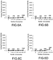
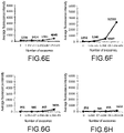
- FIG. 6A to FIG. 6F are respectively graphs of CD9 expressions of exosomes of SKBr3 detected by using Comparative Examples 1 to 3 and Examples 1 and 3 to 4 coupled with anti-human CD63.
- FIG. 6A, FIG. 6C and FIG. 6E are the results of detection with spherical microparticles having diameters of about 2.5 ⁇ m, 4.5 ⁇ m and 8.5 ⁇ m (Comparative Example 2, Comparative Example 3 and Comparative Example 1), respectively.
- FIG. 6B, FIG. 6D and FIG. 6F are the results of detection with knobby particles having diameters of about 2.5 ⁇ m, 4.5 ⁇ m, and 8.5 ⁇ m (Example 3, Example 4, and Example 1), respectively. According to the results of FIG.
- the knobby particles with a diameter of 2.5 ⁇ m may have a lower detected fluorescence signal due to uneven surface morphology.
- FIG. 6C or FIG. 6E
- the number of exosomes (0, 1.05 ⁇ 10 7 , 1.05 ⁇ 10 8 , or 1.05 ⁇ 10 9 ) in Comparative Example 3 (or Comparative Example 1) increases, the number of exosomes that Comparative Example 3 (or Comparative Example 1) binds to increases, and the detected CD9 and average fluorescence intensity also increase.
- FIG. 6D or FIG.
- Example 4 As the number of exosomes (0, 1.05 ⁇ 10 7 , 1.05 ⁇ 10 8 , or 1.05 ⁇ 10 9 ) in Example 4 (or Example 1) increases, the number of exosomes that Example 4 (or Example 1) binds to increases, and the detected CD9 and average fluorescence intensity also increase.
- Comparative Example 3 or Comparative Example 1 containing the spherical microparticle, since Example 4 (or Example 1) containing the knobby particle binds to more exosomes, more CD9 and greater average fluorescence intensity can be detected.
- FIG. 6G to FIG. 6L are respectively graphs of CD9 expressions of exosomes of SKBr3 detected by using Comparative Examples 1 to 3 and Examples 1 and 3 to 4 coupled with anti-human CD9.
- FIG. 6G , FIG. 61 and FIG. 6K are the results of detection with spherical microparticles having diameters of about 2.5 ⁇ m, 4.5 ⁇ m and 8.5 ⁇ m (Comparative Example 2, Comparative Example 3 and Comparative Example 1), respectively.
- FIG. 6H , FIG. 6J and FIG. 6L are the results of detection with knobby particles having diameters of about 2.5 ⁇ m, 4.5 ⁇ m, and 8.5 ⁇ m (Example 3, Example 4, and Example 1), respectively. According to the results of FIG.
- the knobby particles with a diameter of 2.5 ⁇ m may have a lower detected fluorescence signal due to uneven surface morphology.
- FIG. 61 or FIG. 6K
- the number of exosomes (0, 1.05 ⁇ 10 7 , 1.05 ⁇ 10 8 , or 1.05 ⁇ 10 9 ) in Comparative Example 3 (or Comparative Example 1) increases, the number of exosomes that Comparative Example 3 (or Comparative Example 1) binds to increases, and the detected CD9 and average fluorescence intensity also increase.
- FIG. 6J or FIG.
- Example 4 As the number of exosomes (0, 1.05 ⁇ 10 7 , 1.05 ⁇ 10 8 , or 1.05 ⁇ 10 9 ) in Example 4 (or Example 1) increases, the number of exosomes that Example 4 (or Example 1) binds to increases, and the detected CD9 and average fluorescence intensity also increase.
- Comparative Example 3 or Comparative Example 1 containing the spherical microparticle, since Example 4 (or Example 1) containing the knobby particle binds to more exosomes, more CD9 and greater average fluorescence intensity can be detected.
- FIG. 7A to FIG. 7C are respectively graphs of CD9 expressions of exosomes of SKBr3 in different solutions detected by using Example 1 and Example 2 coupled with anti-human CD9.
- exosomes of SKBr3 were first placed in a DMEM culture medium (i.e., supernatant collected from SKBr3 culture medium), a PBS solution (i.e., exosomes of SKBr3 purified and then placed in a PBS solution), or EDTA-containing blood plasma (i.e., exosomes of SKBr3 purified and then placed in EDTA-containing blood plasma).
- DMEM culture medium i.e., supernatant collected from SKBr3 culture medium
- PBS solution i.e., exosomes of SKBr3 purified and then placed in a PBS solution
- EDTA-containing blood plasma i.e., exosomes of SKBr3 purified and then placed in EDTA-containing blood plasma.
- the CD9 expression of the exosomes of SKBr3 in the DMEM culture medium was detected by using Example 1 and Example 2 coupled with anti-human CD9, and the results are shown in FIG. 7A .
- the CD9 expression of the exosomes of SKBr3 in the PBS solution was detected by using Example 1 and Example 2 coupled with anti-human CD9, and the results are shown in FIG. 7B .
- the CD9 expression of the exosomes of SKBr3 in the EDTA-containing blood plasma was detected by using Example 1 and Example 2 coupled with anti-human CD9, and the results are shown in FIG. 7C .
- the above-mentioned blood plasma was from healthy people.
- Example 2 containing the knobby magnetic particle binds to more exosomes located in the DMEM culture medium, more CD9 and greater average fluorescence intensity can be detected.
- FIG. 7B compared to Example 1 containing the knobby particle, since Example 2 containing the knobby magnetic particle binds to more exosomes located in the PBS solution, more CD9 and greater average fluorescence intensity can be detected. According to the results of FIG. 7A , compared to Example 1 containing the knobby particle, since Example 2 containing the knobby magnetic particle binds to more exosomes located in the PBS solution, more CD9 and greater average fluorescence intensity can be detected.
- Example 1 containing the knobby particle or Example 2 containing the knobby magnetic particle may not only detect exosomes located in a PBS solution but also detect exosomes located in relatively complicated environments such as a DMEM culture medium or EDTA-containing blood plasma.
- FIG. 8A to FIG. 8C are respectively graphs of CD81 expressions of exosomes of SKBr3 in different solutions detected by using Example 1 and Example 2 coupled with anti-human CD9.
- exosomes of SKBr3 were first placed in a DMEM culture medium (supernatant collected from SKBr3 culture medium), a PBS solution (exosomes of SKBr3 purified and then placed in a PBS solution), or EDTA-containing blood plasma (exosomes of SKBr3 purified and then placed in EDTA-containing blood plasma).
- the CD81 expression of the exosomes of SKBr3 in the DMEM culture medium was detected by using Example 1 and Example 2 coupled with anti-human CD9, and the results are shown in FIG.
- the CD81 expression of the exosomes of SKBr3 in the PBS solution was detected by using Example 1 and Example 2 coupled with anti-human CD9, and the results are shown in FIG. 8B .
- the CD81 expression of the exosomes of SKBr3 in the EDTA-containing blood plasma was detected by using Example 1 and Example 2 coupled with anti-human CD9, and the results are shown in FIG. 8C .
- the above-mentioned blood plasma was from healthy people.
- Example 2 containing the knobby magnetic particle binds with more exosomes in the DMEM culture medium, more CD81 and greater average fluorescence intensity can be detected.
- Example 1 containing the knobby particle since Example 2 containing the knobby magnetic particle binds to more exosomes in the PBS solution, more CD81 and greater average fluorescence intensity can be detected.
- Example 1 containing the knobby particle since Example 2 containing the knobby magnetic particle binds to more exosomes in the EDTA-containing blood plasma, more CD81 and greater average fluorescence intensity can be detected.
- Example 1 containing the knobby particle or Example 2 containing the knobby magnetic particle may not only detect exosomes located in a PBS solution but also detect exosomes located in relatively complicated environments such as a DMEM culture medium or EDTA-containing blood plasma.
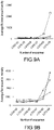
- FIG. 9A to FIG. 9D are graphs of any three of CD9, CD29, CD63, and CD81 expressions of exosomes of SKBr3 detected at the same time by using Example 2 coupled with anti-human CD9.
- a variety of surface antigens on exosomes of breast cancer cell lines SKBr3 were detected at the same time using an experimental procedure similar to that of Embodiment 3, which will thus not be repeatedly described herein.
- the difference between this embodiment and Embodiment 3 lies in that, in this embodiment, exosomes of SKBr3 were first placed in EDTA-containing blood plasma.
- the CD9, CD63, and CD81 expressions of the exosomes were detected at the same time by using Example 2 coupled with anti-human CD9 and a variety of second specific antibodies (anti-human CD9-Alexa488, anti-human CD63-PerCPCy5.5, and anti-human CD81-APC) carrying a variety of fluorescent labels, and the results are shown in FIG. 9A .
- the CD29, CD63, and CD81 expressions of the exosomes were detected at the same time by using Example 2 coupled with anti-human CD9 and a variety of second specific antibodies (anti-human CD29-PE, anti-human CD63-PerCPCy5.5, and anti-human CD81-APC) carrying a variety of fluorescent labels, and the results are shown in FIG.
- FIG. 9B The CD9, CD29, and CD63 expressions of the exosomes were detected at the same time by using Example 2 coupled with anti-human CD9 and a variety of second specific antibodies (anti-human CD9-Alexa488, anti-human CD29-PE, and anti-human CD63-PerCPCy5.5) carrying a variety of fluorescent labels, and the results are shown in FIG. 9C .
- the CD9, CD29, and CD81 expressions of the exosomes were detected at the same time by using Example 2 coupled with anti-human CD9 and a variety of second specific antibodies (anti-human CD9-Alexa488, anti-human CD29-PE, and anti-human CD81-APC) carrying a variety of fluorescent labels, and the results are shown in FIG. 9D .
- the above-mentioned blood plasma was from healthy people.
- the CD9 expression of the exosomes is represented by the symbol ⁇
- the CD29 expression of the exosomes is represented by the symbol ⁇
- the CD63 expression of the exosomes is represented by the symbol O
- the CD81 expression of the exosomes is represented by the symbol ⁇ .
- the expressions of any three of the exosome surface antigens (i.e., any three of CD9, CD29, CD63, and CD81) on the exosomes may still be detected at the same time by using Example 2 (the knobby magnetic particle coupled with anti-human CD9) and any three second specific antibodies (i.e., any three of anti-human CD9-Alexa488, anti-human CD29-PE, anti-human CD63-PerCPCy5.5, and anti-human CD81-APC) carrying fluorescent labels to detect multiple analytes at the same time.
- Example 2 the knobby magnetic particle coupled with anti-human CD9
- any three second specific antibodies i.e., any three of anti-human CD9-Alexa488, anti-human CD29-PE, anti-human CD63-PerCPCy5.5, and anti-human CD81-APC
- the number of analytes that may be detected at the same time is not limited by the disclosure. That is, in some embodiments, two or more than three kinds of analytes may also be detected at the same time.
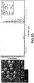
- FIG. 10A to FIG. 10D are graphs of any three of CD9, CD29, CD63, and CD81 expressions of exosomes of SKBr3 detected at the same time by using Example 2 coupled with anti-human HER2. Specifically, exosomes of SKBr3 were first placed in EDTA-containing blood plasma. Then, the CD9, CD63, and CD81 expressions of the exosomes were detected at the same time by using Example 2 coupled with anti-human HER2 and a variety of second specific antibodies (anti-human CD9-Alexa488, anti-human CD63-PerCPCy5.5, and anti-human CD81-APC) carrying a variety of fluorescent labels, and the results are shown in FIG. 10A .
- Example 2 coupled with anti-human HER2 and a variety of second specific antibodies (anti-human CD9-Alexa488, anti-human CD63-PerCPCy5.5, and anti-human CD81-APC) carrying a variety of fluorescent labels, and the results are shown in FIG. 10A .
- the CD29, CD63, and CD81 expressions of the exosomes were detected at the same time by using Example 2 coupled with anti-human HER2 and a variety of second specific antibodies (anti-human CD29-PE, anti-human CD63-PerCPCy5.5, and anti-human CD81-APC) carrying a variety of fluorescent labels, and the results are shown in FIG. 10B .
- the CD9, CD29, and CD63 expressions of the exosomes were detected at the same time by using Example 2 coupled with anti-human HER2 and a variety of second specific antibodies (anti-human CD9-Alexa488, anti-human CD29-PE, and anti-human CD63-PerCPCy5.5) carrying a variety of fluorescent labels, and the results are shown in FIG. 10C .
- the CD9, CD29, and CD81 expressions of the exosomes were detected at the same time by using Example 2 coupled with anti-human HER2 and a variety of second specific antibodies (anti-human CD9-Alexa488, anti-human CD29-PE, and anti-human CD81-APC) carrying a variety of fluorescent labels, and the results are shown in FIG. 10D .
- the above-mentioned blood plasma was from healthy people. Besides, in FIG. 10A to FIG.
- the CD9 expression of the exosomes is represented by the symbol ⁇
- the CD29 expression of the exosomes is represented by the symbol ⁇
- the CD63 expression of the exosomes is represented by the symbol O
- the CD81 expression of the exosomes is represented by the symbol ⁇ .
- the expressions of any three of the exosome surface antigens (i.e., any three of CD9, CD29, CD63, and CD81) on the exosomes may still be detected at the same time by using Example 2 (the knobby magnetic particle coupled with anti-human HER2) and any three second specific antibodies (i.e., any three of anti-human CD9-Alexa488, anti-human CD29-PE, anti-human CD63-PerCPCy5.5, and anti-human CD81-APC) carrying fluorescent labels to detect multiple analytes at the same time.
- Example 2 the knobby magnetic particle coupled with anti-human HER2
- any three second specific antibodies i.e., any three of anti-human CD9-Alexa488, anti-human CD29-PE, anti-human CD63-PerCPCy5.5, and anti-human CD81-APC
- the expressions of the exosome surface antigens i.e., CD9, CD29, CD63, and CD81
- the knobby magnetic particle coupled with whichever of anti-human CD9 and anti-human HER2 binding to the exosomes of SKBr3 accompanied with any three kinds of second specific antibodies (i.e., any three of anti-human CD9-Alexa488, anti-human CD29-PE, anti-human CD63-PerCPCy5.5, and anti-human CD81-APC) carrying fluorescent labels
- the CD9 expression is generally the greatest
- the CD81 expression is slightly greater than or similar to the CD63 expression and the CD29expression, indicating that even if there exist more than two variant parameters in the detection method of this embodiment, the obtained detection results still exhibit comparability.
- FIG. 11A to FIG. 11D are respectively graphs of CD9 and CD63 expressions of exosomes in blood plasma as a clinical specimen detected at the same time by using Example 2 coupled with different first specific antibodies.
- the first specific antibody is anti-human CD9, anti-human HER2, anti-human EGFR, or anti-human EpCAM.
- the clinical specimens was from blood plasma of 8 benign tumors, blood plasma of 20 luminal breast cancers, blood plasma of 8 HER2-positive type breast cancers (HER2), and blood plasma of 4 triple-negative (TN) breast cancers.
- the second specific antibodies carrying fluorescent labels are anti-human CD9-Alexa488 and anti-human CD63-PE.
- CD9 and CD63 of the exosome in the clinical specimens were detected at the same time using an experimental procedure similar to that of Embodiment 3, which will thus not be repeatedly described herein.
- the difference between this embodiment and Embodiment 3 lies in that, in this embodiment, exosomes in different clinical specimens were first placed in EDTA-containing blood plasma.
- the CD9 and CD63 expressions of the exosomes were detected at the same time by using Example 2 coupled with anti-human CD9, and the results are shown in FIG. 11A .
- the CD9 and CD63 expressions of the exosomes were detected at the same time by using Example 2 coupled with anti-human HER2, and the results are shown in FIG. 11B .
- the CD9 and CD63 expressions of the exosomes were detected at the same time by using Example 2 coupled with anti-human EGFR, and the results are shown in FIG. 11C .
- the CD9 and CD63 expressions of the exosomes were detected at the same time by using Example 2 coupled with anti-human EpCAM, and the results are shown in FIG. 11D .
- the expressions of two exosome surface antigens (i.e., CD9 and CD63) on the exosomes may still be detected at the same time by using Example 2 (i.e., the knobby magnetic particle coupled with anti-human CD9, anti-human HER2, anti-human EGFR, or anti-human EpCAM) and two second specific antibodies (i.e., anti-human CD9-Alexa488 and anti-human CD63-PE) carrying fluorescent labels to detect multiple analytes at the same time.
- Example 2 i.e., the knobby magnetic particle coupled with anti-human CD9, anti-human HER2, anti-human EGFR, or anti-human EpCAM
- two second specific antibodies i.e., anti-human CD9-Alexa488 and anti-human CD63-PE
- a probe avian influenza virus (AIV) probe
- AIV avian influenza virus
- the AIV probe has a nucleic acid sequence of sequence identification number: 1, the AB nucleic acid to be analyzed has a nucleic acid sequence of sequence identification number: 2, and the AIV nucleic acid to be analyzed has a nucleic acid sequence of sequence identification number: 3.
- the sequences are shown in detail in the table below.
- AIV probe (SEQ ID NO: 1) 5'-gtctacgctgcagtcctcgctcactgggca AB nucleic acid to be analyzed (SEQ ID NO: 2) 5' -aaaaaaaaaaaaatcctggagctaagtccgta AIV nucleic acid to be analyzed (SEQ ID NO: 3)
- the step in which the AIV probe was coupled to the knobby magnetic particle is as follows: (1) 1 ⁇ 10 6 counts of knobby magnetic particles surface-modified with amine groups were washed with 200 ⁇ L of a MES buffer solution three times. Next, 20 mg of EDC, 20 mg of NHS, and 4 mg of PAA were dissolved in 400 ⁇ L of a MES buffer solution and mixed with the washed knobby magnetic particles. Then, at room temperature, the mixing was performed with a vortex mixer at a rotation speed of 1000 rpm for reaction for 30 minutes. Next, the knobby magnetic particles were collected with a magnet and stayed on the magnet for at least 1 minute.
- the reaction solution was removed and the knobby magnetic particles were repetitively washed with 200 ⁇ L of a MES buffer solution three times, then added with 20 ⁇ L of AIV probes into 80 ⁇ L of a PBS solution, and then reacted at room temperature at a rotation speed of 1000 rpm for 2 hours.
- the knobby magnetic particles were collected with a magnet and stayed on the magnet for at least 1 minute.
- the reaction solution was removed, the knobby magnetic particles were washed with a Tris solution (25 mM of Tris, pH 7.4) three times for at least 1 minute each time.
- the knobby magnetic particles coupled with the AIV probe are dispersed in 100 ⁇ L of a Tris solution.
- the concentration of the knobby magnetic particles was calculated by using an automated cell counter, and knobby magnetic particles coupled with AIV probes on the surface with a concentration of about 3 to 8 ⁇ 10 6 /mL were obtained.
- the step in which the AB nucleic acid to be analyzed or the AIV nucleic acid to be analyzed, carrying biotin, was detected by using the knobby magnetic particles coupled with the AIV probe is generally as follows: (1) 30 ⁇ L of a hybridization solution (TEGO hybridization solution) was mixed with 30 ⁇ L of a solution of AB nucleic acids to be analyzed or AIV nucleic acids to be analyzed, carrying biotin, with different concentrations (0.05 ⁇ M, 0.1 ⁇ M, 0.5 ⁇ M, 5 ⁇ M, 10 ⁇ M).
- knobby magnetic particles coupled with the AIV probe on the surface were added, and mixing was performed at 60°C with a vortex mixer at a rotation speed of 1000 rpm for reaction for 30 minutes. Matching and binding with specificity were performed between the AIV nucleic acid to be analyzed carrying biotin and the AIV probe in the knobby magnetic particles coupled with the AIV probe to form the first complex.
- the knobby magnetic particles were collected with a magnet, and then, after the reaction solution was removed, were washed with 200 ⁇ L of a PBS solution two times.
- the first complex is thus not formed, and the second complex also is thus not formed.
- the measured average fluorescence intensity approaches zero.
- the average fluorescence intensity can be measured.
- the value of average fluorescence intensity basically increases as the concentration of AIV nucleic acid to be analyzed carrying biotin increases.
- the surface area of the microparticle i.e., the sum of the surface area of the body and the surface area of the first protrusions
- the microparticle of the disclosure can be coupled with more first ligands to recognize and bind to more targets, and the strength of detection signal can be increased and the detection sensitivity can be improved.
- the surface of the magnetic substance layer of the knobby magnetic particle is a rough surface (or has small protrusions)
- the surface (i.e., the surface of the body and the surface of the first protrusions) of the knobby magnetic particle is also a rough surface.
- the knobby magnetic particle has a greater surface area.
- the knobby magnetic particle can be coupled with more first ligands, and recognize and bind to more targets to increase the strength of detection signal and improve the detection sensitivity.
- the detection time can be reduced, and the efficiency and convenience of detection can be improved.
Landscapes
- Health & Medical Sciences (AREA)
- Life Sciences & Earth Sciences (AREA)
- Immunology (AREA)
- Engineering & Computer Science (AREA)
- Chemical & Material Sciences (AREA)
- Molecular Biology (AREA)
- Biomedical Technology (AREA)
- Hematology (AREA)
- Urology & Nephrology (AREA)
- Biotechnology (AREA)
- Analytical Chemistry (AREA)
- Microbiology (AREA)
- General Health & Medical Sciences (AREA)
- Biochemistry (AREA)
- Physics & Mathematics (AREA)
- General Physics & Mathematics (AREA)
- Food Science & Technology (AREA)
- Cell Biology (AREA)
- Pathology (AREA)
- Medicinal Chemistry (AREA)
- Tropical Medicine & Parasitology (AREA)
- Organic Chemistry (AREA)
- Proteomics, Peptides & Aminoacids (AREA)
- Chemical Kinetics & Catalysis (AREA)
- Zoology (AREA)
- Wood Science & Technology (AREA)
- Bioinformatics & Cheminformatics (AREA)
- Biophysics (AREA)
- General Engineering & Computer Science (AREA)
- Genetics & Genomics (AREA)
- Toxicology (AREA)
- Measuring Or Testing Involving Enzymes Or Micro-Organisms (AREA)
- Apparatus Associated With Microorganisms And Enzymes (AREA)
- Micro-Organisms Or Cultivation Processes Thereof (AREA)
- Peptides Or Proteins (AREA)
- Investigating Or Analysing Materials By The Use Of Chemical Reactions (AREA)
Abstract
Description
- The disclosure relates to a detection method, and more particularly, to a detection method of multiple analytes.
- Ligand binding assay is a detection method that relies on affinity binding between ligand molecules and analytes, and enzyme-linked immunosorbent assay (ELISA) is the most widely used detection method. In the conventional ELISA, detection is performed by utilizing the property of binding specificity between an antibody and an antigen in combination with enzyme reaction. The technology has been developing toward maturity. However, in ELISA, long detection time is consumed and only one analyte can be detected in one reaction.
- Currently on the market, products applied to multiple-protein immunoassay are based on the conventional sandwich immunoassay. Firstly, spherical microparticles with different colors are utilized to be coupled with antibodies. Next, the spherical microparticles coupled with the antibodies are performed to bind to the target antigens. Then, detection antibodies carrying specific fluorescent labels are utilized to bind to the antigens to be analyzed. Furthermore, analysis is performed with a flow cytometry analyzer. Accordingly, the purpose of detection of multiple proteins is achieved. However, in the above-mentioned multiple protein immunoassay, it is required to use microparticles of various fluorescent colors, it is required to set the fluorescence spectra for microparticles with different fluorescent colors before detection, and, during operation, it is required to distinguish particles carrying different labels before the signal analysis. On the whole, the detection time is long and the operation is complicated, which is likely to cause errors.
- Therefore, it is urgent to develop a detection method in which multiple analytes can be analyzed at the same time, the operation is facilitated, and the detection sensitivity is high.
- The disclosure provides a detection method of multiple analytes, which can have the effect of improving detection sensitivity.
- The detection method of multiple analytes in the disclosure includes the following. A microparticle is provided. The microparticle is coupled with at least one first ligand, and includes a body and a plurality of first protrusions formed on a surface of the body. Next, the microparticle is mixed with a specimen including a variety of analytes to form a first complex. Thereafter, the first complex is mixed with a variety of second ligands carrying a variety of first labels, such that the variety of second ligands bind to the variety of analytes in the first complex and form a second complex. Lastly, the variety of first labels in the second complex are detected.
- In one of exemplary embodiments of the disclosure, the microparticle is a knobby particle. The body of the knobby particle includes a copolymer core, a polymer layer, and a silicon-based layer from the inside to the outside. A plurality of second protrusions are formed on a surface of the copolymer core. An average height of the second protrusions is 100 nanometers to 5000 nanometers.
- In one of exemplary embodiments of the disclosure, the microparticle is a knobby magnetic particle. The body of the knobby magnetic particle includes a copolymer core, a polymer layer, a magnetic substance layer, and a silicon-based layer from the inside to the outside. A plurality of second protrusions are formed on a surface of the copolymer core. An average height of the second protrusions is 100 nanometers to 5000 nanometers.
- In one of exemplary embodiments of the disclosure, a ratio of an average height of the first protrusions to an average diameter of the body is 0.005 to 0.25.
- In one of exemplary embodiments of the disclosure, a ratio of an average volume of the first protrusions to an overall volume of the body is 1×10⁻⁷ to 2×10⁻²
- In one of exemplary embodiments of the disclosure, a total volume of the first protrusions to an average volume of the microparticle is 1×10⁻¹ to 6×10⁻¹.
- In one of exemplary embodiments of the disclosure, an average number of the first protrusions is 5 to 500.
- In one of exemplary embodiments of the disclosure, an average diameter of the microparticle is 1 µm to 20 µm.
- In one of exemplary embodiments of the disclosure, the microparticle is non-spherical.
- In one of exemplary embodiments of the disclosure, the manner in which the at least one first ligand is coupled to the microparticle comprises non-covalent bonding, covalent bonding, avidin-biotin interaction, electrostatic adsorption, hydrophobic adsorption, or a combination of the above.
- In one of exemplary embodiments of the disclosure, the variety of analytes are located on a surface of the specimen. The step of forming the first complex includes performing the at least one first ligand to recognize and directly bind to a target located on the surface of the specimen.
- In one of exemplary embodiments of the disclosure, the at least one first ligand includes a first specific antibody. The first specific antibody includes an antibody against a surface antigen on the human exosome, an antibody against a surface antigen on the human blood cell, an antibody against a surface antigen on the human immune cell, an antibody against a surface antigen on the human tumor cell, or a combination thereof.
- In one of exemplary embodiments of the disclosure, the specimen includes a human exosome, a human blood cell, a human immune cell, a human tumor cell, or a combination thereof. The variety of analytes include a surface antigen on the human exosome, a surface antigen on the human blood cell, a surface antigen on the human immune cell, a surface antigen on the human tumor cell, or a combination thereof.
- In one of exemplary embodiments of the disclosure, the variety of second ligands include a variety of second specific antibodies. The variety of second specific antibodies include antibody against a surface antigen on the human exosome, an antibody against a surface antigen on the human blood cell, an antibody against a surface antigen on the human immune cell, an antibody against a surface antigen on the human tumor cell, or a combination thereof.
- In one of exemplary embodiments of the disclosure, the at least one first ligand includes a variety of first ligands. The step of forming the first complex includes performing the variety of first ligands to recognize and directly bind to the variety of analytes.
- In one of exemplary embodiments of the disclosure, the variety of first ligands include a variety of nucleic acid probes. The variety of nucleic acid probes include a variety of primers or aptamers.
- In one of exemplary embodiments of the disclosure, the variety of analytes include a variety of nucleic acid sequences carrying a variety of second labels.
- In one of exemplary embodiments of the disclosure, the variety of second labels include biotin, a variety of antigenic epitopes, or a combination thereof.
- In one of exemplary embodiments of the disclosure, the variety of second ligands include a variety of specific proteins. The variety of specific proteins include an anti-biotin antibody, avidin, streptavidin, neutravidin, a third specific antibody, or a combination thereof.
- In one of exemplary embodiments of the disclosure, the variety of first labels include a variety of fluorescent labels or a variety of luminescent labels.
- In one of exemplary embodiments of the disclosure, the variety of fluorescent labels include FITC, Alexa, PE, PerCP, BV, APC, Pacific Blue, or a combination thereof. The variety of luminescent labels include luciferase.
- The detection method of multiple analytes in the disclosure includes the following. A microparticle is provided. The microparticle is coupled with a variety of ligands, and includes a body and a plurality of protrusions formed on a surface of the body. Next, the microparticle is mixed with a specimen including a variety of analytes to form a complex. The variety of analytes carry a variety of labels. Lastly, the variety of labels in the complex are detected.
- In one of exemplary embodiments of the disclosure, the variety of ligands include a variety of nucleic acid probes. The variety of labels include a variety of fluorescent labels, a variety of luminescent labels, or a combination thereof. The variety of analytes include a variety of nucleic acid sequences.
- Based on the above, the microparticles of the disclosure can be arranged on the surface of the body by disposing a plurality of first protrusions with irregular shapes, thereby increasing the surface area of the microparticles (ie, the sum of the surface area of the body and the surface area of the first protrusions). In this way, the microparticles can be coupled with more first ligands to identify and bind more targets, thereby enhancing the detection signal and improving the detection sensitivity.
- Several exemplary embodiments accompanied with figures are described in detail below to further describe the disclosure in details.
-
-
FIG. 1 is a flowchart of a detection method of multiple analytes according to an exemplary embodiment. -
FIG. 2 is a schematic view illustrating flows of the detection method of multiple analytes according to an exemplary embodiment. -
FIG. 3A is a schematic cross-sectional view of a knobby particle according to an exemplary embodiment. -
FIG. 3B is a schematic cross-sectional view of a knobby magnetic particle according to an exemplary embodiment. -
FIG. 4 is a schematic view illustrating flows of a detection method of multiple analytes according to another exemplary embodiment. -
FIG. 5A to FIG. 5F are respectively graphs of specifications of spherical microparticles or knobby particles analyzed by using a scanning electron microscope and a multisizer. -
FIG. 6A andFIG. 6F are respectively graphs of CD9 expressions of exosomes of SKBr3 detected by using Comparative Example 1-3 and Example 1 and 3-4 coupled with anti-human CD63. -
FIG. 6G andFIG. 6L are respectively graphs of CD9 expressions of exosomes of SKBr3 detected by using Comparative Example 1-3 and Example 1 and 3-4 coupled with anti-human CD9. -
FIG. 7A to FIG. 7C are respectively graphs of CD9 expressions of exosomes of SKBr3 in different solutions detected by using Example 1 and Example 2 coupled with anti-human CD9. -
FIG. 8A to FIG. 8C are respectively graphs of CD81 expressions of exosomes of SKBr3 in different solutions detected by using Example 1 and Example 2 coupled with anti-human CD9. -
FIG. 9A to FIG. 9D are graphs of any three of CD9, CD29, CD63, and CD81 expressions of exosomes of SKBr3 detected at the same time by using Example 2 coupled with anti-human CD9. -
FIG. 10A to FIG. 10D are graphs of any three of CD9, CD29, CD63, and CD81 expressions of exosomes of SKBr3 detected at the same time by using Example 2 coupled with anti-human HER2. -
FIG. 11A to FIG. 11D are respectively graphs of CD9 and CD63 expressions of exosomes in clinical specimens detected at the same time by using Example 2 coupled with different first specific antibodies. -
FIG. 12A to FIG. 12B are respectively graphs of AB nucleic acid to be analyzed labeled with biotin or AIV nucleic acid to be analyzed labeled with biotin detected by using a knobby magnetic particle coupled with an AIV probe. -
FIG. 1 is a flowchart of a detection method of multiple analytes according to an exemplary embodiment.FIG. 2 is a schematic view illustrating flows of the detection method of multiple analytes according to an exemplary embodiment.FIG. 3A is a schematic cross-sectional view of a knobby particle according to an exemplary embodiment.FIG. 3B is a schematic cross-sectional view of a knobby magnetic particle according to an exemplary embodiment. - With reference to
FIG. 1 ,FIG. 2 ,FIG. 3A, and FIG. 3B together, step S10 is firstly performed, in which amicroparticle 110 is provided. In this embodiment, themicroparticle 110 includes abody 111 and a plurality offirst protrusions 112 formed on asurface 111a of thebody 111. Accordingly, themicroparticle 100 is non-spherical. In terms of appearance, thefirst protrusions 112 have irregular shapes, and may be evenly or unevenly distributed on thesurface 111a of thebody 111. Substantially, thefirst protrusions 112 are integrally formed with thebody 111, and thefirst protrusions 112 are seamlessly connected to thebody 111. In this embodiment, thefirst protrusions 112 have an average height H1 (i.e., the average vertical distance from the top of thefirst protrusions 112 to thesurface 111a of the body 111). - Specifically, with reference to
FIG. 2 ,FIG. 3A, and FIG. 3B together, in this embodiment, themicroparticle 110 may be aknobby particle 110a or a knobbymagnetic particle 110b. Theknobby particle 110a is non-magnetic, and the knobbymagnetic particle 110b is magnetic. As shown inFIG. 3A , thebody 111 of theknobby particle 110a includes acopolymer core 113, apolymer layer 114, and a silicon-basedlayer 115 from the inside to the outside. A plurality ofsecond protrusions 113b are formed on asurface 113a of thecopolymer core 113. Thesecond protrusions 113b have irregular shapes, and may be evenly or unevenly distributed on thesurface 113a of thecopolymer core 113. Substantially, thesecond protrusions 113b are integrally formed with thecopolymer core 113, and thesecond protrusions 113b are seamlessly connected to thecopolymer core 113. In this embodiment, an average height H2 of thesecond protrusions 113b (i.e., the average vertical distance from the top of thesecond protrusions 113b to thesurface 113a of the copolymer core 113) may be 100 nanometers (nm) to 5000 nm, for example but not limited to, about 200 nm to 4500 nm, about 500 nm to 4000 nm, about 800 nm to 3500 nm, about 300 nm to 1000 nm, about 600 nm to 1800 nm, about 750 nm to 2500 nm, about 900 nm to 3000 nm, about 1000 nm to 3600 nm, and about 1500 nm to 4800 nm. In this embodiment, thepolymer layer 114 and the silicon-basedlayer 115 are sequentially formed on thecopolymer core 113 in a substantially conformal manner. Therefore, thefirst protrusions 112 of theknobby particle 110a substantially correspond to thesecond protrusions 113b of thecopolymer core 113, but not limited thereto. - As shown in
FIG. 3B , thebody 111 of the knobbymagnetic particle 110b includes thecopolymer core 113, thepolymer layer 114, amagnetic substance layer 116, and the silicon-basedlayer 115 from the inside to the outside. Thesecond protrusions 113b is formed on thesurface 113a of thecopolymer core 113. Thesecond protrusions 113b have irregular shapes, and may be evenly or unevenly distributed on thesurface 113a of thecopolymer core 113. Substantially, thesecond protrusions 113b are integrally formed with thecopolymer core 113, and thesecond protrusions 113b are seamlessly connected to thecopolymer core 113. In this embodiment, the average height H2 of the second protrusions may be 100 nm to 5000 nm, for example but not limited to, about 200 nm to 4500 nm, about 500 nm to 4000 nm, about 800 nm to 3500 nm, about 300 nm to 1000 nm, about 600 nm to 1800 nm, about 750 nm to 2500 nm, about 900 nm to 3000 nm, about 1000 nm to 3600 nm, and about 1500 nm to 4800 nm. In this embodiment, thepolymer layer 114, themagnetic substance layer 116, and the silicon-basedlayer 115 are sequentially formed on thecopolymer core 113 in a substantially conformal manner. Thefirst protrusions 112 of the knobbymagnetic particle 110b substantially corresponds to thesecond protrusions 113b of thecopolymer core 113, but not limited thereto. - In this embodiment, the
copolymer core 113 is, for example but not limited to, styrene/divinylbenzene copolymer, methyl methacrylate/triethylene glycol dimethacrylate copolymer, methyl methacrylate/ethylene glycol dimethacrylate copolymer, styrene/triethylene glycol dimethacrylate copolymer, styrene/ethylene glycol dimethacrylate copolymer, or methyl methacrylate/divinylbenzene copolymer. Thepolymer layer 114 may be a surface of thecopolymer core 113 modified with functional groups, and includes at least one functional group (for example but not limited to, a carboxyl group, an amino group, or a combination thereof). The material of the silicon-basedlayer 115 may include but is not limited to tetramethoxysilane (TMOS), tetraethoxysilane (TEOS), 3-Aminopropyltriethoxysilane (APTES), and 3-Glycidoxypropyltrimethoxysilane (GOPTS). The material of themagnetic substance layer 116 may include but is not limited to paramagnetic materials, superparamagnetic materials, ferromagnetic materials, ferrimagnetic materials, or a combination thereof. - With further reference to
FIG. 2 ,FIG. 3A, and FIG. 3B , in this embodiment, the average height H1 of thefirst protrusions 112 may be 0.1 micrometer (µm) to 5 µm, for example but not limited to, about 0.2 µm to 4.5 µm, about 0.3 µm to 4 µm, about 0.4 µm to 3.5 µm, about 0.5 µm to 3 µm, and about 0.6 µm to 2.5 µm.An average volume V1 of thefirst protrusions 112 may be 0.00052 cubic micrometer (µm3) to 65 µm3, for example but not limited to, about 0.001 µm3 to 60 µm3, about 0.01 µm3 to 50 µm3, and about 0.1 µm3 to 40 µm3. An average diameter D1 of thebody 111 may be 1 µm to 20 µm, for example but not limited to, about 2 µm to 15 µm, about 3 µm to 18 µm, about 4 µm to 10 µm, and about 5 µm to 12 µm.An average volume V2 of thebody 111 may be 0.52 µm3 to 4200 µm3, for example but not limited to, about 1 µm3 to 4000 µm3, about 10 µm3 to 3800 µm3, about 20 µm3 to 3600 µm3, about 30 µm3 to 3300 µm3, about 40 µm3 to 2900 µm3, about 50 µm3 to 2500 µm3, about 60 µm3 to 2300 µm3, about 80 µm3 to 2000 µm3, and about 100 µm3 to 1000 µm3. A ratio of the average height H1 of thefirst protrusions 112 to the average diameter D1 of thebody 111 may be 0.005 to 0.25, for example but not limited to, about 0.001 to 0.23, about 0.05 to 0.20, about 0.01 to 0.18, and about 0.05 to 0.15. A ratio of the average volume V1 of thefirst protrusions 112 to the average volume V2 of thebody 111 may be 1×10⁻⁷ to 2×10⁻², for example but not limited to, 1×10⁻⁶ to 1.5×10⁻², 1×10⁻⁵ to 1.2×10⁻², and 1×10⁻⁵ to 1×10⁻². An average diameter D of the microparticle 110 (i.e., the average of the shortest diameter and the longest diameter of themicroparticle 110, for example, the average of a shortest diameter D2 and a longest diameter D3 of theknobby particle 110a or of the knobbymagnetic particle 110b) may be 1 µm to 20 µm, for example but not limited to, about 2 µm to 18 µm, about 3 µm to 15 µm, and about 5 µm to 10 µm. - With further reference to
FIG. 2 , in this embodiment, themicroparticle 110 is coupled with at least one first ligand 120 (one first ligand shown inFIG. 1 serving as an example, but not limited thereto). Thefirst ligand 120 is coupled to thesurface 111a of thebody 111 of themicroparticle 110 and thefirst protrusions 112. The manner in which thefirst ligand 120 is coupled to themicroparticle 110 includes non-covalent bonding, covalent bonding, avidin-biotin interaction, electrostatic adsorption, hydrophobic adsorption, or a combination thereof. In this embodiment, thefirst ligand 120 includes, for example but not limited to, a specific antibody. - Next, with further reference to
FIG. 1 andFIG. 2 , step S20 is performed, in which themicroparticle 110 is mixed with a variety ofanalytes first complex 140. In this embodiment, the variety ofanalytes first ligand 120 to recognize and directly bind to a target T on the surface S1 of the specimen S. - In this embodiment, the
first ligand 120 includes a first specific antibody, which is capable of recognizing and directly binding to the target T. The first specific antibody includes but is not limited to an antibody against a surface antigen on the human exosome (e.g., anti-human CD9, anti-human CD63, anti-human CD29, anti-human CD81, anti-human HER2, anti-human EGFR, and anti-human EpCAM), an antibody against a surface antigen on the human blood cell (e.g., anti-human CD45 and anti-human CD235a), an antibody against a surface antigen on the human immune cell (e.g., anti-human CD3 and anti-human CD19), an antibody against a surface antigen on the human tumor cell (e.g., anti-human CEA, anti-human PD-L1, and anti-human HER2) or a combination thereof. - In this embodiment, the specimen S includes but is not limited to human exosomes, human blood cells, human immune cells, human tumor cells, or a combination thereof. The
analytes - In this embodiment, the target T may include but is not limited to at least one of the surface antigens on the human exosome, at least one of the surface antigens on the human blood cell, at least one of the surface antigens on the human immune cell, at least one of the surface antigens on the human tumor cell, or a combination thereof. The target T may be identical or different to any one of the
analytes analyte 130a are not the same surface antigen. For example, as shown inFIG. 2 , the target T and theanalyte 130a are the same kind ofsurface antigen 13a, in which the part that is recognized and bound to by thefirst ligand 120 is namely the target T, while another part that is not bound to by thefirst ligand 120 may serve as theanalyte 130a for subsequent detection. - Then, step S30 is performed, in which the first complex 140 is mixed with a variety of
second ligands first labels second ligands analytes second complex 160. For the sake of clarity, only one specimen S binding to one second ligand (150a, 150b, or 150c) is shown inFIG. 2 . In fact, the specimen S may bind to the variety of (or the plurality of)second ligands analytes - In this embodiment, the
second ligands first labels - Lastly, step S40 is performed, in which the
first labels second ligands analytes - In this embodiment, compared to a general spherical microparticle, in the
microparticle 110 of this embodiment, by disposing the first protrusions having irregular shapes on the surface of the body, the surface area of the microparticle 110 (i.e., the sum of the surface area of the body and the surface area of the first protrusions) is thus increased. Accordingly, themicroparticle 110 of this embodiment may be coupled with morefirst ligands 120 to recognize and bind to more targets T, thereby increasing the strength of detection signals to improve the detection sensitivity. - Besides, in this embodiment, since the surface of the
magnetic substance layer 140 of the knobbymagnetic particle 110b is a rough surface (or has small protrusions), the surface of the knobbymagnetic particle 110b (i.e., thesurface 111a of thebody 111 and the surface of the first protrusions 112) is also a rough surface. Next, compared to theknobby particle 110a, since the surface of the knobbymagnetic particle 110b is a rough surface, the knobbymagnetic particle 110b is of a greater surface area. Accordingly, the knobbymagnetic particle 110b is coupled with morefirst ligands 120, and recognizes and binds to more targets T. In addition,more analytes magnetic particle 110b is magnetic, the detection time can be reduced, and the efficiency and convenience of detection can be improved. - Hereinafter, other embodiments will be described. Note that, the reference numerals and part of the contents of the foregoing embodiments will remain to be used in the following embodiments, where the same reference numerals are used to denote the same or similar elements, and the description of the same technical contents is omitted. Reference may be made to the foregoing embodiments for the description of the omitted part, which will not be repeated in the following embodiments.
-
FIG. 4 is a schematic view illustrating flows of a detection method of multiple analytes according to another embodiment of the disclosure. With reference toFIG. 2 andFIG. 4 together, the method of this embodiment is similar to the method inFIG. 2 , while the main difference lies in that, the at least onefirst ligand 120 includes a variety offirst ligands first ligands analytes second labels second ligands - Specifically, with reference to
FIG. 4 , themicroparticle 110 coupled with thefirst ligand analytes first ligands sequence analytes first complex 140. Next, the first complex 140 is mixed with thesecond ligands first labels second ligands second labels analytes second complex 160. Lastly, thefirst labels second ligands - Besides, in some other embodiments, the
second labels first labels first ligands sequence analytes - Hereinafter, some embodiments of the disclosure accompanied with the drawings will be described. However, the following embodiments and accompanying drawings only serve for aiding the description, instead of limiting the disclosure.
- In this embodiment, specifications of general spherical microparticles or knobby particles are analyzed by using a scanning electron microscope (SEM) and a multisizer. The analysis results are shown in
FIG. 5A to FIG. 5F , in whichFIG. 5A ,FIG. 5C andFIG. 5E are the analysis results of the spherical microparticles, andFIG. 5B ,FIG. 5D andFIG. 5F are the analysis results of the knobby particles. - According to the analysis results of
FIG. 5A to FIG. 5F , the average diameter of the spherical microparticles inFIG. 5A is about 2.421 µm, the average diameter of the knobby particles inFIG. 5B is about 3.280 µm, the average diameter of the spherical microparticles inFIG. 5C is about 4.541 µm, the average diameter of the knobby particles inFIG. 5D is about 4.153 µm, the average diameter of the spherical microparticles inFIG. 5E is about 7.568 µm, and the average diameter of the knobby particles inFIG. 5F is about 7.189 µm. - Next, further analysis of the specifications of the knobby particles of
FIGs. 5B ,5D and5F is performed. In detail, as shown inFIG. 5B , the average volume of the knobby particles is about 13.86 µm3, the average diameter of the bodies is about 2.5 µm, the average volume of the bodies is about 6.14 µm3, the average height of the first protrusions is about 1.2 µm, the average volume of the first protrusions is about 0.679 µm3, and the total volume of the first protrusions is about 7.72 µm3. That is, in the knobby particles ofFIG. 5B , the ratio of the average height of the first protrusions to the average diameter of the bodies is about 0.48, the ratio of the average volume of the first protrusions to the average volume of the bodies is about 0.111, each knobby particle has about 11 first protrusions, and the total volume of the first protrusions is 55.7% of the average volume of the knobby particles. - In
FIG. 5D , the average volume of the knobby particles is about 49.09 µm3, the average diameter of the bodies is about 4.5 µm, the average volume of the bodies is about 35.78 µm3, the average height of the first protrusions is about 0.8 µm, the average volume of the first protrusions is about 0.268 µm3 and the total volume of the first protrusions is about 13.31 µm3. That is, in the knobby particles ofFIG. 5D , the ratio of the average height of the first protrusions to the average diameter of the bodies is about 0.18, the ratio of the average volume of the first protrusions to the average volume of the bodies is about 0.007, each knobby particle has about 49 first protrusions, and the total volume of the first protrusions is 27.1% of the average volume of the knobby particles. - In
FIG. 5F , the average volume of the knobby particles is about 201 µm3, the average diameter of the bodies is about 7.5 µm, the average volume of the bodies is about 166 µm3, the average height of the first protrusions is about 1 µm, the average volume of the first protrusions is about 0.392 µm3, and the total volume of the first protrusions is about 35 µm3. That is, in the knobby particles ofFIG. 5F , the ratio of the average height of the first protrusions to the average diameter of the bodies is about 0.133, the ratio of the average volume of the first protrusions to the average volume of the bodies is about 0.0024, each knobby particle has about 89 first protrusions, and the total volume of the first protrusions is 17.4% of the average volume of the knobby particles. - In particular, in the knobby particles, the ratio of the average height (or average volume) of the first protrusions to the average diameter (or average volume) of the bodies may be related to the surface morphology (ie, overall appearance). For example, in the knobby particles of
FIG. 5B , the ratio of the average height of the first protrusions to the average diameter of the body is greater than 0.25, and the ratio of the average volume of the first protrusions to the average volume of the bodies is greater than 2×10⁻². Therefore, the surface morphology of the knobby particles inFIG. 5B are irregular. In other words, it is difficult to clearly identify the positions of the bodies and the first protrusions. On the contrary, in the knobby particles ofFIG. 5D andFIG. 5F , the ratios of the average height of the first protrusions to the average diameter of the bodies are between 0.005 and 0.25, and the ratios of the average volume of the first protrusions to the average volume of the bodies are between 1×10⁻⁷ and 2×10⁻². Therefore, the positions of the bodies and the first protrusions may be identified in the knobby particles ofFIG. 5D andFIG. 5F . - In this embodiment, the first specific antibody (anti-human CD9, anti-human CD63, or anti-human HER2) was firstly coupled to a general spherical microparticle and a microparticle (a knobby particle or a knobby magnetic particle) of this disclosure. Next, assays and comparisons of the number of grafted specific antibodies were performed on the spherical microparticle coupled with the first specific antibody, the knobby particle coupled with the first specific antibody, and the knobby magnetic particle coupled with the first specific antibody. The classes and diameters of the microparticles used in this embodiment are shown in Table 1.
Table 1: the classes and diameters of the microparticles Class of the microparticle Diameter of the microparticles (µm) Example 1 knobby particle 8.5 Example 2 knobby magnetic particle 8.5 Example 3 knobby particle 2.5 Example 4 knobby particle 4.5 Comparative Example 1 spherical microparticle 8.5 Comparative Example 2 spherical microparticle 2.5 Comparative Example 3 spherical microparticle 4.5 - In this embodiment, the step in which anti-human CD9 (or anti-human CD63 or anti-human HER2) was coupled to the spherical microparticle, the knobby particle, and the knobby magnetic particle is generally as follows: (1) 1×10⁶ particles (i.e., the spherical microparticles of Comparative Examples 1 to 3, the knobby particles of Examples 1 and 3 to 4, or the knobby magnetic particles of Examples 2) surface-modified with amine groups were washed with 200 µL of a MES buffer solution three times. Next, 20 mg of EDC (1-ethyl-3-(3-dimethylaminopropyl) carbodiimide hydrochloride), 20 mg of NHS (N-Hydroxysulfosuccinimide sodium salt), and 4 mg of PAA (15 kDa poly acrylic acid) were dissolved in 400 µL of a MES buffer solution, and were mixed with the washed particles. Next, at room temperature, the mixing was performed with a vortex mixer at a rotation speed of 1000 rpm for reaction for 30 minutes. Next, the particles were collected. The spherical microparticles and the knobby particles were collected by centrifugation at a rotation speed of 10000 rpm for 3 minutes, and the knobby magnetic particles were collected with a magnet, in which the magnet was performed to stay thereon for at least 1 minute. After the reaction solution was removed, the particles were washed with 200 µL of a MES buffer solution three times and added with 20 µL of anti-human CD9 (or anti-human CD63 or anti-human HER2) into a pH3 citric acid-PBS solution (0.625M citric acid dissolved in PBS solution, pH3.0) for reaction overnight at 4°C, such that anti-human CD9 (or anti-human CD63 or anti-human HER2) was coupled on the surface of the particles. Then, the mixture was added into 200 µL of a bovine serum albumin solution (10 mg/mL BSA dissolved in a MES solution) for perform reaction overnight at 4°C to cover the rest of the surface of the particles that has not been coupled with anti-human CD9 (or anti-human CD63 or anti-human HER2). (2) After the reaction was completed, the particles were collected. The spherical microparticles and the knobby particles were collected by centrifugation at a rotation speed of 10000 rpm for 3 minutes. The knobby magnetic particles were collected by a magnet and the magnet was performed to stay thereon for at least 1 minute each time. The reaction solution was removed. The particles were washed with a PBS solution (0.1% BSA and 0.01% sodium azide dissolved in PBS) three times. Lastly, the particles coupled with anti-human CD9 (or anti-human CD63 or anti-human HER2) were dispersed in 100 µL of a PBS solution. (3) The concentration of particles was calculated by using an automated cell counter, and particles coupled with anti-human CD9 (or anti-human CD63 or anti-human HER2) on the surface with a concentration of about 3 to 8×10⁶/mL were obtained.
- Next, the step in which assays of the number of grafted specific antibodies were performed on the particles coupled with anti-human CD9 (or anti-human CD63 or anti-human HER2) is generally as follows: (1) 50 µL (1250 counts) of the particles coupled with anti-human CD9 (or anti-human CD63 or anti-human HER2) on the surface were added into 5 µL of Antimouse IgG-FITC, and then were added into a PBS solution until the total reaction volume was 200 µL. At room temperature, the mixing was performed with a vortex mixer at a rotation speed of 1500 rpm for reaction for 30 minutes. After the reaction was completed, the particles were collected. The spherical microparticles and knobby particles were collected by centrifugation at a rotation speed of 10000 rpm for 3 minutes. The knobby magnetic particles were collected with a magnet and stayed on the magnet for at least 1 minute. (2) The reaction solution was removed, and the collected particles were washed with 200 µLof a PBS solution two times. (3) After washing, the particles were added into 100 µL of a PBS solution, and fluorescence signal analysis was performed on the particles with a flow cytometry analyzer to measure the number of grafted anti-human CD9 (or anti-human CD63 or anti-human HER2) (represented by average fluorescence intensity, i.e., as the average fluorescence intensity increases, the number of grafts increases). (4) Statistical analysis was performed, and the test results were expressed in multiples of fluorescence intensity. In Table 2, comparisons of the grafting number of spherical microparticles (Comparative Example 1), knobby particles (Example 1) and knobby magnetic particles (Example 2) with the same diameter (8.5 µm) is illustrated. Table 3 shows comparisons of the grafting number of spherical microparticles (Comparative Examples 1 to 3) and knobby particles (Examples 1, 3 to 4) with different diameters (i.e., 2.5, 4.5, 8.5 µm). Table 4 shows comparisons of the grafting number of spherical microparticles (Comparative Examples 1 to 3) and knobby particles (Examples 1, 3 to 4) with the same diameter (i.e., 2.5, 4.5, 8.5 µm).
Table 2: comparison of the number of grafted first specific antibodies (anti-human CD9, anti-human CD63 or Anti-Human Her2) in Comparative Example 1, Example 1 and Example 2 Multiples of fluorescence intensity First specific antibody Example 1/Comparative Example 1 (diameter: 8.5 µm) Example 2/Example 1 (diameter: 8.5 µm) Anti-Human CD9 ~1.71 ~2.24 Anti-Human CD63 ∼4.55 - Anti-Human Her2 - ∼3.81 - According to the results in Table 2, when the first specific antibody is anti-human CD9, the number of grafts in Example 1 is about 1.71 times that in Comparative Example 1. When the first specific antibody is anti-human CD63, the number of grafts in Example 1 is about 4.55 times that in Comparative Example 1. That is, whether the first specific antibody is anti-human CD9 or anti-human CD63, the number of grafts in Example 1 is greater than the number of grafts in Comparative Example 1. Therefore, compared with a general spherical microparticle, the knobby particle of this disclosure is significantly coupled with more first specific antibodies.
- In addition, when the first specific antibody is anti-human CD9, the number of grafts in Example 2 is about 2.24 times that in Example 1. When the first specific antibody is anti-human HER2, the number of grafts in Example 2 is about 3.81 times that in Example 1. That is, whether the first specific antibody is anti-human CD9 or anti-human HER2, the number of grafts in Example 2 is greater than the number of grafts in Example 1. Therefore, compared with the knobby particle of this disclosure, the knobby magnetic particle of this disclosure is significantly coupled with more first specific antibodies.
Table 3: comparison of the number of grafted first specific antibodies (anti-human CD9 or Anti-Human CD63) in Comparative Examples 1 to 3 and Examples 1 and 3 to 4 Multiples of fluorescence intensity First specific antibody Comparative Example 3 / Comparative Example 2 Comparative Example 1 / Comparative Example 2 Example 1 / Comparative Example 2 Example 4 / Comparative Example 2 Example 1 / Comparative Example 2 Anti-Human CD9 ∼4.71 ∼5.62 ∼0.90 ∼6.05 ∼9.59 Anti-Human CD63 ∼1.96 ∼1.98 ∼0.63 ∼3.16 ∼9.00 Table 4: comparison of the number of grafted first specific antibodies (anti-human CD9 or Anti-Human CD63) in Comparative Examples 1 to 3 and Examples 1 and 3 to 4 Multiples of fluorescence intensity First specific antibody Example 3 /Comparative Example 2 (diameter: 2.5 µm) Example 4 /Comparative Example 3 (diameter: 4.5 µm) Example 1 /Comparative Example 1 (diameter: 8.5 µm) Anti-Human CD9 ~0.90 ~1.28 ~1.71 Anti-Human CD63 ∼0.63 ~1.61 ∼4.55 - According to the results in Table 3, whether it is spherical microparticles or knobby particles, when the particle size is larger, the surface area that can be coupled to the first specific antibody increases accordingly, so the grafting number of the first specific antibody can be increased. According to the results in Table 4, compared with spherical microparticles (Comparative Example 3 or Comparative Example 1), knobby particles with similar particle sizes (Example 4 or Example 1) can couple significantly more first-specific antibodies. Compared with the spherical microparticles (Comparative Example 2) with a diameter of about 2.5 µm, the knobby particles (Example 3) with a diameter of about 2.5 µm may be due to uneven surface morphology (please refer to
Figure 5B ), resulting in a lower detected fluorescent signal. - In this embodiment, the expression of exosome surface antigens on the surface of exosomes was detected by using a spherical microparticle (Comparative Examples 1, 2 and 3), a knobby particle (Examples 1, 3 and 4), and a knobby magnetic particle (Example 2) coupled with a first specific antibody (anti-human CD9 or anti-human CD63), and using a second specific antibody (anti-human CD9-Alexa488 or anti-human CD81-APC) carrying fluorescent labels. Comparative Example 1 is taken as an example for description. Specifically, Comparative Example 1 coupled with anti-human CD9 (or anti-human CD63) was first mixed with exosomes carrying a variety of exosome surface antigens to allow anti-human CD9 (or anti-human CD63) to recognize and directly bind to CD9 (or CD63) located on the surface of the exosomes to form a first complex. Next, the first complex was mixed with anti-human CD9-Alexa488 (or anti-human CD81-APC) to allow anti-human CD9-Alexa488 (or anti-human CD81-APC) to bind to CD9 (or CD81) on the exosome surface in the first complex and form a second complex. Then, fluorescence signal analysis of the second complex was performed with a flow cytometry analyzer to detect the expression of exosome surface antigens. The exosomes were exosomes purified from breast cancer cell lines (SKBr3). The above-mentioned spherical microparticles included spherical microparticles having an average diameter of about 2.5 µm (Comparative Example 2), spherical microparticles having an average diameter of about 4.5 µm (Comparative Example 3), and spherical microparticles having an average diameter of about 8.5 µm (Comparative Example 1). The above-mentioned knobby particles included knobby particles having an average diameter of about 2.5 µm (Example 3), knobby particles having an average diameter of about 4.5 µm (Example 4), and knobby particles having an average diameter of about 8.5 µm (Example 1). The above-mentioned knobby magnetic particles included knobby magnetic particles having an average diameter of about 8.5 µm (Example 2).
- In this embodiment, the step in which Comparative Examples 1 to 3, Examples 1 and 3 to 4, or Example 2 coupled with anti-human CD9 (or anti-human CD63) was mixed with the exosomes of SKBr3, such that anti-human CD9 (or anti-human CD63) recognized and directly bound to CD9 (or CD63) on the surface of the exosomes to form the first complex is generally as follows: 50 µL (1250 counts) of Comparative Examples 1 to 3, Examples 1 and 3 to 4, or Example 2 was reacted with exosomes of SKBr3 with a volume of 20 to 50 µL, then added into a PBS solution until the total reaction volume was 200 µL, and then mixed and reacted at room temperature for 90 minutes. After the reaction was completed, the first complex containing Comparative Examples 1 to 3 or the first complex of Examples 1 and 3 to 4 was collected by centrifugation at a rotation speed of 10000 rpm for 3 minutes, and the first complex containing Example 2 was collected with a magnet and stayed on the magnet for at least 1 minute. The reaction solution was removed and the first complex was repetitively washed with 200 µL of a PBS solution two times.
- In this embodiment, the step in which the first complex was mixed with anti-human CD9-Alexa488 (or anti-human CD81-APC), such that anti-human CD9-Alexa488 (or anti-human CD81-APC) bound to CD9 (or CD81) on the surface of the exosomes in the first complex and formed the second complex is generally as follows: 100 µL of anti-human CD9-Alexa488 (or anti-human CD81-APC) was added into the first complex. Then, at room temperature, the mixing was performed with a vortex mixer at a rotation speed of 1500 rpm for reaction for 30 minutes to form the second complex. After the reaction was completed, the second complex containing Comparative Examples 1 to 3 or the second complex containing Examples 1 and 3 to 4 was collected by centrifugation at a rotation speed of 10000 rpm for 3 minutes, and the second complex containing Example 2 was collected with a magnet and stayed on the magnet for at least 1 minute. The reaction solution was removed and the second complex was repetitively washed with 200 µL of a PBS solution two times.
- In this embodiment, the step in which fluorescence signal analysis was performed on the second complex to detect the expression of exosome surface antigens is generally as follows: 100 µL of a PBS solution is added to the washed second complex (i.e., the second complex containing Comparative Example 1, the second complex containing Example 1, or the second complex containing Example 2), and fluorescence signal analysis was performed with a flow cytometry analyzer to detect the expression (i.e., average fluorescence intensity) of exosome surface antigens. The results are as shown in
FIG. 6A to FIG. 6L ,FIG. 7A to FIG. 7C , andFIG. 8A to FIG. 8C . -
FIG. 6A to FIG. 6F are respectively graphs of CD9 expressions of exosomes of SKBr3 detected by using Comparative Examples 1 to 3 and Examples 1 and 3 to 4 coupled with anti-human CD63. Specifically,FIG. 6A, FIG. 6C andFIG. 6E are the results of detection with spherical microparticles having diameters of about 2.5 µm, 4.5 µm and 8.5 µm (Comparative Example 2, Comparative Example 3 and Comparative Example 1), respectively.FIG. 6B, FIG. 6D andFIG. 6F are the results of detection with knobby particles having diameters of about 2.5 µm, 4.5 µm, and 8.5 µm (Example 3, Example 4, and Example 1), respectively. According to the results ofFIG. 6A and FIG. 6B , compared with the spherical microparticles with a diameter of 2.5 µm (Comparative Example 2), the knobby particles with a diameter of 2.5 µm (Example 3) may have a lower detected fluorescence signal due to uneven surface morphology. According to the results ofFIG. 6C (orFIG. 6E ), as the number of exosomes (0, 1.05×10⁷, 1.05×10⁸, or 1.05×10⁹) in Comparative Example 3 (or Comparative Example 1) increases, the number of exosomes that Comparative Example 3 (or Comparative Example 1) binds to increases, and the detected CD9 and average fluorescence intensity also increase. According to the results ofFIG. 6D (orFIG. 6F ), as the number of exosomes (0, 1.05×10⁷, 1.05×10⁸, or 1.05×10⁹) in Example 4 (or Example 1) increases, the number of exosomes that Example 4 (or Example 1) binds to increases, and the detected CD9 and average fluorescence intensity also increase. However, according to the results ofFIG. 6C to FIG. 6F , compared to Comparative Example 3 (or Comparative Example 1) containing the spherical microparticle, since Example 4 (or Example 1) containing the knobby particle binds to more exosomes, more CD9 and greater average fluorescence intensity can be detected. -
FIG. 6G to FIG. 6L are respectively graphs of CD9 expressions of exosomes of SKBr3 detected by using Comparative Examples 1 to 3 and Examples 1 and 3 to 4 coupled with anti-human CD9. Specifically,FIG. 6G ,FIG. 61 and FIG. 6K are the results of detection with spherical microparticles having diameters of about 2.5 µm, 4.5 µm and 8.5 µm (Comparative Example 2, Comparative Example 3 and Comparative Example 1), respectively.FIG. 6H ,FIG. 6J and FIG. 6L are the results of detection with knobby particles having diameters of about 2.5 µm, 4.5 µm, and 8.5 µm (Example 3, Example 4, and Example 1), respectively. According to the results ofFIG. 6G and FIG. 6H , compared with the spherical microparticles with a diameter of 2.5 µm (Comparative Example 2), the knobby particles with a diameter of 2.5 µm (Example 3) may have a lower detected fluorescence signal due to uneven surface morphology. According to the results ofFIG. 61 (or FIG. 6K ), as the number of exosomes (0, 1.05×10⁷, 1.05×10⁸, or 1.05×10⁹) in Comparative Example 3 (or Comparative Example 1) increases, the number of exosomes that Comparative Example 3 (or Comparative Example 1) binds to increases, and the detected CD9 and average fluorescence intensity also increase. According to the results ofFIG. 6J (or FIG. 6L ), as the number of exosomes (0, 1.05×10⁷, 1.05×10⁸, or 1.05×10⁹) in Example 4 (or Example 1) increases, the number of exosomes that Example 4 (or Example 1) binds to increases, and the detected CD9 and average fluorescence intensity also increase. However, according to the results ofFIG. 61 to FIG. 6L , compared to Comparative Example 3 (or Comparative Example 1) containing the spherical microparticle, since Example 4 (or Example 1) containing the knobby particle binds to more exosomes, more CD9 and greater average fluorescence intensity can be detected. - It should be noted that according to
FIG. 6A to FIG. 6L , the larger the particle, the stronger the signal, as well as the larger the particle size and the more protruding shape have the best analysis signal. Using flow cytometry analyzer to detect microbeads with larger particle size and uniform size for analysis has better analysis signal. -
FIG. 7A to FIG. 7C are respectively graphs of CD9 expressions of exosomes of SKBr3 in different solutions detected by using Example 1 and Example 2 coupled with anti-human CD9. Specifically, exosomes of SKBr3 were first placed in a DMEM culture medium (i.e., supernatant collected from SKBr3 culture medium), a PBS solution (i.e., exosomes of SKBr3 purified and then placed in a PBS solution), or EDTA-containing blood plasma (i.e., exosomes of SKBr3 purified and then placed in EDTA-containing blood plasma). Next, the CD9 expression of the exosomes of SKBr3 in the DMEM culture medium was detected by using Example 1 and Example 2 coupled with anti-human CD9, and the results are shown inFIG. 7A . The CD9 expression of the exosomes of SKBr3 in the PBS solution was detected by using Example 1 and Example 2 coupled with anti-human CD9, and the results are shown inFIG. 7B . The CD9 expression of the exosomes of SKBr3 in the EDTA-containing blood plasma was detected by using Example 1 and Example 2 coupled with anti-human CD9, and the results are shown inFIG. 7C . The above-mentioned blood plasma was from healthy people. - According to the results of
FIG. 7A , compared to Example 1 containing the knobby particle, since Example 2 containing the knobby magnetic particle binds to more exosomes located in the DMEM culture medium, more CD9 and greater average fluorescence intensity can be detected. According to the results ofFIG. 7B , compared to Example 1 containing the knobby particle, since Example 2 containing the knobby magnetic particle binds to more exosomes located in the PBS solution, more CD9 and greater average fluorescence intensity can be detected. According to the results ofFIG. 7C , compared to Example 1 containing the knobby particle, since Example 2 containing the knobby magnetic particle binds to more exosomes in the EDTA-containing blood plasma, more CD9 and greater average fluorescence intensity can be detected. Besides, according to the results ofFIG. 7A to FIG. 7C , Example 1 containing the knobby particle or Example 2 containing the knobby magnetic particle may not only detect exosomes located in a PBS solution but also detect exosomes located in relatively complicated environments such as a DMEM culture medium or EDTA-containing blood plasma. -
FIG. 8A to FIG. 8C are respectively graphs of CD81 expressions of exosomes of SKBr3 in different solutions detected by using Example 1 and Example 2 coupled with anti-human CD9. Specifically, exosomes of SKBr3 were first placed in a DMEM culture medium (supernatant collected from SKBr3 culture medium), a PBS solution (exosomes of SKBr3 purified and then placed in a PBS solution), or EDTA-containing blood plasma (exosomes of SKBr3 purified and then placed in EDTA-containing blood plasma). Next, the CD81 expression of the exosomes of SKBr3 in the DMEM culture medium was detected by using Example 1 and Example 2 coupled with anti-human CD9, and the results are shown inFIG. 8A . The CD81 expression of the exosomes of SKBr3 in the PBS solution was detected by using Example 1 and Example 2 coupled with anti-human CD9, and the results are shown inFIG. 8B . The CD81 expression of the exosomes of SKBr3 in the EDTA-containing blood plasma was detected by using Example 1 and Example 2 coupled with anti-human CD9, and the results are shown inFIG. 8C . The above-mentioned blood plasma was from healthy people. - According to the results of
FIG. 8A , compared to Example 1 containing the knobby particle, since Example 2 containing the knobby magnetic particle binds with more exosomes in the DMEM culture medium, more CD81 and greater average fluorescence intensity can be detected. According to the result ofFIG. 8B , compared to Example 1 containing the knobby particle, since Example 2 containing the knobby magnetic particle binds to more exosomes in the PBS solution, more CD81 and greater average fluorescence intensity can be detected. According to the results ofFIG. 8C , compared to Example 1 containing the knobby particle, since Example 2 containing the knobby magnetic particle binds to more exosomes in the EDTA-containing blood plasma, more CD81 and greater average fluorescence intensity can be detected. Besides, according to the results ofFIG. 8A to FIG. 8C , Example 1 containing the knobby particle or Example 2 containing the knobby magnetic particle may not only detect exosomes located in a PBS solution but also detect exosomes located in relatively complicated environments such as a DMEM culture medium or EDTA-containing blood plasma. -
FIG. 9A to FIG. 9D are graphs of any three of CD9, CD29, CD63, and CD81 expressions of exosomes of SKBr3 detected at the same time by using Example 2 coupled with anti-human CD9. Specifically, in this embodiment, a variety of surface antigens on exosomes of breast cancer cell lines SKBr3 were detected at the same time using an experimental procedure similar to that ofEmbodiment 3, which will thus not be repeatedly described herein. The difference between this embodiment andEmbodiment 3 lies in that, in this embodiment, exosomes of SKBr3 were first placed in EDTA-containing blood plasma. Then, the CD9, CD63, and CD81 expressions of the exosomes were detected at the same time by using Example 2 coupled with anti-human CD9 and a variety of second specific antibodies (anti-human CD9-Alexa488, anti-human CD63-PerCPCy5.5, and anti-human CD81-APC) carrying a variety of fluorescent labels, and the results are shown inFIG. 9A . The CD29, CD63, and CD81 expressions of the exosomes were detected at the same time by using Example 2 coupled with anti-human CD9 and a variety of second specific antibodies (anti-human CD29-PE, anti-human CD63-PerCPCy5.5, and anti-human CD81-APC) carrying a variety of fluorescent labels, and the results are shown inFIG. 9B . The CD9, CD29, and CD63 expressions of the exosomes were detected at the same time by using Example 2 coupled with anti-human CD9 and a variety of second specific antibodies (anti-human CD9-Alexa488, anti-human CD29-PE, and anti-human CD63-PerCPCy5.5) carrying a variety of fluorescent labels, and the results are shown inFIG. 9C . The CD9, CD29, and CD81 expressions of the exosomes were detected at the same time by using Example 2 coupled with anti-human CD9 and a variety of second specific antibodies (anti-human CD9-Alexa488, anti-human CD29-PE, and anti-human CD81-APC) carrying a variety of fluorescent labels, and the results are shown inFIG. 9D . The above-mentioned blood plasma was from healthy people. Besides, inFIG. 9A to FIG. 9D , the CD9 expression of the exosomes is represented by the symbol □, the CD29 expression of the exosomes is represented by the symbol ×, the CD63 expression of the exosomes is represented by the symbol O, and the CD81 expression of the exosomes is represented by the symbol △. - According to the results of
FIG. 9A to FIG. 9D , even if the exosomes are located in relatively complicated environments such as EDTA-containing blood plasma, by the detection method of this embodiment, the expressions of any three of the exosome surface antigens (i.e., any three of CD9, CD29, CD63, and CD81) on the exosomes may still be detected at the same time by using Example 2 (the knobby magnetic particle coupled with anti-human CD9) and any three second specific antibodies (i.e., any three of anti-human CD9-Alexa488, anti-human CD29-PE, anti-human CD63-PerCPCy5.5, and anti-human CD81-APC) carrying fluorescent labels to detect multiple analytes at the same time. Besides, although detection for three kinds of analytes at the same time is taken as an example in this embodiment, the number of analytes that may be detected at the same time is not limited by the disclosure. That is, in some embodiments, two or more than three kinds of analytes may also be detected at the same time. -
FIG. 10A to FIG. 10D are graphs of any three of CD9, CD29, CD63, and CD81 expressions of exosomes of SKBr3 detected at the same time by using Example 2 coupled with anti-human HER2. Specifically, exosomes of SKBr3 were first placed in EDTA-containing blood plasma. Then, the CD9, CD63, and CD81 expressions of the exosomes were detected at the same time by using Example 2 coupled with anti-human HER2 and a variety of second specific antibodies (anti-human CD9-Alexa488, anti-human CD63-PerCPCy5.5, and anti-human CD81-APC) carrying a variety of fluorescent labels, and the results are shown inFIG. 10A . The CD29, CD63, and CD81 expressions of the exosomes were detected at the same time by using Example 2 coupled with anti-human HER2 and a variety of second specific antibodies (anti-human CD29-PE, anti-human CD63-PerCPCy5.5, and anti-human CD81-APC) carrying a variety of fluorescent labels, and the results are shown inFIG. 10B . The CD9, CD29, and CD63 expressions of the exosomes were detected at the same time by using Example 2 coupled with anti-human HER2 and a variety of second specific antibodies (anti-human CD9-Alexa488, anti-human CD29-PE, and anti-human CD63-PerCPCy5.5) carrying a variety of fluorescent labels, and the results are shown inFIG. 10C . The CD9, CD29, and CD81 expressions of the exosomes were detected at the same time by using Example 2 coupled with anti-human HER2 and a variety of second specific antibodies (anti-human CD9-Alexa488, anti-human CD29-PE, and anti-human CD81-APC) carrying a variety of fluorescent labels, and the results are shown inFIG. 10D . The above-mentioned blood plasma was from healthy people. Besides, inFIG. 10A to FIG. 10D , the CD9 expression of the exosomes is represented by the symbol □, the CD29 expression of the exosomes is represented by the symbol ×, the CD63 expression of the exosomes is represented by the symbol O, and the CD81 expression of the exosomes is represented by the symbol △. - According to the results of
FIG. 10A to FIG. 10D , even if the exosomes are located in relatively complicated environments such as EDTA-containing blood plasma, by the detection method of this embodiment, the expressions of any three of the exosome surface antigens (i.e., any three of CD9, CD29, CD63, and CD81) on the exosomes may still be detected at the same time by using Example 2 (the knobby magnetic particle coupled with anti-human HER2) and any three second specific antibodies (i.e., any three of anti-human CD9-Alexa488, anti-human CD29-PE, anti-human CD63-PerCPCy5.5, and anti-human CD81-APC) carrying fluorescent labels to detect multiple analytes at the same time. - Besides, according to the comparisons of the results of
FIG. 9A to FIG. 10A , ofFIG. 9B to FIG. 10B , ofFIG. 9C to FIG. 10C , and ofFIG. 9D to FIG. 10D , when the expressions of the exosome surface antigens (i.e., CD9, CD29, CD63, and CD81) of SKBr3 are analyzed by using the knobby magnetic particle coupled with whichever of anti-human CD9 and anti-human HER2, high consistency exists between the detection results, indicating that the detection method with high accuracy and reduced errors is used in this embodiment. - In addition, according to the combined results of
FIG. 9A to FIG. 9D andFIG. 10A to FIG. 10D , when the expressions of the exosome surface antigens (i.e., CD9, CD29, CD63, and CD81) is analyzed by using the knobby magnetic particle coupled with whichever of anti-human CD9 and anti-human HER2 binding to the exosomes of SKBr3 accompanied with any three kinds of second specific antibodies (i.e., any three of anti-human CD9-Alexa488, anti-human CD29-PE, anti-human CD63-PerCPCy5.5, and anti-human CD81-APC) carrying fluorescent labels, the CD9 expression is generally the greatest, and the CD81 expression is slightly greater than or similar to the CD63 expression and the CD29expression, indicating that even if there exist more than two variant parameters in the detection method of this embodiment, the obtained detection results still exhibit comparability. -
FIG. 11A to FIG. 11D are respectively graphs of CD9 and CD63 expressions of exosomes in blood plasma as a clinical specimen detected at the same time by using Example 2 coupled with different first specific antibodies. The first specific antibody is anti-human CD9, anti-human HER2, anti-human EGFR, or anti-human EpCAM. The clinical specimens was from blood plasma of 8 benign tumors, blood plasma of 20 luminal breast cancers, blood plasma of 8 HER2-positive type breast cancers (HER2), and blood plasma of 4 triple-negative (TN) breast cancers. The second specific antibodies carrying fluorescent labels are anti-human CD9-Alexa488 and anti-human CD63-PE. Specifically, in this embodiment, CD9 and CD63 of the exosome in the clinical specimens were detected at the same time using an experimental procedure similar to that ofEmbodiment 3, which will thus not be repeatedly described herein. The difference between this embodiment andEmbodiment 3 lies in that, in this embodiment, exosomes in different clinical specimens were first placed in EDTA-containing blood plasma. Next, the CD9 and CD63 expressions of the exosomes were detected at the same time by using Example 2 coupled with anti-human CD9, and the results are shown inFIG. 11A . The CD9 and CD63 expressions of the exosomes were detected at the same time by using Example 2 coupled with anti-human HER2, and the results are shown inFIG. 11B . The CD9 and CD63 expressions of the exosomes were detected at the same time by using Example 2 coupled with anti-human EGFR, and the results are shown inFIG. 11C . The CD9 and CD63 expressions of the exosomes were detected at the same time by using Example 2 coupled with anti-human EpCAM, and the results are shown inFIG. 11D . - According to the results of
FIG. 11A to FIG. 11D , in the detection method of this embodiment, for different clinical specimens, the expressions of two exosome surface antigens (i.e., CD9 and CD63) on the exosomes may still be detected at the same time by using Example 2 (i.e., the knobby magnetic particle coupled with anti-human CD9, anti-human HER2, anti-human EGFR, or anti-human EpCAM) and two second specific antibodies (i.e., anti-human CD9-Alexa488 and anti-human CD63-PE) carrying fluorescent labels to detect multiple analytes at the same time. - In this embodiment, a probe (avian influenza virus (AIV) probe) was first coupled to the knobby magnetic particle of this disclosure. Next, a biotin-labeled nucleic acid sequence (AB nucleic acid to be analyzed or AIV nucleic acid to be analyzed) was added for analysis.
- In this embodiment, the AIV probe has a nucleic acid sequence of sequence identification number: 1, the AB nucleic acid to be analyzed has a nucleic acid sequence of sequence identification number: 2, and the AIV nucleic acid to be analyzed has a nucleic acid sequence of sequence identification number: 3. The sequences are shown in detail in the table below.
AIV probe (SEQ ID NO: 1) 5'-gtctacgctgcagtcctcgctcactgggca AB nucleic acid to be analyzed (SEQ ID NO: 2) 5' -aaaaaaaaaaaaaatcctggagctaagtccgta AIV nucleic acid to be analyzed (SEQ ID NO: 3) 
- In this embodiment, the step in which the AIV probe was coupled to the knobby magnetic particle is as follows: (1) 1×10⁶ counts of knobby magnetic particles surface-modified with amine groups were washed with 200 µL of a MES buffer solution three times. Next, 20 mg of EDC, 20 mg of NHS, and 4 mg of PAA were dissolved in 400 µL of a MES buffer solution and mixed with the washed knobby magnetic particles. Then, at room temperature, the mixing was performed with a vortex mixer at a rotation speed of 1000 rpm for reaction for 30 minutes. Next, the knobby magnetic particles were collected with a magnet and stayed on the magnet for at least 1 minute. The reaction solution was removed and the knobby magnetic particles were repetitively washed with 200 µL of a MES buffer solution three times, then added with 20 µL of AIV probes into 80 µL of a PBS solution, and then reacted at room temperature at a rotation speed of 1000 rpm for 2 hours. (2) After the reaction was completed, the knobby magnetic particles were collected with a magnet and stayed on the magnet for at least 1 minute. The reaction solution was removed, the knobby magnetic particles were washed with a Tris solution (25 mM of Tris, pH 7.4) three times for at least 1 minute each time. Lastly, the knobby magnetic particles coupled with the AIV probe are dispersed in 100µL of a Tris solution. (3) The concentration of the knobby magnetic particles was calculated by using an automated cell counter, and knobby magnetic particles coupled with AIV probes on the surface with a concentration of about 3 to 8×10⁶/mL were obtained.
- In this embodiment, the step in which the AB nucleic acid to be analyzed or the AIV nucleic acid to be analyzed, carrying biotin, was detected by using the knobby magnetic particles coupled with the AIV probe is generally as follows: (1) 30 µL of a hybridization solution (TEGO hybridization solution) was mixed with 30 µL of a solution of AB nucleic acids to be analyzed or AIV nucleic acids to be analyzed, carrying biotin, with different concentrations (0.05 µM, 0.1 µM, 0.5 µM, 5 µM, 10 µM). Next, 50 µL (1250 counts) of the knobby magnetic particles coupled with the AIV probe on the surface were added, and mixing was performed at 60°C with a vortex mixer at a rotation speed of 1000 rpm for reaction for 30 minutes. Matching and binding with specificity were performed between the AIV nucleic acid to be analyzed carrying biotin and the AIV probe in the knobby magnetic particles coupled with the AIV probe to form the first complex. After the reaction was completed, the knobby magnetic particles were collected with a magnet, and then, after the reaction solution was removed, were washed with 200 µL of a PBS solution two times. (2) 100 µL of an anti-biotin antibody (anti-biotin PE) carrying fluorescent labels were added to the knobby magnetic particles, and mixing was performed for reaction at room temperature for 30 minutes. The anti-biotin antibody carrying fluorescent labels bound to the biotin in the first complex to form the second complex. After the reaction was completed, the knobby magnetic particles were collected with a magnet, and, after the reaction solution was removed, were repetitively washed with 200 µL of a PBS buffer solution (PBS solution, pH 7.4) two times. (3) After washing, 150 µL of a PBS buffer solution was added and fluorescence signal analysis was performed with a flow cytometry analyzer. The results are shown in
FIG. 12A and FIG. 12B . - As shown in
FIG. 12A , since the AB nucleic acid to be analyzed carrying biotin does not bind to the AIV probe in the knobby magnetic particles coupled with the AIV probe, the first complex is thus not formed, and the second complex also is thus not formed. As a result, the measured average fluorescence intensity approaches zero. - As shown in
FIG. 12B , matching and binding with specificity were performed between the AIV nucleic acid to be analyzed carrying biotin and the AIV probe in the knobby magnetic particles coupled with the AIV probe, the first complex was formed, and the biotin in the first complex bound to the anti-biotin antibody carrying fluorescent labels to form the second complex. Accordingly, the average fluorescence intensity can be measured. Besides, the value of average fluorescence intensity basically increases as the concentration of AIV nucleic acid to be analyzed carrying biotin increases. - In summary of the foregoing, in the microparticle of the disclosure, by disposing the first protrusions having irregular shapes on the surface of the body, the surface area of the microparticle (i.e., the sum of the surface area of the body and the surface area of the first protrusions) is increased. Accordingly, the microparticle of the disclosure can be coupled with more first ligands to recognize and bind to more targets, and the strength of detection signal can be increased and the detection sensitivity can be improved.
- Besides, in the disclosure, compared to the knobby particle, since the surface of the magnetic substance layer of the knobby magnetic particle is a rough surface (or has small protrusions), the surface (i.e., the surface of the body and the surface of the first protrusions) of the knobby magnetic particle is also a rough surface. As a result, the knobby magnetic particle has a greater surface area. Further, the knobby magnetic particle can be coupled with more first ligands, and recognize and bind to more targets to increase the strength of detection signal and improve the detection sensitivity. Moreover, since the knobby magnetic particle is magnetic, the detection time can be reduced, and the efficiency and convenience of detection can be improved.

Claims (15)
- A detection method of multiple analytes (130a, 130b, 130c), comprising:providing a microparticle (110), wherein the microparticle (110) is coupled with at least one first ligand (120), and the microparticle (110) comprises:a body (111); anda plurality of first protrusions (112) formed on a surface (111a) of the body (111);mixing the microparticle (110) with a specimen (S) comprising a variety of analytes (130a, 130b, 130c) to form a first complex (140);mixing the first complex (140) with a variety of second ligands (150a, 150b, 150c) carrying a variety of first labels (151a, 151b, 151c), such that the variety of second ligands (150a, 150b, 150c) bind to the variety of analytes (130a, 130b, 130c) in the first complex (140) and form a second complex (160); anddetecting the variety of first labels (151a, 151b, 151c) in the second complex (160).
- The method according to claim 1, wherein the microparticle (110) is a knobby particle (110a), and the body (111) of the knobby particle (110a) comprises a copolymer core (113), a polymer layer (114), and a silicon-based layer (115) from the inside to the outside, a plurality of second protrusions (113b) are formed on a surface (113a) of the copolymer core (113), and an average height (H2) of the second protrusions (113b) is 100 nanometers to 5000 nanometers.
- The method according to claim 1, wherein the microparticle (110) is a knobby magnetic particle (110b), the body (111) of the knobby magnetic particle (110b) comprises a copolymer core (113), a polymer layer (114), a magnetic substance layer (116), and a silicon-based layer (115) from the inside to the outside, a plurality of second protrusions (113b) are formed on a surface (113a) of the copolymer core (113), and an average height (H2) of the second protrusions (113b) is 100 nanometers to 5000 nanometers.
- The method according to claim 1, wherein a ratio of an average height (H1) of the first protrusions (112) to an average diameter (D1) of the body (111) is 0.005 to 0.25.
- The method according to claim 1, wherein a ratio of an average volume (V1) of the first protrusions (112) to an average volume (V2) of the body (111) is 1×10⁻⁷ to 2×10⁻², and a total volume of the first protrusions (112) to an overall volume of the microparticle (110) is 1×10⁻¹ to 6×10⁻¹.
- The method according to claim 1, wherein an average number of the first protrusions (112) is 5 to 500, and an average diameter (D) of the microparticle (110) is 1 µm to 20 µm.
- The method according to claim 1, wherein the microparticle (110) is non-spherical.
- The method according to claim 1, wherein the variety of analytes (130a, 130b, 130c) are located on a surface (S1) of the specimen (S), and the step of forming the first complex comprises (140) performing the at least one first ligand (120) to recognize and directly bind to a target (T) located on the surface (S1) of the specimen (S).
- The method according to claim 8, wherein the at least one first ligand (120) comprises a first specific antibody, and the first specific antibody comprises an antibody against a surface antigen on the human exosome, an antibody against a surface antigen on the human blood cell, an antibody against a surface antigen on the human immune cell, an antibody against a surface antigen on the human tumor cell, or a combination thereof.
- The method according to claim 8, wherein the specimen (S) comprises a human exosome, a human blood cell, a human immune cell, a human tumor cell, or a combination thereof, and the variety of analytes comprise a surface antigen on the human exosome, a surface antigen on the human blood cell, a surface antigen on the human immune cell, a surface antigen on the human tumor cell, or a combination thereof.
- The method according to claim 8, wherein the variety of second ligands (150a, 150b, 150c) comprise a variety of second specific antibodies, and the variety of second specific antibodies comprise an antibody against a surface antigen on the human exosome, an antibody against a surface antigen on the human blood cell, an antibody against a surface antigen on the human immune cell, an antibody against a surface antigen on the human tumor cell, or a combination thereof.
- The method according to claim 1, wherein the at least one first ligand (120) comprises a variety of first ligands (120a, 120b, 120c), and the step forming the first complex (140) comprises performing the variety of first ligands (120a, 120b, 120c) to recognize and directly bind to the variety of analytes (130a, 130b, 130c).
- The method according to claim 12, wherein the variety of first ligands (120a, 120b, 120c) comprise a variety of nucleic acid probes, and the variety of nucleic acid probes comprise a variety of primers or aptamers, the variety of analytes (130a, 130b, 130c) comprise a variety of nucleic acid sequences (131a, 131b, 131c) carrying a variety of second labels (132a, 132b, 132c), and the variety of second labels comprise biotin, a variety of antigenic epitopes, or a combination thereof.
- A detection method of multiple analytes (130a, 130b, 130c), comprising:providing a microparticle (110), wherein the microparticle (110) is coupled with a variety of ligands (120a, 120b, 120c), and the microparticle (110) comprises:a body (111); anda plurality of protrusions (112) formed on a surface (111a) of the body (111);mixing the microparticle (110) with a specimen (S) comprising a variety of analytes (130a, 130b, 130c) to form a complex (140), wherein the variety of analytes (130a, 130b, 130c) carry a variety of labels (132a, 132b, 132c); anddetecting the variety of labels (132a, 132b, 132c) in the complex (140).
- The method according to claim 14, wherein the variety of ligands (120a, 120b, 120c) comprise a variety of nucleic acid probes, the variety of labels (132a, 132b, 132c) comprise a variety of fluorescent labels, a variety of luminescent labels, or a combination thereof, and the variety of analytes (130a, 130b, 130c) comprise a variety of nucleic acid sequences (131a, 131b, 131c).
Applications Claiming Priority (2)
| Application Number | Priority Date | Filing Date | Title |
|---|---|---|---|
| US202063130857P | 2020-12-28 | 2020-12-28 | |
| US202163194188P | 2021-05-28 | 2021-05-28 |
Publications (2)
| Publication Number | Publication Date |
|---|---|
| EP4019973A1 true EP4019973A1 (en) | 2022-06-29 |
| EP4019973B1 EP4019973B1 (en) | 2024-05-29 |
Family
ID=81579293
Family Applications (1)
| Application Number | Title | Priority Date | Filing Date |
|---|---|---|---|
| EP21217229.0A Active EP4019973B1 (en) | 2020-12-28 | 2021-12-23 | Detection method of multiple analytes |
Country Status (5)
| Country | Link |
|---|---|
| US (1) | US20220205993A1 (en) |
| EP (1) | EP4019973B1 (en) |
| JP (1) | JP7337903B2 (en) |
| CN (1) | CN114689845B (en) |
| TW (1) | TWI832129B (en) |
Families Citing this family (3)
| Publication number | Priority date | Publication date | Assignee | Title |
|---|---|---|---|---|
| CN115656150B (en) * | 2022-09-26 | 2023-09-08 | 苏州浦隆生物有限公司 | Device and method for detecting biomolecules |
| US20240327895A1 (en) * | 2022-12-30 | 2024-10-03 | Industrial Technology Research Institute | Composition for stabilizing cell-free nucleic acid and/or exosomal nucleic acid in blood, and agent and kit containing the same |
| US20250290920A1 (en) * | 2024-03-13 | 2025-09-18 | Bender MedSystems GmbH | Tag/anti-tag system for signal amplification in immunoassays |
Citations (2)
| Publication number | Priority date | Publication date | Assignee | Title |
|---|---|---|---|---|
| EP3503129A1 (en) * | 2017-12-22 | 2019-06-26 | Industrial Technology Research Institute | Magnetic beads and the manufacturing method thereof |
| US20190310172A1 (en) * | 2018-04-05 | 2019-10-10 | Caris Science, Inc. | Profiling extracellular vesicles |
Family Cites Families (7)
| Publication number | Priority date | Publication date | Assignee | Title |
|---|---|---|---|---|
| WO2002080647A2 (en) * | 2001-04-03 | 2002-10-17 | Surromed, Inc. | Methods and reagents for multiplexed analyte capture, surface array self-assembly, and analysis of complex biological samples |
| KR101388604B1 (en) * | 2013-11-25 | 2014-04-23 | 덕산하이메탈(주) | Metal particle, conductive particle and the manufacturing method of the same |
| EP3121207A4 (en) * | 2014-03-18 | 2017-11-15 | Nippon Shokubai Co., Ltd. | Resin particles, conductive microparticles, and anistropic conductive material using same |
| US20170097339A1 (en) * | 2015-10-02 | 2017-04-06 | Tgr Biosciences Pty Ltd. | Analyte Detection with Multiple Substrates |
| TWI699218B (en) * | 2017-12-22 | 2020-07-21 | 財團法人工業技術研究院 | Method for in vitro activation and/or expansion of immune cells |
| CN109459571A (en) * | 2018-11-08 | 2019-03-12 | 浙江大学 | Based on micro-fluidic and chemiluminescence immune assay superparamagnetic nanoparticle excretion body separation detecting system and its application |
| US20200232979A1 (en) * | 2019-01-22 | 2020-07-23 | Mayo Foundation For Medical Education And Research | Microcapsules and Methods for Analyte Detection |
-
2021
- 2021-12-23 CN CN202111590524.7A patent/CN114689845B/en active Active
- 2021-12-23 TW TW110148376A patent/TWI832129B/en active
- 2021-12-23 US US17/560,276 patent/US20220205993A1/en active Pending
- 2021-12-23 JP JP2021209218A patent/JP7337903B2/en active Active
- 2021-12-23 EP EP21217229.0A patent/EP4019973B1/en active Active
Patent Citations (2)
| Publication number | Priority date | Publication date | Assignee | Title |
|---|---|---|---|---|
| EP3503129A1 (en) * | 2017-12-22 | 2019-06-26 | Industrial Technology Research Institute | Magnetic beads and the manufacturing method thereof |
| US20190310172A1 (en) * | 2018-04-05 | 2019-10-10 | Caris Science, Inc. | Profiling extracellular vesicles |
Non-Patent Citations (5)
| Title |
|---|
| ALESSANDRO GORI ET AL: "Membrane-binding peptides for extracellular vesicles on-chip analysis", JOURNAL OF EXTRACELLULAR VESICLES, vol. 9, no. 1, 17 April 2020 (2020-04-17), UK, pages 1751428, XP055767519, ISSN: 2001-3078, DOI: 10.1080/20013078.2020.1751428 * |
| DE JAGER W ET AL: "Solid-phase and bead-based cytokine immunoassay: A comparison", METHODS, ACADEMIC PRESS, NL, vol. 38, no. 4, 1 April 2006 (2006-04-01), pages 294 - 303, XP024908576, ISSN: 1046-2023, [retrieved on 20060401], DOI: 10.1016/J.YMETH.2005.11.008 * |
| HAN DI ET AL: "Engineering the Surface Pattern of Microparticles: From Raspberry-like to Golf Ball-like", APPLIED MATERIALS & INTERFACES, vol. 13, no. 26, 25 June 2021 (2021-06-25), US, pages 31215 - 31225, XP055922932, ISSN: 1944-8244, DOI: 10.1021/acsami.1c08663 * |
| LEE JOO HYUK ET AL: "Enhanced Cell Adhesion to the Dimpled Surfaces of Golf-Ball-Shaped Microparticles", APPLIED MATERIALS & INTERFACES, vol. 6, no. 19, 29 September 2014 (2014-09-29), US, pages 16493 - 16497, XP055923113, ISSN: 1944-8244, DOI: 10.1021/am505997s * |
| NINA KOLIHA ET AL: "A novel multiplex bead-based platform highlights the diversity of extracellular vesicles", JOURNAL OF EXTRACELLULAR VESICLES, vol. 5, no. 1, 1 January 2016 (2016-01-01), pages 29975, XP055485864, DOI: 10.3402/jev.v5.29975 * |
Also Published As
| Publication number | Publication date |
|---|---|
| TW202234063A (en) | 2022-09-01 |
| JP7337903B2 (en) | 2023-09-04 |
| US20220205993A1 (en) | 2022-06-30 |
| EP4019973B1 (en) | 2024-05-29 |
| TWI832129B (en) | 2024-02-11 |
| CN114689845A (en) | 2022-07-01 |
| CN114689845B (en) | 2025-03-28 |
| JP2022104585A (en) | 2022-07-08 |
Similar Documents
| Publication | Publication Date | Title |
|---|---|---|
| EP4019973B1 (en) | Detection method of multiple analytes | |
| CN111579772B (en) | Detection reagent based on immunomagnetic bead method, preparation method thereof, kit and detection method | |
| CN110763834A (en) | Method, reagent and kit for detecting content of immune marker | |
| CN110346560B (en) | A kind of multi-enzyme signal particle and its preparation method and application | |
| EP3054283A1 (en) | Method for detecting target substance | |
| WO2021258618A1 (en) | Biological sample testing method and test kit | |
| CN119104735B (en) | Magnetic particle chemiluminescence method protein detection method | |
| CN113176404A (en) | Kit for multi-index joint inspection of whole blood sample and use method thereof | |
| CN110346557B (en) | Detection kit | |
| CN114280016A (en) | Exosome detection method | |
| CN110383067B (en) | Anti-human hemoglobin monoclonal antibody or antibody kit, antibody-immobilized insoluble carrier particles and their applications | |
| TW201209413A (en) | Immunomagnetic complex and its use in red blood cell grouping/phenotyping | |
| WO2007127981A2 (en) | Analyte-releasing beads and use thereof in quantitative elispot or fluorispot assay | |
| US20040106157A1 (en) | Immunological assay reagents and assay method | |
| US9632086B2 (en) | Method and kit for determining-antibody sensitivity and clone cell strain | |
| EP4481383A1 (en) | Immunological analysis method, complex, method for producing complex, and reagent for immunological analysis | |
| JPH0763761A (en) | Method for producing magnetic fine particles on which physiologically active substance is immobilized | |
| JP2005069926A (en) | Magnetic particles for immunological tests | |
| CN116643042B (en) | Thrombomotor protein chemiluminescence detection kit | |
| CN118091153A (en) | A quantitative detection kit for allergen-specific IgE antibodies based on liquid phase chip method and its application | |
| CN116165193A (en) | Carcinoembryonic antigen detection reagent and kit based on double-antibody sandwich chemiluminescence method | |
| EP3382394B1 (en) | Method of producing a probe-bound carrier and method of detecting or separating a target substance | |
| CN119804842B (en) | Immunological detection method for to-be-detected object in whole blood sample, magnetic microsphere and magnetic microsphere-protein complex | |
| CN115932005A (en) | Electrochemical biosensor working electrode for detecting protein and preparation method thereof | |
| CN120703386A (en) | Interleukin-33 detection kit, preparation method and application |
Legal Events
| Date | Code | Title | Description |
|---|---|---|---|
| PUAI | Public reference made under article 153(3) epc to a published international application that has entered the european phase |
Free format text: ORIGINAL CODE: 0009012 |
|
| STAA | Information on the status of an ep patent application or granted ep patent |
Free format text: STATUS: REQUEST FOR EXAMINATION WAS MADE |
|
| 17P | Request for examination filed |
Effective date: 20211223 |
|
| AK | Designated contracting states |
Kind code of ref document: A1 Designated state(s): AL AT BE BG CH CY CZ DE DK EE ES FI FR GB GR HR HU IE IS IT LI LT LU LV MC MK MT NL NO PL PT RO RS SE SI SK SM TR |
|
| RBV | Designated contracting states (corrected) |
Designated state(s): AL AT BE BG CH CY CZ DE DK EE ES FI FR GB GR HR HU IE IS IT LI LT LU LV MC MK MT NL NO PL PT RO RS SE SI SK SM TR |
|
| STAA | Information on the status of an ep patent application or granted ep patent |
Free format text: STATUS: EXAMINATION IS IN PROGRESS |
|
| 17Q | First examination report despatched |
Effective date: 20230220 |
|
| GRAP | Despatch of communication of intention to grant a patent |
Free format text: ORIGINAL CODE: EPIDOSNIGR1 |
|
| STAA | Information on the status of an ep patent application or granted ep patent |
Free format text: STATUS: GRANT OF PATENT IS INTENDED |
|
| INTG | Intention to grant announced |
Effective date: 20240214 |
|
| GRAS | Grant fee paid |
Free format text: ORIGINAL CODE: EPIDOSNIGR3 |
|
| GRAA | (expected) grant |
Free format text: ORIGINAL CODE: 0009210 |
|
| STAA | Information on the status of an ep patent application or granted ep patent |
Free format text: STATUS: THE PATENT HAS BEEN GRANTED |
|
| AK | Designated contracting states |
Kind code of ref document: B1 Designated state(s): AL AT BE BG CH CY CZ DE DK EE ES FI FR GB GR HR HU IE IS IT LI LT LU LV MC MK MT NL NO PL PT RO RS SE SI SK SM TR |
|
| REG | Reference to a national code |
Ref country code: CH Ref legal event code: EP |
|
| REG | Reference to a national code |
Ref country code: IE Ref legal event code: FG4D |
|
| REG | Reference to a national code |
Ref country code: DE Ref legal event code: R096 Ref document number: 602021013800 Country of ref document: DE |
|
| REG | Reference to a national code |
Ref country code: LT Ref legal event code: MG9D |
|
| REG | Reference to a national code |
Ref country code: NL Ref legal event code: MP Effective date: 20240529 |
|
| PG25 | Lapsed in a contracting state [announced via postgrant information from national office to epo] |
Ref country code: IS Free format text: LAPSE BECAUSE OF FAILURE TO SUBMIT A TRANSLATION OF THE DESCRIPTION OR TO PAY THE FEE WITHIN THE PRESCRIBED TIME-LIMIT Effective date: 20240929 |
|
| PG25 | Lapsed in a contracting state [announced via postgrant information from national office to epo] |
Ref country code: BG Free format text: LAPSE BECAUSE OF FAILURE TO SUBMIT A TRANSLATION OF THE DESCRIPTION OR TO PAY THE FEE WITHIN THE PRESCRIBED TIME-LIMIT Effective date: 20240529 |
|
| PG25 | Lapsed in a contracting state [announced via postgrant information from national office to epo] |
Ref country code: HR Free format text: LAPSE BECAUSE OF FAILURE TO SUBMIT A TRANSLATION OF THE DESCRIPTION OR TO PAY THE FEE WITHIN THE PRESCRIBED TIME-LIMIT Effective date: 20240529 Ref country code: FI Free format text: LAPSE BECAUSE OF FAILURE TO SUBMIT A TRANSLATION OF THE DESCRIPTION OR TO PAY THE FEE WITHIN THE PRESCRIBED TIME-LIMIT Effective date: 20240529 |
|
| PG25 | Lapsed in a contracting state [announced via postgrant information from national office to epo] |
Ref country code: GR Free format text: LAPSE BECAUSE OF FAILURE TO SUBMIT A TRANSLATION OF THE DESCRIPTION OR TO PAY THE FEE WITHIN THE PRESCRIBED TIME-LIMIT Effective date: 20240830 |
|
| REG | Reference to a national code |
Ref country code: AT Ref legal event code: MK05 Ref document number: 1691063 Country of ref document: AT Kind code of ref document: T Effective date: 20240529 |
|
| PG25 | Lapsed in a contracting state [announced via postgrant information from national office to epo] |
Ref country code: ES Free format text: LAPSE BECAUSE OF FAILURE TO SUBMIT A TRANSLATION OF THE DESCRIPTION OR TO PAY THE FEE WITHIN THE PRESCRIBED TIME-LIMIT Effective date: 20240529 |
|
| PG25 | Lapsed in a contracting state [announced via postgrant information from national office to epo] |
Ref country code: AT Free format text: LAPSE BECAUSE OF FAILURE TO SUBMIT A TRANSLATION OF THE DESCRIPTION OR TO PAY THE FEE WITHIN THE PRESCRIBED TIME-LIMIT Effective date: 20240529 |
|
| PG25 | Lapsed in a contracting state [announced via postgrant information from national office to epo] |
Ref country code: PL Free format text: LAPSE BECAUSE OF FAILURE TO SUBMIT A TRANSLATION OF THE DESCRIPTION OR TO PAY THE FEE WITHIN THE PRESCRIBED TIME-LIMIT Effective date: 20240529 |
|
| PG25 | Lapsed in a contracting state [announced via postgrant information from national office to epo] |
Ref country code: LV Free format text: LAPSE BECAUSE OF FAILURE TO SUBMIT A TRANSLATION OF THE DESCRIPTION OR TO PAY THE FEE WITHIN THE PRESCRIBED TIME-LIMIT Effective date: 20240529 |
|
| PG25 | Lapsed in a contracting state [announced via postgrant information from national office to epo] |
Ref country code: PL Free format text: LAPSE BECAUSE OF FAILURE TO SUBMIT A TRANSLATION OF THE DESCRIPTION OR TO PAY THE FEE WITHIN THE PRESCRIBED TIME-LIMIT Effective date: 20240529 Ref country code: NO Free format text: LAPSE BECAUSE OF FAILURE TO SUBMIT A TRANSLATION OF THE DESCRIPTION OR TO PAY THE FEE WITHIN THE PRESCRIBED TIME-LIMIT Effective date: 20240829 Ref country code: LV Free format text: LAPSE BECAUSE OF FAILURE TO SUBMIT A TRANSLATION OF THE DESCRIPTION OR TO PAY THE FEE WITHIN THE PRESCRIBED TIME-LIMIT Effective date: 20240529 Ref country code: IS Free format text: LAPSE BECAUSE OF FAILURE TO SUBMIT A TRANSLATION OF THE DESCRIPTION OR TO PAY THE FEE WITHIN THE PRESCRIBED TIME-LIMIT Effective date: 20240929 Ref country code: HR Free format text: LAPSE BECAUSE OF FAILURE TO SUBMIT A TRANSLATION OF THE DESCRIPTION OR TO PAY THE FEE WITHIN THE PRESCRIBED TIME-LIMIT Effective date: 20240529 Ref country code: GR Free format text: LAPSE BECAUSE OF FAILURE TO SUBMIT A TRANSLATION OF THE DESCRIPTION OR TO PAY THE FEE WITHIN THE PRESCRIBED TIME-LIMIT Effective date: 20240830 Ref country code: FI Free format text: LAPSE BECAUSE OF FAILURE TO SUBMIT A TRANSLATION OF THE DESCRIPTION OR TO PAY THE FEE WITHIN THE PRESCRIBED TIME-LIMIT Effective date: 20240529 Ref country code: ES Free format text: LAPSE BECAUSE OF FAILURE TO SUBMIT A TRANSLATION OF THE DESCRIPTION OR TO PAY THE FEE WITHIN THE PRESCRIBED TIME-LIMIT Effective date: 20240529 Ref country code: BG Free format text: LAPSE BECAUSE OF FAILURE TO SUBMIT A TRANSLATION OF THE DESCRIPTION OR TO PAY THE FEE WITHIN THE PRESCRIBED TIME-LIMIT Effective date: 20240529 Ref country code: AT Free format text: LAPSE BECAUSE OF FAILURE TO SUBMIT A TRANSLATION OF THE DESCRIPTION OR TO PAY THE FEE WITHIN THE PRESCRIBED TIME-LIMIT Effective date: 20240529 Ref country code: RS Free format text: LAPSE BECAUSE OF FAILURE TO SUBMIT A TRANSLATION OF THE DESCRIPTION OR TO PAY THE FEE WITHIN THE PRESCRIBED TIME-LIMIT Effective date: 20240829 |
|
| PG25 | Lapsed in a contracting state [announced via postgrant information from national office to epo] |
Ref country code: NL Free format text: LAPSE BECAUSE OF FAILURE TO SUBMIT A TRANSLATION OF THE DESCRIPTION OR TO PAY THE FEE WITHIN THE PRESCRIBED TIME-LIMIT Effective date: 20240529 |
|
| PG25 | Lapsed in a contracting state [announced via postgrant information from national office to epo] |
Ref country code: NL Free format text: LAPSE BECAUSE OF FAILURE TO SUBMIT A TRANSLATION OF THE DESCRIPTION OR TO PAY THE FEE WITHIN THE PRESCRIBED TIME-LIMIT Effective date: 20240529 |
|
| PG25 | Lapsed in a contracting state [announced via postgrant information from national office to epo] |
Ref country code: DK Free format text: LAPSE BECAUSE OF FAILURE TO SUBMIT A TRANSLATION OF THE DESCRIPTION OR TO PAY THE FEE WITHIN THE PRESCRIBED TIME-LIMIT Effective date: 20240529 |
|
| PG25 | Lapsed in a contracting state [announced via postgrant information from national office to epo] |
Ref country code: EE Free format text: LAPSE BECAUSE OF FAILURE TO SUBMIT A TRANSLATION OF THE DESCRIPTION OR TO PAY THE FEE WITHIN THE PRESCRIBED TIME-LIMIT Effective date: 20240529 |
|
| PG25 | Lapsed in a contracting state [announced via postgrant information from national office to epo] |
Ref country code: CZ Free format text: LAPSE BECAUSE OF FAILURE TO SUBMIT A TRANSLATION OF THE DESCRIPTION OR TO PAY THE FEE WITHIN THE PRESCRIBED TIME-LIMIT Effective date: 20240529 |
|
| PG25 | Lapsed in a contracting state [announced via postgrant information from national office to epo] |
Ref country code: RO Free format text: LAPSE BECAUSE OF FAILURE TO SUBMIT A TRANSLATION OF THE DESCRIPTION OR TO PAY THE FEE WITHIN THE PRESCRIBED TIME-LIMIT Effective date: 20240529 Ref country code: SK Free format text: LAPSE BECAUSE OF FAILURE TO SUBMIT A TRANSLATION OF THE DESCRIPTION OR TO PAY THE FEE WITHIN THE PRESCRIBED TIME-LIMIT Effective date: 20240529 |
|
| PG25 | Lapsed in a contracting state [announced via postgrant information from national office to epo] |
Ref country code: SM Free format text: LAPSE BECAUSE OF FAILURE TO SUBMIT A TRANSLATION OF THE DESCRIPTION OR TO PAY THE FEE WITHIN THE PRESCRIBED TIME-LIMIT Effective date: 20240529 |
|
| PG25 | Lapsed in a contracting state [announced via postgrant information from national office to epo] |
Ref country code: SM Free format text: LAPSE BECAUSE OF FAILURE TO SUBMIT A TRANSLATION OF THE DESCRIPTION OR TO PAY THE FEE WITHIN THE PRESCRIBED TIME-LIMIT Effective date: 20240529 Ref country code: SK Free format text: LAPSE BECAUSE OF FAILURE TO SUBMIT A TRANSLATION OF THE DESCRIPTION OR TO PAY THE FEE WITHIN THE PRESCRIBED TIME-LIMIT Effective date: 20240529 Ref country code: RO Free format text: LAPSE BECAUSE OF FAILURE TO SUBMIT A TRANSLATION OF THE DESCRIPTION OR TO PAY THE FEE WITHIN THE PRESCRIBED TIME-LIMIT Effective date: 20240529 Ref country code: EE Free format text: LAPSE BECAUSE OF FAILURE TO SUBMIT A TRANSLATION OF THE DESCRIPTION OR TO PAY THE FEE WITHIN THE PRESCRIBED TIME-LIMIT Effective date: 20240529 Ref country code: DK Free format text: LAPSE BECAUSE OF FAILURE TO SUBMIT A TRANSLATION OF THE DESCRIPTION OR TO PAY THE FEE WITHIN THE PRESCRIBED TIME-LIMIT Effective date: 20240529 Ref country code: CZ Free format text: LAPSE BECAUSE OF FAILURE TO SUBMIT A TRANSLATION OF THE DESCRIPTION OR TO PAY THE FEE WITHIN THE PRESCRIBED TIME-LIMIT Effective date: 20240529 |
|
| PG25 | Lapsed in a contracting state [announced via postgrant information from national office to epo] |
Ref country code: IT Free format text: LAPSE BECAUSE OF FAILURE TO SUBMIT A TRANSLATION OF THE DESCRIPTION OR TO PAY THE FEE WITHIN THE PRESCRIBED TIME-LIMIT Effective date: 20240529 |
|
| REG | Reference to a national code |
Ref country code: DE Ref legal event code: R097 Ref document number: 602021013800 Country of ref document: DE |
|
| PLBE | No opposition filed within time limit |
Free format text: ORIGINAL CODE: 0009261 |
|
| STAA | Information on the status of an ep patent application or granted ep patent |
Free format text: STATUS: NO OPPOSITION FILED WITHIN TIME LIMIT |
|
| PG25 | Lapsed in a contracting state [announced via postgrant information from national office to epo] |
Ref country code: SI Free format text: LAPSE BECAUSE OF FAILURE TO SUBMIT A TRANSLATION OF THE DESCRIPTION OR TO PAY THE FEE WITHIN THE PRESCRIBED TIME-LIMIT Effective date: 20240529 |
|
| 26N | No opposition filed |
Effective date: 20250303 |
|
| PG25 | Lapsed in a contracting state [announced via postgrant information from national office to epo] |
Ref country code: MC Free format text: LAPSE BECAUSE OF FAILURE TO SUBMIT A TRANSLATION OF THE DESCRIPTION OR TO PAY THE FEE WITHIN THE PRESCRIBED TIME-LIMIT Effective date: 20240529 |
|
| REG | Reference to a national code |
Ref country code: CH Ref legal event code: PL |
|
| PG25 | Lapsed in a contracting state [announced via postgrant information from national office to epo] |
Ref country code: LU Free format text: LAPSE BECAUSE OF NON-PAYMENT OF DUE FEES Effective date: 20241223 |
|
| PG25 | Lapsed in a contracting state [announced via postgrant information from national office to epo] |
Ref country code: SE Free format text: LAPSE BECAUSE OF FAILURE TO SUBMIT A TRANSLATION OF THE DESCRIPTION OR TO PAY THE FEE WITHIN THE PRESCRIBED TIME-LIMIT Effective date: 20240529 |
|
| REG | Reference to a national code |
Ref country code: BE Ref legal event code: MM Effective date: 20241231 |
|
| PG25 | Lapsed in a contracting state [announced via postgrant information from national office to epo] |
Ref country code: BE Free format text: LAPSE BECAUSE OF NON-PAYMENT OF DUE FEES Effective date: 20241231 |
|
| PG25 | Lapsed in a contracting state [announced via postgrant information from national office to epo] |
Ref country code: FR Free format text: LAPSE BECAUSE OF NON-PAYMENT OF DUE FEES Effective date: 20241231 |
|
| PG25 | Lapsed in a contracting state [announced via postgrant information from national office to epo] |
Ref country code: CH Free format text: LAPSE BECAUSE OF NON-PAYMENT OF DUE FEES Effective date: 20241231 |
|
| PG25 | Lapsed in a contracting state [announced via postgrant information from national office to epo] |
Ref country code: IE Free format text: LAPSE BECAUSE OF NON-PAYMENT OF DUE FEES Effective date: 20241223 |
|
| REG | Reference to a national code |
Ref country code: DE Ref legal event code: R081 Ref document number: 602021013800 Country of ref document: DE Owner name: INNOCELL TECHNOLOGY CO., LTD., TW Free format text: FORMER OWNER: INDUSTRIAL TECHNOLOGY RESEARCH INSTITUTE, CHUTUNG, HSINCHU, TW |
|
| PGFP | Annual fee paid to national office [announced via postgrant information from national office to epo] |
Ref country code: DE Payment date: 20251229 Year of fee payment: 5 |