EP4014997A1 - Stard1 inhibitors for the treatment of lysosomal disorders - Google Patents
Stard1 inhibitors for the treatment of lysosomal disorders Download PDFInfo
- Publication number
- EP4014997A1 EP4014997A1 EP20383102.9A EP20383102A EP4014997A1 EP 4014997 A1 EP4014997 A1 EP 4014997A1 EP 20383102 A EP20383102 A EP 20383102A EP 4014997 A1 EP4014997 A1 EP 4014997A1
- Authority
- EP
- European Patent Office
- Prior art keywords
- stard1
- disease
- inhibitor
- npc
- expression
- Prior art date
- Legal status (The legal status is an assumption and is not a legal conclusion. Google has not performed a legal analysis and makes no representation as to the accuracy of the status listed.)
- Withdrawn
Links
- 208000015439 Lysosomal storage disease Diseases 0.000 title claims abstract description 39
- 238000011282 treatment Methods 0.000 title claims abstract description 37
- 239000003112 inhibitor Substances 0.000 title claims description 58
- 101100096900 Homo sapiens STAR gene Proteins 0.000 claims abstract description 191
- 102100029866 Steroidogenic acute regulatory protein, mitochondrial Human genes 0.000 claims abstract description 172
- 230000014509 gene expression Effects 0.000 claims abstract description 90
- 108090000623 proteins and genes Proteins 0.000 claims abstract description 88
- 102000004169 proteins and genes Human genes 0.000 claims abstract description 66
- 208000037265 diseases, disorders, signs and symptoms Diseases 0.000 claims description 84
- 108020005296 Acid Ceramidase Proteins 0.000 claims description 72
- 201000010099 disease Diseases 0.000 claims description 67
- 238000000034 method Methods 0.000 claims description 49
- 108020004999 messenger RNA Proteins 0.000 claims description 38
- 108091034117 Oligonucleotide Proteins 0.000 claims description 32
- 108020004459 Small interfering RNA Proteins 0.000 claims description 28
- 239000002773 nucleotide Substances 0.000 claims description 28
- 125000003729 nucleotide group Chemical group 0.000 claims description 28
- 239000000074 antisense oligonucleotide Substances 0.000 claims description 24
- 238000012230 antisense oligonucleotides Methods 0.000 claims description 24
- 239000012634 fragment Substances 0.000 claims description 23
- 102000053642 Catalytic RNA Human genes 0.000 claims description 22
- 108090000994 Catalytic RNA Proteins 0.000 claims description 22
- 102000004190 Enzymes Human genes 0.000 claims description 22
- 108090000790 Enzymes Proteins 0.000 claims description 22
- 108091092562 ribozyme Proteins 0.000 claims description 22
- 150000001875 compounds Chemical class 0.000 claims description 19
- 241000282414 Homo sapiens Species 0.000 claims description 17
- 239000003814 drug Substances 0.000 claims description 15
- 238000002560 therapeutic procedure Methods 0.000 claims description 14
- 239000002299 complementary DNA Substances 0.000 claims description 13
- 102100022548 Beta-hexosaminidase subunit alpha Human genes 0.000 claims description 11
- 206010056886 Mucopolysaccharidosis I Diseases 0.000 claims description 11
- 238000000338 in vitro Methods 0.000 claims description 11
- 239000008194 pharmaceutical composition Substances 0.000 claims description 11
- 230000001105 regulatory effect Effects 0.000 claims description 11
- 208000001905 GM2 Gangliosidoses Diseases 0.000 claims description 10
- 201000008905 GM2 gangliosidosis Diseases 0.000 claims description 10
- 101001045440 Homo sapiens Beta-hexosaminidase subunit alpha Proteins 0.000 claims description 10
- 125000003275 alpha amino acid group Chemical group 0.000 claims description 9
- 206010028095 Mucopolysaccharidosis IV Diseases 0.000 claims description 8
- 208000010978 mucopolysaccharidosis type 4 Diseases 0.000 claims description 8
- 208000015178 Hurler syndrome Diseases 0.000 claims description 7
- 201000008333 alpha-mannosidosis Diseases 0.000 claims description 7
- 238000011002 quantification Methods 0.000 claims description 7
- 241000124008 Mammalia Species 0.000 claims description 6
- 201000002273 mucopolysaccharidosis II Diseases 0.000 claims description 6
- 208000022018 mucopolysaccharidosis type 2 Diseases 0.000 claims description 6
- 201000008892 GM1 Gangliosidosis Diseases 0.000 claims description 5
- 208000025816 Sanfilippo syndrome type A Diseases 0.000 claims description 4
- 239000012472 biological sample Substances 0.000 claims description 4
- 208000036710 mucopolysaccharidosis type 3A Diseases 0.000 claims description 4
- 208000012226 mucopolysaccharidosis type IIIA Diseases 0.000 claims description 4
- 102000049867 Steroidogenic acute regulatory protein Human genes 0.000 claims description 3
- 108010018411 Steroidogenic acute regulatory protein Proteins 0.000 claims description 3
- 102000006772 Acid Ceramidase Human genes 0.000 claims 6
- HVYWMOMLDIMFJA-DPAQBDIFSA-N cholesterol Chemical compound C1C=C2C[C@@H](O)CC[C@]2(C)[C@@H]2[C@@H]1[C@@H]1CC[C@H]([C@H](C)CCCC(C)C)[C@@]1(C)CC2 HVYWMOMLDIMFJA-DPAQBDIFSA-N 0.000 abstract description 172
- 230000002438 mitochondrial effect Effects 0.000 abstract description 53
- 108090000765 processed proteins & peptides Proteins 0.000 abstract description 12
- 102000004196 processed proteins & peptides Human genes 0.000 abstract description 10
- 235000012000 cholesterol Nutrition 0.000 description 73
- 102100024005 Acid ceramidase Human genes 0.000 description 68
- 210000002950 fibroblast Anatomy 0.000 description 57
- 241000699670 Mus sp. Species 0.000 description 49
- 238000009825 accumulation Methods 0.000 description 36
- 101100080277 Caenorhabditis elegans ncr-1 gene Proteins 0.000 description 34
- 101100459404 Neurospora crassa (strain ATCC 24698 / 74-OR23-1A / CBS 708.71 / DSM 1257 / FGSC 987) npc-1 gene Proteins 0.000 description 34
- 210000004185 liver Anatomy 0.000 description 30
- 210000004027 cell Anatomy 0.000 description 29
- 238000010186 staining Methods 0.000 description 28
- 230000002401 inhibitory effect Effects 0.000 description 27
- 238000004458 analytical method Methods 0.000 description 26
- 208000024891 symptom Diseases 0.000 description 24
- 229930183931 Filipin Natural products 0.000 description 21
- 210000004556 brain Anatomy 0.000 description 21
- IMQSIXYSKPIGPD-NKYUYKLDSA-N filipin Chemical compound CCCCC[C@H](O)[C@@H]1[C@@H](O)C[C@@H](O)C[C@@H](O)C[C@@H](O)C[C@@H](O)C[C@@H](O)C[C@H](O)\C(C)=C\C=C\C=C\C=C\C=C\[C@H](O)[C@@H](C)OC1=O IMQSIXYSKPIGPD-NKYUYKLDSA-N 0.000 description 21
- 229950000152 filipin Drugs 0.000 description 21
- IMQSIXYSKPIGPD-UHFFFAOYSA-N filipin III Natural products CCCCCC(O)C1C(O)CC(O)CC(O)CC(O)CC(O)CC(O)CC(O)C(C)=CC=CC=CC=CC=CC(O)C(C)OC1=O IMQSIXYSKPIGPD-UHFFFAOYSA-N 0.000 description 21
- 208000002537 Neuronal Ceroid-Lipofuscinoses Diseases 0.000 description 20
- 229940088598 enzyme Drugs 0.000 description 20
- 210000003470 mitochondria Anatomy 0.000 description 20
- 230000000694 effects Effects 0.000 description 18
- 108091032973 (ribonucleotides)n+m Proteins 0.000 description 17
- 208000035475 disorder Diseases 0.000 description 17
- 239000012528 membrane Substances 0.000 description 17
- 108020004414 DNA Proteins 0.000 description 15
- 239000004480 active ingredient Substances 0.000 description 15
- 239000000203 mixture Substances 0.000 description 15
- 230000035772 mutation Effects 0.000 description 15
- 230000032258 transport Effects 0.000 description 15
- 101000628497 Homo sapiens StAR-related lipid transfer protein 3 Proteins 0.000 description 14
- 102100026719 StAR-related lipid transfer protein 3 Human genes 0.000 description 14
- 230000000295 complement effect Effects 0.000 description 14
- 229940079593 drug Drugs 0.000 description 13
- 210000002472 endoplasmic reticulum Anatomy 0.000 description 13
- 230000000692 anti-sense effect Effects 0.000 description 12
- 210000003712 lysosome Anatomy 0.000 description 12
- 230000001868 lysosomic effect Effects 0.000 description 12
- 238000013519 translation Methods 0.000 description 12
- 239000013598 vector Substances 0.000 description 12
- 230000008045 co-localization Effects 0.000 description 11
- 238000004624 confocal microscopy Methods 0.000 description 11
- -1 for example Proteins 0.000 description 11
- 230000035882 stress Effects 0.000 description 11
- 238000001262 western blot Methods 0.000 description 11
- 208000014060 Niemann-Pick disease Diseases 0.000 description 10
- 201000000788 Niemann-Pick disease type C1 Diseases 0.000 description 10
- 230000007812 deficiency Effects 0.000 description 10
- 230000002132 lysosomal effect Effects 0.000 description 10
- 210000001700 mitochondrial membrane Anatomy 0.000 description 10
- 230000002018 overexpression Effects 0.000 description 10
- 238000012762 unpaired Student’s t-test Methods 0.000 description 10
- 239000003795 chemical substances by application Substances 0.000 description 9
- 230000006870 function Effects 0.000 description 9
- 239000000523 sample Substances 0.000 description 9
- 238000013518 transcription Methods 0.000 description 9
- 230000035897 transcription Effects 0.000 description 9
- 108020000948 Antisense Oligonucleotides Proteins 0.000 description 8
- 210000001638 cerebellum Anatomy 0.000 description 8
- 230000003247 decreasing effect Effects 0.000 description 8
- 239000000194 fatty acid Substances 0.000 description 8
- 210000003494 hepatocyte Anatomy 0.000 description 8
- 150000007523 nucleic acids Chemical class 0.000 description 8
- 239000000047 product Substances 0.000 description 8
- 230000004044 response Effects 0.000 description 8
- 239000000725 suspension Substances 0.000 description 8
- 238000001890 transfection Methods 0.000 description 8
- ODLHGICHYURWBS-LKONHMLTSA-N trappsol cyclo Chemical compound CC(O)COC[C@H]([C@H]([C@@H]([C@H]1O)O)O[C@H]2O[C@@H]([C@@H](O[C@H]3O[C@H](COCC(C)O)[C@H]([C@@H]([C@H]3O)O)O[C@H]3O[C@H](COCC(C)O)[C@H]([C@@H]([C@H]3O)O)O[C@H]3O[C@H](COCC(C)O)[C@H]([C@@H]([C@H]3O)O)O[C@H]3O[C@H](COCC(C)O)[C@H]([C@@H]([C@H]3O)O)O3)[C@H](O)[C@H]2O)COCC(O)C)O[C@@H]1O[C@H]1[C@H](O)[C@@H](O)[C@@H]3O[C@@H]1COCC(C)O ODLHGICHYURWBS-LKONHMLTSA-N 0.000 description 8
- 108010065693 Clostridium perfringens theta-toxin Proteins 0.000 description 7
- 229920002683 Glycosaminoglycan Polymers 0.000 description 7
- 241000699666 Mus <mouse, genus> Species 0.000 description 7
- 201000000785 Niemann-Pick disease type C2 Diseases 0.000 description 7
- JLCPHMBAVCMARE-UHFFFAOYSA-N [3-[[3-[[3-[[3-[[3-[[3-[[3-[[3-[[3-[[3-[[3-[[5-(2-amino-6-oxo-1H-purin-9-yl)-3-[[3-[[3-[[3-[[3-[[3-[[5-(2-amino-6-oxo-1H-purin-9-yl)-3-[[5-(2-amino-6-oxo-1H-purin-9-yl)-3-hydroxyoxolan-2-yl]methoxy-hydroxyphosphoryl]oxyoxolan-2-yl]methoxy-hydroxyphosphoryl]oxy-5-(5-methyl-2,4-dioxopyrimidin-1-yl)oxolan-2-yl]methoxy-hydroxyphosphoryl]oxy-5-(6-aminopurin-9-yl)oxolan-2-yl]methoxy-hydroxyphosphoryl]oxy-5-(6-aminopurin-9-yl)oxolan-2-yl]methoxy-hydroxyphosphoryl]oxy-5-(6-aminopurin-9-yl)oxolan-2-yl]methoxy-hydroxyphosphoryl]oxy-5-(6-aminopurin-9-yl)oxolan-2-yl]methoxy-hydroxyphosphoryl]oxyoxolan-2-yl]methoxy-hydroxyphosphoryl]oxy-5-(5-methyl-2,4-dioxopyrimidin-1-yl)oxolan-2-yl]methoxy-hydroxyphosphoryl]oxy-5-(4-amino-2-oxopyrimidin-1-yl)oxolan-2-yl]methoxy-hydroxyphosphoryl]oxy-5-(5-methyl-2,4-dioxopyrimidin-1-yl)oxolan-2-yl]methoxy-hydroxyphosphoryl]oxy-5-(5-methyl-2,4-dioxopyrimidin-1-yl)oxolan-2-yl]methoxy-hydroxyphosphoryl]oxy-5-(6-aminopurin-9-yl)oxolan-2-yl]methoxy-hydroxyphosphoryl]oxy-5-(6-aminopurin-9-yl)oxolan-2-yl]methoxy-hydroxyphosphoryl]oxy-5-(4-amino-2-oxopyrimidin-1-yl)oxolan-2-yl]methoxy-hydroxyphosphoryl]oxy-5-(4-amino-2-oxopyrimidin-1-yl)oxolan-2-yl]methoxy-hydroxyphosphoryl]oxy-5-(4-amino-2-oxopyrimidin-1-yl)oxolan-2-yl]methoxy-hydroxyphosphoryl]oxy-5-(6-aminopurin-9-yl)oxolan-2-yl]methoxy-hydroxyphosphoryl]oxy-5-(4-amino-2-oxopyrimidin-1-yl)oxolan-2-yl]methyl [5-(6-aminopurin-9-yl)-2-(hydroxymethyl)oxolan-3-yl] hydrogen phosphate Chemical class Cc1cn(C2CC(OP(O)(=O)OCC3OC(CC3OP(O)(=O)OCC3OC(CC3O)n3cnc4c3nc(N)[nH]c4=O)n3cnc4c3nc(N)[nH]c4=O)C(COP(O)(=O)OC3CC(OC3COP(O)(=O)OC3CC(OC3COP(O)(=O)OC3CC(OC3COP(O)(=O)OC3CC(OC3COP(O)(=O)OC3CC(OC3COP(O)(=O)OC3CC(OC3COP(O)(=O)OC3CC(OC3COP(O)(=O)OC3CC(OC3COP(O)(=O)OC3CC(OC3COP(O)(=O)OC3CC(OC3COP(O)(=O)OC3CC(OC3COP(O)(=O)OC3CC(OC3COP(O)(=O)OC3CC(OC3COP(O)(=O)OC3CC(OC3COP(O)(=O)OC3CC(OC3COP(O)(=O)OC3CC(OC3COP(O)(=O)OC3CC(OC3CO)n3cnc4c(N)ncnc34)n3ccc(N)nc3=O)n3cnc4c(N)ncnc34)n3ccc(N)nc3=O)n3ccc(N)nc3=O)n3ccc(N)nc3=O)n3cnc4c(N)ncnc34)n3cnc4c(N)ncnc34)n3cc(C)c(=O)[nH]c3=O)n3cc(C)c(=O)[nH]c3=O)n3ccc(N)nc3=O)n3cc(C)c(=O)[nH]c3=O)n3cnc4c3nc(N)[nH]c4=O)n3cnc4c(N)ncnc34)n3cnc4c(N)ncnc34)n3cnc4c(N)ncnc34)n3cnc4c(N)ncnc34)O2)c(=O)[nH]c1=O JLCPHMBAVCMARE-UHFFFAOYSA-N 0.000 description 7
- 235000014113 dietary fatty acids Nutrition 0.000 description 7
- 229930195729 fatty acid Natural products 0.000 description 7
- 150000004665 fatty acids Chemical class 0.000 description 7
- 238000004128 high performance liquid chromatography Methods 0.000 description 7
- 230000007246 mechanism Effects 0.000 description 7
- 102000039446 nucleic acids Human genes 0.000 description 7
- 108020004707 nucleic acids Proteins 0.000 description 7
- 239000000243 solution Substances 0.000 description 7
- 239000003826 tablet Substances 0.000 description 7
- 230000003827 upregulation Effects 0.000 description 7
- 229920000858 Cyclodextrin Polymers 0.000 description 6
- 206010072927 Mucolipidosis type I Diseases 0.000 description 6
- 102100022669 Nuclear receptor subfamily 5 group A member 2 Human genes 0.000 description 6
- 101710105538 Nuclear receptor subfamily 5 group A member 2 Proteins 0.000 description 6
- 108091027967 Small hairpin RNA Proteins 0.000 description 6
- 208000022292 Tay-Sachs disease Diseases 0.000 description 6
- 125000000539 amino acid group Chemical group 0.000 description 6
- 230000027455 binding Effects 0.000 description 6
- 238000004519 manufacturing process Methods 0.000 description 6
- 230000036961 partial effect Effects 0.000 description 6
- 239000000546 pharmaceutical excipient Substances 0.000 description 6
- 238000002360 preparation method Methods 0.000 description 6
- 230000008569 process Effects 0.000 description 6
- 210000001519 tissue Anatomy 0.000 description 6
- 208000011910 type C2 Niemann-Pick disease Diseases 0.000 description 6
- XLYOFNOQVPJJNP-UHFFFAOYSA-N water Substances O XLYOFNOQVPJJNP-UHFFFAOYSA-N 0.000 description 6
- 102000004201 Ceramidases Human genes 0.000 description 5
- 108090000751 Ceramidases Proteins 0.000 description 5
- 108091026890 Coding region Proteins 0.000 description 5
- 102100030497 Cytochrome c Human genes 0.000 description 5
- 108010075031 Cytochromes c Proteins 0.000 description 5
- 102000053602 DNA Human genes 0.000 description 5
- 101000975753 Homo sapiens Acid ceramidase Proteins 0.000 description 5
- MHAJPDPJQMAIIY-UHFFFAOYSA-N Hydrogen peroxide Chemical compound OO MHAJPDPJQMAIIY-UHFFFAOYSA-N 0.000 description 5
- 208000008955 Mucolipidoses Diseases 0.000 description 5
- 230000000903 blocking effect Effects 0.000 description 5
- 239000007859 condensation product Substances 0.000 description 5
- 150000002148 esters Chemical class 0.000 description 5
- 239000000796 flavoring agent Substances 0.000 description 5
- 238000000198 fluorescence anisotropy Methods 0.000 description 5
- 238000009396 hybridization Methods 0.000 description 5
- 150000002632 lipids Chemical class 0.000 description 5
- 239000000463 material Substances 0.000 description 5
- 239000003921 oil Substances 0.000 description 5
- 235000019198 oils Nutrition 0.000 description 5
- 210000000056 organ Anatomy 0.000 description 5
- 230000002441 reversible effect Effects 0.000 description 5
- 239000004055 small Interfering RNA Substances 0.000 description 5
- 239000003765 sweetening agent Substances 0.000 description 5
- 230000008685 targeting Effects 0.000 description 5
- VFNKZQNIXUFLBC-UHFFFAOYSA-N 2',7'-dichlorofluorescein Chemical compound O1C(=O)C2=CC=CC=C2C21C1=CC(Cl)=C(O)C=C1OC1=C2C=C(Cl)C(O)=C1 VFNKZQNIXUFLBC-UHFFFAOYSA-N 0.000 description 4
- VTYYLEPIZMXCLO-UHFFFAOYSA-L Calcium carbonate Chemical compound [Ca+2].[O-]C([O-])=O VTYYLEPIZMXCLO-UHFFFAOYSA-L 0.000 description 4
- IAYPIBMASNFSPL-UHFFFAOYSA-N Ethylene oxide Chemical compound C1CO1 IAYPIBMASNFSPL-UHFFFAOYSA-N 0.000 description 4
- 208000015872 Gaucher disease Diseases 0.000 description 4
- 206010053185 Glycogen storage disease type II Diseases 0.000 description 4
- 208000012902 Nervous system disease Diseases 0.000 description 4
- 241000283973 Oryctolagus cuniculus Species 0.000 description 4
- 208000021811 Sandhoff disease Diseases 0.000 description 4
- 206010041660 Splenomegaly Diseases 0.000 description 4
- 108091081024 Start codon Proteins 0.000 description 4
- 108010048349 Steroidogenic Factor 1 Proteins 0.000 description 4
- 102100029856 Steroidogenic factor 1 Human genes 0.000 description 4
- 150000001413 amino acids Chemical class 0.000 description 4
- 239000007900 aqueous suspension Substances 0.000 description 4
- 230000015556 catabolic process Effects 0.000 description 4
- 230000005754 cellular signaling Effects 0.000 description 4
- 230000007423 decrease Effects 0.000 description 4
- 230000006866 deterioration Effects 0.000 description 4
- 239000002270 dispersing agent Substances 0.000 description 4
- 238000005516 engineering process Methods 0.000 description 4
- 239000012091 fetal bovine serum Substances 0.000 description 4
- 235000013355 food flavoring agent Nutrition 0.000 description 4
- 235000003599 food sweetener Nutrition 0.000 description 4
- 238000001727 in vivo Methods 0.000 description 4
- 239000004615 ingredient Substances 0.000 description 4
- 230000003834 intracellular effect Effects 0.000 description 4
- 238000001990 intravenous administration Methods 0.000 description 4
- 150000002605 large molecules Chemical class 0.000 description 4
- 230000000670 limiting effect Effects 0.000 description 4
- 229920002521 macromolecule Polymers 0.000 description 4
- 238000005259 measurement Methods 0.000 description 4
- 230000004060 metabolic process Effects 0.000 description 4
- 239000013612 plasmid Substances 0.000 description 4
- 229920001184 polypeptide Polymers 0.000 description 4
- 230000009467 reduction Effects 0.000 description 4
- WWUZIQQURGPMPG-KRWOKUGFSA-N sphingosine Chemical compound CCCCCCCCCCCCC\C=C\[C@@H](O)[C@@H](N)CO WWUZIQQURGPMPG-KRWOKUGFSA-N 0.000 description 4
- 235000000346 sugar Nutrition 0.000 description 4
- 239000000375 suspending agent Substances 0.000 description 4
- 239000000080 wetting agent Substances 0.000 description 4
- WWUZIQQURGPMPG-UHFFFAOYSA-N (-)-D-erythro-Sphingosine Natural products CCCCCCCCCCCCCC=CC(O)C(N)CO WWUZIQQURGPMPG-UHFFFAOYSA-N 0.000 description 3
- 102000040650 (ribonucleotides)n+m Human genes 0.000 description 3
- FWMNVWWHGCHHJJ-SKKKGAJSSA-N 4-amino-1-[(2r)-6-amino-2-[[(2r)-2-[[(2r)-2-[[(2r)-2-amino-3-phenylpropanoyl]amino]-3-phenylpropanoyl]amino]-4-methylpentanoyl]amino]hexanoyl]piperidine-4-carboxylic acid Chemical compound C([C@H](C(=O)N[C@H](CC(C)C)C(=O)N[C@H](CCCCN)C(=O)N1CCC(N)(CC1)C(O)=O)NC(=O)[C@H](N)CC=1C=CC=CC=1)C1=CC=CC=C1 FWMNVWWHGCHHJJ-SKKKGAJSSA-N 0.000 description 3
- WEVYAHXRMPXWCK-UHFFFAOYSA-N Acetonitrile Chemical compound CC#N WEVYAHXRMPXWCK-UHFFFAOYSA-N 0.000 description 3
- 208000024827 Alzheimer disease Diseases 0.000 description 3
- 241000283707 Capra Species 0.000 description 3
- 239000006144 Dulbecco’s modified Eagle's medium Substances 0.000 description 3
- 208000020916 Gaucher disease type II Diseases 0.000 description 3
- 208000028735 Gaucher disease type III Diseases 0.000 description 3
- KFZMGEQAYNKOFK-UHFFFAOYSA-N Isopropanol Chemical compound CC(C)O KFZMGEQAYNKOFK-UHFFFAOYSA-N 0.000 description 3
- 102100033342 Lysosomal acid glucosylceramidase Human genes 0.000 description 3
- 241001465754 Metazoa Species 0.000 description 3
- 206010056893 Mucopolysaccharidosis VII Diseases 0.000 description 3
- 208000025915 Mucopolysaccharidosis type 6 Diseases 0.000 description 3
- 206010028980 Neoplasm Diseases 0.000 description 3
- 108010061312 Sphingomyelin Phosphodiesterase Proteins 0.000 description 3
- 229920004890 Triton X-100 Polymers 0.000 description 3
- 239000013504 Triton X-100 Substances 0.000 description 3
- 230000003321 amplification Effects 0.000 description 3
- 238000003556 assay Methods 0.000 description 3
- 230000033228 biological regulation Effects 0.000 description 3
- 239000000090 biomarker Substances 0.000 description 3
- 210000000988 bone and bone Anatomy 0.000 description 3
- 239000000872 buffer Substances 0.000 description 3
- 201000011510 cancer Diseases 0.000 description 3
- 210000003169 central nervous system Anatomy 0.000 description 3
- 229940106189 ceramide Drugs 0.000 description 3
- 239000003153 chemical reaction reagent Substances 0.000 description 3
- 239000003086 colorant Substances 0.000 description 3
- 238000007796 conventional method Methods 0.000 description 3
- 230000034994 death Effects 0.000 description 3
- 230000007547 defect Effects 0.000 description 3
- 238000006731 degradation reaction Methods 0.000 description 3
- 238000001514 detection method Methods 0.000 description 3
- 238000003745 diagnosis Methods 0.000 description 3
- 230000003828 downregulation Effects 0.000 description 3
- 239000000839 emulsion Substances 0.000 description 3
- 230000005284 excitation Effects 0.000 description 3
- 230000004907 flux Effects 0.000 description 3
- 238000009472 formulation Methods 0.000 description 3
- 230000004927 fusion Effects 0.000 description 3
- 239000001963 growth medium Substances 0.000 description 3
- 230000002440 hepatic effect Effects 0.000 description 3
- 238000010166 immunofluorescence Methods 0.000 description 3
- 208000015181 infectious disease Diseases 0.000 description 3
- 238000002955 isolation Methods 0.000 description 3
- 229940057995 liquid paraffin Drugs 0.000 description 3
- 239000002609 medium Substances 0.000 description 3
- UQRORFVVSGFNRO-UTINFBMNSA-N miglustat Chemical compound CCCCN1C[C@H](O)[C@@H](O)[C@H](O)[C@H]1CO UQRORFVVSGFNRO-UTINFBMNSA-N 0.000 description 3
- 208000025919 mucopolysaccharidosis type 7 Diseases 0.000 description 3
- VLKZOEOYAKHREP-UHFFFAOYSA-N n-Hexane Chemical compound CCCCCC VLKZOEOYAKHREP-UHFFFAOYSA-N 0.000 description 3
- 230000007935 neutral effect Effects 0.000 description 3
- 231100000252 nontoxic Toxicity 0.000 description 3
- 230000003000 nontoxic effect Effects 0.000 description 3
- 238000003199 nucleic acid amplification method Methods 0.000 description 3
- 239000004006 olive oil Substances 0.000 description 3
- 235000008390 olive oil Nutrition 0.000 description 3
- 230000036542 oxidative stress Effects 0.000 description 3
- 230000002028 premature Effects 0.000 description 3
- 239000003755 preservative agent Substances 0.000 description 3
- 230000001737 promoting effect Effects 0.000 description 3
- BDERNNFJNOPAEC-UHFFFAOYSA-N propan-1-ol Chemical compound CCCO BDERNNFJNOPAEC-UHFFFAOYSA-N 0.000 description 3
- 230000002829 reductive effect Effects 0.000 description 3
- 210000002966 serum Anatomy 0.000 description 3
- 239000007787 solid Substances 0.000 description 3
- 239000002904 solvent Substances 0.000 description 3
- 210000000952 spleen Anatomy 0.000 description 3
- 238000012360 testing method Methods 0.000 description 3
- 238000005406 washing Methods 0.000 description 3
- PUPZLCDOIYMWBV-UHFFFAOYSA-N (+/-)-1,3-Butanediol Chemical compound CC(O)CCO PUPZLCDOIYMWBV-UHFFFAOYSA-N 0.000 description 2
- ZORQXIQZAOLNGE-UHFFFAOYSA-N 1,1-difluorocyclohexane Chemical compound FC1(F)CCCCC1 ZORQXIQZAOLNGE-UHFFFAOYSA-N 0.000 description 2
- VBICKXHEKHSIBG-UHFFFAOYSA-N 1-monostearoylglycerol Chemical compound CCCCCCCCCCCCCCCCCC(=O)OCC(O)CO VBICKXHEKHSIBG-UHFFFAOYSA-N 0.000 description 2
- IIZPXYDJLKNOIY-JXPKJXOSSA-N 1-palmitoyl-2-arachidonoyl-sn-glycero-3-phosphocholine Chemical compound CCCCCCCCCCCCCCCC(=O)OC[C@H](COP([O-])(=O)OCC[N+](C)(C)C)OC(=O)CCC\C=C/C\C=C/C\C=C/C\C=C/CCCCC IIZPXYDJLKNOIY-JXPKJXOSSA-N 0.000 description 2
- IZHVBANLECCAGF-UHFFFAOYSA-N 2-hydroxy-3-(octadecanoyloxy)propyl octadecanoate Chemical compound CCCCCCCCCCCCCCCCCC(=O)OCC(O)COC(=O)CCCCCCCCCCCCCCCCC IZHVBANLECCAGF-UHFFFAOYSA-N 0.000 description 2
- AZKSAVLVSZKNRD-UHFFFAOYSA-M 3-(4,5-dimethylthiazol-2-yl)-2,5-diphenyltetrazolium bromide Chemical compound [Br-].S1C(C)=C(C)N=C1[N+]1=NC(C=2C=CC=CC=2)=NN1C1=CC=CC=C1 AZKSAVLVSZKNRD-UHFFFAOYSA-M 0.000 description 2
- FWBHETKCLVMNFS-UHFFFAOYSA-N 4',6-Diamino-2-phenylindol Chemical compound C1=CC(C(=N)N)=CC=C1C1=CC2=CC=C(C(N)=N)C=C2N1 FWBHETKCLVMNFS-UHFFFAOYSA-N 0.000 description 2
- 206010000060 Abdominal distension Diseases 0.000 description 2
- 244000215068 Acacia senegal Species 0.000 description 2
- 235000006491 Acacia senegal Nutrition 0.000 description 2
- WHVNXSBKJGAXKU-UHFFFAOYSA-N Alexa Fluor 532 Chemical compound [H+].[H+].CC1(C)C(C)NC(C(=C2OC3=C(C=4C(C(C(C)N=4)(C)C)=CC3=3)S([O-])(=O)=O)S([O-])(=O)=O)=C1C=C2C=3C(C=C1)=CC=C1C(=O)ON1C(=O)CCC1=O WHVNXSBKJGAXKU-UHFFFAOYSA-N 0.000 description 2
- 208000029602 Alpha-N-acetylgalactosaminidase deficiency Diseases 0.000 description 2
- 235000003911 Arachis Nutrition 0.000 description 2
- 244000105624 Arachis hypogaea Species 0.000 description 2
- XKRFYHLGVUSROY-UHFFFAOYSA-N Argon Chemical compound [Ar] XKRFYHLGVUSROY-UHFFFAOYSA-N 0.000 description 2
- CIWBSHSKHKDKBQ-JLAZNSOCSA-N Ascorbic acid Chemical compound OC[C@H](O)[C@H]1OC(=O)C(O)=C1O CIWBSHSKHKDKBQ-JLAZNSOCSA-N 0.000 description 2
- 206010068220 Aspartylglucosaminuria Diseases 0.000 description 2
- 241000416162 Astragalus gummifer Species 0.000 description 2
- 238000000035 BCA protein assay Methods 0.000 description 2
- BPYKTIZUTYGOLE-IFADSCNNSA-N Bilirubin Chemical compound N1C(=O)C(C)=C(C=C)\C1=C\C1=C(C)C(CCC(O)=O)=C(CC2=C(C(C)=C(\C=C/3C(=C(C=C)C(=O)N\3)C)N2)CCC(O)=O)N1 BPYKTIZUTYGOLE-IFADSCNNSA-N 0.000 description 2
- YDNKGFDKKRUKPY-JHOUSYSJSA-N C16 ceramide Natural products CCCCCCCCCCCCCCCC(=O)N[C@@H](CO)[C@H](O)C=CCCCCCCCCCCCCC YDNKGFDKKRUKPY-JHOUSYSJSA-N 0.000 description 2
- 108091033409 CRISPR Proteins 0.000 description 2
- 208000035374 Chronic visceral acid sphingomyelinase deficiency Diseases 0.000 description 2
- 206010011777 Cystinosis Diseases 0.000 description 2
- 101710177611 DNA polymerase II large subunit Proteins 0.000 description 2
- 101710184669 DNA polymerase II small subunit Proteins 0.000 description 2
- 208000011518 Danon disease Diseases 0.000 description 2
- KCXVZYZYPLLWCC-UHFFFAOYSA-N EDTA Chemical compound OC(=O)CN(CC(O)=O)CCN(CC(O)=O)CC(O)=O KCXVZYZYPLLWCC-UHFFFAOYSA-N 0.000 description 2
- LFQSCWFLJHTTHZ-UHFFFAOYSA-N Ethanol Chemical compound CCO LFQSCWFLJHTTHZ-UHFFFAOYSA-N 0.000 description 2
- 208000024720 Fabry Disease Diseases 0.000 description 2
- 208000001948 Farber Lipogranulomatosis Diseases 0.000 description 2
- 208000033149 Farber disease Diseases 0.000 description 2
- 208000004930 Fatty Liver Diseases 0.000 description 2
- 102000001267 GSK3 Human genes 0.000 description 2
- 208000020322 Gaucher disease type I Diseases 0.000 description 2
- 208000010055 Globoid Cell Leukodystrophy Diseases 0.000 description 2
- WQZGKKKJIJFFOK-GASJEMHNSA-N Glucose Natural products OC[C@H]1OC(O)[C@H](O)[C@@H](O)[C@@H]1O WQZGKKKJIJFFOK-GASJEMHNSA-N 0.000 description 2
- 108010070675 Glutathione transferase Proteins 0.000 description 2
- 208000001500 Glycogen Storage Disease Type IIb Diseases 0.000 description 2
- 208000035148 Glycogen storage disease due to LAMP-2 deficiency Diseases 0.000 description 2
- 208000032007 Glycogen storage disease due to acid maltase deficiency Diseases 0.000 description 2
- 229920000084 Gum arabic Polymers 0.000 description 2
- 101710088172 HTH-type transcriptional regulator RipA Proteins 0.000 description 2
- 102100029100 Hematopoietic prostaglandin D synthase Human genes 0.000 description 2
- 241001559542 Hippocampus hippocampus Species 0.000 description 2
- 102000003964 Histone deacetylase Human genes 0.000 description 2
- 108090000353 Histone deacetylase Proteins 0.000 description 2
- 208000015204 Hurler-Scheie syndrome Diseases 0.000 description 2
- 108700037017 Hyaluronidase Deficiency Proteins 0.000 description 2
- 208000005503 Hyaluronidase deficiency Diseases 0.000 description 2
- 208000035343 Infantile neurovisceral acid sphingomyelinase deficiency Diseases 0.000 description 2
- 208000028226 Krabbe disease Diseases 0.000 description 2
- ZDXPYRJPNDTMRX-VKHMYHEASA-N L-glutamine Chemical compound OC(=O)[C@@H](N)CCC(N)=O ZDXPYRJPNDTMRX-VKHMYHEASA-N 0.000 description 2
- 229930182816 L-glutamine Natural products 0.000 description 2
- 101150117895 LAMP2 gene Proteins 0.000 description 2
- 208000003221 Lysosomal acid lipase deficiency Diseases 0.000 description 2
- 102100033448 Lysosomal alpha-glucosidase Human genes 0.000 description 2
- 102000014944 Lysosome-Associated Membrane Glycoproteins Human genes 0.000 description 2
- 108010064171 Lysosome-Associated Membrane Glycoproteins Proteins 0.000 description 2
- 208000027382 Mental deterioration Diseases 0.000 description 2
- 206010027374 Mental impairment Diseases 0.000 description 2
- 201000011442 Metachromatic leukodystrophy Diseases 0.000 description 2
- 206010072928 Mucolipidosis type II Diseases 0.000 description 2
- 102100026502 Mucolipin-1 Human genes 0.000 description 2
- 208000000149 Multiple Sulfatase Deficiency Disease Diseases 0.000 description 2
- 208000035032 Multiple sulfatase deficiency Diseases 0.000 description 2
- 208000007101 Muscle Cramp Diseases 0.000 description 2
- CRJGESKKUOMBCT-VQTJNVASSA-N N-acetylsphinganine Chemical compound CCCCCCCCCCCCCCC[C@@H](O)[C@H](CO)NC(C)=O CRJGESKKUOMBCT-VQTJNVASSA-N 0.000 description 2
- 201000000794 Niemann-Pick disease type A Diseases 0.000 description 2
- 201000000791 Niemann-Pick disease type B Diseases 0.000 description 2
- 208000010577 Niemann-Pick disease type C Diseases 0.000 description 2
- 108020005497 Nuclear hormone receptor Proteins 0.000 description 2
- 101710163270 Nuclease Proteins 0.000 description 2
- 108091028043 Nucleic acid sequence Proteins 0.000 description 2
- 108020005187 Oligonucleotide Probes Proteins 0.000 description 2
- 229930040373 Paraformaldehyde Natural products 0.000 description 2
- 102000035195 Peptidases Human genes 0.000 description 2
- 108091005804 Peptidases Proteins 0.000 description 2
- 108091093037 Peptide nucleic acid Proteins 0.000 description 2
- 102000004160 Phosphoric Monoester Hydrolases Human genes 0.000 description 2
- 108090000608 Phosphoric Monoester Hydrolases Proteins 0.000 description 2
- 108091000080 Phosphotransferase Proteins 0.000 description 2
- 102000019204 Progranulins Human genes 0.000 description 2
- 108010012809 Progranulins Proteins 0.000 description 2
- 239000004365 Protease Substances 0.000 description 2
- 108010029485 Protein Isoforms Proteins 0.000 description 2
- 102000001708 Protein Isoforms Human genes 0.000 description 2
- 238000012228 RNA interference-mediated gene silencing Methods 0.000 description 2
- 208000013608 Salla disease Diseases 0.000 description 2
- 201000002883 Scheie syndrome Diseases 0.000 description 2
- 208000000828 Sialic Acid Storage Disease Diseases 0.000 description 2
- 201000001828 Sly syndrome Diseases 0.000 description 2
- CDBYLPFSWZWCQE-UHFFFAOYSA-L Sodium Carbonate Chemical compound [Na+].[Na+].[O-]C([O-])=O CDBYLPFSWZWCQE-UHFFFAOYSA-L 0.000 description 2
- 208000005392 Spasm Diseases 0.000 description 2
- 208000010346 Sphingolipidoses Diseases 0.000 description 2
- 201000001307 Sphingolipidosis Diseases 0.000 description 2
- 102100026263 Sphingomyelin phosphodiesterase Human genes 0.000 description 2
- 101710201924 Sphingomyelin phosphodiesterase 1 Proteins 0.000 description 2
- 101710095280 Sphingomyelinase C 1 Proteins 0.000 description 2
- 102100021993 Sterol O-acyltransferase 1 Human genes 0.000 description 2
- 229930006000 Sucrose Natural products 0.000 description 2
- CZMRCDWAGMRECN-UGDNZRGBSA-N Sucrose Chemical compound O[C@H]1[C@H](O)[C@@H](CO)O[C@@]1(CO)O[C@@H]1[C@H](O)[C@@H](O)[C@H](O)[C@@H](CO)O1 CZMRCDWAGMRECN-UGDNZRGBSA-N 0.000 description 2
- WYURNTSHIVDZCO-UHFFFAOYSA-N Tetrahydrofuran Chemical compound C1CCOC1 WYURNTSHIVDZCO-UHFFFAOYSA-N 0.000 description 2
- 229920001615 Tragacanth Polymers 0.000 description 2
- 102100029678 Triggering receptor expressed on myeloid cells 2 Human genes 0.000 description 2
- 101710174937 Triggering receptor expressed on myeloid cells 2 Proteins 0.000 description 2
- 208000007824 Type A Niemann-Pick Disease Diseases 0.000 description 2
- 208000008291 Type B Niemann-Pick Disease Diseases 0.000 description 2
- 208000007930 Type C Niemann-Pick Disease Diseases 0.000 description 2
- 108700001567 Type I Schindler Disease Proteins 0.000 description 2
- 241000700605 Viruses Species 0.000 description 2
- 208000026589 Wolman disease Diseases 0.000 description 2
- 238000002835 absorbance Methods 0.000 description 2
- 235000010489 acacia gum Nutrition 0.000 description 2
- 102000010126 acid sphingomyelin phosphodiesterase activity proteins Human genes 0.000 description 2
- 230000009471 action Effects 0.000 description 2
- 230000004913 activation Effects 0.000 description 2
- 208000017478 adult neuronal ceroid lipofuscinosis Diseases 0.000 description 2
- 229960003122 alglucerase Drugs 0.000 description 2
- 108010060162 alglucerase Proteins 0.000 description 2
- 230000004075 alteration Effects 0.000 description 2
- 230000008901 benefit Effects 0.000 description 2
- 201000006486 beta-mannosidosis Diseases 0.000 description 2
- 230000008499 blood brain barrier function Effects 0.000 description 2
- 210000001218 blood-brain barrier Anatomy 0.000 description 2
- 230000037396 body weight Effects 0.000 description 2
- 210000001185 bone marrow Anatomy 0.000 description 2
- 210000005013 brain tissue Anatomy 0.000 description 2
- 229910000019 calcium carbonate Inorganic materials 0.000 description 2
- 239000001506 calcium phosphate Substances 0.000 description 2
- 229910000389 calcium phosphate Inorganic materials 0.000 description 2
- 235000011010 calcium phosphates Nutrition 0.000 description 2
- 125000003178 carboxy group Chemical group [H]OC(*)=O 0.000 description 2
- 239000000969 carrier Substances 0.000 description 2
- 230000003197 catalytic effect Effects 0.000 description 2
- 230000030833 cell death Effects 0.000 description 2
- 210000000170 cell membrane Anatomy 0.000 description 2
- 238000003570 cell viability assay Methods 0.000 description 2
- 230000001413 cellular effect Effects 0.000 description 2
- ZVEQCJWYRWKARO-UHFFFAOYSA-N ceramide Natural products CCCCCCCCCCCCCCC(O)C(=O)NC(CO)C(O)C=CCCC=C(C)CCCCCCCCC ZVEQCJWYRWKARO-UHFFFAOYSA-N 0.000 description 2
- 230000031154 cholesterol homeostasis Effects 0.000 description 2
- 208000010877 cognitive disease Diseases 0.000 description 2
- 238000010226 confocal imaging Methods 0.000 description 2
- VFLDPWHFBUODDF-FCXRPNKRSA-N curcumin Chemical compound C1=C(O)C(OC)=CC(\C=C\C(=O)CC(=O)\C=C\C=2C=C(OC)C(O)=CC=2)=C1 VFLDPWHFBUODDF-FCXRPNKRSA-N 0.000 description 2
- 230000002950 deficient Effects 0.000 description 2
- 230000000593 degrading effect Effects 0.000 description 2
- 238000013461 design Methods 0.000 description 2
- 238000011161 development Methods 0.000 description 2
- 239000003085 diluting agent Substances 0.000 description 2
- 230000004064 dysfunction Effects 0.000 description 2
- 238000001506 fluorescence spectroscopy Methods 0.000 description 2
- 201000008049 fucosidosis Diseases 0.000 description 2
- 239000007903 gelatin capsule Substances 0.000 description 2
- 230000009368 gene silencing by RNA Effects 0.000 description 2
- 230000002068 genetic effect Effects 0.000 description 2
- 201000004502 glycogen storage disease II Diseases 0.000 description 2
- 201000008977 glycoproteinosis Diseases 0.000 description 2
- 239000008187 granular material Substances 0.000 description 2
- BXWNKGSJHAJOGX-UHFFFAOYSA-N hexadecan-1-ol Chemical compound CCCCCCCCCCCCCCCCO BXWNKGSJHAJOGX-UHFFFAOYSA-N 0.000 description 2
- 102000047408 human ASAH1 Human genes 0.000 description 2
- 230000001771 impaired effect Effects 0.000 description 2
- 230000006872 improvement Effects 0.000 description 2
- 208000016245 inborn errors of metabolism Diseases 0.000 description 2
- 238000011534 incubation Methods 0.000 description 2
- 230000006698 induction Effects 0.000 description 2
- 208000017482 infantile neuronal ceroid lipofuscinosis Diseases 0.000 description 2
- 230000005764 inhibitory process Effects 0.000 description 2
- 238000002347 injection Methods 0.000 description 2
- 239000007924 injection Substances 0.000 description 2
- 230000002147 killing effect Effects 0.000 description 2
- 208000025014 late infantile neuronal ceroid lipofuscinosis Diseases 0.000 description 2
- 235000010445 lecithin Nutrition 0.000 description 2
- 239000000787 lecithin Substances 0.000 description 2
- 229940067606 lecithin Drugs 0.000 description 2
- 230000013190 lipid storage Effects 0.000 description 2
- 239000007788 liquid Substances 0.000 description 2
- 210000005228 liver tissue Anatomy 0.000 description 2
- 239000012139 lysis buffer Substances 0.000 description 2
- HQKMJHAJHXVSDF-UHFFFAOYSA-L magnesium stearate Chemical compound [Mg+2].CCCCCCCCCCCCCCCCCC([O-])=O.CCCCCCCCCCCCCCCCCC([O-])=O HQKMJHAJHXVSDF-UHFFFAOYSA-L 0.000 description 2
- 230000002503 metabolic effect Effects 0.000 description 2
- 229960001512 miglustat Drugs 0.000 description 2
- 239000002480 mineral oil Substances 0.000 description 2
- 235000010446 mineral oil Nutrition 0.000 description 2
- 230000004898 mitochondrial function Effects 0.000 description 2
- 230000004048 modification Effects 0.000 description 2
- 238000012986 modification Methods 0.000 description 2
- 239000001788 mono and diglycerides of fatty acids Substances 0.000 description 2
- 208000020460 mucolipidosis II alpha/beta Diseases 0.000 description 2
- 208000000690 mucopolysaccharidosis VI Diseases 0.000 description 2
- 208000011045 mucopolysaccharidosis type 3 Diseases 0.000 description 2
- 210000003205 muscle Anatomy 0.000 description 2
- 201000007642 neuronal ceroid lipofuscinosis 1 Diseases 0.000 description 2
- VVGIYYKRAMHVLU-UHFFFAOYSA-N newbouldiamide Natural products CCCCCCCCCCCCCCCCCCCC(O)C(O)C(O)C(CO)NC(=O)CCCCCCCCCCCCCCCCC VVGIYYKRAMHVLU-UHFFFAOYSA-N 0.000 description 2
- 239000000346 nonvolatile oil Substances 0.000 description 2
- 102000006255 nuclear receptors Human genes 0.000 description 2
- 108020004017 nuclear receptors Proteins 0.000 description 2
- 239000002751 oligonucleotide probe Substances 0.000 description 2
- 230000003287 optical effect Effects 0.000 description 2
- 229920002866 paraformaldehyde Polymers 0.000 description 2
- 230000037361 pathway Effects 0.000 description 2
- 125000002467 phosphate group Chemical group [H]OP(=O)(O[H])O[*] 0.000 description 2
- 102000020233 phosphotransferase Human genes 0.000 description 2
- 229920000136 polysorbate Polymers 0.000 description 2
- 230000029279 positive regulation of transcription, DNA-dependent Effects 0.000 description 2
- 239000000843 powder Substances 0.000 description 2
- 230000002265 prevention Effects 0.000 description 2
- 125000002924 primary amino group Chemical group [H]N([H])* 0.000 description 2
- 230000000750 progressive effect Effects 0.000 description 2
- 230000017854 proteolysis Effects 0.000 description 2
- 201000010108 pycnodysostosis Diseases 0.000 description 2
- 238000010814 radioimmunoprecipitation assay Methods 0.000 description 2
- 238000004064 recycling Methods 0.000 description 2
- 238000011160 research Methods 0.000 description 2
- 230000029058 respiratory gaseous exchange Effects 0.000 description 2
- 238000003757 reverse transcription PCR Methods 0.000 description 2
- 238000012216 screening Methods 0.000 description 2
- 208000011985 sialidosis Diseases 0.000 description 2
- 210000001626 skin fibroblast Anatomy 0.000 description 2
- 238000002415 sodium dodecyl sulfate polyacrylamide gel electrophoresis Methods 0.000 description 2
- DAEPDZWVDSPTHF-UHFFFAOYSA-M sodium pyruvate Chemical compound [Na+].CC(=O)C([O-])=O DAEPDZWVDSPTHF-UHFFFAOYSA-M 0.000 description 2
- 235000011069 sorbitan monooleate Nutrition 0.000 description 2
- 239000001593 sorbitan monooleate Substances 0.000 description 2
- 229940035049 sorbitan monooleate Drugs 0.000 description 2
- 238000007619 statistical method Methods 0.000 description 2
- 229960005322 streptomycin Drugs 0.000 description 2
- 238000006467 substitution reaction Methods 0.000 description 2
- 239000000758 substrate Substances 0.000 description 2
- 229960004793 sucrose Drugs 0.000 description 2
- 230000009747 swallowing Effects 0.000 description 2
- 230000001225 therapeutic effect Effects 0.000 description 2
- 230000000699 topical effect Effects 0.000 description 2
- QORWJWZARLRLPR-UHFFFAOYSA-H tricalcium bis(phosphate) Chemical compound [Ca+2].[Ca+2].[Ca+2].[O-]P([O-])([O-])=O.[O-]P([O-])([O-])=O QORWJWZARLRLPR-UHFFFAOYSA-H 0.000 description 2
- 230000006663 ubiquitin-proteasome pathway Effects 0.000 description 2
- 235000015112 vegetable and seed oil Nutrition 0.000 description 2
- 239000008158 vegetable oil Substances 0.000 description 2
- 239000003981 vehicle Substances 0.000 description 2
- MNULEGDCPYONBU-WMBHJXFZSA-N (1r,4s,5e,5'r,6'r,7e,10s,11r,12s,14r,15s,16s,18r,19s,20r,21e,25s,26r,27s,29s)-4-ethyl-11,12,15,19-tetrahydroxy-6'-[(2s)-2-hydroxypropyl]-5',10,12,14,16,18,20,26,29-nonamethylspiro[24,28-dioxabicyclo[23.3.1]nonacosa-5,7,21-triene-27,2'-oxane]-13,17,23-trio Polymers O([C@@H]1CC[C@@H](/C=C/C=C/C[C@H](C)[C@@H](O)[C@](C)(O)C(=O)[C@H](C)[C@@H](O)[C@H](C)C(=O)[C@H](C)[C@@H](O)[C@H](C)/C=C/C(=O)O[C@H]([C@H]2C)[C@H]1C)CC)[C@]12CC[C@@H](C)[C@@H](C[C@H](C)O)O1 MNULEGDCPYONBU-WMBHJXFZSA-N 0.000 description 1
- MNULEGDCPYONBU-DJRUDOHVSA-N (1s,4r,5z,5'r,6'r,7e,10s,11r,12s,14r,15s,18r,19r,20s,21e,26r,27s)-4-ethyl-11,12,15,19-tetrahydroxy-6'-(2-hydroxypropyl)-5',10,12,14,16,18,20,26,29-nonamethylspiro[24,28-dioxabicyclo[23.3.1]nonacosa-5,7,21-triene-27,2'-oxane]-13,17,23-trione Polymers O([C@H]1CC[C@H](\C=C/C=C/C[C@H](C)[C@@H](O)[C@](C)(O)C(=O)[C@H](C)[C@@H](O)C(C)C(=O)[C@H](C)[C@H](O)[C@@H](C)/C=C/C(=O)OC([C@H]2C)C1C)CC)[C@]12CC[C@@H](C)[C@@H](CC(C)O)O1 MNULEGDCPYONBU-DJRUDOHVSA-N 0.000 description 1
- CADQNXRGRFJSQY-UOWFLXDJSA-N (2r,3r,4r)-2-fluoro-2,3,4,5-tetrahydroxypentanal Chemical compound OC[C@@H](O)[C@@H](O)[C@@](O)(F)C=O CADQNXRGRFJSQY-UOWFLXDJSA-N 0.000 description 1
- LNAZSHAWQACDHT-XIYTZBAFSA-N (2r,3r,4s,5r,6s)-4,5-dimethoxy-2-(methoxymethyl)-3-[(2s,3r,4s,5r,6r)-3,4,5-trimethoxy-6-(methoxymethyl)oxan-2-yl]oxy-6-[(2r,3r,4s,5r,6r)-4,5,6-trimethoxy-2-(methoxymethyl)oxan-3-yl]oxyoxane Chemical compound CO[C@@H]1[C@@H](OC)[C@H](OC)[C@@H](COC)O[C@H]1O[C@H]1[C@H](OC)[C@@H](OC)[C@H](O[C@H]2[C@@H]([C@@H](OC)[C@H](OC)O[C@@H]2COC)OC)O[C@@H]1COC LNAZSHAWQACDHT-XIYTZBAFSA-N 0.000 description 1
- STGXGJRRAJKJRG-JDJSBBGDSA-N (3r,4r,5r)-5-(hydroxymethyl)-3-methoxyoxolane-2,4-diol Chemical compound CO[C@H]1C(O)O[C@H](CO)[C@H]1O STGXGJRRAJKJRG-JDJSBBGDSA-N 0.000 description 1
- MNULEGDCPYONBU-YNZHUHFTSA-N (4Z,18Z,20Z)-22-ethyl-7,11,14,15-tetrahydroxy-6'-(2-hydroxypropyl)-5',6,8,10,12,14,16,28,29-nonamethylspiro[2,26-dioxabicyclo[23.3.1]nonacosa-4,18,20-triene-27,2'-oxane]-3,9,13-trione Polymers CC1C(C2C)OC(=O)\C=C/C(C)C(O)C(C)C(=O)C(C)C(O)C(C)C(=O)C(C)(O)C(O)C(C)C\C=C/C=C\C(CC)CCC2OC21CCC(C)C(CC(C)O)O2 MNULEGDCPYONBU-YNZHUHFTSA-N 0.000 description 1
- MNULEGDCPYONBU-VVXVDZGXSA-N (5e,5'r,7e,10s,11r,12s,14s,15r,16r,18r,19s,20r,21e,26r,29s)-4-ethyl-11,12,15,19-tetrahydroxy-6'-[(2s)-2-hydroxypropyl]-5',10,12,14,16,18,20,26,29-nonamethylspiro[24,28-dioxabicyclo[23.3.1]nonacosa-5,7,21-triene-27,2'-oxane]-13,17,23-trione Polymers C([C@H](C)[C@@H](O)[C@](C)(O)C(=O)[C@@H](C)[C@H](O)[C@@H](C)C(=O)[C@H](C)[C@@H](O)[C@H](C)/C=C/C(=O)OC([C@H]1C)[C@H]2C)\C=C\C=C\C(CC)CCC2OC21CC[C@@H](C)C(C[C@H](C)O)O2 MNULEGDCPYONBU-VVXVDZGXSA-N 0.000 description 1
- WRIDQFICGBMAFQ-UHFFFAOYSA-N (E)-8-Octadecenoic acid Natural products CCCCCCCCCC=CCCCCCCC(O)=O WRIDQFICGBMAFQ-UHFFFAOYSA-N 0.000 description 1
- IXPNQXFRVYWDDI-UHFFFAOYSA-N 1-methyl-2,4-dioxo-1,3-diazinane-5-carboximidamide Chemical compound CN1CC(C(N)=N)C(=O)NC1=O IXPNQXFRVYWDDI-UHFFFAOYSA-N 0.000 description 1
- 102100027831 14-3-3 protein theta Human genes 0.000 description 1
- NHBKXEKEPDILRR-UHFFFAOYSA-N 2,3-bis(butanoylsulfanyl)propyl butanoate Chemical compound CCCC(=O)OCC(SC(=O)CCC)CSC(=O)CCC NHBKXEKEPDILRR-UHFFFAOYSA-N 0.000 description 1
- JKMHFZQWWAIEOD-UHFFFAOYSA-N 2-[4-(2-hydroxyethyl)piperazin-1-yl]ethanesulfonic acid Chemical compound OCC[NH+]1CCN(CCS([O-])(=O)=O)CC1 JKMHFZQWWAIEOD-UHFFFAOYSA-N 0.000 description 1
- LQJBNNIYVWPHFW-UHFFFAOYSA-N 20:1omega9c fatty acid Natural products CCCCCCCCCCC=CCCCCCCCC(O)=O LQJBNNIYVWPHFW-UHFFFAOYSA-N 0.000 description 1
- MNULEGDCPYONBU-UHFFFAOYSA-N 4-ethyl-11,12,15,19-tetrahydroxy-6'-(2-hydroxypropyl)-5',10,12,14,16,18,20,26,29-nonamethylspiro[24,28-dioxabicyclo[23.3.1]nonacosa-5,7,21-triene-27,2'-oxane]-13,17,23-trione Polymers CC1C(C2C)OC(=O)C=CC(C)C(O)C(C)C(=O)C(C)C(O)C(C)C(=O)C(C)(O)C(O)C(C)CC=CC=CC(CC)CCC2OC21CCC(C)C(CC(C)O)O2 MNULEGDCPYONBU-UHFFFAOYSA-N 0.000 description 1
- FJKROLUGYXJWQN-UHFFFAOYSA-M 4-hydroxybenzoate Chemical compound OC1=CC=C(C([O-])=O)C=C1 FJKROLUGYXJWQN-UHFFFAOYSA-M 0.000 description 1
- LQLQRFGHAALLLE-UHFFFAOYSA-N 5-bromouracil Chemical compound BrC1=CNC(=O)NC1=O LQLQRFGHAALLLE-UHFFFAOYSA-N 0.000 description 1
- KSNXJLQDQOIRIP-UHFFFAOYSA-N 5-iodouracil Chemical compound IC1=CNC(=O)NC1=O KSNXJLQDQOIRIP-UHFFFAOYSA-N 0.000 description 1
- DDLZLOKCJHBUHD-WAVHTBQISA-N 6-bromoindirubin-3'-oxime Chemical compound O=C/1NC2=CC(Br)=CC=C2C\1=C\1/C(=N/O)/C2=CC=CC=C2N/1 DDLZLOKCJHBUHD-WAVHTBQISA-N 0.000 description 1
- WDYVUKGVKRZQNM-UHFFFAOYSA-N 6-phosphonohexylphosphonic acid Chemical compound OP(O)(=O)CCCCCCP(O)(O)=O WDYVUKGVKRZQNM-UHFFFAOYSA-N 0.000 description 1
- OGHAROSJZRTIOK-KQYNXXCUSA-O 7-methylguanosine Chemical compound C1=2N=C(N)NC(=O)C=2[N+](C)=CN1[C@@H]1O[C@H](CO)[C@@H](O)[C@H]1O OGHAROSJZRTIOK-KQYNXXCUSA-O 0.000 description 1
- QSBYPNXLFMSGKH-UHFFFAOYSA-N 9-Heptadecensaeure Natural products CCCCCCCC=CCCCCCCCC(O)=O QSBYPNXLFMSGKH-UHFFFAOYSA-N 0.000 description 1
- 230000002407 ATP formation Effects 0.000 description 1
- 102000007469 Actins Human genes 0.000 description 1
- 108010085238 Actins Proteins 0.000 description 1
- 208000005676 Adrenogenital syndrome Diseases 0.000 description 1
- GUBGYTABKSRVRQ-XLOQQCSPSA-N Alpha-Lactose Chemical compound O[C@@H]1[C@@H](O)[C@@H](O)[C@@H](CO)O[C@H]1O[C@@H]1[C@@H](CO)O[C@H](O)[C@H](O)[C@H]1O GUBGYTABKSRVRQ-XLOQQCSPSA-N 0.000 description 1
- 239000005995 Aluminium silicate Substances 0.000 description 1
- UIFFUZWRFRDZJC-UHFFFAOYSA-N Antimycin A1 Natural products CC1OC(=O)C(CCCCCC)C(OC(=O)CC(C)C)C(C)OC(=O)C1NC(=O)C1=CC=CC(NC=O)=C1O UIFFUZWRFRDZJC-UHFFFAOYSA-N 0.000 description 1
- NQWZLRAORXLWDN-UHFFFAOYSA-N Antimycin-A Natural products CCCCCCC(=O)OC1C(C)OC(=O)C(NC(=O)c2ccc(NC=O)cc2O)C(C)OC(=O)C1CCCC NQWZLRAORXLWDN-UHFFFAOYSA-N 0.000 description 1
- 206010003591 Ataxia Diseases 0.000 description 1
- 241000283690 Bos taurus Species 0.000 description 1
- 241000167854 Bourreria succulenta Species 0.000 description 1
- 108091003079 Bovine Serum Albumin Proteins 0.000 description 1
- 206010006187 Breast cancer Diseases 0.000 description 1
- 208000026310 Breast neoplasm Diseases 0.000 description 1
- 238000010354 CRISPR gene editing Methods 0.000 description 1
- AQGNHMOJWBZFQQ-UHFFFAOYSA-N CT 99021 Chemical compound CC1=CNC(C=2C(=NC(NCCNC=3N=CC(=CC=3)C#N)=NC=2)C=2C(=CC(Cl)=CC=2)Cl)=N1 AQGNHMOJWBZFQQ-UHFFFAOYSA-N 0.000 description 1
- BMZRVOVNUMQTIN-UHFFFAOYSA-N Carbonyl Cyanide para-Trifluoromethoxyphenylhydrazone Chemical compound FC(F)(F)OC1=CC=C(NN=C(C#N)C#N)C=C1 BMZRVOVNUMQTIN-UHFFFAOYSA-N 0.000 description 1
- 108091007403 Cholesterol transporters Proteins 0.000 description 1
- 241000193468 Clostridium perfringens Species 0.000 description 1
- 108020004705 Codon Proteins 0.000 description 1
- 102000008186 Collagen Human genes 0.000 description 1
- 108010035532 Collagen Proteins 0.000 description 1
- 102000029816 Collagenase Human genes 0.000 description 1
- 108060005980 Collagenase Proteins 0.000 description 1
- 208000032170 Congenital Abnormalities Diseases 0.000 description 1
- 208000008448 Congenital adrenal hyperplasia Diseases 0.000 description 1
- 208000034656 Contusions Diseases 0.000 description 1
- 206010010904 Convulsion Diseases 0.000 description 1
- 229920002261 Corn starch Polymers 0.000 description 1
- 208000002970 Coxa Vara Diseases 0.000 description 1
- 241000938605 Crocodylia Species 0.000 description 1
- FBPFZTCFMRRESA-KVTDHHQDSA-N D-Mannitol Chemical compound OC[C@@H](O)[C@@H](O)[C@H](O)[C@H](O)CO FBPFZTCFMRRESA-KVTDHHQDSA-N 0.000 description 1
- 150000000780 D-glucose derivatives Chemical class 0.000 description 1
- ZAQJHHRNXZUBTE-WUJLRWPWSA-N D-xylulose Chemical compound OC[C@@H](O)[C@H](O)C(=O)CO ZAQJHHRNXZUBTE-WUJLRWPWSA-N 0.000 description 1
- 208000019505 Deglutition disease Diseases 0.000 description 1
- 206010012289 Dementia Diseases 0.000 description 1
- 208000016192 Demyelinating disease Diseases 0.000 description 1
- 206010012305 Demyelination Diseases 0.000 description 1
- 108010008532 Deoxyribonuclease I Proteins 0.000 description 1
- 102000007260 Deoxyribonuclease I Human genes 0.000 description 1
- QRLVDLBMBULFAL-UHFFFAOYSA-N Digitonin Natural products CC1CCC2(OC1)OC3C(O)C4C5CCC6CC(OC7OC(CO)C(OC8OC(CO)C(O)C(OC9OCC(O)C(O)C9OC%10OC(CO)C(O)C(OC%11OC(CO)C(O)C(O)C%11O)C%10O)C8O)C(O)C7O)C(O)CC6(C)C5CCC4(C)C3C2C QRLVDLBMBULFAL-UHFFFAOYSA-N 0.000 description 1
- 206010013887 Dysarthria Diseases 0.000 description 1
- 208000014094 Dystonic disease Diseases 0.000 description 1
- 108010042407 Endonucleases Proteins 0.000 description 1
- 102000004533 Endonucleases Human genes 0.000 description 1
- 102100030013 Endoribonuclease Human genes 0.000 description 1
- 108010093099 Endoribonucleases Proteins 0.000 description 1
- 241000283073 Equus caballus Species 0.000 description 1
- 108060002716 Exonuclease Proteins 0.000 description 1
- 241000282326 Felis catus Species 0.000 description 1
- 108060006662 GSK3 Proteins 0.000 description 1
- 206010017577 Gait disturbance Diseases 0.000 description 1
- 241000287828 Gallus gallus Species 0.000 description 1
- 208000009796 Gangliosidoses Diseases 0.000 description 1
- 206010056696 Gaze palsy Diseases 0.000 description 1
- 108010010803 Gelatin Proteins 0.000 description 1
- 244000068988 Glycine max Species 0.000 description 1
- 235000010469 Glycine max Nutrition 0.000 description 1
- 108010014905 Glycogen Synthase Kinase 3 Proteins 0.000 description 1
- 102000003886 Glycoproteins Human genes 0.000 description 1
- 108090000288 Glycoproteins Proteins 0.000 description 1
- 206010019842 Hepatomegaly Diseases 0.000 description 1
- 241000282412 Homo Species 0.000 description 1
- 101100021877 Homo sapiens LRRK2 gene Proteins 0.000 description 1
- 201000006347 Intellectual Disability Diseases 0.000 description 1
- 206010023126 Jaundice Diseases 0.000 description 1
- FFEARJCKVFRZRR-BYPYZUCNSA-N L-methionine Chemical compound CSCC[C@H](N)C(O)=O FFEARJCKVFRZRR-BYPYZUCNSA-N 0.000 description 1
- 101150081013 LRRK2 gene Proteins 0.000 description 1
- 229940124786 LRRK2 inhibitor Drugs 0.000 description 1
- GUBGYTABKSRVRQ-QKKXKWKRSA-N Lactose Natural products OC[C@H]1O[C@@H](O[C@H]2[C@H](O)[C@@H](O)C(O)O[C@@H]2CO)[C@H](O)[C@@H](O)[C@H]1O GUBGYTABKSRVRQ-QKKXKWKRSA-N 0.000 description 1
- 206010024264 Lethargy Diseases 0.000 description 1
- 240000007472 Leucaena leucocephala Species 0.000 description 1
- 235000010643 Leucaena leucocephala Nutrition 0.000 description 1
- 239000012097 Lipofectamine 2000 Substances 0.000 description 1
- 229930195725 Mannitol Natural products 0.000 description 1
- 238000012179 MicroRNA sequencing Methods 0.000 description 1
- 108010006519 Molecular Chaperones Proteins 0.000 description 1
- 208000002678 Mucopolysaccharidoses Diseases 0.000 description 1
- 208000010428 Muscle Weakness Diseases 0.000 description 1
- 208000021642 Muscular disease Diseases 0.000 description 1
- 206010028372 Muscular weakness Diseases 0.000 description 1
- 108010083674 Myelin Proteins Proteins 0.000 description 1
- 102000006386 Myelin Proteins Human genes 0.000 description 1
- 201000009623 Myopathy Diseases 0.000 description 1
- 208000011644 Neurologic Gait disease Diseases 0.000 description 1
- 239000000020 Nitrocellulose Substances 0.000 description 1
- 238000000636 Northern blotting Methods 0.000 description 1
- 239000005642 Oleic acid Substances 0.000 description 1
- ZQPPMHVWECSIRJ-UHFFFAOYSA-N Oleic acid Natural products CCCCCCCCC=CCCCCCCCC(O)=O ZQPPMHVWECSIRJ-UHFFFAOYSA-N 0.000 description 1
- 208000002193 Pain Diseases 0.000 description 1
- 235000019483 Peanut oil Nutrition 0.000 description 1
- 241001494479 Pecora Species 0.000 description 1
- 241000009328 Perro Species 0.000 description 1
- 241000276498 Pollachius virens Species 0.000 description 1
- 229920003171 Poly (ethylene oxide) Polymers 0.000 description 1
- 239000004698 Polyethylene Substances 0.000 description 1
- 206010062519 Poor quality sleep Diseases 0.000 description 1
- 206010036437 Posturing Diseases 0.000 description 1
- 108010076504 Protein Sorting Signals Proteins 0.000 description 1
- 238000001190 Q-PCR Methods 0.000 description 1
- 238000009012 ROS assay kit Methods 0.000 description 1
- 108020004511 Recombinant DNA Proteins 0.000 description 1
- 208000035415 Reinfection Diseases 0.000 description 1
- 101150078937 STAR gene Proteins 0.000 description 1
- 102000009147 START domains Human genes 0.000 description 1
- 108050000027 START domains Proteins 0.000 description 1
- 208000025820 Sanfilippo syndrome type B Diseases 0.000 description 1
- 208000025802 Sanfilippo syndrome type C Diseases 0.000 description 1
- 208000025804 Sanfilippo syndrome type D Diseases 0.000 description 1
- 208000020221 Short stature Diseases 0.000 description 1
- 108020004682 Single-Stranded DNA Proteins 0.000 description 1
- 208000032140 Sleepiness Diseases 0.000 description 1
- 101150118355 Smpd1 gene Proteins 0.000 description 1
- FAPWRFPIFSIZLT-UHFFFAOYSA-M Sodium chloride Chemical compound [Na+].[Cl-] FAPWRFPIFSIZLT-UHFFFAOYSA-M 0.000 description 1
- 206010041349 Somnolence Diseases 0.000 description 1
- 238000002105 Southern blotting Methods 0.000 description 1
- 229920002472 Starch Polymers 0.000 description 1
- 235000021355 Stearic acid Nutrition 0.000 description 1
- 229930182558 Sterol Natural products 0.000 description 1
- 102000009822 Sterol Regulatory Element Binding Proteins Human genes 0.000 description 1
- 108010020396 Sterol Regulatory Element Binding Proteins Proteins 0.000 description 1
- YCUVUDODLRLVIC-UHFFFAOYSA-N Sudan black B Chemical compound C1=CC(=C23)NC(C)(C)NC2=CC=CC3=C1N=NC(C1=CC=CC=C11)=CC=C1N=NC1=CC=CC=C1 YCUVUDODLRLVIC-UHFFFAOYSA-N 0.000 description 1
- JDSJDASOXWCHPN-UHFFFAOYSA-N TDZD-8 Chemical compound O=C1N(C)SC(=O)N1CC1=CC=CC=C1 JDSJDASOXWCHPN-UHFFFAOYSA-N 0.000 description 1
- 238000010162 Tukey test Methods 0.000 description 1
- YJQCOFNZVFGCAF-UHFFFAOYSA-N Tunicamycin II Natural products O1C(CC(O)C2C(C(O)C(O2)N2C(NC(=O)C=C2)=O)O)C(O)C(O)C(NC(=O)C=CCCCCCCCCC(C)C)C1OC1OC(CO)C(O)C(O)C1NC(C)=O YJQCOFNZVFGCAF-UHFFFAOYSA-N 0.000 description 1
- 241000251539 Vertebrata <Metazoa> Species 0.000 description 1
- 241000212749 Zesius chrysomallus Species 0.000 description 1
- WERKSKAQRVDLDW-ANOHMWSOSA-N [(2s,3r,4r,5r)-2,3,4,5,6-pentahydroxyhexyl] (z)-octadec-9-enoate Chemical compound CCCCCCCC\C=C/CCCCCCCC(=O)OC[C@H](O)[C@@H](O)[C@H](O)[C@H](O)CO WERKSKAQRVDLDW-ANOHMWSOSA-N 0.000 description 1
- NDDAQHROMJDMKS-XFYXLWKMSA-N [(2s,3s,5s,8r,9s,10s,13s,14s)-2-hydroxy-10,13-dimethyl-17-oxo-1,2,3,4,5,6,7,8,9,11,12,14,15,16-tetradecahydrocyclopenta[a]phenanthren-3-yl]azanium;chloride Chemical compound Cl.C1[C@H](N)[C@@H](O)C[C@]2(C)[C@H]3CC[C@](C)(C(CC4)=O)[C@@H]4[C@@H]3CC[C@H]21 NDDAQHROMJDMKS-XFYXLWKMSA-N 0.000 description 1
- 230000002159 abnormal effect Effects 0.000 description 1
- 230000005856 abnormality Effects 0.000 description 1
- 238000010521 absorption reaction Methods 0.000 description 1
- JVFGXECLSQXABC-UHFFFAOYSA-N ac1l3obq Chemical compound O1C(C(C2O)O)C(COCC(C)O)OC2OC(C(C2O)O)C(COCC(C)O)OC2OC(C(C2O)O)C(COCC(C)O)OC2OC(C(C2O)O)C(COCC(C)O)OC2OC(C(C2O)O)C(COCC(C)O)OC2OC(C(O)C2O)C(COCC(O)C)OC2OC(C(C2O)O)C(COCC(C)O)OC2OC2C(O)C(O)C1OC2COCC(C)O JVFGXECLSQXABC-UHFFFAOYSA-N 0.000 description 1
- DPXJVFZANSGRMM-UHFFFAOYSA-N acetic acid;2,3,4,5,6-pentahydroxyhexanal;sodium Chemical compound [Na].CC(O)=O.OCC(O)C(O)C(O)C(O)C=O DPXJVFZANSGRMM-UHFFFAOYSA-N 0.000 description 1
- 239000002253 acid Substances 0.000 description 1
- 239000011149 active material Substances 0.000 description 1
- 230000001154 acute effect Effects 0.000 description 1
- 239000000654 additive Substances 0.000 description 1
- 239000002671 adjuvant Substances 0.000 description 1
- 230000001476 alcoholic effect Effects 0.000 description 1
- 235000010443 alginic acid Nutrition 0.000 description 1
- 239000000783 alginic acid Substances 0.000 description 1
- 229920000615 alginic acid Polymers 0.000 description 1
- 229960001126 alginic acid Drugs 0.000 description 1
- 150000004781 alginic acids Chemical class 0.000 description 1
- 125000002947 alkylene group Chemical group 0.000 description 1
- 235000012211 aluminium silicate Nutrition 0.000 description 1
- 230000001668 ameliorated effect Effects 0.000 description 1
- 239000000427 antigen Substances 0.000 description 1
- 108091007433 antigens Proteins 0.000 description 1
- 102000036639 antigens Human genes 0.000 description 1
- UIFFUZWRFRDZJC-SBOOETFBSA-N antimycin A Chemical compound C[C@H]1OC(=O)[C@H](CCCCCC)[C@@H](OC(=O)CC(C)C)[C@H](C)OC(=O)[C@H]1NC(=O)C1=CC=CC(NC=O)=C1O UIFFUZWRFRDZJC-SBOOETFBSA-N 0.000 description 1
- PVEVXUMVNWSNIG-UHFFFAOYSA-N antimycin A3 Natural products CC1OC(=O)C(CCCC)C(OC(=O)CC(C)C)C(C)OC(=O)C1NC(=O)C1=CC=CC(NC=O)=C1O PVEVXUMVNWSNIG-UHFFFAOYSA-N 0.000 description 1
- 239000003963 antioxidant agent Substances 0.000 description 1
- 230000006851 antioxidant defense Effects 0.000 description 1
- 230000003078 antioxidant effect Effects 0.000 description 1
- 235000006708 antioxidants Nutrition 0.000 description 1
- 230000036528 appetite Effects 0.000 description 1
- 235000019789 appetite Nutrition 0.000 description 1
- PYMYPHUHKUWMLA-WDCZJNDASA-N arabinose Chemical compound OC[C@@H](O)[C@@H](O)[C@H](O)C=O PYMYPHUHKUWMLA-WDCZJNDASA-N 0.000 description 1
- PYMYPHUHKUWMLA-UHFFFAOYSA-N arabinose Natural products OCC(O)C(O)C(O)C=O PYMYPHUHKUWMLA-UHFFFAOYSA-N 0.000 description 1
- 229910052786 argon Inorganic materials 0.000 description 1
- 238000000149 argon plasma sintering Methods 0.000 description 1
- 235000010323 ascorbic acid Nutrition 0.000 description 1
- 229960005070 ascorbic acid Drugs 0.000 description 1
- 239000011668 ascorbic acid Substances 0.000 description 1
- 230000001580 bacterial effect Effects 0.000 description 1
- 210000004227 basal ganglia Anatomy 0.000 description 1
- 238000002869 basic local alignment search tool Methods 0.000 description 1
- 235000013871 bee wax Nutrition 0.000 description 1
- 239000012166 beeswax Substances 0.000 description 1
- SRBFZHDQGSBBOR-UHFFFAOYSA-N beta-D-Pyranose-Lyxose Natural products OC1COC(O)C(O)C1O SRBFZHDQGSBBOR-UHFFFAOYSA-N 0.000 description 1
- 239000011230 binding agent Substances 0.000 description 1
- 239000003124 biologic agent Substances 0.000 description 1
- 230000004071 biological effect Effects 0.000 description 1
- 230000015572 biosynthetic process Effects 0.000 description 1
- 210000004369 blood Anatomy 0.000 description 1
- 239000008280 blood Substances 0.000 description 1
- 239000012152 bradford reagent Substances 0.000 description 1
- 210000000133 brain stem Anatomy 0.000 description 1
- 235000019437 butane-1,3-diol Nutrition 0.000 description 1
- 235000010216 calcium carbonate Nutrition 0.000 description 1
- 150000001720 carbohydrates Chemical class 0.000 description 1
- 235000014633 carbohydrates Nutrition 0.000 description 1
- 239000001768 carboxy methyl cellulose Substances 0.000 description 1
- 101150038500 cas9 gene Proteins 0.000 description 1
- 125000002091 cationic group Chemical group 0.000 description 1
- 230000003833 cell viability Effects 0.000 description 1
- 238000005119 centrifugation Methods 0.000 description 1
- 239000003892 ceramide glucosyltransferase inhibitor Substances 0.000 description 1
- 150000001783 ceramides Chemical class 0.000 description 1
- 210000003710 cerebral cortex Anatomy 0.000 description 1
- 229960000541 cetyl alcohol Drugs 0.000 description 1
- 238000007385 chemical modification Methods 0.000 description 1
- 238000006243 chemical reaction Methods 0.000 description 1
- 235000019693 cherries Nutrition 0.000 description 1
- 235000013330 chicken meat Nutrition 0.000 description 1
- 210000000349 chromosome Anatomy 0.000 description 1
- 238000003776 cleavage reaction Methods 0.000 description 1
- 229940110456 cocoa butter Drugs 0.000 description 1
- 235000019868 cocoa butter Nutrition 0.000 description 1
- 239000003240 coconut oil Substances 0.000 description 1
- 235000019864 coconut oil Nutrition 0.000 description 1
- 229920001436 collagen Polymers 0.000 description 1
- 229960002424 collagenase Drugs 0.000 description 1
- 238000002648 combination therapy Methods 0.000 description 1
- 230000001276 controlling effect Effects 0.000 description 1
- 239000008120 corn starch Substances 0.000 description 1
- 230000001054 cortical effect Effects 0.000 description 1
- 239000006071 cream Substances 0.000 description 1
- 238000012864 cross contamination Methods 0.000 description 1
- 239000004148 curcumin Substances 0.000 description 1
- 229940109262 curcumin Drugs 0.000 description 1
- 235000012754 curcumin Nutrition 0.000 description 1
- 210000000805 cytoplasm Anatomy 0.000 description 1
- 210000000172 cytosol Anatomy 0.000 description 1
- 230000001086 cytosolic effect Effects 0.000 description 1
- 230000001934 delay Effects 0.000 description 1
- 238000012217 deletion Methods 0.000 description 1
- 230000037430 deletion Effects 0.000 description 1
- 239000005547 deoxyribonucleotide Substances 0.000 description 1
- 125000002637 deoxyribonucleotide group Chemical group 0.000 description 1
- 230000001419 dependent effect Effects 0.000 description 1
- 235000005911 diet Nutrition 0.000 description 1
- 230000037213 diet Effects 0.000 description 1
- 235000013681 dietary sucrose Nutrition 0.000 description 1
- VFLDPWHFBUODDF-UHFFFAOYSA-N diferuloylmethane Natural products C1=C(O)C(OC)=CC(C=CC(=O)CC(=O)C=CC=2C=C(OC)C(O)=CC=2)=C1 VFLDPWHFBUODDF-UHFFFAOYSA-N 0.000 description 1
- UVYVLBIGDKGWPX-KUAJCENISA-N digitonin Chemical compound O([C@@H]1[C@@H]([C@]2(CC[C@@H]3[C@@]4(C)C[C@@H](O)[C@H](O[C@H]5[C@@H]([C@@H](O)[C@@H](O[C@H]6[C@@H]([C@@H](O[C@H]7[C@@H]([C@@H](O)[C@H](O)CO7)O)[C@H](O)[C@@H](CO)O6)O[C@H]6[C@@H]([C@@H](O[C@H]7[C@@H]([C@@H](O)[C@H](O)[C@@H](CO)O7)O)[C@@H](O)[C@@H](CO)O6)O)[C@@H](CO)O5)O)C[C@@H]4CC[C@H]3[C@@H]2[C@@H]1O)C)[C@@H]1C)[C@]11CC[C@@H](C)CO1 UVYVLBIGDKGWPX-KUAJCENISA-N 0.000 description 1
- UVYVLBIGDKGWPX-UHFFFAOYSA-N digitonine Natural products CC1C(C2(CCC3C4(C)CC(O)C(OC5C(C(O)C(OC6C(C(OC7C(C(O)C(O)CO7)O)C(O)C(CO)O6)OC6C(C(OC7C(C(O)C(O)C(CO)O7)O)C(O)C(CO)O6)O)C(CO)O5)O)CC4CCC3C2C2O)C)C2OC11CCC(C)CO1 UVYVLBIGDKGWPX-UHFFFAOYSA-N 0.000 description 1
- 230000008034 disappearance Effects 0.000 description 1
- 238000009826 distribution Methods 0.000 description 1
- 239000002552 dosage form Substances 0.000 description 1
- 230000002222 downregulating effect Effects 0.000 description 1
- 239000003937 drug carrier Substances 0.000 description 1
- 239000000890 drug combination Substances 0.000 description 1
- 239000000975 dye Substances 0.000 description 1
- 208000010118 dystonia Diseases 0.000 description 1
- 238000001962 electrophoresis Methods 0.000 description 1
- 230000008030 elimination Effects 0.000 description 1
- 238000003379 elimination reaction Methods 0.000 description 1
- 239000003995 emulsifying agent Substances 0.000 description 1
- 230000002616 endonucleolytic effect Effects 0.000 description 1
- 210000001163 endosome Anatomy 0.000 description 1
- 239000003623 enhancer Substances 0.000 description 1
- 230000002255 enzymatic effect Effects 0.000 description 1
- 238000002641 enzyme replacement therapy Methods 0.000 description 1
- 230000032050 esterification Effects 0.000 description 1
- 238000005886 esterification reaction Methods 0.000 description 1
- 125000001495 ethyl group Chemical group [H]C([H])([H])C([H])([H])* 0.000 description 1
- 230000029142 excretion Effects 0.000 description 1
- 102000013165 exonuclease Human genes 0.000 description 1
- 238000002474 experimental method Methods 0.000 description 1
- 239000013613 expression plasmid Substances 0.000 description 1
- 239000000284 extract Substances 0.000 description 1
- 238000000605 extraction Methods 0.000 description 1
- 210000003414 extremity Anatomy 0.000 description 1
- 230000001815 facial effect Effects 0.000 description 1
- 206010016256 fatigue Diseases 0.000 description 1
- 238000002875 fluorescence polarization Methods 0.000 description 1
- 239000007850 fluorescent dye Substances 0.000 description 1
- 210000001061 forehead Anatomy 0.000 description 1
- 108020001507 fusion proteins Proteins 0.000 description 1
- 102000037865 fusion proteins Human genes 0.000 description 1
- 201000006440 gangliosidosis Diseases 0.000 description 1
- 210000001035 gastrointestinal tract Anatomy 0.000 description 1
- 229920000159 gelatin Polymers 0.000 description 1
- 239000008273 gelatin Substances 0.000 description 1
- 235000019322 gelatine Nutrition 0.000 description 1
- 235000011852 gelatine desserts Nutrition 0.000 description 1
- 230000030279 gene silencing Effects 0.000 description 1
- 238000001415 gene therapy Methods 0.000 description 1
- 238000003205 genotyping method Methods 0.000 description 1
- 239000011521 glass Substances 0.000 description 1
- 239000008103 glucose Substances 0.000 description 1
- 229940125921 glucosylceramide synthase inhibitor Drugs 0.000 description 1
- 150000002305 glucosylceramides Chemical class 0.000 description 1
- 229940074045 glyceryl distearate Drugs 0.000 description 1
- 229940075507 glyceryl monostearate Drugs 0.000 description 1
- 208000009817 glycoprotein storage disease Diseases 0.000 description 1
- 150000002339 glycosphingolipids Chemical class 0.000 description 1
- 230000012010 growth Effects 0.000 description 1
- 101150090422 gsk-3 gene Proteins 0.000 description 1
- 230000003394 haemopoietic effect Effects 0.000 description 1
- 239000007902 hard capsule Substances 0.000 description 1
- 230000036541 health Effects 0.000 description 1
- 208000016354 hearing loss disease Diseases 0.000 description 1
- 206010019847 hepatosplenomegaly Diseases 0.000 description 1
- FBPFZTCFMRRESA-UHFFFAOYSA-N hexane-1,2,3,4,5,6-hexol Chemical compound OCC(O)C(O)C(O)C(O)CO FBPFZTCFMRRESA-UHFFFAOYSA-N 0.000 description 1
- 150000002402 hexoses Chemical class 0.000 description 1
- 210000001981 hip bone Anatomy 0.000 description 1
- 230000013632 homeostatic process Effects 0.000 description 1
- 230000002209 hydrophobic effect Effects 0.000 description 1
- 125000002887 hydroxy group Chemical group [H]O* 0.000 description 1
- 235000010979 hydroxypropyl methyl cellulose Nutrition 0.000 description 1
- 239000001866 hydroxypropyl methyl cellulose Substances 0.000 description 1
- 229920003088 hydroxypropyl methyl cellulose Polymers 0.000 description 1
- UFVKGYZPFZQRLF-UHFFFAOYSA-N hydroxypropyl methyl cellulose Chemical compound OC1C(O)C(OC)OC(CO)C1OC1C(O)C(O)C(OC2C(C(O)C(OC3C(C(O)C(O)C(CO)O3)O)C(CO)O2)O)C(CO)O1 UFVKGYZPFZQRLF-UHFFFAOYSA-N 0.000 description 1
- 108010039650 imiglucerase Proteins 0.000 description 1
- 229960002127 imiglucerase Drugs 0.000 description 1
- 239000012729 immediate-release (IR) formulation Substances 0.000 description 1
- 238000007654 immersion Methods 0.000 description 1
- 230000003053 immunization Effects 0.000 description 1
- 239000007943 implant Substances 0.000 description 1
- 239000003701 inert diluent Substances 0.000 description 1
- 208000022760 infectious otitis media Diseases 0.000 description 1
- 238000001802 infusion Methods 0.000 description 1
- 208000015978 inherited metabolic disease Diseases 0.000 description 1
- 229940102223 injectable solution Drugs 0.000 description 1
- 229940102213 injectable suspension Drugs 0.000 description 1
- 238000003780 insertion Methods 0.000 description 1
- 230000037431 insertion Effects 0.000 description 1
- 230000003993 interaction Effects 0.000 description 1
- 239000000138 intercalating agent Substances 0.000 description 1
- 238000007918 intramuscular administration Methods 0.000 description 1
- 238000007912 intraperitoneal administration Methods 0.000 description 1
- 238000007913 intrathecal administration Methods 0.000 description 1
- QXJSBBXBKPUZAA-UHFFFAOYSA-N isooleic acid Natural products CCCCCCCC=CCCCCCCCCC(O)=O QXJSBBXBKPUZAA-UHFFFAOYSA-N 0.000 description 1
- 235000015110 jellies Nutrition 0.000 description 1
- NLYAJNPCOHFWQQ-UHFFFAOYSA-N kaolin Chemical compound O.O.O=[Al]O[Si](=O)O[Si](=O)O[Al]=O NLYAJNPCOHFWQQ-UHFFFAOYSA-N 0.000 description 1
- 239000008101 lactose Substances 0.000 description 1
- 208000036546 leukodystrophy Diseases 0.000 description 1
- 229910003002 lithium salt Inorganic materials 0.000 description 1
- 159000000002 lithium salts Chemical class 0.000 description 1
- 208000019423 liver disease Diseases 0.000 description 1
- 210000002311 liver mitochondria Anatomy 0.000 description 1
- 238000011068 loading method Methods 0.000 description 1
- 230000004807 localization Effects 0.000 description 1
- 239000007937 lozenge Substances 0.000 description 1
- 239000000314 lubricant Substances 0.000 description 1
- 210000004072 lung Anatomy 0.000 description 1
- 230000007347 lysosomal proteolysis Effects 0.000 description 1
- 235000019359 magnesium stearate Nutrition 0.000 description 1
- 230000014759 maintenance of location Effects 0.000 description 1
- 239000000594 mannitol Substances 0.000 description 1
- 235000010355 mannitol Nutrition 0.000 description 1
- 238000013507 mapping Methods 0.000 description 1
- 230000001404 mediated effect Effects 0.000 description 1
- 230000003340 mental effect Effects 0.000 description 1
- 208000030159 metabolic disease Diseases 0.000 description 1
- 239000002207 metabolite Substances 0.000 description 1
- 229930182817 methionine Natural products 0.000 description 1
- 229920000609 methyl cellulose Polymers 0.000 description 1
- 235000010981 methylcellulose Nutrition 0.000 description 1
- 239000001923 methylcellulose Substances 0.000 description 1
- YACKEPLHDIMKIO-UHFFFAOYSA-N methylphosphonic acid Chemical compound CP(O)(O)=O YACKEPLHDIMKIO-UHFFFAOYSA-N 0.000 description 1
- 238000002493 microarray Methods 0.000 description 1
- 230000006540 mitochondrial respiration Effects 0.000 description 1
- 238000002156 mixing Methods 0.000 description 1
- 230000037230 mobility Effects 0.000 description 1
- 150000002780 morpholines Chemical class 0.000 description 1
- 238000010172 mouse model Methods 0.000 description 1
- 206010028093 mucopolysaccharidosis Diseases 0.000 description 1
- 208000005340 mucopolysaccharidosis III Diseases 0.000 description 1
- 208000036709 mucopolysaccharidosis type 3B Diseases 0.000 description 1
- 208000036707 mucopolysaccharidosis type 3C Diseases 0.000 description 1
- 208000036725 mucopolysaccharidosis type 3D Diseases 0.000 description 1
- 208000012227 mucopolysaccharidosis type IIIB Diseases 0.000 description 1
- 208000012224 mucopolysaccharidosis type IIIC Diseases 0.000 description 1
- 208000027333 mucopolysaccharidosis type IIID Diseases 0.000 description 1
- 210000005012 myelin Anatomy 0.000 description 1
- VMGAPWLDMVPYIA-HIDZBRGKSA-N n'-amino-n-iminomethanimidamide Chemical compound N\N=C\N=N VMGAPWLDMVPYIA-HIDZBRGKSA-N 0.000 description 1
- NFVJNJQRWPQVOA-UHFFFAOYSA-N n-[2-chloro-5-(trifluoromethyl)phenyl]-2-[3-(4-ethyl-5-ethylsulfanyl-1,2,4-triazol-3-yl)piperidin-1-yl]acetamide Chemical compound CCN1C(SCC)=NN=C1C1CN(CC(=O)NC=2C(=CC=C(C=2)C(F)(F)F)Cl)CCC1 NFVJNJQRWPQVOA-UHFFFAOYSA-N 0.000 description 1
- 125000004123 n-propyl group Chemical group [H]C([H])([H])C([H])([H])C([H])([H])* 0.000 description 1
- 230000000926 neurological effect Effects 0.000 description 1
- 230000002232 neuromuscular Effects 0.000 description 1
- 229920001220 nitrocellulos Polymers 0.000 description 1
- 231100000344 non-irritating Toxicity 0.000 description 1
- QIQXTHQIDYTFRH-UHFFFAOYSA-N octadecanoic acid Chemical compound CCCCCCCCCCCCCCCCCC(O)=O QIQXTHQIDYTFRH-UHFFFAOYSA-N 0.000 description 1
- OQCDKBAXFALNLD-UHFFFAOYSA-N octadecanoic acid Natural products CCCCCCCC(C)CCCCCCCCC(O)=O OQCDKBAXFALNLD-UHFFFAOYSA-N 0.000 description 1
- 239000002674 ointment Substances 0.000 description 1
- ZQPPMHVWECSIRJ-KTKRTIGZSA-N oleic acid Chemical compound CCCCCCCC\C=C/CCCCCCCC(O)=O ZQPPMHVWECSIRJ-KTKRTIGZSA-N 0.000 description 1
- 229940124276 oligodeoxyribonucleotide Drugs 0.000 description 1
- 229930191479 oligomycin Natural products 0.000 description 1
- MNULEGDCPYONBU-AWJDAWNUSA-N oligomycin A Polymers O([C@H]1CC[C@H](/C=C/C=C/C[C@@H](C)[C@H](O)[C@@](C)(O)C(=O)[C@@H](C)[C@H](O)[C@@H](C)C(=O)[C@@H](C)[C@H](O)[C@@H](C)/C=C/C(=O)O[C@@H]([C@@H]2C)[C@@H]1C)CC)[C@@]12CC[C@H](C)[C@H](C[C@@H](C)O)O1 MNULEGDCPYONBU-AWJDAWNUSA-N 0.000 description 1
- 238000001543 one-way ANOVA Methods 0.000 description 1
- 239000007935 oral tablet Substances 0.000 description 1
- 230000008816 organ damage Effects 0.000 description 1
- 210000003463 organelle Anatomy 0.000 description 1
- 201000008482 osteoarthritis Diseases 0.000 description 1
- 208000005923 otitis media with effusion Diseases 0.000 description 1
- 230000001590 oxidative effect Effects 0.000 description 1
- RUUFMHUAHUPZSS-UHFFFAOYSA-N oxido-oxo-sulfinophosphanium Chemical compound P(=O)(=O)S(=O)O RUUFMHUAHUPZSS-UHFFFAOYSA-N 0.000 description 1
- 230000036284 oxygen consumption Effects 0.000 description 1
- 230000020477 pH reduction Effects 0.000 description 1
- 239000012188 paraffin wax Substances 0.000 description 1
- 230000001575 pathological effect Effects 0.000 description 1
- 239000000312 peanut oil Substances 0.000 description 1
- 239000000137 peptide hydrolase inhibitor Substances 0.000 description 1
- 230000010412 perfusion Effects 0.000 description 1
- 230000008823 permeabilization Effects 0.000 description 1
- 210000001539 phagocyte Anatomy 0.000 description 1
- PTMHPRAIXMAOOB-UHFFFAOYSA-L phosphoramidate Chemical compound NP([O-])([O-])=O PTMHPRAIXMAOOB-UHFFFAOYSA-L 0.000 description 1
- 210000002826 placenta Anatomy 0.000 description 1
- 229920000573 polyethylene Polymers 0.000 description 1
- 229920001223 polyethylene glycol Polymers 0.000 description 1
- 235000010482 polyoxyethylene sorbitan monooleate Nutrition 0.000 description 1
- 239000000244 polyoxyethylene sorbitan monooleate Substances 0.000 description 1
- 229920000053 polysorbate 80 Polymers 0.000 description 1
- 239000001267 polyvinylpyrrolidone Substances 0.000 description 1
- 235000013855 polyvinylpyrrolidone Nutrition 0.000 description 1
- 239000002243 precursor Substances 0.000 description 1
- 208000037821 progressive disease Diseases 0.000 description 1
- 238000000751 protein extraction Methods 0.000 description 1
- 238000001742 protein purification Methods 0.000 description 1
- 230000006337 proteolytic cleavage Effects 0.000 description 1
- 230000002685 pulmonary effect Effects 0.000 description 1
- 238000003908 quality control method Methods 0.000 description 1
- 239000001397 quillaja saponaria molina bark Substances 0.000 description 1
- 230000004461 rapid eye movement Effects 0.000 description 1
- 238000003753 real-time PCR Methods 0.000 description 1
- 102000005962 receptors Human genes 0.000 description 1
- 108020003175 receptors Proteins 0.000 description 1
- 230000007115 recruitment Effects 0.000 description 1
- 210000000664 rectum Anatomy 0.000 description 1
- 230000011514 reflex Effects 0.000 description 1
- BOLDJAUMGUJJKM-LSDHHAIUSA-N renifolin D Natural products CC(=C)[C@@H]1Cc2c(O)c(O)ccc2[C@H]1CC(=O)c3ccc(O)cc3O BOLDJAUMGUJJKM-LSDHHAIUSA-N 0.000 description 1
- 210000003705 ribosome Anatomy 0.000 description 1
- 229940080817 rotenone Drugs 0.000 description 1
- JUVIOZPCNVVQFO-UHFFFAOYSA-N rotenone Natural products O1C2=C3CC(C(C)=C)OC3=CC=C2C(=O)C2C1COC1=C2C=C(OC)C(OC)=C1 JUVIOZPCNVVQFO-UHFFFAOYSA-N 0.000 description 1
- CVHZOJJKTDOEJC-UHFFFAOYSA-N saccharin Chemical compound C1=CC=C2C(=O)NS(=O)(=O)C2=C1 CVHZOJJKTDOEJC-UHFFFAOYSA-N 0.000 description 1
- 229940081974 saccharin Drugs 0.000 description 1
- 235000019204 saccharin Nutrition 0.000 description 1
- 239000000901 saccharin and its Na,K and Ca salt Substances 0.000 description 1
- 238000007127 saponification reaction Methods 0.000 description 1
- 229930182490 saponin Natural products 0.000 description 1
- 150000007949 saponins Chemical class 0.000 description 1
- 230000007017 scission Effects 0.000 description 1
- 238000002864 sequence alignment Methods 0.000 description 1
- 208000024036 serous otitis media Diseases 0.000 description 1
- 239000008159 sesame oil Substances 0.000 description 1
- 235000011803 sesame oil Nutrition 0.000 description 1
- 230000008771 sex reversal Effects 0.000 description 1
- 238000007493 shaping process Methods 0.000 description 1
- 231100001055 skeletal defect Toxicity 0.000 description 1
- 230000037321 sleepiness Effects 0.000 description 1
- 210000002460 smooth muscle Anatomy 0.000 description 1
- 235000010413 sodium alginate Nutrition 0.000 description 1
- 239000000661 sodium alginate Substances 0.000 description 1
- 229940005550 sodium alginate Drugs 0.000 description 1
- 229910001467 sodium calcium phosphate Inorganic materials 0.000 description 1
- 229910000029 sodium carbonate Inorganic materials 0.000 description 1
- 235000019812 sodium carboxymethyl cellulose Nutrition 0.000 description 1
- 229920001027 sodium carboxymethylcellulose Polymers 0.000 description 1
- 239000001488 sodium phosphate Substances 0.000 description 1
- 229940054269 sodium pyruvate Drugs 0.000 description 1
- 239000007901 soft capsule Substances 0.000 description 1
- 125000006850 spacer group Chemical group 0.000 description 1
- 230000009870 specific binding Effects 0.000 description 1
- 238000012421 spiking Methods 0.000 description 1
- 239000007921 spray Substances 0.000 description 1
- 235000019698 starch Nutrition 0.000 description 1
- 239000008107 starch Substances 0.000 description 1
- 239000008117 stearic acid Substances 0.000 description 1
- 238000011476 stem cell transplantation Methods 0.000 description 1
- 108010016093 sterol O-acyltransferase 1 Proteins 0.000 description 1
- 150000003432 sterols Chemical class 0.000 description 1
- 235000003702 sterols Nutrition 0.000 description 1
- 238000003860 storage Methods 0.000 description 1
- 239000011232 storage material Substances 0.000 description 1
- 230000002739 subcortical effect Effects 0.000 description 1
- 238000010254 subcutaneous injection Methods 0.000 description 1
- 239000007929 subcutaneous injection Substances 0.000 description 1
- 239000000126 substance Substances 0.000 description 1
- 125000001424 substituent group Chemical group 0.000 description 1
- 239000005720 sucrose Substances 0.000 description 1
- 150000008163 sugars Chemical class 0.000 description 1
- 239000006228 supernatant Substances 0.000 description 1
- 239000000829 suppository Substances 0.000 description 1
- 230000004083 survival effect Effects 0.000 description 1
- 230000002459 sustained effect Effects 0.000 description 1
- 208000011580 syndromic disease Diseases 0.000 description 1
- 239000006188 syrup Substances 0.000 description 1
- 235000020357 syrup Nutrition 0.000 description 1
- 238000007910 systemic administration Methods 0.000 description 1
- 239000000454 talc Substances 0.000 description 1
- 235000012222 talc Nutrition 0.000 description 1
- 229910052623 talc Inorganic materials 0.000 description 1
- YLQBMQCUIZJEEH-UHFFFAOYSA-N tetrahydrofuran Natural products C=1C=COC=1 YLQBMQCUIZJEEH-UHFFFAOYSA-N 0.000 description 1
- 239000002562 thickening agent Substances 0.000 description 1
- 206010043554 thrombocytopenia Diseases 0.000 description 1
- 238000011200 topical administration Methods 0.000 description 1
- 230000001052 transient effect Effects 0.000 description 1
- 230000005945 translocation Effects 0.000 description 1
- 230000001960 triggered effect Effects 0.000 description 1
- RYFMWSXOAZQYPI-UHFFFAOYSA-K trisodium phosphate Chemical compound [Na+].[Na+].[Na+].[O-]P([O-])([O-])=O RYFMWSXOAZQYPI-UHFFFAOYSA-K 0.000 description 1
- ZHSGGJXRNHWHRS-VIDYELAYSA-N tunicamycin Chemical compound O([C@H]1[C@@H]([C@H]([C@@H](O)[C@@H](CC(O)[C@@H]2[C@H]([C@@H](O)[C@@H](O2)N2C(NC(=O)C=C2)=O)O)O1)O)NC(=O)/C=C/CC(C)C)[C@H]1O[C@H](CO)[C@@H](O)[C@H](O)[C@H]1NC(C)=O ZHSGGJXRNHWHRS-VIDYELAYSA-N 0.000 description 1
- MEYZYGMYMLNUHJ-UHFFFAOYSA-N tunicamycin Natural products CC(C)CCCCCCCCCC=CC(=O)NC1C(O)C(O)C(CC(O)C2OC(C(O)C2O)N3C=CC(=O)NC3=O)OC1OC4OC(CO)C(O)C(O)C4NC(=O)C MEYZYGMYMLNUHJ-UHFFFAOYSA-N 0.000 description 1
- 238000007492 two-way ANOVA Methods 0.000 description 1
- 241001430294 unidentified retrovirus Species 0.000 description 1
- 230000035899 viability Effects 0.000 description 1
- 230000002747 voluntary effect Effects 0.000 description 1
- 230000021542 voluntary musculoskeletal movement Effects 0.000 description 1
- 235000012431 wafers Nutrition 0.000 description 1
- 239000003643 water by type Substances 0.000 description 1
- 229940099072 zavesca Drugs 0.000 description 1
Images


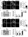


Classifications
-
- A—HUMAN NECESSITIES
- A61—MEDICAL OR VETERINARY SCIENCE; HYGIENE
- A61K—PREPARATIONS FOR MEDICAL, DENTAL OR TOILETRY PURPOSES
- A61K45/00—Medicinal preparations containing active ingredients not provided for in groups A61K31/00 - A61K41/00
-
- A—HUMAN NECESSITIES
- A61—MEDICAL OR VETERINARY SCIENCE; HYGIENE
- A61P—SPECIFIC THERAPEUTIC ACTIVITY OF CHEMICAL COMPOUNDS OR MEDICINAL PREPARATIONS
- A61P43/00—Drugs for specific purposes, not provided for in groups A61P1/00-A61P41/00
Definitions
- the invention relates to peptides for use in the treatment and/or diagnostic of lysosomal storage diseases.
- the present invention relates to the technical field of medical preparations containing peptides for the treatment of the metabolic/nervous system diseases.
- Lysosomal membrane proteins act at several crucial steps of the lysosome life cycle, including lumen acidification, metabolite export, molecular motor recruitment and fusion with other organelles.
- Lysosomal storage diseases are a group of inherited metabolic disorders that result from defects in lysosomal function. Lysosomes are sacs of enzymes within cells that digest large molecules and pass the fragments on to other parts of the cell for recycling. This process requires several critical enzymes. If one of these enzymes is defective (for example, because of a mutation), the large molecules accumulate within the cell, eventually killing it.
- sphingolipidoses are a subgroup of lipid storage disorders called sphingolipidoses in which harmful quantities of fatty substances, or lipids, accumulate in the spleen, liver, lungs, bone marrow, and brain.
- a missense mutation causes complete deficiency of acid sphingomyelinase.
- Sphingomyelin is a component of cell membrane including the organellar membrane, so the enzyme deficiency blocks degradation of sphingomyelin, resulting in the accumulation of sphingomyelin within lysosomes in the macrophage-monocyte phagocyte lineage. Affected cells become enlarged, secondary to the distention of lysosomes with sphingomyelin and cholesterol.
- Niemann-Pick disease Three types of Niemann-Pick disease exist. Mutations in the SMPD1 gene which codes for acid sphingomyelinase, cause Niemann-Pick disease types A and B. Mutations in NPC1 or NPC2 cause Niemann-Pick disease, type C (NPC), which affects a protein used to transport lipids, mostly cholesterol.
- NPC Niemann-Pick type C
- ER endoplasmic reticulum
- NPC The diagnosis of NPC is confirmed by biochemical testing that demonstrates impaired cholesterol esterification and positive filipin staining in cultured fibroblasts. Biochemical testing to detect carrier status is unreliable. Most individuals with NPC exhibit mutations in NPC1; fewer than 20 individuals have been diagnosed with NPC2, caused by mutations in NPC2. Molecular genetic testing of NPC1 and NPC2 detects disease-causing mutations in approximately 94% of individuals with NPC. Despite the advances of diagnosing NPC, there is still a lack of biomarkers which allow to quickly and efficiently diagnose NPC.
- NPC treatment options have been reported by Liu et al. ( Liu et al. Proc Natl Acad Sci USA, 106, 2377(2009 )) which showed that 2-hydroxypropyl- ⁇ -cyclodextrin (HPBCD) intravenously administered to Npc1 gene-defective (Npc1-/-) mice is effective in improving the medical state or prolonging survival, and when HPBCD is directly administered into the brain, the improving effect thereof is increased a few hundred times, compared with systemic administration.
- HPBCD 2-hydroxypropyl- ⁇ -cyclodextrin
- hydroxypropyl-y-cyclodextrin was disclosed in the document EP3078379A1 , where in addition iPS cells reprogramed using the hSendai virus vector which comprise the NPC gene is also disclosed.
- the document EP3505185A1 discloses inhibitors of the Glycogen synthase kinase 3 (GSK 3) for the treatment of NPC, while the document US2014220114A1 discloses inhibitors of acyl-coenzyme A:cholesterol acyltransferase 1 (ACAT1) for the treatment of NPC.
- GSK 3 Glycogen synthase kinase 3
- ACAT1 acyl-coenzyme A:cholesterol acyltransferase 1
- Biological related therapies have also been proposed as for example the use of progranulin (PRGN) disclosed in the document WO2015119989A1 , antibodies that target the protein Tau ( EP2625198B1 ) and antibodies targeting the triggering receptor expressed on myeloid cells 2 (TREM2).
- PRGN progranulin
- TERT2 myeloid cells 2
- compounds and biological agents for the treatment of NPC as of now the only drug approved for treatment of NPC in several countries as Europe, Japan, Canada, Brasil, Turkey and Australia is miglustat (Zavesca), a glucosylceramide synthase inhibitor, which extends the life span of patients, despite increased brain glucosylceramide levels.
- the authors of the present invention have found that the expression levels of the steroidogenic acute regulatory protein 1 (STARD1) gene are increased in subjects suffering from lysosomal storage disorders (LSDs), such as Niemann-Pick C (NPC) disease.
- LSDs lysosomal storage disorders
- NPC Niemann-Pick C
- the authors analyzed mice where the Niemann-Pick protein type C was mutated (NPC1 -/- ) and found that mitochondrial membranes where overly loaded with cholesterol. Therefore, the increase in the levels of STARD1 can be used as a biomarker for the diagnostic of LSDs. Due to this, the authors analyzed potential mechanisms which could lead to an accumulation of mitochondrial cholesterol.
- STARD1 has a crucial role in the intramitochondrial cholesterol trafficking from the outer membrane to inner mitochondrial membrane for metabolism. Therefore, the authors analyzed the levels of expression of the gene STARD1 and found these to be increased.
- Said increase in the levels of expression STARD1 correlate with the accumulation of cholesterol in mitochondria.
- the authors of the present invention further found that decreasing the levels of STARD1 resulted in a decrease of the levels of cholesterol in the mitochondria membranes.
- the accumulation of mitochondrial cholesterol is a critical factor in diseases like steatohepatitis, cancer and Alzheimer's disease
- the present invention opens up a new therapeutic window to patients of LSDs.
- a first aspect of the present invention relates to a steroidogenic acute regulatory protein 1 (STARD1) inhibitor, from here onwards the "inhibitor of the invention", for use in medicine, from here onwards the medical use of the invention.
- Another aspect of the invention relates to a STARD1 inhibitor for use in the treatment of LSD in a subject, from here onwards the treatment use of the invention.
- STARD1 steroidogenic acute regulatory protein 1
- STARD1 gene (Ensembl accession number: ENSG00000147465), also known as StAR gene, codifies for 4 transcripts (splice variants) which code for three distinct protein isoforms (SEQ ID NO: 2 to SEQ ID NO: 4) as shown in the table 1 below.
- Table 1 Transcripts and proteins corresponding to the gene STARD1 ENSG00000147465, Human (GRCh38.p13), located at Chromosome 8: 38,142,700-38,150,992 reverse strand. GRCh38:CM000670.2.
- SEQ ID Transcript ID Ensembl Isoform of STARD1 protein UniProtKB ID STAR-201 SEQ ID NO: 2 ENST00000276449.9 P49675, Q6IBK0 STAR-204 SEQ ID NO: 3 ENST00000522050.1 H0YB94 STAR-203 SEQ ID NO: 4 ENST00000521236.1 E5RH12 STAR-202 - ENST00000520114.1 No protein SEQ ID NO: 4 MQKALGILSN QEGWKKESQQ DNGDKVMSKV VPDVGKVFRL EVVVDQPM
- the STARD1 protein is partly located in mitochondria and synthesized as a conformationally labile precursor with an N-terminal mitochondrial targeting sequence on cytoplasmic ribosomes.
- START domain By virtue of its so called START domain, STARD1 stoichiometrically binds cholesterol within the cytoplasm and shuttles the hydrophobic cargo through both mitochondrial membranes and the enclosed intermembrane space.
- the depletion of STARD1 gene has been associated with several diseases including lipoid congenital adrenal hyperplasia and dose sensitive sex reversal while the overexpression or amplification of STARD1 gene has been associated with cancer, especially breast cancer, and Alzheimer's disease.
- the present invention further links increased levels of expression of STARD1 to LSDs.
- the present invention comprises the use of a STARD1 inhibitor.
- STARD1 inhibitor refers to any molecule capable of completely or partially inhibiting the STARD1 gene expression, both by preventing the expression product of said gene from being produced (interrupting the STARD1 gene transcription, blocking the translation of the mRNA coming from the STARD1 gene expression and/or promoting the premature degradation of the mRNA originating from the STARD1 gene), by directly inhibiting the STARD1 protein transporting activity and by promoting/targeting the STARD1 protein degradation by targeting it to the ubiquitin-proteasome pathway or to lysosomal proteolysis.
- Methods for decreasing/abrogating the expression of the gene encoding the STARD1 protein include, without being limited to, editing technologies such as CRISPR/cas9 or Cas9 nickase technology.
- STARD1 inhibitors can be identified using methods based on the capacity of the so-called inhibitor to reduced/abrogate the level of available cellular protein, which can be assessed using Western blotting with antibodies against STARD1, as exemplified in the examples of the present invention.
- STARD1 inhibitors suitable for use in the present invention include chemical compounds, antisense oligonucleotides, interference RNAs (siRNAs), catalytic RNAs or specific ribozymes and inhibitory antibodies.
- the STARD1 inhibitor is selected from the group consisting of a chemical compound, a STARD1 specific siRNA, a STARD1 specific antisense oligonucleotide, a STARD1 specific ribozyme, a STARD1 specific antibody, and a protein regulating the STARD1 expression.
- An additional aspect of the invention relates to the use of isolated "antisense" nucleic acids to inhibit expression, for example, for inhibiting transcription and/or translation of a nucleic acid which encodes STARD1, the activity of which is to be inhibited.
- the antisense nucleic acids can be bound to the target potential of the drug by means of conventional base complementarity or, for example, in the case of binding to double stranded DNA through specific interaction in the large groove of the double helix.
- these methods refer to a range of techniques generally used in the art and they include any method which is based on the specific binding to oligonucleotide sequences.
- an antisense oligonucleotide can be distributed, for example, as an expression plasmid which, when it is transcribed in cell, produces RNA complementary to at least one unique part of the cellular mRNA encoding STARD1.
- the antisense oligonucleotide is an oligonucleotide probe generated ex vivo which, when introduced into the cell, produces inhibition of gene expression hybridizing with the mRNA and/or gene sequences of a target nucleic acid.
- Such oligonucleotide probes are preferably modified oligonucleotides which are resistant to endogenous nucleases, for example, exonucleases and/or endonucleases and are therefore stable in vivo.
- nucleic acid molecules for use thereof as antisense oligonucleotides are DNA analogs of phosphoramidate, phosphothionate and methylphosphonate.
- the oligodeoxyribonucleotide regions derived from the starting site of the translation for example, between -10 and +10 of the target gene are preferred.
- the antisense approximations involve the oligonucleotide design (either DNA or RNA) that are complementary to the mRNA encoding the target polypeptide.
- the antisense oligonucleotide will be bound to the transcribed mRNA and translation will be prevented.
- the oligonucleotides which are complementary to the 5' end of the mRNA must function in the most efficient manner to inhibit translation. Nevertheless, it has been shown that the sequences complementary to the non-translated 3' sequences of the mRNA are also efficient for inhibiting mRNA translation. Therefore, complementary oligonucleotides could be used at the non-translated 5' or 3' regions, non-coding regions of a gene in an antisense approximation to inhibit the translation of that mRNA.
- the oligonucleotides complementary to the non-translated 5' region of the mRNA must include the complement of the start codon AUG.
- the oligonucleotides complementary to the coding region of the mRNA are less efficient translation inhibitors but they could also be used according to the invention. If they are designed to hybridize with the 5' region, 3' region or the coding region of the mRNA, the antisense nucleic acids must have at least six nucleotides long and preferably have less than approximately 100 and more preferably less than approximately 50, 25, 17 or 10 nucleotides long.
- in vitro studies are performed first to quantify the capacity of the antisense oligonucleotides for inhibiting gene expression.
- these studies use controls which distinguish between antisense gene inhibition and non-specific biological effects of the oligonucleotides.
- these studies compare the levels of target RNA or protein with that of an internal control of RNA or protein. The results obtained using the antisense oligonucleotides can be compared with those obtained using a control oligonucleotide.
- control oligonucleotide is approximately of the same length as the oligonucleotide to be assayed and the oligonucleotide sequence does not differ from the antisense sequence more than it is deemed necessary to prevent the specific hybridization to the target sequence.
- the antisense oligonucleotide can be a single or double stranded DNA or RNA or chimeric mixtures or derivatives or modified versions thereof.
- the oligonucleotide can be modified in the base group, the sugar group or the phosphate backbone, for example, to improve the stability of the molecule, its hybridization capacity, etc.
- the oligonucleotide may include other bound groups, such as peptides (for example, for directing them to the receptors of the host cells), agents for facilitating transport through the cell membrane or the blood-brain barrier, and intercalating agents.
- the oligonucleotide can be conjugated to another molecule, for example, a peptide, a transporting agent, hybridization triggered cleaving agent, etc.
- the antisense oligonucleotides may comprise at least one modified base.
- the antisense oligonucleotide may also comprise at least a modified sugar group selected from the group including but not limited to arabinose, 2-fluoroarabinose, xylulose, and hexose.
- the antisense oligonucleotide may also contain a backbone similar to a neutral peptide. Such molecules are known as peptide nucleic acid (PNA) oligomers.
- PNA peptide nucleic acid
- the antisense oligonucleotide comprises at least one modified phosphate backbone.
- the antisense oligonucleotide is an alpha-anomeric oligonucleotide.
- antisense oligonucleotides complementary to the coding region of the target mRNA sequence can be used, those complementary to the transcribed non translated region can also be used.
- a preferred approximation uses a recombinant DNA construct in which the antisense oligonucleotide is placed under the control of a strong pol III or pol II promoter.
- the target gene expression can be reduced by directing deoxyribonucleotide sequences complementary to the gene regulating region (i.e., the promoter and/or enhancers) to form triple helix structures preventing gene transcription in the target cells in the body.
- the antisense oligonucleotides are antisense morpholines.
- siRNA small interference RNA or siRNA are agents which are capable of inhibiting the expression of a target gene by means of RNA interference.
- a siRNA can be chemically synthesized, can be obtained by means of in vitro transcription or can be synthesized in vivo in the target cell.
- the siRNA consists of a double stranded RNA between 15 and 40 nucleotide long and may contain a 3' and/or 5' protruding region of 1 to 6 nucleotides. The length of the protruding region is independent of the total length of the siRNA molecule.
- the siRNA acts by means of degrading or silencing the target messenger after transcription.
- siRNAs of the invention are substantially homologous to the mRNA of the STARD1 protein encoding gene or to the gene sequence which encodes said protein. "Substantially homologous” is understood as having a sequence which is sufficiently complementary or similar to the target mRNA such that the siRNA is capable of degrading the latter through RNA interference.
- siRNA suitable for causing said interference include siRNA formed by RNA, as well as siRNA containing different chemical modifications such as: (i) siRNA in which the bonds between the nucleotides are different than those appear in nature, such as phosphorothionate bonds; (ii) Conjugates of the RNA strand with a functional reagent, such as a fluorophore; (iii) Modifications of the ends of the RNA strands, particularly of the 3' end by means of the modification with different hydroxyl functional groups in 2' position; (iv) Nucleotides with modified sugars such as 0-alkylated residues on 2' position like 2'-O-methylribose or 2'-O-fluororibose; or (v) Nucleotides with modified bases such as halogenated bases (for example 5-bromouracil and 5-iodouracil), alkylated bases (for example 7-methylguanosine).
- the siRNA can be used in the form of a double stranded RNA with the aforementioned characteristics.
- the use of vectors containing the sense and antisense strand sequence of the siRNA is possible under the control of suitable promoters for the expression thereof in the cell of interest.
- Vectors suitable for expressing siRNA are those in which the two DNA regions encoding the two strands of siRNA are arranged in tandem in one and the same DNA strand separated by a spacer region which, upon transcription, forms a loop and wherein a single promoter directs the transcription of the DNA molecule giving rise to the so-called short hairpin RNA (shRNA).
- shRNA short hairpin RNA
- the siRNA can be generated intracellularly from the shRNA characterized in that the antiparallel strands forming the siRNA are connected by a loop or hairpin region.
- the shRNAs can be encoded by plasmids or viruses, particularly retroviruses, and are under the control of a promoter. Promoters suitable for expressing shRNA are those indicated in the paragraph above for expressing siRNA.
- the siRNA and shRNA of the invention can be obtained using a series of techniques known by the person skilled in the art.
- the region of the nucleotide sequence taken as a basis for designing the siRNA is not limiting and it may contain a region of the coding sequence (between the start codon and the end codon) or it may alternatively contain sequences of the non-translated 5' or 3' region preferably between 25 and 50 nucleotides long and in any position in 3' direction position with respect to the start codon.
- One way of designing an siRNA involves the identification of the AA(N19)TT motifs wherein N can be any nucleotide in the LRRK2 gene sequence, and the selection of those having a high G/C content. If said motif is not found, it is possible to identify the NA(N21) motif wherein N can be any nucleotide.
- the invention also contemplates the use of DNA enzymes to inhibit the expression of the STARD1 gene of the invention.
- DNA enzymes incorporate some of the mechanistic features of both antisense and ribozyme technologies. DNA enzymes are designed such that they recognize a particular target nucleic acid sequence similar to the antisense oligonucleotide, nevertheless like the ribozyme they are catalytic and specifically cleave the target nucleic acid.
- Ribozyme molecules designed for catalytically cleaving transcription products of a target mRNA to prevent the translation of the mRNA which encodes STARD1, the activity of which is to be inhibited, can also be used.
- Ribozymes are enzymatic RNA molecules capable of catalyzing specific RNA cleaving.
- the mechanism of ribozyme action involves a specific hybridization of a ribozyme molecule sequence to a complementary target RNA followed by an endonucleolytic cleavage event.
- the composition of the ribozyme molecules preferably includes one or more sequences complementary to the target mRNA and the well-known sequence responsible for cleaving the mRNA or a functionally equivalent sequence.
- Examples of ribozymes to be used in the present invention include hammer-head ribozymes and endoribonuclease RNA (hereinafter "Cech type ribozymes").
- the ribozymes can be formed by modified oligonucleotides (for example to improve the stability, targeting, etc.) and they should be distributed to cells expressing the target gene in vivo.
- a preferred distribution method involves using a DNA construct which "encodes" the ribozyme under the control of a strong constitutive pol III or pol II promoter such that the transfected cells will produce sufficient amounts of the ribozyme to destroy the endogenous target messengers and to inhibit translation. Since the ribozymes are catalytic, unlike other antisense molecules, a low intracellular concentration is required for its efficiency.
- inhibitory antibody is understood as any antibody capable of binding specifically to the STARD1 protein and inhibiting one or more of the functions of said protein.
- the antibodies can be prepared using any of the methods which are known by the person skilled in the art, some of which have been mentioned above.
- the polyclonal antibodies are prepared by means of immunizing an animal with the protein, or a fragment of said protein, to be inhibited.
- the monoclonal antibodies are prepared using the method described by Kohler and Milstein et al. 1975 (Nature 256: 495-497 ).
- suitable antibodies include intact antibodies comprising a variable antigen binding region and a constant region, "Fab”, “F(ab')2” and “Fab”', Fv, scFv fragments, diabodies and bispecific antibodies. Once antibodies with STARD1 protein binding capacity are identified, those capable of inhibiting the activity of this protein will be selected using an inhibitory agent identification assay.
- STARD1 inhibitory antibodies are commercially available, such as the anti-STARD1 monoclonal antibodies produced in mouse and rabbit (ab58013 and ab133657 respectively, from Abcam), the anti-STARD1 polyclonal antibodies produced in rabbit (ab96637 and ab180804 from Abcam), the anti-STARD1 antibodies produced in rabbit (AP5465PU-N and TA350457 from Origene Technologies), etc.
- Chemical compounds which work as a STARD1 inhibitor are able to prevent the transport of cholesterol to the mitochondrial inner membrane for metabolism, for example by occupying the cargo binding site of STARD1.
- inhibitory protein refers to those proteins capable of binding to the STARD1 protein and inhibiting its activity as well as proteins that can interrupt, inhibit, stop or regulate STARD1 expression, i.e., the formation of functional products such as mRNA and protein, from the STARD1 gene.
- inhibitory proteins also includes fragments of proteins wherein said fragments have the STARD1 inhibitory effect.
- “Inhibitory proteins” can achieve the inhibiting STARD1 effect by inhibiting STARD1 transporting function, such as blocking access to STARD1 transport subtract or inhibiting STARD1 translocation from outer to inner membrane, by sequestering STARD1 from its normal location or stopping STARD1 to locate at its normal location, by promoting degradation of STARD1 protein by the ubiquitin-proteasome pathway or lysosomal pathway, by downregulating or blocking STARD1 gene transcription or downregulation or blocking STARD1 mRNA translation.
- the STARD1 inhibitor is a protein regulating the STARD1 expression.
- the protein regulating the STARD1 expression is an acid ceramidase (ACDase) enzyme or a functionally equivalent variant or fragment thereof.
- Acid ceramidase or its abbreviature (“ACDase”) as used herein refers to an acylsphingosine deacylase or glycosphingolipid ceramide deacylase (EC 3.5.1.23) which cleaves fatty acids from ceramide, producing sphingosine (SPH).
- Ceramidases comprise a heterogeneous family of enzymes whose main function in cells is to break down ceramides to SPH (2-amino-4-octadecene-1, 3-diol). Ceramidase enzymes are classified into three groups (acid, neutral, and alkaline enzymes) based on optimum pH and primary structure. Ceramidase activity can be determined as set forth by Bedia et al., Chembiochem 2007 Apr 16;8(6):642-8 .
- the term "functionally equivalent variant” refers to a protein (or polypeptide) comprising one or more alterations, such as substitutions, insertions, deletions and/or truncations of one or more specific amino acid residues at one or more specific positions in the protein, and having the same function that the original protein.
- variants of the ACDase enzyme protein of the invention may be (i) one in which one or more of the amino acid residues are substituted with a conserved or non-conserved amino acid residue (preferably a conserved amino acid residue) and such substituted amino acid residue may or may not be one encoded by the genetic code, (ii) one in which there are one or more modified amino acid residues, e.g., residues that are modified by the attachment of substituent groups, (iii) one in which the protein is an alternative splice variant of the proteins of the present invention and/or (iv) "fragments" of the protein.
- the "fragments” include proteins generated via proteolytic cleavage (including multi-site proteolysis) of an original sequence.
- the term "functionally equivalent fragment” means a protein or a domain thereof having one or more (e.g., several) amino acids absent from the amino and/or carboxyl terminus of the amino acid sequence, wherein the fragment has the same activity than the whole protein.
- the ACDase enzyme comprises an amino acid sequence having an identity of at least 70% with the sequence SEQ ID NO: 1.
- identity refers to the degree of similarity between two nucleotide or amino acid sequences obtained by aligning the two sequences, i.e. the proportion of identical nucleotide or amino acids between two compared nucleotide sequences or polypeptides/proteins along their full-length sequence. Depending on the number of common residues between the aligned sequences, a different degree of identity, expressed as a percentage, will be obtained.
- the degree of identity between two amino acids or nucleotides sequences may be determined by conventional methods, for example, by standard sequence alignment algorithms known in the state of the art, such as BLAST [ Altschul S.F. et al. Basic local alignment search tool. J Mol Biol.
- BLAST programs for example, BLASTN, BLASTX, and T BLASTX, BLASTP and TBLASTN, are in the public domain at The National Center for Biotechonology Information (NCBI) website.
- NCBI National Center for Biotechonology Information
- the present invention relates to a STARD1 inhibitor for use in the treatment of LSD in a subject, preferably NPA, NPB and NPC.
- treating refers to processes involving a slowing, interrupting, arresting, controlling, stopping, reducing, or reversing the progression or severity of an existing symptom, disorder, condition, or disease, but does not necessarily involve a total elimination of all disease-related symptoms, conditions, or disorders.
- treatment also refers to a process involving the avoiding of the appearance of symptoms which have not manifested themselves yet, but which will manifest themselves due to an untreated progression of the disorder, condition or disease.
- the treatment of a disorder or disease may, for example, lead to a halt in the progression of the disorder or disease (e.g., no deterioration of symptoms) or a delay in the progression of the disorder or disease (in case the halt in progression is of a transient nature only).
- the "treatment" of a disorder or disease may also lead to a partial response (e.g., amelioration of symptoms) or complete response (e.g., disappearance of symptoms) of the subject/patient suffering from the disorder or disease.
- the "treatment" of a disorder or disease may also refer to an amelioration of the disorder or disease, which may, e.g., lead to a halt in the progression of the disorder or disease or a delay in the progression of the disorder or disease.
- the condition or disorder to be treated are LSDs.
- the LSD is NPA, NPB and NPC.
- LSDs lysosomal storage disorders
- lysosomal storage disorders or its abbreviation “LSDs” refers to a group of rare inherited metabolic disorders that result from defects in lysosomal function.
- Lysosomes refer to sacs of enzymes within cells that digest large molecules and pass the fragments on to other parts of the cell for recycling. This process requires several critical enzymes. If one of these enzymes is defective due to a mutation, the large molecules accumulate within the cell, eventually killing it. LSDs are caused by lysosomal dysfunction usually as a consequence of deficiency of a single enzyme required for the metabolism of lipids, glycoproteins (sugar-containing proteins), or so-called mucopolysaccharides.
- the LSDs are generally classified by the nature of the primary stored material involved, and can be broadly broken into the following classification: lipid storage disorders; Sphingolipidoses (including Gaucher's and Niemann-Pick diseases); Gangliosidosis (including Tay-Sachs disease); Leukodystrophies; Mucopolysaccharidoses, (including Hunter syndrome and Hurler disease); Glycoprotein storage disorders; and Mucolipidoses.
- Sphingolipidoses including Gaucher's and Niemann-Pick diseases
- Gangliosidosis including Tay-Sachs disease
- Leukodystrophies including Tay-Sachs disease
- Mucopolysaccharidoses including Hunter syndrome and Hurler disease
- Glycoprotein storage disorders and Mucolipidoses.
- the lysosomal storage disorder is selected from a list consisting of: Farber disease, Krabbe disease, Fabry disease, Schindler disease, GM1 gangliosidosis, GM2 gangliosidosis, GM2 gangliosidosis AB variant, Sandhoff disease, Tay-Sachs, Gaucher disease Type I, Gaucher disease Type II, Gaucher disease Type III, Lysosomal acid lipase deficiency, Niemann-Pick disease Type A, Niemann-Pick disease Type B, Niemann-Pick disease Type C, Metachromatic leukodystrophy, Multiple sulfatase deficiency, Hurler syndrome, Scheie syndrome, Hurler-Scheie syndrome, Hunter syndrome, Sanfilippo syndrome Type A, Sanfilippo syndrome Type B, Sanfilippo syndrome Type C, Sanfilippo syndrome Type D, Morquio syndrome Type A, Morquio syndrome Type B, Maroteaux-
- the lysosomal storage disease is selected from a list consisting of: GM1 gangliosidosis, GM2 gangliosidosis, Alpha-mannosidosis, Hurler syndrome, Hunter syndrome, Sanfilippo syndrome Type A, Morquio syndrome Type A, Morquio syndrome Type B, Niemann-Pick type A (NPA), Niemann-Pick type B (NPC B) or Niemann-Pick type C (NPC) disease.
- lysosomal storage disorders are generally progressive over a period of time.
- the present invention describes how the accumulation of mitochondrial cholesterol in LSDs can be counteracted by inhibiting STARD1 gene expression or inhibiting its functional products in a subject.
- the subject to be treated with the STARD1 inhibitor can be any subject as long as the subject comprises mitochondrial cholesterol accumulation.
- the term “subject” refers to human and non-human animals, such as veterinary subjects.
- non-human animal includes all vertebrates, e.g., mammals and non-mammals, such as non-human primates, mice, rabbits, sheep, dog, cat, horse, cow, chickens, amphibians, and reptiles.
- the subject is a human, man or woman of any age or race.
- the subject object of the present invention is characterized by comprising mitochondrial cholesterol accumulation.
- mitochondrial cholesterol accumulation refers to an increase in the cholesterol present in the membranes of mitochondria of any organs, tissues and/or cells. In a preferred embodiment the mitochondrial cholesterol accumulation occurs in the liver and/or brain.
- An assay to determine if a given subject suffers from mitochondrial cholesterol accumulation comprises the staining of tissue with perfringolysin O fused with glutathione S-transferase (GST-PFO).
- GST-PFO glutathione S-transferase
- the STARD1 inhibitor of the present invention may be used in combination with one or more other drugs in the treatment, control, amelioration, prevention of symptoms of LSDs, where the combination of the drugs together is safer or more effective than either drug alone.
- the inhibitor of the invention is administered in combination with another compound useful for treating LSD.
- Such other compound(s)/drug(s) may be administered by a route and in an amount commonly used therefore, concomitantly or sequentially with the STARD1 inhibitor.
- a pharmaceutical composition in unit dosage form containing such other drugs and the STARD1 inhibitor is preferred.
- the combination therapy may also include therapies in which the STARD1 inhibitor and one or more other drugs are administered on different overlapping schedules. It is also contemplated that when used in combination with one or more other active ingredients, the STARD1 inhibitor and the other active ingredients may be used in lower doses than when each is used singly. Accordingly, the pharmaceutical compositions of the present invention include those that contain one or more other active ingredients, in addition to a STARD1 inhibitor.
- Examples of other compounds useful for treating LSDs which may be used in conjunction with STARD1 inhibitor include, without limiting to, alglucerase, a placenta-derived enzyme, imiglucerase, a synthetic form of alglucerase, miglustat a synthetic analogue of D-glucose, Hydroxypropyl- ⁇ -cyclodextrin or GSK3 inhibitors like lithium salt and the compounds CHIR99021, 6-BIO, TDZD-8, curcumin and histone deacetylase (HDAC) inhibitors.
- alglucerase a placenta-derived enzyme
- imiglucerase imiglucerase
- a synthetic form of alglucerase miglustat a synthetic analogue of D-glucose
- Hydroxypropyl- ⁇ -cyclodextrin or GSK3 inhibitors like lithium salt
- the compounds CHIR99021, 6-BIO, TDZD-8, curcumin and histone deacetylase (HDAC) inhibitors include, without limiting
- the weight ratio of the STARD1 inhibitor of the present invention to the other active ingredient(s) may be varied and will depend upon the effective dose of each ingredient. Generally, an effective dose of each will be used. Thus, for example, when the STARD1 inhibitor of the present invention is combined with another agent, the weight ratio of the STARD1 inhibitor to the other agent will generally range from about 1000:1 to about 1:1000, or from about 200:1 to about 1:200. Combinations of STARD1 inhibitor and other active ingredients will generally also be within the aforementioned range, but in each case, an effective dose of each active ingredient should be used.
- the STARD1 inhibitors of the present invention may be administered by oral, parenteral (e.g., intramuscular, intraperitoneal, intravenous, ICV, intracisternal injection or infusion, subcutaneous injection, or implant), by inhalation spray, nasal, vaginal, rectal, sublingual, buccal or topical routes of administration and may be formulated, alone or together, in suitable dosage unit formulations containing conventional non-toxic pharmaceutically acceptable carriers, adjuvants and vehicles appropriate for each route of administration.
- parenteral e.g., intramuscular, intraperitoneal, intravenous, ICV, intracisternal injection or infusion, subcutaneous injection, or implant
- inhalation spray nasal, vaginal, rectal, sublingual, buccal or topical routes of administration
- nasal, vaginal, rectal, sublingual, buccal or topical routes of administration may be formulated, alone or together, in suitable dosage unit formulations containing conventional non-toxic pharmaceutically acceptable carriers, adjuvants and vehicles appropriate for each route of administration.
- the STARD1 inhibitors of the present invention can be incorporated into pharmaceutical compositions for its administration to the subject.
- the inhibitor of the invention is comprised within a pharmaceutical composition.
- the STARD1 inhibitors of the present invention may conveniently be presented in dosage unit form and may be prepared by any of the methods well known in the art of pharmacy. All methods include the step of bringing the active ingredient into association with the carrier which constitutes one or more accessory ingredients.
- the pharmaceutical compositions are prepared by uniformly and intimately bringing the active ingredient into association with a liquid carrier or a finely divided solid carrier or both, and then, if necessary, shaping the product into the desired formulation.
- the active compound is included in an amount sufficient to produce the desired effect upon the process or condition of diseases.
- composition is intended to encompass a product comprising the specified ingredients in the specified amounts, as well as any product which results, directly or indirectly, from combination of the specified ingredients in the specified amounts.
- compositions containing the active ingredient may be in a form suitable for oral use, for example, as tablets, troches, lozenges, aqueous or oily suspensions, dispersible powders or granules, emulsions, solutions, hard or soft capsules, or syrups or elixirs.
- Compositions intended for oral use may be prepared according to any method known to the art for the manufacture of pharmaceutical compositions and such compositions may contain one or more agents selected from the group consisting of sweetening agents, flavoring agents, coloring agents and preserving agents in order to provide pharmaceutically elegant and palatable preparations. Tablets contain the active ingredient in admixture with non-toxic pharmaceutically acceptable excipients which are suitable for the manufacture of tablets.
- excipients may be for example, inert diluents, such as calcium carbonate, sodium carbonate, lactose, calcium phosphate or sodium phosphate; granulating and disintegrating agents, for example, corn starch, or alginic acid; binding agents, for example starch, gelatin or acacia; and lubricating agents, for example magnesium stearate, stearic acid or talc.
- the tablets may be uncoated or they may be coated by known techniques to delay disintegration and absorption in the gastrointestinal tract and thereby provide a sustained action over a longer period.
- a time delay material such as glyceryl monostearate or glyceryl distearate may be employed.
- Oral tablets may also be formulated for immediate release, such as fast melt tablets or wafers, rapid dissolve tablets or fast dissolve films.
- Formulations for oral use may also be presented as hard gelatin capsules wherein the active ingredient is mixed with an inert solid diluent, for example, calcium carbonate, calcium phosphate or kaolin, or as soft gelatin capsules wherein the active ingredient is mixed with water or an oil medium, for example peanut oil, liquid paraffin, or olive oil.
- an inert solid diluent for example, calcium carbonate, calcium phosphate or kaolin
- water or an oil medium for example peanut oil, liquid paraffin, or olive oil.
- Aqueous suspensions contain the active materials in admixture with excipients suitable for the manufacture of aqueous suspensions.
- excipients are suspending agents, for example sodium carboxymethylcellulose, methylcellulose, hydroxy-propylmethylcellulose, sodium alginate, polyvinyl-pyrrolidone, gum tragacanth and gum acacia; dispersing or wetting agents may be a naturally-occurring phosphatide, for example lecithin, or condensation products of an alkylene oxide with fatty acids, for example polyoxyethylene stearate, or condensation products of ethylene oxide with long chain aliphatic alcohols, for example heptadecaethyleneoxycetanol, or condensation products of ethylene oxide with partial esters derived from fatty acids and a hexitol such as polyoxyethylene sorbitol monooleate, or condensation products of ethylene oxide with partial esters derived from fatty acids and hexitol anhydrides, for example polyethylene sorbitan monoo
- the aqueous suspensions may also contain one or more preservatives, for example ethyl, or n-propyl, p-hydroxybenzoate, one or more coloring agents, one or more flavoring agents, and one or more sweetening agents, such as sucrose or saccharin.
- preservatives for example ethyl, or n-propyl, p-hydroxybenzoate
- coloring agents for example ethyl, or n-propyl, p-hydroxybenzoate
- coloring agents for example ethyl, or n-propyl, p-hydroxybenzoate
- flavoring agents for example ethyl, or n-propyl, p-hydroxybenzoate
- sweetening agents such as sucrose or saccharin.
- Oily suspensions may be formulated by suspending the active ingredient in a vegetable oil, for example arachis oil, olive oil, sesame oil or coconut oil, or in a mineral oil such as liquid paraffin.
- the oily suspensions may contain a thickening agent, for example beeswax, hard paraffin or cetyl alcohol.
- Sweetening agents such as those set forth above, and flavoring agents may be added to provide a palatable oral preparation.
- These compositions may be preserved by the addition of an antioxidant such as ascorbic acid.
- Dispersible powders and granules suitable for preparation of an aqueous suspension by the addition of water provide the active ingredient in admixture with a dispersing or wetting agent, suspending agent and one or more preservatives.
- a dispersing or wetting agent e.g., glycerol, glycerol, glycerol, glycerol, glycerol, glycerol, glycerin, glycerin, glycerin, glycerin, glycerin, sorbitol, sorbitol, sorbitol, sorbitol, sorbitol, sorbitol, sorbitol, sorbitol, sorbitol, sorbitol, glycerol, glycerol, glycerol, glycerol, glycerol, glycerol, glycerol, glycerol, glycerol
- the pharmaceutical compositions of the invention may also be in the form of oil-in-water emulsions.
- the oily phase may be a vegetable oil, for example olive oil or arachis oil, or a mineral oil, for example liquid paraffin or mixtures of these.
- Suitable emulsifying agents may be naturally-occurring gums, for example gum acacia or gum tragacanth, naturally-occurring phosphatides, for example soy bean, lecithin, and esters or partial esters derived from fatty acids and hexitol anhydrides, for example sorbitan monooleate, and condensation products of the said partial esters with ethylene oxide, for example polyoxyethylene sorbitan monooleate.
- the emulsions may also contain sweetening and flavoring agents.
- the pharmaceutical compositions may be in the form of a sterile injectable aqueous or oleagenous suspension.
- This suspension may be formulated according to the known art using those suitable dispersing or wetting agents and suspending agents which have been mentioned above.
- the sterile injectable preparation may also be a sterile injectable solution or suspension in a non-toxic parenterally-acceptable diluent or solvent, for example as a solution in 1,3-butane diol.
- the acceptable vehicles and solvents that may be employed are water, Ringer's solution and isotonic sodium chloride solution.
- sterile, fixed oils are conventionally employed as a solvent or suspending medium.
- any bland fixed oil may be employed including synthetic mono- or diglycerides.
- fatty acids such as oleic acid find use in the preparation of injectables.
- the compounds of the present invention may also be administered in the form of suppositories for rectal administration of the drug.
- These compositions can be prepared by mixing the drug with a suitable non-irritating excipient which is solid at ordinary temperatures but liquid at the rectal temperature and will therefore melt in the rectum to release the drug.
- suitable non-irritating excipient which is solid at ordinary temperatures but liquid at the rectal temperature and will therefore melt in the rectum to release the drug.
- Such materials are cocoa butter and polyethylene glycols.
- transdermal patches may also be used for topical administration.
- an appropriate dosage level of the STARD1 inhibitor of the invention will generally be about 0.01 to 500 mg per kg patient body weight per day which can be administered in single or multiple doses.
- a suitable dosage level may be about 0.01 to 250 mg/kg per day, about 0.05 to 100 mg/kg per day, or about 0.1 to 50 mg/kg per day. Within this range the dosage may be 0.05 to 0.5, 0.5 to 5 or 5 to 50 mg/kg per day.
- the compositions may be provided in the form of tablets containing 1.0 to 1000 milligrams of the active ingredient, particularly 1.0, 5.0, 10.0, 15.0.
- the compounds may be administered on a regimen of 1 to 4 times per day, or may be administered once or twice per day.
- the present invention allows the skilled person in the art to identify LSD patients as well as to determine which patients will benefit from a STARD1-related therapeutic intervention such as STARD1 inhibitors.
- Another aspect of the invention relates to an in vitro method for diagnosing lysosomal storage disorder in a subject, or for designing a customized therapy for a subject suffering from lysosomal storage disorder, here onwards the "method of the invention", comprising:
- diagnosis refers to the procedure by which a certain disease, nosological entity, syndrome, or any health-disease condition is identified through the analysis of a series of clinical parameters or symptoms characteristic of that disease, and which distinguish it from other diseases with similar clinical presentation.
- the disease to be identified is an LSD
- the clinical parameter is the level of expression of the STARD1 gene.
- the LSD to diagnose is select from a list consisting of: Farber disease, Krabbe disease, Fabry disease, Schindler disease, GM1 gangliosidosis, GM2 gangliosidosis, GM2 gangliosidosis AB variant, Sandhoff disease, Tay-Sachs, Gaucher disease Type I, Gaucher disease Type II, Gaucher disease Type III, Lysosomal acid lipase deficiency, Niemann-Pick disease Type A, Niemann-Pick disease Type B, Niemann-Pick disease Type C, Metachromatic leukodystrophy, Multiple sulfatase deficiency, Hurler syndrome, Scheie syndrome, Hurler-Scheie syndrome, Hunter syndrome, Sanfilippo syndrome, Morquio, Maroteaux-Lamy syndrome, Sly syndrome, hyaluronidase deficiency, sialidosis, I-cell disease, phosphotransferase deficiency, mucolipidin 1 de
- the lysosomal storage disease is selected from a list consisting of: GM1 gangliosidosis, GM2 gangliosidosis, Alpha-mannosidosis, Hurler syndrome, Hunter syndrome, Sanfilippo syndrome Type A, Morquio syndrome Type A, Morquio syndrome Type B, Niemann-Pick type A (NPA), Niemann-Pick type B (NPC B) or Niemann-Pick type C (NPC) disease.
- step (i) of the method of the invention refers to the procedure of reporting and/or determine the amount of the level of expression of the nucleotide sequence of the gene STARD1, by the detection and/or determination of the amount of the mRNA, or a fragment of the mRNA, cDNA or a fragment of the cDNA, protein or a fragment of the protein encoded by the STARD1 nucleotide sequence.
- the quantification of the expression levels comprises measuring the level of cDNA, the level of mRNA, and/or the level of the protein encoded by said nucleotide sequence.
- mRNA fragment or "cDNA fragment” as used herein refers to the nucleotide sequence of the STARD1 gene comprising one or more nucleotides absent from the 3' and/or 5' ends with respect to the complete nucleotide sequence of the STARD1 gene.
- the term “protein fragment” as used herein refers to the amino acid sequence of the STARD1 protein(s) comprising one or more amino acids absent from its terminal amino or terminal carboxyl end with respect to the complete amino acid sequence of the STARD1 protein.
- the STARD1 protein fragment is a fragment of the protein sequence encoded by the sequence selected from the list consisting of SEQ ID NO: 2, SEQ ID NO: 3, SEQ ID NO: 4 or any combination thereof.
- the determination of the expression levels of the nucleotide sequence encoding the STARD1 gene may comprise measuring the level of cDNA, the level of mRNA, and/or the level of the protein encoded by said gene.
- the levels of mRNA of said gene can be quantified by means of using conventional methods, for example, methods comprising the amplification of mRNA and the quantification of the product of the amplification of said mRNA, such as electrophoresis and staining, or alternatively, by means of Southern blot and the use of suitable probes, Northern blot and the use of specific probes of mRNA of the STARD1 gene or of the corresponding cDNA thereof, S1 nuclease mapping, hybridisation, RT-Q-PCR, the micro-arrays and RNAsequencing, etc. If the quantification of the expression of STARD1 gene encoding is going to be carried out from the protein, i.e.
- the STARD1 protein then the isolated biological sample of the subject must be treated to extract the proteins.
- Methods for extracting or isolating proteins are known by the person skilled in the art and they are commercially available.
- the levels of gene encoding the STARD1 protein can be quantified by means of any conventional method that enables said protein to be detected and quantified in a sample of a subject.
- the levels of said protein can be quantified, for example, by means of the use of antibodies with the capacity to bind specifically to the STARD1 protein and the subsequent quantification of the complexes formed. Examples of antibodies with the capacity to specifically bind to the STARD1 protein have been disclosed in previous paragraphs.
- comparing the expression levels obtained in step (i) with a reference level refers to the process or procedure of relating, examining or verifying the values obtained for the expression level of the STARD1 gene, and equate or verify the values obtained in step a) with the values expected to be obtained for subjects not suffering from the disease and/or not presenting mitochondrial cholesterol accumulation, where, according to the method of the invention, "an increased STARD1 expression levels with respect to the reference level is indicative that the subject suffers from lysosomal storage disorder, or that the therapy to be administered to the subject is based on a STARD1 inhibitor".
- reference level refers to the expression levels of the nucleotide sequence encoding the STARD1 gene in a sample of a subject not suffering from LSD and/or not comprising mitochondrial cholesterol accumulation.
- the subject is a mammal, preferably, the mammal is a human.
- the method of the invention contemplates the diagnostic as well as the designing of a customized therapy for a subject that suffers from LSD, wherein said customized therapy is based on a STARD1 inhibitor.
- the STARD1 inhibitor is selected from the group consisting of a chemical compound, a STARD1 specific siRNA, a STARD1 specific antisense oligonucleotide, a STARD1 specific ribozyme and a STARD1 specific antibody, and a protein regulating the STARD1 expression.
- the protein regulating the STARD1 expression is an acid ceramidase (ACDase) enzyme or a functionally equivalent variant or functionally equivalent fragment thereof.
- ACDase acid ceramidase
- the ACDase enzyme comprises an amino acid sequence having an identity of at least 70% with the sequence SEQ ID NO: 1.
- Npc1 -/- mice (NPC1NIH, BALB/cJ strain) were obtained from The Jackson Laboratories. At the time of weaning (21 days), mice were genetically identified by PCR using DNA prepared from tail-tips and following the genotyping protocols provided by the supplier, as described previously in Torres et al., Redox Biol. 2017 Apr;11:60-72 . All procedures involving animals and their care were approved by the Ethics Committee of the University of Barcelona and were conducted in accordance with institutional guidelines in compliance with national and international laws and policies.
- Fibroblasts from patients with NPC disease used have been previously described ( Torres et al., Redox Biol. 2017 Apr;11:60-72 ). Briefly, cultured human skin fibroblasts from control individuals (HSF; GM5659D) and patients with NPC disease were a generous gift from Thierry Levade, Laboratoire de Biochimie Metabolique, Institut Federatif de Biologie (CHU Toulouse, France) or obtained from Coriell Institute for Medical Research (GM03123, NJ, USA) and were grown at 37°C in 5% CO2. DMEM (Gibco) culture medium was supplemented with 10% fetal bovine serum (FBS, Gibco, 10-270-106) and 10.000U/ml Penicillin-Streptomycin (Gibco, 15140-122).
- FBS fetal bovine serum
- Penicillin-Streptomycin Gibco, 15140-122
- PMH were isolated by collagenase perfusion at a flow rate of 7-9ml/min and cultured at a density of 2 ⁇ 105 cells in 12-well plates coated with rat tail collagen.
- Hepatocytes were cultured in DMEM/F12 culture media (Gibco, 21331-020) supplemented with 10% FBS for the first 3 hours post isolation, together with 10.000U/ml Penicillin-Streptomycin (Gibco, 15140-122), 200mM L-Glutamine (Gibco, 25030-024), 7.5mM D-Glucose (Sigma, G6152) and 150mM Hepes pH 7.4 (Sigma,). For subsequent treatments, FBS was replaced for 1mM methionine (Sigma, M5308).
- PMH from Npc1 +/+ and human Npc +/+ control fibroblasts were treated with U18666A (2 ⁇ g/ml) or HM ⁇ CD (2mM) for 16 hours.
- PMH from Npc1 -/- and Npc -/- fibroblast from NPC patients were treated with HM ⁇ CD as above for control PMH.
- Fibroblasts from NPC patients were transfected with cDNA to overexpress ACDase (ASAH1) (SEQ ID NO: 5; MGC Human ASAH1 Sequence-Verified cDNA Accession: BC016481 Clone ID: 3923451) commercially purchased from GE Dharmacon. Transfection was performed using Lipofectamine2000 (Invitrogen). Briefly, 250000 fibroblasts were incubated with the transfection mixtures containing 250ng of the sobrexpression ACDase vector or the scrambled control-GFP vector. Cells were assayed 48 h after transfection for mRNA and protein levels.
- SEQ ID NO: 5 MGC Human ASAH1 Sequence-Verified cDNA Accession: BC016481 Clone ID: 3923451
- SEQ ID NO: 5 >ENA
- N-acylsphingosine amidohydrolase (acid ceramidase) 1
- mRNA cDNA clone MGC:9327 IMAGE:3923451
- CT threshold
- Table 1 The primer sequences synthesized from Invitrogen used are displayed in Table 1: Table 1: Primer sequences used Gene Primer SEQ ID Sequence GFP Forward primer (5'-3') SEQ ID NO: 6 CAGGAGCGCACCATCTTCTT Reverse primer (5'-3') SEQ ID NO: 7 CTTGTGCCCCAGGATGTTG ASAH1 (ACDase) Forward primer (5'-3') SEQ ID NO: 8 AGTTGCGTCGCCTTAGTCCT Reverse primer (5'-3') SEQ ID NO: 9 TGCACCTCTGTACGTTGGTC ACTB Forward primer (5'-3') SEQ ID NO: 10 TTGCCGACAGGATGCAGAA Reverse primer (5'-3') SEQ ID NO: 11 GCCGATCCACACGGAGTACT
- the signal peptide was eliminated from the PFO gen to enable the intracellular stance of the protein.
- the resulted sequence was synthesized into the plasmid pGEX 4T-1 (GenScript) between the Bam HI and Sma I restriction sites.
- the plasmid was engineered to make a fusion protein with the GST tag. After bacterial production, and protein extraction and purification, the GST-PFO probe was dialyzed, concentrated, quantified and stored at -80°C.
- Cryopreserved liver, brain and cerebellum samples from were cut on a cryostat at 14 micrometers thin sections and placed on glass slides. Sections were brought to room temperature for 30 min, fixed in 4% paraformaldehyde for 20 minutes and washed with PBS. For Filipin staining tissues were incubated with 25 ⁇ g/mL Filipin (Sigma) over night at 4°C protected from the light. After washing, slices were mounted with prolong antifade mounta nt (Dako). For GST-PFO staining, the sections were permeabilized with 0.2% Triton X-100 in blocking buffer (5% goat serum + 1% BSA in PBS) for 2 h in a dark-humid chamber.
- Mitochondrial fraction was isolated from liver and brain by Percoll density grandient centrifugation as described previously ( Torres et al., Redox Biol. 2017 Apr; 11:60-72 ). Mitochondrial purity and cross contamination with extramitochondrial compartments was checked as described previously ( Fernandez, A et al. J. Hepatol. 2013;59(4):805-813 ). Alternatively, PMH and fibroblasts were fractionated into cytosol and mitochondria by digitonin permeabilization as described previously ( Fernandez, A et al. J. Hepatol. 2013;59(4):805-813 ).
- Total cholesterol determination in homogenates (1mg/ml) or mitochondrial extracts (2.3 mg/ml) was performed upon saponification with alcoholic KOH followed by water:hexane 1:2 extraction. Hexane phase was evaporated in a speed vacuum and used for cholesterol measurement. Free cholesterol was determined from the same unsaponified samples and analyzed by HPLC using Bondapak C18 10 ⁇ m reversed-phase column (30cm x 4mm inner diameter; Waters, Cromatschreib, S.A., Barcelona, Spain), and 2-propanol/acetonitrile/water (60:30:10) as mobile phase at a flow rate of 1ml/min as described. The amount of cholesterol was calculated from standard curves and the identity of the peaks was confirmed by spiking the sample with known standards.
- Fluidity of mitochondrial membranes was evaluated by fluorescence anisotropy of mitochondria-bound dye DPH.
- DPH (20 mM in tetrahydrofuran) was first diluted 100 times with 10 mM Tris-HCI, 150 mM KCI, 1 mM EDTA, pH 7.4. Subsequently, DPH was injected into stirred mitochondrial suspensions (0.5 mg/ml) and the mixture was incubated for 30 min at 37 °C. Fluorescence polarization was measured in a Hitachi spectrofluorometer at wavelengths of 366 nm for excitation and 425 nm for emission.
- I0 and I90 represent the intensities of light when polarizers were in parallel or perpendicular orientation, respectively.
- Light scattering and intrinsic fluorescence were routinely corrected by subtracting the signal obtained from unlabeled samples and the fluorescence of the buffer plus label alone.
- fibroblasts were first treated with hydrogen peroxide (H2O2) 1mM for 24 hours.
- H2O2 hydrogen peroxide
- cells were then incubated in DCF (10 ⁇ M) solution for 30 minutes at 37°C in dark, washed with PBS and evaluated by fluorescence spectroscopy using the Tecan Infinite ® 200 PRO multi-mode microplate reading (485nm for excitation and 528nm for emission). The fluorescence was normalized by protein content, determined by BCA protein assay. The induction of reactive species was calculated with respect to control samples.
- liver homogenized 100mg of liver were homogenized with a pestle homogenizer in 1ml of homobuffer (70mM saccharose, 220mM mannitol, 2mM Tris-HCI pH 7.4, 0,1mM EDTA, 0.1% fatty acid free BSA) supplemented with protease and phosphatase inhibitors (Roche).
- Liver homogenates were diluted 4 times with RIPA lysis buffer (Sigma) with anti-proteases and anti-phosphatases.
- Cells were washed with PBS and lysed with ice-cold RIPA lysis buffer supplemented with protease and phosphatase inhibitors.
- Membranes were incubated overnight with the following primary antibodies: anti-PDI (Cell Signaling, 24465), anti-CHOP (Cell Signaling, 2895S), anti-Phospho eiF2A (Cell Signaling, 9721S), anti-eiF2A (Cell Signaling, 9722) and anti-STARD1 (Abcam, ab58013), anti-BIP (Stressgen, SPA826), anti-XBP1 (Santa Cruz, CS-8015) and anti-Mln64 (Santa Cruz SC-292868), ACDAse (Sigma, SAB3500293), LRH-1 (Santa Cruz, Cs-393369) and anti- ⁇ -Actin HRP-conjugated (Sigma, A3854).
- anti-PDI Cell Signaling, 24465
- anti-CHOP Cell Signaling, 2895S
- anti-Phospho eiF2A Cell Signaling, 9721S
- anti-eiF2A
- Membranes were thoroughly washed with TBS-Tween and incubated for 45min with their respective HRP-conjugated secondary antibodies.
- Pierce ECL Western Blotting Substrate (Thermo Scientific) was used to develop the membranes. Images were digitally captured by LAS4000 (GE Healthcare) and optical density was analyzed with ImageJ software.
- OCR In vivo real-time mitochondrial respiration
- Example 1 Hepatic mitochondrial cholesterol accumulation and STARD1 upregulation in Npc1 -/- mice
- Example 3 Livers from Npc1 -/- mice and fibroblasts from NPC patients exhibit decreased ACDase expression without evidence of ER stress.
- Example 4 Mitochondrial cholesterol status and expression of STARD1 and ACDase in brain and cerebellum from Npc1 - / - mice.
- Example 5 The amphiphilic cationic sterol U18666A reproduces the inverse correlation between STARD and ACDase in Npc1 + / + PMH.
- U18666A also stimulated the accumulation of cholesterol in mitochondria, as seen by the colocalization of filipin and CytC staining of Npc1 + / + PMH ( Figure 5B ), in line with the findings seen in Npc1 -/- PMH. Moreover, similar accumulation of mitochondrial cholesterol was observed in Npc +/+ fibroblasts upon U18666A treatment ( Figure 6A-B ). Interestingly, U18666A treatment significantly increased STARD1 in both PMH from Npc1 + / + mice ( Figure 5C ) and Npc +/+ fibroblasts ( Figure 6C ). Similar findings were observed in the expression of MNL64 although to a lower extent than that of STARD1 ( Figure 5C ; Figure 6C ).
- Example 6 2-hydroxypropyl- ⁇ -cyclodextrin prevents U18666A-mediated ACDase downregulation and STARD1 upregulation.
- HM ⁇ CD 2-hydroxypropyl- ⁇ -cyclodextrin
- Example 7 ACDase overexpression in fibroblasts from NPC patients prevents STARD1 upregulation and mitochondrial cholesterol accumulation without alterations in MLN64 expression.
- Npc - / - fibroblasts from patients with NPC disease exhibited lower mitochondrial free cholesterol levels as indicated by the colocalization of filipin with Cyt C staining, compared to scrambled control-GFP transfected fibrloblasts ( Figure 8C ).
- Example 8 ACDase overexpression in fibroblasts from NPC patients increases mGSH, improves mitochondrial function and protects against oxidative stress and cell death.
- ACDase transfection also resulted in decreased oxidative stress in response to H 2 O 2 challenge as revealed by lower dichlorofluorescein (DCF) fluorescence compared to Npc -/- fibroblasts from NPC patients transfected with control GFP vector.
- DCF dichlorofluorescein
Landscapes
- Health & Medical Sciences (AREA)
- Veterinary Medicine (AREA)
- Chemical & Material Sciences (AREA)
- Medicinal Chemistry (AREA)
- Pharmacology & Pharmacy (AREA)
- Life Sciences & Earth Sciences (AREA)
- Animal Behavior & Ethology (AREA)
- General Health & Medical Sciences (AREA)
- Public Health (AREA)
- Epidemiology (AREA)
- Engineering & Computer Science (AREA)
- Bioinformatics & Cheminformatics (AREA)
- Chemical Kinetics & Catalysis (AREA)
- General Chemical & Material Sciences (AREA)
- Nuclear Medicine, Radiotherapy & Molecular Imaging (AREA)
- Organic Chemistry (AREA)
- Pharmaceuticals Containing Other Organic And Inorganic Compounds (AREA)
Abstract
The invention relates to peptides for use in the treatment and/or diagnostic of lysosomal storage diseases, specifically peptides or proteins that inhibit STARD1 expression levels and subsequently mitochondrial cholesterol levels, and their use in the treatment of lysosomal storage diseases.
Description
- The invention relates to peptides for use in the treatment and/or diagnostic of lysosomal storage diseases. Thus, the present invention relates to the technical field of medical preparations containing peptides for the treatment of the metabolic/nervous system diseases.
- Lysosomal membrane proteins act at several crucial steps of the lysosome life cycle, including lumen acidification, metabolite export, molecular motor recruitment and fusion with other organelles. Lysosomal storage diseases (LSDs) are a group of inherited metabolic disorders that result from defects in lysosomal function. Lysosomes are sacs of enzymes within cells that digest large molecules and pass the fragments on to other parts of the cell for recycling. This process requires several critical enzymes. If one of these enzymes is defective (for example, because of a mutation), the large molecules accumulate within the cell, eventually killing it.
- Among the ∼50 known LSDs several are caused by lysosomal membrane protein dysfunction. Niemann-Pick diseases are a subgroup of lipid storage disorders called sphingolipidoses in which harmful quantities of fatty substances, or lipids, accumulate in the spleen, liver, lungs, bone marrow, and brain. In the classic infantile type-A variant, a missense mutation causes complete deficiency of acid sphingomyelinase. Sphingomyelin is a component of cell membrane including the organellar membrane, so the enzyme deficiency blocks degradation of sphingomyelin, resulting in the accumulation of sphingomyelin within lysosomes in the macrophage-monocyte phagocyte lineage. Affected cells become enlarged, secondary to the distention of lysosomes with sphingomyelin and cholesterol.
- Three types of Niemann-Pick disease exist. Mutations in the SMPD1 gene which codes for acid sphingomyelinase, cause Niemann-Pick disease types A and B. Mutations in NPC1 or NPC2 cause Niemann-Pick disease, type C (NPC), which affects a protein used to transport lipids, mostly cholesterol.
- Niemann-Pick type C (NPC) disease exhibits a wide array of symptoms, ranging from neurological deterioration, liver disease, splenomegaly and ultimately premature death. Besides the mutations in NPC1 or NPC2, disease-causing mutations on quality control pathways involving the lysosome and endoplasmic reticulum (ER) have also been shown to contribute to NPC disease. Defects in NPC1 account for up to 95% of NPC cases and given its role in lysosomal cholesterol egress, the primary biochemical feature of NPC disease translates in the lysosomal accumulation of free cholesterol in brain and liver.
- In addition to lysosomes, cholesterol accumulation has also been reported in mitochondrial fractions of affected organs from Npc1-/- mice, a murine model which reproduces many of the pathological features of the human disease. Mitochondrial cholesterol accumulation has emerged as a critical factor in several diseases, including steatohepatitis, cancer or Alzheimer's disease, due in part to the regulation of mitochondrial function and limitation of mitochondrial antioxidant defenses.
- The diagnosis of NPC is confirmed by biochemical testing that demonstrates impaired cholesterol esterification and positive filipin staining in cultured fibroblasts. Biochemical testing to detect carrier status is unreliable. Most individuals with NPC exhibit mutations in NPC1; fewer than 20 individuals have been diagnosed with NPC2, caused by mutations in NPC2. Molecular genetic testing of NPC1 and NPC2 detects disease-causing mutations in approximately 94% of individuals with NPC. Despite the advances of diagnosing NPC, there is still a lack of biomarkers which allow to quickly and efficiently diagnose NPC.
- Over the past several years the number of treatments available for patients with LSDs has rapidly increased. Haematopoietic stem cell transplantation, enzyme replacement therapy, substrate reduction, and chaperone therapies are currently available, and gene therapies and other treatments are rapidly advancing. Despite remarkable advances, the efficacy of most of these therapies is limited, particularly because the treatments are usually initiated when organ damage has already occurred. To circumvent this limitation, screening in newborn infants for LSDs has been introduced in many countries. However, this screening is complicated by the broad clinical variability of the disorders and the fact that many individuals who will be detected as having an enzyme deficiency will develop symptoms very late or never in their life.
- As examples, NPC treatment options have been reported by Liu et al. (Liu et al. Proc Natl Acad Sci USA, 106, 2377(2009)) which showed that 2-hydroxypropyl-β-cyclodextrin (HPBCD) intravenously administered to Npc1 gene-defective (Npc1-/-) mice is effective in improving the medical state or prolonging survival, and when HPBCD is directly administered into the brain, the improving effect thereof is increased a few hundred times, compared with systemic administration. This disparity in effects of intravenous vs intracerebral administration is due to the inability of HPBCD to cross the blood brain barrier. Based on the outcome of these fundamental researches, especially approved humanistic use of HPBCD to NPC child patients was granted (intravenous administration and intrathecal administration). As a result of continuation for more than 1 year of intravenous instillation of HPBCD (2500 mg/kg per time, 1 to 3 times per week) to NPC child patients, the certain effect of reduction of splenomegaly and improvement in a brain wave in child patients was obtained, but a nervous symptom saw no improvement as of yet, and severe side effects have been observed.
- Furthermore, hydroxypropyl-y-cyclodextrin was disclosed in the document
EP3078379A1 , where in addition iPS cells reprogramed using the hSendai virus vector which comprise the NPC gene is also disclosed. The documentEP3505185A1 discloses inhibitors of the Glycogen synthase kinase 3 (GSK 3) for the treatment of NPC, while the documentUS2014220114A1 discloses inhibitors of acyl-coenzyme A:cholesterol acyltransferase 1 (ACAT1) for the treatment of NPC. - Biological related therapies have also been proposed as for example the use of progranulin (PRGN) disclosed in the document
WO2015119989A1 , antibodies that target the protein Tau (EP2625198B1 ) and antibodies targeting the triggering receptor expressed on myeloid cells 2 (TREM2). Despite the large body of work relating to possible drugs, compounds and biological agents for the treatment of NPC, as of now the only drug approved for treatment of NPC in several countries as Europe, Japan, Canada, Brasil, Turkey and Australia is miglustat (Zavesca), a glucosylceramide synthase inhibitor, which extends the life span of patients, despite increased brain glucosylceramide levels. - Therefore, alternate biomarkers for the diagnosis and alternate targets for the treatment of NPC are required to address the lack of viable options in the diagnosis and treatment of the devastating disease NPC.
- The authors of the present invention have found that the expression levels of the steroidogenic acute regulatory protein 1 (STARD1) gene are increased in subjects suffering from lysosomal storage disorders (LSDs), such as Niemann-Pick C (NPC) disease. The authors analyzed mice where the Niemann-Pick protein type C was mutated (NPC1-/-) and found that mitochondrial membranes where overly loaded with cholesterol. Therefore, the increase in the levels of STARD1 can be used as a biomarker for the diagnostic of LSDs. Due to this, the authors analyzed potential mechanisms which could lead to an accumulation of mitochondrial cholesterol. STARD1 has a crucial role in the intramitochondrial cholesterol trafficking from the outer membrane to inner mitochondrial membrane for metabolism. Therefore, the authors analyzed the levels of expression of the gene STARD1 and found these to be increased.
- Said increase in the levels of expression STARD1 correlate with the accumulation of cholesterol in mitochondria. The authors of the present invention further found that decreasing the levels of STARD1 resulted in a decrease of the levels of cholesterol in the mitochondria membranes. As the accumulation of mitochondrial cholesterol is a critical factor in diseases like steatohepatitis, cancer and Alzheimer's disease, the authors tested if STARD1 could also have a function in LSD. The authors found that inhibiting STARD1 leads to the reduction of cholesterol in mitochondrial membranes and improves the performance and oxidative stress of fibroblasts of patients with NPC. Hence, the present invention opens up a new therapeutic window to patients of LSDs.
- In view of the aforementioned, a first aspect of the present invention relates to a steroidogenic acute regulatory protein 1 (STARD1) inhibitor, from here onwards the "inhibitor of the invention", for use in medicine, from here onwards the medical use of the invention. Another aspect of the invention relates to a STARD1 inhibitor for use in the treatment of LSD in a subject, from here onwards the treatment use of the invention.
- STARD1 gene (Ensembl accession number: ENSG00000147465), also known as StAR gene, codifies for 4 transcripts (splice variants) which code for three distinct protein isoforms (SEQ ID NO: 2 to SEQ ID NO: 4) as shown in the table 1 below.
- Table 1: Transcripts and proteins corresponding to the gene STARD1 ENSG00000147465, Human (GRCh38.p13), located at Chromosome 8: 38,142,700-38,150,992 reverse strand. GRCh38:CM000670.2.
Name SEQ ID Transcript ID Ensembl Isoform of STARD1 protein UniProtKB ID STAR-201 SEQ ID NO: 2 ENST00000276449.9 P49675, Q6IBK0 STAR-204 SEQ ID NO: 3 ENST00000522050.1 H0YB94 STAR-203 SEQ ID NO: 4 ENST00000521236.1 E5RH12 STAR-202 - ENST00000520114.1 No protein  SEQ ID NO: 4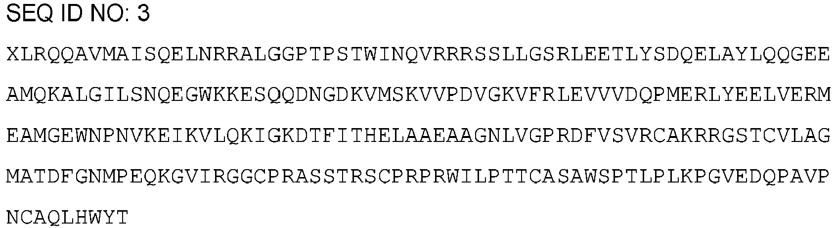
MQKALGILSN QEGWKKESQQ DNGDKVMSKV VPDVGKVFRL EVVVDQPM - The STARD1 protein is partly located in mitochondria and synthesized as a conformationally labile precursor with an N-terminal mitochondrial targeting sequence on cytoplasmic ribosomes. By virtue of its so called START domain, STARD1 stoichiometrically binds cholesterol within the cytoplasm and shuttles the hydrophobic cargo through both mitochondrial membranes and the enclosed intermembrane space. The depletion of STARD1 gene has been associated with several diseases including lipoid congenital adrenal hyperplasia and dose sensitive sex reversal while the overexpression or amplification of STARD1 gene has been associated with cancer, especially breast cancer, and Alzheimer's disease. The present invention, further links increased levels of expression of STARD1 to LSDs.
- The present invention comprises the use of a STARD1 inhibitor. In the present invention, the term "STARD1 inhibitor" refers to any molecule capable of completely or partially inhibiting the STARD1 gene expression, both by preventing the expression product of said gene from being produced (interrupting the STARD1 gene transcription, blocking the translation of the mRNA coming from the STARD1 gene expression and/or promoting the premature degradation of the mRNA originating from the STARD1 gene), by directly inhibiting the STARD1 protein transporting activity and by promoting/targeting the STARD1 protein degradation by targeting it to the ubiquitin-proteasome pathway or to lysosomal proteolysis. Methods for decreasing/abrogating the expression of the gene encoding the STARD1 protein include, without being limited to, editing technologies such as CRISPR/cas9 or Cas9 nickase technology.
- STARD1 inhibitors can be identified using methods based on the capacity of the so-called inhibitor to reduced/abrogate the level of available cellular protein, which can be assessed using Western blotting with antibodies against STARD1, as exemplified in the examples of the present invention.
- By way of non-limiting illustration, STARD1 inhibitors suitable for use in the present invention include chemical compounds, antisense oligonucleotides, interference RNAs (siRNAs), catalytic RNAs or specific ribozymes and inhibitory antibodies. Thus, in a preferred embodiment the STARD1 inhibitor is selected from the group consisting of a chemical compound, a STARD1 specific siRNA, a STARD1 specific antisense oligonucleotide, a STARD1 specific ribozyme, a STARD1 specific antibody, and a protein regulating the STARD1 expression.
- An additional aspect of the invention relates to the use of isolated "antisense" nucleic acids to inhibit expression, for example, for inhibiting transcription and/or translation of a nucleic acid which encodes STARD1, the activity of which is to be inhibited. The antisense nucleic acids can be bound to the target potential of the drug by means of conventional base complementarity or, for example, in the case of binding to double stranded DNA through specific interaction in the large groove of the double helix. Generally, these methods refer to a range of techniques generally used in the art and they include any method which is based on the specific binding to oligonucleotide sequences.
- An antisense oligonucleotide can be distributed, for example, as an expression plasmid which, when it is transcribed in cell, produces RNA complementary to at least one unique part of the cellular mRNA encoding STARD1. Alternatively, the antisense oligonucleotide is an oligonucleotide probe generated ex vivo which, when introduced into the cell, produces inhibition of gene expression hybridizing with the mRNA and/or gene sequences of a target nucleic acid. Such oligonucleotide probes are preferably modified oligonucleotides which are resistant to endogenous nucleases, for example, exonucleases and/or endonucleases and are therefore stable in vivo. Examples of nucleic acid molecules for use thereof as antisense oligonucleotides are DNA analogs of phosphoramidate, phosphothionate and methylphosphonate.
- With respect to the antisense oligonucleotide, the oligodeoxyribonucleotide regions derived from the starting site of the translation, for example, between -10 and +10 of the target gene are preferred. The antisense approximations involve the oligonucleotide design (either DNA or RNA) that are complementary to the mRNA encoding the target polypeptide. The antisense oligonucleotide will be bound to the transcribed mRNA and translation will be prevented.
- The oligonucleotides which are complementary to the 5' end of the mRNA, for example the non-translated 5' sequence up to and including the start codon AUG must function in the most efficient manner to inhibit translation. Nevertheless, it has been shown that the sequences complementary to the non-translated 3' sequences of the mRNA are also efficient for inhibiting mRNA translation. Therefore, complementary oligonucleotides could be used at the non-translated 5' or 3' regions, non-coding regions of a gene in an antisense approximation to inhibit the translation of that mRNA. The oligonucleotides complementary to the non-translated 5' region of the mRNA must include the complement of the start codon AUG. The oligonucleotides complementary to the coding region of the mRNA are less efficient translation inhibitors but they could also be used according to the invention. If they are designed to hybridize with the 5' region, 3' region or the coding region of the mRNA, the antisense nucleic acids must have at least six nucleotides long and preferably have less than approximately 100 and more preferably less than approximately 50, 25, 17 or 10 nucleotides long.
- Preferably, in vitro studies are performed first to quantify the capacity of the antisense oligonucleotides for inhibiting gene expression. Preferably these studies use controls which distinguish between antisense gene inhibition and non-specific biological effects of the oligonucleotides. Also, preferably, these studies compare the levels of target RNA or protein with that of an internal control of RNA or protein. The results obtained using the antisense oligonucleotides can be compared with those obtained using a control oligonucleotide. Preferably the control oligonucleotide is approximately of the same length as the oligonucleotide to be assayed and the oligonucleotide sequence does not differ from the antisense sequence more than it is deemed necessary to prevent the specific hybridization to the target sequence.
- The antisense oligonucleotide can be a single or double stranded DNA or RNA or chimeric mixtures or derivatives or modified versions thereof. The oligonucleotide can be modified in the base group, the sugar group or the phosphate backbone, for example, to improve the stability of the molecule, its hybridization capacity, etc. The oligonucleotide may include other bound groups, such as peptides (for example, for directing them to the receptors of the host cells), agents for facilitating transport through the cell membrane or the blood-brain barrier, and intercalating agents. For this purpose, the oligonucleotide can be conjugated to another molecule, for example, a peptide, a transporting agent, hybridization triggered cleaving agent, etc.
- The antisense oligonucleotides may comprise at least one modified base. The antisense oligonucleotide may also comprise at least a modified sugar group selected from the group including but not limited to arabinose, 2-fluoroarabinose, xylulose, and hexose. The antisense oligonucleotide may also contain a backbone similar to a neutral peptide. Such molecules are known as peptide nucleic acid (PNA) oligomers. In yet another embodiment, the antisense oligonucleotide comprises at least one modified phosphate backbone. In yet another embodiment, the antisense oligonucleotide is an alpha-anomeric oligonucleotide.
- While antisense oligonucleotides complementary to the coding region of the target mRNA sequence can be used, those complementary to the transcribed non translated region can also be used.
- In some cases, it may be difficult to reach the sufficient intracellular concentrations of the antisense to suppress the endogenous mRNA translation. Therefore, a preferred approximation uses a recombinant DNA construct in which the antisense oligonucleotide is placed under the control of a strong pol III or pol II promoter.
- Alternatively, the target gene expression can be reduced by directing deoxyribonucleotide sequences complementary to the gene regulating region (i.e., the promoter and/or enhancers) to form triple helix structures preventing gene transcription in the target cells in the body. In certain embodiments, the antisense oligonucleotides are antisense morpholines.
- Small interference RNA or siRNA are agents which are capable of inhibiting the expression of a target gene by means of RNA interference. A siRNA can be chemically synthesized, can be obtained by means of in vitro transcription or can be synthesized in vivo in the target cell. Typically, the siRNA consists of a double stranded RNA between 15 and 40 nucleotide long and may contain a 3' and/or 5' protruding region of 1 to 6 nucleotides. The length of the protruding region is independent of the total length of the siRNA molecule. The siRNA acts by means of degrading or silencing the target messenger after transcription.
- The siRNAs of the invention are substantially homologous to the mRNA of the STARD1 protein encoding gene or to the gene sequence which encodes said protein. "Substantially homologous" is understood as having a sequence which is sufficiently complementary or similar to the target mRNA such that the siRNA is capable of degrading the latter through RNA interference. The siRNA suitable for causing said interference include siRNA formed by RNA, as well as siRNA containing different chemical modifications such as: (i) siRNA in which the bonds between the nucleotides are different than those appear in nature, such as phosphorothionate bonds; (ii) Conjugates of the RNA strand with a functional reagent, such as a fluorophore; (iii) Modifications of the ends of the RNA strands, particularly of the 3' end by means of the modification with different hydroxyl functional groups in 2' position; (iv) Nucleotides with modified sugars such as 0-alkylated residues on 2' position like 2'-O-methylribose or 2'-O-fluororibose; or (v) Nucleotides with modified bases such as halogenated bases (for example 5-bromouracil and 5-iodouracil), alkylated bases (for example 7-methylguanosine).
- The siRNA can be used in the form of a double stranded RNA with the aforementioned characteristics. Alternatively, the use of vectors containing the sense and antisense strand sequence of the siRNA is possible under the control of suitable promoters for the expression thereof in the cell of interest.
- Vectors suitable for expressing siRNA are those in which the two DNA regions encoding the two strands of siRNA are arranged in tandem in one and the same DNA strand separated by a spacer region which, upon transcription, forms a loop and wherein a single promoter directs the transcription of the DNA molecule giving rise to the so-called short hairpin RNA (shRNA).
- The siRNA can be generated intracellularly from the shRNA characterized in that the antiparallel strands forming the siRNA are connected by a loop or hairpin region. The shRNAs can be encoded by plasmids or viruses, particularly retroviruses, and are under the control of a promoter. Promoters suitable for expressing shRNA are those indicated in the paragraph above for expressing siRNA.
- The siRNA and shRNA of the invention can be obtained using a series of techniques known by the person skilled in the art. The region of the nucleotide sequence taken as a basis for designing the siRNA is not limiting and it may contain a region of the coding sequence (between the start codon and the end codon) or it may alternatively contain sequences of the non-translated 5' or 3' region preferably between 25 and 50 nucleotides long and in any position in 3' direction position with respect to the start codon. One way of designing an siRNA involves the identification of the AA(N19)TT motifs wherein N can be any nucleotide in the LRRK2 gene sequence, and the selection of those having a high G/C content. If said motif is not found, it is possible to identify the NA(N21) motif wherein N can be any nucleotide.
- The invention also contemplates the use of DNA enzymes to inhibit the expression of the STARD1 gene of the invention. DNA enzymes incorporate some of the mechanistic features of both antisense and ribozyme technologies. DNA enzymes are designed such that they recognize a particular target nucleic acid sequence similar to the antisense oligonucleotide, nevertheless like the ribozyme they are catalytic and specifically cleave the target nucleic acid.
- Ribozyme molecules designed for catalytically cleaving transcription products of a target mRNA to prevent the translation of the mRNA which encodes STARD1, the activity of which is to be inhibited, can also be used. Ribozymes are enzymatic RNA molecules capable of catalyzing specific RNA cleaving. The mechanism of ribozyme action involves a specific hybridization of a ribozyme molecule sequence to a complementary target RNA followed by an endonucleolytic cleavage event. The composition of the ribozyme molecules preferably includes one or more sequences complementary to the target mRNA and the well-known sequence responsible for cleaving the mRNA or a functionally equivalent sequence. Examples of ribozymes to be used in the present invention include hammer-head ribozymes and endoribonuclease RNA (hereinafter "Cech type ribozymes").
- The ribozymes can be formed by modified oligonucleotides (for example to improve the stability, targeting, etc.) and they should be distributed to cells expressing the target gene in vivo. A preferred distribution method involves using a DNA construct which "encodes" the ribozyme under the control of a strong constitutive pol III or pol II promoter such that the transfected cells will produce sufficient amounts of the ribozyme to destroy the endogenous target messengers and to inhibit translation. Since the ribozymes are catalytic, unlike other antisense molecules, a low intracellular concentration is required for its efficiency.
- In the context of the present invention, "inhibitory antibody" is understood as any antibody capable of binding specifically to the STARD1 protein and inhibiting one or more of the functions of said protein. The antibodies can be prepared using any of the methods which are known by the person skilled in the art, some of which have been mentioned above. Thus, the polyclonal antibodies are prepared by means of immunizing an animal with the protein, or a fragment of said protein, to be inhibited. The monoclonal antibodies are prepared using the method described by Kohler and Milstein et al. 1975 (Nature 256: 495-497). In the context of the present invention, suitable antibodies include intact antibodies comprising a variable antigen binding region and a constant region, "Fab", "F(ab')2" and "Fab"', Fv, scFv fragments, diabodies and bispecific antibodies. Once antibodies with STARD1 protein binding capacity are identified, those capable of inhibiting the activity of this protein will be selected using an inhibitory agent identification assay. STARD1 inhibitory antibodies are commercially available, such as the anti-STARD1 monoclonal antibodies produced in mouse and rabbit (ab58013 and ab133657 respectively, from Abcam), the anti-STARD1 polyclonal antibodies produced in rabbit (ab96637 and ab180804 from Abcam), the anti-STARD1 antibodies produced in rabbit (AP5465PU-N and TA350457 from Origene Technologies), etc.
- Chemical compounds which work as a STARD1 inhibitor are able to prevent the transport of cholesterol to the mitochondrial inner membrane for metabolism, for example by occupying the cargo binding site of STARD1.
- As used herein, the term "inhibitory protein" refers to those proteins capable of binding to the STARD1 protein and inhibiting its activity as well as proteins that can interrupt, inhibit, stop or regulate STARD1 expression, i.e., the formation of functional products such as mRNA and protein, from the STARD1 gene. In addition, the term "inhibitory proteins" also includes fragments of proteins wherein said fragments have the STARD1 inhibitory effect. "Inhibitory proteins" can achieve the inhibiting STARD1 effect by inhibiting STARD1 transporting function, such as blocking access to STARD1 transport subtract or inhibiting STARD1 translocation from outer to inner membrane, by sequestering STARD1 from its normal location or stopping STARD1 to locate at its normal location, by promoting degradation of STARD1 protein by the ubiquitin-proteasome pathway or lysosomal pathway, by downregulating or blocking STARD1 gene transcription or downregulation or blocking STARD1 mRNA translation. In a preferred embodiment the STARD1 inhibitor is a protein regulating the STARD1 expression. In another preferred embodiment the protein regulating the STARD1 expression is an acid ceramidase (ACDase) enzyme or a functionally equivalent variant or fragment thereof.
- The term "acid ceramidase" or its abbreviature ("ACDase") as used herein refers to an acylsphingosine deacylase or glycosphingolipid ceramide deacylase (EC 3.5.1.23) which cleaves fatty acids from ceramide, producing sphingosine (SPH). Ceramidases comprise a heterogeneous family of enzymes whose main function in cells is to break down ceramides to SPH (2-amino-4-octadecene-1, 3-diol). Ceramidase enzymes are classified into three groups (acid, neutral, and alkaline enzymes) based on optimum pH and primary structure. Ceramidase activity can be determined as set forth by Bedia et al., Chembiochem 2007 Apr 16;8(6):642-8.
- The term "functionally equivalent variant" refers to a protein (or polypeptide) comprising one or more alterations, such as substitutions, insertions, deletions and/or truncations of one or more specific amino acid residues at one or more specific positions in the protein, and having the same function that the original protein. Thus, variants of the ACDase enzyme protein of the invention may be (i) one in which one or more of the amino acid residues are substituted with a conserved or non-conserved amino acid residue (preferably a conserved amino acid residue) and such substituted amino acid residue may or may not be one encoded by the genetic code, (ii) one in which there are one or more modified amino acid residues, e.g., residues that are modified by the attachment of substituent groups, (iii) one in which the protein is an alternative splice variant of the proteins of the present invention and/or (iv) "fragments" of the protein. The "fragments" include proteins generated via proteolytic cleavage (including multi-site proteolysis) of an original sequence. Variants are deemed to be within the scope of those skilled in the art from the teaching herein. The term "functionally equivalent fragment" means a protein or a domain thereof having one or more (e.g., several) amino acids absent from the amino and/or carboxyl terminus of the amino acid sequence, wherein the fragment has the same activity than the whole protein.
- In the context of the present invention, it is considered that peptides or polypeptides or proteins with an identity of 70% with SEQ ID NO: 1 are functionally equivalent variants of the SEQ ID NO: 1 and maintain the same properties as the sequence to which they refer, i.e., they show STARD1 inhibitory activity. Therefore, in a preferred embodiment the ACDase enzyme comprises an amino acid sequence having an identity of at least 70% with the sequence SEQ ID NO: 1.

- The term "identity" or "sequence identity" as used herein refers to the degree of similarity between two nucleotide or amino acid sequences obtained by aligning the two sequences, i.e. the proportion of identical nucleotide or amino acids between two compared nucleotide sequences or polypeptides/proteins along their full-length sequence. Depending on the number of common residues between the aligned sequences, a different degree of identity, expressed as a percentage, will be obtained. The degree of identity between two amino acids or nucleotides sequences may be determined by conventional methods, for example, by standard sequence alignment algorithms known in the state of the art, such as BLAST [Altschul S.F. et al. Basic local alignment search tool. J Mol Biol. 1990 Oct 5; 215(3): 403-10]. The BLAST programs, for example, BLASTN, BLASTX, and T BLASTX, BLASTP and TBLASTN, are in the public domain at The National Center for Biotechonology Information (NCBI) website. The skilled person in the art understands that mutations in the nucleotide sequence of genes that lead to conservative amino acid substitutions at non-critical positions for the functionality of the protein are evolutionarily neutral mutations which do not affect its global structure or its functionality.
- The present invention relates to a STARD1 inhibitor for use in the treatment of LSD in a subject, preferably NPA, NPB and NPC.
- As used herein, the term "treating" (or "treat" or "treatment" or "therapy") refers to processes involving a slowing, interrupting, arresting, controlling, stopping, reducing, or reversing the progression or severity of an existing symptom, disorder, condition, or disease, but does not necessarily involve a total elimination of all disease-related symptoms, conditions, or disorders. Furthermore, the "treatment" also refers to a process involving the avoiding of the appearance of symptoms which have not manifested themselves yet, but which will manifest themselves due to an untreated progression of the disorder, condition or disease. The treatment of a disorder or disease may, for example, lead to a halt in the progression of the disorder or disease (e.g., no deterioration of symptoms) or a delay in the progression of the disorder or disease (in case the halt in progression is of a transient nature only). The "treatment" of a disorder or disease may also lead to a partial response (e.g., amelioration of symptoms) or complete response (e.g., disappearance of symptoms) of the subject/patient suffering from the disorder or disease. Accordingly, the "treatment" of a disorder or disease may also refer to an amelioration of the disorder or disease, which may, e.g., lead to a halt in the progression of the disorder or disease or a delay in the progression of the disorder or disease. Such a partial or complete response may be followed by a relapse. It is to be understood that a subject/patient may experience a broad range of responses to a treatment (such as the exemplary responses as described herein above). In the present invention, the condition or disorder to be treated are LSDs. Specifically, the LSD is NPA, NPB and NPC.
- The term "lysosomal storage disorders", or its abbreviation "LSDs", as used herein refers to a group of rare inherited metabolic disorders that result from defects in lysosomal function. "Lysosomes" refer to sacs of enzymes within cells that digest large molecules and pass the fragments on to other parts of the cell for recycling. This process requires several critical enzymes. If one of these enzymes is defective due to a mutation, the large molecules accumulate within the cell, eventually killing it. LSDs are caused by lysosomal dysfunction usually as a consequence of deficiency of a single enzyme required for the metabolism of lipids, glycoproteins (sugar-containing proteins), or so-called mucopolysaccharides. The LSDs are generally classified by the nature of the primary stored material involved, and can be broadly broken into the following classification: lipid storage disorders; Sphingolipidoses (including Gaucher's and Niemann-Pick diseases); Gangliosidosis (including Tay-Sachs disease); Leukodystrophies; Mucopolysaccharidoses, (including Hunter syndrome and Hurler disease); Glycoprotein storage disorders; and Mucolipidoses. Therefore, in a preferred embodiment of the medical use of the invention or of the treatment use of the invention, the lysosomal storage disorder is selected from a list consisting of: Farber disease, Krabbe disease, Fabry disease, Schindler disease, GM1 gangliosidosis, GM2 gangliosidosis, GM2 gangliosidosis AB variant, Sandhoff disease, Tay-Sachs, Gaucher disease Type I, Gaucher disease Type II, Gaucher disease Type III, Lysosomal acid lipase deficiency, Niemann-Pick disease Type A, Niemann-Pick disease Type B, Niemann-Pick disease Type C, Metachromatic leukodystrophy, Multiple sulfatase deficiency, Hurler syndrome, Scheie syndrome, Hurler-Scheie syndrome, Hunter syndrome, Sanfilippo syndrome Type A, Sanfilippo syndrome Type B, Sanfilippo syndrome Type C, Sanfilippo syndrome Type D, Morquio syndrome Type A, Morquio syndrome Type B, Maroteaux-Lamy syndrome, Sly syndrome, hyaluronidase deficiency, sialidosis, I-cell disease, phosphotransferase deficiency, mucolipidin 1 deficiency, Santavuori-Haltia disease, Jansky-Bielschowsky disease, Batten-Spielmeyer-Vogt disease, Kufs disease, Neuronal ceroid lipofuscinoses Type 5, Neuronal ceroid lipofuscinoses type 6, Neuronal ceroid lipofuscinoses Type 7, Neuronal ceroid lipofuscinoses Type 8, Neuronal ceroid lipofuscinoses Type 9, Neuronal ceroid lipofuscinoses Type 10, Wolman disease, Alpha-mannosidosis, Beta-mannosidosis, Aspartylglucosaminuria, Fucosidosis, Cystinosis, Pycnodysostosis, Salla disease, Infantile free sialic acid storage disease, Pompe disease and Danon disease. In a further preferred embodiment of the medical use of the invention or the treatment use of the invention, the lysosomal storage disease is selected from a list consisting of: GM1 gangliosidosis, GM2 gangliosidosis, Alpha-mannosidosis, Hurler syndrome, Hunter syndrome, Sanfilippo syndrome Type A, Morquio syndrome Type A, Morquio syndrome Type B, Niemann-Pick type A (NPA), Niemann-Pick type B (NPC B) or Niemann-Pick type C (NPC) disease.
- Although the signs and symptoms vary from disease to disease in this group, symptoms occur in each case because of an enzyme deficiency that inhibits the ability of the lysosomes present in each of the body's cells to perform their normal function. The symptoms of lysosomal storage disorders are generally progressive over a period of time. Some lysosomal storage diseases and a few of their characteristic signs and symptoms are as follows:
- Gaucher Disease Types I, II, and III: Gaucher disease is the most common type of lysosomal storage disorder. Researchers have identified three distinct types of Gaucher disease based upon the absence (type I) or presence and extent of (types II and III) neurological complications. Most affected individuals have type I, and they may experience easy bruising, chronic fatigue, and an abnormally enlarged liver and/or spleen (hepatosplenomegaly). Gaucher disease type II occurs in newborns and infants, and is characterized by neurological complications that may include involuntary muscle spasms, difficulty swallowing and the loss of previously acquired motor skills. Gaucher disease type III appears during the first decade of life. Neurological complications may include mental deterioration, an inability to coordinate voluntary movements, and muscle spasms of the arms, legs, or entire body.
- GM2-Gangliosidosis Type I (Tay Sachs Disease): Two main forms of Tay Sachs disease exist: the classic or infantile form and the late-onset form. In individuals with infantile Tay Sachs disease, symptoms typically first appear between three and five months of age. These may include feeding problems, general weakness (lethargy), and an exaggerated startle reflex in response to sudden loud noises. Motor delays and mental deterioration are progressive. In individuals with the late-onset form, symptoms may become apparent anytime from adolescence through the mid-30s. The infantile form often progresses rapidly, resulting in significant mental and physical deterioration. A characteristic symptom of Tay Sachs disease, which occurs in 90 percent of cases, is the development of cherry red spots in the backs of the eyes. Symptoms of late-onset Tay Sachs disease vary widely from case to case. This disorder progresses much more slowly than the infantile form.
- GM2-Gangliosidosis Type II (Sandhoff Disease): The first symptoms of Sandhoff disease typically begin between the ages of three and six months. The disease is clinically indistinguishable from GM2-Gangliosidosis Type I.
- Mucopolysaccharide (MPS) Storage Diseases (Hurler Disease and variants, Hunter, Sanfilippo Types A, B, C, D, Morquio Types A and B, Maroteaux-Lamy and Sly diseases): The MPS diseases are caused by disturbances in the normal breakdown of complex carbohydrates known as mucopolysaccharides. All of the MPS diseases have certain characteristics in common, which include deformities of the bones and joints that interfere with mobility and often cause osteoarthritis, especially of the large, weight-bearing joints. All of the MPS diseases except Sanfilippo disease interfere with growth, causing short stature.
- Alpha-mannosidosis: Alpha-mannosidosis is a lifelong multi-systemic progressive disease, with neuromuscular and skeletal deterioration over decades. The timing of the appearance of symptoms correlates with the severity of the disease. The onset of the most severe form of the disease occurs within the first months of life and involves skeletal abnormalities and intellectual disability, with rapid progression leading to death from primary central nervous system involvement or myopathy. The first decade of life is characterized by the development of hearing impairment, psychomotor delay, recurrent infections, especially upper airway infections, pulmonary infections and acute/serous otitis media infections. Significant changes in a number of facial features may occur, such as: protruding forehead; flattened nasal bridge; small nose; wide mouth; and widely spaced teeth. Muscular weakness or spinal abnormalities can occur due to the build-up of storage materials in the muscle.
- Niemann-Pick disease: Niemann-Pick diseases are an inherited LSD wherein sphingomyelin/cholesterol accumulates in lysosomes. Said accumulation can lead enlargement of the liver and/or spleen enlargement (hepatospenomegaly and/or splenomegaly), accumulation of sphingomyelin in the bones and in the central nervous system (CNS), including cerebellum, basal ganglia, upper brainstem, cerebral cortex and subcortical structures. Symptoms of Niemann-Pick disease are related to the organs where the accumulation of sphingomyelin occurs. Therefore, Niemann-Pick disease symptoms may include reduced appetite, abdominal distension, pain, low levels of platelets in the blood (thrombocytopenia), unsteady gait (ataxia), slurring of speech (dysarthria), difficulty swallowing (dysphagia), abnormal posturing of the limbs, trunk, and face (dystonia), impaired voluntary rapid eye movements (supranuclear gaze palsy), gradual loss of intellectual abilities, causing dementia and seizures, enlarged bone marrow cavities, thinned cortical bone, distortion of the hip bone (coxa vara), sleep inversion, sleepiness during the day and wakefulness at night, sudden loss of muscle tone when the affected patient laughs, prolong jaundice or elevated bilirubin at birth and premature death. Three types of Niemann-Pick diseases exist: Type A (NPA), B (NPB) and C (NPC). NPA and NPB are caused by mutations in the Sphingomyelin phosphodiesterase 1 (SMPD1) gene which leads to a deficiency in the activity of the of the lysosomal enzyme acid sphingomyelinase, that breaks down the lipid sphingomyelin. Mutations in genes Niemann-Pick disease type C 1 or 2 (NPC1 or NPC2), which affects a protein used to transport lipids.
- The present invention describes how the accumulation of mitochondrial cholesterol in LSDs can be counteracted by inhibiting STARD1 gene expression or inhibiting its functional products in a subject. The subject to be treated with the STARD1 inhibitor can be any subject as long as the subject comprises mitochondrial cholesterol accumulation.
- As used herein, the term "subject" refers to human and non-human animals, such as veterinary subjects. The term "non-human animal" includes all vertebrates, e.g., mammals and non-mammals, such as non-human primates, mice, rabbits, sheep, dog, cat, horse, cow, chickens, amphibians, and reptiles. In a particular embodiment, the subject is a human, man or woman of any age or race.
- The subject object of the present invention is characterized by comprising mitochondrial cholesterol accumulation. The term "mitochondrial cholesterol accumulation" as used herein refers to an increase in the cholesterol present in the membranes of mitochondria of any organs, tissues and/or cells. In a preferred embodiment the mitochondrial cholesterol accumulation occurs in the liver and/or brain.
- An assay to determine if a given subject suffers from mitochondrial cholesterol accumulation comprises the staining of tissue with perfringolysin O fused with glutathione S-transferase (GST-PFO). In tissues, PFO is able to track cholesterol of outer and inner membranes when these have more than 30% mol of cholesterol and the fusion with GST allows the detection by immunofluorescence techniques widely known by the skilled person in the art.
- The STARD1 inhibitor of the present invention may be used in combination with one or more other drugs in the treatment, control, amelioration, prevention of symptoms of LSDs, where the combination of the drugs together is safer or more effective than either drug alone. Thus, in particular embodiment, the inhibitor of the invention is administered in combination with another compound useful for treating LSD. Such other compound(s)/drug(s) may be administered by a route and in an amount commonly used therefore, concomitantly or sequentially with the STARD1 inhibitor. When the STARD1 inhibitor is used concomitantly with one or more other drugs, a pharmaceutical composition in unit dosage form containing such other drugs and the STARD1 inhibitor is preferred. However, the combination therapy may also include therapies in which the STARD1 inhibitor and one or more other drugs are administered on different overlapping schedules. It is also contemplated that when used in combination with one or more other active ingredients, the STARD1 inhibitor and the other active ingredients may be used in lower doses than when each is used singly. Accordingly, the pharmaceutical compositions of the present invention include those that contain one or more other active ingredients, in addition to a STARD1 inhibitor.
- Examples of other compounds useful for treating LSDs which may be used in conjunction with STARD1 inhibitor include, without limiting to, alglucerase, a placenta-derived enzyme, imiglucerase, a synthetic form of alglucerase, miglustat a synthetic analogue of D-glucose, Hydroxypropyl-γ-cyclodextrin or GSK3 inhibitors like lithium salt and the compounds CHIR99021, 6-BIO, TDZD-8, curcumin and histone deacetylase (HDAC) inhibitors.
- The weight ratio of the STARD1 inhibitor of the present invention to the other active ingredient(s) may be varied and will depend upon the effective dose of each ingredient. Generally, an effective dose of each will be used. Thus, for example, when the STARD1 inhibitor of the present invention is combined with another agent, the weight ratio of the STARD1 inhibitor to the other agent will generally range from about 1000:1 to about 1:1000, or from about 200:1 to about 1:200. Combinations of STARD1 inhibitor and other active ingredients will generally also be within the aforementioned range, but in each case, an effective dose of each active ingredient should be used.
- The STARD1 inhibitors of the present invention may be administered by oral, parenteral (e.g., intramuscular, intraperitoneal, intravenous, ICV, intracisternal injection or infusion, subcutaneous injection, or implant), by inhalation spray, nasal, vaginal, rectal, sublingual, buccal or topical routes of administration and may be formulated, alone or together, in suitable dosage unit formulations containing conventional non-toxic pharmaceutically acceptable carriers, adjuvants and vehicles appropriate for each route of administration. In addition to the treatment of warm-blooded animals the STARD1 inhibitors of the invention are effective for use in humans.
- The STARD1 inhibitors of the present invention can be incorporated into pharmaceutical compositions for its administration to the subject. Thus, in a particular embodiment, the inhibitor of the invention is comprised within a pharmaceutical composition. The STARD1 inhibitors of the present invention may conveniently be presented in dosage unit form and may be prepared by any of the methods well known in the art of pharmacy. All methods include the step of bringing the active ingredient into association with the carrier which constitutes one or more accessory ingredients. In general, the pharmaceutical compositions are prepared by uniformly and intimately bringing the active ingredient into association with a liquid carrier or a finely divided solid carrier or both, and then, if necessary, shaping the product into the desired formulation. In the pharmaceutical composition the active compound is included in an amount sufficient to produce the desired effect upon the process or condition of diseases. As used herein, the term "composition" is intended to encompass a product comprising the specified ingredients in the specified amounts, as well as any product which results, directly or indirectly, from combination of the specified ingredients in the specified amounts.
- The pharmaceutical compositions containing the active ingredient may be in a form suitable for oral use, for example, as tablets, troches, lozenges, aqueous or oily suspensions, dispersible powders or granules, emulsions, solutions, hard or soft capsules, or syrups or elixirs. Compositions intended for oral use may be prepared according to any method known to the art for the manufacture of pharmaceutical compositions and such compositions may contain one or more agents selected from the group consisting of sweetening agents, flavoring agents, coloring agents and preserving agents in order to provide pharmaceutically elegant and palatable preparations. Tablets contain the active ingredient in admixture with non-toxic pharmaceutically acceptable excipients which are suitable for the manufacture of tablets. These excipients may be for example, inert diluents, such as calcium carbonate, sodium carbonate, lactose, calcium phosphate or sodium phosphate; granulating and disintegrating agents, for example, corn starch, or alginic acid; binding agents, for example starch, gelatin or acacia; and lubricating agents, for example magnesium stearate, stearic acid or talc. The tablets may be uncoated or they may be coated by known techniques to delay disintegration and absorption in the gastrointestinal tract and thereby provide a sustained action over a longer period. For example, a time delay material such as glyceryl monostearate or glyceryl distearate may be employed. Oral tablets may also be formulated for immediate release, such as fast melt tablets or wafers, rapid dissolve tablets or fast dissolve films.
- Formulations for oral use may also be presented as hard gelatin capsules wherein the active ingredient is mixed with an inert solid diluent, for example, calcium carbonate, calcium phosphate or kaolin, or as soft gelatin capsules wherein the active ingredient is mixed with water or an oil medium, for example peanut oil, liquid paraffin, or olive oil.
- Aqueous suspensions contain the active materials in admixture with excipients suitable for the manufacture of aqueous suspensions. Such excipients are suspending agents, for example sodium carboxymethylcellulose, methylcellulose, hydroxy-propylmethylcellulose, sodium alginate, polyvinyl-pyrrolidone, gum tragacanth and gum acacia; dispersing or wetting agents may be a naturally-occurring phosphatide, for example lecithin, or condensation products of an alkylene oxide with fatty acids, for example polyoxyethylene stearate, or condensation products of ethylene oxide with long chain aliphatic alcohols, for example heptadecaethyleneoxycetanol, or condensation products of ethylene oxide with partial esters derived from fatty acids and a hexitol such as polyoxyethylene sorbitol monooleate, or condensation products of ethylene oxide with partial esters derived from fatty acids and hexitol anhydrides, for example polyethylene sorbitan monooleate. The aqueous suspensions may also contain one or more preservatives, for example ethyl, or n-propyl, p-hydroxybenzoate, one or more coloring agents, one or more flavoring agents, and one or more sweetening agents, such as sucrose or saccharin.
- Oily suspensions may be formulated by suspending the active ingredient in a vegetable oil, for example arachis oil, olive oil, sesame oil or coconut oil, or in a mineral oil such as liquid paraffin. The oily suspensions may contain a thickening agent, for example beeswax, hard paraffin or cetyl alcohol. Sweetening agents such as those set forth above, and flavoring agents may be added to provide a palatable oral preparation. These compositions may be preserved by the addition of an antioxidant such as ascorbic acid.
- Dispersible powders and granules suitable for preparation of an aqueous suspension by the addition of water provide the active ingredient in admixture with a dispersing or wetting agent, suspending agent and one or more preservatives. Suitable dispersing or wetting agents and suspending agents are exemplified by those already mentioned above. Additional excipients, for example sweetening, flavoring and coloring agents, may also be present.
- The pharmaceutical compositions of the invention may also be in the form of oil-in-water emulsions. The oily phase may be a vegetable oil, for example olive oil or arachis oil, or a mineral oil, for example liquid paraffin or mixtures of these. Suitable emulsifying agents may be naturally-occurring gums, for example gum acacia or gum tragacanth, naturally-occurring phosphatides, for example soy bean, lecithin, and esters or partial esters derived from fatty acids and hexitol anhydrides, for example sorbitan monooleate, and condensation products of the said partial esters with ethylene oxide, for example polyoxyethylene sorbitan monooleate. The emulsions may also contain sweetening and flavoring agents.
- The pharmaceutical compositions may be in the form of a sterile injectable aqueous or oleagenous suspension. This suspension may be formulated according to the known art using those suitable dispersing or wetting agents and suspending agents which have been mentioned above. The sterile injectable preparation may also be a sterile injectable solution or suspension in a non-toxic parenterally-acceptable diluent or solvent, for example as a solution in 1,3-butane diol. Among the acceptable vehicles and solvents that may be employed are water, Ringer's solution and isotonic sodium chloride solution. In addition, sterile, fixed oils are conventionally employed as a solvent or suspending medium. For this purpose, any bland fixed oil may be employed including synthetic mono- or diglycerides. In addition, fatty acids such as oleic acid find use in the preparation of injectables.
- The compounds of the present invention may also be administered in the form of suppositories for rectal administration of the drug. These compositions can be prepared by mixing the drug with a suitable non-irritating excipient which is solid at ordinary temperatures but liquid at the rectal temperature and will therefore melt in the rectum to release the drug. Such materials are cocoa butter and polyethylene glycols.
- For topical use, creams, ointments, jellies, solutions or suspensions and the like, containing the compounds of the present invention are employed. Similarly, transdermal patches may also be used for topical administration.
- In the treatment, control, amelioration, prevention of symptoms or reduction of risk of LSD, an appropriate dosage level of the STARD1 inhibitor of the invention will generally be about 0.01 to 500 mg per kg patient body weight per day which can be administered in single or multiple doses. A suitable dosage level may be about 0.01 to 250 mg/kg per day, about 0.05 to 100 mg/kg per day, or about 0.1 to 50 mg/kg per day. Within this range the dosage may be 0.05 to 0.5, 0.5 to 5 or 5 to 50 mg/kg per day. For oral administration, the compositions may be provided in the form of tablets containing 1.0 to 1000 milligrams of the active ingredient, particularly 1.0, 5.0, 10.0, 15.0. 20.0, 25.0, 50.0, 75.0, 100.0, 150.0, 200.0, 250.0, 300.0, 400.0, 500.0, 600.0, 750.0, 800.0, 900.0, and 1,000.0 milligrams of the LRRK2 inhibitor for the symptomatic adjustment of the dosage to the patient to be treated. The compounds may be administered on a regimen of 1 to 4 times per day, or may be administered once or twice per day.
- It will be understood, however, that the specific dose level and frequency of dosage for any particular patient may be varied and will depend upon a variety of factors including the activity of the specific compound employed, the metabolic stability and length of action of that compound, the age, body weight, general health, sex, diet, mode and time of administration, rate of excretion, drug combination, the severity of the particular condition, and the host undergoing therapy.
- As explained previously in the present description, the present invention allows the skilled person in the art to identify LSD patients as well as to determine which patients will benefit from a STARD1-related therapeutic intervention such as STARD1 inhibitors.
- Therefore, another aspect of the invention relates to an in vitro method for diagnosing lysosomal storage disorder in a subject, or for designing a customized therapy for a subject suffering from lysosomal storage disorder, here onwards the "method of the invention", comprising:
- (i) quantifying the expression levels of the nucleotide sequence encoding the STARD1 in a biological sample isolates from the subject, and
- (ii) comparing the expression levels obtained in step (i) with a reference level, wherein an increased STARD1 expression levels with respect to the reference level is indicative that the subject suffers from lysosomal storage disorder, or that the therapy to be administered to the subject is based on a STARD1 inhibitor.
- The terms "therapy", "lysosomal storage disorder", "subject", "STARD1" and "STARD1 inhibitor" have been explained above in the present description, and these explanations are applicable to the present inventive step.
- The term "diagnosing" as used herein refers to the procedure by which a certain disease, nosological entity, syndrome, or any health-disease condition is identified through the analysis of a series of clinical parameters or symptoms characteristic of that disease, and which distinguish it from other diseases with similar clinical presentation. In the present invention the disease to be identified is an LSD, and the clinical parameter is the level of expression of the STARD1 gene. In a preferred embodiment of the method of the invention the LSD to diagnose is select from a list consisting of: Farber disease, Krabbe disease, Fabry disease, Schindler disease, GM1 gangliosidosis, GM2 gangliosidosis, GM2 gangliosidosis AB variant, Sandhoff disease, Tay-Sachs, Gaucher disease Type I, Gaucher disease Type II, Gaucher disease Type III, Lysosomal acid lipase deficiency, Niemann-Pick disease Type A, Niemann-Pick disease Type B, Niemann-Pick disease Type C, Metachromatic leukodystrophy, Multiple sulfatase deficiency, Hurler syndrome, Scheie syndrome, Hurler-Scheie syndrome, Hunter syndrome, Sanfilippo syndrome, Morquio, Maroteaux-Lamy syndrome, Sly syndrome, hyaluronidase deficiency, sialidosis, I-cell disease, phosphotransferase deficiency, mucolipidin 1 deficiency, Santavuori-Haltia disease, Jansky-Bielschowsky disease, Batten-Spielmeyer-Vogt disease, Kufs disease, Neuronal ceroid lipofuscinoses Type 5, Neuronal ceroid lipofuscinoses type 6, Neuronal ceroid lipofuscinoses Type 7, Neuronal ceroid lipofuscinoses Type 8, Neuronal ceroid lipofuscinoses Type 9, Neuronal
ceroid lipofuscinoses Type 10, Wolman disease, Alpha-mannosidosis, Beta-mannosidosis, Aspartylglucosaminuria, Fucosidosis, Cystinosis, Pycnodysostosis, Salla disease, Infantile free sialic acid storage disease, Pompe disease and Danon disease. In a further preferred embodiment of the method of the invention, the lysosomal storage disease is selected from a list consisting of: GM1 gangliosidosis, GM2 gangliosidosis, Alpha-mannosidosis, Hurler syndrome, Hunter syndrome, Sanfilippo syndrome Type A, Morquio syndrome Type A, Morquio syndrome Type B, Niemann-Pick type A (NPA), Niemann-Pick type B (NPC B) or Niemann-Pick type C (NPC) disease. - The expression "quantifying the expression levels of the nucleotide sequence encoding the STARD1 in a biological sample isolates from the subject" form step (i) of the method of the invention refers to the procedure of reporting and/or determine the amount of the level of expression of the nucleotide sequence of the gene STARD1, by the detection and/or determination of the amount of the mRNA, or a fragment of the mRNA, cDNA or a fragment of the cDNA, protein or a fragment of the protein encoded by the STARD1 nucleotide sequence. Thus, in a preferred embodiment of the method of the invention the quantification of the expression levels comprises measuring the level of cDNA, the level of mRNA, and/or the level of the protein encoded by said nucleotide sequence.
- The terms "mRNA fragment" or "cDNA fragment" as used herein refers to the nucleotide sequence of the STARD1 gene comprising one or more nucleotides absent from the 3' and/or 5' ends with respect to the complete nucleotide sequence of the STARD1 gene.
- Likewise, the term "protein fragment" as used herein refers to the amino acid sequence of the STARD1 protein(s) comprising one or more amino acids absent from its terminal amino or terminal carboxyl end with respect to the complete amino acid sequence of the STARD1 protein. In a preferred realization, the STARD1 protein fragment is a fragment of the protein sequence encoded by the sequence selected from the list consisting of SEQ ID NO: 2, SEQ ID NO: 3, SEQ ID NO: 4 or any combination thereof.
- Methods for determining the expression levels of a nucleotide sequence are widely known in the state of the art, and any of them can be used in the context of the present invention. The determination of the expression levels of the nucleotide sequence encoding the STARD1 gene may comprise measuring the level of cDNA, the level of mRNA, and/or the level of the protein encoded by said gene. By way of illustration and not limitation, the levels of mRNA of said gene can be quantified by means of using conventional methods, for example, methods comprising the amplification of mRNA and the quantification of the product of the amplification of said mRNA, such as electrophoresis and staining, or alternatively, by means of Southern blot and the use of suitable probes, Northern blot and the use of specific probes of mRNA of the STARD1 gene or of the corresponding cDNA thereof, S1 nuclease mapping, hybridisation, RT-Q-PCR, the micro-arrays and RNAsequencing, etc. If the quantification of the expression of STARD1 gene encoding is going to be carried out from the protein, i.e. the STARD1 protein, then the isolated biological sample of the subject must be treated to extract the proteins. Methods for extracting or isolating proteins are known by the person skilled in the art and they are commercially available. The levels of gene encoding the STARD1 protein can be quantified by means of any conventional method that enables said protein to be detected and quantified in a sample of a subject. By way of illustration and not limitation, the levels of said protein can be quantified, for example, by means of the use of antibodies with the capacity to bind specifically to the STARD1 protein and the subsequent quantification of the complexes formed. Examples of antibodies with the capacity to specifically bind to the STARD1 protein have been disclosed in previous paragraphs.
- The expression "comparing the expression levels obtained in step (i) with a reference level" in the method of the invention refers to the process or procedure of relating, examining or verifying the values obtained for the expression level of the STARD1 gene, and equate or verify the values obtained in step a) with the values expected to be obtained for subjects not suffering from the disease and/or not presenting mitochondrial cholesterol accumulation, where, according to the method of the invention, "an increased STARD1 expression levels with respect to the reference level is indicative that the subject suffers from lysosomal storage disorder, or that the therapy to be administered to the subject is based on a STARD1 inhibitor".
- The term "reference level" refers to the expression levels of the nucleotide sequence encoding the STARD1 gene in a sample of a subject not suffering from LSD and/or not comprising mitochondrial cholesterol accumulation. In a preferred embodiment of the method of the invention the subject is a mammal, preferably, the mammal is a human.
- The method of the invention contemplates the diagnostic as well as the designing of a customized therapy for a subject that suffers from LSD, wherein said customized therapy is based on a STARD1 inhibitor. In a preferred embodiment of the method of the invention, the STARD1 inhibitor is selected from the group consisting of a chemical compound, a STARD1 specific siRNA, a STARD1 specific antisense oligonucleotide, a STARD1 specific ribozyme and a STARD1 specific antibody, and a protein regulating the STARD1 expression.
- In another preferred embodiment of the method of the invention, the protein regulating the STARD1 expression is an acid ceramidase (ACDase) enzyme or a functionally equivalent variant or functionally equivalent fragment thereof.
- In yet another preferred embodiment of the method of the invention, the ACDase enzyme comprises an amino acid sequence having an identity of at least 70% with the sequence SEQ ID NO: 1.
- The terms "chemical compound", "siRNA", antisense oligonucleotide, "ribozyme", "antibody", "protein", "acid ceramidase", "ACDase", "functionally equivalent variant", "functionally equivalent fragment" and "identity" have been explained and exemplified above in the present description, and these explanations and examples are applicable to the present inventive step.
- Unless otherwise defined, all technical and scientific terms used herein have the same meaning as commonly understood by one of ordinary skilled in the art to which this invention belongs. Methods and materials similar or equivalent to those described herein can be used in the practice of the present invention. Throughout the description and claims the word "comprise" and its variations are not intended to exclude other technical features, additives, components, or steps. Additional objects, advantages and features of the invention will become apparent to those skilled in the art upon examination of the description or may be learned by practice of the invention. The following examples, drawings and sequence listing are provided by way of illustration and are not intended to be limiting of the present invention.
-
-
Figure 1 . Mitochondrial cholesterol trafficking and STARD1 expression in livers of Npc1-/- mice. Npc1+/+ and Npc1-/- mice were sacrificed at 6 weeks of age to isolate mitochondria from liver. (A) Filipin and GST-PFO staining. (B) Homogenate and (C) mitochondrial cholesterol levels by HPLC analyses. (D) Mitochondrial membrane order measured by DPH fluorescence anisotropy. (E) Cytochrome c and Filipin co-staining for confocal microscopy analyses of Npc1+/+ and Npc1-/- primary mouse hepatocytes (F) Staining markers colocalization analysis using ImageJ software. (G) Western blot analyses and quantification of the mitochondrial cholesterol carriers STARD1 and MLN64 in liver of 6-weeks old Npc1+/+ and Npc1-/- mice. Data are presented as means ± SEM (n>4, Unpaired Student's t-test (two-tailed)). -
Figure 2 . Mitochondrial cholesterol trafficking and STARD1 expression in human skin fibroblasts from control subjects or patients with NPC disease. (A) Homogenate and (B) mitochondrial cholesterol levels by HPLC analyses. (C) Cytochrome c and Filipin co-staining for confocal microscopy analyses (D) Staining markers colocalization analysis using ImageJ software. (E) Western blot analyses and quantification of the mitochondrial cholesterol carriers STARD1 and MLN64. Data are presented as means ± SD (n>5, Unpaired Student's t-test (two-tailed)). -
Figure 3 . ACDase and ER stress markers protein expression in livers from Npc1-1- mice and fibroblasts from NPC patients. A) ER stress markers and (B) ACDase protein expression analyzed by Western blot. Data are presented as means ± SEM for mice and ± SD for fibroblasts (n>3, Unpaired Student's t-test (two-tailed)). -
Figure 4 . STARD1 upregulation and acid ceramidase downregulation in brain of Npc1-/- mice. Npc1+/+ and Npc1-1- mice were sacrificed at 6 weeks of age to isolate mitochondria from brain. (A) Filipin and GST-PFO staining. (B) Homogenate and (C) mitochondrial cholesterol levels by HPLC analyses. (D) Mitochondrial membrane order measured by DPH fluorescence anisotropy. (E) STARD1 and MLN64 cholesterol transporters, (F) ER stress markers and (G) ACDase protein expression analyzed by Western blot. Data are presented as means ± SEM (n>4, Unpaired Student's t-test (two-tailed)). -
Figure 5 . Effects of U18666A treatment on liver mitochondrial cholesterol trafficking. Primary mouse hepatocytes from Npc1+/+ were treated with U18666A (2µg/µl, 16h) to determine (A) Lysosomes and Filipin co-staining, (B) Mitochondria and Filipin co-staining for confocal microscopy analyses. Staining markers colocalization analysis using Image J software. (C) STARD1, MLN64, ACDase and (D) ER stress markers protein expression analyzed by Western blot. Data are presented as means ± SD (n>5, Unpaired Student's t-test (two-tailed)). -
Figure 6 . Effects of HMβCD on hepatocytes from Npc1+/+ mice treated with U18666A. Primary mouse hepatocytes from Npc1+/+ treated with U18666A (2µg/µl, 16h) and primary mouse hepatocytes from Npc1-/- were co-treated with HMβCD (2mM, 16h) to determine (A-C) Mitochondria and Filipin co-staining for confocal microscopy analyses. Staining markers colocalization analysis using ImageJ software. (B-D) STARD1, MLN64 and ACDase protein expression analyzed by Western blot. Data are presented as means ± SD (n>5, Unpaired Student's t-test (two-tailed)). -
Figure 7 . Effects of U18666A on mitochondrial cholesterol trafficking and HMβCD modulation in human fibroblasts. Fibroblasts from control patients treated with U18666A (2µg/µl, 16h) were co-treated with HMβCD (2mM, 16h) to determine (A) Mitochondria and Filipin co-staining for confocal microscopy analyses. (B) Staining markers colocalization analysis using ImageJ software. (C) STARD1, MLN64 and ACDase protein expression analyzed by Western blot. Data are presented as means ± SD (n>5, Unpaired Student's t-test (two-tailed)). -
Figure 8 . ACDase overexpression in fibroblasts from NPC patients. Fibroblasts from NPC patients were transfected with an overexpression ACDase vector or the scrambled control-GFP vector to analyze (A) GFP and ACDase mRNA expression and (B) STARD1, MLN64 and ACDase protein expression by Western blot. (C) Mitochondria and Filipin co-staining for confocal microscopy analyses. Staining markers colocalization analysis using ImageJ software. Data are presented as means ± SD (n>5, Unpaired Student's t-test (two-tailed)). (D) LRH expression in fibroblasts from patients with NPC disease. Data are presented as means ± SD (Unpaired Student's t-test (two-tailed)). - Npc1-/- mice (NPC1NIH, BALB/cJ strain) were obtained from The Jackson Laboratories. At the time of weaning (21 days), mice were genetically identified by PCR using DNA prepared from tail-tips and following the genotyping protocols provided by the supplier, as described previously in Torres et al., Redox Biol. 2017 Apr;11:60-72. All procedures involving animals and their care were approved by the Ethics Committee of the University of Barcelona and were conducted in accordance with institutional guidelines in compliance with national and international laws and policies.
- Fibroblasts from patients with NPC disease used have been previously described (Torres et al., Redox Biol. 2017 Apr;11:60-72). Briefly, cultured human skin fibroblasts from control individuals (HSF; GM5659D) and patients with NPC disease were a generous gift from Thierry Levade, Laboratoire de Biochimie Metabolique, Institut Federatif de Biologie (CHU Toulouse, France) or obtained from Coriell Institute for Medical Research (GM03123, NJ, USA) and were grown at 37°C in 5% CO2. DMEM (Gibco) culture medium was supplemented with 10% fetal bovine serum (FBS, Gibco, 10-270-106) and 10.000U/ml Penicillin-Streptomycin (Gibco, 15140-122).
- PMH were isolated by collagenase perfusion at a flow rate of 7-9ml/min and cultured at a density of 2×10⁵ cells in 12-well plates coated with rat tail collagen. Hepatocytes were cultured in DMEM/F12 culture media (Gibco, 21331-020) supplemented with 10% FBS for the first 3 hours post isolation, together with 10.000U/ml Penicillin-Streptomycin (Gibco, 15140-122), 200mM L-Glutamine (Gibco, 25030-024), 7.5mM D-Glucose (Sigma, G6152) and 150mM Hepes pH 7.4 (Sigma,). For subsequent treatments, FBS was replaced for 1mM methionine (Sigma, M5308).
- PMH from Npc1+/+ and human Npc+/+ control fibroblasts were treated with U18666A (2µg/ml) or HMβCD (2mM) for 16 hours. In some other cases, PMH from Npc1-/- and Npc-/- fibroblast from NPC patients were treated with HMβCD as above for control PMH.
- Fibroblasts from NPC patients were transfected with cDNA to overexpress ACDase (ASAH1) (SEQ ID NO: 5; MGC Human ASAH1 Sequence-Verified cDNA Accession: BC016481 Clone ID: 3923451) commercially purchased from GE Dharmacon. Transfection was performed using Lipofectamine2000 (Invitrogen). Briefly, 250000 fibroblasts were incubated with the transfection mixtures containing 250ng of the sobrexpression ACDase vector or the scrambled control-GFP vector. Cells were assayed 48 h after transfection for mRNA and protein levels.
SEQ ID NO: 5
>ENA|BC016481|BC016481.1 Homo sapiens N-acylsphingosine amidohydrolase (acid ceramidase) 1, mRNA (cDNA clone MGC:9327 IMAGE:3923451), complete cds.


- Total RNA was isolated from fibroblasts with Trizol reagent following original protocol from Trizol's Reagent (manufacter's protocol). Quantitative Reverse Transcription Polymerase Chain Reaction (qRT-PCR) was performed using the iScript One-Step RT-PCR Kit with SYBR Green (Bio-Rad, Hercules, CA) following the manufacturer's instructions. Each reaction was run in duplicate to determine the threshold (CT) for each mRNA, and the amount of each cDNA relative to the β-Actin endogenous control was determined using the 2-ΔΔCt method. The primer sequences synthesized from Invitrogen used are displayed in Table 1:
Table 1: Primer sequences used Gene Primer SEQ ID Sequence GFP Forward primer (5'-3') SEQ ID NO: 6 CAGGAGCGCACCATCTTCTT Reverse primer (5'-3') SEQ ID NO: 7 CTTGTGCCCCAGGATGTTG ASAH1 (ACDase) Forward primer (5'-3') SEQ ID NO: 8 AGTTGCGTCGCCTTAGTCCT Reverse primer (5'-3') SEQ ID NO: 9 TGCACCTCTGTACGTTGGTC ACTB Forward primer (5'-3') SEQ ID NO: 10 TTGCCGACAGGATGCAGAA Reverse primer (5'-3') SEQ ID NO: 11 GCCGATCCACACGGAGTACT - To assess an effective localization of cholesterol in membranes of liver, brain and liver tissues, we used a recombinant Perfringolysin O (PFO) fusioned with Glutathione-S-Transferase (GST-PFO). In tissues, PFO is able to track cholesterol of outer and inner membranes when these have more than 30% mol of cholesterol and the fusion with GST allows the detection by immunofluorescence techniques. The production of recombinant GST-PFO probe has been described in (Kwiatkowska, K et al. Orphanet J Rare Dis 2014; 9 (1) 64). The sequence to design the plasmid was from NCBI database: DNA M36704, the gene of PFO in the Clostridium perfringens genome. Additionally, the signal peptide was eliminated from the PFO gen to enable the intracellular stance of the protein. The resulted sequence was synthesized into the plasmid pGEX 4T-1 (GenScript) between the BamHI and SmaI restriction sites. The plasmid was engineered to make a fusion protein with the GST tag. After bacterial production, and protein extraction and purification, the GST-PFO probe was dialyzed, concentrated, quantified and stored at -80°C.
- Cryopreserved liver, brain and cerebellum samples from were cut on a cryostat at 14 micrometers thin sections and placed on glass slides. Sections were brought to room temperature for 30 min, fixed in 4% paraformaldehyde for 20 minutes and washed with PBS. For Filipin staining tissues were incubated with 25µg/mL Filipin (Sigma) over night at 4°C protected from the light. After washing, slices were mounted with prolong antifade mounta nt (Dako). For GST-PFO staining, the sections were permeabilized with 0.2% Triton X-100 in blocking buffer (5% goat serum + 1% BSA in PBS) for 2 h in a dark-humid chamber. Then, slices were incubated 3 h with the probe GST-PFO (20µg/ml) in 1% Goat Serum containing 0.05% Triton X-100 in PBS. After washing ×3 with PBS, samples were incubated O.N. at 4°C with primary antibody Glutathione-STransferase (GST) (Santa Cruz). Secondary antibodies were diluted 1:200 in 1% Goat Serum containing 0.05% Triton X-100 in PBS and incubated for 90 min at RT with the mix of anti-mouse Alexa fluor-532 (for GST). After washing, slices were incubated 5 min in sudan black 0.1% in 70% EtOH to minimize autofluorescence and mounted with prolong antifade mountant (Dako). Images for all samples were taken with a Leica TCS SP5 laser scanning confocal system with a 63x oil immersion objective APO CS numerical aperture 1.4 equipped with a DMI6000 inverted microscope.
- Mitochondrial fraction was isolated from liver and brain by Percoll density grandient centrifugation as described previously (Torres et al., Redox Biol. 2017 Apr; 11:60-72). Mitochondrial purity and cross contamination with extramitochondrial compartments was checked as described previously (Fernandez, A et al. J. Hepatol. 2013;59(4):805-813). Alternatively, PMH and fibroblasts were fractionated into cytosol and mitochondria by digitonin permeabilization as described previously (Fernandez, A et al. J. Hepatol. 2013;59(4):805-813).
- Total cholesterol determination in homogenates (1mg/ml) or mitochondrial extracts (2.3 mg/ml) was performed upon saponification with alcoholic KOH followed by water:hexane 1:2 extraction. Hexane phase was evaporated in a speed vacuum and used for cholesterol measurement. Free cholesterol was determined from the same unsaponified samples and analyzed by HPLC using Bondapak C18 10µm reversed-phase column (30cm x 4mm inner diameter; Waters, Cromatografia, S.A., Barcelona, Spain), and 2-propanol/acetonitrile/water (60:30:10) as mobile phase at a flow rate of 1ml/min as described. The amount of cholesterol was calculated from standard curves and the identity of the peaks was confirmed by spiking the sample with known standards.
- Fluidity of mitochondrial membranes was evaluated by fluorescence anisotropy of mitochondria-bound dye DPH. DPH (20 mM in tetrahydrofuran) was first diluted 100 times with 10 mM Tris-HCI, 150 mM KCI, 1 mM EDTA, pH 7.4. Subsequently, DPH was injected into stirred mitochondrial suspensions (0.5 mg/ml) and the mixture was incubated for 30 min at 37 °C. Fluorescence polarization was measured in a Hitachi spectrofluorometer at wavelengths of 366 nm for excitation and 425 nm for emission. The results are expressed as anisotropy units (r), where r = (I0/I90)/(I0+2I90). I0 and I90 represent the intensities of light when polarizers were in parallel or perpendicular orientation, respectively. Light scattering and intrinsic fluorescence were routinely corrected by subtracting the signal obtained from unlabeled samples and the fluorescence of the buffer plus label alone.
- For ROS production and cell viability assays, fibroblasts were first treated with hydrogen peroxide (H2O2) 1mM for 24 hours. For ROS production analysis, cells were then incubated in DCF (10µM) solution for 30 minutes at 37°C in dark, washed with PBS and evaluated by fluorescence spectroscopy using the
Tecan Infinite ® 200 PRO multi-mode microplate reading (485nm for excitation and 528nm for emission). The fluorescence was normalized by protein content, determined by BCA protein assay. The induction of reactive species was calculated with respect to control samples. For cell viability analysis, cells were then incubated with Thiazolyl Blue Tetrazolium Bromide (MTT) (1,2mM) solution for 30 minutes at 37°C in dark. Medium was subsequently aspirated and cells were resuspended in 1-propanol and evaluated by fluorescence spectroscopy (570nm for formazan and 630nm as background). Viability was finally calculated as absorbance (570-630) x 100/ absorbance control (570-630). - 100mg of liver were homogenized with a pestle homogenizer in 1ml of homobuffer (70mM saccharose, 220mM mannitol, 2mM Tris-HCI pH 7.4, 0,1mM EDTA, 0.1% fatty acid free BSA) supplemented with protease and phosphatase inhibitors (Roche). Liver homogenates were diluted 4 times with RIPA lysis buffer (Sigma) with anti-proteases and anti-phosphatases. Cells were washed with PBS and lysed with ice-cold RIPA lysis buffer supplemented with protease and phosphatase inhibitors. Samples were incubated 15min at 4°C, vortexed and spinned down for 5min at 10000rpm. All supernatants were collected and quantified for protein concentration using Quick-Bradford reagent (Bio-Rad) and 20-50µg of protein were subjected to 4-12% SDS-polyacrylamide gel electrophoresis (SDS-PAGE) (Bio-Rad, XT-Criterion). Proteins were electrotransferred at 4°C onto Hybond ECL nitrocellulose membranes (Amersham) and 5% BSA solution in TBS-Tween was used to block the membranes for 1h at room temperature. Membranes were incubated overnight with the following primary antibodies: anti-PDI (Cell Signaling, 24465), anti-CHOP (Cell Signaling, 2895S), anti-Phospho eiF2A (Cell Signaling, 9721S), anti-eiF2A (Cell Signaling, 9722) and anti-STARD1 (Abcam, ab58013), anti-BIP (Stressgen, SPA826), anti-XBP1 (Santa Cruz, CS-8015) and anti-Mln64 (Santa Cruz SC-292868), ACDAse (Sigma, SAB3500293), LRH-1 (Santa Cruz, Cs-393369) and anti-β-Actin HRP-conjugated (Sigma, A3854). Membranes were thoroughly washed with TBS-Tween and incubated for 45min with their respective HRP-conjugated secondary antibodies. Pierce ECL Western Blotting Substrate (Thermo Scientific) was used to develop the membranes. Images were digitally captured by LAS4000 (GE Healthcare) and optical density was analyzed with ImageJ software.
- PMH were fixed for 15min with 3.7% paraformaldehyde and permeabilized for 5min with 0.2% saponin dissolved in 0.5% BSA-fatty acid free (FAF) in PBS commercial buffer. Cells were blocked with 1% BSA FAF for 15minutes. Cytochrome c (Cytc) (BD Pharmigen, #556432) and Lamp2 (Abcam, #ab13524) primary antibodies were incubated overnight in BSA 1% followed by a secondary antibody for 1 hour at room temperature in BSA 0.1%. Filipin (50 mg/ml, Sigma) was added during the secondary antibody incubation and the following steps were performed in the dark. Stained samples were embedded in fluoromont (Sigma) and digital images were taken in a Leica SP2 laser scanning confocal microscope equipped with UV excitation, an argon laser, a 633/1.32 OIL PH3 CS objective and a confocal pinhole set at 1 Airy unit. All the confocal images shown were single optical sections. Scale bar represents 25 µm. Percentage of cholesterol mass containing mitochondria was analyzed with the Colocalization nBits nimages plugin (Confocal Microscopy Unit, Facultad de Medicina, Universidad de Barcelona) in the ImageJ Software in 5 consecutive images of each experimental condition.
- In vivo real-time mitochondrial respiration (OCR) was monitored with the Seahorse XFe24 Flux Analyser (Seahorse Bioscience) according to the manufacturer's instructions. Fibroblasts were seeded at 20000 fibroblasts/well density in 24-well plates and cultured overnight in DMEM culture media. For assessment of the real-time OCR, cells were incubated with unbuffered assay media (XF Media Base with 10mM glucose, 1mM L-glutamine and 1mM sodium pyruvate) followed by a sequential injection of 2µM oligomycin, 1µM carbonyl cyanide-4-(trifluoromethoxy) phenylhydrazone and 1µM antimycin A plus Rotenone. OCR measurements were normalized to µg of total protein following BCA protein assay.
- Statistical analyses were performed using GraphPad Prism 6 (Graphpad Software Inc). Unpaired Student's t-test (two tailed) was performed between two groups and one or two-way ANOVA followed by Tukey's Multiple Comparison test were used for statistical comparisons between three or more groups, with p<0.05 considered as significant (*= p<0.05, **= p<0.01, ***= p<0.001, ****= p<0.0001). The corresponding number of experiments is indicated in the figure legends. Data in graphs are shown as mean±s.e.m.
- Accumulation of cholesterol in lysosomes caused by mutations in NPC1 is a hallmark of NPC disease. Besides this event, accumulation of cholesterol in mitochondria of affected organs from Npc1-/- mice has also been initially reported and not well characterized (Yu W et al. J. Biol. Chem. 2005;280(12):11731-11739). Hence, before exploring potential mechanisms, we further analyzed hepatic mitochondrial cholesterol homeostasis in Npc1-1- mice. First, staining of liver sections with GST-PFO, which detects free cholesterol levels in membranes, increased in liver from Npc1-/- mice compared to Npc1+/+ mice, with similar findings seen with filipin staining (
Figure 1A ). These results were further confirmed by HPLC analyses of liver homogenates and isolated mitochondrial fraction from Npc1-/- mice (Figure 1B-C ). Consistent with the increase in mitochondrial cholesterol loading, fluorescence anisotropy analysis of liver mitochondria labeled with the fluorescent probe DPH revealed an increase in mitochondrial membrane order from Npc1-/- mice compared to Npc1+/+ mice (Figure 1D ). In addition, confocal microscopy analyses of primary mouse hepatocytes (PMH) from 6-week-old Npc1-/- mice indicated increased filipin staining and colocalization with cytochrome c-labeled mitochondria (Figure 1E, F ), indicating the increase in mitochondrial cholesterol levels. As a potential mechanism accounting for the increase in mitochondrial cholesterol content, we examined the expression of STARD1, which plays a crucial role in the intramitochondrial cholesterol trafficking from the outer to inner mitochondrial membrane for metabolism. Remarkably, STARD1 expression increased 3-fold at the protein and mRNA level in liver of Npc1-/- mice (Figure 1G ). Moreover, the levels of MLN64, an endosomal member of the STAR family involved in egress of cholesterol from endosomes to STARD1 in the mitochondrial outer membrane, increased in liver of Npc1-/- mice compared to Npc1+/+ mice (Figure 1G ). These findings establish the increase in hepatic mitochondrial cholesterol levels that correlate with expression of STARD1 and MLN64. - To validate the relevance of the preceding findings to human disease, we examined the homeostasis of mitochondrial cholesterol in fibroblasts from patients with NPC disease. In line with findings in liver, fibroblasts from patients with NPC disease (Npc-/-) exhibited increased free cholesterol levels determined by HPLC analysis in homogenate and isolated mitochondrial fractions compared to fibroblasts from control subjects (Npc+/+) (
Figure 2A-B ). Furthermore, confocal microscopy analyses of Npc-/- fibroblasts revealed mitochondrial free cholesterol accumulation with respect to Npc+/+ fibroblasts, as indicated by the colocalization of filipin and cytochrome c staining (Figure 2C-D ). Moreover, STARD1 and MLN64 expression also increased in Npc-/- fibroblasts from patients with NPC disease compared to control Npc+/+ fibroblasts (Figure 2E ). Thus, these findings confirm the outcome observed in liver from Npc1-/- mice and reveal the mitochondrial cholesterol accumulation in human NPC disease. - Given the relevance of STARD1 in the trafficking of cholesterol to mitochondrial inner membrane, we next examined potential mechanisms involved in the upregulation of STARD1 in NPC disease. ER stress has been shown to stimulate the transcriptional upregulation of STARD1 independently of SREBP activation, and hence we analyzed the expression of ER stress markers in liver from Npc1-/- mice and Npc-/- fibroblasts from patients with NPC disease. While tunicamycin induced markers of ER stress (BIP, PDI and CHOP) in control Npc+/+ fibroblasts, the levels of ER stress markers BIP, PDI, CHOP, p-EIF2α and s-XBP1 in liver fractions from Npc1-/- mice or Npc-/- fibroblasts from patients with NPC disease were similar to those seen in Npc1+/+ mice or control Npc+/+ fibroblasts (
Figure 3A ). Given these findings, we searched for alternative mechanisms involved in STARD1 regulation. As STARD1 is also regulated by steroidogenic factor 1 (SF-1), a member of the nuclear receptor family, and ACDase has been shown to repress SF-1 and subsequently STARD1 upregulation, we next examined ACDase expression in livers from Npc1-/- mice and Npc-/- fibroblasts from patients with NPC disease. Interestingly, the expression of ACDase decreased in liver or PMH from Npc1-/- mice and Npc-/- fibroblasts from NPC patients (Figure 3B ) and was confirmed by confocal microscopy analyses. Moreover, as liver receptor homolog 1 (LRH-1), another member of the nuclear receptor family highly related to SF-1, regulates STARD1, we examined its expression in NPC disease. As seen, LRH-1 levels increased in liver from Npc1-/- mice and in Npc-/- fibroblasts from NPC patients. Thus, these data revealed for the first time an inverse correlation between ACDase and STARD1 expression in NPC disease. - We next examined the cholesterol homeostasis in brain homogenate from Npc1-1- mice. In contrast to liver, brain homogenate from Npc1 -/- mice exhibited lower free cholesterol levels compared to Npc1 +/+ mice, as revealed by filipin and GST-PFO staining or HPLC analyses (
Figure 4A, B ), with similar findings observed in cerebellum from Npc1 -/- mice. Since myelin is the major source of cholesterol in the brain, these findings are consistent with previous reports showing that in NPC disease cholesterol accumulates in most tissues but not the brain due to the extensive demyelination that occurs in this disease. Quite interestingly, however, despite lack of increase of cholesterol levels in brain homogenate, isolated brain mitochondria exhibited increased mitochondrial cholesterol accumulation that resulted in increased mitochondrial membrane order (Figure 4C, D ), indicating the activation of a specific mechanism leading to increased mitochondrial cholesterol trafficking. Indeed, expression of STARD1 increased 4-fold in brain and cerebellum from Npc1 -/- mice (Figure 4E ). MLN64 expression increased as well although to a lower extent than STARD1 (Figure 4E ). In agreement with findings in liver, brain or cerebellum from Npc1 -/- mice failed to exhibit signs of ER stress as revealed by the expression of BIP, PDI, CHOP, sXBP-1 and p-EIF2 (Figure 4F ). However, decreased levels of ACDase were observed in brain or cerebellum from Npc1 -/- mice (Fig 4G ), in line with findings in liver. Overall, these findings confirm the accumulation of mitochondrial cholesterol in brain and cerebellum from Npc1 -/- mice and further establish an inverse relationship between STARD1 and ACDase in these affected organs. - We next further characterized the relationship between STARD1 and ACDase. Since ACDase is a lysosomal enzyme, we first addressed whether the decrease in ACDase expression in NPC cells could be a consequence of the accumulation of cholesterol in lysosomes. Hence, we used the amphiphilic aminosteroid U186661, which antagonizes NPC1 and increases intracellular cholesterol accumulation. Confocal imaging analysis of PMH from Npc1 +/+ mice stained with filipin and Lamp2 revealed that U18666A significantly increased lysosomal cholesterol accumulation, reproducing the primary feature of NPC disease (
Figure 5A ). In addition, U18666A also stimulated the accumulation of cholesterol in mitochondria, as seen by the colocalization of filipin and CytC staining of Npc1 +/+ PMH (Figure 5B ), in line with the findings seen in Npc1 -/- PMH. Moreover, similar accumulation of mitochondrial cholesterol was observed in Npc +/+ fibroblasts upon U18666A treatment (Figure 6A-B ). Interestingly, U18666A treatment significantly increased STARD1 in both PMH from Npc1 +/+ mice (Figure 5C ) and Npc +/+ fibroblasts (Figure 6C ). Similar findings were observed in the expression of MNL64 although to a lower extent than that of STARD1 (Figure 5C ;Figure 6C ). Moreover, in line with preceding findings in liver or brain tissues, U18666A treatment decreased the expression of ACDase in PMH from Npc1 +/+ mice (Figure 5C ) or control Npc+/+ fibroblasts (Figure 6C ). In addition, incubation of Npc1 +/+ PMH with U18666A failed to induce ER stress markers (Figure 5D ), in line with findings in Npc1 -/- PMH. Thus, these findings provide evidence for a link between lysosomal cholesterol accumulation with ACDase repression and enhanced STARD1 expression. - To further establish the role of cholesterol in the relationship between STARD1 and ACDase, we next examined the impact of 2-hydroxypropyl-β-cyclodextrin (HMβCD) in the expression of ACDase and STARD1 in response to U18666A. Treatment of Npc+/+ fibroblasts from control patients or PMH from Npc1 +/+ mice with HMβCD prevented U18666A-induced cholesterol accumulation and trafficking to mitochondria in both cell types (
Figure 6A-B andFigure 7A ). Moreover, this outcome paralleled the ability of HMβCD to reverse the increase of STARD1 and the repression of ACDase induced by U18666A in control Npc+/+ fibroblasts and PMH from Npc1 +/+ mice (Figure 6C andFigure 7B ). In addition, PMH from Npc1 -/- mice and Npc-/- fibroblasts from NPC patients exhibited the same changes induced by HMβCD regarding the reversed relationship between the expression levels of STARD1 and ACDase and the impact in mitochondrial cholesterol increase (Figure 7C-D ). In addition to these changes, HMβCDprevented the increased expression of MLN64 induced by U18666A in PMH from Npc1 +/+ mice or control Npc +/+ fibroblasts (Figure 6C andFigure 7B ). These findings further establish a cholesterol-dependent relationship between STARD1 and ACDase. - To examine the cause-and-effect relationship between the decreased expression of ACDase and the induction of STARD1, we analyzed whether ACDase overexpression has a specific impact in the regulation of STARD1. For this purpose, Npc-/- fibroblasts from NPC patients were transfected with cDNA encoding ACDase (SEQ ID NO: 5; MGC Human ASAH1 Sequence-Verified cDNA Accession: BC016481). Compared to transfection with scrambled control GFP vector, ACDase transfection resulted in a 30-fold increase in ACDase expression (
Figure 8A ), which translated in significantly higher ACDase protein levels (Figure 8B ). While this event barely affected MLN64 expression, ACDase overexpression markedly downregulated the expression of STARD1 in Npc -/- fibroblasts (Figure 8B ). Interestingly, given the role of LRH-1 in the transcriptional activation of STARD1, ACDase transfection repressed the expression of LRH-1 in Npc -/- fibroblasts from patients with NPC disease. To determine the impact of ACDase overexpression on the trafficking and accumulation of cholesterol in mitochondria, we performed confocal microscopy analyses of Npc -/- fibroblasts transfected with ACDase. Npc -/- fibroblasts from patients with NPC disease exhibited lower mitochondrial free cholesterol levels as indicated by the colocalization of filipin with Cyt C staining, compared to scrambled control-GFP transfected fibrloblasts (Figure 8C ). These findings indicate that ACDase expression represses STARD1 upregulation and decreases mitochondrial cholesterol accumulation in human NPC disease. - We next examined the functional impact of ACDase overexpression in fibroblasts from NPC patients. Consistent with the ability of ACDase to decrease STARD1 expression and mitochondrial cholesterol accumulation (
Figure 8 ), Npc -/- fibroblasts from patients with NPC transfected with ACDase exhibited increased mGSH levels compared to fibroblasts transfected with control GFP vector. Moreover, ACDase expression increased real-time oxygen consumption rates determined by an extracellular flux analyser compared to Npc -/- fibroblasts from NPC patients transfected with control GFP control vector, leading to enhanced rates of basal respiration, ATP production and maximal respiration. Furthermore, ACDase transfection also resulted in decreased oxidative stress in response to H2O2 challenge as revealed by lower dichlorofluorescein (DCF) fluorescence compared to Npc -/- fibroblasts from NPC patients transfected with control GFP vector. In line with these observations, Npc -/- fibroblasts from NPC patients transfected with control GFP vector were sensitive to H2O2-induced oxidative cell death and this outcome was ameliorated by ACDase transfection, consistent with the replenishment of mGSH levels and attenuation of DCF fluorescence. Overall, these findings indicate that expression of ACDase has important functional consequences in fibroblasts from NPC patients.
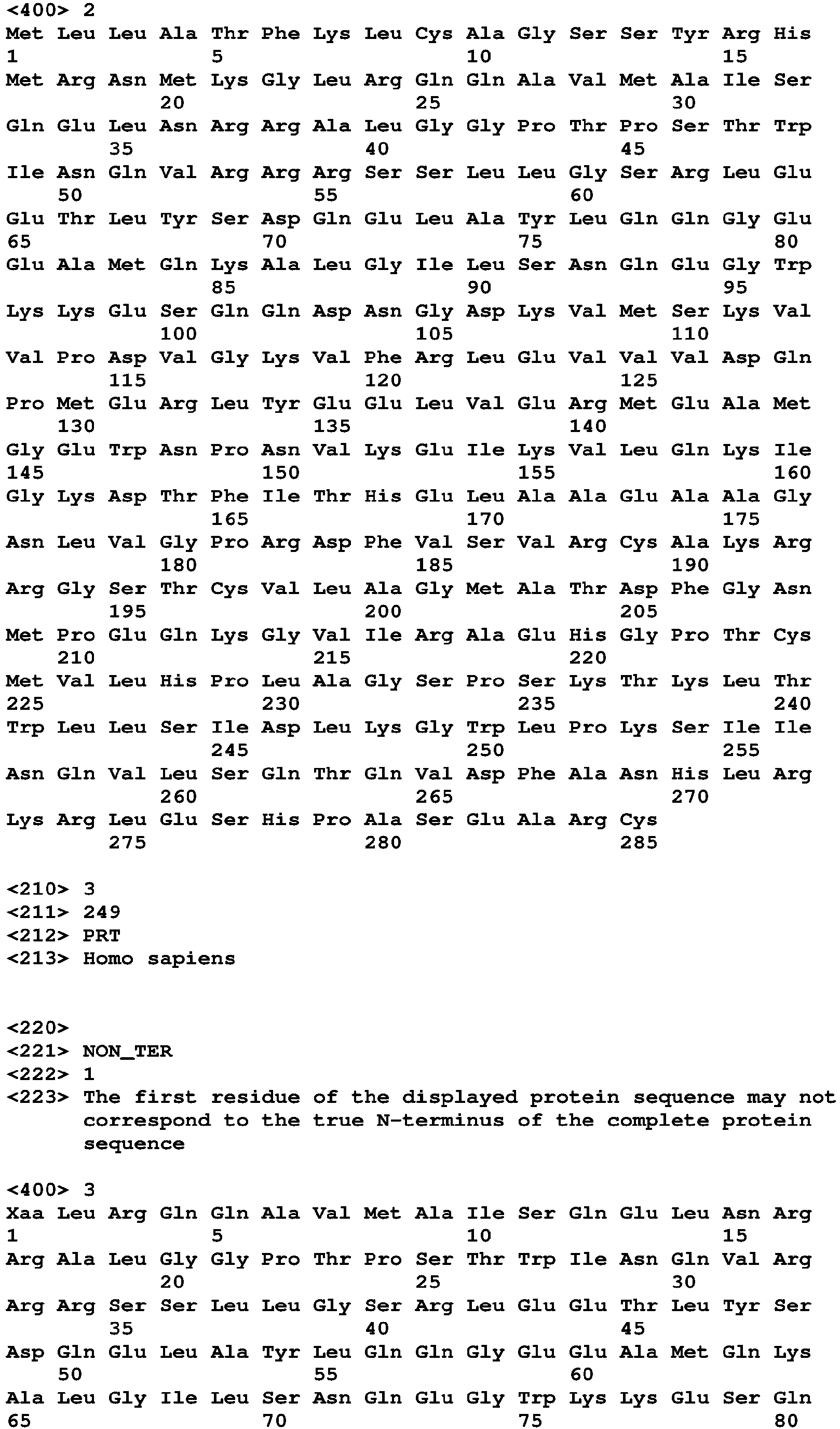

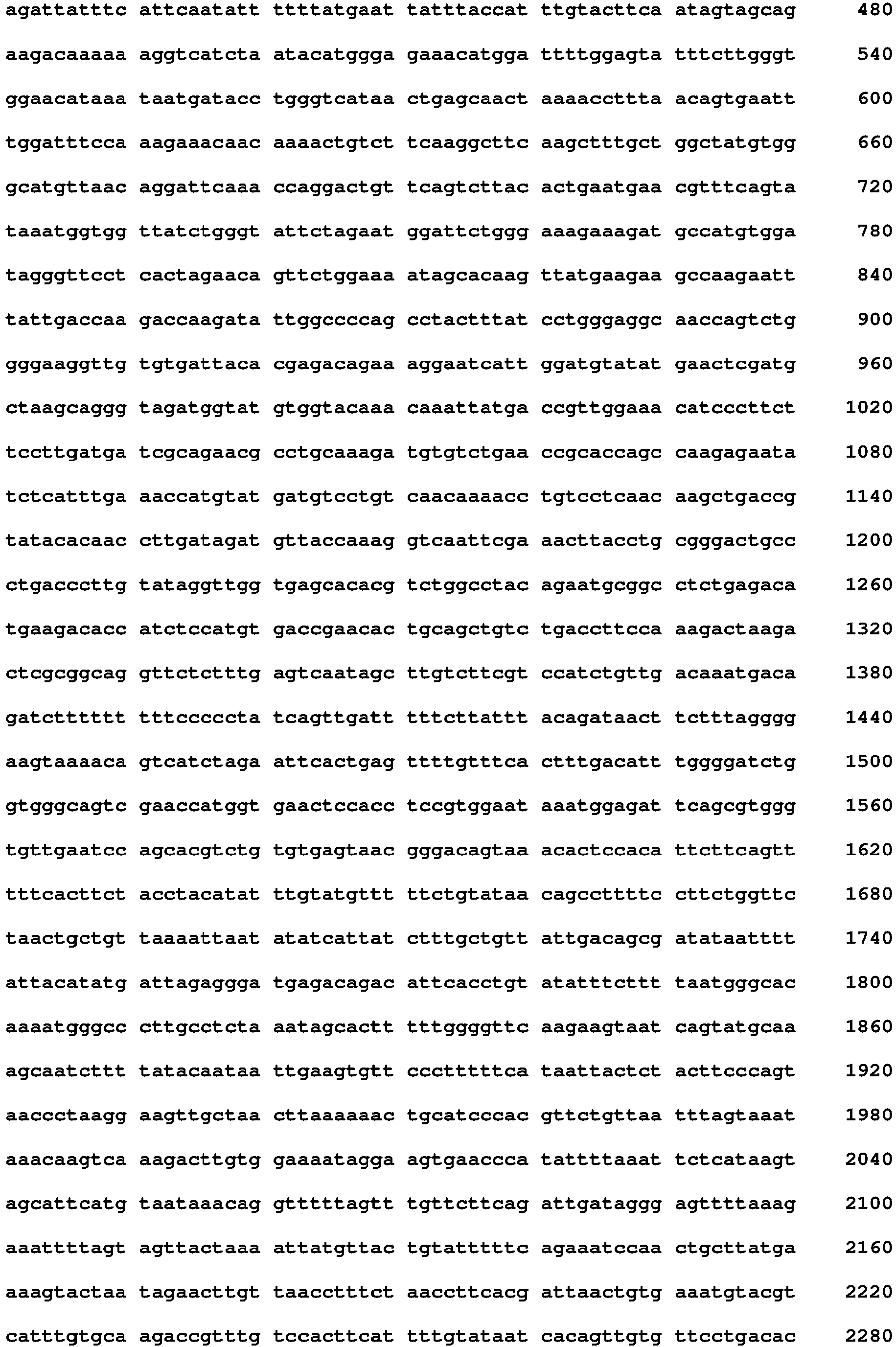


Claims (14)
- A steroidogenic acute regulatory protein 1 (STARD1) inhibitor for use in medicine.
- A STARD1 inhibitor for use in the treatment of lysosomal storage disorder in a subject.
- A STARD1 inhibitor for use according to claim 1 or 2, wherein the lysosomal storage disorder is Niemann-Pick type C (NPC) disease.
- A STARD1 inhibitor for use according to any one of claims 1 to 3, wherein the STARD1 inhibitor is selected from the group consisting of a chemical compound, a STARD1 specific siRNA, a STARD1 specific antisense oligonucleotide, a STARD1 specific ribozyme and a STARD1 specific antibody, and a protein regulating the STARD1 expression.
- A STARD1 inhibitor for use according to claim 4, wherein the protein regulating the STARD1 expression is an acid ceramidase (ACDase) enzyme or a functionally equivalent fragment thereof.
- A STARD1 inhibitor for use according to claim 5, wherein the ACDase enzyme comprises an amino acid sequence having an identity of at least 70% with the sequence SEQ ID NO: 1.
- A STARD1 inhibitor for use according to any one of claims 1 to 6, wherein the STARD1 inhibitor is comprised within a pharmaceutical composition.
- An in vitro method for diagnosing lysosomal storage disorder in a subject, or for designing a customized therapy for a subject suffering from lysosomal storage disorder, comprising:(i) quantifying the expression levels of the nucleotide sequence encoding the STARD1 in a biological sample isolates from the subject, and(ii) comparing the expression levels obtained in step (i) with a reference level,wherein an increased STARD1 expression levels with respect to the reference level is indicative that the subject suffers from lysosomal storage disorder, or that the therapy to be administered to the subject is based on a STARD1 inhibitor.
- An in vitro method according to claim 8, wherein the lysosomal storage disorder is selected from a list consisting of: GM1 gangliosidosis, GM2 gangliosidosis, Alpha-mannosidosis, Hurler syndrome, Hunter syndrome, Sanfilippo syndrome Type A, Morquio syndrome Type A, Morquio syndrome Type B, Niemann-Pick type A (NPA), Niemann-Pick type B (NPC B) or Niemann-Pick type C (NPC) disease.
- An in vitro method according to claim 8 or 9, wherein the quantification of the expression levels comprises measuring the level of cDNA, the level of mRNA, and/or the level of the protein encoded by said nucleotide sequence.
- An in vitro method according to any one of claim 8 to 10, wherein the subject is a mammal, preferably, the mammal is a human.
- An in vitro method according to any one of claims 8 to 11, wherein the STARD1 inhibitor is a STARD1 inhibitor is selected from the group consisting of a chemical compound, a STARD1 specific siRNA, a STARD1 specific antisense oligonucleotide, a STARD1 specific ribozyme and a STARD1 specific antibody, and a protein regulating the STARD1 expression.
- An in vitro method according to any one of claims 8 to 12, wherein the protein regulating the STARD1 expression is an acid ceramidase (ACDase) enzyme or a functionally equivalent fragment thereof.
- An in vitro method according to claim 13, wherein the ACDase enzyme comprises an amino acid sequence having an identity of at least 70% with the sequence SEQ ID NO: 1.
Priority Applications (1)
| Application Number | Priority Date | Filing Date | Title |
|---|---|---|---|
| EP20383102.9A EP4014997A1 (en) | 2020-12-16 | 2020-12-16 | Stard1 inhibitors for the treatment of lysosomal disorders |
Applications Claiming Priority (1)
| Application Number | Priority Date | Filing Date | Title |
|---|---|---|---|
| EP20383102.9A EP4014997A1 (en) | 2020-12-16 | 2020-12-16 | Stard1 inhibitors for the treatment of lysosomal disorders |
Publications (1)
| Publication Number | Publication Date |
|---|---|
| EP4014997A1 true EP4014997A1 (en) | 2022-06-22 |
Family
ID=74103902
Family Applications (1)
| Application Number | Title | Priority Date | Filing Date |
|---|---|---|---|
| EP20383102.9A Withdrawn EP4014997A1 (en) | 2020-12-16 | 2020-12-16 | Stard1 inhibitors for the treatment of lysosomal disorders |
Country Status (1)
| Country | Link |
|---|---|
| EP (1) | EP4014997A1 (en) |
Citations (6)
| Publication number | Priority date | Publication date | Assignee | Title |
|---|---|---|---|---|
| US20140220114A1 (en) | 2011-07-28 | 2014-08-07 | Trustees Of Dartmouth College | Methods for treating niemann-pick type c disease |
| EP2625198B1 (en) | 2010-10-07 | 2015-07-22 | AC Immune S.A. | Antibodies recognising phospho-tau |
| WO2015119989A1 (en) | 2014-02-04 | 2015-08-13 | New York University | Progranulin (pgrn) and its derivatives for diagnosis and treatment of lysosomal storage diseases |
| EP3078379A1 (en) | 2013-12-05 | 2016-10-12 | National University Corporation Kumamoto University | Drug for the treatment of cholesterol accumulation disorders, and screening method for same |
| EP3505185A1 (en) | 2016-10-08 | 2019-07-03 | Wuxi Hanqiang Pharmaceutical Technology Co., Ltd. | Application of gsk3 inhibitor in preparing a drug to treat niemann-pick disease type c |
| WO2020152532A1 (en) * | 2019-01-23 | 2020-07-30 | Enzyvant Therapeutics Gmbh | Method of ameliorating a pro-inflammatory immunophenotype in farber disease subjects by repeated administration of a recombinant human acid ceramidase |
-
2020
- 2020-12-16 EP EP20383102.9A patent/EP4014997A1/en not_active Withdrawn
Patent Citations (6)
| Publication number | Priority date | Publication date | Assignee | Title |
|---|---|---|---|---|
| EP2625198B1 (en) | 2010-10-07 | 2015-07-22 | AC Immune S.A. | Antibodies recognising phospho-tau |
| US20140220114A1 (en) | 2011-07-28 | 2014-08-07 | Trustees Of Dartmouth College | Methods for treating niemann-pick type c disease |
| EP3078379A1 (en) | 2013-12-05 | 2016-10-12 | National University Corporation Kumamoto University | Drug for the treatment of cholesterol accumulation disorders, and screening method for same |
| WO2015119989A1 (en) | 2014-02-04 | 2015-08-13 | New York University | Progranulin (pgrn) and its derivatives for diagnosis and treatment of lysosomal storage diseases |
| EP3505185A1 (en) | 2016-10-08 | 2019-07-03 | Wuxi Hanqiang Pharmaceutical Technology Co., Ltd. | Application of gsk3 inhibitor in preparing a drug to treat niemann-pick disease type c |
| WO2020152532A1 (en) * | 2019-01-23 | 2020-07-30 | Enzyvant Therapeutics Gmbh | Method of ameliorating a pro-inflammatory immunophenotype in farber disease subjects by repeated administration of a recombinant human acid ceramidase |
Non-Patent Citations (11)
| Title |
|---|
| ALTSCHUL S.F. ET AL.: "Basic local alignment search tool", J MOL BIOL., vol. 215, no. 3, 5 October 1990 (1990-10-05), pages 403 - 10, XP002949123, DOI: 10.1006/jmbi.1990.9999 |
| BEDIA ET AL., CHEMBIOCHEM, vol. 8, no. 6, 16 April 2007 (2007-04-16), pages 642 - 8 |
| FERNANDEZ, A ET AL., J. HEPATOL., vol. 59, no. 4, 2013, pages 805 - 813 |
| KOHLERMILSTEIN ET AL., NATURE, vol. 256, 1975, pages 495 - 497 |
| KWIATKOWSKA, K ET AL., ORPHANET J RARE DIS, vol. 9, no. 1, 2014, pages 64 |
| LIU ET AL., PROC NATL ACAD SCI USA, vol. 106, 2009, pages 2377 |
| MONTERO JOAN ET AL: "Mitochondrial Cholesterol Contributes to Chemotherapy Resistance in Hepatocellular Carcinoma", CANCER RESEARCH, vol. 68, no. 13, 1 July 2008 (2008-07-01), US, pages 5246 - 5256, XP055802391, ISSN: 0008-5472, DOI: 10.1158/0008-5472.CAN-07-6161 * |
| TORRES ET AL., REDOX BIOL, vol. 11, April 2017 (2017-04-01), pages 60 - 72 |
| TORRES S. ET AL: "The hepatic phenotype of niemann-pick type C disease exhibits stard1 upregulation independently of endoplasmic reticulum stress", 70TH ANNUAL MEETING OF THE AMERICAN ASSOCIATION FOR THE STUDY OF LIVER DISEASES, vol. 70 S1, 1 October 2019 (2019-10-01), pages 1100A - 1101A, XP055802409 * |
| TORRES SANDRA ET AL: "Mitochondrial Cholesterol in Alzheimer's Disease and Niemann-Pick Type C Disease", FRONTIERS IN NEUROLOGY, vol. 10, 7 November 2019 (2019-11-07), pages 1168, XP055802434, Retrieved from the Internet <URL:https://www.ncbi.nlm.nih.gov/pmc/articles/PMC6854033/pdf/fneur-10-01168.pdf> DOI: 10.3389/fneur.2019.01168 * |
| YU W ET AL., J. BIOL. CHEM., vol. 280, no. 12, 2005, pages 11731 - 11739 |
Similar Documents
| Publication | Publication Date | Title |
|---|---|---|
| Xu et al. | Progerin accumulation in nucleus pulposus cells impairs mitochondrial function and induces intervertebral disc degeneration and therapeutic effects of sulforaphane | |
| Lee et al. | Single‐dose intracerebroventricular administration of galactocerebrosidase improves survival in a mouse model of globoid cell leukodystrophy | |
| US9393221B2 (en) | Methods and compounds for reducing intracellular lipid storage | |
| EP2646044B1 (en) | Methods for increasing intracellular activity of hsp70 | |
| JP5860493B2 (en) | Use of Hsp70 as a regulator of enzyme activity | |
| EP3804755A1 (en) | Complement inhibition for improved nerve regeneration | |
| AU2013273473B2 (en) | Allosteric chaperones and uses thereof | |
| KR20140107481A (en) | Combination of a compound having the ability to rearrange a lysosomal enzyme and ambroxol and/or a derivative of ambroxol | |
| US9206425B2 (en) | Methods for treating Niemann-Pick type C disease | |
| US11548936B2 (en) | Compositions and methods for treating lysosomal storage diseases and disorders | |
| de Zélicourt et al. | CD38‐NADase is a new major contributor to Duchenne muscular dystrophic phenotype | |
| RS67913B1 (en) | Methods of treating fabry disease | |
| Riillo et al. | c. 376A> G,(p. Ser126Gly) Alpha-Galactosidase A mutation induces ER stress, unfolded protein response and reduced enzyme trafficking to lysosome: Possible relevance in the pathogenesis of late-onset forms of Fabry Disease | |
| EP3989962B1 (en) | Combination therapy with acetyl-leucine and miglustat for treating a lysosomal storage disease | |
| EP3979989A1 (en) | Production and use of extracellular vesicle-contained enampt | |
| WO2006100797A1 (en) | Method and pharmaceutical composition for treating psoriasis, squamous cell carcinoma and/or parakeratosis by inhibiting expression of squamous cell carcinoma-related antigen | |
| Pastores et al. | GM2-gangliosidoses | |
| Rintz et al. | Synergistic effects of resveratrol and enzyme replacement therapy in the Mucopolysaccharidosis type I | |
| US20230277485A1 (en) | Methods for the treatment of lysosomal storage diseases | |
| Mohamed | Elucidating the Impact of Amino Acid Substitutions in Lysosomal Storage Disorders: Insights on Infantile Gm1-Gangliosidosis and Schindler Disease | |
| HK40127928A (en) | Combination therapy with acetyl-leucine and miglustat for treating a lysosomal storage disease | |
| Padilla | Autophagy and Muscle Dysfunction in Lysosomal Storage Diseases | |
| Kamel et al. | STUDIES ON ADIPOSE–DERIVED STEM CELLS AS TREATMENT OF MUSCULAR DYSTROPHY INDUCED IN RATS | |
| CN116832025A (en) | Application of verapamil in preparation of product for preventing, reducing or delaying intervertebral disc degeneration | |
| HK40050049A (en) | Complement inhibition for improved nerve regeneration |
Legal Events
| Date | Code | Title | Description |
|---|---|---|---|
| PUAI | Public reference made under article 153(3) epc to a published international application that has entered the european phase |
Free format text: ORIGINAL CODE: 0009012 |
|
| STAA | Information on the status of an ep patent application or granted ep patent |
Free format text: STATUS: THE APPLICATION HAS BEEN PUBLISHED |
|
| AK | Designated contracting states |
Kind code of ref document: A1 Designated state(s): AL AT BE BG CH CY CZ DE DK EE ES FI FR GB GR HR HU IE IS IT LI LT LU LV MC MK MT NL NO PL PT RO RS SE SI SK SM TR |
|
| STAA | Information on the status of an ep patent application or granted ep patent |
Free format text: STATUS: THE APPLICATION IS DEEMED TO BE WITHDRAWN |
|
| 18D | Application deemed to be withdrawn |
Effective date: 20221223 |