EP4002262A1 - Computer-implemented method for handling artifacts in multi-energy computed tomography, x-ray imaging device, computer program and electronically readable storage medium - Google Patents
Computer-implemented method for handling artifacts in multi-energy computed tomography, x-ray imaging device, computer program and electronically readable storage medium Download PDFInfo
- Publication number
- EP4002262A1 EP4002262A1 EP20209020.5A EP20209020A EP4002262A1 EP 4002262 A1 EP4002262 A1 EP 4002262A1 EP 20209020 A EP20209020 A EP 20209020A EP 4002262 A1 EP4002262 A1 EP 4002262A1
- Authority
- EP
- European Patent Office
- Prior art keywords
- energy
- artifacts
- subregion
- dataset
- deviation values
- Prior art date
- Legal status (The legal status is an assumption and is not a legal conclusion. Google has not performed a legal analysis and makes no representation as to the accuracy of the status listed.)
- Granted
Links
Images

Classifications
-
- G—PHYSICS
- G06—COMPUTING OR CALCULATING; COUNTING
- G06T—IMAGE DATA PROCESSING OR GENERATION, IN GENERAL
- G06T12/00—Tomographic reconstruction from projections
- G06T12/30—Image post-processing, e.g. metal artefact correction
-
- A—HUMAN NECESSITIES
- A61—MEDICAL OR VETERINARY SCIENCE; HYGIENE
- A61B—DIAGNOSIS; SURGERY; IDENTIFICATION
- A61B6/00—Apparatus or devices for radiation diagnosis; Apparatus or devices for radiation diagnosis combined with radiation therapy equipment
- A61B6/02—Arrangements for diagnosis sequentially in different planes; Stereoscopic radiation diagnosis
- A61B6/03—Computed tomography [CT]
- A61B6/032—Transmission computed tomography [CT]
-
- A—HUMAN NECESSITIES
- A61—MEDICAL OR VETERINARY SCIENCE; HYGIENE
- A61B—DIAGNOSIS; SURGERY; IDENTIFICATION
- A61B6/00—Apparatus or devices for radiation diagnosis; Apparatus or devices for radiation diagnosis combined with radiation therapy equipment
- A61B6/52—Devices using data or image processing specially adapted for radiation diagnosis
- A61B6/5258—Devices using data or image processing specially adapted for radiation diagnosis involving detection or reduction of artifacts or noise
-
- G—PHYSICS
- G06—COMPUTING OR CALCULATING; COUNTING
- G06T—IMAGE DATA PROCESSING OR GENERATION, IN GENERAL
- G06T12/00—Tomographic reconstruction from projections
- G06T12/10—Image preprocessing, e.g. calibration, positioning of sources or scatter correction
-
- G—PHYSICS
- G06—COMPUTING OR CALCULATING; COUNTING
- G06T—IMAGE DATA PROCESSING OR GENERATION, IN GENERAL
- G06T7/00—Image analysis
- G06T7/0002—Inspection of images, e.g. flaw detection
- G06T7/0012—Biomedical image inspection
-
- G—PHYSICS
- G06—COMPUTING OR CALCULATING; COUNTING
- G06V—IMAGE OR VIDEO RECOGNITION OR UNDERSTANDING
- G06V10/00—Arrangements for image or video recognition or understanding
- G06V10/20—Image preprocessing
- G06V10/22—Image preprocessing by selection of a specific region containing or referencing a pattern; Locating or processing of specific regions to guide the detection or recognition
-
- G—PHYSICS
- G06—COMPUTING OR CALCULATING; COUNTING
- G06T—IMAGE DATA PROCESSING OR GENERATION, IN GENERAL
- G06T2207/00—Indexing scheme for image analysis or image enhancement
- G06T2207/10—Image acquisition modality
- G06T2207/10072—Tomographic images
- G06T2207/10081—Computed x-ray tomography [CT]
-
- G—PHYSICS
- G06—COMPUTING OR CALCULATING; COUNTING
- G06T—IMAGE DATA PROCESSING OR GENERATION, IN GENERAL
- G06T2207/00—Indexing scheme for image analysis or image enhancement
- G06T2207/10—Image acquisition modality
- G06T2207/10116—X-ray image
-
- G—PHYSICS
- G06—COMPUTING OR CALCULATING; COUNTING
- G06T—IMAGE DATA PROCESSING OR GENERATION, IN GENERAL
- G06T2207/00—Indexing scheme for image analysis or image enhancement
- G06T2207/30—Subject of image; Context of image processing
- G06T2207/30004—Biomedical image processing
- G06T2207/30061—Lung
-
- G—PHYSICS
- G06—COMPUTING OR CALCULATING; COUNTING
- G06T—IMAGE DATA PROCESSING OR GENERATION, IN GENERAL
- G06T2211/00—Image generation
- G06T2211/40—Computed tomography
- G06T2211/408—Dual energy
-
- G—PHYSICS
- G06—COMPUTING OR CALCULATING; COUNTING
- G06V—IMAGE OR VIDEO RECOGNITION OR UNDERSTANDING
- G06V2201/00—Indexing scheme relating to image or video recognition or understanding
- G06V2201/03—Recognition of patterns in medical or anatomical images
Definitions
- the invention concerns a computer-implemented method for automatically estimating and/or correcting an error due to artifacts in a multi-energy computed tomography result dataset relating to at least one target material, wherein at least two three-dimensional energy datasets of an imaging region of a patient are acquired using an x-ray imaging device, in particular a computed tomography device, using different x-ray energy spectra, and the result dataset is determined by evaluating the energy datasets using material-specific energy dependencies of x-ray attenuation.
- the invention further concerns an x-ray imaging device, a computer program, and an electronically readable storage medium.
- Multi-energy computed tomography is a technique for energy-resolved x-ray imaging, in particular in medicine.
- This imaging technique exploits different energy dependencies of x-ray attenuation, for example to provide quantitative image data and/or reduce image artifacts by so-called material decomposition.
- Dual-energy CT that is, imaging at two energy levels, is one of the most widely used variants of this technique.
- the advent of photon-counting x-ray detectors the potential for, in particular simultaneous, measurement at a larger number of energy spectra has been provided.
- multi-energy imaging data which may also be termed “spectral data”
- spectral data allows for the calculation of material-specific images, often called material maps, by, for example, applying a material decomposition algorithm to acquired energy datasets, each associated with a different energy spectrum and thus a different energy level.
- material maps often called material maps
- the ability to extract iodine images/iodine maps from multi-energy data, and/or to quantify the iodine distribution in tissue as a result dataset opens up a huge potential.
- energy datasets are acquired at different x-ray energy spectra, that is, different energy levels.
- the energy datasets are then evaluated to determine a result dataset, for example a material map and/or other, in particular quantitative, values.
- a result dataset for example a material map and/or other, in particular quantitative, values.
- known material-specific energy dependences of x-ray attenuation for this material are used.
- the x-ray attenuation of two materials for example iodine and calcium
- the change in measured attenuation values between 80 and 140 kV (and/or other energy spectra) will enable differentiating such two materials, for example iodine and calcium.
- the primary spectral computed tomography image data that is, the energy datasets
- the energy datasets can contain image artifacts caused by, for example, the acquisition technique, patient size, and/or patient motion.
- the errors induced by such artifacts are amplified and propagated, such that incorrect image values, in particular HU or density values, may be determined for the result dataset.
- image values of the result data set are correct or impacted by artifacts. If a user has doubts regarding the evaluation results, they will ask for an additional scan and/or a different imaging modality.
- a computer-implemented method for automatically estimating and/or correcting an error due to image artifacts in a multi-energy computed tomography result dataset relating to at least one target material wherein at least two three-dimensional energy datasets of an imaging region of a patient are acquired using an x-ray imaging device, in particular a computed tomography device, using different x-ray energy spectra and the result dataset is determined by evaluating the energy datasets using material-specific energy dependences of x-ray attenuation, according to the invention, the following steps are performed for estimation of the error due to artifacts:
- the invention proposes the detection and quantification of image artifacts, in particular image artifacts of low spatial frequency, which are relevant for multi-energy evaluation, by analyzing first subregions of the imaging region that do not contain the target material and/or other materials with a strong energy dependency.
- Suitable second materials in the first subregions may, for example, comprise air surrounding a patient and inside hollow organs of the patient, but also other anatomical subregions, for example containing subcutaneous fat and/or muscle as second materials, can be used.
- the energy values that is, image values of the energy dataset
- HU values of air should be identical for all energy datasets at different energy spectra/energy levels. Differences in energy values indicate the presence of image artifacts, due to basic artifacts differently influencing the energy datasets. The higher the difference, the stronger the multi-energy evaluation relevant artifact might be.
- the error of result datasets due to such variations of basic artifacts can be estimated and provided as an additional information alongside the result values, that is, image values of the result dataset, to increase the confidence of the result values.
- derived information about the strength of the artifacts can be used to apply a respective correction, for example by using a correction algorithm, to the energy datasets, such that also the result datasets are corrected and errors due to image artifacts are at least reduced.
- At least one of the energy values is corrected according to the estimated deviation values before determining the result values of the result dataset and/or, additional to the result dataset, an error dataset showing the spatially resolved deviation values and/or the error propagated through the evaluation process is determined, and/or the result dataset is modified, in particular colored, according to the deviation values and/or error values.
- the deviation values may be provided as additional information by adding a respective colorization to the result dataset, for example indicating regions with higher artifact strength in red and regions having low artifact strength in green, wherein different colors and shades may be used therebetween to provide a high resolution of the additional information.
- error information may also be provided as an additional dataset, in particular an error dataset showing the spatially resolved deviation values and/or the propagated error values. It is, of course, also possible to propagate the deviation values through the evaluation process, such that error propagation effects are also covered by the resulting error values, which may also be provided as an error dataset. Preferably, however, the deviation values, in particular in the subregions containing the target material, are used for correction of the energy datasets and thus the result dataset.
- the at least one first subregion is determined by segmenting the energy datasets regarding at least the at least one second material, wherein, in particular, threshold segmenting and/or an anatomical atlas is used.
- threshold segmenting and/or an anatomical atlas are used.
- At least one subregion containing a second material having an at least essentially energy-spectra invariant behaviour is used as the at least one first subregion.
- the expected change in energy value between the used energy spectra for the second material may be lower than a threshold value.
- This threshold value may, for example, be chosen as one fifth of a predetermined expected artifact-induced change.
- artifacts have been observed that have a strength of, for example, 50 to 80 HU, such that, for example, 65 HU could be chosen as a basis of assessment.
- the HU value only changes by about 10, which would be well below 20 % of 65.
- second materials may be chosen which either do not show measurable change in HU value or whose change in HU value is negligible compared to the expected strength of image artifacts.
- At least one reference energy value may be calculated using the known dependence from the measured energy values in one of the energy datasets.
- the other measured energy values from other energy datasets may be compared to the respective energy value to determine the deviation value.
- the expected HU value is -1000 HU for each energy spectrum.
- air may be used as the second material, while, additionally, fat tissue and/or muscle tissue may be used.
- the at least one subregion may comprise a region outside the patient and/or at least one hollow organ comprising air, in particular the trachea and/or the intestines and/or at least one lung.
- first subregions inside the patient may be chosen additionally, providing further sampling points for deviation values and, consequently, the quality of extrapolation and/or interpolation is improved.
- newest-neighbor interpolation and/or inverse distance weighting and/or spline interpolation may be employed. Since the artifacts mainly discussed here occur with a low spatial frequency, the strength also varies slowly, such that already simple interpolation and/or extrapolation techniques provide good results regarding the deviation values.
- the energy datasets may be acquired by spiral computed tomography, wherein, during acquisition, the acquisition arrangement comprising the x-ray source and the x-ray detector also moves in a z-direction (along the rotational axis/along the longitudinal direction of the patient) relatively to the patient/imaging region.
- the acquisition arrangement comprising the x-ray source and the x-ray detector also moves in a z-direction (along the rotational axis/along the longitudinal direction of the patient) relatively to the patient/imaging region.
- artifacts that may be detected and estimated and/or corrected using the method according to the invention may, for example, include scattering artifacts and/or undersampling artifacts.
- the current invention provides a very simple to realize estimation process yielding good results. From easily discernible first subregions, for example subregions containing air, where deviation values may be easily determined by simple comparison of energy values, that is, image values of the energy datasets, simple extrapolation and/or interposition approaches allow the determination of deviation values for at least one second subregion, in particular containing the target material. These deviation values directly correlate with the strength of artifacts at the corresponding position. However, in some embodiments, the inventive approach may also form the basis for more sophisticated processes.
- the interpolation and/or extrapolation may be performed taking into account at least one expected property of at least one of the at least one artifact, in particular an expected spatial frequency or an expected spatial frequency range of the artifacts.
- at least one expected property of at least one of the at least one artifact in particular an expected spatial frequency or an expected spatial frequency range of the artifacts.
- certain acquisition parameters of the x-ray imaging device directly correlate with the expected spatial frequencies of the image artifacts. For example, in spiral CT, when a pitch of about 3 is used, minima and maxima of the spiral artifacts usually follow each other every 6 cm.
- interpolation and/or extrapolation may include using an artificial intelligence algorithm.
- artificial intelligence algorithms which may also be called trained functions, may be trained using ground truth information about artifacts in training datasets, for example by employing deep learning approaches.
- the artificial intelligence algorithm may, for example, comprise a neural network.
- ground truth information about artifacts is hard to determine, in particular for image artifacts of low spatial frequency.
- a patient would have to be imaged at least twice, increasing the amount of radiation the patient is subjected to, which would be undesirable.
- using artificial intelligence would require more computation power, while the simpler approaches explained above already yield great results.
- deviation values may be determined for each pair of energy datasets, wherein, in an embodiment, one reference energy dataset may be determined and deviation values of all other energy datasets with regard to the reference energy dataset may be determined and used for respective relative correction.
- deviation values may be calculated for each pair and propagated to calculate error values regarding the result dataset by error propagation. Further, statistical methods may be employed to calculate an overall error value and/or determine an overall correction.
- the inventive method may, for example, be employed regarding iodine measurements. That is, iodine may be used as the target material, wherein, for example, an iodine material map may be determined as at least one of the at least one result dataset.
- the iodine material map may be quantitative or qualitative.
- the invention further concerns an x-ray imaging device, in particular a computed tomography device, comprising at least one acquisition arrangement, which comprises an x-ray source and an x-ray detector, and a control device configured to perform a method according to the invention.
- a computed tomography device comprising at least one acquisition arrangement, which comprises an x-ray source and an x-ray detector, and a control device configured to perform a method according to the invention.
- a computer program according to the invention can be, for example, loaded directly into a storage means of a control device of an x-ray imaging device and comprises program means to perform the steps of a method according to the invention when the computer program is executed in the control device of the x-ray imaging device.
- the computer program according to the invention may be stored on an electronically readable storage medium according to the invention, which thus comprises electronically readable control information stored thereon, wherein the control information comprises at least one computer program according to the invention and is configured such that a method according to the invention is performed if the electronically readable storage medium is used in a control device of an x-ray imaging device.
- the current invention allows to access the level of image artifacts in spectral images by comparing in particular energy-spectra invariant first subregions, for example of air or fat, in the energy datasets.
- the user receives information about the expected error due to these artifacts in addition to the result values. This allows for an improved diagnosis, since the accuracy of the result values is known to the user, in particular a physician.
- Quantitative information about the image artifacts levels in the different subregions allows for a respective correction of these image artifacts in the energy datasets and/or directly in the result dataset.
- the invention by calculating deviation values, focuses on information relevant to the multi energy CT evaluation process, i.e. "difference artifacts" that are relevant for evaluating the change from one energy spectrum to the other.
- Such artifacts are the result by having different strengths of basic artifacts in at least two of the at least two energy datasets.
- Artifact strengths described by deviation values are thus understood to relate to differences between energy datasets, which is exactly the information used in multi energy CT evaluation, for example material decomposition.
- Fig. 1 is a flowchart of an embodiment of a method according to the invention.
- dual-energy computed tomography of a patient is to be performed to determine a quantitative distribution of iodine, for example for tumor or lesion characterization.
- iodine is the target material of the examination.
- a step S1 two energy datasets are acquired at different x-ray energy spectra.
- one of the spectra may be centered around 80 kV (low energy), the other at 140 kV (high energy).
- a computed tomography device is used such that a spiral trajectory of the x-ray source is achieved.
- spiral CT is employed, wherein a patient table carrying the patient is moved through the gantry during rotation of the acquisition assembly.
- Spiral CT can lead to certain image artifacts, for example spiral artifacts. These artifacts may differ between energy datasets, leading to a distortion of the effects induced by different x-ray attenuation for different energies.
- the method described here allows to estimate the strength of such spiral artifacts relevant to dual energy CT evaluation and other artifacts of a low spatial frequency, for example scattering artifacts, that result in different effects in different energy datasets.
- step S1 projection images of an imaging region of the patient are acquired and a three-dimensional energy dataset is reconstructed for each energy spectrum.
- a step S2 at least one first subregion of the imaging region is determined, which is free from the target material, in this case iodine, and contains exactly one second material, in this case air, which has a known material-specific energy dependence of x-ray attenuation, in this case being invariant, meaning the HU value of air should be the same for each energy dataset.
- the first subregions may be determined using a segmentation algorithm, for example threshold-based segmentation, in particular in combination with information from an anatomic atlas. Of course, other approaches as known in the art may also be employed.
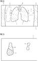
- An exemplary two-dimensional energy image of one of the energy datasets is schematically shown in fig. 2 .
- the energy image 1 in this case, shows parts of three first subregions 2 containing air, namely areas outside the patient (the left and right first subregions 2) and an area comprising the trachea 3 and the lungs 4.
- the energy values that is the image value of the energy datasets, for each dataset are compared to determine deviation values.
- These deviation values indicate the presence of artifacts relevant to the evaluation, since the energy value for air should be the same in each of the energy datasets without artifacts.
- the deviation values may be determined by computing the difference between the energy value of a voxel of the low energy dataset and the energy value of the voxel in the high energy dataset.
- step S3 This comparison and determination of the deviation values takes place in step S3 (see fig. 1 ).
- fig. 2 deviation values over the whole range of the energy datasets are now known, such that a lot of sampling points exist, even inside the patient.
- This allows, in a step S4, to interpolate and/or extrapolate from the deviation value determined in step S3 to calculate estimated deviation values for second subregions 5 in a step S4.
- the second subregions 5, as shown in fig. 2 may be regions of interest (ROI), wherein iodine is expected and the iodine concentration is to be determined. However, it is also possible to cover all remaining parts of the imaging region by at least one second subregion 5.
- ROI regions of interest
- the determined estimated deviation values may be used to correct the energy datasets, however, it is also possible to use them as an error value indicating the strength of image artifacts or using them as a starting point of error propagation, as will be further discussed below.
- a step S5 the energy datasets, possibly corrected, are used, as known in the state of the art, to derive a result dataset, in this embodiment a material map regarding iodine as target material, and/or a quantitative distribution of iodine. Material decomposition approaches may be employed.
- an error propagation process may be performed to propagate the error described by the estimated deviation values in the second subregion 5 through the evaluation process to determine the at least one result dataset.
- the voxel-wise error value derived from the error propagation process in particular provided as an error dataset, or, in other variants, the estimated deviation values as error values, may be used to modify the result dataset for display.
- a result image 7 of the result dataset may use color-coding, as indicated for areas 8, 9 in fig. 3 , inside the second subregions 5 to indicate the reliability of the determined iodine distribution. For example, green color may indicate high reliability, while red may indicate low reliability, that is, large error value.
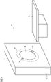
- Fig. 4 shows a principle drawing of a computed tomography device 10 according to the current invention.
- the computed tomography device 10 comprises a gantry 11 in which an acquisition arrangement 12 comprising an x-ray source 13 and an x-ray detector 14 is rotatably mounted.
- a patient table 15 may be used to position a patient inside an opening 16 of the gantry 11 such that an imaging region may be imaged.
- spiral CT by in particular continuously moving the table 15 during acquisition of projection images, larger imaging regions may be imaged.
- the computed tomography device 10 further comprises a control device 17 for controlling the computed tomography device 10, which is also configured to perform the steps of a method according to the invention.
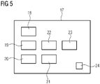
- Fig. 5 shows the functional structure of the control device 17.
- the control device 17 comprises an acquisition device 18 for acquiring image datasets, in particular also energy datasets for multi-energy CT (spectral CT). That is, the acquisition unit 18 is configured to perform S1.
- acquisition device 18 for acquiring image datasets, in particular also energy datasets for multi-energy CT (spectral CT). That is, the acquisition unit 18 is configured to perform S1.
- the first subregion 2 may be determined.
- a comparison unit 20 compares the energy values of the energy datasets according to step S3, to determine deviation values. These deviation values may be used by a calculating unit 21 to determine estimated deviation values by interpolating and/or extrapolating from the determined deviation values in at least one second subregion 5.
- the calculation unit 21 may, in some embodiments, also be adapted to correct energy datasets according to the estimated deviation values.
- the result dataset may be determined as discussed with regard to step S5.
- the evaluation unit 22 is further configured to perform the error propagation process to determine an error dataset with propagated error values.
- This error dataset or an error dataset comprising the estimated deviation value may be used in a display unit 23 to modify the result dataset for display or to display the error estimation information in the error dataset additionally to the result dataset.
- the functional units described above may be realized by software and/or hardware in a processor of the control device 17, which also comprises the storage means 24.
- the functional units may be implemented in the form of a computer program according to the invention.
Landscapes
- Engineering & Computer Science (AREA)
- Health & Medical Sciences (AREA)
- Physics & Mathematics (AREA)
- Theoretical Computer Science (AREA)
- Life Sciences & Earth Sciences (AREA)
- Medical Informatics (AREA)
- General Physics & Mathematics (AREA)
- General Health & Medical Sciences (AREA)
- Nuclear Medicine, Radiotherapy & Molecular Imaging (AREA)
- Radiology & Medical Imaging (AREA)
- Surgery (AREA)
- Biomedical Technology (AREA)
- Veterinary Medicine (AREA)
- Biophysics (AREA)
- High Energy & Nuclear Physics (AREA)
- Optics & Photonics (AREA)
- Pathology (AREA)
- Computer Vision & Pattern Recognition (AREA)
- Heart & Thoracic Surgery (AREA)
- Molecular Biology (AREA)
- Public Health (AREA)
- Animal Behavior & Ethology (AREA)
- Quality & Reliability (AREA)
- Pulmonology (AREA)
- Multimedia (AREA)
- Apparatus For Radiation Diagnosis (AREA)
Abstract
Description
- The invention concerns a computer-implemented method for automatically estimating and/or correcting an error due to artifacts in a multi-energy computed tomography result dataset relating to at least one target material, wherein at least two three-dimensional energy datasets of an imaging region of a patient are acquired using an x-ray imaging device, in particular a computed tomography device, using different x-ray energy spectra, and the result dataset is determined by evaluating the energy datasets using material-specific energy dependencies of x-ray attenuation. The invention further concerns an x-ray imaging device, a computer program, and an electronically readable storage medium.
- Multi-energy computed tomography (multi-energy CT), sometimes also termed "spectral imaging" or "spectral CT", is a technique for energy-resolved x-ray imaging, in particular in medicine. This imaging technique exploits different energy dependencies of x-ray attenuation, for example to provide quantitative image data and/or reduce image artifacts by so-called material decomposition. Dual-energy CT, that is, imaging at two energy levels, is one of the most widely used variants of this technique. However, since the advent of photon-counting x-ray detectors, the potential for, in particular simultaneous, measurement at a larger number of energy spectra has been provided.
- In particular, multi-energy imaging data, which may also be termed "spectral data", allows for the calculation of material-specific images, often called material maps, by, for example, applying a material decomposition algorithm to acquired energy datasets, each associated with a different energy spectrum and thus a different energy level. In the case of tumor/lesion characterization, tumor response assessment and/or other concrete tasks in medicine, the ability to extract iodine images/iodine maps from multi-energy data, and/or to quantify the iodine distribution in tissue as a result dataset, opens up a huge potential.
- In known approaches, energy datasets are acquired at different x-ray energy spectra, that is, different energy levels. The energy datasets are then evaluated to determine a result dataset, for example a material map and/or other, in particular quantitative, values. In this evaluation, known material-specific energy dependences of x-ray attenuation for this material, are used. For example, the x-ray attenuation of two materials, for example iodine and calcium, may not only be different in an energy spectrum centered at 140 kV and an energy spectrum centered at 80 kV, but each material may also have its own characteristic changing pattern from 80 kV to 140 kV. Thus, the change in measured attenuation values between 80 and 140 kV (and/or other energy spectra) will enable differentiating such two materials, for example iodine and calcium.
- However, for a reliable diagnosis, a reading physician has to be able to trust the quantitative information provided in the result dataset. On the other hand, similar to single-energy computed tomography images, the primary spectral computed tomography image data, that is, the energy datasets, can contain image artifacts caused by, for example, the acquisition technique, patient size, and/or patient motion. Within the material decomposition process or other evaluation processes, the errors induced by such artifacts are amplified and propagated, such that incorrect image values, in particular HU or density values, may be determined for the result dataset. Today, for the reading physician and/or, in general, user, it is not possible to recognize if image values of the result data set are correct or impacted by artifacts. If a user has doubts regarding the evaluation results, they will ask for an additional scan and/or a different imaging modality.
- It is an object of the present invention to provide information regarding image artifacts in result datasets and/or to provide result datasets with improved quality regarding artifacts.
- This object is achieved by providing a computer-implemented method, an x-ray imaging device, a computer program and an electronically readable storage medium according to the independent claims. Advantageous embodiments are described in the dependent claims.
- In a computer-implemented method for automatically estimating and/or correcting an error due to image artifacts in a multi-energy computed tomography result dataset relating to at least one target material, wherein at least two three-dimensional energy datasets of an imaging region of a patient are acquired using an x-ray imaging device, in particular a computed tomography device, using different x-ray energy spectra and the result dataset is determined by evaluating the energy datasets using material-specific energy dependences of x-ray attenuation, according to the invention, the following steps are performed for estimation of the error due to artifacts:
- determining at least one first subregion of the imaging region, which is free from the target material and contains at least one, in particular exactly one, second material with known material-specific energy dependence of x-ray attenuation,
- for each first subregion, comparing the image values of the energy dataset for each voxel, taking into account the known energy dependence, to determine deviation values indicative of artifacts,
- for at least a part of the at least one remaining second subregion of the imaging region, calculating estimated deviation values by interpolating and/or extrapolating from the determined deviation values in the at least one first subregion, wherein the estimated deviation values are used as estimated error due to artifacts.
- It has been discovered in the course of the current invention, that most artifacts occurring in multi-energy computed tomography have a low spatial frequency. If such basic artifacts of the energy datasets differ, derived artifacts ("difference artifacts" relating to at least two energy datasets) when evaluating the differences between energy values of different energy datasets result, which also have a low spatial frequency and are highly relevant when evaluating x-ray attenuation changes between different energy spectra. It is these derived or difference artifacts that the invention is focused on, to derive their artifact strength.
- Due to the low spatial frequency, if an estimation of artifact strength for a large enough portion of the imaging region can be determined, it is possible to extrapolate and/or interpolate these artifact strengths, described by deviation values in the current invention, such that conclusions regarding the artifact strength in other, second subregions of the imaging region can be drawn. In particular in medical imaging, areas without the target material, for example iodine or bone, are usually contained in the imaging region and may be determined as first subregions. In particular, the invention proposes the detection and quantification of image artifacts, in particular image artifacts of low spatial frequency, which are relevant for multi-energy evaluation, by analyzing first subregions of the imaging region that do not contain the target material and/or other materials with a strong energy dependency. Suitable second materials in the first subregions may, for example, comprise air surrounding a patient and inside hollow organs of the patient, but also other anatomical subregions, for example containing subcutaneous fat and/or muscle as second materials, can be used. In a concrete example, without any artifacts present, the energy values (that is, image values of the energy dataset), in particular HU values, of air should be identical for all energy datasets at different energy spectra/energy levels. Differences in energy values indicate the presence of image artifacts, due to basic artifacts differently influencing the energy datasets. The higher the difference, the stronger the multi-energy evaluation relevant artifact might be. By assessing any deviation found and the error propagation within the evaluation process, in particular the material decomposition process, the error of result datasets due to such variations of basic artifacts can be estimated and provided as an additional information alongside the result values, that is, image values of the result dataset, to increase the confidence of the result values. Additionally and/or alternatively, derived information about the strength of the artifacts can be used to apply a respective correction, for example by using a correction algorithm, to the energy datasets, such that also the result datasets are corrected and errors due to image artifacts are at least reduced.
- That is, in preferred embodiments of the current invention, at least one of the energy values, that is, the image values of the energy datasets, is corrected according to the estimated deviation values before determining the result values of the result dataset and/or, additional to the result dataset, an error dataset showing the spatially resolved deviation values and/or the error propagated through the evaluation process is determined, and/or the result dataset is modified, in particular colored, according to the deviation values and/or error values. For example, the deviation values may be provided as additional information by adding a respective colorization to the result dataset, for example indicating regions with higher artifact strength in red and regions having low artifact strength in green, wherein different colors and shades may be used therebetween to provide a high resolution of the additional information. However, error information may also be provided as an additional dataset, in particular an error dataset showing the spatially resolved deviation values and/or the propagated error values. It is, of course, also possible to propagate the deviation values through the evaluation process, such that error propagation effects are also covered by the resulting error values, which may also be provided as an error dataset. Preferably, however, the deviation values, in particular in the subregions containing the target material, are used for correction of the energy datasets and thus the result dataset.
- In embodiments, the at least one first subregion is determined by segmenting the energy datasets regarding at least the at least one second material, wherein, in particular, threshold segmenting and/or an anatomical atlas is used. Such techniques have already been used in the state of the art for other applications and thus need not be described in detail here.
- As already explained, preferably, at least one subregion containing a second material having an at least essentially energy-spectra invariant behaviour is used as the at least one first subregion. In particular, the expected change in energy value between the used energy spectra for the second material may be lower than a threshold value. This threshold value may, for example, be chosen as one fifth of a predetermined expected artifact-induced change. For example, in spiral CT, artifacts have been observed that have a strength of, for example, 50 to 80 HU, such that, for example, 65 HU could be chosen as a basis of assessment. On the other hand, for usually employed x-ray energy spectra, for example 80 and 140 kV, the HU value only changes by about 10, which would be well below 20 % of 65. In other words, preferably, second materials may be chosen which either do not show measurable change in HU value or whose change in HU value is negligible compared to the expected strength of image artifacts.
- It is, however, noted, that it is also possible to choose second materials having a larger dependence on energy spectra variation. As long as this dependence is known, for example, at least one reference energy value may be calculated using the known dependence from the measured energy values in one of the energy datasets. The other measured energy values from other energy datasets may be compared to the respective energy value to determine the deviation value.
- However, it is preferred to use a second material whose energy value does not or only negligibly change over the energy spectra, for example air, since energy values of a pair of two energy datasets can directly be compared to determine the deviation value. For example, for air, the expected HU value is -1000 HU for each energy spectrum.
- Thus, in concrete, preferred embodiments, air may be used as the second material, while, additionally, fat tissue and/or muscle tissue may be used. In the case of air as second material, the at least one subregion may comprise a region outside the patient and/or at least one hollow organ comprising air, in particular the trachea and/or the intestines and/or at least one lung. Considering the fact that air is also present inside the patient, at least for some imaging regions, along with the air subregions outside the patient, first subregions inside the patient may be chosen additionally, providing further sampling points for deviation values and, consequently, the quality of extrapolation and/or interpolation is improved.
- Regarding extrapolation and/or interpolation, newest-neighbor interpolation and/or inverse distance weighting and/or spline interpolation may be employed. Since the artifacts mainly discussed here occur with a low spatial frequency, the strength also varies slowly, such that already simple interpolation and/or extrapolation techniques provide good results regarding the deviation values.
- In a concrete example, the energy datasets may be acquired by spiral computed tomography, wherein, during acquisition, the acquisition arrangement comprising the x-ray source and the x-ray detector also moves in a z-direction (along the rotational axis/along the longitudinal direction of the patient) relatively to the patient/imaging region. Thus, artifactual representation of structures that are changing in shape or position along the z-axis can occur, as they will be in different positions for different projection images used in the reconstruction of the energy dataset. In particular increasing pitch can worsen these artifacts. Such spiral artifacts usually have a low spatial frequency and can thus be adequately described by interpolation and/or extrapolation from certain sampled points.
- Other examples for artifacts that may be detected and estimated and/or corrected using the method according to the invention may, for example, include scattering artifacts and/or undersampling artifacts.
- It is noted that the current invention provides a very simple to realize estimation process yielding good results. From easily discernible first subregions, for example subregions containing air, where deviation values may be easily determined by simple comparison of energy values, that is, image values of the energy datasets, simple extrapolation and/or interposition approaches allow the determination of deviation values for at least one second subregion, in particular containing the target material. These deviation values directly correlate with the strength of artifacts at the corresponding position. However, in some embodiments, the inventive approach may also form the basis for more sophisticated processes.
- For example, in embodiments, the interpolation and/or extrapolation may be performed taking into account at least one expected property of at least one of the at least one artifact, in particular an expected spatial frequency or an expected spatial frequency range of the artifacts. In some cases, for example in the case of spiral artifacts, certain acquisition parameters of the x-ray imaging device directly correlate with the expected spatial frequencies of the image artifacts. For example, in spiral CT, when a pitch of about 3 is used, minima and maxima of the spiral artifacts usually follow each other every 6 cm. However, using such information as a constraint during interpolation and/or extrapolation, other artifacts may be supressed and/or not taken into account, such that such use of prior knowledge should, preferably, be extensive enough to cover all forms of expected artifacts.
- In some cases, interpolation and/or extrapolation may include using an artificial intelligence algorithm. Such artificial intelligence algorithms, which may also be called trained functions, may be trained using ground truth information about artifacts in training datasets, for example by employing deep learning approaches. The artificial intelligence algorithm may, for example, comprise a neural network. However, ground truth information about artifacts is hard to determine, in particular for image artifacts of low spatial frequency. In particular, a patient would have to be imaged at least twice, increasing the amount of radiation the patient is subjected to, which would be undesirable. Additionally, using artificial intelligence would require more computation power, while the simpler approaches explained above already yield great results.
- It should be noted at this point that, in the case of dual energy computed tomography, only two energy datasets and thus one deviation value can be calculated. However, in the case of more than two energy datasets, deviation values may be determined for each pair of energy datasets, wherein, in an embodiment, one reference energy dataset may be determined and deviation values of all other energy datasets with regard to the reference energy dataset may be determined and used for respective relative correction. However, in other approaches, deviation values may be calculated for each pair and propagated to calculate error values regarding the result dataset by error propagation. Further, statistical methods may be employed to calculate an overall error value and/or determine an overall correction.
- The inventive method may, for example, be employed regarding iodine measurements. That is, iodine may be used as the target material, wherein, for example, an iodine material map may be determined as at least one of the at least one result dataset. The iodine material map may be quantitative or qualitative.
- The invention further concerns an x-ray imaging device, in particular a computed tomography device, comprising at least one acquisition arrangement, which comprises an x-ray source and an x-ray detector, and a control device configured to perform a method according to the invention. All features and remarks regarding the inventive method analogously apply to the x-ray imaging device according to the invention, such that the same advantages are achieved. In this manner, additional information and/or correction may be provided directly at the x-ray imaging device.
- A computer program according to the invention can be, for example, loaded directly into a storage means of a control device of an x-ray imaging device and comprises program means to perform the steps of a method according to the invention when the computer program is executed in the control device of the x-ray imaging device. The computer program according to the invention may be stored on an electronically readable storage medium according to the invention, which thus comprises electronically readable control information stored thereon, wherein the control information comprises at least one computer program according to the invention and is configured such that a method according to the invention is performed if the electronically readable storage medium is used in a control device of an x-ray imaging device.
- In summary, the current invention allows to access the level of image artifacts in spectral images by comparing in particular energy-spectra invariant first subregions, for example of air or fat, in the energy datasets. In particular when quantitative evaluation is performed, the user receives information about the expected error due to these artifacts in addition to the result values. This allows for an improved diagnosis, since the accuracy of the result values is known to the user, in particular a physician. Quantitative information about the image artifacts levels in the different subregions allows for a respective correction of these image artifacts in the energy datasets and/or directly in the result dataset.
- The invention, by calculating deviation values, focuses on information relevant to the multi energy CT evaluation process, i.e. "difference artifacts" that are relevant for evaluating the change from one energy spectrum to the other. Such artifacts are the result by having different strengths of basic artifacts in at least two of the at least two energy datasets. Artifact strengths described by deviation values are thus understood to relate to differences between energy datasets, which is exactly the information used in multi energy CT evaluation, for example material decomposition.
- Other objects and features of the present invention will become apparent from the following detailed description considered in conjunction with the accompanying drawings. The drawings, however, are only principle sketches designed solely for the purpose of illustration and do not limit the invention. The drawings show:
- Fig. 1
- a flow chart of an embodiment of a method according to the invention,
- Fig. 2
- a schematical drawing of an energy image in an energy dataset,
- Fig. 3
- an exemplary view of a result image in a result dataset,
- Fig. 4
- a computed tomography device according to the invention, and
- Fig. 5
- the functional structure of a control device of the computed tomography device of
fig. 4 . -
Fig. 1 is a flowchart of an embodiment of a method according to the invention. In this embodiment, dual-energy computed tomography of a patient is to be performed to determine a quantitative distribution of iodine, for example for tumor or lesion characterization. In particular, iodine is the target material of the examination. - In a step S1, two energy datasets are acquired at different x-ray energy spectra. For example, one of the spectra may be centered around 80 kV (low energy), the other at 140 kV (high energy). For the acquisition of the three-dimensional energy datasets, a computed tomography device is used such that a spiral trajectory of the x-ray source is achieved. In other words, spiral CT is employed, wherein a patient table carrying the patient is moved through the gantry during rotation of the acquisition assembly. Spiral CT, however, can lead to certain image artifacts, for example spiral artifacts. These artifacts may differ between energy datasets, leading to a distortion of the effects induced by different x-ray attenuation for different energies.
- The method described here allows to estimate the strength of such spiral artifacts relevant to dual energy CT evaluation and other artifacts of a low spatial frequency, for example scattering artifacts, that result in different effects in different energy datasets.
- In step S1, as known, projection images of an imaging region of the patient are acquired and a three-dimensional energy dataset is reconstructed for each energy spectrum.
- In a step S2, at least one first subregion of the imaging region is determined, which is free from the target material, in this case iodine, and contains exactly one second material, in this case air, which has a known material-specific energy dependence of x-ray attenuation, in this case being invariant, meaning the HU value of air should be the same for each energy dataset. The first subregions may be determined using a segmentation algorithm, for example threshold-based segmentation, in particular in combination with information from an anatomic atlas. Of course, other approaches as known in the art may also be employed.
- An exemplary two-dimensional energy image of one of the energy datasets, for example an MPR image, is schematically shown in
fig. 2 . The energy image 1, in this case, shows parts of threefirst subregions 2 containing air, namely areas outside the patient (the left and right first subregions 2) and an area comprising the trachea 3 and thelungs 4. - For these first subregions containing air as a second material, since the attenuation behaviour of air is invariant with regard to different energy spectra, for each voxel, the energy values, that is the image value of the energy datasets, for each dataset are compared to determine deviation values. These deviation values indicate the presence of artifacts relevant to the evaluation, since the energy value for air should be the same in each of the energy datasets without artifacts. For example, the deviation values may be determined by computing the difference between the energy value of a voxel of the low energy dataset and the energy value of the voxel in the high energy dataset.
- This comparison and determination of the deviation values takes place in step S3 (see
fig. 1 ). As can be seen fromfig. 2 , deviation values over the whole range of the energy datasets are now known, such that a lot of sampling points exist, even inside the patient. This allows, in a step S4, to interpolate and/or extrapolate from the deviation value determined in step S3 to calculate estimated deviation values forsecond subregions 5 in a step S4. Thesecond subregions 5, as shown infig. 2 , may be regions of interest (ROI), wherein iodine is expected and the iodine concentration is to be determined. However, it is also possible to cover all remaining parts of the imaging region by at least onesecond subregion 5. - The process of interpolation and/or extrapolation is indicated in
fig. 2 byarrow 6. - The determined estimated deviation values may be used to correct the energy datasets, however, it is also possible to use them as an error value indicating the strength of image artifacts or using them as a starting point of error propagation, as will be further discussed below.
- In a step S5, the energy datasets, possibly corrected, are used, as known in the state of the art, to derive a result dataset, in this embodiment a material map regarding iodine as target material, and/or a quantitative distribution of iodine. Material decomposition approaches may be employed. Parallel to this evaluation, an error propagation process may be performed to propagate the error described by the estimated deviation values in the
second subregion 5 through the evaluation process to determine the at least one result dataset. - In any case, in a step S6, the voxel-wise error value derived from the error propagation process, in particular provided as an error dataset, or, in other variants, the estimated deviation values as error values, may be used to modify the result dataset for display. For example, as shown in
fig. 3 , a result image 7 of the result dataset may use color-coding, as indicated forareas 8, 9 infig. 3 , inside thesecond subregions 5 to indicate the reliability of the determined iodine distribution. For example, green color may indicate high reliability, while red may indicate low reliability, that is, large error value. - Of course, other ways to display the estimated error of artifacts may also be used. In variants, it may also be useful, in case the energy datasets have been corrected, to display information regarding the estimated error nonetheless.
-
Fig. 4 shows a principle drawing of a computedtomography device 10 according to the current invention. The computedtomography device 10 comprises agantry 11 in which anacquisition arrangement 12 comprising anx-ray source 13 and anx-ray detector 14 is rotatably mounted. A patient table 15 may be used to position a patient inside anopening 16 of thegantry 11 such that an imaging region may be imaged. Using spiral CT, by in particular continuously moving the table 15 during acquisition of projection images, larger imaging regions may be imaged. - The computed
tomography device 10 further comprises acontrol device 17 for controlling the computedtomography device 10, which is also configured to perform the steps of a method according to the invention. -
Fig. 5 shows the functional structure of thecontrol device 17. As in principle known from the state of the art, thecontrol device 17 comprises anacquisition device 18 for acquiring image datasets, in particular also energy datasets for multi-energy CT (spectral CT). That is, theacquisition unit 18 is configured to perform S1. - In a
determination unit 19, as described with regard to step S2, thefirst subregion 2 may be determined. Acomparison unit 20 compares the energy values of the energy datasets according to step S3, to determine deviation values. These deviation values may be used by a calculatingunit 21 to determine estimated deviation values by interpolating and/or extrapolating from the determined deviation values in at least onesecond subregion 5. Thecalculation unit 21 may, in some embodiments, also be adapted to correct energy datasets according to the estimated deviation values. - In an
evaluation unit 22, the result dataset may be determined as discussed with regard to step S5. In some embodiments, theevaluation unit 22 is further configured to perform the error propagation process to determine an error dataset with propagated error values. - This error dataset or an error dataset comprising the estimated deviation value may be used in a
display unit 23 to modify the result dataset for display or to display the error estimation information in the error dataset additionally to the result dataset. - It is noted that the functional units described above may be realized by software and/or hardware in a processor of the
control device 17, which also comprises the storage means 24. In particular, the functional units may be implemented in the form of a computer program according to the invention. - Although the present invention has been described in detail with reference to the preferred embodiment, the present invention is not limited by the disclosed examples from which the skilled person is able to derive other variations without departing from the scope of the invention.
Claims (13)
- Computer-implemented method for automatically estimating and/or correcting an error due to artifacts in a multi-energy computed tomography result dataset relating to at least one target material, wherein at least two three-dimensional energy datasets of an imaging region of a patient are acquired using an x-ray imaging device using different x-ray energy spectra, and the result dataset is determined by evaluating the energy datasets using material-specific energy dependences of x-ray attenuation, characterized in that the following steps are performed for estimation of the error due to artifacts:- determining at least one first subregion (2) of the imaging region, which is free from the target material and contains at least one, in particular exactly one, second material with known material-specific energy dependence of x-ray attenuation,- for each first subregion (2), comparing the image values of the energy dataset for each voxel, taking into account the known energy dependence, to determine deviation values indicative of artifacts,- for at least a part of the at least one remaining second subregion (5) of the imaging region, calculating estimated deviation values by interpolating and/or extrapolating from the determined deviation values in the first subregion (2), wherein the estimated deviation values are used as estimated error due to artifacts.
- Method according to claim 1, characterized in that at least one of the energy values is corrected according to the estimated deviation values before determining the result values of the result dataset and/or, additional to the result dataset, an error dataset showing the spatially resolved deviation values and/or the error propagated through the evaluation process is determined and/or the result dataset is modified, in particular colored, according to the deviation values.
- Method according to claim 1 or 2, characterized in that the first subregions (2) are determined by segmenting the energy datasets regarding at least the at least one second material, wherein, in particular, threshold segmenting and/or an anatomical atlas is used.
- Method according to one of the preceding claims, characterized in that at least one subregion (2) containing a second material having an at least essentially energy-spectra invariant behavior is used as the at least one first subregion (2), wherein, in particular, the expected change in image value between the used energy spectra for the second material is lower than a threshold value, in particular less than one fifth of a predetermined expected artifact-induced change.
- Method according to any of the preceding claims, characterized in that air and/or fat tissue is used as the second material.
- Method according to claim 5, characterized in that the at least one first subregion (2), in the case of air as second material, comprises a region outside the patient and/or at least one hollow organ comprising air, in particular the trachea (3) and/or the intestines and/or at least one lung (4).
- Method according to any of the preceding claims, characterized in that, for extrapolation and/or interpolation, nearest-neighbor interpolation und/or inverse distance weighting and/or spline interpolation is employed.
- Method according to one of the preceding claims, characterized in that the energy datasets are acquired by spiral computed tomography, wherein the artifacts comprise spiral artifacts.
- Method according to one of the preceding claims, characterized in that the interpolation and/or extrapolation is performed taking into account at least one expected property of at least one of the at least one artifact, in particular an expected spatial frequency or an expected spatial frequency range of the artifact.
- Method according to one of the preceding claims, characterized in that iodine is used as the target material and/or at least one material map is determined as at least one of the at least one result dataset.
- X-ray imaging device, in particular computed tomography device (10), comprising at least one acquisition arrangement (12), which comprises an x-ray source (13) and an x-ray detector (14), and a control device (17) configured to perform a method according to one of the preceding claims.
- Computer program, which, when the computer program is executed on a control device (17) of an x-ray imaging device, performs the steps of a method according to any of the claims 1 to 10.
- Electronically readable storage medium, on which a computer program according to claim 12 is stored.
Priority Applications (3)
| Application Number | Priority Date | Filing Date | Title |
|---|---|---|---|
| EP20209020.5A EP4002262B1 (en) | 2020-11-20 | 2020-11-20 | Computer-implemented method for handling artifacts in multi-energy computed tomography, x-ray imaging device, computer program and electronically readable storage medium |
| US17/529,459 US12614327B2 (en) | 2020-11-20 | 2021-11-18 | Computer-implemented method for handling artifacts in multi-energy computed tomography, x-ray imaging device, computer program and electronically readable storage medium |
| CN202111376399.XA CN114519751B (en) | 2020-11-20 | 2021-11-19 | Method and apparatus for treating artifacts in multi-energy computed tomography |
Applications Claiming Priority (1)
| Application Number | Priority Date | Filing Date | Title |
|---|---|---|---|
| EP20209020.5A EP4002262B1 (en) | 2020-11-20 | 2020-11-20 | Computer-implemented method for handling artifacts in multi-energy computed tomography, x-ray imaging device, computer program and electronically readable storage medium |
Publications (3)
| Publication Number | Publication Date |
|---|---|
| EP4002262A1 true EP4002262A1 (en) | 2022-05-25 |
| EP4002262B1 EP4002262B1 (en) | 2025-01-15 |
| EP4002262C0 EP4002262C0 (en) | 2025-01-15 |
Family
ID=73543179
Family Applications (1)
| Application Number | Title | Priority Date | Filing Date |
|---|---|---|---|
| EP20209020.5A Active EP4002262B1 (en) | 2020-11-20 | 2020-11-20 | Computer-implemented method for handling artifacts in multi-energy computed tomography, x-ray imaging device, computer program and electronically readable storage medium |
Country Status (3)
| Country | Link |
|---|---|
| US (1) | US12614327B2 (en) |
| EP (1) | EP4002262B1 (en) |
| CN (1) | CN114519751B (en) |
Citations (1)
| Publication number | Priority date | Publication date | Assignee | Title |
|---|---|---|---|---|
| WO2016003957A2 (en) * | 2014-07-03 | 2016-01-07 | Duke University | Spectral estimation and poly-energetic reconstruction methods and x-ray systems |
Family Cites Families (11)
| Publication number | Priority date | Publication date | Assignee | Title |
|---|---|---|---|---|
| US8280135B2 (en) | 2009-01-20 | 2012-10-02 | Mayo Foundation For Medical Education And Research | System and method for highly attenuating material artifact reduction in x-ray computed tomography |
| US8811709B2 (en) | 2012-11-14 | 2014-08-19 | General Electric Company | System and method for multi-material correction of image data |
| DE102013218819B3 (en) | 2013-09-19 | 2014-10-30 | Siemens Aktiengesellschaft | Method of reducing artifacts in an image data set and X-ray device |
| US9964499B2 (en) * | 2014-11-04 | 2018-05-08 | Toshiba Medical Systems Corporation | Method of, and apparatus for, material classification in multi-energy image data |
| US9498179B1 (en) * | 2015-05-07 | 2016-11-22 | General Electric Company | Methods and systems for metal artifact reduction in spectral CT imaging |
| US10573030B2 (en) * | 2017-04-07 | 2020-02-25 | Photo Diagnostic Systems, Inc. | Method for artifact reduction using monoenergetic data in computed tomography |
| EP3451284A1 (en) * | 2017-09-05 | 2019-03-06 | Siemens Healthcare GmbH | Method for automatically recognising artefacts in computed tomography image data |
| EP3582184B1 (en) * | 2018-06-13 | 2021-11-03 | Siemens Healthcare GmbH | Multi-energy metal artefact reduction |
| CN109146994B (en) * | 2018-09-17 | 2023-06-20 | 南京航空航天大学 | A Metal Artifact Correction Method for Multi-energy X-ray CT Imaging |
| DE102018222592A1 (en) * | 2018-12-20 | 2020-06-25 | Siemens Healthcare Gmbh | Process for artifact reduction in a medical image data set, X-ray device, computer program and electronically readable data carrier |
| US11176642B2 (en) * | 2019-07-09 | 2021-11-16 | GE Precision Healthcare LLC | System and method for processing data acquired utilizing multi-energy computed tomography imaging |
-
2020
- 2020-11-20 EP EP20209020.5A patent/EP4002262B1/en active Active
-
2021
- 2021-11-18 US US17/529,459 patent/US12614327B2/en active Active
- 2021-11-19 CN CN202111376399.XA patent/CN114519751B/en active Active
Patent Citations (1)
| Publication number | Priority date | Publication date | Assignee | Title |
|---|---|---|---|---|
| WO2016003957A2 (en) * | 2014-07-03 | 2016-01-07 | Duke University | Spectral estimation and poly-energetic reconstruction methods and x-ray systems |
Non-Patent Citations (1)
| Title |
|---|
| LIN YUAN ET AL: "Development and validation of a segmentation-free polyenergetic algorithm for dynamic perfusion computed tomography", JOURNAL OF MEDICAL IMAGING, SOCIETY OF PHOTO-OPTICAL INSTRUMENTATION ENGINEERS, 1000 20TH ST. BELLINGHAM WA 98225-6705 USA, vol. 3, no. 3, 1 July 2016 (2016-07-01), pages 33503, XP060075333, ISSN: 2329-4302, [retrieved on 20160823], DOI: 10.1117/1.JMI.3.3.033503 * |
Also Published As
| Publication number | Publication date |
|---|---|
| CN114519751A (en) | 2022-05-20 |
| US12614327B2 (en) | 2026-04-28 |
| US20220165003A1 (en) | 2022-05-26 |
| EP4002262B1 (en) | 2025-01-15 |
| CN114519751B (en) | 2025-07-04 |
| EP4002262C0 (en) | 2025-01-15 |
Similar Documents
| Publication | Publication Date | Title |
|---|---|---|
| CN105830123B (en) | Compose image real time transfer | |
| CN110944586B (en) | Inflammation Estimation from X-ray Image Data | |
| US7889834B2 (en) | Method for preparing reconstructed CT image data records and CT system | |
| CN108348204B (en) | Generate lung condition map | |
| Petersilka et al. | Strategies for scatter correction in dual source CT | |
| Malkus et al. | A method to extract image noise level from patient images in CT | |
| US10524754B2 (en) | Bone and hard plaque segmentation in spectral CT | |
| US9554766B2 (en) | Reducing artifacts in an image data set and X-ray device | |
| Messerli et al. | Emphysema quantification and lung volumetry in chest X-ray equivalent ultralow dose CT–intra-individual comparison with standard dose CT | |
| JP2004174261A (en) | Method and apparatus for quantifying tissue fat content | |
| Burger et al. | PET quantification with a histogram derived total activity metric: superior quantitative consistency compared to total lesion glycolysis with absolute or relative SUV thresholds in phantoms and lung cancer patients | |
| Dou et al. | A method for assessing ground-truth accuracy of the 5DCT technique | |
| CN106462952A (en) | Noise reduction in tomograms | |
| CN107530037A (en) | Method for generating contrast medium concentrations figure | |
| Padgett et al. | Local noise estimation in low-dose chest CT images | |
| Cover et al. | Color intensity projections: A rapid approach for evaluating four-dimensional CT scans in treatment planning | |
| Dehmeshki et al. | Volumetric quantification of atherosclerotic plaque in CT considering partial volume effect | |
| CN109416833A (en) | Calcium content is determined according to spectrum CT data | |
| Soret et al. | Biases affecting the measurements of tumor-to-background activity ratio in PET | |
| Dettmer et al. | Bronchial wall thickness measurement in computed tomography: effect of intravenous contrast agent and reconstruction kernel | |
| EP4002262B1 (en) | Computer-implemented method for handling artifacts in multi-energy computed tomography, x-ray imaging device, computer program and electronically readable storage medium | |
| JP2007521905A (en) | Cross-sectional image processing apparatus and method | |
| US20200359985A1 (en) | Method of data processing for computed tomography | |
| KR20140120234A (en) | Method for measuring diameter of vessel | |
| US20140365137A1 (en) | System, method and computer-accessible medium for quantifying fdg uptake on pet |
Legal Events
| Date | Code | Title | Description |
|---|---|---|---|
| PUAI | Public reference made under article 153(3) epc to a published international application that has entered the european phase |
Free format text: ORIGINAL CODE: 0009012 |
|
| STAA | Information on the status of an ep patent application or granted ep patent |
Free format text: STATUS: THE APPLICATION HAS BEEN PUBLISHED |
|
| AK | Designated contracting states |
Kind code of ref document: A1 Designated state(s): AL AT BE BG CH CY CZ DE DK EE ES FI FR GB GR HR HU IE IS IT LI LT LU LV MC MK MT NL NO PL PT RO RS SE SI SK SM TR |
|
| STAA | Information on the status of an ep patent application or granted ep patent |
Free format text: STATUS: REQUEST FOR EXAMINATION WAS MADE |
|
| 17P | Request for examination filed |
Effective date: 20221125 |
|
| RBV | Designated contracting states (corrected) |
Designated state(s): AL AT BE BG CH CY CZ DE DK EE ES FI FR GB GR HR HU IE IS IT LI LT LU LV MC MK MT NL NO PL PT RO RS SE SI SK SM TR |
|
| RAP1 | Party data changed (applicant data changed or rights of an application transferred) |
Owner name: SIEMENS HEALTHINEERS AG |
|
| GRAP | Despatch of communication of intention to grant a patent |
Free format text: ORIGINAL CODE: EPIDOSNIGR1 |
|
| STAA | Information on the status of an ep patent application or granted ep patent |
Free format text: STATUS: GRANT OF PATENT IS INTENDED |
|
| INTG | Intention to grant announced |
Effective date: 20240812 |
|
| GRAS | Grant fee paid |
Free format text: ORIGINAL CODE: EPIDOSNIGR3 |
|
| GRAA | (expected) grant |
Free format text: ORIGINAL CODE: 0009210 |
|
| STAA | Information on the status of an ep patent application or granted ep patent |
Free format text: STATUS: THE PATENT HAS BEEN GRANTED |
|
| REG | Reference to a national code |
Ref country code: DE Ref legal event code: R081 Ref document number: 602020044743 Country of ref document: DE Owner name: SIEMENS HEALTHINEERS AG, DE Free format text: FORMER OWNER: ANMELDERANGABEN UNKLAR / UNVOLLSTAENDIG, 80297 MUENCHEN, DE |
|
| AK | Designated contracting states |
Kind code of ref document: B1 Designated state(s): AL AT BE BG CH CY CZ DE DK EE ES FI FR GB GR HR HU IE IS IT LI LT LU LV MC MK MT NL NO PL PT RO RS SE SI SK SM TR |
|
| REG | Reference to a national code |
Ref country code: CH Ref legal event code: EP Ref country code: GB Ref legal event code: FG4D |
|
| REG | Reference to a national code |
Ref country code: DE Ref legal event code: R096 Ref document number: 602020044743 Country of ref document: DE |
|
| REG | Reference to a national code |
Ref country code: IE Ref legal event code: FG4D |
|
| U01 | Request for unitary effect filed |
Effective date: 20250123 |
|
| U07 | Unitary effect registered |
Designated state(s): AT BE BG DE DK EE FI FR IT LT LU LV MT NL PT RO SE SI Effective date: 20250129 |
|
| PG25 | Lapsed in a contracting state [announced via postgrant information from national office to epo] |
Ref country code: RS Free format text: LAPSE BECAUSE OF FAILURE TO SUBMIT A TRANSLATION OF THE DESCRIPTION OR TO PAY THE FEE WITHIN THE PRESCRIBED TIME-LIMIT Effective date: 20250415 |
|
| PG25 | Lapsed in a contracting state [announced via postgrant information from national office to epo] |
Ref country code: PL Free format text: LAPSE BECAUSE OF FAILURE TO SUBMIT A TRANSLATION OF THE DESCRIPTION OR TO PAY THE FEE WITHIN THE PRESCRIBED TIME-LIMIT Effective date: 20250115 |
|
| PG25 | Lapsed in a contracting state [announced via postgrant information from national office to epo] |
Ref country code: ES Free format text: LAPSE BECAUSE OF FAILURE TO SUBMIT A TRANSLATION OF THE DESCRIPTION OR TO PAY THE FEE WITHIN THE PRESCRIBED TIME-LIMIT Effective date: 20250115 |
|
| PG25 | Lapsed in a contracting state [announced via postgrant information from national office to epo] |
Ref country code: IS Free format text: LAPSE BECAUSE OF FAILURE TO SUBMIT A TRANSLATION OF THE DESCRIPTION OR TO PAY THE FEE WITHIN THE PRESCRIBED TIME-LIMIT Effective date: 20250515 Ref country code: NO Free format text: LAPSE BECAUSE OF FAILURE TO SUBMIT A TRANSLATION OF THE DESCRIPTION OR TO PAY THE FEE WITHIN THE PRESCRIBED TIME-LIMIT Effective date: 20250415 |
|
| PG25 | Lapsed in a contracting state [announced via postgrant information from national office to epo] |
Ref country code: HR Free format text: LAPSE BECAUSE OF FAILURE TO SUBMIT A TRANSLATION OF THE DESCRIPTION OR TO PAY THE FEE WITHIN THE PRESCRIBED TIME-LIMIT Effective date: 20250115 |
|
| PG25 | Lapsed in a contracting state [announced via postgrant information from national office to epo] |
Ref country code: GR Free format text: LAPSE BECAUSE OF FAILURE TO SUBMIT A TRANSLATION OF THE DESCRIPTION OR TO PAY THE FEE WITHIN THE PRESCRIBED TIME-LIMIT Effective date: 20250416 |
|
| PG25 | Lapsed in a contracting state [announced via postgrant information from national office to epo] |
Ref country code: SM Free format text: LAPSE BECAUSE OF FAILURE TO SUBMIT A TRANSLATION OF THE DESCRIPTION OR TO PAY THE FEE WITHIN THE PRESCRIBED TIME-LIMIT Effective date: 20250115 |
|
| PG25 | Lapsed in a contracting state [announced via postgrant information from national office to epo] |
Ref country code: CZ Free format text: LAPSE BECAUSE OF FAILURE TO SUBMIT A TRANSLATION OF THE DESCRIPTION OR TO PAY THE FEE WITHIN THE PRESCRIBED TIME-LIMIT Effective date: 20250115 |
|
| PG25 | Lapsed in a contracting state [announced via postgrant information from national office to epo] |
Ref country code: SK Free format text: LAPSE BECAUSE OF FAILURE TO SUBMIT A TRANSLATION OF THE DESCRIPTION OR TO PAY THE FEE WITHIN THE PRESCRIBED TIME-LIMIT Effective date: 20250115 |
|
| PLBE | No opposition filed within time limit |
Free format text: ORIGINAL CODE: 0009261 |
|
| STAA | Information on the status of an ep patent application or granted ep patent |
Free format text: STATUS: NO OPPOSITION FILED WITHIN TIME LIMIT |
|
| 26N | No opposition filed |
Effective date: 20251016 |
|
| U20 | Renewal fee for the european patent with unitary effect paid |
Year of fee payment: 6 Effective date: 20251120 |