EP3991703A1 - Device configured to be used in a lasik surgery procedure - Google Patents
Device configured to be used in a lasik surgery procedure Download PDFInfo
- Publication number
- EP3991703A1 EP3991703A1 EP21204122.2A EP21204122A EP3991703A1 EP 3991703 A1 EP3991703 A1 EP 3991703A1 EP 21204122 A EP21204122 A EP 21204122A EP 3991703 A1 EP3991703 A1 EP 3991703A1
- Authority
- EP
- European Patent Office
- Prior art keywords
- eye
- flap
- fixating
- cornea
- lasik
- Prior art date
- Legal status (The legal status is an assumption and is not a legal conclusion. Google has not performed a legal analysis and makes no representation as to the accuracy of the status listed.)
- Granted
Links
Images

Classifications
-
- A—HUMAN NECESSITIES
- A61—MEDICAL OR VETERINARY SCIENCE; HYGIENE
- A61F—FILTERS IMPLANTABLE INTO BLOOD VESSELS; PROSTHESES; DEVICES PROVIDING PATENCY TO, OR PREVENTING COLLAPSING OF, TUBULAR STRUCTURES OF THE BODY, e.g. STENTS; ORTHOPAEDIC, NURSING OR CONTRACEPTIVE DEVICES; FOMENTATION; TREATMENT OR PROTECTION OF EYES OR EARS; BANDAGES, DRESSINGS OR ABSORBENT PADS; FIRST-AID KITS
- A61F9/00—Methods or devices for treatment of the eyes; Devices for putting in contact-lenses; Devices to correct squinting; Apparatus to guide the blind; Protective devices for the eyes, carried on the body or in the hand
- A61F9/007—Methods or devices for eye surgery
- A61F9/008—Methods or devices for eye surgery using laser
- A61F9/00825—Methods or devices for eye surgery using laser for photodisruption
-
- A—HUMAN NECESSITIES
- A61—MEDICAL OR VETERINARY SCIENCE; HYGIENE
- A61F—FILTERS IMPLANTABLE INTO BLOOD VESSELS; PROSTHESES; DEVICES PROVIDING PATENCY TO, OR PREVENTING COLLAPSING OF, TUBULAR STRUCTURES OF THE BODY, e.g. STENTS; ORTHOPAEDIC, NURSING OR CONTRACEPTIVE DEVICES; FOMENTATION; TREATMENT OR PROTECTION OF EYES OR EARS; BANDAGES, DRESSINGS OR ABSORBENT PADS; FIRST-AID KITS
- A61F9/00—Methods or devices for treatment of the eyes; Devices for putting in contact-lenses; Devices to correct squinting; Apparatus to guide the blind; Protective devices for the eyes, carried on the body or in the hand
- A61F9/007—Methods or devices for eye surgery
- A61F9/008—Methods or devices for eye surgery using laser
- A61F2009/00861—Methods or devices for eye surgery using laser adapted for treatment at a particular location
- A61F2009/00872—Cornea
-
- A—HUMAN NECESSITIES
- A61—MEDICAL OR VETERINARY SCIENCE; HYGIENE
- A61F—FILTERS IMPLANTABLE INTO BLOOD VESSELS; PROSTHESES; DEVICES PROVIDING PATENCY TO, OR PREVENTING COLLAPSING OF, TUBULAR STRUCTURES OF THE BODY, e.g. STENTS; ORTHOPAEDIC, NURSING OR CONTRACEPTIVE DEVICES; FOMENTATION; TREATMENT OR PROTECTION OF EYES OR EARS; BANDAGES, DRESSINGS OR ABSORBENT PADS; FIRST-AID KITS
- A61F9/00—Methods or devices for treatment of the eyes; Devices for putting in contact-lenses; Devices to correct squinting; Apparatus to guide the blind; Protective devices for the eyes, carried on the body or in the hand
- A61F9/007—Methods or devices for eye surgery
- A61F9/008—Methods or devices for eye surgery using laser
- A61F9/00825—Methods or devices for eye surgery using laser for photodisruption
- A61F9/00836—Flap cutting
Definitions
- the invention relates to a device configured to be used in a LASIK surgery procedure.
- LASIK surgery is a type of laser refractive surgery that is performed on the eye for the purpose of improving visual acuity and quality and to thereby remove the need for using external refractive accessories with the eye.
- LASIK is the abbreviation of laser in situ keratomileusis and LASIK surgery is designed to correct refractive errors. LASIK surgery is commonly referred to as laser eye surgery or laser vision correction and is applied for the correction of myopia, hyperopia, and astigmatism. For most people, LASIK surgery provides a long-lasting alternative to glasses or contact lenses.
- the LASIK surgery procedure involves steps of i) creating a corneal flap using a microkeratome or femtosecond laser, ii) reshaping the cornea using an excimer laser to remove tissue from the underlying stromal bed, and iii) putting the flap back in place.
- the excimer laser, suction ring, microkeratome and blade are checked by the technician and the surgeon. The surgeon also confirms that the correct treatment data are entered into the laser computer.
- An eyelid speculum is placed in the operative eye, which has been anesthetized with drops, and the fellow eye is covered.
- the cornea is marked to aid in postoperative flap alignment.
- a suction ring is placed on the eye to achieve fixation.
- the microkeratome (or femtosecond laser) is used to create a hinged corneal flap. After the flap has been created, it is reflected away from the cut surface to an opened position.
- Excimer laser ablation is performed, centered on the pupil or on the corneal vertex. Eye-tracker and iris registration technology are increasingly used to ensure a well-centered laser treatment. Following the excimer laser ablation, the flap is put back in place.
- LASIK flap The most common complication or side effect following LASIK surgery is dry eyes. Complications involving the LASIK flap include free, incomplete, or buttonholed flaps, striae/folds or slipped/displaced flaps. If the flap created during the LASIK surgery procedure is irregular, incomplete, or buttonholed, laser treatment cannot safely be performed in the same session. However, after a healing period, a secondary LASIK or PRK (photo-refractive keratomileusis) procedure may be performed in some cases. Flap-related complications are extremely rare when the flap is prepared using a femtosecond laser.
- Complications that occur at the level of the interface between the flap and the stromal bed include diffuse lamellar keratitis, infection, and epithelial ingrowth.
- Post-LASIK ectasia may occur when the biomechanical stability of the cornea is altered. It is uncertain if there is any relationship between LASIK and an increased incidence of postoperative retinal detachment. Ischemic optic neuropathy is a rare complication that has been reported following LASIK.
- special efforts to center the ablation zone are necessary; for example, by eye-tracking systems that consider the visual axis.
- Active eye tracking helps improve the visual outcome and reduces postoperative cylindrical astigmatism.
- lasers used during LASIK surgery can compensate for eye movements during the procedure, adding an important level of assurance.
- Postoperative residual or induced astigmatism may limit incorrected visual acuity (UCVA) and cause starbursts and glare. Irregular astigmatism can also cause loss of best-corrected visual acuity (BCVA), monocular diplopia, and ghosting of images. It has been suggested that even subtle irregularities in the corneal flap can reduce visual acuity postoperatively and even regular normal flaps may induce optical aberrations. Creating and handling the corneal flap are crucial steps in the LASIK surgery procedure.
- the hinge of the flap should be created at a slightly eccentric position by placing the suction ring at a slightly eccentric position, both in superior and nasal hinges, in order to avoid ablation of the flap during the procedure, especially in the case of a large ablation zone.
- flap protection is to avoid inadvertent flap ablation.
- Proper flap protection involves making sure that the whole inner surface of the flap that is in the opened position is shielded.
- Inadvertent flap ablation can cause ablation of both the stromal bed and the overlying stromal side of the flap leading to a double ablation effect. As described by Reinstein et al, this double ablation may cause over-flattening of the cornea in the hinge side and therefore cause irregular astigmatism.
- decentered ablations have as results various symptoms such as halos, ghost images, or night driving difficulties. Similar symptoms might also be caused by accidental flap ablation.
- the invention provides a device that is capable of both ensuring eye fixation and protection of the corneal flap.
- the device has an eye fixating portion that is based on one, two, three or four points of scleral support around the cornea, and a flap protecting portion for completely covering the corneal flap.
- the device 1 according to the invention is configured to be used in a LASIK surgery procedure and to be put in an operational position on an operative eye 10 after a corneal flap 11 has been created and has been put to an opened position.
- the operational position of the device 1 on the operative eye 10 is illustrated in figures 3 and 4 .
- the cornea 12 and the corneal flap 11 of the eye 10 are diagrammatically depicted in figure 3
- the eyeball 13 and the corneal flap 11 of the eye 10 are diagrammatically depicted in figure 4 .
- the device 1 comprises an eye fixating portion 20 configured to engage on the sclera 14 around the cornea 12 and to thereby perform a fixating action on the eye 10 while enabling free access to the cornea 12 when the device 1 is in the operational position on the eye 10, and also comprises a generally plate-like flap protecting portion 30 configured to completely cover the corneal flap 11 in the opened position thereof when the device 1 is in the operational position on the eye 10. Further, the device 1 comprises a handle portion 40. In this respect, it is noted that only a part of the handle portion 40 is shown in the figures. In the shown example, the handle portion 40 is connected to the flap protecting portion 30 and has a curved appearance at the position of connection to the flap protecting portion 30.
- the eye fixating portion 20 comprises at least one ridge-like support element 21 having a contact surface 22 through which the eye fixating portion 20 is to contact the sclera 14 when the device 1 is in the operational position on the eye 10.
- the number of support elements 21 may be chosen freely, wherein one, two, three or four are practical examples.
- the eye fixating portion 20 comprises two curved support elements 21 configured to contact the sclera 14 at opposite sides of the cornea 12.
- a space 23 between the two support elements 21 along the flap protecting portion 30 is configured to accommodate a hinge portion 11a of the corneal flap 11.
- the contact surface 22 of the respective support elements 21 is at a protruding level relative to the flap protecting portion 30.
- the flap protecting portion 30 is generally shaped as a minor segment of a circle. As can be seen in figures 1, 2 and 3 , the two support elements 21 extend from opposite corner areas 31 of the flap protecting portion 30 in this embodiment.
- the eye fixating portion 20 and the flap protecting portion 30 are made as a single integral entirety. It is also possible that the eye fixating portion 20, the flap protecting portion 30 and the handle portion 40 are made as a single integral entirety, which implies that the device 1 may be provided as a single integral entirety. On the other hand, an option such as the handle portion 40 being releasably coupled to the flap protecting portion 30 is also feasible in the framework of the invention.
Landscapes
- Health & Medical Sciences (AREA)
- Ophthalmology & Optometry (AREA)
- Heart & Thoracic Surgery (AREA)
- Vascular Medicine (AREA)
- Optics & Photonics (AREA)
- Surgery (AREA)
- Engineering & Computer Science (AREA)
- Biomedical Technology (AREA)
- Physics & Mathematics (AREA)
- Nuclear Medicine, Radiotherapy & Molecular Imaging (AREA)
- Life Sciences & Earth Sciences (AREA)
- Animal Behavior & Ethology (AREA)
- General Health & Medical Sciences (AREA)
- Public Health (AREA)
- Veterinary Medicine (AREA)
- Prostheses (AREA)
Abstract
Description
- The invention relates to a device configured to be used in a LASIK surgery procedure.
- LASIK surgery is a type of laser refractive surgery that is performed on the eye for the purpose of improving visual acuity and quality and to thereby remove the need for using external refractive accessories with the eye.
- LASIK is the abbreviation of laser in situ keratomileusis and LASIK surgery is designed to correct refractive errors. LASIK surgery is commonly referred to as laser eye surgery or laser vision correction and is applied for the correction of myopia, hyperopia, and astigmatism. For most people, LASIK surgery provides a long-lasting alternative to glasses or contact lenses.
- The LASIK surgery procedure involves steps of i) creating a corneal flap using a microkeratome or femtosecond laser, ii) reshaping the cornea using an excimer laser to remove tissue from the underlying stromal bed, and iii) putting the flap back in place.
- As of 2018, roughly 9.5 million Americans have had LASIK surgery. Globally, between 1991 and 2016, more than 40 million LASIK surgeries were performed. However, LASIK surgery seems to be a declining option in the United States, and the number of surgeries has dropped more than 50%, from about 1.5 million surgeries in 2007 to 604,000 in 2015, according to the Market Scope eye-care data source. A study in the Cornea journal determined the frequency with which LASIK was searched on Google from 2007 to 2011. Within this time frame, LASIK searched declined by 40% in the United States. Countries such as the UK and India also showed a decline, 22% and 24%, respectively. Canada, however, showed an increase in LASIK searches by 8%. By 2015 in the United States, LASIK declined by 50%. This decrease in interest can be attributed to several factors such as the emergence of refractive cataract surgery, the economic recession in 2008, and unfavorable media coverage from the FDA's 2008 press release on LASIK.
- Before LASIK surgery, the excimer laser, suction ring, microkeratome and blade (or femtosecond laser settings) are checked by the technician and the surgeon. The surgeon also confirms that the correct treatment data are entered into the laser computer. An eyelid speculum is placed in the operative eye, which has been anesthetized with drops, and the fellow eye is covered. The cornea is marked to aid in postoperative flap alignment. A suction ring is placed on the eye to achieve fixation. The microkeratome (or femtosecond laser) is used to create a hinged corneal flap. After the flap has been created, it is reflected away from the cut surface to an opened position. Excimer laser ablation is performed, centered on the pupil or on the corneal vertex. Eye-tracker and iris registration technology are increasingly used to ensure a well-centered laser treatment. Following the excimer laser ablation, the flap is put back in place.
- Complications occur in LASIK surgery as in any other surgical procedure. Serious adverse complications leading to significant permanent visual loss rarely occur. Less serious side effects such as dry eyes, night time starbursts, and/or reduced sensitivity occur relatively frequently.
- The most common complication or side effect following LASIK surgery is dry eyes. Complications involving the LASIK flap include free, incomplete, or buttonholed flaps, striae/folds or slipped/displaced flaps. If the flap created during the LASIK surgery procedure is irregular, incomplete, or buttonholed, laser treatment cannot safely be performed in the same session. However, after a healing period, a secondary LASIK or PRK (photo-refractive keratomileusis) procedure may be performed in some cases. Flap-related complications are extremely rare when the flap is prepared using a femtosecond laser.
- Complications that occur at the level of the interface between the flap and the stromal bed include diffuse lamellar keratitis, infection, and epithelial ingrowth. Post-LASIK ectasia may occur when the biomechanical stability of the cornea is altered. It is uncertain if there is any relationship between LASIK and an increased incidence of postoperative retinal detachment. Ischemic optic neuropathy is a rare complication that has been reported following LASIK.
- For the correction of low to moderate myopia of less than -6D and low to moderate astigmatism of less than 2D, results from studies in the literature have shown that LASIK is effective and predictable in terms of obtaining very good to excellent uncorrected visual acuity, and that it is safe in terms of minimal loss of visual acuity. In a study evaluating the outcome of LASIK in high myopia, Reinstein et al showed that postoperative spherical equivalent was ±0.50 D in 55% and ±1.00 D in 83% of eyes after primary treatment. After retreatment, 69% of eyes where within ±0.50 D and 95% were within ±1.00 D.
- Eyes with decentered ablations had a significantly higher magnitude of induced aberrations and lower uncorrected visual acuity than eyes with well-centered ablations. To minimize higher-order optical errors, special efforts to center the ablation zone are necessary; for example, by eye-tracking systems that consider the visual axis. Active eye tracking helps improve the visual outcome and reduces postoperative cylindrical astigmatism. With eye tracking, lasers used during LASIK surgery can compensate for eye movements during the procedure, adding an important level of assurance.
- Postoperative residual or induced astigmatism may limit incorrected visual acuity (UCVA) and cause starbursts and glare. Irregular astigmatism can also cause loss of best-corrected visual acuity (BCVA), monocular diplopia, and ghosting of images. It has been suggested that even subtle irregularities in the corneal flap can reduce visual acuity postoperatively and even regular normal flaps may induce optical aberrations. Creating and handling the corneal flap are crucial steps in the LASIK surgery procedure. The hinge of the flap should be created at a slightly eccentric position by placing the suction ring at a slightly eccentric position, both in superior and nasal hinges, in order to avoid ablation of the flap during the procedure, especially in the case of a large ablation zone.
- Following flap lifting and before ablation ensues, attention may be given to the "protection" of the flap during the procedure, in order to protect the flap from laser ablation. Although protecting the flap during laser ablation is an accepted practice in some clinics, not all surgeons routinely protect the flap during LASIK surgery, and this procedure has yet to be validated.
- As mentioned, the purpose of flap protection is to avoid inadvertent flap ablation. Proper flap protection involves making sure that the whole inner surface of the flap that is in the opened position is shielded. Inadvertent flap ablation can cause ablation of both the stromal bed and the overlying stromal side of the flap leading to a double ablation effect. As described by Reinstein et al, this double ablation may cause over-flattening of the cornea in the hinge side and therefore cause irregular astigmatism.
- It is generally known that protecting the corneal flap during myopic astigmatic LASIK may prevent double ablation and lead to better refractive outcomes. Currently, most surgeons use a sponge to shield the flap from the laser during ablation. This is associated with a better efficacy index and index of success when compared with the no-protection group. However, stabilization of movements is not obtained in this way, and protection of the flap is often incomplete and inadequate.
- It is known that decentered ablations have as results various symptoms such as halos, ghost images, or night driving difficulties. Similar symptoms might also be caused by accidental flap ablation.
- It is an objective of the invention to provide better eye centralization and stabilization by avoiding unexpected eye movement and better flap protection avoiding undesirable effects of a double ablation. In view thereof, the invention provides a device that is capable of both ensuring eye fixation and protection of the corneal flap. The device has an eye fixating portion that is based on one, two, three or four points of scleral support around the cornea, and a flap protecting portion for completely covering the corneal flap. The various aspects of the invention will be apparent from and elucidated with reference to the following detailed description of a practical embodiment of a device comprising the eye fixating portion and the flap protecting portion as mentioned.
- The invention will now be explained in greater detail with reference to the figures, in which equal or similar parts are indicated by the same reference signs, and in which:
-
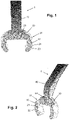
Figures 1 and 2 diagrammatically show perspective views of a portion of a device according to the invention, and -
Figures 3 and 4 illustrate an operational position of the device on an operative eye. - The
device 1 according to the invention is configured to be used in a LASIK surgery procedure and to be put in an operational position on anoperative eye 10 after acorneal flap 11 has been created and has been put to an opened position. As indicated in the foregoing, the operational position of thedevice 1 on theoperative eye 10 is illustrated infigures 3 and 4 . To this end, thecornea 12 and thecorneal flap 11 of theeye 10 are diagrammatically depicted infigure 3 , and theeyeball 13 and thecorneal flap 11 of theeye 10 are diagrammatically depicted infigure 4 . - The
device 1 comprises aneye fixating portion 20 configured to engage on thesclera 14 around thecornea 12 and to thereby perform a fixating action on theeye 10 while enabling free access to thecornea 12 when thedevice 1 is in the operational position on theeye 10, and also comprises a generally plate-likeflap protecting portion 30 configured to completely cover thecorneal flap 11 in the opened position thereof when thedevice 1 is in the operational position on theeye 10. Further, thedevice 1 comprises ahandle portion 40. In this respect, it is noted that only a part of thehandle portion 40 is shown in the figures. In the shown example, thehandle portion 40 is connected to theflap protecting portion 30 and has a curved appearance at the position of connection to theflap protecting portion 30. - The
eye fixating portion 20 comprises at least one ridge-like support element 21 having acontact surface 22 through which theeye fixating portion 20 is to contact the sclera 14 when thedevice 1 is in the operational position on theeye 10. In the framework of the invention, the number ofsupport elements 21 may be chosen freely, wherein one, two, three or four are practical examples. In the shown embodiment of thedevice 1 according to the invention, theeye fixating portion 20 comprises twocurved support elements 21 configured to contact the sclera 14 at opposite sides of thecornea 12. - In
figure 3 , it can be seen that aspace 23 between the twosupport elements 21 along theflap protecting portion 30 is configured to accommodate ahinge portion 11a of thecorneal flap 11. Infigures 1, 2 and3 , it can be seen that at a side of thedevice 1 that is configured to face theeye 10, thecontact surface 22 of therespective support elements 21 is at a protruding level relative to theflap protecting portion 30. - In the embodiment of the
device 1 according to the invention shown in the figures, theflap protecting portion 30 is generally shaped as a minor segment of a circle. As can be seen infigures 1, 2 and3 , the twosupport elements 21 extend fromopposite corner areas 31 of theflap protecting portion 30 in this embodiment. - Advantageously, the
eye fixating portion 20 and theflap protecting portion 30 are made as a single integral entirety. It is also possible that theeye fixating portion 20, theflap protecting portion 30 and thehandle portion 40 are made as a single integral entirety, which implies that thedevice 1 may be provided as a single integral entirety. On the other hand, an option such as thehandle portion 40 being releasably coupled to theflap protecting portion 30 is also feasible in the framework of the invention. - It will be clear to a person skilled in the art that the scope of the invention is not limited to the examples discussed in the foregoing, but that several amendments and modifications thereof are possible without deviating from the scope of the invention as defined in the attached claims. It is intended that the invention be construed as including all such amendments and modifications insofar they come within the scope of the claims or the equivalents thereof. While the invention has been illustrated and described in detail in the figures and the description, such illustration and description are to be considered illustrative or exemplary only, and not restrictive. The invention is not limited to the disclosed embodiments. The drawings are schematic, wherein details which are not required for understanding the invention may have been omitted, and not necessarily to scale.
Claims (12)
- Device (1) configured to be used in a LASIK surgery procedure and to be put in an operational position on an operative eye (10) after a corneal flap (11) has been created and has been put to an opened position, comprising:- an eye fixating portion (20) configured to engage on the sclera (14) around the cornea (12) and to thereby perform a fixating action on the eye (10) while enabling free access to the cornea (12) when the device (1) is in the operational position on the eye (10), and- a flap protecting portion (30) configured to completely cover the corneal flap (11) in the opened position thereof when the device (1) is in the operational position on the eye (10),wherein the eye fixating portion (20) comprises at least one support element (21) having a contact surface (22) through which the eye fixating portion (20) is to contact the sclera (14) when the device (1) is in the operational position on the eye (10).
- Device (1) according to claim 1, wherein the eye fixation portion (20) comprises one, two, three or four support elements (21).
- Device (1) according to claim 1 or 2, wherein the eye fixating portion (20) comprises two curved support elements (21) configured to contact the sclera (14) at opposite sides of the cornea (12).
- Device (1) according to any of claims 1-3, wherein a space (23) between two support elements (21) along the flap protecting portion (30) is configured to accommodate a hinge portion (11a) of the corneal flap (11).
- Device (1) according to any of claims 1-4, wherein, at a side of the device (1) that is configured to face the eye (10), the contact surface (22) of the at least one support element (21) is at a protruding level relative to the flap protecting portion (30).
- Device (1) according to any of claims 1-5, wherein the flap protecting portion (30) is generally shaped as a minor segment of a circle.
- Device (1) according to claim 6, wherein two support elements (21) extend from opposite corner areas (31) of the flap protecting portion (30).
- Device (1) according to any of claims 1-7, further comprising a handle portion (40).
- Device (1) according to claim 8, wherein the handle portion (40) is connected to the flap protecting portion (30).
- Device (1) according to claim 9, wherein the handle portion (40) has a curved appearance at the position where the handle portion (40) is connected to the flap protecting portion (30).
- Device (1) according to any of claims 1-10, wherein the eye fixating portion (20) and the flap protecting portion (30) are made as a single integral entirety.
- Device (1) according to claim 11 insofar as dependent on any of claims 8-10, wherein the eye fixating portion (20), the flap protecting portion (30) and the handle portion (40) are made as a single integral entirety.
Applications Claiming Priority (1)
| Application Number | Priority Date | Filing Date | Title |
|---|---|---|---|
| NL2026779A NL2026779B1 (en) | 2020-10-27 | 2020-10-27 | Device configured to be used in a LASIK surgery procedure |
Publications (2)
| Publication Number | Publication Date |
|---|---|
| EP3991703A1 true EP3991703A1 (en) | 2022-05-04 |
| EP3991703B1 EP3991703B1 (en) | 2025-04-02 |
Family
ID=73498247
Family Applications (1)
| Application Number | Title | Priority Date | Filing Date |
|---|---|---|---|
| EP21204122.2A Active EP3991703B1 (en) | 2020-10-27 | 2021-10-22 | Device configured to be used in a lasik surgery procedure |
Country Status (2)
| Country | Link |
|---|---|
| EP (1) | EP3991703B1 (en) |
| NL (1) | NL2026779B1 (en) |
Citations (4)
| Publication number | Priority date | Publication date | Assignee | Title |
|---|---|---|---|---|
| US6220246B1 (en) * | 1999-11-23 | 2001-04-24 | Becton Dickinson And Company | Ophthalmic surgical sponge for protection of a corneal cap |
| US20030045894A1 (en) * | 2001-09-06 | 2003-03-06 | Holladay Jack T. | Method of using a cornea cut to track eye movement during laser vision correction surgery |
| US6569153B1 (en) * | 2001-06-28 | 2003-05-27 | Lahaye Leon C. | Multi-function surgical instrument for facilitating ophthalmic laser surgery |
| RU2631108C1 (en) * | 2016-10-06 | 2017-09-18 | Федеральное государственное автономное учреждение "Межотраслевой научно-технический комплекс "Микрохирургия глаза" имени академика С.Н. Федорова" Министерства здравоохранения Российской Федерации | Tools for corneal flap protection against laser impact during excimer laser surgery |
-
2020
- 2020-10-27 NL NL2026779A patent/NL2026779B1/en active
-
2021
- 2021-10-22 EP EP21204122.2A patent/EP3991703B1/en active Active
Patent Citations (4)
| Publication number | Priority date | Publication date | Assignee | Title |
|---|---|---|---|---|
| US6220246B1 (en) * | 1999-11-23 | 2001-04-24 | Becton Dickinson And Company | Ophthalmic surgical sponge for protection of a corneal cap |
| US6569153B1 (en) * | 2001-06-28 | 2003-05-27 | Lahaye Leon C. | Multi-function surgical instrument for facilitating ophthalmic laser surgery |
| US20030045894A1 (en) * | 2001-09-06 | 2003-03-06 | Holladay Jack T. | Method of using a cornea cut to track eye movement during laser vision correction surgery |
| RU2631108C1 (en) * | 2016-10-06 | 2017-09-18 | Федеральное государственное автономное учреждение "Межотраслевой научно-технический комплекс "Микрохирургия глаза" имени академика С.Н. Федорова" Министерства здравоохранения Российской Федерации | Tools for corneal flap protection against laser impact during excimer laser surgery |
Also Published As
| Publication number | Publication date |
|---|---|
| NL2026779B1 (en) | 2022-06-21 |
| EP3991703B1 (en) | 2025-04-02 |
Similar Documents
| Publication | Publication Date | Title |
|---|---|---|
| Eckardt et al. | Macular rotation with and without counter-rotation of the globe in patients with age-related macular degeneration | |
| US8591025B1 (en) | Eye covering and refractive correction methods for LASIK and other applications | |
| US20140155800A1 (en) | Eye coverings for corneal healing and methods of use | |
| Bauerberg | Centered vs. inferior off-center ablation to correct hyperopia and presbyopia | |
| Jankov et al. | Topography-guided treatment of irregular astigmatism with the wavelight excimer laser | |
| Huang et al. | Overview of laser refractive surgery | |
| Kim et al. | Laser in situ keratomileusis for refractive error after cataract surgery | |
| Yap et al. | Diplopia as a complication of laser in situ keratomileusis surgery | |
| Lukenda et al. | Excimer laser correction of hyperopia, hyperopic and mixed astigmatism: past, present, and future | |
| EP3991703B1 (en) | Device configured to be used in a lasik surgery procedure | |
| JPH02501363A (en) | Intraocular lenses for correcting refractive errors | |
| Forseto et al. | Laser in situ keratomileusis for undercorrection after radial keratotomy | |
| Syrdalen | Pars plana technique for removal of congenital subluxated lenses in young patients | |
| Barbara et al. | Laser ablation in eyes with congenital nystagmus | |
| RU2751755C1 (en) | Method for correction of induced ametropia after episcleral filling | |
| US6146405A (en) | Ophthalmic applanator | |
| Talamo et al. | Decentration | |
| Huang et al. | Application progress of smile surgery and V4C ICL implantation in the treatment of high myopia | |
| Sekundo et al. | Centration in SMILE and SMILE Pro | |
| Ribeiro et al. | Reasons not to select patients for corneal refractive surgery | |
| Sidhu et al. | MYOPIC ASTIGMATISM | |
| Allan | September consultation# 2 | |
| Elmassry et al. | year follow up implantable Collamer lenses (ICLs) for the treatment of pediatric Anisometropic amblyopia | |
| Santhiago | Highly irregular topography after excimer laser enhancement: December consultation# 1 | |
| Tassignon | Refractive surgical problem: Reply |
Legal Events
| Date | Code | Title | Description |
|---|---|---|---|
| PUAI | Public reference made under article 153(3) epc to a published international application that has entered the european phase |
Free format text: ORIGINAL CODE: 0009012 |
|
| STAA | Information on the status of an ep patent application or granted ep patent |
Free format text: STATUS: THE APPLICATION HAS BEEN PUBLISHED |
|
| AK | Designated contracting states |
Kind code of ref document: A1 Designated state(s): AL AT BE BG CH CY CZ DE DK EE ES FI FR GB GR HR HU IE IS IT LI LT LU LV MC MK MT NL NO PL PT RO RS SE SI SK SM TR |
|
| STAA | Information on the status of an ep patent application or granted ep patent |
Free format text: STATUS: REQUEST FOR EXAMINATION WAS MADE |
|
| 17P | Request for examination filed |
Effective date: 20221101 |
|
| RBV | Designated contracting states (corrected) |
Designated state(s): AL AT BE BG CH CY CZ DE DK EE ES FI FR GB GR HR HU IE IS IT LI LT LU LV MC MK MT NL NO PL PT RO RS SE SI SK SM TR |
|
| GRAP | Despatch of communication of intention to grant a patent |
Free format text: ORIGINAL CODE: EPIDOSNIGR1 |
|
| STAA | Information on the status of an ep patent application or granted ep patent |
Free format text: STATUS: GRANT OF PATENT IS INTENDED |
|
| P01 | Opt-out of the competence of the unified patent court (upc) registered |
Free format text: CASE NUMBER: UPC_APP_119023/2023 Effective date: 20230510 |
|
| INTG | Intention to grant announced |
Effective date: 20241105 |
|
| GRAS | Grant fee paid |
Free format text: ORIGINAL CODE: EPIDOSNIGR3 |
|
| GRAA | (expected) grant |
Free format text: ORIGINAL CODE: 0009210 |
|
| STAA | Information on the status of an ep patent application or granted ep patent |
Free format text: STATUS: THE PATENT HAS BEEN GRANTED |
|
| AK | Designated contracting states |
Kind code of ref document: B1 Designated state(s): AL AT BE BG CH CY CZ DE DK EE ES FI FR GB GR HR HU IE IS IT LI LT LU LV MC MK MT NL NO PL PT RO RS SE SI SK SM TR |
|
| REG | Reference to a national code |
Ref country code: GB Ref legal event code: FG4D |
|
| REG | Reference to a national code |
Ref country code: CH Ref legal event code: EP |
|
| REG | Reference to a national code |
Ref country code: DE Ref legal event code: R096 Ref document number: 602021028443 Country of ref document: DE |
|
| REG | Reference to a national code |
Ref country code: IE Ref legal event code: FG4D |
|
| REG | Reference to a national code |
Ref country code: NL Ref legal event code: FP |
|
| REG | Reference to a national code |
Ref country code: AT Ref legal event code: MK05 Ref document number: 1780573 Country of ref document: AT Kind code of ref document: T Effective date: 20250402 |
|
| PG25 | Lapsed in a contracting state [announced via postgrant information from national office to epo] |
Ref country code: FI Free format text: LAPSE BECAUSE OF FAILURE TO SUBMIT A TRANSLATION OF THE DESCRIPTION OR TO PAY THE FEE WITHIN THE PRESCRIBED TIME-LIMIT Effective date: 20250402 Ref country code: PT Free format text: LAPSE BECAUSE OF FAILURE TO SUBMIT A TRANSLATION OF THE DESCRIPTION OR TO PAY THE FEE WITHIN THE PRESCRIBED TIME-LIMIT Effective date: 20250804 Ref country code: ES Free format text: LAPSE BECAUSE OF FAILURE TO SUBMIT A TRANSLATION OF THE DESCRIPTION OR TO PAY THE FEE WITHIN THE PRESCRIBED TIME-LIMIT Effective date: 20250402 |
|
| REG | Reference to a national code |
Ref country code: LT Ref legal event code: MG9D |
|
| PG25 | Lapsed in a contracting state [announced via postgrant information from national office to epo] |
Ref country code: GR Free format text: LAPSE BECAUSE OF FAILURE TO SUBMIT A TRANSLATION OF THE DESCRIPTION OR TO PAY THE FEE WITHIN THE PRESCRIBED TIME-LIMIT Effective date: 20250703 Ref country code: NO Free format text: LAPSE BECAUSE OF FAILURE TO SUBMIT A TRANSLATION OF THE DESCRIPTION OR TO PAY THE FEE WITHIN THE PRESCRIBED TIME-LIMIT Effective date: 20250702 |
|
| PG25 | Lapsed in a contracting state [announced via postgrant information from national office to epo] |
Ref country code: PL Free format text: LAPSE BECAUSE OF FAILURE TO SUBMIT A TRANSLATION OF THE DESCRIPTION OR TO PAY THE FEE WITHIN THE PRESCRIBED TIME-LIMIT Effective date: 20250402 |
|
| PG25 | Lapsed in a contracting state [announced via postgrant information from national office to epo] |
Ref country code: BG Free format text: LAPSE BECAUSE OF FAILURE TO SUBMIT A TRANSLATION OF THE DESCRIPTION OR TO PAY THE FEE WITHIN THE PRESCRIBED TIME-LIMIT Effective date: 20250402 |
|
| PG25 | Lapsed in a contracting state [announced via postgrant information from national office to epo] |
Ref country code: HR Free format text: LAPSE BECAUSE OF FAILURE TO SUBMIT A TRANSLATION OF THE DESCRIPTION OR TO PAY THE FEE WITHIN THE PRESCRIBED TIME-LIMIT Effective date: 20250402 |
|
| PG25 | Lapsed in a contracting state [announced via postgrant information from national office to epo] |
Ref country code: AT Free format text: LAPSE BECAUSE OF FAILURE TO SUBMIT A TRANSLATION OF THE DESCRIPTION OR TO PAY THE FEE WITHIN THE PRESCRIBED TIME-LIMIT Effective date: 20250402 |
|
| PG25 | Lapsed in a contracting state [announced via postgrant information from national office to epo] |
Ref country code: RS Free format text: LAPSE BECAUSE OF FAILURE TO SUBMIT A TRANSLATION OF THE DESCRIPTION OR TO PAY THE FEE WITHIN THE PRESCRIBED TIME-LIMIT Effective date: 20250702 |
|
| PG25 | Lapsed in a contracting state [announced via postgrant information from national office to epo] |
Ref country code: IS Free format text: LAPSE BECAUSE OF FAILURE TO SUBMIT A TRANSLATION OF THE DESCRIPTION OR TO PAY THE FEE WITHIN THE PRESCRIBED TIME-LIMIT Effective date: 20250802 |
|
| PG25 | Lapsed in a contracting state [announced via postgrant information from national office to epo] |
Ref country code: LV Free format text: LAPSE BECAUSE OF FAILURE TO SUBMIT A TRANSLATION OF THE DESCRIPTION OR TO PAY THE FEE WITHIN THE PRESCRIBED TIME-LIMIT Effective date: 20250402 |
|
| REG | Reference to a national code |
Ref country code: DE Ref legal event code: R097 Ref document number: 602021028443 Country of ref document: DE |
|
| PG25 | Lapsed in a contracting state [announced via postgrant information from national office to epo] |
Ref country code: DK Free format text: LAPSE BECAUSE OF FAILURE TO SUBMIT A TRANSLATION OF THE DESCRIPTION OR TO PAY THE FEE WITHIN THE PRESCRIBED TIME-LIMIT Effective date: 20250402 Ref country code: SM Free format text: LAPSE BECAUSE OF FAILURE TO SUBMIT A TRANSLATION OF THE DESCRIPTION OR TO PAY THE FEE WITHIN THE PRESCRIBED TIME-LIMIT Effective date: 20250402 |
|
| PG25 | Lapsed in a contracting state [announced via postgrant information from national office to epo] |
Ref country code: CZ Free format text: LAPSE BECAUSE OF FAILURE TO SUBMIT A TRANSLATION OF THE DESCRIPTION OR TO PAY THE FEE WITHIN THE PRESCRIBED TIME-LIMIT Effective date: 20250402 |
|
| PG25 | Lapsed in a contracting state [announced via postgrant information from national office to epo] |
Ref country code: EE Free format text: LAPSE BECAUSE OF FAILURE TO SUBMIT A TRANSLATION OF THE DESCRIPTION OR TO PAY THE FEE WITHIN THE PRESCRIBED TIME-LIMIT Effective date: 20250402 |
|
| PG25 | Lapsed in a contracting state [announced via postgrant information from national office to epo] |
Ref country code: SK Free format text: LAPSE BECAUSE OF FAILURE TO SUBMIT A TRANSLATION OF THE DESCRIPTION OR TO PAY THE FEE WITHIN THE PRESCRIBED TIME-LIMIT Effective date: 20250402 Ref country code: RO Free format text: LAPSE BECAUSE OF FAILURE TO SUBMIT A TRANSLATION OF THE DESCRIPTION OR TO PAY THE FEE WITHIN THE PRESCRIBED TIME-LIMIT Effective date: 20250402 |
|
| PG25 | Lapsed in a contracting state [announced via postgrant information from national office to epo] |
Ref country code: IT Free format text: LAPSE BECAUSE OF FAILURE TO SUBMIT A TRANSLATION OF THE DESCRIPTION OR TO PAY THE FEE WITHIN THE PRESCRIBED TIME-LIMIT Effective date: 20250402 |
|
| PLBE | No opposition filed within time limit |
Free format text: ORIGINAL CODE: 0009261 |
|
| STAA | Information on the status of an ep patent application or granted ep patent |
Free format text: STATUS: NO OPPOSITION FILED WITHIN TIME LIMIT |
|
| REG | Reference to a national code |
Ref country code: CH Ref legal event code: L10 Free format text: ST27 STATUS EVENT CODE: U-0-0-L10-L00 (AS PROVIDED BY THE NATIONAL OFFICE) Effective date: 20260211 |
|
| 26N | No opposition filed |
Effective date: 20260105 |