EP3989160A1 - Pathological section image processing method, apparatus, system, and storage medium - Google Patents
Pathological section image processing method, apparatus, system, and storage medium Download PDFInfo
- Publication number
- EP3989160A1 EP3989160A1 EP20887251.5A EP20887251A EP3989160A1 EP 3989160 A1 EP3989160 A1 EP 3989160A1 EP 20887251 A EP20887251 A EP 20887251A EP 3989160 A1 EP3989160 A1 EP 3989160A1
- Authority
- EP
- European Patent Office
- Prior art keywords
- stained
- image
- view
- cells
- result
- Prior art date
- Legal status (The legal status is an assumption and is not a legal conclusion. Google has not performed a legal analysis and makes no representation as to the accuracy of the status listed.)
- Pending
Links
Images


Classifications
-
- G—PHYSICS
- G06—COMPUTING OR CALCULATING; COUNTING
- G06T—IMAGE DATA PROCESSING OR GENERATION, IN GENERAL
- G06T7/00—Image analysis
- G06T7/0002—Inspection of images, e.g. flaw detection
- G06T7/0012—Biomedical image inspection
-
- G—PHYSICS
- G06—COMPUTING OR CALCULATING; COUNTING
- G06T—IMAGE DATA PROCESSING OR GENERATION, IN GENERAL
- G06T7/00—Image analysis
- G06T7/10—Segmentation; Edge detection
-
- G—PHYSICS
- G06—COMPUTING OR CALCULATING; COUNTING
- G06T—IMAGE DATA PROCESSING OR GENERATION, IN GENERAL
- G06T7/00—Image analysis
- G06T7/10—Segmentation; Edge detection
- G06T7/11—Region-based segmentation
-
- G—PHYSICS
- G06—COMPUTING OR CALCULATING; COUNTING
- G06T—IMAGE DATA PROCESSING OR GENERATION, IN GENERAL
- G06T7/00—Image analysis
- G06T7/10—Segmentation; Edge detection
- G06T7/136—Segmentation; Edge detection involving thresholding
-
- G—PHYSICS
- G06—COMPUTING OR CALCULATING; COUNTING
- G06T—IMAGE DATA PROCESSING OR GENERATION, IN GENERAL
- G06T7/00—Image analysis
- G06T7/10—Segmentation; Edge detection
- G06T7/187—Segmentation; Edge detection involving region growing; involving region merging; involving connected component labelling
-
- G—PHYSICS
- G06—COMPUTING OR CALCULATING; COUNTING
- G06T—IMAGE DATA PROCESSING OR GENERATION, IN GENERAL
- G06T7/00—Image analysis
- G06T7/70—Determining position or orientation of objects or cameras
-
- G—PHYSICS
- G06—COMPUTING OR CALCULATING; COUNTING
- G06V—IMAGE OR VIDEO RECOGNITION OR UNDERSTANDING
- G06V10/00—Arrangements for image or video recognition or understanding
- G06V10/20—Image preprocessing
- G06V10/34—Smoothing or thinning of the pattern; Morphological operations; Skeletonisation
-
- G—PHYSICS
- G06—COMPUTING OR CALCULATING; COUNTING
- G06V—IMAGE OR VIDEO RECOGNITION OR UNDERSTANDING
- G06V20/00—Scenes; Scene-specific elements
- G06V20/60—Type of objects
- G06V20/69—Microscopic objects, e.g. biological cells or cellular parts
- G06V20/695—Preprocessing, e.g. image segmentation
-
- G—PHYSICS
- G06—COMPUTING OR CALCULATING; COUNTING
- G06V—IMAGE OR VIDEO RECOGNITION OR UNDERSTANDING
- G06V20/00—Scenes; Scene-specific elements
- G06V20/60—Type of objects
- G06V20/69—Microscopic objects, e.g. biological cells or cellular parts
- G06V20/698—Matching; Classification
-
- G—PHYSICS
- G16—INFORMATION AND COMMUNICATION TECHNOLOGY [ICT] SPECIALLY ADAPTED FOR SPECIFIC APPLICATION FIELDS
- G16H—HEALTHCARE INFORMATICS, i.e. INFORMATION AND COMMUNICATION TECHNOLOGY [ICT] SPECIALLY ADAPTED FOR THE HANDLING OR PROCESSING OF MEDICAL OR HEALTHCARE DATA
- G16H10/00—ICT specially adapted for the handling or processing of patient-related medical or healthcare data
- G16H10/40—ICT specially adapted for the handling or processing of patient-related medical or healthcare data for data related to laboratory analysis, e.g. patient specimen analysis
-
- G—PHYSICS
- G16—INFORMATION AND COMMUNICATION TECHNOLOGY [ICT] SPECIALLY ADAPTED FOR SPECIFIC APPLICATION FIELDS
- G16H—HEALTHCARE INFORMATICS, i.e. INFORMATION AND COMMUNICATION TECHNOLOGY [ICT] SPECIALLY ADAPTED FOR THE HANDLING OR PROCESSING OF MEDICAL OR HEALTHCARE DATA
- G16H30/00—ICT specially adapted for the handling or processing of medical images
-
- G—PHYSICS
- G16—INFORMATION AND COMMUNICATION TECHNOLOGY [ICT] SPECIALLY ADAPTED FOR SPECIFIC APPLICATION FIELDS
- G16H—HEALTHCARE INFORMATICS, i.e. INFORMATION AND COMMUNICATION TECHNOLOGY [ICT] SPECIALLY ADAPTED FOR THE HANDLING OR PROCESSING OF MEDICAL OR HEALTHCARE DATA
- G16H30/00—ICT specially adapted for the handling or processing of medical images
- G16H30/40—ICT specially adapted for the handling or processing of medical images for processing medical images, e.g. editing
-
- G—PHYSICS
- G16—INFORMATION AND COMMUNICATION TECHNOLOGY [ICT] SPECIALLY ADAPTED FOR SPECIFIC APPLICATION FIELDS
- G16H—HEALTHCARE INFORMATICS, i.e. INFORMATION AND COMMUNICATION TECHNOLOGY [ICT] SPECIALLY ADAPTED FOR THE HANDLING OR PROCESSING OF MEDICAL OR HEALTHCARE DATA
- G16H50/00—ICT specially adapted for medical diagnosis, medical simulation or medical data mining; ICT specially adapted for detecting, monitoring or modelling epidemics or pandemics
- G16H50/20—ICT specially adapted for medical diagnosis, medical simulation or medical data mining; ICT specially adapted for detecting, monitoring or modelling epidemics or pandemics for computer-aided diagnosis, e.g. based on medical expert systems
-
- G—PHYSICS
- G16—INFORMATION AND COMMUNICATION TECHNOLOGY [ICT] SPECIALLY ADAPTED FOR SPECIFIC APPLICATION FIELDS
- G16H—HEALTHCARE INFORMATICS, i.e. INFORMATION AND COMMUNICATION TECHNOLOGY [ICT] SPECIALLY ADAPTED FOR THE HANDLING OR PROCESSING OF MEDICAL OR HEALTHCARE DATA
- G16H70/00—ICT specially adapted for the handling or processing of medical references
- G16H70/60—ICT specially adapted for the handling or processing of medical references relating to pathologies
-
- G—PHYSICS
- G06—COMPUTING OR CALCULATING; COUNTING
- G06T—IMAGE DATA PROCESSING OR GENERATION, IN GENERAL
- G06T2207/00—Indexing scheme for image analysis or image enhancement
- G06T2207/10—Image acquisition modality
- G06T2207/10024—Color image
-
- G—PHYSICS
- G06—COMPUTING OR CALCULATING; COUNTING
- G06T—IMAGE DATA PROCESSING OR GENERATION, IN GENERAL
- G06T2207/00—Indexing scheme for image analysis or image enhancement
- G06T2207/10—Image acquisition modality
- G06T2207/10056—Microscopic image
-
- G—PHYSICS
- G06—COMPUTING OR CALCULATING; COUNTING
- G06T—IMAGE DATA PROCESSING OR GENERATION, IN GENERAL
- G06T2207/00—Indexing scheme for image analysis or image enhancement
- G06T2207/10—Image acquisition modality
- G06T2207/10056—Microscopic image
- G06T2207/10061—Microscopic image from scanning electron microscope
-
- G—PHYSICS
- G06—COMPUTING OR CALCULATING; COUNTING
- G06T—IMAGE DATA PROCESSING OR GENERATION, IN GENERAL
- G06T2207/00—Indexing scheme for image analysis or image enhancement
- G06T2207/20—Special algorithmic details
- G06T2207/20036—Morphological image processing
-
- G—PHYSICS
- G06—COMPUTING OR CALCULATING; COUNTING
- G06T—IMAGE DATA PROCESSING OR GENERATION, IN GENERAL
- G06T2207/00—Indexing scheme for image analysis or image enhancement
- G06T2207/20—Special algorithmic details
- G06T2207/20081—Training; Learning
-
- G—PHYSICS
- G06—COMPUTING OR CALCULATING; COUNTING
- G06T—IMAGE DATA PROCESSING OR GENERATION, IN GENERAL
- G06T2207/00—Indexing scheme for image analysis or image enhancement
- G06T2207/20—Special algorithmic details
- G06T2207/20084—Artificial neural networks [ANN]
-
- G—PHYSICS
- G06—COMPUTING OR CALCULATING; COUNTING
- G06T—IMAGE DATA PROCESSING OR GENERATION, IN GENERAL
- G06T2207/00—Indexing scheme for image analysis or image enhancement
- G06T2207/30—Subject of image; Context of image processing
- G06T2207/30004—Biomedical image processing
- G06T2207/30024—Cell structures in vitro; Tissue sections in vitro
-
- G—PHYSICS
- G06—COMPUTING OR CALCULATING; COUNTING
- G06T—IMAGE DATA PROCESSING OR GENERATION, IN GENERAL
- G06T2207/00—Indexing scheme for image analysis or image enhancement
- G06T2207/30—Subject of image; Context of image processing
- G06T2207/30004—Biomedical image processing
- G06T2207/30068—Mammography; Breast
-
- G—PHYSICS
- G06—COMPUTING OR CALCULATING; COUNTING
- G06T—IMAGE DATA PROCESSING OR GENERATION, IN GENERAL
- G06T2207/00—Indexing scheme for image analysis or image enhancement
- G06T2207/30—Subject of image; Context of image processing
- G06T2207/30004—Biomedical image processing
- G06T2207/30096—Tumor; Lesion
-
- G—PHYSICS
- G06—COMPUTING OR CALCULATING; COUNTING
- G06T—IMAGE DATA PROCESSING OR GENERATION, IN GENERAL
- G06T2207/00—Indexing scheme for image analysis or image enhancement
- G06T2207/30—Subject of image; Context of image processing
- G06T2207/30242—Counting objects in image
-
- G—PHYSICS
- G06—COMPUTING OR CALCULATING; COUNTING
- G06V—IMAGE OR VIDEO RECOGNITION OR UNDERSTANDING
- G06V2201/00—Indexing scheme relating to image or video recognition or understanding
- G06V2201/03—Recognition of patterns in medical or anatomical images
Definitions
- Embodiments of this application relate to the field of intelligent medical technologies of artificial intelligence (AI), and in particular, to a pathological section image processing method and apparatus, a system, and a storage medium.
- AI artificial intelligence
- HER2 human epidermal growth factor receptor-2
- Embodiments of this application provide a pathological section image processing method and apparatus, a system, and a storage medium, which can be used to resolve a technical problem of inaccurate analysis of pathological sections in the related art.
- the technical solutions are as follows:
- an embodiment of this application provides a pathological section image processing method, executable by a computer device, the method including:
- an embodiment of this application provides a pathological section image processing apparatus, including:
- an embodiment of this application provides an intelligent microscope system, including: a microscope, a camera, and a computer device, where
- an embodiment of this application provides a computer device, including a processor and a memory, the memory storing at least one instruction, at least one program, a code set, or an instruction set, and the at least one instruction, the at least one program, the code set, or the instruction set being loaded and executed by the processor to implement the foregoing pathological section image processing method.
- an embodiment of this application provides a computer-readable storage medium, storing at least one instruction, at least one program, a code set, or an instruction set, the at least one instruction, the at least one program, the code set, or the instruction set being loaded and executed by a processor to implement the foregoing pathological section image processing method.
- an embodiment of this application provides a computer program product, the computer program product, when executed by a processor, being used for implementing the foregoing pathological section image processing method.
- the technical solutions provided in the embodiments of this application may include the following beneficial effects: Stained images of a pathological section after cell membrane staining are obtained, each stained image is generated from a respective one of n fields of view of the pathological section under a microscope, and cell nucleus detection and cell membrane description are performed on each stained image, to obtain cell nucleus positions and a cell membrane description result of cancer cells in each stained image.
- the two aspects of information are combined to determine quantities of cells of types, and further determine an analysis result of the pathological section. Therefore, a technical solution for systematic cell membrane staining analysis on a pathological section is provided, to improve the accuracy of a detection result.
- a deep learning method is not directly used to directly analyze a stained image of a pathological section, and an analysis result is directly outputted by a model.
- Such a black box processing manner does not conform to relevant diagnostic guidelines for HER2 grading.
- cancer cells are detected and classified according to the definition of the relevant diagnostic guidelines, so that the HER2 grading can be performed according to the determining criteria of the guidelines, thereby improving the standardization and accuracy of a final HER2 grading results.
- AI Artificial Intelligence
- AI is a theory, method, technology, and application system that use a digital computer or a machine controlled by a digital computer to simulate, extend, and expand human intelligence, perceive the environment, obtain knowledge, and use the knowledge to obtain the best result.
- AI is a comprehensive technology of computer science, which attempts to understand essence of intelligence and produces a new intelligent machine that responds in a manner similar to human intelligence.
- AI is to study design principles and implementation methods of various intelligent machines, so that the machines have the functions of perception, reasoning, and decision-making.
- the AI technology is a comprehensive discipline, covering a wide range of fields including both hardware-level technologies and software-level technologies.
- the basic AI technology generally includes technologies such as sensors, dedicated AI chips, cloud computing, distributed storage, big data processing technologies, operating/interaction systems, and mechatronics.
- AI software technologies mainly include a computer vision technology, a speech processing technology, a natural language processing technology, machine learning/deep learning, and the like.
- Computer vision is a science that studies how to enable a machine to "see”, and to be specific, to implement machine vision such as recognition, tracking, measurement, and the like for a target by using a camera and a computer in replacement of human eyes, and further perform graphic processing, so that the computer processes the target into an image more suitable for human eyes to observe, or more suitable to be transmitted to an instrument for detection.
- CV studies related theories and technologies, and attempts to establish an AI system that can obtain information from images or multidimensional data.
- the CV technologies generally include technologies such as image processing, image recognition, image semantic understanding, image retrieval, optical character recognition (OCR), video processing, video semantic understanding, video content/behavior recognition, three-dimensional (3D) object reconstruction, a 3D technology, virtual reality, augmented reality, synchronous positioning, and map construction, and further include biometric feature recognition technologies such as common face recognition and fingerprint recognition.
- technologies such as image processing, image recognition, image semantic understanding, image retrieval, optical character recognition (OCR), video processing, video semantic understanding, video content/behavior recognition, three-dimensional (3D) object reconstruction, a 3D technology, virtual reality, augmented reality, synchronous positioning, and map construction, and further include biometric feature recognition technologies such as common face recognition and fingerprint recognition.
- Machine learning is a multi-field interdisciplinary subject involving the probability theory, statistics, the approximation theory, convex analysis, the algorithm complexity theory, and the like. ML specializes in studying how a computer simulates or implements a human learning behavior to obtain new knowledge or skills, and reorganize an existing knowledge structure, so as to keep improving its performance. ML is the core of AI, is a basic way to make the computer intelligent, and is applied to various fields of AI. ML and deep learning generally include technologies such as an artificial neural network, a belief network, reinforcement learning, transfer learning, inductive learning, and learning from demonstrations.
- the AI technology is studied and applied in a plurality of fields such as a common smart home, a smart wearable device, a virtual assistant, a smart speaker, smart marketing, unmanned driving, automatic driving, an unmanned aerial vehicle, a robot, smart medical care, and smart customer service. It is believed that with the development of technologies, the AI technology will be applied to more fields, and play an increasingly important role.
- the solutions provided in the embodiments of this application relate to the field of intelligent medical technologies.
- the CV technology is used to perform image processing and analysis on a pathological section after cell membrane staining, to finally determine an analysis result corresponding to the pathological section, thereby assisting doctors in diseases diagnosis and treatment.
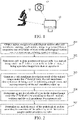
- FIG. 1 is a schematic diagram of an intelligent microscope system according to an embodiment of this application.
- the intelligent microscope system may include: a microscope 11, a camera 12, and a computer device 13.
- the microscope 11 is configured to observe a pathological section.
- the microscope 11 is configured to observe a pathological section after cell membrane staining.
- the camera 12 is configured to capture and obtain a pathological section image of the pathological section under a field of view of the microscope 11.
- the pathological section is an HER2 stained section.
- HER2 staining stains a membrane of a positive cancer cell brown and a nucleus thereof blue.
- HER2 is generally graded into four categories: 0, 1+, 2+, and 3+.
- the pathological section image needs to be acquired under a medium-high-power field of view of the microscope (such as 10 times, 20 times, or 40 times).
- the computer device 13 is configured to obtain an analysis result corresponding to the pathological section image by executing a method procedure described in detail later based on the pathological section image captured by the camera 12.
- the analysis result may be an HER2 grading result.
- the computer device 13 may be any electronic device with computing and storage capabilities, such as a personal computer (PC).
- the microscope 11, the camera 12, and the computer device 13 may be configured at the same physical position, or even configured in the same physical device. Alternatively, the microscope 11, the camera 12, and the computer device 13 may be configured at different positions, which are connected through a wired or wireless communication network to transmit data or commands between each other.
- FIG. 2 is a flowchart of a pathological section image processing method according to an embodiment of this application.
- the execution entity of each step of the method may be the computer device in the intelligent microscope system described above.
- the method may include the following steps:
- Step 201 Obtain stained images of a pathological section after cell membrane staining, wherein each stained image is generated from a respective one of n fields of view of the pathological section under a microscope, n being a positive integer.
- the n fields are a plurality of microscope fields including cancer cells obtained after observing the pathological section.
- the n fields may be selected by a doctor. For example, determining of HER2 grading of the pathological section is used as an example. Because the HER2 grading is defined on a staining status of infiltrated cancer cells, the doctor may be responsible for acquiring a plurality of microscope fields that typically include regions of the infiltrated cancer cells.
- n is an integer greater than 1, for example, n is 10.
- relevant diagnostic guidelines for example, breast cancer HER2 detection guidelines
- the HER2 grading is performed by adopting the strategy of using a plurality of typical field images selected by the doctor to approximately replace a full-section diagnosis result, which can avoid full-section scan and obtain an accurate HER2 grading result.
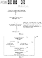
- FIG. 3 is a schematic diagram of stained images, under a plurality of fields of view of a microscope, of a pathological section after cell membrane staining, where images 31, 32, and 33 each represent a stained image of a same pathological section under three fields of view of a microscope.
- the stained image is actually an RGB image, on which a membrane of a positive cancer cell is stained brown and a nucleus thereof is stained blue.
- Step 202 Determine cell nucleus positions of cancer cells in a stained image under an i th field of view in the n fields of view, i being a positive integer less than or equal to n.
- a deep learning method may be used to perform cancer cell detection on a stained image to determine cell nucleus positions of cancer cells in the stained image.
- a fully convolutional network FCN
- FCN fully convolutional network
- An input of the cell nucleus detection model may be a stained image (the stained image is an RGB image), and the input is a Gauss-like response image centered on a cell center point. All cancer cells can be obtained by searching for a local maximum response position of an inputted heat map.
- the stained image under the i th field of view is processed by using the cell nucleus detection model to obtain the cell nucleus positions of the cancer cells in the stained image under the i th field of view.
- the cell nucleus positions may include a coordinate set (marked as Ddetect) of the cancer cells in the stained image under the i th field of view, where the coordinate set Ddetect includes position coordinates of the sell nuclei of the cancer cells in the stained image under the i th field of view. For example, as shown in FIG.
- an input image 41 of the cell nucleus detection model is on the left side, that is, a stained image under a certain field of view, and a stained image 42 on which cell nucleus positions of detected cancer cells are marked is on the right side, where the cell nucleus positions are marked with small black dots.
- the cancer cell detection method described above is only exemplary and explanatory, and the embodiments of this application are not limited to other cancer cell detection methods.
- Step 203 Generate a cell membrane description result of the stained image under the i th field of view, the cell membrane description result being used for indicating completeness and staining intensity of the cell membrane staining.
- the completeness of cell membrane staining refers to whether a stained cell membrane is a complete cell membrane, and the staining intensity refers to a staining depth of the stained cell membrane.
- a stained image may be processed and analyzed to obtain a cell membrane description result of the stained image.
- a cell membrane description result of the stained image For the related process, reference may be made to the description in the following embodiments.
- Step 204 Determine quantities of cells of types in the stained image under the i th field of view according to the cell nucleus positions and the cell membrane description result.
- cells are classified into a plurality of types according to cell membrane staining.
- the types include: completely and strongly stained cells, incompletely and strongly stained cells, completely and weakly stained cells, incompletely and weakly stained cells, and non-stained cells.
- the completely and strongly stained cells refer to cells with complete cell membrane staining and high staining intensity
- the incompletely and strongly stained cells refer to cells with incomplete cell membrane staining and high staining intensity
- the completely and weakly stained cells refer to cells with complete cell membrane staining and low staining intensity
- the incompletely and weakly stained cells refer to cells with incomplete cell membrane staining and low staining intensity
- non-stained cells refer to cells not stained.
- quantities of cells of types in the stained image may be determined, to implement quantitative analysis and determining of the cells. For example, it is determined that quantities of cells of types in a stained image under a certain field of view include: 170 completely and strongly stained cells, 230 incompletely and strongly stained cells, 2 completely and weakly stained cells, 104 incompletely and weakly stained cells, and 47 non-stained cells.
- Step 205 Determine an analysis result of the pathological section according to quantities of the cells of types in the stained images under the n fields of view.
- n is an integer greater than 1
- the foregoing steps 202 to 204 may be performed, to obtain quantities of cells of types in the stained image in each field of view.
- the quantities of the cells of types in the stained images under the n fields of view are then comprehensively counted.
- quantities of the each type of cell in the foregoing stained images are summed to obtain a total quantity of the each type of cell in all the n stained images.
- proportions of the cells of types in the stained images under the n fields of view are determined according to the quantities of the cells of types in the stained images under the n fields of view; and the analysis result of the pathological section is determined according to the proportions of the cells of types.
- An example is used in which HER2 grading is performed on a breast cancer pathological section. If a proportion of completely and strongly stained cells is greater than 10%, an HER2 grade is 3+. If a proportion of completely and weakly stained cells is greater than 10% or the proportion of completely and strongly stained cells is less than or equal to 10%, the HER2 grade is 2+. If a proportion of incompletely and weakly stained cells is greater than 10%, the HER2 grade is 1+.
- the HER2 grade is 0+. When the HER2 grade is 3+, it is determined as HAR2 positive. When the HER2 grade is 2+, a method of in situ hybridization needs to be further used to perform HER2 gene expansion status detection, or different tissue blocks may be selected for re-detection. When the HER2 grade is 1+ or 0, it is determined as HAR2 negative.
- stained images of a pathological section after cell membrane staining are obtained, each stained image being generated from a respective one of n fields of view of the pathological section under a microscope, and cell nucleus detection and cell membrane description are performed on each stained image, to obtain cell nucleus positions and a cell membrane description result of cancer cells in each stained image.
- the two aspects of information are combined to determine quantities of cells of types, and further determine an analysis result of the pathological section. Therefore, a technical solution for systematic cell membrane staining analysis on a pathological section is provided, which helps to improve the accuracy of a detection result.
- a deep learning method is not directly used to directly analyze a stained image of a pathological section, and an analysis result is directly outputted by a model.
- Such a black box processing manner does not conform to relevant diagnostic guidelines for HER2 grading.
- cancer cells are detected and classified according to the definition of the relevant diagnostic guidelines, so that the HER2 grading can be performed according to the determining criteria of the guidelines, thereby improving the standardization and accuracy of a final HER2 grading results.
- step 203 may include the following sub-steps:
- the cell membrane description result includes a first region segmentation result, a second region segmentation result, and a third region segmentation result.
- the first region segmentation result is used for indicating positions of cell membranes with complete staining
- the second region segmentation result is used for indicating positions of cell membranes whose staining intensities are greater than a first limit value
- the third region segmentation result is used for indicating positions of cell membranes whose staining intensities are greater than a second limit value.
- the first limit value is less than the second limit value, that is, cells whose staining intensities are greater than the first limit value and less than or equal to the second limit value are weakly stained cells, cells whose staining intensities are greater than the second limit value are strongly stained cells, and cells whose staining intensities are less than the first limit value may be considered as non-stained cells.
- this step may include the following several sub-steps: 2.1. Perform threshold segmentation processing on the target stained channel image by using a first threshold, to obtain a weakly-stained segmented image; For example, for each pixel in the target stained channel image, if a pixel value of the pixel is greater than the first threshold, the pixel value of the pixel is recorded as 1, and if the pixel value of the pixel is less than or equal to the first threshold, the pixel value of the pixel is recorded as 0. Finally, the target stained channel image is converted into a binary weakly-stained segmented image.
- the first threshold may be preset in combination with actual situations, and this is not limited in this embodiment of this application.
- 2.2. Perform threshold segmentation processing on the target stained channel image by using a second threshold, to obtain a strongly-stained segmented image; For example, for each pixel in the target stained channel image, if a pixel value of the pixel is greater than the second threshold, the pixel value of the pixel is recorded as 1, and if the pixel value of the pixel is less than or equal to the second threshold, the pixel value of the pixel is recorded as 0. Finally, the target stained channel image is converted into a binary strongly-stained segmented image.
- the second threshold may be preset in combination with actual situations, and this is not limited in this embodiment of this application.
- the weakly-stained segmented image includes not only weakly stained cells but also strongly stained cells that are stronger than being weakly stained, but the strongly-stained segmented image includes only strongly stained cells.
- first morphological processing may be performed on the weakly-stained segmented image to obtain the first region segmentation result; second morphological processing may be performed on the weakly-stained segmented image to obtain the second region segmentation result; and the second morphological processing may be performed on the strongly-stained segmented image to obtain the third region segmentation result.
- the performing first morphological processing on the weakly-stained segmented image includes: performing cytoskeleton extraction processing on the weakly-stained segmented image to obtain a cytoskeleton in the weakly-stained segmented image; searching for an enclosed region in which the cytoskeleton is closed; filling, when the enclosed region is an innermost-layer enclosed region, the innermost-layer enclosed region to obtain an enclosed region segmented image; and extracting position information of foreground pixels in the enclosed region segmented image to obtain the first region segmentation result.
- centerline extraction processing is performed on the weakly-stained segmented image Mlight
- cell membrane delineation is performed to obtain the cytoskeleton
- statistical analysis is performed on the centerline to count the enclosed region formed by each connected boundary.
- the innermost-layer enclosed region is determined as a delineated boundary of a complete cell membrane. All innermost-layer enclosed regions are filled to obtain an enclosed region segmented image Menclosed, as shown in FIG. 5 , and a position coordinate set of all foreground pixels in the enclosed region segmented image Menclosed is defined as Penclosed, where Penclosed is the first region segmentation result.
- the performing second morphological processing on the weakly-stained segmented image includes: performing region expansion processing on the weakly-stained segmented image to obtain a processed weakly-stained segmented image; and extracting position information of foreground pixels in the processed weakly-stained segmented image to obtain the second region segmentation result.
- region expansion processing is performed on the weakly-stained segmented image Mlight to obtain a processed weakly-stained segmented image Elight, and a position coordinate set of all foreground pixels in the processed weakly-stained segmented image Elight is defined as Plight, where Plight is the second region segmentation result.
- the performing the second morphological processing on the strongly-stained segmented image includes: performing region expansion processing on the strongly-stained segmented image to obtain a processed strongly-stained segmented image; and extracting position information of foreground pixels in the processed strongly-stained segmented image to obtain the third region segmentation result.
- region expansion processing is performed on the strongly-stained segmented image Mheavy to obtain a processed strongly-stained segmented image Eheavy, and a position coordinate set of all foreground pixels in the processed strongly-stained segmented image Eheavy is defined as Pheavy, where Pheavy is the third region segmentation result.
- the determining quantities of cells of types in the stained image under the i th field of view according to the cell nucleus positions and the cell membrane description result includes the flowing sub-steps:
- FIG. 6 is a flowchart of a complete technical solution according to this application.
- Stained images of a pathological section after cell membrane staining under a plurality of fields of view of a microscope are obtained, and cell nucleus detection and cell membrane description are performed on a stained image under a single field of view, to obtain cell nucleus positions and a cell membrane description result of cancer cells in the stained image under the single field of view.
- the two aspects of information are combined to obtain a cell classification result under the single field of view, that is, quantities of cells of types.
- the cell classification results under each single field of view are combined to obtain a cell classification result under a plurality of fields of view, and the HER2 grade corresponding to the pathological section is then determined according to the cell classification result.
- the cell classification result under the plurality of fields of view is: 20% of completely and strongly stained cells, 35% of incompletely and strongly stained cells, 5% of completely and weakly stained cells, 10% of incompletely and weakly stained cells, and 30% of non-stained cells.
- the HER2 grade is 3+.
- FIG. 7 is a block diagram of a pathological section image processing apparatus according to an embodiment of this application.
- the apparatus has functions of implementing the foregoing method embodiments. The functions may be implemented by hardware, or may be implemented by hardware executing corresponding software.
- the apparatus may be the computer device described above, or may be disposed in the computer device.
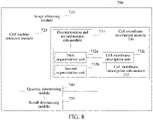
- the apparatus 700 may include: an image obtaining module 710, a cell nucleus detection module 720, a cell membrane description module 730, a quantity determining module 740, and a result determining module 750.
- the image obtaining module 710 is configured to obtain stained images of a pathological section after cell membrane staining, wherein each stained image is generated from a respective one of n fields of view of the pathological section under a microscope, n being a positive integer.
- the cell nucleus detection module 720 is configured to determine cell nucleus positions of cancer cells in a stained image under an i th field of view in the n fields of view, i being a positive integer less than or equal to n.
- the cell membrane description module 730 is configured to generate a cell membrane description result of the stained image under the i th field of view, the cell membrane description result being used for indicating completeness and staining intensity of the cell membrane staining.
- the quantity determining module 740 is configured to determine quantities of cells of types in the stained image under the i th field of view according to the cell nucleus positions and the cell membrane description result.
- the result determining module 750 is configured to determine an analysis result of the pathological section according to the quantities of the cells of types in the stained images under the n fields of view.
- stained images of a pathological section after cell membrane staining are obtained, each stained image being generated from a respective one of n fields of view of the pathological section under a microscope, and cell nucleus detection and cell membrane description are performed on each stained image, to obtain cell nucleus positions and a cell membrane description result of cancer cells in each stained image.
- the two aspects of information are combined to determine quantities of cells of types, and further determine an analysis result of the pathological section. Therefore, a technical solution for systematic cell membrane staining analysis on a pathological section is provided, which helps to improve the accuracy of a detection result.
- the cell membrane description module 730 includes: a decomposition and recombination sub-module 731 and a cell membrane description sub-module 732.
- the decomposition and recombination sub-module 731 is configured to perform color channel decomposition and recombination on the stained image under the i th field of view to obtain a target stained channel image.
- the cell membrane description sub-module 732 is configured to generate the cell membrane description result according to the target stained channel image, the cell membrane description result including a first region segmentation result, a second region segmentation result, and a third region segmentation result, where
- the first region segmentation result being used for indicating positions of cell membranes with complete staining
- the second region segmentation result being used for indicating positions of cell membranes whose staining intensities are greater than a first limit value
- the third region segmentation result being used for indicating positions of cell membranes whose staining intensities are greater than a second limit value.
- the cell membrane description sub-module 732 includes: a first segmentation unit 732a, a second segmentation unit 732b, and a cell membrane description unit 732c.
- the first segmentation unit 732a is configured to perform threshold segmentation processing on the target stained channel image by using a first threshold, to obtain a weakly-stained segmented image.
- the second segmentation unit 732b is configured to perform threshold segmentation processing on the target stained channel image by using a second threshold, to obtain a strongly-stained segmented image.
- the cell membrane description unit 732c is configured to generate the cell membrane description result according to the weakly-stained segmented image and the strongly-stained segmented image.
- the cell membrane description unit 732c is configured to:
- the cell membrane description unit 732c is configured to:
- the cell membrane description unit 732c is configured to:
- the cell membrane description unit 732c is configured to:
- the types include: completely and strongly stained cells, incompletely and strongly stained cells, completely and weakly stained cells, incompletely and weakly stained cells, and non-stained cells.
- the quantity determining module 740 is configured to:
- the cell nucleus detection module 720 is configured to process the stained image under the i th field of view by using a cell nucleus detection model to obtain the cell nucleus positions.
- the result determining module 750 is configured to:
- the division of the foregoing functional modules is merely an example for description.
- the functions may be assigned to and completed by different functional modules according to the requirements, that is, the internal structure of the device is divided into different functional modules, to implement all or some of the functions described above.
- the apparatus and method embodiments provided in the foregoing embodiments belong to the same conception. For the specific implementation process, reference may be made to the method embodiments, and details are not described herein again.
- An exemplary embodiment of this application further provides an intelligent microscope system, including: a microscope, a camera, and a computer device.
- the microscope is configured to observe a pathological section after cell membrane staining.
- the camera is configured to obtain stained images of the pathological section, wherein each stained image is generated from a respective one of n fields of view of the pathological section under a microscope, n being a positive integer.
- the computer device is configured to determine cell nucleus positions of cancer cells in a stained image under an i th field of view in the n fields of view, i being a positive integer less than or equal to n; generate a cell membrane description result of the stained image under the i th field of view, the cell membrane description result being used for indicating completeness and staining intensity of the cell membrane staining; determine quantities of cells of types in the stained image under the i th field of view according to the cell nucleus positions and the cell membrane description result; and determine an analysis result of the pathological section according to the quantities of the cells of types in the stained images under the n fields of view.
- the computer device is further configured to perform other operations described in the foregoing method embodiments, and this is not limited in the embodiments of this application.
- FIG. 9 is a schematic structural diagram of a computer device according to an embodiment of this application.
- the computer device 900 includes a central processing unit (CPU) 901, a system memory 904 including a random access memory (RAM) 902 and a read only memory (ROM) 903, and a system bus 905 connecting the system memory 904 and the central processing unit 901.
- the computer device 900 further includes a basic input/output system (I/O) system 906 for facilitating information transmission between various devices in a computer and a mass storage device 907 configured to store an operating system 913, an application 914, and other application modules 915.
- I/O basic input/output system
- the basic input/output system 906 includes a display 908 configured to display information and an input device 909 such as a mouse and a keyboard for a user to input information.
- the display 908 and the input device 909 are both connected to the central processing unit 901 through an input/output controller 910 connected to the system bus 905.
- the basic input/output system 906 may further include the input/output controller 910 for receiving and processing input from a plurality of other devices such as a keyboard, a mouse, an electronic stylus, or the like.
- the input/output controller 910 further provides output to a display screen, a printer, or other types of output devices.
- the mass storage device 907 is connected to the central processing unit 901 through a mass storage controller (not shown) connected to the system bus 905.
- the mass storage device 907 and an associated computer-readable medium provide non-transitory storage for the computer device 900. That is, the mass storage device 907 may include a computer-readable medium (not shown) such as a hard disk or a compact disc ROM (CD-ROM) drive.
- a computer-readable medium such as a hard disk or a compact disc ROM (CD-ROM) drive.
- the computer-readable medium may include a computer storage medium and a communication medium.
- the computer-storage medium includes volatile and non-transitory media, and removable and non-removable media implemented by using any method or technology used for storing information such as computer-readable instructions, data structures, program modules, or other data.
- the computer storage medium includes a RAM, a ROM, an erasable programmable ROM (EPROM), a flash memory or another solid-state storage technology, a CD-ROM, or another optical storage, a magnetic cassette, a magnetic tape, or a magnetic disk storage or another magnetic storage device.
- EPROM erasable programmable ROM
- the computer device 900 may further be connected, through a network such as the Internet, to a remote computer on the network for running. That is, the computer device 900 may be connected to a network 912 by using a network interface unit 911 connected to the system bus 905, or may be connected to another type of network or a remote computer system (not shown) by using a network interface unit 911.
- the memory further includes at least one instruction, at least one program, a code set, or an instruction set.
- the at least one instruction, the at least one program, the code set, or the instruction set is stored in the memory and is configured to be executed by one or more processors to implement the foregoing pathological section image processing method.
- a computer-readable storage medium is further provided, the storage medium storing at least one instruction, at least one program, a code set, or an instruction set, the at least one instruction, the at least one program, the code set, or the instruction set, when executed by a processor of a terminal, implementing the foregoing pathological section image processing method.
- the computer-readable storage medium may include: a ROM, a RAM, a solid state drive (SSD), an optical disc, or the like.
- the RAM may include a resistance random access memory (ReRAM) and a dynamic random access memory (DRAM).
- a computer program product is further provided, the computer program product, when executed by a processor of a terminal, being used for implementing the foregoing pathological section image processing method.
- step numbers described in this specification merely schematically show a possible execution sequence of the steps. In some other embodiments, the steps may not be performed according to the number sequence. For example, two steps with different numbers may be performed simultaneously, or two steps with different numbers may be performed according to a sequence contrary to the sequence shown in the figure. This is not limited in the embodiments of this application.
Landscapes
- Engineering & Computer Science (AREA)
- Health & Medical Sciences (AREA)
- General Health & Medical Sciences (AREA)
- Physics & Mathematics (AREA)
- General Physics & Mathematics (AREA)
- Theoretical Computer Science (AREA)
- Medical Informatics (AREA)
- Public Health (AREA)
- Biomedical Technology (AREA)
- Computer Vision & Pattern Recognition (AREA)
- Primary Health Care (AREA)
- Epidemiology (AREA)
- Multimedia (AREA)
- Nuclear Medicine, Radiotherapy & Molecular Imaging (AREA)
- Radiology & Medical Imaging (AREA)
- Life Sciences & Earth Sciences (AREA)
- Molecular Biology (AREA)
- Quality & Reliability (AREA)
- Data Mining & Analysis (AREA)
- Databases & Information Systems (AREA)
- Pathology (AREA)
- Investigating Or Analysing Biological Materials (AREA)
Abstract
Description
- This application claims priority to
Chinese Patent Application No. 201911115369.6, entitled "PATHOLOGICAL SECTION IMAGE PROCESSING METHOD AND APPARATUS, SYSTEM, AND STORAGE MEDIUM" and filed on November 14, 2019 - Embodiments of this application relate to the field of intelligent medical technologies of artificial intelligence (AI), and in particular, to a pathological section image processing method and apparatus, a system, and a storage medium.
- The correct detection and evaluation of the human epidermal growth factor receptor-2 (HER2) protein expression and gene amplification status of breast cancer is critical to the clinical treatment and prognosis of breast cancer.
- In the related art, cell membrane staining is performed on a pathological section, and the pathological section after the cell membrane staining is then observed and analyzed under a microscope, to assist doctors in HER2 detection. However, there is a lack of systematic cell membrane staining analysis on pathological sections in the related art, resulting in inaccurate final detection results.
- Embodiments of this application provide a pathological section image processing method and apparatus, a system, and a storage medium, which can be used to resolve a technical problem of inaccurate analysis of pathological sections in the related art. The technical solutions are as follows:
- According to an aspect, an embodiment of this application provides a pathological section image processing method, executable by a computer device, the method including:
- obtaining stained images of a pathological section after cell membrane staining, wherein each stained image is generated from a respective one of n fields of view of the pathological section under a microscope, n being a positive integer;
- determining cell nucleus positions of cancer cells in a stained image under an ith field of view in the n fields of view, i being a positive integer less than or equal to n;
- generating a cell membrane description result of the stained image under the ith field of view, the cell membrane description result being used for indicating completeness and staining intensity of the cell membrane staining;
- determining quantities of cells of types in the stained image under the ith field of view according to the cell nucleus positions and the cell membrane description result; and
- determining an analysis result of the pathological section according to the quantities of the cells of types in the stained images under the n fields of view.
- According to another aspect, an embodiment of this application provides a pathological section image processing apparatus, including:
- an image obtaining module, configured to obtain stained images of a pathological section after cell membrane staining, wherein each stained image is generated from a respective one of n fields of view of the pathological section under a microscope, n being a positive integer;
- a cell nucleus detection module, configured to determine cell nucleus positions of cancer cells in a stained image under an ith field of view in the n fields of view, i being a positive integer less than or equal to n;
- a cell membrane description module, configured to generate a cell membrane description result of the stained image under the ith field of view, the cell membrane description result being used for indicating completeness and staining intensity of the cell membrane staining;
- a quantity determining module, configured to determine quantities of cells of types in the stained image under the ith field of view according to the cell nucleus positions and the cell membrane description result; and
- a result determining module, configured to determine an analysis result of the pathological section according to the quantities of the cells of types in the stained images under the n fields of view.
- According to another aspect, an embodiment of this application provides an intelligent microscope system, including: a microscope, a camera, and a computer device, where
- the microscope is configured to observe a pathological section after cell membrane staining;
- the camera is configured to obtain stained images of the pathological section, wherein each stained image is generated from a respective one of n fields of view of the pathological section under a microscope, n being a positive integer; and
- the computer device is configured to determine cell nucleus positions of cancer cells in a stained image under an ith field of view in the n fields of view, i being a positive integer less than or equal to n; generate a cell membrane description result of the stained image under the ith field of view, the cell membrane description result being used for indicating completeness and staining intensity of the cell membrane staining; determine quantities of cells of types in the stained image under the ith field of view according to the cell nucleus positions and the cell membrane description result; and determine an analysis result of the pathological section according to the quantities of the cells of types in the stained images under the n fields of view.
- According to still another aspect, an embodiment of this application provides a computer device, including a processor and a memory, the memory storing at least one instruction, at least one program, a code set, or an instruction set, and the at least one instruction, the at least one program, the code set, or the instruction set being loaded and executed by the processor to implement the foregoing pathological section image processing method.
- According to yet another aspect, an embodiment of this application provides a computer-readable storage medium, storing at least one instruction, at least one program, a code set, or an instruction set, the at least one instruction, the at least one program, the code set, or the instruction set being loaded and executed by a processor to implement the foregoing pathological section image processing method.
- According to still yet another aspect, an embodiment of this application provides a computer program product, the computer program product, when executed by a processor, being used for implementing the foregoing pathological section image processing method.
- The technical solutions provided in the embodiments of this application may include the following beneficial effects:
Stained images of a pathological section after cell membrane staining are obtained, each stained image is generated from a respective one of n fields of view of the pathological section under a microscope, and cell nucleus detection and cell membrane description are performed on each stained image, to obtain cell nucleus positions and a cell membrane description result of cancer cells in each stained image. The two aspects of information are combined to determine quantities of cells of types, and further determine an analysis result of the pathological section. Therefore, a technical solution for systematic cell membrane staining analysis on a pathological section is provided, to improve the accuracy of a detection result. - In addition, in the technical solutions provided in the embodiments of this application, a deep learning method is not directly used to directly analyze a stained image of a pathological section, and an analysis result is directly outputted by a model. Such a black box processing manner does not conform to relevant diagnostic guidelines for HER2 grading. In the technical solutions provided in the embodiments of this application, cancer cells are detected and classified according to the definition of the relevant diagnostic guidelines, so that the HER2 grading can be performed according to the determining criteria of the guidelines, thereby improving the standardization and accuracy of a final HER2 grading results.
- To describe technical solutions in embodiments of this application more clearly, the following briefly introduces the accompanying drawings required for describing the embodiments. Apparently, the accompanying drawings in the following description show only some embodiments of this application, and a person of ordinary skill in the art may still derive other accompanying drawings from these accompanying drawings without creative efforts.
-
FIG. 1 is a schematic diagram of an intelligent microscope system according to an embodiment of this application. -
FIG. 2 is a flowchart of a pathological section image processing method according to an embodiment of this application. -
FIG. 3 is a schematic diagram of stained images under a plurality of fields of view of a microscope according to this application. -
FIG. 4 is a schematic diagram of input and output of a cell nucleus detection model according to this application. -
FIG. 5 is a schematic diagram of an original image of a stained image, an enclosed region segmented image, a weakly-stained segmented image, and a strongly-stained segmented image according to this application. -
FIG. 6 is a flowchart of a complete technical solution according to this application. -
FIG. 7 is a block diagram of a pathological section image processing apparatus according to an embodiment of this application. -
FIG. 8 is a block diagram of a pathological section image processing apparatus according to another embodiment of this application. -
FIG. 9 is a schematic structural diagram of a computer device according to an embodiment of this application. - To make objectives, technical solutions, and advantages of this application clearer, the following further describes implementations of this application in detail with reference to the accompanying drawings.
- Artificial Intelligence (AI) is a theory, method, technology, and application system that use a digital computer or a machine controlled by a digital computer to simulate, extend, and expand human intelligence, perceive the environment, obtain knowledge, and use the knowledge to obtain the best result. In other words, AI is a comprehensive technology of computer science, which attempts to understand essence of intelligence and produces a new intelligent machine that responds in a manner similar to human intelligence. AI is to study design principles and implementation methods of various intelligent machines, so that the machines have the functions of perception, reasoning, and decision-making.
- The AI technology is a comprehensive discipline, covering a wide range of fields including both hardware-level technologies and software-level technologies. The basic AI technology generally includes technologies such as sensors, dedicated AI chips, cloud computing, distributed storage, big data processing technologies, operating/interaction systems, and mechatronics. AI software technologies mainly include a computer vision technology, a speech processing technology, a natural language processing technology, machine learning/deep learning, and the like.
- Computer vision (CV) is a science that studies how to enable a machine to "see", and to be specific, to implement machine vision such as recognition, tracking, measurement, and the like for a target by using a camera and a computer in replacement of human eyes, and further perform graphic processing, so that the computer processes the target into an image more suitable for human eyes to observe, or more suitable to be transmitted to an instrument for detection. As a scientific subject, CV studies related theories and technologies, and attempts to establish an AI system that can obtain information from images or multidimensional data. The CV technologies generally include technologies such as image processing, image recognition, image semantic understanding, image retrieval, optical character recognition (OCR), video processing, video semantic understanding, video content/behavior recognition, three-dimensional (3D) object reconstruction, a 3D technology, virtual reality, augmented reality, synchronous positioning, and map construction, and further include biometric feature recognition technologies such as common face recognition and fingerprint recognition.
- Machine learning (ML) is a multi-field interdisciplinary subject involving the probability theory, statistics, the approximation theory, convex analysis, the algorithm complexity theory, and the like. ML specializes in studying how a computer simulates or implements a human learning behavior to obtain new knowledge or skills, and reorganize an existing knowledge structure, so as to keep improving its performance. ML is the core of AI, is a basic way to make the computer intelligent, and is applied to various fields of AI. ML and deep learning generally include technologies such as an artificial neural network, a belief network, reinforcement learning, transfer learning, inductive learning, and learning from demonstrations.
- With the research and progress of the AI technology, the AI technology is studied and applied in a plurality of fields such as a common smart home, a smart wearable device, a virtual assistant, a smart speaker, smart marketing, unmanned driving, automatic driving, an unmanned aerial vehicle, a robot, smart medical care, and smart customer service. It is believed that with the development of technologies, the AI technology will be applied to more fields, and play an increasingly important role.
- The solutions provided in the embodiments of this application relate to the field of intelligent medical technologies. The CV technology is used to perform image processing and analysis on a pathological section after cell membrane staining, to finally determine an analysis result corresponding to the pathological section, thereby assisting doctors in diseases diagnosis and treatment.
- The technical solutions of this application are described below by using several embodiments.
-
FIG. 1 is a schematic diagram of an intelligent microscope system according to an embodiment of this application. The intelligent microscope system may include: amicroscope 11, acamera 12, and acomputer device 13. - The
microscope 11 is configured to observe a pathological section. In the embodiments of this application, themicroscope 11 is configured to observe a pathological section after cell membrane staining. - The
camera 12 is configured to capture and obtain a pathological section image of the pathological section under a field of view of themicroscope 11. Alternatively, the pathological section is an HER2 stained section. HER2 staining stains a membrane of a positive cancer cell brown and a nucleus thereof blue. HER2 is generally graded into four categories: 0, 1+, 2+, and 3+. Generally, the pathological section image needs to be acquired under a medium-high-power field of view of the microscope (such as 10 times, 20 times, or 40 times). - The
computer device 13 is configured to obtain an analysis result corresponding to the pathological section image by executing a method procedure described in detail later based on the pathological section image captured by thecamera 12. In the case that the pathological section is a HER2 stained section, the analysis result may be an HER2 grading result. Thecomputer device 13 may be any electronic device with computing and storage capabilities, such as a personal computer (PC). - The
microscope 11, thecamera 12, and thecomputer device 13 may be configured at the same physical position, or even configured in the same physical device. Alternatively, themicroscope 11, thecamera 12, and thecomputer device 13 may be configured at different positions, which are connected through a wired or wireless communication network to transmit data or commands between each other. -
FIG. 2 is a flowchart of a pathological section image processing method according to an embodiment of this application. The execution entity of each step of the method may be the computer device in the intelligent microscope system described above. The method may include the following steps: - Step 201: Obtain stained images of a pathological section after cell membrane staining, wherein each stained image is generated from a respective one of n fields of view of the pathological section under a microscope, n being a positive integer.
- In some embodiments, the n fields are a plurality of microscope fields including cancer cells obtained after observing the pathological section. The n fields may be selected by a doctor. For example, determining of HER2 grading of the pathological section is used as an example. Because the HER2 grading is defined on a staining status of infiltrated cancer cells, the doctor may be responsible for acquiring a plurality of microscope fields that typically include regions of the infiltrated cancer cells.
- In an example, n is an integer greater than 1, for example, n is 10. Because the HER2 grading in relevant diagnostic guidelines (for example, breast cancer HER2 detection guidelines) is defined based on full-section analysis of the pathological section, but images in a microscope application scenario are obtained from a plurality of fields of view, it is difficult to splice a complete pathological section image. In this application, the HER2 grading is performed by adopting the strategy of using a plurality of typical field images selected by the doctor to approximately replace a full-section diagnosis result, which can avoid full-section scan and obtain an accurate HER2 grading result.
- For example,
FIG. 3 is a schematic diagram of stained images, under a plurality of fields of view of a microscope, of a pathological section after cell membrane staining, whereimages - Step 202: Determine cell nucleus positions of cancer cells in a stained image under an ith field of view in the n fields of view, i being a positive integer less than or equal to n.
- In an exemplary embodiment, a deep learning method may be used to perform cancer cell detection on a stained image to determine cell nucleus positions of cancer cells in the stained image. For example, a fully convolutional network (FCN) may be used to construct a cell nucleus detection model, the cell nucleus detection model may be trained by heat map regression, and the trained cell nucleus detection model may be used for nucleus detection. An input of the cell nucleus detection model may be a stained image (the stained image is an RGB image), and the input is a Gauss-like response image centered on a cell center point. All cancer cells can be obtained by searching for a local maximum response position of an inputted heat map.
- In some embodiments, the stained image under the ith field of view is processed by using the cell nucleus detection model to obtain the cell nucleus positions of the cancer cells in the stained image under the ith field of view. The cell nucleus positions may include a coordinate set (marked as Ddetect) of the cancer cells in the stained image under the ith field of view, where the coordinate set Ddetect includes position coordinates of the sell nuclei of the cancer cells in the stained image under the ith field of view. For example, as shown in
FIG. 4 , aninput image 41 of the cell nucleus detection model is on the left side, that is, a stained image under a certain field of view, and astained image 42 on which cell nucleus positions of detected cancer cells are marked is on the right side, where the cell nucleus positions are marked with small black dots. - Certainly, the cancer cell detection method described above is only exemplary and explanatory, and the embodiments of this application are not limited to other cancer cell detection methods.
- Step 203: Generate a cell membrane description result of the stained image under the ith field of view, the cell membrane description result being used for indicating completeness and staining intensity of the cell membrane staining.
- Because the completeness and staining intensity of cell membrane staining determine an analysis result of the pathological section, the cell membrane description result of the stained image needs to be obtained. The completeness of cell membrane staining refers to whether a stained cell membrane is a complete cell membrane, and the staining intensity refers to a staining depth of the stained cell membrane.
- In this embodiment of this application, a stained image may be processed and analyzed to obtain a cell membrane description result of the stained image. For the related process, reference may be made to the description in the following embodiments.
- Step 204: Determine quantities of cells of types in the stained image under the ith field of view according to the cell nucleus positions and the cell membrane description result.
- In this embodiment of this application, cells are classified into a plurality of types according to cell membrane staining. In some embodiments, the types include: completely and strongly stained cells, incompletely and strongly stained cells, completely and weakly stained cells, incompletely and weakly stained cells, and non-stained cells. The completely and strongly stained cells refer to cells with complete cell membrane staining and high staining intensity, the incompletely and strongly stained cells refer to cells with incomplete cell membrane staining and high staining intensity, the completely and weakly stained cells refer to cells with complete cell membrane staining and low staining intensity, the incompletely and weakly stained cells refer to cells with incomplete cell membrane staining and low staining intensity, and non-stained cells refer to cells not stained.
- By combining cell nucleus positions and a cell membrane description result of a stained image, quantities of cells of types in the stained image may be determined, to implement quantitative analysis and determining of the cells. For example, it is determined that quantities of cells of types in a stained image under a certain field of view include: 170 completely and strongly stained cells, 230 incompletely and strongly stained cells, 2 completely and weakly stained cells, 104 incompletely and weakly stained cells, and 47 non-stained cells.
- Step 205: Determine an analysis result of the pathological section according to quantities of the cells of types in the stained images under the n fields of view.
- When n is an integer greater than 1, for a stained image under each of the plurality of fields of view, the foregoing
steps 202 to 204 may be performed, to obtain quantities of cells of types in the stained image in each field of view. The quantities of the cells of types in the stained images under the n fields of view are then comprehensively counted. For each type of cell, quantities of the each type of cell in the foregoing stained images are summed to obtain a total quantity of the each type of cell in all the n stained images. - In some embodiments, proportions of the cells of types in the stained images under the n fields of view are determined according to the quantities of the cells of types in the stained images under the n fields of view; and the analysis result of the pathological section is determined according to the proportions of the cells of types. An example is used in which HER2 grading is performed on a breast cancer pathological section. If a proportion of completely and strongly stained cells is greater than 10%, an HER2 grade is 3+. If a proportion of completely and weakly stained cells is greater than 10% or the proportion of completely and strongly stained cells is less than or equal to 10%, the HER2 grade is 2+. If a proportion of incompletely and weakly stained cells is greater than 10%, the HER2 grade is 1+. If a proportion of non-stained cells or incompletely and weakly stained cells is less than or equal to 10%, the HER2 grade is 0+. When the HER2 grade is 3+, it is determined as HAR2 positive. When the HER2 grade is 2+, a method of in situ hybridization needs to be further used to perform HER2 gene expansion status detection, or different tissue blocks may be selected for re-detection. When the HER2 grade is 1+ or 0, it is determined as HAR2 negative. Certainly, the foregoing description of determining the analysis result of the pathological section according to the proportions of the cells of types is only exemplary and explanatory. In practical applications, the corresponding analysis result determining standards may be set in combination with actual situations, and this is not limited in this embodiment of this application.
- Based on the above, according to the technical solution provided in this embodiment of this application, stained images of a pathological section after cell membrane staining are obtained, each stained image being generated from a respective one of n fields of view of the pathological section under a microscope, and cell nucleus detection and cell membrane description are performed on each stained image, to obtain cell nucleus positions and a cell membrane description result of cancer cells in each stained image. The two aspects of information are combined to determine quantities of cells of types, and further determine an analysis result of the pathological section. Therefore, a technical solution for systematic cell membrane staining analysis on a pathological section is provided, which helps to improve the accuracy of a detection result.
- In addition, in the technical solutions provided in the embodiments of this application, a deep learning method is not directly used to directly analyze a stained image of a pathological section, and an analysis result is directly outputted by a model. Such a black box processing manner does not conform to relevant diagnostic guidelines for HER2 grading. In the technical solutions provided in the embodiments of this application, cancer cells are detected and classified according to the definition of the relevant diagnostic guidelines, so that the HER2 grading can be performed according to the determining criteria of the guidelines, thereby improving the standardization and accuracy of a final HER2 grading results.
- In an exemplary embodiment, step 203 may include the following sub-steps:
- 1. Perform color channel decomposition and recombination on the stained image under the ith field of view to obtain a target stained channel image;
The stained image is an RGB three-channel image, and may be expressed as IRGB. The color channel decomposition and recombination are performed on the stained image IRGB, to generate three immunohistochemical channel image, namely, IH (hematoxylin) and IE (eosin), and IDAB (Diaminobenzidine), where an IDAB channel image is a brown stained channel image. In this embodiment of this application, the target stained channel image is the brown stained channel image, and an expected cell membrane description result can be obtained by analyzing the brown stained channel image. - 2. Generate the cell membrane description result according to the target stained channel image.
- In this embodiment of this application, the cell membrane description result includes a first region segmentation result, a second region segmentation result, and a third region segmentation result. The first region segmentation result is used for indicating positions of cell membranes with complete staining, the second region segmentation result is used for indicating positions of cell membranes whose staining intensities are greater than a first limit value, and the third region segmentation result is used for indicating positions of cell membranes whose staining intensities are greater than a second limit value. If a larger value of a staining intensity indicates a higher staining intensity, the first limit value is less than the second limit value, that is, cells whose staining intensities are greater than the first limit value and less than or equal to the second limit value are weakly stained cells, cells whose staining intensities are greater than the second limit value are strongly stained cells, and cells whose staining intensities are less than the first limit value may be considered as non-stained cells.
- In some embodiment, this step may include the following several sub-steps:
2.1. Perform threshold segmentation processing on the target stained channel image by using a first threshold, to obtain a weakly-stained segmented image;
For example, for each pixel in the target stained channel image, if a pixel value of the pixel is greater than the first threshold, the pixel value of the pixel is recorded as 1, and if the pixel value of the pixel is less than or equal to the first threshold, the pixel value of the pixel is recorded as 0. Finally, the target stained channel image is converted into a binary weakly-stained segmented image. The first threshold may be preset in combination with actual situations, and this is not limited in this embodiment of this application. - 2.2. Perform threshold segmentation processing on the target stained channel image by using a second threshold, to obtain a strongly-stained segmented image;
For example, for each pixel in the target stained channel image, if a pixel value of the pixel is greater than the second threshold, the pixel value of the pixel is recorded as 1, and if the pixel value of the pixel is less than or equal to the second threshold, the pixel value of the pixel is recorded as 0. Finally, the target stained channel image is converted into a binary strongly-stained segmented image. The second threshold may be preset in combination with actual situations, and this is not limited in this embodiment of this application. - 2.3. Generate the cell membrane description result according to the weakly-stained segmented image and the strongly-stained segmented image.
- The target stained channel image being the foregoing IDAB channel image is used as an example. Because a pixel value of each pixel in the IDAB channel image is a negative number, a first threshold t1 may be used to perform threshold segmentation processing on the image IDAB to obtain a weakly-stained segmented image Mlight, and a second threshold t2 may be used to perform threshold segmentation processing on the image IDAB to obtain a strongly-stained segmented image Mheavy, where t2 = a×t1, and a is a coefficient less than 1. For example, t1 = -0.35, and a = 0.9.
- The weakly-stained segmented image includes not only weakly stained cells but also strongly stained cells that are stronger than being weakly stained, but the strongly-stained segmented image includes only strongly stained cells.
- In somen embodiments, after the weakly-stained segmented image and the strongly-stained segmented image are obtained, first morphological processing may be performed on the weakly-stained segmented image to obtain the first region segmentation result; second morphological processing may be performed on the weakly-stained segmented image to obtain the second region segmentation result; and the second morphological processing may be performed on the strongly-stained segmented image to obtain the third region segmentation result.
- The performing first morphological processing on the weakly-stained segmented image includes: performing cytoskeleton extraction processing on the weakly-stained segmented image to obtain a cytoskeleton in the weakly-stained segmented image; searching for an enclosed region in which the cytoskeleton is closed; filling, when the enclosed region is an innermost-layer enclosed region, the innermost-layer enclosed region to obtain an enclosed region segmented image; and extracting position information of foreground pixels in the enclosed region segmented image to obtain the first region segmentation result. Optionally, centerline extraction processing is performed on the weakly-stained segmented image Mlight, cell membrane delineation is performed to obtain the cytoskeleton, and statistical analysis is performed on the centerline to count the enclosed region formed by each connected boundary. If the enclosed region is an innermost-layer enclosed region, the innermost-layer enclosed region is determined as a delineated boundary of a complete cell membrane. All innermost-layer enclosed regions are filled to obtain an enclosed region segmented image Menclosed, as shown in
FIG. 5 , and a position coordinate set of all foreground pixels in the enclosed region segmented image Menclosed is defined as Penclosed, where Penclosed is the first region segmentation result. - The performing second morphological processing on the weakly-stained segmented image includes: performing region expansion processing on the weakly-stained segmented image to obtain a processed weakly-stained segmented image; and extracting position information of foreground pixels in the processed weakly-stained segmented image to obtain the second region segmentation result. An expansion distance d of the region expansion processing may be preset in combination with actual situations, for example, d = 45, and this is not limited in this embodiment of this application. As shown in
FIG. 5 , it is assumed that region expansion processing is performed on the weakly-stained segmented image Mlight to obtain a processed weakly-stained segmented image Elight, and a position coordinate set of all foreground pixels in the processed weakly-stained segmented image Elight is defined as Plight, where Plight is the second region segmentation result. - The performing the second morphological processing on the strongly-stained segmented image includes: performing region expansion processing on the strongly-stained segmented image to obtain a processed strongly-stained segmented image; and extracting position information of foreground pixels in the processed strongly-stained segmented image to obtain the third region segmentation result. An expansion distance d of the region expansion processing may be preset in combination with actual situations, for example, d = 45, and this is not limited in this embodiment of this application. As shown in
FIG. 5 , it is assumed that region expansion processing is performed on the strongly-stained segmented image Mheavy to obtain a processed strongly-stained segmented image Eheavy, and a position coordinate set of all foreground pixels in the processed strongly-stained segmented image Eheavy is defined as Pheavy, where Pheavy is the third region segmentation result. - Correspondingly, the determining quantities of cells of types in the stained image under the ith field of view according to the cell nucleus positions and the cell membrane description result includes the flowing sub-steps:
- 1. Determine a quantity of elements in an intersection among the cell nucleus positions, the first region segmentation result, and the third region segmentation result as a quantity of the completely and strongly stained cells;
That is, a coordinate set of the completely and strongly stained cells is Ddetect∩Penclosed∩Pheavy, and the quantity of the corresponding cells is a quantity of elements in the set, namely, card(Ddetect∩Penclosed∩Pheavy). - 2. Determine a quantity of elements in an intersection among the cell nucleus positions, a complementary set of the first region segmentation result with respect to the stained image under the ith field of view, and the second region segmentation result as a quantity of the incompletely and strongly stained cells;
That is, a coordinate set of the incompletely and strongly stained cells is Ddetect∩CUPenclosed∩Pheavy, where CUPenclosed is a complement of Penclosed to the full image U (that is, the stained image under the ith field of view). The quantity of the corresponding cells is card (Ddetect∩CUPenclosed∩Pheavy). - 3. Determine a quantity of elements in an intersection among the cell nucleus positions, the first region segmentation result, a complementary set of the third region segmentation result with respect to the stained image under the ith field of view, and the second region segmentation result as a quantity of the completely and weakly stained cells;
That is, a coordinate set of the completely and weakly stained cells is Ddetect∩Penclosed∩CUPheavy∩Plight, where CUPheavy is a complement of Pheavy to the full image U (that is, the stained image under the ith field of view). The quantity of the corresponding cells is card(Ddetect∩Penclosed∩CUPheavy∩Plight). - 4. Determine a quantity of elements in an intersection among the cell nucleus positions, the complementary set of the first region segmentation result with respect to the stained image under the ith field of view, the complementary set of the third region segmentation result with respect to the stained image under the ith field of view, and the second region segmentation result as a quantity of the incompletely and weakly stained cells;
That is, a coordinate set of the incompletely and weakly stained cells is Ddetect∩CUPenc1osed∩CUPheavy∩Plight. The quantity of the corresponding cells is card(Ddetect∩CUPenclosed∩CUPheavy∩Plight). - 5. Determine a quantity of elements in an intersection between the cell nucleus positions and a complementary set of the second region segmentation result with respect to the stained image under the ith field of view as a quantity of the non-stained cells.
That is, a coordinate set of the non-stained cells is Ddetect∩CUPlight, where CUPlight is a complement of Plight to the full image U (that is, the stained image under the ith field of view). The quantity of the corresponding cells is card(Ddetect∩CUPlight). - In this embodiment of this application, by using the foregoing methods, quantitative statistics and analysis are performed on the various cell types in the stained images of the pathological section, thereby providing reliable data support for HER2 grading.
-
FIG. 6 is a flowchart of a complete technical solution according to this application. Stained images of a pathological section after cell membrane staining under a plurality of fields of view of a microscope are obtained, and cell nucleus detection and cell membrane description are performed on a stained image under a single field of view, to obtain cell nucleus positions and a cell membrane description result of cancer cells in the stained image under the single field of view. The two aspects of information are combined to obtain a cell classification result under the single field of view, that is, quantities of cells of types. The cell classification results under each single field of view are combined to obtain a cell classification result under a plurality of fields of view, and the HER2 grade corresponding to the pathological section is then determined according to the cell classification result. For example, the cell classification result under the plurality of fields of view is: 20% of completely and strongly stained cells, 35% of incompletely and strongly stained cells, 5% of completely and weakly stained cells, 10% of incompletely and weakly stained cells, and 30% of non-stained cells. In this case, the HER2 grade is 3+. - The following is an apparatus embodiment of this application, which may be used for performing the method embodiments of this application. For details not disclosed in the apparatus embodiment of this application, reference may be made to the method embodiments of this application.
-
FIG. 7 is a block diagram of a pathological section image processing apparatus according to an embodiment of this application. The apparatus has functions of implementing the foregoing method embodiments. The functions may be implemented by hardware, or may be implemented by hardware executing corresponding software. The apparatus may be the computer device described above, or may be disposed in the computer device. Theapparatus 700 may include: animage obtaining module 710, a cellnucleus detection module 720, a cellmembrane description module 730, aquantity determining module 740, and aresult determining module 750. - The
image obtaining module 710 is configured to obtain stained images of a pathological section after cell membrane staining, wherein each stained image is generated from a respective one of n fields of view of the pathological section under a microscope, n being a positive integer. - The cell
nucleus detection module 720 is configured to determine cell nucleus positions of cancer cells in a stained image under an ith field of view in the n fields of view, i being a positive integer less than or equal to n. - The cell
membrane description module 730 is configured to generate a cell membrane description result of the stained image under the ith field of view, the cell membrane description result being used for indicating completeness and staining intensity of the cell membrane staining. - The
quantity determining module 740 is configured to determine quantities of cells of types in the stained image under the ith field of view according to the cell nucleus positions and the cell membrane description result. - The
result determining module 750 is configured to determine an analysis result of the pathological section according to the quantities of the cells of types in the stained images under the n fields of view. - Based on the above, according to the technical solution provided in this embodiment of this application, stained images of a pathological section after cell membrane staining are obtained, each stained image being generated from a respective one of n fields of view of the pathological section under a microscope, and cell nucleus detection and cell membrane description are performed on each stained image, to obtain cell nucleus positions and a cell membrane description result of cancer cells in each stained image. The two aspects of information are combined to determine quantities of cells of types, and further determine an analysis result of the pathological section. Therefore, a technical solution for systematic cell membrane staining analysis on a pathological section is provided, which helps to improve the accuracy of a detection result.
- In an exemplary embodiment, as shown in
FIG. 8 , the cellmembrane description module 730 includes: a decomposition and recombination sub-module 731 and a cellmembrane description sub-module 732. - The decomposition and
recombination sub-module 731 is configured to perform color channel decomposition and recombination on the stained image under the ith field of view to obtain a target stained channel image. - The cell
membrane description sub-module 732 is configured to generate the cell membrane description result according to the target stained channel image, the cell membrane description result including a first region segmentation result, a second region segmentation result, and a third region segmentation result, where - the first region segmentation result being used for indicating positions of cell membranes with complete staining, the second region segmentation result being used for indicating positions of cell membranes whose staining intensities are greater than a first limit value, and the third region segmentation result being used for indicating positions of cell membranes whose staining intensities are greater than a second limit value.
- In an exemplary embodiment, as shown in
FIG. 8 , the cellmembrane description sub-module 732 includes: afirst segmentation unit 732a, asecond segmentation unit 732b, and a cellmembrane description unit 732c. - The
first segmentation unit 732a is configured to perform threshold segmentation processing on the target stained channel image by using a first threshold, to obtain a weakly-stained segmented image. - The
second segmentation unit 732b is configured to perform threshold segmentation processing on the target stained channel image by using a second threshold, to obtain a strongly-stained segmented image. - The cell
membrane description unit 732c is configured to generate the cell membrane description result according to the weakly-stained segmented image and the strongly-stained segmented image. - In an exemplary embodiment, as shown in
FIG. 8 , the cellmembrane description unit 732c is configured to: - perform first morphological processing on the weakly-stained segmented image to obtain the first region segmentation result;
- perform second morphological processing on the weakly-stained segmented image to obtain the second region segmentation result; and
- perform the second morphological processing on the strongly-stained segmented image to obtain the third region segmentation result.
- In an exemplary embodiment, the cell
membrane description unit 732c is configured to: - perform cytoskeleton extraction processing on the weakly-stained segmented image to obtain a cytoskeleton in the weakly-stained segmented image;
- search for an enclosed region in which the cytoskeleton is closed;
- fill, when the enclosed region is an innermost-layer enclosed region, the innermost-layer enclosed region to obtain an enclosed region segmented image; and
- extract position information of foreground pixels in the enclosed region segmented image to obtain the first region segmentation result.
- In an exemplary embodiment, the cell
membrane description unit 732c is configured to: - perform region expansion processing on the weakly-stained segmented image to obtain a processed weakly-stained segmented image; and
- extract position information of foreground pixels in the processed weakly-stained segmented image to obtain the second region segmentation result.
- In an exemplary embodiment, the cell
membrane description unit 732c is configured to: - perform region expansion processing on the strongly-stained segmented image to obtain a processed strongly-stained segmented image; and
- extract position information of foreground pixels in the processed strongly-stained segmented image to obtain the third region segmentation result.
- In an exemplary embodiment, the types include: completely and strongly stained cells, incompletely and strongly stained cells, completely and weakly stained cells, incompletely and weakly stained cells, and non-stained cells.
- In an exemplary embodiment, the
quantity determining module 740 is configured to: - determine a quantity of elements in an intersection among the cell nucleus positions, the first region segmentation result, and the third region segmentation result as a quantity of the completely and strongly stained cells;
- determine a quantity of elements in an intersection among the cell nucleus positions, a complementary set of the first region segmentation result with respect to the stained image under the ith field of view, and the second region segmentation result as a quantity of the incompletely and strongly stained cells;
- determine a quantity of elements in an intersection among the cell nucleus positions, the first region segmentation result, a complementary set of the third region segmentation result with respect to the stained image under the ith field of view, and the second region segmentation result as a quantity of the completely and weakly stained cells;
- determine a quantity of elements in an intersection among the cell nucleus positions, the complementary set of the first region segmentation result with respect to the stained image under the ith field of view, the complementary set of the third region segmentation result with respect to the stained image under the ith field of view, and the second region segmentation result as a quantity of the incompletely and weakly stained cells; and
- determine a quantity of elements in an intersection between the cell nucleus positions and a complementary set of the second region segmentation result with respect to the stained image under the ith field of view as a quantity of the non-stained cells,
- the cell membrane description result including the first region segmentation result, the second region segmentation result, and the third region segmentation result, the first region segmentation result being used for indicating the positions of the cell membranes with complete staining, the second region segmentation result being used for indicating the positions of the cell membranes whose staining intensities are greater than a first limit value, and the third region segmentation result being used for indicating the positions of the cell membranes whose staining intensities are greater than a second limit value.
- In an exemplary embodiment, the cell
nucleus detection module 720 is configured to process the stained image under the ith field of view by using a cell nucleus detection model to obtain the cell nucleus positions. - In an exemplary embodiment, the
result determining module 750 is configured to: - determine proportions of the cells of types in the stained images under the n fields of view according to the quantities of the cells of types in the stained images under the n fields of view; and
- determine the analysis result of the pathological section according to the proportions of the cells of types.
- When the apparatus provided in the foregoing embodiments implements functions of the apparatus, the division of the foregoing functional modules is merely an example for description. In the practical application, the functions may be assigned to and completed by different functional modules according to the requirements, that is, the internal structure of the device is divided into different functional modules, to implement all or some of the functions described above. In addition, the apparatus and method embodiments provided in the foregoing embodiments belong to the same conception. For the specific implementation process, reference may be made to the method embodiments, and details are not described herein again.
- An exemplary embodiment of this application further provides an intelligent microscope system, including: a microscope, a camera, and a computer device.
- The microscope is configured to observe a pathological section after cell membrane staining.
- The camera is configured to obtain stained images of the pathological section, wherein each stained image is generated from a respective one of n fields of view of the pathological section under a microscope, n being a positive integer.
- The computer device is configured to determine cell nucleus positions of cancer cells in a stained image under an ith field of view in the n fields of view, i being a positive integer less than or equal to n; generate a cell membrane description result of the stained image under the ith field of view, the cell membrane description result being used for indicating completeness and staining intensity of the cell membrane staining; determine quantities of cells of types in the stained image under the ith field of view according to the cell nucleus positions and the cell membrane description result; and determine an analysis result of the pathological section according to the quantities of the cells of types in the stained images under the n fields of view.
- Optionally, the computer device is further configured to perform other operations described in the foregoing method embodiments, and this is not limited in the embodiments of this application.
-
FIG. 9 is a schematic structural diagram of a computer device according to an embodiment of this application. Specifically,
thecomputer device 900 includes a central processing unit (CPU) 901, asystem memory 904 including a random access memory (RAM) 902 and a read only memory (ROM) 903, and asystem bus 905 connecting thesystem memory 904 and thecentral processing unit 901. Thecomputer device 900 further includes a basic input/output system (I/O)system 906 for facilitating information transmission between various devices in a computer and amass storage device 907 configured to store anoperating system 913, anapplication 914, andother application modules 915. - The basic input/
output system 906 includes adisplay 908 configured to display information and aninput device 909 such as a mouse and a keyboard for a user to input information. Thedisplay 908 and theinput device 909 are both connected to thecentral processing unit 901 through an input/output controller 910 connected to thesystem bus 905. The basic input/output system 906 may further include the input/output controller 910 for receiving and processing input from a plurality of other devices such as a keyboard, a mouse, an electronic stylus, or the like. Similarly, the input/output controller 910 further provides output to a display screen, a printer, or other types of output devices. - The
mass storage device 907 is connected to thecentral processing unit 901 through a mass storage controller (not shown) connected to thesystem bus 905. Themass storage device 907 and an associated computer-readable medium provide non-transitory storage for thecomputer device 900. That is, themass storage device 907 may include a computer-readable medium (not shown) such as a hard disk or a compact disc ROM (CD-ROM) drive. - In general, the computer-readable medium may include a computer storage medium and a communication medium. The computer-storage medium includes volatile and non-transitory media, and removable and non-removable media implemented by using any method or technology used for storing information such as computer-readable instructions, data structures, program modules, or other data. The computer storage medium includes a RAM, a ROM, an erasable programmable ROM (EPROM), a flash memory or another solid-state storage technology, a CD-ROM, or another optical storage, a magnetic cassette, a magnetic tape, or a magnetic disk storage or another magnetic storage device. Certainly, those skilled in the art may learn that the computer storage medium is not limited to the above. The
system memory 904 and the large-capacity storage device 907 may be collectively referred to as a memory. - According to the various embodiments of this application, the
computer device 900 may further be connected, through a network such as the Internet, to a remote computer on the network for running. That is, thecomputer device 900 may be connected to anetwork 912 by using anetwork interface unit 911 connected to thesystem bus 905, or may be connected to another type of network or a remote computer system (not shown) by using anetwork interface unit 911. - The memory further includes at least one instruction, at least one program, a code set, or an instruction set. The at least one instruction, the at least one program, the code set, or the instruction set is stored in the memory and is configured to be executed by one or more processors to implement the foregoing pathological section image processing method.
- In an exemplary embodiment, a computer-readable storage medium is further provided, the storage medium storing at least one instruction, at least one program, a code set, or an instruction set, the at least one instruction, the at least one program, the code set, or the instruction set, when executed by a processor of a terminal, implementing the foregoing pathological section image processing method.
- Optionally, the computer-readable storage medium may include: a ROM, a RAM, a solid state drive (SSD), an optical disc, or the like. The RAM may include a resistance random access memory (ReRAM) and a dynamic random access memory (DRAM).
- In an exemplary embodiment, a computer program product is further provided, the computer program product, when executed by a processor of a terminal, being used for implementing the foregoing pathological section image processing method.
- It is to be understood that "plurality of" mentioned in this specification means two or more. "And/or" describes an association relationship for associated objects and represents that three relationships may exist. For example, A and/or B may represent the following three cases: only A exists, both A and B exist, and only B exists. The character "/" generally indicates an "or" relationship between the associated objects. In addition, the step numbers described in this specification merely schematically show a possible execution sequence of the steps. In some other embodiments, the steps may not be performed according to the number sequence. For example, two steps with different numbers may be performed simultaneously, or two steps with different numbers may be performed according to a sequence contrary to the sequence shown in the figure. This is not limited in the embodiments of this application.
- The foregoing descriptions are merely exemplary embodiments of this application, but are not intended to limit this application. Any modification, equivalent replacement, or improvement made within the spirit and principle of this application shall fall within the protection scope of this application.
Claims (15)
- A pathological section image processing method, executable by a computer device, the method comprising:obtaining stained images of a pathological section after cell membrane staining, wherein each stained image is generated from a respective one of n fields of view of the pathological section under a microscope, n being a positive integer;determining cell nucleus positions of cancer cells in a stained image under an ith field of view in the n fields of view, i being a positive integer less than or equal to n;generating a cell membrane description result of the stained image under the ith field of view, the cell membrane description result being used for indicating completeness and staining intensity of the cell membrane staining;determining quantities of cells of types in the stained image under the ith field of view according to the cell nucleus positions and the cell membrane description result; anddetermining an analysis result of the pathological section according to the quantities of the cells of types in the stained images under the n fields of view.
- The method according to claim 1, wherein the generating a cell membrane description result of the stained image under the ith field of view comprises:performing color channel decomposition and recombination on the stained image under the ith field of view to obtain a target stained channel image; andgenerating the cell membrane description result according to the target stained channel image, the cell membrane description result comprising a first region segmentation result, a second region segmentation result, and a third region segmentation result,the first region segmentation result being used for indicating positions of cell membranes with complete staining, the second region segmentation result being used for indicating positions of cell membranes whose staining intensities are greater than a first limit value, and the third region segmentation result being used for indicating positions of cell membranes whose staining intensities are greater than a second limit value.
- The method according to claim 2, wherein the generating the cell membrane description result according to the target stained channel image comprises:performing threshold segmentation processing on the target stained channel image by using a first threshold, to obtain a weakly-stained segmented image;performing threshold segmentation processing on the target stained channel image by using a second threshold, to obtain a strongly-stained segmented image; andgenerating the cell membrane description result according to the weakly-stained segmented image and the strongly-stained segmented image.
- The method according to claim 3, wherein the generating the cell membrane description result according to the weakly-stained segmented image and the strongly-stained segmented image comprises:performing first morphological processing on the weakly-stained segmented image to obtain the first region segmentation result;performing second morphological processing on the weakly-stained segmented image to obtain the second region segmentation result; andperforming the second morphological processing on the strongly-stained segmented image to obtain the third region segmentation result.
- The method according to claim 4, wherein the performing first morphological processing on the weakly-stained segmented image to obtain the first region segmentation result comprises:performing cytoskeleton extraction processing on the weakly-stained segmented image to obtain a cytoskeleton in the weakly-stained segmented image;searching for an enclosed region in which the cytoskeleton is closed;filling, when the enclosed region is an innermost-layer enclosed region, the innermost-layer enclosed region to obtain an enclosed region segmented image; andextracting position information of foreground pixels in the enclosed region segmented image to obtain the first region segmentation result.
- The method according to claim 4, wherein the performing second morphological processing on the weakly-stained segmented image to obtain the second region segmentation result comprises:performing region expansion processing on the weakly-stained segmented image to obtain a processed weakly-stained segmented image; andextracting position information of foreground pixels in the processed weakly-stained segmented image to obtain the second region segmentation result.
- The method according to claim 4, wherein the performing the second morphological processing on the strongly-stained segmented image to obtain the third region segmentation result comprises:performing region expansion processing on the strongly-stained segmented image to obtain a processed strongly-stained segmented image; andextracting position information of foreground pixels in the processed strongly-stained segmented image to obtain the third region segmentation result.
- The method according to any one of claims 1 to 7, wherein the types comprise: completely and strongly stained cells, incompletely and strongly stained cells, completely and weakly stained cells, incompletely and weakly stained cells, and non-stained cells.
- The method according to claim 8, wherein the determining quantities of cells of types in the stained image under the ith field of view according to the cell nucleus positions and the cell membrane description result comprises:determining a quantity of elements in an intersection among the cell nucleus positions, the first region segmentation result, and the third region segmentation result as a quantity of the completely and strongly stained cells;determining a quantity of elements in an intersection among the cell nucleus positions, a complementary set of the first region segmentation result with respect to the stained image under the ith field of view, and the second region segmentation result as a quantity of the incompletely and strongly stained cells;determining a quantity of elements in an intersection among the cell nucleus positions, the first region segmentation result, a complementary set of the third region segmentation result with respect to the stained image under the ith field of view, and the second region segmentation result as a quantity of the completely and weakly stained cells;determining a quantity of elements in an intersection among the cell nucleus positions, the complementary set of the first region segmentation result with respect to the stained image under the ith field of view, the complementary set of the third region segmentation result with respect to the stained image under the ith field of view, and the second region segmentation result as a quantity of the incompletely and weakly stained cells; anddetermining a quantity of elements in an intersection between the cell nucleus positions and a complementary set of the second region segmentation result with respect to the stained image under the ith field of view as a quantity of the non-stained cells,the cell membrane description result comprising the first region segmentation result, the second region segmentation result, and the third region segmentation result, the first region segmentation result being used for indicating positions of cell membranes with complete staining, the second region segmentation result being used for indicating positions of cell membranes whose staining intensities are greater than a first limit value, and the third region segmentation result being used for indicating positions of cell membranes whose staining intensities are greater than a second limit value.
- The method according to any one of claims 1 to 7, wherein the determining cell nucleus positions of cancer cells in a stained image under an ith field of view comprises:
processing the stained image under the ith field of view by using a cell nucleus detection model to obtain the cell nucleus positions. - The method according to any one of claims 1 to 7, wherein the determining an analysis result of the pathological section according to the quantities of the cells of types in the stained images under the n fields of view comprises:determining proportions of the cells of types in the stained images under the n fields of view according to the quantities of the cells of types in the stained images under the n fields of view; anddetermining the analysis result of the pathological section according to the proportions of the cells of types.
- A pathological section image processing apparatus, comprising:an image obtaining module, configured to obtain stained images of a pathological section after cell membrane staining, wherein each stained image is generated from a respective one of n fields of view of the pathological section under a microscope, n being a positive integer;a cell nucleus detection module, configured to determine cell nucleus positions of cancer cells in a stained image under an ith field of view in the n fields of view, i being a positive integer less than or equal to n;a cell membrane description module, configured to generate a cell membrane description result of the stained image under the ith field of view, the cell membrane description result being used for indicating completeness and staining intensity of the cell membrane staining;a quantity determining module, configured to determine quantities of cells of types in the stained image under the ith field of view according to the cell nucleus positions and the cell membrane description result; anda result determining module, configured to determine an analysis result of the pathological section according to the quantities of the cells of types in the stained images under the n fields of view.
- An intelligent microscope system, comprising: a microscope, a camera, and a computer device, whereinthe microscope is configured to observe a pathological section after cell membrane staining;the camera is configured to obtain stained images of the pathological section, wherein each stained image is generated from a respective one of n fields of view of the pathological section under a microscope, n being a positive integer; andthe computer device is configured to determine cell nucleus positions of cancer cells in a stained image under an ith field of view in the n fields of view, i being a positive integer less than or equal to n; generate a cell membrane description result of the stained image under the ith field of view, the cell membrane description result being used for indicating completeness and staining intensity of the cell membrane staining; determine quantities of cells of types in the stained image under the ith field of view according to the cell nucleus positions and the cell membrane description result; and determine an analysis result of the pathological section according to the quantities of the cells of types in the stained images under the n fields of view.
- A computer device, comprising a processor and a memory, the memory storing at least one instruction, at least one program, a code set, or an instruction set, the at least one instruction, the at least one program, the code set, or the instruction set being loaded and executed by the processor to implement the method according to any one of claims 1 to 11.
- A computer-readable storage medium, storing at least one instruction, at least one program, a code set, or an instruction set, the at least one instruction, the at least one program, the code set, or the instruction set being loaded and executed by a processor to implement the method according to any one of claims 1 to 11.
Applications Claiming Priority (2)
| Application Number | Priority Date | Filing Date | Title |
|---|---|---|---|
| CN201911115369.6A CN110853022B (en) | 2019-11-14 | 2019-11-14 | Pathological section image processing method, device and system and storage medium |
| PCT/CN2020/115842 WO2021093451A1 (en) | 2019-11-14 | 2020-09-17 | Pathological section image processing method, apparatus, system, and storage medium |
Publications (2)
| Publication Number | Publication Date |
|---|---|
| EP3989160A1 true EP3989160A1 (en) | 2022-04-27 |
| EP3989160A4 EP3989160A4 (en) | 2022-08-24 |
Family
ID=69600907
Family Applications (1)
| Application Number | Title | Priority Date | Filing Date |
|---|---|---|---|
| EP20887251.5A Pending EP3989160A4 (en) | 2019-11-14 | 2020-09-17 | Pathological section image processing method, apparatus, system, and storage medium |
Country Status (4)
| Country | Link |
|---|---|
| US (1) | US11967069B2 (en) |
| EP (1) | EP3989160A4 (en) |
| CN (1) | CN110853022B (en) |
| WO (1) | WO2021093451A1 (en) |
Families Citing this family (18)
| Publication number | Priority date | Publication date | Assignee | Title |
|---|---|---|---|---|
| CN110853022B (en) | 2019-11-14 | 2020-11-06 | 腾讯科技(深圳)有限公司 | Pathological section image processing method, device and system and storage medium |
| CN111430011B (en) * | 2020-03-31 | 2023-08-29 | 杭州依图医疗技术有限公司 | Display method, processing method and storage medium of cell staining image |
| CN111768425B (en) * | 2020-07-23 | 2021-08-10 | 腾讯科技(深圳)有限公司 | Image processing method, device and equipment |
| CN113705318B (en) * | 2021-04-22 | 2023-04-18 | 腾讯医疗健康(深圳)有限公司 | Image-based identification method, device, equipment and readable storage medium |
| CN113077486B (en) * | 2021-04-30 | 2021-10-08 | 深圳世源工程技术有限公司 | Method and system for monitoring vegetation coverage rate in mountainous area |
| CN114638782B (en) * | 2022-01-10 | 2023-02-07 | 武汉中纪生物科技有限公司 | Method for detecting cervical exfoliated cell specimen |
| CN115130543B (en) * | 2022-04-29 | 2024-04-12 | 腾讯科技(深圳)有限公司 | Image recognition method and device, storage medium and electronic equipment |
| CN114943723B (en) * | 2022-06-08 | 2024-05-28 | 北京大学口腔医学院 | A method for segmenting and counting irregular cells and related equipment |
| CN115222710B (en) * | 2022-07-26 | 2025-09-05 | 腾讯科技(深圳)有限公司 | Physiological image processing method, model training method, device, equipment and medium |
| CN115222734B (en) * | 2022-09-20 | 2023-01-17 | 山东大学齐鲁医院 | An image analysis method and system for intestinal metaplasia of gastric mucosa |
| CN116309304A (en) * | 2023-01-10 | 2023-06-23 | 武汉楚精灵医疗科技有限公司 | Method and device for determining cell polarity disorder parameters |
| CN116246019B (en) * | 2023-02-27 | 2024-01-05 | 上海迪派生物科技有限公司 | 3D reconstruction method, device, equipment and medium for pathological section |
| CN117078922B (en) * | 2023-06-15 | 2026-02-06 | 四川数字经济产业发展研究院 | Nuclear image segmentation method and system based on flexible interaction points |
| CN116823761B (en) * | 2023-06-25 | 2025-04-29 | 青岛百创智能制造技术有限公司 | Information processing method, device, equipment and storage medium based on cell segmentation |
| CN117218139B (en) * | 2023-09-12 | 2024-05-24 | 珠海横琴圣澳云智科技有限公司 | Method and device for determining cell density of sample |
| CN117575977B (en) * | 2024-01-17 | 2024-04-02 | 锦恒科技(大连)有限公司 | A follicle area enhancement method for ovarian tissue analysis |
| CN118135620B (en) * | 2024-03-04 | 2024-11-26 | 广州锟元方青医疗科技有限公司 | Method and system for identifying microvascular invasion area of liver cancer based on pathological slice images |
| CN119941513B (en) * | 2025-04-03 | 2025-06-10 | 江苏省疾病预防控制中心(江苏省预防医学科学院) | High-resolution large-view-field imaging system based on multi-image stitching and fusion |
Family Cites Families (29)
| Publication number | Priority date | Publication date | Assignee | Title |
|---|---|---|---|---|
| CN1036118C (en) * | 1992-12-29 | 1997-10-15 | 陈立奇 | Tumor image diagnostic method and system |
| US20100279341A1 (en) * | 2000-10-24 | 2010-11-04 | Tissuegnostics Gmbh | Methods and system for analyzing cells |
| US7899624B2 (en) * | 2005-07-25 | 2011-03-01 | Hernani Del Mundo Cualing | Virtual flow cytometry on immunostained tissue-tissue cytometer |
| CN101560544B (en) * | 2008-04-18 | 2012-09-19 | 麦克奥迪实业集团有限公司 | Method for integrative detection of cells |
| WO2012041333A1 (en) * | 2010-09-30 | 2012-04-05 | Visiopharm A/S | Automated imaging, detection and grading of objects in cytological samples |
| IN2014CN01734A (en) * | 2011-09-13 | 2015-05-29 | Koninkl Philips Nv | |
| JP5804194B2 (en) * | 2012-03-30 | 2015-11-04 | コニカミノルタ株式会社 | Medical image processing apparatus and program |
| US9147104B2 (en) * | 2012-11-05 | 2015-09-29 | The United States Of America As Represented By The Secretary Of The Air Force | Systems and methods for processing low contrast images |
| EP2929507A1 (en) * | 2012-12-07 | 2015-10-14 | Canon Kabushiki Kaisha | Image generating apparatus and image generating method |
| AU2013369439B2 (en) * | 2012-12-28 | 2019-01-17 | Cleveland Clinic Foundation | Image analysis for breast cancer prognosis |
| WO2015124777A1 (en) * | 2014-02-21 | 2015-08-27 | Ventana Medical Systems, Inc. | Medical image analysis for identifying biomarker-positive tumor cells |
| CA2965564C (en) * | 2014-11-10 | 2024-01-02 | Ventana Medical Systems, Inc. | Classifying nuclei in histology images |
| US10078895B2 (en) * | 2015-12-30 | 2018-09-18 | Case Western Reserve University | Prediction of recurrence of non-small cell lung cancer with tumor infiltrating lymphocyte (TIL) graphs |
| CN105741266B (en) * | 2016-01-22 | 2018-08-21 | 北京航空航天大学 | A kind of pathological image nucleus method for rapidly positioning |
| JP6968177B2 (en) * | 2016-12-22 | 2021-11-17 | ベンタナ メディカル システムズ, インコーポレイテッド | Computer scoring based on primary staining and immunohistochemical images |
| CN106940889B (en) * | 2017-03-30 | 2020-09-04 | 福建师范大学 | Segmentation method of lymph node HE staining pathological image based on pixel neighborhood feature clustering |
| WO2019065105A1 (en) * | 2017-09-27 | 2019-04-04 | 富士フイルム株式会社 | Image analysis device, method, and program |
| US12522862B2 (en) * | 2017-10-23 | 2026-01-13 | The University Of Western Australia | Cell analysis |
| WO2019110567A1 (en) * | 2017-12-05 | 2019-06-13 | Ventana Medical Systems, Inc. | Method of computing tumor spatial and inter-marker heterogeneity |
| EP3729369A2 (en) * | 2017-12-24 | 2020-10-28 | Ventana Medical Systems, Inc. | Computational pathology approach for retrospective analysis of tissue-based companion diagnostic driven clinical trial studies |
| CN108181334B (en) * | 2018-01-25 | 2021-11-16 | 浙江海洋大学 | Cytology division method for metamorphosis stage of turbot spermatids |
| CN108346145B (en) * | 2018-01-31 | 2020-08-04 | 浙江大学 | A method for identifying unconventional cells in pathological sections |
| US12020493B2 (en) * | 2018-07-27 | 2024-06-25 | Ventana Medical Systems, Inc. | Systems for automated in situ hybridization analysis |
| CN109215017B (en) | 2018-08-16 | 2020-06-02 | 腾讯科技(深圳)有限公司 | Picture processing method and device, user terminal, server and storage medium |
| CN109903284B (en) * | 2019-03-04 | 2022-07-05 | 武汉大学 | A method and system for automatic identification of HER2 immunohistochemical images |
| CN110021013A (en) * | 2019-03-27 | 2019-07-16 | 广州金域医学检验中心有限公司 | Kind identification method, device and the computer equipment of pathological section cell |
| CN110363762B (en) * | 2019-07-23 | 2023-03-14 | 腾讯医疗健康(深圳)有限公司 | Cell detection method, cell detection device, intelligent microscope system and readable storage medium |
| CN110390676A (en) * | 2019-07-26 | 2019-10-29 | 腾讯科技(深圳)有限公司 | The cell detection method of medicine dye image, intelligent microscope system under microscope |
| CN110853022B (en) * | 2019-11-14 | 2020-11-06 | 腾讯科技(深圳)有限公司 | Pathological section image processing method, device and system and storage medium |
-
2019
- 2019-11-14 CN CN201911115369.6A patent/CN110853022B/en active Active
-
2020
- 2020-09-17 EP EP20887251.5A patent/EP3989160A4/en active Pending
- 2020-09-17 WO PCT/CN2020/115842 patent/WO2021093451A1/en not_active Ceased
-
2021
- 2021-10-29 US US17/515,170 patent/US11967069B2/en active Active
Also Published As
| Publication number | Publication date |
|---|---|
| US20220051404A1 (en) | 2022-02-17 |
| US11967069B2 (en) | 2024-04-23 |
| CN110853022A (en) | 2020-02-28 |
| WO2021093451A1 (en) | 2021-05-20 |
| CN110853022B (en) | 2020-11-06 |
| EP3989160A4 (en) | 2022-08-24 |
Similar Documents
| Publication | Publication Date | Title |
|---|---|---|
| US11967069B2 (en) | Pathological section image processing method and apparatus, system, and storage medium | |
| EP3961484B1 (en) | Medical image segmentation method and device, electronic device and storage medium | |
| US11935644B2 (en) | Deep learning automated dermatopathology | |
| US12394171B2 (en) | Region recognition method, apparatus and device, and readable storage medium | |
| CN110473192B (en) | Digestive tract endoscope image recognition model training and recognition method, device and system | |
| EP3989119A1 (en) | Detection model training method and apparatus, computer device, and storage medium | |
| CN111260677B (en) | Cell analysis method, device, equipment and storage medium based on microscopic image | |
| CN112330690B (en) | Cell segmentation method, device and equipment based on microscopic image and storage medium | |
| CN112419295B (en) | Medical image processing method, medical image processing device, computer equipment and storage medium | |
| CN113706562B (en) | Image segmentation method, device and system and cell segmentation method | |
| CN114550169B (en) | Training method, device, equipment and medium for cell classification model | |
| CN113781387B (en) | Model training method, image processing method, device, equipment and storage medium | |
| CN110490882B (en) | Cell membrane staining image analysis method, device and system | |
| CN111651626B (en) | Image classification method, device and readable storage medium | |
| CN115861250A (en) | Self-adaptive data set semi-supervised medical image organ segmentation method and system | |
| CN114283406B (en) | Cell image recognition method, device, equipment, medium and computer program product | |
| CN114764948B (en) | Liveness detection method, device, equipment and storage medium | |
| CN113706449B (en) | Pathological image-based cell analysis method, device, equipment and storage medium | |
| CN113822903A (en) | Segmentation model training method, image processing method, device, equipment and medium | |
| CN110363762A (en) | Cell detection method, device, intelligent microscope system and readable storage medium storing program for executing | |
| HK40021541B (en) | Pathological section image processing method and device, system and storage medium | |
| HK40021541A (en) | Pathological section image processing method and device, system and storage medium | |
| US20250378701A1 (en) | Method, system, and computer program product for processing medical image | |
| CN111582404A (en) | Content classification method and device and readable storage medium | |
| CN116433681B (en) | Image segmentation methods, apparatus, computer equipment, storage media and program products |
Legal Events
| Date | Code | Title | Description |
|---|---|---|---|
| STAA | Information on the status of an ep patent application or granted ep patent |
Free format text: STATUS: THE INTERNATIONAL PUBLICATION HAS BEEN MADE |
|
| PUAI | Public reference made under article 153(3) epc to a published international application that has entered the european phase |
Free format text: ORIGINAL CODE: 0009012 |
|
| STAA | Information on the status of an ep patent application or granted ep patent |
Free format text: STATUS: REQUEST FOR EXAMINATION WAS MADE |
|
| 17P | Request for examination filed |
Effective date: 20220112 |
|
| AK | Designated contracting states |
Kind code of ref document: A1 Designated state(s): AL AT BE BG CH CY CZ DE DK EE ES FI FR GB GR HR HU IE IS IT LI LT LU LV MC MK MT NL NO PL PT RO RS SE SI SK SM TR |
|
| RAP3 | Party data changed (applicant data changed or rights of an application transferred) |
Owner name: TENCENT TECHNOLOGY (SHENZHEN) COMPANY LIMITED |
|
| A4 | Supplementary search report drawn up and despatched |
Effective date: 20220725 |
|
| RIC1 | Information provided on ipc code assigned before grant |
Ipc: G16H 70/60 20180101ALI20220719BHEP Ipc: G16H 50/20 20180101ALI20220719BHEP Ipc: G16H 30/40 20180101ALI20220719BHEP Ipc: G06V 10/34 20220101ALI20220719BHEP Ipc: G06V 20/69 20220101ALI20220719BHEP Ipc: G01N 33/574 20060101ALI20220719BHEP Ipc: G06T 7/00 20170101AFI20220719BHEP |
|
| DAV | Request for validation of the european patent (deleted) | ||
| DAX | Request for extension of the european patent (deleted) | ||
| STAA | Information on the status of an ep patent application or granted ep patent |
Free format text: STATUS: EXAMINATION IS IN PROGRESS |
|
| 17Q | First examination report despatched |
Effective date: 20230424 |