EP3977400B1 - A computer based method for classifying a mass of an organ as a cyst - Google Patents
A computer based method for classifying a mass of an organ as a cyst Download PDFInfo
- Publication number
- EP3977400B1 EP3977400B1 EP20742901.0A EP20742901A EP3977400B1 EP 3977400 B1 EP3977400 B1 EP 3977400B1 EP 20742901 A EP20742901 A EP 20742901A EP 3977400 B1 EP3977400 B1 EP 3977400B1
- Authority
- EP
- European Patent Office
- Prior art keywords
- serous
- region
- cyst
- algorithm
- regions
- Prior art date
- Legal status (The legal status is an assumption and is not a legal conclusion. Google has not performed a legal analysis and makes no representation as to the accuracy of the status listed.)
- Active
Links
Images



Classifications
-
- A—HUMAN NECESSITIES
- A61—MEDICAL OR VETERINARY SCIENCE; HYGIENE
- A61B—DIAGNOSIS; SURGERY; IDENTIFICATION
- A61B8/00—Diagnosis using ultrasonic, sonic or infrasonic waves
- A61B8/08—Clinical applications
- A61B8/0833—Clinical applications involving detecting or locating foreign bodies or organic structures
- A61B8/085—Clinical applications involving detecting or locating foreign bodies or organic structures for locating body or organic structures, e.g. tumours, calculi, blood vessels, nodules
-
- A—HUMAN NECESSITIES
- A61—MEDICAL OR VETERINARY SCIENCE; HYGIENE
- A61B—DIAGNOSIS; SURGERY; IDENTIFICATION
- A61B8/00—Diagnosis using ultrasonic, sonic or infrasonic waves
- A61B8/52—Devices using data or image processing specially adapted for diagnosis using ultrasonic, sonic or infrasonic waves
- A61B8/5215—Devices using data or image processing specially adapted for diagnosis using ultrasonic, sonic or infrasonic waves involving processing of medical diagnostic data
- A61B8/5223—Devices using data or image processing specially adapted for diagnosis using ultrasonic, sonic or infrasonic waves involving processing of medical diagnostic data for extracting a diagnostic or physiological parameter from medical diagnostic data
-
- G—PHYSICS
- G06—COMPUTING OR CALCULATING; COUNTING
- G06T—IMAGE DATA PROCESSING OR GENERATION, IN GENERAL
- G06T7/00—Image analysis
- G06T7/0002—Inspection of images, e.g. flaw detection
- G06T7/0012—Biomedical image inspection
-
- G—PHYSICS
- G06—COMPUTING OR CALCULATING; COUNTING
- G06T—IMAGE DATA PROCESSING OR GENERATION, IN GENERAL
- G06T7/00—Image analysis
- G06T7/10—Segmentation; Edge detection
- G06T7/11—Region-based segmentation
-
- G—PHYSICS
- G06—COMPUTING OR CALCULATING; COUNTING
- G06T—IMAGE DATA PROCESSING OR GENERATION, IN GENERAL
- G06T7/00—Image analysis
- G06T7/10—Segmentation; Edge detection
- G06T7/12—Edge-based segmentation
-
- G—PHYSICS
- G06—COMPUTING OR CALCULATING; COUNTING
- G06T—IMAGE DATA PROCESSING OR GENERATION, IN GENERAL
- G06T7/00—Image analysis
- G06T7/60—Analysis of geometric attributes
-
- G—PHYSICS
- G06—COMPUTING OR CALCULATING; COUNTING
- G06T—IMAGE DATA PROCESSING OR GENERATION, IN GENERAL
- G06T2207/00—Indexing scheme for image analysis or image enhancement
- G06T2207/10—Image acquisition modality
- G06T2207/10132—Ultrasound image
-
- G—PHYSICS
- G06—COMPUTING OR CALCULATING; COUNTING
- G06T—IMAGE DATA PROCESSING OR GENERATION, IN GENERAL
- G06T2207/00—Indexing scheme for image analysis or image enhancement
- G06T2207/20—Special algorithmic details
- G06T2207/20081—Training; Learning
-
- G—PHYSICS
- G06—COMPUTING OR CALCULATING; COUNTING
- G06T—IMAGE DATA PROCESSING OR GENERATION, IN GENERAL
- G06T2207/00—Indexing scheme for image analysis or image enhancement
- G06T2207/20—Special algorithmic details
- G06T2207/20084—Artificial neural networks [ANN]
-
- G—PHYSICS
- G06—COMPUTING OR CALCULATING; COUNTING
- G06T—IMAGE DATA PROCESSING OR GENERATION, IN GENERAL
- G06T2207/00—Indexing scheme for image analysis or image enhancement
- G06T2207/30—Subject of image; Context of image processing
- G06T2207/30004—Biomedical image processing
- G06T2207/30096—Tumor; Lesion
Definitions
- the present invention relates to the field of computer based methodologies for processing ultrasound images of cysts and providing support for diagnosis. Specifically, the present invention relates to a computer based method for characterizing and classifying masses present in organs of the human body.
- Ultrasound imaging methods are known for providing data to support the preparation of a diagnosis. There is a need to process data automatically to early detect harmful asymptomatic pathologies, such as an ovarian tumor, and, therefore, normally diagnosed too late for effective and minimally invasive therapy.
- US Patent Application No. 6858007 describes a system and method for the classification and evaluation of ovarian masses. This method outputs an evaluation in the form of a malignancy score of the examined mass.
- the present invention at least partially solves the drawbacks specified above by means of a computer based method according to appended claim 1.
- the main steps of the ultrasound image processing according to the present invention are to recognize whether a cyst is present in the image or not; and identifying morphological or geometric features of the cyst.
- the cyst may not be present because the ultrasound image shows, for example, an ultrasound image of a healthy ovary or with healthy and non-pathological formations.
- neural networks are particularly effective at recognizing a cyst regardless of the various types and forms that the latter can take both with respect to a perfectly healthy configuration and in the case of functional and pathological formations.
- neural networks identify both whether the cyst is present, whether it is serous or solid, and through the imaging algorithms the number of loculi, equal to or greater than one, is identified.
- the processing of the geometric parameters of an ultrasound image through known algorithms of computational graphics, in particular based on the delimitation of the contours of the serous areas easily enable to provide more detailed parameters.
- the combination in any order of these tools provides a classification and sub-classification useful for the doctor to evaluate any further in-depth examination or diagnose a normal situation.
- the method is also applicable by performing the identification through the computational graphics algorithms of the geometric features, such as fluid regions, e.g. serum, blood etc., or solid boundary regions, centroids, minor and major axis of the serous regions; the subsequent calculation of quantitative parameters such as the extension, i.e. the number of pixels, of the serous or boundary regions, the position of centroids, the extension and the inclination with respect to an image reference system and an irregularity index of the edge regions such as roughness in an ultrasound image and subsequently associating the data with the result of the recognition process through neural networks, which allows to associate quantitative data with the actual presence of a cyst or, otherwise, the indication that the image does not include a cyst.
- the geometric features such as fluid regions, e.g. serum, blood etc., or solid boundary regions, centroids, minor and major axis of the serous regions
- quantitative parameters such as the extension, i.e. the number of pixels, of the serous or boundary regions, the position of centroids, the extension and the inclination with respect to an
- both classification and sub-classification are generated, without however a quantification of the number of loculi, and the related redundant data with those obtained through the computational graphics algorithms, i.e. solid or serous based on the percentage of serous area with respect to the overall area of the cyst, and unilocular or multilocular, are compared with those obtained by computational graphics algorithms.
- classification and sub-classification are presented as a result of the method of the present invention when the result of the processing through the neural network and that of the computational graphics algorithms agree.
- the obtaining step comprises the step of obtaining a multiframe clip of ultrasound images of the examined anatomic entity including the anatomic mass; and comprising the step of providing an overall class or sub-class of the clip so that multilocular prevails over unilocular and / or solid prevails over serous when a first subclass of a first frame and a second subclass of a second frame of the clip are different.
- the method of the present invention can be applied to individual frames and, subsequently, an overall classification of the anatomical mass of the clip must be provided. Since it is possible that a frame illustrates two loculi and a further frame illustrates a single loculum or provides a serous classification in one frame and a solid classification in another frame, prevalence criteria must be provided so that the method provides a single classification of the anatomic mass in the presence of frames with different classifications.
- images are shown relating to a non-limiting classification that finds inspiration from the IOTA classification, used to divide ovarian cysts and support a doctor in the diagnosis. It should be noted that this method is also applicable to the recognition and classification of renal and pancreatic cysts.
- a unilocular mass has a single region filled with liquid of various nature, which determines its ecogenicity, i.e. the intensity of echo reflected by the material.
- a non-ecogenic mass will appear as completely black.
- a hypoechoic mass will have soft and little diffused grays within the mass itself.
- An isoechoic mass will present a content which in gray scale will be comparable with the tissues external to the mass.
- a hyperechoic mass instead will be a particularly bright mass in ultrasound.
- the level of echogenicity will be low or not present (see the region below the unilocular mass shown in figure 1 ).
- this will be hypoechoic or at most isoechogenic (see the multilocular mass in figure 1 ).
- the presence of solid masses instead inside the cyst will give it an isoechoic or hyperechoic appearance. Serum and blood spills are not associated with an important level of risk, while the presence of solid masses is one of the main signs of potential malignancy, even if this is not a direct implication.
- solid mass instead we mean mass that protrudes from the walls of the cyst and whose growth tends to fill the cystic space. If this is greater than 3 mm in height, it is called papilla.
- the solid unilocular mass has numerous papillae distributed along the entire cystic boundary, which assumes an irregular structure.
- a multilocular cyst can have all the characteristics described above, but the cystic space is not unique but divided by septa in multiple chambers.
- the septum is therefore a portion of tissue that develops through the content of the cyst and divides it into multiple spaces.
- Intersections between septa or between septa and a cystic wall are a preferential place for the development of a solid mass and must be adequately monitored.
- a mass is instead defined as solid when the solid component has occupied the cystic space for more than 80% of the mass itself; an example of how a computation graphics algorithm can associate binary segments of an image with a cyst is described below to define the overall area of the cyst and calculate the serous percentage.
- the neural network it is also possible through appropriate training of the neural network, that the latter is able to classify images related to solid or serous unilocular cysts. Basically, the level of risk associated with the mass increases with this classification, starting from a level of less than 1% for a unilocular serum mass, up to a high level of risk for solid masses, that can therefore be defined clear carcinomas.
- a 'miscellaneous' class is preferably associated, for example called background.
- This therefore does not only include ultrasounds regarding tissues of other organs and completely “gray”, but can include areas that recall the cystic shape and echogenicity, such as for example intestinal loops, blood vessels, etc., or transition areas between the cystic mass and the adjacent tissues, therefore regions where the quality of the cystic image would not be high enough to allow the accurate training of the algorithms and would even risk affecting it.
- the background image shown in figure 1 was chosen as representative of the class.
- a two-dimensional ultrasound image is initially processed to allow a better operation of the method according to the present invention.
- the image is cut out around the region of interest (ROI - automatically identified for example through template matching or through crop / crop based on a frame of pre-set size and position without human intervention) and segmented through global thresholding; both of these steps respectively enable to remove unnecessary parts from the image (in particular the metadata, imprinted as a watermark on the image) and to ease the application of computational graphics algorithms which will be more detailed below.
- ROI region of interest
- the image is processed to reduce speckle noise, i.e. despeckling.
- Despeckling is necessary to prevent variations in brightness linked to the phenomenon of sound wave interference from covering or merging with the salient aspects of the image.
- the chosen filtering is median filtering.
- Other alternatives are, but are not limited to, algorithms such as anisotropic diffusion or wavelet denoising.
- a subsequent phase of edge detection has the object of extracting the contours of the region of interest within the image, in order to derive relevant geometric features (cyst area, contours), as shown in a first example in Figure 2 .
- the different regions of the ultrasound image are characterized by different levels of entropy: low, in correspondence with uniform areas, such as for example the content of serous cysts, high in correspondence with abrupt variations, typically in correspondence with boundaries.
- a feed-forward network is an acyclic graph, structured in a sequence of layers. This architecture makes the network particularly suitable for performing the role of classifier.
- the network In a classification problem, given a set of objects, such as, for example, several ultrasound images of cysts, the network must be able to place each of them in the respective class, for example to determine if the cyst in a given ultrasound image belongs to the class of unilocular cysts or to that of multiloculars.
- the network generates increasingly abstract and complex representations of the object to be classified along the layers, emphasizing the characteristics that makes it a member of a given class rather than another ( Figure 4 ).
- the peculiar feature that distinguishes neural networks from other types of classifiers is that the former are able to make these features abstract through a set of examples, without these having to be specified a priori.
- the network assigns for each example the probability that it belongs to a certain class rather than another. If the classification has been successful, the class associated with the highest probability will coincide with the actual class to which the object belongs.
- this condition is reached only possibly at the end of the learning step, during which the synaptic connections of the network are modified in such a way as to minimize a cost function that contains information on the number of incorrectly classified examples.
- the cost function namely the cross-entropy function: where M indicates the number of examples, p the effective probability that an example ⁇ belongs to the class c and q the probability that an example ⁇ belongs to the class c, however assigned by the network on the basis of the current configuration of its synaptic connections W.
- the cost function is optimized by means of optimization algorithms, such as for example Gradient Descent (GD) and its variants.
- GD Gradient Descent
- These algorithms provide the prescription, or the learning rule, through which the network connections must be modified in such a way as to allow it to correctly classify the whole set of examples.
- the synaptic connections are updated along the direction of the gradient: where ⁇ determines the update step and is therefore called the learning rate.
- Adam the use as an optimization algorithm of the variant known as Adam was tested, in which the learning rate instead of remaining constant throughout the training and the same for each synaptic connection, is adapted based on the first and second momentum of the gradient.
- localization is performed by a neural network, in particular by a convolutional neural network.
- a neural network in particular by a convolutional neural network.
- several complexities must be taken into account, including:
- an approach is applied ( figure 5 ) that approximates the cyst from the inside and from the outside; that is, it aims to recognize the serous (internal) components and the boundary tissues. This is useful since the serous component alone may not be totally indicative of pathologies and, in general, the boundary tissues represent an important element for evaluating the state of the cyst.
- an ultrasound cropping is performed so as to eliminate unnecessary portions of the ultrasound, such as rasterized metadata with the actual ultrasound image (e.g. patient registry, echo scale in mm).
- the entropy of the image is then calculated by means of an entropic filter ( figure 6 ) in order to make more evident the areas whose gray levels are homogeneous. Incatively this makes the serous areas and the surrounding tissues more visible, when actually visually homogeneous.
- filters to Hessian matrices.
- the crop of the entropic image is binarized through specific thresholds and then segmented 110 in order to extract three different segmentations.
- Two of these ( figures 7A, 7B ) represent an approximation of the serous zone with respective different but both low thresholds to identify the areas with high contrast and low entropy, the remaining instead the boundary tissues ( figure 7C ) through the application of a high threshold, i.e. to identify areas with high entropy.
- the two approximations of the serous zone therefore internal to the cyst, have slightly different visual characteristics and are useful for validating the results.
- the two internal segmentations differ in that the respective thresholds adopted for binarization have similar but not identical values.
- the centroids of the connected dark components are calculated, i.e. the closed areas representing the serous areas, of an image ( figure 8A ) and a check is made that they are contained within the connected components of the other segmentation with low threshold.
- this and other conditions e.g. connected component area above a predetermined and adjustable threshold, distant connected component, e.g. outside a predetermined and adjustable limit distance, from the boundaries of the echo graphic image, and a predetermined and adjustable form factor on the basis of the relationship between a major axis and a minor axis of the component connected approximated with an ellipse, then the components are considered connected as actually representing a serous zone ( figure 8B ).
- a relatively high value e.g. beyond an adjustable threshold, is associated with the presence of papillae. It is understood that the roughness and related quantities such as local peak, local valley, reference line, peak and valley, can be calculated at the perimeter of a connected area, preferably of a binary image, in the same way as it happens to the surface of a body.
- the method of the present invention is capable of identifying multilocular cysts and locating their individual loculi.
- the external segmentation 130 that is, the one that identifies the contours of the cyst, must be compared with the serous areas actually identified and selected in step 120.
- the contours sufficiently close to the serum are selected.
- the Euclidean distances between the centroids of the serous zones ( figure 8B ) and those of the individual segmented boundaries ( figure 9A ) are considered: if this distance is less than a predefined and adjustable threshold, the examined boundary is associated with the zone and therefore selected ( figure 9B ).
- the default threshold can be fixed or adaptive, i.e. be expressed as a function of the size of the serous zone, for example of one or both of the half axes. In this way each frame will be associated with its boundaries.
- this information is merged into a single image 140. This eases the visual identification of the areas of interest and enables easier reading of the ultrasound scan.
- the method of the present invention includes the step of calculating some geometric properties of the cyst. Specifically, all the measures are calculated which, in themselves, are calculated in a known way and not further described. For clarity, the most useful and significant ones are:
- Cyst area measured in pixels, it approximates the effective extent of the cyst.
- Figures 10 and 11 respectively illustrate a captured ultrasound image and the result of the graphic processing of the method according to the present invention.
- Each frame of the clip is labeled so as to associate a numerical label for each individual frame that makes up the clip.
- the clip is then associated with a list that reports the numeric label for each frame that composes the video and each image is processed as described in the preceding paragraphs so that the list provides, for each frame identified by a label, the relative class and subclass.
- the list is subsequently processed in order to provide an overall classification of the cyst represented by the frames, whereas some frames may not include a cyst, that instead is present in other frames.
- multilocular sub-classification prevails over unilocular sub-classification, i.e. if at least one frame of the clip is multilocular, the cyst is classified as multilocular. Similarly, if at least one frame is classified as solid, this class prevails to describe the cyst if other frames are classified as serous.
- the counting of the frames can also be performed by storing the position of the centroids of each loculum for each frame. If in a frame a centroid of a loculum has a greater distance than a predetermined threshold compared to other centroids of other loculi, this loculum is considered as an additional loculum and not as a loculum already present in other frames and therefore the total number of loculi is updated accordingly.
Landscapes
- Engineering & Computer Science (AREA)
- Health & Medical Sciences (AREA)
- Physics & Mathematics (AREA)
- Life Sciences & Earth Sciences (AREA)
- Computer Vision & Pattern Recognition (AREA)
- General Physics & Mathematics (AREA)
- Theoretical Computer Science (AREA)
- General Health & Medical Sciences (AREA)
- Medical Informatics (AREA)
- Nuclear Medicine, Radiotherapy & Molecular Imaging (AREA)
- Radiology & Medical Imaging (AREA)
- Biophysics (AREA)
- Animal Behavior & Ethology (AREA)
- Veterinary Medicine (AREA)
- Public Health (AREA)
- Pathology (AREA)
- Biomedical Technology (AREA)
- Heart & Thoracic Surgery (AREA)
- Molecular Biology (AREA)
- Surgery (AREA)
- Physiology (AREA)
- Geometry (AREA)
- Quality & Reliability (AREA)
- Vascular Medicine (AREA)
- Ultra Sonic Daignosis Equipment (AREA)
- Image Processing (AREA)
- Image Analysis (AREA)
- Combined Means For Separation Of Solids (AREA)
Description
- The present invention relates to the field of computer based methodologies for processing ultrasound images of cysts and providing support for diagnosis. Specifically, the present invention relates to a computer based method for characterizing and classifying masses present in organs of the human body.
- Ultrasound imaging methods are known for providing data to support the preparation of a diagnosis. There is a need to process data automatically to early detect harmful asymptomatic pathologies, such as an ovarian tumor, and, therefore, normally diagnosed too late for effective and minimally invasive therapy.
-
US Patent Application No. 6858007 describes a system and method for the classification and evaluation of ovarian masses. This method outputs an evaluation in the form of a malignancy score of the examined mass. - International Patent Application No.
2019/005722 describes a system and method for classifying cystic lesions of the pancreas unlike the present invention which focuses on ovarian-type cysts. - Paper published by Zimmer y et al "An automatic approach for morphological analysis and malignancy evaluation of ovarian masses using B-scans" ULTRASOUND IN MEDICINE AND BIOLOGY, NEW YORK, NY, US, vol. 29, no. 11, 1 November 2003 (2003-11-01), pages 1561-1570, XP004477169, ISSN: 0301-5629, DOI: 10.1016/J.ULTRASMEDBI0.2003.08.013 describes imaging methods for segmentation and classification with good results for serous-type ovarian cysts, whereas the results for mucinous-type ovarian cysts are not at the same level.
- The present invention at least partially solves the drawbacks specified above by means of a computer based method according to appended
claim 1. - The main steps of the ultrasound image processing according to the present invention are to recognize whether a cyst is present in the image or not; and identifying morphological or geometric features of the cyst. For example, the cyst may not be present because the ultrasound image shows, for example, an ultrasound image of a healthy ovary or with healthy and non-pathological formations.
- The inventors have determined that neural networks are particularly effective at recognizing a cyst regardless of the various types and forms that the latter can take both with respect to a perfectly healthy configuration and in the case of functional and pathological formations. For example, according to a particular non-limiting embodiment, neural networks identify both whether the cyst is present, whether it is serous or solid, and through the imaging algorithms the number of loculi, equal to or greater than one, is identified. In fact, the processing of the geometric parameters of an ultrasound image through known algorithms of computational graphics, in particular based on the delimitation of the contours of the serous areas, easily enable to provide more detailed parameters. The combination in any order of these tools provides a classification and sub-classification useful for the doctor to evaluate any further in-depth examination or diagnose a normal situation.
- Furthermore, the method is also applicable by performing the identification through the computational graphics algorithms of the geometric features, such as fluid regions, e.g. serum, blood etc., or solid boundary regions, centroids, minor and major axis of the serous regions; the subsequent calculation of quantitative parameters such as the extension, i.e. the number of pixels, of the serous or boundary regions, the position of centroids, the extension and the inclination with respect to an image reference system and an irregularity index of the edge regions such as roughness in an ultrasound image and subsequently associating the data with the result of the recognition process through neural networks, which allows to associate quantitative data with the actual presence of a cyst or, otherwise, the indication that the image does not include a cyst. It is also possible, with particular reference to the use of convolutional neural networks, that both classification and sub-classification are generated, without however a quantification of the number of loculi, and the related redundant data with those obtained through the computational graphics algorithms, i.e. solid or serous based on the percentage of serous area with respect to the overall area of the cyst, and unilocular or multilocular, are compared with those obtained by computational graphics algorithms. Preferably, classification and sub-classification are presented as a result of the method of the present invention when the result of the processing through the neural network and that of the computational graphics algorithms agree. According to a preferred embodiment, the obtaining step comprises the step of obtaining a multiframe clip of ultrasound images of the examined anatomic entity including the anatomic mass; and comprising the step of providing an overall class or sub-class of the clip so that multilocular prevails over unilocular and / or solid prevails over serous when a first subclass of a first frame and a second subclass of a second frame of the clip are different.
- The method of the present invention can be applied to individual frames and, subsequently, an overall classification of the anatomical mass of the clip must be provided. Since it is possible that a frame illustrates two loculi and a further frame illustrates a single loculum or provides a serous classification in one frame and a solid classification in another frame, prevalence criteria must be provided so that the method provides a single classification of the anatomic mass in the presence of frames with different classifications.
- Further advantageous characteristics will become more evident from the following description of preferred but not exclusive embodiments, provided purely by way of non-limiting example.
- The present invention will be described below by means of some preferred embodiments, given as a non-limiting example, with reference to the attached drawings. These drawings illustrate different aspects and examples of the present invention and, where appropriate, structures, components, materials and / or similar elements in different figures are indicated by similar reference numbers.
-
Fig. 1 is a series of ultrasound images relating to ovarian cysts of different classes; -
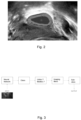
Fig. 2 is an ultrasound image processed for the calculation of a centroid of an internal area of the image; -
Fig. 3 is a block diagram of the method according to a non-limiting embodiment of the present invention; -
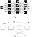
Fig 4 is a simplified scheme of a neural network used in the method of the present invention; -
Fig. 5 is a simplified block diagram of a computer graphics process performed by the method of the present invention; -
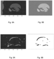
Fig. 6A is an ultrasound image andfigure 6B is the result of applying an entropic filter to the image ofFig. 6A ; -
Figs. 7A and 7B are respective binary images through a first and a second slightly different threshold andFig. 7C is a binarized image by a third threshold ofFigure 6B ; -
Fig. 8 illustrates two subsequent process steps of the image offigures 7A, 7B ; -
Fig. 9 illustrates two subsequent process steps of image 7C; -
Fig. 10 is an enlargement ofFig. 6A ; is -
Fig. 11 is an example of output of the process offigure 5 applied toFig. 6A . - While the invention is susceptible of various modifications and alternative constructions, some preferred embodiments are shown in the drawings and will be described in detail below.
- With reference to
FIG. 1 , images are shown relating to a non-limiting classification that finds inspiration from the IOTA classification, used to divide ovarian cysts and support a doctor in the diagnosis. It should be noted that this method is also applicable to the recognition and classification of renal and pancreatic cysts. - In greater detail, according to a non-limiting example of classification, a unilocular mass has a single region filled with liquid of various nature, which determines its ecogenicity, i.e. the intensity of echo reflected by the material. A non-ecogenic mass will appear as completely black. A hypoechoic mass will have soft and little diffused grays within the mass itself. An isoechoic mass will present a content which in gray scale will be comparable with the tissues external to the mass. A hyperechoic mass instead will be a particularly bright mass in ultrasound.
- In the event that the liquid is serum, the level of echogenicity will be low or not present (see the region below the unilocular mass shown in
figure 1 ). In the event that there are blood spills or the presence of mucinous masses in the cyst, this will be hypoechoic or at most isoechogenic (see the multilocular mass infigure 1 ). The presence of solid masses instead inside the cyst will give it an isoechoic or hyperechoic appearance. Serum and blood spills are not associated with an important level of risk, while the presence of solid masses is one of the main signs of potential malignancy, even if this is not a direct implication. - By solid mass instead we mean mass that protrudes from the walls of the cyst and whose growth tends to fill the cystic space. If this is greater than 3 mm in height, it is called papilla. In
figure 1 the solid unilocular mass has numerous papillae distributed along the entire cystic boundary, which assumes an irregular structure. - A multilocular cyst can have all the characteristics described above, but the cystic space is not unique but divided by septa in multiple chambers. The septum is therefore a portion of tissue that develops through the content of the cyst and divides it into multiple spaces.
- Intersections between septa or between septa and a cystic wall are a preferential place for the development of a solid mass and must be adequately monitored.
- A mass is instead defined as solid when the solid component has occupied the cystic space for more than 80% of the mass itself; an example of how a computation graphics algorithm can associate binary segments of an image with a cyst is described below to define the overall area of the cyst and calculate the serous percentage. In addition, it is also possible through appropriate training of the neural network, that the latter is able to classify images related to solid or serous unilocular cysts. Basically, the level of risk associated with the mass increases with this classification, starting from a level of less than 1% for a unilocular serum mass, up to a high level of risk for solid masses, that can therefore be defined clear carcinomas.
- Lastly, for the frames of clips that cannot be associated with any of the classes described above, a 'miscellaneous' class is preferably associated, for example called background. This therefore does not only include ultrasounds regarding tissues of other organs and completely "gray", but can include areas that recall the cystic shape and echogenicity, such as for example intestinal loops, blood vessels, etc., or transition areas between the cystic mass and the adjacent tissues, therefore regions where the quality of the cystic image would not be high enough to allow the accurate training of the algorithms and would even risk affecting it. In this regard, the background image shown in
figure 1 was chosen as representative of the class. - A two-dimensional ultrasound image, whether selected by the operator who performs the ultrasound and stored by means of a special ultrasound function or frame of a stored ultrasound clip, is initially processed to allow a better operation of the method according to the present invention. In particular, the image is cut out around the region of interest (ROI - automatically identified for example through template matching or through crop / crop based on a frame of pre-set size and position without human intervention) and segmented through global thresholding; both of these steps respectively enable to remove unnecessary parts from the image (in particular the metadata, imprinted as a watermark on the image) and to ease the application of computational graphics algorithms which will be more detailed below.
- In addition, the image is processed to reduce speckle noise, i.e. despeckling.
- Despeckling is necessary to prevent variations in brightness linked to the phenomenon of sound wave interference from covering or merging with the salient aspects of the image. The chosen filtering is median filtering. Other alternatives are, but are not limited to, algorithms such as anisotropic diffusion or wavelet denoising.
- A subsequent phase of edge detection has the object of extracting the contours of the region of interest within the image, in order to derive relevant geometric features (cyst area, contours), as shown in a first example in
Figure 2 . - In general, it is assumed that the different regions of the ultrasound image are characterized by different levels of entropy: low, in correspondence with uniform areas, such as for example the content of serous cysts, high in correspondence with abrupt variations, typically in correspondence with boundaries. These algorithms will be described in more detail later.
- The automatic classification of the main tumor features of the ovary, such as loculi and papillae, is carried out according to the present invention through feed-forward neural networks. A feed-forward network is an acyclic graph, structured in a sequence of layers. This architecture makes the network particularly suitable for performing the role of classifier.
- In a classification problem, given a set of objects, such as, for example, several ultrasound images of cysts, the network must be able to place each of them in the respective class, for example to determine if the cyst in a given ultrasound image belongs to the class of unilocular cysts or to that of multiloculars.
- To accomplish this task, the network generates increasingly abstract and complex representations of the object to be classified along the layers, emphasizing the characteristics that makes it a member of a given class rather than another (
Figure 4 ). However, the peculiar feature that distinguishes neural networks from other types of classifiers, is that the former are able to make these features abstract through a set of examples, without these having to be specified a priori. - During this step, called training, the network assigns for each example the probability that it belongs to a certain class rather than another. If the classification has been successful, the class associated with the highest probability will coincide with the actual class to which the object belongs.
- However, this condition is reached only possibly at the end of the learning step, during which the synaptic connections of the network are modified in such a way as to minimize a cost function that contains information on the number of incorrectly classified examples. In the specific case of cyst classification, one of the most used in Machine Learning was chosen as the cost function, namely the cross-entropy function:

- The cost function is optimized by means of optimization algorithms, such as for example Gradient Descent (GD) and its variants. These algorithms provide the prescription, or the learning rule, through which the network connections must be modified in such a way as to allow it to correctly classify the whole set of examples. In particular, in the case of the GD and its variants, the synaptic connections are updated along the direction of the gradient:

- To improve the performance of the network in general, there are functions that can prevent overfitting on training examples, such as dropouts.
- It is also possible to use convolutional feed-forward networks. In an initial phase of the project, it was decided to successfully implement a simpler type of feed-forward network, namely the Perceptron multilayer. In the preferred but non-limiting case in which the ultrasound scan is classified as containing a serous cyst, it is possible, with appreciable results, to locate it and automatically extract some geometric characteristics. Specifically, the two steps of localization and geometric characterization are preferably approached with computational graphics techniques.
- According to the claimed invention, localization is performed by a neural network, in particular by a convolutional neural network. Preferably, with reference to localization, in order to localize the position of the serous cyst, several complexities must be taken into account, including:
- Multilocular cyst: it is common for the cyst to present several loculi and the potential presence of more than one serous area should be taken into account;
- Serum and Mucin: the serum must also be identified in the presence of mucus;
- Visually similar ultrasound areas that do not represent serous cysts. It is necessary to distinguish between generic dark parts and dark parts that actually represent the cyst serum.
- To take all these elements into consideration, an approach is applied (
figure 5 ) that approximates the cyst from the inside and from the outside; that is, it aims to recognize the serous (internal) components and the boundary tissues. This is useful since the serous component alone may not be totally indicative of pathologies and, in general, the boundary tissues represent an important element for evaluating the state of the cyst. - In the
preprocessing step 100 an ultrasound cropping is performed so as to eliminate unnecessary portions of the ultrasound, such as rasterized metadata with the actual ultrasound image (e.g. patient registry, echo scale in mm). The entropy of the image is then calculated by means of an entropic filter (figure 6 ) in order to make more evident the areas whose gray levels are homogeneous. Indicatively this makes the serous areas and the surrounding tissues more visible, when actually visually homogeneous. Alternatively, it is possible to apply filters to Hessian matrices. - Subsequently, the crop of the entropic image is binarized through specific thresholds and then segmented 110 in order to extract three different segmentations. Two of these (
figures 7A, 7B ) represent an approximation of the serous zone with respective different but both low thresholds to identify the areas with high contrast and low entropy, the remaining instead the boundary tissues (figure 7C ) through the application of a high threshold, i.e. to identify areas with high entropy. The two approximations of the serous zone, therefore internal to the cyst, have slightly different visual characteristics and are useful for validating the results. In particular, the two internal segmentations differ in that the respective thresholds adopted for binarization have similar but not identical values. - Specifically, taken the two segmentations from the inside, the centroids of the connected dark components are calculated, i.e. the closed areas representing the serous areas, of an image (
figure 8A ) and a check is made that they are contained within the connected components of the other segmentation with low threshold. When this and other conditions have occurred, e.g. connected component area above a predetermined and adjustable threshold, distant connected component, e.g. outside a predetermined and adjustable limit distance, from the boundaries of the echo graphic image, and a predetermined and adjustable form factor on the basis of the relationship between a major axis and a minor axis of the component connected approximated with an ellipse, then the components are considered connected as actually representing a serous zone (figure 8B ). - When the roughness of the perimeters of the identified areas is also considered, a relatively high value, e.g. beyond an adjustable threshold, is associated with the presence of papillae. It is understood that the roughness and related quantities such as local peak, local valley, reference line, peak and valley, can be calculated at the perimeter of a connected area, preferably of a binary image, in the same way as it happens to the surface of a body.
- In this way it is possible to locate a number of serous zones as desired, the maximum number of which is defined by means of a specific modifiable parameter; consequently the method of the present invention is capable of identifying multilocular cysts and locating their individual loculi.
- The
external segmentation 130, that is, the one that identifies the contours of the cyst, must be compared with the serous areas actually identified and selected instep 120. - Preferably, only the contours sufficiently close to the serum are selected. To calculate the distance between the boundary tissues and the serous area, the Euclidean distances between the centroids of the serous zones (
figure 8B ) and those of the individual segmented boundaries (figure 9A ) are considered: if this distance is less than a predefined and adjustable threshold, the examined boundary is associated with the zone and therefore selected (figure 9B ). The default threshold can be fixed or adaptive, i.e. be expressed as a function of the size of the serous zone, for example of one or both of the half axes. In this way each frame will be associated with its boundaries. - Once both the serous areas and the relative boundaries have been extracted, this information is merged into a
single image 140. This eases the visual identification of the areas of interest and enables easier reading of the ultrasound scan. - To further ease the reading of the ultrasound image, the method of the present invention includes the step of calculating some geometric properties of the cyst. Specifically, all the measures are calculated which, in themselves, are calculated in a known way and not further described. For clarity, the most useful and significant ones are:
- Minor and major axes of the ellipse most similar to the identified area: they represent a good estimate of measurement (in pixels) of the width and height of the cyst.
- Orientation: i.e. the angle between the horizontal reference axis of the image and the major axis.
- Centroid: the center of gravity of the identified area.
- Cyst area: measured in pixels, it approximates the effective extent of the cyst.
-
Figures 10 and 11 respectively illustrate a captured ultrasound image and the result of the graphic processing of the method according to the present invention. - The previous paragraphs refer to the application of the method according to the present invention to a single ultrasound image.
- According to the present invention, it is also possible to process the frames of an ultrasound clip as follows.
- Each frame of the clip is labeled so as to associate a numerical label for each individual frame that makes up the clip. The clip is then associated with a list that reports the numeric label for each frame that composes the video and each image is processed as described in the preceding paragraphs so that the list provides, for each frame identified by a label, the relative class and subclass.
- The list is subsequently processed in order to provide an overall classification of the cyst represented by the frames, whereas some frames may not include a cyst, that instead is present in other frames.
- For this purpose, in order to classify the cyst to which the clip refers, multilocular sub-classification prevails over unilocular sub-classification, i.e. if at least one frame of the clip is multilocular, the cyst is classified as multilocular. Similarly, if at least one frame is classified as solid, this class prevails to describe the cyst if other frames are classified as serous.
- Furthermore, the counting of the frames can also be performed by storing the position of the centroids of each loculum for each frame. If in a frame a centroid of a loculum has a greater distance than a predetermined threshold compared to other centroids of other loculi, this loculum is considered as an additional loculum and not as a loculum already present in other frames and therefore the total number of loculi is updated accordingly.
Claims (11)
- A computer based method for finding and characterizing an anatomic mass of a cyst and providing a class indicating the presence of a serum in the cyst and a subclass indicating the presence of one or more serous loculi in the cyst and/or the number thereof, the method comprising the steps of:(a) obtaining at least an ultrasound image of an examined anatomic entity including the anatomic mass;(b) using a first algorithm based on a neural network elaborating the image for outputting the characterization as a cyst and/or the class of the cyst;(c) using a second algorithm of computational graphics for:i. identifying inner regions of the cyst, each of said inner regions being a fluid region or a boundary region, based on an at least a first and a second threshold applied to a mathematical function of the echogenicity of the image configured such as to increase the differentiation between a low echogenic level fluid region and a high echogenic level surrounding boundary solid region;ii. classifying the inner region as a serous region or a solid region based on a quantitative morphologic parameter of the inner region and on a aggregation model generating an association between at least a fluid internal region and a boundary internal region when a proximity condition is verified between the regions;iii. counting the number of serous regions as serous loculi and/or a number of solid regions;(d) providing an image classification comprising the class and the sub-class based on the combination of outputs from the first and second algorithm.
- The method according to claim 1, wherein the step of identifying comprises the step of associating to the same anatomical entity, wherein the anatomical entity comprises the cyst, a region of a low echogenic level to a region of a high echogenic level, based on the distance between the relative centroids.
- The method according to claim 1 or 2, wherein the step of obtaining comprises the step of obtaining a multiframe clip of ultrasound images of the anatomic mass; and comprising the step of providing an overall class or sub-class of the clip such that multilocular prevails over unilocular and/or solid prevails over serous when a first sub-class of a first frame and a second sub-class of a second frame image of the clip have different outputs from said step of providing an image classification.
- The method of either claim 1, 2 or 3, wherein the step of using a second algorithm comprises the step of outputting the number of loculi as a part of the subclass.
- The method of any of the preceding claims, wherein the step of using the second algorithm comprises the step of comparing an overall area of the serous regions and an overall area of the solid regions and that the step of providing the classification comprises the step of indicating the subclass based on the step of comparing.
- The method of any of the preceding claims, wherein the step of using the second algorithm is such that serous regions and boundary regions around serous regions are identified as respective geometrical features, that the relative centroids are calculated as respective further geometrical features and that the positions of the centroids are calculated as respective quantitative parameters.
- The method of claim 6, wherein the step of using the second algorithm is such that the centroid is associated to a given serous region based on predefined maximum distance between the centroid of the serous region and the centroids of the boundary regions.
- The method of any of the preceding claims, wherein the step of using the second algorithm is such that the mathematical function is an entropic filter or a Hessian matrix filter applied to the image.
- The method of any of the preceding claims, wherein the neural network is either a feed-forward convolutional network or a feed-forward multilayer network.
- The method according to any of the preceding claims, wherein both the first and the second algorithm output both a serous or solid classification and unilocular or multilocular sub-classification and wherein the step of providing is based in that the outputs of the first and second algorithm are the same.
- The method according to any of the preceding claims, wherein the second algorithm is a region extraction algorithm.
Applications Claiming Priority (2)
| Application Number | Priority Date | Filing Date | Title |
|---|---|---|---|
| IT102019000007806A IT201900007806A1 (en) | 2019-05-31 | 2019-05-31 | Computerized method of classifying a mass of an organ as a cyst |
| PCT/IB2020/055058 WO2020240455A1 (en) | 2019-05-31 | 2020-05-28 | A computer based method for classifying a mass of an organ as a cyst |
Publications (3)
| Publication Number | Publication Date |
|---|---|
| EP3977400A1 EP3977400A1 (en) | 2022-04-06 |
| EP3977400C0 EP3977400C0 (en) | 2025-03-19 |
| EP3977400B1 true EP3977400B1 (en) | 2025-03-19 |
Family
ID=68343200
Family Applications (1)
| Application Number | Title | Priority Date | Filing Date |
|---|---|---|---|
| EP20742901.0A Active EP3977400B1 (en) | 2019-05-31 | 2020-05-28 | A computer based method for classifying a mass of an organ as a cyst |
Country Status (6)
| Country | Link |
|---|---|
| US (1) | US12089987B2 (en) |
| EP (1) | EP3977400B1 (en) |
| JP (1) | JP7692612B2 (en) |
| CN (1) | CN113924046B (en) |
| IT (1) | IT201900007806A1 (en) |
| WO (1) | WO2020240455A1 (en) |
Families Citing this family (3)
| Publication number | Priority date | Publication date | Assignee | Title |
|---|---|---|---|---|
| EP4309586A4 (en) * | 2021-03-19 | 2024-09-04 | FUJIFILM Corporation | ULTRASONIC DIAGNOSTIC DEVICE AND METHOD FOR CONTROLLING ULTRASONIC DIAGNOSTIC DEVICE |
| EP4141785A1 (en) | 2021-08-30 | 2023-03-01 | Sycai Technologies, Sl | Method and computer program for the automatic classification of pancreatic cysts |
| WO2023242805A1 (en) * | 2022-06-15 | 2023-12-21 | Syndiag S.R.L. | Processing method of an ultrasound image to associate information supporting tumour diagnosis of cysts and solid lesions |
Family Cites Families (12)
| Publication number | Priority date | Publication date | Assignee | Title |
|---|---|---|---|---|
| US5487387A (en) * | 1994-06-03 | 1996-01-30 | Duke University | Method and apparatus for distinguishing between solid masses and fluid-filled cysts |
| US5984870A (en) * | 1997-07-25 | 1999-11-16 | Arch Development Corporation | Method and system for the automated analysis of lesions in ultrasound images |
| IL127254A0 (en) * | 1998-11-25 | 1999-09-22 | Univ Ramot | Method and system for automatic classification and quantitative evaluation of adnexal masses bases on a cross-sectional or projectional images of the adnex |
| JP2009148499A (en) * | 2007-12-21 | 2009-07-09 | Panasonic Corp | Ultrasonic diagnostic equipment |
| US20130289381A1 (en) * | 2011-11-02 | 2013-10-31 | Seno Medical Instruments, Inc. | Dual modality imaging system for coregistered functional and anatomical mapping |
| TWI598577B (en) * | 2014-03-28 | 2017-09-11 | 梅托珍尼克斯股份有限公司 | Method for obtaining data for determining possibility of carcinogenesis of endometriotic ovarian cysts, and diagnostic apparatus thereof |
| CN105447872A (en) * | 2015-12-03 | 2016-03-30 | 中山大学 | Method for automatically identifying liver tumor type in ultrasonic image |
| US11880980B2 (en) * | 2016-06-17 | 2024-01-23 | Children's National Medical Center | Medical anatomy quantification: computer-aided diagnosis tool |
| CN106530298A (en) * | 2016-11-14 | 2017-03-22 | 同济大学 | Three-way-decision-based liver tumor CT image classification method |
| CA3067824C (en) * | 2017-06-26 | 2025-05-27 | The Research Foundation For The State University Of New York | System, method, and computer-accessible medium for virtual pancreatography |
| US11553900B2 (en) * | 2018-05-08 | 2023-01-17 | Fujifilm Sonosite, Inc. | Ultrasound system with automated wall tracing |
| CN108875829A (en) * | 2018-06-20 | 2018-11-23 | 鲁东大学 | A kind of classification method and system of tumor of breast image |
-
2019
- 2019-05-31 IT IT102019000007806A patent/IT201900007806A1/en unknown
-
2020
- 2020-05-28 WO PCT/IB2020/055058 patent/WO2020240455A1/en not_active Ceased
- 2020-05-28 US US17/615,102 patent/US12089987B2/en active Active
- 2020-05-28 JP JP2021564098A patent/JP7692612B2/en active Active
- 2020-05-28 EP EP20742901.0A patent/EP3977400B1/en active Active
- 2020-05-28 CN CN202080040553.3A patent/CN113924046B/en active Active
Also Published As
| Publication number | Publication date |
|---|---|
| EP3977400C0 (en) | 2025-03-19 |
| US12089987B2 (en) | 2024-09-17 |
| IT201900007806A1 (en) | 2020-12-01 |
| CN113924046A (en) | 2022-01-11 |
| JP2022537481A (en) | 2022-08-26 |
| EP3977400A1 (en) | 2022-04-06 |
| CN113924046B (en) | 2025-01-28 |
| US20220230325A1 (en) | 2022-07-21 |
| JP7692612B2 (en) | 2025-06-16 |
| WO2020240455A1 (en) | 2020-12-03 |
Similar Documents
| Publication | Publication Date | Title |
|---|---|---|
| EP2378978B1 (en) | Method and system for automated generation of surface models in medical images | |
| EP0757544B1 (en) | Computerized detection of masses and parenchymal distortions | |
| US6309353B1 (en) | Methods and apparatus for tumor diagnosis | |
| US10303986B2 (en) | Automated measurement of brain injury indices using brain CT images, injury data, and machine learning | |
| EP2916737B1 (en) | System and method for automated detection of lung nodules in medical images | |
| Kumar et al. | Texture Pattern Based Lung Nodule Detection (TPLND) Technique in CT Images | |
| US9159127B2 (en) | Detecting haemorrhagic stroke in CT image data | |
| Villalobos-Castaldi et al. | A fast, efficient and automated method to extract vessels from fundus images | |
| EP3035287A1 (en) | Image processing apparatus, and image processing method | |
| US20110002523A1 (en) | Method and System of Segmenting CT Scan Data | |
| US12089987B2 (en) | Computer based method for classifying a mass of an organ as a cyst | |
| Rebelo et al. | Automatic epicardial fat segmentation and volume quantification on non-contrast cardiac Computed Tomography | |
| Giachetti et al. | Multiresolution localization and segmentation of the optical disc in fundus images using inpainted background and vessel information | |
| Jayachandran et al. | X-ray image analysis in identification of bone cancer using laws features and machine learning model | |
| Napier et al. | A CAD system for brain haemorrhage detection in head CT scans | |
| Undrill et al. | Use of texture analysis and boundary refinement to delineate suspicious masses in mammography | |
| Gomathi et al. | Detection of mammogram using improved watershed segmentation algorithm and classifying with feed forward neural network (FNN) | |
| Medhi et al. | Improved analysis of diabetic maculopathy using level set spatial fuzzy clustering | |
| CN108765415A (en) | There is one kind shade management to monitor system | |
| Kathiravan et al. | Optimization Enabled Online Tiger‐Claw Fuzzy Region With Clustering Based Neovascularization Segmentation and Classification Using YOLO‐V5 From Retinal Fundus Images | |
| Parimala et al. | Assessment on liver disease classification using medical image processing | |
| Bargsten et al. | Deep learning for guidewire detection in intravascular ultrasound images | |
| Catalá | Pensión de jubilación y trabajadores a tiempo parcial,¿ posible discriminación? | |
| Himaga et al. | Accurate retinal blood vessel segmentation by using multi-resolution matched filtering and directional region growing | |
| Paradowski et al. | Capillary blood vessel tracking using polar coordinates based model identification |
Legal Events
| Date | Code | Title | Description |
|---|---|---|---|
| STAA | Information on the status of an ep patent application or granted ep patent |
Free format text: STATUS: UNKNOWN |
|
| STAA | Information on the status of an ep patent application or granted ep patent |
Free format text: STATUS: THE INTERNATIONAL PUBLICATION HAS BEEN MADE |
|
| PUAI | Public reference made under article 153(3) epc to a published international application that has entered the european phase |
Free format text: ORIGINAL CODE: 0009012 |
|
| STAA | Information on the status of an ep patent application or granted ep patent |
Free format text: STATUS: REQUEST FOR EXAMINATION WAS MADE |
|
| 17P | Request for examination filed |
Effective date: 20211105 |
|
| AK | Designated contracting states |
Kind code of ref document: A1 Designated state(s): AL AT BE BG CH CY CZ DE DK EE ES FI FR GB GR HR HU IE IS IT LI LT LU LV MC MK MT NL NO PL PT RO RS SE SI SK SM TR |
|
| DAV | Request for validation of the european patent (deleted) | ||
| DAX | Request for extension of the european patent (deleted) | ||
| GRAP | Despatch of communication of intention to grant a patent |
Free format text: ORIGINAL CODE: EPIDOSNIGR1 |
|
| STAA | Information on the status of an ep patent application or granted ep patent |
Free format text: STATUS: GRANT OF PATENT IS INTENDED |
|
| INTG | Intention to grant announced |
Effective date: 20241122 |
|
| GRAS | Grant fee paid |
Free format text: ORIGINAL CODE: EPIDOSNIGR3 |
|
| GRAA | (expected) grant |
Free format text: ORIGINAL CODE: 0009210 |
|
| STAA | Information on the status of an ep patent application or granted ep patent |
Free format text: STATUS: THE PATENT HAS BEEN GRANTED |
|
| AK | Designated contracting states |
Kind code of ref document: B1 Designated state(s): AL AT BE BG CH CY CZ DE DK EE ES FI FR GB GR HR HU IE IS IT LI LT LU LV MC MK MT NL NO PL PT RO RS SE SI SK SM TR |
|
| REG | Reference to a national code |
Ref country code: GB Ref legal event code: FG4D |
|
| REG | Reference to a national code |
Ref country code: CH Ref legal event code: EP |
|
| REG | Reference to a national code |
Ref country code: DE Ref legal event code: R096 Ref document number: 602020047934 Country of ref document: DE |
|
| REG | Reference to a national code |
Ref country code: IE Ref legal event code: FG4D |
|
| REG | Reference to a national code |
Ref country code: CH Ref legal event code: PK Free format text: BERICHTIGUNGEN |
|
| U01 | Request for unitary effect filed |
Effective date: 20250414 |
|
| U07 | Unitary effect registered |
Designated state(s): AT BE BG DE DK EE FI FR IT LT LU LV MT NL PT RO SE SI Effective date: 20250422 |
|
| U20 | Renewal fee for the european patent with unitary effect paid |
Year of fee payment: 6 Effective date: 20250520 |
|
| PG25 | Lapsed in a contracting state [announced via postgrant information from national office to epo] |
Ref country code: RS Free format text: LAPSE BECAUSE OF FAILURE TO SUBMIT A TRANSLATION OF THE DESCRIPTION OR TO PAY THE FEE WITHIN THE PRESCRIBED TIME-LIMIT Effective date: 20250619 |
|
| PGFP | Annual fee paid to national office [announced via postgrant information from national office to epo] |
Ref country code: GB Payment date: 20250524 Year of fee payment: 6 |
|
| PG25 | Lapsed in a contracting state [announced via postgrant information from national office to epo] |
Ref country code: NO Free format text: LAPSE BECAUSE OF FAILURE TO SUBMIT A TRANSLATION OF THE DESCRIPTION OR TO PAY THE FEE WITHIN THE PRESCRIBED TIME-LIMIT Effective date: 20250619 |
|
| PG25 | Lapsed in a contracting state [announced via postgrant information from national office to epo] |
Ref country code: HR Free format text: LAPSE BECAUSE OF FAILURE TO SUBMIT A TRANSLATION OF THE DESCRIPTION OR TO PAY THE FEE WITHIN THE PRESCRIBED TIME-LIMIT Effective date: 20250319 |
|
| PG25 | Lapsed in a contracting state [announced via postgrant information from national office to epo] |
Ref country code: GR Free format text: LAPSE BECAUSE OF FAILURE TO SUBMIT A TRANSLATION OF THE DESCRIPTION OR TO PAY THE FEE WITHIN THE PRESCRIBED TIME-LIMIT Effective date: 20250620 |
|
| PG25 | Lapsed in a contracting state [announced via postgrant information from national office to epo] |
Ref country code: SM Free format text: LAPSE BECAUSE OF FAILURE TO SUBMIT A TRANSLATION OF THE DESCRIPTION OR TO PAY THE FEE WITHIN THE PRESCRIBED TIME-LIMIT Effective date: 20250319 |
|
| PG25 | Lapsed in a contracting state [announced via postgrant information from national office to epo] |
Ref country code: ES Free format text: LAPSE BECAUSE OF FAILURE TO SUBMIT A TRANSLATION OF THE DESCRIPTION OR TO PAY THE FEE WITHIN THE PRESCRIBED TIME-LIMIT Effective date: 20250319 |
|
| PG25 | Lapsed in a contracting state [announced via postgrant information from national office to epo] |
Ref country code: PL Free format text: LAPSE BECAUSE OF FAILURE TO SUBMIT A TRANSLATION OF THE DESCRIPTION OR TO PAY THE FEE WITHIN THE PRESCRIBED TIME-LIMIT Effective date: 20250319 |
|
| PG25 | Lapsed in a contracting state [announced via postgrant information from national office to epo] |
Ref country code: CZ Free format text: LAPSE BECAUSE OF FAILURE TO SUBMIT A TRANSLATION OF THE DESCRIPTION OR TO PAY THE FEE WITHIN THE PRESCRIBED TIME-LIMIT Effective date: 20250319 |
|
| PG25 | Lapsed in a contracting state [announced via postgrant information from national office to epo] |
Ref country code: SK Free format text: LAPSE BECAUSE OF FAILURE TO SUBMIT A TRANSLATION OF THE DESCRIPTION OR TO PAY THE FEE WITHIN THE PRESCRIBED TIME-LIMIT Effective date: 20250319 |
|
| PG25 | Lapsed in a contracting state [announced via postgrant information from national office to epo] |
Ref country code: IS Free format text: LAPSE BECAUSE OF FAILURE TO SUBMIT A TRANSLATION OF THE DESCRIPTION OR TO PAY THE FEE WITHIN THE PRESCRIBED TIME-LIMIT Effective date: 20250719 |
|
| REG | Reference to a national code |
Ref country code: CH Ref legal event code: H13 Free format text: ST27 STATUS EVENT CODE: U-0-0-H10-H13 (AS PROVIDED BY THE NATIONAL OFFICE) Effective date: 20251223 |
|
| PG25 | Lapsed in a contracting state [announced via postgrant information from national office to epo] |
Ref country code: CH Free format text: LAPSE BECAUSE OF NON-PAYMENT OF DUE FEES Effective date: 20250531 |
|
| PLBE | No opposition filed within time limit |
Free format text: ORIGINAL CODE: 0009261 |
|
| STAA | Information on the status of an ep patent application or granted ep patent |
Free format text: STATUS: NO OPPOSITION FILED WITHIN TIME LIMIT |
|
| REG | Reference to a national code |
Ref country code: CH Ref legal event code: L10 Free format text: ST27 STATUS EVENT CODE: U-0-0-L10-L00 (AS PROVIDED BY THE NATIONAL OFFICE) Effective date: 20260128 |
|
| PG25 | Lapsed in a contracting state [announced via postgrant information from national office to epo] |
Ref country code: MC Free format text: LAPSE BECAUSE OF FAILURE TO SUBMIT A TRANSLATION OF THE DESCRIPTION OR TO PAY THE FEE WITHIN THE PRESCRIBED TIME-LIMIT Effective date: 20250319 |
|
| 26N | No opposition filed |
Effective date: 20251222 |
|
| PG25 | Lapsed in a contracting state [announced via postgrant information from national office to epo] |
Ref country code: IE Free format text: LAPSE BECAUSE OF NON-PAYMENT OF DUE FEES Effective date: 20250528 |