EP3977133B1 - Method for identifying ubiquitin and ubiquitin-like enzyme activities - Google Patents
Method for identifying ubiquitin and ubiquitin-like enzyme activities Download PDFInfo
- Publication number
- EP3977133B1 EP3977133B1 EP20728499.3A EP20728499A EP3977133B1 EP 3977133 B1 EP3977133 B1 EP 3977133B1 EP 20728499 A EP20728499 A EP 20728499A EP 3977133 B1 EP3977133 B1 EP 3977133B1
- Authority
- EP
- European Patent Office
- Prior art keywords
- proteins
- protein
- beads
- ubiquitin
- sumo
- Prior art date
- Legal status (The legal status is an assumption and is not a legal conclusion. Google has not performed a legal analysis and makes no representation as to the accuracy of the status listed.)
- Active
Links
Images



Classifications
-
- G—PHYSICS
- G01—MEASURING; TESTING
- G01N—INVESTIGATING OR ANALYSING MATERIALS BY DETERMINING THEIR CHEMICAL OR PHYSICAL PROPERTIES
- G01N33/00—Investigating or analysing materials by specific methods not covered by groups G01N1/00 - G01N31/00
- G01N33/48—Biological material, e.g. blood, urine; Haemocytometers
- G01N33/50—Chemical analysis of biological material, e.g. blood, urine; Testing involving biospecific ligand binding methods; Immunological testing
- G01N33/68—Chemical analysis of biological material, e.g. blood, urine; Testing involving biospecific ligand binding methods; Immunological testing involving proteins, peptides or amino acids
- G01N33/6803—General methods of protein analysis not limited to specific proteins or families of proteins
- G01N33/6845—Methods of identifying protein-protein interactions in protein mixtures
-
- G—PHYSICS
- G01—MEASURING; TESTING
- G01N—INVESTIGATING OR ANALYSING MATERIALS BY DETERMINING THEIR CHEMICAL OR PHYSICAL PROPERTIES
- G01N33/00—Investigating or analysing materials by specific methods not covered by groups G01N1/00 - G01N31/00
- G01N33/48—Biological material, e.g. blood, urine; Haemocytometers
- G01N33/50—Chemical analysis of biological material, e.g. blood, urine; Testing involving biospecific ligand binding methods; Immunological testing
- G01N33/53—Immunoassay; Biospecific binding assay; Materials therefor
- G01N33/573—Immunoassay; Biospecific binding assay; Materials therefor for enzymes or isoenzymes
-
- G—PHYSICS
- G01—MEASURING; TESTING
- G01N—INVESTIGATING OR ANALYSING MATERIALS BY DETERMINING THEIR CHEMICAL OR PHYSICAL PROPERTIES
- G01N33/00—Investigating or analysing materials by specific methods not covered by groups G01N1/00 - G01N31/00
- G01N33/48—Biological material, e.g. blood, urine; Haemocytometers
- G01N33/50—Chemical analysis of biological material, e.g. blood, urine; Testing involving biospecific ligand binding methods; Immunological testing
- G01N33/53—Immunoassay; Biospecific binding assay; Materials therefor
- G01N33/531—Production of immunochemical test materials
- G01N33/532—Production of labelled immunochemicals
- G01N33/533—Production of labelled immunochemicals with fluorescent label
-
- G—PHYSICS
- G01—MEASURING; TESTING
- G01N—INVESTIGATING OR ANALYSING MATERIALS BY DETERMINING THEIR CHEMICAL OR PHYSICAL PROPERTIES
- G01N33/00—Investigating or analysing materials by specific methods not covered by groups G01N1/00 - G01N31/00
- G01N33/48—Biological material, e.g. blood, urine; Haemocytometers
- G01N33/50—Chemical analysis of biological material, e.g. blood, urine; Testing involving biospecific ligand binding methods; Immunological testing
- G01N33/53—Immunoassay; Biospecific binding assay; Materials therefor
- G01N33/543—Immunoassay; Biospecific binding assay; Materials therefor with an insoluble carrier for immobilising immunochemicals
- G01N33/54313—Immunoassay; Biospecific binding assay; Materials therefor with an insoluble carrier for immobilising immunochemicals the carrier being characterised by its particulate form
- G01N33/54326—Magnetic particles
-
- G—PHYSICS
- G01—MEASURING; TESTING
- G01N—INVESTIGATING OR ANALYSING MATERIALS BY DETERMINING THEIR CHEMICAL OR PHYSICAL PROPERTIES
- G01N33/00—Investigating or analysing materials by specific methods not covered by groups G01N1/00 - G01N31/00
- G01N33/48—Biological material, e.g. blood, urine; Haemocytometers
- G01N33/50—Chemical analysis of biological material, e.g. blood, urine; Testing involving biospecific ligand binding methods; Immunological testing
- G01N33/68—Chemical analysis of biological material, e.g. blood, urine; Testing involving biospecific ligand binding methods; Immunological testing involving proteins, peptides or amino acids
- G01N33/6872—Intracellular protein regulatory factors and their receptors, e.g. including ion channels
-
- G—PHYSICS
- G01—MEASURING; TESTING
- G01N—INVESTIGATING OR ANALYSING MATERIALS BY DETERMINING THEIR CHEMICAL OR PHYSICAL PROPERTIES
- G01N2440/00—Post-translational modifications [PTMs] in chemical analysis of biological material
- G01N2440/36—Post-translational modifications [PTMs] in chemical analysis of biological material addition of addition of other proteins or peptides, e.g. SUMOylation, ubiquitination
-
- G—PHYSICS
- G01—MEASURING; TESTING
- G01N—INVESTIGATING OR ANALYSING MATERIALS BY DETERMINING THEIR CHEMICAL OR PHYSICAL PROPERTIES
- G01N2500/00—Screening for compounds of potential therapeutic value
-
- G—PHYSICS
- G01—MEASURING; TESTING
- G01N—INVESTIGATING OR ANALYSING MATERIALS BY DETERMINING THEIR CHEMICAL OR PHYSICAL PROPERTIES
- G01N2500/00—Screening for compounds of potential therapeutic value
- G01N2500/02—Screening involving studying the effect of compounds C on the interaction between interacting molecules A and B (e.g. A = enzyme and B = substrate for A, or A = receptor and B = ligand for the receptor)
Definitions
- the present invention relates to a method for identifying ubiquitin and ubiquitin-like enzymes activity, in particular enzyme activity further to a drug treatment.
- Post-translational protein modification is the chemical modification of proteins prior to or following protein biosynthesis, and includes phosphorylation, ubiquitylation, methylation, acetylation, and modifications by ubiquitin-like modifiers (UBLs).
- ULBs ubiquitin-like modifiers
- UDLs ubiquitin-like proteins
- UBLs proteins conjugated to UBLs are typically not targeted for degradation by the proteasome, but rather function in diverse regulatory activities. Attachment of UBLs might alter substrate conformation, affect the affinity for ligands or other interacting molecules, alter substrate localization and influence protein stability.
- UBLs are structurally similar to ubiquitin and are processed, activated, conjugated and released from conjugates by enzymatic steps that are similar to the corresponding mechanisms for ubiquitin. UBLs are also translated with C-terminal extensions that are processed to expose the invariant C-terminal GG motif. These modifiers have their own specific E1 (activating), E2 (conjugating) and E3 (ligating) enzymes that conjugate the UBLs to intracellular targets. These conjugates can be reversed by UBL-specific isopeptidases that have similar mechanisms to that of the deubiquitinating enzymes.
- UBLs play a central function in many cellular processes and can therefore be deregulated, such a deregulation being involved in many diseases and syndromes.
- the international application WO2004/020458 discloses methods and compositions for assaying for deubiquitinating agents that are enzymatic components of ubiquitin-mediated proteolysis, their function, and agents that modulate the activity of such deubiquitinating agents.
- the international application WO2016/034895 teaches a method for investigating molecular interactions and enzyme activities related to the ubiquitin-proteasome system (UPS), the method comprising: a) providing at least one surface with a target moiety bound to at least a portion thereof; b) exposing said surface to a medium comprising a labelled ubiquitin moiety such as ubiquitin, SUMO and NEDD8, and a component or components of the ubiquitination system suitable to conjugate the ubiquitin moiety to a target moiety.
- a labelled ubiquitin moiety such as ubiquitin, SUMO and NEDD8
- a component or components of the ubiquitination system suitable to conjugate the ubiquitin moiety to a target moiety.
- One aim of the invention is to propose such a method and kit liable to carry out such a method.
- the invention relates to a method for quantifying the activity of the proteins or enzymes involved in the conjugation of the SUMO/Ubiquitin/Nedd8 proteins in a cell of a biological sample, said method comprising:
- the invention is based on the unexpected observation made by the inventors that evaluation of the modifications by Ubiquitin, SUMO and NEDD8 of at least 3 specific proteins belonging to a group of 46 proteins is highly significant to measure the activity of said pathways and to monitor the effect of drugs targeting these pathways.
- purified determined proteins (said at least 3 proteins) that are modified by neither ubiquitin, nor by SUMO, nor by NEDD8, the inventors are able to easily quantify the activity of the SUMO/Ubiquitin/Nedd8 pathway.
- the cellular extract that is used to determine the activity of the proteins/enzymes involved in the conjugation of the SUMO/Ubiquitin/Nedd8 proteins is in a determined state liable to modify such activity (for instance treated with a drug or a compound, placed in a specific environment, placed at a determined temperature and/or pressure), it is then relevant to compare the level of modification with the level of modification of cells that are not placed in this determined state, i.e. control cells.
- Ubiquitin is a globular protein consisting of 76 amino acid residues, and the UPS (ubiquitin-proteasome system) is responsible for degrading 80-90% of intracellular proteins.
- Ub binds the lysine residues of target proteins by a series of enzymes including the ubiquitin-activating enzyme E1, ubiquitin-conjugating enzyme E2 and the ubiquitin E3 ligases. Subsequently, these ubiquitylated proteins are generally recognized and degraded by the 26S proteasome.
- the structures of all three SUMO1, SUMO2 and SUMO3 paralogues resemble the globular and compact Ub-like fold.
- the differences of SUMO1 and SUMO2 are mostly found in the second ⁇ -strand and the ⁇ -helix of both proteins.
- different SUMO paralogues appear to share common properties but also have some distinct functions.
- the promyelocytic leukemia protein is conjugated to all three SUMO paralogs, whereas RanGAP1 is preferentially modified with SUMO1 and topoisomerase II with SUMO2/3 during mitosis.
- SUMO1 and SUMO2/3 are mostly found in the nucleoplasm, SUMO1 is uniquely found within the nucleoli, the nuclear envelope, and cytoplasmic foci, whereas SUMO2/3 are accrued on chromosomes at an earlier point in the nuclear reformation process. Interestingly, there is a larger pool of free, non-conjugated SUMO2/3 than of SUMO1.
- NEDD8 is an 81-amino acid protein with 9 kDa relative molecular mass and is 60% identical and 80% homologous to ubiquitin.
- NEDD8 has a dedicated E1-activating enzyme (AppBp1/UBA3, or NAE) and E2-conjugating enzymes (UBC12, UBE2F) and is essential for the enzymatic activity of the CRL family of E3 ligases, through conjugation to the cullin scaffold.
- Other components of the neddylation pathway include DEN1, which processes NEDD8 to its mature, 76-amino acid form, and the COP9 signalosome complex, which is responsible for removing NEDD8 from cullin proteins.
- CAND1 (cullin-associated and neddylation-dissociated) is an additional component that regulates CRL complex assembly by binding to the cullin in the absence of NEDD8 activation.
- the set of 46 protein (i.e. the set consisting of the 46 proteins) is constituted by the following proteins: [Table 1] Name SEQ ID Sequence ZMYM5 1 BEAN 2 OTUD6B 3 ANKRD3 9 4 LDLRAD 3 5 MXI1 6 OTUB2 7 TMEM92 8 UBE3A 9 WBP2 10 ARRDC3 11 ARRDC3 12 ANKRD1 3D 13 BIRC6 14 BIRC8 15 C18orf1 16 C9orf74 17 CUEDC1 18 DAZAP2 19 DDI1 20 DUS3L 21 FAM70B 22 IGHG3 23 KIAA024 7 24 LAPTM5 25 LDLRAD 4 26 LITAF 27 MLANA 28 NDFIP2 29 PMEPA1 30 PSMD4 31 PIAS2 32 POLI 33 RHOBT B1 34 RNF13 35 RNF4 36 RNF34 37 SAE2 38 TAGAP 39 TMEM13 9 40 TMEM17 4 41 TMEM55 B 42 TMEPAI 43
- the at least 3 proteins as set forth in SEQ ID NO: 1 to 3 are deposited on a support and then contacted with a cell extract of cells to be compared (e.g, cells that were treated or not with a compound liable to inhibit UBL pathways).
- the inventors evaluate specifically the modifications by Ubiquitin/SUMO/NEDD8 of the proteins SEQ ID NO: 1 to 3, and not all the proteins contained in the cell extract.
- the enzymes responsible of the addition of Ubiquitin/SUMO/NEDD8 contained in the extract will modify the proteins SEQ ID NO: 1 to 3.
- the Ubiquitination, SUMOylation and NEDDylation amount will therefore depend upon the activity of the enzymes, and is directly correlated to the effect of the condition to be analyzed (cell type, inhibitor).
- the proteins which are modified by ubiquitin and/or SUMO and/or NEDD8 proteins will therefore interact with antibodies directed against ubiquitin and/or SUMO and/or NEED8 proteins to form a molecular complex.
- the complexes can be detected by secondary antibodies that recognize (and interact with) constant part FC chain of said antibodies directed against ubiquitin and/or SUMO and/or NEDD8 proteins.
- Said complex can be identified when the secondary antibodies are labeled with reporter molecule, such as fluorescent protein, peroxidase, fluorescent dyes etc...
- reporter molecule such as fluorescent protein, peroxidase, fluorescent dyes etc...
- protein SEQ ID NO: 1 can be modified by SUMO1 and SUMO2
- protein SEQ ID NO: 2 is modified preferably by Ubiquitin
- protein SEQ ID NO: 3 can be modified by NEDD8.
- the invention relates to the above described method, wherein said method comprises a step of contacting a cellular extract with each protein of a group of 10 proteins, said group of 10 proteins belonging to a set of 46 proteins.
- the invention advantageously relates to the above mentioned method, said method comprising
- the invention relates to the method as defined above, wherein said method comprises a step of contacting a cellular extract with each protein of said set of 46 proteins.
- the invention advantageously relates to the above mentioned method, said method comprising
- the invention relates to the method as defined above, wherein said at least 3 proteins are supported by beads.
- the invention relates to the method as defined above, wherein said beads are fluorescent beads.
- the invention relates to the method as defined above, wherein each of said at least 3 protein is supported by a determined bead having a fluorescent property which is different from the fluorescent properties of the other beads, and wherein all the beads having the same fluorescent property supporting the same protein.
- the method can be performed with the XMap technology developed by Luminex. It consists in uniquely colored Mag-Plex metal-based microsphere (500 different colors with variable levels of two different dyes). They can be coupled with specific proteins using carboiimide coupling techniques. Protein-coupled beads can be multiplexed and used with cellular extracts as described in the examples thereafter. The different beads can be distinguished using dedicated analyzer or a flow cytometer.
- Proteins according to the invention are grafted covalently on the fluorescent beads by any means well known in the art.
- the invention relates to a method for quantifying the activity of the proteins/enzymes involved identifying the effect of a drug on protein modification by the conjugation of the SUMO/Ubiquitin/Nedd8 proteins in a cell of a biological sample, for example to identify the effect of a drug on UBL protein modification, said method comprising
- the invention advantageously relates to the above mentioned method, said method comprising
- the invention relates to the above mentioned method, said method comprising
- the invention relates to a method for identifying the effect of a drug on protein modification by the SUMO/Ubiquitin/Nedd8 proteins in a cell of a biological sample, said method comprising
- step a) and b) described above apply here mutatis mutandis.
- proteins SEQ ID NO: 1 to 3 are modified at a level similar to the modification obtained with a control cellular extract of the same nature (e.g cell that was not treated with the inhibitor).
- the tested condition e.g. drug treatment
- the amount of modification by Ubiquitin/SUMO/NEDD8 of proteins SEQ ID NO: 1 to 3 will be increased or decreased compared to the amount of modification observed in the control condition (e.g. non treated cells)
- the proteins which are modified by ubiquitin and/or SUMO and/or NEDD8 proteins will therefore interact with antibodies directed against ubiquitin and/or SUMO and/or NEED8 proteins to form a molecular complex.
- the complexes can be detected by secondary antibodies that recognize (and interact with) constant part FC chain of said antibodies directed against ubiquitin and/or SUMO and/or NEDD8 proteins.
- Said complex can be identified when the secondary antibodies are labeled with reporter molecule, such as fluorescent protein, peroxidase, fluorescent dyes etc...
- reporter molecule such as fluorescent protein, peroxidase, fluorescent dyes etc...
- This value is then compared to a reference value obtained for the same proteins contacted with a control cellular extract (e.g cells of the same nature than those treated with the inhibitor, but which were not treated with said inhibitor).
- a ratio is established between the first value, i.e. the value obtained with the cellular extract to be tested (e.g treated with the inhibitor) and the second value i.e. the value obtained with the control cellular extract (e.g not treated with the inhibitor).
- i is either 1, 2 or 3.
- the tested condition e.g. drug treatment
- the tested condition has no effect on UBL pathways when all the ratios calculated for each protein for each Ubiquitin, SUMO and NEDD8 are equal to about 1.
- the modifications that occur on proteins SEQ ID NO: 1 to 3 are similar between the cellular extract to be tested (e.g. treated with said drug) and the control (e.g. non treated).
- protein SEQ ID NO: 1 can be modified by SUMO1 and SUMO2
- protein SEQ ID NO: 2 is modified preferably by Ubiquitin
- protein SEQ ID NO: 3 can be modified by NEDD8.
- the invention relates to the above described method, wherein said method comprises a step of contacting a cellular extract to be tested (e.g of cell treated with said drug), with each protein of a group of 10 proteins, said group of 10 proteins belonging to a set of 46 proteins.
- the invention advantageously relates to the above mentioned method, said method comprising
- the invention relates to the method as defined above, wherein said method comprises a step of contacting a cellular extract to be tested (e.g of cell treated with said drug), with each protein of a set of 46 proteins.
- the invention advantageously relates to the above mentioned method, said method comprising
- the invention relates to the method as defined above, wherein said at least 3 proteins are supported by beads, said beads being advantageously fluorescent beads.
- the invention relates to the method as defined above, wherein each of said at least 3 protein is supported by a determined bead having a fluorescent property which is different from the fluorescent properties of the other beads, and wherein all the beads having the same fluorescent property supporting the same protein.
- the invention also relates to a composition
- a composition comprising at least 3 proteins chosen among a group of 10 proteins, said group of 10 proteins belonging to a set of 46 proteins;
- the above mentioned composition comprise therefore at least beads supporting the above mentioned proteins SEQ ID NO: 1 to 3, or more proteins until 46 proteins of the set described above, and can be used to evaluate Ubiquitination, SUMOylation or NEDDylation of said proteins.
- the invention relates to the composition as mentioned above, comprising at least the 10 proteins group of 10 proteins, said group belonging to a set of 46 proteins;
- the invention relates advantageously of the composition as mentioned above, comprising beads supporting at least 10 proteins as set forth in SEQ ID NO: 1 to 10, until 46 proteins of the set described above.
- the invention relates to the composition as mentioned above, comprising 46 proteins;
- the invention relates to the use of the composition defined above, for quantifying UBL pathway activities, or for identifying in vitro the effect of a drug on the modification by SUMO/Ubiquitin/Nedd8 proteins in a cell of a biological sample.
- composition according to the invention can be used for instance to carry out the method as defined above.
- Proteins are coated/grafted to the beads according to the following protocol
- Example 1 measure of the activity ubiquitine-like system
- the inventors have optimized a method, using MagPlex Xmap beads, allowing a quantitative measurement of the ubiquitinylation, SUMOylation, and NEDDylation activities in cell extracts.
- the inventors therefore had intended to use this method in order to evaluate the inhibiting activity of compounds that target enzymes of the ubiquitine-like systems.
- the inventors used 10 proteins highly modified by Ubiquitin/SUMO/NEDD8, that were grafted on fluorescent beads.
- the 10 chosen proteins represents the 4 different pathways, namely Ubiquitin, SUMO 1 and 2 and NEDD8. Some of the chosen proteins can be modified by different enzyme simultaneously.
- [Table 2] SUMO1 SUMO2 Ubiquitin NEDD8 LDLRAD3 LDLRAD3 LDLRAD3 OTUD6B TMEM92 TMEM92 TMEM92 ANKRD39 UBE3A UBE3A UBE3A OTUB2 BEAN BEAN BEAN MXI1 MXI1 MXI1 WBP2 WBP2 WBP2 ZMYM5
- This table represents the proteins chosen for the measure of the inhibiting activity of the UbL system.
- the first step was to check if the proteins can be modified according to the inventor's method. Proteins were then produced in bacteria, then coupled to beads with different fluorescent properties, and used HL60 cell extract sensitive to drugs.
- ZMYM5 was used as a marker of SUMOylation
- OTUD6B as a marker of NEDDylation
- TMEM92, BEAN and WBP2 as a marker of ubiquitinylation
- the inventors wanted to validate their method by assessing SUMOylation inhibition.
- the inventors used HL60 cell lines expressing shRNA inhibiting expression of UBC9, the E2 enzyme of SUMO. Three different cell lines were used with a different shRNA, and a control cell line was used as control. UBC9 expression inhibition was assessed by RT-qPCR by quantifying the relative amount of UBC9 RNAs.
- shRNA1 induces a limited decrease (non-significant)
- shRNA2 induces a decrease of about 20%
- the inventors apply their method to the cell extract treated with the above-mentioned shRNA.
- the inventors measured ubiquitinylation, and measured by western blot SUMOylation by SUMO1 and SUMO2. ( Figures 1 to 4 )
- SUMOylation level measured by western blot is not significantly modified, including in cells having a reduction of 50% of Ubc9 RNA.
- the proteins detected by immunoblot are the ones that are significantly modified by SUMOylation, and which correspond to the proteins that are the less sensitive to a decrease of the amount of Ubc9.
- the method according to the invention allows a detection of global SUMOylation in the cells texted in the experiments.
- SUMOylation activity measured by the method according to the invention is directly correlated to UBC9 expression assessed by qPCR.
- the inventors evaluated if the method of the invention could be used to measure the effect of inhibitors of UbL pathways.
- the inventors have evaluated the method by using two known SUMOylation inhibitors: anacardic acid, which inhibit E1 enzyme of SUMO, and 2D08, which inhibit E2 enzyme of SUMO.
- HL60 cells were treated with 50 ⁇ M of these compounds for 6h, and then cell extracts were prepared and used to carry out the method according to the invention.
- the inventors intended to validate the method on samples from patients treated with commercial inhibitors that have been proven to be therapeutically efficient on Ubiquitin and Nedd8 pathways: namely MLN7243 (Ubiquitine), and MLN4924 (NEDD8).
- MLN7243 (TAK-243) is a cell permeable small molecule inhibitor targeting ubiquitin-activating enzymes (UAE, also known as E1 enzymes).
- UAE ubiquitin-activating enzymes
- the enzymes found more active in cancer cells than in normal, healthy, cells, catalyze the first step in ubiquitination reaction, targeting a protein for degradation via Proteasome. This covalent attachment of ubiquitin or ubiquitin-like proteins to targeted proteins is a major mechanism for regulating protein function in eukaryotic organisms.
- MLN7243 is the first-in-class inhibitor specifically targeting this class of enzyme studied clinically.
- MLN7243 has the following formula:
- MLN4924 (also called Pevonedistat) is a small molecule inhibitor of the NEDD8-Activating Enzyme (NAE), a key component of the protein homeostasis pathway. It is the first small molecule inhibitor specifically targeting this class of enzyme to be studied clinically, and is currently being examined in Phase I clinical trials.
- MLN4924 has the following formula:
- HL60 cells were first treated with 1 ⁇ M of MLN4924, DMSO, or only with the culture medium for 6h.
- Beads supporting UBC12, (NEDDylation E2 enzyme, which was previously reported to be modified by NEDD8) were contacted with said cell extracts, and NEDD8 level was assessed.
- Results are shown on figure 7 .
- the inventors then evaluated the effect of different amount of MLN7243, E1 enzyme of the Ubiquitin pathway.
- Cells were then treated with 0, 5, 10, 50, 100 or 500 nM MLN7243 for 6h.
- Cells extracts were prepared and contacted with beads coated with either TMEM92, BEAN1 or WBP2 proteins.
- MLN7243 inhibitor also affects SUMOylation activity, but with large amounts of inhibitor which is consistent with the in vitro IC 50 of 850nM.
- MLN7243 in vitro IC 50 is 28nM on OTUD6B protein, but the data show a decrease slightly different.
- HL-60 (DSMZ, Germany) were cultured at 37 °C in RPMI medium supplemented with 10 % fetal bovine serum (FBS) and streptomycin/penicillin in the presence of 5 % CO 2 .
- FBS fetal bovine serum
- HL-60 were authenticated by the ATCC using Short-Tandem-Repeat analysis. All cells were regularly tested negative for mycoplasma. After thawing, cells were passaged at a density of 3.10 5 /ml every 2-3 days for no more than 10 passages.
- Cell lysis was achieved through 4 freezing/thawing cycles using liquid nitrogen and DNA was sheared owing to 10 passages through a 20-1/2G needle. Extracts were finally centrifuged twice (16000 g) at 4°C for 20 min and supernatants were aliquoted, flash-frozen and kept at -80 °C until use.
- cDNA encoding for the proteins of interest were recovered from the Ultimate ORF library (Thermofisher) and cloned in the bacterial expression vector pGGWA vector using the Gateway technology according to manufacturer's protocol (Life Technologies). Constructs were then transformed in BL21 (DE3) E . coli strain. Protein production was induced with 1 mM isopropyl ⁇ -D-1-thiogalactopyranoside (IPTG) for 6 hrs in exponentially growing bacteria at 25°C.
- IPTG isopropyl ⁇ -D-1-thiogalactopyranoside
- Bacterial pellets were resuspended in 50 mM Tris-HCl pH 8.6 containing 500 mM NaCl and 50 mM MgSO 4 , and flash-frozen in liquid N 2 . After thawing, bacterial suspensions were supplemented with 1 mg/mL lysozyme (Sigma-Aldrich), 8 mM ⁇ -mercaptoethanol, 1 mg/L aprotinin, leupeptin and pepstatin and incubated at 4°C for 1 hr. Bacterial debris were spun down (100 000 g for 1 hr).
- the extract was then bound to Glutathion agarose beads (Generon) equilibrated in Tris 50 mM pH 8.6, NaCl 500 mM, MgSO4 50 mM, 8 mM ⁇ -mercaptoethanol, 1 mg/L aprotinin, leupeptin, pepstatin.
- the column was then extensively washed with Tris 50 mM pH 8.6, NaCl 150 mM, MgSO4 50 mM, 8 mM ⁇ -mercaptoethanol, 1 mg/L aprotinin, leupeptin, pepstatin and eluted by addition of 20 mM reduced glutathione (Sigma-Aldrich).
- Beads were then washed in PBS containing 500 mM NaCl and incubated with 7 ⁇ g of recombinant protein to be coupled in 100 ⁇ L PBS at room temperature for 2 hrs. They were then washed twice with PBS containing 0.1 % BSA, 0.02 % Tween 20, 0.05 % sodium azide and 500 mM NaCland stored at 4 °C in PBS containing 0.1 % BSA, 0.02 % Tween-20, 0.05 % sodium azide.
- SUMO-1-, SUMO-2-, NEDD8 and Ubiquitin vinyl sulfones were added to cellular extracts (10 ⁇ L), which were incubated at 4 °C for 15 min. Control extracts were also incubated with 50 mM NEM.
- the inventors then added to the extract 10 3 protein-coupled XMap beads contained in 10 ⁇ L of a reaction buffer containing 20 mM HEPES pH 7.3, 110 mM KOAc, 2 mM Mg(OAc) 2 , 0.05 % Tween-20, 0.5 mM EGTA, 0.2 mg/mL ovalbumine, 1 mM DTT, 1 mg/L aprotinin, leupeptin and pepstatin, 1 mM ATP, 30 ⁇ M Flag-ubiquitin, 15 ⁇ M NEDD8, 15 ⁇ M SUMO-1 and 15 ⁇ M SUMO-2. Reaction were performed at 30 °C for 45 min.
- Beads were washed twice for 5 min with PBS containing 0.05 % Tween-20 and 0.5 % SDS and 3 times for 5 min with PBS containing 0.05 % Tween-20. They were then incubated with 1 ⁇ g/mL of anti-SUMO-1 (21C7) and anti-Flag antibodies or anti-SUMO-2 (8A2) and anti-NEDD8 for 1 hr under agitation at room temperature. After washing in PBS containing 0.05% Tween-20 for 5 min, they were incubated for 30 min at room temperature with anti-mouse Alexa Fluor 488- and anti-rabbit Alexa Fluor 405 antibodies in 100 ⁇ L of PBS containing 0.05 % Tween-20.
- Extracts from HL-60 or U937 were supplemented with UbL-vinyl-sulfone and recombinant UbL and incubated on Protoarrays (Life Technologies). After extensive washes, the arrays were incubated with primary mouse anti-SUMO-1 and rabbit anti-Flag (tag present on the recombinant Ubiquitin added to the reaction) antibodies followed by fluorescently coupled secondary antibodies and scanned for fluorescence. The antibodies were then removed and the arrays incubated with primary mouse anti-SUMO-2 and rabbit anti-NEDD8 antibodies followed by fluorescently coupled secondary antibodies and scanned for fluorescence.
Landscapes
- Health & Medical Sciences (AREA)
- Life Sciences & Earth Sciences (AREA)
- Engineering & Computer Science (AREA)
- Molecular Biology (AREA)
- Immunology (AREA)
- Urology & Nephrology (AREA)
- Biomedical Technology (AREA)
- Hematology (AREA)
- Chemical & Material Sciences (AREA)
- Physics & Mathematics (AREA)
- Cell Biology (AREA)
- Medicinal Chemistry (AREA)
- Pathology (AREA)
- Food Science & Technology (AREA)
- Biotechnology (AREA)
- Microbiology (AREA)
- Analytical Chemistry (AREA)
- Biochemistry (AREA)
- General Health & Medical Sciences (AREA)
- General Physics & Mathematics (AREA)
- Proteomics, Peptides & Aminoacids (AREA)
- Bioinformatics & Cheminformatics (AREA)
- Bioinformatics & Computational Biology (AREA)
- Biophysics (AREA)
- Peptides Or Proteins (AREA)
- Measuring Or Testing Involving Enzymes Or Micro-Organisms (AREA)
- Medicines That Contain Protein Lipid Enzymes And Other Medicines (AREA)
- Investigating Or Analysing Biological Materials (AREA)
Description
- The present invention relates to a method for identifying ubiquitin and ubiquitin-like enzymes activity, in particular enzyme activity further to a drug treatment.
- Precursor proteins are not active, and thus, need further processing to become functional mature proteins. Post-translational protein modification is the chemical modification of proteins prior to or following protein biosynthesis, and includes phosphorylation, ubiquitylation, methylation, acetylation, and modifications by ubiquitin-like modifiers (UBLs).
- Although ubiquitin is the most well understood post-translation modifier, there is a growing family of ubiquitin-like proteins (UBLs) that modify cellular targets in a pathway that is parallel to but distinct from ubiquitin proteasome pathway. These alternative modifiers include: SUMO (Sentrin, Smt3 in yeast), NEDD8 (Rub1 in yeast), ISG15 (UCRP), APG8, APG12, FAT10, Ufm1, URM1 and Hub1.
- These related molecules have novel functions and influence diverse biological processes. There is also cross-regulation between the various conjugation pathways since some proteins can become modified by more than one UBL, and sometimes even at the same lysine residue. For instance, SUMO modification can act antagonistically to that of ubiquitination and serve to stabilize protein substrates. Proteins conjugated to UBLs are typically not targeted for degradation by the proteasome, but rather function in diverse regulatory activities. Attachment of UBLs might alter substrate conformation, affect the affinity for ligands or other interacting molecules, alter substrate localization and influence protein stability.
- UBLs are structurally similar to ubiquitin and are processed, activated, conjugated and released from conjugates by enzymatic steps that are similar to the corresponding mechanisms for ubiquitin. UBLs are also translated with C-terminal extensions that are processed to expose the invariant C-terminal GG motif. These modifiers have their own specific E1 (activating), E2 (conjugating) and E3 (ligating) enzymes that conjugate the UBLs to intracellular targets. These conjugates can be reversed by UBL-specific isopeptidases that have similar mechanisms to that of the deubiquitinating enzymes.
- Thus UBLs play a central function in many cellular processes and can therefore be deregulated, such a deregulation being involved in many diseases and syndromes.
- Therefore, there is a need to control ubiquitin and UBLs modifications, and to evaluate abnormal variations.
- However, to date, global evaluation of ubiquitin and UBLs modifications, either as the result of a disease, or further to the treatment with inhibitors, is not easy to carry out.
- Indeed, the existing techniques quantify the level of modification of individual proteins and not directly the activity of the enzymes.
- The international application
WO2004/020458 discloses methods and compositions for assaying for deubiquitinating agents that are enzymatic components of ubiquitin-mediated proteolysis, their function, and agents that modulate the activity of such deubiquitinating agents. - The international application
WO2016/034895 teaches a method for investigating molecular interactions and enzyme activities related to the ubiquitin-proteasome system (UPS), the method comprising: a) providing at least one surface with a target moiety bound to at least a portion thereof; b) exposing said surface to a medium comprising a labelled ubiquitin moiety such as ubiquitin, SUMO and NEDD8, and a component or components of the ubiquitination system suitable to conjugate the ubiquitin moiety to a target moiety. - Therefore, there is a need to provide a method allowing to monitor the activity of the enzymes involved in ubiquitin and UBLs modification.
- One aim of the invention is to propose such a method and kit liable to carry out such a method.
- The invention relates to a method for quantifying the activity of the proteins or enzymes involved in the conjugation of the SUMO/Ubiquitin/Nedd8 proteins in a cell of a biological sample, said method comprising:
- a) a step of contacting a cellular extract of at least a cell with each protein of a subgroup of at least 3 proteins chosen among a group of 10 proteins, said group of 10 proteins belonging to a set of 46 proteins;
- said at least 3 proteins being immobilised on a support,
- said at least 3 proteins being substantially not conjugated by SUMO/Ubiquitin/Nedd8 proteins before their contact with said cellular extract,
- wherein said at least 3 proteins corresponds to the proteins consisting essentially or consisting of the sequences SEQ ID NO: 1 to 3, wherein said 10 proteins corresponds to the proteins consisting essentially or consisting of the sequences SEQ ID NO: 1 to 10, and wherein said group of 46 proteins corresponds to the proteins consisting essentially or consisting of the sequences SEQ ID NO: 1 to 46,
- wherein SEQ ID NO: 1 corresponds to ZMYM5 protein; SEQ ID NO: 2 corresponds to BEAN protein and SEQ ID NO: 3 corresponds to OTUD6B, and
- b) a step of simultaneously measuring ubiquitination, SUMOylation and NEDDylation level of each of said at least 3 proteins to obtain a first value for ubiquitin, SUMO and Nedd8.
- The invention is based on the unexpected observation made by the inventors that evaluation of the modifications by Ubiquitin, SUMO and NEDD8 of at least 3 specific proteins belonging to a group of 46 proteins is highly significant to measure the activity of said pathways and to monitor the effect of drugs targeting these pathways.
- Indeed, the inventors made the observation that assessing the modifications induced by addition of Ubiquitin and/or SUMO and/or NEDD8 on said 3 proteins provides a good information on the activity of the specific enzymes involved in their conjugation and the ability of a compound to modify Ubl pathways.
- By using purified determined proteins (said at least 3 proteins) that are modified by neither ubiquitin, nor by SUMO, nor by NEDD8, the inventors are able to easily quantify the activity of the SUMO/Ubiquitin/Nedd8 pathway.
- Moreover if the cellular extract that is used to determine the activity of the proteins/enzymes involved in the conjugation of the SUMO/Ubiquitin/Nedd8 proteins is in a determined state liable to modify such activity (for instance treated with a drug or a compound, placed in a specific environment, placed at a determined temperature and/or pressure), it is then relevant to compare the level of modification with the level of modification of cells that are not placed in this determined state, i.e. control cells.
- Ubiquitin (Ub) is a globular protein consisting of 76 amino acid residues, and the UPS (ubiquitin-proteasome system) is responsible for degrading 80-90% of intracellular proteins. Ub binds the lysine residues of target proteins by a series of enzymes including the ubiquitin-activating enzyme E1, ubiquitin-conjugating enzyme E2 and the ubiquitin E3 ligases. Subsequently, these ubiquitylated proteins are generally recognized and degraded by the 26S proteasome.
- The structures of all three SUMO1, SUMO2 and SUMO3 paralogues resemble the globular and compact Ub-like fold. The differences of SUMO1 and SUMO2 are mostly found in the second β-strand and the α-helix of both proteins. In cells, different SUMO paralogues appear to share common properties but also have some distinct functions. For example, the promyelocytic leukemia protein is conjugated to all three SUMO paralogs, whereas RanGAP1 is preferentially modified with SUMO1 and topoisomerase II with SUMO2/3 during mitosis. Furthermore, although both SUMO1 and SUMO2/3 are mostly found in the nucleoplasm, SUMO1 is uniquely found within the nucleoli, the nuclear envelope, and cytoplasmic foci, whereas SUMO2/3 are accrued on chromosomes at an earlier point in the nuclear reformation process. Interestingly, there is a larger pool of free, non-conjugated SUMO2/3 than of SUMO1.
- NEDD8 is an 81-amino acid protein with 9 kDa relative molecular mass and is 60% identical and 80% homologous to ubiquitin. NEDD8 has a dedicated E1-activating enzyme (AppBp1/UBA3, or NAE) and E2-conjugating enzymes (UBC12, UBE2F) and is essential for the enzymatic activity of the CRL family of E3 ligases, through conjugation to the cullin scaffold. Other components of the neddylation pathway include DEN1, which processes NEDD8 to its mature, 76-amino acid form, and the COP9 signalosome complex, which is responsible for removing NEDD8 from cullin proteins. CAND1 (cullin-associated and neddylation-dissociated) is an additional component that regulates CRL complex assembly by binding to the cullin in the absence of NEDD8 activation.
- In the invention, the set of 46 protein (i.e. the set consisting of the 46 proteins) is constituted by the following proteins:
[Table 1] Name SEQ ID Sequence ZMYM5 1 
BEAN 2 
OTUD6B 3 
ANKRD3 9 4 
LDLRAD 3 5 
MXI1 6 
OTUB2 7 
TMEM92 8 
UBE3A 9 
WBP2 10 
ARRDC3 11 
ARRDC3 12 
ANKRD1 3D 13 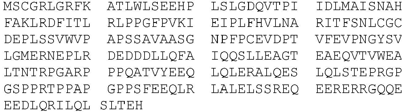
BIRC6 14 
BIRC8 15 
C18orf1 16 
C9orf74 17 
CUEDC1 18 
DAZAP2 19 
DDI1 20 
DUS3L 21 
FAM70B 22 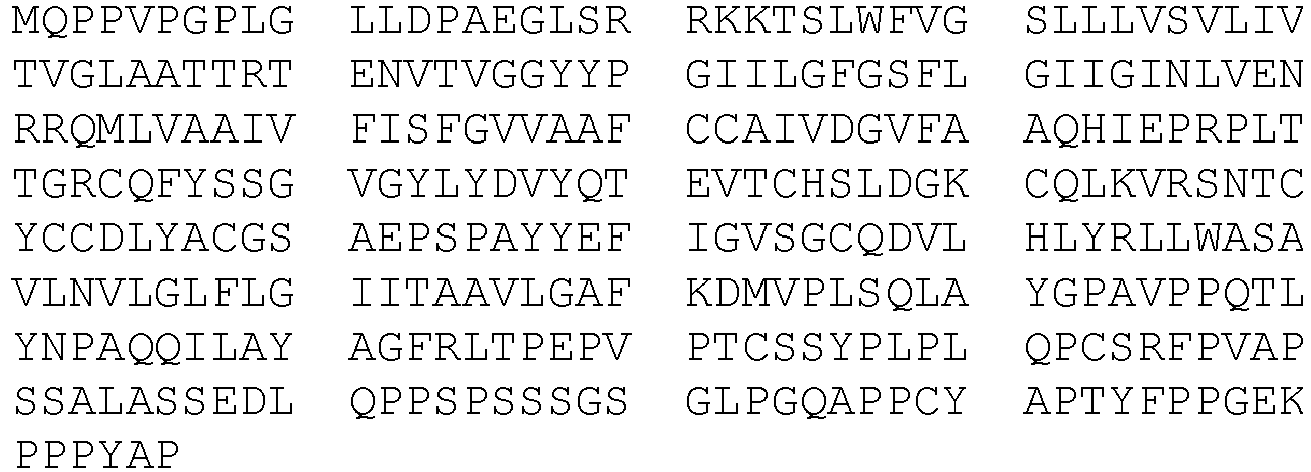
IGHG3 23 
KIAA024 7 24 
LAPTM5 25 
LDLRAD 4 26 
LITAF 27 
MLANA 28 
NDFIP2 29 
PMEPA1 30 
PSMD4 31 
PIAS2 32 
POLI 33 
RHOBT B1 34 
RNF13 35 
RNF4 36 
RNF34 37 
SAE2 38 
TAGAP 39 
TMEM13 9 40 
TMEM17 4 41 
TMEM55 B 42 
TMEPAI 43 
UBE2I 44 
UNQ846 45 
ZNF364 46 
- In the above mentioned method, the at least 3 proteins as set forth in SEQ ID NO: 1 to 3 are deposited on a support and then contacted with a cell extract of cells to be compared (e.g, cells that were treated or not with a compound liable to inhibit UBL pathways).
- With this method, the inventors evaluate specifically the modifications by Ubiquitin/SUMO/NEDD8 of the proteins SEQ ID NO: 1 to 3, and not all the proteins contained in the cell extract.
- During the period of contact, the enzymes responsible of the addition of Ubiquitin/SUMO/NEDD8 contained in the extract will modify the proteins SEQ ID NO: 1 to 3. The Ubiquitination, SUMOylation and NEDDylation amount will therefore depend upon the activity of the enzymes, and is directly correlated to the effect of the condition to be analyzed (cell type, inhibitor...).
- Further to the contacting between the proteins and the cellular extract, modifications by Ubiquitin/SUMO/NEDD8 are measured by means of immunological technics using specific antibodies directed against either Ubiquitin, or SUMO, or NEDD8.
- For instance, the proteins which are modified by ubiquitin and/or SUMO and/or NEDD8 proteins will therefore interact with antibodies directed against ubiquitin and/or SUMO and/or NEED8 proteins to form a molecular complex. The complexes can be detected by secondary antibodies that recognize (and interact with) constant part FC chain of said antibodies directed against ubiquitin and/or SUMO and/or NEDD8 proteins.
- Said complex can be identified when the secondary antibodies are labeled with reporter molecule, such as fluorescent protein, peroxidase, fluorescent dyes etc...
- The skilled person knows how to quantify the complexes protein/anti ubiquitin, anti SUMO or anti NEDD8 antibodies, in particular by using flow cytometers.
- Then a value of modification is obtained for each protein SEQ ID NO: 1 to 3, for each of Ubiquitin, SUMO and NEDD8.
- It is to be noted that protein SEQ ID NO: 1 can be modified by SUMO1 and SUMO2, whereas protein SEQ ID NO: 2 is modified preferably by Ubiquitin and protein SEQ ID NO: 3 can be modified by NEDD8.
- It is therefore possible, when assessing said at least proteins, to determine UBL pathway activity without interference and to obtain a quantitative activity of the UBL pathways in a cell.
-
Figure 9 shows the specificity of these proteins. - Advantageously, the invention relates to the above described method, wherein said method comprises a step of contacting a cellular extract with each protein of a group of 10 proteins, said group of 10 proteins belonging to a set of 46 proteins.
- In other words, the invention advantageously relates to the above mentioned method, said method comprising
- a) a step of contacting a cellular extract of cell with each protein of a subgroup of at least 10 proteins chosen among a set of 46 proteins;
- said at least 3 proteins being immobilised on a support,
- said at least 10 proteins being substantially not conjugated by SUMO/Ubiquitin/Nedd8 proteins before their contact with said cellular extract,
- wherein said 10 proteins corresponds to the proteins consisting essentially or consisting of the sequences SEQ ID NO: 1 to 10, and wherein said set of 46 proteins corresponds to the proteins consisting essentially or consisting of the sequences SEQ ID NO: 1 to 46,
and
- b) a step of simultaneously measuring ubiquitination, SUMOylation and NEDDylation level of each of said at least 3 proteins to obtain a first value for ubiquitin, SUMO and Nedd8.
- More advantageously, the invention relates to the method as defined above, wherein said method comprises a step of contacting a cellular extract with each protein of said set of 46 proteins.
- In other words, the invention advantageously relates to the above mentioned method, said method comprising
- a) a step of contacting a cellular extract of cell with each protein of a 46 proteins;
- said at 46 proteins being immobilised on a support,
- said 46 proteins being substantially not conjugated by SUMO/Ubiquitin/Nedd8 proteins before their contact with said cellular extract,
- said set of 46 proteins corresponds to the proteins consisting essentially or consisting of the sequences SEQ ID NO: 1 to 46,
- b) a step of simultaneously measuring ubiquitination, SUMOylation and NEDDylation level of each of said 46 proteins to obtain a first value for ubiquitin, SUMO and Nedd8 ;
- More advantageously, the invention relates to the method as defined above, wherein said at least 3 proteins are supported by beads.
- Advantageously, the invention relates to the method as defined above, wherein said beads are fluorescent beads.
- Advantageously, the invention relates to the method as defined above, wherein each of said at least 3 protein is supported by a determined bead having a fluorescent property which is different from the fluorescent properties of the other beads, and
wherein all the beads having the same fluorescent property supporting the same protein. - For example, the method can be performed with the XMap technology developed by Luminex. It consists in uniquely colored Mag-Plex metal-based microsphere (500 different colors with variable levels of two different dyes). They can be coupled with specific proteins using carboiimide coupling techniques. Protein-coupled beads can be multiplexed and used with cellular extracts as described in the examples thereafter. The different beads can be distinguished using dedicated analyzer or a flow cytometer.
- Proteins according to the invention are grafted covalently on the fluorescent beads by any means well known in the art.
- In one aspect, the invention relates to a method for quantifying the activity of the proteins/enzymes involved identifying the effect of a drug on protein modification by the conjugation of the SUMO/Ubiquitin/Nedd8 proteins in a cell of a biological sample, for example to identify the effect of a drug on UBL protein modification, said method comprising
- a) a step of contacting a cellular extract to be tested (e.g. of cell treated with said drug), with each protein of a subgroup of at least 3 proteins chosen among a group of 10 proteins, said group of 10 proteins belonging to a set of 46 proteins;
- said at least 3 proteins being immobilised on a support,
- said at least 3 proteins being substantially not conjugated by SUMO/Ubiquitin/Nedd8 proteins before their contact with said cellular extract,
- wherein said at least 3 proteins corresponds to the proteins consisting essentially or consisting of the sequences SEQ ID NO: 1 to 3, wherein said 10 proteins corresponds to the proteins consisting essentially or consisting of the sequences SEQ ID NO: 1 to 10, and wherein said group of 46 proteins corresponds to the proteins consisting essentially or consisting of the sequences SEQ ID NO: 1 to 46,
- wherein SEQ ID NO: 1 corresponds to ZMYM5 protein; SEQ ID NO: 2 corresponds to BEAN protein and SEQ ID NO: 3 corresponds to OTUD6B,
- b) a step of simultaneously measuring ubiquitination, SUMOylation and NEDDylation level of each of said at least 3 proteins to obtain a first value for ubiquitin, SUMO and Nedd8 ;
- c) a step of comparing the first value obtained at the previous step to a second value of ubiquitination, sumoylation and neddylation of said at least 3 proteins obtained when said at least proteins are contacted with said control cellular extract (e.g, of cell that was not treated with said drug); to obtain a ratio between the first value and the second value; and
- d) a step of determining that
- i- If the ratio of ubiquitination, sumoylation and neddylation of each of said at least 3 proteins is not significantly different to 1, then said tested drug condition (e.g. drug treatment) has no effect on protein modification by SUMO/Ubiquitin/Nedd8 proteins, and
- ii- If the ratio of ubiquitination, sumoylation and neddylation of at least one of said at least 3 proteins is significantly lower or higher than 1, then said tested drug condition (e.g. drug treatment) affects protein modification by SUMO/Ubiquitin/Nedd8 proteins.
- Advantageously, the invention advantageously relates to the above mentioned method, said method comprising
- a) a step of contacting a cellular extract to be tested (e.g of cell treated with said drug), with each protein of a subgroup of at least 10 proteins of a group, said group of 10 proteins belonging to a set of 46 proteins;
- said at least 10 proteins being immobilised on a support,
- said at least 10 proteins being substantially not conjugated by SUMO/Ubiquitin/Nedd8 proteins before their contact with said cellular extract,
- wherein said 10 proteins corresponds to the proteins consisting essentially or consisting of the sequences SEQ ID NO: 1 to 10, and wherein said group of 46 proteins corresponds to the proteins consisting essentially or consisting of the sequences SEQ ID NO: 1 to 46,
- b) a step of simultaneously measuring ubiquitination, sumoylation and neddylation level of each of said at least 10 proteins to obtain a first value for ubiquitin, SUMO and Nedd8 ;
- c) a step of comparing the first value obtained at the previous step to a second value of ubiquitination, sumoylation and neddylation of said at least 10 proteins obtained when said at least proteins are contacted with said control cellular extract (e.g of cell that was not treated with said drug); to obtain a ratio between the first value and the second value; and
- d) a step of determining that
- i. If the ratio of ubiquitination, sumoylation and neddylation of each of said at least 10 proteins is not significantly different to 1, then said tested condition (e.g. drug treatment) drug has no effect on protein modification by SUMO/Ubiquitin/Nedd8 proteins, and
- ii. If the ratio of ubiquitination, sumoylation and neddylation of at least one of said at least 10 proteins is significantly lower or higher than 1, then said tested drug condition (e.g. drug treatment) affects protein modification by SUMO/Ubiquitin/Nedd8 proteins.
- More advantageously, the invention relates to the above mentioned method, said method comprising
- a) a step of contacting a cellular extract of cell to be tested (e.g treated with said drug), with each protein set of 46 proteins;
- said 46 proteins being immobilised on a support,
- said 46 proteins being substantially not conjugated by SUMO/Ubiquitin/Nedd8 proteins before their contact with said cellular extract,
- wherein said 46 consisting essentially or consisting of the sequences SEQ ID NO: 1 to 46,
- b) a step of simultaneously measuring ubiquitination, sumoylation and neddylation level of each of said 46 proteins to obtain a first value for ubiquitin, SUMO and Nedd8 ;
- c) a step of comparing the first value obtained at the previous step to a second value of ubiquitination, sumoylation and neddylation of said 46 proteins obtained when said at least proteins are contacted with said control cellular extract (e.g. of cell that was not treated with said drug); to obtain a ratio between the first value and the second value; and
- d) a step of determining that
- i. If the ratio of ubiquitination, sumoylation and neddylation of each of said 46 proteins is not significantly different to 1, then said tested condition (e.g. drug treatment) has no effect on protein modification by SUMO/Ubiquitin/Nedd8 proteins, and
- ii. If the ratio of ubiquitination, sumoylation and neddylation of at least one of said 46 proteins is significantly lower or higher than 1, then said tested condition (e.g. drug treatment) affects protein modification by SUMO/Ubiquitin/Nedd8 proteins.
- The invention relates to a method for identifying the effect of a drug on protein modification by the SUMO/Ubiquitin/Nedd8 proteins in a cell of a biological sample, said method comprising
- a) a step of contacting a cellular extract of cell treated with said drug, with each protein of a subgroup of at least 3 proteins chosen among a group of 10 proteins, said group of 10 proteins belonging to a set of 46 proteins;
- said at least 3 proteins being immobilised on a support,
- said at least 3 proteins being substantially not conjugated by SUMO/Ubiquitin/Nedd8 proteins before their contact with said cellular extract,
- wherein said at least 3 proteins corresponds to the proteins consisting essentially or consisting of the sequences SEQ ID NO: 1 to 3, wherein said 10 proteins corresponds to the proteins consisting essentially or consisting of the sequences SEQ ID NO: 1 to 10, and wherein said group of 46 proteins corresponds to the proteins consisting essentially or consisting of the sequences SEQ ID NO: 1 to 46,
- wherein SEQ ID NO: 1 corresponds to ZMYM5 protein; SEQ ID NO: 2 corresponds to BEAN protein and SEQ ID NO: 3 corresponds to OTUD6B,
- a) a step of simultaneously measuring ubiquitination, SUMOylation and NEDDylation level of each of said at least 3 proteins to obtain a first value for ubiquitin, SUMO and Nedd8 ;
- b) a step of comparing the first value obtained at the previous step to a second value of ubiquitination, sumoylation and neddylation of said at least 3 proteins obtained when said at least proteins are contacted with said cellular extract of cell that was not treated with said drug; to obtain a ratio between the first value and the second value; and
- c) a step of determining that
- i- If the ratio of ubiquitination, sumoylation and neddylation of each of said at least 3 proteins is not significantly different to 1, then said drug has no effect on protein modification by SUMO/Ubiquitin/Nedd8 proteins, and
- ii- If the ratio of ubiquitination, sumoylation and neddylation of at least one of said at least 3 proteins is significantly lower or higher than 1, then said drug affects protein modification by SUMO/Ubiquitin/Nedd8 proteins.
- The above conditions for step a) and b) described above apply here mutatis mutandis.
- When the tested condition (e.g. drug treatment) has no effect on the processes of Ubiquitination, SUMOylation and NEDDylation, then proteins SEQ ID NO: 1 to 3 are modified at a level similar to the modification obtained with a control cellular extract of the same nature (e.g cell that was not treated with the inhibitor).
- On the contrary, if the tested condition (e.g. drug treatment) has an effect on UBL pathways, the amount of modification by Ubiquitin/SUMO/NEDD8 of proteins SEQ ID NO: 1 to 3 will be increased or decreased compared to the amount of modification observed in the control condition (e.g. non treated cells)
- Further to the contacting between the proteins and the cellular extract, modifications by Ubiquitin/SUMO/NEDD8 are measured by means of immunological technics using specific antibodies directed against either Ubiquitin, or SUMO, or NEDD8.
- For instance, the proteins which are modified by ubiquitin and/or SUMO and/or NEDD8 proteins will therefore interact with antibodies directed against ubiquitin and/or SUMO and/or NEED8 proteins to form a molecular complex. The complexes can be detected by secondary antibodies that recognize (and interact with) constant part FC chain of said antibodies directed against ubiquitin and/or SUMO and/or NEDD8 proteins.
- Said complex can be identified when the secondary antibodies are labeled with reporter molecule, such as fluorescent protein, peroxidase, fluorescent dyes etc...
- The skilled person knows how to quantify the complexes protein/anti ubiquitin, anti SUMO or anti NEDD8 antibodies, in particular by using flow cytometers.
- Then a value of modification is obtained for each protein SEQ ID NO: 1 to 3, for each of Ubiquitin, SUMO and NEDD8.
- This value is then compared to a reference value obtained for the same proteins contacted with a control cellular extract (e.g cells of the same nature than those treated with the inhibitor, but which were not treated with said inhibitor).
- A ratio is established between the first value, i.e. the value obtained with the cellular extract to be tested (e.g treated with the inhibitor) and the second value i.e. the value obtained with the control cellular extract (e.g not treated with the inhibitor).
- Then for one protein, three different ratios are obtained: one form Ubiquitin, one for SUMO and one for NEDD8:
- RUbiquitin (i) = value for ubiquitin of protein SEQ ID NO: i with cell extract of cells to be tested (e.g treated with the inhibitor) / value for ubiquitin of protein SEQ ID NO: i with cell extract of control cells (e.g not treated with the inhibitor);
- RSUMO (i) = value for SUMO of protein SEQ ID NO: i with cell extract of cells to be tested (e.g treated with the inhibitor) / value for SUMO of protein SEQ ID NO: i with cell extract of control cells (e.g not treated with the inhibitor); and
- RNEDD8 (i) = value for NEDD8 of protein SEQ ID NO: i with cell extract of cells to be tested (e.g treated with the inhibitor)/ value for NEDD8 of protein SEQ ID NO: i with cell extract of control cells (e.g not treated with the inhibitor).
- In the above example, i is either 1, 2 or 3.
- When the ratios are established, it is concluded that the tested condition (e.g. drug treatment) has no effect on UBL pathways when all the ratios calculated for each protein for each Ubiquitin, SUMO and NEDD8 are equal to about 1. This means that the modifications that occur on proteins SEQ ID NO: 1 to 3 are similar between the cellular extract to be tested (e.g. treated with said drug) and the control (e.g. non treated).
- On the contrary, if one at least of the above mentioned ratios are significantly different from 1, then it could be concluded that the tested condition (e.g. drug treatment) has an effect on UBL pathway.
- It is to be noted that protein SEQ ID NO: 1 can be modified by SUMO1 and SUMO2, whereas protein SEQ ID NO: 2 is modified preferably by Ubiquitin and protein SEQ ID NO: 3 can be modified by NEDD8.
- It is therefore possible, when assessing said at least protein, to determine which UBL pathway is affected by the tested condition (e.g inhibitor treatment).
-
Figure 9 shows the specificity of these proteins. - Advantageously, the invention relates to the above described method, wherein said method comprises a step of contacting a cellular extract to be tested (e.g of cell treated with said drug), with each protein of a group of 10 proteins, said group of 10 proteins belonging to a set of 46 proteins.
- In other words, the invention advantageously relates to the above mentioned method, said method comprising
- a) a step of contacting a cellular extract to be tested (e.g of cell treated with said drug), with each protein of a subgroup of at least 10 proteins of a group, said group of 10 proteins belonging to a set of 46 proteins;
- said at least 10 proteins being immobilised on a support,
- said at least 10 proteins being substantially not conjugated by SUMO/Ubiquitin/Nedd8 proteins before their contact with said cellular extract, wherein said 10 proteins corresponds to the proteins consisting essentially or consisting of the sequences SEQ ID NO: 1 to 10, and wherein said group of 46 proteins corresponds to the proteins consisting essentially or consisting of the sequences SEQ ID NO: 1 to 46,
- b) a step of simultaneously measuring ubiquitination, sumoylation and neddylation level of each of said at least 10 proteins to obtain a first value for ubiquitin, SUMO and Nedd8 ;
- c) a step of comparing the first value obtained at the previous step to a second value of ubiquitination, sumoylation and neddylation of said at least 10 proteins obtained when said at least proteins are contacted with said control cellular extract (e.g cell that was not treated with said drug); to obtain a ratio between the first value and the second value; and
- d) a step of determining that
- a. If the ratio of ubiquitination, sumoylation and neddylation of each of said at least 10 proteins is not significantly different to 1, then said tested condition (e.g. drug treatment) has no effect on protein modification by SUMO/Ubiquitin/Nedd8 proteins, and
- b. If the ratio of ubiquitination, sumoylation and neddylation of at least one of said at least 10 proteins is significantly lower or higher than 1, then said tested condition (e.g. drug treatment) affects protein modification by SUMO/Ubiquitin/Nedd8 proteins.
- More advantageously, the invention relates to the method as defined above, wherein said method comprises a step of contacting a cellular extract to be tested (e.g of cell treated with said drug), with each protein of a set of 46 proteins.
- In other words, the invention advantageously relates to the above mentioned method, said method comprising
- a) a step of contacting a cellular extract of cell to be tested (e.g treated with said drug), with each protein set of 46 proteins;
- said 46 proteins being immobilised on a support,
- said 46 proteins being substantially not conjugated by SUMO/Ubiquitin/Nedd8 proteins before their contact with said cellular extract,
- wherein said 46 consisting essentially or consisting of the sequences SEQ ID NO: 1 to 46,
- b) a step of simultaneously measuring ubiquitination, sumoylation and neddylation level of each of said 46 proteins to obtain a first value for ubiquitin, SUMO and Nedd8 ;
- c) a step of comparing the first value obtained at the previous step to a second value of ubiquitination, sumoylation and neddylation of said 46 proteins obtained when said at least proteins are contacted with said control cellular extract (e.g. of cell that was not treated with said drug); to obtain a ratio between the first value and the second value; and
- d) a step of determining that
- a. If the ratio of ubiquitination, sumoylation and neddylation of each of said 46 proteins is not significantly different to 1, then said tested condition (e.g. drug treatment) has no effect on protein modification by SUMO/Ubiquitin/Nedd8 proteins, and
- b. If the ratio of ubiquitination, sumoylation and neddylation of at least one of said 46 proteins is significantly lower or higher than 1, then said tested condition (e.g. drug treatment) affects protein modification by SUMO/Ubiquitin/Nedd8 proteins.
- More advantageously, the invention relates to the method as defined above, wherein said at least 3 proteins are supported by beads, said beads being advantageously fluorescent beads.
- Advantageously, the invention relates to the method as defined above, wherein each of said at least 3 protein is supported by a determined bead having a fluorescent property which is different from the fluorescent properties of the other beads, and
wherein all the beads having the same fluorescent property supporting the same protein. - The invention also relates to a composition comprising at least 3 proteins chosen among a group of 10 proteins, said group of 10 proteins belonging to a set of 46 proteins;
- wherein said at least 3 proteins corresponding to the proteins consisting essentially or consisting of the sequences SEQ ID NO: 1 to 3, wherein said 10 proteins corresponds to the proteins consisting essentially or consisting of the sequences SEQ ID NO: 1 to 10, and wherein said group of 46 proteins corresponds to the proteins consisting essentially or consisting of the sequences SEQ ID NO: 1 to 46,
- wherein said at least 3 proteins are supported on fluorescent beads,
- wherein each of said at least 3 proteins is supported by a bead having a determined fluorescent property, to obtain at least 3 distinct protein-supporting beads,
- wherein the bead supporting one of said at least 3 proteins has a determined fluorescent property which is different from the fluorescent properties of the at least 2 other protein-supporting beads, and
- wherein all the fluorescent beads, having a same fluorescent property, support a same protein.
- The above mentioned composition comprise therefore at least beads supporting the above mentioned proteins SEQ ID NO: 1 to 3, or more proteins until 46 proteins of the set described above, and can be used to evaluate Ubiquitination, SUMOylation or NEDDylation of said proteins.
- Advantageously, the invention relates to the composition as mentioned above, comprising at least the 10 proteins group of 10 proteins, said group belonging to a set of 46 proteins;
- wherein said at least 10 proteins are supported on fluorescent beads,
- wherein each of said at least 10 protein is supported by a bead having a determined fluorescent property, to obtain at least 10 distinct protein-supporting beads
- wherein the bead supporting one of said at least 10 protein has a determined fluorescent property which is different from the fluorescent properties of the at least 9 other protein-supporting beads, and
- wherein all the fluorescent beads, having a same fluorescent property, support a same protein.
- In other words, the invention relates advantageously of the composition as mentioned above, comprising beads supporting at least 10 proteins as set forth in SEQ ID NO: 1 to 10, until 46 proteins of the set described above.
- Advantageously, the invention relates to the composition as mentioned above, comprising 46 proteins;
- wherein said 46 proteins are supported on fluorescent beads,
- wherein each of said 46 proteins is supported by a bead having a determined fluorescent property, to obtain 46 distinct protein-supporting beads
- wherein the bead supporting one of said 46 proteins has a determined fluorescent property which is different from the fluorescent properties of the at least 45 other protein-supporting beads, and
- wherein all the fluorescent beads, having a same fluorescent property, support a same protein.
- In one other aspect, the invention relates to the use of the composition defined above, for quantifying UBL pathway activities, or for identifying in vitro the effect of a drug on the modification by SUMO/Ubiquitin/Nedd8 proteins in a cell of a biological sample.
- The composition according to the invention can be used for instance to carry out the method as defined above.
- The invention will be better explained and illustrated in view of the following figures and examples.
-
- [
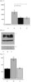
Fig. 1] Figure 1 represents a graph showing the modification of SUMOylation activity in cells treated with UBC9 shRNA (B, C and D) versus cells treated with a control shRNA (A). Y- axis represents the amount of UBC9 mRNA relative to control amount. N.S.: not significant. **: p<0.01 - [
Fig. 2] Figure 2 represents a graph showing SUMOylation activity on ZMYM5 protein, in cells treated with UBC9 shRNA (B, C and D) versus cells treated with a control shRNA (A). A cell extract was also treated with NEM (N-Ethylmaleimide) to inhibit all SUMO conjugation (negative control) (N) Y- axis represents the amount of SUMO on ZMYM5. N.S.: not significant. *: p<0.05 **: p<0.01 - [
Fig. 3] Figure 3 represents a graph showing Ubiquitinylation activity on BIRC7 protein, in cells treated with UBC9 shRNA (B, C and D) versus cells treated with a control shRNA (A). A cell extract was also treated with NEM (N-Ethylmaleimide) to inhibit all SUMO conjugation (negative control) (N) Y- axis represents the amount of Ubiquitin on BIRC7. N.S.: not significant. *: p<0.05 **: p<0.01 - [
Fig. 4] Figure 4 represents western blots results of cells extracts treated with UBC9 shRNA (B, B', C, C', D and D') versus control cell extracts (A, A'). Blots were labelled with anti-SUMO1 (A) and anti-SUMO2 (B) antibodies. - [
Fig. 5] Figure 5 represents a graph showing SUMOylation activity modulation on ZMYM5 protein in HL60 cells extract of cells treated with anacardic acid (C) or 2D08 (D) versus extracts of cells treated with dimethylsulfoxide (DMSO- B) or of N-ethylmaleimide (NEM) treated cells (negative control) (A). Y- axis represents the amount of SUMO on ZMYM5. N.S.: not significant. *: p<0.05 - [
Fig. 6] Figure 6 represents immunoblots of cells extracts corresponding to cells treated with DMSO (A), with anacardic acid (B) or 2D08 (C) inhibitors, labelled with anti-SUMO1 antibody. 1. represents SUMOylated proteins and 2. represents free SUMO. - [
Fig. 7] Figure 7 represents a graph showing the NEDDylation activity modulation on cell extracts of HL60 treated with MLN4924 inhibitor (C) versus cell extracts of cells treated with DMSO (B) or N-ethylmaleimide (NEM) treated cells (A). - [
Fig. 8] Figure 8 represents graphs evaluating Ubiquitin addition on beads coated with TMEM92 (1), BEAN1(2), WBP2(3) proteins, SUMO1 addition on beads coated with ZMYM5 (4) or NEDD8 addition on beads coated with OTUD6B (5). HL60 cells were treated for 6h with 0 (B), 5 (C), 10 (D), 50 (E), 100 (F) and 500nM (G) MLN7243. As control, cell extracts treated N-ethylmaleimide (NEM) are used (A). - [
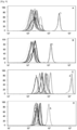
Fig. 9] Figure 9 represents graphs showing the fluorescence level of 10 proteins coated on fluorescent beads, incubated with SUMO1 (A), SUMO2 (B), Ubiquitin (C) or NEDD8 (D), and contacted with cellular extracts. 1. represents ZMYM5 protein (SEQ ID NO: 1); 2. represents BEAN protein (SEQ ID NO: 2) and 3. represents OTUD6B protein (SEQ ID NO: 3). Y-axis represents cell count and X-axis represents fluorescence intensity (arbitrary units). T represent control beads (without treatment with cell extracts). - Proteins are coated/grafted to the beads according to the following protocol
- 1. Resuspend the stock uncoupled beads suspension according to the instructions of the manufacturer.
- 2. Transfer 5.0×10⁶ of the stock beads to a recommended microcentrifuge tube.
- 3. Place the tube into a magnetic separator and allow separation to occur for 30 to 60 seconds.
- 4. With the tube still positioned in the magnetic separator, remove the supernatant. Take care not to disturb the beads.
- 5. Remove the tube from the magnetic separator and resuspend the beads in 100 µL dH2O by vortex and sonication for approximately 20 seconds.
- 6. Place the tube into a magnetic separator and allow separation to occur for 30 to 60 seconds.
- 7. With the tube still positioned in the magnetic separator, remove the supernatant. Take care not to disturb the beads.
- 8. Remove the tube from the magnetic separator and resuspend the washed beads in 80 µL 0.1M Sodium Phosphate (monobasic), pH 6.2 by vortex and sonication for approximately 20 seconds. Note: Beads should be protected from prolonged exposure to light throughout this procedure.
- 9. Add 10 µL of 50 mg/mL Sulfo-NHS (diluted in dH20) to the beads and mix gently by vortex.
- 10. Add 10 µL of 50 mg/mL EDC (diluted in dH20) to the beads and mix gently by vortex.
- 11. Incubate for 20 minutes at room temperature with gentle mixing by vortex at 10 minute intervals.
- 12. Place the tube into a magnetic separator and allow separation to occur for 30 to 60 seconds.
- 13. With the tube still positioned in the magnetic separator, remove the supernatant. Take care not to disturb the beads.
- 14. Remove the tube from the magnetic separator and resuspend the beads in 250 µL of 50 mM MES, pH 5.0 by vortex and sonication for approximately 20 seconds.
- 15. Repeat steps 12 and 13 for a total of two washes with 50 mM MES, pH 5.0.
- 16. Remove the tube from the magnetic separator and resuspend the activated and washed beads in 100 µL of 50 mM MES, pH 5.0 by vortex and sonication for approximately 20 seconds.
- 17. Add 25 µg protein to the resuspended beads (i.e., 5 µg/1 million beads). (Note: 5 µg protein per 1 million beads typically performs well. We recommend titrating up and/or down as needed to achieve optimal assay performance.)
- 18. Bring total volume to 500 µL with 50 mM MES, pH 5.0.
- 19. Mix coupling reaction by vortex.
- 20. Incubate for 2 hours with mixing (by rotation) at room temperature.
- 21. Place the tube into a magnetic separator and allow separation to occur for 30 to 60 seconds.
- 22. With the tube still positioned in the magnetic separator, remove the supernatant. Take care not to disturb the beads.
- 23. Remove the tube from the magnetic separator and resuspend the coupled beads in 500 µL of PBS-TBN by vortex and sonication for approximately 20 seconds.
- 24. Optional blocking step-Incubate for 30 minutes with mixing (by rotation) at room temperature. (Note: Perform this step when using the beads the same day.)
- 25. Place the tube into a magnetic separator and allow separation to occur for 30 to 60 seconds.
- 26. With the tube still positioned in the magnetic separator, remove the supernatant. Take care not to disturb the beads.
- 27. Remove the tube from the magnetic separator and resuspend the beads in 1 mL of PBS-TBN by vortex and sonication for approximately 20 seconds.
- 28. Repeat steps 25 and 26. This is a total of two washes with 1 mL PBS-TBN.
- 29. Remove the tube from the magnetic separator and resuspend the coupled and washed beads in 250-1000 µL of PBS-TBN.
- 30. Count the number of beads recovered after the coupling reaction using a cell counter or hemacytometer.
- 31. Store coupled beads refrigerated at 2-8°C in the dark
- The inventors have optimized a method, using MagPlex Xmap beads, allowing a quantitative measurement of the ubiquitinylation, SUMOylation, and NEDDylation activities in cell extracts.
- The inventors therefore had intended to use this method in order to evaluate the inhibiting activity of compounds that target enzymes of the ubiquitine-like systems.
- In order to carry out this method, the inventors used 10 proteins highly modified by Ubiquitin/SUMO/NEDD8, that were grafted on fluorescent beads. The 10 chosen proteins represents the 4 different pathways, namely Ubiquitin,
SUMO [Table 2] SUMO1 SUMO2 Ubiquitin NEDD8 LDLRAD3 LDLRAD3 LDLRAD3 OTUD6B TMEM92 TMEM92 TMEM92 ANKRD39 UBE3A UBE3A UBE3A OTUB2 BEAN BEAN BEAN MXI1 MXI1 MXI1 WBP2 WBP2 WBP2 ZMYM5 - This table represents the proteins chosen for the measure of the inhibiting activity of the UbL system.
- The first step was to check if the proteins can be modified according to the inventor's method. Proteins were then produced in bacteria, then coupled to beads with different fluorescent properties, and used HL60 cell extract sensitive to drugs.
- ZMYM5 was used as a marker of SUMOylation, OTUD6B as a marker of NEDDylation, and TMEM92, BEAN and WBP2 as a marker of ubiquitinylation.
- The inventors wanted to validate their method by assessing SUMOylation inhibition.
- In a first approach, the inventors used HL60 cell lines expressing shRNA inhibiting expression of UBC9, the E2 enzyme of SUMO. Three different cell lines were used with a different shRNA, and a control cell line was used as control. UBC9 expression inhibition was assessed by RT-qPCR by quantifying the relative amount of UBC9 RNAs.
shRNA1 induces a limited decrease (non-significant), shRNA2 induces a decrease of about 20% and shRNA3 of 50% of Ubc9 expression. - Thus, the inventors apply their method to the cell extract treated with the above-mentioned shRNA. In the same reaction, the inventors measured ubiquitinylation, and measured by western blot SUMOylation by SUMO1 and SUMO2. (
Figures 1 to 4 ) - Surprisingly, SUMOylation level measured by western blot is not significantly modified, including in cells having a reduction of 50% of Ubc9 RNA.
- This can be explained by the fact that the proteins detected by immunoblot are the ones that are significantly modified by SUMOylation, and which correspond to the proteins that are the less sensitive to a decrease of the amount of Ubc9.
- However, the method according to the invention allows a detection of global SUMOylation in the cells texted in the experiments. Indeed, SUMOylation activity measured by the method according to the invention is directly correlated to UBC9 expression assessed by qPCR.
- In a second time, the inventors evaluated if the method of the invention could be used to measure the effect of inhibitors of UbL pathways. For this purpose, the inventors have evaluated the method by using two known SUMOylation inhibitors: anacardic acid, which inhibit E1 enzyme of SUMO, and 2D08, which inhibit E2 enzyme of SUMO.
- HL60 cells were treated with 50µM of these compounds for 6h, and then cell extracts were prepared and used to carry out the method according to the invention.
- Cells were then lysed in Laemmli buffer, and samples were loaded on acrylamide-bisacrylamide gel for SDS-PAGE separation. Proteins were then transferred onto a PVDF membrane and labelled with an anti-SUMO1 antibody.
- By using the method according to the invention, it is possible to detect a SUMOylation decrease of 50% in cell extracts, whereas immunoblot does not show such decrease. This can be explained by the fact that in western blot all the protein liable to be modified by SUMO are detected, and specifically the one that are significantly modified.
- In samples treated with the drugs, it is possible to detect free SUMO1, which is the hallmark of the decrease of SUMOylation. The method according to the invention is therefore more efficient than western blot based methods.
- In this example, the inventors intended to validate the method on samples from patients treated with commercial inhibitors that have been proven to be therapeutically efficient on Ubiquitin and Nedd8 pathways: namely MLN7243 (Ubiquitine), and MLN4924 (NEDD8).
- MLN7243 (TAK-243) is a cell permeable small molecule inhibitor targeting ubiquitin-activating enzymes (UAE, also known as E1 enzymes). The enzymes, found more active in cancer cells than in normal, healthy, cells, catalyze the first step in ubiquitination reaction, targeting a protein for degradation via Proteasome. This covalent attachment of ubiquitin or ubiquitin-like proteins to targeted proteins is a major mechanism for regulating protein function in eukaryotic organisms. Inhibition of the enzyme prevents both protein ubiquitination and subsequent ubiquitin-mediated proteasomal degradation, resulting in an excess of proteins in the cells that may lead to endoplasmic reticulum (ER) stress-mediated apoptosis, thus inhibit tumor (cancer) cell proliferation and survival. MLN7243 is the first-in-class inhibitor specifically targeting this class of enzyme studied clinically.
- MLN7243 has the following formula:

- MLN4924 (also called Pevonedistat) is a small molecule inhibitor of the NEDD8-Activating Enzyme (NAE), a key component of the protein homeostasis pathway. It is the first small molecule inhibitor specifically targeting this class of enzyme to be studied clinically, and is currently being examined in Phase I clinical trials.
- MLN4924 has the following formula:

- HL60 cells were first treated with 1 µM of MLN4924, DMSO, or only with the culture medium for 6h. Beads supporting UBC12, (NEDDylation E2 enzyme, which was previously reported to be modified by NEDD8) were contacted with said cell extracts, and NEDD8 level was assessed.
- Results are shown on
figure 7 . - Whereas NEDDylation measured in the control samples was low, treatment with 1µM MLN4924 shows a decrease of UBC12 protein NEDDylation.
- The inventors then evaluated the effect of different amount of MLN7243, E1 enzyme of the Ubiquitin pathway.
- Cells were then treated with 0, 5, 10, 50, 100 or 500 nM MLN7243 for 6h. Cells extracts were prepared and contacted with beads coated with either TMEM92, BEAN1 or WBP2 proteins.
- These results show that more than 50% of the ubiquitination activity is inhibited with 10nM or more of MLN7243 (In vitro IC50: 1nM; In vitro toxicity on Acute Myeloid Leukemia cell lines IC50: 20 to 40nM)
- With 500nM MLN7243, TMEM92 NEDDylation inhibition is maximal, WBP2 and BEAN1 remain ubiquitynated at residual levels.
- MLN7243 inhibitor also affects SUMOylation activity, but with large amounts of inhibitor which is consistent with the in vitro IC50 of 850nM.
- Regarding NEDDylation, MLN7243 in vitro IC50 is 28nM on OTUD6B protein, but the data show a decrease slightly different.
- HL-60 (DSMZ, Germany) were cultured at 37 °C in RPMI medium supplemented with 10 % fetal bovine serum (FBS) and streptomycin/penicillin in the presence of 5 % CO2. HL-60 were authenticated by the ATCC using Short-Tandem-Repeat analysis. All cells were regularly tested negative for mycoplasma. After thawing, cells were passaged at a density of 3.105/ml every 2-3 days for no more than 10 passages.
- Cells grown at a 5-8.105/mL density were spun down (300 g) at 4 °C for 5 min and washed once with PBS. After pellet resuspension in 1 mL of PBS, they were centrifuged again (16,000 g) at 4 °C for 5 min. Pellets were resuspended and incubated at 4 °C for 30 min in a hypotonic buffer (20 mM HEPES pH 7.5, 1.5 mM MgCl2, 5 mM KCI, 1 mM DTT and 1 mg/L of aprotinine, leupeptin and pepstatin) in a volume of 25 µL per 2.106 cells. Cell lysis was achieved through 4 freezing/thawing cycles using liquid nitrogen and DNA was sheared owing to 10 passages through a 20-1/2G needle. Extracts were finally centrifuged twice (16000 g) at 4°C for 20 min and supernatants were aliquoted, flash-frozen and kept at -80 °C until use.
- cDNA encoding for the proteins of interest were recovered from the Ultimate ORF library (Thermofisher) and cloned in the bacterial expression vector pGGWA vector using the Gateway technology according to manufacturer's protocol (Life Technologies). Constructs were then transformed in BL21 (DE3) E. coli strain. Protein production was induced with 1 mM isopropyl β-D-1-thiogalactopyranoside (IPTG) for 6 hrs in exponentially growing bacteria at 25°C.
- Bacterial pellets were resuspended in 50 mM Tris-HCl pH 8.6 containing 500 mM NaCl and 50 mM MgSO4, and flash-frozen in liquid N2. After thawing, bacterial suspensions were supplemented with 1 mg/mL lysozyme (Sigma-Aldrich), 8 mM β-mercaptoethanol, 1 mg/L aprotinin, leupeptin and pepstatin and incubated at 4°C for 1 hr. Bacterial debris were spun down (100 000 g for 1 hr). The extract was then bound to Glutathion agarose beads (Generon) equilibrated in Tris 50 mM pH 8.6, NaCl 500 mM, MgSO4 50 mM, 8 mM β-mercaptoethanol, 1 mg/L aprotinin, leupeptin, pepstatin. The column was then extensively washed with Tris 50 mM pH 8.6, NaCl 150 mM, MgSO4 50 mM, 8 mM β-mercaptoethanol, 1 mg/L aprotinin, leupeptin, pepstatin and eluted by addition of 20 mM reduced glutathione (Sigma-Aldrich).
- 2.105 magnetic MagPlex XMap beads (low concentration) from Luminex were transferred to a low binding microtube (Eppendorf) and washed using 500 mM NaCl. They were then resuspended in 50 µL of 50 mM MES pH 6.1 and incubated in the presence of 5 mg/mL 1-ethyl-3-(3-dimethylaminopropyl)carbodiimide hydrochloride (EDC, Pierce) and 5 mg/mL Sulfo-NHS (Pierce) at room temperature for 20 min. Beads were then washed in PBS containing 500 mM NaCl and incubated with 7 µg of recombinant protein to be coupled in 100 µL PBS at room temperature for 2 hrs. They were then washed twice with PBS containing 0.1 % BSA, 0.02
% Tween 20, 0.05 % sodium azide and 500 mM NaCland stored at 4 °C in PBS containing 0.1 % BSA, 0.02 % Tween-20, 0.05 % sodium azide. - SUMO-1-, SUMO-2-, NEDD8 and Ubiquitin vinyl sulfones (0.5 µM each) were added to cellular extracts (10 µL), which were incubated at 4 °C for 15 min. Control extracts were also incubated with 50 mM NEM. The inventors then added to the
extract 103 protein-coupled XMap beads contained in 10µL of a reaction buffer containing 20 mM HEPES pH 7.3, 110 mM KOAc, 2 mM Mg(OAc)2, 0.05 % Tween-20, 0.5 mM EGTA, 0.2 mg/mL ovalbumine, 1 mM DTT, 1 mg/L aprotinin, leupeptin and pepstatin, 1 mM ATP, 30 µM Flag-ubiquitin, 15 µM NEDD8, 15 µM SUMO-1 and 15 µM SUMO-2. Reaction were performed at 30 °C for 45 min. Beads were washed twice for 5 min with PBS containing 0.05 % Tween-20 and 0.5 % SDS and 3 times for 5 min with PBS containing 0.05 % Tween-20. They were then incubated with 1 µg/mL of anti-SUMO-1 (21C7) and anti-Flag antibodies or anti-SUMO-2 (8A2) and anti-NEDD8 for 1 hr under agitation at room temperature. After washing in PBS containing 0.05% Tween-20 for 5 min, they were incubated for 30 min at room temperature with anti-mouse Alexa Fluor 488- and anti-rabbit Alexa Fluor 405 antibodies in 100 µL of PBS containing 0.05 % Tween-20. Beads were again washed for 5 min with PBS containing 0.05 % Tween-20. They were then resuspended in 200 µL PBS and flow-cytometry-analysed using the LSR Fortessa device from BD Biosciences. Results were analysed using the FlowJow software. - Extracts from HL-60 or U937 were supplemented with UbL-vinyl-sulfone and recombinant UbL and incubated on Protoarrays (Life Technologies). After extensive washes, the arrays were incubated with primary mouse anti-SUMO-1 and rabbit anti-Flag (tag present on the recombinant Ubiquitin added to the reaction) antibodies followed by fluorescently coupled secondary antibodies and scanned for fluorescence. The antibodies were then removed and the arrays incubated with primary mouse anti-SUMO-2 and rabbit anti-NEDD8 antibodies followed by fluorescently coupled secondary antibodies and scanned for fluorescence. The normalized fluorescence data obtained for all modifiers on all arrays were compared to the averaged signal of the control arrays (NEM) to identify proteins, which are robustly modified. 46 proteins showing a significant difference between the two groups using both Welch and Wilcoxon-Mann-Whitney and having mean fluorescence intensities values higher than 800 on the Protoarrays were selected.
Claims (15)
- Method for quantifying the activity of the proteins or enzymes involved in the conjugation of the SUMO/Ubiquitin/Nedd8 proteins in a cell of a biological sample, said method comprising:a) a step of contacting a cellular extract of a cell with each protein of a subgroup of at least 3 proteins chosen among a group of 10 proteins,said group of 10 proteins belonging to a set of 46 proteins;said at least 3 proteins being immobilised on a support,said at least 3 proteins being substantially not conjugated by SUMO/ Ubiquitin/Nedd8 proteins before their contact with said cellular extract, wherein said at least 3 proteins corresponds to the proteins consisting essentially or consisting of the sequences SEQ ID NO: 1 to 3, wherein said 10 proteins corresponds to the proteins consisting essentially or consisting of the sequences SEQ ID NO: 1 to 10, and wherein said group of 46 proteins corresponds to the proteins consisting essentially or consisting of the sequences SEQ ID NO: 1 to 46,wherein SEQ ID NO: 1 corresponds to ZMYM5 protein; SEQ ID NO: 2 corresponds to BEAN protein and SEQ ID NO: 3 corresponds to OTUD6B, andb) a step of simultaneously measuring ubiquitination, SUMOylation and NEDDylation level of each of said at least 3 proteins to obtain a first value for ubiquitin, SUMO and Nedd8.
- The method according to claim 1, wherein said method comprises a step of contacting a cellular extract with each protein of a group of 10 proteins, said group of 10 proteins belonging to said set of 46 proteins.
- The method according to claim 1 or 2, wherein said method comprises a step of contacting a cellular extract with each protein of a set of 46 proteins.
- The method according to anyone of claims 1 to 3, wherein said at least 3 proteins are supported by beads.
- The method according to claim 4, wherein said beads are fluorescent beads.
- The method according to claim 4 or 5, wherein each of said at least 3 protein is supported by a determined bead having a fluorescent property which is different from the fluorescent properties of the other beads, and
wherein all the beads having the same fluorescent property supporting the same protein. - A method for identifying the effect of a drug on protein modification by SUMO/Ubiquitin/Nedd8 proteins in a cell of a biological sample, said method comprisinga) a step of contacting a cellular extract of cell treated with said drug, with each protein of a subgroup of at least 3 proteins chosen among a group of 10 proteins, said group of 10 proteins belonging to a set of 46 proteins;said at least 3 proteins being immobilised on a support,said at least 3 proteins being substantially not conjugated by SUMO/ Ubiquitin/Nedd8 proteins before their contact with said cellular extract, wherein said at least 3 proteins corresponds to the proteins consisting essentially or consisting of the sequences SEQ ID NO: 1 to 3, wherein said 10 proteins corresponds to the proteins consisting essentially or consisting of the sequences SEQ ID NO: 1 to 10, and wherein said group of 46 proteins corresponds to the proteins consisting essentially or consisting of the sequences SEQ ID NO: 1 to 46,wherein SEQ ID NO: 1 corresponds to ZMYM5 protein; SEQ ID NO: 2 corresponds to BEAN protein and SEQ ID NO: 3 corresponds to OTUD6B,b) a step of simultaneously measuring ubiquitination, sumoylation and neddylation level of each of said at least 3 proteins to obtain a first value for ubiquitin, SUMO and Nedd8 ;c) a step of comparing the first value obtained at the previous step to a second value of ubiquitination, sumoylation and neddylation of said at least 3 proteins obtained when said at least proteins are contacted with said cellular extract of cell that was not treated with said drug; to obtain a ratio between the first value and the second value; andd) a step of determining thata. If the ratio of ubiquitination, sumoylation and neddylation of each of said at least 3 proteins is not significantly different to 1, then said drug has no effect on protein modification by SUMO/Ubiquitin/Nedd8 proteins, andb. If the ratio of ubiquitination, sumoylation and neddylation of at least one of said at least 3 proteins is significantly lower or higher than 1, then said drug affects protein modification by SUMO/Ubiquitin/Nedd8 proteins.
- The method for identifying the effect of a drug according to claim 7,
wherein said method comprises a step of contacting a cellular extract of cell treated with said drug, with each protein of a group of 10 proteins, said group of 10 proteins belonging to a set of 46 proteins. - The method for identifying the effect of a drug according to claim 8,
wherein said method comprises a step of contacting a cellular extract of cell treated with said drug, with each protein of a set of 46 proteins. - The method according to anyone of claims 7 to 9, wherein said at least 3 proteins are supported by beads, advantageously wherein said beads are fluorescent beads.
- The method according to claim 10, wherein each of said at least 3protein is supported by a determined bead having a fluorescent property which is different from the fluorescent properties of the other beads, andwherein all the beads having the same fluorescent property supporting the same protein.
- A composition comprising at least 3 proteins chosen among a group of 10 proteins, said group of 10 proteins belonging to a set of 46 proteins;wherein said at least 3 proteins corresponding to the proteins consisting essentially or consisting of the sequences SEQ ID NO: 1 to 3, wherein said 10 proteins corresponds to the proteins consisting essentially or consisting of the sequences SEQ ID NO: 1 to 10, and wherein said group of 46 proteins corresponds to the proteins consisting essentially or consisting of the sequences SEQ ID NO: 1 to 46,wherein said at least 3 proteins are supported on fluorescent beads, wherein each of said at least 3 proteins is supported by a bead having a determined florescent property, to obtain at least 3 distinct protein-supporting beadswherein the bead supporting one of said at least 3 proteins has a determined fluorescent property which is different from the fluorescent properties of the at least 2 other protein-supporting beads, andwherein all the fluorescent beads, having a same fluorescent property, support a same protein.
- The composition according to claim 12, comprising at least the 10proteins group of 10 proteins, said group belonging to a set of 46 proteins;wherein said at least 10 proteins are supported on fluorescent beads, wherein each of said at least 10 protein is supported by a bead having a determined florescent property, to obtain at least 10 distinct protein-supporting beadswherein the bead supporting one of said at least 10 protein has a determined fluorescent property which is different from the fluorescent properties of the at least 9 other protein-supporting beads, and wherein all the fluorescent beads, having a same fluorescent property, support a same protein.
- The composition according to claim 12 or 13, comprising 46 proteins;wherein said 46 proteins are supported on fluorescent beads, wherein each of said 46 proteins is supported by a bead having a determined florescent property, to obtain 46 distinct protein-supporting beadswherein the bead supporting one of said 46 proteins has a determined fluorescent property which is different from the fluorescent properties of the at least 45 other protein-supporting beads, andwherein all the fluorescent beads, having a same fluorescent property, support a same protein.
- Use of composition according anyone of claims 12 to 14, for
quantifying UBL pathway activities, or identifying in vitro the effect of a drug on the modification by SUMO/Ubiquitin/Nedd8 proteins in a cell of a biological sample.
Applications Claiming Priority (2)
| Application Number | Priority Date | Filing Date | Title |
|---|---|---|---|
| EP19305690 | 2019-05-29 | ||
| PCT/EP2020/064911 WO2020239949A1 (en) | 2019-05-29 | 2020-05-28 | Method for identifying ubiquitin and ubiquitin-like enzyme activities |
Publications (2)
| Publication Number | Publication Date |
|---|---|
| EP3977133A1 EP3977133A1 (en) | 2022-04-06 |
| EP3977133B1 true EP3977133B1 (en) | 2023-06-28 |
Family
ID=67060341
Family Applications (1)
| Application Number | Title | Priority Date | Filing Date |
|---|---|---|---|
| EP20728499.3A Active EP3977133B1 (en) | 2019-05-29 | 2020-05-28 | Method for identifying ubiquitin and ubiquitin-like enzyme activities |
Country Status (7)
| Country | Link |
|---|---|
| US (1) | US12546780B2 (en) |
| EP (1) | EP3977133B1 (en) |
| JP (1) | JP7271727B2 (en) |
| KR (1) | KR102637287B1 (en) |
| CA (1) | CA3140947C (en) |
| ES (1) | ES2956457T3 (en) |
| WO (1) | WO2020239949A1 (en) |
Family Cites Families (7)
| Publication number | Priority date | Publication date | Assignee | Title |
|---|---|---|---|---|
| US20040053324A1 (en) * | 2002-08-30 | 2004-03-18 | Brian Wong | Assays and compositions for identifying agents that modulate the activity of deubiquitinating agents |
| US20080039413A1 (en) * | 2003-10-21 | 2008-02-14 | Morris David W | Novel compositions and methods in cancer |
| WO2005113812A2 (en) * | 2004-04-23 | 2005-12-01 | Invitrogen Corporation | Collections of matched biological reagents and methods for identifying matched reagents |
| US20090263821A1 (en) * | 2004-12-01 | 2009-10-22 | Proteologics, Inc. | Ubiquitin Ligase Assays And Related Reagents |
| EP2362909A4 (en) * | 2008-09-30 | 2012-10-10 | Zirus Inc | INFECTIONS INVOLVED MIRACLE GENES |
| GB201415688D0 (en) | 2014-09-04 | 2014-10-22 | Univ Edinburgh | Method and Systems |
| US20160209414A1 (en) | 2014-11-07 | 2016-07-21 | University Of Washington Through Its Center For Commercialization | Compositions and methods for the diagnosis and treatment of breast cancer |
-
2020
- 2020-05-28 KR KR1020217040494A patent/KR102637287B1/en active Active
- 2020-05-28 JP JP2021570461A patent/JP7271727B2/en active Active
- 2020-05-28 US US17/614,739 patent/US12546780B2/en active Active
- 2020-05-28 CA CA3140947A patent/CA3140947C/en active Active
- 2020-05-28 WO PCT/EP2020/064911 patent/WO2020239949A1/en not_active Ceased
- 2020-05-28 ES ES20728499T patent/ES2956457T3/en active Active
- 2020-05-28 EP EP20728499.3A patent/EP3977133B1/en active Active
Also Published As
| Publication number | Publication date |
|---|---|
| KR20220008871A (en) | 2022-01-21 |
| KR102637287B1 (en) | 2024-02-15 |
| EP3977133A1 (en) | 2022-04-06 |
| US12546780B2 (en) | 2026-02-10 |
| JP7271727B2 (en) | 2023-05-11 |
| CA3140947A1 (en) | 2020-12-03 |
| JP2022534270A (en) | 2022-07-28 |
| ES2956457T3 (en) | 2023-12-21 |
| WO2020239949A1 (en) | 2020-12-03 |
| US20220357326A1 (en) | 2022-11-10 |
| CA3140947C (en) | 2023-08-01 |
Similar Documents
| Publication | Publication Date | Title |
|---|---|---|
| Hewings et al. | Activity‐based probes for the ubiquitin conjugation–deconjugation machinery: new chemistries, new tools, and new insights | |
| Hewings et al. | Reactive-site-centric chemoproteomics identifies a distinct class of deubiquitinase enzymes | |
| Swaney et al. | Phosphorylation of ubiquitin at Ser65 affects its polymerization, targets, and proteome‐wide turnover | |
| Gopinath et al. | Chemical and semisynthetic approaches to study and target deubiquitinases | |
| Xiao et al. | Protein N-terminal processing: substrate specificity of Escherichia coli and human methionine aminopeptidases | |
| Hassiepen et al. | A sensitive fluorescence intensity assay for deubiquitinating proteases using ubiquitin-rhodamine110-glycine as substrate | |
| Ndubaku et al. | Inhibiting the deubiquitinating enzymes (DUBs) miniperspective | |
| Ohayon et al. | Targeting deubiquitinases enabled by chemical synthesis of proteins | |
| Weber et al. | A linear diubiquitin-based probe for efficient and selective detection of the deubiquitinating enzyme OTULIN | |
| Liu et al. | A colorimetric strategy for assay of protease activity based on gold nanoparticle growth controlled by ascorbic acid and Cu (II)-coordinated peptide | |
| JP2003511063A (en) | Methods for identifying methionine aminopeptidase inhibitors | |
| Wu et al. | Target-activated streptavidin–biotin controlled binding probe | |
| Liu et al. | An amplification strategy using DNA-Peptide dendrimer probe and mass spectrometry for sensitive MicroRNA detection in breast cancer | |
| WO2009072728A1 (en) | Method for diagnosis of disease using quantitative monitoring of protein tyrosine phosphatase | |
| WO2014179755A2 (en) | Method for identifying modulators of ubiquitin ligases | |
| Chakraborty et al. | Profiling of the drug resistance of thousands of src tyrosine kinase mutants uncovers a regulatory network that couples autoinhibition to catalytic domain dynamics | |
| Ha et al. | Structures of proteases for ubiqutin and ubiquitin-like modifiers | |
| US12546780B2 (en) | Method for identifying ubiquitin and ubiquitin-like enzyme activities | |
| Wang et al. | Rapidly quantitative analysis of γ-glutamyltranspeptidase activity in the lysate and blood via a rational design of the molecular probe by matrix-assisted laser desorption ionization mass spectrometry | |
| Shinohara et al. | Design of an electroactive peptide probe for sensing of a protein | |
| Shu et al. | Detecting active deconjugating enzymes with genetically encoded activity-based ubiquitin and ubiquitin-like protein probes | |
| Singh et al. | One-step sortase-mediated chemoenzymatic semisynthesis of deubiquitinase-resistant Ub-peptide conjugates | |
| US10995119B2 (en) | Peptide exhibiting hydrolytic activity and use thereof | |
| Sun et al. | Icotinib induces mechanism-based inactivation of recombinant human CYP3A4/5 possibly via heme destruction by ketene intermediate | |
| CN115948527A (en) | Glutathione detection method |
Legal Events
| Date | Code | Title | Description |
|---|---|---|---|
| STAA | Information on the status of an ep patent application or granted ep patent |
Free format text: STATUS: UNKNOWN |
|
| STAA | Information on the status of an ep patent application or granted ep patent |
Free format text: STATUS: THE INTERNATIONAL PUBLICATION HAS BEEN MADE |
|
| PUAI | Public reference made under article 153(3) epc to a published international application that has entered the european phase |
Free format text: ORIGINAL CODE: 0009012 |
|
| STAA | Information on the status of an ep patent application or granted ep patent |
Free format text: STATUS: REQUEST FOR EXAMINATION WAS MADE |
|
| 17P | Request for examination filed |
Effective date: 20211117 |
|
| AK | Designated contracting states |
Kind code of ref document: A1 Designated state(s): AL AT BE BG CH CY CZ DE DK EE ES FI FR GB GR HR HU IE IS IT LI LT LU LV MC MK MT NL NO PL PT RO RS SE SI SK SM TR |
|
| DAV | Request for validation of the european patent (deleted) | ||
| DAX | Request for extension of the european patent (deleted) | ||
| GRAP | Despatch of communication of intention to grant a patent |
Free format text: ORIGINAL CODE: EPIDOSNIGR1 |
|
| STAA | Information on the status of an ep patent application or granted ep patent |
Free format text: STATUS: GRANT OF PATENT IS INTENDED |
|
| INTG | Intention to grant announced |
Effective date: 20230208 |
|
| GRAS | Grant fee paid |
Free format text: ORIGINAL CODE: EPIDOSNIGR3 |
|
| GRAA | (expected) grant |
Free format text: ORIGINAL CODE: 0009210 |
|
| STAA | Information on the status of an ep patent application or granted ep patent |
Free format text: STATUS: THE PATENT HAS BEEN GRANTED |
|
| AK | Designated contracting states |
Kind code of ref document: B1 Designated state(s): AL AT BE BG CH CY CZ DE DK EE ES FI FR GB GR HR HU IE IS IT LI LT LU LV MC MK MT NL NO PL PT RO RS SE SI SK SM TR |
|
| REG | Reference to a national code |
Ref country code: CH Ref legal event code: EP |
|
| REG | Reference to a national code |
Ref country code: AT Ref legal event code: REF Ref document number: 1583145 Country of ref document: AT Kind code of ref document: T Effective date: 20230715 |
|
| REG | Reference to a national code |
Ref country code: IE Ref legal event code: FG4D |
|
| REG | Reference to a national code |
Ref country code: DE Ref legal event code: R096 Ref document number: 602020013041 Country of ref document: DE |
|
| REG | Reference to a national code |
Ref country code: LT Ref legal event code: MG9D |
|
| PG25 | Lapsed in a contracting state [announced via postgrant information from national office to epo] |
Ref country code: SE Free format text: LAPSE BECAUSE OF FAILURE TO SUBMIT A TRANSLATION OF THE DESCRIPTION OR TO PAY THE FEE WITHIN THE PRESCRIBED TIME-LIMIT Effective date: 20230628 Ref country code: NO Free format text: LAPSE BECAUSE OF FAILURE TO SUBMIT A TRANSLATION OF THE DESCRIPTION OR TO PAY THE FEE WITHIN THE PRESCRIBED TIME-LIMIT Effective date: 20230928 |
|
| REG | Reference to a national code |
Ref country code: NL Ref legal event code: MP Effective date: 20230628 |
|
| REG | Reference to a national code |
Ref country code: AT Ref legal event code: MK05 Ref document number: 1583145 Country of ref document: AT Kind code of ref document: T Effective date: 20230628 |
|
| PG25 | Lapsed in a contracting state [announced via postgrant information from national office to epo] |
Ref country code: RS Free format text: LAPSE BECAUSE OF FAILURE TO SUBMIT A TRANSLATION OF THE DESCRIPTION OR TO PAY THE FEE WITHIN THE PRESCRIBED TIME-LIMIT Effective date: 20230628 Ref country code: NL Free format text: LAPSE BECAUSE OF FAILURE TO SUBMIT A TRANSLATION OF THE DESCRIPTION OR TO PAY THE FEE WITHIN THE PRESCRIBED TIME-LIMIT Effective date: 20230628 Ref country code: LV Free format text: LAPSE BECAUSE OF FAILURE TO SUBMIT A TRANSLATION OF THE DESCRIPTION OR TO PAY THE FEE WITHIN THE PRESCRIBED TIME-LIMIT Effective date: 20230628 Ref country code: LT Free format text: LAPSE BECAUSE OF FAILURE TO SUBMIT A TRANSLATION OF THE DESCRIPTION OR TO PAY THE FEE WITHIN THE PRESCRIBED TIME-LIMIT Effective date: 20230628 Ref country code: HR Free format text: LAPSE BECAUSE OF FAILURE TO SUBMIT A TRANSLATION OF THE DESCRIPTION OR TO PAY THE FEE WITHIN THE PRESCRIBED TIME-LIMIT Effective date: 20230628 Ref country code: GR Free format text: LAPSE BECAUSE OF FAILURE TO SUBMIT A TRANSLATION OF THE DESCRIPTION OR TO PAY THE FEE WITHIN THE PRESCRIBED TIME-LIMIT Effective date: 20230929 |
|
| REG | Reference to a national code |
Ref country code: ES Ref legal event code: FG2A Ref document number: 2956457 Country of ref document: ES Kind code of ref document: T3 Effective date: 20231221 |
|
| PG25 | Lapsed in a contracting state [announced via postgrant information from national office to epo] |
Ref country code: FI Free format text: LAPSE BECAUSE OF FAILURE TO SUBMIT A TRANSLATION OF THE DESCRIPTION OR TO PAY THE FEE WITHIN THE PRESCRIBED TIME-LIMIT Effective date: 20230628 |
|
| PG25 | Lapsed in a contracting state [announced via postgrant information from national office to epo] |
Ref country code: SK Free format text: LAPSE BECAUSE OF FAILURE TO SUBMIT A TRANSLATION OF THE DESCRIPTION OR TO PAY THE FEE WITHIN THE PRESCRIBED TIME-LIMIT Effective date: 20230628 |
|
| PG25 | Lapsed in a contracting state [announced via postgrant information from national office to epo] |
Ref country code: IS Free format text: LAPSE BECAUSE OF FAILURE TO SUBMIT A TRANSLATION OF THE DESCRIPTION OR TO PAY THE FEE WITHIN THE PRESCRIBED TIME-LIMIT Effective date: 20231028 |
|
| PG25 | Lapsed in a contracting state [announced via postgrant information from national office to epo] |
Ref country code: SM Free format text: LAPSE BECAUSE OF FAILURE TO SUBMIT A TRANSLATION OF THE DESCRIPTION OR TO PAY THE FEE WITHIN THE PRESCRIBED TIME-LIMIT Effective date: 20230628 Ref country code: SK Free format text: LAPSE BECAUSE OF FAILURE TO SUBMIT A TRANSLATION OF THE DESCRIPTION OR TO PAY THE FEE WITHIN THE PRESCRIBED TIME-LIMIT Effective date: 20230628 Ref country code: RO Free format text: LAPSE BECAUSE OF FAILURE TO SUBMIT A TRANSLATION OF THE DESCRIPTION OR TO PAY THE FEE WITHIN THE PRESCRIBED TIME-LIMIT Effective date: 20230628 Ref country code: PT Free format text: LAPSE BECAUSE OF FAILURE TO SUBMIT A TRANSLATION OF THE DESCRIPTION OR TO PAY THE FEE WITHIN THE PRESCRIBED TIME-LIMIT Effective date: 20231030 Ref country code: IS Free format text: LAPSE BECAUSE OF FAILURE TO SUBMIT A TRANSLATION OF THE DESCRIPTION OR TO PAY THE FEE WITHIN THE PRESCRIBED TIME-LIMIT Effective date: 20231028 Ref country code: EE Free format text: LAPSE BECAUSE OF FAILURE TO SUBMIT A TRANSLATION OF THE DESCRIPTION OR TO PAY THE FEE WITHIN THE PRESCRIBED TIME-LIMIT Effective date: 20230628 Ref country code: CZ Free format text: LAPSE BECAUSE OF FAILURE TO SUBMIT A TRANSLATION OF THE DESCRIPTION OR TO PAY THE FEE WITHIN THE PRESCRIBED TIME-LIMIT Effective date: 20230628 Ref country code: AT Free format text: LAPSE BECAUSE OF FAILURE TO SUBMIT A TRANSLATION OF THE DESCRIPTION OR TO PAY THE FEE WITHIN THE PRESCRIBED TIME-LIMIT Effective date: 20230628 |
|
| PG25 | Lapsed in a contracting state [announced via postgrant information from national office to epo] |
Ref country code: PL Free format text: LAPSE BECAUSE OF FAILURE TO SUBMIT A TRANSLATION OF THE DESCRIPTION OR TO PAY THE FEE WITHIN THE PRESCRIBED TIME-LIMIT Effective date: 20230628 |
|
| REG | Reference to a national code |
Ref country code: DE Ref legal event code: R097 Ref document number: 602020013041 Country of ref document: DE |
|
| PG25 | Lapsed in a contracting state [announced via postgrant information from national office to epo] |
Ref country code: DK Free format text: LAPSE BECAUSE OF FAILURE TO SUBMIT A TRANSLATION OF THE DESCRIPTION OR TO PAY THE FEE WITHIN THE PRESCRIBED TIME-LIMIT Effective date: 20230628 |
|
| PLBE | No opposition filed within time limit |
Free format text: ORIGINAL CODE: 0009261 |
|
| STAA | Information on the status of an ep patent application or granted ep patent |
Free format text: STATUS: NO OPPOSITION FILED WITHIN TIME LIMIT |
|
| 26N | No opposition filed |
Effective date: 20240402 |
|
| PG25 | Lapsed in a contracting state [announced via postgrant information from national office to epo] |
Ref country code: SI Free format text: LAPSE BECAUSE OF FAILURE TO SUBMIT A TRANSLATION OF THE DESCRIPTION OR TO PAY THE FEE WITHIN THE PRESCRIBED TIME-LIMIT Effective date: 20230628 |
|
| PG25 | Lapsed in a contracting state [announced via postgrant information from national office to epo] |
Ref country code: BG Free format text: LAPSE BECAUSE OF FAILURE TO SUBMIT A TRANSLATION OF THE DESCRIPTION OR TO PAY THE FEE WITHIN THE PRESCRIBED TIME-LIMIT Effective date: 20230628 |
|
| PG25 | Lapsed in a contracting state [announced via postgrant information from national office to epo] |
Ref country code: BG Free format text: LAPSE BECAUSE OF FAILURE TO SUBMIT A TRANSLATION OF THE DESCRIPTION OR TO PAY THE FEE WITHIN THE PRESCRIBED TIME-LIMIT Effective date: 20230628 |
|
| PG25 | Lapsed in a contracting state [announced via postgrant information from national office to epo] |
Ref country code: MC Free format text: LAPSE BECAUSE OF FAILURE TO SUBMIT A TRANSLATION OF THE DESCRIPTION OR TO PAY THE FEE WITHIN THE PRESCRIBED TIME-LIMIT Effective date: 20230628 |
|
| PG25 | Lapsed in a contracting state [announced via postgrant information from national office to epo] |
Ref country code: LU Free format text: LAPSE BECAUSE OF NON-PAYMENT OF DUE FEES Effective date: 20240528 |
|
| PG25 | Lapsed in a contracting state [announced via postgrant information from national office to epo] |
Ref country code: MC Free format text: LAPSE BECAUSE OF FAILURE TO SUBMIT A TRANSLATION OF THE DESCRIPTION OR TO PAY THE FEE WITHIN THE PRESCRIBED TIME-LIMIT Effective date: 20230628 Ref country code: LU Free format text: LAPSE BECAUSE OF NON-PAYMENT OF DUE FEES Effective date: 20240528 |
|
| PG25 | Lapsed in a contracting state [announced via postgrant information from national office to epo] |
Ref country code: IE Free format text: LAPSE BECAUSE OF NON-PAYMENT OF DUE FEES Effective date: 20240528 |
|
| PGFP | Annual fee paid to national office [announced via postgrant information from national office to epo] |
Ref country code: DE Payment date: 20250626 Year of fee payment: 6 |
|
| PGFP | Annual fee paid to national office [announced via postgrant information from national office to epo] |
Ref country code: GB Payment date: 20250528 Year of fee payment: 6 |
|
| PGFP | Annual fee paid to national office [announced via postgrant information from national office to epo] |
Ref country code: IT Payment date: 20250528 Year of fee payment: 6 Ref country code: BE Payment date: 20250527 Year of fee payment: 6 |
|
| PGFP | Annual fee paid to national office [announced via postgrant information from national office to epo] |
Ref country code: FR Payment date: 20250528 Year of fee payment: 6 |
|
| PGFP | Annual fee paid to national office [announced via postgrant information from national office to epo] |
Ref country code: CH Payment date: 20250627 Year of fee payment: 6 |
|
| PG25 | Lapsed in a contracting state [announced via postgrant information from national office to epo] |
Ref country code: CY Free format text: LAPSE BECAUSE OF FAILURE TO SUBMIT A TRANSLATION OF THE DESCRIPTION OR TO PAY THE FEE WITHIN THE PRESCRIBED TIME-LIMIT; INVALID AB INITIO Effective date: 20200528 |
|
| PGFP | Annual fee paid to national office [announced via postgrant information from national office to epo] |
Ref country code: ES Payment date: 20250728 Year of fee payment: 6 |
|
| PG25 | Lapsed in a contracting state [announced via postgrant information from national office to epo] |
Ref country code: HU Free format text: LAPSE BECAUSE OF FAILURE TO SUBMIT A TRANSLATION OF THE DESCRIPTION OR TO PAY THE FEE WITHIN THE PRESCRIBED TIME-LIMIT; INVALID AB INITIO Effective date: 20200528 |