EP3973988A1 - Combination therapy of immunotoxin and checkpoint inhibitor - Google Patents
Combination therapy of immunotoxin and checkpoint inhibitor Download PDFInfo
- Publication number
- EP3973988A1 EP3973988A1 EP21193555.6A EP21193555A EP3973988A1 EP 3973988 A1 EP3973988 A1 EP 3973988A1 EP 21193555 A EP21193555 A EP 21193555A EP 3973988 A1 EP3973988 A1 EP 3973988A1
- Authority
- EP
- European Patent Office
- Prior art keywords
- immunotoxin
- tumor
- antibody
- checkpoint inhibitor
- immune checkpoint
- Prior art date
- Legal status (The legal status is an assumption and is not a legal conclusion. Google has not performed a legal analysis and makes no representation as to the accuracy of the status listed.)
- Pending
Links
Images







Classifications
-
- A—HUMAN NECESSITIES
- A61—MEDICAL OR VETERINARY SCIENCE; HYGIENE
- A61K—PREPARATIONS FOR MEDICAL, DENTAL OR TOILETRY PURPOSES
- A61K39/00—Medicinal preparations containing antigens or antibodies
- A61K39/395—Antibodies; Immunoglobulins; Immune serum, e.g. antilymphocytic serum
-
- A—HUMAN NECESSITIES
- A61—MEDICAL OR VETERINARY SCIENCE; HYGIENE
- A61K—PREPARATIONS FOR MEDICAL, DENTAL OR TOILETRY PURPOSES
- A61K39/00—Medicinal preparations containing antigens or antibodies
- A61K39/395—Antibodies; Immunoglobulins; Immune serum, e.g. antilymphocytic serum
- A61K39/39533—Antibodies; Immunoglobulins; Immune serum, e.g. antilymphocytic serum against materials from animals
- A61K39/3955—Antibodies; Immunoglobulins; Immune serum, e.g. antilymphocytic serum against materials from animals against proteinaceous materials, e.g. enzymes, hormones, lymphokines
-
- A—HUMAN NECESSITIES
- A61—MEDICAL OR VETERINARY SCIENCE; HYGIENE
- A61K—PREPARATIONS FOR MEDICAL, DENTAL OR TOILETRY PURPOSES
- A61K45/00—Medicinal preparations containing active ingredients not provided for in groups A61K31/00 - A61K41/00
- A61K45/06—Mixtures of active ingredients without chemical characterisation, e.g. antiphlogistics and cardiaca
-
- A—HUMAN NECESSITIES
- A61—MEDICAL OR VETERINARY SCIENCE; HYGIENE
- A61K—PREPARATIONS FOR MEDICAL, DENTAL OR TOILETRY PURPOSES
- A61K38/00—Medicinal preparations containing peptides
- A61K38/16—Peptides having more than 20 amino acids; Gastrins; Somatostatins; Melanotropins; Derivatives thereof
- A61K38/164—Peptides having more than 20 amino acids; Gastrins; Somatostatins; Melanotropins; Derivatives thereof from bacteria
-
- A—HUMAN NECESSITIES
- A61—MEDICAL OR VETERINARY SCIENCE; HYGIENE
- A61K—PREPARATIONS FOR MEDICAL, DENTAL OR TOILETRY PURPOSES
- A61K47/00—Medicinal preparations characterised by the non-active ingredients used, e.g. carriers or inert additives; Targeting or modifying agents chemically bound to the active ingredient
- A61K47/50—Medicinal preparations characterised by the non-active ingredients used, e.g. carriers or inert additives; Targeting or modifying agents chemically bound to the active ingredient the non-active ingredient being chemically bound to the active ingredient, e.g. polymer-drug conjugates
- A61K47/51—Medicinal preparations characterised by the non-active ingredients used, e.g. carriers or inert additives; Targeting or modifying agents chemically bound to the active ingredient the non-active ingredient being chemically bound to the active ingredient, e.g. polymer-drug conjugates the non-active ingredient being a modifying agent
- A61K47/68—Medicinal preparations characterised by the non-active ingredients used, e.g. carriers or inert additives; Targeting or modifying agents chemically bound to the active ingredient the non-active ingredient being chemically bound to the active ingredient, e.g. polymer-drug conjugates the non-active ingredient being a modifying agent the modifying agent being an antibody, an immunoglobulin or a fragment thereof, e.g. an Fc-fragment
- A61K47/6801—Drug-antibody or immunoglobulin conjugates defined by the pharmacologically or therapeutically active agent
- A61K47/6803—Drugs conjugated to an antibody or immunoglobulin, e.g. cisplatin-antibody conjugates
- A61K47/6811—Drugs conjugated to an antibody or immunoglobulin, e.g. cisplatin-antibody conjugates the drug being a protein or peptide, e.g. transferrin or bleomycin
- A61K47/6817—Toxins
- A61K47/6829—Bacterial toxins, e.g. diphteria toxins or Pseudomonas exotoxin A
-
- A—HUMAN NECESSITIES
- A61—MEDICAL OR VETERINARY SCIENCE; HYGIENE
- A61K—PREPARATIONS FOR MEDICAL, DENTAL OR TOILETRY PURPOSES
- A61K47/00—Medicinal preparations characterised by the non-active ingredients used, e.g. carriers or inert additives; Targeting or modifying agents chemically bound to the active ingredient
- A61K47/50—Medicinal preparations characterised by the non-active ingredients used, e.g. carriers or inert additives; Targeting or modifying agents chemically bound to the active ingredient the non-active ingredient being chemically bound to the active ingredient, e.g. polymer-drug conjugates
- A61K47/51—Medicinal preparations characterised by the non-active ingredients used, e.g. carriers or inert additives; Targeting or modifying agents chemically bound to the active ingredient the non-active ingredient being chemically bound to the active ingredient, e.g. polymer-drug conjugates the non-active ingredient being a modifying agent
- A61K47/68—Medicinal preparations characterised by the non-active ingredients used, e.g. carriers or inert additives; Targeting or modifying agents chemically bound to the active ingredient the non-active ingredient being chemically bound to the active ingredient, e.g. polymer-drug conjugates the non-active ingredient being a modifying agent the modifying agent being an antibody, an immunoglobulin or a fragment thereof, e.g. an Fc-fragment
- A61K47/6835—Medicinal preparations characterised by the non-active ingredients used, e.g. carriers or inert additives; Targeting or modifying agents chemically bound to the active ingredient the non-active ingredient being chemically bound to the active ingredient, e.g. polymer-drug conjugates the non-active ingredient being a modifying agent the modifying agent being an antibody, an immunoglobulin or a fragment thereof, e.g. an Fc-fragment the modifying agent being an antibody or an immunoglobulin bearing at least one antigen-binding site
- A61K47/6849—Medicinal preparations characterised by the non-active ingredients used, e.g. carriers or inert additives; Targeting or modifying agents chemically bound to the active ingredient the non-active ingredient being chemically bound to the active ingredient, e.g. polymer-drug conjugates the non-active ingredient being a modifying agent the modifying agent being an antibody, an immunoglobulin or a fragment thereof, e.g. an Fc-fragment the modifying agent being an antibody or an immunoglobulin bearing at least one antigen-binding site the antibody targeting a receptor, a cell surface antigen or a cell surface determinant
-
- A—HUMAN NECESSITIES
- A61—MEDICAL OR VETERINARY SCIENCE; HYGIENE
- A61K—PREPARATIONS FOR MEDICAL, DENTAL OR TOILETRY PURPOSES
- A61K47/00—Medicinal preparations characterised by the non-active ingredients used, e.g. carriers or inert additives; Targeting or modifying agents chemically bound to the active ingredient
- A61K47/50—Medicinal preparations characterised by the non-active ingredients used, e.g. carriers or inert additives; Targeting or modifying agents chemically bound to the active ingredient the non-active ingredient being chemically bound to the active ingredient, e.g. polymer-drug conjugates
- A61K47/51—Medicinal preparations characterised by the non-active ingredients used, e.g. carriers or inert additives; Targeting or modifying agents chemically bound to the active ingredient the non-active ingredient being chemically bound to the active ingredient, e.g. polymer-drug conjugates the non-active ingredient being a modifying agent
- A61K47/68—Medicinal preparations characterised by the non-active ingredients used, e.g. carriers or inert additives; Targeting or modifying agents chemically bound to the active ingredient the non-active ingredient being chemically bound to the active ingredient, e.g. polymer-drug conjugates the non-active ingredient being a modifying agent the modifying agent being an antibody, an immunoglobulin or a fragment thereof, e.g. an Fc-fragment
- A61K47/6835—Medicinal preparations characterised by the non-active ingredients used, e.g. carriers or inert additives; Targeting or modifying agents chemically bound to the active ingredient the non-active ingredient being chemically bound to the active ingredient, e.g. polymer-drug conjugates the non-active ingredient being a modifying agent the modifying agent being an antibody, an immunoglobulin or a fragment thereof, e.g. an Fc-fragment the modifying agent being an antibody or an immunoglobulin bearing at least one antigen-binding site
- A61K47/6851—Medicinal preparations characterised by the non-active ingredients used, e.g. carriers or inert additives; Targeting or modifying agents chemically bound to the active ingredient the non-active ingredient being chemically bound to the active ingredient, e.g. polymer-drug conjugates the non-active ingredient being a modifying agent the modifying agent being an antibody, an immunoglobulin or a fragment thereof, e.g. an Fc-fragment the modifying agent being an antibody or an immunoglobulin bearing at least one antigen-binding site the antibody targeting a determinant of a tumour cell
-
- A—HUMAN NECESSITIES
- A61—MEDICAL OR VETERINARY SCIENCE; HYGIENE
- A61P—SPECIFIC THERAPEUTIC ACTIVITY OF CHEMICAL COMPOUNDS OR MEDICINAL PREPARATIONS
- A61P35/00—Antineoplastic agents
-
- C—CHEMISTRY; METALLURGY
- C07—ORGANIC CHEMISTRY
- C07K—PEPTIDES
- C07K16/00—Immunoglobulins [IG], e.g. monoclonal or polyclonal antibodies
- C07K16/18—Immunoglobulins [IG], e.g. monoclonal or polyclonal antibodies against material from animals or humans
- C07K16/28—Immunoglobulins [IG], e.g. monoclonal or polyclonal antibodies against material from animals or humans against receptors, cell surface antigens or cell surface determinants
- C07K16/2803—Immunoglobulins [IG], e.g. monoclonal or polyclonal antibodies against material from animals or humans against receptors, cell surface antigens or cell surface determinants against the immunoglobulin superfamily
- C07K16/2818—Immunoglobulins [IG], e.g. monoclonal or polyclonal antibodies against material from animals or humans against receptors, cell surface antigens or cell surface determinants against the immunoglobulin superfamily against CD28 or CD152
-
- C—CHEMISTRY; METALLURGY
- C07—ORGANIC CHEMISTRY
- C07K—PEPTIDES
- C07K16/00—Immunoglobulins [IG], e.g. monoclonal or polyclonal antibodies
- C07K16/18—Immunoglobulins [IG], e.g. monoclonal or polyclonal antibodies against material from animals or humans
- C07K16/28—Immunoglobulins [IG], e.g. monoclonal or polyclonal antibodies against material from animals or humans against receptors, cell surface antigens or cell surface determinants
- C07K16/2863—Immunoglobulins [IG], e.g. monoclonal or polyclonal antibodies against material from animals or humans against receptors, cell surface antigens or cell surface determinants against receptors for growth factors, growth regulators
-
- C—CHEMISTRY; METALLURGY
- C07—ORGANIC CHEMISTRY
- C07K—PEPTIDES
- C07K16/00—Immunoglobulins [IG], e.g. monoclonal or polyclonal antibodies
- C07K16/18—Immunoglobulins [IG], e.g. monoclonal or polyclonal antibodies against material from animals or humans
- C07K16/28—Immunoglobulins [IG], e.g. monoclonal or polyclonal antibodies against material from animals or humans against receptors, cell surface antigens or cell surface determinants
- C07K16/30—Immunoglobulins [IG], e.g. monoclonal or polyclonal antibodies against material from animals or humans against receptors, cell surface antigens or cell surface determinants from tumour cells
-
- A—HUMAN NECESSITIES
- A61—MEDICAL OR VETERINARY SCIENCE; HYGIENE
- A61K—PREPARATIONS FOR MEDICAL, DENTAL OR TOILETRY PURPOSES
- A61K39/00—Medicinal preparations containing antigens or antibodies
- A61K2039/505—Medicinal preparations containing antigens or antibodies comprising antibodies
-
- A—HUMAN NECESSITIES
- A61—MEDICAL OR VETERINARY SCIENCE; HYGIENE
- A61K—PREPARATIONS FOR MEDICAL, DENTAL OR TOILETRY PURPOSES
- A61K39/00—Medicinal preparations containing antigens or antibodies
- A61K2039/505—Medicinal preparations containing antigens or antibodies comprising antibodies
- A61K2039/507—Comprising a combination of two or more separate antibodies
-
- C—CHEMISTRY; METALLURGY
- C07—ORGANIC CHEMISTRY
- C07K—PEPTIDES
- C07K2317/00—Immunoglobulins specific features
- C07K2317/60—Immunoglobulins specific features characterized by non-natural combinations of immunoglobulin fragments
- C07K2317/62—Immunoglobulins specific features characterized by non-natural combinations of immunoglobulin fragments comprising only variable region components
- C07K2317/622—Single chain antibody (scFv)
-
- C—CHEMISTRY; METALLURGY
- C07—ORGANIC CHEMISTRY
- C07K—PEPTIDES
- C07K2317/00—Immunoglobulins specific features
- C07K2317/60—Immunoglobulins specific features characterized by non-natural combinations of immunoglobulin fragments
- C07K2317/62—Immunoglobulins specific features characterized by non-natural combinations of immunoglobulin fragments comprising only variable region components
- C07K2317/624—Disulfide-stabilized antibody (dsFv)
-
- C—CHEMISTRY; METALLURGY
- C07—ORGANIC CHEMISTRY
- C07K—PEPTIDES
- C07K2317/00—Immunoglobulins specific features
- C07K2317/70—Immunoglobulins specific features characterized by effect upon binding to a cell or to an antigen
- C07K2317/73—Inducing cell death, e.g. apoptosis, necrosis or inhibition of cell proliferation
-
- C—CHEMISTRY; METALLURGY
- C07—ORGANIC CHEMISTRY
- C07K—PEPTIDES
- C07K2317/00—Immunoglobulins specific features
- C07K2317/70—Immunoglobulins specific features characterized by effect upon binding to a cell or to an antigen
- C07K2317/76—Antagonist effect on antigen, e.g. neutralization or inhibition of binding
-
- C—CHEMISTRY; METALLURGY
- C07—ORGANIC CHEMISTRY
- C07K—PEPTIDES
- C07K2319/00—Fusion polypeptide
- C07K2319/55—Fusion polypeptide containing a fusion with a toxin, e.g. diphteria toxin
Definitions
- This invention is related to the area of immunotherapy.
- it relates to combination regimens for treating tumors and kits and medicaments for accomplishing them.
- Glioblastoma is the most dismal malignant brain tumor among all primary brain and central nervous system tumors.
- the median survival time for glioblastoma patients with the current standard treatment or even newly developed agents is less than 15 months.
- a method for treating a tumor in a patient.
- An immunotoxin and an immune checkpoint inhibitor are administered to the patient.
- the immunotoxin comprises a single chain variable region antibody fused to a PE38 truncated Pseudomonas exotoxin.
- the single chain variable region antibody has CDR1, CDR2, and CDR3 regions as shown in SEQ ID NO: 6-11.
- a kit for treating a tumor.
- the kit comprises an immunotoxin and an immune checkpoint inhibitor.
- the immunotoxin comprises a single chain variable region antibody fused to a PE38 truncated Pseudomonas exotoxin, wherein the single chain variable region antibody has CDR1, CDR2, and CDR3 regions as shown in SEQ ID NO: 6-11;
- the inventors have developed targeted immunotoxins (IT), D2C7-(scdsFv)-PE38KDEL (D2C7-IT), by fusing the single chain variable fragment (scFv) from the D2C7 monoclonal antibody (mAb) with the Pseudomonas exotoxin A (PE), optionally fused to KDEL peptide.
- D2C7-IT reacts with both the wild-type epidermal growth factor receptor (EGFRwt) and the EGFR variant III (EGFRvIII), two proteins that are overexpressed in glioblastoma.
- the robust antitumor efficacy of D2C7-IT is mediated through PE in orthotopic glioma xenograft models in immunocompromised mice.
- the immunotoxin monotherapy induces a secondary antitumor immune response through the engagement of T cells.
- the immunotoxin is administered in a combination regimen with an immune checkpoint inhibitor, improved and synergistic results are observed.
- moieties which can be attached to the antibodies include those which provide additional beneficial properties.
- a KDEL (lys-asp-glu-leu) tetra-peptide can be added at the carboxy-terminus of the protein to provide retention in the endoplasmic reticulum.
- Variants such as DKEL, RDEL, and KNEL which function similarly can also be used.
- Tumors which can be treated are any that react with the D2C7 antibody. These include but are not limited to those in which at least one EGFRvIII allele is present. These may be found in breast, head and neck, brain, glioblastoma multiforme, astrocytoma, lung, or other tumors. It may be desirable to determine the presence of such an allele prior to therapy. This can be done using an oligonucleotide-based technique, such as PCR, or using an immunological technique, such as immunohistochemistry. It may be desirable to determine the amount, fraction, ratio, or percentage of cells in the tumor which express EGFR and/or EGFRvIII. The more cells which express EGFR on their surfaces, the more beneficial such antibody therapy is likely to be.
- tumors that express little to no EGFRvIII may be treated due to the ability of the antibody to bind to wild-type EGFR.
- tumors may be tested prior to treatment for reactivity with D2C7 antibody.
- the immunotoxin itself could be used as an immunohistochemistry agent, before treatment, during treatment, or after treatment.
- a secondary reagent could be used with the immunotoxin for detection. It could, for example, recognize the Pseudomonas component of the immunotoxin.
- Immunotoxins can be administered by any technique known in the art. Compartmental delivery may be desirable to avoid cytotoxicity for normal tissues that express EGFR. Suitable compartmental delivery methods include, but are not limited to delivery to the brain, delivery to a surgically created tumor resection cavity, delivery to a natural tumor cyst, and delivery to tumor parenchyma.
- Tumors which can be treated by the method of the present invention are any which express epidermal growth factor receptor (EGFR), whether wild type, EGFRvIII, or other variants.
- EGFR epidermal growth factor receptor
- the tumor expresses the receptor in amounts far exceeding expression by normal tissues.
- the mechanism of high level expression may be by genetic amplification, other alterations, whether genetic or epigenetic or post translational modifications.
- Exemplary tumors which can be treated include without limitation: malignant gliomas, breast cancer, head and neck squamous cell carcinoma, lung cancer.
- Blockade of T cell immune checkpoint receptors can be performed against any such targets, including but not limited to PD-1, PD-L1, TIM-3, LAG-3, CTLA-4, and CSF-1R and combinations of such checkpoint inhibitors.
- the immune checkpoint receptors may be on tumor cells or immune cells such as T cells, monocytes, microglia, and macrophages, without limitation.
- the agents which assert immune checkpoint blockade may be small chemical entities or polymers, antibodies, antibody fragments, single chain antibodies or other antibody constructs, including but not limited to bispecific antibodies and diabodies.
- Immune checkpoint inhibitors which may be used according to the invention include any that disrupt the inhibitory interaction of cytotoxic T cells and tumor cells. These include but are not limited to anti-PD-1 antibody, anti-PD-Ll antibody, anti-CTLA4 antibody, anti-LAG-3 antibody, anti-TIM-3 antibody.
- the inhibitor need not be an antibody, but can be a small molecule or other polymer. If the inhibitor is an antibody it can be a polyclonal, monoclonal, fragment, single chain, or other antibody variant construct.
- Inhibitors may target any immune checkpoint known in the art, including but not limited to, CTLA-4, PDL1, PDL2, PD1, B7-H3, B7-H4, BTLA, HVEM, TIM3, GAL9, LAG3, CSF-1R, VISTA, KIR, 2B4, CD160, CGEN-15049, CHK 1, CHK2, IDO, A2aR, and the B-7 family of ligands. Combinations of inhibitors for a single target immune checkpoint or different inhibitors for different immune checkpoints may be used.
- inhibitors of CSF-1R which may be used in the combination therapy with the immunotoxin include, without limitation, the following agents which are in clinical development: PLX3397, PLX486, RG7155, AMG820, ARRY-382, FPA008, IMC-CS4, JNJ-40346527, and MCS110.
- the immune checkpoint inhibitor may be administered at the same time, before, or after the immunotoxin. Typically the two agents will be administered within 30, 28, 21, 14, 7, 4, 2, or 1 day(s) of each other. The agents may be given repeatedly, either serially or in a cycle of first and second agents. It may be advantageous but not necessary for the vaccine to be administered prior to the checkpoint inhibitor. But the reverse order may also be used. Priming of a cytotoxic T lymphocyte response by the immunotoxin may take from about 5 to about 14 days. Administration of the checkpoint inhibitor may beneficially be commenced during or after the priming period.
- Immune checkpoint inhibitors may be administered by any appropriate means known in the art for the particular inhibitor. These include intravenous, oral, intraperitoneal, sublingual, intrathecal, intracavitary, intramuscularly, and subcutaneously.
- Treatment regimens may include, in addition to delivery of the immunotoxin and immune checkpoint inhibitor(s), surgical removal of the tumor, surgical reduction of the tumor, chemotherapy, biological therapy, radiotherapy. These modalities are standard of care in many disease states, and the patient need not be denied the standard of care.
- the immunotoxin and immune checkpoint inhibitor(s) may be administered before, during, or after the standard of care.
- the immunotoxin and immune checkpoint inhibitor(s) may be administered after failure of the standard of care.
- Kits may comprise, in a single divided or undivided container, the immunotoxin or its components or its encoding DNA and the immune checkpoint inhibitor or combination of immune checkpoint inhibitors. Storage stability may vary between the two agents so separate vessels may be used. Optionally one or both agents may be lyophilized or frozen.
- Immunotoxins can directly kill cancer cells that express high levels of the targeted tumor antigen.
- Immunotoxin monotherapy can efficiently and directly destroy tumor cells expressing targeted epitopes, such as EGFRwt and/or its truncated variant, EGFRvIII, in malignant brain tumor xenograft models in immunocompromised mice.
- Immunotoxin therapy can induce a secondary anti-tumor immune response in a mouse model, which is different from the direct killing mechanism and needs the cooperation of the immune system. Since malignant brain tumors are always a heterogeneous mass, it is possible that some tumor cells can escape from the direct targeted attack of the immunotoxin therapy due to the lack of epitopes. For this reason, the secondary anti-tumor immune response stimulated by the immunotoxin may play an important role in eliminating those tumor cells not directly targeted.
- gliomas including glioblastomas
- glioblastomas have relatively low mutation rates, which may generate fewer and subtle tumor antigens, leading to relatively poor basal immunogenicity compared to other tumor types that respond well to immunotherapies, for example, melanoma and NSCLC. Therefore, a combination of targeted cytotoxic immunotherapy and immune checkpoint inhibitors may provide a synergistic anti-tumor effect.
- the ideal combinatorial therapy may have a lower dose of targeted cytotoxic immunotherapy to limit its side effects, and achieve long-term anti-tumor immunity.
- Immunotoxin therapy can efficiently and directly kill cancer cells that express high levels of the targeted antigen through its unique cytotoxic mechanism. Cancer cells destroyed by localized immunotoxin therapy release tumor antigens and/or other neoantigens. These antigens can then be presented by the APCs to host T cells in the local draining lymph nodes, which activate CTLs to migrate and eliminate the remaining or recurrent tumor cells expressing specific tumor antigens at the tumor site.
- Immune checkpoint inhibitors such as anti-CTLA4 and anti-PD1 mAbs, can block these immunosuppressive pathways and therefore augment tumor cell death caused by lymphocytes activated by the targeted immunotoxin therapy.
- mice rejected the mD2C7-negative tumors primary subcutaneous rechallenge, whereas tumors grew in untreated naive mice, suggesting that the combinatorial treatment provided long lasting anti-tumor immunity that even extended to mD2C7-negative parental cells, as well ( Fig. 9A ). All nine cured mice then rejected the CT2A-mD2C7 secondary intracranial rechallenge in the brain, whereas tumors grew in untreated naive mice, indicating that the combinatorial treatment also provided long lasting anti-tumor immunity that extended to the remote immune-privileged CNS as well ( Fig. 9B ), in which the central memory T cells (T CM ) might play an important role.
- T CM central memory T cells
- the left untreated tumors also grew much slower compared to the control group ( Fig. 10B and 11A ), which indicates that a high dose of localized D2C7-IT monotherapy can achieve a similar anti-tumor immunity on the left untreated tumors compared to the systemic immune checkpoint inhibitor monotherapy.
- the combination therapy in the right tumors led to the most significantly delayed growth of the left untreated tumors in the mice ( Fig. 10B and 11B ), which suggests that immune checkpoint inhibitors can enhance the anti-tumor immunity induced by the localized immunotoxin therapy to restrict the tumor growth in the remote area.
- D2C7-IT induces secondary anti-tumor immunity, which destroys not only mD2C7-expressing tumor cells, but also tumor cells not expressing mD2C7 at the systemic level.
- a combination of D2C7 immunotoxin with immune checkpoint inhibitors can enhance this immunotoxin-induced anti-tumor immunity to achieve a synergistic long-term anti-tumor effect.
- CT-2A-mD2C7 overexpressing the D2C7-IT antigen mouse EGFRvIII (mEGFRvIII).
- mEGFRvIII D2C7-IT antigen mouse EGFRvIII
- WST1 in vitro cytotoxicity assays
- WST-1 is a reagent for measuring cell proliferation. It is used for the nonradioactive, spectrophotometric quantification of cell proliferation and viability in cell populations.
- the assay is based on the cleavage of the tetrazolium salt WST-1 to formazan by cellular mitochondrial dehydrogenases. Expansion in the number of viable cells results in an increase in the activity of the mitochondrial dehydrogenases, which in turn leads to increase in the amount of formazan dye formed.
- D2C7-(scdsFv)-PE38KDEL immunotoxin The carboxyl terminus of the D2C7 V H domain was connected to the amino terminus of the V L domain by a 15-amino-acid peptide (Gly 4 Ser) 3 linker.
- Gly 4 Ser 15-amino-acid peptide
- Targeting tumor and tumor-associated macrophages for glioblastoma therapy We evaluated the ability of D2C7-IT and BLZ945 combination treatment to function synergistically and produce an effective antitumor response in immunocompetent glioblastoma mouse models.
- Intracranial growth curve for CT2A-mD2C7 in C57BL/6 mice To determine the time course of CT2A-mD2C7 intracranial tumor growth, 3 ⁇ 10 5 cells/3 ⁇ l were implanted into 9 female C57BL/6 mice, and a survival curve was plotted ( Fig. 4 ). The CT2A-mD2C7 survival curve demonstrated that 100% death occurred at day 25 post-tumor implantation. Based on our previous studies, post-tumor implantation day 8 was chosen as the optimal day to initiate D2C7-IT infusion.
- Phenotypic profile of the immune cells populating the CT2A-mD2C7 brain tumor microenvironment To characterize the immune cell phenotype of intracranial CT2A-mD2C7 tumors, C57BL/6 immunocompetent mice were implanted with 3 ⁇ 10 5 tumor cells. The mice were followed to assess tumor development and were euthanized when they became moribund. Upon euthanization, the brains were harvested and the tumors were analyzed for infiltrating immune cells by flow cytometry. Cells isolated from naive C57BL/6 mice were used as the control.
- Anti-tumor efficacy of D2C7-IT and BLZ945 combination therapy against the intracranial CT2A-mD2C7 glioma model The experimental outline for D2C7-IT+BLZ945 combination therapy against the CT2A-mD2C7 cell line is shown in Fig. 6 .
- the in vivo efficacy of D2C7-IT or BLZ945 monotherapy or D2C7-IT+BLZ945 combination therapy was evaluated in intracranial CT2A-mD2C7 glioma-bearing C57BL/6 immunocompetent mice ( Fig. 7 ).
- D2C7-IT (0.018 ⁇ g total dose) was infused by an osmotic pump via convection-enhanced delivery (CED) for 72 hours from Day 8 to Day 11 post-tumor inoculation.
- BLZ945 200 mg/kg was delivered once daily through oral gavage on day 7 and on days 12-25.
- D2C7-IT monotherapy extended the median survival to 42 days ( Fig. 7 ).
- the median survival for the D2C7-IT+BLZ945 combination therapy group was 53 days ( Fig. 7 ).
- complete cures were observed only in the combination therapy group (2/6 mice). The preliminary data suggests that glioblastoma patients will benefit from D2C7-IT and BLZ945 combination therapy.
- mice were then intracranially (IC) rechallenged (2° ICR) for a second time on Day 126 (after the initial subcutaneous tumor challenge) with a dose of 3 ⁇ 10 5 CT2A-mD2C7 cells in the brain.
- IC intracranially
- tumor cells were inoculated in both sides of the flank simultaneously in C57BL/6 mice, with a high density (3 ⁇ 10 6 cells) on the right side and a low density (10 6 cells) on the left side.
- the larger tumors (on the right) were treated with four doses (every two days) of D2C7-IT or ⁇ CTLA4 or ⁇ PD1 monotherapy or D2C7-IT+ ⁇ CTLA4 or D2C7-IT+ ⁇ PD1 combination therapy (immune checkpoint inhibitors administered on the same day of immunotoxin therapy), while the left tumors were untreated.
- the D2C7-IT monotherapy (high dose, 4.5 ⁇ g per mouse per dose, intratumoral), D2C7-IT+ ⁇ CTLA4 (100 ⁇ g per mouse per dose, intraperitoneal), and D2C7-IT+ ⁇ PD1 (250 ⁇ g per mouse per dose, intraperitoneal.) combinatorial therapies led to significant growth delays of the right tumor (P ⁇ 0.01), which cured 4/10, 6/10, and 5/10 right tumors, respectively ( Fig. 10A ).
- the left untreated tumors also grew much slower compared to the control group ( Fig. 10B and 11A ), which indicates that a high dose of localized D2C7-IT monotherapy can achieve a similar anti-tumor immunity in the left untreated tumors compared to the systemic immune checkpoint inhibitor monotherapy.
- the D2C7-IT+ ( ⁇ CTLA-4 or ⁇ PD-1) combinatorial therapy in the right tumors led to the most significantly delayed growth of the left untreated tumors in the mice ( Fig. 10B and 11B ), which suggests that immune checkpoint inhibitors can enhance the anti-tumor immunity induced by the immunotoxin to restrict the tumor growth in the remote area.
Landscapes
- Health & Medical Sciences (AREA)
- Chemical & Material Sciences (AREA)
- Life Sciences & Earth Sciences (AREA)
- Medicinal Chemistry (AREA)
- Immunology (AREA)
- General Health & Medical Sciences (AREA)
- Bioinformatics & Cheminformatics (AREA)
- Engineering & Computer Science (AREA)
- Animal Behavior & Ethology (AREA)
- Public Health (AREA)
- Veterinary Medicine (AREA)
- Pharmacology & Pharmacy (AREA)
- Organic Chemistry (AREA)
- Epidemiology (AREA)
- Proteomics, Peptides & Aminoacids (AREA)
- Molecular Biology (AREA)
- Biochemistry (AREA)
- Biophysics (AREA)
- Genetics & Genomics (AREA)
- Cell Biology (AREA)
- Microbiology (AREA)
- Mycology (AREA)
- Toxicology (AREA)
- General Chemical & Material Sciences (AREA)
- Chemical Kinetics & Catalysis (AREA)
- Nuclear Medicine, Radiotherapy & Molecular Imaging (AREA)
- Endocrinology (AREA)
- Gastroenterology & Hepatology (AREA)
- Medicines That Contain Protein Lipid Enzymes And Other Medicines (AREA)
- Medicines Containing Antibodies Or Antigens For Use As Internal Diagnostic Agents (AREA)
- Pharmaceuticals Containing Other Organic And Inorganic Compounds (AREA)
- Medicinal Preparation (AREA)
- Plural Heterocyclic Compounds (AREA)
- Nitrogen Condensed Heterocyclic Rings (AREA)
- Peptides Or Proteins (AREA)
Abstract
Description
-
- This invention is related to the area of immunotherapy. In particular, it relates to combination regimens for treating tumors and kits and medicaments for accomplishing them.
- Glioblastoma is the most dismal malignant brain tumor among all primary brain and central nervous system tumors. The median survival time for glioblastoma patients with the current standard treatment or even newly developed agents is less than 15 months. Thus, there is an urgent need to develop advanced and efficient therapeutic approaches to improve the poor survival outlook of glioblastoma patients as well as other tumors expressing EGFR receptors.
- According to one aspect of the invention a method is provided for treating a tumor in a patient. An immunotoxin and an immune checkpoint inhibitor are administered to the patient. The immunotoxin comprises a single chain variable region antibody fused to a PE38 truncated Pseudomonas exotoxin. The single chain variable region antibody has CDR1, CDR2, and CDR3 regions as shown in SEQ ID NO: 6-11.
- According to another aspect of the invention a kit is provided for treating a tumor. The kit comprises an immunotoxin and an immune checkpoint inhibitor. The immunotoxin comprises a single chain variable region antibody fused to a PE38 truncated Pseudomonas exotoxin, wherein the single chain variable region antibody has CDR1, CDR2, and CDR3 regions as shown in SEQ ID NO: 6-11;
- These and other embodiments which will be apparent to those of skill in the art upon reading the specification provide the art with treatment methods, regimens, kits, and agents for treating glioblastomas and other tumors expressing epidermal growth factor (EGF) receptors i.e., EGFR and its mutants, e.g., EGFR variant III.
-
-
Fig. 1 . Possible mechanism for combination of regional cytotoxic therapy and immune checkpoint blockade. Cytotoxic therapy may engender targeted destruction of primary tumors and an 'in situ vaccine' effect. Simultaneous blockade of immune checkpoint receptors PD1, PD-L1, TIM-3, LAG-3, and/or CSF-1R may augment the vaccine-mediated immunity. APCs: Antigen Presenting Cells; PD1: ProgrammedDeath molecule 1; PD-L1: Programmed Death-Ligand 1; TIM-3: T cell Immunoglobulin and Mucin domain-3; LAG-3: Lymphocyte Activation Gene-3; CSF-1R:Colony Stimulating Factor 1 Receptor; NK/NKT: natural killer/natural killer T cells. -
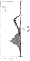
Figs. 2A-2D Flow cytometry analyses of the CT-2A-mD2C7 cell line. (Fig. 2 A) mouse H-2Kb (MHC class I) expression, (Fig. 2 B) mouse H-2Db (MHC class I) expression, (Fig. 2 C) mouse PD-L1 expression, and (Fig. 2 D) D2C7-IT target antigen mEGFRvIII expression on CT-2A-mD2C7 cells. -
Fig. 3 In vitro cytotoxicity of D2C7-IT on CT-2A-mD2C7 cells. WST1 assay was utilized to determine the in vitro cytotoxicity of D2C7-IT on CT-2A-mD2C7 cells. The IC50 of D2C7-IT on CT-2A-mD2C7 cells was 0.47 ng/ml (lower line) compared to that of the negative control immunotoxin, P588-IT (upper line, IC50 > 1000 ng/ml). -
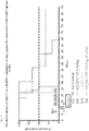
Fig. 4 . Survival of C57BL/6 mice injected intracranially with CT2A-mD2C7 cells. -
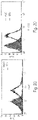
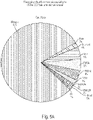
Figs. 5A-5B . Phenotypic profile of the immune cells populating the CT2A-mD2C7 brain tumor microenvironment.Fig. 5A control brain andFig. 5B CT2A-mD2C7 tumors. (F480lo + F480int + F480hi) = macrophages -
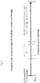
Fig. 6 . Experimental outline for D2C7-IT, BLZ945 combination therapy. -
Fig. 7 . Anti-tumor efficacy of D2C7-IT and BLZ945 combination therapy against the intracranial CT2A-mD2C7 glioma model. -
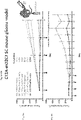
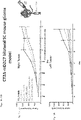
Figs. 8A-8B . In vivo efficacy of D2C7-IT+ (αCTLA-4 or αPD-1) mAb combination therapy in subcutaneous CT2A-mD2C7 glioma-bearing C57BL/6 immunocompetent mice.Fig. 8A shows all the treatment groups followed up toDay 35.Fig. 8B shows treatment groups G4-6 followed up toDay 62 after initial tumor inoculation. Note that 4 out of 10 and 5 out of 10 mice were cured only in treatment groups G5 and G6, respectively. -
Figs. 9A-9B . Tumor rechallenging studies for the cured mice in the combinational treatment groups (G5 and G6).Fig. 9A . Cured mice were first rechallenged with 106 CT2A parental cells on the left flank on Day 72.Fig. 9B shows subsequent challenge with 3×10⁵ CT2A-mD2C7 cells in the brain onDay 126 after the initial CT2A-mD2C7 cell inoculation on the right flank. Tumors grew in all naive mice (G0), whereas no tumor grew in those cured mice (G5 and G6). -
Figs. 10A-10B . In vivo efficacy of D2C7-IT+ (αCTLA-4 or αPD-1) mAb combination therapy in bilateral subcutaneous CT2A-mD2C7 glioma-bearing C57BL/6 immunocompetent mice.Fig. 10A shows the tumor growth curve for the right (treated) tumors, which was similar to the previous one-side model.Fig. 10B shows the tumor growth curve for the left (untreated) tumors, which showed that the combinational therapy on the right side could also have an antitumor effect on the distant left tumor. -
Figs. 11A-11B . Left tumor volume among groups onDay 35 andDay 43 after the initial tumor inoculation.Fig. 11A . OnDay 35, compared to treatment group G1, the left tumor growth was significantly delayed in all the other treatment groups, p<0.05 for treatment groups G2, G3, and G4; p<0.01 for treatment groups G5 and G6.Figs. 11B . OnDay 43, the left tumor growth was significantly delayed by the combinational therapy compared to the D2C7-IT monotherapy on the right tumor, p<0.05. - The inventors have developed targeted immunotoxins (IT), D2C7-(scdsFv)-PE38KDEL (D2C7-IT), by fusing the single chain variable fragment (scFv) from the D2C7 monoclonal antibody (mAb) with the Pseudomonas exotoxin A (PE), optionally fused to KDEL peptide. D2C7-IT reacts with both the wild-type epidermal growth factor receptor (EGFRwt) and the EGFR variant III (EGFRvIII), two proteins that are overexpressed in glioblastoma. The robust antitumor efficacy of D2C7-IT is mediated through PE in orthotopic glioma xenograft models in immunocompromised mice. In addition to direct tumor cell killing, the immunotoxin monotherapy induces a secondary antitumor immune response through the engagement of T cells. When the immunotoxin is administered in a combination regimen with an immune checkpoint inhibitor, improved and synergistic results are observed.
- Other moieties which can be attached to the antibodies include those which provide additional beneficial properties. For example, a KDEL (lys-asp-glu-leu) tetra-peptide can be added at the carboxy-terminus of the protein to provide retention in the endoplasmic reticulum. Variants such as DKEL, RDEL, and KNEL which function similarly can also be used.
- Tumors which can be treated are any that react with the D2C7 antibody. These include but are not limited to those in which at least one EGFRvIII allele is present. These may be found in breast, head and neck, brain, glioblastoma multiforme, astrocytoma, lung, or other tumors. It may be desirable to determine the presence of such an allele prior to therapy. This can be done using an oligonucleotide-based technique, such as PCR, or using an immunological technique, such as immunohistochemistry. It may be desirable to determine the amount, fraction, ratio, or percentage of cells in the tumor which express EGFR and/or EGFRvIII. The more cells which express EGFR on their surfaces, the more beneficial such antibody therapy is likely to be. Even tumors that express little to no EGFRvIII may be treated due to the ability of the antibody to bind to wild-type EGFR. Optionally, tumors may be tested prior to treatment for reactivity with D2C7 antibody. The immunotoxin itself could be used as an immunohistochemistry agent, before treatment, during treatment, or after treatment. A secondary reagent could be used with the immunotoxin for detection. It could, for example, recognize the Pseudomonas component of the immunotoxin.
- Immunotoxins can be administered by any technique known in the art. Compartmental delivery may be desirable to avoid cytotoxicity for normal tissues that express EGFR. Suitable compartmental delivery methods include, but are not limited to delivery to the brain, delivery to a surgically created tumor resection cavity, delivery to a natural tumor cyst, and delivery to tumor parenchyma.
- Tumors which can be treated by the method of the present invention are any which express epidermal growth factor receptor (EGFR), whether wild type, EGFRvIII, or other variants. Preferably the tumor expresses the receptor in amounts far exceeding expression by normal tissues. The mechanism of high level expression may be by genetic amplification, other alterations, whether genetic or epigenetic or post translational modifications. Exemplary tumors which can be treated include without limitation: malignant gliomas, breast cancer, head and neck squamous cell carcinoma, lung cancer.
- Blockade of T cell immune checkpoint receptors, can be performed against any such targets, including but not limited to PD-1, PD-L1, TIM-3, LAG-3, CTLA-4, and CSF-1R and combinations of such checkpoint inhibitors. The immune checkpoint receptors may be on tumor cells or immune cells such as T cells, monocytes, microglia, and macrophages, without limitation. The agents which assert immune checkpoint blockade may be small chemical entities or polymers, antibodies, antibody fragments, single chain antibodies or other antibody constructs, including but not limited to bispecific antibodies and diabodies.
- Immune checkpoint inhibitors which may be used according to the invention include any that disrupt the inhibitory interaction of cytotoxic T cells and tumor cells. These include but are not limited to anti-PD-1 antibody, anti-PD-Ll antibody, anti-CTLA4 antibody, anti-LAG-3 antibody, anti-TIM-3 antibody. The inhibitor need not be an antibody, but can be a small molecule or other polymer. If the inhibitor is an antibody it can be a polyclonal, monoclonal, fragment, single chain, or other antibody variant construct. Inhibitors may target any immune checkpoint known in the art, including but not limited to, CTLA-4, PDL1, PDL2, PD1, B7-H3, B7-H4, BTLA, HVEM, TIM3, GAL9, LAG3, CSF-1R, VISTA, KIR, 2B4, CD160, CGEN-15049,
CHK 1, CHK2, IDO, A2aR, and the B-7 family of ligands. Combinations of inhibitors for a single target immune checkpoint or different inhibitors for different immune checkpoints may be used. - Examples of inhibitors of CSF-1R which may be used in the combination therapy with the immunotoxin include, without limitation, the following agents which are in clinical development: PLX3397, PLX486, RG7155, AMG820, ARRY-382, FPA008, IMC-CS4, JNJ-40346527, and MCS110.
- The immune checkpoint inhibitor may be administered at the same time, before, or after the immunotoxin. Typically the two agents will be administered within 30, 28, 21, 14, 7, 4, 2, or 1 day(s) of each other. The agents may be given repeatedly, either serially or in a cycle of first and second agents. It may be advantageous but not necessary for the vaccine to be administered prior to the checkpoint inhibitor. But the reverse order may also be used. Priming of a cytotoxic T lymphocyte response by the immunotoxin may take from about 5 to about 14 days. Administration of the checkpoint inhibitor may beneficially be commenced during or after the priming period.
- Immune checkpoint inhibitors may be administered by any appropriate means known in the art for the particular inhibitor. These include intravenous, oral, intraperitoneal, sublingual, intrathecal, intracavitary, intramuscularly, and subcutaneously.
- Treatment regimens may include, in addition to delivery of the immunotoxin and immune checkpoint inhibitor(s), surgical removal of the tumor, surgical reduction of the tumor, chemotherapy, biological therapy, radiotherapy. These modalities are standard of care in many disease states, and the patient need not be denied the standard of care. The immunotoxin and immune checkpoint inhibitor(s) may be administered before, during, or after the standard of care. The immunotoxin and immune checkpoint inhibitor(s) may be administered after failure of the standard of care.
- Kits may comprise, in a single divided or undivided container, the immunotoxin or its components or its encoding DNA and the immune checkpoint inhibitor or combination of immune checkpoint inhibitors. Storage stability may vary between the two agents so separate vessels may be used. Optionally one or both agents may be lyophilized or frozen.
- Immunotoxins can directly kill cancer cells that express high levels of the targeted tumor antigen. Immunotoxin monotherapy can efficiently and directly destroy tumor cells expressing targeted epitopes, such as EGFRwt and/or its truncated variant, EGFRvIII, in malignant brain tumor xenograft models in immunocompromised mice. Immunotoxin therapy can induce a secondary anti-tumor immune response in a mouse model, which is different from the direct killing mechanism and needs the cooperation of the immune system. Since malignant brain tumors are always a heterogeneous mass, it is possible that some tumor cells can escape from the direct targeted attack of the immunotoxin therapy due to the lack of epitopes. For this reason, the secondary anti-tumor immune response stimulated by the immunotoxin may play an important role in eliminating those tumor cells not directly targeted.
- Recently, several studies successfully demonstrated that tumor regression and significantly improved survival were achieved in murine glioma models by suppressing co-inhibitory molecules, such as CTLA4, CSF-1R, IDO, and PD1. Based on the promising preclinical data, several clinical trials have started to investigate the utilization of immune checkpoint inhibitors to treat malignant brain tumors, either as monotherapy or combinatorial therapy with other anti-tumor agents.
- However, malignant gliomas, including glioblastomas, have relatively low mutation rates, which may generate fewer and subtle tumor antigens, leading to relatively poor basal immunogenicity compared to other tumor types that respond well to immunotherapies, for example, melanoma and NSCLC. Therefore, a combination of targeted cytotoxic immunotherapy and immune checkpoint inhibitors may provide a synergistic anti-tumor effect.
- The ideal combinatorial therapy may have a lower dose of targeted cytotoxic immunotherapy to limit its side effects, and achieve long-term anti-tumor immunity. Immunotoxin therapy can efficiently and directly kill cancer cells that express high levels of the targeted antigen through its unique cytotoxic mechanism. Cancer cells destroyed by localized immunotoxin therapy release tumor antigens and/or other neoantigens. These antigens can then be presented by the APCs to host T cells in the local draining lymph nodes, which activate CTLs to migrate and eliminate the remaining or recurrent tumor cells expressing specific tumor antigens at the tumor site. Throughout this process, various co-inhibitory checkpoint pathways between T cells and APCs and/or between T cells and tumor cells can trigger different mechanisms to de-activate T cells, and to adjust the continuation and intensity of the anti-tumor immunity. Immune checkpoint inhibitors, such as anti-CTLA4 and anti-PD1 mAbs, can block these immunosuppressive pathways and therefore augment tumor cell death caused by lymphocytes activated by the targeted immunotoxin therapy.
- We established a subcutaneous mouse CT2A-mD2C7 glioma model in C57BL/6 immunocompetent mice with six groups, in which the mice were treated by the control immunotoxin P588-IT or D2C7-IT, combined with αCTLA4 or αPD1 inhibitors after the tumor grew to a certain size. In this in vivo subcutaneous CT2A-mD2C7 glioma model, four doses of the low-dose D2C7-IT but not αCTLA4 or αPD1 monotherapy, and D2C7-IT+αCTLA-4 or αPD-1 combinatorial therapy generated a significant delay in tumor growth compared to the control immunotoxin P588-IT treatment groups (
Fig. 8A and 8B ). Importantly, complete cures were only observed in D2C7-IT+αCTLA4 (n = 4/10) and D2C7-IT+αPD-1 (n = 5/10) combinatorial therapy groups (Fig. 8A and 8B ), although D2C7-IT monotherapy could also significantly delay the tumor growth. These results demonstrated that low doses of cytotoxic immunotoxin therapy can significantly delay the tumor growth but fail to cure the tumor-bearing mice. Combined with immune checkpoint inhibitors, cytotoxic immunotoxin therapy can increase the initial cure rate of low-dose immunotoxin therapy from zero to over 40%. Furthermore, all cured mice rejected the mD2C7-negative tumors primary subcutaneous rechallenge, whereas tumors grew in untreated naive mice, suggesting that the combinatorial treatment provided long lasting anti-tumor immunity that even extended to mD2C7-negative parental cells, as well (Fig. 9A ). All nine cured mice then rejected the CT2A-mD2C7 secondary intracranial rechallenge in the brain, whereas tumors grew in untreated naive mice, indicating that the combinatorial treatment also provided long lasting anti-tumor immunity that extended to the remote immune-privileged CNS as well (Fig. 9B ), in which the central memory T cells (TCM) might play an important role. - Subsequently, we established a bilateral subcutaneous mouse glioma model to investigate whether a localized high-dose immunotoxin treatment can provide a systemic anti-tumor effect on the tumors in the distal region, and whether the combination of immune checkpoint inhibitors can enhance this systemic anti-tumor immunity induced by the localized immunotoxin therapy. The D2C7-IT monotherapy, D2C7-IT+αCTLA4, and D2C7-IT+αPD1 combinatorial therapy led to significant growth delay of the right tumors (P < 0.01), and cured 4/10, 6/10, and 5/10 right tumors, respectively (
Fig. 10A ). Interestingly, in the groups where the right tumors were treated by D2C7-IT or αCTLA-4 or αPD-1 monotherapy or D2C7-IT+ (αCTLA-4 or αPD-1) combinatorial therapy, the left untreated tumors also grew much slower compared to the control group (Fig. 10B and11A ), which indicates that a high dose of localized D2C7-IT monotherapy can achieve a similar anti-tumor immunity on the left untreated tumors compared to the systemic immune checkpoint inhibitor monotherapy. Furthermore, the combination therapy in the right tumors led to the most significantly delayed growth of the left untreated tumors in the mice (Fig. 10B and11B ), which suggests that immune checkpoint inhibitors can enhance the anti-tumor immunity induced by the localized immunotoxin therapy to restrict the tumor growth in the remote area. - We have demonstrated that the intratumoral delivery of D2C7-IT induces secondary anti-tumor immunity, which destroys not only mD2C7-expressing tumor cells, but also tumor cells not expressing mD2C7 at the systemic level. A combination of D2C7 immunotoxin with immune checkpoint inhibitors can enhance this immunotoxin-induced anti-tumor immunity to achieve a synergistic long-term anti-tumor effect.
- The above disclosure generally describes the present invention. All references disclosed herein are expressly incorporated by reference. A more complete understanding can be obtained by reference to the following specific examples which are provided herein for purposes of illustration only, and are not intended to limit the scope of the invention.
- We established a mouse glioma line, CT-2A-mD2C7, overexpressing the D2C7-IT antigen mouse EGFRvIII (mEGFRvIII). The reactivity and therapeutic efficacy of D2C7-IT against CT-2A-mD2C7 cells was determined by flow cytometry and in vitro cytotoxicity assays (WST1), respectively. CT-2A-mD2C7 cells were further analyzed for MHC class I and PD-L1 expression by flow cytometry. In vivo efficacy of D2C7-IT or αCTLA-4 or αPD-1 monotherapy or D2C7-IT+αCTLA-4 or D2C7-IT+αPD-1 combination therapy was evaluated in subcutaneous CT-2A-mD2C7 glioma-bearing C57BL/6 immunocompetent mice.
- WST-1 is a reagent for measuring cell proliferation. It is used for the nonradioactive, spectrophotometric quantification of cell proliferation and viability in cell populations. The assay is based on the cleavage of the tetrazolium salt WST-1 to formazan by cellular mitochondrial dehydrogenases. Expansion in the number of viable cells results in an increase in the activity of the mitochondrial dehydrogenases, which in turn leads to increase in the amount of formazan dye formed. The formazan dye produced by viable cells can be quantified by measuring the absorbance at λ=440 nm.
- Flow cytometry analysis confirmed the specific binding ability of D2C7 monoclonal antibody to the CT-2A-mD2C7 cells (
Fig. 2D ). Flow cytometry also demonstrated a high expression of both MHC class I molecules (Fig. 2A and2B ) and PD-L1 (Fig. 2C ) on tumor cell surface. D2C7-IT was highly cytotoxic (IC50 = 0.47 ng/mL) against CT-2A-mD2C7 cells in in vitro WST1 cytotoxicity assay (Fig. 3 ). - Construction, expression, and purification of D2C7-(scdsFv)-PE38KDEL immunotoxin. The carboxyl terminus of the D2C7 VH domain was connected to the amino terminus of the VL domain by a 15-amino-acid peptide (Gly4Ser)3 linker. In order to obtain a stable IT, it is essential to ensure that during renaturation VH is positioned near VL. This was achieved by mutating a single key residue in each chain to cysteine, for the stabilizing disulfide bond to form. On the basis of predictions using molecular modeling and empirical data with other dsFv-recombinant ITs, we chose one amino acid in each chain to mutate to cysteine. These are
residues 44 in the framework region 2 (FR2) of VH and 100 in the FR4 of VL (according to the Kabat numbering). Thus, we prepared an Fv that contains both a peptide linker and a disulfide bond generated by cysteine residues that replace Ser44 of VH and Gly100 of VL. The D2C7 (scdsFv) PCR fragment was then fused to DNA for domains II and III of Pseudomonas exotoxin A. The version of Pseudomonas exotoxin A used here, PE38KDEL, has a modified C terminus which increases its intracellular retention, in turn enhancing its cytotoxicity. The D2C7-(scdsFv)-PE38KDEL was expressed in E. coli under the control of T7 promoter and harvested as inclusion bodies. - Targeting tumor and tumor-associated macrophages for glioblastoma therapy: We evaluated the ability of D2C7-IT and BLZ945 combination treatment to function synergistically and produce an effective antitumor response in immunocompetent glioblastoma mouse models.
- Intracranial growth curve for CT2A-mD2C7 in C57BL/6 mice: To determine the time course of CT2A-mD2C7 intracranial tumor growth, 3×10⁵ cells/3 µl were implanted into 9 female C57BL/6 mice, and a survival curve was plotted (
Fig. 4 ). The CT2A-mD2C7 survival curve demonstrated that 100% death occurred atday 25 post-tumor implantation. Based on our previous studies,post-tumor implantation day 8 was chosen as the optimal day to initiate D2C7-IT infusion. - Phenotypic profile of the immune cells populating the CT2A-mD2C7 brain tumor microenvironment: To characterize the immune cell phenotype of intracranial CT2A-mD2C7 tumors, C57BL/6 immunocompetent mice were implanted with 3×10⁵ tumor cells. The mice were followed to assess tumor development and were euthanized when they became moribund. Upon euthanization, the brains were harvested and the tumors were analyzed for infiltrating immune cells by flow cytometry. Cells isolated from naive C57BL/6 mice were used as the control. The primary cell types in the normal brain were microglia (80%), macrophages (F480lo + F480int + F480hi = 11%), and T cells (5%) (
Fig. 5A ). However, there was a significant change in the percentage of microglia (8%), macrophages (F480lo + F480int + F480hi = 63%), and T cells (19%) in tumor-bearing mice (Fig. 5B ). - Anti-tumor efficacy of D2C7-IT and BLZ945 combination therapy against the intracranial CT2A-mD2C7 glioma model: The experimental outline for D2C7-IT+BLZ945 combination therapy against the CT2A-mD2C7 cell line is shown in
Fig. 6 . The in vivo efficacy of D2C7-IT or BLZ945 monotherapy or D2C7-IT+BLZ945 combination therapy was evaluated in intracranial CT2A-mD2C7 glioma-bearing C57BL/6 immunocompetent mice (Fig. 7 ). D2C7-IT (0.018 µg total dose) was infused by an osmotic pump via convection-enhanced delivery (CED) for 72 hours fromDay 8 to Day 11 post-tumor inoculation. BLZ945 (200 mg/kg) was delivered once daily through oral gavage onday 7 and on days 12-25. The survival curves for mice treated with the vehicle control and with BLZ945 monotherapy looked similar (Median Survival = 22-24 days,Fig. 7 ). At a total dose of 0.018 µg, D2C7-IT monotherapy extended the median survival to 42 days (Fig. 7 ). The median survival for the D2C7-IT+BLZ945 combination therapy group was 53 days (Fig. 7 ). Significantly, complete cures were observed only in the combination therapy group (2/6 mice). The preliminary data suggests that glioblastoma patients will benefit from D2C7-IT and BLZ945 combination therapy. - In previous pilot studies, we observed that the subcutaneous rechallenged mouse glioma allografts were rejected in those immunocompetent mice bearing SC mouse glioma allografts cured by the intratumoral (i.t.) immunotoxin therapy, suggesting that there can be a memory anti-tumor immune response following the SC immunotoxin therapy. This phenomenon was also reported in the SC melanoma mouse model treated by an immunotoxin targeting IL-13, in which CTLs played a major role in mediating this immunotoxin-induced anti-tumor response, although melanoma is a dramatically different type of tumor compared to malignant gliomas in the CNS. Therefore, it is necessary to establish appropriate mouse glioma models to investigate the secondary immune response, induced by immunotoxins, against glioblastomas, and to determine how to enhance this response by the combinatorial therapy of immune checkpoint inhibitors, such as anti-CTLA4 or anti-PDl antibodies (αCTLA4 or αPD1), in order to achieve a long-lasting remission.
- We established a subcutaneous mouse CT2A-mD2C7 glioma model in C57BL/6 immunocompetent mice with six groups, in which the mice were treated by the control immunotoxin P588-IT or D2C7-IT, combined with αCTLA4 or αPD1 inhibitors. In this in vivo subcutaneous CT2A-mD2C7 glioma model, four doses (every 3 days) of the D2C7-IT (low dose, 1.5 µg per mouse per dose, i.t.) but not αCTLA4 (100 µg per mouse per dose, intraperitoneal [i.p]) or αPD1 (250 µg per mouse per dose, i.p.) monotherapy and D2C7-IT+αCTLA-4 or αPD-1 combinatorial therapy (immune checkpoint inhibitors administered on the next day after IT therapy) generated a significant delay in tumor growth compared to the control immunotoxin P588-IT treatment groups (P < 0.01,
Fig. 8A ). Importantly, complete cures were only observed in D2C7-IT+αCTLA4 (n = 4/10) and D2C7-IT+αPD-1 (n = 5/10) combinatorial therapy groups (Fig. 8A and 8B ), although D2C7-IT monotherapy could also significantly delay the tumor growth. - To determine whether those cured mice from D2C7-IT and immune checkpoint inhibitors combinatorial treatment groups can recall a protective anti-tumor memory immune response, on Day 72 after the initial tumor challenge, all nine cured mice were then first subcutaneously rechallenged (1° SCR) with a dose of 106 CT2A parental cells in the left flank. All these mice rejected the mD2C7-negative tumors, whereas tumors grew in all untreated naive mice, suggesting that the combinatorial treatment provided long lasting anti-tumor immunity that extended to mD2C7-negative parental cells as well (
Fig. 9A ). - To determine whether this protective anti-tumor immunity can protect the mice from the tumor rechallenging in a remote immune-privileged region, for example, the brain, all nine cured mice were then intracranially (IC) rechallenged (2° ICR) for a second time on Day 126 (after the initial subcutaneous tumor challenge) with a dose of 3 × 10⁵ CT2A-mD2C7 cells in the brain. At the end of this study, all surviving mice were euthanized for brain histopathologic examination, which did not show tumors in the brains (data not shown). All these mice rejected the CT2A-mD2C7 intracranial (IC) rechallenge (2° ICR), whereas tumors grew in all untreated naive mice, suggesting that the combinatorial treatment also provided long lasting anti-tumor immunity that extended to the remote immune-privileged CNS as well (
Fig. 9B ). - In an in vivo bilateral subcutaneous CT2A-mD2C7 glioma model, tumor cells were inoculated in both sides of the flank simultaneously in C57BL/6 mice, with a high density (3 × 10⁶ cells) on the right side and a low density (106 cells) on the left side. The larger tumors (on the right) were treated with four doses (every two days) of D2C7-IT or αCTLA4 or αPD1 monotherapy or D2C7-IT+αCTLA4 or D2C7-IT+αPD1 combination therapy (immune checkpoint inhibitors administered on the same day of immunotoxin therapy), while the left tumors were untreated. The D2C7-IT monotherapy (high dose, 4.5 µg per mouse per dose, intratumoral), D2C7-IT+αCTLA4 (100 µg per mouse per dose, intraperitoneal), and D2C7-IT+αPD1 (250 µg per mouse per dose, intraperitoneal.) combinatorial therapies led to significant growth delays of the right tumor (P < 0.01), which cured 4/10, 6/10, and 5/10 right tumors, respectively (
Fig. 10A ). - Interestingly, in the groups where the right tumors were treated with D2C7-IT or αCTLA-4 or αPD-1 monotherapy or D2C7-IT+ (αCTLA-4 or αPD-1) combinatorial therapy, the left untreated tumors also grew much slower compared to the control group (
Fig. 10B and11A ), which indicates that a high dose of localized D2C7-IT monotherapy can achieve a similar anti-tumor immunity in the left untreated tumors compared to the systemic immune checkpoint inhibitor monotherapy. Furthermore, the D2C7-IT+ (αCTLA-4 or αPD-1) combinatorial therapy in the right tumors led to the most significantly delayed growth of the left untreated tumors in the mice (Fig. 10B and11B ), which suggests that immune checkpoint inhibitors can enhance the anti-tumor immunity induced by the immunotoxin to restrict the tumor growth in the remote area. -
- 1. A method of treating a tumor in a patient, comprising:
- administering to the patient an immunotoxin comprising a single chain variable region antibody fused to a PE38 truncated Pseudomonas exotoxin,
- wherein the single chain variable region antibody has CDR1, CDR2, and
- CDR3 regions as shown in SEQ ID NO: 6-11; and
- administering an immune checkpoint inhibitor to the patient.
- 2. The method of
clause 1 wherein the tumor is a malignant glioma. - 3. The method of
clause 1 wherein the tumor is breast cancer. - 4. The method of
clause 1 wherein the tumor is head and neck squamous cell carcinoma. - 5. The method of
clause 1 wherein the tumor is lung cancer. - 6. The method of
clause 1 wherein the immunotoxin is administered directly to the tumor. - 7. The method of
clause 1 wherein the immune checkpoint is selected from the group consisting of PD-1, PD-L1, CTLA-4, LAG-3, TIM-3, and CSF-1R. - 8. The method of
clause 1 wherein the checkpoint inhibitor is an anti-PD-1 antibody. - 9. The method of
clause 1 wherein the checkpoint inhibitor is an anti-PD-Ll antibody. - 10. The method of
clause 1 wherein the checkpoint inhibitor is an anti-CTLA4 antibody. - 11. The method of
clause 1 wherein the checkpoint inhibitor is an anti-LAG-3 antibody. - 12. The method of
clause 1 wherein the checkpoint inhibitor is an anti-TIM-3 antibody. - 13. The method of
clause 1 wherein the checkpoint inhibitor is an anti-CSF-lR antibody. - 14. The method of
clause 1 wherein the checkpoint inhibitor is a small molecule inhibitor of IDO. - 15. The method of
clause 1 wherein the checkpoint inhibitor is a small molecule inhibitor of CSF-1R. - 16. The method of
clause 1 wherein the immune checkpoint inhibitor is administered within 30 days of administering the immunotoxin. - 17. The method of
clause 1 wherein the immune checkpoint inhibitor is administered within 7 days of administering the immunotoxin. - 18. The method of
clause 1 wherein the PE38 truncated Pseudomonas exotoxin is fused to a KDEL peptide. - 19. A kit for treating a tumor, comprising:
- an immunotoxin comprising a single chain variable region antibody fused to a PE38 truncated Pseudomonas exotoxin, wherein the single chain variable region antibody has CDR1, CDR2, and CDR3 regions as shown in SEQ ID NO: 6-11; and
- an immune checkpoint inhibitor.
- 20. The kit of
clause 19 wherein the checkpoint inhibitor is an anti-PD-1 antibody. - 21. The kit of
clause 19 wherein the checkpoint inhibitor is an anti-PDL-1 antibody. - 22. The kit of
clause 19 wherein the checkpoint inhibitor is an anti-CTLA4 antibody. - 23. The kit of
clause 19 wherein the checkpoint inhibitor is an anti-LAG-3 antibody. - 24. The kit of
clause 19 wherein the checkpoint inhibitor is an anti-TIM-3 antibody. - 25. The kit of
clause 19 wherein the checkpoint inhibitor is an anti-CSF-Rl antibody. - 26. The kit of
clause 19 wherein the checkpoint inhibitor is a small molecule inhibitor of IDO. - 27. The kit of
clause 19 wherein the checkpoint inhibitor is a small molecule inhibitor of CSF-R1. - 28. The kit of
clause 19 wherein the PE38 truncated Pseudomonas exotoxin is fused to a KDEL peptide.
Claims (15)
- An immunotoxin comprising a single chain variable region antibody fused to a PE38 truncated Pseudomonas exotoxin, wherein the single chain variable region antibody has CDR1, CDR2, and CDR3 regions as shown in SEQ ID NO: 6-11, and an immune checkpoint inhibitor for use in treating a tumor in a patient, wherein the immune checkpoint inhibitor is administered at the same time, before , or after the immunotoxin.
- The immunotoxin and immune checkpoint inhibitor for use according to claim 1,
wherein the tumor is a malignant glioma. - The immunotoxin and immune checkpoint inhibitor for use according to claim 1
wherein the tumor is breast cancer. - The immunotoxin and immune checkpoint inhibitor for use according to claim 1 wherein: (a) the tumor is head and neck squamous cell carcinoma; or (b) the tumor is lung cancer.
- The immunotoxin and immune checkpoint inhibitor for use according to claim 1
wherein the use in treating the tumor comprises administration of the immunotoxin directly to the tumor. - The immunotoxin and immune checkpoint inhibitor for use according to claim 1
wherein the immune checkpoint is selected from the group consisting of PD-1, PD-L1, CTLA-4, LAG-3, TIM-3, and CSF-1R. - The immunotoxin and immune checkpoint inhibitor for use according to claim 1
wherein the checkpoint inhibitor is an anti-PD-1 antibody, an anti-PD-L1 antibody, an anti-CTLA4 antibody, an anti-LAG-3 antibody, an anti-TIM-3 antibody, an anti-CSF-1R antibody, a small molecule inhibitor of IDO, or a small molecule inhibitor of CSF-1R. - The immunotoxin and immune checkpoint inhibitor for use according to claim 1
wherein the use in treating the tumor comprises administration of the immune checkpoint inhibitor within 30 days of administration of the immunotoxin. - The immunotoxin and immune checkpoint inhibitor for use according to claim 1
wherein the use in treating the tumor comprises administration of the immune checkpoint inhibitor within 7 days of administration of the immunotoxin. - The immunotoxin and immune checkpoint inhibitor for use according to claim 1
wherein the PE38 truncated Pseudomonas exotoxin is fused to a KDEL peptide. - A kit for treating a tumor, comprising:an immunotoxin comprising a single chain variable region antibody fused to a PE38 truncated Pseudomonas exotoxin, wherein the single chain variable region antibody has CDR1, CDR2, and CDR3 regions as shown in SEQ ID NO: 6-11; andan immune checkpoint inhibitor.
- The kit of claim 11 wherein the checkpoint inhibitor is an anti-PD-1 antibody, an anti-PDL-1 antibody, an anti-CTLA4 antibody, an anti-LAG-3 antibody, an anti-TIM-3 antibody, an anti-CSF-Rl antibody, a small molecule inhibitor of IDO or a small molecule inhibitor of CSF-R1.
- The kit of claim 11 wherein the PE38 truncated Pseudomonas exotoxin is fused to a KDEL peptide.
- An immunotoxin comprising a single chain variable region antibody fused to a PE38 truncated Pseudomonas exotoxin, wherein the single chain variable region antibody has CDR1, CDR2, and CDR3 regions as shown in SEQ ID NO: 6-11, for use with an immune checkpoint inhibitor in treating a tumor in a patient.
- An immune checkpoint inhibitor for use with an immunotoxin comprising a single chain variable region antibody fused to a PE38 truncated Pseudomonas exotoxin, wherein the single chain variable region antibody has CDR1, CDR2, and CDR3 regions as shown in SEQ ID NO: 6-11, in treating a tumor in a patient.
Applications Claiming Priority (3)
| Application Number | Priority Date | Filing Date | Title |
|---|---|---|---|
| US201562250749P | 2015-11-04 | 2015-11-04 | |
| PCT/US2016/060469 WO2017079520A1 (en) | 2015-11-04 | 2016-11-04 | Combination therapy of immunotoxin and checkpoint inhibitor |
| EP16863025.9A EP3370773B1 (en) | 2015-11-04 | 2016-11-04 | Combination therapy of immunotoxin and checkpoint inhibitor |
Related Parent Applications (2)
| Application Number | Title | Priority Date | Filing Date |
|---|---|---|---|
| EP16863025.9A Division-Into EP3370773B1 (en) | 2015-11-04 | 2016-11-04 | Combination therapy of immunotoxin and checkpoint inhibitor |
| EP16863025.9A Division EP3370773B1 (en) | 2015-11-04 | 2016-11-04 | Combination therapy of immunotoxin and checkpoint inhibitor |
Publications (1)
| Publication Number | Publication Date |
|---|---|
| EP3973988A1 true EP3973988A1 (en) | 2022-03-30 |
Family
ID=58662982
Family Applications (2)
| Application Number | Title | Priority Date | Filing Date |
|---|---|---|---|
| EP21193555.6A Pending EP3973988A1 (en) | 2015-11-04 | 2016-11-04 | Combination therapy of immunotoxin and checkpoint inhibitor |
| EP16863025.9A Active EP3370773B1 (en) | 2015-11-04 | 2016-11-04 | Combination therapy of immunotoxin and checkpoint inhibitor |
Family Applications After (1)
| Application Number | Title | Priority Date | Filing Date |
|---|---|---|---|
| EP16863025.9A Active EP3370773B1 (en) | 2015-11-04 | 2016-11-04 | Combination therapy of immunotoxin and checkpoint inhibitor |
Country Status (5)
| Country | Link |
|---|---|
| US (3) | US11065332B2 (en) |
| EP (2) | EP3973988A1 (en) |
| JP (1) | JP6587756B2 (en) |
| CN (2) | CN108367070B (en) |
| WO (1) | WO2017079520A1 (en) |
Families Citing this family (8)
| Publication number | Priority date | Publication date | Assignee | Title |
|---|---|---|---|---|
| CN108367070B (en) * | 2015-11-04 | 2022-10-28 | 杜克大学 | Combination therapy of immunotoxins with checkpoint inhibitors |
| CN110115767A (en) * | 2018-02-05 | 2019-08-13 | 美国安吉益民公司 | The method of immunomodulator joint inspection point inhibitor for treating cancer |
| EP3773650A4 (en) * | 2018-04-02 | 2021-12-29 | Duke University | Neoadjuvant cancer treatment |
| CN112423778A (en) * | 2018-05-16 | 2021-02-26 | 杜克大学 | Neoadjuvant cancer therapy using immunotoxins in combination with checkpoint inhibitors |
| WO2020156500A1 (en) * | 2019-01-31 | 2020-08-06 | 正大天晴药业集团股份有限公司 | Use of anti-pd-l1 antibody in treatment of head and neck cancer |
| EP4051321A4 (en) * | 2019-10-30 | 2023-11-22 | Duke University | IMMUNOTHERAPY WITH COMBINATION THERAPY WITH AN IMMUNE TOXIN |
| US20250195644A1 (en) * | 2021-03-05 | 2025-06-19 | Duke University | Compositions For and Methods of Preventing Metastases |
| EP4344409A4 (en) * | 2021-07-01 | 2025-05-21 | Duke University | D2C7-EGFR and EGFR VIII bispecific chimeric antigen receptor constructs and methods for making and using the same |
Citations (3)
| Publication number | Priority date | Publication date | Assignee | Title |
|---|---|---|---|---|
| CA154291A (en) | 1913-10-21 | 1914-03-10 | A. Edwin Aldred | Packing |
| CA197264A (en) | 1920-02-17 | K. Kosich Demster | Fruit picker | |
| US20130022598A1 (en) * | 2008-04-11 | 2013-01-24 | The United States Government As Represented By The Secretary, Department Of Health And Human Service | Dual Specific Immunotoxin for Brain Tumor Therapy |
Family Cites Families (13)
| Publication number | Priority date | Publication date | Assignee | Title |
|---|---|---|---|---|
| CA2520172C (en) * | 2003-03-27 | 2012-10-02 | Lankenau Institute For Medical Research | Novel methods for the treatment of cancer with an indoleamine 2, 3-dioxygenase inhibitor |
| FR2926560A1 (en) | 2008-01-21 | 2009-07-24 | Millipore Corp | PROCESS FOR EXTRACTING AND PURIFYING NUCLEIC ACIDS ON MEMBRANE |
| US7994662B2 (en) | 2009-06-23 | 2011-08-09 | Anorad Corporation | Thermal block and thermal rail |
| LT2769737T (en) * | 2009-07-20 | 2017-06-26 | Bristol-Myers Squibb Company | Combination of an anti-CTLA4 antibody with etoposide for the synergistic treatment of proliferative diseases |
| AR080698A1 (en) * | 2010-04-01 | 2012-05-02 | Imclone Llc | ANTIBODY OR FRAGMENT OF THE SAME THAT SPECIFICALLY LINKS THE VARIOUS OF HUMAN CSF -1R, PHARMACEUTICAL COMPOSITION THAT INCLUDES IT, ITS USE FOR THE MANUFACTURE OF A USEFUL MEDICINAL PRODUCT FOR THE TREATMENT OF CANCER AND METHOD TO DETERMINE IF A BASED SUBJECT MATTER AND |
| LT2704713T (en) * | 2011-05-05 | 2017-04-25 | Novartis Ag | Csf-1r inhibitors for treatment of brain tumors |
| DK2806883T3 (en) * | 2012-01-25 | 2019-07-22 | Dnatrix Inc | BIOMARKERS AND COMBINATION THERAPIES WITH USING ONCOLYTIC VIRUS AND IMMUNOMODULATION |
| US10966998B2 (en) * | 2013-09-05 | 2021-04-06 | The Johns Hopkins University | Cancer therapy via a combination of epigenetic modulation and immune modulation |
| IL292510A (en) * | 2013-11-05 | 2022-06-01 | Cognate Bioservices Inc | Combinations of checkpoint inhibitors and cancer drugs |
| CA2935423C (en) | 2014-01-24 | 2023-11-07 | Dana-Farber Cancer Institute, Inc. | Antibody molecules to pd-1 and uses thereof |
| AU2015210750B2 (en) | 2014-01-31 | 2020-08-20 | Children's Medical Center Corporation | Antibody molecules to TIM-3 and uses thereof |
| WO2015138920A1 (en) | 2014-03-14 | 2015-09-17 | Novartis Ag | Antibody molecules to lag-3 and uses thereof |
| CN108367070B (en) * | 2015-11-04 | 2022-10-28 | 杜克大学 | Combination therapy of immunotoxins with checkpoint inhibitors |
-
2016
- 2016-11-04 CN CN201680070875.6A patent/CN108367070B/en active Active
- 2016-11-04 WO PCT/US2016/060469 patent/WO2017079520A1/en not_active Ceased
- 2016-11-04 CN CN202211240353.XA patent/CN116327955A/en active Pending
- 2016-11-04 US US15/773,418 patent/US11065332B2/en active Active
- 2016-11-04 EP EP21193555.6A patent/EP3973988A1/en active Pending
- 2016-11-04 JP JP2018543049A patent/JP6587756B2/en active Active
- 2016-11-04 EP EP16863025.9A patent/EP3370773B1/en active Active
-
2021
- 2021-07-15 US US17/376,319 patent/US20210338811A1/en not_active Abandoned
-
2024
- 2024-01-31 US US18/428,597 patent/US20240350624A1/en active Pending
Patent Citations (3)
| Publication number | Priority date | Publication date | Assignee | Title |
|---|---|---|---|---|
| CA197264A (en) | 1920-02-17 | K. Kosich Demster | Fruit picker | |
| CA154291A (en) | 1913-10-21 | 1914-03-10 | A. Edwin Aldred | Packing |
| US20130022598A1 (en) * | 2008-04-11 | 2013-01-24 | The United States Government As Represented By The Secretary, Department Of Health And Human Service | Dual Specific Immunotoxin for Brain Tumor Therapy |
Non-Patent Citations (3)
| Title |
|---|
| GLEN C MACDONALD ET AL: "A phase I clinical study of VB4-845: weekly intratumoral administration of an anti-EpCAM recombinant fusion protein in patients with squamous cell carcinoma of the head and neck", DRUG DESIGN, DEVELOPMENT AND THERAPY, 1 January 2009 (2009-01-01), New Zealand, pages 105 - 114, XP055089137, Retrieved from the Internet <URL:http://www.pubmedcentral.nih.gov/articlerender.fcgi?artid=2761172&tool=pmcentrez&rendertype=abstract> * |
| JASMIN LESHEM ET AL: "Combining anti-CTLA4 with RG7787, an immunotoxin targeting mesothelin, promotes tumor eradication", JOURNAL FOR IMMUNOTHERAPY OF CANCER, BIOMED CENTRAL LTD, LONDON, UK, vol. 3, no. 2, 4 November 2015 (2015-11-04), pages 1 - 2, XP021235245, DOI: 10.1186/2051-1426-3-S2-P190 * |
| PASTAN IRA ET AL: "Immunotoxin therapy of cancer", NATURE REVIEWS. CA, NATUR PUBLISHING GROUP, LONDON, GB, vol. 6, no. 7, 1 July 2006 (2006-07-01), pages 559 - 565, XP009136267, ISSN: 1474-175X * |
Also Published As
| Publication number | Publication date |
|---|---|
| EP3370773B1 (en) | 2022-01-05 |
| CN108367070A (en) | 2018-08-03 |
| US20210338811A1 (en) | 2021-11-04 |
| WO2017079520A1 (en) | 2017-05-11 |
| CN108367070B (en) | 2022-10-28 |
| JP2018533626A (en) | 2018-11-15 |
| US11065332B2 (en) | 2021-07-20 |
| EP3370773A4 (en) | 2019-05-29 |
| US20180311346A1 (en) | 2018-11-01 |
| US20240350624A1 (en) | 2024-10-24 |
| CN116327955A (en) | 2023-06-27 |
| EP3370773A1 (en) | 2018-09-12 |
| JP6587756B2 (en) | 2019-10-09 |
Similar Documents
| Publication | Publication Date | Title |
|---|---|---|
| US20240350624A1 (en) | Combination Therapy of Immunotoxin and Checkpoint Inhibitor | |
| KR102362803B1 (en) | Compositions comprising anti-ceacam1 and anti-pd antibodies for cancer therapy | |
| EP3468999B1 (en) | Anti-gitr antibodies and uses thereof | |
| JP2019503361A (en) | Combination of anti-PD-1 antibody and bispecific anti-CD20 / anti-CD3 antibody for treating cancer | |
| JP2018521008A (en) | Therapeutic compositions and therapeutic methods for treating tumorigenesis | |
| JP7754506B2 (en) | Antibodies to the poliovirus receptor (PVR) and uses thereof | |
| TW202409067A (en) | Il-12 variants, anti-pd1 antibodies, fusion proteins, and uses thereof | |
| JP2024163118A (en) | Neoadjuvant cancer treatment using a combination of immunotoxins and checkpoint inhibitors | |
| CN115843255A (en) | CD22 targeting moieties for the treatment of B cell type acute lymphoblastic leukemia (B-ALL) | |
| WO2024041651A1 (en) | Methods of cancer treatment using anti-pd1 antibodies in combination with anti-tim3 antibodies and anti-lag3 antibodies | |
| IL296043A (en) | Anti-gitr antibodies and uses thereof | |
| HK40073036A (en) | Combination therapy of immunotoxin and checkpoint inhibitor | |
| US12514927B2 (en) | Immunotherapy with combination therapy comprising an immunotoxin | |
| CN118475618A (en) | Methods of treating cancer with bispecific anti-CD 3xMUC antibodies and anti-CTLA-4 antibodies | |
| KR20230082038A (en) | Antibody combination for cancer treatment that reduces cytokine release syndrome | |
| EP4588939A1 (en) | Interleukin-13 receptor subunit alpha-2 (il13ralpha2) targeting moieties for the treatment and prevention of il13ralpha2-positive cancer | |
| TW202611078A (en) | Methods of treating multiple myeloma with bispecific bcma x cd3 antibodies | |
| TW202539732A (en) | Methods of treating recurrent ovarian cancer and endometrial cancer with bispecific anti-muc16 x anti-cd28 antibodies in combination with anti-pd-1 antibodies or bispecific anti-muc16 x anti-cd3 antibodies | |
| Zhang | Cancer Immunotherapy | |
| WO2025171124A1 (en) | Methods of treating prostate cancer with bispecific anti-psma x anti-cd3 antibodies and bispecific anti-psma x anti-cd28 antibodies | |
| TW202540162A (en) | Combination therapy involving bispecific binding agents binding to cldn18.2 and cd3 and anti-vegfr2 antibodies | |
| CN121604974A (en) | Methods of treating metastatic castration-resistant prostate cancer with bispecific anti-psma x anti-CD 28 antibodies | |
| KR20230061499A (en) | Method for treating cancer pain by administering a PD-1 inhibitor | |
| KR20200130380A (en) | Compositions and methods for treating end-stage lung cancer | |
| WO2015198311A1 (en) | Compositions comprising antibodies to ceacam-1 and kir for cancer therapy |
Legal Events
| Date | Code | Title | Description |
|---|---|---|---|
| PUAI | Public reference made under article 153(3) epc to a published international application that has entered the european phase |
Free format text: ORIGINAL CODE: 0009012 |
|
| STAA | Information on the status of an ep patent application or granted ep patent |
Free format text: STATUS: THE APPLICATION HAS BEEN PUBLISHED |
|
| AC | Divisional application: reference to earlier application |
Ref document number: 3370773 Country of ref document: EP Kind code of ref document: P |
|
| AK | Designated contracting states |
Kind code of ref document: A1 Designated state(s): AL AT BE BG CH CY CZ DE DK EE ES FI FR GB GR HR HU IE IS IT LI LT LU LV MC MK MT NL NO PL PT RO RS SE SI SK SM TR |
|
| STAA | Information on the status of an ep patent application or granted ep patent |
Free format text: STATUS: REQUEST FOR EXAMINATION WAS MADE |
|
| 17P | Request for examination filed |
Effective date: 20220930 |
|
| RBV | Designated contracting states (corrected) |
Designated state(s): AL AT BE BG CH CY CZ DE DK EE ES FI FR GB GR HR HU IE IS IT LI LT LU LV MC MK MT NL NO PL PT RO RS SE SI SK SM TR |
|
| REG | Reference to a national code |
Ref country code: HK Ref legal event code: DE Ref document number: 40073036 Country of ref document: HK |
|
| STAA | Information on the status of an ep patent application or granted ep patent |
Free format text: STATUS: EXAMINATION IS IN PROGRESS |
|
| 17Q | First examination report despatched |
Effective date: 20240523 |