EP3968272A1 - Medical endoscope image identification method and system, and endoscope image system - Google Patents
Medical endoscope image identification method and system, and endoscope image system Download PDFInfo
- Publication number
- EP3968272A1 EP3968272A1 EP20802793.8A EP20802793A EP3968272A1 EP 3968272 A1 EP3968272 A1 EP 3968272A1 EP 20802793 A EP20802793 A EP 20802793A EP 3968272 A1 EP3968272 A1 EP 3968272A1
- Authority
- EP
- European Patent Office
- Prior art keywords
- endoscope
- image
- images
- target
- lesion
- Prior art date
- Legal status (The legal status is an assumption and is not a legal conclusion. Google has not performed a legal analysis and makes no representation as to the accuracy of the status listed.)
- Pending
Links
Images



Classifications
-
- G—PHYSICS
- G06—COMPUTING OR CALCULATING; COUNTING
- G06F—ELECTRIC DIGITAL DATA PROCESSING
- G06F18/00—Pattern recognition
- G06F18/20—Analysing
- G06F18/24—Classification techniques
-
- A—HUMAN NECESSITIES
- A61—MEDICAL OR VETERINARY SCIENCE; HYGIENE
- A61B—DIAGNOSIS; SURGERY; IDENTIFICATION
- A61B1/00—Instruments for performing medical examinations of the interior of cavities or tubes of the body by visual or photographical inspection, e.g. endoscopes; Illuminating arrangements therefor
- A61B1/00002—Operational features of endoscopes
- A61B1/00004—Operational features of endoscopes characterised by electronic signal processing
- A61B1/00009—Operational features of endoscopes characterised by electronic signal processing of image signals during a use of endoscope
- A61B1/000094—Operational features of endoscopes characterised by electronic signal processing of image signals during a use of endoscope extracting biological structures
-
- A—HUMAN NECESSITIES
- A61—MEDICAL OR VETERINARY SCIENCE; HYGIENE
- A61B—DIAGNOSIS; SURGERY; IDENTIFICATION
- A61B1/00—Instruments for performing medical examinations of the interior of cavities or tubes of the body by visual or photographical inspection, e.g. endoscopes; Illuminating arrangements therefor
- A61B1/00002—Operational features of endoscopes
- A61B1/00004—Operational features of endoscopes characterised by electronic signal processing
- A61B1/00009—Operational features of endoscopes characterised by electronic signal processing of image signals during a use of endoscope
- A61B1/000096—Operational features of endoscopes characterised by electronic signal processing of image signals during a use of endoscope using artificial intelligence
-
- G—PHYSICS
- G06—COMPUTING OR CALCULATING; COUNTING
- G06N—COMPUTING ARRANGEMENTS BASED ON SPECIFIC COMPUTATIONAL MODELS
- G06N3/00—Computing arrangements based on biological models
- G06N3/02—Neural networks
- G06N3/04—Architecture, e.g. interconnection topology
- G06N3/045—Combinations of networks
-
- G—PHYSICS
- G06—COMPUTING OR CALCULATING; COUNTING
- G06N—COMPUTING ARRANGEMENTS BASED ON SPECIFIC COMPUTATIONAL MODELS
- G06N3/00—Computing arrangements based on biological models
- G06N3/02—Neural networks
- G06N3/08—Learning methods
-
- G—PHYSICS
- G06—COMPUTING OR CALCULATING; COUNTING
- G06T—IMAGE DATA PROCESSING OR GENERATION, IN GENERAL
- G06T7/00—Image analysis
- G06T7/0002—Inspection of images, e.g. flaw detection
- G06T7/0012—Biomedical image inspection
-
- G—PHYSICS
- G06—COMPUTING OR CALCULATING; COUNTING
- G06T—IMAGE DATA PROCESSING OR GENERATION, IN GENERAL
- G06T7/00—Image analysis
- G06T7/70—Determining position or orientation of objects or cameras
-
- G—PHYSICS
- G16—INFORMATION AND COMMUNICATION TECHNOLOGY [ICT] SPECIALLY ADAPTED FOR SPECIFIC APPLICATION FIELDS
- G16H—HEALTHCARE INFORMATICS, i.e. INFORMATION AND COMMUNICATION TECHNOLOGY [ICT] SPECIALLY ADAPTED FOR THE HANDLING OR PROCESSING OF MEDICAL OR HEALTHCARE DATA
- G16H30/00—ICT specially adapted for the handling or processing of medical images
- G16H30/40—ICT specially adapted for the handling or processing of medical images for processing medical images, e.g. editing
-
- A—HUMAN NECESSITIES
- A61—MEDICAL OR VETERINARY SCIENCE; HYGIENE
- A61B—DIAGNOSIS; SURGERY; IDENTIFICATION
- A61B1/00—Instruments for performing medical examinations of the interior of cavities or tubes of the body by visual or photographical inspection, e.g. endoscopes; Illuminating arrangements therefor
- A61B1/00002—Operational features of endoscopes
- A61B1/00043—Operational features of endoscopes provided with output arrangements
- A61B1/00045—Display arrangement
- A61B1/0005—Display arrangement combining images e.g. side-by-side, superimposed or tiled
-
- G—PHYSICS
- G06—COMPUTING OR CALCULATING; COUNTING
- G06T—IMAGE DATA PROCESSING OR GENERATION, IN GENERAL
- G06T2207/00—Indexing scheme for image analysis or image enhancement
- G06T2207/10—Image acquisition modality
- G06T2207/10016—Video; Image sequence
-
- G—PHYSICS
- G06—COMPUTING OR CALCULATING; COUNTING
- G06T—IMAGE DATA PROCESSING OR GENERATION, IN GENERAL
- G06T2207/00—Indexing scheme for image analysis or image enhancement
- G06T2207/10—Image acquisition modality
- G06T2207/10068—Endoscopic image
-
- G—PHYSICS
- G06—COMPUTING OR CALCULATING; COUNTING
- G06T—IMAGE DATA PROCESSING OR GENERATION, IN GENERAL
- G06T2207/00—Indexing scheme for image analysis or image enhancement
- G06T2207/20—Special algorithmic details
- G06T2207/20024—Filtering details
-
- G—PHYSICS
- G06—COMPUTING OR CALCULATING; COUNTING
- G06T—IMAGE DATA PROCESSING OR GENERATION, IN GENERAL
- G06T2207/00—Indexing scheme for image analysis or image enhancement
- G06T2207/20—Special algorithmic details
- G06T2207/20076—Probabilistic image processing
-
- G—PHYSICS
- G06—COMPUTING OR CALCULATING; COUNTING
- G06T—IMAGE DATA PROCESSING OR GENERATION, IN GENERAL
- G06T2207/00—Indexing scheme for image analysis or image enhancement
- G06T2207/20—Special algorithmic details
- G06T2207/20081—Training; Learning
-
- G—PHYSICS
- G06—COMPUTING OR CALCULATING; COUNTING
- G06T—IMAGE DATA PROCESSING OR GENERATION, IN GENERAL
- G06T2207/00—Indexing scheme for image analysis or image enhancement
- G06T2207/20—Special algorithmic details
- G06T2207/20084—Artificial neural networks [ANN]
-
- G—PHYSICS
- G06—COMPUTING OR CALCULATING; COUNTING
- G06T—IMAGE DATA PROCESSING OR GENERATION, IN GENERAL
- G06T2207/00—Indexing scheme for image analysis or image enhancement
- G06T2207/30—Subject of image; Context of image processing
- G06T2207/30004—Biomedical image processing
- G06T2207/30028—Colon; Small intestine
-
- G—PHYSICS
- G06—COMPUTING OR CALCULATING; COUNTING
- G06T—IMAGE DATA PROCESSING OR GENERATION, IN GENERAL
- G06T2207/00—Indexing scheme for image analysis or image enhancement
- G06T2207/30—Subject of image; Context of image processing
- G06T2207/30004—Biomedical image processing
- G06T2207/30092—Stomach; Gastric
-
- G—PHYSICS
- G06—COMPUTING OR CALCULATING; COUNTING
- G06T—IMAGE DATA PROCESSING OR GENERATION, IN GENERAL
- G06T2207/00—Indexing scheme for image analysis or image enhancement
- G06T2207/30—Subject of image; Context of image processing
- G06T2207/30004—Biomedical image processing
- G06T2207/30096—Tumor; Lesion
Definitions
- Embodiments of this disclosure relate to the field of computer application technologies, and in particular, to a medical endoscope image recognition method and system, and an endoscopic imaging system.
- Various category identifications executed based on deep learning are generally important tools for solving classification for a large amount of data in various application scenarios. For example, in application scenarios such as image and natural language processing, large-scale classification and recognition of a large amount of data may be implemented, so as to rapidly and accurately obtain a related classification prediction result and accelerate the implementation of functions in the application scenarios.
- images for classification prediction and methods for classification prediction are also different from one another.
- AI Artificial Intelligence + a medical scenario as an example, with continuously photographing in the alimentary canal by the endoscope, a large quantity of endoscope images are formed, the classification prediction method is thus required to classify and recognize the large quantity of endoscope images.
- the related medical image has a single classification prediction function, which cannot be adapted to the whole process of photographing by the endoscope for generating a medical endoscope video stream; moreover, since capturing the medical endoscope image would be unavoidably affected by switching and shaking of the endoscope, and during photographing, a lens of the endoscope would unavoidably encounter various liquids and foreign matters, the obtained endoscope image would often have a large amount of interference and noise, rendering weak robustness.
- it is expected to provide a method and system for recognizing a medical endoscope image, so that photographing by the endoscope in the alimentary canal can be adapted to the whole photographing process and the robustness is relatively strong.
- embodiments of this disclosure provide a medical endoscope image recognition method and system, an endoscopic imaging system, and medical endoscope image recognition.
- a medical endoscope image recognition method is provided, applicable to a workstation of an endoscopic imaging system, the method including:
- a medical endoscope image recognition system including:
- a machine device including:
- a storage medium is provided, storing at least one instruction, the at least one instruction being loaded and executed by a processor to implement the foregoing medical endoscope image recognition method.
- An endoscopic imaging system including:
- first original endoscope images are obtained therefrom, and then the obtained original endoscope image are filtered by using a neural network to obtain target endoscope images, to eliminate a large amount of interference existing under the condition of switching and shaking during the photographing by the endoscope and encountering various liquids and foreign matters, so that robustness is enhanced.
- corresponding organ information is recognized from the generated target endoscope image, so as to identify an image type suitable for the target endoscope images according to the corresponding organ information by using a classification network, and finally, in a photographing mode corresponding to the image type, according to a part indicated by the organ information, a lesion region is localized and the lesion category thereof is identified to implement the classification prediction for the whole photographing process of the endoscope in the alimentary canal, and systematic and complete image recognition is implemented.
- FIG. 1 is a schematic diagram of an implementation environment according to an embodiment of this disclosure.
- the implementation environment includes an endoscopic imaging system including an endoscope 110, a display device 130, and a workstation 150.
- the endoscope 110 is used as a data source for image recognition; along with the movement and photographing of the endoscope 110 in alimentary canals, the display device 130 continuously displays video images. For example, the images are displayed by using each endoscope image frame captured by the endoscope 110.
- the image captured by the alimentary canal endoscope in this embodiment of this disclosure is also recognized by using the workstation 150, so as to examine the alimentary canal endoscope, to implement systematic and comprehensive classification prediction, thereby obtaining lesion region distribution in the endoscope image and a category of the distributed lesion region.
- the workstation 150 is a host deployed for the endoscope, such as a micro-computer with a large or small volume that only needs to meet performance requirements.
- this disclosure includes a physical medical device, such as an endoscopic imaging system, which at least includes:
- the medical endoscope video stream inputted to the workstation may be currently captured in real time by the endoscope, and may also be obtained by photographing at any time, which is not limited herein.
- the endoscopic imaging system also includes an endoscope; the accessed endoscope provides a data source for the workstation, and then the endoscope inputs the medical endoscope video to the workstation, so as to implement real-time recognition of a video image.
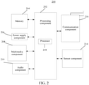
- FIG. 2 is a block diagram of an apparatus according to an exemplary embodiment.
- the apparatus 200 may be a workstation 150 in an implementation environment shown in FIG. 1 .
- the workstation 150 may be a micro-computer in any form as long as it meets the performance requirements.
- the workstation 150 may be a host connected to an endoscope.
- the apparatus 200 includes at least the following components: a processing component 202, a memory 204, a power supply component 206, a multimedia component 208, an audio component 210, a sensor component 214, and a communication component 216.
- the processing component 202 generally controls integral operations of the apparatus 200, such as operations related to displaying, a phone call, data communication, a camera operation, and a record operation.
- the processing component 202 includes at least one or more processors 218 to execute instructions, to implement all or some steps of the following method.
- the processing component 202 includes at least one or more modules, to facilitate the interaction between the processing component 202 and other components.
- the processing component 202 may include a multimedia module, to facilitate the interaction between the multimedia component 208 and the processing component 202.
- the memory 204 is configured to store various types of data to support operations on the apparatus 200. Examples of the types of data include instructions of any application program or method to be operated on the apparatus 200.
- the memory 204 is at least implemented by using a volatile or non-volatile storage device of any type or a combination thereof, for example, a static random access memory (SRAM), an electrically erasable programmable read-only memory (EEPROM), an erasable programmable read-only memory (EPROM), a programmable read-only memory (PROM), a read-only memory (ROM), a magnetic memory, a flash memory, a disk, or an optical disc.
- SRAM static random access memory
- EEPROM electrically erasable programmable read-only memory
- EPROM erasable programmable read-only memory
- PROM programmable read-only memory
- ROM read-only memory
- magnetic memory a magnetic memory
- flash memory a flash memory
- disk or an optical disc.
- the memory 204 further stores one or more modules, and the one or more modules are configured to be executed by the one or more processor 218, to implement all or some steps of the following method shown in any of FIG. 3 , FIG. 4 , FIG. 5 , FIG. 6 , FIG. 7 , FIG. 8 , FIG. 9 , and FIG. 10 .
- the power supply component 206 provides power to various components of the apparatus 200.
- the power supply component 206 includes at least a power supply management system, one or more power supplies, and other components associated with generating, managing and allocating power for the apparatus 200.
- the multimedia component 208 includes a screen providing an output interface between the apparatus 200 and a user.
- the screen may include a liquid crystal display (LCD) and a touch panel. If the screen includes the touch panel, the screen may be implemented as a touchscreen to receive an input signal from the user.
- the touch panel includes one or more touch sensors to sense a touch, a slide, and a gesture on the touch panel. The touch sensor may not only sense the boundary of touching or sliding operations, but also detect duration and pressure related to the touching or sliding operations.
- the screen further includes an organic light emitting diode (OLED) display.
- OLED organic light emitting diode
- the audio component 210 is configured to output and/or input an audio signal.
- the audio component 210 includes a microphone (MIC).
- the microphone When the apparatus 200 is in an operating mode, such as a call mode, a record mode, and a speech recognition mode, the microphone is configured to receive an external audio signal.
- the received audio signal may further be stored in the memory 204 or be sent by using the communication component 216.
- the audio component 210 further includes a speaker, configured to output an audio signal.
- the sensor component 214 includes one or more sensors, configured to provide status evaluation in each aspect to the apparatus 200. For example, the sensor component 214 detects a power-on/off state of the apparatus 200 and a relative location of a component. The sensor component 214 further detects changes in a location of the apparatus 200 or a component of the apparatus 200 and a temperature change of the apparatus 200. In some embodiments, the sensor component 214 further includes a magnetic sensor, a pressure sensor or a temperature sensor.
- the communication component 216 is configured to facilitate communication in a wired or wireless manner between the apparatus 200 and other devices.
- the apparatus 200 accesses a communication standard-based wireless network, such as Wireless Fidelity (Wi-Fi).
- Wi-Fi Wireless Fidelity
- the communication component 216 receives, by using a broadcast channel, a broadcast signal or broadcast-related information from an external broadcast management system.
- the communication component 216 further includes a near field communication (NFC) module to facilitate short-distance communication.
- the NFC module may be implemented based on a radio frequency identification (RFID for short) technology, an Infrared Data Association (IrDA for short) technology, an ultra wideband (UWB) technology, a Bluetooth technology, and another technology.
- RFID radio frequency identification
- IrDA Infrared Data Association
- UWB ultra wideband
- Bluetooth a Bluetooth technology
- the apparatus 200 is implemented by using one or more application-specific integrated circuits (ASICs), a digital signal processor, a digital signal processing device, a programmable logic device, a field-programmable gate array, a controller, a micro controller, a microprocessor, or other electronic elements, and is configured to perform the following method.
- ASICs application-specific integrated circuits
- a digital signal processor a digital signal processing device
- a programmable logic device a field-programmable gate array
- a controller a micro controller
- microprocessor or other electronic elements
- FIG. 3 is a flowchart of a medical endoscope image recognition method according to an exemplary embodiment, taking the workstation executing the method according to FIG. 1 as an example.
- the medical endoscope image recognition method as shown in FIG. 3 , can include at least the following steps:
- step 310 original endoscope images are obtained according to a medical endoscope video stream.
- the medical endoscope video stream is a video stream captured by an endoscope in a medical environment, for example, a real hospital usage environment.
- the medical endoscope video stream presents video endoscopy captured by a lens of the endoscope.
- original endoscope image frames can be obtained according to the medical endoscope video stream. Since each original endoscope image frame describes the video endoscopy captured by the endoscope at a time point, based on each original endoscope image frame, the medical endoscope image can be recognized.
- an endoscope captures the medical endoscope video stream in an organism, for example, a human body.
- the endoscope captures the medical endoscope video stream in a tract communicated with the outside or a sealed body cavity.
- the indicated tract communicated with the outside may be an alimentary canal, a respiratory tract, a urinary tract, and the like;
- the sealed body cavity may be a cavity body that needs an incision for the endoscope to be fed such as chest, an abdominal cavity, and a joint cavity. Capturing and recognizing the medical endoscope video stream by using the endoscope can obtain organ conditions in the corresponding tract.
- the obtained medical endoscope video stream is inputted to the workstation for recognizing the medical endoscope image.
- the medical endoscope video stream obtained before, for example, a historical medical endoscope video stream may also be subjected to the medical endoscope image recognition. That is, recognition may be performed based on a real-time captured image, and may also be performed based on a large quantity of stored medical endoscope video streams.
- the medical endoscope video stream obtained through real-time photographing by the endoscope in the alimentary canal is taken as an example for detailed explanations below.
- the obtained medical endoscope video stream is inputted to the workstation, and the corresponding original endoscope image is obtained from the current alimentary canal image, for real-time recognition based on the original endoscope image.
- the current image displayed by the display device 130 of the endoscopic imaging system is the alimentary canal image.
- the display device 130 displays the video of alimentary canal images through the inputted medical endoscope video stream, and at this time, the original endoscope image required by image recognition is obtained from the current image.
- the original endoscope image is an original image directly obtained under the photographing by the endoscope, and on this basis, the medical endoscope image is recognized.
- step 310 includes: obtaining the original endoscope images from the inputted medical endoscope video stream along with movement and photographing of the endoscope in the tract or the sealed body cavity.
- the tract communicated with the outside and the sealed tract do not only have a single organ; taking the alimentary canal as an example, the alimentary canal includes a plurality of sub-organs, such as stomach, esophagus, pharynx and duodenum.
- the endoscope moves and continuously takes photos in the alimentary canal so as to obtain a video stream related to the sub-organs. Accordingly, the original endoscope images constituting the video stream are images related to the sub-organ where the endoscope is located, and indicate the state of the sub-organ.
- the photographing of the sub-organ where the endoscope is located is not limited to a single original endoscope image; that is, a plurality of original endoscope images obtained all correspond to one sub-organ, and therefore, the subsequent recognition of the medical endoscope image using the original endoscope images as inputs actually relates to classification prediction of the sub-organ where the endoscope is located.
- step 310 can include:
- the stored medical endoscope video stream is recognized one by one using the medical endoscope image recognition method provided by the embodiments of this disclosure, so as to recognize the lesion region and lesion category of the organ part related to the original endoscope image, to implement the processing of a large quantity of historical medical endoscope video streams.
- the medical endoscope video stream is no longer obtained through the output of the endoscope, but the stored medical endoscope video streams are obtained, so as to obtain the original endoscope image therefrom.
- a large quantity of stored medical endoscope video streams can also be recognized so as to facilitate medical researches, and provide automatic video image recognition for the real medical environment.
- step 330 the original endoscope images are filtered by using a neural network, to generate target endoscope images.
- the original endoscope images obtained by photographing during switching, shaking, or switching and shaking of the endoscope and the original endoscope images obtained when a lens encounters various liquids and foreign matters during photographing are low-quality images, exist as interferences for the recognition, and need to be recognized and filtered out by using the neural network.
- the used neural network is obtained through training by using the low-quality images as examples.
- the original endoscope images obtained through photographing include a large quantity of low-quality and noisy images. Recognizing and filtering the low-quality images from the original endoscope images obtained in step 310 by using the neural network shields the influence on image recognition by the low-quality images, greatly improving the robustness. In addition, useless and unnecessary images are filtered out in advance through recognizing and filtering the low-quality images, so that computing resources consumed by executing the follow-up steps are reduced, and therefore, speed and real-time performance can be effectively improved.
- the low-quality image recognition performed on the original endoscope images is implemented by using the trained neural network. Moreover, this neural network is trained according to endoscope image samples that are the low-quality images and endoscope image samples that are the non-low-quality images.
- the trained neural network can output, for the inputted original endoscope image, a probability of being the low-quality image and a probability of being the non-low-quality image, so as to finally determine whether the original endoscope image is the low-quality image or the non-low-quality image; the original endoscope image determined to be the low-quality image is filtered out and no follow-up steps are adopted for processing.
- Recognition of the low-quality images for the obtained multiple original endoscope images is conducted by using the neural network, to filter out the low-quality images included in the obtained multiple original endoscope images, complete filtering of the low-quality images, and generate target endoscope images, so as to recognize organ parts that the endoscope enters.
- the original endoscope images inputted into the neural network for low-quality image recognition are necessarily adapted to the neural network to ensure consistence and accuracy of actual prediction.
- pre-processing the original endoscope images is further required, for example, size adjusting, cutting, and the like, to obtain the original endoscope images with the size adapted to the neural network.
- the target endoscope images are the remaining original endoscope images after eliminating the low-quality images in the original video images. At this point, by filtering the original endoscope images, the generated target endoscope images can shield interference, reduce a data amount, and also enhance the accuracy of recognition.
- the same original endoscope image may be a low-quality image and may also be a non-low-quality image.
- step 350 organ information corresponding to the target endoscope images is recognized by using the neural network.
- the endoscope As the movement and continuously photographing in the tract by the endoscope, the endoscope is located in the tract, for example, on a certain sub-organ in the alimentary canal, however, the endoscope would not output the organ part where it is located, the organ part is often required to be recognized by checking using the endoscope image in a manual way, so as to facilitate the implementation of accurate classification prediction of the endoscope image for the organ part where the endoscope is located.
- the alimentary canal part where the endoscope is currently located is recognized. For example recognition is performed to obtain the organ information corresponding to the target endoscope image, and the organ information indicates the organ part in the tract where the endoscope is located when capturing the target endoscope image.
- recognizing the organ information of the target endoscope image is also implemented by using the constructed neural network; the target endoscope image is used as an input, and information of an organ where the endoscope is located when capturing the target endoscope image is outputted.
- the constructed neural network may by a four-class network.
- the four-class network is pre-constructed, to recognize the target endoscope image so as to recognize an organ location where the endoscope is located.
- the four-class network is obtained by training using the original endoscope image in which corresponding alimentary canal parts, that is the sub-organs on the alimentary canal, are annotated.
- the four-class network adapted to the alimentary canal parts executes the recognition of the alimentary canal part where the endoscope is currently located, the endoscope images for network training, such as samples of the four-class network that cover all the alimentary canal parts, and therefore, it is no longer limited to the recognition of a single sub-organ, thereby enhancing the recognition performance of an image captured by the endoscope on the alimentary canal.
- step 370 an image type suitable for the target endoscope images is identified according to the corresponding organ information by using a classification network.
- the photographing mode for the target endoscope image can be switched.
- the image type to which the target endoscope image is adapted is an image type that can best enhances the image endoscopy in the target endoscope image.
- the most proper photographing mode can be determined for the target endoscope image. For example, based on the image type, the photographing mode corresponding to the image type can be switched.
- the recognition of the image type to which the target endoscope image is adapted is implemented by using the classification network.
- the classification network may be a three-class network, that is a classification network that divides images into three image types, so as to implement the recognition of the target endoscope image for the three image types.
- the photographing modes correspond to different image types, and therefore, when the photographing modes are set as three, such as white light, Narrow Band Imaging (NBI), and iodine dyeing modes, three corresponding image types exist.
- the photographing mode to which an image content in the target endoscope image is adapted can be determined through the recognition of the image type, that is the photographing mode corresponding to the image type can be identified.
- the target endoscope image is the image type corresponding to the NBI, and the image type corresponds to the NBI photographing mode.
- step 390 a lesion region in each of the target endoscope images is localized according to a part indicated by the organ information, and a lesion category of the lesion region in the photographing mode corresponding to the image type is identified.
- the target endoscope image in the photographing mode to which the target endoscope image corresponding to the sub-organ can be obtained, so as to implement the localization of the lesion region and the recognition of the lesion category of the lesion region.
- the organ part indicated by the organ information corresponds to multiple target endoscope images, and therefore, the target endoscope image adapted to the photographing mode can be obtained from the multiple target endoscope images obtained by photographing on the organ part, to localize the lesion region on the target endoscope image and identify the lesion category of the lesion region for the organ part.
- the photographing mode aims at the target endoscope image that images the organ part.
- the photographing mode includes the image type, dyeing type, etc.
- the photographing mode includes three modes of white light, NBI, and iodine dyeing.
- different lesion conditions on the target endoscope image are adapted to different photographing modes.
- the white light mode is adopted, and when a suspicious lesion or a disease lesion region exists on the organ part, the white light mode is switched to the NBI mode. Since the image colors, textures, and details corresponding to the target endoscope images in different photographing modes are greatly different, through switching the photographing mode, the lesion region can be more accurately localized, so as to identify the lesion category of the lesion region.
- the corresponding photographing mode is determined, so as to directly switch the target endoscope image into the determined photographing mode, thereby obtaining the target endoscope image in the photographing mode to which the organ part where the endoscope is currently located is adapted, so as to enhance the accuracy of the image content represented by the target endoscope image.
- dynamic adjustment is performed on the photographing mode for the target endoscope image, so as to enhance the accuracy rate of image recognition.
- the organ information is obtained; the organ information corresponds to the target endoscope image obtained by filtering out the low-quality image; based on the organ information, localizing the lesion region of the target endoscope image and identifying the lesion category thereof can be performed in the adapted mode.
- the target endoscope image obtained by filtering out the low-quality image corresponding to the organ information can have the following two meanings: on one hand, the target endoscope image obtained by filtering out the low-quality image has been adapted to the photographing mode suitable for the current alimentary canal part; for example, the adapted photographing mode is the white light mode, while the target endoscope image obtained by filtering out the low-quality image corresponds to the white light mode, which is consistent with the photographing mode that needs to be used.
- the target endoscope image obtained by filtering out the low-quality image has a photographing mode that is not adapted to the photographing mode suitable for the organ part, for example, the photographing mode used by the endoscope image is the white light mode, while the photographing mode needing to be used is the NBI mode. Hence, it is required to switch the photographing mode of the target endoscope image.
- the executed lesion region localization and lesion category identification are both implemented by using a deep learning network.
- the lesion region localization may adopt a localization detection network, for example, and end-to-end real-time target location network YOLO (You Only Look Once, a deep learning network for target detection), and may also adopt other detection networks (e.g., FasterRCNN); the lesion category identification is implemented using the classification network; this classification network may be a Densely Connected Convolutional network (DenseNet for short).
- DenseNet Densely Connected Convolutional network
- a localization detection network deployed for the lesion region localization may be deployed uniformly.
- the localization detection network may be separately deployed according to corresponding organ information, such as the alimentary canal parts.
- the classification network deployed for the lesion category identification is also like this, and is determined according to experimental effects. If the network is separately deployed according to the alimentary canal parts, there is only a need to train the deep learning network separately.
- a more complete and available medical endoscope image recognition system with strong robustness can be implemented, so as to comprehensively assist a doctor in diagnosis in many respects and improve diagnosis efficiency.
- Localizing the lesion region of the target endoscope image and identifying the lesion category thereof effectively can help avoid missed diagnosis of the alimentary canal examination by the endoscope, effectively assist the doctor to determine the lesion property in real time, and improve the accuracy rate of the determination.
- the medical endoscope image recognition according to the exemplary embodiment above is implemented by means of deep learning, manual intervention is no longer needed for profound understanding of the medical image, and a manually made feature extraction solution is no longer needed as well, thereby avoiding omission and erroneous judgment caused by incomplete feature extraction.
- FIG. 4 is a flowchart of step 330 according to the embodiment corresponding to FIG. 3 .
- step 330 includes at least the following steps.
- step 331 the original endoscope images are processed according to a set size to generate standard endoscope images.
- the standard endoscope image is adapted to the endoscope image with the size required by the neural network for the neural network to be inputted.
- it is required to pre-process the data, such as adjusting the size of the obtained original endoscope image, so that the generated standard endoscope image can be adapted to the input to the neural network and the consistency is ensured.
- the process of processing the original endoscope images includes: first executing a resize operation, and then using an image scaling method, such as a center crop method (a rounded corner cutting method) to cut to obtain the standard endoscope image with the set size.
- an image scaling method such as a center crop method (a rounded corner cutting method) to cut to obtain the standard endoscope image with the set size.
- the resize operation is an adjusting operation for the original endoscope image; exemplarily, the execution process of the resize operation may be: maintaining a length-width ratio, scaling a short edge to 224 pixels, and a long edge being greater than or equal to 224 pixels.
- the execution process of the center crop method may be: using a long edge of the original endoscope image as a standard, and cutting a central region of the original endoscope image, so that the long edge becomes 224, so as to obtain a standard endoscope image conforming to the set size, to ensure the consistency of network prediction.
- step 333 prediction of whether the standard endoscope images are low-quality images or non-low-quality images is performed by using the neural network.
- the low-quality image is a standard endoscope image having interference.
- the neural network is used for implementing a classification task, so as to filter the low-quality images of the standard endoscope images.
- the neural network may be a deep convolutional neural network, such as Densenet.
- the target endoscope image is an endoscope image adapted to the neural network and subjected to the size processing on the original endoscope image.
- the trained neural network is constituted by executing the network training process after a large quantity of original endoscope images are divided into the low-quality images and the non-low-quality images.
- the large quantity of original endoscope images as examples can be obtained by expanding the original endoscope images, so as to provide more samples for the training of the neural network.
- step 335 the standard endoscope images that are low-quality images are filtered out to obtain the target endoscope images.
- the endoscope image corresponding to the low-quality image in the original endoscope images can be determined.
- the original endoscope image that is the low-quality image can be filtered out, which can effectively preventing useless and unnecessary images from entering the follow-up recognition process of the medical endoscope image.
- recognition and filtering of the low-quality image are implemented for the medical endoscope image recognition, so as to be actually applied to a real production environment, such as a hospital, without influencing from the switching and shaking of the endoscope in the tract, and also without the influences from various liquids and foreign matters encountered in the tract by the endoscope.
- FIG. 5 is a flowchart of step 390 according to the embodiment corresponding to FIG. 3 .
- step 390 includes the following steps.
- step 391 a foreign matter in each target endoscope image in the photographing mode corresponding to the image type is detected, to obtain a foreign matter frame distributed in the each target endoscope image, the foreign matter frame being used for indicating a region having a foreign matter in the each target endoscope image.
- step 393 the target endoscope images are filtered according to the foreign matter frame, the lesion region is localized by using the target endoscope images that remain after the filtering, and the lesion category of the lesion region is identified.
- the foreign matters in the target endoscope image are further detected and localized, so as to filter out the foreign matters that influence the image content in the target endoscope image.
- the alimentary canal in the alimentary canal, there are often special intraoperative instruments, saliva and other foreign matters in the esophagus and stomach.
- the image content of the target endoscope image captured by the endoscope in the alimentary canal can mostly contains intraoperative instruments, saliva and other foreign matters.
- the target endoscope image in which the foreign matter is detected cannot be directly filtered out.
- the detection of the foreign matter faces the target endoscope image adapted to the photographing mode; for this target endoscope image, the neural network is used for detecting the foreign matter in the image, and obtaining a foreign matter frame localized on the target endoscope image.
- the foreign matter frame is used for indicating a region occupied by the foreign matter in the target endoscope image. It is to be understood that the foreign matter frame annotates the distribution of the foreign matter in the target endoscope image.
- the foreign matter frame is substantially a region occupied by the intraoperative instruments or a region occupied by the saliva.
- the obtained foreign matter frame distributed on the target endoscope image is represented in the form of coordinates.
- This process implements the detection of the target by using the neural network; and under the action of the neural network, in addition to outputting the coordinates representing the foreign matter frame, a confidence of the foreign matter frame corresponding to the foreign matter is further outputted, such as the probability.
- the coordinates of the foreign matter may be determined by the square frame, and may include four pieces of coordinate information, such as xmin, ymin, xmax, and ymax.
- the foreign matter frame distributed in the target endoscope image can be used for evaluating whether to filter out the target endoscope image to shield the interference caused by excess foreign matters.
- step 391 includes: inputting the each target endoscope image in the photographing mode corresponding to the image type into the neural network, performing target detection by using the neural network, and outputting coordinates and a confidence that correspond to the foreign matter frame, where the coordinates are used for indicating a distribution of the foreign matter frame in the each target endoscope image.
- the neural network for foreign matter detection may be a YOLO location network, and may also be another deep detection network, which is not limited herein.
- the entire target endoscope image is used as an input, the location of the foreign matter frame is regressed at an output layer, such as the coordinates and the category thereof, and this category is the foreign matter. That is, the confidence outputted by the neural network represents the possibility that the foreign matter corresponds to the localized foreign matter frame.
- FIG. 6 is a flowchart of step 393 according to the embodiment corresponding to FIG. 5 .
- Step 393, as shown in FIG. 6 includes the following steps:
- step 401 an area proportion factor of an area occupied by the foreign matter in each target endoscope image is determined according to the coordinates and the confidence corresponding to the foreign matter frame in the each target endoscope image.
- the area proportion factor of all foreign matters on the target endoscope image is calculated.
- a foreign matter frame area Si is first calculated according to the coordinates.
- the foreign matter frame area is an area occupied by the foreign matter frame.
- a corresponding confidence Pi is used as a coefficient to correct the foreign matter frame area, such as PiSi, and finally corrected foreign matter frame areas of all the foreign matter frames are added, such as by summing the area PiSi of each foreign matter frame, and performing a proportion calculation on the sum and a total area of the target endoscope image, to finally obtain the area proportion factor of the area occupied by the foreign matter in the target endoscope image.
- step 403 interference of the foreign matter with the each target endoscope image is determined according to the area proportion factor, and filter out the target endoscope images with foreign matter interference.
- the interference of the foreign matter with the each target endoscope image can be determined according to the numeral value of the area proportion factor.
- target endoscope images with relatively larger area proportion factors are filtered out, and these images are considered to be the target endoscope images with the foreign matter interference.
- a threshold f 0 is first set, and the default value of f 0 may be 0.1.
- f is greater than the threshold fo, it is determined that the target endoscope image is the target endoscope image with foreign matter interference, and the target endoscope image needs to be filtered out.
- lesion region localization for the target endoscope image and lesion category identification for the localized lesion region are continued.
- foreign matter localization and anti-inference are implemented, so as to resolve the special foreign matter problems such as intraoperative instruments and saliva in the esophagus and stomach in the alimentary canal, thereby reducing the influence on image recognition due to the presence of the foreign matter.
- FIG. 7 is a flowchart of step 390 according to the embodiment corresponding to FIG. 3 .
- step 390 at least includes the following steps.
- step 501a an image type of the target endoscope image is detected.
- a photographing mode corresponding to the target endoscope image is switched according to the photographing mode corresponding to the identified image type when the image type is inconsistent with the identified image type, to obtain a target endoscope image in the photographing mode corresponding to the image type.
- the photographing mode of the target endoscope image is switched to obtain the target endoscope image in the photographing mode corresponding to the image type suitable for the target endoscope images.
- FIG. 8 is a flowchart of step 390 according to the embodiment corresponding to FIG. 3 .
- step 390 includes the following steps.
- step 501b continuous feature extraction for each target endoscope image in the photographing mode corresponding to the image type is performed by using each layer of a localization detection network until the lesion region in the each target endoscope image is finally obtained through regression.
- step 503b a lesion property of the lesion region in the each target endoscope image is classified by using the classification network, to obtain the lesion category of the lesion region.
- the localization detection network is used for performing target detection on the target endoscope image, to implement the lesion localization in the target endoscope image, so as to output two-dimensional coordinates of the lesion region.
- the localization detection network is an end-to-end real-time target detection algorithm, such as YOLO, to meet real-time requirements for image recognition.
- the localization detection network may also use other detection networks for replacement, such as FasterRCNN
- a process of performing continuous feature extraction by using each layer of the localization detection network to obtain the lesion region in the each target endoscope image through regression obtains more features and is more comprehensive, and thus can avoid incomplete feature extraction and omission and misjudgment caused thereby.
- the lesion region obtained upon location detection would be represented in the form of two-dimensional coordinates.
- the localization detection network finally outputs the two-dimensional coordinates for localizing the lesion region on the target endoscope image.
- the localization problem of the lesion region by the YOLO relates to the extraction image bounding boxes and category probability regression problems.
- the two-dimensional coordinates and probability are finally obtained by regression, and therefore, the accuracy of localization is improved while ensuring the real-time performance of the detection.
- network training is performed using an image data set with an open source, so as to obtain parameters and weight values of each network layer, for example, the parameters and weight values of the convolution layer can be obtained, so as to construct a localization detection network having more generalization performance.
- the data amount of the image data set with an open source is above a million; training the localization detection network using the image data set with an open source may avoid overfitting, so that the network training can be better converged to an optimal point.
- the low-quality image is also added into the training process of the localization detection network. For example, based on the low-quality endoscope image, training the localization detection network, to enhance the robustness and anti-noise capability of the localization detection network and to reduce a false positive ratio.
- the recognition of the lesion category of the lesion region can be executed.
- the category may include normal, precancerous disease lesion, early cancer, advanced cancer, inflammatory disease lesion, and other disease lesions, which are not limited herein.
- the classification network implementing the lesion category identification may be based on Densenet.
- the input of the classification network is a lesion region in the target endoscope image, and the output thereof is the lesion category corresponding to the lesion region.
- the lesion region localization and lesion region recognition can implement a more complete and available image recognition solution, without being limited to a single function, ensuring the comprehensiveness of the supported functions.
- FIG. 9 is a flowchart of step 503b according to the embodiment corresponding to FIG. 8 .
- step 503b includes the following steps:
- step 601 the lesion region in the each target endoscope image is extended, to obtain an extended region corresponding to the lesion region.
- the localized lesion regions in the target endoscope image are not consistent with each other in size.
- the lesion region is first extended so as to obtain an extended region corresponding to each lesion region subjected to the lesion category identification.
- the external expansion of the region can ensure that the lesion region for recognition can obtain certain context semantic information.
- Features related to lesions often exist around the lesion region.
- the lesion is not strictly provided with a boundary, and the lesion is a gradually changed process. Therefore, the external expansion of the region can provide more information to the classification network to learn, so as to avoid missing useful boundary information.
- the external expansion of the lesion region is a process of setting the proportions of up, down, left, and right external expansion of this lesion region.
- the lesion region is extended by 10% upwards, downwards, leftwards, and rightwards.
- step 603 the extended region is pre-processed to normalize the extended region into a classification network input image meeting an input size.
- the extended region is pre-processed so that the extended region is normalized as an image with the input size, so as to ensure the input of the classification network may be met.
- the pre-processing process includes an execution process using a center crop method.
- the classification network training process corresponding thereto requires to implement the pre-processing of the classification network input image through a data enhancing method, so as to expand the samples.
- step 605 network prediction on a lesion category of the input image is performed by using the classification network, to obtain the lesion category of the corresponding lesion region in the each target endoscope image.
- the image After obtaining the image of the extended region including the context information through the preceding steps, the image is inputted into the classification network, so that network prediction of the lesion category can be performed on the corresponding lesion region; in a similar fashion, the lesion category of the lesion region in the endoscope image can be identified.
- the classification network for implementing category identification may be a Densenet model.
- the lesion categories outputted by the classification network may be six categories, such as normal, precancerous disease lesion, early cancer, advanced cancer, inflammatory disease lesion, and other disease lesions.
- the classification network is actually a six-class network.
- Identifying the lesion category of the lesion region in the endoscope image can output in real time the specific property of the lesion of the alimentary canal through image recognition in the alimentary canal detection process of the endoscope, so as to assist the doctor in the alimentary canal endoscope image diagnosis.
- a medical endoscope image recognition method further includes:
- the neural network is used for generating target endoscope images.
- the endoscope image may be the original endoscope image, and may be a standard endoscope image matched with the neural network size processing, which is not limited herein.
- the trained neural network recognizing, by using the trained neural network, whether it is a low-quality image, so as to filter out the endoscope image corresponding to the low-quality image, avoids the occurrence of noise that is useless and may influence the processing efficiency.
- FIG. 10 is a flowchart of a step of training a neural network by using low-quality images and non-low-quality images captured by an endoscope as samples, to obtain a neural network corresponding to a low-quality image category output probability and a non-low quality image category output probability according to an exemplary embodiment.
- the steps include the following steps:
- step 801 the low-quality images and the non-low-quality images captured by the endoscope as the samples are adjusted to a fixed size.
- step 803 data enhancement is performed on the low-quality images and the non-low-quality images that have been adjusted to the fixed size, to obtain sample images meeting an input size of a neural network.
- step 805 a network training process for the neural network is executed by using the sample images as inputs.
- the sample of the neural network for recognizing the low-quality image includes the low-quality image and non-low-quality image captured by the endoscope, but is not limited to the low-quality image and non-low-quality image captured by the endoscope, and further includes images expanded from the low-quality image and non-low-quality image captured, so as to form the sample image inputted to the neural network.
- the low-quality image and non-low-quality image captured by the endoscope are not obtained by one endoscope examination but are endoscope images widely obtained through various modes.
- parameters and weight values of each network layer are obtained through a large scale of sample inputs, and the data amount of the sample also determines the generalization performance and classification accuracy of the trained neural network.
- the data amount of the sample also determines the generalization performance and classification accuracy of the trained neural network.
- the executed fixed-size adjustment is the process of adjusting the image to a fixed size, for example, the process of adjusting the image to 227 ⁇ 227 pixels.
- data enhancement is data pre-processing using a random cutting method, etc. and combining a series of operations such as random rotation, brightness, color, contrast, and random jitter to perform fixed-size adjustment and data enhancement on the low-quality images and the non-low-quality images, so as to obtain various images, for example, images at different angles, to enhance the generalization performance and prevent the occurrence of the overfitting phenomenon.
- Data enhancement is performed on each of the low-quality images and the non-low-quality images, to change one image into multiple images, so as to together form sample images meeting the input size of the neural network.
- the sample data can be expanded for the network training process; on the basis of the existing low-quality image and non-low-quality image captured by the endoscope, sufficient sample data may be provided to implement the network training process of converging to the optimal point.
- the video stream is inputted, such as the medical endoscope video stream of the alimentary canal, while executing the current image display synchronously, the original endoscope image is correspondingly obtained.
- FIG. 11 is a schematic diagram of an overall framework of image recognition photographed by the alimentary canal endoscope according to an exemplary embodiment.
- the medical endoscope video stream of the alimentary canal is outputted.
- step 910 is first executed to perform recognition and filtering on the low-quality image on each original endoscope image to remove the original endoscope image belonging to the low-quality image to generate the target endoscope image.
- Densenet can be selected to construct the used neural network, and in the execution process of the neural network, the sample is processed through the data enhancing method, but for the network prediction process, the data enhancing method is no longer executed, only a single cutting method, for example, the center crop method, is used for ensuring consistency, so as to avoid the increase of time consumption caused by the data enhancement, ensuring the real-time performance.
- Filtering the low-quality image through step 910 can effectively remove the low-quality image in the original endoscope image so that the non-low-quality image can execute the following image recognition process.
- step 920 is executed to recognize the organ part.
- the Densenet can also be selected to construct the used neural network, such as the preceding indicated four-class network.
- the organ part where the endoscope is currently located in the alimentary canal can be localized in the process of continuous movement and photographing of the endoscope, so as to provide a proper available photographing mode for the endoscope that photographs the organ part.
- the image type identification in step 930 is substantially the identification of the photographing mode suitable for the endoscope image.
- the photographing mode of the endoscope image can be switched according to this image type, thereby obtaining the photographing mode suitable for the endoscope image for each endoscope image obtained by filtering out the low-quality image.
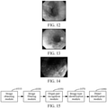
- FIG. 12 is a schematic diagram of an endoscope image in a white light photographing mode according to an exemplary embodiment.
- FIG. 13 is a schematic diagram of an endoscope image in an NBI mode according to the embodiment corresponding to FIG. 12.
- FIG. 14 is a schematic diagram of an endoscope image in an iodine dyeing mode according to the embodiment corresponding to FIG. 12 .
- the image colors, textures, and details of the three images are greatly different, and therefore, in the process of recognition, adaptively switching the photographing mode upon recognition of the image type greatly enhances the accuracy of image recognition.
- step 930 the image type is identified.
- the step also needs to implement the classification task.
- the Densenet model can also be selected to construct the used classification network, such as the three-class network, and therefore, the network training process thereof is similar to the training process of the low-quality image filtering network.
- the foreign matter localization and anti-interference implementing processes in step 940 are executed, to eliminate the interference of the foreign matter, so as to complete the lesion region localization and the lesion category identification.
- an endoscope image frame would be processed in an average of 150 milliseconds, which meets the real-time performance requirement, and has very high accuracy.
- the implementation of the execution process can be deployed to a hospital, so as to assist a doctor in diagnosis of the alimentary canal endoscope image in real-time, improving the diagnosis efficiency of the doctor.
- a more complete and available system with strong robustness for assisting the alimentary canal endoscope examination can be implemented, so as to more comprehensively implement assistance; in the processing of the endoscope image, the smoothness of a video frame rate can be ensured, for example, an average of less than 150 milliseconds per frame.
- the alimentary canal endoscope diagnosis system directly applied to a hospital production environment is obtained, and under the current situation of scarce and uneven medical resources, the system can assist the doctor to localize and discover the alimentary canal lesion, and prevent misdiagnosis.
- Apparatus embodiments of this disclosure are described below, and can be used to perform the embodiments of the foregoing medical endoscope image recognition method of this disclosure. For details not disclosed in the apparatus embodiments of this disclosure, refer to the embodiments of the medical endoscope image recognition method of this disclosure.
- FIG. 15 is a block diagram of a medical endoscope image recognition system according to an exemplary embodiment.
- the physical medical endoscope image recognition system includes, but is not limited to: an image obtaining module 1010, an image filtering module 1030, an organ part recognition module 1050, an image type identification module 1070, and a detail identification module 1090.
- the image obtaining module 1010 is configured to obtain original endoscope images according to a medical endoscope video stream.
- the image filtering module 1030 is configured to filter the original endoscope images by using a neural network, to generate target endoscope images.
- the organ part recognition module 1050 is configured to recognize organ information corresponding to the target endoscope images by using the neural network.
- the image type identification module 1070 is configured to identify an image type suitable for the target endoscope images according to the corresponding organ information by using a classification network.
- the detail identification module 1090 is configured to localize a lesion region in each of the target endoscope images according to a part indicated by the organ information, and identify a lesion category of the lesion region in a photographing mode corresponding to the image type.
- this disclosure further provides a machine device.
- the machine device may be applied to the implementation environment in FIG. 1 , to perform all or some of the steps in the method shown in any one of FIG. 3 , FIG. 5 , FIG. 6 , FIG. 8 , FIG. 9 , and FIG. 10 .
- the apparatus can include a processor and a memory.
- the memory is configured to store processor executable instructions.
- the processor is configured to implement one or more of the foregoing methods.
Landscapes
- Engineering & Computer Science (AREA)
- Health & Medical Sciences (AREA)
- Life Sciences & Earth Sciences (AREA)
- Physics & Mathematics (AREA)
- Medical Informatics (AREA)
- General Health & Medical Sciences (AREA)
- Surgery (AREA)
- Theoretical Computer Science (AREA)
- Nuclear Medicine, Radiotherapy & Molecular Imaging (AREA)
- Radiology & Medical Imaging (AREA)
- Biomedical Technology (AREA)
- Biophysics (AREA)
- Molecular Biology (AREA)
- General Physics & Mathematics (AREA)
- Public Health (AREA)
- Veterinary Medicine (AREA)
- Heart & Thoracic Surgery (AREA)
- Pathology (AREA)
- Optics & Photonics (AREA)
- Animal Behavior & Ethology (AREA)
- Computer Vision & Pattern Recognition (AREA)
- Artificial Intelligence (AREA)
- Evolutionary Computation (AREA)
- Signal Processing (AREA)
- Data Mining & Analysis (AREA)
- Quality & Reliability (AREA)
- General Engineering & Computer Science (AREA)
- Computing Systems (AREA)
- Computational Linguistics (AREA)
- Mathematical Physics (AREA)
- Software Systems (AREA)
- Evolutionary Biology (AREA)
- Bioinformatics & Computational Biology (AREA)
- Bioinformatics & Cheminformatics (AREA)
- Epidemiology (AREA)
- Primary Health Care (AREA)
- Endoscopes (AREA)
- Image Processing (AREA)
- Image Analysis (AREA)
Abstract
Description
- This application claims priority to
Chinese Patent Application No. 201910372711.4 - Embodiments of this disclosure relate to the field of computer application technologies, and in particular, to a medical endoscope image recognition method and system, and an endoscopic imaging system.
- Various category identifications executed based on deep learning are generally important tools for solving classification for a large amount of data in various application scenarios. For example, in application scenarios such as image and natural language processing, large-scale classification and recognition of a large amount of data may be implemented, so as to rapidly and accurately obtain a related classification prediction result and accelerate the implementation of functions in the application scenarios.
- During classification prediction performed on images, for different deployed application scenarios, images for classification prediction and methods for classification prediction are also different from one another. Taking Artificial Intelligence (AI) + a medical scenario as an example, with continuously photographing in the alimentary canal by the endoscope, a large quantity of endoscope images are formed, the classification prediction method is thus required to classify and recognize the large quantity of endoscope images.
- However, the related medical image has a single classification prediction function, which cannot be adapted to the whole process of photographing by the endoscope for generating a medical endoscope video stream; moreover, since capturing the medical endoscope image would be unavoidably affected by switching and shaking of the endoscope, and during photographing, a lens of the endoscope would unavoidably encounter various liquids and foreign matters, the obtained endoscope image would often have a large amount of interference and noise, rendering weak robustness. Hence, it is expected to provide a method and system for recognizing a medical endoscope image, so that photographing by the endoscope in the alimentary canal can be adapted to the whole photographing process and the robustness is relatively strong.
- To resolve the technical problems in the related art that classification prediction of medical images cannot be adapted to the whole process of capturing medical endoscope images by an endoscope and the robustness is weak, embodiments of this disclosure provide a medical endoscope image recognition method and system, an endoscopic imaging system, and medical endoscope image recognition.
- A medical endoscope image recognition method is provided, applicable to a workstation of an endoscopic imaging system, the method including:
- obtaining original endoscope images according to a medical endoscope video stream;
- filtering the original endoscope images by using a neural network, to generate target endoscope images;
- recognizing organ information corresponding to the target endoscope images by using the neural network;
- identifying an image type suitable for the target endoscope images according to the corresponding organ information by using a classification network; and
- localizing a lesion region in each of the target endoscope images according to a part indicated by the organ information, and identifying a lesion category of the lesion region in a photographing mode corresponding to the image type.
- A medical endoscope image recognition system is provided, including:
- an image obtaining module, configured to obtain original endoscope images according to a medical endoscope video stream;
- an image filtering module, configured to filter the original endoscope images by using a neural network, to generate target endoscope images;
- an organ part recognition module, configured to recognize organ information corresponding to the target endoscope images by using the neural network;
- an image type identification module, configured to identify an image type suitable for the target endoscope images according to the corresponding organ information by using a classification network; and
- a detail identification module, configured to localize a lesion region in each of the target endoscope images according to a part indicated by the organ information, and identify a lesion category of the lesion region in a photographing mode corresponding to the image type.
- A machine device is provided, including:
- a processor; and
- a memory, storing computer-readable instructions, the computer-readable instructions, when executed by the processor, implementing the foregoing medical endoscope image recognition method.
- A storage medium is provided, storing at least one instruction, the at least one instruction being loaded and executed by a processor to implement the foregoing medical endoscope image recognition method.
- An endoscopic imaging system is provided, including:
- a display device for a medical endoscope video; and
- a workstation, implementing the foregoing medical endoscope image recognition method by using a medical endoscope video stream outputted by an endoscope as an input.
- The technical solutions provided in the embodiments of this disclosure may include the following beneficial effects:
- For a given medical endoscope video stream, first original endoscope images are obtained therefrom, and then the obtained original endoscope image are filtered by using a neural network to obtain target endoscope images, to eliminate a large amount of interference existing under the condition of switching and shaking during the photographing by the endoscope and encountering various liquids and foreign matters, so that robustness is enhanced.
- After filtering the original endoscope images, corresponding organ information is recognized from the generated target endoscope image, so as to identify an image type suitable for the target endoscope images according to the corresponding organ information by using a classification network, and finally, in a photographing mode corresponding to the image type, according to a part indicated by the organ information, a lesion region is localized and the lesion category thereof is identified to implement the classification prediction for the whole photographing process of the endoscope in the alimentary canal, and systematic and complete image recognition is implemented.
- It is to be understood that the foregoing general descriptions and the following detailed descriptions are only exemplary, and cannot be limited in the embodiments of this disclosure.
- The accompanying drawings, which are incorporated herein and constitute a part of this specification, illustrate embodiments consistent with this disclosure and, together with the specification, serve to explain the principles of the embodiments of this disclosure.
-
FIG. 1 is a schematic diagram of an implementation environment according to an embodiment of this disclosure. -
FIG. 2 is a block diagram of an apparatus according to an exemplary embodiment. -
FIG. 3 is a flowchart of a medical endoscope image recognition method according to an exemplary embodiment. -
FIG. 4 is a flowchart ofstep 330 according to the embodiment corresponding toFIG. 3 . -
FIG. 5 is a flowchart ofstep 390 according to the embodiment corresponding toFIG. 3 . -
FIG. 6 is a flowchart ofstep 393 according to the embodiment corresponding toFIG. 5 . -
FIG. 7 is a flowchart ofstep 390 according to the embodiment corresponding toFIG. 3 . -
FIG. 8 is a flowchart ofstep 390 according to the embodiment corresponding toFIG. 7 . -
FIG. 9 is a flowchart of step 5003b according to the embodiment corresponding toFIG. 3 . -
FIG. 10 is a flowchart of a step of training a neural network by using low-quality images and non-low-quality images captured by an alimentary canal endoscope as samples, to obtain a neural network corresponding to a low-quality image category output probability and a non-low quality image category output probability according to an exemplary embodiment. -
FIG. 11 is a schematic diagram of an overall framework of image recognition under photographing by an alimentary canal endoscope according to an exemplary embodiment. -
FIG. 12 is a schematic diagram of an endoscope image in a white light photographing mode according to an exemplary embodiment. -
FIG. 13 is a schematic diagram of an endoscope image in an NBI mode according to the embodiment corresponding toFIG. 12 . -
FIG. 14 is a schematic diagram of an endoscope image in an iodine dyeing mode according to the embodiment corresponding toFIG. 12 . -
FIG. 15 is a block diagram of a medical endoscope image recognition system according to an exemplary embodiment. - Exemplary embodiments are described in detail herein, and examples of the exemplary embodiments are shown in the accompanying drawings. When the following descriptions are made with reference to the accompanying drawings, unless otherwise indicated, the same numbers in different accompanying drawings represent the same or similar elements. The implementations described in the following exemplary embodiments do not represent all implementations consistent with embodiments of this disclosure. On the contrary, the implementations are merely examples of apparatuses and methods consistent with those are described in detail in the appended claims and some aspects of the embodiments of this disclosure.
-
FIG. 1 is a schematic diagram of an implementation environment according to an embodiment of this disclosure. In an exemplary embodiment, the implementation environment includes an endoscopic imaging system including anendoscope 110, adisplay device 130, and aworkstation 150. Theendoscope 110 is used as a data source for image recognition; along with the movement and photographing of theendoscope 110 in alimentary canals, thedisplay device 130 continuously displays video images. For example, the images are displayed by using each endoscope image frame captured by theendoscope 110. - On this basis, the image captured by the alimentary canal endoscope in this embodiment of this disclosure is also recognized by using the
workstation 150, so as to examine the alimentary canal endoscope, to implement systematic and comprehensive classification prediction, thereby obtaining lesion region distribution in the endoscope image and a category of the distributed lesion region. - The
workstation 150 is a host deployed for the endoscope, such as a micro-computer with a large or small volume that only needs to meet performance requirements. - Hence, this disclosure includes a physical medical device, such as an endoscopic imaging system, which at least includes:
- a display device for a medical endoscope video; and
- a workstation, implementing the following embodiments of the medical endoscope image recognition method by using a medical endoscope video stream outputted by an endoscope as an input.
- Exemplarily, in the endoscopic imaging system, the medical endoscope video stream inputted to the workstation may be currently captured in real time by the endoscope, and may also be obtained by photographing at any time, which is not limited herein.
- In an exemplary embodiment, the endoscopic imaging system also includes an endoscope; the accessed endoscope provides a data source for the workstation, and then the endoscope inputs the medical endoscope video to the workstation, so as to implement real-time recognition of a video image.
-
FIG. 2 is a block diagram of an apparatus according to an exemplary embodiment. For example, theapparatus 200 may be aworkstation 150 in an implementation environment shown inFIG. 1 . Theworkstation 150 may be a micro-computer in any form as long as it meets the performance requirements. For example, theworkstation 150 may be a host connected to an endoscope. - Referring to
FIG. 2 , theapparatus 200 includes at least the following components: aprocessing component 202, amemory 204, apower supply component 206, amultimedia component 208, anaudio component 210, asensor component 214, and acommunication component 216. - The
processing component 202 generally controls integral operations of theapparatus 200, such as operations related to displaying, a phone call, data communication, a camera operation, and a record operation. Theprocessing component 202 includes at least one ormore processors 218 to execute instructions, to implement all or some steps of the following method. In addition, theprocessing component 202 includes at least one or more modules, to facilitate the interaction between theprocessing component 202 and other components. For example, theprocessing component 202 may include a multimedia module, to facilitate the interaction between themultimedia component 208 and theprocessing component 202. - The
memory 204 is configured to store various types of data to support operations on theapparatus 200. Examples of the types of data include instructions of any application program or method to be operated on theapparatus 200. Thememory 204 is at least implemented by using a volatile or non-volatile storage device of any type or a combination thereof, for example, a static random access memory (SRAM), an electrically erasable programmable read-only memory (EEPROM), an erasable programmable read-only memory (EPROM), a programmable read-only memory (PROM), a read-only memory (ROM), a magnetic memory, a flash memory, a disk, or an optical disc. Thememory 204 further stores one or more modules, and the one or more modules are configured to be executed by the one ormore processor 218, to implement all or some steps of the following method shown in any ofFIG. 3 ,FIG. 4 ,FIG. 5 ,FIG. 6 ,FIG. 7 ,FIG. 8 ,FIG. 9 , andFIG. 10 . - The
power supply component 206 provides power to various components of theapparatus 200. Thepower supply component 206 includes at least a power supply management system, one or more power supplies, and other components associated with generating, managing and allocating power for theapparatus 200. - The
multimedia component 208 includes a screen providing an output interface between theapparatus 200 and a user. In some embodiments, the screen may include a liquid crystal display (LCD) and a touch panel. If the screen includes the touch panel, the screen may be implemented as a touchscreen to receive an input signal from the user. The touch panel includes one or more touch sensors to sense a touch, a slide, and a gesture on the touch panel. The touch sensor may not only sense the boundary of touching or sliding operations, but also detect duration and pressure related to the touching or sliding operations. The screen further includes an organic light emitting diode (OLED) display. - The
audio component 210 is configured to output and/or input an audio signal. For example, theaudio component 210 includes a microphone (MIC). When theapparatus 200 is in an operating mode, such as a call mode, a record mode, and a speech recognition mode, the microphone is configured to receive an external audio signal. The received audio signal may further be stored in thememory 204 or be sent by using thecommunication component 216. In some embodiments, theaudio component 210 further includes a speaker, configured to output an audio signal. - The
sensor component 214 includes one or more sensors, configured to provide status evaluation in each aspect to theapparatus 200. For example, thesensor component 214 detects a power-on/off state of theapparatus 200 and a relative location of a component. Thesensor component 214 further detects changes in a location of theapparatus 200 or a component of theapparatus 200 and a temperature change of theapparatus 200. In some embodiments, thesensor component 214 further includes a magnetic sensor, a pressure sensor or a temperature sensor. - The
communication component 216 is configured to facilitate communication in a wired or wireless manner between theapparatus 200 and other devices. Theapparatus 200 accesses a communication standard-based wireless network, such as Wireless Fidelity (Wi-Fi). In an exemplary embodiment, thecommunication component 216 receives, by using a broadcast channel, a broadcast signal or broadcast-related information from an external broadcast management system. In an exemplary embodiment, thecommunication component 216 further includes a near field communication (NFC) module to facilitate short-distance communication. For example, the NFC module may be implemented based on a radio frequency identification (RFID for short) technology, an Infrared Data Association (IrDA for short) technology, an ultra wideband (UWB) technology, a Bluetooth technology, and another technology. - In an exemplary embodiment, the
apparatus 200 is implemented by using one or more application-specific integrated circuits (ASICs), a digital signal processor, a digital signal processing device, a programmable logic device, a field-programmable gate array, a controller, a micro controller, a microprocessor, or other electronic elements, and is configured to perform the following method. -
FIG. 3 is a flowchart of a medical endoscope image recognition method according to an exemplary embodiment, taking the workstation executing the method according toFIG. 1 as an example. In an exemplary embodiment, the medical endoscope image recognition method, as shown inFIG. 3 , can include at least the following steps: - In
step 310, original endoscope images are obtained according to a medical endoscope video stream. - The medical endoscope video stream is a video stream captured by an endoscope in a medical environment, for example, a real hospital usage environment. During the movement and photographing of the endoscope, the medical endoscope video stream presents video endoscopy captured by a lens of the endoscope. Hence, original endoscope image frames can be obtained according to the medical endoscope video stream. Since each original endoscope image frame describes the video endoscopy captured by the endoscope at a time point, based on each original endoscope image frame, the medical endoscope image can be recognized.
- As can be understood, during the implemented medical endoscope image recognition, an endoscope captures the medical endoscope video stream in an organism, for example, a human body. Exemplarily, the endoscope captures the medical endoscope video stream in a tract communicated with the outside or a sealed body cavity. For example, the indicated tract communicated with the outside may be an alimentary canal, a respiratory tract, a urinary tract, and the like; the sealed body cavity may be a cavity body that needs an incision for the endoscope to be fed such as chest, an abdominal cavity, and a joint cavity. Capturing and recognizing the medical endoscope video stream by using the endoscope can obtain organ conditions in the corresponding tract.
- In the process of using the endoscope to examine the tract, the obtained medical endoscope video stream is inputted to the workstation for recognizing the medical endoscope image. Besides, the medical endoscope video stream obtained before, for example, a historical medical endoscope video stream, may also be subjected to the medical endoscope image recognition. That is, recognition may be performed based on a real-time captured image, and may also be performed based on a large quantity of stored medical endoscope video streams. The medical endoscope video stream obtained through real-time photographing by the endoscope in the alimentary canal is taken as an example for detailed explanations below.
- In the process of using the endoscope to examine the alimentary canal, the obtained medical endoscope video stream is inputted to the workstation, and the corresponding original endoscope image is obtained from the current alimentary canal image, for real-time recognition based on the original endoscope image.
- As can be understood, the current image displayed by the
display device 130 of the endoscopic imaging system is the alimentary canal image. Along with the movement and photographing of the endoscope in alimentary canals, thedisplay device 130 displays the video of alimentary canal images through the inputted medical endoscope video stream, and at this time, the original endoscope image required by image recognition is obtained from the current image. The original endoscope image is an original image directly obtained under the photographing by the endoscope, and on this basis, the medical endoscope image is recognized. - In an exemplary embodiment,
step 310 includes: obtaining the original endoscope images from the inputted medical endoscope video stream along with movement and photographing of the endoscope in the tract or the sealed body cavity. - The tract communicated with the outside and the sealed tract do not only have a single organ; taking the alimentary canal as an example, the alimentary canal includes a plurality of sub-organs, such as stomach, esophagus, pharynx and duodenum. During examination of the alimentary canal, the endoscope moves and continuously takes photos in the alimentary canal so as to obtain a video stream related to the sub-organs. Accordingly, the original endoscope images constituting the video stream are images related to the sub-organ where the endoscope is located, and indicate the state of the sub-organ.
- It is explained that when examining the tract, with the movement and continuous photographing of the endoscope, the photographing of the sub-organ where the endoscope is located is not limited to a single original endoscope image; that is, a plurality of original endoscope images obtained all correspond to one sub-organ, and therefore, the subsequent recognition of the medical endoscope image using the original endoscope images as inputs actually relates to classification prediction of the sub-organ where the endoscope is located.
- In another exemplary embodiment, the inputted medical endoscope video stream is not obtained through real-time photographing. For example, the recognition of the medical endoscope image according to this embodiment of this disclosure is conducted based on the stored medical endoscope video streams. In this scenario, step 310 can include:
- obtaining the stored medical endoscope video stream; and
- obtaining the original endoscope images from the medical endoscope video stream, the original endoscope image being used for recognizing a lesion region in a tract or a sealed body cavity photographed by an endoscope and identifying a lesion category of the lesion region.
- The stored medical endoscope video stream is recognized one by one using the medical endoscope image recognition method provided by the embodiments of this disclosure, so as to recognize the lesion region and lesion category of the organ part related to the original endoscope image, to implement the processing of a large quantity of historical medical endoscope video streams.
- In this exemplary embodiment, the medical endoscope video stream is no longer obtained through the output of the endoscope, but the stored medical endoscope video streams are obtained, so as to obtain the original endoscope image therefrom.
- Through the implementation of this exemplary embodiment, a large quantity of stored medical endoscope video streams can also be recognized so as to facilitate medical researches, and provide automatic video image recognition for the real medical environment.
- In
step 330, the original endoscope images are filtered by using a neural network, to generate target endoscope images. - First, it is to be explained that, all the original endoscope images obtained from the medical endoscope video stream need to be filtered to filter out the interference in the images. As can be understood, not all of the large quantity of original endoscope images obtained from the medical endoscope video stream can be used for the recognition of the medical endoscope images, some of which cannot be used for the recognition due to influences caused by various factors during photographing; these images would exist as interference and thus need to be filtered out.
- For example, the original endoscope images obtained by photographing during switching, shaking, or switching and shaking of the endoscope and the original endoscope images obtained when a lens encounters various liquids and foreign matters during photographing. These original endoscope images are low-quality images, exist as interferences for the recognition, and need to be recognized and filtered out by using the neural network.
- For example, whether the obtained original endoscope images are the low-quality images is recognized by using the neural network and the original endoscope images that are the low-quality images are filtered out. Accordingly, the used neural network is obtained through training by using the low-quality images as examples.
- In the process of actual usage of the endoscope, since the endoscope would unavoidably be switched and shaken in the alimentary canal and a photographing lens would also unavoidably encounter various liquids and foreign matters, the original endoscope images obtained through photographing include a large quantity of low-quality and noisy images. Recognizing and filtering the low-quality images from the original endoscope images obtained in
step 310 by using the neural network shields the influence on image recognition by the low-quality images, greatly improving the robustness. In addition, useless and unnecessary images are filtered out in advance through recognizing and filtering the low-quality images, so that computing resources consumed by executing the follow-up steps are reduced, and therefore, speed and real-time performance can be effectively improved. - The low-quality image recognition performed on the original endoscope images is implemented by using the trained neural network. Moreover, this neural network is trained according to endoscope image samples that are the low-quality images and endoscope image samples that are the non-low-quality images. The trained neural network can output, for the inputted original endoscope image, a probability of being the low-quality image and a probability of being the non-low-quality image, so as to finally determine whether the original endoscope image is the low-quality image or the non-low-quality image; the original endoscope image determined to be the low-quality image is filtered out and no follow-up steps are adopted for processing.
- Recognition of the low-quality images for the obtained multiple original endoscope images is conducted by using the neural network, to filter out the low-quality images included in the obtained multiple original endoscope images, complete filtering of the low-quality images, and generate target endoscope images, so as to recognize organ parts that the endoscope enters.
- In an exemplary embodiment, the original endoscope images inputted into the neural network for low-quality image recognition are necessarily adapted to the neural network to ensure consistence and accuracy of actual prediction. Hence, before predicting the low-quality images, pre-processing the original endoscope images is further required, for example, size adjusting, cutting, and the like, to obtain the original endoscope images with the size adapted to the neural network.
- The target endoscope images are the remaining original endoscope images after eliminating the low-quality images in the original video images. At this point, by filtering the original endoscope images, the generated target endoscope images can shield interference, reduce a data amount, and also enhance the accuracy of recognition.
- It is to understood that, when filtering the original endoscope images, for the low-quality images and the non-low-quality images for training the neural network, two major categories of the original endoscope images are relative, regarding required different filtering precision, the same original endoscope image may be a low-quality image and may also be a non-low-quality image.
- In
step 350, organ information corresponding to the target endoscope images is recognized by using the neural network. - As the movement and continuously photographing in the tract by the endoscope, the endoscope is located in the tract, for example, on a certain sub-organ in the alimentary canal, however, the endoscope would not output the organ part where it is located, the organ part is often required to be recognized by checking using the endoscope image in a manual way, so as to facilitate the implementation of accurate classification prediction of the endoscope image for the organ part where the endoscope is located.
- Moreover, in the recognition implemented by this exemplary embodiment, for the target endoscope image generated through filtering the low-quality image, the alimentary canal part where the endoscope is currently located is recognized. For example recognition is performed to obtain the organ information corresponding to the target endoscope image, and the organ information indicates the organ part in the tract where the endoscope is located when capturing the target endoscope image.
- In an exemplary embodiment, recognizing the organ information of the target endoscope image is also implemented by using the constructed neural network; the target endoscope image is used as an input, and information of an organ where the endoscope is located when capturing the target endoscope image is outputted.
- For example, using the alimentary canal as an example, the constructed neural network may by a four-class network. To be adapted to the sub-organs on the alimentary canal, such as stomach, esophagus, pharynx and duodenum, the four-class network is pre-constructed, to recognize the target endoscope image so as to recognize an organ location where the endoscope is located.
- Accordingly, the four-class network is obtained by training using the original endoscope image in which corresponding alimentary canal parts, that is the sub-organs on the alimentary canal, are annotated. The four-class network adapted to the alimentary canal parts executes the recognition of the alimentary canal part where the endoscope is currently located, the endoscope images for network training, such as samples of the four-class network that cover all the alimentary canal parts, and therefore, it is no longer limited to the recognition of a single sub-organ, thereby enhancing the recognition performance of an image captured by the endoscope on the alimentary canal.
- In
step 370, an image type suitable for the target endoscope images is identified according to the corresponding organ information by using a classification network. - Through executing the steps above, after localizing the organ part and recognizing the organ information on the target endoscope image, according to the organ information, the photographing mode for the target endoscope image can be switched.
- The image type to which the target endoscope image is adapted is an image type that can best enhances the image endoscopy in the target endoscope image. Through identifying the image type, the most proper photographing mode can be determined for the target endoscope image. For example, based on the image type, the photographing mode corresponding to the image type can be switched.
- Exemplarily, the recognition of the image type to which the target endoscope image is adapted is implemented by using the classification network. In an exemplary embodiment, corresponding to image type division, the classification network may be a three-class network, that is a classification network that divides images into three image types, so as to implement the recognition of the target endoscope image for the three image types.
- It is to be understood that different photographing modes correspond to different image types, and therefore, when the photographing modes are set as three, such as white light, Narrow Band Imaging (NBI), and iodine dyeing modes, three corresponding image types exist. Hence, the photographing mode to which an image content in the target endoscope image is adapted can be determined through the recognition of the image type, that is the photographing mode corresponding to the image type can be identified.
- For example, according to the alimentary canal part indicated in the organ information and the image content of a suspicious lesion or disease lesion region in the target endoscope image, through the recognition of the neural network, it is obtained that the target endoscope image is the image type corresponding to the NBI, and the image type corresponds to the NBI photographing mode.
- In
step 390, a lesion region in each of the target endoscope images is localized according to a part indicated by the organ information, and a lesion category of the lesion region in the photographing mode corresponding to the image type is identified. - During the execution of the preceding steps, after the sub-organ where the endoscope is located when photographing the target endoscope image is known, for example the organ part where it is located is determined, the target endoscope image in the photographing mode to which the target endoscope image corresponding to the sub-organ can be obtained, so as to implement the localization of the lesion region and the recognition of the lesion category of the lesion region.
- It is to be explained that the organ part indicated by the organ information corresponds to multiple target endoscope images, and therefore, the target endoscope image adapted to the photographing mode can be obtained from the multiple target endoscope images obtained by photographing on the organ part, to localize the lesion region on the target endoscope image and identify the lesion category of the lesion region for the organ part.
- The photographing mode aims at the target endoscope image that images the organ part. Exemplarily, the photographing mode includes the image type, dyeing type, etc. For example, the photographing mode includes three modes of white light, NBI, and iodine dyeing. For imaging the organ part, different lesion conditions on the target endoscope image are adapted to different photographing modes.
- For example, normally, the white light mode is adopted, and when a suspicious lesion or a disease lesion region exists on the organ part, the white light mode is switched to the NBI mode. Since the image colors, textures, and details corresponding to the target endoscope images in different photographing modes are greatly different, through switching the photographing mode, the lesion region can be more accurately localized, so as to identify the lesion category of the lesion region.
- After recognizing the target endoscope image to obtain the image type to which the target endoscope image is adapted, according to the identified image type, the corresponding photographing mode is determined, so as to directly switch the target endoscope image into the determined photographing mode, thereby obtaining the target endoscope image in the photographing mode to which the organ part where the endoscope is currently located is adapted, so as to enhance the accuracy of the image content represented by the target endoscope image. Through the exemplary embodiment, dynamic adjustment is performed on the photographing mode for the target endoscope image, so as to enhance the accuracy rate of image recognition.
- In the photographing mode to which the organ part where the endoscope is located is adapted, localizing the lesion region of the target endoscope image and identifying the lesion category thereof can greatly improve the system performance and accuracy rate of the recognition result.
- Through the execution of
step 350, the organ information is obtained; the organ information corresponds to the target endoscope image obtained by filtering out the low-quality image; based on the organ information, localizing the lesion region of the target endoscope image and identifying the lesion category thereof can be performed in the adapted mode. The target endoscope image obtained by filtering out the low-quality image corresponding to the organ information can have the following two meanings: on one hand, the target endoscope image obtained by filtering out the low-quality image has been adapted to the photographing mode suitable for the current alimentary canal part; for example, the adapted photographing mode is the white light mode, while the target endoscope image obtained by filtering out the low-quality image corresponds to the white light mode, which is consistent with the photographing mode that needs to be used. On the other hand, the target endoscope image obtained by filtering out the low-quality image has a photographing mode that is not adapted to the photographing mode suitable for the organ part, for example, the photographing mode used by the endoscope image is the white light mode, while the photographing mode needing to be used is the NBI mode. Hence, it is required to switch the photographing mode of the target endoscope image. - Exemplarily, the executed lesion region localization and lesion category identification are both implemented by using a deep learning network. The lesion region localization may adopt a localization detection network, for example, and end-to-end real-time target location network YOLO (You Only Look Once, a deep learning network for target detection), and may also adopt other detection networks (e.g., FasterRCNN); the lesion category identification is implemented using the classification network; this classification network may be a Densely Connected Convolutional network (DenseNet for short).
- It is to be further explained that a localization detection network deployed for the lesion region localization may be deployed uniformly. For example different organs use the same localization detection network, and the localization detection network may be separately deployed according to corresponding organ information, such as the alimentary canal parts. Moreover, the classification network deployed for the lesion category identification is also like this, and is determined according to experimental effects. If the network is separately deployed according to the alimentary canal parts, there is only a need to train the deep learning network separately.
- Through the exemplary embodiment as stated above, a more complete and available medical endoscope image recognition system with strong robustness can be implemented, so as to comprehensively assist a doctor in diagnosis in many respects and improve diagnosis efficiency. Localizing the lesion region of the target endoscope image and identifying the lesion category thereof effectively can help avoid missed diagnosis of the alimentary canal examination by the endoscope, effectively assist the doctor to determine the lesion property in real time, and improve the accuracy rate of the determination.
- In addition, using the neural network to filter the low-quality image for the endoscope image effectively can improve a noise proof capability and also improves the system availability.
- The medical endoscope image recognition according to the exemplary embodiment above is implemented by means of deep learning, manual intervention is no longer needed for profound understanding of the medical image, and a manually made feature extraction solution is no longer needed as well, thereby avoiding omission and erroneous judgment caused by incomplete feature extraction.
-
FIG. 4 is a flowchart ofstep 330 according to the embodiment corresponding toFIG. 3 . In an exemplary embodiment, as shown inFIG. 4 ,step 330 includes at least the following steps. - In
step 331, the original endoscope images are processed according to a set size to generate standard endoscope images. - The standard endoscope image is adapted to the endoscope image with the size required by the neural network for the neural network to be inputted. For recognition and filtering of the low-quality image facing the original endoscope image, first, it is required to pre-process the data, such as adjusting the size of the obtained original endoscope image, so that the generated standard endoscope image can be adapted to the input to the neural network and the consistency is ensured.
- For example, according to the set size, the process of processing the original endoscope images includes: first executing a resize operation, and then using an image scaling method, such as a center crop method (a rounded corner cutting method) to cut to obtain the standard endoscope image with the set size.
- The resize operation is an adjusting operation for the original endoscope image; exemplarily, the execution process of the resize operation may be: maintaining a length-width ratio, scaling a short edge to 224 pixels, and a long edge being greater than or equal to 224 pixels. The execution process of the center crop method may be: using a long edge of the original endoscope image as a standard, and cutting a central region of the original endoscope image, so that the long edge becomes 224, so as to obtain a standard endoscope image conforming to the set size, to ensure the consistency of network prediction.
- In
step 333, prediction of whether the standard endoscope images are low-quality images or non-low-quality images is performed by using the neural network. The low-quality image is a standard endoscope image having interference. - In the real hospital usage environment, there can be many types of low-quality images, including vague, abnormally colored, over-exposed unqualified images. Based on the unqualified images, the neural network is used for implementing a classification task, so as to filter the low-quality images of the standard endoscope images. Exemplarily, the neural network may be a deep convolutional neural network, such as Densenet.
- Taking the standard endoscope image processed to the set size as an input, prediction of the low-quality image and non-low-quality image is performed by using the trained neural network, so as to output, by the neural network, a probability for the standard endoscope image to be the low-quality image and a probability for the standard endoscope image to be the non-low-quality image, and finally determine whether the standard endoscope image is the low-quality image or the non-low-quality image, so as to obtain the target endoscope images. In this exemplary embodiment, accordingly, the target endoscope image is an endoscope image adapted to the neural network and subjected to the size processing on the original endoscope image.
- The trained neural network is constituted by executing the network training process after a large quantity of original endoscope images are divided into the low-quality images and the non-low-quality images. In an exemplary embodiment, the large quantity of original endoscope images as examples can be obtained by expanding the original endoscope images, so as to provide more samples for the training of the neural network.
- In
step 335, the standard endoscope images that are low-quality images are filtered out to obtain the target endoscope images. - After the original endoscope image obtained from the medical endoscope video stream is processed and predicted based on the steps above, the endoscope image corresponding to the low-quality image in the original endoscope images can be determined. In this case, the original endoscope image that is the low-quality image can be filtered out, which can effectively preventing useless and unnecessary images from entering the follow-up recognition process of the medical endoscope image.
- Through this exemplary embodiment as stated above, recognition and filtering of the low-quality image are implemented for the medical endoscope image recognition, so as to be actually applied to a real production environment, such as a hospital, without influencing from the switching and shaking of the endoscope in the tract, and also without the influences from various liquids and foreign matters encountered in the tract by the endoscope.
-
FIG. 5 is a flowchart ofstep 390 according to the embodiment corresponding toFIG. 3 . In an exemplary embodiment, as shown inFIG. 5 ,step 390 includes the following steps. - In
step 391, a foreign matter in each target endoscope image in the photographing mode corresponding to the image type is detected, to obtain a foreign matter frame distributed in the each target endoscope image, the foreign matter frame being used for indicating a region having a foreign matter in the each target endoscope image. - In
step 393, the target endoscope images are filtered according to the foreign matter frame, the lesion region is localized by using the target endoscope images that remain after the filtering, and the lesion category of the lesion region is identified. - For the target endoscope image in the photographing mode corresponding to the adapted image type, before localizing the lesion region and identifying the lesion category, the foreign matters in the target endoscope image are further detected and localized, so as to filter out the foreign matters that influence the image content in the target endoscope image.
- It is to be understood, taking an alimentary canal as an example, in the alimentary canal, there are often special intraoperative instruments, saliva and other foreign matters in the esophagus and stomach. Moreover, the image content of the target endoscope image captured by the endoscope in the alimentary canal can mostly contains intraoperative instruments, saliva and other foreign matters. Hence, the target endoscope image in which the foreign matter is detected cannot be directly filtered out.
- In this case, it is necessary to estimate whether the existing foreign matter would interfere with the follow-up lesion region localization of the target endoscope image according to the distribution of the foreign matter in the target endoscope image; filtering out the target endoscope images with high foreign matter interference improves the noise proof capability of the system and enhances availability of image recognition.
- For example, the detection of the foreign matter faces the target endoscope image adapted to the photographing mode; for this target endoscope image, the neural network is used for detecting the foreign matter in the image, and obtaining a foreign matter frame localized on the target endoscope image.
- The foreign matter frame is used for indicating a region occupied by the foreign matter in the target endoscope image. It is to be understood that the foreign matter frame annotates the distribution of the foreign matter in the target endoscope image. The foreign matter frame is substantially a region occupied by the intraoperative instruments or a region occupied by the saliva.
- Through foreign matter detection, the obtained foreign matter frame distributed on the target endoscope image is represented in the form of coordinates. This process implements the detection of the target by using the neural network; and under the action of the neural network, in addition to outputting the coordinates representing the foreign matter frame, a confidence of the foreign matter frame corresponding to the foreign matter is further outputted, such as the probability.
- Exemplarily, for a foreign matter, if the foreign matter frame corresponding to the foreign matter is a square frame, the coordinates of the foreign matter may be determined by the square frame, and may include four pieces of coordinate information, such as xmin, ymin, xmax, and ymax.
- After performing the foreign matter detection on the target endoscope image to obtain the foreign matter frame distributed in the target endoscope image, the foreign matter frame distributed in the target endoscope image can be used for evaluating whether to filter out the target endoscope image to shield the interference caused by excess foreign matters.
- In an exemplary embodiment,
step 391 includes: inputting the each target endoscope image in the photographing mode corresponding to the image type into the neural network, performing target detection by using the neural network, and outputting coordinates and a confidence that correspond to the foreign matter frame, where the coordinates are used for indicating a distribution of the foreign matter frame in the each target endoscope image. - The neural network for foreign matter detection may be a YOLO location network, and may also be another deep detection network, which is not limited herein. By using the deployed neural network, the entire target endoscope image is used as an input, the location of the foreign matter frame is regressed at an output layer, such as the coordinates and the category thereof, and this category is the foreign matter. That is, the confidence outputted by the neural network represents the possibility that the foreign matter corresponds to the localized foreign matter frame.
- Further, in an exemplary embodiment,
FIG. 6 is a flowchart ofstep 393 according to the embodiment corresponding toFIG. 5 .Step 393, as shown inFIG. 6 , includes the following steps: - In
step 401, an area proportion factor of an area occupied by the foreign matter in each target endoscope image is determined according to the coordinates and the confidence corresponding to the foreign matter frame in the each target endoscope image. - After obtaining the coordinates and confidence corresponding to the foreign matter frame in the target endoscope image through foreign matter detection, according to the coordinates and the confidence, the area proportion factor of all foreign matters on the target endoscope image is calculated.
- Exemplarily, a foreign matter frame area Si is first calculated according to the coordinates. The foreign matter frame area is an area occupied by the foreign matter frame. Then a corresponding confidence Pi is used as a coefficient to correct the foreign matter frame area, such as PiSi, and finally corrected foreign matter frame areas of all the foreign matter frames are added, such as by summing the area PiSi of each foreign matter frame, and performing a proportion calculation on the sum and a total area of the target endoscope image, to finally obtain the area proportion factor of the area occupied by the foreign matter in the target endoscope image.
- In an exemplary embodiment, the area proportion factor corresponding to the target endoscope image may be calculated through the following formula:

where f is the area proportion factor; H is the height of the target endoscope image; W is the width of the target endoscope image; i is an identifier of the foreign matter frame; the value of i is greater than or equal to 1; Pi is the confidence of the ith foreign matter frame, that is Pi=confidence, Si is the area of the ith foreign matter frame, Si=(xmaxi-xmini)∗(ymaxi-ymini). - In
step 403, interference of the foreign matter with the each target endoscope image is determined according to the area proportion factor, and filter out the target endoscope images with foreign matter interference. - After obtaining the area proportion factor of the area occupied by the foreign matter in the target endoscope image through calculation, the interference of the foreign matter with the each target endoscope image can be determined according to the numeral value of the area proportion factor. As can be understood, the greater the numeral value of the area proportion factor is, the interference with the target endoscope image is greater; the smaller the numeral value of the area proportion factor is, the interference with the target endoscope image is tinier, and the less influence is caused to the following lesion region localization and category identification of the target endoscope image.
- Hence, target endoscope images with relatively larger area proportion factors are filtered out, and these images are considered to be the target endoscope images with the foreign matter interference.
- In an exemplary embodiment, a threshold f0 is first set, and the default value of f0 may be 0.1. When f is greater than the threshold fo, it is determined that the target endoscope image is the target endoscope image with foreign matter interference, and the target endoscope image needs to be filtered out.
- When f is smaller than the threshold fo, lesion region localization for the target endoscope image and lesion category identification for the localized lesion region are continued.
- Through this exemplary embodiment, foreign matter localization and anti-inference are implemented, so as to resolve the special foreign matter problems such as intraoperative instruments and saliva in the esophagus and stomach in the alimentary canal, thereby reducing the influence on image recognition due to the presence of the foreign matter.
-
FIG. 7 is a flowchart ofstep 390 according to the embodiment corresponding toFIG. 3 . In an exemplary embodiment, as shown inFIG. 6 , step 390 at least includes the following steps. - In
step 501a, an image type of the target endoscope image is detected. - In
step 503a, a photographing mode corresponding to the target endoscope image is switched according to the photographing mode corresponding to the identified image type when the image type is inconsistent with the identified image type, to obtain a target endoscope image in the photographing mode corresponding to the image type. - After obtaining the image type suitable for the target endoscope images through recognition, according to the image type of the target endoscope image, whether to switch the photographing mode can be estimated, to ensure that the photographing mode of the target endoscope image is suitable.
- For example, only when the image type of the target endoscope image is inconsistent with the image type obtained through recognition, the photographing mode of the target endoscope image is switched to obtain the target endoscope image in the photographing mode corresponding to the image type suitable for the target endoscope images.
-
FIG. 8 is a flowchart ofstep 390 according to the embodiment corresponding toFIG. 3 . In an exemplary embodiment, as shown inFIG. 8 ,step 390 includes the following steps. - In
step 501b, continuous feature extraction for each target endoscope image in the photographing mode corresponding to the image type is performed by using each layer of a localization detection network until the lesion region in the each target endoscope image is finally obtained through regression. - In
step 503b, a lesion property of the lesion region in the each target endoscope image is classified by using the classification network, to obtain the lesion category of the lesion region. - The localization detection network is used for performing target detection on the target endoscope image, to implement the lesion localization in the target endoscope image, so as to output two-dimensional coordinates of the lesion region. Exemplarily, the localization detection network is an end-to-end real-time target detection algorithm, such as YOLO, to meet real-time requirements for image recognition. The localization detection network may also use other detection networks for replacement, such as FasterRCNN
- A process of performing continuous feature extraction by using each layer of the localization detection network to obtain the lesion region in the each target endoscope image through regression obtains more features and is more comprehensive, and thus can avoid incomplete feature extraction and omission and misjudgment caused thereby.
- In an exemplary embodiment, the lesion region obtained upon location detection would be represented in the form of two-dimensional coordinates. The localization detection network finally outputs the two-dimensional coordinates for localizing the lesion region on the target endoscope image.
- For example, the localization problem of the lesion region by the YOLO relates to the extraction image bounding boxes and category probability regression problems. At this point, through the continuous feature extraction in each layer of the network, the two-dimensional coordinates and probability are finally obtained by regression, and therefore, the accuracy of localization is improved while ensuring the real-time performance of the detection.
- For the localization detection network for implementing the lesion region localization, in an exemplary embodiment, network training is performed using an image data set with an open source, so as to obtain parameters and weight values of each network layer, for example, the parameters and weight values of the convolution layer can be obtained, so as to construct a localization detection network having more generalization performance. The data amount of the image data set with an open source is above a million; training the localization detection network using the image data set with an open source may avoid overfitting, so that the network training can be better converged to an optimal point.
- In addition, the low-quality image is also added into the training process of the localization detection network. For example, based on the low-quality endoscope image, training the localization detection network, to enhance the robustness and anti-noise capability of the localization detection network and to reduce a false positive ratio.
- After localizing the lesion region, the recognition of the lesion category of the lesion region can be executed. Exemplarily, the category may include normal, precancerous disease lesion, early cancer, advanced cancer, inflammatory disease lesion, and other disease lesions, which are not limited herein.
- In an exemplary embodiment, the classification network implementing the lesion category identification may be based on Densenet. The input of the classification network is a lesion region in the target endoscope image, and the output thereof is the lesion category corresponding to the lesion region.
- At this point, the lesion region localization and lesion region recognition can implement a more complete and available image recognition solution, without being limited to a single function, ensuring the comprehensiveness of the supported functions.
-
FIG. 9 is a flowchart ofstep 503b according to the embodiment corresponding toFIG. 8 . In an exemplary embodiment, as shown inFIG. 10 ,step 503b includes the following steps: - In
step 601, the lesion region in the each target endoscope image is extended, to obtain an extended region corresponding to the lesion region. - As can be understood, the localized lesion regions in the target endoscope image are not consistent with each other in size. For example, for each lesion region, the lesion region is first extended so as to obtain an extended region corresponding to each lesion region subjected to the lesion category identification.
- The external expansion of the region can ensure that the lesion region for recognition can obtain certain context semantic information. Features related to lesions often exist around the lesion region. For example, the lesion is not strictly provided with a boundary, and the lesion is a gradually changed process. Therefore, the external expansion of the region can provide more information to the classification network to learn, so as to avoid missing useful boundary information.
- In an exemplary embodiment, the external expansion of the lesion region is a process of setting the proportions of up, down, left, and right external expansion of this lesion region. For example, the lesion region is extended by 10% upwards, downwards, leftwards, and rightwards.
- In
step 603, the extended region is pre-processed to normalize the extended region into a classification network input image meeting an input size. - The extended region is pre-processed so that the extended region is normalized as an image with the input size, so as to ensure the input of the classification network may be met.
- In an exemplary embodiment, the pre-processing process includes an execution process using a center crop method. Moreover, the classification network training process corresponding thereto requires to implement the pre-processing of the classification network input image through a data enhancing method, so as to expand the samples.
- In
step 605, network prediction on a lesion category of the input image is performed by using the classification network, to obtain the lesion category of the corresponding lesion region in the each target endoscope image. - After obtaining the image of the extended region including the context information through the preceding steps, the image is inputted into the classification network, so that network prediction of the lesion category can be performed on the corresponding lesion region; in a similar fashion, the lesion category of the lesion region in the endoscope image can be identified.
- Exemplarily, the classification network for implementing category identification may be a Densenet model. The lesion categories outputted by the classification network may be six categories, such as normal, precancerous disease lesion, early cancer, advanced cancer, inflammatory disease lesion, and other disease lesions. In this case, the classification network is actually a six-class network.
- Identifying the lesion category of the lesion region in the endoscope image can output in real time the specific property of the lesion of the alimentary canal through image recognition in the alimentary canal detection process of the endoscope, so as to assist the doctor in the alimentary canal endoscope image diagnosis.
- In an exemplary embodiment, a medical endoscope image recognition method further includes:
- training a neural network by using low-quality images and non-low-quality images captured by an endoscope as samples, to obtain a neural network corresponding to a low-quality image category output probability and a non-low quality image category output probability. The neural network is used for generating target endoscope images. Corresponding to the preceding description, the endoscope image may be the original endoscope image, and may be a standard endoscope image matched with the neural network size processing, which is not limited herein.
- As described above, for the original endoscope image obtained from the medical endoscope video stream, recognizing, by using the trained neural network, whether it is a low-quality image, so as to filter out the endoscope image corresponding to the low-quality image, avoids the occurrence of noise that is useless and may influence the processing efficiency.
-
FIG. 10 is a flowchart of a step of training a neural network by using low-quality images and non-low-quality images captured by an endoscope as samples, to obtain a neural network corresponding to a low-quality image category output probability and a non-low quality image category output probability according to an exemplary embodiment. In an exemplary embodiment, as shown inFIG. 10 , the steps include the following steps: - In
step 801, the low-quality images and the non-low-quality images captured by the endoscope as the samples are adjusted to a fixed size. - In
step 803, data enhancement is performed on the low-quality images and the non-low-quality images that have been adjusted to the fixed size, to obtain sample images meeting an input size of a neural network. - In
step 805, a network training process for the neural network is executed by using the sample images as inputs. - First it is to be explained, the sample of the neural network for recognizing the low-quality image includes the low-quality image and non-low-quality image captured by the endoscope, but is not limited to the low-quality image and non-low-quality image captured by the endoscope, and further includes images expanded from the low-quality image and non-low-quality image captured, so as to form the sample image inputted to the neural network.
- The low-quality image and non-low-quality image captured by the endoscope are not obtained by one endoscope examination but are endoscope images widely obtained through various modes.
- For the network training process, parameters and weight values of each network layer are obtained through a large scale of sample inputs, and the data amount of the sample also determines the generalization performance and classification accuracy of the trained neural network. Hence, for the low-quality image and non-low-quality image captured by the endoscope, while performing size adjustment according to the input requirements of the neural network, it is also needed to continuously expand the data amount of the sample, such as by performing data enhancement on the image upon completed size adjustment, to obtain more sample images.
- The executed fixed-size adjustment is the process of adjusting the image to a fixed size, for example, the process of adjusting the image to 227∗227 pixels. Moreover, data enhancement is data pre-processing using a random cutting method, etc. and combining a series of operations such as random rotation, brightness, color, contrast, and random jitter to perform fixed-size adjustment and data enhancement on the low-quality images and the non-low-quality images, so as to obtain various images, for example, images at different angles, to enhance the generalization performance and prevent the occurrence of the overfitting phenomenon.
- Data enhancement is performed on each of the low-quality images and the non-low-quality images, to change one image into multiple images, so as to together form sample images meeting the input size of the neural network.
- Through the exemplary embodiment, the sample data can be expanded for the network training process; on the basis of the existing low-quality image and non-low-quality image captured by the endoscope, sufficient sample data may be provided to implement the network training process of converging to the optimal point.
- Through the exemplary embodiment as stated above, real-time image recognition of each original endoscope image is performed for the endoscope photographing, and it can implement the accurate and rapid capture of the lesion in the endoscope examination while having the real-time performance.
- Now at the angle of executing the alimentary canal endoscope examination, it is elaborated by combining the method implementation above.
- During the process for a doctor to use the endoscope to examine the alimentary canal, the video stream is inputted, such as the medical endoscope video stream of the alimentary canal, while executing the current image display synchronously, the original endoscope image is correspondingly obtained.
- At this time, a series of processes of low-quality image recognition and filtering, alimentary canal part localization, lesion region localization, and category identification would be performed on the original endoscope image, and therefore, real-time and accurate assistance is continuously provided in the endoscope examination, to rapidly provide complete and accurate processing on the generation of a large quantity of original endoscope images during the alimentary canal photographing process of the endoscope, so that the generation of the large quantity of medical images would no longer be a bottleneck of the alimentary canal endoscope examination.
- For example,
FIG. 11 is a schematic diagram of an overall framework of image recognition photographed by the alimentary canal endoscope according to an exemplary embodiment. In an application of an exemplary embodiment, as shown inFIG. 11 , in the process of photographing the alimentary canal using the endoscope, along with the movement and photographing of the endoscope in the alimentary canal, the medical endoscope video stream of the alimentary canal is outputted. - For the medical endoscope video stream of the alimentary canal,
step 910 is first executed to perform recognition and filtering on the low-quality image on each original endoscope image to remove the original endoscope image belonging to the low-quality image to generate the target endoscope image. - At this point, it is to be further indicated that for the recognition and filtering of the low-quality image as the classification task, Densenet can be selected to construct the used neural network, and in the execution process of the neural network, the sample is processed through the data enhancing method, but for the network prediction process, the data enhancing method is no longer executed, only a single cutting method, for example, the center crop method, is used for ensuring consistency, so as to avoid the increase of time consumption caused by the data enhancement, ensuring the real-time performance.
- Filtering the low-quality image through
step 910 can effectively remove the low-quality image in the original endoscope image so that the non-low-quality image can execute the following image recognition process. - For the target endoscope image,
step 920 is executed to recognize the organ part. Moreover, for the organ part recognition as the classification task, the Densenet can also be selected to construct the used neural network, such as the preceding indicated four-class network. - Through recognizing the organ part of the endoscope image, the organ part where the endoscope is currently located in the alimentary canal can be localized in the process of continuous movement and photographing of the endoscope, so as to provide a proper available photographing mode for the endoscope that photographs the organ part.
- Different photographing modes correspond to different image types, and therefore, the image type identification in
step 930 is substantially the identification of the photographing mode suitable for the endoscope image. After recognizing to obtain the image type that is to be set for the endoscope image, the photographing mode of the endoscope image can be switched according to this image type, thereby obtaining the photographing mode suitable for the endoscope image for each endoscope image obtained by filtering out the low-quality image. - For example,
FIG. 12 is a schematic diagram of an endoscope image in a white light photographing mode according to an exemplary embodiment.FIG. 13 is a schematic diagram of an endoscope image in an NBI mode according to the embodiment corresponding toFIG. 12. FIG. 14 is a schematic diagram of an endoscope image in an iodine dyeing mode according to the embodiment corresponding toFIG. 12 . - As can be seen from
FIG. 12 to FIG. 14 , the image colors, textures, and details of the three images are greatly different, and therefore, in the process of recognition, adaptively switching the photographing mode upon recognition of the image type greatly enhances the accuracy of image recognition. - In
step 930, the image type is identified. The step also needs to implement the classification task. Hence, the Densenet model can also be selected to construct the used classification network, such as the three-class network, and therefore, the network training process thereof is similar to the training process of the low-quality image filtering network. - After obtaining the target endoscope image in the photographing mode suitable for the alimentary canal part where the endoscope is currently located after completing the image type identification, the foreign matter localization and anti-interference implementing processes in
step 940 are executed, to eliminate the interference of the foreign matter, so as to complete the lesion region localization and the lesion category identification. Through the execution process stated above, an endoscope image frame would be processed in an average of 150 milliseconds, which meets the real-time performance requirement, and has very high accuracy. The implementation of the execution process can be deployed to a hospital, so as to assist a doctor in diagnosis of the alimentary canal endoscope image in real-time, improving the diagnosis efficiency of the doctor. - Based on the execution process stated above, a more complete and available system with strong robustness for assisting the alimentary canal endoscope examination can be implemented, so as to more comprehensively implement assistance; in the processing of the endoscope image, the smoothness of a video frame rate can be ensured, for example, an average of less than 150 milliseconds per frame.
- Through the execution process stated above, the alimentary canal endoscope diagnosis system directly applied to a hospital production environment is obtained, and under the current situation of scarce and uneven medical resources, the system can assist the doctor to localize and discover the alimentary canal lesion, and prevent misdiagnosis.
- Apparatus embodiments of this disclosure are described below, and can be used to perform the embodiments of the foregoing medical endoscope image recognition method of this disclosure. For details not disclosed in the apparatus embodiments of this disclosure, refer to the embodiments of the medical endoscope image recognition method of this disclosure.
-
FIG. 15 is a block diagram of a medical endoscope image recognition system according to an exemplary embodiment. In an exemplary embodiment, as shown inFIG. 15 , the physical medical endoscope image recognition system includes, but is not limited to: animage obtaining module 1010, animage filtering module 1030, an organpart recognition module 1050, an imagetype identification module 1070, and adetail identification module 1090. - The
image obtaining module 1010 is configured to obtain original endoscope images according to a medical endoscope video stream. - The
image filtering module 1030 is configured to filter the original endoscope images by using a neural network, to generate target endoscope images. - The organ
part recognition module 1050 is configured to recognize organ information corresponding to the target endoscope images by using the neural network. - The image
type identification module 1070 is configured to identify an image type suitable for the target endoscope images according to the corresponding organ information by using a classification network. - Further, the
detail identification module 1090 is configured to localize a lesion region in each of the target endoscope images according to a part indicated by the organ information, and identify a lesion category of the lesion region in a photographing mode corresponding to the image type. - In some embodiments, this disclosure further provides a machine device. The machine device may be applied to the implementation environment in
FIG. 1 , to perform all or some of the steps in the method shown in any one ofFIG. 3 ,FIG. 5 ,FIG. 6 ,FIG. 8 ,FIG. 9 , andFIG. 10 . The apparatus can include a processor and a memory. - The memory is configured to store processor executable instructions.
- The processor is configured to implement one or more of the foregoing methods.
- Exemplary implementations of operations performed by the processor of the apparatus in this embodiment are described in detail in the foregoing embodiments. Details are not described herein.
- It is to be understood that this disclosure is not limited to the precise structures described above and shown in the accompanying drawings, and various modifications and changes may be made without departing from the scope of this disclosure. The scope of this application is subject only to the appended claims.
Claims (18)
- A medical endoscope image recognition method, applicable to a workstation of an endoscopic imaging system, the method comprising:obtaining original endoscope images according to a medical endoscope video stream;filtering the original endoscope images with a neural network, to generate target endoscope images;recognizing organ information corresponding to the target endoscope images via the neural network;identifying an image type suitable for the target endoscope images according to the corresponding organ information with a classification network; andlocalizing a lesion region in each of the target endoscope images according to a part indicated by the organ information, and identifying a lesion category of the lesion region in a photographing mode corresponding to the image type.
- The method according to claim 1, wherein the medical endoscope video stream is captured by an endoscope in a tract connected to outside or a sealed body cavity.
- The method according to claim 1 or 2, wherein the obtaining original endoscope images according to a medical endoscope video stream comprises:
obtaining the original endoscope images from an inputted medical endoscope video stream along with movement and photographing of the endoscope in the tract or the sealed body cavity. - The method according to claim 1, wherein the obtaining original endoscope images according to a medical endoscope video stream comprises:obtaining the stored medical endoscope video stream; andobtaining the original endoscope images from the medical endoscope video stream, the original endoscope image being used for recognizing a lesion region in a tract or a sealed body cavity photographed by an endoscope and identifying a lesion category of the lesion region.
- The method according to claim 1, wherein the filtering the original endoscope images by using a neural network, to generate target endoscope images comprises:processing the original endoscope images according to a set size to generate standard endoscope images;predicting whether the standard endoscope images are low-quality images or non-low-quality images via the neural network, the low-quality image being a standard endoscope image having interference; andfiltering out the standard endoscope images that are low-quality images, to obtain the target endoscope images.
- The method according to claim 1, wherein the localizing a lesion region in each of the target endoscope images according to a part indicated by the organ information, and identifying a lesion category of the lesion region in a photographing mode corresponding to the image type comprises:detecting a foreign matter in each target endoscope image in the photographing mode corresponding to the image type, to obtain a foreign matter frame distributed in the respective target endoscope image, the foreign matter frame being used for indicating a region having a foreign matter in the respective target endoscope image; andfiltering the target endoscope images according to the foreign matter frame, localizing the lesion region with the target endoscope images that remain after the filtering, and identifying the lesion category of the lesion region.
- The method according to claim 6, wherein before the detecting a foreign matter in each target endoscope image in the photographing mode corresponding to the image type, to obtain a foreign matter frame distributed in the each target endoscope image,
the localizing a lesion region in each of the target endoscope images according to a part indicated by the organ information, and identifying a lesion category of the lesion region in a photographing mode corresponding to the image type further comprises:detecting an image type of the target endoscope image; andswitching a photographing mode corresponding to the target endoscope image according to the photographing mode corresponding to the identified image type when the image type is different from the identified image type, to obtain a target endoscope image in the photographing mode corresponding to the image type. - The method according to claim 6, wherein the detecting a foreign matter in each target endoscope image in the photographing mode corresponding to the image type, to obtain a foreign matter frame distributed in the each target endoscope image comprises:
inputting the each target endoscope image in the photographing mode corresponding to the image type into the neural network, performing target detection via the neural network, and outputting coordinates and a confidence that correspond to the foreign matter frame, the coordinates indicating a distribution of the foreign matter frame in the each target endoscope image. - The method according to claim 8, wherein the filtering the target endoscope images according to the foreign matter frame comprises:determining an area proportion factor of an area occupied by the foreign matter in each target endoscope image according to the coordinates and the confidence corresponding to the foreign matter frame in the respective target endoscope image; anddetermining whether the foreign matter interferes with the respective target endoscope image according to the area proportion factor, and filtering out the target endoscope images with foreign matter interference.
- The method according to claim 1, wherein the localizing a lesion region in each of the target endoscope images according to a part indicated by the organ information, and identifying a lesion category of the lesion region in a photographing mode corresponding to the image type, comprises:performing continuous feature extraction for each target endoscope image in the photographing mode corresponding to the image type by using each layer of a localization detection network, until the lesion region in the each target endoscope image is finally obtained through regression; andclassifying a lesion property of the lesion region in the each target endoscope image with the classification network, to obtain the lesion category of the lesion region.
- The method according to claim 10, wherein the classifying a lesion property of the lesion region in the each target endoscope image by using the classification network, to obtain the lesion category of the lesion region comprises:extending the lesion region in the each target endoscope image, to obtain an extended region corresponding to the lesion region;pre-processing the extended region to normalize the extended region into a classification network input image meeting an input size; andperforming network prediction on a lesion category of the input image via the classification network, to obtain the lesion category of the corresponding lesion region in the each endoscope image.
- The method according to any one of claims 1 to 11, wherein the method further comprises:
training a neural network by using low-quality images and non-low-quality images captured by an endoscope as samples, to obtain a neural network corresponding to a low-quality image category output probability and a non-low quality image category output probability, the neural network being used for generating target endoscope images. - The method according to claim 12, wherein the training a neural network by using low-quality images and non-low-quality images captured by an endoscope as samples, to obtain a neural network corresponding to a low-quality image category output probability and a non-low quality image category output probability comprises:adjusting the low-quality images and the non-low-quality images captured by the endoscope as the samples to a fixed size;performing data enhancement on the low-quality images and the non-low-quality images that have been adjusted to the fixed size, to obtain sample images meeting an input size of the neural network; andexecuting a network training process for the neural network by using the sample images as inputs.
- The method according to claim 1, wherein recognition of information about an organ part corresponding to the target endoscope images is executed by a classification network suitable for the organ part, and the classification network is obtained by training with endoscope images in which the corresponding organ part is annotated,
wherein classification network samples formed by the endoscope images cover all part in the medical endoscope video stream. - A medical endoscope image recognition system, comprising:an image obtaining module, configured to obtain original endoscope images according to a medical endoscope video stream;an image filtering module, configured to filter the original endoscope images by using a neural network, to generate target endoscope images;an organ part recognition module, configured to recognize organ information corresponding to the target endoscope images by using the neural network;an image type identification module, configured to identify an image type suitable for the target endoscope images according to the corresponding organ information by using a classification network; anda detail identification module, configured to localize a lesion region in each of the target endoscope images according to a part indicated by the organ information, and identify a lesion category of the lesion region in a photographing mode corresponding to the image type.
- A machine device, comprising:a processor; anda memory, storing computer-readable instructions, the computer-readable instructions, when executed by the processor, implementing the method according to any one of claims 1 to 14.
- A storage medium, storing at least one instruction, the at least one instruction being loaded and executed by a processor to implement the medical endoscope image recognition method according to any of claims 1 to 14.
- An endoscopic imaging system, comprising:a display device for a medical endoscope video; anda workstation, implementing the method according to any of claims 1 to 14 by using a medical endoscope video stream outputted by an endoscope as an input.
Applications Claiming Priority (2)
| Application Number | Priority Date | Filing Date | Title |
|---|---|---|---|
| CN201910372711.4A CN110136106B (en) | 2019-05-06 | 2019-05-06 | Medical endoscope image recognition method, system, device and endoscope image system |
| PCT/CN2020/087184 WO2020224470A1 (en) | 2019-05-06 | 2020-04-27 | Medical endoscope image identification method and system, and endoscope image system |
Publications (2)
| Publication Number | Publication Date |
|---|---|
| EP3968272A1 true EP3968272A1 (en) | 2022-03-16 |
| EP3968272A4 EP3968272A4 (en) | 2022-07-06 |
Family
ID=67576294
Family Applications (1)
| Application Number | Title | Priority Date | Filing Date |
|---|---|---|---|
| EP20802793.8A Pending EP3968272A4 (en) | 2019-05-06 | 2020-04-27 | Medical endoscope image identification method and system, and endoscope image system |
Country Status (4)
| Country | Link |
|---|---|
| US (1) | US11969145B2 (en) |
| EP (1) | EP3968272A4 (en) |
| CN (2) | CN110490856B (en) |
| WO (1) | WO2020224470A1 (en) |
Cited By (2)
| Publication number | Priority date | Publication date | Assignee | Title |
|---|---|---|---|---|
| CN114419521A (en) * | 2022-03-28 | 2022-04-29 | 武汉楚精灵医疗科技有限公司 | Method and device for monitoring intestinal endoscopy |
| CN116596927A (en) * | 2023-07-17 | 2023-08-15 | 浙江核睿医疗科技有限公司 | An endoscopic video processing method, system and device |
Families Citing this family (48)
| Publication number | Priority date | Publication date | Assignee | Title |
|---|---|---|---|---|
| WO2018020560A1 (en) * | 2016-07-25 | 2018-02-01 | オリンパス株式会社 | Image processing device, image processing method, and program |
| CN110490856B (en) * | 2019-05-06 | 2021-01-15 | 腾讯医疗健康(深圳)有限公司 | Method, system, machine device, and medium for processing medical endoscope image |
| CN110495847B (en) * | 2019-08-23 | 2021-10-08 | 重庆天如生物科技有限公司 | Auxiliary diagnosis system and inspection device for early gastrointestinal cancer based on deep learning |
| CN110742690A (en) * | 2019-09-12 | 2020-02-04 | 东南大学苏州医疗器械研究院 | Method for configuring endoscope and terminal equipment |
| CN110689025B (en) * | 2019-09-16 | 2023-10-27 | 腾讯医疗健康(深圳)有限公司 | Image recognition method, device, system and endoscopic image recognition method and device |
| CN110613417A (en) * | 2019-09-24 | 2019-12-27 | 浙江同花顺智能科技有限公司 | Method, equipment and storage medium for outputting upper digestion endoscope operation information |
| CN110866908B (en) | 2019-11-12 | 2021-03-26 | 腾讯科技(深圳)有限公司 | Image processing method, device, server and storage medium |
| CN110867233B (en) * | 2019-11-19 | 2023-05-05 | 西安邮电大学 | System and method for generating electronic laryngoscope medical test reports |
| CN111128396B (en) * | 2019-12-20 | 2020-11-03 | 山东大学齐鲁医院 | An auxiliary diagnosis system for digestive tract diseases based on deep learning |
| CN111105410A (en) * | 2019-12-27 | 2020-05-05 | 中国人民解放军陆军军医大学第二附属医院 | Hematopoietic tissue proportion determining device and method based on bone marrow biopsy image |
| CN111242920A (en) * | 2020-01-10 | 2020-06-05 | 腾讯科技(深圳)有限公司 | Biological tissue image detection method, device, equipment and medium |
| CN111513660B (en) * | 2020-04-28 | 2024-05-17 | 深圳开立生物医疗科技股份有限公司 | Image processing method and device applied to endoscope and related equipment |
| CN111899229A (en) * | 2020-07-14 | 2020-11-06 | 武汉楚精灵医疗科技有限公司 | Advanced gastric cancer auxiliary diagnosis method based on deep learning multi-model fusion technology |
| CN112435735B (en) * | 2020-12-01 | 2024-07-05 | 重庆金山医疗技术研究院有限公司 | Switching method, device, equipment, medium and system |
| CN112884666B (en) * | 2021-02-02 | 2024-03-19 | 杭州海康慧影科技有限公司 | Image processing method, device and computer storage medium |
| CN113012131A (en) * | 2021-03-19 | 2021-06-22 | 重庆金山医疗器械有限公司 | Endoscope image processing method, device, equipment and medium |
| CN113068015A (en) * | 2021-03-24 | 2021-07-02 | 南京锐普创科科技有限公司 | Endoscope image distortion correction system based on optical fiber probe |
| CN113160149B (en) * | 2021-03-31 | 2024-03-01 | 杭州海康威视数字技术股份有限公司 | A target display method, device, electronic equipment and endoscope system |
| US12004966B2 (en) | 2021-04-12 | 2024-06-11 | Kaliber Labs Inc. | Systems and methods for using image analysis in superior capsule reconstruction |
| WO2022221342A1 (en) | 2021-04-12 | 2022-10-20 | Mark Ruiz | Systems and methods for ai-assisted medical image annotation |
| CN113139937B (en) * | 2021-04-13 | 2022-11-01 | 上海市第一人民医院 | Digestive tract endoscope video image identification method based on deep learning |
| CN113284110A (en) * | 2021-05-26 | 2021-08-20 | 复旦大学附属中山医院 | Gastroscope video position identification network structure based on double-flow method |
| CN113409312B (en) * | 2021-08-03 | 2021-11-02 | 广东博创佳禾科技有限公司 | Image processing method and device for biomedical images |
| CN113407980B (en) * | 2021-08-18 | 2022-02-15 | 深圳市信润富联数字科技有限公司 | Data annotation system |
| CN113487609B (en) * | 2021-09-06 | 2021-12-07 | 北京字节跳动网络技术有限公司 | Tissue cavity positioning method and device, readable medium and electronic equipment |
| CN113496489B (en) * | 2021-09-06 | 2021-12-24 | 北京字节跳动网络技术有限公司 | Training method of endoscope image classification model, image classification method and device |
| CN114049934B (en) * | 2021-11-10 | 2022-09-16 | 富士胶片(中国)投资有限公司 | Auxiliary diagnosis method, device, system, equipment and medium |
| CN113902736A (en) * | 2021-11-22 | 2022-01-07 | 南昌大学 | Digestive tract endoscope video image processing method and focus auxiliary identification method |
| CN113935993B (en) * | 2021-12-15 | 2022-03-01 | 武汉楚精灵医疗科技有限公司 | Enteroscope image recognition system, terminal device, and storage medium |
| WO2023168306A2 (en) * | 2022-03-01 | 2023-09-07 | Kaliber Labs Inc. | Arthroscopic surgery assistance apparatus and method |
| TWI825643B (en) * | 2022-03-30 | 2023-12-11 | 緯創資通股份有限公司 | Medical auxiliary information generation method and medical auxiliary information generation system |
| US12062169B2 (en) * | 2022-04-25 | 2024-08-13 | Hong Kong Applied Science and Technology Research Institute Company Limited | Multi-functional computer-aided gastroscopy system optimized with integrated AI solutions and method |
| CN115349889B (en) * | 2022-08-09 | 2025-04-01 | 合肥合滨智能机器人有限公司 | A dual-model fusion method for fetal age and weight prediction |
| CN115736797A (en) * | 2022-09-20 | 2023-03-07 | 尹珺 | Digital ENT (ear, nose and throat) inspectoscope |
| CN115331787A (en) * | 2022-10-11 | 2022-11-11 | 武汉楚精灵医疗科技有限公司 | Medical image processing method, device and related equipment |
| CN115761247B (en) * | 2022-11-23 | 2025-12-19 | 厦门大学附属第一医院 | Medical data processing method and system |
| CN115908349B (en) * | 2022-12-01 | 2024-01-30 | 北京锐影医疗技术有限公司 | Automatic endoscope parameter adjusting method and device based on tissue identification |
| CN116051486B (en) * | 2022-12-29 | 2024-07-02 | 抖音视界有限公司 | Training method of endoscope image recognition model, image recognition method and device |
| CN116091438A (en) * | 2022-12-30 | 2023-05-09 | 浙江华诺康科技有限公司 | Image detection method, device, system and computer equipment |
| TWI821146B (en) * | 2023-04-26 | 2023-11-01 | 國立中正大學 | Method for detect tissue hemorrhage with image analysis |
| CN116777786B (en) * | 2023-06-26 | 2025-09-23 | 杭州海康慧影科技有限公司 | Video image generation method and device based on endoscopic imaging and endoscope system |
| US20260007309A1 (en) * | 2023-07-23 | 2026-01-08 | Manta Implant LLC | Device and method to assess rectal effluent |
| CN117137410B (en) * | 2023-10-31 | 2024-01-23 | 广东实联医疗器械有限公司 | Medical endoscope image processing method and system |
| CN118096772A (en) * | 2024-04-29 | 2024-05-28 | 西安交通大学医学院第一附属医院 | Anatomical part identification system, control method, medium, device and terminal |
| CN118338077B (en) * | 2024-06-13 | 2024-09-17 | 天津御锦人工智能医疗科技有限公司 | Intelligent medical live broadcast method, system, device and storage medium |
| CN118710973B (en) * | 2024-06-27 | 2025-02-14 | 青岛域群环信息技术有限公司 | Three-dimensional CT image lesion recognition method and system based on big data and deep learning |
| CN119168984B (en) * | 2024-09-12 | 2025-09-16 | 新华手术器械有限公司 | Endoscope display enhancement method and system |
| CN119112076B (en) * | 2024-10-22 | 2025-06-24 | 深圳市视诺祺电子科技有限公司 | Endoscope detection system based on intelligent mirror seed identification |
Family Cites Families (26)
| Publication number | Priority date | Publication date | Assignee | Title |
|---|---|---|---|---|
| JP4615963B2 (en) * | 2004-10-29 | 2011-01-19 | オリンパス株式会社 | Capsule endoscope device |
| US20070015989A1 (en) * | 2005-07-01 | 2007-01-18 | Avantis Medical Systems, Inc. | Endoscope Image Recognition System and Method |
| JP2007236598A (en) * | 2006-03-08 | 2007-09-20 | Pentax Corp | Processor and electronic endoscope system |
| JP5572326B2 (en) * | 2009-03-26 | 2014-08-13 | オリンパス株式会社 | Image processing apparatus, imaging apparatus, image processing program, and image processing method |
| EP2424422B1 (en) * | 2009-04-29 | 2019-08-14 | Koninklijke Philips N.V. | Real-time depth estimation from monocular endoscope images |
| JP5506273B2 (en) * | 2009-07-31 | 2014-05-28 | 富士フイルム株式会社 | Image processing apparatus and method, data processing apparatus and method, and program |
| JP5506274B2 (en) * | 2009-07-31 | 2014-05-28 | 富士フイルム株式会社 | Image processing apparatus and method, data processing apparatus and method, and program |
| JP5526044B2 (en) * | 2011-01-11 | 2014-06-18 | オリンパス株式会社 | Image processing apparatus, image processing method, and image processing program |
| WO2013096766A2 (en) * | 2011-12-21 | 2013-06-27 | Shachaf Catherine M | System for imaging lesions aligning tissue surfaces |
| JP6112879B2 (en) * | 2013-01-28 | 2017-04-12 | オリンパス株式会社 | Image processing apparatus, endoscope apparatus, operation method of image processing apparatus, and image processing program |
| KR20160086629A (en) * | 2015-01-12 | 2016-07-20 | 한국전자통신연구원 | Method and Apparatus for Coordinating Position of Surgery Region and Surgical Tool During Image Guided Surgery |
| US10861151B2 (en) * | 2015-08-07 | 2020-12-08 | The Arizona Board Of Regents On Behalf Of Arizona State University | Methods, systems, and media for simultaneously monitoring colonoscopic video quality and detecting polyps in colonoscopy |
| CN105354852A (en) * | 2015-11-27 | 2016-02-24 | 浙江工业大学 | Visual attention mechanism based endoscopic picture focus positioning method |
| US10806334B2 (en) * | 2017-02-28 | 2020-10-20 | Verily Life Sciences Llc | System and method for multiclass classification of images using a programmable light source |
| CN107240091B (en) * | 2017-04-21 | 2019-09-03 | 安翰科技(武汉)股份有限公司 | Capsule endoscope image preprocessing system and method |
| CN107564580B (en) | 2017-09-11 | 2019-02-12 | 合肥工业大学 | System and method of gastroscopic image auxiliary processing based on ensemble learning |
| CN107610187B (en) * | 2017-09-19 | 2021-01-26 | 海信视像科技股份有限公司 | Control method and device for image acquisition of medical endoscopic device and endoscopic system |
| CN107625513B (en) * | 2017-09-30 | 2019-05-21 | 华中科技大学 | Enhanced display narrow-band imaging endoscope system and imaging method thereof |
| CN108615037A (en) * | 2018-05-31 | 2018-10-02 | 武汉大学人民医院(湖北省人民医院) | Controllable capsule endoscopy operation real-time auxiliary system based on deep learning and operating method |
| CN112689469B (en) * | 2018-09-12 | 2024-06-28 | 富士胶片株式会社 | Endoscope device, endoscope processor, and method for operating endoscope device |
| CN109523522B (en) * | 2018-10-30 | 2023-05-09 | 腾讯医疗健康(深圳)有限公司 | Endoscopic image processing method, device, system and storage medium |
| CN109447973B (en) * | 2018-10-31 | 2021-11-26 | 腾讯医疗健康(深圳)有限公司 | Method, device and system for processing colon polyp image |
| CN109447987A (en) * | 2018-11-28 | 2019-03-08 | 武汉大学人民医院(湖北省人民医院) | Ulcerative colitis assistant diagnosis system and method under colonoscopy based on deep learning |
| CN109300134A (en) * | 2018-11-30 | 2019-02-01 | 中国科学院电工研究所 | A capsule endoscope image reduction control system |
| CN109480746A (en) * | 2019-01-14 | 2019-03-19 | 深圳市资福医疗技术有限公司 | Intelligent control capsule endoscopic is in alimentary canal different parts working method and device |
| CN110490856B (en) * | 2019-05-06 | 2021-01-15 | 腾讯医疗健康(深圳)有限公司 | Method, system, machine device, and medium for processing medical endoscope image |
-
2019
- 2019-05-06 CN CN201910756501.5A patent/CN110490856B/en active Active
- 2019-05-06 CN CN201910372711.4A patent/CN110136106B/en active Active
-
2020
- 2020-04-27 WO PCT/CN2020/087184 patent/WO2020224470A1/en not_active Ceased
- 2020-04-27 EP EP20802793.8A patent/EP3968272A4/en active Pending
-
2021
- 2021-08-26 US US17/446,095 patent/US11969145B2/en active Active
Cited By (4)
| Publication number | Priority date | Publication date | Assignee | Title |
|---|---|---|---|---|
| CN114419521A (en) * | 2022-03-28 | 2022-04-29 | 武汉楚精灵医疗科技有限公司 | Method and device for monitoring intestinal endoscopy |
| CN114419521B (en) * | 2022-03-28 | 2022-07-01 | 武汉楚精灵医疗科技有限公司 | Method and device for monitoring intestinal endoscopy |
| CN116596927A (en) * | 2023-07-17 | 2023-08-15 | 浙江核睿医疗科技有限公司 | An endoscopic video processing method, system and device |
| CN116596927B (en) * | 2023-07-17 | 2023-09-26 | 浙江核睿医疗科技有限公司 | Endoscopic video processing method, system and device |
Also Published As
| Publication number | Publication date |
|---|---|
| EP3968272A4 (en) | 2022-07-06 |
| WO2020224470A1 (en) | 2020-11-12 |
| CN110136106B (en) | 2022-12-27 |
| US20210390693A1 (en) | 2021-12-16 |
| CN110490856B (en) | 2021-01-15 |
| CN110490856A (en) | 2019-11-22 |
| CN110136106A (en) | 2019-08-16 |
| US11969145B2 (en) | 2024-04-30 |
Similar Documents
| Publication | Publication Date | Title |
|---|---|---|
| US11969145B2 (en) | Medical endoscope image recognition method and system, and endoscopic imaging system | |
| CN109886243B (en) | Image processing method, device, storage medium, equipment and system | |
| US10860930B2 (en) | Learning method, image recognition device, and computer-readable storage medium | |
| US9445713B2 (en) | Apparatuses and methods for mobile imaging and analysis | |
| WO2021147429A1 (en) | Endoscopic image display method, apparatus, computer device, and storage medium | |
| CN110662476B (en) | Information processing apparatus, control method, and program | |
| US12295545B2 (en) | Information processing apparatus, control method, and program | |
| US8913807B1 (en) | System and method for detecting anomalies in a tissue imaged in-vivo | |
| CN114693593B (en) | Image processing method, device, and computer device | |
| CN115170464A (en) | Lung image processing method and device, electronic equipment and storage medium | |
| CN114830107A (en) | Image processing system, image processing apparatus, endoscope system, interface, and image processing method | |
| AU2024227749A1 (en) | Dynamic adjustment of flash intensity based on retinal pigmentation | |
| CN108697310A (en) | Image processing apparatus, image processing method and program | |
| CN116912247A (en) | Medical image processing method and device, storage medium and electronic equipment | |
| KR20240159306A (en) | Method and apparatus for providing endoscopic image analysis service using deep learning model | |
| CN120823382B (en) | Endoscopic image polyp segmentation method, apparatus, computer equipment and storage medium | |
| US20250356494A1 (en) | Image processing device, endoscope, image processing method, and program | |
| US12413842B2 (en) | Electronic device and method for controlling the electronic device | |
| US20260120287A1 (en) | Image processing device, image processing method, and storage medium | |
| WO2024095674A1 (en) | Medical assistance device, endoscope, medical assistance method, and program | |
| WO2024185468A1 (en) | Medical assistance device, endoscope system, medical assistance method, and program | |
| WO2026047408A1 (en) | Methods and systems for real-time skin health monitoring and wellness assessment | |
| CN121259267A (en) | Image processing method and device, electronic equipment and storage medium | |
| CN121837699A (en) | Feature classification method and system based on images in alimentary canal | |
| CN120976300A (en) | Size assessment methods, apparatus, computer equipment, and endoscopic systems |
Legal Events
| Date | Code | Title | Description |
|---|---|---|---|
| STAA | Information on the status of an ep patent application or granted ep patent |
Free format text: STATUS: THE INTERNATIONAL PUBLICATION HAS BEEN MADE |
|
| PUAI | Public reference made under article 153(3) epc to a published international application that has entered the european phase |
Free format text: ORIGINAL CODE: 0009012 |
|
| STAA | Information on the status of an ep patent application or granted ep patent |
Free format text: STATUS: REQUEST FOR EXAMINATION WAS MADE |
|
| 17P | Request for examination filed |
Effective date: 20210617 |
|
| AK | Designated contracting states |
Kind code of ref document: A1 Designated state(s): AL AT BE BG CH CY CZ DE DK EE ES FI FR GB GR HR HU IE IS IT LI LT LU LV MC MK MT NL NO PL PT RO RS SE SI SK SM TR |
|
| A4 | Supplementary search report drawn up and despatched |
Effective date: 20220603 |
|
| RIC1 | Information provided on ipc code assigned before grant |
Ipc: A61B 1/00 20060101ALI20220530BHEP Ipc: G06N 3/08 20060101ALI20220530BHEP Ipc: G06T 7/00 20170101AFI20220530BHEP |
|
| RAP3 | Party data changed (applicant data changed or rights of an application transferred) |
Owner name: TENCENT TECHNOLOGY (SHENZHEN) COMPANY LIMITED |
|
| DAV | Request for validation of the european patent (deleted) | ||
| DAX | Request for extension of the european patent (deleted) | ||
| STAA | Information on the status of an ep patent application or granted ep patent |
Free format text: STATUS: EXAMINATION IS IN PROGRESS |
|
| 17Q | First examination report despatched |
Effective date: 20240925 |