Field of the invention
-
The present invention pertains in the fields of antibody technology, protein engineering, medicine, pharmacology, infection biology, virology, and medical diagnostics. More specifically, the present disclosure provides VHH antibodies that prevent cell entry of and infection by SARS-CoV-2, a strategy for an enhanced block of the homotrimeric viral spike proteins by symmetry-matching VHH-fusions, implementations of this strategy, as well as VHH antibodies for sensitive detection of SARS-CoV2-infections.
Background of the invention
-
The COVID-19 pandemics has plunged the world into a nearly unprecedented medical and economic crisis - mainly because only insufficient means for treatment are available. COVID-19 is caused by SARS-CoV-2 - a recently evolved virus belonging to the Coronaviridae. The virus infects the upper airways initially but can also cause pneumonia and more systemic infections with damages to lungs, kidneys and/or the circulatory system. Approximately 5% of the WHO-documented infections had a lethal outcome so far, whereby the probability of a severe course increases with the age of the individuals and with risk factors such as obesity, diabetes, and chronic lung or kidney diseases.
-
SARS-CoV-2 enters cells with the help of its homotrimeric spike protein, which docks to the ACE2 receptor of the host and subsequently mediates fusion between the viral and host cell membranes to allow entry of the viral genomic RNA into the now infected host cell. The host cell is then re-programmed to produce viral proteins, to replicate the viral RNA, and package replicated RNA into the next generation of viral particles, which are then shed in large numbers off the host cell to infect further cells.
-
The adaptive immune system can control viral infections by a T-cell mediated cytotoxic response that eliminates infected cells and/ or by B-cells producing antibodies that bind to the virus's surface and block infection (for a textbook view see: Murphy and Weaver, 2016). Such infection-blocking antibodies are referred to as neutralizing ones. Typically, however, only a small fraction of produced anti-viral antibodies also has neutralizing properties.
-
Immunoglobulin G (IgG) is not only the most abundant class of antibodies but also the most important one in terms of long-term immunity. IgGs are built from four polypeptide chains, two light and two heavy ones. They contain two antigen-binding sites formed each by one heavy and one light chain. Such a composite binding site can confer a virus-neutralizing activity. In addition, IgGs comprise an Fc fragment, formed only by the two heavy chains. The Fc fragment is a prototypic immune effector domain that can interact with cell surface receptors (called Fc receptors) as well as with components of the complement system.
-
In some cases, however, antibodies might even help viral entry by an effect called ADE (for antibody-dependent enhancement) that is mediated by Fc-receptor interactions (Kliks et al., 1989; Littaua et al., 1990). Likewise, the immune system might aggravate the condition of patients with COVID-19 by a systemic over-reaction that manifests itself as a cytokine storm (Chen et al., 2020b; Mehta et al., 2020) or an inappropriate complement activation (Magro et al., 2020).
-
An effective immune response will either prevent a virus from establishing an infection in the first place or reduce, after an initial infection, the virus-load to non-pathogenic levels. An insufficiently controlled SARS-CoV-2 infection, however, might have a lethal outcome. In survivors, it often causes permanent damage (e.g. lung dysfunction) and impairs quality of life. An efficient anti-viral therapy would be highly desirable, ideally with orthogonal lines of treatment that target distinct steps of the viral infection cycle independently.
-
Remdesivir represents so far one, still experimental line of treatment. This nucleoside analogue inhibits the SARS-CoV-2 RNA polymerase. As of May 2020, clinical studies gave mixed outcomes: from no decrease in mortality to a modest reduction in hospitalization time (Chen et al., 2020a).
-
Another treatment option for severely infected patients is passive immunization with polyclonal antibodies/blood plasma from individuals who have already recovered from COVD-19 (Duan et al., 2020). This approach is considered as a last resort and generally has several drawbacks, such as the risk of ADE in case of an insufficient neutralization, limited availability of sera from convalescent individuals, inevitable batch-to-batch variability of plasma samples, the risk of transmitting other infectious diseases, and all other potential adverse effects when large amounts of antibodies of mixed specificity are transferred from one person to another.
-
A more advanced approach is the isolation of monoclonal antibodies from SARS-CoV-2 infected or convalescent patients (Wang et al., 2020). These can be characterized in-depth and produced reproducibly in a recombinant form. This solves some of the issues raised for the plasma-therapy mentioned above. However, monoclonal antibodies are extremely costly to produce, making it hard to imagine that it will be feasible to scale up production to levels needed for the treatment of large numbers of patients. In addition, immunological side effects, such as aggravating cytokine storms, inappropriate complement activation, or ADE, cannot be ruled out either, mainly due to the Fc region present in human/ humanized antibodies.
-
Virus diagnostics is another important line for dealing with local, epidemic or even pandemic virus infections. Once infected individuals have been identified, they can be isolated from the still healthy part of the population and thereby a spread of the disease be contained. Also, a proper diagnosis is essential for a targeted treatment, for monitoring the course of an infection as well as for monitoring the success of a treatment. A SARS-CoV-2 infection can be diagnosed by three principles: (1) by a detection of viral RNA e.g. through RT PCR. This is potentially very sensitive and reports acute infections; (2) by a detection of anti-viral antibodies that patients have produced in response to an infection. This allows potentially higher throughputs but yields false negative results at an early stage of infection, allows no conclusions as to the current viral load and cannot distinguish between past and ongoing infections. (3) The third possibility is to detect viral proteins by means of anti-viral antibodies. This way of detection reports on acute infections but is likely of limited sensitivity, at least when bulk detection in ELISA or lateral flow setups is being used, and when patient's antibodies mask the respective epitopes.
-
Camelids are unusual in that they contain heavy chain-only antibodies, whose far simpler antigen-binding domain can easily be cloned and recombinantly expressed in bacteria or yeast (Arbabi Ghahroudi et al., 1997). Such isolated antigen-binding domains are then referred to as nanobodies or VHH antibodies (variable domain of heavy homodimer antibodies). They are just ∼14kDa in size - about one-tenth of a traditional IgG molecule. They have found numerous useful applications, for example as crystallization chaperones (Rasmussen et al., 2011; Chug et al., 2015), as tools in affinity chromatography, for localizing proteins in cells or as an animal-friendly substitute for secondary antibodies (Pleiner et al., 2015; Pleiner et al., 2018).
Summary of the invention
-
A first aspect of the invention relates to an VHH antibody recognizing the SARS-CoV-2 spike protein S1 domain, particularly the receptor-binding domain (RBD) of the SARS-CoV-2 S1 domain.
-
The VHH antibody is selected from the group of VHH antibodies comprising a CDR3 sequence as disclosed herein or a related CDR3 sequence. In certain embodiments, the VHH antibody is selected from the group of VHH antibodies comprising a combination of CDR1, CDR2 and CDR3 sequences as disclosed herein or a related combination of CDR1, CDR2 and CDR3 sequences. In certain embodiments, the VHH antibody is selected from the group of antibodies comprising a VHH sequence as disclosed herein or a VHH sequence.
-
In certain embodiments, the VHH antibody as described above is covalently or non-covalently conjugated to a heterologous moiety, which may be selected from a labeling group, a capture group or an effector group.
-
In certain embodiments, the VHH antibody as described above is fused to a heterologous polypeptide moiety, which may be selected from a multimerization module, e.g. dimerization, trimerization or tetramerization module. In particular embodiments, the multimerization module is trimerization module.
-
In certain embodiments, the VHH antibody is conjugated to one or several polymer moieties, preferably hydrophilic polymer moieties, such as polyethylene glycol (PEG), to increase the molecular weight of the antibody conjugate and, thus, delay renal clearance. The molecular weight of a polymer moiety may vary over a broad range, for example in the range of about 5 kDa to about 80 kDa. Such coupling may be performed through e.g. amino or carboxyl groups already present in the VHHs and/or through the side chains of engineered lysine, aspartic acid, glutamic acid or cysteine residues and involve known chemistries for forming amide bonds, secondary amine bonds or thioether bonds.
-
In certain embodiments, the VHH antibody as described above is directed against the SARS-CoV-2 spike protein receptor-binding domain (RBD). In certain embodiments, the VHH antibody is capable of virus neutralization.
-
A further aspect of the present invention relates to a set of two or more different VHH antibodies as described above, wherein the different VHH antibodies may recognize different epitopes, particularly different epitopes on the RBD, and more particularly non-overlapping epitopes on the RBD.
-
A VHH antibody as described above is suitable for use in medicine, e.g. human medicine, particularly for use in therapy, e.g. in the prevention or treatment of a disorder caused by and/or associated with an infection with SARS-CoV-2, or in diagnostics, e.g. for detecting SARS-CoV-2 virus or viral components in a patient sample, e.g. in a body fluid or tissue sample.
-
Still a further aspect of the invention relates to a nucleic acid molecule encoding a VHH antibody as described above, particularly in operative linkage with a heterologous expression control sequence, a vector comprising said nucleic acid molecule or a recombinant cell or non-human organism transformed or transfected with said nucleic acid molecule or said vector.
-
Still a further aspect of the invention relates to a method for recombinant production of a VHH antibody as described above, comprising cultivating a cell or an organism as described above in a suitable medium and obtaining the VHH antibody from the cell or organism or from the medium.
-
Still a further aspect of the invention relates to a method for the prevention or treatment of a disorder caused by and/or associated with an infection with SARS-CoV-2, comprising administering an effective dose of the VHH antibody as described above or the set of at least two different VHH antibodies as described above to a subject in need thereof, particularly to a human subject infected with SARS-CoV-2 or at risk from being infected with SARS-CoV-2.
-
A list of VHH antibodies of the present invention and their utility for virus detection and virus neutralization is shown in the following Table 1:
Table 1 | VHH | Anti | Spike protein detection in | Virus neutralization |
| transfected cells* | infected cells** |
| Re5A08 | RBD | Yes | Yes | Yes |
| Re5B06 | RBD | Yes | Yes | Yes |
| Re5C01 | RBD | Yes | Yes | Yes |
| Re5C08 | RBD | Yes | weak | Yes |
| Re5D06 | RBD | Yes | Yes | Yes |
| Re5E03 | RBD | ND | ND | Yes |
| Re5E11 | RBD | ND | ND | Yes |
| Re5F10 | RBD | ND | ND | Yes |
| Re5F11 | RBD | ND | ND | Yes |
| Re5G05 | RBD | ND | ND | Yes |
| Re6A11 | RBD | Yes*** | ND | Yes |
| Re6B02 | RBD | Yes | Yes | Yes |
| Re6B06 | RBD | Yes | weak | Yes |
| Re6B07 | RBD | Yes | weak | Yes |
| Re6D06 | RBD | Yes | Yes | Yes |
| Re6D08 | RBD | Yes | ND | Yes |
| Re6D09 | RBD | Yes | Yes | Yes |
| Re6E11 | RBD | ND | ND | Yes |
| Re6F06 | RBD | ND | ND | Yes |
| Re6G03 | RBD | ND | ND | Yes |
| Re6H06 | RBD | ND | ND | Yes |
| Re6H10 | RBD | ND | ND | Yes |
| Re7A01 | RBD | Yes | Yes | Yes |
| Re7B01 | RBD | Yes | Yes | Yes |
| Re7D05 | RBD | Yes | weak | No |
| Re7E02 | RBD | Yes | Yes | Yes |
| Re7H02 | RBD | Yes | Yes | Yes |
| Re8A03 | S1ΔRBD | Yes | weak | ND |
| Re8A06 | S1ΔRBD | Yes | weak | ND |
| Re8C06 | S1ΔRBD | Yes | Yes | ND |
| Re8E12 | S1ΔRBD | Yes | Yes | ND |
| Re8F03 | S1ΔRBD | Yes | Yes | ND |
| Re9B09 | RBD | Yes | Yes | Yes |
| Re9B10 | RBD | Yes | Yes | Yes |
| Re9C07 | RBD | Yes | Yes | Yes |
| Re9C08 | RBD | Yes | Yes | Yes |
| Re9D02 | RBD | weak | weak | Yes |
| Re9G05 | RBD | Yes | Yes | Yes |
| Re9G12 | RBD | weak | weak | Yes |
| Re9H01 | RBD | Yes | Yes | Yes |
| Re10B02 | RBD | ND | ND | Yes |
| Re10B10 | RBD | Yes | Yes | Yes |
| Re10F10 | RBD | Yes | Yes | Yes |
| Re11C10 | RBD | Yes | Yes | Yes |
| Re11E11 | RBD | Yes | Yes | Yes |
| Re11F07 | RBD | Yes | Yes | Yes |
| Re11F11 | RBD | Yes | Yes | Yes |
| Re11G09 | RBD | Yes | Yes | Yes |
| Re11H04 | RBD | weak | weak | Yes |
| * SARS-CoV2 spike protein detection in transiently transfected HeLa cells with 5 minutes fixation with 4% PFA; ** in SARS-CoV-2 infected Vero E6 cells with 120 minutes fixation with 4% PFA. *** Sensitive detection only with trimerized VHH. ND, not determined. |
-
The variable regions (CDRs) of each VHH antibody of Table 1 as well as assignment of SEQ ID NO.s for VHHs and CDRs are listed in the following Table 2:
Table 2 | VHH | SEQ ID NO. | CDR1 | SEQ ID NO. | CDR2 | SEQ ID NO. | CDR3 | SEQ ID NO. |
| Re5A08 | 1 | HTFTANRMG | 2 | | 3 | AARNHVTGEFDSW | 4 |
| Re5B06 | 5 | SIRSIYATV | 6 | WVGSITSSNVTTYA | 7 | NVHFASEYSDYAQ IQ | 8 |
| Re5C01 | 9 | RTFSSYAMG | 10 | | 11 | YAVSSGSDYDGGMDYW | 12 |
| Re5C08 | 13 | RTFNDYNMV | 14 | | 15 | YTVGPEGDYW | 16 |
| Re5D06 | 17 | ITLDYYAIG | 18 | | 19 | | 20 |
| Re5E03 | 21 | FTLDYYAIG | 22 | | 23 | | 24 |
| Re5E11 | 25 | FTLDYYAIG | 26 | | 27 | | 28 |
| Re5F10 | 29 | FTFSSFAMG | 30 | WVATITITGGSTNYA | 31 | NPDPGCRR | 32 |
| Re5F11 | 33 | SISSYRMG | 34 | LVAFITIGGITDYI | 35 | NADPPLFNW | 36 |
| Re5G05 | 37 | FTATSYAMG | 38 | | 39 | NPDPGCDW | 40 |
| Re6A11 | 41 | RTFSNDALG | 42 | FVAAINWNSGTYYA | 43 | AAASDYGLPREDFLYDYW | 44 |
| Re6B02 | 45 | FTLDYYAIG | 46 | | 47 | | 48 |
| Re6B06 | 49 | RAFSSAPMS | 50 | | 51 | KRGPYW | 52 |
| Re6B07 | 53 | FTLDYYAIG | 54 | GVSYIRSSDGTTYYA | 55 | | 56 |
| Re6D06 | 57 | RMFGVYRMG | 58 | FVAGISTSVGTTNYA | 59 | AARDPTTYEYDYW | 60 |
| Re6D08 | 61 | RTFSSYAMG | 62 | | 63 | | 64 |
| Re6D09 | 65 | RTFNNYNMV | 66 | | 67 | YTVGPEGDYW | 68 |
| Re6E11 | 69 | LTLDYYAIG | 70 | | 71 | | 72 |
| Re6F06 | 73 | FTFSNYAMG | 74 | FVAVITITGSNTNYA | 75 | NPDPGCESQ | 76 |
| Re6G03 | 77 | RTFSTYRMA | 78 | | 79 | NAHLSTGQEGPGEYFGMDYW | 80 |
| Re6H06 | 81 | VTLDYYAIG | 82 | | 83 | | 84 |
| Re6H10 | 85 | FTFSSYAMG | 86 | FVAVITITGGSTNYA | 87 | NPDPGCRGG | 88 |
| Re7A01 | 89 | RTFSSYAMG | 90 | FVATISFSGSTSYA | 91 | HAVTRASDQDGGMDYW | 92 |
| Re7B01 | 93 | FTLDYYAIG | 94 | | 95 | | 96 |
| Re7D05 | 97 | RTFSSYAMG | 98 | | 99 | NAVTHHSDQDGGMDYW | 100 |
| Re7E02 | 101 | FTLDYYAIG | 102 | GVSYIRSSDGTTYYA | 103 | | 104 |
| Re7H02 | 105 | RAFESAPMS | 106 | | 107 | KRGPYW | 108 |
| Re8A03 | 109 | RITGFNGMG | 110 | LVASITNGGITKYA | 111 | YFWRPEFPNLYW | 112 |
| Re8A06 | 113 | SIFSINAMG | 114 | LVAAMGSSGWINYA | 115 | RGTGGVGPTSADYW | 116 |
| Re8C06 | 117 | RTDTIYNMG | 118 | FVAAISWSDGKTTFA | 119 | AAKAFLVAGRSLEEYDYS | 120 |
| Re8E12 | 121 | | 122 | | 123 | KVGVYW | 124 |
| Re8F03 | 125 | | 126 | | 127 | KVGVYW | 128 |
| Re9B09 | 129 | FTLDYYAIG | 130 | | 131 | | 132 |
| Re9B10 | 133 | RMFGVYRMG | 134 | FVAGISTSVGTTNYA | 135 | AARDPTTYEYDYW | 136 |
| Re9C07 | 137 | RTFSRYAMG | 138 | FVAAITWNADTTYYA | 139 | | 140 |
| Re9C08 | 141 | NIFGITAWD | 142 | LVAHITSRGDTYYL | 143 | YLRTFGPPNDHW | 144 |
| Re9D02 | 145 | RTFSNYAMG | 146 | | 147 | AADRGLSYYYDRVTEYDYW | 148 |
| Re9G05 | 149 | NISSITAWD | 150 | LVAHITSRGDTMYL | 151 | YLRTFGPPYDYW | 152 |
| Re9G12 | 153 | RTFSSYVMG | 154 | | 155 | AADRGASYYYTWASEYNYW | 156 |
| Re9H01 | 157 | NIFSINAMG | 158 | LVAFITSRGSTNYT | 159 | RGGYSDYDIYFGSW | 160 |
| Re10B02 | 161 | SISSYRMG | 162 | LVAFITIGGITDYI | 163 | NADPPLFNW | 164 |
| Re10B10 | 165 | FTLDYYAIG | 166 | | 167 | | 168 |
| Re10F10 | 169 | FTLDYYAIG | 170 | | 171 | | 172 |
| Re11C10 | 173 | RTLDNYNAV | 174 | | 175 | YTVGPEGDYW | 176 |
| Re11E11 | 177 | RTFNNYNIV | 178 | | 179 | YTVGPEGDYW | 180 |
| Re11F07 | 181 | RAFSSGTMG | 182 | | 183 | YAVSSGSDYDGGMDYW | 184 |
| Re11F11 | 185 | FTFSNYHMS | 186 | LVADITSGGDYTHYA | 187 | HVRIFGPGFPVDYR | 188 |
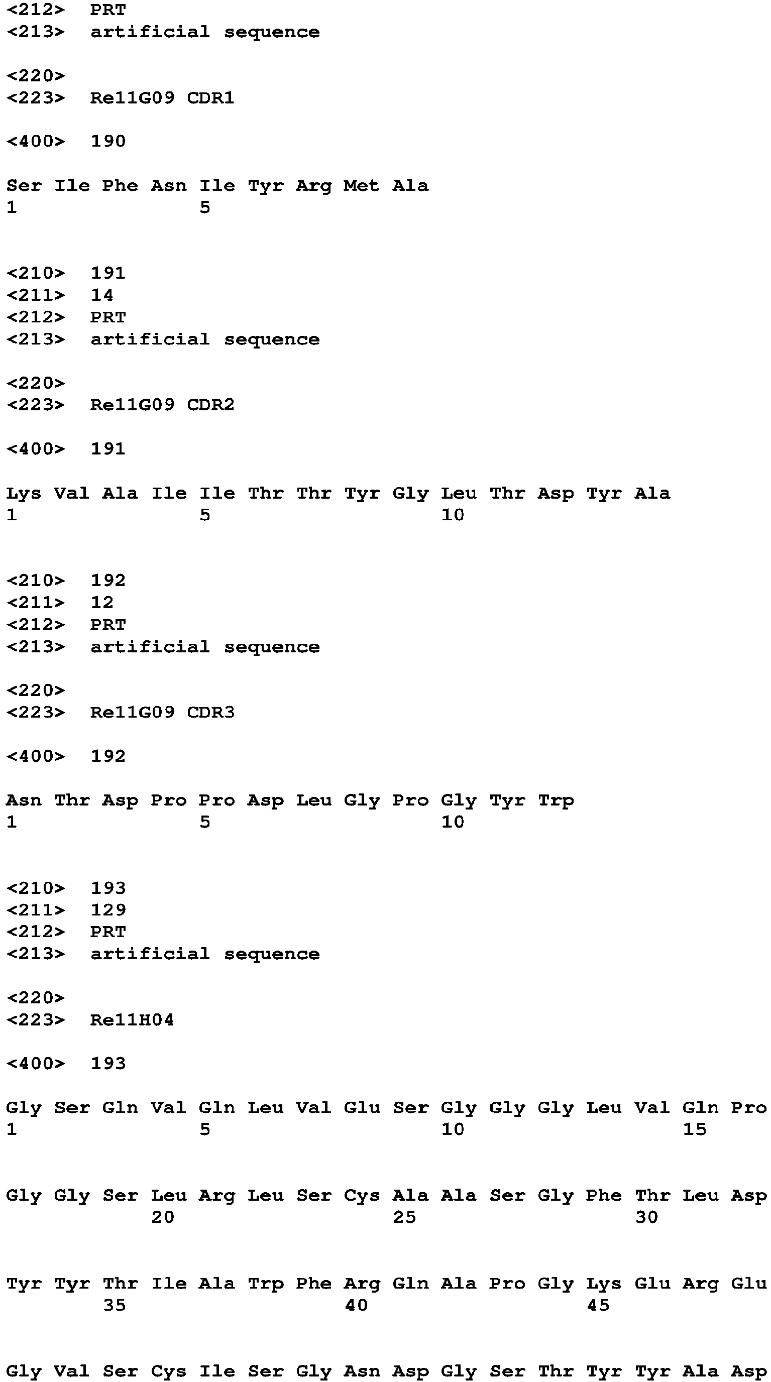
| Re11G09 | 189 | SIFNIYRMA | 190 | KVAIITTYGLTDYA | 191 | NTDPPDLGPGYW | 192 |
| Re11H04 | 193 | FTLDYYTIA | 194 | | 195 | AADRGESYYPFRPSEYHYW | 196 |
-
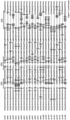
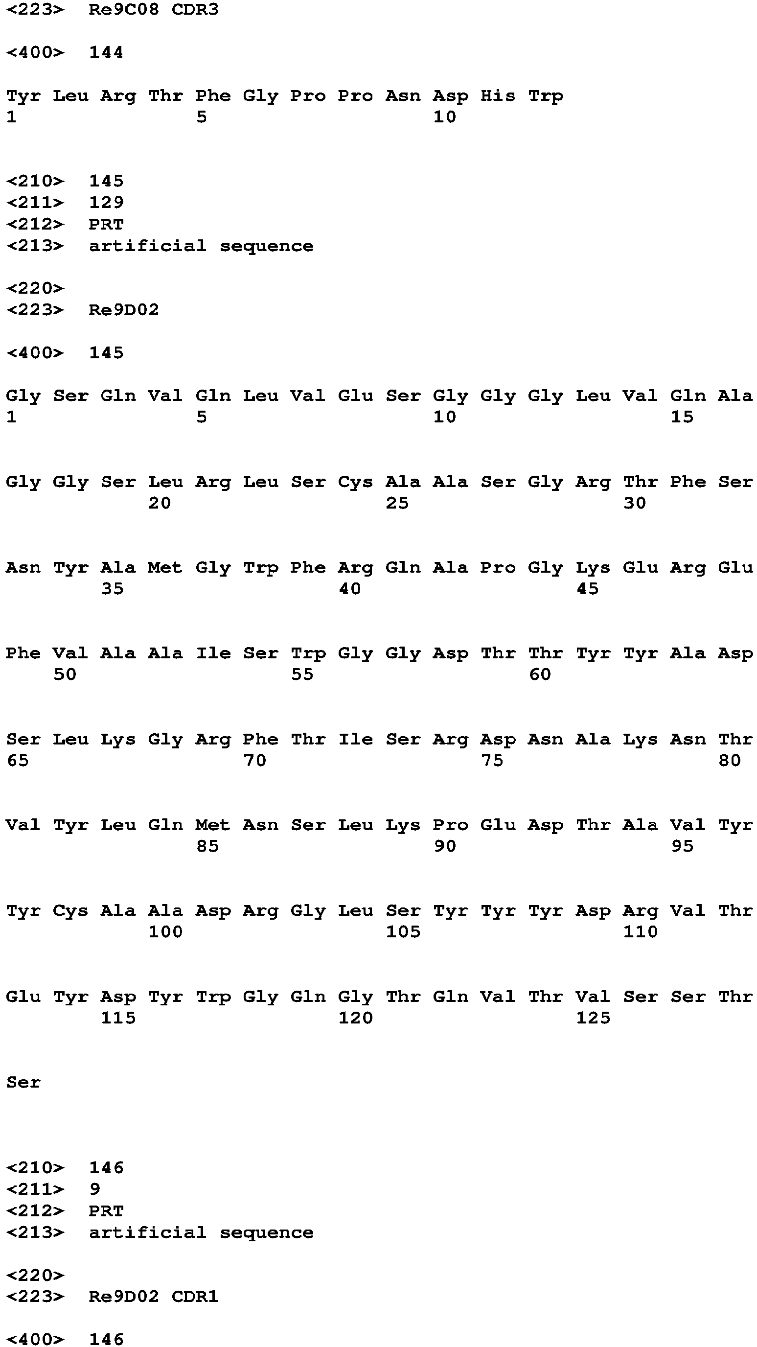
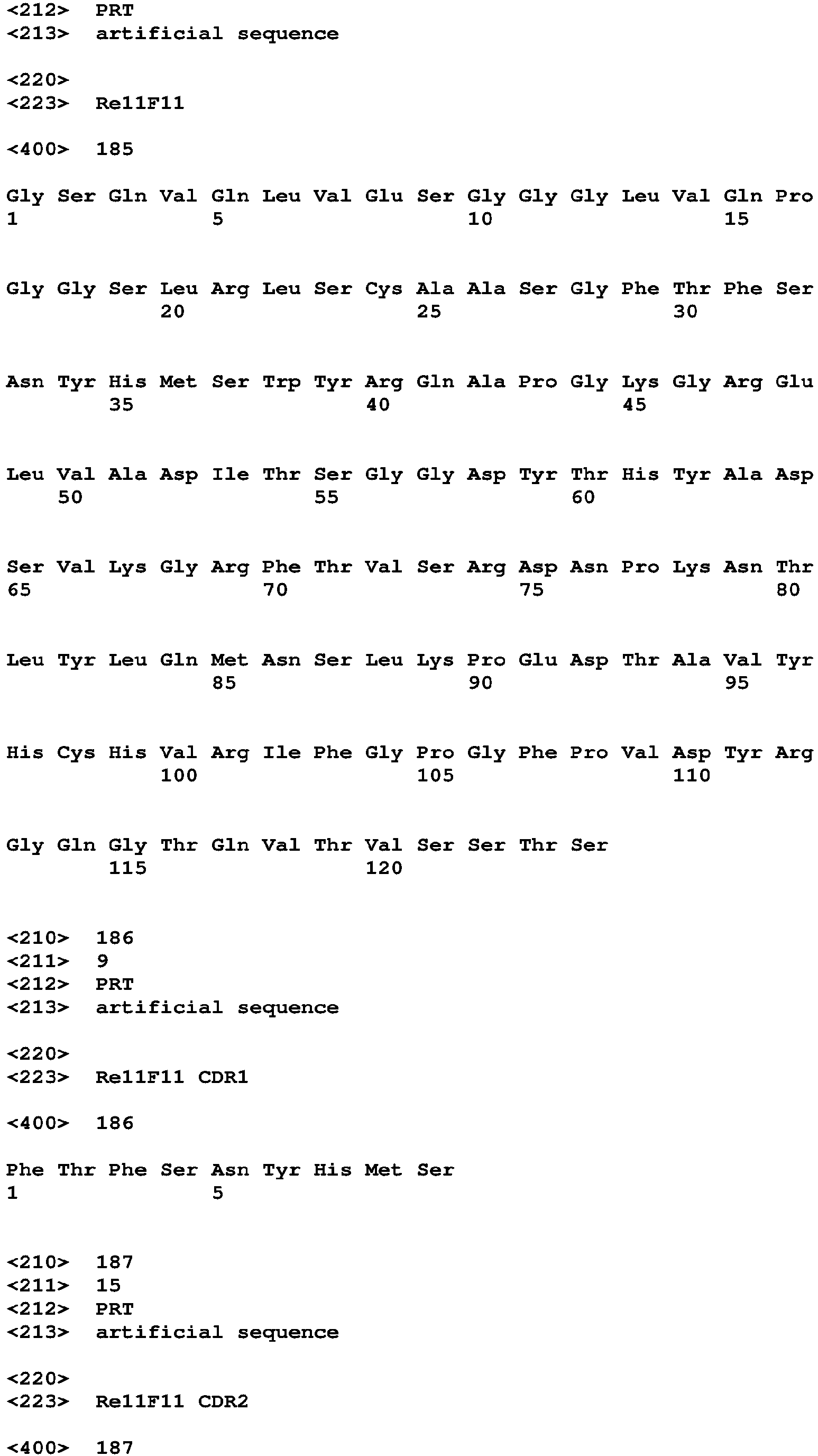
A list of complete sequences of VHH antibodies is shown at the end of the specification and in Figure 1.
-
A further independent aspect of the present invention relates to a homomultimeric VHH antibody, particularly a homotrimeric VHH antibody. The homomultimeric VHH antibody comprises a plurality of individual subunits, particularly 3 individual subunits. An individual subunit may be a fusion polypeptide comprising a VHH antibody, e.g. a VHH antibody as described above, directly linked or linked via a spacer to a multimerization module, particularly to a trimerization module.
-
In certain embodiments, an individual subunit may comprise at its N-terminus the VHH antibody and at its C-terminus the multimerization module, optionally linked via a peptide spacer. In other embodiments, an individual subunit may comprise the multimerization module at its N-terminus and the VHH antibody at its C-terminus, optionally linked via spacer.
-
In certain embodiments, the spacer is a peptide spacer having a length of about 5 to about 100 amino acids, particularly of about 10 to about 50 amino acids.
-
In certain embodiments, the peptide spacer comprises amino acids selected from glycine, serine, threonine, alanine, proline, glutamate and aspartate. In certain embodiments, the spacer is composed to at least about 50%, about 60%, about 70%, about 80%, about 90% or completely of these amino acids.
-
In certain embodiments, the spacer is composed of a succession of tripeptides, whose first two residues are selected from glycine, serine, threonine, alanine and proline, and the third amino acid is selected from glutamate and aspartate. In certain embodiments, each tripeptide differs in at least one amino acid from adjacent tripeptides.
-
In certain embodiments, the linker is substantially free or free from amino acids selected from basic amino acids lysine and arginine, as well as from amino acids selected from tryptophan, tyrosine, phenylalanine, methionine, leucine, isoleucine, valine, histidine and cysteine.
-
In certain embodiments, the homomultimeric VHH antibody is a homotrimeric VHH antibody.
-
In certain embodiments of this aspect, the VHH antibody portion of the homomultimeric VHH antibody is directed to the SARS-CoV-2 spike protein S1 domain, particularly the receptor-binding domain (RBD) of the SARS-CoV-2 S1 domain. In particular embodiments, the VHH antibody portion of the homomultimeric VHH antibody is a VHH antibody as described above, i.e. an antibody comprising a CDR3 sequence as shown in Table 2 or a sequence related thereto, a combination of CDR1, CDR2 and CDR3 sequences, as shown in Table 2 or a related sequence, or a VHH sequence as shown in Figure 1 or a related sequence.
-
According to this aspect, the VHH antibody portion may be directed to another target structure, particularly a target structure having a threefold-rotational symmetry, e.g. viral or non-viral target structure comprising a homotrimeric protein or protein assembly.
Embodiments of the invention
-
In the following, specific embodiments of the invention are disclosed as follows:
- 1. A VHH antibody recognizing the SARS-CoV-2 spike protein S1 domain, particularly the receptor-binding domain (RBD) of the SARS-CoV-2 S1 domain comprising
- (a) a CDR3 sequence as shown in SEQ ID NO. 20, 4, 8, 12, 16, 24, 28, 32, 36, 40, 44, 48, 52, 56, 60, 64, 68, 72, 76, 80, 84, 88, 92, 96, 100, 104, 108, 112, 116, 120, 124, 128, 132, 136, 140, 144, 148, 152, 156, 160, 164, 168, 172, 176, 180, 184, 188, 192 or 196,
- (b) a CDR3 sequence, which has an identity of at least 80% or at least 90% to a CDR3 sequence of (a), or
- (c) a VHH antibody, which competes with a VHH antibody of (a) for the binding to the SARS-CoV-2 spike protein S1 domain, particularly the receptor-binding domain (RBD) of the SARS-CoV-2 S1 domain.
- 2. The VHH antibody according to embodiment 1 comprising
- (a) a combination of CDR1, CDR2 and CDR3 sequences as shown in SEQ ID NO. 18-20, 2-4, 6-8, 10-12, 14-16, 22-24, 26-28, 30-32, 34-36, 38-40, 42-44, 46-48, 50-52, 54-56, 58-60, 62-64, 66-68, 70-72, 74-76, 78-80, 82-84, 86-88, 90-92, 94-96, 98-100, 102-104, 106-108, 110-112, 114-116, 118-120, 122-124, 126-128, 130-132, 134-136, 138-140, 142-144, 146-148, 150-152, 154-156, 158-160, 162-164, 166-168, 170-172, 174-176, 178-180, 182-184, 186-188, 190-192 or 194-196,
- (b) a combination of CDR1, CDR2 and CDR3 sequences which has an identity of at least 80%, at least 90% or at least 95% to a combination of CDR1, CDR2 and CDR3 sequences of (a), or
- (c) a VHH antibody, which competes with a VHH antibody of (a) for the binding to the SARS-CoV-2 spike protein S1 domain, particularly the receptor-binding domain (RBD) of the SARS-CoV-2 S1 domain.
- 3. The VHH antibody according to embodiment 1 or 2 comprising
- (a) a VHH sequence as shown in SEQ ID NO. 17, 1, 5, 9, 13, 21, 25, 29, 33, 37, 41, 45, 49, 53, 57, 61, 65, 69, 73, 77, 81, 85, 89, 93, 97, 101, 105, 109, 113, 117, 121, 125, 129, 133, 137, 141, 145, 149, 153, 157, 161, 165, 169, 173, 177, 181, 185, 189 or 193,
- (b) a sequence which has an identity of at least 70%, at least 80%, at least 90%, at least 95% or at least 99% to a VHH sequence of (a), or
- (c) a VHH antibody, which competes with a VHH antibody of (a) for the binding to the SARS-CoV-2 spike protein S1 domain, particularly the receptor-binding domain (RBD) of the SARS-CoV-2 S1 domain.
- 4. The VHH antibody of any one of embodiments 1-3, which recognizes the receptor-binding domain (RBD) of the SARS-CoV-2 S1 domain.
- 5. The VHH antibody of any one of embodiments 1-4, which is capable of virus neutralization.
- 6. The VHH antibody of any one of embodiments 1-5, which is covalently or non-covalently conjugated to a heterologous moiety.
- 7. The VHH antibody of embodiment 6, which is conjugated to a labeling group, a capture group or an effector group.
- 8. The VHH antibody of embodiment 6 or 7, wherein the heterologous moiety is selected from a fluorescence group, biotin, an enzyme such as a peroxidase, phosphatase or luciferase, a hapten, an affinity tag, or a nucleic acid such as an oligonucleotide.
- 9. The VHH antibody of any one of embodiments 1-5, which is fused to a heterologous polypeptide moiety.
- 10. The VHH antibody of embodiment 9, wherein the heterologous polypeptide moiety is a multimerization module, e.g. dimerization, trimerization or tetramerization module.
- 11. The VHH antibody of embodiment 10, which is a homo-trimerized VHH antibody fused to a trimerization module, e.g. a collagen trimerization moiety, particularly a human collagen moiety, a lung surfactant protein D moiety.
- 12. The VHH antibody of any of embodiments 9-11, which is fused to a heterologous polypeptide moiety directly or via a spacer, e.g. a spacer having a chain length of 1-50 amino acids, particularly selected from Gly, Ser, Glu and/or Asp.
- 13. The VHH antibody of any one of embodiments 1-12, which is non-glycosylated.
- 14. The VHH antibody of any of embodiments 1-13, which is conjugated to one or several polymer moieties, preferably hydrophilic polymer moieties, such as polyethylene glycol (PEG).
- 15. A set of two or more different VHH antibodies of any of embodiments 1-14.
- 16. The set of embodiment 15, wherein the different VHH antibodies recognize different epitopes on the RBD, particularly non-overlapping epitopes on the RBD.
- 17. The VHH antibody of any one of embodiments 1-14 or the set of embodiment 15 or 16 for use in medicine, e.g. human medicine, particularly for use in therapy or diagnostics.
- 18. The VHH antibody of any one of embodiments 1-14 or the set of embodiment 15 or 16 for the prevention or treatment of a disorder caused by and/or associated with an infection with SARS-CoV-2.
- 19. The VHH antibody of any one of embodiments 1-14 or the set of embodiment 15 or 16 for the prevention or treatment of Covid-19 in a human subject.
- 20. The VHH antibody of any one of embodiments 1-14 or the set of embodiment 15 or 16 for the detection of SARS-CoV-2 virus comprising
- (i) a set of at least two VHH antibodies recognizing different, particularly non-overlapping epitopes on the RBD,
- (ii) a set of at least two VHH antibodies comprising a set of capturing antibodies conjugated to a capturing moiety and a set of labeling antibodies conjugated to a labeling moiety, and/or
- (iii) a set of at least two VHH antibodies comprising a first set of labeling antibodies conjugated to a first labeling moiety, and a second set of labeling antibodies conjugated to a second labeling moiety, wherein the first labeling moiety is different from the second labeling moiety, wherein the first and the second labeling moieties are particularly selected from two spectrally different fluorescence labeling moieties.
- 21. Use of the VHH antibody of any one of embodiments 1-14 or the set of embodiment 15 or 16 for detecting SARS-CoV-2 virus or viral components in a patient sample, e.g. in a body fluid or tissue sample.
- 22. Use of the VHH antibody of any one of embodiments 1-14 or the set of embodiment 15 or 16 for detecting SARS-CoV-2 virus or viral components in a virus culture, e.g. in a culture of SARS-CoV-2 or variants thereof including pseudo typed variants, or in a genetically modified organism, e.g. in an organism producing viruses or viral components.
- 23. The use of embodiment 22 for monitoring, quantification and/or quality control during production of viruses or viral components.
- 24. A nucleic acid molecule encoding a VHH antibody according to any one of embodiments 1-14, preferably in operative linkage with a heterologous expression control sequence.
- 25. A vector comprising a nucleic acid molecule according to embodiment 24.
- 26. A recombinant cell or non-human organism transformed or transfected with a nucleic acid molecule according to embodiment 24 or a vector according to 25.
- 27. The cell or organism of embodiment 26 which is selected from a bacterium such as E. coli Bacillus sp., a unicellular eukaryotic organism such as yeast or Leishmania, an insect cell, a mammalian cell or a plant cell.
- 28. A method for recombinant production of a VHH antibody of any one of embodiments 1-14, comprising cultivating a cell or an organism of embodiment 26 or 27 in a suitable medium and obtaining the VHH antibody from the cell or organism or from the medium.
- 29. A method for the prevention or treatment of a disorder caused by and/or associated with an infection with SARS-CoV-2, comprising administering an effective dose of the VHH antibody of any one of embodiments 1-14 or the set of embodiment 15 or 16 to a subject in need thereof, particularly to a human subject.
- 30. A homotrimeric VHH antibody comprising 3 VHH antibody subunits recognizing the same target structure, wherein each VHH antibody subunit comprises a VHH antibody directly linked or linked via a spacer to a trimerization module.
- 31. The homotrimeric VHH antibody of embodiment 30, which comprises three identical VHH antibody subunits.
- 32. The homotrimeric VHH antibody of embodiment 30 or 31 wherein the VHH antibody is directed to a target structure having a threefold-rotationally symmetric subunit structure.
- 33. The homotrimeric VHH antibody of any one of embodiments 30-32 wherein the VHH antibody is directed to a target structure comprising a homo-trimeric protein or protein assembly.
- 34. The homotrimeric VHH antibody of any one of embodiments 30-33 wherein the target structure is a viral protein or protein assembly, particularly a homo-trimeric viral protein or protein assembly, more particularly selected from a Coronavirus trimeric spike protein, an Orthomyxovirus trimeric HA protein, a Paramyxovirus trimeric fusion protein, a Dengue fever virus or Zikavirus trimeric or higher order protein assembly, a Herpesvirus trimeric gB fusion protein or a HIV trimeric gp120 protein.
- 35. The homotrimeric VHH antibody of any one of embodiments 30-34 wherein the target structure is the SARS-CoV-2 spike protein S1 domain, particularly the receptor-binding domain (RBD) of the SARS-CoV-2 S1 domain.
- 36. The homotrimeric VHH antibody of embodiment 35 wherein the VHH antibody is selected from the VHH antibodies according to any one of embodiments 1-5.
- 37. The homotrimeric VHH antibody of any one of embodiments 30-36 wherein the target structure is a non-viral protein or protein assembly, particularly a member of the TNF Ligand superfamily or a member of the TNF Receptor superfamily.
- 38. The homotrimeric VHH antibody of any one of embodiments 30-37 wherein each subunit comprises a VHH antibody linked via a spacer to a trimerization module.
- 39. The homotrimeric VHH antibody of embodiment 38 wherein the spacer has a length of about 5 to about 100 amino acids, particularly of about 10 to about 50 amino acids.
- 40. The homotrimeric VHH antibody of embodiment 38 or 39 wherein the spacer comprises amino acids selected from Gly, Ser, Ala, Thr, Pro, Asp and Glu, particularly in an amount of at least about 50%, about 60%, about 70%, about 80%, about 90% or 100%.
- 41. The homotrimeric VHH antibody of any one of embodiments 38-40 wherein the spacer comprises a succession of tripeptides, wherein the first and the second amino acid are selected from Gly, Ser, Ala, Thr, and Pro, and the third amino acid is selected from Asp and Glu.
- 42. The homotrimeric VHH antibody of any one of embodiments 38-41 wherein the spacer is substantially free or free of amino acids selected from Lys, Arg, Trp, Tyr, Phe, Met, Leu, Ile, Val, His and Cys.
- 43. The homotrimeric VHH antibody of any one of embodiments 30-42 wherein the trimerization module is selected from:
- (a) a collagen trimerization domain, e.g. the NC1 domain of collagen XV, the NC1 domain of collagen XVIII or the NC1 domain of collagen X, or the lung surfactant protein D.
- (b) a trimerization domain which has an identity of at least 80%, particularly at least 90% to a trimerization domain of (a).
- 44. A nucleic acid molecule encoding a homotrimeric VHH antibody according to any one of embodiments 30-43, preferably in operative linkage with a heterologous expression control sequence.
- 45. A vector comprising a nucleic acid molecule according to embodiment 44.
- 46. A recombinant cell or non-human organism transformed or transfected with a nucleic acid molecule according to embodiment 44 or a vector according to 45.
- 46. The homotrimeric VHH antibody of any one of embodiments 30-47, which is non-glycosylated.
- 47. The homotrimeric VHH antibody of any one of embodiments 30-47, which is conjugated to one or several polymer moieties, preferably hydrophilic polymer moieties, such as polyethylene glycol (PEG).
- 48. The homotrimeric VHH antibody of any one of embodiments 30-43, 46 or 47 for use in medicine, e.g. human medicine, particularly for use in therapy or diagnostics.
Detailed description of the invention
-
According to the present invention, SARS-CoV-2-neutralising VHH antibodies are provided, which are suitable for the effective treatment of COVID-19 patients. Specifically, the invention provides VHH antibodies that neutralize virus already at a very low concentration. Further, the invention provides multimeric, particularly trimeric VHH antibodies for enhancing the potency of VHH antibodies, which may also be directed against other target structures, e.g. for blocking infections by other viruses.
SARS-CoV-2-neutralising VHH antibodies
-
The primary aim of this aspect of the invention has been to generate, select, engineer and optimize SARS-CoV-2-neutralising VHH antibodies for effective treatment of COVID-19 patients. Specifically, we aimed for VHH antibodies that are capable of virus neutralization already at a very low concentration.
-
The present invention relates to a VHH antibody, which is a monovalent heavy chain-only antibody comprising a CDR1 domain, a CDR2 domain and a CDR3 domain linked by framework regions including, but not being limited to, whole VHH antibodies, e.g. native VHH antibodies comprising framework regions derived from camelids, and modified VHH antibodies comprising modified framework regions, VHH antibody fragments and VHH antibody fusion proteins, e.g. a fusion protein with an immunoglobulin or non-immunoglobulin peptide or polypeptide, as long as it shows the properties according to the invention. The present invention is also directed to a covalent or non-covalent conjugate of a VHH antibody molecule to a non-proteinaceous structure, for example, a labeling group, a capture group such a solid phase-binding group, or an effector group such as a toxin. For example, the heterologous moiety may be from a fluorescence group, biotin, an enzyme such as a peroxidase, phosphatase or luciferase, a hapten, an affinity tag, or a nucleic acid such as an oligonucleotide.
-
The VHH antibody of the present invention is particularly a monoclonal VHH antibody characterized by a specific amino acid sequence. The VHH antibody may be produced in a prokaryotic host cell, a yeast cell or a mammalian cell. In certain embodiments, the VHH antibody is non-glycosylated. In certain embodiments, the VHH antibody is glycosylated, wherein a carbohydrate structure may be derived from a glycosylation site introduced into the VHH sequence and/or from a fusion partner.
-
A VHH antibody according to the present invention is characterized by (i) a CDR3 sequence, (ii) a combination of a CDR1 sequence, a CDR2 sequence, and a CDR3 sequence, (iii) by a complete VHH sequence, or (iv) by competition with a specific reference antibody. Specific CDR and VHH sequences are provided in the Tables, Figures and the Sequence Listing. According to the present invention, sequences related to the above sequences are encompassed. These related sequences are defined by having a minimum identity to a specifically indicated amino acid sequence, e.g. a CDR or VHH sequence. This identity is indicated over the whole length of the respective reference sequence and may be determined by using well known algorithms such as BLAST.
-
Further, the invention refers to a VHH antibody, which competes with a specific VHH antibody disclosed herein for the binding to the SARS-CoV-2 spike protein S1 domain. In certain embodiments, a competing VHH antibody binds the same or an overlapping epitope on the SARS-CoV-2 spike protein S1 domain, particularly the receptor-binding domain (RBD) of the SARS-CoV-2 S1 domain as a specific VHH antibody disclosed herein. For example, the invention refers to a VHH antibody, which competes either with the VHH antibodies Re5D06, Re6D06, Re6H06, Re9B09, Re9C08, or Re9H01, or with VHH antibodies Re6B07 or Re7E02.
-
In certain embodiments, the invention relates to a VHH antibody competing with VHH antibody Re5D06, such as any one of the VHH antibodies Re6B02, Re7B01, Re10B10, or Re10F10. In certain embodiments, the invention relates to a VHH antibody competing with VHH antibody Re7E02, such as the VHH antibody Re6B07.
-
Competition is measured either as an at least 90% loss of fluorescence staining of the SARS-CoV-2 spike protein by a fluorophore-labelled candidate VHH antibody in the presence of a 100-fold molar excess of an unlabelled competitor VHH antibody e.g. selected from Re5D06, Re6D06, Re6H06, Re9B09, Re9C08, Re9H01, or Re7E02, or conversely as an at least 90% loss of spike protein-staining by a fluorophore-labelled VHH antibody e.g. selected from Re5D06, Re6D06, Re6H06, Re9B09, Re9C08, Re9H01, or Re7E02 in the presence of a 100-fold molar excess of the unlabelled candidate VHH antibody as a competitor.
-
Further, the invention refers to a VHH antibody, which competes with a specific VHH antibody disclosed herein for the binding to the SARS-CoV-2 spike protein S1 domain. In certain embodiments, a competing VHH antibody binds the same or an overlapping epitope on the SARS-CoV-2 spike protein S1 domain, particularly the receptor-binding domain (RBD) of the SARS-CoV-2 S1 domain as a specific VHH antibody disclosed herein. For example, the invention refers to a VHH antibody, which competes either with the VHH antibodies Re5D06 and/or Re6D06 or with VHH antibodies Re6B07 and/or Re7E02.
-
In particular embodiments, an amino acid of a reference sequence, including an amino acid in a CDR1, CDR2 or CDR3 sequence, is replaced by a conservative amino acid substitution, i.e. a substitution of an amino acid by another amino acid with similar biochemical properties, for example a substitution of an aliphatic amino acid, e.g. Gly, Ala, Val, Leu or Ile, for another aliphatic amino acid; a substitution of a basic amino acid, e.g. His, Lys or Arg, against another basic amino acid or against Met; a substitution of an acidic amino acid or an amide thereof, e.g. Asp, Glu, Asn or Gln, against another acidic amino acid or an amide thereof; a substitution of an aromatic amino acid, e.g. Phe, Tyr or Trp, against another aromatic amino acid, or a substitution of a hydroxy or sulfur containing amino acid, e.g. Ser, Thr, Met or Cys, against another hydroxy or sulfur containing amino acid.
-
In further particular embodiments, the amino acid Lys of a reference sequence including an amino acid in a CDR1, CDR2 or CDR3 sequence, e.g. the amino acid Lys in the CDR3 sequence of any one of the VHH antibodies Re5D06, Re6B02, Re7B01, Re10B10 Re10F10 or of another VHH antibody may be replaced by a different amino acid, e.g. Arg or Met. The VHH antibody of the present invention is binding to the S1 domain, particularly to the RBD of the S1 domain of SARS-CoV-2. In certain embodiments, a VHH antibody is capable of virus neutralization at a concentration of about 10 nM or less, of about 1 nM or less, of about 500 pM or less, of about 100 pM or less or even at about 50 pM or less.
-
Further, the present invention relates to a nucleic acid molecule, e.g. a DNA molecule, encoding a VHH as indicated above, or a vector, comprising said nucleic acid molecule as indicated above in operative linkage with an expression control sequence, particularly with a heterologous expression control sequence. Furthermore, the invention relates to a cell comprising a nucleic acid molecule or a vector as described above. Vectors for the recombinant production of VHH antibodies are well known in the art. The cell may be known host cell for producing antibodies or antibody fragments, e.g. a prokaryotic cell such as an E. coli or a Bacillus sp. cell, a yeast cell, an insect cell or a mammalian cell, e.g. a CHO cell, or a plant.
-
Still a further aspect of the present invention is a method of recombinantly producing a VHH antibody by growing a cell as described above in a culture medium and obtaining the VHH antibody from the cell or the culture medium. Suitable culture media and culture conditions are well known in the art.
-
In a further aspect, the present invention relates to a set comprising at least 2, 3, 4 or more of the above VHH antibodies. In such a set, the individual VHH antibodies are present in suitable molar ratios. Typically, the molar ratios are in the range of about 2:1 to about 1:2, particularly about 1.5:1 to about 1:1.5, even more particularly about 1:1. In certain embodiments, the VHH antibody set may comprise a single composition wherein the VHH antibodies in said set consist of a predetermined number of different species of VHH antibodies as described above. The VHH antibody set may comprise a plurality of compositions each comprising a different species of VHH antibody as described above. The set of the present invention may be free from other VHH antibodies.
-
The inventors' target structure has been the S1 fragment of the SARS-CoV-2 spike protein (Wrapp et al., 2020), which contains two main globular domains: A large N-terminal, lectin-like domain (residues 27-290) as well as a receptor-binding domain (RBD, residues 330-521). The RBD mediates the interaction with the ACE2 host cell receptor, and is thus essential for infectivity.
-
The inventors applied five subcutaneous immunizations in weekly intervals such that the S1 fragment and in particular the RBD were presented in multiple contexts to the animals' immune systems. As antigens were used: transfected HEK293 cells expressing the complete spike protein on its surface (107 cells per immunization) and up to 100 µg of the following purified proteins: fusions of the RBD and the entire S1 domain to mouse and human Fc fragments as well as an RBD fusion to the E.coli DsbG dimer. Four days after the last immunization, blood samples were collected, RNA from ∼108 lymphocytes each was isolated, VHH-coding regions were amplified by nested RT-PCRs, cloned as cDNAs into a phagemid to yield three separate immune libraries with complexities of around 109 independent clones each. These libraries were subjected to phage display, using as baits either an immobilized RBD (yielding anti-RBD VHH) or an immobilized S1 fragment with competition by an excess of free RBD (to yield anti-S1ΔRBD VHH antibodies). Bovine Serum Albumin, as well as human and murine serum, were included as non-specific competitors.
-
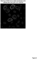
A total of 672 selected clones were so far sequenced and classified according to sequence similarity. Representatives of all VHH classes were then expressed in E.coli and purified (see Figure 1 for specific sequences of VHHs pursued in this invention). One set of nanobodies was expressed with ectopic cysteines for labelling with fluorophore-maleimides (Pleiner et al., 2015). These labelled nanobodies were used for immunofluorescence on HeLa cells that had been transfected with a SARS CoV2 spike expression construct. Because only a fraction of cells gets transfected, a typical field of view contains non-transfected cells (serving as internal negative control) as well as transfected ones. Several of the labelled nanobodies stained the expressed spike protein very brightly. Figure 2 shows VHH Re6D09 labelled with Alexa568 as an example. Table 1 (supra) lists the VHHs that have been tested positive for anti-Spike fluorescence.
-
In a second set of experiments, the inventors used cultured cells infected with SARS-CoV-2 isolated from a patient's sample. It was observed that numerous VHH antibodies (e.g. Re5A08, Re5B06, Re5D05, Re6D06, and Re6D09) stained viral structures brightly and specifically as judged by a lack of signal in non-infected cells (see Figure 3 for example). Thus, VHH antibodies of this invention are suitable for detecting SARS-CoV-2 spike components in biological samples.
-
In the third set of experiments, the inventors performed tests if any of the obtained nanobodies can neutralize SARS-CoV-2 and prevent the virus from infecting a target cell. In these experiments, the Vero E6 cell line was used, which expresses high levels of the SARS-CoV-2 receptor ACE2 (Li et al., 2003) but is deficient in its interferon response (Emeny and Morgan, 1979); it is therefore extremely susceptible to viral infection. Moreover, a SARS-CoV-2 patient derived virus strain was used carrying the infection-enhancing D614G spike mutation (Zhang et al., 2020). This together therefore combines into a highly stringent test system for anti-viral inhibition. Without VHH antibodies, the addition of virus caused a pronounced cytopathic effect (CPE), and the amount of virus increased within three days of incubation approximately 10 000-fold.
-
When tested at a concentration of 500 nM, 30 out of a first set of 35 VHHs were able to protect cells from the CPE and prevented virus replication completely (see Figure 4 for examples). These and neutralizing VHH antibodies discovered in later rounds of testings (see Tables 1-4) are now excellent candidates for treating SARS-CoV-2-infected patients.
Homotrimeric VHH antibodies
-
An important issue in pharmaceutical development is the lower limit of concentration at which an anti-viral antibody is still effective. Indeed, it makes a great difference if grams (as in the case of rabies post-exposure prophylaxis) or one milligram are required for an effective therapeutic dose. This determines the costs per treatment and, if many patients need to be treated, it matters how many therapeutic doses can be produced at all. Likewise, adverse side effects by the antibody itself will scale with the applied dose. Finally, contaminants, such as endotoxins, can cause side effects, and the absolute amount of contaminant will scale-up with the administered dose. It is thus highly desirable to obtain VHH antibodies that effectively block viral infection at the lowest possible concentration.
-
The binding strength of an antibody or any other binder is in the first instance ruled by the Kd of the relevant interaction (dissociation constant or the ratio of off and on rates). If the free concentration of the binder equals the Kd, then half of the binding sites are blocked. As a SARS-CoV-2 viral particle contains >100 spike trimers (Bar-On et al., 2020), efficient neutralization probably only occurs if their majority, i.e. 99% or even 99.9% are blocked. This requires the binder to be present at concentrations that are 100-fold or a 1000-fold higher than the Kd.
-
A solution to this problem is to fuse the monovalent VHH antibody to the Fc part of an immunoglobulin, e.g. a human IgG and thus make the VHH bivalent. This could impose a stronger binding by avidity effects, but only if the distance of binding sites fits the geometry of the antibody. Apart from this reservation, Fc fusions are associated with additional potential drawbacks: First, production of such Fc fusions should be carried out in human cells to ensure proper folding and a human glycosylation pattern. Second, the Fc fragment is an immune effector domain that might aggravate the problem of inappropriate complement activation and cytokine storms, which are hallmarks of the most severe SARS-CoV-2 pathologies. Third, a dimeric IgG might well dock to two of the spike molecules of a homotrimeric spike, but this would leave the third one in an unbound state.
-
Thus, the inventors have considered that a homo-trimeric ligand would be far more appropriate for blocking a homotrimeric target (Figure 5). This principle of 'symmetry-matching' is a central element of this approach.
-
Homotrimerization of a VHH antibody requires an appropriate polypeptide fusion partner as trimerization module. According to the present invention, a fusion partner is preferred, comprising at least one of the following features:
- (1) it has a length of about 250 amino acids or less, of about 200 amino acids or less or of about 150 amino acids or less;
- (2) it is capable of folding properly in commonly used recombinant expression systems (bacteria, yeast, insect and human cells), and assemble into trimers with a very low Kd of e.g. less than 1nM in the picomolar range - to avoid disassembly at low concentrations; and
- (3) it is of mammalian, particularly of human origin to be as non-immunogenic as possible.
-
Preferred trimerization modules include domains derived from collagen, the most abundant protein of the human body, including the trimerization (NC1) domains of collagen XV (PDB 3N3F, 54 residues) (Wirz et al., 2011), collagen XVIII (PDB 3HSH; 54 residues) (Boudko et al., 2009) and collagen X (PDB 1GR3; 160 amino acids) (Bogin et al., 2002). Another very appropriate homotrimeric fusion partner is the lung surfactant protein D (LSFPD) that recognizes sugars on viral and bacterial pathogens (see e.g. PDB 1B08, 3IKN, 3IKQ, 3IKR). As the SARS-CoV-2 spike protein is heavily glycosylated, an VHH-LSPD fusion could confer a bimodal inhibition of spike function.
-
The collagen XVIII NC1 module has been used for trimerizing VHHs before (Alvarez-Cienfuegos et al., 2016), however, not for the purpose of symmetry-matching and targeting homotrimeric targets. Further, this earlier study used expression in human cells.
-
The inventors now fused 12 different VHH antibodies through a flexible spacer to this trimerization module and found good expression and solubility in E.coli, and succeeded in just a single purification step to obtain an essentially pure fusion protein.
-
According to the present invention, it is preferred that the VHH antibody is linked to the trimerization domain via a spacer comprising at least one of the following features:
- (1) it is long enough to allow a simultaneous binding of all three VHH copies to the same spike trimer, e.g. having a length of at least about 5 amino acids, particularly about 5 amino acids to about 100 amino acids, and more particularly of about 10 to about 50 amino acid, also depending on the specific target structure;
- (2) it is substantially free (i.e. comprising an amount of 10% or less, 5% or less or 2.5% or less) or it is free from amino acid residues selected from Lys, Arg, Trp, Tyr, Phe, Met, Leu, Ile, Val, His and Cys;
- (3) it does not contain a strong B-cell epitope;
- (4) it is not particularly protease-sensitive, e.g. it does not contain a trypsin or chymotrypsin cleavage site;
- (5) it does not contain repetitive sequence portions, e.g. repetitive sequence portions having a length of at least 3 amino acid residues;
- (6) it comprises a negative net charge;
- (7) it comprises amino acids selected from Gly, Ser, Ala, Thr, Pro, Asp and Glu, particularly in an amount of at least about 50%, at least about 60%, at least about 70%, at least about 80%, at least about 90% or 100%; and
- (8) it comprises a succession of tripeptides, wherein the first and the second amino acid are selected from Gly, Ser, Ala, Thr, and Pro, particularly Gly, Ser, Ala and Thr, and more particularly Gly, Ser and Ala, and the third amino acid is selected from Asp and Glu; and particularly without direct repetitions of the concatenated tripeptide motifs.
-
It should be note that different arrangements for the subunits of homomultimeric VHH antibodies are possible. As just described, the trimerization module might be located N-terminally, followed by the spacer and the VHH antibody. Alternatively, the VHH antibody might be located N-terminally, followed by spacer and trimerization module.
-
The concept of providing homomultimeric, particularly homotrimeric VHH antibodies, is not limited to SARS-CoV-2. It is quite remarkable that many viruses rely on homotrimeric entry receptors and/ or fusion proteins with C3 rotational symmetry. The symmetry-mismatch to bivalent antibodies is perhaps of advantage for those viruses when it comes to delaying a neutralizing immune response. Thus, our symmetry-matching approach to homotrimeric neutralizing VHH antibodies is also suitable against any target structure having a threefold-rotationally symmetric subunit structure. Such a target structure is present on other viruses, such as other members of the Coronaviridae, Orthomyxoviridae (with their trimeric HA-proteins), many Paramyxoviridae (with their trimeric fusion proteins), Dengue fever- and Zika-viruses (3-fold or higher order protein assemblies on their surface), as well as Herpesviridae (trimeric gB fusion protein) or Human Immunodeficiency Virus (with its trimeric gp120 protein).
-
Moreover, the approach can even be extended to efficiently inhibit non-viral targets that also show a threefold rotational symmetry, such as the members of the TNF Ligand and TNF Receptor superfamilies, e.g. the death ligands and receptors that trigger the extrinsic pathway of apoptosis (e.g. CD95, TNFα, or Trail). Also here, a stable and non-immunogenic trimerization module as the ones derived from human collagens X, XV, or XVIII may be used.
-
The lengths of spacers between VHHs and trimerization unit may be dependent from the specific target structure and thus subject to optimization: If a spacer is too short, then it will not allow all three binding sites to be used at the same time or cause tension in the bound state, while a spacer that is too long might be entropically suboptimal in terms of increasing the local VHH concentration. Based on the above teaching, an optimal spacer may be developed, which will not only provide the maximum enhancement in binding strength but also a gain specificity as multivalent binding is most favored to objects of a complementary geometry.
Therapeutic applications
-
Still a further aspect of the present invention is the use of an active agent selected from the VHH antibody, the set of at least 2 different VHH antibodies or the multimeric trimeric VHH antibody in medicine, particularly for therapeutic and/or in vitro or vivo diagnostic use. In certain embodiments, the VHH antibody, the set of VHH antibodies or the multimeric, particularly trimeric VHH antibody is used in human medicine.
-
In particular embodiments, a therapeutic VHH antibody is selected from (i) a VHH antibody comprising the CDR3 sequence of VHH antibody Re5D06 or a related sequence, (ii) a VHH antibody comprising the CDR1, CDR2 and CDR3 sequences of VHH antibody Re5D06 or related sequences, (iii) a VHH antibody comprising the VHH sequence of VHH antibody Re5D06 or a related sequence, or (iv) a VHH antibody competing with VHH antibody Re5D06.
-
In particular embodiments, a therapeutic VHH antibody is selected from (i) a VHH antibody comprising the CDR3 sequence of VHH antibody Re6H06 or a related sequence, (ii) a VHH antibody comprising the CDR1, CDR2 and CDR3 sequences of VHH antibody Re6H06 or related sequences, (iii) a VHH antibody comprising the VHH sequence of VHH antibody Re6H06 or a related sequence, or (iv) a VHH antibody competing with VHH antibody Re6H06.
-
In particular embodiments, a therapeutic VHH antibody is selected from (i) a VHH antibody comprising the CDR3 sequence of VHH antibody Re9H01 or a related sequence, (ii) a VHH antibody comprising the CDR1, CDR2 and CDR3 sequences of VHH antibody Re9H01 or related sequences, (iii) a VHH antibody comprising the VHH sequence of VHH antibody Re5D06 or a related sequence, or (iv) a VHH antibody competing with VHH antibody Re9H01.
-
In particular embodiments, a therapeutic VHH antibody is selected from (i) a VHH antibody comprising the CDR3 sequence of VHH antibody Re7E02 or a related sequence, (ii) a VHH antibody comprising the CDR1, CDR2 and CDR3 sequences of VHH antibody Re7E02 or related sequences, (iii) a VHH antibody comprising the VHH sequence of VHH antibody Re7E02 or a related sequence, or (iv) a VHH antibody competing with VHH antibody Re7E02.
-
Therapeutic applications include methods for preventing or treating a disorder caused by and/or associated with a Coronavirus infection, particularly a disorder caused by and/or associated with a SARS-CoV-2 infection and more particularly of preventing or treating COVID-19. As outlined, above, however, the trimeric VHH antibody may be directed to other target structures, and thus, is useful in methods for preventing or treating a disorder caused by and/or associated with a virus infection, particularly a disorder caused by and/or associated with a Coronavirus infection, an Orthomyxovirus, e.g. an Influenza A, B, or C virus, a Paramyxovirus, e.g. a Parainfluenza virus, Respiratory Syncytial virus, Measles virus, or Mumps virus, Dengue fever virus, Zikavirus, a Herpesvirus or HIV. Further, the trimeric VHH antibody is useful in methods for preventing or treating a non-viral disorder, particularly a disorder caused by and/or associated with a member of the TNF ligand superfamily and/or a member of the TNF Receptor superfamily.
-
In therapeutic applications, the active agent is administered in an effective amount to a subject in need thereof, particularly to a human subject. The dose will depend from the specific type of agent, e.g. monovalent VHH antibody or multimeric, particularly trimeric VHH antibody, and the type of disease.
-
In the prevention or treatment of COVID-19, a daily dose of a monomeric VHH antibody for a human subject is any suitable dose based on the severity of the disease, the specific type of agent, and the mode of administration, e.g. locally or systemically, particularly from about 5 µg to about 5000 mg per day.
-
In the prevention or the treatment of COVID-19, a daily dose of a trimeric VHH antibody for a human subject is any suitable dose based on the severity of the disease, the specific type of agent, and the mode of administration, e.g. locally or systemically, particularly from about 1 µg to about 2500 mg per day.
-
In the prevention or treatment of COVID-19, a unit dose of a monomeric VHH antibody for a human subject is any suitable dose based on the severity of the disease, the specific type of agent, and the mode of administration, e.g. locally or systemically, particularly from about 5 µg to about 5000 mg per unit.
-
In the prevention or the treatment of COVID-19, a unit dose of a trimeric VHH antibody for a human subject is any suitable dose based on the severity of the disease, the specific type of agent, and the mode of administration, e.g. locally or systemically, particularly from about 1 µg to about 2500 mg per unit.
-
Typically, the VHH antibody is administered as a pharmaceutical composition comprising the active agent and a pharmaceutically acceptable carrier or excipient. Examples of suitable carriers and excipients for formulating antibodies or antibody fragments are well known in the art.
-
Depending on the stage and the severity of the disorder, the pharmaceutical composition may be administered once or several times during the course of the disorder. For example, it may be administered once or several times daily, each second day, two times weekly or weekly for a suitable period of time.
-
In certain embodiments, the pharmaceutical composition is administered parenterally, e.g. by subcutaneous, intramuscular or intravenous injection or by infusion. In further embodiments, the pharmaceutical composition may be administered locally, e.g. orally, nasally or intrapulmonary, for example by inhalation as an aerosol.
-
In certain embodiments, the pharmaceutical composition is administered in an early infection stage, e.g. in an infection stage where the upper airways such as the oral cavity, the nasal cavity, the paranasal sinus, the pharynx and/or the throat are infected, but the lower airways such as the bronchi and/or the lung are not infected. In further embodiments, the pharmaceutical composition is administered in a late infection stage wherein the lower airways such as the bronchi and/or the lung are infected. Further, the pharmaceutical composition may be administered in an infection stage where the subject suffers from a respiratory dysfunction and optionally is ventilated. In still further embodiments, the pharmaceutical composition may be administered prophylactically to a subject being at risk of a Coronavirus infection, e.g. a subject, which has previously, for example, within 1, 2, 3, 4, or 5 days or even longer, been in close contact with an infected subject.
-
The VHH antibody may be administered alone or together with a further active agent, which may be selected from anti-viral agents such as remdesivir or methotrexate.
-
In order to determine suitable administration schemes, the inventors first tested the principle of avidity enhancement for the rather weakly binding Re6A11 variant (labelled with two Alexa488 fluorescence groups per VHH module), whose monomeric version is readily washed off its targets and hence showed only very weak staining of Spike molecules in transfected HeLa cells (Figure 6). The trimerized version, however, showed a strong signal even when used at lower concentrations and with just one Alexa488 per VHH module. This strong staining persisted also during very excessive washing steps and when the VHH trimer was used at a very low concentration (1 nM).
-
As second proof of principle for the avidity enhancement, the inventors used the virus neutralization assay as described above. Table 3 shows the minimum SARS-CoV-2-neutralizing concentrations of selected nanobody monomers and trimers. Virus neutralization experiments were performed as in
Figure 4, with VHH concentrations being titrated down in
factor steps. Lowest SARS-CoV-2-neutralizing concentrations of selected VHH monomers and trimers are listed that fully suppressed infection over a period of three days. Concentrations refer to the respective VHH units. The trimers were tandem fusions of VHH, a 39 residues spacer followed by the collagen XVIII NC1 domain.
Table 3 | | Lowest neutralizing VHH concentration |
| VHH | As monomer | As trimer |
| Re5A08 | 5 nM | ≤ 0.017 nM |
| Re5C08 | 500 nM | 0.50 nM |
| Re6A11 | 50 nM | 0.050 nM |
| Re6B06 | 500 nM | 0.50 nM |
| Re6D06 | 0.5 nM | ≤ 0.017 nM |
-
The monomeric version of the VHH antibody Re5A08 blocked infection down to a concentration of 5 nM. In trimerized form, however, the block was still tight at the lowest tested concentration of 17 pM (calculated for the VHH concentration). As lower concentrations have not yet been tested, the enhancement by trimerization is most probably still underestimated. In fact, for the given geometry and length of spacers (39 residues in the present example), an increase in binding strength by a factor of 102 or even more, e.g. up to 106-109 (dependent on specific assumption) is possible. In such regime and for any reasonably strong binder, the binding constant will no longer be limiting, but only the on-rate for the binding reaction.
-
While the gain in virus-neutralizing power by the symmetry-matching trimerization is experimentally best demonstrated for weakly-binding VHHs, it should be noted that we isolated several VHHs that neutralize even as monomers at picomolar concentrations. Re5D06 (or other members of the same class) or Re6H06 are such examples: they fully suppressed infection already at the lowest tested concentration, 50 pM. This corresponds to ∼ 0.7 µg VHH antibody per liter. When injected into a patient, and generously assuming that it gets diluted into an accessible volume of 50 I, then 100 µg should already be sufficient to reach a therapeutically effective concentration in the extracellular space. As also Re5D06 is being combined with avidity-enhancing trimerization module, there is a comfortable safety margin in this estimate. Following optimization, we usually obtain one gram of nanobody from 100 g E. coli biomass. Thus, an industry-standard 1000 l fermenter might yield >1 million therapeutic doses within a single run.
-
Table 4 depicts the lowest SARS-CoV-2-neutralizing concentrations of the most potent anti-viral VHH monomers. As described above, testings were under highly stringent conditions, and neutralization was only scored as such if the infection block remained tight over a period of three days, i.e. when no amplification of viral RNA, no cytopathic effect and no new synthesis of the viral Spike protein over this period of time occurred.
Table 4 | VHH | Class | Lowest SARS-CoV-2-neutralizing concentration |
| Re5D06 | Re5D06 | 0.050 nM |
| Re10B10 | Re5D06 | 0.050 nM |
| Re6B02 | Re5D06 | 0.050 nM |
| Re10F10 | Re5D06 | 0.050 nM |
| Re7B01 | Re5D06 | 0.050 nM |
| Re6H06 | | 0.050 nM |
| Re9H01 | | 0.050 nM |
| Re9B10 | Re6D06 | 0.500 nM |
| Re6D06 | Re6D06 | 0.500 nM |
| Re9B09 | | 0.500 nM |
| Re9C08 | | 0.500 nM |
| Re7E02 | Re6B07 | 0.500 nM |
-
From this data, it can be gathered that several VHH antibodies belong to a certain class of VHH antibodies, e.g. class Re5D6, class Re6B07 or Re6D06, i.e. they compete with the respective VHH antibody Re5D6, Re6B07 or Re6D06 for the binding to the RBD. Thus, the VHH antibodies of an individual class compete with each other, e.g. they recognize the same or an overlapping epitope on the RBD.
-
A further aspect that needs to be considered is the plasma half-life of a VHH antibody. Proteins smaller than 40kDa are subject to considerable renal clearance, and an isolated VHH antibody (∼15 kDa) shows, therefore, a plasma half-life of only 4 hours (Hoefman et al., 2015). Thus, the administration might comprise repeated daily injections or continuous perfusion during anti-viral therapy that might last for days to weeks. One solution to this problem is to increase the size of the protein. Fusion to spacer and trimerization module increases the size to ≥70kDa, and such fusion should then have a far longer plasma half-life that is no longer limited by renal clearance. One can, however, also imagine alternative application routes. In fact, inhalation of an anti-SARS-CoV-2 VHH antibody formulation would bring the therapeutic agent directly to the site of the highest and most problematic virus load, namely the airway epithelia and/or the lung.
-
The here outlined antiviral clinical strategy will become more robust if not just one but an entire set of several VHH antibodies is deployed. First, one should expect synergistic effects for neutralizing antibodies that bind to non-overlapping sites. Second, patients might develop anti-idiotypic antibodies against a given therapeutic VHH antibody and render it ineffective in treatment. A switch to another VHH would solve the problem. Third, and perhaps most importantly there is the clear risk that SARS-CoV2 evolves further, that escape mutations in its RBD might destroy the epitope of a given VHH antibody and render such VHH inefficient in therapy. This risk can be minimized by utilizing a panel of neutralizing antibodies that recognize non-identical and possibly even non-overlapping epitopes, or by at least switching to another VHH that still neutralizes a given escape mutant.
-
It will be a critical clinical decision when, during an anti-viral therapy, the treatment regime needs to be adjusted and switched from one VHH antibody to another. It is therefore important to reiterate that the here described therapeutic VHH antibodies are also suitable for diagnostic purposes and can thus guide such decision. When, for example, a mutation in the RBD abolishes the binding and thus impairs the neutralizing power of a given VHH antibody, this can be reported through a diagnostic test using the same VHH. Conversely, if a mutant virus isolate can still be stained with a fluorescent VHH antibody that neutralizes the prototypic SARS-CoV2, then this VHH antibody will also be likely to neutralize the mutant one. The dual therapeutic and diagnostic use of anti-SARS-CoV-2 VHHs is, therefore, central to this invention.
Diagnostic applications
-
Diagnostic applications include in vitro methods wherein a VHH antibody is used for detecting Coronavirus spike protein in a sample, e.g. in a body fluid such as saliva, blood, serum or plasma, stool samples or in a tissue or biopsy sample. Diagnostic applications further include in vivo methods wherein a VHH antibody is used for detecting Coronavirus spike protein in a subject, particularly in a human patient. For diagnostic applications the VHH antibody carry a label for direct detection or used in combination with secondary detection reagents, e.g. antibodies, including conventional antibodies or VHH antibodies, for indirect detection according to established techniques in the art.
-
In particular embodiments, a diagnostic VHH antibody is selected from (i) a VHH antibody comprising the CDR3 sequence of VHH antibody Re6D06 or a related sequence, (ii) a VHH antibody comprising the CDR1, CDR2 and CDR3 sequences of VHH antibody Re6D06 or related sequences, (iii) a VHH antibody comprising the VHH sequence of VHH antibody Re6D06 or a related sequence, or (iv) a VHH antibody competing with VHH antibody Re6D06.
-
In particular embodiments, a diagnostic VHH antibody is selected from (i) a VHH antibody comprising the CDR3 sequence of VHH antibody Re8E12 or a related sequence, (ii) a VHH antibody comprising the CDR1, CDR2 and CDR3 sequences of VHH antibody Re8E12 or related sequences, (iii) a VHH antibody comprising the VHH sequence of VHH antibody Re8E12 or a related sequence, or (iv) a VHH antibody competing with VHH antibody Re8E12.
-
Further, the present invention is explained in more detail by the following Figures and Examples.
Figures
-
- Figure 1 : Sequence alignment and highlighting of variable regions
Figure shows an alignment of VHH sequences from Table 1. Residues that deviate from the consensus are shown in colour. The three variable CDR regions are indicated.
- Figure 2 : Staining of transfected cells transiently expressing the SARS-CoV-2 spike protein.
Hela cells where transfected with a plasmid carrying the humanized coding sequence of the SARS-CoV-2 spike protein under the control of a CMV promoter. 36 hours post-transfection, cells were fixed for 5 minutes with 4% paraformaldehyde (PFA), permeabilized, blocked with 5% BSA, incubated with 30 nM VHH Re6D09 carrying two Alexa568 fluorophores per molecule, extensively washed and finally imaged with an LSM780 confocal laser scanning microscope using the 405 nm and 568 nm laser lines for excitation. Image shows the overlaid DAPI and 568 channels, detecting DNA and VHH antibody in blue and red, respectively. Note that the transfection efficiency was only ∼30%. The bright red signal corresponds to transfected cells, while non-transfected ones served as negative control.
- Figure 3 : Fluorophore-labelled anti-SARS-CoV-2 VHH antibodies specifically detect the spike protein and assembling viruses in infected cells
Vero E6 cells were infected by SARS-CoV-2 for three days, fixed for two hours with 4% paraformaldehyde, and stained as described above with 30 nM of the indicated Alexa4888-labelled VHH antibodies. Imaging was with a standard epifluorescence microscope.
- Figure 4 : SARS-CoV-2 neutralizing VHH antibodies. At day 0, Vero E6 cells were inoculated with SARS-CoV-2, in the presence or absence of the indicated VHH antibodies. Three days later, the virus-load increased ∼10 000-fold when the antibodies had been omitted (compare "inoculation" and "no Nb"). One nanobody (Re7D02) had no effect. Three (Re7D05-Re6B06) had a weak impact. The other 18 VHH antibodies of this experiment blocked viral infection completely. Quantitation of viral RNA in the supernatants of infected cells was by quantitative reverse transcription (RT) PCR. Note the Log10-scale of the figure.
-
Figure 5
: Scheme of a homotrimeric VHH fusion to match the C3 rotational symmetry of the Spike of SARS-CoV-2 and other viruses.
- Figure 6 : Trimerization caused a strong avidity effect for the VHH-spike interaction
Hela cells were transfected as described in Figure 2 to transiently express the SARS-CoV2 spike protein. Following fixation, they were stained with Alexa488-labelled VHH Re6A11. In monomeric form, the staining was very weak even at 30 nM and with 2 fluorophores per VHH. In contrast, staining was strong with the trimerized version, even at a much lower concentration of 1 nM VHH and with only one fluorophore per VHH.
VHH antibody sequences
-
- >Re5A08
- >Re5B06
- >Re5C01
- >Re5C08
- >Re5D06
- >Re5E03
- >Re5E11
- >Re5F10
- >Re5F11
- >Re5G05
- >Re6A11
- >Re6B02
- >Re6B06
- >Re6B07
- >Re6D06
- >Re6D08
- >Re6D09
- >Re6E11
- >Re6F06
- >Re6G03
- >Re6H06
- >Re6H10
- >Re7A01
- >Re7B01
- >Re7D05
- >Re7E02
- >Re7H02
- >Re8A03
- >Re8A06
- >Re8C06
- >Re8E12
- >Re8F03
- >Re9B09
- >Re9B10
- >Re9C07
- >Re9C08
- >Re9D02
- >Re9G05
- >Re9G12
- >Re9H01
- >Re10B02
- >Re10B10
- >Re10F10
- >Re11C10
- >Re11E11
- >Re11F07
- >Re11F11
- >Re11G09
- >Re11H04
Examples
Example 1 - Periplasmic expression and purification of VHH antibody monomers
-
VHH antibody Re6H06 was expressed with an N-terminal pelB signal sequence and a C-terminal His10 tag from a Kan-ColE1 plasmid harboring a T5/lac promoter in E. coli NEBExpress (New England Biolabs). A 125-ml preculture in Terrific Broth (TB) containing 50 µg/ml Kanamycin was grown overnight at 28 °C to early stationary phase. The culture was then diluted with fresh medium (500 ml, pre-warmed to 37 °C). After 30 minutes of growth at 37 °C, protein expression was induced with 0.05 mM IPTG and growth was continued for 2 hours at 37 °C, whereby the culture reached a final OD600 of ∼8. Bacteria were harvested by centrifugation and lysed by osmotic shock lysis: cell pellets were resuspended in 14ml 130 mM Tris/HCI pH 8.0, 10 mM EDTA, and sucrose was immediately added to 20% (w/v). After gentle mixing at 23 °C for 30 min, four volumes of ice-cold water were added and mixing was continued at 4 °C for 30 min. 20 mM Tris/HCI pH 7.5, 50 mM NaCl and 20 mM imidazole were added to the cell suspension. Periplasmic extract was then recovered as the supernatant of two consecutive centrifugation steps at 4 °C: a low-speed spin at 4000 xg (20 min, F13 rotor, Thermo Fisher Scientific) and a high-speed spin at 38000 rpm (∼1 hour, T647.5 rotor). The VHH antibody was purified at 4 °C via Ni2+ EDTA-amide chelate affinity chromatography (1ml matrix). Beads were washed with ten column volumes of 50 mM Tris/HCI pH 7.5, 300 mM NaCl, 20 mM imidazole, 0.2% (w/v) Triton X-100 and ten column volumes of buffer lacking detergent. After elution with 50 mM Tris/HCI pH 7.5, 300 mM NaCl, 500 mM imidazole, the buffer was exchanged to 50 mM Tris/HCI pH 7.5, 300 mM NaCl, 250 mM sucrose via a PD 10 desalting column (GE Healthcare). Aliquots were frozen in liquid nitrogen and stored at -80 °C. This expression/ purification strategy was used for all VHH antibodies containing four cysteines and thus two disulfide bonds (Re5E03, Re5E11, Re5G05, Re6E11, Re6F06, Re6G03, Re6H06, Re6H10) as well as for Re5F11.
Example 2 - Cytoplasmic expression and purification of VHH monomers
-
All other VHH antibodies comprising just two cysteines and thus a single disulfide bond were produced as His14-ScSUMO fusions by cytoplasmic expression in E.coli NEBExpress Shuffle, which allows forming disulfide bonds in the bacterial cytoplasm. In brief, 125 ml pre-cultures were grown overnight at 35°C in TB+50 µg/ml kanamycin in 5 liter flasks to early stationary phase. They were then diluted with 250ml fresh medium, shifted to 21°C and induced for 5 hours with 0.08 mM IPTG. 5 mM EDTA was added, bacteria were pelleted, resuspended in 50 mM Tris/HCI pH 7.5, 20 mM imidazole/HCI pH 7.5, 300 mM NaCl, frozen in liquid nitrogen and lysed by thawing plus sonication. Insoluble material was removed by ultracentrifugation at 38000 rpm (∼1 hour, T647.5 rotor).
-
The supernatant was applied to a 1 ml Ni2+ EDTA-amide chelate column. The matrix was sequentially washed in resuspension buffer, resuspension buffer + 0.2% TritonX100, resuspension buffer + 700 mM NaCl, low salt buffer (resuspension buffer minus NaCl), and protease buffer (resuspension buffer with an imidazole concentration lowered to 10mM and supplemented with 250 mM sucrose). The VHH antibody was finally eluted by cleaving the His14-SUMO-tag using 100 nM S. cerevisiae Ulp1 for 2 hours at room temperature. The eluted VHH antibodies were frozen in liquid nitrogen and stored an -80°C until further use.
Example 3 - Cytoplasmic expression and purification of VHH trimers
-
The VHH trimers listed in Table 3 were expressed and purified as described for the VHH monomers, the only difference being that the VHH sequence was C-terminally extended by a 31 residues long spacer (in single letter code: EGSEGPESSDGSDSTDPGEQGEGADASDGSE) (SEQ ID NO: 197) and by the 57 residues long human collagen XVIII NC trimerization domain (in single letter code: GASSGVRLWATRQAMLGQVHEVPEGWLIFVAEQEELYVRVQNGFRKVQLEARTP LPR) (SEQ ID NO:198).
Example 4 - Spike protein-staining in transfected cells
-
To prepare fluorophore-labelled probes, monomeric VHH antibodies were expressed with two additional cysteines, one at the N- and one at the C-terminus. These were then used for labelling with fluorophore-maleimides as described by Pleiner et al., 2015. The trimerized VHH antibody Re6A11 was labelled through a single N-terminal cysteine. Labelings were essentially quantitative as judged by ratiometric UV-VIS spectroscopy and electrophoretic size shifts.
-
HeLa cells were cultivated in DMEM+ 5% FCS, seeded in 10-well slides, and transiently transfected with a plasmid allowing expression of the SARS-CoV-2 spike protein from a CMV promoter and using the PolyJet transfection reagent according to the manufacturer's instruction (SignaGen). 2 days later, cells were fixed for 5 minutes with 4% paraformaldehyde (PFA) in PBS, washed in PBS, permeabilized with 0.5% saponin, blocked with 5% BSA in PBS, and incubated with gentle shaking for 60 min with fluorophore-labelled VHH antibodies (in PBS+ 1% BSA) at concentrations given in the figures. Following washes with PBS and counterstaining with DAPI, images were taken by confocal laser scanning microscopy.
Example 5 - Virus infection and neutralization assays
-
Virus stocks were prepared as supernatants from Vero E6 cells infected with SARS-CoV-2. The virus stocks contained between 1011 and1012 copies of the virus genome per ml, as determined by reverse transcription and quantitative PCR (qRT-PCR) according to a standard protocol (Corman et al., 2020). 2∗106 virus copies were diluted in 100 µl of cell culture media, DMEM/ 2% FBS, in the presence or absence of varying concentrations of the VHH antibodies under study. After 60 min incubation at 37 °C, this was added to a monolayer of Vero E6 cells, i.e. 5000 cells in a well of a standard 96 well cell culture plate. After 48 or 72 hours of incubation, the supernatants of the cells were harvested. To quantify the amount of newly produced virus particles, the RNA from this material was isolated by the Trizol method, followed by qRT-PCR.
Example 6 - Spike protein detection in SARS-CoV-2 infected cells
-
Stainings were performed with Vero E6 cells infected for 2 days as described above. Fixation was with 4% paraformaldehyde (PFA) for 2 hours. This long period of fixation was due to safety reasons to make sure that no infectious material remained. As a consequence, only highly fixation-resistant epitopes remain visible - explaining the difference between the staining of transfected and virus-infected cells in Table 1. The fluorescence staining themselves were performed as described above. Figure 3 shows epifluorescence images. The evaluations of Table 1 are based on confocal laser scans.
References
-
- Alvarez-Cienfuegos, A., Nuñez-Prado, N., Compte, M., Cuesta, A. M., Blanco-Toribio, A., Harwood, S. L., Villate, M., Merino, N., Bonet, J., Navarro, R., Muñoz-Briones, C., Sørensen, K. M., Mølgaard, K., Oliva, B., Sanz, L., Blanco, F. J., and Alvarez-Vallina, L. (2016). Intramolecular trimerization, a novel strategy for making multispecific antibodies with controlled orientation of the antigen binding domains. Sci Rep 6, 28643.
- Arbabi Ghahroudi, M., Desmyter, A., Wyns, L., Hamers, R., and Muyldermans, S. (1997). Selection and identification of single domain antibody fragments from camel heavy-chain antibodies. FEBS Lett 414, 521-526.
- Bar-On, Y. M., Flamholz, A., Phillips, R., and Milo, R. (2020). SARS-CoV-2 (COVID-19) by the numbers. eLife 9,
- Bogin, O., Kvansakul, M., Rom, E., Singer, J., Yayon, A., and Hohenester, E. (2002). Insight into Schmid metaphyseal chondrodysplasia from the crystal structure of the collagen X NC1 domain trimer. .
- Boudko, S. P., Sasaki, T., Engel, J., Lerch, T. F., Nix, J., Chapman, M. S., and Bächinger, H. P. (2009). Crystal structure of human collagen XVIII trimerization domain: A novel collagen trimerization Fold. J Mol Biol 392, 787-802.
- Chen, C., Zhang, Y., Huang, J., Yin, P., Cheng, Z., Wu, J., Chen, S., Zhang, Y., Chen, B., Lu, M., Luo, Y., Ju, L., Zhang, J., and Wang, X. (2020a). Favipiravir versus Arbidol for COVID-19: A Randomized Clinical Trial.
-
Chen, X., Zhao, B., Qu, Y., Chen, Y., Xiong, J., Feng, Y., Men, D., Huang, Q., Liu, Y., Yang, B., Ding, J., and Li, F. (2020b). Detectable serum SARS-CoV-2 viral load (RNAaemia) is closely correlated with drastically elevated interleukin 6 (IL-6) level in critically ill COVID-19 patients. Clin Infect Dis
- Chug, H., Trakhanov, S., Hülsmann, B. B., Pleiner, T., and Gorlich, D. (2015). Crystal structure of the metazoan Nup62•Nup58•Nup54 nucleoporin complex. Science 350, 106-110.
- Corman, V. M., Landt, O., Kaiser, M., Molenkamp, R., Meijer, A., Chu, D. K., Bleicker, T., Brünink, S., Schneider, J., Schmidt, M. L., Mulders, D. G., Haagmans, B. L., van der Veer, B., van den Brink, S., Wijsman, L., Goderski, G., Romette, J. L., Ellis, J., Zambon, M., Peiris, M., Goossens, H., Reusken, C., Koopmans, M. P., and Drosten, C. (2020). Detection of 2019 novel coronavirus (2019-nCoV) by real-time RT-PCR. Euro Surveill 25,
- Duan, K., Liu, B., Li, C., Zhang, H., Yu, T., Qu, J., Zhou, M., Chen, L., Meng, S., Hu, Y., Peng, C., Yuan, M., Huang, J., Wang, Z., Yu, J., Gao, X., Wang, D., Yu, X., Li, L., Zhang, J., Wu, X., Li, B., Xu, Y., Chen, W., Peng, Y., Hu, Y., Lin, L., Liu, X., Huang, S., Zhou, Z., Zhang, L., Wang, Y., Zhang, Z., Deng, K., Xia, Z., Gong, Q., Zhang, W., Zheng, X., Liu, Y., Yang, H., Zhou, D., Yu, D., Hou, J., Shi, Z., Chen, S., Chen, Z., Zhang, X., and Yang, X. (2020). Effectiveness of convalescent plasma therapy in severe COVID-19 patients. Proceedings of the National Academy of Sciences 117, 9490-9496.
- Emeny, J. M., and Morgan, M. J. (1979). Regulation of the interferon system: evidence that Vero cells have a genetic defect in interferon production. J Gen Virol 43, 247-252.
- Hoefman, S., Ottevaere, I., Baumeister, J., and Sargentini-Maier, M. (2015). Pre-Clinical Intravenous Serum Pharmacokinetics of Albumin Binding and Non-Half-Life Extended Nanobodies®. Antibodies 4, 141-156.
- Kliks, S. C., Nisalak, A., Brandt, W. E., Wahl, L., and Burke, D. S. (1989). Antibody-dependent enhancement of dengue virus growth in human monocytes as a risk factor for dengue hemorrhagic fever. Am J Trop Med Hyg 40, 444-451.
- Li, W., Moore, M. J., Vasilieva, N., Sui, J., Wong, S. K., Berne, M. A., Somasundaran, M., Sullivan, J. L., Luzuriaga, K., Greenough, T. C., Choe, H., and Farzan, M. (2003). Angiotensin-converting .
- Littaua, R., Kurane, I., and Ennis, F. A. (1990). Human IgG Fc receptor II mediates antibody-dependent enhancement of dengue virus infection. J Immunol 144, 3183-3186.
-
Magro, C., Mulvey, J. J., Berlin, D., Nuovo, G., Salvatore, S., Harp, J., Baxter-Stoltzfus, A., and Laurence, J. (2020). Complement associated microvascular injury and thrombosis in the pathogenesis of severe COVID-19 infection: a report of five cases. Transl Res
- Mehta, P., McAuley, D. F., Brown, M., Sanchez, E., Tattersall, R. S., Manson, J. J., and HLH Across Speciality Collaboration, U. K. (2020). COVID-19: consider cytokine storm syndromes and immunosuppression. Lancet 395, 1033-1034.
- Murphy, K., and Weaver, C. (2016). Janeway's Immunobiology Garland Science).
- Pleiner, T., Bates, M., and Gorlich, D. (2018). A toolbox of anti-mouse and anti-rabbit IgG secondary nanobodies. J Cell Biol 217, 1143-1154.
- Pleiner, T., Bates, M., Trakhanov, S., Lee, C. T., Schliep, J. E., Chug, H., Böhning, M., Stark, H., Urlaub, H., and Gorlich, D. (2015). Nanobodies: site-specific labeling for super-resolution imaging, rapid epitope-mapping and native protein complex isolation. Elife 4, e11349.
- Rasmussen, S. G., Choi, H. J., Fung, J. J., Pardon, E., Casarosa, P., Chae, P. S., Devree, B. T., Rosenbaum, D. M., Thian, F. S., Kobilka, T. S., Schnapp, A., Konetzki, I., Sunahara, R. K., Gellman, S. H., Pautsch, A., Steyaert, J., Weis, W. I., and Kobilka, B. K. (2011). Structure of a nanobody-stabilized active state of the β(2) adrenoceptor. Nature 469, 175-180.
- Wang, C., Li, W., Drabek, D., Okba, N. M. A., van Haperen, R., Osterhaus, A. D. M. E., van Kuppeveld, F. J. M., Haagmans, B. L., Grosveld, F., and Bosch, B. J. (2020). A human monoclonal antibody blocking SARS-CoV-2 infection. Nat Commun 11, 2251.
- Wirz, J. A., Boudko, S. P., Lerch, T. F., Chapman, M. S., and Bächinger, H. P. (2011). Crystal structure of the human collagen XV trimerization domain: A potent trimerizing unit common to multiplexin collagens. .
- Wrapp, D., Wang, N., Corbett, K. S., Goldsmith, J. A., Hsieh, C. L., Abiona, O., Graham, B. S., and McLellan, J. S. (2020). Cryo-EM structure of the 2019-nCoV spike in the prefusion conformation. Science 367, 1260-1263.
- Zhang, L., Jackson, C. B., Mou, H., Ojha, A., Rangarajan, E. S., Izard, T., Farzan, M., and Choe, H. (2020). The D614G mutation in the SARS-CoV-2 spike protein reduces S1 shedding and increases infectivity. bioRxiv https://doi.org/10.1101/2020.06.12.148726.