EP3940390A1 - Method for fabricating analysis substrate, analysis substrate, and analysis unit - Google Patents
Method for fabricating analysis substrate, analysis substrate, and analysis unit Download PDFInfo
- Publication number
- EP3940390A1 EP3940390A1 EP20768913.4A EP20768913A EP3940390A1 EP 3940390 A1 EP3940390 A1 EP 3940390A1 EP 20768913 A EP20768913 A EP 20768913A EP 3940390 A1 EP3940390 A1 EP 3940390A1
- Authority
- EP
- European Patent Office
- Prior art keywords
- analysis
- substrate
- antibodies
- antibody
- aggregations
- Prior art date
- Legal status (The legal status is an assumption and is not a legal conclusion. Google has not performed a legal analysis and makes no representation as to the accuracy of the status listed.)
- Withdrawn
Links
- 238000004458 analytical method Methods 0.000 title claims abstract description 137
- 239000000758 substrate Substances 0.000 title claims abstract description 113
- 238000000034 method Methods 0.000 title claims abstract 4
- 238000004220 aggregation Methods 0.000 claims abstract description 51
- 230000002776 aggregation Effects 0.000 claims abstract description 51
- 238000001514 detection method Methods 0.000 claims abstract description 38
- 239000013076 target substance Substances 0.000 claims abstract description 23
- 239000000427 antigen Substances 0.000 claims abstract description 21
- 102000036639 antigens Human genes 0.000 claims abstract description 21
- 108091007433 antigens Proteins 0.000 claims abstract description 21
- 238000007654 immersion Methods 0.000 claims abstract description 19
- 238000004519 manufacturing process Methods 0.000 claims abstract description 15
- 239000004094 surface-active agent Substances 0.000 claims description 29
- 239000011859 microparticle Substances 0.000 claims description 21
- 239000012488 sample solution Substances 0.000 claims description 10
- 230000035515 penetration Effects 0.000 claims description 8
- 230000002209 hydrophobic effect Effects 0.000 claims 1
- 238000001179 sorption measurement Methods 0.000 claims 1
- 210000001808 exosome Anatomy 0.000 description 46
- 239000000243 solution Substances 0.000 description 42
- 238000004140 cleaning Methods 0.000 description 30
- 230000035945 sensitivity Effects 0.000 description 12
- 239000007853 buffer solution Substances 0.000 description 10
- 230000000052 comparative effect Effects 0.000 description 10
- 201000010099 disease Diseases 0.000 description 8
- 208000037265 diseases, disorders, signs and symptoms Diseases 0.000 description 8
- 230000003287 optical effect Effects 0.000 description 7
- 238000002347 injection Methods 0.000 description 6
- 239000007924 injection Substances 0.000 description 6
- 239000000232 Lipid Bilayer Substances 0.000 description 5
- 239000012528 membrane Substances 0.000 description 5
- 238000001000 micrograph Methods 0.000 description 5
- 238000000926 separation method Methods 0.000 description 4
- 239000000126 substance Substances 0.000 description 4
- 229920001213 Polysorbate 20 Polymers 0.000 description 3
- 230000000694 effects Effects 0.000 description 3
- 238000011534 incubation Methods 0.000 description 3
- 230000002093 peripheral effect Effects 0.000 description 3
- 239000000256 polyoxyethylene sorbitan monolaurate Substances 0.000 description 3
- 235000010486 polyoxyethylene sorbitan monolaurate Nutrition 0.000 description 3
- 102000004169 proteins and genes Human genes 0.000 description 3
- 108090000623 proteins and genes Proteins 0.000 description 3
- 238000001878 scanning electron micrograph Methods 0.000 description 3
- 108091005703 transmembrane proteins Proteins 0.000 description 3
- 102000035160 transmembrane proteins Human genes 0.000 description 3
- 238000004166 bioassay Methods 0.000 description 2
- 239000000090 biomarker Substances 0.000 description 2
- 210000001124 body fluid Anatomy 0.000 description 2
- 239000010839 body fluid Substances 0.000 description 2
- 238000009826 distribution Methods 0.000 description 2
- 239000002736 nonionic surfactant Substances 0.000 description 2
- 108091032973 (ribonucleotides)n+m Proteins 0.000 description 1
- JYCQQPHGFMYQCF-UHFFFAOYSA-N 4-tert-Octylphenol monoethoxylate Chemical compound CC(C)(C)CC(C)(C)C1=CC=C(OCCO)C=C1 JYCQQPHGFMYQCF-UHFFFAOYSA-N 0.000 description 1
- 208000024827 Alzheimer disease Diseases 0.000 description 1
- 229920000089 Cyclic olefin copolymer Polymers 0.000 description 1
- 108010052285 Membrane Proteins Proteins 0.000 description 1
- FAPWRFPIFSIZLT-UHFFFAOYSA-M Sodium chloride Chemical compound [Na+].[Cl-] FAPWRFPIFSIZLT-UHFFFAOYSA-M 0.000 description 1
- 239000013504 Triton X-100 Substances 0.000 description 1
- 229920004890 Triton X-100 Polymers 0.000 description 1
- 210000004369 blood Anatomy 0.000 description 1
- 239000008280 blood Substances 0.000 description 1
- 201000011510 cancer Diseases 0.000 description 1
- 239000011545 carbonate/bicarbonate buffer Substances 0.000 description 1
- 210000004027 cell Anatomy 0.000 description 1
- 238000003018 immunoassay Methods 0.000 description 1
- 238000007689 inspection Methods 0.000 description 1
- 238000002372 labelling Methods 0.000 description 1
- 150000002632 lipids Chemical class 0.000 description 1
- 239000007788 liquid Substances 0.000 description 1
- 239000000463 material Substances 0.000 description 1
- 238000005259 measurement Methods 0.000 description 1
- 108020004999 messenger RNA Proteins 0.000 description 1
- 108091070501 miRNA Proteins 0.000 description 1
- 239000002679 microRNA Substances 0.000 description 1
- 238000012986 modification Methods 0.000 description 1
- 230000004048 modification Effects 0.000 description 1
- 229920002113 octoxynol Polymers 0.000 description 1
- 239000008363 phosphate buffer Substances 0.000 description 1
- 229920005668 polycarbonate resin Polymers 0.000 description 1
- 239000004431 polycarbonate resin Substances 0.000 description 1
- 229920005989 resin Polymers 0.000 description 1
- 239000011347 resin Substances 0.000 description 1
- 210000003296 saliva Anatomy 0.000 description 1
- 210000002966 serum Anatomy 0.000 description 1
- 239000011780 sodium chloride Substances 0.000 description 1
- 230000001225 therapeutic effect Effects 0.000 description 1
- 210000002700 urine Anatomy 0.000 description 1
- XLYOFNOQVPJJNP-UHFFFAOYSA-N water Substances O XLYOFNOQVPJJNP-UHFFFAOYSA-N 0.000 description 1
Images





Classifications
-
- G—PHYSICS
- G01—MEASURING; TESTING
- G01N—INVESTIGATING OR ANALYSING MATERIALS BY DETERMINING THEIR CHEMICAL OR PHYSICAL PROPERTIES
- G01N33/00—Investigating or analysing materials by specific methods not covered by groups G01N1/00 - G01N31/00
- G01N33/48—Biological material, e.g. blood, urine; Haemocytometers
- G01N33/50—Chemical analysis of biological material, e.g. blood, urine; Testing involving biospecific ligand binding methods; Immunological testing
- G01N33/53—Immunoassay; Biospecific binding assay; Materials therefor
- G01N33/543—Immunoassay; Biospecific binding assay; Materials therefor with an insoluble carrier for immobilising immunochemicals
- G01N33/544—Immunoassay; Biospecific binding assay; Materials therefor with an insoluble carrier for immobilising immunochemicals the carrier being organic
- G01N33/545—Synthetic resin
- G01N33/547—Synthetic resin with antigen or antibody attached to the carrier via a bridging agent
-
- G—PHYSICS
- G01—MEASURING; TESTING
- G01N—INVESTIGATING OR ANALYSING MATERIALS BY DETERMINING THEIR CHEMICAL OR PHYSICAL PROPERTIES
- G01N33/00—Investigating or analysing materials by specific methods not covered by groups G01N1/00 - G01N31/00
- G01N33/48—Biological material, e.g. blood, urine; Haemocytometers
- G01N33/50—Chemical analysis of biological material, e.g. blood, urine; Testing involving biospecific ligand binding methods; Immunological testing
- G01N33/53—Immunoassay; Biospecific binding assay; Materials therefor
- G01N33/543—Immunoassay; Biospecific binding assay; Materials therefor with an insoluble carrier for immobilising immunochemicals
-
- G—PHYSICS
- G01—MEASURING; TESTING
- G01N—INVESTIGATING OR ANALYSING MATERIALS BY DETERMINING THEIR CHEMICAL OR PHYSICAL PROPERTIES
- G01N33/00—Investigating or analysing materials by specific methods not covered by groups G01N1/00 - G01N31/00
- G01N33/48—Biological material, e.g. blood, urine; Haemocytometers
- G01N33/50—Chemical analysis of biological material, e.g. blood, urine; Testing involving biospecific ligand binding methods; Immunological testing
- G01N33/53—Immunoassay; Biospecific binding assay; Materials therefor
- G01N33/5302—Apparatus specially adapted for immunological test procedures
-
- G—PHYSICS
- G01—MEASURING; TESTING
- G01N—INVESTIGATING OR ANALYSING MATERIALS BY DETERMINING THEIR CHEMICAL OR PHYSICAL PROPERTIES
- G01N33/00—Investigating or analysing materials by specific methods not covered by groups G01N1/00 - G01N31/00
- G01N33/48—Biological material, e.g. blood, urine; Haemocytometers
- G01N33/50—Chemical analysis of biological material, e.g. blood, urine; Testing involving biospecific ligand binding methods; Immunological testing
- G01N33/53—Immunoassay; Biospecific binding assay; Materials therefor
- G01N33/536—Immunoassay; Biospecific binding assay; Materials therefor with immune complex formed in liquid phase
- G01N33/537—Immunoassay; Biospecific binding assay; Materials therefor with immune complex formed in liquid phase with separation of immune complex from unbound antigen or antibody
-
- G—PHYSICS
- G01—MEASURING; TESTING
- G01N—INVESTIGATING OR ANALYSING MATERIALS BY DETERMINING THEIR CHEMICAL OR PHYSICAL PROPERTIES
- G01N33/00—Investigating or analysing materials by specific methods not covered by groups G01N1/00 - G01N31/00
- G01N33/48—Biological material, e.g. blood, urine; Haemocytometers
- G01N33/50—Chemical analysis of biological material, e.g. blood, urine; Testing involving biospecific ligand binding methods; Immunological testing
- G01N33/53—Immunoassay; Biospecific binding assay; Materials therefor
- G01N33/543—Immunoassay; Biospecific binding assay; Materials therefor with an insoluble carrier for immobilising immunochemicals
- G01N33/54313—Immunoassay; Biospecific binding assay; Materials therefor with an insoluble carrier for immobilising immunochemicals the carrier being characterised by its particulate form
-
- G—PHYSICS
- G01—MEASURING; TESTING
- G01N—INVESTIGATING OR ANALYSING MATERIALS BY DETERMINING THEIR CHEMICAL OR PHYSICAL PROPERTIES
- G01N33/00—Investigating or analysing materials by specific methods not covered by groups G01N1/00 - G01N31/00
- G01N33/48—Biological material, e.g. blood, urine; Haemocytometers
- G01N33/50—Chemical analysis of biological material, e.g. blood, urine; Testing involving biospecific ligand binding methods; Immunological testing
- G01N33/53—Immunoassay; Biospecific binding assay; Materials therefor
- G01N33/543—Immunoassay; Biospecific binding assay; Materials therefor with an insoluble carrier for immobilising immunochemicals
- G01N33/54366—Apparatus specially adapted for solid-phase testing
Definitions
- the present disclosure relates to a method of fabricating a substrate for analysis for analyzing biosamples, a substrate for analysis, and an analysis unit.
- Immunoassays are known that quantitatively analyze disease detection and therapeutic effects by detecting specific antigens or antibodies associated with diseases as biomarkers.
- Patent Literature 1 uses a unit for analysis that includes a plurality of wells, fixes antibodies to a substrate for analysis, causes exosomes as detection target substances to be bound and captured onto the antibodies, and causes microparticles to be bound to the exosomes and quantifies the microparticles, thereby the detection target substances (the exosomes) are indirectly quantified.
- a cleaning solution is injected to the wells through an injection nozzle, and the cleaning solution and other solutions are sucked with a suction nozzle, thereby the wells are cleaned.
- Exosomes are membrane vesicles secreted from many kinds of cells and formed of lipid bilayers with a size of about 50 nm to 100 nm, and circulate in body fluids such as blood, saliva, and urine. Exosomes include various kinds of substances on the surfaces or in internal substances thereof, such as proteins, lipids, RNA, mRNA, and micro RNA, which are expected to serve as biomarkers for malignant tumors or many kinds of diseases such as Alzheimer's disease.
- the quantitation of the number of exosomes having specific surface antigens derived from a specific disease and the quantitation of the substances included therein from various kinds of exosomes contained in body liquids are presumed to contribute to inspection and early detection of the disease with a higher accuracy.
- Patent Literature 1 Japanese Unexamined Patent Application Publication No. 2014-219384
- An object of an embodiment is to provide a method of fabricating a substrate for analysis, a substrate for analysis, and an analysis unit capable of reliably capturing detection target substances such as exosomes on the substrate for analysis, and avoiding or reducing separation of the exosomes from the substrate for analysis during cleaning.
- a method of fabricating a substrate for analysis fixes antibodies that react with specific antigens included in detection target substances to the substrate for analysis in which convex portions and recesses alternately arranged, subjects the substrate for analysis to which the antibodies are fixed to immersion treatment with a predetermined treatment solution so as to form antibody aggregations in which the antibodies are aggregated at boundary regions between the recesses and the convex portions, injects a sample solution containing the detection target substances to the substrate for analysis on which the antibody aggregations are formed, and captures the detection target substances on the substrate for analysis.
- a substrate for analysis according to a second aspect of the embodiment includes a track region in which convex portions and recesses alternately arranged, and antibody aggregations in which antibodies that react with antigens included in detection target substances are aggregated, the antibody aggregations being formed at boundary regions between the convex portions and the recesses.
- a unit for analysis according to a third aspect of the embodiment includes a substrate for analysis having a track region in which convex portions and recesses alternately arranged, and a cartridge provided with a plurality of penetration holes, wherein a plurality of wells are composed of the plural penetration holes and the substrate for analysis serving as a bottom surface of the respective wells, and antibody aggregations in which antibodies that react with antigens included in detection target substances are aggregated are formed at boundary regions between the convex portions and the recesses in the wells.
- the method of fabricating the substrate for analysis, the substrate for analysis, and the analysis unit according to the embodiment can reliably capture the detection target substances such as exosomes on the substrate for analysis, and avoid or reduce the separation of the exosomes from the substrate for analysis during cleaning.
- the unit for analysis 1 includes a substrate for analysis 10 and a cartridge 20.
- Fig. 1 illustrates the unit for analysis 1 as viewed on the cartridge 20 side.
- the substrate for analysis 10 has a disc-like shape equivalent to optical discs such as Blu-ray discs (BDs), DVDs, and compact discs (CDs), for example.
- the substrate for analysis 10 is formed of resin material, such as polycarbonate resin and cycloolefin polymer, used for common optical discs.
- the substrate for analysis 10 is not limited to the optical disc as described above, and may be any optical disc according to other configurations or conforming to prescribed standards.
- Fig. 2 is a cross-sectional view showing the unit for analysis 1 taken along line A-A in Fig. 1.
- Fig. 3 is a view showing a state in which the cartridge 20 is removed from the substrate for analysis 10.
- the substrate for analysis 10 has a central hole 11 and a slit 12.
- the central hole 11 is provided in the middle of the substrate for analysis 10.
- the slit 12 is provided at the circumferential part of the substrate for analysis 10.
- the slit 12 serves as a reference-position defining portion for defining a reference position of the substrate for analysis 10 in a rotating direction.
- Fig. 4 is a partly enlarged view showing a well 30 taken along line B-B in Fig. 1 .
- the surface of the substrate for analysis 10 includes track regions 15 in which recesses 13 and convex portions 14 alternately arranged in a radial direction.
- the recesses 13 and the convex portions 14 are formed in a spiral or concentric state from the inner circumference to the outer circumference of the substrate for analysis 10.
- the recesses 13 correspond to grooves of an optical disc.
- the convex portions 14 correspond to lands of an optical disc.
- a track pitch of the substrate for analysis 10 corresponding to a track pitch of an optical disc is 340 nm, for example.
- the cartridge 20 is provided with a plurality of cylindrical penetration holes 21 along the circumferential direction.
- the penetration holes 21 are arranged at regular intervals such that the respective center points are located on the common circle.
- the cartridge 20 includes a convex portion 22 provided in the middle, and a convex portion 23 provided at the circumferential part.
- the operator when attaching the cartridge 20 to the substrate for analysis 10, inserts the convex portion 22 to the central hole 11 of the substrate for analysis 10, and inserts the convex portion 23 to the slit 12. This enables the operator to position to align the cartridge 20 and the substrate for analysis 10 with each other.
- the unit for analysis 1 includes a plurality of wells 30 formed by the penetration holes 21 of the cartridge 20 and the surface (the respective track regions 15) of the substrate for analysis 10.
- the wells 30 each have a hollow shape composed of a bottom surface B30, an inner peripheral surface P30, and an open part A30.
- the surface of the substrate for analysis 10 serves as the bottom surfaces B30 of the respective wells 30.
- the inner peripheral surface on the inside of the respective penetration holes 21 serves as the inner peripheral surfaces P30 of the respective wells 30.
- the open part A30 is located on the opposite side of the bottom surface B30 in the cartridge 20.
- the wells 30 each serve as a container for storing a solution such as a sample solution, a buffer solution, and a cleaning solution. While Fig. 1 illustrates the eight wells 30, the number of the wells 30 is not limited to eight.
- the cartridge 20 is detachable from the substrate for analysis 10.
- the detection and the measurement of microparticles for labeling detection target substances are made by use of the substrate for analysis 10 itself separated from the cartridge 20.
- FIG. 5 An example of a method of fabricating the substrate for analysis according to an embodiment is described below with reference to the flowchart shown in Fig. 5 , and Fig. 6 to Fig. 9 . More particularly, a method of fabricating the substrate for analysis 10 to which antibodies are fixed that specifically react with specific antigens included in detection target substances is described below. The embodiment is illustrated below with a case in which the detection target substances are exosomes.
- an injection device injects an antibody buffer solution to the wells 30.
- the antibody buffer solution to be used may be a carbonate-bicarbonate buffer solution including antibodies 41 (first antibodies) that specifically react with specific antigens in exosomes.
- a reaction device incubates the unit for analysis 1 in which the antibody buffer solution is injected to the wells 30. The incubation causes part of the antibodies 41 that has hydrophobicity to be adsorbed to part of the substrate for analysis 10 that has hydrophobicity.
- the antibodies 41 are hydrophobically adsorbed to a region corresponding to the respective bottom surfaces B30 of the wells 30 on the substrate for analysis 10.
- the antibodies 41 are fixed to the substrate for analysis 10.
- Fig. 6 is an enlarged cross-sectional view of the recess 13 on the track region 15 of the substrate for analysis 10, and schematically illustrates a state in which the antibodies 41 are hydrophobically adsorbed to the recess 13.
- a cleaning device drains the antibody buffer solution from the wells 30.
- the injection device injects a treatment solution containing a surfactant to the wells 30, and executes immersion treatment at a set temperature for a set period.
- the injection device may execute the immersion treatment while keeping the unit for analysis 1 stopped, or may execute the immersion treatment while keeping the unit for analysis 1 shaken.
- the treatment solution to be used can be a buffer solution for bioassay.
- a preferred example of the buffer solution for bioassay is phosphate-buffer saline (PBS).
- PBS phosphate-buffer saline
- a nonionic surfactant can be used as the surfactant contained in the treatment solution.
- a preferred example of the nonionic surfactant is polyoxyethylene sorbitan monolaurate (Tween 20) or octylphenol ethoxylate (Triton X-100).
- the immersion treatment executed in step S4 leads the antibodies 41 hydrophobically adsorbed to the substrate for analysis 10 to be aggregated so as to form antibody aggregations 42. That is, each antibody aggregation 42 is composed of the plural antibodies 41.
- Fig. 7 corresponding to Fig. 6 schematically illustrates the antibody aggregations 42 in which the antibodies 41 are aggregated.
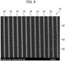
- Fig. 8 is an image of the track region 15 observed by a scanning electron microscope (SEM) corresponding to the bottom surface B30 of the well 30 on the substrate for analysis 10.
- SEM scanning electron microscope
- the antibody aggregations 42 in which the antibodies 41 are aggregated are mainly formed from the vicinity of the boundaries between the recesses 13 and the convex portions 14 to inner side surfaces P13 of the recesses 13. Namely, the antibody aggregations 42 are formed at the boundary regions between the convex portions 14 and the recesses 13, and are also formed in contact with the respective inner side surfaces P13 of the recesses 13.
- Fig. 9 to Fig. 11 show the substrate for analysis 10 of comparative examples not provided with the track regions
- Fig. 12 to Fig. 14 show the substrate for analysis 10 of the present examples provided with the track regions 15.
- Fig. 9 and Fig. 12 are micrographs showing a case in which the antibody aggregations 42 are formed on the substrate for analysis 10 through the immersion treatment with the treatment solution containing the surfactant having a first concentration.
- Fig. 10 and Fig. 13 are micrographs showing a case in which the antibody aggregations 42 are formed on the substrate for analysis 10 through the immersion treatment with the treatment solution containing the surfactant having a second concentration higher than the first concentration.
- Fig. 11 and Fig. 14 are micrographs showing a case in which the antibody aggregations 42 are formed on the substrate for analysis 10 through the immersion treatment with the treatment solution containing the surfactant having a third concentration higher than the second concentration.
- the distribution and the size of the antibody aggregations 42 tend to be influenced by the concentration of the surfactant contained in the treatment solution when the track regions 15 are not provided on the substrate for analysis 10, and are thus difficult to adjust with a high accuracy.
- the distribution and the size of the antibody aggregations 42 are not easily influenced by the concentration of the surfactant contained in the treatment solution when the track regions 15 are provided on the substrate for analysis 10, and thus can be adjusted accurately.
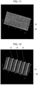
- Fig. 15 is a micrograph showing a case in which the pitch between the recesses 13 and the convex portions 14 in the radial direction of the substrate for analysis 10 is 740 nm, and the width of the respective recesses 13 is 370 nm.
- Fig. 16 is a micrograph showing a case in which the pitch between the recesses 13 and the convex portions 14 in the radial direction of the substrate for analysis 10 is 340 nm, and the width of the respective recesses 13 is 220 nm.
- Increasing the width of the recesses 13 more than the width of the convex portions 14 increases the density of the antibodies 41 adjacent to the recesses 13 more than the density of the antibodies 41 adjacent to the convex portions 14, thereby the antibody aggregations 42 can be selectively formed in the recesses 13.
- the width of the convex portions 14 is thus smaller than the width of the recesses 13.
- the width of the recesses 13 in the radial direction of the substrate for analysis 10 is preferably set in a range of 150 nm to 250 nm
- the depth of the recesses 13 (the height of the convex portions 14) is preferably set in a range of 20 nm to 100 nm, and more preferably in a range of 65 nm to 70 nm in order to form the antibody aggregations 42 mainly in the recesses 13.
- the antibody aggregations 42 preferably have a size in a range of 5 nm to 40 nm, and more preferably in a range of 10 nm to 20 nm.
- the size of the antibody aggregations 42 can be adjusted depending on the concentration of the surfactant contained in the treatment solution and the immersion time. For example, executing the immersion treatment for a period in a range of one to two hours while using Tween 20 as the surfactant and setting the concentration of the surfactant contained in the treatment solution to 0.05 wt% can adjust the size of the antibody aggregations 42 to about 10 nm to 20 nm.
- step S5 the cleaning device drains the treatment solution from the wells 30.
- the cleaning device cleans the inside of the wells 30 with a cleaning solution after the removal of the treatment solution.
- the cleaning solution may be either pure water or a solution containing a surfactant.
- the immersion time in step S4 is longer than the cleaning time in step S5, and the concentration of the surfactant contained in the treatment solution is higher than or equal to the concentration of the surfactant contained in the cleaning solution.
- step S6 the injection device injects, to the wells 30, a sample solution that contains or has a probability of containing exosomes as a target to be detected.
- a sample solution that contains or has a probability of containing exosomes as a target to be detected.
- the following example is illustrated with a case in which the sample solution contains exosomes as a target to be detected for brevity.
- an exosome 50 is covered with a lipid bilayer membrane 51.
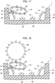
- Fig. 17 corresponds to Fig. 7 .
- the lipid bilayer membrane 51 contains a plurality of transmembrane proteins. The number of the transmembrane proteins and the position in the lipid bilayer membrane 51 vary depending on the type of the exosomes.
- Fig. 17 schematically illustrates an example of the exosome 50 in which the lipid bilayer membrane 51 contains nine transmembrane proteins, and four of the membrane proteins are antigens 52 as a target to be detected.
- step S7 the reaction device incubates the unit for analysis 1 at a set temperature for a set time.
- the antigens 52 in the exosomes 50 contained in the sample solution specifically react with the antibodies 41 included in the antibody aggregations 42.
- the incubation leads the exosomes 50 to be captured on the substrate for analysis 10.
- the antibody aggregations 42 include the plural antibodies 41, the antibody aggregations 42 can efficiently capture the exosomes 50 contained in the sample solution. As illustrated in Fig. 17 , the plural antigens 52 in the exosome 50 and the plural antibodies 41 in the antibody aggregations 42 sometimes specifically react with each other.
- the antibody aggregations 42 are mainly formed in contact with the inner side surfaces P13 of the recesses 13, thereby the exosomes 50 can be strongly captured in the vicinity of the inner side surfaces P13 of the recesses 13 as compared with a case without the antibody aggregations 42 formed.
- step S8 the cleaning device drains the sample solution from the wells 30.
- the cleaning device cleans the inside of the wells 30 with a cleaning solution after the removal of the sample solution.
- the exosomes 50 are mainly captured adjacent to the inner side surfaces P13 of the recesses 13, and are not thus easily influenced by the flow of the cleaning solution. Since the exosomes 50 are not easily removed from the substrate for analysis 10, the separation of the exosomes 50 captured on the substrate for analysis 10 thus can be avoided.
- step S9 the injection device injects a buffer solution containing microparticles (referred to below as a "microparticle buffer solution”) to the wells 30.
- a buffer solution containing microparticles referred to below as a "microparticle buffer solution”
- the surface of the microparticles 60 is provided with a plurality of antibodies 61 (second antibodies) that specifically react with the antigens 52 in the exosome 50.
- step S10 the reaction device incubates the unit for analysis 1 at a set temperature for a set time.
- the antibodies 61 of the microparticles 60 contained in the microparticle buffer solution specifically react with the antigens 52 in the exosomes 50.
- the incubation leads the microparticles 61 to be captured on the substrate for analysis 10, and leads the exosomes 50 to be captured on the substrate for analysis 10 in a sandwiching way between the antibodies 41 and the microparticles 60.
- the exosomes 50 as detection target substances are thus arranged between the antibody aggregations 42 and the microparticles 60.
- the exosomes 50 are labeled by the microparticles 60.
- the analysis device quantifies (counts) the number of the microparticles 60 captured on the substrate for analysis 10, thereby the number of the exosomes 50 can be quantified (counted) indirectly.
- the operator may execute at least any of the steps S1 to S 10, or may execute part of the treatment in the steps.
- the antibodies 61 formed on the surfaces of the microparticles 60 may be either the antibodies that specifically react with the antigens 52 in the exosomes 50, or antibodies or proteins that specifically react with other antigens or proteins expressed on the surfaces of the exosomes 50.
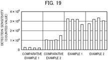
- Fig. 19 shows the counted values of the microparticles 60 in the wells 30 indicated as detection sensitivity of the exosomes 50.
- Comparative Examples 1 and 2 shown in Fig. 19 each indicate the detection sensitivity in a case of not executing the immersion treatment with the treatment solution containing the surfactant in step S4 according to the present embodiment.
- Comparative Example 1 indicates the detection sensitivity in the case in which the wells 30 are cleaned with the cleaning solution not containing the surfactant after step S3 according to the present embodiment.
- Comparative Example 2 indicates the detection sensitivity in the case in which the wells 30 are cleaned with the cleaning solution containing the surfactant after step S3 according to the present embodiment.
- Examples 1 and 2 shown in Fig. 19 each indicate the detection sensitivity in a case of executing the immersion treatment with the treatment solution containing the surfactant in step S4 according to the present embodiment.
- Example 1 corresponds to Comparative Example 1, and indicates the detection sensitivity in the case of cleaning the wells 30 with the cleaning solution not containing the surfactant after executing the immersion treatment with the treatment solution containing the surfactant in step S4 according to the present embodiment.
- Example 2 corresponds to Comparative Example 2, and indicates the detection sensitivity in the case of cleaning the wells 30 with the cleaning solution containing the surfactant after executing the immersion treatment with the treatment solution containing the surfactant in step S4 according to the present embodiment.
- Example 1 The detection sensitivity in Example 1 is about eight times as high as that in Comparative Example 1, and the detection sensitivity in Example 2 is about three times as high as that in Comparative Example 2.
- the method of fabricating the substrate for analysis, the substrate for analysis, and the analysis unit according to the present embodiment cause the antibodies 41 that specifically react with the specific antigens 52 in the exosomes 50 as the detection target substances to be hydrophobically adsorbed to the recesses 13 provided on the track regions 15 on the substrate for analysis 10.
- the substrate for analysis 10 is subjected to the immersion treatment with the treatment solution containing the surfactant so as to form the antibody aggregations 42 at the boundary regions between the recesses 13 and the convex portions 14.
- the treatment solution used in the immersion treatment may be any treatment solution containing any other substance other than the surfactant that has the effect of leading the antibodies 41 to be aggregated.
- the method of fabricating the substrate for analysis, the substrate for analysis, and the analysis unit according to the present embodiment lead the exosomes 50 in the sample solution to be captured on the antibody aggregations 42 formed at the boundaries between the recesses 13 and the convex portions 14 provided on the track regions 15 on the substrate for analysis 10, thereby the separation of the exosomes 50 from the substrate for analysis 10 in the cleaning step can be avoided or reduced.
- the present disclosure relates to the subject matter of Japanese Patent Application No. P2019-045916 and Japanese Patent Application No. P2019-045919 filed on March 13, 2019 , the entire contents of which are incorporated herein by reference.
Landscapes
- Health & Medical Sciences (AREA)
- Immunology (AREA)
- Life Sciences & Earth Sciences (AREA)
- Engineering & Computer Science (AREA)
- Molecular Biology (AREA)
- Biomedical Technology (AREA)
- Chemical & Material Sciences (AREA)
- Hematology (AREA)
- Urology & Nephrology (AREA)
- Biotechnology (AREA)
- Microbiology (AREA)
- Cell Biology (AREA)
- Food Science & Technology (AREA)
- Medicinal Chemistry (AREA)
- Physics & Mathematics (AREA)
- Analytical Chemistry (AREA)
- Biochemistry (AREA)
- General Health & Medical Sciences (AREA)
- General Physics & Mathematics (AREA)
- Pathology (AREA)
- Automatic Analysis And Handling Materials Therefor (AREA)
Abstract
Description
- The present disclosure relates to a method of fabricating a substrate for analysis for analyzing biosamples, a substrate for analysis, and an analysis unit.
- Immunoassays are known that quantitatively analyze disease detection and therapeutic effects by detecting specific antigens or antibodies associated with diseases as biomarkers.
- An analysis method disclosed in
Patent Literature 1 uses a unit for analysis that includes a plurality of wells, fixes antibodies to a substrate for analysis, causes exosomes as detection target substances to be bound and captured onto the antibodies, and causes microparticles to be bound to the exosomes and quantifies the microparticles, thereby the detection target substances (the exosomes) are indirectly quantified. In the respective steps of fixing the antibodies to the substrate for analysis, capturing the exosomes on the antibodies, and causing the microparticles to be bound to the exosomes, a cleaning solution is injected to the wells through an injection nozzle, and the cleaning solution and other solutions are sucked with a suction nozzle, thereby the wells are cleaned. - Exosomes are membrane vesicles secreted from many kinds of cells and formed of lipid bilayers with a size of about 50 nm to 100 nm, and circulate in body fluids such as blood, saliva, and urine. Exosomes include various kinds of substances on the surfaces or in internal substances thereof, such as proteins, lipids, RNA, mRNA, and micro RNA, which are expected to serve as biomarkers for malignant tumors or many kinds of diseases such as Alzheimer's disease.
- The quantitation of the number of exosomes having specific surface antigens derived from a specific disease and the quantitation of the substances included therein from various kinds of exosomes contained in body liquids are presumed to contribute to inspection and early detection of the disease with a higher accuracy.
- Patent Literature 1:
Japanese Unexamined Patent Application Publication No. 2014-219384 - To discover a disease earlier, it is important to detect specific exosomes from specimens including a small number of exosomes having specific surface antigens derived from the disease contained in body fluids such as serum at an early stage of the outbreak of the disease. A larger number of exosomes thus needs to be bound to antibodies and captured on the substrate for analysis. It is also important to prevent the exosomes captured on the substrate for analysis from being separated therefrom during cleaning of the wells.
- An object of an embodiment is to provide a method of fabricating a substrate for analysis, a substrate for analysis, and an analysis unit capable of reliably capturing detection target substances such as exosomes on the substrate for analysis, and avoiding or reducing separation of the exosomes from the substrate for analysis during cleaning.
- A method of fabricating a substrate for analysis according to a first aspect of the embodiment fixes antibodies that react with specific antigens included in detection target substances to the substrate for analysis in which convex portions and recesses alternately arranged, subjects the substrate for analysis to which the antibodies are fixed to immersion treatment with a predetermined treatment solution so as to form antibody aggregations in which the antibodies are aggregated at boundary regions between the recesses and the convex portions, injects a sample solution containing the detection target substances to the substrate for analysis on which the antibody aggregations are formed, and captures the detection target substances on the substrate for analysis.
- A substrate for analysis according to a second aspect of the embodiment includes a track region in which convex portions and recesses alternately arranged, and antibody aggregations in which antibodies that react with antigens included in detection target substances are aggregated, the antibody aggregations being formed at boundary regions between the convex portions and the recesses.
- A unit for analysis according to a third aspect of the embodiment includes a substrate for analysis having a track region in which convex portions and recesses alternately arranged, and a cartridge provided with a plurality of penetration holes, wherein a plurality of wells are composed of the plural penetration holes and the substrate for analysis serving as a bottom surface of the respective wells, and antibody aggregations in which antibodies that react with antigens included in detection target substances are aggregated are formed at boundary regions between the convex portions and the recesses in the wells.
- The method of fabricating the substrate for analysis, the substrate for analysis, and the analysis unit according to the embodiment can reliably capture the detection target substances such as exosomes on the substrate for analysis, and avoid or reduce the separation of the exosomes from the substrate for analysis during cleaning.
-
- [
Fig. 1] Fig. 1 is a plan view showing an example of a configuration of a unit for analysis. - [
Fig. 2] Fig. 2 is a cross-sectional view of the unit for analysis taken along line A-A inFig. 1 . - [
Fig. 3] Fig. 3 is a cross-sectional view showing a state in which a cartridge is removed from a substrate for analysis. - [
Fig. 4] Fig. 4 is an enlarged perspective view showing a well taken along line B-B inFig. 1 . - [
Fig. 5] Fig. 5 is a flowchart showing an example of a method of fabricating the substrate for analysis according to an embodiment. - [
Fig. 6] Fig. 6 is a schematic view showing a state in which antibodies are hydrophobically adsorbed to a recess on the substrate for analysis. - [
Fig. 7] Fig. 7 is a schematic view showing a state in which antibody aggregations are formed on the recess on the substrate for analysis. - [
Fig. 8] Fig. 8 is a scanning electron micrograph (SEM) showing a state in which the antibody aggregations are formed on the recesses on the substrate for analysis. - [
Fig. 9] Fig. 9 is a view showing a state in which the antibody aggregations are formed on the substrate for analysis observed with an atomic force microscope. - [
Fig. 10] Fig. 10 is a view showing a state in which the antibody aggregations are formed on the substrate for analysis observed with the atomic force microscope. - [
Fig. 11] Fig. 11 is a view showing a state in which the antibody aggregations are formed on the substrate for analysis observed with the atomic force microscope. - [
Fig. 12] Fig. 12 is a view showing a state in which the antibody aggregations are formed on the substrate for analysis observed with the atomic force microscope. - [
Fig. 13] Fig. 13 is a view showing a state in which the antibody aggregations are formed on the substrate for analysis observed with the atomic force microscope. - [
Fig. 14] Fig. 14 is a view showing a state in which the antibody aggregations are formed on the substrate for analysis observed with the atomic force microscope. - [
Fig. 15] Fig. 15 is a view showing a state in which the antibody aggregations are formed on the substrate for analysis observed with the atomic force microscope. - [
Fig. 16] Fig. 16 is a view showing a state in which the antibody aggregations are formed on the substrate for analysis observed with the atomic force microscope. - [
Fig. 17] Fig. 17 is a schematic view showing a state in which an exosome is captured on the recess of the substrate for analysis. - [
Fig. 18] Fig. 18 is a schematic view showing a state in which an exosome is captured between antibodies and a microparticle on the recess of the substrate for analysis. - [
Fig. 19] Fig. 19 is a view showing count values of microparticles indicated as detection sensitivity in Comparative Examples 1 and 2 and Examples 1 and 2. - An example of a unit for analysis is described below with reference to
Fig. 1 to Fig. 4 . As illustrated inFig. 1 , the unit foranalysis 1 includes a substrate foranalysis 10 and acartridge 20.Fig. 1 illustrates the unit foranalysis 1 as viewed on thecartridge 20 side. - The substrate for
analysis 10 has a disc-like shape equivalent to optical discs such as Blu-ray discs (BDs), DVDs, and compact discs (CDs), for example. The substrate foranalysis 10 is formed of resin material, such as polycarbonate resin and cycloolefin polymer, used for common optical discs. The substrate foranalysis 10 is not limited to the optical disc as described above, and may be any optical disc according to other configurations or conforming to prescribed standards. -
Fig. 2 is a cross-sectional view showing the unit foranalysis 1 taken along line A-A inFig. 1. Fig. 3 is a view showing a state in which thecartridge 20 is removed from the substrate foranalysis 10. As illustrated inFig. 1, Fig. 2, or Fig. 3 , the substrate foranalysis 10 has acentral hole 11 and aslit 12. Thecentral hole 11 is provided in the middle of the substrate foranalysis 10. Theslit 12 is provided at the circumferential part of the substrate foranalysis 10. Theslit 12 serves as a reference-position defining portion for defining a reference position of the substrate foranalysis 10 in a rotating direction. -
Fig. 4 is a partly enlarged view showing a well 30 taken along line B-B inFig. 1 . The surface of the substrate foranalysis 10 includestrack regions 15 in which recesses 13 and convexportions 14 alternately arranged in a radial direction. Therecesses 13 and theconvex portions 14 are formed in a spiral or concentric state from the inner circumference to the outer circumference of the substrate foranalysis 10. Therecesses 13 correspond to grooves of an optical disc. Theconvex portions 14 correspond to lands of an optical disc. A track pitch of the substrate foranalysis 10 corresponding to a track pitch of an optical disc is 340 nm, for example. - As illustrated in
Fig. 1, Fig. 2, or Fig. 3 , thecartridge 20 is provided with a plurality ofcylindrical penetration holes 21 along the circumferential direction. Thepenetration holes 21 are arranged at regular intervals such that the respective center points are located on the common circle. Thecartridge 20 includes aconvex portion 22 provided in the middle, and aconvex portion 23 provided at the circumferential part. - The operator, when attaching the
cartridge 20 to the substrate foranalysis 10, inserts theconvex portion 22 to thecentral hole 11 of the substrate foranalysis 10, and inserts theconvex portion 23 to theslit 12. This enables the operator to position to align thecartridge 20 and the substrate foranalysis 10 with each other. - As illustrated in
Fig. 2 orFig. 4 , the unit foranalysis 1 includes a plurality ofwells 30 formed by thepenetration holes 21 of thecartridge 20 and the surface (the respective track regions 15) of the substrate foranalysis 10. Thewells 30 each have a hollow shape composed of a bottom surface B30, an inner peripheral surface P30, and an open part A30. The surface of the substrate foranalysis 10 serves as the bottom surfaces B30 of therespective wells 30. The inner peripheral surface on the inside of the respective penetration holes 21 serves as the inner peripheral surfaces P30 of therespective wells 30. - The open part A30 is located on the opposite side of the bottom surface B30 in the
cartridge 20. Thewells 30 each serve as a container for storing a solution such as a sample solution, a buffer solution, and a cleaning solution. WhileFig. 1 illustrates the eightwells 30, the number of thewells 30 is not limited to eight. - As illustrated in
Fig. 3 , thecartridge 20 is detachable from the substrate foranalysis 10. The detection and the measurement of microparticles for labeling detection target substances are made by use of the substrate foranalysis 10 itself separated from thecartridge 20. - An example of a method of fabricating the substrate for analysis according to an embodiment is described below with reference to the flowchart shown in
Fig. 5 , andFig. 6 toFig. 9 . More particularly, a method of fabricating the substrate foranalysis 10 to which antibodies are fixed that specifically react with specific antigens included in detection target substances is described below. The embodiment is illustrated below with a case in which the detection target substances are exosomes. - In step S1 in
Fig. 5 , an injection device injects an antibody buffer solution to thewells 30. The antibody buffer solution to be used may be a carbonate-bicarbonate buffer solution including antibodies 41 (first antibodies) that specifically react with specific antigens in exosomes. In step S2, a reaction device incubates the unit foranalysis 1 in which the antibody buffer solution is injected to thewells 30. The incubation causes part of theantibodies 41 that has hydrophobicity to be adsorbed to part of the substrate foranalysis 10 that has hydrophobicity. - As illustrated in
Fig. 6 , theantibodies 41 are hydrophobically adsorbed to a region corresponding to the respective bottom surfaces B30 of thewells 30 on the substrate foranalysis 10. Theantibodies 41 are fixed to the substrate foranalysis 10.Fig. 6 is an enlarged cross-sectional view of therecess 13 on thetrack region 15 of the substrate foranalysis 10, and schematically illustrates a state in which theantibodies 41 are hydrophobically adsorbed to therecess 13. - In step S3, a cleaning device drains the antibody buffer solution from the
wells 30. In step S4, the injection device injects a treatment solution containing a surfactant to thewells 30, and executes immersion treatment at a set temperature for a set period. The injection device may execute the immersion treatment while keeping the unit foranalysis 1 stopped, or may execute the immersion treatment while keeping the unit foranalysis 1 shaken. - The treatment solution to be used can be a buffer solution for bioassay. A preferred example of the buffer solution for bioassay is phosphate-buffer saline (PBS). A nonionic surfactant can be used as the surfactant contained in the treatment solution. A preferred example of the nonionic surfactant is polyoxyethylene sorbitan monolaurate (Tween 20) or octylphenol ethoxylate (Triton X-100).
- As illustrated in
Fig. 7 orFig. 8 , the immersion treatment executed in step S4 leads theantibodies 41 hydrophobically adsorbed to the substrate foranalysis 10 to be aggregated so as to formantibody aggregations 42. That is, eachantibody aggregation 42 is composed of theplural antibodies 41.Fig. 7 corresponding toFig. 6 schematically illustrates theantibody aggregations 42 in which theantibodies 41 are aggregated.Fig. 8 is an image of thetrack region 15 observed by a scanning electron microscope (SEM) corresponding to the bottom surface B30 of the well 30 on the substrate foranalysis 10. The antibody aggregations 42 are mainly formed on therecesses 13 of the substrate foranalysis 10. In particular, theantibody aggregations 42 in which theantibodies 41 are aggregated are mainly formed from the vicinity of the boundaries between therecesses 13 and theconvex portions 14 to inner side surfaces P13 of therecesses 13. Namely, theantibody aggregations 42 are formed at the boundary regions between theconvex portions 14 and therecesses 13, and are also formed in contact with the respective inner side surfaces P13 of therecesses 13. - A relationship between a concentration of the surfactant contained in the treatment solution and the
antibody aggregations 42 formed on the substrate foranalysis 10 is described below with reference toFig. 9 to Fig. 14 .Fig. 9 to Fig. 11 show the substrate foranalysis 10 of comparative examples not provided with thetrack regions 15, andFig. 12 to Fig. 14 show the substrate foranalysis 10 of the present examples provided with thetrack regions 15. -
Fig. 9 andFig. 12 are micrographs showing a case in which theantibody aggregations 42 are formed on the substrate foranalysis 10 through the immersion treatment with the treatment solution containing the surfactant having a first concentration.Fig. 10 andFig. 13 are micrographs showing a case in which theantibody aggregations 42 are formed on the substrate foranalysis 10 through the immersion treatment with the treatment solution containing the surfactant having a second concentration higher than the first concentration.Fig. 11 andFig. 14 are micrographs showing a case in which theantibody aggregations 42 are formed on the substrate foranalysis 10 through the immersion treatment with the treatment solution containing the surfactant having a third concentration higher than the second concentration. - As shown in
Fig. 9 to 11 , the distribution and the size of theantibody aggregations 42 tend to be influenced by the concentration of the surfactant contained in the treatment solution when thetrack regions 15 are not provided on the substrate foranalysis 10, and are thus difficult to adjust with a high accuracy. As shown inFig. 12 to Fig. 14 , the distribution and the size of theantibody aggregations 42 are not easily influenced by the concentration of the surfactant contained in the treatment solution when thetrack regions 15 are provided on the substrate foranalysis 10, and thus can be adjusted accurately. - A relationship between a width of the
respective recesses 13 and theantibody aggregations 42 formed on the substrate foranalysis 10 is described below with reference toFig. 15 and Fig. 16. Fig. 15 is a micrograph showing a case in which the pitch between therecesses 13 and theconvex portions 14 in the radial direction of the substrate foranalysis 10 is 740 nm, and the width of therespective recesses 13 is 370 nm.Fig. 16 is a micrograph showing a case in which the pitch between therecesses 13 and theconvex portions 14 in the radial direction of the substrate foranalysis 10 is 340 nm, and the width of therespective recesses 13 is 220 nm. - Increasing the width of the
recesses 13 more than the width of theconvex portions 14 increases the density of theantibodies 41 adjacent to therecesses 13 more than the density of theantibodies 41 adjacent to theconvex portions 14, thereby theantibody aggregations 42 can be selectively formed in therecesses 13. The width of theconvex portions 14 is thus smaller than the width of therecesses 13. - When the pitch between the
recesses 13 and theconvex portions 14 in the radial direction of the substrate foranalysis 10 is 340 nm, the width of therecesses 13 in the radial direction of the substrate foranalysis 10 is preferably set in a range of 150 nm to 250 nm, and the depth of the recesses 13 (the height of the convex portions 14) is preferably set in a range of 20 nm to 100 nm, and more preferably in a range of 65 nm to 70 nm in order to form theantibody aggregations 42 mainly in therecesses 13. - The antibody aggregations 42 preferably have a size in a range of 5 nm to 40 nm, and more preferably in a range of 10 nm to 20 nm. The size of the
antibody aggregations 42 can be adjusted depending on the concentration of the surfactant contained in the treatment solution and the immersion time. For example, executing the immersion treatment for a period in a range of one to two hours while usingTween 20 as the surfactant and setting the concentration of the surfactant contained in the treatment solution to 0.05 wt% can adjust the size of theantibody aggregations 42 to about 10 nm to 20 nm. - In step S5, the cleaning device drains the treatment solution from the
wells 30. The cleaning device cleans the inside of thewells 30 with a cleaning solution after the removal of the treatment solution. The cleaning solution may be either pure water or a solution containing a surfactant. When the cleaning solution contains a surfactant, the immersion time in step S4 is longer than the cleaning time in step S5, and the concentration of the surfactant contained in the treatment solution is higher than or equal to the concentration of the surfactant contained in the cleaning solution. - In step S6, the injection device injects, to the
wells 30, a sample solution that contains or has a probability of containing exosomes as a target to be detected. The following example is illustrated with a case in which the sample solution contains exosomes as a target to be detected for brevity. - As illustrated in
Fig. 17 , anexosome 50 is covered with alipid bilayer membrane 51.Fig. 17 corresponds toFig. 7 . Thelipid bilayer membrane 51 contains a plurality of transmembrane proteins. The number of the transmembrane proteins and the position in thelipid bilayer membrane 51 vary depending on the type of the exosomes.Fig. 17 schematically illustrates an example of theexosome 50 in which thelipid bilayer membrane 51 contains nine transmembrane proteins, and four of the membrane proteins areantigens 52 as a target to be detected. - In step S7, the reaction device incubates the unit for
analysis 1 at a set temperature for a set time. Theantigens 52 in theexosomes 50 contained in the sample solution specifically react with theantibodies 41 included in theantibody aggregations 42. The incubation leads theexosomes 50 to be captured on the substrate foranalysis 10. - Since the
antibody aggregations 42 include theplural antibodies 41, theantibody aggregations 42 can efficiently capture theexosomes 50 contained in the sample solution. As illustrated inFig. 17 , theplural antigens 52 in theexosome 50 and theplural antibodies 41 in theantibody aggregations 42 sometimes specifically react with each other. The antibody aggregations 42 are mainly formed in contact with the inner side surfaces P13 of therecesses 13, thereby theexosomes 50 can be strongly captured in the vicinity of the inner side surfaces P13 of therecesses 13 as compared with a case without theantibody aggregations 42 formed. - In step S8, the cleaning device drains the sample solution from the
wells 30. The cleaning device cleans the inside of thewells 30 with a cleaning solution after the removal of the sample solution. Theexosomes 50 are mainly captured adjacent to the inner side surfaces P13 of therecesses 13, and are not thus easily influenced by the flow of the cleaning solution. Since theexosomes 50 are not easily removed from the substrate foranalysis 10, the separation of theexosomes 50 captured on the substrate foranalysis 10 thus can be avoided. - In step S9, the injection device injects a buffer solution containing microparticles (referred to below as a "microparticle buffer solution") to the
wells 30. As illustrated inFig. 18 , the surface of themicroparticles 60 is provided with a plurality of antibodies 61 (second antibodies) that specifically react with theantigens 52 in theexosome 50. - In step S10, the reaction device incubates the unit for
analysis 1 at a set temperature for a set time. Theantibodies 61 of themicroparticles 60 contained in the microparticle buffer solution specifically react with theantigens 52 in theexosomes 50. The incubation leads themicroparticles 61 to be captured on the substrate foranalysis 10, and leads theexosomes 50 to be captured on the substrate foranalysis 10 in a sandwiching way between theantibodies 41 and themicroparticles 60. Theexosomes 50 as detection target substances are thus arranged between theantibody aggregations 42 and themicroparticles 60. - The
exosomes 50 are labeled by themicroparticles 60. The analysis device quantifies (counts) the number of themicroparticles 60 captured on the substrate foranalysis 10, thereby the number of theexosomes 50 can be quantified (counted) indirectly. The operator may execute at least any of the steps S1 toS 10, or may execute part of the treatment in the steps. Theantibodies 61 formed on the surfaces of themicroparticles 60 may be either the antibodies that specifically react with theantigens 52 in theexosomes 50, or antibodies or proteins that specifically react with other antigens or proteins expressed on the surfaces of theexosomes 50. - The functional effects of the method of fabricating the substrate for analysis according to the present embodiment are described below with reference to
Fig. 19 . The vertical axis inFig. 19 shows the counted values of themicroparticles 60 in thewells 30 indicated as detection sensitivity of theexosomes 50. - Comparative Examples 1 and 2 shown in
Fig. 19 each indicate the detection sensitivity in a case of not executing the immersion treatment with the treatment solution containing the surfactant in step S4 according to the present embodiment. In particular, Comparative Example 1 indicates the detection sensitivity in the case in which thewells 30 are cleaned with the cleaning solution not containing the surfactant after step S3 according to the present embodiment. Comparative Example 2 indicates the detection sensitivity in the case in which thewells 30 are cleaned with the cleaning solution containing the surfactant after step S3 according to the present embodiment. The results revealed that the cleaning of the inside of thewells 30 with the cleaning solution containing the surfactant after step S3 improves the detection sensitivity to some extent, as compared with the case of cleaning the inside of thewells 30 with the cleaning solution not containing the surfactant, due to effect of the surfactant contained in the cleaning solution. - Examples 1 and 2 shown in
Fig. 19 each indicate the detection sensitivity in a case of executing the immersion treatment with the treatment solution containing the surfactant in step S4 according to the present embodiment. In particular, Example 1 corresponds to Comparative Example 1, and indicates the detection sensitivity in the case of cleaning thewells 30 with the cleaning solution not containing the surfactant after executing the immersion treatment with the treatment solution containing the surfactant in step S4 according to the present embodiment. Example 2 corresponds to Comparative Example 2, and indicates the detection sensitivity in the case of cleaning thewells 30 with the cleaning solution containing the surfactant after executing the immersion treatment with the treatment solution containing the surfactant in step S4 according to the present embodiment. - The detection sensitivity in Example 1 is about eight times as high as that in Comparative Example 1, and the detection sensitivity in Example 2 is about three times as high as that in Comparative Example 2. The results revealed that Examples 1 and 2 both exhibit the detection sensitivity that is greatly improved as compared with Comparative Examples 1 and 2.
- The method of fabricating the substrate for analysis, the substrate for analysis, and the analysis unit according to the present embodiment cause the
antibodies 41 that specifically react with thespecific antigens 52 in theexosomes 50 as the detection target substances to be hydrophobically adsorbed to therecesses 13 provided on thetrack regions 15 on the substrate foranalysis 10. The substrate foranalysis 10 is subjected to the immersion treatment with the treatment solution containing the surfactant so as to form theantibody aggregations 42 at the boundary regions between therecesses 13 and theconvex portions 14. The treatment solution used in the immersion treatment may be any treatment solution containing any other substance other than the surfactant that has the effect of leading theantibodies 41 to be aggregated. - The method of fabricating the substrate for analysis, the substrate for analysis, and the analysis unit according to the present embodiment lead the
exosomes 50 in the sample solution to be captured on theantibody aggregations 42 formed at the boundaries between therecesses 13 and theconvex portions 14 provided on thetrack regions 15 on the substrate foranalysis 10, thereby the separation of theexosomes 50 from the substrate foranalysis 10 in the cleaning step can be avoided or reduced. - It should be understood that the present invention is not intended to be limited to the respective embodiments described above, and various modifications will be apparent to those skilled in the art without departing from the scope of the present invention.
- The present disclosure relates to the subject matter of
Japanese Patent Application No. P2019-045916 Japanese Patent Application No. P2019-045919 filed on March 13, 2019
Claims (6)
- A method of fabricating a substrate for analysis, the method comprising:fixing antibodies that react with specific antigens included in detection target substances to the substrate for analysis in which convex portions and recesses alternately arranged;subjecting the substrate for analysis to which the antibodies are fixed to immersion treatment with a predetermined treatment solution so as to form antibody aggregations in which the antibodies are aggregated at boundary regions between the recesses and the convex portions;injecting a sample solution containing the detection target substances to the substrate for analysis on which the antibody aggregations are formed; andcapturing the detection target substances on the substrate for analysis.
- The method of fabricating the substrate for analysis according to claim 1, wherein:the antibodies are fixed to the substrate for analysis by hydrophobic adsorption; andthe antibody aggregations are formed by use of a surfactant as the predetermined treatment solution.
- The method of fabricating the substrate for analysis according to claim 1 or 2, further comprising:fixing, to the detection target substances, microparticles provided with the antibodies or other antibodies different from the antibodies on surfaces of the microparticles; andarranging the detection target substances between the antibody aggregations and the microparticles.
- A substrate for analysis comprising:a track region in which convex portions and recesses alternately arranged; andantibody aggregations in which antibodies that react with antigens included in detection target substances are aggregated, the antibody aggregations being formed at boundary regions between the convex portions and the recesses.
- The substrate for analysis according to claim 4, wherein the recesses have a wider width than the convex portions.
- A unit for analysis comprising:a substrate for analysis having a track region in which convex portions and recesses alternately arranged; anda cartridge provided with a plurality of penetration holes,wherein a plurality of wells are composed of the plural penetration holes and the substrate for analysis serving as a bottom surface of the respective wells, andantibody aggregations in which antibodies that react with antigens included in detection target substances are aggregated are formed at boundary regions between the convex portions and the recesses in the wells.
Applications Claiming Priority (3)
| Application Number | Priority Date | Filing Date | Title |
|---|---|---|---|
| JP2019045919A JP2020148599A (en) | 2019-03-13 | 2019-03-13 | Analytical board and analytical unit |
| JP2019045916A JP2020148598A (en) | 2019-03-13 | 2019-03-13 | Method for manufacturing analytical substrate |
| PCT/JP2020/009410 WO2020184377A1 (en) | 2019-03-13 | 2020-03-05 | Method for fabricating analysis substrate, analysis substrate, and analysis unit |
Publications (2)
| Publication Number | Publication Date |
|---|---|
| EP3940390A1 true EP3940390A1 (en) | 2022-01-19 |
| EP3940390A4 EP3940390A4 (en) | 2022-05-04 |
Family
ID=72427446
Family Applications (1)
| Application Number | Title | Priority Date | Filing Date |
|---|---|---|---|
| EP20768913.4A Withdrawn EP3940390A4 (en) | 2019-03-13 | 2020-03-05 | Method for fabricating analysis substrate, analysis substrate, and analysis unit |
Country Status (4)
| Country | Link |
|---|---|
| US (1) | US20210405036A1 (en) |
| EP (1) | EP3940390A4 (en) |
| CN (1) | CN113518925A (en) |
| WO (1) | WO2020184377A1 (en) |
Family Cites Families (11)
| Publication number | Priority date | Publication date | Assignee | Title |
|---|---|---|---|---|
| JP2007139658A (en) * | 2005-11-21 | 2007-06-07 | Sharp Corp | Allergen detection method and allergen detection apparatus |
| JP4970217B2 (en) * | 2007-11-07 | 2012-07-04 | 株式会社東芝 | Method for measuring substances |
| JP6077858B2 (en) * | 2011-01-07 | 2017-02-08 | 東亞合成株式会社 | Antigen preparation method for obtaining anti-hydrophobic peptide antibody |
| JP6210004B2 (en) * | 2013-04-09 | 2017-10-11 | 株式会社Jvcケンウッド | Sample analysis device and exosome capture method |
| DE102013106713A1 (en) * | 2013-06-26 | 2014-12-31 | Forschungszentrum Jülich GmbH | Method for identifying indicators for the determination of diseases |
| JP2016080565A (en) * | 2014-10-20 | 2016-05-16 | 有限会社マイテック | Raman spectrum detection method for ebola virus, and ebola virus antibody-immobilized plate |
| JP2017058242A (en) * | 2015-09-16 | 2017-03-23 | 株式会社Jvcケンウッド | Specimen detection unit, analyzer, and analysis method |
| JP2017138112A (en) * | 2016-02-01 | 2017-08-10 | 株式会社Jvcケンウッド | Specimen detection apparatus and specimen detection method |
| JP6740703B2 (en) * | 2016-05-16 | 2020-08-19 | 株式会社Jvcケンウッド | Analysis method and analyzer |
| JP2019045919A (en) | 2017-08-30 | 2019-03-22 | キヤノンマーケティングジャパン株式会社 | Information processing device, control method, program |
| JP7128457B2 (en) | 2017-08-30 | 2022-08-31 | クリナップ株式会社 | hanging cabinet |
-
2020
- 2020-03-05 EP EP20768913.4A patent/EP3940390A4/en not_active Withdrawn
- 2020-03-05 WO PCT/JP2020/009410 patent/WO2020184377A1/en not_active Ceased
- 2020-03-05 CN CN202080018371.6A patent/CN113518925A/en not_active Withdrawn
-
2021
- 2021-09-10 US US17/471,622 patent/US20210405036A1/en not_active Abandoned
Also Published As
| Publication number | Publication date |
|---|---|
| US20210405036A1 (en) | 2021-12-30 |
| CN113518925A (en) | 2021-10-19 |
| EP3940390A4 (en) | 2022-05-04 |
| WO2020184377A1 (en) | 2020-09-17 |
Similar Documents
| Publication | Publication Date | Title |
|---|---|---|
| US10962531B2 (en) | Method of capturing exosomes | |
| CN106796232B (en) | Lateral flow assay device with filtered flow control | |
| JP6182547B2 (en) | Dry sample carrier with dissolvable sample collection area | |
| CN108474722A (en) | Sample collection device, holder for the sample collection device, and sample pretreatment method using the sample collection device | |
| WO2017134906A1 (en) | Sample detection device and sample detection method | |
| JP5442699B2 (en) | Apparatus and method for separating liquid components of a blood sample, and analytical apparatus comprising such an apparatus | |
| KR20090123039A (en) | Blood separation device | |
| US20210405036A1 (en) | Method of fabricating substrate for analysis, substrate for analysis, and analysis unit | |
| US20070196813A1 (en) | Extraction of substances of interest from blood for mass spectrometric analysis | |
| JP2020148598A (en) | Method for manufacturing analytical substrate | |
| JP2019074351A (en) | Inspection device | |
| JP7172745B2 (en) | ANALYSIS UNIT, CLEANING DEVICE, AND CLEANING METHOD | |
| JP2009210392A (en) | Chip | |
| JP2020148599A (en) | Analytical board and analytical unit | |
| EP4188600B1 (en) | Device for separation and/or preservation of a biofluid at a point-of-care, kit and method for identifying a disease | |
| JP7095613B2 (en) | Cleaning equipment and cleaning method | |
| US20210299671A1 (en) | Plate for sampling apparatus and microcentrifuge vial for microsampling apparatus | |
| EP4377666B1 (en) | Apparatus and method for preparation of a biological sample for analytical or diagnostic purposes | |
| KR20170022687A (en) | Bio-chip for seperating and detecting cells | |
| JP6747350B2 (en) | Detection target substance capture unit and extraction device | |
| US12472500B1 (en) | Microfluidic device | |
| WO2019058596A1 (en) | Dispensing unit | |
| CN109517792B (en) | A kind of separation medium of new and old red blood cells and preparation method and application thereof | |
| JP2017122618A (en) | Analysis substrate and manufacturing method thereof | |
| KR20260052348A (en) | Blood Analysis Platform |
Legal Events
| Date | Code | Title | Description |
|---|---|---|---|
| STAA | Information on the status of an ep patent application or granted ep patent |
Free format text: STATUS: THE INTERNATIONAL PUBLICATION HAS BEEN MADE |
|
| PUAI | Public reference made under article 153(3) epc to a published international application that has entered the european phase |
Free format text: ORIGINAL CODE: 0009012 |
|
| STAA | Information on the status of an ep patent application or granted ep patent |
Free format text: STATUS: REQUEST FOR EXAMINATION WAS MADE |
|
| 17P | Request for examination filed |
Effective date: 20210913 |
|
| AK | Designated contracting states |
Kind code of ref document: A1 Designated state(s): AL AT BE BG CH CY CZ DE DK EE ES FI FR GB GR HR HU IE IS IT LI LT LU LV MC MK MT NL NO PL PT RO RS SE SI SK SM TR |
|
| A4 | Supplementary search report drawn up and despatched |
Effective date: 20220404 |
|
| RIC1 | Information provided on ipc code assigned before grant |
Ipc: G01N 33/543 20060101ALI20220328BHEP Ipc: G01N 37/00 20060101AFI20220328BHEP |
|
| DAV | Request for validation of the european patent (deleted) | ||
| DAX | Request for extension of the european patent (deleted) | ||
| STAA | Information on the status of an ep patent application or granted ep patent |
Free format text: STATUS: THE APPLICATION IS DEEMED TO BE WITHDRAWN |
|
| 18D | Application deemed to be withdrawn |
Effective date: 20221108 |