EP3939626A1 - Biomaterial implants and methods of making the same - Google Patents
Biomaterial implants and methods of making the same Download PDFInfo
- Publication number
- EP3939626A1 EP3939626A1 EP21185228.0A EP21185228A EP3939626A1 EP 3939626 A1 EP3939626 A1 EP 3939626A1 EP 21185228 A EP21185228 A EP 21185228A EP 3939626 A1 EP3939626 A1 EP 3939626A1
- Authority
- EP
- European Patent Office
- Prior art keywords
- cortical
- bone
- fibers
- demineralized
- biomaterial
- Prior art date
- Legal status (The legal status is an assumption and is not a legal conclusion. Google has not performed a legal analysis and makes no representation as to the accuracy of the status listed.)
- Pending
Links
- 239000012620 biological material Substances 0.000 title claims abstract description 78
- 238000000034 method Methods 0.000 title claims abstract description 59
- 239000007943 implant Substances 0.000 title abstract description 36
- 230000001054 cortical effect Effects 0.000 claims abstract description 148
- 210000000988 bone and bone Anatomy 0.000 claims abstract description 147
- 239000000835 fiber Substances 0.000 claims abstract description 128
- 239000000203 mixture Substances 0.000 claims description 56
- 239000000463 material Substances 0.000 claims description 36
- 239000000243 solution Substances 0.000 claims description 35
- XLYOFNOQVPJJNP-UHFFFAOYSA-N water Chemical compound O XLYOFNOQVPJJNP-UHFFFAOYSA-N 0.000 claims description 33
- VEXZGXHMUGYJMC-UHFFFAOYSA-N Hydrochloric acid Chemical compound Cl VEXZGXHMUGYJMC-UHFFFAOYSA-N 0.000 claims description 26
- KFZMGEQAYNKOFK-UHFFFAOYSA-N Isopropanol Chemical compound CC(C)O KFZMGEQAYNKOFK-UHFFFAOYSA-N 0.000 claims description 21
- 230000002328 demineralizing effect Effects 0.000 claims description 13
- 238000010306 acid treatment Methods 0.000 claims description 12
- 239000008223 sterile water Substances 0.000 claims description 12
- 230000001413 cellular effect Effects 0.000 claims description 9
- 238000004108 freeze drying Methods 0.000 claims description 9
- FAPWRFPIFSIZLT-UHFFFAOYSA-M Sodium chloride Chemical compound [Na+].[Cl-] FAPWRFPIFSIZLT-UHFFFAOYSA-M 0.000 claims description 8
- 230000010478 bone regeneration Effects 0.000 claims description 8
- MHAJPDPJQMAIIY-UHFFFAOYSA-N Hydrogen peroxide Chemical compound OO MHAJPDPJQMAIIY-UHFFFAOYSA-N 0.000 claims description 7
- 239000000872 buffer Substances 0.000 claims description 7
- 229960003964 deoxycholic acid Drugs 0.000 claims description 7
- 238000007710 freezing Methods 0.000 claims description 7
- 230000008014 freezing Effects 0.000 claims description 7
- 230000007935 neutral effect Effects 0.000 claims description 6
- 238000013019 agitation Methods 0.000 claims description 5
- 239000007864 aqueous solution Substances 0.000 claims description 5
- 238000003754 machining Methods 0.000 claims description 5
- 238000004519 manufacturing process Methods 0.000 claims description 5
- 210000001772 blood platelet Anatomy 0.000 claims description 4
- 210000002798 bone marrow cell Anatomy 0.000 claims description 4
- 210000003743 erythrocyte Anatomy 0.000 claims description 4
- 210000000265 leukocyte Anatomy 0.000 claims description 4
- 238000000465 moulding Methods 0.000 claims description 4
- 210000002381 plasma Anatomy 0.000 claims description 4
- KXGVEGMKQFWNSR-LLQZFEROSA-N deoxycholic acid Chemical compound C([C@H]1CC2)[C@H](O)CC[C@]1(C)[C@@H]1[C@@H]2[C@@H]2CC[C@H]([C@@H](CCC(O)=O)C)[C@@]2(C)[C@@H](O)C1 KXGVEGMKQFWNSR-LLQZFEROSA-N 0.000 claims 1
- 230000008439 repair process Effects 0.000 abstract description 25
- 241000124008 Mammalia Species 0.000 abstract description 8
- 230000029663 wound healing Effects 0.000 abstract description 6
- 230000001737 promoting effect Effects 0.000 abstract description 5
- 239000000306 component Substances 0.000 abstract description 4
- PEDCQBHIVMGVHV-UHFFFAOYSA-N Glycerine Chemical compound OCC(O)CO PEDCQBHIVMGVHV-UHFFFAOYSA-N 0.000 description 32
- RVGRUAULSDPKGF-UHFFFAOYSA-N Poloxamer Chemical compound C1CO1.CC1CO1 RVGRUAULSDPKGF-UHFFFAOYSA-N 0.000 description 19
- 239000000047 product Substances 0.000 description 16
- 210000002805 bone matrix Anatomy 0.000 description 13
- 229920001983 poloxamer Polymers 0.000 description 13
- 230000008569 process Effects 0.000 description 13
- 239000000499 gel Substances 0.000 description 12
- 239000000017 hydrogel Substances 0.000 description 12
- 229960000502 poloxamer Drugs 0.000 description 12
- 235000011187 glycerol Nutrition 0.000 description 11
- 229920002134 Carboxymethyl cellulose Polymers 0.000 description 10
- 238000011282 treatment Methods 0.000 description 10
- 229920002385 Sodium hyaluronate Polymers 0.000 description 9
- 239000001768 carboxy methyl cellulose Substances 0.000 description 9
- 235000010948 carboxy methyl cellulose Nutrition 0.000 description 9
- 239000008112 carboxymethyl-cellulose Substances 0.000 description 9
- 229940105329 carboxymethylcellulose Drugs 0.000 description 9
- 229960005150 glycerol Drugs 0.000 description 9
- 230000002138 osteoinductive effect Effects 0.000 description 9
- 239000012071 phase Substances 0.000 description 9
- 229940010747 sodium hyaluronate Drugs 0.000 description 9
- YWIVKILSMZOHHF-QJZPQSOGSA-N sodium;(2s,3s,4s,5r,6r)-6-[(2s,3r,4r,5s,6r)-3-acetamido-2-[(2s,3s,4r,5r,6r)-6-[(2r,3r,4r,5s,6r)-3-acetamido-2,5-dihydroxy-6-(hydroxymethyl)oxan-4-yl]oxy-2-carboxy-4,5-dihydroxyoxan-3-yl]oxy-5-hydroxy-6-(hydroxymethyl)oxan-4-yl]oxy-3,4,5-trihydroxyoxane-2- Chemical compound [Na+].CC(=O)N[C@H]1[C@H](O)O[C@H](CO)[C@@H](O)[C@@H]1O[C@H]1[C@H](O)[C@@H](O)[C@H](O[C@H]2[C@@H]([C@@H](O[C@H]3[C@@H]([C@@H](O)[C@H](O)[C@H](O3)C(O)=O)O)[C@H](O)[C@@H](CO)O2)NC(C)=O)[C@@H](C(O)=O)O1 YWIVKILSMZOHHF-QJZPQSOGSA-N 0.000 description 9
- 210000004027 cell Anatomy 0.000 description 8
- 239000003102 growth factor Substances 0.000 description 8
- 230000000278 osteoconductive effect Effects 0.000 description 8
- 239000007788 liquid Substances 0.000 description 7
- 229920001992 poloxamer 407 Polymers 0.000 description 7
- 229940044476 poloxamer 407 Drugs 0.000 description 7
- 238000012545 processing Methods 0.000 description 7
- 210000001519 tissue Anatomy 0.000 description 7
- FHHPUSMSKHSNKW-SMOYURAASA-M sodium deoxycholate Chemical compound [Na+].C([C@H]1CC2)[C@H](O)CC[C@]1(C)[C@@H]1[C@@H]2[C@@H]2CC[C@H]([C@@H](CCC([O-])=O)C)[C@@]2(C)[C@@H](O)C1 FHHPUSMSKHSNKW-SMOYURAASA-M 0.000 description 6
- WSFSSNUMVMOOMR-UHFFFAOYSA-N Formaldehyde Chemical compound O=C WSFSSNUMVMOOMR-UHFFFAOYSA-N 0.000 description 5
- 239000002253 acid Substances 0.000 description 5
- 210000001185 bone marrow Anatomy 0.000 description 5
- 230000007547 defect Effects 0.000 description 5
- 201000010099 disease Diseases 0.000 description 5
- 208000037265 diseases, disorders, signs and symptoms Diseases 0.000 description 5
- 238000009826 distribution Methods 0.000 description 5
- 238000000605 extraction Methods 0.000 description 5
- 125000006850 spacer group Chemical group 0.000 description 5
- IJGRMHOSHXDMSA-UHFFFAOYSA-N Atomic nitrogen Chemical compound N#N IJGRMHOSHXDMSA-UHFFFAOYSA-N 0.000 description 4
- SXRSQZLOMIGNAQ-UHFFFAOYSA-N Glutaraldehyde Chemical compound O=CCCCC=O SXRSQZLOMIGNAQ-UHFFFAOYSA-N 0.000 description 4
- 241001465754 Metazoa Species 0.000 description 4
- 239000004696 Poly ether ether ketone Substances 0.000 description 4
- 239000002202 Polyethylene glycol Substances 0.000 description 4
- 230000002902 bimodal effect Effects 0.000 description 4
- 230000008468 bone growth Effects 0.000 description 4
- 238000005115 demineralization Methods 0.000 description 4
- LOKCTEFSRHRXRJ-UHFFFAOYSA-I dipotassium trisodium dihydrogen phosphate hydrogen phosphate dichloride Chemical compound P(=O)(O)(O)[O-].[K+].P(=O)(O)([O-])[O-].[Na+].[Na+].[Cl-].[K+].[Cl-].[Na+] LOKCTEFSRHRXRJ-UHFFFAOYSA-I 0.000 description 4
- 238000009472 formulation Methods 0.000 description 4
- 230000004927 fusion Effects 0.000 description 4
- 229910052500 inorganic mineral Inorganic materials 0.000 description 4
- 230000002188 osteogenic effect Effects 0.000 description 4
- 239000002953 phosphate buffered saline Substances 0.000 description 4
- 229920002530 polyetherether ketone Polymers 0.000 description 4
- 229920001223 polyethylene glycol Polymers 0.000 description 4
- 239000011780 sodium chloride Substances 0.000 description 4
- 239000000126 substance Substances 0.000 description 4
- KIUKXJAPPMFGSW-DNGZLQJQSA-N (2S,3S,4S,5R,6R)-6-[(2S,3R,4R,5S,6R)-3-Acetamido-2-[(2S,3S,4R,5R,6R)-6-[(2R,3R,4R,5S,6R)-3-acetamido-2,5-dihydroxy-6-(hydroxymethyl)oxan-4-yl]oxy-2-carboxy-4,5-dihydroxyoxan-3-yl]oxy-5-hydroxy-6-(hydroxymethyl)oxan-4-yl]oxy-3,4,5-trihydroxyoxane-2-carboxylic acid Chemical compound CC(=O)N[C@H]1[C@H](O)O[C@H](CO)[C@@H](O)[C@@H]1O[C@H]1[C@H](O)[C@@H](O)[C@H](O[C@H]2[C@@H]([C@@H](O[C@H]3[C@@H]([C@@H](O)[C@H](O)[C@H](O3)C(O)=O)O)[C@H](O)[C@@H](CO)O2)NC(C)=O)[C@@H](C(O)=O)O1 KIUKXJAPPMFGSW-DNGZLQJQSA-N 0.000 description 3
- QTBSBXVTEAMEQO-UHFFFAOYSA-N Acetic acid Chemical compound CC(O)=O QTBSBXVTEAMEQO-UHFFFAOYSA-N 0.000 description 3
- 208000027418 Wounds and injury Diseases 0.000 description 3
- 210000003484 anatomy Anatomy 0.000 description 3
- 239000003242 anti bacterial agent Substances 0.000 description 3
- 230000001857 anti-mycotic effect Effects 0.000 description 3
- 239000002543 antimycotic Substances 0.000 description 3
- 230000008901 benefit Effects 0.000 description 3
- 230000003115 biocidal effect Effects 0.000 description 3
- 239000003124 biologic agent Substances 0.000 description 3
- 239000000969 carrier Substances 0.000 description 3
- 238000010382 chemical cross-linking Methods 0.000 description 3
- KRKNYBCHXYNGOX-UHFFFAOYSA-N citric acid Chemical compound OC(=O)CC(O)(C(O)=O)CC(O)=O KRKNYBCHXYNGOX-UHFFFAOYSA-N 0.000 description 3
- 238000004140 cleaning Methods 0.000 description 3
- 238000007906 compression Methods 0.000 description 3
- 230000006835 compression Effects 0.000 description 3
- 238000004132 cross linking Methods 0.000 description 3
- 230000006378 damage Effects 0.000 description 3
- 239000008367 deionised water Substances 0.000 description 3
- 229910021641 deionized water Inorganic materials 0.000 description 3
- 238000000227 grinding Methods 0.000 description 3
- 230000035876 healing Effects 0.000 description 3
- 229920002674 hyaluronan Polymers 0.000 description 3
- 229960003160 hyaluronic acid Drugs 0.000 description 3
- 238000007373 indentation Methods 0.000 description 3
- 208000014674 injury Diseases 0.000 description 3
- 239000011707 mineral Substances 0.000 description 3
- 230000011164 ossification Effects 0.000 description 3
- 210000000963 osteoblast Anatomy 0.000 description 3
- 239000002245 particle Substances 0.000 description 3
- 210000002320 radius Anatomy 0.000 description 3
- 230000008929 regeneration Effects 0.000 description 3
- 238000011069 regeneration method Methods 0.000 description 3
- 230000001954 sterilising effect Effects 0.000 description 3
- 238000001356 surgical procedure Methods 0.000 description 3
- 208000024891 symptom Diseases 0.000 description 3
- 230000008719 thickening Effects 0.000 description 3
- 210000002303 tibia Anatomy 0.000 description 3
- 210000000623 ulna Anatomy 0.000 description 3
- 210000000689 upper leg Anatomy 0.000 description 3
- OYPRJOBELJOOCE-UHFFFAOYSA-N Calcium Chemical compound [Ca] OYPRJOBELJOOCE-UHFFFAOYSA-N 0.000 description 2
- KFSLWBXXFJQRDL-UHFFFAOYSA-N Peracetic acid Chemical compound CC(=O)OO KFSLWBXXFJQRDL-UHFFFAOYSA-N 0.000 description 2
- NBIIXXVUZAFLBC-UHFFFAOYSA-N Phosphoric acid Chemical compound OP(O)(O)=O NBIIXXVUZAFLBC-UHFFFAOYSA-N 0.000 description 2
- 108010038512 Platelet-Derived Growth Factor Proteins 0.000 description 2
- 102000010780 Platelet-Derived Growth Factor Human genes 0.000 description 2
- QAOWNCQODCNURD-UHFFFAOYSA-N Sulfuric acid Chemical compound OS(O)(=O)=O QAOWNCQODCNURD-UHFFFAOYSA-N 0.000 description 2
- RTAQQCXQSZGOHL-UHFFFAOYSA-N Titanium Chemical compound [Ti] RTAQQCXQSZGOHL-UHFFFAOYSA-N 0.000 description 2
- 108010073929 Vascular Endothelial Growth Factor A Proteins 0.000 description 2
- 102000005789 Vascular Endothelial Growth Factors Human genes 0.000 description 2
- 108010019530 Vascular Endothelial Growth Factors Proteins 0.000 description 2
- 239000000654 additive Substances 0.000 description 2
- 238000013459 approach Methods 0.000 description 2
- 239000011575 calcium Substances 0.000 description 2
- 229910052791 calcium Inorganic materials 0.000 description 2
- 230000015556 catabolic process Effects 0.000 description 2
- 239000006143 cell culture medium Substances 0.000 description 2
- 238000006731 degradation reaction Methods 0.000 description 2
- 229940111685 dibasic potassium phosphate Drugs 0.000 description 2
- XBDQKXXYIPTUBI-UHFFFAOYSA-N dimethylselenoniopropionate Natural products CCC(O)=O XBDQKXXYIPTUBI-UHFFFAOYSA-N 0.000 description 2
- 210000002950 fibroblast Anatomy 0.000 description 2
- 239000012467 final product Substances 0.000 description 2
- 239000008098 formaldehyde solution Substances 0.000 description 2
- 239000007789 gas Substances 0.000 description 2
- 230000012010 growth Effects 0.000 description 2
- 238000003306 harvesting Methods 0.000 description 2
- 210000002758 humerus Anatomy 0.000 description 2
- 238000002513 implantation Methods 0.000 description 2
- NOESYZHRGYRDHS-UHFFFAOYSA-N insulin Chemical compound N1C(=O)C(NC(=O)C(CCC(N)=O)NC(=O)C(CCC(O)=O)NC(=O)C(C(C)C)NC(=O)C(NC(=O)CN)C(C)CC)CSSCC(C(NC(CO)C(=O)NC(CC(C)C)C(=O)NC(CC=2C=CC(O)=CC=2)C(=O)NC(CCC(N)=O)C(=O)NC(CC(C)C)C(=O)NC(CCC(O)=O)C(=O)NC(CC(N)=O)C(=O)NC(CC=2C=CC(O)=CC=2)C(=O)NC(CSSCC(NC(=O)C(C(C)C)NC(=O)C(CC(C)C)NC(=O)C(CC=2C=CC(O)=CC=2)NC(=O)C(CC(C)C)NC(=O)C(C)NC(=O)C(CCC(O)=O)NC(=O)C(C(C)C)NC(=O)C(CC(C)C)NC(=O)C(CC=2NC=NC=2)NC(=O)C(CO)NC(=O)CNC2=O)C(=O)NCC(=O)NC(CCC(O)=O)C(=O)NC(CCCNC(N)=N)C(=O)NCC(=O)NC(CC=3C=CC=CC=3)C(=O)NC(CC=3C=CC=CC=3)C(=O)NC(CC=3C=CC(O)=CC=3)C(=O)NC(C(C)O)C(=O)N3C(CCC3)C(=O)NC(CCCCN)C(=O)NC(C)C(O)=O)C(=O)NC(CC(N)=O)C(O)=O)=O)NC(=O)C(C(C)CC)NC(=O)C(CO)NC(=O)C(C(C)O)NC(=O)C1CSSCC2NC(=O)C(CC(C)C)NC(=O)C(NC(=O)C(CCC(N)=O)NC(=O)C(CC(N)=O)NC(=O)C(NC(=O)C(N)CC=1C=CC=CC=1)C(C)C)CC1=CN=CN1 NOESYZHRGYRDHS-UHFFFAOYSA-N 0.000 description 2
- 230000003993 interaction Effects 0.000 description 2
- 210000002901 mesenchymal stem cell Anatomy 0.000 description 2
- BDAGIHXWWSANSR-UHFFFAOYSA-N methanoic acid Natural products OC=O BDAGIHXWWSANSR-UHFFFAOYSA-N 0.000 description 2
- 238000003801 milling Methods 0.000 description 2
- 230000000116 mitigating effect Effects 0.000 description 2
- 238000012986 modification Methods 0.000 description 2
- 230000004048 modification Effects 0.000 description 2
- 230000000921 morphogenic effect Effects 0.000 description 2
- 229910052757 nitrogen Inorganic materials 0.000 description 2
- 230000008447 perception Effects 0.000 description 2
- 210000004623 platelet-rich plasma Anatomy 0.000 description 2
- 102000004169 proteins and genes Human genes 0.000 description 2
- 108090000623 proteins and genes Proteins 0.000 description 2
- -1 scaffolds Substances 0.000 description 2
- 239000007790 solid phase Substances 0.000 description 2
- 238000004659 sterilization and disinfection Methods 0.000 description 2
- 239000000758 substrate Substances 0.000 description 2
- 229910052719 titanium Inorganic materials 0.000 description 2
- 239000010936 titanium Substances 0.000 description 2
- 239000011800 void material Substances 0.000 description 2
- IXPNQXFRVYWDDI-UHFFFAOYSA-N 1-methyl-2,4-dioxo-1,3-diazinane-5-carboximidamide Chemical compound CN1CC(C(N)=N)C(=O)NC1=O IXPNQXFRVYWDDI-UHFFFAOYSA-N 0.000 description 1
- MGWGWNFMUOTEHG-UHFFFAOYSA-N 4-(3,5-dimethylphenyl)-1,3-thiazol-2-amine Chemical compound CC1=CC(C)=CC(C=2N=C(N)SC=2)=C1 MGWGWNFMUOTEHG-UHFFFAOYSA-N 0.000 description 1
- OSWFIVFLDKOXQC-UHFFFAOYSA-N 4-(3-methoxyphenyl)aniline Chemical compound COC1=CC=CC(C=2C=CC(N)=CC=2)=C1 OSWFIVFLDKOXQC-UHFFFAOYSA-N 0.000 description 1
- FHVDTGUDJYJELY-UHFFFAOYSA-N 6-{[2-carboxy-4,5-dihydroxy-6-(phosphanyloxy)oxan-3-yl]oxy}-4,5-dihydroxy-3-phosphanyloxane-2-carboxylic acid Chemical compound O1C(C(O)=O)C(P)C(O)C(O)C1OC1C(C(O)=O)OC(OP)C(O)C1O FHVDTGUDJYJELY-UHFFFAOYSA-N 0.000 description 1
- 208000032170 Congenital Abnormalities Diseases 0.000 description 1
- IAYPIBMASNFSPL-UHFFFAOYSA-N Ethylene oxide Chemical compound C1CO1 IAYPIBMASNFSPL-UHFFFAOYSA-N 0.000 description 1
- 206010017076 Fracture Diseases 0.000 description 1
- 102000004877 Insulin Human genes 0.000 description 1
- 108090001061 Insulin Proteins 0.000 description 1
- 206010061246 Intervertebral disc degeneration Diseases 0.000 description 1
- 206010023509 Kyphosis Diseases 0.000 description 1
- 208000007623 Lordosis Diseases 0.000 description 1
- 206010028980 Neoplasm Diseases 0.000 description 1
- GRYLNZFGIOXLOG-UHFFFAOYSA-N Nitric acid Chemical compound O[N+]([O-])=O GRYLNZFGIOXLOG-UHFFFAOYSA-N 0.000 description 1
- CBENFWSGALASAD-UHFFFAOYSA-N Ozone Chemical compound [O-][O+]=O CBENFWSGALASAD-UHFFFAOYSA-N 0.000 description 1
- 229920000463 Poly(ethylene glycol)-block-poly(propylene glycol)-block-poly(ethylene glycol) Polymers 0.000 description 1
- 208000005250 Spontaneous Fractures Diseases 0.000 description 1
- 208000013201 Stress fracture Diseases 0.000 description 1
- 206010049514 Traumatic fracture Diseases 0.000 description 1
- 102000004142 Trypsin Human genes 0.000 description 1
- 108090000631 Trypsin Proteins 0.000 description 1
- 238000010521 absorption reaction Methods 0.000 description 1
- IKHGUXGNUITLKF-XPULMUKRSA-N acetaldehyde Chemical compound [14CH]([14CH3])=O IKHGUXGNUITLKF-XPULMUKRSA-N 0.000 description 1
- 239000008186 active pharmaceutical agent Substances 0.000 description 1
- 150000001299 aldehydes Chemical class 0.000 description 1
- 229940072056 alginate Drugs 0.000 description 1
- 235000010443 alginic acid Nutrition 0.000 description 1
- 229920000615 alginic acid Polymers 0.000 description 1
- 230000000735 allogeneic effect Effects 0.000 description 1
- 229910000147 aluminium phosphate Inorganic materials 0.000 description 1
- 230000015572 biosynthetic process Effects 0.000 description 1
- 210000002449 bone cell Anatomy 0.000 description 1
- 230000010072 bone remodeling Effects 0.000 description 1
- 239000012876 carrier material Substances 0.000 description 1
- 230000021164 cell adhesion Effects 0.000 description 1
- 230000008614 cellular interaction Effects 0.000 description 1
- 210000001612 chondrocyte Anatomy 0.000 description 1
- 150000001875 compounds Chemical class 0.000 description 1
- 239000000470 constituent Substances 0.000 description 1
- 229920001577 copolymer Polymers 0.000 description 1
- 239000003431 cross linking reagent Substances 0.000 description 1
- 238000005520 cutting process Methods 0.000 description 1
- 238000001804 debridement Methods 0.000 description 1
- 230000006735 deficit Effects 0.000 description 1
- 230000002939 deleterious effect Effects 0.000 description 1
- 238000013461 design Methods 0.000 description 1
- 238000011161 development Methods 0.000 description 1
- 230000018109 developmental process Effects 0.000 description 1
- 239000012153 distilled water Substances 0.000 description 1
- 239000003814 drug Substances 0.000 description 1
- 210000002889 endothelial cell Anatomy 0.000 description 1
- 210000002919 epithelial cell Anatomy 0.000 description 1
- 210000003414 extremity Anatomy 0.000 description 1
- 239000000945 filler Substances 0.000 description 1
- 238000011049 filling Methods 0.000 description 1
- 235000019253 formic acid Nutrition 0.000 description 1
- 230000003301 hydrolyzing effect Effects 0.000 description 1
- 125000002887 hydroxy group Chemical group [H]O* 0.000 description 1
- 239000004615 ingredient Substances 0.000 description 1
- 229940125396 insulin Drugs 0.000 description 1
- 230000001788 irregular Effects 0.000 description 1
- 210000002510 keratinocyte Anatomy 0.000 description 1
- 238000002684 laminectomy Methods 0.000 description 1
- 150000002632 lipids Chemical class 0.000 description 1
- 239000006193 liquid solution Substances 0.000 description 1
- WSFSSNUMVMOOMR-NJFSPNSNSA-N methanone Chemical compound O=[14CH2] WSFSSNUMVMOOMR-NJFSPNSNSA-N 0.000 description 1
- 125000000956 methoxy group Chemical group [H]C([H])([H])O* 0.000 description 1
- 230000005012 migration Effects 0.000 description 1
- 238000013508 migration Methods 0.000 description 1
- 150000007522 mineralic acids Chemical class 0.000 description 1
- 229910017604 nitric acid Inorganic materials 0.000 description 1
- JCXJVPUVTGWSNB-UHFFFAOYSA-N nitrogen dioxide Inorganic materials O=[N]=O JCXJVPUVTGWSNB-UHFFFAOYSA-N 0.000 description 1
- 150000007524 organic acids Chemical class 0.000 description 1
- 235000005985 organic acids Nutrition 0.000 description 1
- 230000000399 orthopedic effect Effects 0.000 description 1
- 210000002997 osteoclast Anatomy 0.000 description 1
- 230000004820 osteoconduction Effects 0.000 description 1
- 230000004819 osteoinduction Effects 0.000 description 1
- 210000004663 osteoprogenitor cell Anatomy 0.000 description 1
- 230000001575 pathological effect Effects 0.000 description 1
- 210000004197 pelvis Anatomy 0.000 description 1
- 230000003239 periodontal effect Effects 0.000 description 1
- 150000003904 phospholipids Chemical class 0.000 description 1
- 239000004626 polylactic acid Substances 0.000 description 1
- 230000018039 positive regulation of osteoblast differentiation Effects 0.000 description 1
- 230000028886 positive regulation of osteoblast proliferation Effects 0.000 description 1
- 239000000843 powder Substances 0.000 description 1
- 238000002360 preparation method Methods 0.000 description 1
- 230000002265 prevention Effects 0.000 description 1
- 102000004196 processed proteins & peptides Human genes 0.000 description 1
- 108090000765 processed proteins & peptides Proteins 0.000 description 1
- 235000019260 propionic acid Nutrition 0.000 description 1
- 239000008213 purified water Substances 0.000 description 1
- IUVKMZGDUIUOCP-BTNSXGMBSA-N quinbolone Chemical compound O([C@H]1CC[C@H]2[C@H]3[C@@H]([C@]4(C=CC(=O)C=C4CC3)C)CC[C@@]21C)C1=CCCC1 IUVKMZGDUIUOCP-BTNSXGMBSA-N 0.000 description 1
- 230000005855 radiation Effects 0.000 description 1
- 230000004044 response Effects 0.000 description 1
- 206010039722 scoliosis Diseases 0.000 description 1
- 239000000661 sodium alginate Substances 0.000 description 1
- 235000010413 sodium alginate Nutrition 0.000 description 1
- 229940005550 sodium alginate Drugs 0.000 description 1
- 210000004872 soft tissue Anatomy 0.000 description 1
- 239000007787 solid Substances 0.000 description 1
- 210000000130 stem cell Anatomy 0.000 description 1
- 239000003206 sterilizing agent Substances 0.000 description 1
- 239000000725 suspension Substances 0.000 description 1
- 230000008961 swelling Effects 0.000 description 1
- 210000000115 thoracic cavity Anatomy 0.000 description 1
- 231100000331 toxic Toxicity 0.000 description 1
- 230000002588 toxic effect Effects 0.000 description 1
- 230000009261 transgenic effect Effects 0.000 description 1
- 230000000472 traumatic effect Effects 0.000 description 1
- 229920000428 triblock copolymer Polymers 0.000 description 1
- 239000012588 trypsin Substances 0.000 description 1
Images


Classifications
-
- A—HUMAN NECESSITIES
- A61—MEDICAL OR VETERINARY SCIENCE; HYGIENE
- A61L—METHODS OR APPARATUS FOR STERILISING MATERIALS OR OBJECTS IN GENERAL; DISINFECTION, STERILISATION OR DEODORISATION OF AIR; CHEMICAL ASPECTS OF BANDAGES, DRESSINGS, ABSORBENT PADS OR SURGICAL ARTICLES; MATERIALS FOR BANDAGES, DRESSINGS, ABSORBENT PADS OR SURGICAL ARTICLES
- A61L27/00—Materials for grafts or prostheses or for coating grafts or prostheses
- A61L27/36—Materials for grafts or prostheses or for coating grafts or prostheses containing ingredients of undetermined constitution or reaction products thereof, e.g. transplant tissue, natural bone, extracellular matrix
- A61L27/3604—Materials for grafts or prostheses or for coating grafts or prostheses containing ingredients of undetermined constitution or reaction products thereof, e.g. transplant tissue, natural bone, extracellular matrix characterised by the human or animal origin of the biological material, e.g. hair, fascia, fish scales, silk, shellac, pericardium, pleura, renal tissue, amniotic membrane, parenchymal tissue, fetal tissue, muscle tissue, fat tissue, enamel
- A61L27/3608—Bone, e.g. demineralised bone matrix [DBM], bone powder
-
- A—HUMAN NECESSITIES
- A61—MEDICAL OR VETERINARY SCIENCE; HYGIENE
- A61F—FILTERS IMPLANTABLE INTO BLOOD VESSELS; PROSTHESES; DEVICES PROVIDING PATENCY TO, OR PREVENTING COLLAPSING OF, TUBULAR STRUCTURES OF THE BODY, e.g. STENTS; ORTHOPAEDIC, NURSING OR CONTRACEPTIVE DEVICES; FOMENTATION; TREATMENT OR PROTECTION OF EYES OR EARS; BANDAGES, DRESSINGS OR ABSORBENT PADS; FIRST-AID KITS
- A61F2/00—Filters implantable into blood vessels; Prostheses, i.e. artificial substitutes or replacements for parts of the body; Appliances for connecting them with the body; Devices providing patency to, or preventing collapsing of, tubular structures of the body, e.g. stents
- A61F2/02—Prostheses implantable into the body
- A61F2/28—Bones
-
- A—HUMAN NECESSITIES
- A61—MEDICAL OR VETERINARY SCIENCE; HYGIENE
- A61F—FILTERS IMPLANTABLE INTO BLOOD VESSELS; PROSTHESES; DEVICES PROVIDING PATENCY TO, OR PREVENTING COLLAPSING OF, TUBULAR STRUCTURES OF THE BODY, e.g. STENTS; ORTHOPAEDIC, NURSING OR CONTRACEPTIVE DEVICES; FOMENTATION; TREATMENT OR PROTECTION OF EYES OR EARS; BANDAGES, DRESSINGS OR ABSORBENT PADS; FIRST-AID KITS
- A61F2/00—Filters implantable into blood vessels; Prostheses, i.e. artificial substitutes or replacements for parts of the body; Appliances for connecting them with the body; Devices providing patency to, or preventing collapsing of, tubular structures of the body, e.g. stents
- A61F2/02—Prostheses implantable into the body
- A61F2/30—Joints
- A61F2/46—Special tools for implanting artificial joints
- A61F2/4644—Preparation of bone graft, bone plugs or bone dowels, e.g. grinding or milling bone material
-
- A—HUMAN NECESSITIES
- A61—MEDICAL OR VETERINARY SCIENCE; HYGIENE
- A61L—METHODS OR APPARATUS FOR STERILISING MATERIALS OR OBJECTS IN GENERAL; DISINFECTION, STERILISATION OR DEODORISATION OF AIR; CHEMICAL ASPECTS OF BANDAGES, DRESSINGS, ABSORBENT PADS OR SURGICAL ARTICLES; MATERIALS FOR BANDAGES, DRESSINGS, ABSORBENT PADS OR SURGICAL ARTICLES
- A61L27/00—Materials for grafts or prostheses or for coating grafts or prostheses
- A61L27/36—Materials for grafts or prostheses or for coating grafts or prostheses containing ingredients of undetermined constitution or reaction products thereof, e.g. transplant tissue, natural bone, extracellular matrix
- A61L27/3604—Materials for grafts or prostheses or for coating grafts or prostheses containing ingredients of undetermined constitution or reaction products thereof, e.g. transplant tissue, natural bone, extracellular matrix characterised by the human or animal origin of the biological material, e.g. hair, fascia, fish scales, silk, shellac, pericardium, pleura, renal tissue, amniotic membrane, parenchymal tissue, fetal tissue, muscle tissue, fat tissue, enamel
- A61L27/3616—Blood, e.g. platelet-rich plasma
-
- A—HUMAN NECESSITIES
- A61—MEDICAL OR VETERINARY SCIENCE; HYGIENE
- A61L—METHODS OR APPARATUS FOR STERILISING MATERIALS OR OBJECTS IN GENERAL; DISINFECTION, STERILISATION OR DEODORISATION OF AIR; CHEMICAL ASPECTS OF BANDAGES, DRESSINGS, ABSORBENT PADS OR SURGICAL ARTICLES; MATERIALS FOR BANDAGES, DRESSINGS, ABSORBENT PADS OR SURGICAL ARTICLES
- A61L27/00—Materials for grafts or prostheses or for coating grafts or prostheses
- A61L27/36—Materials for grafts or prostheses or for coating grafts or prostheses containing ingredients of undetermined constitution or reaction products thereof, e.g. transplant tissue, natural bone, extracellular matrix
- A61L27/3641—Materials for grafts or prostheses or for coating grafts or prostheses containing ingredients of undetermined constitution or reaction products thereof, e.g. transplant tissue, natural bone, extracellular matrix characterised by the site of application in the body
- A61L27/3645—Connective tissue
- A61L27/365—Bones
-
- A—HUMAN NECESSITIES
- A61—MEDICAL OR VETERINARY SCIENCE; HYGIENE
- A61L—METHODS OR APPARATUS FOR STERILISING MATERIALS OR OBJECTS IN GENERAL; DISINFECTION, STERILISATION OR DEODORISATION OF AIR; CHEMICAL ASPECTS OF BANDAGES, DRESSINGS, ABSORBENT PADS OR SURGICAL ARTICLES; MATERIALS FOR BANDAGES, DRESSINGS, ABSORBENT PADS OR SURGICAL ARTICLES
- A61L27/00—Materials for grafts or prostheses or for coating grafts or prostheses
- A61L27/36—Materials for grafts or prostheses or for coating grafts or prostheses containing ingredients of undetermined constitution or reaction products thereof, e.g. transplant tissue, natural bone, extracellular matrix
- A61L27/3683—Materials for grafts or prostheses or for coating grafts or prostheses containing ingredients of undetermined constitution or reaction products thereof, e.g. transplant tissue, natural bone, extracellular matrix subjected to a specific treatment prior to implantation, e.g. decellularising, demineralising, grinding, cellular disruption/non-collagenous protein removal, anti-calcification, crosslinking, supercritical fluid extraction, enzyme treatment
-
- A—HUMAN NECESSITIES
- A61—MEDICAL OR VETERINARY SCIENCE; HYGIENE
- A61L—METHODS OR APPARATUS FOR STERILISING MATERIALS OR OBJECTS IN GENERAL; DISINFECTION, STERILISATION OR DEODORISATION OF AIR; CHEMICAL ASPECTS OF BANDAGES, DRESSINGS, ABSORBENT PADS OR SURGICAL ARTICLES; MATERIALS FOR BANDAGES, DRESSINGS, ABSORBENT PADS OR SURGICAL ARTICLES
- A61L27/00—Materials for grafts or prostheses or for coating grafts or prostheses
- A61L27/36—Materials for grafts or prostheses or for coating grafts or prostheses containing ingredients of undetermined constitution or reaction products thereof, e.g. transplant tissue, natural bone, extracellular matrix
- A61L27/3683—Materials for grafts or prostheses or for coating grafts or prostheses containing ingredients of undetermined constitution or reaction products thereof, e.g. transplant tissue, natural bone, extracellular matrix subjected to a specific treatment prior to implantation, e.g. decellularising, demineralising, grinding, cellular disruption/non-collagenous protein removal, anti-calcification, crosslinking, supercritical fluid extraction, enzyme treatment
- A61L27/3687—Materials for grafts or prostheses or for coating grafts or prostheses containing ingredients of undetermined constitution or reaction products thereof, e.g. transplant tissue, natural bone, extracellular matrix subjected to a specific treatment prior to implantation, e.g. decellularising, demineralising, grinding, cellular disruption/non-collagenous protein removal, anti-calcification, crosslinking, supercritical fluid extraction, enzyme treatment characterised by the use of chemical agents in the treatment, e.g. specific enzymes, detergents, capping agents, crosslinkers, anticalcification agents
-
- A—HUMAN NECESSITIES
- A61—MEDICAL OR VETERINARY SCIENCE; HYGIENE
- A61L—METHODS OR APPARATUS FOR STERILISING MATERIALS OR OBJECTS IN GENERAL; DISINFECTION, STERILISATION OR DEODORISATION OF AIR; CHEMICAL ASPECTS OF BANDAGES, DRESSINGS, ABSORBENT PADS OR SURGICAL ARTICLES; MATERIALS FOR BANDAGES, DRESSINGS, ABSORBENT PADS OR SURGICAL ARTICLES
- A61L27/00—Materials for grafts or prostheses or for coating grafts or prostheses
- A61L27/36—Materials for grafts or prostheses or for coating grafts or prostheses containing ingredients of undetermined constitution or reaction products thereof, e.g. transplant tissue, natural bone, extracellular matrix
- A61L27/3683—Materials for grafts or prostheses or for coating grafts or prostheses containing ingredients of undetermined constitution or reaction products thereof, e.g. transplant tissue, natural bone, extracellular matrix subjected to a specific treatment prior to implantation, e.g. decellularising, demineralising, grinding, cellular disruption/non-collagenous protein removal, anti-calcification, crosslinking, supercritical fluid extraction, enzyme treatment
- A61L27/3691—Materials for grafts or prostheses or for coating grafts or prostheses containing ingredients of undetermined constitution or reaction products thereof, e.g. transplant tissue, natural bone, extracellular matrix subjected to a specific treatment prior to implantation, e.g. decellularising, demineralising, grinding, cellular disruption/non-collagenous protein removal, anti-calcification, crosslinking, supercritical fluid extraction, enzyme treatment characterised by physical conditions of the treatment, e.g. applying a compressive force to the composition, pressure cycles, ultrasonic/sonication or microwave treatment, lyophilisation
-
- A—HUMAN NECESSITIES
- A61—MEDICAL OR VETERINARY SCIENCE; HYGIENE
- A61L—METHODS OR APPARATUS FOR STERILISING MATERIALS OR OBJECTS IN GENERAL; DISINFECTION, STERILISATION OR DEODORISATION OF AIR; CHEMICAL ASPECTS OF BANDAGES, DRESSINGS, ABSORBENT PADS OR SURGICAL ARTICLES; MATERIALS FOR BANDAGES, DRESSINGS, ABSORBENT PADS OR SURGICAL ARTICLES
- A61L27/00—Materials for grafts or prostheses or for coating grafts or prostheses
- A61L27/36—Materials for grafts or prostheses or for coating grafts or prostheses containing ingredients of undetermined constitution or reaction products thereof, e.g. transplant tissue, natural bone, extracellular matrix
- A61L27/38—Materials for grafts or prostheses or for coating grafts or prostheses containing ingredients of undetermined constitution or reaction products thereof, e.g. transplant tissue, natural bone, extracellular matrix containing added animal cells
- A61L27/3804—Materials for grafts or prostheses or for coating grafts or prostheses containing ingredients of undetermined constitution or reaction products thereof, e.g. transplant tissue, natural bone, extracellular matrix containing added animal cells characterised by specific cells or progenitors thereof, e.g. fibroblasts, connective tissue cells, kidney cells
-
- A—HUMAN NECESSITIES
- A61—MEDICAL OR VETERINARY SCIENCE; HYGIENE
- A61L—METHODS OR APPARATUS FOR STERILISING MATERIALS OR OBJECTS IN GENERAL; DISINFECTION, STERILISATION OR DEODORISATION OF AIR; CHEMICAL ASPECTS OF BANDAGES, DRESSINGS, ABSORBENT PADS OR SURGICAL ARTICLES; MATERIALS FOR BANDAGES, DRESSINGS, ABSORBENT PADS OR SURGICAL ARTICLES
- A61L2430/00—Materials or treatment for tissue regeneration
- A61L2430/02—Materials or treatment for tissue regeneration for reconstruction of bones; weight-bearing implants
-
- A—HUMAN NECESSITIES
- A61—MEDICAL OR VETERINARY SCIENCE; HYGIENE
- A61L—METHODS OR APPARATUS FOR STERILISING MATERIALS OR OBJECTS IN GENERAL; DISINFECTION, STERILISATION OR DEODORISATION OF AIR; CHEMICAL ASPECTS OF BANDAGES, DRESSINGS, ABSORBENT PADS OR SURGICAL ARTICLES; MATERIALS FOR BANDAGES, DRESSINGS, ABSORBENT PADS OR SURGICAL ARTICLES
- A61L2430/00—Materials or treatment for tissue regeneration
- A61L2430/40—Preparation and treatment of biological tissue for implantation, e.g. decellularisation, cross-linking
-
- A—HUMAN NECESSITIES
- A61—MEDICAL OR VETERINARY SCIENCE; HYGIENE
- A61L—METHODS OR APPARATUS FOR STERILISING MATERIALS OR OBJECTS IN GENERAL; DISINFECTION, STERILISATION OR DEODORISATION OF AIR; CHEMICAL ASPECTS OF BANDAGES, DRESSINGS, ABSORBENT PADS OR SURGICAL ARTICLES; MATERIALS FOR BANDAGES, DRESSINGS, ABSORBENT PADS OR SURGICAL ARTICLES
- A61L27/00—Materials for grafts or prostheses or for coating grafts or prostheses
- A61L27/50—Materials characterised by their function or physical properties, e.g. injectable or lubricating compositions, shape-memory materials, surface modified materials
- A61L27/56—Porous materials, e.g. foams or sponges
Definitions
- the present application relates generally to bone and wound healing biomaterials and implants formed therefrom, methods of making the biomaterials and implants, methods of promoting bone or wound healing in a mammal, and kits that include one or more of the biomaterials, implants, or components thereof.
- Bone grafting is a surgical procedure that replaces missing bone and/or repairs bone. Bone generally has the ability to regenerate but may require a scaffold to do so. Bone grafts may be allograft (cadaveric bone e.g., from a bone bank), autologous (i.e., bone harvested from the patient's own body, for example from the iliac crest), or synthetic. The bone grafts may be resorbed and replaced as the natural bone heals over time.
- Biomaterials may include osteoconduction (guiding the reparative growth of the natural bone), osteoinduction (encouraging undifferentiated cells to become active osteoblasts), osteostimulative (active stimulation of osteoblast proliferation and differentiation) and/or osteogenesis (living bone cells in the graft material contributing to bone remodeling).
- osteoconduction guiding the reparative growth of the natural bone
- osteoinduction encouraging undifferentiated cells to become active osteoblasts
- osteostimulative active stimulation of osteoblast proliferation and differentiation
- osteogenesis living bone cells in the graft material contributing to bone remodeling.
- biomaterials described herein may be advantageous for bone healing and repair and without the drawbacks of present allograft or autograft products.
- the biomaterial compositions or implants prepared therefrom may include demineralized bone matrix (e.g., in the form fibers) alone or combined with additional components.
- the biomaterial composition may also include a carrier such as poloxamer, glycerol, carboxymethyl cellulose, or sodium hyaluronate.
- a bone graft or implantable biomaterial for aiding bone regeneration includes only entangled and compressed demineralized cortical fibers.
- the bone graft may be produced by obtaining whole bone, debriding and sectioning the whole bone shafts into cortical rings, machining the cortical rings into cortical fibers having lengths of 10-50mm, widths of 0.5-2mm, and thicknesses of 0.1-1mm, demineralizing the cortical fibers, suspending and entangling the demineralized cortical fibers in an aqueous solution, compressing the demineralized cortical fibers in a mold for a time and with pressure sufficient to form a molded biomaterial composition of a given shape, freezing the molded biomaterial composition, and freeze-drying the molded biomaterial composition to form the implantable biomaterial. Any excess water present following suspension of demineralized cortical fibers may be drained from the bone graft before, during and/or after the molding process.
- the cortical rings may be sectioned into lengths of 10-30mm.
- the cortical rings may be cleaned with a 0.1 % v/v sodium deoxycholate/sterile saline solution, a 3% hydrogen peroxide solution, a 70% isopropyl alcohol solution, and warm sterile water.
- Two-thirds of the cortical ring wall thickness may be machined into fibers and the remaining one-third may be discarded.
- the fibers may be demineralized using an acid treatment with a 0.6N-1 N hydrochloric acid treatment followed by a buffer rinse to obtain a neutral pH.
- the demineralized cortical fibers may be suspended in sterile water and entangled under agitation.
- the cortical fibers may be compressed in the mold and drain holes in the mold may allow for drainage of excess water.
- the mold may be packed at a 1:1 to 2:1 ratio, in cubic centimeters, of demineralized cortical fiber to mold volume.
- the loaded mold may be frozen and subsequently freeze-dried to produce the final product.
- the dried implantable biomaterial may be reconstituted with saline or a cellular material, such as red blood cells, white blood cells, platelets, blood plasma, and/or bone marrow cells.
- a bone graft or implantable biomaterial for aiding bone regeneration includes only a mixture of about 66% demineralized cortical fibers having lengths of 15-30 mm and about 33% non-demineralized cortico-cancellous chips having a size less than 1mm (e.g., a ratio of 2:1 fibers to chips).
- the bone graft may be produced by providing a whole bone, debridement of soft tissues, sectioning the whole bone into condyles and cortical shafts, grinding the condyles into cortico-cancellous chips having a size less than 1mm, sectioning cortical shafts into cortical rings and machining the cortical rings into cortical fibers, demineralizing the cortical fibers, combining the cortico-cancellous chips and demineralized cortical fibers in a mixture including about 66% demineralized cortical fibers and about 33% non-demineralized cortico-cancellous chips, molding the demineralized fibers in a mold to form a molded biomaterial composition of a given shape, optionally freezing the filled mold, and freeze-drying the molded biomaterial composition to form the implantable biomaterial.
- the bone graft may be in the form of a gel, putty, crunch, or strip.
- the gel may include a combination of cortico-cancellous chips and reverse phase hydrogel carrier.
- the gel may include 30-60% demineralized cortico-cancellous chips and 40-70% carrier.
- the putty and crunch may include a combination of cortico-cancellous chips, demineralized cortical fibers, and reverse phase hydrogel carrier.
- the putty may include 40-70% demineralized cortical fibers, 20-40% cancellous chips in a size range of 100-500 ⁇ m, and 10-40% carrier solution.
- the crunch may include 40-70% cortical fibers, 10-30% cancellous chips in a size range of 100-500 ⁇ m, 10-30% cancellous chips in a size range of 500-1500 ⁇ m, and 10-40% carrier solution.
- the strip may include cortico-cancellous chips and demineralized cortical fibers.
- the strip may include 75% demineralized cortical fibers and 25% cortico-cancellous chips having a size in a range of 100-500 ⁇ m.
- the strip may be freeze-dried and crosslinked. For example, the strips may be cross-linked with a 0.25% glutaraldehyde or formaldehyde solution for about one minute.
- the condyles and cortical shafts may undergo a liquid processing including an antibiotic/antimycotic solution for 10-15minutes, 0.05-0.15% v/v sodium deoxycholate solution for 15-35 minutes, 2-6% (w/v) hydrogen peroxide solution for 15-35 minutes, and 70% isopropyl alcohol for 5-15 minutes with intermediate rinse steps using sterile water.
- the cortico-cancellous chips may have a unimodal or bimodal distribution (e.g., 100-500 ⁇ m and 500-1500 ⁇ m).
- the carrier may be a 25-45% (w/v) solution with poloxamer, glycerol, carboxymethyl cellulose, or sodium hyaluronate.
- the carrier may be a 35% (w/v) solution with poloxamer.
- a method of promoting bone or wound healing in a mammal includes providing a compressed, freeze dried implantable biomaterial comprised of demineralized bone in fiber form; and administering the implantable biomaterial into a target repair site to facilitate repair or regeneration of bone at the target repair site.
- the target repair site may include an injury or defect in the spine.
- the implantable biomaterial may form a standalone implant configured to be positioned between adjacent vertebrae or may be positioned in or near a cage or other suitable interbody device.
- a kit includes one or more biomaterials, implants, or components thereof described herein.
- the kit may contain putty, gel, strip, crunch, and/or boat versions of the biomaterial compositions.
- the kit may contain biomaterial compositions of the same or different types.
- the kit may include other components known in the art, including, but not limited to, carriers or scaffolds, cages or interbody devices (e.g., titanium and/or polyether ether ketone (PEEK) spacers), allograft spacers, cell culture media, phosphate buffered saline (PBS), a tissue culture substrate, bone graft harvesting tools, bone marrow aspirate retrieval tools, or the like.
- carriers or scaffolds e.g., cages or interbody devices (e.g., titanium and/or polyether ether ketone (PEEK) spacers), allograft spacers, cell culture media, phosphate buffered saline (PBS), a tissue culture substrate,
- the present application relates generally to biomaterials and implants made therefrom, methods of making the biomaterial and implants, methods of promoting bone or wound healing in a mammal by administering the biomaterial or implant to the mammal, and kits that include one or more of the biomaterials, implants, or components thereof are also provided.
- PEG may be used interchangeably with polyethylene glycol.
- Embodiments described herein may be generally directed to biomaterials, implants made therefrom, methods of making the same, and methods of using the same to promote healing or fusion of bone.
- biomaterials or implants may be discussed separately, it will be appreciated by one of ordinary skill in the art that the biomaterials described may be used in and of itself or may be used to create implants of different shapes, sizes, and orientations for a number of different clinical outcomes. Thus, the discussion of biomaterials may apply equally to the discussion on implants and vice versa.
- the biomaterial composition may be osteoconductive, osteoinductive, osteogenic, and/or osteostimulative, which may be advantageous for bone healing and repair.
- the biomaterials may be osteoconductive when the material serves as a scaffold that provides surface area for new bone growth.
- the biomaterials may be osteoinductive if they stimulate osteoprogenitor cells or induce mesenchymal stem cells to differentiate into osteoblasts that then begin new bone formation.
- Biomaterials may be osteogenic if they contain cells (e.g., viable cells) that are capable of bone regeneration.
- the biomaterial may be osteostimulative if the material accelerates the bone formation process.
- the composition may be "biocompatible” as that term refers to the ability (e.g., of a composition or material) to perform with an appropriate host response in a specific application, or at least to perform without having a toxic or otherwise deleterious effect on a biological system of the host, locally or systemically.
- the biomaterial and/or implant may be "biologically degradable” in that the material may be degraded by cellular absorption and/or hydrolytic degradation in a patient's body. According to some embodiments, it may be desirable that the biomaterials possess sufficient osteoconductivity, porosity, mechanical strength, and degradation times.
- the biomaterial composition may be configured to facilitate repair or regeneration of bone at a target repair site.
- the target repair site can be, for example, a void, gap, or other defect or surgeon created opening in a bone, between bones, or other bony structure in a body of a patient.
- the biomaterial composition can be configured to facilitate bone growth at a target repair site in the spine, pelvis, an extremity, the cranium, or another bone, between bones, or bony structure in the patient's body.
- the biomaterial composition may be configured to be directly implanted or otherwise disposed at or in contact with the target repair site.
- the biomaterial composition may include demineralized bone matrix (e.g., in the form of fibers, chips, or particulates), and optionally one or more additional components each of which is described in more detail herein.
- Demineralized bone matrix also known as DBM
- DBM may provide osteoconductive, osteoinductive and/or osteogenic properties. Thus, it induces the formation of bone tissue.
- demineralized bone may be used interchangeably.
- the demineralized bone for example, in the form of fibers, chips, and/or particles, can be disposed on, embedded within, and/or mixed within the biomaterial composition.
- Demineralized bone matrix may be in the form of fibers, threads, strips, chips, shards, sheets, elongated particles, powder, or particulates, for example.
- the demineralized bone matrix may include bone pieces of varying shapes, sizes, thickness, and configurations that possess regular, irregular, or random geometries.
- the bone used to manufacture the demineralized bone matrix may be cortical, cancellous, cortico-cancellous of autogenous, allogeneic, xenogeneic or transgenic in origin.
- the fibers, chips, or particulates, for example may include cortical, cancellous, or cortico-cancellous bone unless specified otherwise.
- the bone material may be treated to clean, defat, sterilize, virally inactivate, disinfect, demineralize, dehydrate, and/or dry the bone matrix.
- Methods for preparing demineralized bone matrix may include, but are not limited to, shaving bone into thin shavings or fibers, milling, grinding, or crushing bone into chips or particles, or the like.
- the bone material may be subjected to demineralization so as to reduce inorganic content to low levels.
- demineralized bone may be produced by acid extraction, thermal freezing, irradiation, or physical extraction of inorganic minerals from human or animal bone.
- inorganic acids such as hydrochloric acid or phosphoric acid
- organic acids such as formic acid, acetic acid, peracetic acid, citric acid, propionic acid, etc.
- the amount and depth of demineralization into the bone surface can be controlled by adjusting the treatment time, temperature of the demineralizing solution, concentration of the demineralizing solution, agitation intensity during treatment, and the like.
- demineralized refers to bone or bone material containing less than its original mineral content (e.g., calcium content) and may encompass “substantially demineralized,” “partially demineralized,” and “completely demineralized” bone material.
- the demineralized bone may include less than 10%, less than 9%, less than 8%, less than 7%, less than 6%, less than 5%, less than 4%, less than 3%, less than 2%, or less than 1% of the original mineral content (e.g., calcium content) of the bone.
- the demineralized bone matrix includes only demineralized cortical bone fibers that are suspended, entangled and compressed to form demineralized cortical fiber strips.
- the process produces an allograft with ideal handling characteristics for use in surgical applications, for example, as bone void filler, without the need for chemical crosslinking or an added carrier.
- the demineralized cortical bone fiber allograft may be in the form of a molded strip, which maintains structural integrity, pliability, compressibility, osteoconductivity, and osteoinductive potential.
- the demineralized cortical fiber strips also provide a high surface area scaffold for potential cellular adhesion and migration due to the physical dimensions of the cortical fibers within the molded allograft.
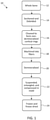
- a process 10 for preparing the demineralized cortical fiber strips may include one or more of the following steps.
- Whole bone is obtained in step 12.
- the whole bones may include long bones, such as femur, tibia, humerus, radius and ulna, or other suitable bones.
- the bones may be obtained from human or other animal sources.
- the whole bones may be sectioned and debrided in step 14.
- the whole bones may be sectioned into cortical rings, for example, in lengths of about 10-50mm, about 20-40mm, about 20-30mm, or about 28-30mm.
- Each cortical ring central canal is then debrided or cleared in preparation for liquid processing.
- the cortical rings may be cleaned in step 16, prior to fiber generation.
- the cortical rings may be cleaned with chemical and/or physical cleaning procedures that reduce the bone marrow and lipid elements associated with normal human tissue.
- the cleaning procedure may use one or more liquid solutions for suitable time periods to ensure thorough cleaning.
- the cortical rings may be cleaned with a 0.1 % v/v sodium deoxycholate/sterile saline solution, a 3% hydrogen peroxide solution, a 70% isopropyl alcohol solution, warm sterile water, combinations of these, or other suitable solutions.
- obtaining a whole bone in step 12 comprises providing a whole bone.
- the whole bone may be provided for example by a cadaveric bone, e.g., from a bone bank, a tissue bank, or a bone graft.
- the bone graft may be autologous (i.e., bone harvested from the patient's own body, for example from the iliac crest), or synthetic.
- the non-demineralized cortical rings are machined into cortical fibers.
- the cortical fibers may be produced, for example, with a horizontal milling machine. In one embodiment, the cortical fibers are produced with an end mill having about a 0-30 degree helix angle.
- the cortical rings may be loaded onto the machine vise and fed across the cutting surface to produce the cortical fibers. In one embodiment, about two-thirds of the cortical ring wall thickness is milled, and the remaining one-third is discarded. It will be appreciated that the entire cortical wall thickness may be milled or any suitable portion thereof.
- the non-demineralized cortical rings may be machined into thin fibers, for example, of lengths of about 10-50mm, widths of about 0.5-2mm, and thicknesses of about 0.1-Imm. It will be appreciated that some variation in dimension is possible in the production of the fibers.
- the non-demineralized cortical fibers then undergo a demineralization process in step 20 to remove the mineral components of bone.
- the demineralization may include an acid treatment with a 0.6N-1 N hydrochloric acid treatment.
- the acid treatment may be followed by a buffer rinse to obtain a neutral pH (e.g., 6.8-7.4).
- a neutral pH e.g. 6.8-7.4
- the demineralized cortical fibers are suspended, entangled, and compressed in a mold in step 22.
- the demineralized cortical fibers may be suspended in an aqueous solution.
- the aqueous solution may include water and an optional solute.
- the fibers may be suspended in sterile water, deionized water, distilled water, or purified water.
- the demineralized cortical fibers may be entangled naturally or by agitation.
- the entangled solution of fibers is compressed in a mold.
- the mold may contain drain holes for the removal of excess water.
- Each mold cavity may be packed at a 1:1 to 2:1 ratio of cortical fiber to mold volume (cc's).
- cc's cortical fiber to mold volume
- the dried allograft may be reconstituted at time of use, for example, in less than 5 minutes.
- the allograft may be reconstituted with an acceptable sterile irrigant (e.g., saline).
- the dried allograft may also be reconstituted with a cellular material such as chondrocytes, red blood cells, white blood cells, platelets, blood plasma, bone marrow cells, mesenchymal stem cells, pluripotential cells, osteoblasts, osteoclasts, and fibroblasts, epithelial cells, and endothelial cells.
- the properties of the demineralized cortical fiber allograft maintain structural integrity, pliability, and compressibility at lower cortical fiber densities than other compositions.
- the process produces consistent densities and mechanical properties per product.
- the cortical fiber structure provides a larger surface area than cancellous bone and other demineralized bone matrix products with cancellous or cortical fiber chips making the cortical fiber allograft a superior osteoconductive scaffold than other allografts.
- the bone graft is a mixture of osteoinductive demineralized cortical bone fibers and osteoconductive cortico-cancellous chips.
- Autograft obtained from the patient, may be desirable for the generation of new bone. Due to limited availability and donor site morbidity, however, there are drawbacks to autograft. Accordingly, the development of matrices that possess osteoconductive and osteoinductive components to promote bone regeneration are desirable in the area of bone repair.
- the demineralized bone matrix has osteoinductive properties due to intrinsic growth factors, such as bone morphogenic proteins (BMP), in the bone.
- BMP bone morphogenic proteins
- a scaffold with osteoconductive and osteoinductive properties is used to aid in bone regeneration.
- the demineralized cortical fibers allow for increased surface area of the product, which promotes increased cell adhesion and interaction with growth factors following implantation.
- a process 30 for preparing the demineralized cortical fiber and non-demineralized cortico-cancellous bone graft may include one or more of the following steps.
- Whole bone is obtained in step 32.
- the whole bones such as long bones including femur, tibia, radius and ulna, or other suitable bones may be processed.
- the bones may be obtained from human or other animal sources.
- the condyles of the long bones are separated from the cortical shafts.
- the cortical shafts are completely cleaned to remove any fat tissue and marrow. Cortical shafts are subsequently sectioned into cortical rings.
- the condyles are cut into smaller wedges and ground, for example, using a bone mill to produce cortico-cancellous chips and/or cancellous chips.
- the size of the cortico-cancellous chips may be less than about 1mm, about 100 ⁇ m1mm, or about 100-500 ⁇ m.
- the product may or may not include cortico-cancellous chips.
- the cortical rings are machined into cortical fibers.
- the cortical fibers are then demineralized in step 40.
- the cortical fibers may be demineralized using an acid extraction, for example, with 0.5-1 N hydrochloric acid.
- the non-demineralized cortico-cancellous chips and the demineralized cortical fibers are combined.
- the mixture may include demineralized cortical fibers in an amount of about 50-100%, about 66-100%, about 66-80%, about 60-70% or about 66%.
- the mixture may include cancellous chips in an amount of about 0-50%, about 0-33%, about 20-33%, about 30-40%, or about 33%.
- the formulation includes demineralized cortical fibers and non-demineralized cortico-cancellous chips premixed at a 2:1 ratio.
- obtaining a whole bone in step 32 comprises providing a whole bone.
- the whole bone may be provided for example by a cadaveric bone, e.g., from a bone bank, a tissue bank, or a bone graft.
- the bone graft may be autologous (i.e., bone harvested from the patient's own body, for example from the iliac crest), or synthetic.
- the bone graft may be in the form of a putty, strip, or boat, for example.
- the putty, strip, or boat formulations may each use the same base mixture.
- the mixture is transferred to molds.
- the biomaterial composition may be added to the mold to form a molded biomaterial composition.
- the form or mold may be of any suitable size and shape to obtain the desired shaped implant or a portion thereof.
- the mold may be provided under a given pressure and temperature necessary to form a compressed implant.
- the composition may be compressed for a time and pressure sufficient to create the desired shaped implant or a portion thereof.
- the pressure exerted upon the biomaterial composition may cause the components to contact one another and adhere together.
- the mold may be provided under an elevated pressure (i.e., greater than atmospheric) sufficient to compress the biomaterial into a solid form.
- the composition may be placed in a mold with no compression. If desired, a pattern or design may be cut into or out of the molded implant to form other desired shapes.
- the composition is freeze-dried. Freeze-drying may include freezing the biomaterial (e.g., in liquid nitrogen) and reducing the surrounding pressure to allow the frozen water in the material to sublimate directly from the solid phase to the gas phase.
- the dried allograft may be reconstituted, for example, with saline or a suitable cellular material.
- FIGS. 3A-3C show side and perspective views, respectively, of a strip 50.
- the strip 50 may be generally quadrilateral in shape (e.g., rectangular).
- the resulting implant may be molded to have substantially planar side walls.
- the top and bottom bone engaging surfaces may be substantially flat or planar. It is also envisioned that the top and bottom, bone engaging surfaces may be curved, angled, or of any suitable shape to better fit the anatomy of the adjacent vertebrae. Alternatively, the bone engaging surfaces may include protrusions, teeth, or the like.
- FIGS. 4A-4F show top, side, bottom, front, and perspective views, respectively, of a boat 60.
- the boat 60 may have an indentation 62 in the center providing a raised periphery 64 around the edges.
- the indentation 62 in the center of the boat 60 may allow the surgeon to pack bone material therein (e.g., autologous bone).
- the strips and boats may come in lengths up to about 200mm, thicknesses of about 4-l0mm, and widths of about 10-25mm.
- the large indentation 62 in the center of the boat 60 may be about 2-10mm from each edge with a depth of about 2-5mm.
- the putty may come in lengths up to about 200mm, thicknesses of about 4-15mm, and widths of about 5-25mm.
- the biomaterial may be formed into a specific size and shape for a desired application.
- the implant may have a footprint suitable for cervical, thoracic, or lumbar applications.
- the implant may be shaped, for example, in the form of a strip, boat, ring, cylinder, plug, or the like.
- the implant may be provided with one or more openings or windows suitable to be filled with the biomaterials described herein or other graft materials known in the art.
- the implant may be used alone or in combination with a cage, frame, allograft, autograft, graft material, or other biomaterials known in the art.
- the implants may be suitable for an anterior, posterior, lateral, oblique, anterolateral, transforaminal approach, or other suitable approach known in the art.
- demineralized cortical fibers may provide a higher surface area for interactions with cells and the local environment.
- the demineralizing process exposes native growth factors in the fibers, which may help to promote bone repair.
- the cancellous chips provide additional scaffolding for cells to adhere to as well as additional bulking material for the product. The chips may help to resist compression of the product, mitigating the perception that volume utilized is less than it should be due to the compressive nature of the fibers.
- the components provide cohesive, putty-like handling characteristics for the putty form.
- the freeze-dried and entangled components allow for a product that maintains structural integrity when utilized, while also being flexible enough to match the contours of the bony anatomy at the application site.
- the bone graft is a mixture of demineralized fibers, non-demineralized and/or demineralized cortico-cancellous chips, and an optional carrier.
- the carrier may affect the overall handling of the material and may influence the safety, efficacy, and functionality of the material (e.g., osteoinductivity).
- the carrier is inert or enhances osteogenic, osteoinductive, osteoconductive, and/or osteostimulative properties of the composition.
- Suitable carriers, scaffolds, or additives may include, but are not limited to, poloxamers, hydrogels, phospholipids, carboxylmethylcellulose (CMC), glycerin, glycerol, polyethylene glycol (PEG), polylactic acid (PLA), polylactic-co-glycolic acid (PLGA), other copolymers of the same family, and combinations thereof.
- the carrier may include a hydrogel, including a reverse phase hydrogel or temperature sensitive hydrogel, such as a poloxamer (e.g., a PEO-PPO-PEO triblock copolymer).
- a poloxamer e.g., a PEO-PPO-PEO triblock copolymer
- the poloxamer may include poloxamer 407, poloxamer P188, poloxamer P338, or the like.
- the poloxamer may also be chemically modified, for example, where one or more of the terminal hydroxyl groups are replaced with methoxy groups.
- Other suitable materials may include hyaluronic acid (HA), sodium alginate, saline or bone marrow aspirate, for instance.
- the carrier, scaffold materials, or processing additives may be either water-based or non-water based.
- the materials may be swellable in volume.
- the carrier e.g., poloxamer
- the carrier may be mixed with water, a buffer, or an acid, such as hydrochloric acid, nitric acid, sulfuric acid, or the like, which causes the carrier to swell in volume.
- swelling of the hydrogel may be influenced by a number of factors, such as temperature, surface area, molecular weight, degree of crosslinking, pH, or the like.
- a process 70 for preparing the bone graft in the form of a gel, putty, crunch, or strip may include one or more of the following steps.
- Whole bone is obtained in step 72.
- the whole bones such as long bones including femur, tibia, humerus, radius and ulna, or other suitable bones may be processed.
- the bones may be obtained from human or animal sources.
- the condyles of the long bones are separated from the cortical shafts.
- the cortical shafts are completely cleaned to remove any fat tissue and marrow and subsequently sectioned into cortical rings.
- the condyles and/or the cortical rings may undergo a liquid processing.
- the liquid processing may include one or more of an antibiotic/antimycotic solution for 10-15minutes, 0.05-0.15% v/v sodium deoxycholate solution for 15-35 minutes, 2-6% (w/v) hydrogen peroxide solution for 15-35 minutes, 70% isopropyl alcohol for 5-15 minutes, intermediate rinse steps using sterile water, combinations of these, or other suitable solutions.
- the condyles are cut into smaller wedges and ground, for example, using a bone mill to produce cortico-cancellous chips and/or cancellous chips.
- the size of the cortico-cancellous chips may be less than about 1.5mm, about 100 ⁇ m-1.5mm, about 500-1.5mm, about 100 ⁇ m-1 mm, or about 100-500 ⁇ m.
- the cortico-cancellous chips may have a unimodal or bimodal distribution.
- the cortico-cancellous chips may be divided into two size distributions: a first size of 100-500 ⁇ m and a second size of 500-1500 ⁇ m.
- the product may or may not include cortico-cancellous chips.
- the cortical rings are machined into cortical fibers.
- the cortical rings may be put through a fiber generator to produce fibers, for example, about 10-50mm, about 20-40mm, or about 15-30 mm in length.
- the fibers and optionally the chips are then demineralized in step 80.
- the fibers and/or chips may be demineralized using an acid extraction, for example, with 0.5-1N hydrochloric acid for about 15-40 minutes.
- the hydrochloric acid may be decanted and the fibers/chips may be rinsed in 25/75 mono-dibasic potassium phosphate solution and/or deionized water until the pH is between 6.5 and 7.5.
- the allograft may be produced as a gel form.
- the gel may include demineralized cortical-cancellous bone chips mixed with a carrier in step 84.
- the carrier may include a reverse phase hydrogel carrier solution.
- the bone chips may be about 100-500 ⁇ m in size.
- the carrier may be prepared as a 35% (w/v) solution with a thickening material, such as poloxamer 407, glycerol, carboxymethyl cellulose, or sodium hyaluronate.
- the carrier material may compose about 40-70% of the final product.
- the gel product is in an extrudable form.
- a putty may be formed.
- the putty may include demineralized cortical fibers, non-demineralized cortico-cancellous chips, and optionally demineralized cortico-cancellous chips mixed with a carrier in step 88.
- the carrier may include a reverse phase hydrogel carrier.
- the carrier may be prepared as a 25-45% (w/v) solution with a thickening material, such as poloxamer 407, glycerol, carboxymethyl cellulose, or sodium hyaluronate.
- the putty may include about 40-70% demineralized cortical fibers, about 20-40% cancellous chips (e.g., 100-500 ⁇ m), and about 10-40% carrier solution.
- the allograft may be produced in a crunch form.
- the crunch may include demineralized cortical fibers, non-demineralized cortico-cancellous chips, and optionally demineralized cortico-cancellous chips mixed with a carrier in step 92.
- the carrier may include a reverse phase hydrogel carrier.
- the carrier may be prepared as a 25-45% (w/v) solution with a thickening material, such as poloxamer 407, glycerol, carboxymethyl cellulose, or sodium hyaluronate.
- the product may include about 40-70% cortical fibers, about 10-30% cancellous chips (e.g., 100-500 ⁇ m), about 10-30% cancellous chips (e.g., 500-1500 ⁇ m) and about 10-40% carrier solution.
- the crunch composition may include a bimodal distribution of the cortico-cancellous chips.
- a strip may be formed.
- the strip may include demineralized cortical fibers and cortico-cancellous chips (e.g., 100-500 ⁇ m) in step 96.
- the fibers and chips may be premixed in a 3:1 ratio.
- the mix may be transferred to molds.
- the molds may be freeze dried.
- the composition may be dehydrated or dried, for example, by air or by freeze-drying. Freeze-drying may include freezing the material (e.g., in liquid nitrogen) and reducing the surrounding pressure to allow the frozen water in the material to sublimate directly from the solid phase to the gas phase.
- the freeze-dried strips may be partially of fully crosslinked.
- crosslinking may occur by exposing the material to a chemical crosslinking agent including mono aldehydes such as formaldehyde, acetaldehyde, or glutaraldehyde.
- the strips may be cross-linked with with a 0.25% glutaraldehyde or formaldehyde solution for about one minute, followed by rinsing with water, and additional freeze drying.
- Chemical crosslinking may also include exposure to a chemical sterilizing agent, such as ethylene oxide, nitrogen dioxide, ozone, or the like.
- Radiation crosslinking may include exposing the material to a sterilizing source such as gamma, ultraviolet, x-ray, electron irradiation, and/or thermal sources.
- a sterilizing source such as gamma, ultraviolet, x-ray, electron irradiation, and/or thermal sources.
- the strips may be cross-linked through the terminal sterilization process, in which the product is subjected to about 25-35 kGy of E-beam sterilization.
- the demineralized cortical fibers may provide a higher surface area for interactions with cells and the local environment.
- the demineralizing process exposes native growth factors in the fibers which may promote bone repair.
- the cancellous chips provide additional scaffolding for cells to adhere to as well as additional bulking material for the product. The chips may help to resist compression of the product, mitigating the perception that volume utilized is lessened due to the compressive nature of the fibers.
- the synergy of the components provides cohesive, putty-like handling characteristics for the putty and crunch forms.
- the cross-linked components allow for a product that maintains structural integrity when utilized, while also being flexible enough to match the contours of the bony anatomy at the application site.
- biological agents may be added to any of the biomaterials or implants described herein. These biological agents may comprise bone morphogenic protein (BMP), peptides, bone growth factors such as platelet derived growth factor (PDGF), vascular endothelial growth factor (VEGF), insulin derived growth factor (IDGF), a keratinocyte derived growth factor (KDGF), or a fibroblast derived growth factor (FDGF), stem cells, bone marrow, and platelet rich plasma (PRP), to name a few.
- BMP bone morphogenic protein
- PDGF platelet derived growth factor
- VEGF vascular endothelial growth factor
- IDGF insulin derived growth factor
- KDGF keratinocyte derived growth factor
- FDGF fibroblast derived growth factor
- stem cells bone marrow, and platelet rich plasma (PRP)
- PRP platelet rich plasma
- one or more active pharmaceutical ingredients or medicaments may be incorporated into or added to the biomaterial or implant as well.
- Biological agents
- the biomaterials and implants formed therefrom described herein are intended to be applied at a bone repair site, e.g., one resulting from injury or defect.
- the implants can be utilized in a wide variety of orthopedic, periodontal, neurosurgical, oral and maxillofacial surgical procedures.
- the biomaterials may be suitable for repairs of the vertebral column including spinal fusion and internal fixation; tumor surgery, e.g., deficit filling; discectomy; laminectomy; scoliosis, lordosis and kyphosis treatments.
- Possible clinical applications may include e.g., the treatment of spinal disc degeneration or disease, traumatic, pathologic, or stress fractures, congenital defects or fractures, or operative defects in any bone or between bones of the body.
- compositions and implants may be configured for use at various target repair sites within a body of a patient to facilitate bone growth therein.
- the composition is configured for use at a target repair site in the patient's spine.
- the composition can facilitate growth of bone between the body of a first vertebra and the body of a second vertebra to achieve interbody fusion of the two vertebrae.
- the composition may be used in conjunction with one or more mechanical supports (e.g., a cage or frame, spacer, plate, a plurality of screws and/or rods, or the like).
- the spine is described, the composition can be configured to be implanted into or at a target repair site in or at a different bone or bony structure of the patient's body.
- treating and the phrases “treatment of a disease” and “treatment of a condition” refer to executing a protocol that may include the use of the compositions, devices and methods herein and/or administering one or more biomaterials to a patient (human, normal or otherwise, or other mammal), in an effort to alleviate signs or symptoms of the disease or condition. Alleviation can occur prior to signs or symptoms of the disease or condition appearing, as well as after their appearance. Thus, “treating” or “treatment” includes “preventing” or “prevention” of disease or undesirable condition. In addition, “treating” or “treatment” does not require complete alleviation of signs or symptoms and does not require a cure to the ailment.
- kits that include components for making the present biomaterials and implants, including for example, carriers or scaffolds, cages (e.g., titanium and/or polyether ether ketone (PEEK) spacers), allograft spacers, demineralized bone materials, cell culture media, phosphate buffered saline (PBS), a tissue culture substrate such as a flask, trypsin, or mixtures, bone graft harvesting tools, bone marrow aspirate retrieval tools, or the like. Additional components, instructions and/or apparatus may also be included.
- cages e.g., titanium and/or polyether ether ketone (PEEK) spacers
- PEEK polyether ether ketone
- allograft spacers demineralized bone materials
- cell culture media phosphate buffered saline (PBS)
- PBS phosphate buffered saline
- tissue culture substrate such as a flask, trypsin, or mixtures
- This example demonstrates how to make bone grafts that include only demineralized cortical fibers.
- the whole bone is sectioned and debrided to form cortical rings.
- the cortical rings are cleaned and machined into cortical fibers having lengths of 10-50mm, widths of 0.5-2mm, and thicknesses of 0.1-lmm. Two-thirds of the cortical ring wall thickness is milled, and the remaining one-third of the cortical ring wall is discarded.
- the fibers are demineralized using a 0.6N hydrochloric acid treatment followed by a buffer rinse to obtain a neutral pH (6.8-7.4). Once demineralized, the cortical fibers are suspended in sterile water and entangled with one another.
- the entangled fibers are compressed in a mold and drain holes in the mold allow any excess water to drain.
- the mold is packed at a 2:1 ratio (cc) of cortical fiber to mold volume.
- the packed molds are frozen and freeze-dried to obtain the allograft.
- This example demonstrates how to make bone grafts that include demineralized cortical fibers and non-demineralized cortico-cancellous chips.
- the whole bone is separated into condyles and cortical shafts.
- the condyles are ground into cortico-cancellous chips of 100-500 ⁇ m.
- the cortical shafts are cleaned and machined into cortical fibers having lengths of 15-30mm, widths of 0.1-2mm, and thicknesses of 0.1-lmm.
- the fibers are demineralized using a 0.5-1.0N hydrochloric acid treatment for 15-40 minutes followed by a buffer rinse to obtain a neutral pH.
- the demineralized cortical fibers are mixed with the non-demineralized cortico-cancellous chips in a ratio of 2:1.
- the mixture is transferred to molds and freeze-dried to obtain the allograft.
- the allograft may be in strip, boat, or putty form.
- This example demonstrates how to make bone grafts that include demineralized cortical fibers and/or cortico-cancellous chips with an optional carrier.
- the whole bone is separated into condyles and cortical shafts.
- the condyles are ground into cortico-cancellous chips with a first size of 100-500 ⁇ m and a second size of 500-1500 ⁇ m.
- the cortical shafts are cleaned and machined into cortical fibers having lengths of 15-30mm, widths of 0.1-2mm, and thicknesses of 0.1-lmm.
- the fibers and optionally the chips are demineralized using a 0.5-0.7N hydrochloric acid treatment for 15-40 minutes.
- the hydrochloric acid is decanted and the fibers/chips are rinsed in 25/75 mono-dibasic potassium phosphate solution and/or deionized water until the pH is 6.5-7.5.
- the gel, putty, crunch, and strip formulations are as follows: Gel Putty Crunch Strip Demineralized cortical fibers - 40-70% 40-70% 75% Non-demineralized cortico-cancellous chips (100-500 ⁇ m) - 20-40% 10-30% 25% Non-demineralized cortico-cancellous chips (500-1500 ⁇ m) - - 10-30% - Demineralized cortico-cancellous chips (100-500 ⁇ m) 30-60% - - - Carrier 40-70% (35% (w/v) solution with poloxamer 407, glycerol, carboxymethyl cellulose, or sodium hyaluronate) 10-40% (25-45% (w/v) solution with poloxamer 407,
- the strip is molded, freeze-dried, and cross-linked with 0.25% glutaraldehyde or formaldehyde for one minute followed by rinsing with water and freeze drying.
- the cross-linked strip provides for structural integrity while still being flexible.
- the gel is extrudable, and the putty and crunch have putty-like handling characteristics.
Landscapes
- Health & Medical Sciences (AREA)
- Life Sciences & Earth Sciences (AREA)
- Chemical & Material Sciences (AREA)
- Engineering & Computer Science (AREA)
- Biomedical Technology (AREA)
- Transplantation (AREA)
- Oral & Maxillofacial Surgery (AREA)
- Veterinary Medicine (AREA)
- Public Health (AREA)
- General Health & Medical Sciences (AREA)
- Animal Behavior & Ethology (AREA)
- Chemical Kinetics & Catalysis (AREA)
- Epidemiology (AREA)
- Dermatology (AREA)
- Botany (AREA)
- Medicinal Chemistry (AREA)
- Molecular Biology (AREA)
- Orthopedic Medicine & Surgery (AREA)
- Zoology (AREA)
- Urology & Nephrology (AREA)
- Vascular Medicine (AREA)
- Cardiology (AREA)
- Heart & Thoracic Surgery (AREA)
- Hematology (AREA)
- Physical Education & Sports Medicine (AREA)
- General Chemical & Material Sciences (AREA)
- Cell Biology (AREA)
- Materials For Medical Uses (AREA)
- Medicinal Preparation (AREA)
- Medicines Containing Material From Animals Or Micro-Organisms (AREA)
Abstract
Biomaterials, implants made therefrom, methods of making the biomaterial and implants, methods of promoting bone or wound healing in a mammal by administering the biomaterial or implant to the mammal, and kits that include such biomaterials, implants, or components thereof. The biomaterials may include demineralized cortical fibers, which help to promote bone repair.
Description
- The present application relates generally to bone and wound healing biomaterials and implants formed therefrom, methods of making the biomaterials and implants, methods of promoting bone or wound healing in a mammal, and kits that include one or more of the biomaterials, implants, or components thereof.
- Bone grafting is a surgical procedure that replaces missing bone and/or repairs bone. Bone generally has the ability to regenerate but may require a scaffold to do so. Bone grafts may be allograft (cadaveric bone e.g., from a bone bank), autologous (i.e., bone harvested from the patient's own body, for example from the iliac crest), or synthetic. The bone grafts may be resorbed and replaced as the natural bone heals over time.
- Biomaterials may include osteoconduction (guiding the reparative growth of the natural bone), osteoinduction (encouraging undifferentiated cells to become active osteoblasts), osteostimulative (active stimulation of osteoblast proliferation and differentiation) and/or osteogenesis (living bone cells in the graft material contributing to bone remodeling). Although traditional bone grafts may exhibit certain advantages, traditional allograft may not exhibit the properties desired, may be difficult to obtain, or may not be in a shape or form suitable for implantation.
- To meet this and other needs, biomaterials described herein may be advantageous for bone healing and repair and without the drawbacks of present allograft or autograft products. The biomaterial compositions or implants prepared therefrom may include demineralized bone matrix (e.g., in the form fibers) alone or combined with additional components. If desired, the biomaterial composition may also include a carrier such as poloxamer, glycerol, carboxymethyl cellulose, or sodium hyaluronate.
- According to one embodiment, a bone graft or implantable biomaterial for aiding bone regeneration includes only entangled and compressed demineralized cortical fibers. The bone graft may be produced by obtaining whole bone, debriding and sectioning the whole bone shafts into cortical rings, machining the cortical rings into cortical fibers having lengths of 10-50mm, widths of 0.5-2mm, and thicknesses of 0.1-1mm, demineralizing the cortical fibers, suspending and entangling the demineralized cortical fibers in an aqueous solution, compressing the demineralized cortical fibers in a mold for a time and with pressure sufficient to form a molded biomaterial composition of a given shape, freezing the molded biomaterial composition, and freeze-drying the molded biomaterial composition to form the implantable biomaterial. Any excess water present following suspension of demineralized cortical fibers may be drained from the bone graft before, during and/or after the molding process.
- The cortical rings may be sectioned into lengths of 10-30mm. The cortical rings may be cleaned with a 0.1 % v/v sodium deoxycholate/sterile saline solution, a 3% hydrogen peroxide solution, a 70% isopropyl alcohol solution, and warm sterile water. Two-thirds of the cortical ring wall thickness may be machined into fibers and the remaining one-third may be discarded. The fibers may be demineralized using an acid treatment with a 0.6N-1 N hydrochloric acid treatment followed by a buffer rinse to obtain a neutral pH. The demineralized cortical fibers may be suspended in sterile water and entangled under agitation. The cortical fibers may be compressed in the mold and drain holes in the mold may allow for drainage of excess water. The mold may be packed at a 1:1 to 2:1 ratio, in cubic centimeters, of demineralized cortical fiber to mold volume. The loaded mold may be frozen and subsequently freeze-dried to produce the final product. Prior to use, the dried implantable biomaterial may be reconstituted with saline or a cellular material, such as red blood cells, white blood cells, platelets, blood plasma, and/or bone marrow cells.
- According to another embodiment, a bone graft or implantable biomaterial for aiding bone regeneration includes only a mixture of about 66% demineralized cortical fibers having lengths of 15-30 mm and about 33% non-demineralized cortico-cancellous chips having a size less than 1mm (e.g., a ratio of 2:1 fibers to chips). The bone graft may be produced by providing a whole bone, debridement of soft tissues, sectioning the whole bone into condyles and cortical shafts, grinding the condyles into cortico-cancellous chips having a size less than 1mm, sectioning cortical shafts into cortical rings and machining the cortical rings into cortical fibers, demineralizing the cortical fibers, combining the cortico-cancellous chips and demineralized cortical fibers in a mixture including about 66% demineralized cortical fibers and about 33% non-demineralized cortico-cancellous chips, molding the demineralized fibers in a mold to form a molded biomaterial composition of a given shape, optionally freezing the filled mold, and freeze-drying the molded biomaterial composition to form the implantable biomaterial.
- According to another embodiment, the bone graft may be in the form of a gel, putty, crunch, or strip. The gel may include a combination of cortico-cancellous chips and reverse phase hydrogel carrier. The gel may include 30-60% demineralized cortico-cancellous chips and 40-70% carrier. The putty and crunch may include a combination of cortico-cancellous chips, demineralized cortical fibers, and reverse phase hydrogel carrier. The putty may include 40-70% demineralized cortical fibers, 20-40% cancellous chips in a size range of 100-500µm, and 10-40% carrier solution. The crunch may include 40-70% cortical fibers, 10-30% cancellous chips in a size range of 100-500µm, 10-30% cancellous chips in a size range of 500-1500µm, and 10-40% carrier solution. The strip may include cortico-cancellous chips and demineralized cortical fibers. The strip may include 75% demineralized cortical fibers and 25% cortico-cancellous chips having a size in a range of 100-500µm. The strip may be freeze-dried and crosslinked. For example, the strips may be cross-linked with a 0.25% glutaraldehyde or formaldehyde solution for about one minute.
- The condyles and cortical shafts may undergo a liquid processing including an antibiotic/antimycotic solution for 10-15minutes, 0.05-0.15% v/v sodium deoxycholate solution for 15-35 minutes, 2-6% (w/v) hydrogen peroxide solution for 15-35 minutes, and 70% isopropyl alcohol for 5-15 minutes with intermediate rinse steps using sterile water. The cortico-cancellous chips may have a unimodal or bimodal distribution (e.g., 100-500µm and 500-1500µm). The carrier may be a 25-45% (w/v) solution with poloxamer, glycerol, carboxymethyl cellulose, or sodium hyaluronate. For example, the carrier may be a 35% (w/v) solution with poloxamer.
- According to yet another embodiment, a method of promoting bone or wound healing in a mammal includes providing a compressed, freeze dried implantable biomaterial comprised of demineralized bone in fiber form; and administering the implantable biomaterial into a target repair site to facilitate repair or regeneration of bone at the target repair site. The target repair site may include an injury or defect in the spine. The implantable biomaterial may form a standalone implant configured to be positioned between adjacent vertebrae or may be positioned in or near a cage or other suitable interbody device.
- According to yet another embodiment, a kit includes one or more biomaterials, implants, or components thereof described herein. For example, the kit may contain putty, gel, strip, crunch, and/or boat versions of the biomaterial compositions. The kit may contain biomaterial compositions of the same or different types. In addition, the kit may include other components known in the art, including, but not limited to, carriers or scaffolds, cages or interbody devices (e.g., titanium and/or polyether ether ketone (PEEK) spacers), allograft spacers, cell culture media, phosphate buffered saline (PBS), a tissue culture substrate, bone graft harvesting tools, bone marrow aspirate retrieval tools, or the like.
- A more complete understanding of the present invention, and the attendant advantages and features thereof, will be more readily understood by reference to the following detailed description when considered in conjunction with the accompanying drawings, wherein:
-
FIG. 1 shows a process flowchart of the steps for producing a demineralized cortical fiber allograft according to one embodiment; -
FIG. 2 shows a process flowchart of the steps for producing a demineralized cortical fiber and non-demineralized cortico-cancellous chip allograft according to one embodiment; -
FIGS. 3A-3C depict an allograft in strip form according to one embodiment; -
FIGS. 4A-4F depict an allograft in boat form according to one embodiment; and -
FIG. 5 shows a process flowchart of the steps for producing allografts in gel, putty, crunch, or strip form according to one embodiment. - The present application relates generally to biomaterials and implants made therefrom, methods of making the biomaterial and implants, methods of promoting bone or wound healing in a mammal by administering the biomaterial or implant to the mammal, and kits that include one or more of the biomaterials, implants, or components thereof are also provided.
- Additional aspects, advantages and/or other features of example embodiments of the invention will become apparent in view of the following detailed description. It should be apparent to those skilled in the art that the described embodiments provided herein are merely exemplary and illustrative and not limiting. Numerous embodiments of modifications thereof are contemplated as falling within the scope of this disclosure and equivalents thereto.
- In describing example embodiments, specific terminology is employed for the sake of clarity. However, the embodiments are not intended to be limited to this specific terminology. Unless otherwise noted, technical terms are used according to conventional usage.
- As used herein and in the claims, the terms "comprising" and "including" are inclusive or open-ended and do not exclude additional unrecited elements, compositional components, or method steps. Accordingly, the terms "comprising" and "including" encompass the more restrictive terms "consisting essentially of' and "consisting of."
- Unless specified otherwise, all values provided herein include up to and including the endpoints given, and the values of the constituents or components of the compositions are expressed in weight percent or % by weight of each ingredient in the composition.
- Each compound used herein may be discussed interchangeably with respect to its chemical formula, chemical name, abbreviation, etc. For example, PEG may be used interchangeably with polyethylene glycol.
- Embodiments described herein may be generally directed to biomaterials, implants made therefrom, methods of making the same, and methods of using the same to promote healing or fusion of bone. Although biomaterials or implants may be discussed separately, it will be appreciated by one of ordinary skill in the art that the biomaterials described may be used in and of itself or may be used to create implants of different shapes, sizes, and orientations for a number of different clinical outcomes. Thus, the discussion of biomaterials may apply equally to the discussion on implants and vice versa.
- The biomaterial composition may be osteoconductive, osteoinductive, osteogenic, and/or osteostimulative, which may be advantageous for bone healing and repair. The biomaterials may be osteoconductive when the material serves as a scaffold that provides surface area for new bone growth. The biomaterials may be osteoinductive if they stimulate osteoprogenitor cells or induce mesenchymal stem cells to differentiate into osteoblasts that then begin new bone formation. Biomaterials may be osteogenic if they contain cells (e.g., viable cells) that are capable of bone regeneration. The biomaterial may be osteostimulative if the material accelerates the bone formation process.
- The composition may be "biocompatible" as that term refers to the ability (e.g., of a composition or material) to perform with an appropriate host response in a specific application, or at least to perform without having a toxic or otherwise deleterious effect on a biological system of the host, locally or systemically. The biomaterial and/or implant may be "biologically degradable" in that the material may be degraded by cellular absorption and/or hydrolytic degradation in a patient's body. According to some embodiments, it may be desirable that the biomaterials possess sufficient osteoconductivity, porosity, mechanical strength, and degradation times.
- According to one embodiment, the biomaterial composition may be configured to facilitate repair or regeneration of bone at a target repair site. The target repair site can be, for example, a void, gap, or other defect or surgeon created opening in a bone, between bones, or other bony structure in a body of a patient. For example, the biomaterial composition can be configured to facilitate bone growth at a target repair site in the spine, pelvis, an extremity, the cranium, or another bone, between bones, or bony structure in the patient's body. The biomaterial composition may be configured to be directly implanted or otherwise disposed at or in contact with the target repair site.
- The biomaterial composition may include demineralized bone matrix (e.g., in the form of fibers, chips, or particulates), and optionally one or more additional components each of which is described in more detail herein. Demineralized bone matrix (also known as DBM) may provide osteoconductive, osteoinductive and/or osteogenic properties. Thus, it induces the formation of bone tissue. As used herein, the terms "demineralized bone", "demineralized bone matrix", and "DBM" may be used interchangeably. The demineralized bone, for example, in the form of fibers, chips, and/or particles, can be disposed on, embedded within, and/or mixed within the biomaterial composition.
- Demineralized bone matrix may be in the form of fibers, threads, strips, chips, shards, sheets, elongated particles, powder, or particulates, for example. The demineralized bone matrix may include bone pieces of varying shapes, sizes, thickness, and configurations that possess regular, irregular, or random geometries. In some embodiments, the bone used to manufacture the demineralized bone matrix may be cortical, cancellous, cortico-cancellous of autogenous, allogeneic, xenogeneic or transgenic in origin. Thus, the fibers, chips, or particulates, for example, may include cortical, cancellous, or cortico-cancellous bone unless specified otherwise.
- To prepare bone matrix, the bone material may be treated to clean, defat, sterilize, virally inactivate, disinfect, demineralize, dehydrate, and/or dry the bone matrix. Methods for preparing demineralized bone matrix may include, but are not limited to, shaving bone into thin shavings or fibers, milling, grinding, or crushing bone into chips or particles, or the like. Before or after processing the bone, the bone material may be subjected to demineralization so as to reduce inorganic content to low levels. For example, demineralized bone may be produced by acid extraction, thermal freezing, irradiation, or physical extraction of inorganic minerals from human or animal bone. In an acid extraction, inorganic acids such as hydrochloric acid or phosphoric acid, or organic acids such as formic acid, acetic acid, peracetic acid, citric acid, propionic acid, etc. may be used. As would be recognized by one of ordinary skill in the art, the amount and depth of demineralization into the bone surface can be controlled by adjusting the treatment time, temperature of the demineralizing solution, concentration of the demineralizing solution, agitation intensity during treatment, and the like.
- The term "demineralized" refers to bone or bone material containing less than its original mineral content (e.g., calcium content) and may encompass "substantially demineralized," "partially demineralized," and "completely demineralized" bone material. For example, the demineralized bone may include less than 10%, less than 9%, less than 8%, less than 7%, less than 6%, less than 5%, less than 4%, less than 3%, less than 2%, or less than 1% of the original mineral content (e.g., calcium content) of the bone.
- According to one embodiment, the demineralized bone matrix includes only demineralized cortical bone fibers that are suspended, entangled and compressed to form demineralized cortical fiber strips. The process produces an allograft with ideal handling characteristics for use in surgical applications, for example, as bone void filler, without the need for chemical crosslinking or an added carrier. The demineralized cortical bone fiber allograft may be in the form of a molded strip, which maintains structural integrity, pliability, compressibility, osteoconductivity, and osteoinductive potential. The demineralized cortical fiber strips also provide a high surface area scaffold for potential cellular adhesion and migration due to the physical dimensions of the cortical fibers within the molded allograft.
- Referring now to
FIG. 1 , aprocess 10 for preparing the demineralized cortical fiber strips may include one or more of the following steps. Whole bone is obtained instep 12. The whole bones may include long bones, such as femur, tibia, humerus, radius and ulna, or other suitable bones. The bones may be obtained from human or other animal sources. The whole bones may be sectioned and debrided instep 14. The whole bones may be sectioned into cortical rings, for example, in lengths of about 10-50mm, about 20-40mm, about 20-30mm, or about 28-30mm. Each cortical ring central canal is then debrided or cleared in preparation for liquid processing. The cortical rings may be cleaned instep 16, prior to fiber generation. For example, the cortical rings may be cleaned with chemical and/or physical cleaning procedures that reduce the bone marrow and lipid elements associated with normal human tissue. The cleaning procedure may use one or more liquid solutions for suitable time periods to ensure thorough cleaning. For example, the cortical rings may be cleaned with a 0.1 % v/v sodium deoxycholate/sterile saline solution, a 3% hydrogen peroxide solution, a 70% isopropyl alcohol solution, warm sterile water, combinations of these, or other suitable solutions. - It is considered that obtaining a whole bone in
step 12 comprises providing a whole bone. The whole bone may be provided for example by a cadaveric bone, e.g., from a bone bank, a tissue bank, or a bone graft. The bone graft may be autologous (i.e., bone harvested from the patient's own body, for example from the iliac crest), or synthetic. - In
step 18, the non-demineralized cortical rings are machined into cortical fibers. The cortical fibers may be produced, for example, with a horizontal milling machine. In one embodiment, the cortical fibers are produced with an end mill having about a 0-30 degree helix angle. The cortical rings may be loaded onto the machine vise and fed across the cutting surface to produce the cortical fibers. In one embodiment, about two-thirds of the cortical ring wall thickness is milled, and the remaining one-third is discarded. It will be appreciated that the entire cortical wall thickness may be milled or any suitable portion thereof. Instep 18, the non-demineralized cortical rings may be machined into thin fibers, for example, of lengths of about 10-50mm, widths of about 0.5-2mm, and thicknesses of about 0.1-Imm. It will be appreciated that some variation in dimension is possible in the production of the fibers. - The non-demineralized cortical fibers then undergo a demineralization process in
step 20 to remove the mineral components of bone. For example, the demineralization may include an acid treatment with a 0.6N-1 N hydrochloric acid treatment. The acid treatment may be followed by a buffer rinse to obtain a neutral pH (e.g., 6.8-7.4). Once demineralized, the demineralized cortical fibers are suspended, entangled, and compressed in a mold instep 22. The demineralized cortical fibers may be suspended in an aqueous solution. The aqueous solution may include water and an optional solute. For example, the fibers may be suspended in sterile water, deionized water, distilled water, or purified water. While in solution, the demineralized cortical fibers may be entangled naturally or by agitation. The entangled solution of fibers is compressed in a mold. The mold may contain drain holes for the removal of excess water. Each mold cavity may be packed at a 1:1 to 2:1 ratio of cortical fiber to mold volume (cc's). Instep 24, the packed molds are then frozen and placed into a freeze dryer to produce the final freeze-dried strip product. - The dried allograft may be reconstituted at time of use, for example, in less than 5 minutes. For example, the allograft may be reconstituted with an acceptable sterile irrigant (e.g., saline). The dried allograft may also be reconstituted with a cellular material such as chondrocytes, red blood cells, white blood cells, platelets, blood plasma, bone marrow cells, mesenchymal stem cells, pluripotential cells, osteoblasts, osteoclasts, and fibroblasts, epithelial cells, and endothelial cells.
- The properties of the demineralized cortical fiber allograft maintain structural integrity, pliability, and compressibility at lower cortical fiber densities than other compositions. The process produces consistent densities and mechanical properties per product. The cortical fiber structure provides a larger surface area than cancellous bone and other demineralized bone matrix products with cancellous or cortical fiber chips making the cortical fiber allograft a superior osteoconductive scaffold than other allografts.
- According to another embodiment, the bone graft is a mixture of osteoinductive demineralized cortical bone fibers and osteoconductive cortico-cancellous chips. Autograft, obtained from the patient, may be desirable for the generation of new bone. Due to limited availability and donor site morbidity, however, there are drawbacks to autograft. Accordingly, the development of matrices that possess osteoconductive and osteoinductive components to promote bone regeneration are desirable in the area of bone repair. The demineralized bone matrix has osteoinductive properties due to intrinsic growth factors, such as bone morphogenic proteins (BMP), in the bone. In this embodiment, a scaffold with osteoconductive and osteoinductive properties is used to aid in bone regeneration. The demineralized cortical fibers allow for increased surface area of the product, which promotes increased cell adhesion and interaction with growth factors following implantation.
- With reference to
FIG. 2 , aprocess 30 for preparing the demineralized cortical fiber and non-demineralized cortico-cancellous bone graft may include one or more of the following steps. Whole bone is obtained instep 32. The whole bones, such as long bones including femur, tibia, radius and ulna, or other suitable bones may be processed. The bones may be obtained from human or other animal sources. Instep 34, the condyles of the long bones are separated from the cortical shafts. The cortical shafts are completely cleaned to remove any fat tissue and marrow. Cortical shafts are subsequently sectioned into cortical rings. Instep 36, the condyles are cut into smaller wedges and ground, for example, using a bone mill to produce cortico-cancellous chips and/or cancellous chips. The size of the cortico-cancellous chips may be less than about 1mm, about 100µm1mm, or about 100-500µm. The product may or may not include cortico-cancellous chips. Instep 38, the cortical rings are machined into cortical fibers. The cortical fibers are then demineralized instep 40. The cortical fibers may be demineralized using an acid extraction, for example, with 0.5-1 N hydrochloric acid. Instep 42, the non-demineralized cortico-cancellous chips and the demineralized cortical fibers are combined. The mixture may include demineralized cortical fibers in an amount of about 50-100%, about 66-100%, about 66-80%, about 60-70% or about 66%. The mixture may include cancellous chips in an amount of about 0-50%, about 0-33%, about 20-33%, about 30-40%, or about 33%. In one embodiment, the formulation includes demineralized cortical fibers and non-demineralized cortico-cancellous chips premixed at a 2:1 ratio. - It is considered that obtaining a whole bone in
step 32 comprises providing a whole bone. The whole bone may be provided for example by a cadaveric bone, e.g., from a bone bank, a tissue bank, or a bone graft. The bone graft may be autologous (i.e., bone harvested from the patient's own body, for example from the iliac crest), or synthetic. - The bone graft may be in the form of a putty, strip, or boat, for example. The putty, strip, or boat formulations may each use the same base mixture. For the strip and boat, in
step 44, the mixture is transferred to molds. The biomaterial composition may be added to the mold to form a molded biomaterial composition. The form or mold may be of any suitable size and shape to obtain the desired shaped implant or a portion thereof. The mold may be provided under a given pressure and temperature necessary to form a compressed implant. The composition may be compressed for a time and pressure sufficient to create the desired shaped implant or a portion thereof. The pressure exerted upon the biomaterial composition may cause the components to contact one another and adhere together. The mold may be provided under an elevated pressure (i.e., greater than atmospheric) sufficient to compress the biomaterial into a solid form. Alternatively, the composition may be placed in a mold with no compression. If desired, a pattern or design may be cut into or out of the molded implant to form other desired shapes. Instep 46, the composition is freeze-dried. Freeze-drying may include freezing the biomaterial (e.g., in liquid nitrogen) and reducing the surrounding pressure to allow the frozen water in the material to sublimate directly from the solid phase to the gas phase. Prior to use, the dried allograft may be reconstituted, for example, with saline or a suitable cellular material. - The putty, strip, and boat may have a length, thickness, and width.
FIGS. 3A-3C show side and perspective views, respectively, of astrip 50. Thestrip 50 may be generally quadrilateral in shape (e.g., rectangular). The resulting implant may be molded to have substantially planar side walls. As shown inFIGS. 3A-3C , the top and bottom bone engaging surfaces may be substantially flat or planar. It is also envisioned that the top and bottom, bone engaging surfaces may be curved, angled, or of any suitable shape to better fit the anatomy of the adjacent vertebrae. Alternatively, the bone engaging surfaces may include protrusions, teeth, or the like.FIGS. 4A-4F show top, side, bottom, front, and perspective views, respectively, of aboat 60. Theboat 60 may have anindentation 62 in the center providing a raisedperiphery 64 around the edges. Theindentation 62 in the center of theboat 60 may allow the surgeon to pack bone material therein (e.g., autologous bone). The strips and boats may come in lengths up to about 200mm, thicknesses of about 4-l0mm, and widths of about 10-25mm. Thelarge indentation 62 in the center of theboat 60 may be about 2-10mm from each edge with a depth of about 2-5mm. The putty may come in lengths up to about 200mm, thicknesses of about 4-15mm, and widths of about 5-25mm. - The biomaterial may be formed into a specific size and shape for a desired application. For example, the implant may have a footprint suitable for cervical, thoracic, or lumbar applications. The implant may be shaped, for example, in the form of a strip, boat, ring, cylinder, plug, or the like. The implant may be provided with one or more openings or windows suitable to be filled with the biomaterials described herein or other graft materials known in the art. The implant may be used alone or in combination with a cage, frame, allograft, autograft, graft material, or other biomaterials known in the art. The implants may be suitable for an anterior, posterior, lateral, oblique, anterolateral, transforaminal approach, or other suitable approach known in the art.
- The use of demineralized cortical fibers may provide a higher surface area for interactions with cells and the local environment. The demineralizing process exposes native growth factors in the fibers, which may help to promote bone repair. The cancellous chips provide additional scaffolding for cells to adhere to as well as additional bulking material for the product. The chips may help to resist compression of the product, mitigating the perception that volume utilized is less than it should be due to the compressive nature of the fibers. Working together, the components provide cohesive, putty-like handling characteristics for the putty form. For the strip and boat, the freeze-dried and entangled components allow for a product that maintains structural integrity when utilized, while also being flexible enough to match the contours of the bony anatomy at the application site.
- According to another embodiment, the bone graft is a mixture of demineralized fibers, non-demineralized and/or demineralized cortico-cancellous chips, and an optional carrier. The carrier may affect the overall handling of the material and may influence the safety, efficacy, and functionality of the material (e.g., osteoinductivity). Preferably, the carrier is inert or enhances osteogenic, osteoinductive, osteoconductive, and/or osteostimulative properties of the composition. Suitable carriers, scaffolds, or additives may include, but are not limited to, poloxamers, hydrogels, phospholipids, carboxylmethylcellulose (CMC), glycerin, glycerol, polyethylene glycol (PEG), polylactic acid (PLA), polylactic-co-glycolic acid (PLGA), other copolymers of the same family, and combinations thereof.
- By way of example, the carrier may include a hydrogel, including a reverse phase hydrogel or temperature sensitive hydrogel, such as a poloxamer (e.g., a PEO-PPO-PEO triblock copolymer). In particular, the poloxamer may include poloxamer 407, poloxamer P188, poloxamer P338, or the like. The poloxamer may also be chemically modified, for example, where one or more of the terminal hydroxyl groups are replaced with methoxy groups. Other suitable materials may include hyaluronic acid (HA), sodium alginate, saline or bone marrow aspirate, for instance. The carrier, scaffold materials, or processing additives may be either water-based or non-water based.
- In the case of a hydrogel, such as a poloxamer, hyaluronic acid, or alginate, the materials may be swellable in volume. For example, the carrier (e.g., poloxamer) may be mixed with water, a buffer, or an acid, such as hydrochloric acid, nitric acid, sulfuric acid, or the like, which causes the carrier to swell in volume. As will be recognized by one of ordinary skill in the art, swelling of the hydrogel may be influenced by a number of factors, such as temperature, surface area, molecular weight, degree of crosslinking, pH, or the like.
- With reference to
FIG. 5 , aprocess 70 for preparing the bone graft in the form of a gel, putty, crunch, or strip may include one or more of the following steps. Whole bone is obtained instep 72. The whole bones, such as long bones including femur, tibia, humerus, radius and ulna, or other suitable bones may be processed. The bones may be obtained from human or animal sources. Instep 74, the condyles of the long bones are separated from the cortical shafts. The cortical shafts are completely cleaned to remove any fat tissue and marrow and subsequently sectioned into cortical rings. The condyles and/or the cortical rings may undergo a liquid processing. The liquid processing may include one or more of an antibiotic/antimycotic solution for 10-15minutes, 0.05-0.15% v/v sodium deoxycholate solution for 15-35 minutes, 2-6% (w/v) hydrogen peroxide solution for 15-35 minutes, 70% isopropyl alcohol for 5-15 minutes, intermediate rinse steps using sterile water, combinations of these, or other suitable solutions. Instep 76, the condyles are cut into smaller wedges and ground, for example, using a bone mill to produce cortico-cancellous chips and/or cancellous chips. The size of the cortico-cancellous chips may be less than about 1.5mm, about 100µm-1.5mm, about 500-1.5mm, about 100µm-1 mm, or about 100-500µm. The cortico-cancellous chips may have a unimodal or bimodal distribution. For example, the cortico-cancellous chips may be divided into two size distributions: a first size of 100-500µm and a second size of 500-1500µm. The product may or may not include cortico-cancellous chips. - In
step 78, the cortical rings are machined into cortical fibers. The cortical rings may be put through a fiber generator to produce fibers, for example, about 10-50mm, about 20-40mm, or about 15-30 mm in length. The fibers and optionally the chips are then demineralized instep 80. The fibers and/or chips may be demineralized using an acid extraction, for example, with 0.5-1N hydrochloric acid for about 15-40 minutes. At the end of the treatment, the hydrochloric acid may be decanted and the fibers/chips may be rinsed in 25/75 mono-dibasic potassium phosphate solution and/or deionized water until the pH is between 6.5 and 7.5. - In
option 82, the allograft may be produced as a gel form. The gel may include demineralized cortical-cancellous bone chips mixed with a carrier instep 84. The carrier may include a reverse phase hydrogel carrier solution. The bone chips may be about 100-500µm in size. The carrier may be prepared as a 35% (w/v) solution with a thickening material, such as poloxamer 407, glycerol, carboxymethyl cellulose, or sodium hyaluronate. The carrier material may compose about 40-70% of the final product. The gel product is in an extrudable form. - In
option 86, a putty may be formed. The putty may include demineralized cortical fibers, non-demineralized cortico-cancellous chips, and optionally demineralized cortico-cancellous chips mixed with a carrier in step 88. The carrier may include a reverse phase hydrogel carrier. The carrier may be prepared as a 25-45% (w/v) solution with a thickening material, such as poloxamer 407, glycerol, carboxymethyl cellulose, or sodium hyaluronate. The putty may include about 40-70% demineralized cortical fibers, about 20-40% cancellous chips (e.g., 100-500µm), and about 10-40% carrier solution. - In
option 90, the allograft may be produced in a crunch form. The crunch may include demineralized cortical fibers, non-demineralized cortico-cancellous chips, and optionally demineralized cortico-cancellous chips mixed with a carrier instep 92. The carrier may include a reverse phase hydrogel carrier. The carrier may be prepared as a 25-45% (w/v) solution with a thickening material, such as poloxamer 407, glycerol, carboxymethyl cellulose, or sodium hyaluronate. For the crunch formulation, the product may include about 40-70% cortical fibers, about 10-30% cancellous chips (e.g., 100-500µm), about 10-30% cancellous chips (e.g., 500-1500µm) and about 10-40% carrier solution. Thus, the crunch composition may include a bimodal distribution of the cortico-cancellous chips. - In
option 94, a strip may be formed. The strip may include demineralized cortical fibers and cortico-cancellous chips (e.g., 100-500µm) instep 96. For example, the fibers and chips may be premixed in a 3:1 ratio. Instep 98, the mix may be transferred to molds. Instep 100, the molds may be freeze dried. The composition may be dehydrated or dried, for example, by air or by freeze-drying. Freeze-drying may include freezing the material (e.g., in liquid nitrogen) and reducing the surrounding pressure to allow the frozen water in the material to sublimate directly from the solid phase to the gas phase. - In
step 102, the freeze-dried strips may be partially of fully crosslinked. For example, crosslinking may occur by exposing the material to a chemical crosslinking agent including mono aldehydes such as formaldehyde, acetaldehyde, or glutaraldehyde. For example, the strips may be cross-linked with with a 0.25% glutaraldehyde or formaldehyde solution for about one minute, followed by rinsing with water, and additional freeze drying. Chemical crosslinking may also include exposure to a chemical sterilizing agent, such as ethylene oxide, nitrogen dioxide, ozone, or the like. Radiation crosslinking may include exposing the material to a sterilizing source such as gamma, ultraviolet, x-ray, electron irradiation, and/or thermal sources. For example, the strips may be cross-linked through the terminal sterilization process, in which the product is subjected to about 25-35 kGy of E-beam sterilization. - The demineralized cortical fibers may provide a higher surface area for interactions with cells and the local environment. The demineralizing process exposes native growth factors in the fibers which may promote bone repair. The cancellous chips provide additional scaffolding for cells to adhere to as well as additional bulking material for the product. The chips may help to resist compression of the product, mitigating the perception that volume utilized is lessened due to the compressive nature of the fibers. The synergy of the components provides cohesive, putty-like handling characteristics for the putty and crunch forms. For the strip, the cross-linked components allow for a product that maintains structural integrity when utilized, while also being flexible enough to match the contours of the bony anatomy at the application site.
- If desired, biological agents may be added to any of the biomaterials or implants described herein. These biological agents may comprise bone morphogenic protein (BMP), peptides, bone growth factors such as platelet derived growth factor (PDGF), vascular endothelial growth factor (VEGF), insulin derived growth factor (IDGF), a keratinocyte derived growth factor (KDGF), or a fibroblast derived growth factor (FDGF), stem cells, bone marrow, and platelet rich plasma (PRP), to name a few. If desired, one or more active pharmaceutical ingredients or medicaments may be incorporated into or added to the biomaterial or implant as well. Biological agents may be added in any suitable pharmaceutically acceptable and effective amounts known in the art.
- The biomaterials and implants formed therefrom described herein are intended to be applied at a bone repair site, e.g., one resulting from injury or defect. The implants can be utilized in a wide variety of orthopedic, periodontal, neurosurgical, oral and maxillofacial surgical procedures. In particular, the biomaterials may be suitable for repairs of the vertebral column including spinal fusion and internal fixation; tumor surgery, e.g., deficit filling; discectomy; laminectomy; scoliosis, lordosis and kyphosis treatments. Possible clinical applications may include e.g., the treatment of spinal disc degeneration or disease, traumatic, pathologic, or stress fractures, congenital defects or fractures, or operative defects in any bone or between bones of the body.
- The compositions and implants may be configured for use at various target repair sites within a body of a patient to facilitate bone growth therein. In some embodiments, the composition is configured for use at a target repair site in the patient's spine. For example, the composition can facilitate growth of bone between the body of a first vertebra and the body of a second vertebra to achieve interbody fusion of the two vertebrae. In a spinal fusion procedure, the composition may be used in conjunction with one or more mechanical supports (e.g., a cage or frame, spacer, plate, a plurality of screws and/or rods, or the like). Although the spine is described, the composition can be configured to be implanted into or at a target repair site in or at a different bone or bony structure of the patient's body.
- The term "treating" and the phrases "treatment of a disease" and "treatment of a condition" refer to executing a protocol that may include the use of the compositions, devices and methods herein and/or administering one or more biomaterials to a patient (human, normal or otherwise, or other mammal), in an effort to alleviate signs or symptoms of the disease or condition. Alleviation can occur prior to signs or symptoms of the disease or condition appearing, as well as after their appearance. Thus, "treating" or "treatment" includes "preventing" or "prevention" of disease or undesirable condition. In addition, "treating" or "treatment" does not require complete alleviation of signs or symptoms and does not require a cure to the ailment.
- Further example embodiments are directed to kits that include components for making the present biomaterials and implants, including for example, carriers or scaffolds, cages (e.g., titanium and/or polyether ether ketone (PEEK) spacers), allograft spacers, demineralized bone materials, cell culture media, phosphate buffered saline (PBS), a tissue culture substrate such as a flask, trypsin, or mixtures, bone graft harvesting tools, bone marrow aspirate retrieval tools, or the like. Additional components, instructions and/or apparatus may also be included.
- The following examples are provided to further illustrate various non-limiting embodiments and techniques. It should be understood, however, that these examples are meant to be illustrative and do not limit the scope of the claims. As would be apparent to skilled artisans, many variations and modifications are intended to be encompassed within the spirit and scope of the invention.
- This example demonstrates how to make bone grafts that include only demineralized cortical fibers. The whole bone is sectioned and debrided to form cortical rings. The cortical rings are cleaned and machined into cortical fibers having lengths of 10-50mm, widths of 0.5-2mm, and thicknesses of 0.1-lmm. Two-thirds of the cortical ring wall thickness is milled, and the remaining one-third of the cortical ring wall is discarded. The fibers are demineralized using a 0.6N hydrochloric acid treatment followed by a buffer rinse to obtain a neutral pH (6.8-7.4). Once demineralized, the cortical fibers are suspended in sterile water and entangled with one another. The entangled fibers are compressed in a mold and drain holes in the mold allow any excess water to drain. The mold is packed at a 2:1 ratio (cc) of cortical fiber to mold volume. The packed molds are frozen and freeze-dried to obtain the allograft.
- This example demonstrates how to make bone grafts that include demineralized cortical fibers and non-demineralized cortico-cancellous chips. The whole bone is separated into condyles and cortical shafts. The condyles are ground into cortico-cancellous chips of 100-500µm. The cortical shafts are cleaned and machined into cortical fibers having lengths of 15-30mm, widths of 0.1-2mm, and thicknesses of 0.1-lmm. The fibers are demineralized using a 0.5-1.0N hydrochloric acid treatment for 15-40 minutes followed by a buffer rinse to obtain a neutral pH. The demineralized cortical fibers are mixed with the non-demineralized cortico-cancellous chips in a ratio of 2:1. The mixture is transferred to molds and freeze-dried to obtain the allograft. The allograft may be in strip, boat, or putty form.
- This example demonstrates how to make bone grafts that include demineralized cortical fibers and/or cortico-cancellous chips with an optional carrier. The whole bone is separated into condyles and cortical shafts. The condyles are ground into cortico-cancellous chips with a first size of 100-500µm and a second size of 500-1500µm. The cortical shafts are cleaned and machined into cortical fibers having lengths of 15-30mm, widths of 0.1-2mm, and thicknesses of 0.1-lmm. The fibers and optionally the chips are demineralized using a 0.5-0.7N hydrochloric acid treatment for 15-40 minutes. At the end of the treatment, the hydrochloric acid is decanted and the fibers/chips are rinsed in 25/75 mono-dibasic potassium phosphate solution and/or deionized water until the pH is 6.5-7.5. The gel, putty, crunch, and strip formulations are as follows:
Gel Putty Crunch Strip Demineralized cortical fibers - 40-70% 40-70% 75% Non-demineralized cortico-cancellous chips (100-500µm) - 20-40% 10-30% 25% Non-demineralized cortico-cancellous chips (500-1500µm) - - 10-30% - Demineralized cortico-cancellous chips (100-500µm) 30-60% - - - Carrier 40-70% (35% (w/v) solution with poloxamer 407, glycerol, carboxymethyl cellulose, or sodium hyaluronate) 10-40% (25-45% (w/v) solution with poloxamer 407, glycerol, carboxymethyl cellulose, or sodium hyaluronate) 10-40% (25-45% (w/v) solution with poloxamer 407, glycerol, carboxymethyl cellulose, or sodium hyaluronate) - - The strip is molded, freeze-dried, and cross-linked with 0.25% glutaraldehyde or formaldehyde for one minute followed by rinsing with water and freeze drying. The cross-linked strip provides for structural integrity while still being flexible. The gel is extrudable, and the putty and crunch have putty-like handling characteristics.
- It will be further understood that various changes in the details, materials, and arrangements of the parts which have been described and illustrated in order to explain the nature of this invention may be made by those skilled in the art without departing from the scope of the invention as expressed in the claims. One skilled in the art will appreciate that the embodiments discussed above are non-limiting. It will also be appreciated that one or more features of one embodiment may be partially or fully incorporated into one or more other embodiments described herein.
- The invention could be defined inter alai by the following examples:
- 1. A method of making an implantable biomaterial for aiding bone regeneration, the method comprising: obtaining whole bone; sectioning and debriding the whole bone into cortical rings; machining the cortical rings into cortical fibers having lengths of 10-50mm, widths of 0.5-2mm, and thicknesses of 0.1-1mm; demineralizing the cortical fibers to form demineralized cortical fibers; suspending and entangling the demineralized cortical fibers in an aqueous solution; compressing the demineralized cortical fibers in solution in a mold to form a molded biomaterial composition of a given shape; freezing the molded biomaterial composition; and freeze-drying the molded biomaterial composition to form the implantable biomaterial.
- 2. The method of Example 1, wherein the cortical rings are sectioned into lengths of 10-30mm.
- 3. The method of Example 1, wherein the cortical rings are cleaned with a 0.1 % v/v sodium deoxycholate/sterile saline solution, a 3% hydrogen peroxide solution, a 70% isopropyl alcohol solution, and warm sterile water.
- 4. The method of Example 1, wherein an outer two-thirds of the cortical ring wall thickness is machined into fibers and a remaining one-third is discarded.
- 5. The method of Example 1, wherein the cortical fibers are demineralized using an acid treatment with a 0.6N hydrochloric acid treatment followed by a buffer rinse to obtain a neutral pH.
- 6. The method of Example 1, wherein the cortical fibers are suspended in sterile water and entangled under agitation.
- 7. The method of Example 1, wherein the cortical fibers are compressed in the mold and drain holes in the mold allow for drainage of excess water.
- 8. The method of Example 1, wherein the mold is packed at a 1:1 To 2:1 ratio, in cubic centimeters, of demineralized cortical fiber to mold volume.
- 9. The method of Example 1, wherein prior to use, further comprising reconstituting the dried implantable biomaterial with a cellular material.
- 10. The method of Example 9, wherein the cellular material is red blood cells, white blood cells, platelets, blood plasma, and/or bone marrow cells.
- 11. A bone graft consisting of: entangled and compressed demineralized cortical fibers having lengths of 10-50mm, widths of 0.5-2mm, and thicknesses of 0.1-1mm, wherein any excess water drained from the bone graft during molding.
- 12. A method of promoting bone or wound healing in a mammal, the method comprising: providing the bone graft of Example 11; and administering the bone graft into a target repair site to facilitate repair or regeneration of bone at the target repair site.
- 13. The method of Example 12, wherein the target repair site is an injury or defect in the spine.
- 14. A method of making an implantable biomaterial for aiding bone regeneration, the method comprising: obtaining whole bone; separating the whole bone into condyles and cortical shafts; grinding the condyles into cortico-cancellous chips having a size less than 1.5mm; machining the cortical shafts into cortical fibers having a size of 15-30 mm; demineralizing the cortical fibers and optionally demineralizing the cortico-cancellous chips; combining the cortico-cancellous chips and demineralized cortical fibers with a carrier, wherein the carrier is a reverse phase hydrogel.
- 15. The method of Example 14, wherein the carrier is a 25-45% (w/v) solution with poloxamer, glycerol, carboxymethyl cellulose, or sodium hyaluronate.
- 16. The method of Example 15, wherein the carrier is a 35% (w/v) solution with poloxamer.
- 17. The method of Example 14, wherein the implantable biomaterial is a putty including 40-70% demineralized cortical fibers, 20-40% cancellous chips in a size range of 100-500µm, and 10-40% carrier.
- 18. The method of Example 14, wherein the cortico-cancellous chips have a bimodal distribution of 100-500µm and 500-1500µm.
- 19. The method of Example 18, wherein the implantable biomaterial is a crunch including 40-70% demineralized cortical fibers, 10-30% cortico-cancellous chips in a size range of 100-500µm, 10-30% cortico-cancellous chips in a size range of 500-1500µm, and 10-40% carrier.
- 20. The method of Example 14, wherein the condyles and cortical shafts undergo a liquid processing including an antibiotic/antimycotic solution for 10-15minutes, 0.05-0.15% v/v sodium deoxycholate solution for 15-35 minutes, 2-6% (w/v) hydrogen peroxide solution for 15-35 minutes, and 70% isopropyl alcohol for 5-15 minutes with intermediate rinse steps using sterile water.
Claims (12)
- A method of making an implantable biomaterial for aiding bone regeneration, the method comprising:sectioning and debriding a whole bone into cortical rings;machining the cortical rings into cortical fibers having lengths of 10-50mm, widths of 0.5-2mm, and thicknesses of 0.1-1mm;demineralizing the cortical fibers to form demineralized cortical fibers;suspending and entangling the demineralized cortical fibers in an aqueous solution;compressing the demineralized cortical fibers in solution in a mold to form a molded biomaterial composition of a given shape;freezing the molded biomaterial composition; andfreeze-drying the molded biomaterial composition to form the implantable biomaterial.
- The method of claim 1, and further comprising providing a whole bone.
- The method of claim 1, wherein the cortical rings are sectioned into lengths of 10-30mm.
- The method of claim 1, wherein the cortical rings are cleaned with a 0.1 % v/v sodium deoxycholate/sterile saline solution, a 3% hydrogen peroxide solution, a 70% isopropyl alcohol solution, and warm sterile water.
- The method of claim 1, wherein an outer two-thirds of the cortical ring wall thickness is machined into fibers and a remaining one-third is discarded.
- The method of claim 1, wherein the cortical fibers are demineralized using an acid treatment with a 0.6N hydrochloric acid treatment followed by a buffer rinse to obtain a neutral pH.
- The method of claim 1, wherein the cortical fibers are suspended in sterile water and entangled under agitation.
- The method of claim 1, wherein the cortical fibers are compressed in the mold and drain holes in the mold allow for drainage of excess water.
- The method of claim 1, wherein the mold is packed at a 1:1 To 2:1 ratio, in cubic centimeters, of demineralized cortical fiber to mold volume.
- The method of claim 1, wherein prior to use, further comprising reconstituting the dried implantable biomaterial with a cellular material.
- The method of claim 10, wherein the cellular material is red blood cells, white blood cells, platelets, blood plasma, and/or bone marrow cells.
- A bone graft consisting of:entangled and compressed demineralized cortical fibers having lengths of 10-50mm, widths of 0.5-2mm, and thicknesses of 0.1-1mm, wherein any excess water drained from the bone graft during molding.
Applications Claiming Priority (1)
| Application Number | Priority Date | Filing Date | Title |
|---|---|---|---|
| US16/926,905 US11896736B2 (en) | 2020-07-13 | 2020-07-13 | Biomaterial implants and methods of making the same |
Publications (1)
| Publication Number | Publication Date |
|---|---|
| EP3939626A1 true EP3939626A1 (en) | 2022-01-19 |
Family
ID=76920519
Family Applications (1)
| Application Number | Title | Priority Date | Filing Date |
|---|---|---|---|
| EP21185228.0A Pending EP3939626A1 (en) | 2020-07-13 | 2021-07-13 | Biomaterial implants and methods of making the same |
Country Status (3)
| Country | Link |
|---|---|
| US (2) | US11896736B2 (en) |
| EP (1) | EP3939626A1 (en) |
| JP (1) | JP2022017214A (en) |
Families Citing this family (7)
| Publication number | Priority date | Publication date | Assignee | Title |
|---|---|---|---|---|
| EP3188693B1 (en) | 2014-09-07 | 2020-05-27 | Ossio Ltd. | Anisotropic biocomposite material, medical implants comprising same and methods of treatment thereof |
| CN107106210B (en) | 2014-12-26 | 2021-05-04 | 奥西奥有限公司 | Continuous Fiber Reinforced Biocomposite Medical Implants |
| KR102456201B1 (en) * | 2016-06-27 | 2022-10-19 | 오씨오 리미티드 | Fiber-reinforced biocomposite medical implants with high mineral content |
| FI3678566T3 (en) | 2017-09-07 | 2024-11-28 | Ossio Ltd | Fiber reinforced biocomposite threaded implants |
| GB2589600B (en) * | 2019-12-04 | 2022-03-02 | Veterinary Tissue Bank Ltd | A DBM scaffold product and a method of manufacturing the same |
| US11896736B2 (en) * | 2020-07-13 | 2024-02-13 | Globus Medical, Inc | Biomaterial implants and methods of making the same |
| IL294542B1 (en) | 2021-07-19 | 2026-02-01 | Ossio Ltd | Cannulated implant delivery device with adjustable insertion depth |
Citations (3)
| Publication number | Priority date | Publication date | Assignee | Title |
|---|---|---|---|---|
| WO2016077718A1 (en) * | 2014-11-14 | 2016-05-19 | Warsaw Orthopedic, Inc. | Bone graft materials, devices and methods of use |
| US20170312079A1 (en) * | 2016-04-28 | 2017-11-02 | Warsaw Orthopedic, Inc. | Mechanically entangled demineralized bone fibers |
| US10130736B1 (en) * | 2010-05-14 | 2018-11-20 | Musculoskeletal Transplant Foundation | Tissue-derived tissuegenic implants, and methods of fabricating and using same |
Family Cites Families (200)
| Publication number | Priority date | Publication date | Assignee | Title |
|---|---|---|---|---|
| NL8101674A (en) | 1981-04-03 | 1982-11-01 | Delphi Dental Ind | IMPLANT MATERIAL FROM CERAMIC MATERIAL. |
| DE68929472T2 (en) | 1988-04-08 | 2004-05-13 | Stryker Corp., Kalamazoo | Biosynthetic bone-forming proteins and bone-forming devices containing them |
| US6919308B2 (en) | 1988-04-08 | 2005-07-19 | Stryker Corporation | Osteogenic devices |
| CA2027259C (en) | 1989-10-17 | 2000-12-19 | Thangavel Kuberasampath | Bone collagen matrix for implants |
| US5231169A (en) | 1990-10-17 | 1993-07-27 | Norian Corporation | Mineralized collagen |
| ES2076467T3 (en) | 1990-10-31 | 1995-11-01 | El Gendler | FLEXIBLE MEMBRANES PRODUCED WITH ORGANIC BONE MATTER FOR THE REPAIR AND RECONSTRUCTION OF PARTS OF THE SKELETON. |
| US5314476A (en) | 1992-02-04 | 1994-05-24 | Osteotech, Inc. | Demineralized bone particles and flowable osteogenic composition containing same |
| US5507813A (en) | 1993-12-09 | 1996-04-16 | Osteotech, Inc. | Shaped materials derived from elongate bone particles |
| US7963997B2 (en) | 2002-07-19 | 2011-06-21 | Kensey Nash Corporation | Device for regeneration of articular cartilage and other tissue |
| US5981825A (en) | 1994-05-13 | 1999-11-09 | Thm Biomedical, Inc. | Device and methods for in vivo culturing of diverse tissue cells |
| US5707962A (en) | 1994-09-28 | 1998-01-13 | Gensci Regeneration Sciences Inc. | Compositions with enhanced osteogenic potential, method for making the same and therapeutic uses thereof |
| US6180606B1 (en) | 1994-09-28 | 2001-01-30 | Gensci Orthobiologics, Inc. | Compositions with enhanced osteogenic potential, methods for making the same and uses thereof |
| US5776193A (en) | 1995-10-16 | 1998-07-07 | Orquest, Inc. | Bone grafting matrix |
| US5700289A (en) | 1995-10-20 | 1997-12-23 | North Shore University Hospital Research Corporation | Tissue-engineered bone repair using cultured periosteal cells |
| US5681872A (en) | 1995-12-07 | 1997-10-28 | Orthovita, Inc. | Bioactive load bearing bone graft compositions |
| US6048964A (en) | 1995-12-12 | 2000-04-11 | Stryker Corporation | Compositions and therapeutic methods using morphogenic proteins and stimulatory factors |
| US6436138B1 (en) | 1996-01-17 | 2002-08-20 | Osteotech, Inc. | Process for producing flexible sheets from demineralized, elongate, bone particles |
| US5914356A (en) | 1996-12-06 | 1999-06-22 | Orthovita, Inc. | Bioactive load bearing bone bonding compositions |
| US5939039A (en) | 1997-01-16 | 1999-08-17 | Orthovita, Inc. | Methods for production of calcium phosphate |
| US20020098222A1 (en) | 1997-03-13 | 2002-07-25 | John F. Wironen | Bone paste |
| US20010016646A1 (en) | 1998-03-20 | 2001-08-23 | David C. Rueger | Osteogenic devices and methods of use thereof for repair of endochondral bone, osteochondral and chondral defects |
| US7041641B2 (en) | 1997-03-20 | 2006-05-09 | Stryker Corporation | Osteogenic devices and methods of use thereof for repair of endochondral bone and osteochondral defects |
| US6309659B1 (en) | 1997-09-02 | 2001-10-30 | Gensci Orthobiologics, Inc. | Reverse phase connective tissue repair composition |
| US20020076429A1 (en) | 1998-01-28 | 2002-06-20 | John F. Wironen | Bone paste subjected to irradiative and thermal treatment |
| US6123731A (en) | 1998-02-06 | 2000-09-26 | Osteotech, Inc. | Osteoimplant and method for its manufacture |
| US7019192B2 (en) | 1998-02-27 | 2006-03-28 | Musculoskeletal Transplant Foundation | Composition for filling bone defects |
| US6458375B1 (en) | 1998-02-27 | 2002-10-01 | Musculoskeletal Transplant Foundation | Malleable paste with allograft bone reinforcement for filling bone defects |
| US6437018B1 (en) | 1998-02-27 | 2002-08-20 | Musculoskeletal Transplant Foundation | Malleable paste with high molecular weight buffered carrier for filling bone defects |
| US6030635A (en) | 1998-02-27 | 2000-02-29 | Musculoskeletal Transplant Foundation | Malleable paste for filling bone defects |
| US7262003B2 (en) | 1998-12-09 | 2007-08-28 | Council Of Scientific And Industrial Research | Plant test procedure to detect natural, semi-synthetic, synthetic compounds and physical stress factors through expression of distinct responses |
| ES2209820T3 (en) | 1999-02-04 | 2004-07-01 | Sdgi Holdings, Inc. | HIGHLY MINERALIZED OSTEOGENIC SPONGE COMPOSITIONS, AND USES OF THE SAME. |
| US6696073B2 (en) | 1999-02-23 | 2004-02-24 | Osteotech, Inc. | Shaped load-bearing osteoimplant and methods of making same |
| AU782394B2 (en) | 1999-06-29 | 2005-07-21 | J. Alexander Marchosky | Compositions and methods for forming and strengthening bone |
| US6632651B1 (en) | 1999-07-06 | 2003-10-14 | Ramot At Tel Aviv University Ltd. | Tissue maintenance system that applies rhythmic pulses of pressure |
| US6458162B1 (en) | 1999-08-13 | 2002-10-01 | Vita Special Purpose Corporation | Composite shaped bodies and methods for their production and use |
| US20030228288A1 (en) | 1999-10-15 | 2003-12-11 | Scarborough Nelson L. | Volume maintaining osteoinductive/osteoconductive compositions |
| US20030158302A1 (en) | 1999-12-09 | 2003-08-21 | Cyric Chaput | Mineral-polymer hybrid composition |
| US20010037091A1 (en) | 1999-12-29 | 2001-11-01 | Wironen John F. | System for reconstituting pastes and methods of using same |
| AU2001241594A1 (en) | 2000-02-18 | 2001-08-27 | Regeneration Technologies, Inc. | Implantable tissues infused with growth factors and other additives |
| US6350283B1 (en) | 2000-04-19 | 2002-02-26 | Gary K. Michelson | Bone hemi-lumbar interbody spinal implant having an asymmetrical leading end and method of installation thereof |
| WO2001095837A1 (en) | 2000-06-13 | 2001-12-20 | Michelson Gary K | Manufactured major long bone ring implant shaped to conform to a prepared intervertebral implantation space |
| AU2001270153B2 (en) | 2000-07-03 | 2006-07-06 | Osteotech, Inc. | Osteogenic implants derived from bone |
| US9387094B2 (en) | 2000-07-19 | 2016-07-12 | Warsaw Orthopedic, Inc. | Osteoimplant and method of making same |
| US6787584B2 (en) | 2000-08-11 | 2004-09-07 | Pentron Corporation | Dental/medical compositions comprising degradable polymers and methods of manufacture thereof |
| US20030175656A1 (en) | 2000-09-05 | 2003-09-18 | Erella Livne | Hydrogel incorporated with bone growth promoting agents for dental and oral surgery |
| US6432436B1 (en) | 2000-10-03 | 2002-08-13 | Musculoskeletal Transplant Foundation | Partially demineralized cortical bone constructs |
| AU2002225831A1 (en) | 2000-11-03 | 2002-05-15 | Osteotech, Inc. | Spinal intervertebral implant and method of making |
| US6752831B2 (en) | 2000-12-08 | 2004-06-22 | Osteotech, Inc. | Biocompatible osteogenic band for repair of spinal disorders |
| US7323193B2 (en) | 2001-12-14 | 2008-01-29 | Osteotech, Inc. | Method of making demineralized bone particles |
| JP2004533276A (en) | 2001-01-25 | 2004-11-04 | リジェネレーション テクノロジーズ インク. | Injectable porous bone graft material |
| US7931692B2 (en) | 2001-02-14 | 2011-04-26 | Osteotech, Inc. | Implant derived from bone |
| US6855169B2 (en) | 2001-02-28 | 2005-02-15 | Synthes (Usa) | Demineralized bone-derived implants |
| US6723131B2 (en) | 2001-02-28 | 2004-04-20 | The Cleveland Clinic Foundation | Composite bone marrow graft material with method and kit |
| US6949251B2 (en) | 2001-03-02 | 2005-09-27 | Stryker Corporation | Porous β-tricalcium phosphate granules for regeneration of bone tissue |
| US6749636B2 (en) | 2001-04-02 | 2004-06-15 | Gary K. Michelson | Contoured spinal fusion implants made of bone or a bone composite material |
| US6989031B2 (en) | 2001-04-02 | 2006-01-24 | Sdgi Holdings, Inc. | Hemi-interbody spinal implant manufactured from a major long bone ring or a bone composite |
| US20030055512A1 (en) | 2001-05-21 | 2003-03-20 | Genin Francois Y. | Calcium based neutral and bioresorbable bone graft |
| WO2003005938A1 (en) | 2001-07-12 | 2003-01-23 | Osteotech, Inc. | Intervertebral impant with movement resistant structure |
| US7132110B2 (en) | 2001-08-30 | 2006-11-07 | Isotis Orthobiologics, Inc. | Tissue repair compositions and methods for their manufacture and use |
| CA2433038A1 (en) | 2001-10-12 | 2003-04-17 | Osteotech, Inc. | Bone graft compositions with improved growth factor stability |
| US6478825B1 (en) | 2001-11-28 | 2002-11-12 | Osteotech, Inc. | Implant, method of making same and use of the implant for the treatment of bone defects |
| US8323700B2 (en) | 2002-03-29 | 2012-12-04 | Warsaw Orthopedic, Inc. | Method of making bone particles |
| US7166133B2 (en) | 2002-06-13 | 2007-01-23 | Kensey Nash Corporation | Devices and methods for treating defects in the tissue of a living being |
| NZ537093A (en) | 2002-06-19 | 2006-01-27 | Dr | Hydraulic cement based on calcium phosphate for surgical use |
| US7498040B2 (en) | 2005-10-12 | 2009-03-03 | Lifenet Health | Compositions for repair of defects in osseous tissues, and methods of making the same |
| US7744597B2 (en) | 2002-06-26 | 2010-06-29 | Lifenet Health | Device and process for producing fiber products and fiber products produced thereby |
| SG103933A1 (en) | 2002-07-15 | 2004-05-26 | Pentax Corp | Cao-sio2-based bioactive glass and sintered calcium phosphate glass using same |
| US8876532B2 (en) | 2002-07-31 | 2014-11-04 | Dentsply International Inc. | Bone repair putty |
| AU2003268167B2 (en) | 2002-08-20 | 2009-10-22 | Exactech, Inc. | Composition for the carrying and delivery of bone growth inducing material and methods for producing and applying the composition |
| US7582309B2 (en) | 2002-11-15 | 2009-09-01 | Etex Corporation | Cohesive demineralized bone compositions |
| US20050251267A1 (en) | 2004-05-04 | 2005-11-10 | John Winterbottom | Cell permeable structural implant |
| KR20050088380A (en) | 2002-12-12 | 2005-09-05 | 오스테오테크, 인코포레이티드 | Formable and settable polymer bone composite and method of production thereof |
| CA2521623C (en) | 2003-04-11 | 2015-03-17 | Etex Corporation | Osteoinductive bone material |
| US7494811B2 (en) | 2003-05-01 | 2009-02-24 | Lifenet Health | In vitro growth of tissues suitable to the formation of bone and bone forming tissue formed thereby |
| US7537617B2 (en) | 2003-06-05 | 2009-05-26 | Warsaw Orthopedic, Inc. | Bone strip implants and method of making same |
| KR20060031808A (en) | 2003-06-11 | 2006-04-13 | 오스테오테크, 인코포레이티드 | Osteoimplants and methods for their manufacture |
| US6974862B2 (en) | 2003-06-20 | 2005-12-13 | Kensey Nash Corporation | High density fibrous polymers suitable for implant |
| EP3498312A1 (en) | 2003-09-05 | 2019-06-19 | Synthes GmbH | Bone cement compositions having fiber-reinforcement and/or increased flowability |
| NZ547140A (en) | 2003-10-22 | 2009-09-25 | Encelle Inc | Bioactive hydrogel compositions in dehydrated form for regenerating connective tissue |
| US7723395B2 (en) | 2004-04-29 | 2010-05-25 | Kensey Nash Corporation | Compressed porous materials suitable for implant |
| EP1701672A4 (en) | 2003-12-19 | 2011-04-27 | Osteotech Inc | Tissue-derived mesh for orthopedic regeneration |
| US8328876B2 (en) | 2003-12-31 | 2012-12-11 | Warsaw Orthopedic, Inc. | Bone matrix compositions and methods |
| WO2005070328A1 (en) | 2004-01-09 | 2005-08-04 | Regeneration Technologies, Inc. | Muscle-based grafts/implants |
| WO2005072656A1 (en) | 2004-01-27 | 2005-08-11 | Osteotech, Inc. | Stabilized bone graft |
| US7189263B2 (en) | 2004-02-03 | 2007-03-13 | Vita Special Purpose Corporation | Biocompatible bone graft material |
| EP1722810A1 (en) | 2004-02-04 | 2006-11-22 | Stryker Corporation | Combination of morphogenic proteins having tissue inductive properties |
| WO2005089127A2 (en) | 2004-03-05 | 2005-09-29 | The Trustees Of Columbia University In The City Of New York | Polymer-ceramic-hydrogel composite scaffold for osteochondral repair |
| US20050281856A1 (en) | 2004-05-10 | 2005-12-22 | Mcglohorn Jonathan | Implantable biostructure comprising an osteoconductive member and an osteoinductive material |
| WO2005117836A2 (en) | 2004-05-28 | 2005-12-15 | Therics, Inc. | Polymeric microbeads having characteristics favorable for bone growth, and process including three dimensional printing upon such microbeads |
| US7175858B2 (en) | 2004-07-26 | 2007-02-13 | Skeletal Kinetics Llc | Calcium phosphate cements and methods for using the same |
| CN101084025A (en) | 2004-09-14 | 2007-12-05 | 新加坡科技研究局 | Porous biomaterial-filler composite and a method for making the same |
| US7473678B2 (en) | 2004-10-14 | 2009-01-06 | Biomimetic Therapeutics, Inc. | Platelet-derived growth factor compositions and methods of use thereof |
| MX2007008561A (en) | 2005-01-14 | 2008-02-21 | Osteotech Inc | Expandable osteoimplant. |
| DE602006018023D1 (en) | 2005-08-08 | 2010-12-16 | Angstrom Medica Inc | CEMENT PRODUCTS AND METHOD FOR THE PRODUCTION AND USE THEREOF |
| US7498041B2 (en) | 2005-10-12 | 2009-03-03 | Lifenet Health | Composition for repair of defects in osseous tissues |
| US20070083270A1 (en) | 2005-10-12 | 2007-04-12 | Leila Masinaei | Orthopedic prosthetic devices containing compositions for repair of defects in osseous tissues |
| US9005646B2 (en) | 2005-10-12 | 2015-04-14 | Lifenet Health | Compositions for repair of defects in tissues, and methods of making the same |
| CN101365499A (en) | 2005-11-01 | 2009-02-11 | 骨骼技术股份有限公司 | Bone matrix compositions and methods |
| WO2007056671A1 (en) | 2005-11-02 | 2007-05-18 | Osteotech, Inc. | Hemostatic bone graft |
| US7517489B2 (en) | 2005-11-04 | 2009-04-14 | Ceramatec, Inc. | Process for making ceramic, mineral, and metal beads from powder |
| TWI274591B (en) | 2005-11-07 | 2007-03-01 | Univ Tsinghua | Composite scaffold for remedying articular cartilage tissue and preparation thereof |
| US8147860B2 (en) | 2005-12-06 | 2012-04-03 | Etex Corporation | Porous calcium phosphate bone material |
| WO2007084609A2 (en) | 2006-01-19 | 2007-07-26 | Osteotech, Inc. | Porous osteoimplant |
| EP1991282B1 (en) | 2006-02-09 | 2013-06-19 | BioMimetic Therapeutics, LLC | Compositions and methods for treating bone |
| US7785634B2 (en) | 2006-02-27 | 2010-08-31 | Globus Medical, Inc. | Bone graft materials derived from mineralized gelatin |
| US8784477B2 (en) | 2011-01-05 | 2014-07-22 | Abbott Cardiovascular Systems Inc. | Stent graft with two layer ePTFE layer system with high plasticity and high rigidity |
| US20080262633A1 (en) | 2006-05-08 | 2008-10-23 | Williams Michelle Leroux | Cancellous bone treated with collagenase and essentially free of blood cells |
| GB0610333D0 (en) | 2006-05-24 | 2006-07-05 | Orthogem Ltd | Bone repair or augmentation device |
| AU2007265379B9 (en) | 2006-06-29 | 2014-05-29 | Orthovita, Inc. | Bioactive bone graft substitute |
| US20110276147A1 (en) | 2006-07-20 | 2011-11-10 | Nuvasive, Inc. | Bone Graft Substitutes and Methods Thereof |
| US20080033572A1 (en) | 2006-08-03 | 2008-02-07 | Ebi L.P. | Bone graft composites and methods of treating bone defects |
| US8524265B2 (en) | 2006-08-17 | 2013-09-03 | Warsaw Orthopedic, Inc. | Medical implant sheets useful for tissue regeneration |
| US9066994B2 (en) | 2006-08-31 | 2015-06-30 | Warsaw Orthopedic, Inc. | Demineralized cancellous strip DBM graft |
| DE102006042142A1 (en) | 2006-09-06 | 2008-03-27 | Curasan Ag | Phase- and sedimentation-stable, plastically deformable preparation with intrinsic pore formation, for example for filling bone defects or for use as a bone substitute material, and method for their preparation |
| WO2008039488A2 (en) | 2006-09-25 | 2008-04-03 | Vita Special Purpose Corporation | Bioactive load-bearing composites |
| US8394488B2 (en) | 2006-10-06 | 2013-03-12 | Cordis Corporation | Bioabsorbable device having composite structure for accelerating degradation |
| US8106008B2 (en) | 2006-11-03 | 2012-01-31 | Biomimetic Therapeutics, Inc. | Compositions and methods for arthrodetic procedures |
| US8067078B1 (en) | 2006-11-13 | 2011-11-29 | Northwestern University | Nacre composites, methods of synthesis, and methods of use |
| US7718616B2 (en) | 2006-12-21 | 2010-05-18 | Zimmer Orthobiologics, Inc. | Bone growth particles and osteoinductive composition thereof |
| WO2008095307A1 (en) | 2007-02-07 | 2008-08-14 | Mcgill University | Bioceramic implants having bioactive substance |
| US8753391B2 (en) | 2007-02-12 | 2014-06-17 | The Trustees Of Columbia University In The City Of New York | Fully synthetic implantable multi-phased scaffold |
| WO2008102214A2 (en) | 2007-02-22 | 2008-08-28 | Ghassemian Pour Bavandi, Madjid | Endodontic filling material |
| US8663677B2 (en) | 2007-03-08 | 2014-03-04 | Kaohsiung Medical University | Controlled release system and manufacturing method thereof |
| US8574825B2 (en) | 2007-06-01 | 2013-11-05 | Bacterin International, Inc. | Process for demineralization of bone matrix with preservation of natural growth factors |
| WO2008157492A2 (en) | 2007-06-15 | 2008-12-24 | Osteotech, Inc. | Osteoinductive demineralized cancellous bone |
| CA2945295C (en) | 2007-06-15 | 2020-01-14 | Warsaw Orthopedic, Inc. | Method of treating tissue |
| CA2690457C (en) | 2007-06-15 | 2018-02-20 | Osteotech, Inc. | Bone matrix compositions and methods |
| US9492278B2 (en) | 2007-07-10 | 2016-11-15 | Warsaw Orthopedic, Inc. | Delivery system |
| US8641774B2 (en) | 2007-09-14 | 2014-02-04 | The Curators Of The University Of Missouri | Synthetic osteochondral composite and method of fabrication thereof |
| CA2702499A1 (en) | 2007-10-19 | 2009-04-23 | Osteotech, Inc. | Demineralized bone matrix compositions and methods |
| US8663326B2 (en) | 2007-12-13 | 2014-03-04 | Said G. Osman | Biologic artificial bone |
| EP2080528B1 (en) | 2008-01-17 | 2014-06-25 | Tadeusz Cieslik | Preparation for regeneration of postoperative and post-traumatic bone defects |
| US8435343B2 (en) | 2008-02-20 | 2013-05-07 | Augma Biomaterials Ltd. | Bone graft material and uses thereof |
| US9314545B2 (en) | 2008-04-15 | 2016-04-19 | Life Science Enterprises, Inc. | Minimally invasive treatment of vertebra (MITV) using a calcium phosphate combination bone cement |
| US8367747B2 (en) | 2008-05-23 | 2013-02-05 | Bezwada Biomedical, Llc | Bioabsorbable polymers from bioabsorbable polyisocyanates and uses thereof |
| US20090312842A1 (en) | 2008-06-16 | 2009-12-17 | Predrag Bursac | Assembled Cartilage Repair Graft |
| TWI394597B (en) | 2008-06-24 | 2013-05-01 | Sunmax Biotechnology Co Ltd | Biodegradable scaffold bone graft for orthopaedic use |
| US20100055078A1 (en) | 2008-08-15 | 2010-03-04 | The Government of the U.S.A, as represented by the Department of Veterans Affairs | Methods of making a transplantable bone repair system using multipotent stem cells |
| WO2012134540A2 (en) | 2010-10-22 | 2012-10-04 | Vanderbilt University | Injectable synthetic pur composite |
| KR101769805B1 (en) | 2008-11-12 | 2017-08-21 | 스트리커 디벨롭먼트, 엘엘씨 | Tetra calcium phosphate based organophosphorus compositions and methods |
| GB0821927D0 (en) | 2008-12-01 | 2009-01-07 | Ucl Business Plc | Article and method of surface treatment of an article |
| WO2010094813A1 (en) | 2009-02-10 | 2010-08-26 | Azurebio, S. L. | Osseous regeneration material from combinations of monetite with other bioactive calcium compounds |
| WO2010093959A2 (en) | 2009-02-12 | 2010-08-19 | Osteotech, Inc. | Delivery systems, tools, and methods of use |
| KR101041784B1 (en) | 2009-06-26 | 2011-06-17 | (주)시지바이오 | Bone Regeneration Composition |
| BRPI1014607B1 (en) | 2009-06-29 | 2022-09-27 | Bonalive Biomaterials Oy | BIOACTIVE GLASS FOR USE IN CONDITIONS RELATED TO BONE INFECTIONS |
| AU2010279618A1 (en) | 2009-08-03 | 2012-03-01 | Osteotech, Inc. | Bone matrix compositions and methods |
| US8093313B2 (en) | 2009-10-27 | 2012-01-10 | Empire Technology Development Llc | Tissue scaffolding composites |
| KR20120101021A (en) | 2009-10-29 | 2012-09-12 | 프로시다이안 인코포레이티드 | Bone graft material |
| WO2011063152A1 (en) | 2009-11-18 | 2011-05-26 | Affinergy, Inc. | Methods and compositions for soft tissue repair |
| US8778378B2 (en) | 2009-12-21 | 2014-07-15 | Orthovita, Inc. | Bioactive antibacterial bone graft materials |
| US8475824B2 (en) | 2010-01-26 | 2013-07-02 | Warsaw Orthopedic, Inc. | Resorbable matrix having elongated particles |
| US9486500B2 (en) | 2010-01-28 | 2016-11-08 | Warsaw Orthopedic, Inc. | Osteoimplant and methods for making |
| US20130150227A1 (en) | 2010-02-05 | 2013-06-13 | National Taipei University Of Technology | Composite Bio-Ceramic Dental Implant and Fabricating Method Thereof |
| JP2013525299A (en) | 2010-04-16 | 2013-06-20 | アパテック リミテッド | Biomaterial |
| TWI436779B (en) | 2010-05-12 | 2014-05-11 | Sunmax Biotechnology Co Ltd | Biodegradable filler for restoration of alveolar bones |
| US8883210B1 (en) | 2010-05-14 | 2014-11-11 | Musculoskeletal Transplant Foundation | Tissue-derived tissuegenic implants, and methods of fabricating and using same |
| CA2809606C (en) | 2010-08-26 | 2020-12-01 | University Of Louisville Research Foundation, Inc. | Compositions and methods for treating bone defects |
| US8926710B2 (en) | 2010-10-25 | 2015-01-06 | Warsaw Orthopedic, Inc. | Osteoinductive bone graft injectable cement |
| US8877221B2 (en) | 2010-10-27 | 2014-11-04 | Warsaw Orthopedic, Inc. | Osteoconductive matrices comprising calcium phosphate particles and statins and methods of using the same |
| AU2011329054B2 (en) | 2010-11-15 | 2015-05-28 | Zimmer Orthobiologics, Inc. | Bone void fillers |
| US8551525B2 (en) | 2010-12-23 | 2013-10-08 | Biostructures, Llc | Bone graft materials and methods |
| US20120195971A1 (en) | 2011-01-28 | 2012-08-02 | Biomet Manufacturing Corp. | Method for Preparing Mechanically Macerated Demineralized Bone Materials and Compositions Comprising the same |
| US20120207839A1 (en) | 2011-02-14 | 2012-08-16 | Maxigen Biotech Inc. | Mineralized Collagen/Bioceramic Composite and Manufacturing Method Thereof |
| US8834928B1 (en) | 2011-05-16 | 2014-09-16 | Musculoskeletal Transplant Foundation | Tissue-derived tissugenic implants, and methods of fabricating and using same |
| US20140079753A1 (en) | 2011-05-18 | 2014-03-20 | Affinergy, Llc | Bmp binding peptides |
| EP2529764A1 (en) | 2011-05-31 | 2012-12-05 | Curasan AG | Biodegradable composite material |
| TWI449546B (en) | 2011-09-07 | 2014-08-21 | Ind Tech Res Inst | Biomedical materials applied to repair and regeneration of soft and hard tissues |
| KR101398406B1 (en) | 2011-09-29 | 2014-05-28 | (주)시지바이오 | Bone-repair composition |
| KR101345704B1 (en) | 2011-09-29 | 2013-12-27 | (주)시지바이오 | Process for preparing demineralized bone matrix in form of fibers |
| RU2665361C2 (en) | 2011-10-24 | 2018-08-29 | СИНЕРДЖИ БАЙОМЕДИКАЛ ЭлЭлСи | Compositions and use thereof in bone regeneration |
| US20130122057A1 (en) | 2011-11-10 | 2013-05-16 | Venkat R. Garigapati | Organophosphorous, Multivalent Metal Compounds, and Bioactive Glass Material Macromolecular Network Compositions and Methods |
| WO2013096797A2 (en) | 2011-12-23 | 2013-06-27 | Cerapedics, Inc. | Implantable materials for bone repair |
| US20130189338A1 (en) | 2012-01-19 | 2013-07-25 | Warsaw Orthopedic, Inc. | Bone fibers having extended length |
| US9775862B2 (en) | 2012-01-30 | 2017-10-03 | Warsaw Orthopedic, Inc. | Modification of reactivity of bone constructs |
| US20130202670A1 (en) | 2012-02-03 | 2013-08-08 | Orthovita, Inc. | Bioactive antibacterial bone graft materials containing silver |
| US9345810B2 (en) | 2012-02-07 | 2016-05-24 | Lifenet Health | Liquefaction of bone matrix |
| US9730801B2 (en) | 2012-04-17 | 2017-08-15 | Warsaw Orthopedic, Inc. | Interbody bone implant device |
| US8771368B2 (en) | 2012-04-24 | 2014-07-08 | William F. McKay | Interspinous bone implant device |
| US9480567B2 (en) | 2012-05-07 | 2016-11-01 | Warsaw Orthopedic, Inc. | Bone implants and methods comprising demineralized bone material |
| US9265609B2 (en) | 2013-01-08 | 2016-02-23 | Warsaw Orthopedic, Inc. | Osteograft implant |
| US9675645B2 (en) | 2013-01-22 | 2017-06-13 | Warsaw Orthopedic, Inc. | Method of preparing bone material having enhanced osteoinductivity |
| US10071120B2 (en) | 2013-01-28 | 2018-09-11 | Warsaw Orthopedic, Inc. | Bone fiber compositions |
| ITUD20130024A1 (en) | 2013-02-22 | 2014-08-23 | Carlo Galli | APTAMERS FOR THE IMPLEMENTATION OF BIOMEDICAL IMPLANTABLE FABRIC AND RELATIVE METHOD |
| US9040093B2 (en) | 2013-03-13 | 2015-05-26 | Orthovita, Inc. | Bone graft materials containing calcium phosphate and chitosan |
| US8889178B2 (en) | 2013-03-14 | 2014-11-18 | Prosidyan, Inc | Bioactive porous bone graft compositions in synthetic containment |
| US9381274B2 (en) | 2013-03-14 | 2016-07-05 | Prosidyan, Inc. | Bone graft implants containing allograft |
| US20140294913A1 (en) | 2013-03-28 | 2014-10-02 | Nesrin Hasirci | Biodegradable bone fillers, membranes and scaffolds containing composite particles |
| AU2014253753B2 (en) * | 2013-04-19 | 2017-03-30 | Arteriocyte Medical Systems, Inc. | Demineralized bone fibers having controlled geometry and shapes and methods thereof |
| US10173375B2 (en) | 2014-03-05 | 2019-01-08 | Bacterin International, Inc. | Shaped fiber-based products and method of manufacture thereof |
| US9623153B2 (en) | 2014-09-23 | 2017-04-18 | Warsaw Orthopedic, Inc. | Bone graft materials and methods of use |
| EP3297694A1 (en) | 2015-05-21 | 2018-03-28 | Musculoskeletal Transplant Foundation | Modified demineralized cortical bone fibers |
| AU2016304809B2 (en) | 2015-08-07 | 2020-09-24 | Allosource | Rapid allograft treatment systems and methods |
| US11007305B1 (en) * | 2016-04-11 | 2021-05-18 | Theracell, Inc. | Bone grafts with controlled release calcium |
| US9788950B1 (en) | 2016-04-22 | 2017-10-17 | Vivex Biomedical, Inc. | Cohesive bone composition |
| CA3027490C (en) * | 2016-06-13 | 2021-08-10 | Lifenet Health | Demineralized bone fibers and preparation thereof |
| US10813763B2 (en) | 2016-07-26 | 2020-10-27 | Warsaw Orthopedic, Inc. | Implantable mesh |
| WO2019033082A1 (en) | 2017-08-11 | 2019-02-14 | Theracell, Inc. | Demineralized bone fiber composition for augmentation of fixation |
| US11103618B2 (en) | 2018-02-22 | 2021-08-31 | Warsaw Orthopedic, Inc. | Demineralized bone matrix having improved handling characteristics |
| US20190314550A1 (en) | 2018-04-16 | 2019-10-17 | Brahm Holdings, Llc | Bone substitute composition and related methods |
| US11896736B2 (en) * | 2020-07-13 | 2024-02-13 | Globus Medical, Inc | Biomaterial implants and methods of making the same |
-
2020
- 2020-07-13 US US16/926,905 patent/US11896736B2/en active Active
-
2021
- 2021-07-13 EP EP21185228.0A patent/EP3939626A1/en active Pending
- 2021-07-13 JP JP2021115871A patent/JP2022017214A/en active Pending
-
2024
- 2024-02-13 US US18/439,809 patent/US20240238481A1/en active Pending
Patent Citations (3)
| Publication number | Priority date | Publication date | Assignee | Title |
|---|---|---|---|---|
| US10130736B1 (en) * | 2010-05-14 | 2018-11-20 | Musculoskeletal Transplant Foundation | Tissue-derived tissuegenic implants, and methods of fabricating and using same |
| WO2016077718A1 (en) * | 2014-11-14 | 2016-05-19 | Warsaw Orthopedic, Inc. | Bone graft materials, devices and methods of use |
| US20170312079A1 (en) * | 2016-04-28 | 2017-11-02 | Warsaw Orthopedic, Inc. | Mechanically entangled demineralized bone fibers |
Also Published As
| Publication number | Publication date |
|---|---|
| JP2022017214A (en) | 2022-01-25 |
| US20220008615A1 (en) | 2022-01-13 |
| US11896736B2 (en) | 2024-02-13 |
| US20240238481A1 (en) | 2024-07-18 |
Similar Documents
| Publication | Publication Date | Title |
|---|---|---|
| US20240238481A1 (en) | Biomaterial implants and methods of making the same | |
| EP1283693B1 (en) | Partially demineralized cortical bone constructs | |
| EP1838248B1 (en) | Expandable osteoimplant | |
| US9999520B2 (en) | Osteoimplant and method of making same | |
| EP3530295B1 (en) | Demineralized bone matrix having improved handling characteristics | |
| KR20030036620A (en) | Osteoimplant and method of making same | |
| US10016529B2 (en) | Biomaterial compositions, implants, and methods of making the same | |
| EP4027950B1 (en) | Demineralized bone matrix fibers, methods of making and using them | |
| EP3167909A1 (en) | Bioactive bone graft substitutes | |
| US11426489B2 (en) | Biomaterial compositions, implants, and methods of making the same | |
| US20120195971A1 (en) | Method for Preparing Mechanically Macerated Demineralized Bone Materials and Compositions Comprising the same | |
| US10183095B2 (en) | Treatment of skeletal voids with implantable substrate hydrated with bone marrow concentrate | |
| WO2024197316A1 (en) | Bone growth composition | |
| WO2025248557A1 (en) | Osseoinductive bone graft preparation from human hard tissues and methods for customizable bone regeneration | |
| US20120259425A1 (en) | Precision Shaped Compressed Demineralized Cancellous Bone Product and Method to Make Same | |
| AU2013205656A1 (en) | Expandable osteoimplant |
Legal Events
| Date | Code | Title | Description |
|---|---|---|---|
| PUAI | Public reference made under article 153(3) epc to a published international application that has entered the european phase |
Free format text: ORIGINAL CODE: 0009012 |
|
| STAA | Information on the status of an ep patent application or granted ep patent |
Free format text: STATUS: REQUEST FOR EXAMINATION WAS MADE |
|
| 17P | Request for examination filed |
Effective date: 20210713 |
|
| AK | Designated contracting states |
Kind code of ref document: A1 Designated state(s): AL AT BE BG CH CY CZ DE DK EE ES FI FR GB GR HR HU IE IS IT LI LT LU LV MC MK MT NL NO PL PT RO RS SE SI SK SM TR |
|
| STAA | Information on the status of an ep patent application or granted ep patent |
Free format text: STATUS: EXAMINATION IS IN PROGRESS |
|
| 17Q | First examination report despatched |
Effective date: 20250219 |