EP3936189A1 - Salt of pentacyclic compound and crystals thereof - Google Patents
Salt of pentacyclic compound and crystals thereof Download PDFInfo
- Publication number
- EP3936189A1 EP3936189A1 EP20766851.8A EP20766851A EP3936189A1 EP 3936189 A1 EP3936189 A1 EP 3936189A1 EP 20766851 A EP20766851 A EP 20766851A EP 3936189 A1 EP3936189 A1 EP 3936189A1
- Authority
- EP
- European Patent Office
- Prior art keywords
- thieno
- compound
- crystal
- diffraction
- powder
- Prior art date
- Legal status (The legal status is an assumption and is not a legal conclusion. Google has not performed a legal analysis and makes no representation as to the accuracy of the status listed.)
- Pending
Links
- MBDLEUICTWNYLK-UHFFFAOYSA-N CN(CC1)Cc2c1c(C(N(c([s]cc1)c1C(N(C)C1)=O)C1=N1)=O)c1[s]2 Chemical compound CN(CC1)Cc2c1c(C(N(c([s]cc1)c1C(N(C)C1)=O)C1=N1)=O)c1[s]2 MBDLEUICTWNYLK-UHFFFAOYSA-N 0.000 description 2
Images







Classifications
-
- C—CHEMISTRY; METALLURGY
- C07—ORGANIC CHEMISTRY
- C07D—HETEROCYCLIC COMPOUNDS
- C07D495/00—Heterocyclic compounds containing in the condensed system at least one hetero ring having sulfur atoms as the only ring hetero atoms
- C07D495/22—Heterocyclic compounds containing in the condensed system at least one hetero ring having sulfur atoms as the only ring hetero atoms in which the condensed system contains four or more hetero rings
-
- A—HUMAN NECESSITIES
- A61—MEDICAL OR VETERINARY SCIENCE; HYGIENE
- A61K—PREPARATIONS FOR MEDICAL, DENTAL OR TOILETRY PURPOSES
- A61K31/00—Medicinal preparations containing organic active ingredients
- A61K31/33—Heterocyclic compounds
- A61K31/395—Heterocyclic compounds having nitrogen as a ring hetero atom, e.g. guanethidine or rifamycins
- A61K31/55—Heterocyclic compounds having nitrogen as a ring hetero atom, e.g. guanethidine or rifamycins having seven-membered rings, e.g. azelastine, pentylenetetrazole
- A61K31/551—Heterocyclic compounds having nitrogen as a ring hetero atom, e.g. guanethidine or rifamycins having seven-membered rings, e.g. azelastine, pentylenetetrazole having two nitrogen atoms, e.g. dilazep
-
- A—HUMAN NECESSITIES
- A61—MEDICAL OR VETERINARY SCIENCE; HYGIENE
- A61P—SPECIFIC THERAPEUTIC ACTIVITY OF CHEMICAL COMPOUNDS OR MEDICINAL PREPARATIONS
- A61P25/00—Drugs for disorders of the nervous system
-
- A—HUMAN NECESSITIES
- A61—MEDICAL OR VETERINARY SCIENCE; HYGIENE
- A61P—SPECIFIC THERAPEUTIC ACTIVITY OF CHEMICAL COMPOUNDS OR MEDICINAL PREPARATIONS
- A61P25/00—Drugs for disorders of the nervous system
- A61P25/14—Drugs for disorders of the nervous system for treating abnormal movements, e.g. chorea, dyskinesia
- A61P25/16—Anti-Parkinson drugs
-
- A—HUMAN NECESSITIES
- A61—MEDICAL OR VETERINARY SCIENCE; HYGIENE
- A61P—SPECIFIC THERAPEUTIC ACTIVITY OF CHEMICAL COMPOUNDS OR MEDICINAL PREPARATIONS
- A61P25/00—Drugs for disorders of the nervous system
- A61P25/28—Drugs for disorders of the nervous system for treating neurodegenerative disorders of the central nervous system, e.g. nootropic agents, cognition enhancers, drugs for treating Alzheimer's disease or other forms of dementia
-
- A—HUMAN NECESSITIES
- A61—MEDICAL OR VETERINARY SCIENCE; HYGIENE
- A61P—SPECIFIC THERAPEUTIC ACTIVITY OF CHEMICAL COMPOUNDS OR MEDICINAL PREPARATIONS
- A61P43/00—Drugs for specific purposes, not provided for in groups A61P1/00-A61P41/00
-
- C—CHEMISTRY; METALLURGY
- C07—ORGANIC CHEMISTRY
- C07B—GENERAL METHODS OF ORGANIC CHEMISTRY; APPARATUS THEREFOR
- C07B2200/00—Indexing scheme relating to specific properties of organic compounds
- C07B2200/13—Crystalline forms, e.g. polymorphs
Definitions
- the present invention relates to pharmaceutically acceptable salts of a pentacyclic compound having cholinergic neuron activation and/or neuroprotective effect, as well as crystals of the pentacyclic compound and the pharmaceutically acceptable salts thereof.
- the present invention also relates to pharmaceutical compositions comprising the above salts or crystals as an active ingredient.
- Non-Patent Literature 1 Cholinergic neurons which release acetylcholine as a transmitter are widely projected in the forebrain from the nucleus basalis of Meynert and the septal nucleus of the basal forebrain to the hippocampus, amygdala and cerebral cortex, and are involved in the modulation of memory, learning, cognition, and attention (Non-Patent Literature 1).
- Non-Patent Literatures 2 to 4 cholinergic neurons in the pedunculopontine tegmental nucleus and laterodorsal tegmental nucleus of the brain stem are projected in the striatum, accumbens nucleus, substantia nigra and thalamus, and are considered to be involved in the control of motivation and vigilance (Non-Patent Literatures 2 to 4).
- Non-Patent Literatures 5 to 7 the correlation between functional disorder of cholinergic neurons and decreased memory and learning has been shown in the animal models (Non-Patent Literatures 5 to 7), and it has been shown that cognitive performance is improved by increasing the amount of acetylcholine using a cholinesterase inhibitor, and enhancing the function of cholinergic neurons (Non-Patent Literatures 8 to 12).
- NGF Nerve Growth Factor
- AD Alzheimer's disease
- loss of cholinergic neurons is found from early stage of AD and is one of the pathological features of AD.
- Accumulation of senile plaques by deposits of amyloid beta and neurofibrillary tangles by tau protein aggregation are also pathological features of AD, and particularly neurofibrillary tangles are known to increase with the progress of the disease status and bring neuronal death.
- Neurofibrillary tangles are found in nucleus basalis of Meynert and entorhinal cortex from the early stage of AD.
- Non-Patent Literatures 16 and 17 it is reported that loss of cholinergic neurons in nucleus basalis of Meynert by tau protein aggregation is found at earlier stage and that there is a correlation between the loss and a decrease in cognitive function score.
- Non-Patent Literatures 16 and 17 hyperphosphorylation and abnormal accumulation of tau protein is found in genetically modified mice having a P301S mutation which has been found in familial frontotemporal dementia (human tau P301S transgenic mice). Consequently, neurofibrillary tangles, a pathological feature of AD, are formed (Non-Patent Literature 18) and bring cognitive dysfunction by synaptic impairment, neurodegeneration and loss of neurons.
- Non-Patent Literatures 23-25 multiple analyses using genetically modified mice and animal models of disorders suggest that axonal transport deficit is one of the causes of loss of cholinergic neurons.
- Non-Patent Literatures 23-25 the axon of cholinergic neurons which projects from septal area to hippocampus is impaired in a fimbria-fornix lesioned model and impairment of retrograde transport of molecules involved with survival and function brings loss of neurons (Non-Patent Literatures 26-28).
- the impairment of retrograde transport is found also in genetically modified mice (Non-Patent Literatures 23 and 24) and loss of cholinergic neurons by fimbria-fornix lesion reflects one aspect of the disease status. Accordingly, improvement of cognitive decline and suppression of disease status progress in Alzheimer's disease can be expected by suppression or improvement of loss of cholinergic neurons in this model of the disorder.
- Dementia with Lewy bodies (DLB) and Parkinson disease (PD) are progressive neurodegenerative disorders in which abnormal inclusion bodies (Lewy bodies) mainly composed of alpha synuclein appear in neurons and bring degeneration and loss of neurons.
- Cognitive dysfunction develops if Lewy bodies are mainly distributed in cerebral cortex and Parkinsonism develops if Lewy bodies are mainly distributed in brain stem.
- psychiatric symptoms such as visual hallucination, hallucination and delusion, sleep disorder and autonomic symptoms also develop.
- the diagnosis is dementia with Lewy bodies if dementia appears before or within one year from the onset of Parkinsonism and the diagnosis is Parkinson disease with dementia (PDD) if Parkinsonism has appeared before one year or more from the onset of dementia.
- Lewy body disease Dementia with Lewy bodies, Parkinson disease with dementia and Parkinson disease are pathologically same diseases and comprehensively referred to as Lewy body disease (LBD) though these are different in cognitive dysfunction and appearance order and degree of Parkinsonism.
- LBD Lewy body disease
- neurons of nucleus basalis of Meynert, a nuclei of origin of cholinergic nerve are degenerated and lost and it is reported that severe cholinergic neuron disorder appears in hippocampus and cortex (Non-Patent Literatures 29-31).
- Non-Patent Literature 29 there is a correlation between progress of cholinergic neuron disorder and cognitive dysfunction (Non-Patent Literature 29), and cholinesterase inhibitors have been demonstrated to improve cognitive function.
- cognitive function improves by the improvement of function of cholinergic neurons, and similarly to Alzheimer's disease, improvement of cognitive decline and suppression of disease status progress in dementia with Lewy bodies and Parkinson disease with dementia can be expected by suppression or improvement of loss of cholinergic neurons in several models of the disorder.
- the present inventors have found that the compound represented by the following formula (I) (5,10-dimethyl-5,6,9,10,11,12-hexahydropyrido[4",3":4',5']thieno[2',3':4,5]pyrimido[1,2-a]thieno[3,2-f][1,4]diazepine-4,13-dione, hereinafter, referred to as "the compound (I)”) has cholinergic neuron activation and/or neuroprotective effect. Therefore, the compound (I) has a potential use as an agent for improving the reduced cognitive performance caused by the dysfunction of cholinergic neurons.
- formula (I) 5,10-dimethyl-5,6,9,10,11,12-hexahydropyrido[4",3":4',5']thieno[2',3':4,5]pyrimido[1,2-a]thieno[3,2-f][1,4]diazepine-4,13
- an object of the present invention is to provide pharmaceutically acceptable salts of the compound (I) having a potential use as a drug substance for pharmaceuticals, and crystals thereof.
- the present invention relates to the following ⁇ 1> to ⁇ 35>.
- salts of the compound (I) and crystals thereof that are expected to have a potential use as drug substances of pharmaceuticals and have good physical properties can be provided.
- salt refer to a chemical substance consisting of the compound (I) as a basic component and a certain number of equivalents of acid relative to the compound (I).
- salts with inorganic acids examples include salts with inorganic acids, salts with organic acids, and salts with acidic amino acids, and among these, pharmaceutically acceptable salts are preferable.
- Examples of the salts with inorganic acids include salts with hydrochloric acid, hydrobromic acid, sulfuric acid, nitric acid and phosphoric acid; and examples of the salts with organic acids include salts with organic carboxylic acids such as acetic acid, succinic acid, fumaric acid, maleic acid, tartaric acid, malic acid, citric acid, lactic acid, stearic acid and benzoic acid, and salts with organic sulfonic acids such as methanesulfonic acid (mesylate), ethanesulfonic acid, benzenesulfonic acid and p-toluenesulfonic acid (tosylate); and among these, hydrochloric acid, hydrobromic acid and phosphoric acid are preferable.
- organic carboxylic acids such as acetic acid, succinic acid, fumaric acid, maleic acid, tartaric acid, malic acid, citric acid, lactic acid, stearic acid and benzoic acid
- salts with acidic amino acids include salts with aspartic acid and glutamic acid.
- the salt of the present invention may be an anhydrate, or a hydrate or a solvate.
- the hydrate or the solvate as used herein refer to a solid that the compound (I) or the salt thereof and water molecules or solvent molecules together form; and the solid may be a crystal; and examples of the solvent of the solvate include ketone-based solvents such as acetone, 2-butanone and cyclohexanone; ester-based solvents such as methyl acetate and ethyl acetate; ether-based solvents such as 1,2-dimethoxyethane and t-butylmethylether; alcohol-based solvents such as methanol, ethanol, 1-propanol and isopropanol; polar solvents such as N-methyl-2-pyrrolidone, N,N-dimethylformamide and dimethylsulfoxide.
- the number of water molecules or solvent molecules to the compound (I) or the salt thereof is not particularly limited, and for example, it may be one molecule
- crystal refers to the crystal of an anhydrate or a hydrate of the compound (I) or the salt thereof.
- crystal of the compound (I) and the hydrochloride and the hydrobromide of the compound (I) include:
- the diffraction peaks in the powder X-ray diffraction, the chemical shifts in the 13 C solid state NMR spectrum, and the Raman shift peaks in the Raman spectroscopy measurement, described above, are unique to each of the crystal of the compound (I), the A to F-type crystals of the compound (I) monohydrochloride, and the crystal of the compound (I) monohydrobromide, and they are characteristic peaks for the crystals.
- the diffraction angles (2 ⁇ ) in the powder X-ray diffraction may contain errors within the range of ⁇ 0.2°, so that the values of the diffraction angles described above need to be considered to include numerical values within the range of about ⁇ 0.2°.
- crystals whose diffraction angles of the peaks in the powder X-ray diffraction perfectly match, but also crystals whose diffraction angles of the peaks match within an error of about ⁇ 0.2° are the same in a certain compound or the salt thereof and included in the present invention.
- the peak intensity or the half-width of the diffraction angles (2 ⁇ ) in the powder X-ray diffraction is different for each measurement depending on different measurement conditions and variations in size and form of the particles of the powder crystals used as a measurement sample, and a constant peak intensity or half-width is not always shown.
- a constant peak intensity or half-width is not always shown.
- the crystal having a powder X-ray diffraction pattern which has such differences with respect to the diffraction peak characteristic of the specific crystal of the present invention has the same crystalline form as the crystal of the present invention.
- “having the powder X-ray diffraction pattern of Figure 1" means that all the crystal showing the powder X-ray diffraction pattern shown in Figure 1 is the same crystal as the crystal of the present invention, not only in the case where a powder X-ray diffraction pattern having characteristic diffraction peaks matches the powder X-ray diffraction pattern shown in Figure 1 within the error range of ⁇ 0.2°, but also in the case of a powder X-ray diffraction pattern having different peak intensity or half-width while having characteristic diffraction angles that match the powder X-ray diffraction pattern shown in Figure 1 within the error range of ⁇ 0.2°.
- chemical shifts ( ⁇ ⁇ 0.5 ppm) of 164.0 ppm, 129.6 ppm and 36.5 ppm mean "to have peaks substantially equivalent to chemical shifts ( ⁇ ⁇ 0.5 ppm) of 164.0 ppm, 129.6 ppm and 36.5 ppm when a 13 C solid state NMR spectrum measurement is carried out under the usual measurement conditions or under the conditions substantially the same as the present specification".
- the chemical shift ⁇ in the 13 C solid state NMR spectrum may contain errors within the range of ⁇ 0.5 ppm, so that the values of the chemical shifts described above need to be considered to include numerical values within the range of about ⁇ 0.5 ppm.
- crystals whose chemical shifts in the 13 C solid state NMR spectrum perfectly match, but also crystals whose chemical shifts match within an error of about ⁇ 0.5 ppm are included in the present invention.
- having a peak at a chemical shift ( ⁇ ⁇ 0.5 ppm) of 164.0 ppm means having a peak at a chemical shift ( ⁇ ) within the range of 163.5 ppm to 164.5 ppm and the same applies to the case of other chemical shifts in the 13 C solid state NMR spectrum.
- the Raman shift peaks (cm -1 ) in the Raman spectroscopy measurement may contain errors within the range of ⁇ 2 cm -1 , so that the values of the peaks described above need to be considered to include numerical values within the range of about ⁇ 2 cm -1 .
- crystals whose Raman shift peaks in the Raman spectroscopy measurement perfectly match, but also crystals whose Raman shift peaks match within an error of about ⁇ 2 cm -1 are the same in a certain compound or the salt thereof and included in the present invention.
- the peak intensity or the half-width of the Raman shift in the Raman spectroscopy measurement is different for each measurement depending on different measurement conditions and variations in size and form of the particles of the powder crystals used as a measurement sample, and a constant peak intensity or half-width is not always shown.
- a difference in peak intensity or half-width at the same Raman shift peak (cm -1 ) it does not mean that the difference derives from the difference of crystalline forms.
- the crystal having a Raman spectrum which has such differences with respect to the Raman shift peaks characteristic of a certain crystal of the present invention has the same crystalline form as the crystal of the present invention.
- “having the spectrum of Figure 19 in a Raman spectroscopy measurement” means that all the crystal showing the Raman spectrum shown in Figure 19 is the same crystal as the crystal of the present invention, not only in the case where a Raman spectrum having characteristic Raman shift peaks (cm -1 ) matches the Raman spectrum shown in Figure 19 within the error range of ⁇ 2 cm -1 , but also in the case of a Raman spectrum having different peak intensities or half-widths despite of having characteristic Raman shift peaks that match within the error range of ⁇ 2 cm -1 .
- the compound (I) may be one produced by methods well-known to those skilled in the art.
- the compound (I) can be synthesized by a method described in Reference Example described later.
- the salts of the compound (I) according to the present invention can be obtained by methods for producing ordinary salts.
- the salts can be produced by, for example, suspending or dissolving the compound (I) into a solvent, while heating as necessary, and then adding an acid into the resulting suspension or solution, followed by stirring or allowing the mixture to stand at room temperature or while cooling for several minutes to several days.
- the salts of the compound (I) can be obtained as crystalline or amorphous forms.
- the amorphous form can be obtained by performing an operation such as lyophilization in addition to these production methods as necessary.
- solvent used herein examples include alcohol-based solvents such as ethanol, 1-propanol and isopropanol; acetonitrile; ketone-based solvents such as acetone and 2-butanone; ester-based solvents such as ethyl acetate; saturated hydrocarbon-based solvents such as hexane and heptane; ether-based solvents such as t-butylmethylether and water. These solvents may be used singly or in combination of two or more thereof.
- the crystal of the compound (I) or the salt thereof can be produced by the aforementioned method for producing the compound (I), or the method for producing the salt thereof, or can be produced by heating and dissolving the compound (I) or the salt thereof in a solvent and then cooling the resulting solution while stirring for crystallization.
- the compound (I) or the salt thereof used for crystallization may be any form, that is, may be a solvate or a hydrate or an anhydrate, amorphous or crystalline forms (including one consisting of a plurality of polymorphic crystal), or the mixture thereof.
- Examples of the solvent used for crystallization include alcohol-based solvents such as methanol, ethanol, isopropanol and 1-propanol; acetonitrile; amide-based solvents such as N,N-dimethylformamide; ester-based solvents such as ethyl acetate; saturated hydrocarbon-based solvents such as hexane and heptane; ketone-based solvents such as acetone and 2-butanone; ether-based solvents such as t-butylmethylether and water. These solvents may be used singly or in combination of two or more thereof.
- alcohol-based solvents such as methanol, ethanol, isopropanol and 1-propanol
- acetonitrile such as N,N-dimethylformamide
- ester-based solvents such as ethyl acetate
- saturated hydrocarbon-based solvents such as hexane and heptane
- ketone-based solvents such as ace
- the amount of the solvent to be used can be appropriately selected, provided that an amount capable of dissolving the compound (I) or the salt thereof by heating or an amount that enables the suspension to be stirred is the lower limit and an amount with which the yield of the crystal is not significantly reduced is the upper limit.
- a seed crystal for example, the desired crystal of the compound (I) or the salt thereof
- the temperature at which the seed crystal is added is not particularly limited, and preferably 0 to 80°C.
- the temperature for heating and dissolving the compound (I) or the salt thereof may be appropriately selected depending on a solvent such that the compound (I) or the salt thereof can be dissolved at the temperature, but the temperature is preferably within the range of 50°C to the temperature at which a recrystallization solvent starts to reflux, more preferably 55 to 80°C.
- the cooling during crystallization is desirable to be carried out by appropriately controlling the cooling rate in consideration of the effect on quality and grade of the crystals, and is preferably cooling at a rate of, for example, 5 to 40°C/hour. More preferably, it is cooling at a rate of, for example, 5 to 25°C/hour.
- the final crystallization temperature may be appropriately selected depending on the yield, the quality, and the like of the crystals, and is preferably -25 to 30°C.
- the target crystal can be obtained by separating the crystal obtained by crystallization by an ordinary filtration procedure, washing the filtered crystal with a solvent if necessary, and then drying.
- a solvent to be used for washing the crystal those similar to the solvents used for crystallization can be used.
- examples thereof include ethanol, acetone, 2-butanone, ethyl acetate, diethylether, t-butylmethylether and hexane. These solvents may be used singly or in combination of two or more thereof.
- the crystal separated by the filtration procedure can be dried by appropriately leaving in the air or under a nitrogen stream, or by heating.
- the drying time may be appropriately selected as the time until the amount of the residual solvent falls below a predetermined amount, depending on the production amount, the drying apparatus, the drying temperature, and the like. Drying may also be carried out under airflow or under reduced pressure. The degree of pressure reduction may be appropriately selected depending on the production amount, the drying apparatus, the drying temperature, and the like. The resulting crystal may also be left in the air as necessary after drying.
- the crystal of the compound (I) and salts of the compound (I) obtained by the production method described above have cholinergic neuron activation and/or neuroprotective effect, as shown in activity data in pharmacological test examples described later, and have a potential use as an agent for improving the reduced cognitive performance caused by the dysfunction of cholinergic neurons.
- Another embodiment of the present invention is a pharmaceutical composition
- a pharmaceutical composition comprising the crystal of the compound (I) and pharmaceutically acceptable additives.
- a pharmaceutical composition can be produced by admixing pharmaceutically acceptable additives with the crystal of the compound (I).
- the pharmaceutical composition according to the present invention can be produced in accordance with a known method, for example, the method described in General Rules for Preparations in the Japanese Pharmacopoeia 17th Edition.
- the pharmaceutical composition according to the present embodiment can be appropriately administered to a patient depending on the dosage form thereof.
- the dose of the compounds (I) according to the present invention varies depending on the severity of symptoms, age, sex, body weight, dosage form, type of salt, specific type of disease, and other conditions; however, in general, the dose for an adult per day by oral administration is about 30 ⁇ g to 10 g, preferably 100 ⁇ g to 5 g, and more preferably 100 ⁇ g to 1 g; the dose for an adult per day by injection administration is about 30 ⁇ g to 1 g, preferably 100 ⁇ g to 500 mg, and more preferably 100 ⁇ g to 300 mg; and the above dose is administered once or several times.
- the crystal of the compound (I) of the present invention can be produced by, for example, the methods described in the following Examples, and the effect of the compound can be confirmed by the methods described in the following Test Examples.
- the powder X-ray crystal diffraction of the crystals produced in the following Examples was carried out by mounting the obtained crystal onto the sample stage of a powder X-ray diffraction apparatus and measured under any of the following conditions.
- a sample was precisely weighed in an aluminum sample pan and the thermal analysis was carried out under the following conditions.
- the 13 C solid state NMR spectrum of the crystal was measured by enclosing about 300 mg of a solid sample into a sample tube under the following conditions.
- the Raman spectrum of the crystals was measured by placing a sample onto the sample stage of a Raman microspectroscope under the following measurement conditions.
- room temperature in the following Examples and Reference Examples generally refers to about 10°C to about 35°C. % refers to weight percent unless otherwise specified.
- silica gel As silica gel, Silica Gel60 produced by Merck (70-230 mesh ASTM) or PSQ60B produced by Fuji Silysia Chemical Ltd. was used, or a pre-packed column ⁇ column: Hi-Flash (TM) Column (Silicagel) produced by YAMAZEN, size: one of S (16 x 60 mm), M (20 x 75 mm), L (26 x 100 mm), 2L (26 x 150 mm) and 3L (46 x 130 mm); or Biotage (TM) SNAP Ultra Silica Cartridge produced by Biotage, size: one of 10 g, 25 g and 50 g ⁇ was used.
- TM Hi-Flash
- NH silica gel CHROMATOREX NH-DM2035 produced by Fuji Silysia Chemical Ltd. was used, or a pre-packed column ⁇ column: Hi-Flash (TM) Column (Amino) produced YAMAZEN, size: one of S (16 x 60 mm), M (20 x 75 mm), L (26 x 100 mm), 2L (26 x 150 mm) and 3L (46 x 130 mm); or Presep (TM) (Luer Lock) NH2(HC) produced by Wako Pure Chemical Industries, Ltd., size: one of type M (14 g/25 mL), type L (34 g/70 mL), type 2L (50 g/100 mL) and type 3L (110 g/200 mL) ⁇ was used.
- Phosphorus oxychloride (1.43 mL, 15.3 mmol) was added at room temperature to a mixture of 4-methyl-3,4-dihydro-1H-thieno[2,3-e][1,4]diazepine-2,5-dione (1.00 g, 5.10 mmol) obtained in step (2), ethyl 2-amino-6-methyl-4,5,6,7-tetrahydrothieno[2,3-c]pyridine-3-carboxylate (1.84 g, 7.64 mmol) obtained in step (1), and 1,4-dioxane (30 mL). The reaction mixture was stirred at room temperature for 5 minutes, and stirred at 90°C for 2 hours.
- Example 3 Into a platinum crucible, 303 mg of the hydrochloride crystal obtained in Example 3 was added and heated at 160°C for 15 minutes to yield the title crystal (293 mg).
- Example 2 Into a screw-top test tube, 101 mg of the hydrochloride crystal obtained in Example 2 and 5 mL of ethanol were added. The mixture was stirred with a stirrer at 60°C for one day. The sample was collected by filtration with a filter (0.2 ⁇ m). The obtained solid and 5 mL of ethanol were added into the screw-top test tube again and stirred with a stirrer at 60°C for 4 hours. The sample was collected by filtration with a filter (0.2 ⁇ m) to yield the title crystal (75.0 mg).
- Powder X-ray diffraction peak (transmission method, 2 ⁇ ⁇ 0.2°): 5.9°, 7.3°, 9.3°, 10.7°, 13.8°, 15.6°, 16.4°, 18.7°, 25.1°, 26.8°
- the septal area was isolated from Sprague-Dawley (SD) rats (Charles River Laboratories Japan, Inc.) at a fetal age of 18 days, and cultured. Specifically, fetuses were aseptically removed from pregnant rats under isoflurane anesthesia. The brain was extracted from each fetus, and immersed in ice-cooled L-15 medium (11415-064, Thermo Fisher Scientific). The septal area was dissected from the extracted brain under a stereoscopic microscope.
- the dissected septal area was subjected to enzyme treatment in an enzyme solution containing 0.25% trypsin (15050-065, Thermo Fisher Scientific) and 0.01% DNase (D5025-150KU, Sigma) at 37°C for 30 minutes, thereby dispersing the cells.
- the enzyme reaction was terminated by adding inactivated horse serum (26050-088, Thermo Fisher Scientific).
- the enzyme-treated solution was centrifuged at 1000 rpm for 3 minutes, and the supernatant was removed. A medium in an amount of 10 mL was added to the obtained cell mass.
- the medium used was Dulbecco's Modified Eagle's Medium (044-29765, WAKO) supplemented with N2 supplement (17502-048, Thermo Fisher Scientific), 1 mM sodium pyruvate (11360-070, Thermo Fisher Scientific), and Penicillin-Streptomycin (15140-1221, Thermo Fisher Scientific).
- the cells of the cell mass to which the medium was added were redispersed by gentle pipetting, and then centrifuged again at 1000 rpm for 3 minutes, and the supernatant was removed.
- the medium in an amount of 10 mL was added to the obtained cell mass, and the cell dispersion was filtered through a 40- ⁇ m nylon mesh (Cell Strainer) to remove the cell mass, thereby obtaining a neuronal cell suspension.
- the neuronal cell suspension was diluted with the medium, and 10% inactivated bovine serum (26140-079, Thermo Fisher Scientific) and 10% inactivated horse serum were added. Thereafter, 100 ⁇ L/well of the suspension was seeded in a 96-well plate (354461, CORNING) pre-coated with poly-D-lysine so that the initial culture density was 1.4 x 10 5 cells/cm 2 . After the seeded cells were cultured under 5%CO 2 -95% air in a 37°C incubator for 2 days, the entire medium was replaced with 120 ⁇ L of fresh medium, and the cells were subsequently cultured for 5 days.
- ACh release was measured by HPLC in the following manner.
- a warmed buffer was added at 100 ⁇ L/well to the well after the medium was eliminated, and the buffer was immediately removed. Thereafter, a buffer to which 10 ⁇ m choline, 10 ⁇ m physostigmine, and 6 mM KCl were added was added at 120 ⁇ L/well.
- the buffer was prepared by adding 125 mM NaCl, 25 mM 4-(2-hydroxyethyl)-1-piperazineethanesulfonic acid, 1.2 mM KH 2 PO 4 , 1.2 mM MgSO 4 , 2.2 mM CaCl 2 (2H 2 O), and 10 mM glucose to sterilized water, and the final pH of the solution was set to 7.4. After the 96-well plate to which the buffer was added was incubated under 5% CO 2 -95% air in a 37°C incubator for 40 minutes, 80 ⁇ L of buffer was collected.
- ChAT choline acetyltransferase
- the whole brain tissue was collected under pentobarbital anesthesia.
- the medial septum was isolated from whole brain on ice and frozen with liquid nitrogen, and then stored at -80°C.
- RNA purification was performed by the method described in the kit. After RNA purification, the total RNA concentration was measured by using QIAxpert Instrument (QIAGEN). cDNA was synthesized using SuperScript (R) VILO (TM) cDNA Synthesis Kit (#11754: Thermo Fisher Scientific). The synthesis of cDNA was performed by the method described in the kit. The synthesized cDNA was diluted 4 times with RNase free water, and the diluted cDNA solution was used as a sample.
- CSF cerebrospinal fluid
- Fischer344 male rats (Charles River Laboratories Japan, Inc.) with a body weight of about 150 to 250 g were used.
- the test compounds were orally administered to the rats once a day at 10 mg/kg for three days.
- the vehicle used was 0.01 mol/L hydrochloric acid.
- the CSF was collected from cisterna magna in a tube containing AchE inhibitors under pentobarbital anesthesia.
- the collected CSF was centrifuged at 3500 x g at 4 °C for 10 minutes and the supernatant was collected.
- the collected supernatant was frozen with liquid nitrogen, and then stored at -80°C.
- test compounds were orally administered to human tau P301S transgenic mice once a day for three months from four-month-old to seven-month-old.
- vehicle used was 0.01 mol/L hydrochloric acid.
- mice of vehicle administration group and test compound administration group were anesthetized under pentobarbital (50mg/kg, i.p.) and perfused with PBS. After the perfusion, the forebrain including the medial septal area was collected and fixed with 4% paraformaldehyde.
- the collected forebrain including the medial septal area was immersed and shaken overnight in 4% paraformaldehyde.
- the immersion solution was replaced with 7.5% sucrose solution. It was immersed and shaken overnight in 7.5% sucrose solution, and the immersion solution was replaced with 15% sucrose solution and it was immersed and shaken overnight.
- the immersion solution was replaced with 30% sucrose solution and it was immersed and shaken overnight.
- Brain coronal frozen sections with 30 ⁇ m thickness were prepared from the forebrain including the medial septal area by using a microtome (Leica, SM2000R).
- the prepared brain coronal frozen sections were stained with DAB (DAB PEROXIDASE SUBSTRATE KIT (Vector, SK-4100)) using a ChAT antibody (Santa Cruz, SC-20672) as a primary antibody.
- the section image including the medial septal area as shown in " The mouse Brain in stereotaxic coordinates" (COMPACT THIRD EDITION, Keith B.J. Franklin & George Paxinos ) was taken by an all-in-one fluorescence microscope (KEYENCE, BZ-X710) and ChAT positive cells around the major axis of the medial septal area were counted by BZ analysis software (KEYENCE).
- the results were shown as a percentage of the number of ChAT positive cells in the vehicle administration group and the test compound administration group with respect to the number of ChAT positive cells at the time of initial administration (four-month-old). Data are expressed as the mean ⁇ SEM.
- the differences between the group at the time of initial administration and the vehicle-treated group (significant: *) was analyzed by an unpaired t-test, and also the differences between the vehicle-treated group and compound-treated group (significant: # ) was analyzed by unpaired t-test. A value of P ⁇ 0.05 was considered statistically significant.
- Statistical analyses were performed using the GraphPad Prism version 7.02. The results were shown in Table 1.
- Sprague-Dawley male rats (Charles River Laboratories Japan, Inc.) with a body weight of about 250 to 350 g were used.
- the rat was anesthetized under the combination of three drugs: midazolam (2 mg/kg s.c.), medetomidine hydrochloride (0.15 mg/kg s.c.) and butorphanol tartrate (2.5 mg/kg s.c.) and fixed with a brain stereotaxis apparatus (Narishige Co., Ltd.).
- the cranial was exposed and a hole with 5 mm width was drilled in the skull from the median line 2 mm posterior to Bregma.
- a razor with 4 mm width was pierced into the Bregma in 5.5 mm depth to cut fimbria-fornix. After hemostasis, the scalp was sutured. After the operation, the rat was brought back to the cage and recovered from the anesthesia. In the sham-operated group, a hole with 5 mm width was drilled in the skull from the median line 2 mm posterior to Bregma, but no razor was pierced.
- test compounds were orally administered to the rats once a day from five days to nine days after the operation (Example 1: 10 mg/kg) or from seven days to fourteen days after the operation (Example 3: 3 mg/kg).
- the vehicle used was 0.01 mol/L hydrochloric acid. In the sham-operated group, the vehicle was orally administered once a day similarly to the test compound administration group.
- the rats were anesthetized under pentobarbital and transcardially perfused with ice-cold PBS. After the perfusion, the forebrain including the medial septal area was collected and immersed and shaken overnight with 4% paraformaldehyde. The immersion solution was replaced with 7.5% sucrose solution. It was immersed and shaken overnight in 7.5% sucrose solution, and the immersion solution was replaced with 15% sucrose solution and it was immersed and shaken overnight. The immersion solution was replaced with 30% sucrose solution and it was immersed and shaken overnight. Brain coronal frozen sections with 30 ⁇ m thickness were prepared from the forebrain including the medial septal area by using a microtome (Leica, SM2000R).
- the prepared brain coronal frozen sections were stained with DAB (DAB PEROXIDASE SUBSTRATE KIT (Vector, SK-4100)) using a ChAT antibody (Santa Cruz, SC-20672) or a VAChT antibody (Merck Millipore, ABN100) as a primary antibody.
- DAB DAB PEROXIDASE SUBSTRATE KIT
- a ChAT antibody Santa Cruz, SC-20672
- VAChT antibody Merck Millipore, ABN100
Landscapes
- Health & Medical Sciences (AREA)
- Chemical & Material Sciences (AREA)
- Organic Chemistry (AREA)
- Engineering & Computer Science (AREA)
- Bioinformatics & Cheminformatics (AREA)
- Neurosurgery (AREA)
- Neurology (AREA)
- Biomedical Technology (AREA)
- Life Sciences & Earth Sciences (AREA)
- Veterinary Medicine (AREA)
- General Health & Medical Sciences (AREA)
- Animal Behavior & Ethology (AREA)
- Public Health (AREA)
- Pharmacology & Pharmacy (AREA)
- Medicinal Chemistry (AREA)
- Nuclear Medicine, Radiotherapy & Molecular Imaging (AREA)
- Chemical Kinetics & Catalysis (AREA)
- General Chemical & Material Sciences (AREA)
- Epidemiology (AREA)
- Psychology (AREA)
- Hospice & Palliative Care (AREA)
- Psychiatry (AREA)
- Heterocyclic Carbon Compounds Containing A Hetero Ring Having Oxygen Or Sulfur (AREA)
- Pharmaceuticals Containing Other Organic And Inorganic Compounds (AREA)
- Nitrogen And Oxygen Or Sulfur-Condensed Heterocyclic Ring Systems (AREA)
Abstract
Description
- The present invention relates to pharmaceutically acceptable salts of a pentacyclic compound having cholinergic neuron activation and/or neuroprotective effect, as well as crystals of the pentacyclic compound and the pharmaceutically acceptable salts thereof. The present invention also relates to pharmaceutical compositions comprising the above salts or crystals as an active ingredient.
- Cholinergic neurons which release acetylcholine as a transmitter are widely projected in the forebrain from the nucleus basalis of Meynert and the septal nucleus of the basal forebrain to the hippocampus, amygdala and cerebral cortex, and are involved in the modulation of memory, learning, cognition, and attention (Non-Patent Literature 1). Moreover, cholinergic neurons in the pedunculopontine tegmental nucleus and laterodorsal tegmental nucleus of the brain stem are projected in the striatum, accumbens nucleus, substantia nigra and thalamus, and are considered to be involved in the control of motivation and vigilance (Non-Patent Literatures 2 to 4).
- In particular, the role of cholinergic neurons in the basal forebrain has been more clarified by analysis using many animal models such as lesion model. Especially, the correlation between functional disorder of cholinergic neurons and decreased memory and learning has been shown in the animal models (Non-Patent Literatures 5 to 7), and it has been shown that cognitive performance is improved by increasing the amount of acetylcholine using a cholinesterase inhibitor, and enhancing the function of cholinergic neurons (Non-Patent
Literatures 8 to 12). - It has been reported that Nerve Growth Factor (NGF) shows the neuroprotective effect on cholinergic neurons in the animal model indicating loss of cholinergic neurons. (Non-Patent Literature 13 to 15).
- Particularly for Alzheimer's disease (AD), loss of cholinergic neurons is found from early stage of AD and is one of the pathological features of AD. Accumulation of senile plaques by deposits of amyloid beta and neurofibrillary tangles by tau protein aggregation are also pathological features of AD, and particularly neurofibrillary tangles are known to increase with the progress of the disease status and bring neuronal death. Neurofibrillary tangles are found in nucleus basalis of Meynert and entorhinal cortex from the early stage of AD. Among them, it is reported that loss of cholinergic neurons in nucleus basalis of Meynert by tau protein aggregation is found at earlier stage and that there is a correlation between the loss and a decrease in cognitive function score (
Non-Patent Literatures 16 and 17). Similarly to AD, hyperphosphorylation and abnormal accumulation of tau protein is found in genetically modified mice having a P301S mutation which has been found in familial frontotemporal dementia (human tau P301S transgenic mice). Consequently, neurofibrillary tangles, a pathological feature of AD, are formed (Non-Patent Literature 18) and bring cognitive dysfunction by synaptic impairment, neurodegeneration and loss of neurons. Based on these findings, human tau P301S transgenic mice are widely used as AD-like animal models (Non-Patent Literatures 19-22), and improvement of cognitive decline and suppression of disease status progress in Alzheimer's disease can be expected by suppressing AD-like pathological changes in human tau P301S transgenic mice. - Furthermore, multiple analyses using genetically modified mice and animal models of disorders suggest that axonal transport deficit is one of the causes of loss of cholinergic neurons (Non-Patent Literatures 23-25). Among them, the axon of cholinergic neurons which projects from septal area to hippocampus is impaired in a fimbria-fornix lesioned model and impairment of retrograde transport of molecules involved with survival and function brings loss of neurons (Non-Patent Literatures 26-28). The impairment of retrograde transport is found also in genetically modified mice (Non-Patent Literatures 23 and 24) and loss of cholinergic neurons by fimbria-fornix lesion reflects one aspect of the disease status. Accordingly, improvement of cognitive decline and suppression of disease status progress in Alzheimer's disease can be expected by suppression or improvement of loss of cholinergic neurons in this model of the disorder.
- Dementia with Lewy bodies (DLB) and Parkinson disease (PD) are progressive neurodegenerative disorders in which abnormal inclusion bodies (Lewy bodies) mainly composed of alpha synuclein appear in neurons and bring degeneration and loss of neurons. Cognitive dysfunction develops if Lewy bodies are mainly distributed in cerebral cortex and Parkinsonism develops if Lewy bodies are mainly distributed in brain stem. In addition to that, psychiatric symptoms such as visual hallucination, hallucination and delusion, sleep disorder and autonomic symptoms also develop. The diagnosis is dementia with Lewy bodies if dementia appears before or within one year from the onset of Parkinsonism and the diagnosis is Parkinson disease with dementia (PDD) if Parkinsonism has appeared before one year or more from the onset of dementia. Dementia with Lewy bodies, Parkinson disease with dementia and Parkinson disease are pathologically same diseases and comprehensively referred to as Lewy body disease (LBD) though these are different in cognitive dysfunction and appearance order and degree of Parkinsonism. In dementia with Lewy bodies and Parkinson disease with dementia, similarly to Alzheimer's disease, neurons of nucleus basalis of Meynert, a nuclei of origin of cholinergic nerve, are degenerated and lost and it is reported that severe cholinergic neuron disorder appears in hippocampus and cortex (Non-Patent Literatures 29-31). Furthermore, there is a correlation between progress of cholinergic neuron disorder and cognitive dysfunction (Non-Patent Literature 29), and cholinesterase inhibitors have been demonstrated to improve cognitive function. Based on these findings, cognitive function improves by the improvement of function of cholinergic neurons, and similarly to Alzheimer's disease, improvement of cognitive decline and suppression of disease status progress in dementia with Lewy bodies and Parkinson disease with dementia can be expected by suppression or improvement of loss of cholinergic neurons in several models of the disorder.
- Therefore, based on these findings, an improvement in reduced cognitive performance caused by the dysfunction of cholinergic neurons can be expected by achieving functional activation and/or neuroprotective effect on cholinergic neurons in clinical practice.
-
- [Non-Patent Literature 1] Everitt BJ et al. "Central cholinergic systems and cognition." Annu. Rev. Psychol. 48 (1997) 649-684.
- [Non-Patent Literature 2] Gulledge AT. et al. "Cholinergic inhibition of neocortical pyramidal neurons." J. Neurosci. 25 (2005) 10308-20.
- [Non-Patent Literature 3] Daniel Dautan D. et al. "A major external source of cholinergic innervation of the striatum and nucleus accumbens originates in the brainstem." J. Neurosci. 34 (2014) 4509-18.
- [Non-Patent Literature 4] M Steriade M. et al. "Neuronal activities in brain-stem cholinergic nuclei related to tonic activation processes in thalamocortical systems." J. Neurosci. 10 (1990) 2541-59.
- [Non-Patent Literature 5] Fischer W. et al. "Progressive decline in spatial learning and integrity of forebrain cholinergic neurons in rats during aging." Neurobiol. Aging 13 (1992) 9-23.
- [Non-Patent Literature 6] Leanza G.et al. "Selective lesioning of the basal forebrain cholinergic system by intraventricular 192 IgG-saporin: behavioural, biochemical and stereological studies in the rat." Eur. J. Neurosci. 7 (1995) 329-43.
- [Non-Patent Literature 7] Leanza G. et al. "Selective immunolesioning of the basal forebrain cholinergic system disrupts short-term memory in rats." Eur. J. Neurosci. 8 (1996) 1535-44.
- [Non-Patent Literature 8] Ogura H. et al. "Donepezil, a centrally acting acetylcholinesterase inhibitor, alleviates learning deficits in hypocholinergic models in rats." Methods Find Exp Clin Pharmacol. 22 (2000) 89-95.
- [Non-Patent Literature 9] Spowart-Manning L. et al. "Spatial discrimination deficits by excitotoxic lesions in the Morris water escape task." Behav Brain Res. 156 (2005) 269-76.
- [Non-Patent Literature 10] Bruce AP. et al. "Choline acetyltransferase activity and cognitive domain score of Alzheimer's patients." Neurobiol. Aging. 21 (2000) 11-17
- [Non-Patent Literature 11] Rogers SL. et al. "The efficacy and safety of donepezil in patients with Alzheimer's disease: results of a US Multicentre, Randomized, Double-Blind, Placebo-Controlled Trial. The Donepezil Study Group." Dementia. 7 (1996) 293-303
- [Non-Patent Literature 12] Mori E. et al. "Donepezil for dementia with Lewy bodies: a randomized, placebo-controlled trial." Ann Neurol. 72 (2012) 41-52
- [Non-Patent Literature 13] Mufson EJ. et al. "Human cholinergic basal forebrain: chemoanatomy and neurologic dysfunction." J. Chem. Neuroanat. 26 (2003) 233-242
- [Non-Patent Literature 14] Mufson EJ. et al. "Cholinergic system during the progression of Alzheimer's disease: therapeutic implication." Expert. Rev. Neurother. 8 (2008) 1703-1718
- [Non-Patent Literature 15] Schliebs R. et al. "The significance of the cholinergic system in the brain during aging and in Alzheimer's disease." J. Neural. Transm 113 (2006) 1625-1644
- [Non-Patent Literature 16] Vana L et al. "Progression of tau pathology in cholinergic Basal forebrain neurons in mild cognitive impairment and Alzheimer's disease." Am J Pathol. 179 (2011) 2533-2550.
- [Non-Patent Literature 17] Gomez-Isla T et al. "Neuronal loss correlates with but exceeds neurofibrillary tangles in Alzheimer's disease." Ann Neurol. 41 (1997) 17-24.
- [Non-Patent Literature 18] Lee VM et al. "Neurodegenerative tauopathies." Annu. Rev. Neurosci. 24 (2001) 1121-1159.
- [Non-Patent Literature 19] Allen B et al. "Abundant tau filaments and nonapoptotic neurodegeneration in transgenic mice expressing human P301S tau protein." J. Neurosci. 22 (2002) 9340-9351.
- [Non-Patent Literature 20] Xu H et al. "Memory deficits correlate with tau and spine pathology in P301S MAPT transgenic mice." Neuropathol. Appl. Neurobiol. 40 (2014) 833-43.
- [Non-Patent Literature 21] Yoshiyama Y et al. "Synapse loss and microglial activation precede tangles in a P301S tauopathy mouse model." Neuron. 53 (2007) 337-351.
- [Non-Patent Literature 22] Hoffmann NA et al. "Impaired plasticity of cortical dendritic spines in P301S tau transgenic mice." Acta Neuropathol Commun. 1 (2013) 82.
- [Non-Patent Literature 23] Salehi A et al. "Increased App Expression in a Mouse Model of Down's Syndrome Disrupts NGF Transport and Causes Cholinergic Neuron Degeneration" Neuron 51 (2006) 29-42.
- [Non-Patent Literature 24] Onishi T et al. "Early-onset cognitive deficits and axonal transport dysfunction inP301S mutant tau transgenic mice" Neuroscience Research 80 (2014) 76-85.
- [Non-Patent Literature 25] Xu W et al. "Amyloid precursor protein-mediated endocytic pathway disruption induces axonal dysfunction and neurodegeneration" J. Clin. Invest. 126 (2016) 1815-33.
- [Non-Patent Literature 26] Lapchak PA et al. "Effect of recombinant human nerve growth factor on presynaptic cholinergic function in rat hippocampal slices following partial septohippocampal lesions: measures of [3H]acetylcholine synthesis, [3H]acetylcholine release and choline acetyltransferase activity" Neuroscience 42 (1991) 639-49.
- [Non-Patent Literature 27] Gilmor ML et al. "Coordinate expression of the vesicular acetylcholine transporter and choline acetyltransferase following septohippocampal pathway lesions" J. Neurochem. 71 (1998) 2411-20.
- [Non-Patent Literature 28] Gu H et al. "Recombinant human NGF-loaded microspheres promote survival of basal forebrain cholinergic neurons and improve memory impairments of spatial learning in the rat model of Alzheimer's disease with fimbria-fornix lesion" Neurosci. Lett. 453 (2009) 204-9.
- [Non-Patent Literature 29] Shimada, H. et al., "Mapping of brain acetylcholinesterase alterations in Lewy body disease by PET" Neurology, vol.73, pp. 273-278,2009.
- [Non-Patent Literature 30] Tiraboschi, P. et al., "Cholinergic dysfunction in diseases with Lewy bodies" Neurology 54 (2000) 407-411.
- [Non-Patent Literature 31] Perry, E. K. et. al., "Neocortical cholinergic activities differentiate Lewy body dementia from classical Alzheimer's disease", NeuroReport, vol.5, pp.747-749 (1994).
- The present inventors have found that the compound represented by the following formula (I) (5,10-dimethyl-5,6,9,10,11,12-hexahydropyrido[4",3":4',5']thieno[2',3':4,5]pyrimido[1,2-a]thieno[3,2-f][1,4]diazepine-4,13-dione, hereinafter, referred to as "the compound (I)") has cholinergic neuron activation and/or neuroprotective effect. Therefore, the compound (I) has a potential use as an agent for improving the reduced cognitive performance caused by the dysfunction of cholinergic neurons.

- In general, physical properties of compounds used as pharmaceuticals and salts thereof, and crystals thereof greatly affect the bioavailability of the drug, the purity of the drug substance, and the formulation of the pharmaceutical preparation. Therefore, an object of the present invention is to provide pharmaceutically acceptable salts of the compound (I) having a potential use as a drug substance for pharmaceuticals, and crystals thereof.
- As a result of diligent studies on the compound (I) in view of the above circumstances, the present inventors have found salts of the compound (I) and crystals thereof, thereby completed the present invention.
- That is, the present invention relates to the following <1> to <35>.
- <1> A monohydrochloride or monohydrobromide of 5,10-dimethyl-5,6,9,10,11,12-hexahydropyrido[4",3":4',5']thieno[2',3':4,5]pyrimido[1,2-a]thieno[3,2-f][1,4]diazepine-4,13-dione represented by formula (I):

- <2> A crystal of 5,10-dimethyl-5,6,9,10,11,12-hexahydropyrido[4",3":4',5']thieno[2',3':4,5]pyrimido[1,2-a]thieno[3,2-f][1,4]diazepine-4,13-dione or a monohydrochloride or monohydrobromide of 5,10-dimethyl-5,6,9,10,11,12-hexahydropyrido[4",3":4',5']thieno[2',3':4,5]pyrimido[1,2-a]thieno[3,2-f][1,4]diazepine-4,13-dione.
- <3> A crystal of 5,10-dimethyl-5,6,9,10,11,12-hexahydropyrido[ 4",3":4',5']thieno[2',3':4,5]pyrimido[1,2-a]thieno[3,2-f][1,4]diazepine-4,13-dione having diffraction peaks at diffraction angles (2θ ± 0.2°) of 9.0°, 11.1° and 23.6° in a powder X-ray diffraction using CuKα as an X-ray source.
- <3.1> A crystal of 5,10-dimethyl-5,6,9,10,11,12-hexahydropyrido[4",3":4',5']thieno[2',3':4,5]pyrimido[1,2-a]thieno[3,2-f][1,4]diazepine-4,13-dione having diffraction peaks at diffraction angles (2θ ± 0.2°) of 9.0°, 11.1°, 14.5°, 18.1°, 20.0°, 21.9°, 23.6°, 24.4°, 24.9° and 28.5° in a powder X-ray diffraction using CuKα as an X-ray source.
- <3.2> A crystal of 5,10-dimethyl-5,6,9,10,11,12-hexahydropyrido[4",3":4',5']thieno[2',3':4,5]pyrimido[1,2-a]thieno[3,2-f][1,4]diazepine-4,13-dione having the powder X-ray diffraction pattern of
Figure 1 in a powder X-ray diffraction using CuKα as an X-ray source. - <4> An A-type crystal of 5,10-dimethyl-5,6,9,10,11,12-hexahydropyrido[4",3":4',5']thieno[2',3':4,5]pyrimido[1,2-a]thieno[3,2-f][1,4]diazepine-4,13-dione monohydrochloride having diffraction peaks at diffraction angles (2θ ± 0.2°) of 11.6°, 20.8° and 25.7° in a powder X-ray diffraction using CuKα as an X-ray source.
- <4.1> An A-type crystal of 5,10-dimethyl-5,6,9,10,11,12-hexahydropyrido[4",3":4',5']thieno[2',3':4,5]pyrimido[1,2-a]thieno[3,2-f][1,4]diazepine-4,13-dione monohydrochloride having diffraction peaks at diffraction angles (2θ ± 0.2°) of 6.1°, 7.8°, 11.6°, 16.2°, 19.9°, 20.8°, 25.2°, 25.7°, 26.9° and 29.9° in a powder X-ray diffraction using CuKα as an X-ray source.
- <4.2> An A-type crystal of 5,10-dimethyl-5,6,9,10,11,12-hexahydropyrido[4",3":4',5']thieno[2',3':4,5]pyrimido[1,2-a]thieno[3,2-f][1,4]diazepine-4,13-dione monohydrochloride having the powder X-ray diffraction pattern of
Figure 2 in a powder X-ray diffraction using CuKα as an X-ray source. - <4.3> An A-type crystal of 5,10-dimethyl-5,6,9,10,11,12-hexahydropyrido[4",3":4',5']thieno[2',3':4,5]pyrimido[1,2-a]thieno[3,2-f][1,4]diazepine-4,13-dione monohydrochloride having peaks at chemical shifts (δ ± 0.5 ppm) of 164.0 ppm, 129.6 ppm and 36.5 ppm in a 13C solid state NMR spectrum with glycine (176.03 ppm) as an external reference.
- <5> A B-type crystal of 5,10-dimethyl-5,6,9,10,11,12-hexahydropyrido[4",3":4',5']thieno[2',3':4,5]pyrimido[1,2-a]thieno[3,2-f][1,4]diazepine-4,13-dione monohydrochloride having diffraction peaks at diffraction angles (2θ ± 0.2°) of 9.7°, 10.1° and 17.9° in a powder X-ray diffraction using CuKα as an X-ray source.
- <5.1> A B-type crystal of 5,10-dimethyl-5,6,9,10,11,12-hexahydropyrido[4",3":4',5']thieno[2',3':4,5]pyrimido[1,2-a]thieno[3,2-f][1,4]diazepine-4,13-dione monohydrochloride having diffraction peaks at diffraction angles (2θ ± 0.2°) of 6.3°, 9.7°, 10.1°, 17.9°, 19.0°, 19.4°, 23.4°, 26.3°, 27.3° and 32.0° in a powder X-ray diffraction using CuKα as an X-ray source.
- <5.2> A B-type crystal of 5,10-dimethyl-5,6,9,10,11,12-hexahydropyrido[4",3":4',5']thieno[2',3':4,5]pyrimido[1,2-a]thieno[3,2-f][1,4]diazepine-4,13-dione monohydrochloride having the powder X-ray diffraction pattern of
Figure 3 in a powder X-ray diffraction using CuKα as an X-ray source. - <5.3> A B-type crystal of 5,10-dimethyl-5,6,9,10,11,12-hexahydropyrido[4",3":4',5']thieno[2',3':4,5]pyrimido[1,2-a]thieno[3,2-f][1,4]diazepine-4,13-dione monohydrochloride having peaks at chemical shifts (δ ± 0.5 ppm) of 160.1 ppm, 133.4 ppm and 130.7 ppm in a 13C solid state NMR spectrum with glycine (176.03 ppm) as an external reference.
- <6> A C-type crystal of 5,10-dimethyl-5,6,9,10,11,12-hexahydropyrido[4",3":4',5']thieno[2',3':4,5]pyrimido[1,2-a]thieno[3,2-f][1,4]diazepine-4,13-dione monohydrochloride having diffraction peaks at diffraction angles (2θ ± 0.2°) of 6.0°, 7.7° and 16.9° in a powder X-ray diffraction using CuKα as an X-ray source.
- <6.1> A C-type crystal of 5,10-dimethyl-5,6,9,10,11,12-hexahydropyrido[4",3":4',5']thieno[2',3':4,5]pyrimido[1,2-a]thieno[3,2-f][1,4]diazepine-4,13-dione monohydrochloride having diffraction peaks at diffraction angles (2θ ± 0.2°) of 6.0°, 7.7°, 9.7°, 11.4°, 15.8°, 16.9°, 18.1°, 23.2°, 25.4° and 27.6° in a powder X-ray diffraction using CuKα as an X-ray source.
- <6.2> A C-type crystal of 5,10-dimethyl-5,6,9,10,11,12-hexahydropyrido[4",3":4',5']thieno[2',3':4,5]pyrimido[1,2-a]thieno[3,2-f][1,4]diazepine-4,13-dione monohydrochloride having the powder X-ray diffraction pattern of
Figure 4 in a powder X-ray diffraction using CuKα as an X-ray source. - <6.3> A C-type crystal of 5,10-dimethyl-5,6,9,10,11,12-hexahydropyrido[4",3":4',5']thieno[2',3':4,5]pyrimido[1,2-a]thieno[3,2-f][1,4]diazepine-4,13-dione monohydrochloride having peaks at chemical shifts (δ ± 0.5 ppm) of 159.6 ppm, 127.6 ppm and 38.9 ppm in a 13C solid state NMR spectrum with glycine (176.03 ppm) as an external reference.
- <7> A D-type crystal of 5,10-dimethyl-5,6,9,10,11,12-hexahydropyrido[4",3":4',5']thieno[2',3':4,5]pyrimido[1,2-a]thieno[3,2-f][1,4]diazepine-4,13-dione monohydrochloride having diffraction peaks at diffraction angles (2θ ± 0.2°) of 6.6°, 14.6° and 26.4° in a powder X-ray diffraction using CuKα as an X-ray source.
- <7.1> A D-type crystal of 5,10-dimethyl-5,6,9,10,11,12-hexahydropyrido[4",3":4',5']thieno[2',3':4,5]pyrimido[1,2-a]thieno[3,2-f][1,4]diazepine-4,13-dione monohydrochloride having diffraction peaks at diffraction angles (2θ ± 0.2°) of 6.6°, 14.6°, 16.1°, 20.5°, 21.0°, 23.0°, 24.5°, 26.4°, 28.0° and 32.5° in a powder X-ray diffraction using CuKα as an X-ray source.
- <7.2> A D-type crystal of 5,10-dimethyl-5,6,9,10,11,12-hexahydropyrido[4",3":4',5']thieno[2',3':4,5]pyrimido[1,2-a]thieno[3,2-f][1,4]diazepine-4,13-dione monohydrochloride having the powder X-ray diffraction pattern of
Figure 5 in a powder X-ray diffraction using CuKα as an X-ray source. - <8> An E-type crystal of 5,10-dimethyl-5,6,9,10,11,12-hexahydropyrido[4",3":4',5']thieno[2',3':4,5]pyrimido[1,2-a]thieno[3,2-f][1,4]diazepine-4,13-dione monohydrochloride having diffraction peaks at diffraction angles (2θ ± 0.2°) of 6.4°, 11.3° and 27.3° in a powder X-ray diffraction using CuKα as an X-ray source.
- <8.1> An E-type crystal of 5,10-dimethyl-5,6,9,10,11,12-hexahydropyrido[4",3":4',5']thieno[2',3':4,5]pyrimido[1,2-a]thieno[3,2-f][1,4]diazepine-4,13-dione monohydrochloride having diffraction peaks at diffraction angles (2θ ± 0.2°) of 6.4°, 11.3°, 15.7°, 18.0°, 19.2°, 22.8°, 24.6°, 25.4°, 26.0° and 27.3° in a powder X-ray diffraction using CuKα as an X-ray source.
- <8.2> An E-type crystal of the compound (I) monohydrochloride having the powder X-ray diffraction pattern of
Figure 6 in a powder X-ray diffraction using CuKα as an X-ray source. - <9> An F-type crystal of 5,10-dimethyl-5,6,9,10,11,12-hexahydropyrido[4",3":4',5']thieno[2',3':4,5]pyrimido[1,2-a]thieno[3,2-f][1,4]diazepine-4,13-dione monohydrochloride having diffraction peaks at diffraction angles (2θ ± 0.2°) of 7.3°, 9.3° and 10.7° in a powder X-ray diffraction using CuKα as an X-ray source.
- <9.1> An F-type crystal of 5,10-dimethyl-5,6,9,10,11,12-hexahydropyrido[4",3":4',5']thieno[2',3':4,5]pyrimido[1,2-a]thieno[3,2-f][1,4]diazepine-4,13-dione monohydrochloride having diffraction peaks at diffraction angles (2θ ± 0.2°) of 5.9°, 7.3°, 9.3°, 10.7°, 13.8°, 15.6°, 16.4°, 18.7°, 25.1° and 26.8° in a powder X-ray diffraction using CuKα as an X-ray source.
- <9.2> An F-type crystal of 5,10-dimethyl-5,6,9,10,11,12-hexahydropyrido[4",3":4',5']thieno[2',3':4,5]pyrimido[1,2-a]thieno[3,2-f][1,4]diazepine-4,13-dione monohydrochloride having the powder X-ray diffraction pattern of
Figure 7 in a powder X-ray diffraction using CuKα as an X-ray source. - <10> A crystal of 5,10-dimethyl-5,6,9,10,11,12-hexahydropyrido[4",3":4',5']thieno[2',3':4,5]pyrimido[1,2-a]thieno[3,2-f][1,4]diazepine-4,13-dione monohydrobromide having diffraction peaks at diffraction angles (2θ ± 0.2°) of 7.8°, 24.5° and 25.2° in a powder X-ray diffraction using CuKα as an X-ray source.
- <10.1> A crystal of 5,10-dimethyl-5,6,9,10,11,12-hexahydropyrido[4",3":4',5']thieno[2',3':4,5]pyrimido[1,2-a]thieno[3,2-f][1,4]diazepine-4,13-dione monohydrobromide having diffraction peaks at diffraction angles (2θ ± 0.2°) of 6.0°, 7.8°, 10.0°, 11.7°, 17.8°, 20.8°, 23.5°, 24.5°, 25.2° and 27.3° in a powder X-ray diffraction using CuKα as an X-ray source.
- <10.2> A crystal of 5,10-dimethyl-5,6,9,10,11,12-hexahydropyrido[4",3":4',5']thieno[2',3':4,5]pyrimido[1,2-a]thieno[3,2-f][1,4]diazepine-4,13-dione monohydrobromide having the powder X-ray diffraction pattern of
Figure 8 in a powder X-ray diffraction using CuKα as an X-ray source. - <11> An A-type crystal of 5,10-dimethyl-5,6,9,10,11,12-hexahydropyrido[4",3":4',5']thieno[2',3':4,5]pyrimido[1,2-a]thieno[3,2-f][1,4]diazepine-4,13-dione monohydrochloride having a Raman shift peak (± 2 cm-1) at 587 cm-1 in a Raman spectroscopy measurement.
- <12> An A-type crystal of 5,10-dimethyl-5,6,9,10,11,12-hexahydropyrido[4",3":4',5']thieno[2',3':4,5]pyrimido[1,2-a]thieno[3,2-f][1,4]diazepine-4,13-dione monohydrochloride having Raman shift peaks (± 2 cm-1) at 587 cm-1, 1428 cm-1 and 1493 cm-1 in a Raman spectroscopy measurement.
- <13> An A-type crystal of 5,10-dimethyl-5,6,9,10,11,12-hexahydropyrido[4",3":4',5']thieno[2',3':4,5]pyrimido[1,2-a]thieno[3,2-f][1,4]diazepine-4,13-dione monohydrochloride having Raman shift peaks (± 2 cm-1) at 587 cm-1, 763 cm-1, 1428 cm -1, 1493 cm-1 and 1688 cm-1 in a Raman spectroscopy measurement.
- <14> An A-type crystal of 5,10-dimethyl-5,6,9,10,11,12-hexahydropyrido[4",3":4',5']thieno[2',3':4,5]pyrimido[1,2-a]thieno[3,2-f][1,4]diazepine-4,13-dione monohydrochloride having Raman shift peaks (± 2 cm-1) at 409 cm-1, 587 cm-1, 763 cm-1, 976 cm-1, 1428 cm-1, 1493 cm-1 and 1688 cm-1 in a Raman spectroscopy measurement.
- <15> An A-type crystal of 5,10-dimethyl-5,6,9,10,11,12-hexahydropyrido[4",3":4',5']thieno[2',3':4,5]pyrimido[1,2-a]thieno[3,2-f][1,4]diazepine-4,13-dione monohydrochloride having the spectrum of
Figure 19 in a Raman spectroscopy measurement. - <16> A pharmaceutical composition comprising the salt according to <1> or the crystal according to any one of <2> to <15>.
- <17> The pharmaceutical composition according to <16>, which is a cholinergic neuron activating agent.
- <18> The pharmaceutical composition according to <16>, which is a cholinergic neuron protecting agent.
- <19> The pharmaceutical composition according to <16> for the treatment of cognitive dysfunction.
- <20> A therapeutic agent for cognitive dysfunction comprising the salt according to <1> or the crystal according to any one of <2> to <15>.
- <21> A method of treating cognitive dysfunction, comprising administering the salt according to <1> or the crystal according to any one of <2> to <15> to a patient.
- <22> The salt according to <1> or the crystal according to any one of <2> to <15> for use in the treatment of cognitive dysfunction.
- <23> Use of the salt according to <1> or the crystal according to any one of <2> to <15> for the manufacture of a therapeutic agent for cognitive dysfunction.
- <24> A therapeutic agent for Alzheimer's disease comprising the salt according to <1> or the crystal according to any one of <2> to <15>.
- <25> A method of treating Alzheimer's disease, comprising administering the salt according to <1> or the crystal according to any one of <2> to <15> to a patient.
- <26> The salt according to <1> or the crystal according to any one of <2> to <15> for use in the treatment of Alzheimer's disease.
- <27> Use of the salt according to <1> or the crystal according to any one of <2> to <15> for the manufacture of a therapeutic agent for Alzheimer's disease.
- <28> A therapeutic agent for Dementia with Lewy bodies comprising the salt according to <1> or the crystal according to any one of <2> to <15>.
- <29> A method of treating Dementia with Lewy bodies, comprising administering the salt according to <1> or the crystal according to any one of <2> to <15> to a patient.
- <30> The salt according to <1> or the crystal according to any one of <2> to <15> for use in the treatment of Dementia with Lewy bodies.
- <31> Use of the salt according to <1> or the crystal according to any one of <2> to <15> for the manufacture of a therapeutic agent for Dementia with Lewy bodies.
- <32> A therapeutic agent for Parkinson disease with dementia comprising the salt according to <1> or the crystal according to any one of <2> to <15>.
- <33> A method of treating Parkinson disease with dementia, comprising administering the salt according to <1> or the crystal according to any one of <2> to <15> to a patient.
- <34> The salt according to <1> or the crystal according to any one of <2> to <15> for use in the treatment of Parkinson disease with dementia.
- <35> Use of the salt according to <1> or the crystal according to any one of <2> to <15> for the manufacture of a therapeutic agent for Parkinson disease with dementia.
- According to the present invention, salts of the compound (I) and crystals thereof that are expected to have a potential use as drug substances of pharmaceuticals and have good physical properties can be provided.
-
-
Figure 1 is a powder X-ray diffraction pattern of the crystal of the compound (I) obtained in Example 1. The abscissa represents the diffraction angle (2θ) and the ordinate represents the peak intensity. -
Figure 2 is a powder X-ray diffraction pattern of the A-type crystal of the compound (I) monohydrochloride obtained in Example 2. The abscissa represents the diffraction angle (2θ) and the ordinate represents the peak intensity. -
Figure 3 is a powder X-ray diffraction pattern of the B-type crystal of the compound (I) monohydrochloride obtained in Example 4. The abscissa represents the diffraction angle (2θ) and the ordinate represents the peak intensity. -
Figure 4 is a powder X-ray diffraction pattern of the C-type crystal of the compound (I) monohydrochloride obtained in Example 3. The abscissa represents the diffraction angle (2θ) and the ordinate represents the peak intensity. -
Figure 5 is a powder X-ray diffraction pattern of the D-type crystal of the compound (I) monohydrochloride obtained in Example 5. The abscissa represents the diffraction angle (2θ) and the ordinate represents the peak intensity. -
Figure 6 is a powder X-ray diffraction pattern of the E-type crystal of the compound (I) monohydrochloride obtained in Example 6. The abscissa represents the diffraction angle (2θ) and the ordinate represents the peak intensity. -
Figure 7 is a powder X-ray diffraction pattern of the F-type crystal of the compound (I) monohydrochloride obtained in Example 7. The abscissa represents the diffraction angle (2θ) and the ordinate represents the peak intensity. -
Figure 8 is a powder X-ray diffraction pattern of the crystal of the compound (I) monohydrobromide obtained in Example 8. The abscissa represents the diffraction angle (2θ) and the ordinate represents the peak intensity. -
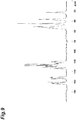
Figure 9 is a 13C solid state NMR spectrum of the A-type crystal of the compound (I) monohydrochloride obtained in Example 2. The abscissa represents the chemical shift (δ) and the ordinate represents the peak intensity. -
Figure 10 is a 13C solid state NMR spectrum of the B-type crystal of the compound (I) monohydrochloride obtained in Example 4. The abscissa represents the chemical shift (δ) and the ordinate represents the peak intensity. -
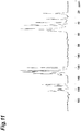
Figure 11 is a 13C solid state NMR spectrum of the C-type crystal of the compound (I) monohydrochloride obtained in Example 3. The abscissa represents the chemical shift (δ) and the ordinate represents the peak intensity. -
Figure 12 is a thermal analysis TG-DTA chart of the A-type crystal of the compound (I) monohydrochloride obtained in Example 2. The abscissa represents the temperature, the left ordinate represents the weight change of TG, and the right ordinate represents the heat flow of DTA. -
Figure 13 is a thermal analysis TG-DTA chart of the B-type crystal of the compound (I) monohydrochloride obtained in Example 4. The abscissa represents the temperature, the left ordinate represents the weight change of TG, and the right ordinate represents the heat flow of DTA. -
Figure 14 is a thermal analysis TG-DTA chart of the C-type crystal of the compound (I) monohydrochloride obtained in Example 3. The abscissa represents the temperature, the left ordinate represents the weight change of TG, and the right ordinate represents the heat flow of DTA. -
Figure 15 is a thermal analysis TG-DTA chart of the D-type crystal of the compound (I) monohydrochloride obtained in Example 5. The abscissa represents the temperature, the left ordinate represents the weight change of TG, and the right ordinate represents the heat flow of DTA. -
Figure 16 is a thermal analysis TG-DTA chart of the E-type crystal of the compound (I) monohydrochloride obtained in Example 6. The abscissa represents the temperature, the left ordinate represents the weight change of TG, and the right ordinate represents the heat flow of DTA. -
Figure 17 is a thermal analysis TG-DTA chart of the F-type crystal of the compound (I) monohydrochloride obtained in Example 7. The abscissa represents the temperature, the left ordinate represents the weight change of TG, and the right ordinate represents the heat flow of DTA. -
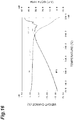
Figure 18 is a thermal analysis TG-DTA chart of the crystal of the compound (I) monohydrobromide obtained in Example 8. The abscissa represents the temperature, the left ordinate represents the weight change of TG, and the right ordinate represents the heat flow of DTA. -
Figure 19 shows a Raman spectrum of the A-type crystal of the compound (I) monohydrochloride obtained in Example 2. - Hereinafter, salts of the compound (I) of the present invention, crystals thereof, and a production method thereof will be described in detail.
- As used herein, the "salt" refer to a chemical substance consisting of the compound (I) as a basic component and a certain number of equivalents of acid relative to the compound (I).
- Examples of the "salt" as used herein include salts with inorganic acids, salts with organic acids, and salts with acidic amino acids, and among these, pharmaceutically acceptable salts are preferable.
- Examples of the salts with inorganic acids include salts with hydrochloric acid, hydrobromic acid, sulfuric acid, nitric acid and phosphoric acid; and examples of the salts with organic acids include salts with organic carboxylic acids such as acetic acid, succinic acid, fumaric acid, maleic acid, tartaric acid, malic acid, citric acid, lactic acid, stearic acid and benzoic acid, and salts with organic sulfonic acids such as methanesulfonic acid (mesylate), ethanesulfonic acid, benzenesulfonic acid and p-toluenesulfonic acid (tosylate); and among these, hydrochloric acid, hydrobromic acid and phosphoric acid are preferable.
- Examples of the salts with acidic amino acids include salts with aspartic acid and glutamic acid.
- The salt of the present invention may be an anhydrate, or a hydrate or a solvate. The hydrate or the solvate as used herein refer to a solid that the compound (I) or the salt thereof and water molecules or solvent molecules together form; and the solid may be a crystal; and examples of the solvent of the solvate include ketone-based solvents such as acetone, 2-butanone and cyclohexanone; ester-based solvents such as methyl acetate and ethyl acetate; ether-based solvents such as 1,2-dimethoxyethane and t-butylmethylether; alcohol-based solvents such as methanol, ethanol, 1-propanol and isopropanol; polar solvents such as N-methyl-2-pyrrolidone, N,N-dimethylformamide and dimethylsulfoxide. The number of water molecules or solvent molecules to the compound (I) or the salt thereof is not particularly limited, and for example, it may be one molecule or two molecules.
- As used herein, the "crystal" refer to the crystal of an anhydrate or a hydrate of the compound (I) or the salt thereof.
- As used herein, preferable examples of the crystal of the compound (I) and the hydrochloride and the hydrobromide of the compound (I) include:
- a crystal of the compound (I) having diffraction peaks at diffraction angles (2θ ± 0.2°) of 9.0°, 11.1° and 23.6° in a powder X-ray diffraction;
- a crystal of the compound (I) having diffraction peaks at diffraction angles (2θ ± 0.2°) of 9.0°, 11.1°, 14.5°, 18.1°, 20.0°, 21.9°, 23.6°, 24.4°, 24.9° and 28.5° in a powder X-ray diffraction;
- an A-type crystal of the compound (I) monohydrochloride having diffraction peaks at diffraction angles (2θ ± 0.2°) of 11.6°, 20.8° and 25.7° in a powder X-ray diffraction;
- an A-type crystal of the compound (I) monohydrochloride having diffraction peaks at diffraction angles (2θ ± 0.2°) of 6.1°, 7.8°, 11.6°, 16.2°, 19.9°, 20.8°, 25.2°, 25.7°, 26.9° and 29.9° in a powder X-ray diffraction;
- an A-type crystal of the compound (I) monohydrochloride having peaks at chemical shifts (δ ± 0.5 ppm) of 164.0 ppm, 129.6 ppm and 36.5 ppm in a 13C solid state NMR spectrum;
- an A-type crystal of the compound (I) monohydrochloride having a Raman shift peak (± 2 cm-1) at 587 cm-1 in a Raman spectroscopy measurement;
- an A-type crystal of the compound (I) monohydrochloride having Raman shift peaks (± 2 cm-1) at 587 cm-1, 1428 cm-1 and 1493 cm-1 in a Raman spectroscopy measurement;
- an A-type crystal of the compound (I) monohydrochloride having Raman shift peaks (± 2 cm-1) at 587 cm-1, 763 cm-1, 1428 cm-1, 1493 cm-1 and 1688 cm-1 in a Raman spectroscopy measurement;
- an A-type crystal of the compound (I) monohydrochloride having Raman shift peaks (± 2 cm-1) at 409 cm-1, 587 cm-1, 763 cm-1, 976 cm-1, 1428 cm-1, 1493 cm-1 and 1688 cm-1 in a Raman spectroscopy measurement;
- an A-type crystal of the compound (I) monohydrochloride substantially having a spectrum shown in
Figure 19 in a Raman spectroscopy measurement; - a B-type crystal of the compound (I) monohydrochloride having diffraction peaks at diffraction angles (2θ ± 0.2°) of 9.7°, 10.1° and 17.9° in a powder X-ray diffraction;
- a B-type crystal of the compound (I) monohydrochloride having diffraction peaks at diffraction angles (2θ ± 0.2°) of 6.3°, 9.7°, 10.1°, 17.9°, 19.0°, 19.4°, 23.4°, 26.3°, 27.3° and 32.0° in a powder X-ray diffraction;
- a B-type crystal of the compound (I) monohydrochloride having peaks at chemical shifts (δ ± 0.5 ppm) of 160.1 ppm, 133.4 ppm and 130.7 ppm in a 13C solid state NMR spectrum ;
- a C-type crystal of the compound (I) monohydrochloride having diffraction peaks at diffraction angles (2θ ± 0.2°) of 6.0°, 7.7° and 16.9° in a powder X-ray diffraction;
- a C-type crystal of the compound (I) monohydrochloride having diffraction peaks at diffraction angles (2θ ± 0.2°) of 6.0°, 7.7°, 9.7°, 11.4°, 15.8°, 16.9°, 18.1°, 23.2°, 25.4° and 27.6° in a powder X-ray diffraction;
- a C-type crystal of the compound (I) monohydrochloride having peaks at chemical shifts (δ ± 0.5 ppm) of 159.6 ppm, 127.6 ppm and 38.9 ppm in a 13C solid state NMR spectrum;
- a D-type crystal of the compound (I) monohydrochloride having diffraction peaks at diffraction angles (2θ ± 0.2°) of 6.6°, 14.6° and 26.4° in a powder X-ray diffraction;
- a D-type crystal of the compound (I) monohydrochloride having diffraction peaks at diffraction angles (2θ ± 0.2°) of 6.6°, 14.6°, 16.1°, 20.5°, 21.0°, 23.0°, 24.5°, 26.4°, 28.0° and 32.5° in a powder X-ray diffraction;
- an E-type crystal of the compound (I) monohydrochloride having diffraction peaks at diffraction angles (2θ ± 0.2°) of 6.4°, 11.3 ° and 27.3 ° in a powder X-ray diffraction;
- an E-type crystal of the compound (I) monohydrochloride having diffraction peaks at diffraction angles (2θ ± 0.2°) of 6.4°, 11.3°, 15.7°, 18.0°, 19.2°, 22,8°, 24.6°, 25.4°, 26.0° and 27.3° in a powder X-ray diffraction;
- an F-type crystal of the compound (I) monohydrochloride having diffraction peaks at diffraction angles (2θ ± 0.2°) of 7.3°, 9.3° and 10.7° in a powder X-ray diffraction;
- an F-type crystal of the compound (I) monohydrochloride having diffraction peaks at diffraction angles (2θ ± 0.2°) of 5.9°, 7.3°, 9.3°, 10.7°, 13.8°, 15.6°, 16.4°, 18.7°, 25.1° and 26.8° in a powder X-ray diffraction;
- a crystal of the compound (I) monohydrobromide having diffraction peaks at diffraction angles (2θ ± 0.2°) of 7.8°, 24.5° and 25.2° in a powder X-ray diffraction; and
- a crystal of the compound (I) monohydrobromide having diffraction peaks at diffraction angles (2θ ± 0.2°) of 6.0°, 7.8°, 10.0°, 11.7°, 17.8°, 20.8°, 23.5°, 24.5°, 25.2° and 27.3° in a powder X-ray diffraction.
- The diffraction peaks in the powder X-ray diffraction, the chemical shifts in the 13C solid state NMR spectrum, and the Raman shift peaks in the Raman spectroscopy measurement, described above, are unique to each of the crystal of the compound (I), the A to F-type crystals of the compound (I) monohydrochloride, and the crystal of the compound (I) monohydrobromide, and they are characteristic peaks for the crystals.
- In general, the diffraction angles (2θ) in the powder X-ray diffraction may contain errors within the range of ± 0.2°, so that the values of the diffraction angles described above need to be considered to include numerical values within the range of about ± 0.2°. Thus, not only crystals whose diffraction angles of the peaks in the powder X-ray diffraction perfectly match, but also crystals whose diffraction angles of the peaks match within an error of about ± 0.2° are the same in a certain compound or the salt thereof and included in the present invention.
- As used herein, for example, "having a diffraction peak at a diffraction angle (2θ ± 0.2°) of 9.0°" means "having a diffraction peak at a diffraction angle (2θ) of 8.8° to 9.2°", and the same applies to the case of other diffraction angles.
- In general, even when the crystalline form is the same, the peak intensity or the half-width of the diffraction angles (2θ) in the powder X-ray diffraction is different for each measurement depending on different measurement conditions and variations in size and form of the particles of the powder crystals used as a measurement sample, and a constant peak intensity or half-width is not always shown. Hence, in comparing the powder X-ray diffraction patterns, when there is a difference in peak intensity or half-width at the same diffraction angle (2θ), it does not mean that the difference derives from the difference of crystalline forms. Thus, it means that the crystal having a powder X-ray diffraction pattern which has such differences with respect to the diffraction peak characteristic of the specific crystal of the present invention has the same crystalline form as the crystal of the present invention. In addition, as used herein, "having the powder X-ray diffraction pattern of
Figure 1 " means that all the crystal showing the powder X-ray diffraction pattern shown inFigure 1 is the same crystal as the crystal of the present invention, not only in the case where a powder X-ray diffraction pattern having characteristic diffraction peaks matches the powder X-ray diffraction pattern shown inFigure 1 within the error range of ± 0.2°, but also in the case of a powder X-ray diffraction pattern having different peak intensity or half-width while having characteristic diffraction angles that match the powder X-ray diffraction pattern shown inFigure 1 within the error range of ± 0.2°. - As used herein, "chemical shifts (δ ± 0.5 ppm) of 164.0 ppm, 129.6 ppm and 36.5 ppm" mean "to have peaks substantially equivalent to chemical shifts (δ ± 0.5 ppm) of 164.0 ppm, 129.6 ppm and 36.5 ppm when a 13C solid state NMR spectrum measurement is carried out under the usual measurement conditions or under the conditions substantially the same as the present specification".
- In general, in determining whether or not "to have peaks substantially equivalent to", the chemical shift δ in the 13C solid state NMR spectrum may contain errors within the range of ± 0.5 ppm, so that the values of the chemical shifts described above need to be considered to include numerical values within the range of about ± 0.5 ppm. Thus, not only crystals whose chemical shifts in the 13C solid state NMR spectrum perfectly match, but also crystals whose chemical shifts match within an error of about ± 0.5 ppm are included in the present invention. Hence, as used herein, for example, "having a peak at a chemical shift (δ ± 0.5 ppm) of 164.0 ppm" means having a peak at a chemical shift (δ) within the range of 163.5 ppm to 164.5 ppm and the same applies to the case of other chemical shifts in the 13C solid state NMR spectrum.
- In general, the Raman shift peaks (cm-1) in the Raman spectroscopy measurement may contain errors within the range of ± 2 cm-1, so that the values of the peaks described above need to be considered to include numerical values within the range of about ± 2 cm-1. Thus, not only crystals whose Raman shift peaks in the Raman spectroscopy measurement perfectly match, but also crystals whose Raman shift peaks match within an error of about ± 2 cm-1 are the same in a certain compound or the salt thereof and included in the present invention.
- As used herein, for example, "having a Raman shift peak (± 2 cm-1) at 587 cm-1 in a Raman spectroscopy measurement" means "having a Raman shift peak at 585 cm-1 to 589 cm-1 in a Raman spectroscopy measurement", and the same applies to the case of other Raman shifts.
- In general, even when the crystalline form is the same, the peak intensity or the half-width of the Raman shift in the Raman spectroscopy measurement is different for each measurement depending on different measurement conditions and variations in size and form of the particles of the powder crystals used as a measurement sample, and a constant peak intensity or half-width is not always shown. Hence, in comparing the Raman spectroscopy measurements, when there is a difference in peak intensity or half-width at the same Raman shift peak (cm-1), it does not mean that the difference derives from the difference of crystalline forms. Thus, it means that the crystal having a Raman spectrum which has such differences with respect to the Raman shift peaks characteristic of a certain crystal of the present invention has the same crystalline form as the crystal of the present invention. In addition, as used herein, "having the spectrum of
Figure 19 in a Raman spectroscopy measurement" means that all the crystal showing the Raman spectrum shown inFigure 19 is the same crystal as the crystal of the present invention, not only in the case where a Raman spectrum having characteristic Raman shift peaks (cm-1) matches the Raman spectrum shown inFigure 19 within the error range of ± 2 cm-1, but also in the case of a Raman spectrum having different peak intensities or half-widths despite of having characteristic Raman shift peaks that match within the error range of ± 2 cm-1. - Hereinafter, methods for producing salts of the compound (I), crystals, and the like, which are one embodiment of the present invention, will be described.
- The compound (I) may be one produced by methods well-known to those skilled in the art. For example, the compound (I) can be synthesized by a method described in Reference Example described later.
- The salts of the compound (I) according to the present invention can be obtained by methods for producing ordinary salts. Specifically, the salts can be produced by, for example, suspending or dissolving the compound (I) into a solvent, while heating as necessary, and then adding an acid into the resulting suspension or solution, followed by stirring or allowing the mixture to stand at room temperature or while cooling for several minutes to several days. By using these production methods, the salts of the compound (I) can be obtained as crystalline or amorphous forms. Moreover, the amorphous form can be obtained by performing an operation such as lyophilization in addition to these production methods as necessary. Examples of the solvent used herein include alcohol-based solvents such as ethanol, 1-propanol and isopropanol; acetonitrile; ketone-based solvents such as acetone and 2-butanone; ester-based solvents such as ethyl acetate; saturated hydrocarbon-based solvents such as hexane and heptane; ether-based solvents such as t-butylmethylether and water. These solvents may be used singly or in combination of two or more thereof.
- The crystal of the compound (I) or the salt thereof can be produced by the aforementioned method for producing the compound (I), or the method for producing the salt thereof, or can be produced by heating and dissolving the compound (I) or the salt thereof in a solvent and then cooling the resulting solution while stirring for crystallization.
- The compound (I) or the salt thereof used for crystallization may be any form, that is, may be a solvate or a hydrate or an anhydrate, amorphous or crystalline forms (including one consisting of a plurality of polymorphic crystal), or the mixture thereof.
- Examples of the solvent used for crystallization include alcohol-based solvents such as methanol, ethanol, isopropanol and 1-propanol; acetonitrile; amide-based solvents such as N,N-dimethylformamide; ester-based solvents such as ethyl acetate; saturated hydrocarbon-based solvents such as hexane and heptane; ketone-based solvents such as acetone and 2-butanone; ether-based solvents such as t-butylmethylether and water. These solvents may be used singly or in combination of two or more thereof.
- The amount of the solvent to be used can be appropriately selected, provided that an amount capable of dissolving the compound (I) or the salt thereof by heating or an amount that enables the suspension to be stirred is the lower limit and an amount with which the yield of the crystal is not significantly reduced is the upper limit.
- In the crystallization, a seed crystal (for example, the desired crystal of the compound (I) or the salt thereof) may be added or not be added. The temperature at which the seed crystal is added is not particularly limited, and preferably 0 to 80°C.
- The temperature for heating and dissolving the compound (I) or the salt thereof may be appropriately selected depending on a solvent such that the compound (I) or the salt thereof can be dissolved at the temperature, but the temperature is preferably within the range of 50°C to the temperature at which a recrystallization solvent starts to reflux, more preferably 55 to 80°C.
- Since rapid cooling may produce crystals in different aspects (polymorphism), the cooling during crystallization is desirable to be carried out by appropriately controlling the cooling rate in consideration of the effect on quality and grade of the crystals, and is preferably cooling at a rate of, for example, 5 to 40°C/hour. More preferably, it is cooling at a rate of, for example, 5 to 25°C/hour.
- The final crystallization temperature may be appropriately selected depending on the yield, the quality, and the like of the crystals, and is preferably -25 to 30°C.
- The target crystal can be obtained by separating the crystal obtained by crystallization by an ordinary filtration procedure, washing the filtered crystal with a solvent if necessary, and then drying. As the solvent to be used for washing the crystal, those similar to the solvents used for crystallization can be used. Preferably, examples thereof include ethanol, acetone, 2-butanone, ethyl acetate, diethylether, t-butylmethylether and hexane. These solvents may be used singly or in combination of two or more thereof.
- The crystal separated by the filtration procedure can be dried by appropriately leaving in the air or under a nitrogen stream, or by heating.
- The drying time may be appropriately selected as the time until the amount of the residual solvent falls below a predetermined amount, depending on the production amount, the drying apparatus, the drying temperature, and the like. Drying may also be carried out under airflow or under reduced pressure. The degree of pressure reduction may be appropriately selected depending on the production amount, the drying apparatus, the drying temperature, and the like. The resulting crystal may also be left in the air as necessary after drying.
- The crystal of the compound (I) and salts of the compound (I) obtained by the production method described above have cholinergic neuron activation and/or neuroprotective effect, as shown in activity data in pharmacological test examples described later, and have a potential use as an agent for improving the reduced cognitive performance caused by the dysfunction of cholinergic neurons.
- Another embodiment of the present invention is a pharmaceutical composition comprising the crystal of the compound (I) and pharmaceutically acceptable additives. A pharmaceutical composition can be produced by admixing pharmaceutically acceptable additives with the crystal of the compound (I). The pharmaceutical composition according to the present invention can be produced in accordance with a known method, for example, the method described in General Rules for Preparations in the Japanese Pharmacopoeia 17th Edition.
- The pharmaceutical composition according to the present embodiment can be appropriately administered to a patient depending on the dosage form thereof.
- The dose of the compounds (I) according to the present invention varies depending on the severity of symptoms, age, sex, body weight, dosage form, type of salt, specific type of disease, and other conditions; however, in general, the dose for an adult per day by oral administration is about 30 µg to 10 g, preferably 100 µg to 5 g, and more preferably 100 µg to 1 g; the dose for an adult per day by injection administration is about 30 µg to 1 g, preferably 100 µg to 500 mg, and more preferably 100 µg to 300 mg; and the above dose is administered once or several times.
- The crystal of the compound (I) of the present invention can be produced by, for example, the methods described in the following Examples, and the effect of the compound can be confirmed by the methods described in the following Test Examples. However, these are just examples, and the present invention is not limited to the following specific examples in any case and may be modified within a range that does not depart from the scope of the present invention.
- The powder X-ray crystal diffraction of the crystals produced in the following Examples was carried out by mounting the obtained crystal onto the sample stage of a powder X-ray diffraction apparatus and measured under any of the following conditions.
-
- X-ray source: CuKα
- Voltage: 45 kV
- Current: 40 mA
- Optical system: focusing mirror
- Soller slit: 0.02°
- Detector : X'Celerator (semiconductor detector)
- Scan range: 5° to 35°
- Step size: 0.017°
- Scan step time: 600 sec
- Sample holder: kapton film
-
- X-ray source: CuKα
- Voltage: 50 kV
- Current: 300 mA
- Slit: divergence slit 0.5 mm, scattering slit open, light receiving slit open
- Detector: scintillation counter
- Scan rate: 5°/min
- Sampling interval: 0.02°
- Scan range: 5° to 35°
- Sample holder: aluminum holder
- A sample was precisely weighed in an aluminum sample pan and the thermal analysis was carried out under the following conditions.
-
- Atmosphere: under nitrogen gas stream (100 mL/min)
- Control: empty aluminum sample pan
- Temperature rising rate: 10°C/min
- Sampling interval: 1 sec
- Measurement temperature range: room temperature to 320°C
- The 13C solid state NMR spectrum of the crystal was measured by enclosing about 300 mg of a solid sample into a sample tube under the following conditions.
-
- Apparatus used:
Avance 400 MHz (manufactured by BRUKER) 7 mm-CPMAS probe (manufactured by BRUKER) - Nuclei measured: 13C (resonance frequency 100.6248425 MHz)
- Measurement temperature: room temperature
- Pulse mode: CPTOSS measurement
- Rotational speed: 5000 Hz
- Pulse repeating time: 3 sec
- Contact time: 1 msec
- Cumulative number of times: 5120 times
- Reference material: glycine (external reference: 176.03 ppm)
- The Raman spectrum of the crystals was measured by placing a sample onto the sample stage of a Raman microspectroscope under the following measurement conditions.
-
- Apparatus used: RENISHAW Raman Microscope in Via Reflex
- Laser wavelength: 785 nm
- Diffraction grating: 1200 lines/mm
- Objective: 50 times
- Scan mode: continuous
- Exposure time: 5 sec
- Cumulative number of times: 5 times
- Measurement range: 400 to 1800 cm-1 (Raman shift)
- Error: ± 2 cm-1
- Compounds described with document names, etc., indicate that the compounds were produced in accordance with the documents, etc.
- Moreover, the abbreviations used in the present specification are well-known and common to a person skilled in the art. In the present specification, the following abbreviations are used.
- DMSO: dimethylsulfoxide
- IPA: isopropanol
- n-: normal
- TEA: triethylamine
- THF: tetrahydrofuran
- 1H-NMR: proton nuclear magnetic resonance spectrometry
- MS: mass spectrometry
- The term "room temperature" in the following Examples and Reference Examples generally refers to about 10°C to about 35°C. % refers to weight percent unless otherwise specified.
- Chemical shifts of proton nuclear magnetic resonance spectra are denoted in δ-unit (ppm) relative to tetramethylsilane, and coupling constants are recorded in Hertz (Hz). The abbreviations of splitting patterns are as follows.
s: singlet, d: doublet, t: triplet, q: quartet, m: multiplet, br.s: broad singlet. - In the reactions using a microwave reactor in the Reference Examples, Initiator (TM) or Initiator+ (TM) produced by Biotage was used.
- Regarding chromatography, as silica gel, Silica Gel60 produced by Merck (70-230 mesh ASTM) or PSQ60B produced by Fuji Silysia Chemical Ltd. was used, or a pre-packed column {column: Hi-Flash (TM) Column (Silicagel) produced by YAMAZEN, size: one of S (16 x 60 mm), M (20 x 75 mm), L (26 x 100 mm), 2L (26 x 150 mm) and 3L (46 x 130 mm); or Biotage (TM) SNAP Ultra Silica Cartridge produced by Biotage, size: one of 10 g, 25 g and 50 g} was used.
- As NH silica gel, CHROMATOREX NH-DM2035 produced by Fuji Silysia Chemical Ltd. was used, or a pre-packed column {column: Hi-Flash (TM) Column (Amino) produced YAMAZEN, size: one of S (16 x 60 mm), M (20 x 75 mm), L (26 x 100 mm), 2L (26 x 150 mm) and 3L (46 x 130 mm); or Presep (TM) (Luer Lock) NH2(HC) produced by Wako Pure Chemical Industries, Ltd., size: one of type M (14 g/25 mL), type L (34 g/70 mL), type 2L (50 g/100 mL) and type 3L (110 g/200 mL)} was used.
- As neutral alumina, Aluminum oxide 90 active neutral, 70 - 230 mesh, Merck, E6NXX was used.
- As names of the compounds shown below, except for generally used reagents, those shown in the "E-Notebook" Version 12 (PerkinElmer) were used.
-

- TEA (61.6 mL, 442 mmol) was added at room temperature to a mixture of 1-methyl-4-piperidone (CAS No. 1445-73-4) (51.5 mL, 442 mmol), ethyl cyanoacetate (CAS No. 105-56-6) (47.2 mL, 442 mmol), sulfur (CAS No. 7704-34-9) (14.2 g, 442 mmol) and ethanol (800 mL). The reaction mixture was stirred at 40°C for 15 hours, and then concentrated under reduced pressure. The residue was purified by column chromatography (NH silica gel, ethyl acetate). The obtained concentrated residue was triturated with ethyl acetate. The precipitates were collected by filtration, washed with ethyl acetate, and dried under reduced pressure to yield the title compound (58.4 g).
- 1H-NMR (400 MHz, CDCl3) δ (ppm): 1.33 (t, J = 7.0 Hz, 3H), 2.44 (s, 3H), 2.62-2.70 (m, 2H), 2.79-2.88 (m, 2H), 3.37 (t, J = 2.0 Hz, 2H), 4.26 (q, J = 7.3 Hz, 2H), 5.97 (br. s, 2H).
- MS (ESI) m/z: 241 [M+H]+
- 1H,2H,4H-Thieno[2,3-d][1,3]oxazine-2,4-dione (CAS No. 103979-54-0) (600 mg, 3.55 mmol) was added to a solution of sarcosine (790 mg, 8.87 mmol) in water (12 mL). The reaction mixture was heated under reflux for 1.5 hours. The reaction mixture was cooled to room temperature. Chloroform was added to the reaction mixture, and the organic layer was separated. The aqueous layer was extracted with chloroform (twice) and ethyl acetate (3 times). The combined organic layer was dried over anhydrous sodium sulfate and filtered, and the filtrate was concentrated under reduced pressure. The obtained solid was dried to yield the title compound (430 mg).
- 1H-NMR (400 MHz, CDCl3) δ (ppm): 3.23 (s, 3H), 3.99 (s, 2H), 6.90 (d, J = 5.9 Hz, 1H), 7.29 (d, J = 5.7 Hz, 1H), 8.39 (br. s, 1H).
- MS (ESI) m/z: 197 [M+H]+
- Phosphorus oxychloride (1.43 mL, 15.3 mmol) was added at room temperature to a mixture of 4-methyl-3,4-dihydro-1H-thieno[2,3-e][1,4]diazepine-2,5-dione (1.00 g, 5.10 mmol) obtained in step (2), ethyl 2-amino-6-methyl-4,5,6,7-tetrahydrothieno[2,3-c]pyridine-3-carboxylate (1.84 g, 7.64 mmol) obtained in step (1), and 1,4-dioxane (30 mL). The reaction mixture was stirred at room temperature for 5 minutes, and stirred at 90°C for 2 hours. Sodium ethoxide (a 20% solution in ethanol, 21.7 mL, 56.1 mmol) was added over 5 minutes to the reaction mixture cooled to room temperature. The reaction mixture was stirred at room temperature for 1.5 hours. Ethyl acetate, a saturated sodium hydrogen carbonate aqueous solution, and water were sequentially added to the reaction mixture, and the organic layer was separated. The aqueous layer was extracted with ethyl acetate. The combined organic layer was dried over anhydrous magnesium sulfate and filtered, and the filtrate was concentrated under reduced pressure. The residue was purified by column chromatography (silica gel, 20%-50% methanol/ethyl acetate). The obtained solid was triturated with ethanol, and the precipitates were collected by filtration. The obtained solid was washed with ethanol, and dried under reduced pressure to yield the title compound (712 mg).
- 1H-NMR (400 MHz, CDCl3) δ (ppm): 2.52 (s, 3H), 2.71-2.87 (m, 2H), 3.05-3.30 (m, 5H), 3.59-3.75 (m, 2H), 4.23 (d, J = 14.8 Hz, 1H), 4.57 (d, J = 14.8 Hz, 1H), 7.35 (d, J = 6.2 Hz, 1H), 7.39 (d, J = 5.9 Hz, 1H).
- MS (ESI) m/z: 373 [M+H]+
- To 1.5 L of 0.3 M hydrochloric acid, 152.08 g of the compound (I) was added, and 450 ml of ethyl acetate was added to this solution, followed by stirring for 5 minutes. The aqueous layer was separated and washed with 450 ml of ethyl acetate, and the insoluble matter was removed by filtration. To the filtrate, 100 ml of a 1 N aqueous solution of sodium hydroxide was added in a water bath at 20°C and the mixture was stirred for 15 minutes. To the mixture, 350 ml of a 1 N aqueous solution of sodium hydroxide was added and the obtained suspension was stirred for 2
hours 30 minutes. The resulting crystal was collected by filtration, washed sequentially with 300 ml, 450 ml, and 300 ml of water, 300 ml, 350 ml, and 300 ml of ethanol, and dried under reduced pressure to yield 141.7 g of the title crystal.
Powder X-ray diffraction peak (reflection method, 2θ ± 0.2°): 9.0°, 11.1°, 14.5°, 18.1°, 20.0°, 21.9°, 23.6°, 24.4°, 24.9°, 28.5° - The powder X-ray diffraction pattern of the crystal of the compound (I) obtained by the above-mentioned method is shown in
Figure 1 . - Into a screw-top test tube, 101 mg of the compound (I) was added. Thereinto, 0.2 mL of 1.5 M hydrochloric acid was added and dissolved. Thereinto, 1.8 mL of IPA was added, irradiated with ultrasonic waves, and then stirred with a stirrer at 40°C for one day. After stirring at room temperature for further 1 hour, the sample was collected by filtration by using a filter (0.2 µm), rinsed with 0.5 mL of IPA/water (9/1, v/v), and air-dried under a nitrogen stream. The residue was dried at 70°C for about 1 hour to yield the title crystal (103 mg).
- Powder X-ray diffraction peaks (transmission method, 2θ ± 0.2°): 6.1°, 7.8°, 11.6°, 16.2°, 19.9°, 20.8°, 25.2°, 25.7°, 26.9°, 29.9°
- 13C-NMR (100 MHz, solid state) δ (ppm): 164.0, 162.5, 160.5, 153.9, 151.6, 150.7, 133.6, 131.1, 129.6, 128.4, 126.9, 125.2, 123.7, 121.3, 120.3, 119.5, 53.7, 52.0, 50.9,44.7,36.5,22.6
- Raman shift peaks (cm-1): 409, 587, 763, 976, 1428, 1493, 1688
- The powder X-ray diffraction pattern of the A-type crystal of the compound (I) monohydrochloride obtained by the above-mentioned method is shown in
Figure 2 , the 13C solid state NMR spectrum is shown inFigure 9 , the thermal analysis TG-DTA chart is shown inFigure 12 , and the Raman spectrum is shown inFigure 19 . - Into a screw-top test tube, 1020 mg of the compound (I) was added. In 20 mL of methanol, 1.5 equivalent of hydrochloric acid (353 µL) was dissolved and this solution was added to the sample. The sample was stirred with a stirrer at room temperature for 2 days. The sample was collected by filtration with a filter (0.2 µm). The obtained solid was dried under reduced pressure for about 2 hours, then dried at 70°C for 1 hour to yield the title crystal (1048 mg).
- Powder X-ray diffraction peak (transmission method, 2θ ± 0.2°): 6.0°, 7.7°, 9.7°, 11.4°, 15.8°, 16.9°, 18.1°, 23.2°, 25.4°, 27.6°
- 13C-NMR (100MHz, solid state) δ (ppm): 162.5, 160.5, 159.6, 153.8, 151.1, 134.1, 131.6, 128.4, 127.6, 125.6, 120.0, 54.0, 52.6, 50.9, 44.3, 43.5, 38.9, 32.3,22.4
- The powder X-ray diffraction pattern of the C-type crystal of the compound (I) monohydrochloride obtained by the above-mentioned method is shown in
Figure 4 , the 13C solid state NMR spectrum is shown inFigure 11 , and the thermal analysis TG-DTA chart is shown inFigure 14 . - Into a platinum crucible, 303 mg of the hydrochloride crystal obtained in Example 3 was added and heated at 160°C for 15 minutes to yield the title crystal (293 mg).
- Powder X-ray diffraction peak (transmission method, 2θ ± 0.2°): 6.3°, 9.7°, 10.1°, 17.9°, 19.0°, 19.4°, 23.4°, 26.3°, 27.3°, 32.0°
- 13C-NMR (100 MHz, solid state) δ (ppm): 162.0, 160.1, 153.8, 151.1, 133.4, 130.7, 128.3, 126.9, 125.6, 120.3, 51.2, 43.6, 32.3, 22.3
- The powder X-ray diffraction pattern of the B-type crystal of the compound (I) monohydrochloride obtained by the above-mentioned method is shown in
Figure 3 , the 13C solid state NMR spectrum is shown inFigure 10 , and the thermal analysis TG-DTA chart is shown inFigure 13 . - Into a screw-top test tube, 227 mg of the mixture of the hydrochloride crystals obtained in Examples 2 and 3, and 8 mL of ethanol were added. The mixture was stirred with a stirrer at 65°C. After about 1 hour, the mixture was irradiated with ultrasonic waves and stirred at the same temperature for one day. The sample was collected by filtration with a filter (0.2 µm) to yield the title crystal (203 mg).
Powder X-ray diffraction peak (transmission method, 2θ ± 0.2°): 6.6°, 14.6°, 16.1°, 20.5°, 21.0°, 23.0°, 24.5°, 26.4°, 28.0°, 32.5° - The powder X-ray diffraction pattern of the D-type crystal of the compound (I) monohydrochloride obtained by the above-mentioned method is shown in
Figure 5 and the thermal analysis TG-DTA chart is shown inFigure 15 . - Into a screw-top test tube, 108 mg of the hydrochloride crystal obtained in Example 2 and 5 mL of acetonitrile were added. The mixture was stirred with a stirrer at 60°C for one day and the sample was collected by filtration with a filter (0.2 µm). The obtained solid and 5 mL of acetonitrile were added into the screw-top test tube again and stirred with a stirrer at 60°C for one day. The sample was collected by filtration with a filter (0.2 µm) under a nitrogen stream to yield the title crystal (89.7 mg).
Powder X-ray diffraction peak (transmission method, 2θ ± 0.2°): 6.4°, 11.3°, 15.7°, 18.0°, 19.2°, 22.8°, 24.6°, 25.4°, 26.0°, 27.3° - The powder X-ray diffraction pattern of the E-type crystal of the compound (I) monohydrochloride obtained by the above-mentioned method is shown in
Figure 6 and the thermal analysis TG-DTA chart is shown inFigure 16 . - Into a screw-top test tube, 101 mg of the hydrochloride crystal obtained in Example 2 and 5 mL of ethanol were added. The mixture was stirred with a stirrer at 60°C for one day. The sample was collected by filtration with a filter (0.2 µm). The obtained solid and 5 mL of ethanol were added into the screw-top test tube again and stirred with a stirrer at 60°C for 4 hours. The sample was collected by filtration with a filter (0.2 µm) to yield the title crystal (75.0 mg).
Powder X-ray diffraction peak (transmission method, 2θ ± 0.2°): 5.9°, 7.3°, 9.3°, 10.7°, 13.8°, 15.6°, 16.4°, 18.7°, 25.1°, 26.8° - The powder X-ray diffraction pattern of the F-type crystal of the compound (I) monohydrochloride obtained by the above-mentioned method is shown in
Figure 7 and the thermal analysis TG-DTA chart is shown inFigure 17 . - Into a screw-top test tube, 933 mg of the compound (I) was added. Into 20 mL of methanol, 1.5 equivalent (434 µL) of hydrobromic acid was dissolved and this solution was added to the sample. The mixture was stirred with a stirrer at room temperature for 3 days. The sample was collected by filtration with a filter (0.2 µm) and dried at 60°C for 1 hour to yield the title crystal (1111 mg).
Powder X-ray diffraction peak (transmission method, 2θ ± 0.2°): 6.0°, 7.8°, 10.0°, 11.7°, 17.8°, 20.8°, 23.5°, 24.5°, 25.2°, 27.3° - The powder X-ray diffraction pattern of the compound (I) monohydrobromide obtained by the above-mentioned method is shown in
Figure 8 and the thermal analysis TG-DTA chart is shown inFigure 18 . - The septal area was isolated from Sprague-Dawley (SD) rats (Charles River Laboratories Japan, Inc.) at a fetal age of 18 days, and cultured. Specifically, fetuses were aseptically removed from pregnant rats under isoflurane anesthesia. The brain was extracted from each fetus, and immersed in ice-cooled L-15 medium (11415-064, Thermo Fisher Scientific). The septal area was dissected from the extracted brain under a stereoscopic microscope. The dissected septal area was subjected to enzyme treatment in an enzyme solution containing 0.25% trypsin (15050-065, Thermo Fisher Scientific) and 0.01% DNase (D5025-150KU, Sigma) at 37°C for 30 minutes, thereby dispersing the cells. In this case, the enzyme reaction was terminated by adding inactivated horse serum (26050-088, Thermo Fisher Scientific). The enzyme-treated solution was centrifuged at 1000 rpm for 3 minutes, and the supernatant was removed. A medium in an amount of 10 mL was added to the obtained cell mass. The medium used was Dulbecco's Modified Eagle's Medium (044-29765, WAKO) supplemented with N2 supplement (17502-048, Thermo Fisher Scientific), 1 mM sodium pyruvate (11360-070, Thermo Fisher Scientific), and Penicillin-Streptomycin (15140-1221, Thermo Fisher Scientific). The cells of the cell mass to which the medium was added were redispersed by gentle pipetting, and then centrifuged again at 1000 rpm for 3 minutes, and the supernatant was removed. The medium in an amount of 10 mL was added to the obtained cell mass, and the cell dispersion was filtered through a 40-µm nylon mesh (Cell Strainer) to remove the cell mass, thereby obtaining a neuronal cell suspension. The neuronal cell suspension was diluted with the medium, and 10% inactivated bovine serum (26140-079, Thermo Fisher Scientific) and 10% inactivated horse serum were added. Thereafter, 100 µL/well of the suspension was seeded in a 96-well plate (354461, CORNING) pre-coated with poly-D-lysine so that the initial culture density was 1.4 x 10⁵ cells/cm2. After the seeded cells were cultured under 5%CO2-95% air in a 37°C incubator for 2 days, the entire medium was replaced with 120 µL of fresh medium, and the cells were subsequently cultured for 5 days.
- On the 7th day of culture, compound was added in the following manner. A solution of the test compound in DMSO was diluted with the medium so that the concentration was 10 times higher than the final concentration. NGF (450-01, PEPRO TECH, INC.) was prepared at 0.3 ng/mL. These two solutions were added each in an amount of 15 µL/well, and the mixture was mixed well. The final DMSO concentration was 0.1% or less. Moreover, only DMSO and NGF were added to the control group.
- One day after compound addition, an amount of ACh release was measured by HPLC in the following manner. A warmed buffer was added at 100 µL/well to the well after the medium was eliminated, and the buffer was immediately removed. Thereafter, a buffer to which 10 µm choline, 10 µm physostigmine, and 6 mM KCl were added was added at 120 µL/well. The buffer was prepared by adding 125 mM NaCl, 25 mM 4-(2-hydroxyethyl)-1-piperazineethanesulfonic acid, 1.2 mM KH2PO4, 1.2 mM MgSO4, 2.2 mM CaCl2 (2H2O), and 10 mM glucose to sterilized water, and the final pH of the solution was set to 7.4. After the 96-well plate to which the buffer was added was incubated under 5% CO2-95% air in a 37°C incubator for 40 minutes, 80 µL of buffer was collected. An internal standard solution IPHC (5 x 10⁻⁷ M) was added in an amount of 6 µL to the collected buffer, and the buffer was transferred to a tube for HPLC measurement and subjected to HPLC measurement. The results are represented by the effect of each compound as the percentage (% of control) of the ACh concentration in the buffer of the control group, and the compound concentration of Reference Example 1 showing a 20% increase from the ACh concentration in the buffer of the control group was 0.1 µM.
- In this study, SD male rats (Charles River Laboratories Japan, Inc.) with a body weight of about 250 to 350 g were used. The compound was dissolved in 0.01 mol/L hydrochloric acid, and orally administered.
- At 24 hours after the administration of the compound, the whole brain tissue was collected under pentobarbital anesthesia. The medial septum was isolated from whole brain on ice and frozen with liquid nitrogen, and then stored at -80°C.
- For RNA purification, RNeasy Plus Mini Kit (#74136: QIAGEN) was used in this study. RNA purification was performed by the method described in the kit. After RNA purification, the total RNA concentration was measured by using QIAxpert Instrument (QIAGEN). cDNA was synthesized using SuperScript (R) VILO (TM) cDNA Synthesis Kit (#11754: Thermo Fisher Scientific). The synthesis of cDNA was performed by the method described in the kit. The synthesized cDNA was diluted 4 times with RNase free water, and the diluted cDNA solution was used as a sample. Taqman Universal PCR Master Mix (#4304437: Thermo Fisher Scientific), Taqman (R) Gene Expression Assays, INVENTORIED (#4331182: Thermo Fisher Scientific), RNase free water, and the cDNA solution were mixed in amounts of 10 µl, 1 µl, 4 µl, and 5 µl, respectively, and the resulting mixture was used as a measurement sample solution. Quantitative polymerase chain reaction (qPCR) was conducted using ABI PRISM (R) 7900HT (Thermo Fisher Scientific) by a fluorescence probe method. Analysis was performed by SDS 2.4 (Thermo Fisher Scientific). The results were calculated by the percentage of the amount of ChAT mRNA expression levels in the compound administration group of Reference Example 1 increased from the amount of ChAT mRNA expression levels in the vehicle administration group to be 56.4% at 10 mg/kg.
- Correlation between increase and decrease of intracerebral neurotransmitters and those in cerebrospinal fluid (CSF) was revealed by studies on rodents and the correlation was also seen in human (Lowe S et al. Psychopharmacology 219 (2012) 959-970). Thus, the changes in acetylcholine in CSF were measured in order to determine the changes in intracerebral acetylcholine by the test compounds.
- In this study, Fischer344 male rats (Charles River Laboratories Japan, Inc.) with a body weight of about 150 to 250 g were used. The test compounds were orally administered to the rats once a day at 10 mg/kg for three days. The vehicle used was 0.01 mol/L hydrochloric acid.
- At 24 hours after the administration of the vehicle and the test compounds, the CSF was collected from cisterna magna in a tube containing AchE inhibitors under pentobarbital anesthesia. The collected CSF was centrifuged at 3500 x g at 4 °C for 10 minutes and the supernatant was collected. The collected supernatant was frozen with liquid nitrogen, and then stored at -80°C.
- To 10 µL of the CSF was added 50 µL of acetylcholine-d9 chloride (ACh-d9) at a final concentration of 0.34 nmol/L as an internal standard. The mixture was pipetted and centrifuged at 1500 x g at 4 °C for 10 minutes. The supernatant was collected and subjected to LC/MS (NexeraX2 (MS), TSQ Altis (HPLC)), and Ach was detected as precursor ion at m/z 146.050 and as product ion at m/z 87.071 and ACh-d9 as an internal standard was detected as precursor ion at m/z 155.088 and as product ion at m/z 87.000. The results were calculated by the percentage (% of control) of the ACh concentration in CSF in the compound administration group of Reference Example 1 increased from the ACh concentration in CSF in the vehicle administration group to be 156.8%.
- In this study, the test compounds were orally administered to human tau P301S transgenic mice once a day for three months from four-month-old to seven-month-old. The vehicle used was 0.01 mol/L hydrochloric acid.
- At the initial day of the administration (four-month-old) and at the next day of the final administration, mice of vehicle administration group and test compound administration group were anesthetized under pentobarbital (50mg/kg, i.p.) and perfused with PBS. After the perfusion, the forebrain including the medial septal area was collected and fixed with 4% paraformaldehyde.
- The collected forebrain including the medial septal area was immersed and shaken overnight in 4% paraformaldehyde. The immersion solution was replaced with 7.5% sucrose solution. It was immersed and shaken overnight in 7.5% sucrose solution, and the immersion solution was replaced with 15% sucrose solution and it was immersed and shaken overnight. The immersion solution was replaced with 30% sucrose solution and it was immersed and shaken overnight. Brain coronal frozen sections with 30 µm thickness were prepared from the forebrain including the medial septal area by using a microtome (Leica, SM2000R).
- The prepared brain coronal frozen sections were stained with DAB (DAB PEROXIDASE SUBSTRATE KIT (Vector, SK-4100)) using a ChAT antibody (Santa Cruz, SC-20672) as a primary antibody. The section image including the medial septal area as shown in "The mouse Brain in stereotaxic coordinates" (COMPACT THIRD EDITION, Keith B.J. Franklin & George Paxinos) was taken by an all-in-one fluorescence microscope (KEYENCE, BZ-X710) and ChAT positive cells around the major axis of the medial septal area were counted by BZ analysis software (KEYENCE). The results were shown as a percentage of the number of ChAT positive cells in the vehicle administration group and the test compound administration group with respect to the number of ChAT positive cells at the time of initial administration (four-month-old). Data are expressed as the mean ±SEM. The differences between the group at the time of initial administration and the vehicle-treated group (significant: *) was analyzed by an unpaired t-test, and also the differences between the vehicle-treated group and compound-treated group (significant: #) was analyzed by unpaired t-test. A value of P<0.05 was considered statistically significant. Statistical analyses were performed using the GraphPad Prism version 7.02. The results were shown in Table 1.
[Table 1] Treatment Group Ratio (%) of number of ChAT positive cells compared to that in the initial administration Group at the time of initial administration 100.0±4.5 Vehicle administration group 83.0±5.8* Reference Example 1 compound administration group (Dose: 5mg/kg) 105.3±4.3# - In this study, Sprague-Dawley male rats (Charles River Laboratories Japan, Inc.) with a body weight of about 250 to 350 g were used. The rat was anesthetized under the combination of three drugs: midazolam (2 mg/kg s.c.), medetomidine hydrochloride (0.15 mg/kg s.c.) and butorphanol tartrate (2.5 mg/kg s.c.) and fixed with a brain stereotaxis apparatus (Narishige Co., Ltd.). The cranial was exposed and a hole with 5 mm width was drilled in the skull from the median line 2 mm posterior to Bregma. A razor with 4 mm width was pierced into the Bregma in 5.5 mm depth to cut fimbria-fornix. After hemostasis, the scalp was sutured. After the operation, the rat was brought back to the cage and recovered from the anesthesia. In the sham-operated group, a hole with 5 mm width was drilled in the skull from the median line 2 mm posterior to Bregma, but no razor was pierced.
- The test compounds were orally administered to the rats once a day from five days to nine days after the operation (Example 1: 10 mg/kg) or from seven days to fourteen days after the operation (Example 3: 3 mg/kg). The vehicle used was 0.01 mol/L hydrochloric acid. In the sham-operated group, the vehicle was orally administered once a day similarly to the test compound administration group.
- The rats were anesthetized under pentobarbital and transcardially perfused with ice-cold PBS. After the perfusion, the forebrain including the medial septal area was collected and immersed and shaken overnight with 4% paraformaldehyde. The immersion solution was replaced with 7.5% sucrose solution. It was immersed and shaken overnight in 7.5% sucrose solution, and the immersion solution was replaced with 15% sucrose solution and it was immersed and shaken overnight. The immersion solution was replaced with 30% sucrose solution and it was immersed and shaken overnight. Brain coronal frozen sections with 30 µm thickness were prepared from the forebrain including the medial septal area by using a microtome (Leica, SM2000R).
- The prepared brain coronal frozen sections were stained with DAB (DAB PEROXIDASE SUBSTRATE KIT (Vector, SK-4100)) using a ChAT antibody (Santa Cruz, SC-20672) or a VAChT antibody (Merck Millipore, ABN100) as a primary antibody. The section image including the medial septal area or hippocampus as shown in "The mouse Brain in stereotaxic coordinates" (COMPACT THIRD EDITION, Keith B.J. Franklin & George Paxinos) was taken by an all-in-one fluorescence microscope (KEYENCE, BZ-X710) and ChAT positive cells of the medial septal area or optical density (OD) in hippocampal VAChT were measured by BZ analysis software (KEYENCE). The results were shown as a percentage of the number of ChAT positive cells of the medial septal area or OD in hippocampal VAChT in the vehicle administration group and the test compound administration group with respect to the number of ChAT positive cells of the medial septal area or OD in hippocampal VAChT in the sham-operated group. Data are expressed as the mean ±SEM. The differences between the vehicle-treated group and compound-treated (significant: #) was analyzed by unpaired t-test. A value of P<0.05 was considered statistically significant. Statistical analyses were performed using the GraphPad Prism version 7.02. The results were shown in Tables 2 and 3.
[Table 2] Compound Number of ChAT positive cells (%) at initial administration Number of ChAT positive cells (%) in vehicle administration group Number of ChAT positive cells (%) in test compound administration group Reference Example 1 57.0±7.5 38.4±5.0 74.1±9.3# [Table 3] Compound OD in hippocampal VAChT (%) at initial administration OD in hippocampal VAChT (%) in vehicle administration group OD in hippocampal VAChT (%) in test compound administration group Reference Example 1 51.7±13.1 19.5±6.4 66.1±14.2#
Claims (15)
- A monohydrochloride or monohydrobromide of 5,10-dimethyl-5,6,9,10,11,12-hexahydropyrido[4",3":4',5']thieno[2',3':4,5]pyrimido[1,2-a]thieno[3,2-f][1,4]diazepine-4,13-dione represented by formula (I):

- A crystal of 5,10-dimethyl-5,6,9,10,11,12-hexahydropyrido[4",3":4',5']thieno[2',3':4,5]pyrimido[1,2-a]thieno[3,2-f][1,4]diazepine-4,13-dione represented by formula (I):


- A crystal of 5,10-dimethyl-5,6,9,10,11,12-hexahydropyrido[4",3":4',5']thieno[2',3':4,5]pyrimido[1,2-a]thieno[3,2-f][1,4]diazepine-4,13-dione represented by formula (I):

- An A-type crystal of 5,10-dimethyl-5,6,9,10,11,12-hexahydropyrido[4",3":4',5']thieno[2',3':4,5]pyrimido[1,2-a]thieno[3,2-f][1,4]diazepine-4,13-dione monohydrochloride represented by formula (I):

- A B-type crystal of 5,10-dimethyl-5,6,9,10,11,12-hexahydropyrido[4",3":4',5']thieno[2',3':4,5]pyrimido[1,2-a]thieno[3,2-f][1,4]diazepine-4,13-dione monohydrochloride represented by formula (I):

- A C-type crystal of 5,10-dimethyl-5,6,9,10,11,12-hexahydropyrido[4",3":4',5']thieno[2',3':4,5]pyrimido[1,2-a]thieno[3,2-f][1,4]diazepine-4,13-dione monohydrochloride represented by formula (I):

- A D-type crystal of 5,10-dimethyl-5,6,9,10,11,12-hexahydropyrido[4",3":4',5']thieno[2',3':4,5]pyrimido[1,2-a]thieno[3,2-f][1,4]diazepine-4,13-dione monohydrochloride represented by formula (I):

- An E-type crystal of 5,10-dimethyl-5,6,9,10,11,12-hexahydropyrido[4",3":4',5']thieno[2',3':4,5]pyrimido[1,2-a]thieno[3,2-f][1,4]diazepine-4,13-dione monohydrochloride represented by formula (I):

- An F-type crystal of 5,10-dimethyl-5,6,9,10,11,12-hexahydropyrido[4",3":4',5']thieno[2',3':4,5]pyrimido[1,2-a]thieno[3,2-f][1,4]diazepine-4,13-dione monohydrochloride represented by formula (I):

- A crystal of 5,10-dimethyl-5,6,9,10,11,12-hexahydropyrido[4",3":4',5']thieno[2',3':4,5]pyrimido[1,2-a]thieno[3,2-f][1,4]diazepine-4,13-dione monohydrobromide represented by formula (I):

- An A-type crystal of 5,10-dimethyl-5,6,9,10,11,12-hexahydropyrido[4",3":4',5']thieno[2',3':4,5]pyrimido[1,2-a]thieno[3,2-f][1,4]diazepine-4,13-dione monohydrochloride represented by formula (I):

- An A-type crystal of 5,10-dimethyl-5,6,9,10,11,12-hexahydropyrido[4",3":4',5']thieno[2',3':4,5]pyrimido[1,2-a]thieno[3,2-f][1,4]diazepine-4,13-dione monohydrochloride represented by formula (I):

- An A-type crystal of 5,10-dimethyl-5,6,9,10,11,12-hexahydropyrido[4",3":4',5']thieno[2',3':4,5]pyrimido[1,2-a]thieno[3,2-f][1,4]diazepine-4,13-dione monohydrochloride represented by formula (I):
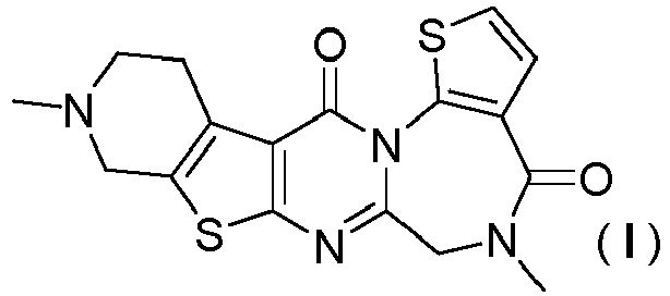
- An A-type crystal of 5,10-dimethyl-5,6,9,10,11,12-hexahydropyrido[4",3":4',5']thieno[2',3':4,5]pyrimido[1,2-a]thieno[3,2-f][1,4]diazepine-4,13-dione monohydrochloride represented by formula (I):

- A pharmaceutical composition comprising the salt according to claim 1 or the crystal according to any one of claims 2 to 14.
Applications Claiming Priority (2)
| Application Number | Priority Date | Filing Date | Title |
|---|---|---|---|
| JP2019039349 | 2019-03-05 | ||
| PCT/JP2020/008889 WO2020179781A1 (en) | 2019-03-05 | 2020-03-03 | Salt of pentacyclic compound and crystals thereof |
Publications (2)
| Publication Number | Publication Date |
|---|---|
| EP3936189A1 true EP3936189A1 (en) | 2022-01-12 |
| EP3936189A4 EP3936189A4 (en) | 2022-12-14 |
Family
ID=72337486
Family Applications (1)
| Application Number | Title | Priority Date | Filing Date |
|---|---|---|---|
| EP20766851.8A Pending EP3936189A4 (en) | 2019-03-05 | 2020-03-03 | SALT OF PENTACYCLIC COMPOUND AND CRYSTALS THEREOF |
Country Status (11)
| Country | Link |
|---|---|
| US (1) | US11420980B2 (en) |
| EP (1) | EP3936189A4 (en) |
| JP (1) | JP7508441B2 (en) |
| KR (1) | KR102921161B1 (en) |
| CN (1) | CN113646046B (en) |
| AU (1) | AU2020233452B2 (en) |
| BR (1) | BR112021015979A2 (en) |
| CA (1) | CA3129764A1 (en) |
| MX (1) | MX2021009774A (en) |
| TW (2) | TWI849070B (en) |
| WO (1) | WO2020179781A1 (en) |
Families Citing this family (2)
| Publication number | Priority date | Publication date | Assignee | Title |
|---|---|---|---|---|
| EP3936190A4 (en) | 2019-03-05 | 2022-11-16 | Eisai R&D Management Co., Ltd. | HETEROCYCLIC PENTACYCLIC COMPOUND |
| PT119624A (en) * | 2024-07-29 | 2026-01-29 | Silva Escada Guilherme | Modular machine for bending PVC pipes. |
Family Cites Families (15)
| Publication number | Priority date | Publication date | Assignee | Title |
|---|---|---|---|---|
| US4187306A (en) * | 1977-07-26 | 1980-02-05 | Bayer Aktiengesellschaft | Benzodiazepine-diones, a process for their production and their use as medicaments |
| DD258234A1 (en) | 1987-03-05 | 1988-07-13 | Univ Leipzig | PROCESS FOR THE PREPARATION OF PYRIDO / 3 ', 2': 4,5 / THIENO / 2,3-E / IMIDAZO- / 1,2-C / PYRIMIDINES AND STRUCTURE ANALOGER -PYRIMIDO / 1,2-C / PYRIMIDINE BZW. -1,3-diazepino / 1.2-C / PYRIMIDINES |
| RU2117670C1 (en) | 1988-10-31 | 1998-08-20 | Эйсай Ко., Лтд. | Derivatives of triazolo[1,4]diazepine and methods of their synthesis |
| TW197442B (en) * | 1990-02-08 | 1993-01-01 | Pfizer | |
| US5756494A (en) * | 1992-07-24 | 1998-05-26 | Cephalon, Inc. | Protein kinase inhibitors for treatment of neurological disorders |
| US5621100A (en) * | 1992-07-24 | 1997-04-15 | Cephalon, Inc. | K-252a derivatives for treatment of neurological disorders |
| KR100203456B1 (en) * | 1995-06-20 | 1999-06-15 | 한승수 | Alzheimer's disease treatment agent for Alzheimer's disease containing dihydroebodiamine-HC1 as an active ingredient |
| CL2004000348A1 (en) | 1997-04-15 | 2005-01-14 | Cephalon Inc & Kyowa Hakko Kog | COMPOSITE DERIVED FROM BUR-N-SUBSTITUTED STAUROSPORINE, USEFUL TO POWER THE FUNCTION OF COLLERGED, ESTRIATED NEURONES, BASAL AND SENSORY PROSENCEPHALUS. |
| CL2004000352A1 (en) | 1998-05-26 | 2005-05-27 | Cephalon Inc & Kyowa Hakko Kog | USE OF COMPOUNDS DERIVED FROM STAUROSPORINE TO POTENTIATE THE FUNCTION OF COLLERGENIC, ESTRIATED NEURONES, BASAL AND SENSORY PROSENCEFALO. |
| FR2779652B1 (en) | 1998-06-15 | 2001-06-08 | Sod Conseils Rech Applic | USE OF DIAZEPINES FOR THE PREPARATION OF MEDICINES FOR THE TREATMENT OF CONDITIONS OR DISEASES IN WHICH ONE OF THE SOMATOSTATIN RECEPTORS IS INVOLVED |
| AU2003299378A1 (en) | 2002-10-11 | 2004-05-04 | Board Of Regents, The University Of Texas System | Method and compounds for inhibiting hec1 activity for the treatment of proliferative diseases |
| MX2007004640A (en) | 2004-10-20 | 2007-06-08 | Hoffmann La Roche | Halogen substituted benzodiazepine derivatives. |
| US9314318B2 (en) * | 2008-08-26 | 2016-04-19 | Zest Ip Holdings, Llc | Dental anchor apparatus and method |
| MA50093B1 (en) * | 2017-09-07 | 2022-05-31 | Eisai R&D Man Co Ltd | Pentacyclic compound |
| EP3936190A4 (en) | 2019-03-05 | 2022-11-16 | Eisai R&D Management Co., Ltd. | HETEROCYCLIC PENTACYCLIC COMPOUND |
-
2020
- 2020-03-03 CA CA3129764A patent/CA3129764A1/en active Pending
- 2020-03-03 EP EP20766851.8A patent/EP3936189A4/en active Pending
- 2020-03-03 KR KR1020217025603A patent/KR102921161B1/en active Active
- 2020-03-03 MX MX2021009774A patent/MX2021009774A/en unknown
- 2020-03-03 TW TW109106928A patent/TWI849070B/en active
- 2020-03-03 WO PCT/JP2020/008889 patent/WO2020179781A1/en not_active Ceased
- 2020-03-03 BR BR112021015979-7A patent/BR112021015979A2/en unknown
- 2020-03-03 TW TW113114065A patent/TWI870286B/en active
- 2020-03-03 AU AU2020233452A patent/AU2020233452B2/en active Active
- 2020-03-03 CN CN202080014060.2A patent/CN113646046B/en active Active
- 2020-03-03 JP JP2021504108A patent/JP7508441B2/en active Active
- 2020-09-15 US US17/021,544 patent/US11420980B2/en active Active
Also Published As
| Publication number | Publication date |
|---|---|
| US11420980B2 (en) | 2022-08-23 |
| JPWO2020179781A1 (en) | 2020-09-10 |
| KR20210135223A (en) | 2021-11-12 |
| AU2020233452B2 (en) | 2025-04-10 |
| CN113646046B (en) | 2024-05-17 |
| WO2020179781A1 (en) | 2020-09-10 |
| JP7508441B2 (en) | 2024-07-01 |
| AU2020233452A1 (en) | 2021-09-02 |
| MX2021009774A (en) | 2021-09-08 |
| TWI870286B (en) | 2025-01-11 |
| KR102921161B1 (en) | 2026-02-02 |
| BR112021015979A2 (en) | 2021-10-05 |
| TW202430531A (en) | 2024-08-01 |
| CN113646046A (en) | 2021-11-12 |
| US20210024541A1 (en) | 2021-01-28 |
| CA3129764A1 (en) | 2020-09-10 |
| EP3936189A4 (en) | 2022-12-14 |
| TWI849070B (en) | 2024-07-21 |
| TW202100531A (en) | 2021-01-01 |
Similar Documents
| Publication | Publication Date | Title |
|---|---|---|
| JP5599610B2 (en) | Treatment of neurodegenerative diseases by inhibition of Hsp90 | |
| TW202345809A (en) | Piperazine substituted indazole compounds as inhibitors of parg | |
| EP3680243B1 (en) | Pentacyclic compound | |
| CN104039786A (en) | PAK inhibitors for the treatment of fragile X syndrome | |
| US11420980B2 (en) | Salt of pentacyclic compound and crystal thereof | |
| RU2820938C2 (en) | Pentacyclic compound salt and crystal thereof | |
| KR102352624B1 (en) | Pyridopyrimidine derivatives as lrrk2 (leucine rich repeat kinase 2) inhinitor | |
| BR112020003197B1 (en) | Pentacyclic compound | |
| HK40022802B (en) | Pentacyclic compound | |
| HK40022802A (en) | Pentacyclic compound |
Legal Events
| Date | Code | Title | Description |
|---|---|---|---|
| STAA | Information on the status of an ep patent application or granted ep patent |
Free format text: STATUS: THE INTERNATIONAL PUBLICATION HAS BEEN MADE |
|
| PUAI | Public reference made under article 153(3) epc to a published international application that has entered the european phase |
Free format text: ORIGINAL CODE: 0009012 |
|
| STAA | Information on the status of an ep patent application or granted ep patent |
Free format text: STATUS: REQUEST FOR EXAMINATION WAS MADE |
|
| 17P | Request for examination filed |
Effective date: 20210811 |
|
| AK | Designated contracting states |
Kind code of ref document: A1 Designated state(s): AL AT BE BG CH CY CZ DE DK EE ES FI FR GB GR HR HU IE IS IT LI LT LU LV MC MK MT NL NO PL PT RO RS SE SI SK SM TR |
|
| DAV | Request for validation of the european patent (deleted) | ||
| DAX | Request for extension of the european patent (deleted) | ||
| A4 | Supplementary search report drawn up and despatched |
Effective date: 20221116 |
|
| RIC1 | Information provided on ipc code assigned before grant |
Ipc: A61K 31/551 20060101ALI20221110BHEP Ipc: C07D 495/22 20060101ALI20221110BHEP Ipc: A61P 43/00 20060101ALI20221110BHEP Ipc: A61P 25/28 20060101AFI20221110BHEP |
|
| P01 | Opt-out of the competence of the unified patent court (upc) registered |
Effective date: 20230512 |
|
| STAA | Information on the status of an ep patent application or granted ep patent |
Free format text: STATUS: EXAMINATION IS IN PROGRESS |
|
| 17Q | First examination report despatched |
Effective date: 20251013 |