EP3919519A1 - Monoclonal antibody specifically binding to ccsp-2 and use thereof - Google Patents
Monoclonal antibody specifically binding to ccsp-2 and use thereof Download PDFInfo
- Publication number
- EP3919519A1 EP3919519A1 EP20747642.5A EP20747642A EP3919519A1 EP 3919519 A1 EP3919519 A1 EP 3919519A1 EP 20747642 A EP20747642 A EP 20747642A EP 3919519 A1 EP3919519 A1 EP 3919519A1
- Authority
- EP
- European Patent Office
- Prior art keywords
- seq
- cancer
- chain variable
- ccsp
- variable region
- Prior art date
- Legal status (The legal status is an assumption and is not a legal conclusion. Google has not performed a legal analysis and makes no representation as to the accuracy of the status listed.)
- Pending
Links
Images







Classifications
-
- C—CHEMISTRY; METALLURGY
- C07—ORGANIC CHEMISTRY
- C07K—PEPTIDES
- C07K16/00—Immunoglobulins [IG], e.g. monoclonal or polyclonal antibodies
- C07K16/18—Immunoglobulins [IG], e.g. monoclonal or polyclonal antibodies against material from animals or humans
- C07K16/28—Immunoglobulins [IG], e.g. monoclonal or polyclonal antibodies against material from animals or humans against receptors, cell surface antigens or cell surface determinants
- C07K16/30—Immunoglobulins [IG], e.g. monoclonal or polyclonal antibodies against material from animals or humans against receptors, cell surface antigens or cell surface determinants from tumour cells
-
- C—CHEMISTRY; METALLURGY
- C07—ORGANIC CHEMISTRY
- C07K—PEPTIDES
- C07K16/00—Immunoglobulins [IG], e.g. monoclonal or polyclonal antibodies
- C07K16/18—Immunoglobulins [IG], e.g. monoclonal or polyclonal antibodies against material from animals or humans
- C07K16/22—Immunoglobulins [IG], e.g. monoclonal or polyclonal antibodies against material from animals or humans against growth factors ; against growth regulators
-
- C—CHEMISTRY; METALLURGY
- C07—ORGANIC CHEMISTRY
- C07K—PEPTIDES
- C07K16/00—Immunoglobulins [IG], e.g. monoclonal or polyclonal antibodies
- C07K16/18—Immunoglobulins [IG], e.g. monoclonal or polyclonal antibodies against material from animals or humans
- C07K16/32—Immunoglobulins [IG], e.g. monoclonal or polyclonal antibodies against material from animals or humans against translation products of oncogenes
-
- G—PHYSICS
- G01—MEASURING; TESTING
- G01N—INVESTIGATING OR ANALYSING MATERIALS BY DETERMINING THEIR CHEMICAL OR PHYSICAL PROPERTIES
- G01N33/00—Investigating or analysing materials by specific methods not covered by groups G01N1/00 - G01N31/00
- G01N33/48—Biological material, e.g. blood, urine; Haemocytometers
- G01N33/50—Chemical analysis of biological material, e.g. blood, urine; Testing involving biospecific ligand binding methods; Immunological testing
- G01N33/53—Immunoassay; Biospecific binding assay; Materials therefor
- G01N33/575—Immunoassay; Biospecific binding assay; Materials therefor for cancer
-
- G—PHYSICS
- G01—MEASURING; TESTING
- G01N—INVESTIGATING OR ANALYSING MATERIALS BY DETERMINING THEIR CHEMICAL OR PHYSICAL PROPERTIES
- G01N33/00—Investigating or analysing materials by specific methods not covered by groups G01N1/00 - G01N31/00
- G01N33/48—Biological material, e.g. blood, urine; Haemocytometers
- G01N33/50—Chemical analysis of biological material, e.g. blood, urine; Testing involving biospecific ligand binding methods; Immunological testing
- G01N33/53—Immunoassay; Biospecific binding assay; Materials therefor
- G01N33/575—Immunoassay; Biospecific binding assay; Materials therefor for cancer
- G01N33/57535—Immunoassay; Biospecific binding assay; Materials therefor for cancer of the large intestine, e.g. colon, rectum or anus
-
- G—PHYSICS
- G01—MEASURING; TESTING
- G01N—INVESTIGATING OR ANALYSING MATERIALS BY DETERMINING THEIR CHEMICAL OR PHYSICAL PROPERTIES
- G01N33/00—Investigating or analysing materials by specific methods not covered by groups G01N1/00 - G01N31/00
- G01N33/48—Biological material, e.g. blood, urine; Haemocytometers
- G01N33/50—Chemical analysis of biological material, e.g. blood, urine; Testing involving biospecific ligand binding methods; Immunological testing
- G01N33/53—Immunoassay; Biospecific binding assay; Materials therefor
- G01N33/575—Immunoassay; Biospecific binding assay; Materials therefor for cancer
- G01N33/5758—Immunoassay; Biospecific binding assay; Materials therefor for cancer involving compounds serving as markers for tumours, cancers or neoplasias, e.g. cellular determinants, receptors, heat shock/stress proteins, A-protein, oligosaccharides or metabolites
- G01N33/5759—Immunoassay; Biospecific binding assay; Materials therefor for cancer involving compounds serving as markers for tumours, cancers or neoplasias, e.g. cellular determinants, receptors, heat shock/stress proteins, A-protein, oligosaccharides or metabolites involving compounds localised on the membrane of tumour or cancer cells
-
- C—CHEMISTRY; METALLURGY
- C07—ORGANIC CHEMISTRY
- C07K—PEPTIDES
- C07K2317/00—Immunoglobulins specific features
- C07K2317/20—Immunoglobulins specific features characterized by taxonomic origin
- C07K2317/23—Immunoglobulins specific features characterized by taxonomic origin from birds
-
- C—CHEMISTRY; METALLURGY
- C07—ORGANIC CHEMISTRY
- C07K—PEPTIDES
- C07K2317/00—Immunoglobulins specific features
- C07K2317/50—Immunoglobulins specific features characterized by immunoglobulin fragments
- C07K2317/56—Immunoglobulins specific features characterized by immunoglobulin fragments variable (Fv) region, i.e. VH and/or VL
-
- C—CHEMISTRY; METALLURGY
- C07—ORGANIC CHEMISTRY
- C07K—PEPTIDES
- C07K2317/00—Immunoglobulins specific features
- C07K2317/50—Immunoglobulins specific features characterized by immunoglobulin fragments
- C07K2317/56—Immunoglobulins specific features characterized by immunoglobulin fragments variable (Fv) region, i.e. VH and/or VL
- C07K2317/565—Complementarity determining region [CDR]
-
- C—CHEMISTRY; METALLURGY
- C07—ORGANIC CHEMISTRY
- C07K—PEPTIDES
- C07K2317/00—Immunoglobulins specific features
- C07K2317/60—Immunoglobulins specific features characterized by non-natural combinations of immunoglobulin fragments
- C07K2317/62—Immunoglobulins specific features characterized by non-natural combinations of immunoglobulin fragments comprising only variable region components
- C07K2317/622—Single chain antibody (scFv)
-
- C—CHEMISTRY; METALLURGY
- C07—ORGANIC CHEMISTRY
- C07K—PEPTIDES
- C07K2317/00—Immunoglobulins specific features
- C07K2317/90—Immunoglobulins specific features characterized by (pharmaco)kinetic aspects or by stability of the immunoglobulin
- C07K2317/92—Affinity (KD), association rate (Ka), dissociation rate (Kd) or EC50 value
-
- C—CHEMISTRY; METALLURGY
- C07—ORGANIC CHEMISTRY
- C07K—PEPTIDES
- C07K2319/00—Fusion polypeptide
-
- G—PHYSICS
- G01—MEASURING; TESTING
- G01N—INVESTIGATING OR ANALYSING MATERIALS BY DETERMINING THEIR CHEMICAL OR PHYSICAL PROPERTIES
- G01N2333/00—Assays involving biological materials from specific organisms or of a specific nature
- G01N2333/435—Assays involving biological materials from specific organisms or of a specific nature from animals; from humans
- G01N2333/46—Assays involving biological materials from specific organisms or of a specific nature from animals; from humans from vertebrates
- G01N2333/47—Assays involving proteins of known structure or function as defined in the subgroups
-
- G—PHYSICS
- G01—MEASURING; TESTING
- G01N—INVESTIGATING OR ANALYSING MATERIALS BY DETERMINING THEIR CHEMICAL OR PHYSICAL PROPERTIES
- G01N2333/00—Assays involving biological materials from specific organisms or of a specific nature
- G01N2333/435—Assays involving biological materials from specific organisms or of a specific nature from animals; from humans
- G01N2333/475—Assays involving growth factors
- G01N2333/485—Epidermal growth factor [EGF] (urogastrone)
Definitions
- the present invention relates to a monoclonal antibody specifically binding to an EGF2 domain of human colon cancer secreted protein-2 (CCSP-2), or an antigen-binding fragment thereof.
- CCSP-2 human colon cancer secreted protein-2
- the large intestine belongs to the digestive system and is an organ located between the small intestine and the anus.
- the total length is approximately 1.5 meters on average, and classified into the cecum, the ascending colon, the transverse colon, the descending colon, the sigmoid colon and the rectum from the right.
- Colorectal cancer is a malignant tumor that occurs in the colon and rectum, and malignant tumor cells (cancer cells) mainly arise from epithelial cells.
- colon cancer cancer that occurs in the rectum
- rectal cancer cancer that occurs in the rectum
- these are collectively called colorectal cancer.
- colorectal cancer Another reason for a continuous increase in colorectal cancer mortality is that there are few initial symptoms of colorectal cancer, and therefore, the early detection rate of colorectal cancer in Korea is low as less than 10%.
- Many colorectal cancer patients are diagnosed with metastatic colorectal cancer in stages 3 and 4, when metastasis to other organs begins.
- the survival rate of Stage 3 colorectal cancer is quite low, 28%, and the survival rate of Stage 4 colorectal cancer is 6%, which is lower than the survival rate of Stage 3 colorectal cancer. Accordingly, the early diagnosis of colorectal cancer is very important.
- a double contrast barium-enema is an examination method of inserting a small tube through the anus after cleaning the large intestine to inject a contrast agent called barium, coating the wall of the large intestine with a thin layer of barium while expanding the large intestine by injecting air, and taking images using an X-ray fluoroscope.
- Advantages of this method are identification of the overall contour and shape of the large intestine, easy identification of the overall location of colorectal cancer, and easy testing for the changes in the large intestinal wall such as inflammatory or ischemic changes and the distal end of the small intestine.
- tissue cannot be obtained with testing, and accuracy for small polyps is inferior to colonoscopy.
- colonoscopy is the most accurate method for diagnosing a colorectal disease, and this is because a doctor can directly observe the area of bleeding and the surface of a lesion, and determine the condition of tissue.
- a biopsy is also possible at the same time as an endoscopy, and when a conscious sedation endoscopy is performed while a patient is sleeping by intravenously injecting a sedative acting only for a short period of time, testing can be done without discomfort.
- Colonoscopy is the best diagnostic tool for finding colorectal cancer, but the frequency at which polyps missed in the testing are detected as having advanced to cancer in the patient's next test is high. Since polyps larger than 1 cm have a relatively and significantly increasing possibility of progression to cancer over time, it is necessary to improve the technology so as not to miss a small polyp during an endoscopy, and to attempt to avoid missing lesions behind flexures and mucosal folds with sufficient margin during withdrawal inspection. As one of these attempts, research is being conducted on the development of a biomarker and a marker, which are used in molecular imaging, to improve a tumor detection rate.
- a colon cancer secreted protein (e.g., CCSP-2) is a colorectal cancer-specific marker (biomarker), and information on the gene encoding the same is well known in the related art.
- the CCSP-2 may be human CCSP-2, and the information on the human CCSP-2 protein is registered at National Center for Biotechnology Information (NCBI) under Accession No. AAT77225.1, and the information on a gene encoding the same is registered at NCBI under Accession No. AY572972.1.
- NCBI National Center for Biotechnology Information
- the function and role of the CCSP-2 are not well known, but at the RNA level, it is known that the average expression level thereof in colorectal cancer cells and/or tissue is 78-fold higher than in normal tissue (colorectal cancer cell-free tissue).
- the CCSP-2 is a secretory protein and an extracellular matrix (ECM) protein, which is increased in colorectal cancer-specific expression, and has advantages of labeling colorectal cancer tissue through targeting CCSP-2 protein accumulated in colorectal cancer tissue as well as diagnosis of colorectal cancer using blood. Accordingly, when the expression and/or expression level of CCSP-2 is(are) measured at the gene and/or protein level(s), the presence of colorectal cancer and/or the degree of progression of colorectal cancer may be accurately and easily confirmed.
- ECM extracellular matrix
- the inventors developed a peptide specifically binding to CCSP-2, confirmed the effect of specifically detecting CCSP-2 at the protein level, and proved that colorectal cancer and/or colorectal cancer cells and/or tissue can be detected, which is disclosed in Korean Patent Application Publication No. 10-2018-0060184 .
- the inventors While continuing research on the technology of specifically detecting the CCSP-2, the inventors developed a monoclonal antibody specifically binding to the EGF2 domain of CCSP-2, and confirmed that the antibody is significantly improved in affinity and particularly more improved in specificity, compared with a conventional peptide specifically binding to CCSP-2, and thus the present invention was completed.
- the present invention is directed to providing a monoclonal antibody specifically binding to colon cancer secreted protein-2 (CCSP-2), or an antigen-binding fragment thereof.
- CCSP-2 colon cancer secreted protein-2
- the present invention is also directed to providing a polynucleotide encoding the monoclonal antibody or an antigen-binding fragment thereof, an expression vector including the polynucleotide and a transformant into which the vector is introduced.
- the present invention is also directed to providing a composition for diagnosing cancer, which includes the monoclonal antibody or the antigen-binding fragment thereof.
- the present invention is also directed to providing a kit for diagnosing cancer, which includes the monoclonal antibody or the antigen-binding fragment thereof.
- the present invention is also directed to providing a method of providing information for cancer diagnosis, which includes detecting a CCSP-2 protein in a biological sample isolated from a subject suspected of having cancer using the monoclonal antibody or the antigen-binding fragment thereof through an antigen-antibody reaction.
- the present invention provides a monoclonal antibody specifically binding to the EGF2 domain of human CCSP-2, which includes a heavy chain variable region and a light chain variable region selected from the group consisting of 1) to 4) below, or an antigen-binding fragment thereof:
- the antigen-binding fragment may be selected from the group consisting of Fab, Fab', F(ab')2, scFv, Fv, dsFv, a diabody, Fd and Fd'.
- the monoclonal antibody or the antigen-binding fragment thereof may include a heavy chain variable region and a light chain variable region, which are selected from the group consisting of a heavy chain variable region including a polypeptide sequence represented by SEQ ID NO: 25 and a light chain variable region including a polypeptide sequence represented by SEQ ID NO: 26; a heavy chain variable region including a polypeptide sequence represented by SEQ ID NO: 27 and a light chain variable region including a polypeptide sequence represented by SEQ ID NO: 28; a heavy chain variable region including a polypeptide sequence represented by SEQ ID NO: 29 and a light chain variable region including a polypeptide sequence represented by SEQ ID NO: 30; and a heavy chain variable region including a polypeptide sequence represented by SEQ ID NO: 31 and a light chain variable region including a polypeptide sequence represented by SEQ ID NO: 32.
- the present invention also provides a polynucleotide encoding the monoclonal antibody, or an antigen-binding fragment thereof.
- the present invention also provides an expression vector including the polynucleotide.
- the present invention also provides a transformant into which the expression vector is introduced, excluding a human.
- the present invention also provides a composition for diagnosing cancer, which includes the monoclonal antibody or the antigen-binding fragment thereof.
- the cancer may be selected from the group consisting of esophageal cancer, gastric cancer, colorectal cancer, rectal cancer, oral cancer, pharyngeal cancer, laryngeal cancer, lung cancer, colon cancer, breast cancer, cervical cancer, endometrial cancer, ovarian cancer, prostate cancer, testicular cancer, bladder cancer, kidney cancer, liver cancer, pancreatic cancer, bone cancer, connective tissue cancer, skin cancer, brain cancer, thyroid cancer, leukemia, Hodgkin's disease, soft tissue sarcoma, lymphoma and multiple myeloma hematologic cancer.
- the present invention also provides a kit for diagnosing cancer, which includes the composition.
- the present invention also provides a method of providing information for cancer diagnosis, which includes detecting CCSP-2 protein in a biological sample isolated from a subject suspected of having cancer using the monoclonal antibody or the antigen-binding fragment thereof through an antigen-antibody reaction.
- CCSP-2 As a monoclonal antibody of the present invention or an antigen-binding fragment thereof specifically binds to the EGF2 domain of CCSP-2, compared with a conventional peptide specifically binding to CCSP-2, affinity can be significantly improved, and compared with a conventional antibody against CCSP-2, specificity can be more improved. Accordingly, CCSP-2 is specifically detected at the protein level and thus enables accurate diagnosis in the future.
- the present invention provides a monoclonal antibody specifically binding to the EGF2 domain of human colon cancer secreted protein-2 (CCSP-2), or an antigen-binding fragment.
- CCSP-2 is specific for colorectal cancer (e.g., expressed specifically in colorectal cancer).
- the inventors identified antibodies specifically recognizing CCSP-2, and the amino acid sequence of a heavy chain variable region thereof, the amino acid sequence of a light chain variable region thereof and coding nucleotide sequences thereof.
- colorectal cancer and/or colorectal cancer cells and/or tissue can be accurately diagnosed by specifically detecting CCSP-2 by providing a monoclonal antibody or an antigen-binding fragment thereof specifically binding to CCSP-2, and particularly, it may be effectively applied to the endoscopic diagnosis of colorectal cancer by visualizing colorectal tissue with the antibody conjugated with a visible labeling material.
- antibody refers to a protein molecule serving as a receptor specifically recognizing an antigen, which includes an immunoglobulin molecule immunologically having reactivity to a specific antigen, and includes a polyclonal antibody, a monoclonal antibody, and an antigen-binding fragment (antibody fragment) of an antibody molecule as well as a complete antibody form.
- the term includes a chimeric antibody, a humanized antibody, a bivalent or bispecific molecule (e.g., bispecific antibody), a diabody, a triabody, and a tetrabody.
- the term "complete antibody” has a structure having two full-length light chains and two full-length heavy chains, in which each light chain is connected with a heavy chain by a disulfide bond.
- the complete antibody includes IgA, IgD, IgE, IgM and IgG, in which IgG has subtypes, for example, IgG1, IgG2, IgG3 and IgG4.
- antigen-binding fragment of an antibody refers to a fragment that retains an antigen-antibody binding function in a complete antibody molecule, and includes Fab, Fab', F(ab')2, scFv, Fv, dsFv, a diabody, Fd and Fd'.
- the Fab has a structure having light and heavy chain variable regions, and a constant region of the light chain and the first constant region (CHI domain) of the heavy chain, and has one antigen-binding site.
- An Fab' is distinguished from Fab in that there is a hinge region having one or more cysteine residues at the C terminus of the CH1 domain of the heavy chain.
- An F(ab')2 antibody is generated by forming a disulfide bond between cysteine residues of the hinge region of Fab'.
- a variable fragment (Fv) refers to the smallest antibody fragment only having a heavy chain variable region and a light chain variable region.
- Double chain Fv (dsFv) is formed by connecting a heavy chain variable region and a light chain variable region using a disulfide bond, and single chain Fv (scFv) generally has the same structure as a dimer such as dsFv since a heavy chain variable region is connected with a light chain variable region by a covalent bond using a peptide linker, or may be directly connected to the C-terminus.
- a diabody is a complex of two or more polypeptide chains or proteins, and refers to a complex including at least one VL domain and at least one VH domain or fragments thereof, and having both domains in a single polypeptide chain.
- the diabody includes a molecule including an Fc or hinge-Fc domain.
- the polypeptide chains of such a complex may be the same or different, in other words, the diabody may be a mono polymer or a hetero polymer.
- the antigen-binding fragment may be obtained using proteases (e.g., Fab may be obtained by restriction of a complete antibody by papain, and a F(ab') 2 fragment may be obtained by cleavage by a pepsin), and preferably is manufactured using gene recombination technology.
- proteases e.g., Fab may be obtained by restriction of a complete antibody by papain, and a F(ab') 2 fragment may be obtained by cleavage by a pepsin
- heavy chain used herein refers to both a full-length heavy chain which consists of a variable region domain VH and three constant region domains CH1, CH2 and CH3, and a fragment thereof, which includes an amino acid sequence having a sufficient variable region sequence imparting specificity to an antigen.
- light chain used herein refers to both a full-length light chain which consists of a variable region domain VL and a constant region domain CL, or a fragment thereof, which includes an amino acid sequence having a sufficient variable region sequence imparting specificity to an antigen.
- an immunoglobulin refers to an antibody molecule of single molecular composition obtained from substantially the same antibody population, and exhibits single binding specificity and affinity for a specific epitope.
- an immunoglobulin has a heavy chain and a light chain, in which each of the heavy chain and the light chain includes a constant region and a variable region (these regions are also known as domains).
- the variable regions of the light chain and heavy chain includes three variable regions called complementarity-determining regions (hereinafter, referred to as "CDRs") and four framework regions (FRs).
- CDRs mainly serve to bind to an epitope of an antigen.
- the CDRs of each chain are typically called CDR1, CDR2 and CDR3 sequentially from the N-terminus, and also distinguished by a chain in which a specific CDR is located.
- CDR complementarity determining region
- the monoclonal antibody may be in the form of a chimeric antibody or humanized antibody which has reduced immunogenicity to be applied to a human as described above.
- chimeric antibody is an antibody formed by combining a variable region of a heterogeneous antibody which is derived from a mouse or chicken and a constant region of a human antibody through DNA recombination technology, and the immune reaction of the chimeric antibody is considerably improved compared with a heterogeneous antibody derived from a mouse or chicken, and thus can be used clinically.
- humanized antibody refers to an antibody formed by transplanting all or a part of the CDR sequence of a heterogeneous monoclonal antibody derived from a mouse or chicken into a human antibody, a humanized variable region may be formed by recombining CDRs of a chicken or mouse monoclonal antibody with a human antibody-derived FR, and the human antibody may be formed by recombining the variable region with a constant region of a preferred human antibody, but the present invention is not limited thereto.
- the affinity of a humanized antibody is decreased when only the chicken or mouse-derived CDRs are inserted, the affinity may be improved by substituting an FR amino acid residue, which may be considered to affect the three-dimensional structure of a CDR, with an amino acid of a chicken or mouse antibody, but the present invention is not limited thereto.
- CCSP-2 colon cancer secreted protein-2
- the term "monoclonal antibody specifically binding to the EGF2 domain of colon cancer secreted protein-2 (CCSP-2)” refers to an antibody capable of specifically binding to CCSP-2, and may be interchangeably used with an anti-CCSP-2 antibody.
- the monoclonal antibody specifically binding to the CCSP-2 protein may be, without limitation, any monoclonal antibody that binds to CCSP-2 to inhibit the biological activity of CCSP-2.
- the form of monoclonal antibody may include both of a complete antibody and an antigen-binding fragment as described above, and may be a chimeric antibody or a humanized antibody, but the present invention is not limited thereto.
- the monoclonal antibody of the present invention may specifically bind to the EGF2 domain of CCSP-2 to inhibit signaling by CCSP-2, resulting in suppression of biological activity, and thus may be effectively used for prevention or treatment of a disease such as cancer mediated by CCSP-2.
- CCSP-2 overexpression is reported as a specific phenomenon in cancer, and the antibody of the present invention capable of specifically binding to CCSP-2 has diagnosis ability with high sensitivity and specificity in the diagnosis of cancer, the antibody may be effectively used in diagnosis of cancer.
- an antigen-binding fragment specifically binding to CCSP-2 of the present invention was formed using the EGF2 site of CCSP-2 as an antigen protein.
- the monoclonal antibody specifically binding to the EGF2 domain of the human CCSP-2, or an antigen-binding fragment thereof may include any one pair of a heavy chain variable region and a light chain variable region selected from the group consisting of 1) to 4) below, but the present invention is not limited thereto:
- the antigen-binding fragment is preferably Fab, Fab', F(ab')2, scFv, Fv, dsFv, a diabody, Fd, Fd', a light chain or heavy chain including a CDR region of the present invention, or a variable domain including a CDR region of the present invention, but the present invention is not limited thereto.
- the monoclonal antibody or an antigen-binding fragment thereof preferably includes one pair of a heavy chain variable region and a light chain variable region selected from the group consisting of a heavy chain variable region including a polypeptide sequence represented by SEQ ID NO: 25 and a light chain variable region including a polypeptide sequence represented by SEQ ID NO: 26; a heavy chain variable region including a polypeptide sequence represented by SEQ ID NO: 27 and a light chain variable region including polypeptide sequence represented by SEQ ID NO: 28; a heavy chain variable region including a polypeptide sequence represented by SEQ ID NO: 29 and a light chain variable region including a polypeptide sequence represented by SEQ ID NO: 30; and a heavy chain variable region including a polypeptide sequence represented by SEQ ID NO: 31 and a light chain variable region including a polypeptide sequence represented by SEQ ID NO: 32.
- the antibody of the present invention or an antigen-binding fragment thereof also includes all mutants achieving the effects of the present invention, which are obtained through one or more mutations such as substitution, deletion, inversion or translocation of an antibody limited by the above-described sequence.
- the present invention provides a polynucleotide encoding the monoclonal antibody or the antigen-binding fragment thereof.
- the polynucleotide may be selected from the group consisting of SEQ ID Nos: 33 to 36, but the present invention is not limited thereto.
- the present invention also provides an expression vector including the polynucleotide and a transformant into which the vector is introduced.
- the expression vector including the polynucleotide encoding the monoclonal antibody according to the present invention may be, but is not particularly limited to, a vector capable of replicating and/or expressing the polynucleotide in eukaryotic or prokaryotic cells including mammalian cells (e.g., human, monkey, rabbit, rat, hamster, and mouse cells), plant cells, yeast cells or bacterial cells (e.g., Escherichia coli ), and preferably, a vector that is operably linked to a suitable promoter such that the nucleotide can be expressed in host cells and includes at least one selection marker.
- the expression vector may be a form in which the polynucleotide is introduced into a phage, a plasmid, a cosmid, a mini-chromosome, a virus or a retrovirus vector.
- the expression vector including a polynucleotide encoding the monoclonal antibody may be an expression vector which includes a polynucleotide encoding a heavy or light chain of the monoclonal antibody or an expression vector which includes both of polynucleotides encoding a heavy chain and a light chain.
- a transformant into which the expression vector of the present invention is introduced may be, but is not particularly limited to, bacterial cells, such as E. coli , Streptomyces or Salmonella typhimurium ; yeast cells; fungal cells such as Pichia pastoris ; insect cells such as Drozophila or Spodoptera Sf9 cells; animal cells such as Chinese hamster ovary (CHO) cells, SP2/0 (mouse myeloma), human lymphoblastoid, COS, NSO (mouse myeloma), 293T, Bowes melanoma cells, HT-1080, baby hamster kidney (BHK) cells, human embryonic kidney (HEK) cells or PERC.6 (human retinal cells); or plant cells, which are transformed by introducing the expression vector.
- bacterial cells such as E. coli , Streptomyces or Salmonella typhimurium
- yeast cells fungal cells such as Pichia pastoris
- insect cells such as Drozophila or
- introduction refers to a method of delivering the vector including a polynucleotide encoding the monoclonal antibody to host cells.
- the introduction may be performed by various methods known in the art, such as calcium phosphate-DNA co-precipitation, DEAE-dextran-mediated transfection, polybrene-mediated transfection, electroporation, microinjection, liposome fusion, Lipofectamine and protoplast fusion.
- transduction refers to the delivery of a desired material into cells using a viral particle through infection.
- transduction may be introduction of a vector into host cells via gene bombardment. In the present invention, the introduction may be interchangeably used with transformation.
- the present invention also provides a composition for diagnosing cancer, which includes the monoclonal antibody.
- cancer used herein may be any cancer expressing CCSP-2, and examples include, but are not limited to, esophageal cancer, gastric cancer, colorectal cancer, rectal cancer, oral cancer, pharyngeal cancer, laryngeal cancer, lung cancer, colon cancer, breast cancer, cervical cancer, endometrial cancer, ovarian cancer, prostate cancer, testicular cancer, bladder cancer, kidney cancer, liver cancer, pancreatic cancer, bone cancer, connective tissue cancer, skin cancer, brain cancer, thyroid cancer, leukemia, Hodgkin's disease, soft tissue sarcoma, lymphoma and multiple myeloma hematologic cancer, and is preferably colorectal cancer or pancreatic cancer.
- diagnosis refers to confirmation of the presence, severity (symptoms) and/or characteristics of a pathological state.
- diagnosis of colorectal cancer is confirmation of the pathological state of colorectal cancer, for example, the onset of colorectal cancer, the progression of a colorectal cancer lesion, the location of colorectal cancer (colorectal cancer cells), or the stage of colorectal cancer.
- it is important to accurately and rapidly distinguish colorectal cancer cells and/or tissue from normal cells or tissue.
- the present invention provides a monoclonal antibody specifically binding to CCSP-2, thereby specifically detecting CCSP-2 at the protein level, and thus it is possible to accurately diagnose colorectal cancer and/or colorectal cancer cells and/or tissue.
- colorectal tissue may be visualized using a labeling material for visualization, conjugated to the antibody, and thus can be effectively applied to endoscopic diagnosis of colorectal cancer.
- a disease associated with the expression or the progression of the expression of CCSP-2 or a CCSP-2-mediated disease, for example, cancer may be diagnosed using a diagnostic composition including the CCSP-2-specific monoclonal antibody of the present invention.
- the present invention also provides a kit for diagnosing cancer, which includes the composition for diagnosing cancer.
- kit for diagnosing cancer may be configured to further include a composition, solution or apparatus, which has one or more types of components suitable for an analysis method.
- CCSP-2 which is a biomarker specifically expressed in colorectal cancer
- a means useful for the measurement of CCSP-2 expression and/or a CCSP-2 expression level is provided, and therefore, this means may be effectively applied to the diagnosis of cancer, particularly, colorectal cancer.
- the antibody may be used in the visualization of cancer when used with various labeling materials, and applied in colorectal cancer endoscopy to provide more accurate information on the confirmation of the presence of colorectal cancer and lesions, and morphological observation of cancer sites, and may reduce a misdiagnosis rate related to colorectal cancer and contribute to early diagnosis.
- a bioactive material such as a drug
- the bioactive material may be specifically delivered to colorectal cancer, and therefore it may be used as a composition for the colorectal cancer-targeted delivery of the bioactive material.
- the present invention provides a method of providing information for cancer diagnosis, which includes detecting a CCSP-2 protein in a biological sample isolated from a subject suspected of having cancer using the monoclonal antibody through an antigen-antibody reaction.
- the CCSP-2 protein may be detected by reacting a monoclonal antibody specific for CCSP-2 of the present invention with a biological sample isolated from a subject suspected of having cancer and detecting the formation of an antigen-antibody complex, thereby providing information for cancer diagnosis. Since the CCSP-2 is overexpressed in various cancer cells such as colorectal cancer or pancreatic cancer, cancer may be diagnosed by comparing the expression level with that of a control such as normal cells or tissue, but the present invention is not limited thereto.
- biological sample used herein may refer to tissue, cells, whole blood, serum, plasma, a tissue autopsy sample (brain, skin, lymph node, spinal cord, etc.), cell culture supernatant, ruptured eukaryotic cells and a bacterial expression system, but the present invention is not limited thereto. These biological samples may be reacted with the antibody of the present invention with or without manipulation, thereby confirming the presence of a CCSP-2 protein or the presence or absence of cancer.
- antigen-antibody complex refers to a conjugate of a CCSP-2 protein antigen in a sample and the monoclonal antibody according to the present invention recognizing the same, and the formation of such an antigen-antibody complex may be detected by any method selected from the group consisting of a colorimetric method, an electrochemical method, a fluorometric method, luminometry, a particle counting method, visual assessment and a scintillation counting method.
- the method is not particularly limited thereto, and various applications may be used.
- the marker may be selected from the group consisting of an enzyme, a fluorescent material, a ligand, a luminescent material, a microparticle, and a radioactive isotope, but the present invention is not limited thereto.
- Enzymes used as a detection marker include acetylcholinesterase, alkaline phosphatase, ⁇ -D-galactosidase, horseradish peroxidase, and ⁇ -lactamase, fluorescent materials used as a detection marker include fluorescein, Eu 3+ , Eu 3+ chelate and cryptate, ligands used as a detection marker include a biotin derivative, luminescent materials used as a detection marker include acridinium ester and an isoluminol derivative, microparticles used as a detection marker include colloidal gold and colored latex, and radioactive isotopes used as a detection marker include 57Co, 3H, 1251, 125I-Bonton and Hunter reagents.
- the antigen-antibody complex may be detected by ELISA.
- ELISA includes various ELISA method, for example, direct ELISA using a labeled antibody recognizing an antigen attached to a solid support, indirect ELISA using a labeled secondary antibody recognizing a capture antibody in an antibody complex recognizing an antigen attached to a solid support, direct sandwich ELISA using another labeled antibody recognizing an antigen in an antibody-antigen complex attached to a solid support, and indirect sandwich ELISA reacting with another antibody recognizing an antigen in an antibody-antigen complex attached to a solid support and using a labeled secondary antibody recognizing the antibody.
- the monoclonal antibody may have a detection marker, and when there is no detection marker, these monoclonal antibodies may be captured and detected by treating another antibody having a detection marker.
- the anti-CCSP-2 antibody of the present invention specifically recognizes CCSP-2 using an antigen-antibody reaction, it was shown that the antibody of the present invention can be effectively used for diagnosis of various types of cancer such as colorectal cancer.
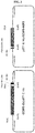
- FIG. 1 is a schematic diagram illustrating the strategy of cloning a variable region of a scFv antibody according to one embodiment of the present invention.
- FIG. 2 is a schematic diagram illustrating CCSP-2 antigens (NT, CT and EGF2) according to one embodiment of the present invention.
- CCSP-2-CT and CCSP-2-EGF2 contain the EGF2 domain.
- CCSP-2-CT (268 aa - 755 aa of AY57297231) was inserted into a pBT7-C-his vector (Bioneer, Korea) to be transduced into Origami2 (DE3) (Novagen, MA, USA).
- CCSP-2-EGF2 (712 aa - 755 aa of AY572972.1) was inserted into a pBT7-N-His vector (Bioneer, Korea) to be transduced into Origami2 (DE3) (Novagen, MA, USA).
- FIG. 3 is a schematic diagram illustrating respective expression vectors of CCSP-2-CT and CCSP-2-EGF2.
- the CCSP-2-CT and the CCSP-2-EGF2 were purified with Ni-NTA agarose beads (Qiagen, USA).
- Peripheral blood mononuclear cells were collected by treating 16 samples of human tonsil tissue with a Ficoll-Paque solution (GE Healthcare), and total RNA was extracted using a TRI reagent (Invitrogen). First strand cDNA was synthesized using oligo-dT primers and the Superscript ® III First-Strand Synthesis System (Invitrogen).
- a scFv library was constructed from cDNA obtained from the human mononuclear cells using primers in the following Table 1, specific for a heavy chain variable region and a light chain variable region of an immunoglobulin, and the Expand High Fidelity PCR system (Roche Molecular Systems).
- PCR was performed under the following conditions: 30 cycles, 15 sec at 94°C, 30 sec at 56 °C, 90 sec at 72 °C, and 10 min at 72 °C (final extension).
- a fragment with a length of approximately 350 bp was loaded on a 1.5% agarose gel for electrophoresis, and purified using a QIAGEN II Gel Extraction Kit (QIAGEN).
- first VL and VH products were randomly linked by overlap extension PCR.
- Each PCR was performed with a mixture having a final concentration of 100 ⁇ L, which was prepared by mixing 100 ng of the purified VL and VH products, 60 pmol of each primer, 10 ⁇ L of 10X reaction buffer, 8 ⁇ L of 2.5 mM dNTP and 0.5 ⁇ L of Taq DNA polymerase with water.
- PCR was performed under the following conditions: 25 cycles, 15 sec at 94 °C, 30 sec at 56 °C, 2 min at 72 °C, and 10 min at 72 °C (final extension).
- scFv fragment which is the PCR product
- pComb3X-SS vector The Scripps Research Institute
- 10 ⁇ g of the purified overlap PCR product was mixed with 360 units of SifI (16 units per ⁇ g DNA, Roche Molecular Systems), 20 ⁇ L of 10X reaction buffer and water, and adjusted to have a final volume of 200 ⁇ L.
- 20 ⁇ g of the pComb3X-SS vector was mixed with 120 units of SfiI (6 units per ⁇ g DNA), 20 ⁇ L of 10X reaction buffer and water, and adjusted to have a final volume of 200 ⁇ L. The mixture was cleaved for 8 hours at 50 °C.
- a scFv fragment having a length of approximately 700 bp and a vector having a length of 3,400 bp were loaded on a 1% agarose gel for electrophoresis, and purified using a QIAGEN II Gel Extraction Kit (QIAGEN).
- 1,400 ng of the SfiI-cleaved pComb3X vector and 700 ng of a scFv fragment were mixed with 40 ⁇ L of 5X ligase buffer, 10 ⁇ L of T4 DNA ligase (Invitrogen) and water so that a final volume became 200 ⁇ L, and incubated at 16 °C for 16 hours, followed by ligation. Afterward, the ligated product was precipitated with ethanol, and only the DNA pellet was dissolved in 15 ⁇ L of water.
- the ligated library sample was transformed into an E . coli strain, ER2738 (New England Biolabs, Inc.) through electroporation using a gene pulser (Bio-Rad Laboratories).
- the cells were mixed in 5 mL of a Super Broth (SB) medium at 37 °C, and incubated while stirring for 1 hour at 250 rpm. Afterward, 10 mL of SB medium and 3 ⁇ L of 100 mg/mL carbenicillin were added to the culture medium.
- 0.1 ⁇ L, 1 ⁇ L or 10 ⁇ L of the culture was plated on a Luria Broth (LB) agar plate containing 50 ⁇ g/mL of carbenicillin to determine a library size.
- LB Luria Broth
- the cell culture was further stirred for 1 hour, and then stirred for one more hour after 4.5 ⁇ L of 100 mg/mL carbenicillin was added.
- 2 mL of VCM13 helper phage (> 1,011 cfu/mL), 183 mL of pre-warmed SB and 92.5 ⁇ L of 100 mg/mL carbenicillin were added to the cell culture, and stirred at 250 rpm and 37 °C for 2 hours.
- 280 ⁇ L (50 mg/mL) of kanamycin was added to the cell culture, and stirred at 250 rpm and 37 °C overnight.
- the cell culture was centrifuged at 3,000 g and 4 °C using a highspeed centrifuge (Beckman, JA-10 rotor). Afterward, a bacterial pellet was stored to prepare phagemid DNA, and a supernatant was transferred to a sterilized centrifuge tube. Subsequently, 8 g of polyethylene glycol-8000 (PEG-8000, Sigma) and 6 g of sodium chloride (NaCl, Merck) were added and stored on ice for 30 minutes, and the supernatant was centrifuged at 15,000 g and 4 °C for 15 minutes. The supernatant was discarded, and the phage pellet was resuspended in Tris-buffered saline (TBS) containing 1% BSA.
- TBS Tris-buffered saline
- Example 1-2 Library panning on immobilized antigen (bio-panning)
- Bio-panning was performed using magnetic beads (Dynabeads M-270 Epoxy, Invitrogen). 3 ⁇ g of a C-terminus fragment EGF2 recombinant protein was agitated with 1 ⁇ 10 7 beads at room temperature for 20 hours to coat an antigen. The coated beads were washed with PBS four times, blocked with 3% BSA-containing phosphate-buffered saline (PBS) at room temperature for 1 hour, and incubated with the phage-displayed scFv obtained in Example 3-3 at room temperature for 2 hours.
- PBS BSA-containing phosphate-buffered saline
- a phage not binding to the bead-coated antigen after washing with 0.05% Tween20/PBS, the bound phage was eluted using 50 ⁇ L of 0.1M glycine/hydrogen chloride (0.1 M Glycine-HCl, pH 2.2), and neutralized with 3 ⁇ L of 2 M Tris-HCl (pH 9.1).
- E. coli ER2738 was transfected with the phage-containing supernatant, and rescued using a VCSM13 helper phage for overnight amplification of phages.
- a cell culture infected by the phage was plated on a LB agar plate containing 50 ⁇ g/mL of carbenicillin to determine input and output phage titers. The next day, PEG-8000 and NaCl were added to precipitate only phages, and the precipitated phages were used for the next round of bio-panning.
- Panning was performed up to four times by repeating the above-described process.
- phages having high affinity were selected and enriched by gradually increasing the number of washings, for example, once in the first round and eventually three times in the fourth round.
- Example 1-3 Selection of clones by phage ELISA
- ELISA was performed to confirm whether a randomly-selected phage-displayed scFv individual clone has binding ability to a CCSP-2-EGF2 recombinant protein (monomer).
- the CCSP-2-EGF2 recombinant protein (monomer) was diluted in 0.1 M NaHCO 3 buffer, and coated on a 96-well microtiter plate at 100 ng/well at 4 °C for 16 hours, and the next day, blocked with 3% BSA/PBS at 37 °C for 1 hour. Subsequently, a phage supernatant was mixed with 6% BSA/PBS at an equal amount, and incubated at 37 °C for 2 hours.
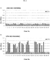
- a HRP conjugated anti-M13 antibody (a-M13-HRP, Pierce Chemical Co.) was diluted at a ratio of 1/5000 and added at 50 ⁇ L each to the plate, followed by incubation for 1 hour at 37 °C. After incubation, for a color reaction after washing, 1 ⁇ g/mL of 2,2'-azino-bis(3-ethylbenzothiazoline-6-sulphonic acid) (ABTS, Amresco) in 0.05 M citrate buffer and 0.1% H 2 O 2 were added to each well, followed by color development. And then, absorbance was measured at 405 nm. The result is shown in FIGS. 4A and 4B .
- FIGS. 4A and 4B show phage ELISA results obtained by ELISA on 48 clones from produced phages (output) of third and fourth bio-panning, respectively.
- a total of 11 types of scFv clones with different sequences were obtained by analyzing the genetic sequences of 24 clones with high absorbance among clones binding to the CCSP-2-EGF2 recombinant protein.
- Example 1-4 Preparation of anti-CCSP-2-EGF2 scFv hFc1 fusion protein
- IgG2 hinge and human IgG1 hybrid CH2-CH3 were inserted into a pCEP4 vector (Invitrogen) using restriction enzymes HindIII (New England Biolabs) and XhoI (New England Biolabs).
- a gene encoding anti-CCSP-2-EGF2 scFv was subcloned into the 5' end of the Fc region by two SfiI restriction sites.
- a human immunoglobulin Fc gene was subcloned into a mammalian expression vector.
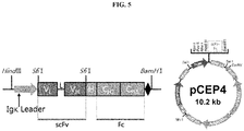
- genes encoding a human CH1 region and a region from an IgG2 hinge to a human IgG1 hybrid CH2-CH3 region were subcloned into a mammalian expression vector (see FIG. 5 ).
- FIG. 5 is a genetic map of an expression vector according to one embodiment of the present invention.
- Transfection was performed on an overexpressed recombinant protein.
- 2 ⁇ g of a mammalian expression vector and 4 ⁇ g of polyethyleneimine (PEI, Polysciences, Warrington, PA, USA) per mL of a culture volume were mixed in 150 mM sodium chloride (NaCl, Merck) corresponding to 1/10 of the cell culture volume, and maintained at room temperature for 15 minutes.
- PEI polyethyleneimine
- NaCl sodium chloride
- the mixture was added to mammalian cells, HEK293F (1 ⁇ 10 6 cells/mL, Invitrogen) used for a protein overexpression system, and cultured in a Freestyle TM 293 expression medium (Invitrogen) containing 100 U/mL penicillin and streptomycin (Invitrogen) for 6 days at 37 °C and 7% CO 2 while stirring at 135 rpm.
- HEK293F 1 ⁇ 10 6 cells/mL, Invitrogen
- Freestyle TM 293 expression medium Invitrogen
- penicillin and streptomycin Invitrogen
- a supernatant of the cell culture was harvested, and to purify an Fc-fusion protein, an affinity gel chromatography method using protein A was used. After isolated scFv was purified through dialysis using PBS buffer, SDS-PAGE was performed to confirm a molecular weight of approximately 55 kDa (see FIG. 6 ).
- FIG. 6 is an SDS-PAGE gel image showing the molecular weight of HN-1 of scFv produced by the present invention.
- CCSP-2 protein was prepared per domain, denaturation buffer was added thereto, followed by heating for 10 minutes. Then, SDS-PAGE electrophoresis was performed by loading a denatured sample on a prepared gel. After electrophoresis, the gel was not stained, and for an immunological assay, the protein was transferred to Trans-Blot Turbo TM Mini PVDF Transfer Packs (Bio-Rad) through an electro-transfer method. To inhibit a non-specific reaction, the gel was immersed in a 5% skim milk/TBST solution and treated on a stirrer for 1 hour.
- scFv obtained in Example 1-4-2 was diluted to a predetermined concentration, and as a secondary antibody, a HRP-conjugated anti-human immunoglobulin Fc secondary antibody (Invitrogen) was used. Each antibody reaction was performed for 1 hour, and after each step, the reaction product was washed with a 0.1% Tween 20-containing TBST solution three times. Finally, to confirm an antigen-antibody reaction, an enhanced chemiluminescent (ECL) substrate was used (see FIG. 7 ).
- ECL enhanced chemiluminescent
- FIG. 7 shows a western blotting result for confirming the binding ability per antigen (CCSP-2) domain of HN-1 in scFv of the present invention.
- the scFv antibody according to the present invention specifically binds to the EGF2 domain.
- the scFv obtained according to Example 4-2 was coated on a 96-well plate at 10 ⁇ g/mL, followed by blocking with 5% skim milk/TBST for 1 hour.
- the antigen-immobilized wells were then washed with a 0.05% Tween 20-containing PBS buffer three times, the CCSP-2 protein was dispensed at 900 ng/mL, 300 ng/mL, 100 ng/mL, 33.3 ng/mL or 11.1 ng/mL to induce an antigen-antibody reaction for 1 hour.
- a reaction was performed for 1 hour by sequentially using an anti-CCSP-2 primary antibody (Cloud-Clone Corp., TX, USA) diluted at a ratio of 1:500 and a HRP-conjugated anti-rabbit IgG secondary antibody (Cell Signaling) diluted at a ratio of 1:2000. Afterward, color development was induced at room temperature for 20 minutes by dispensing a tetramethyl benzidine (TMB) substrate into the wells, and absorbance was then measured at 450 nm using a microplate reader (see FIG. 8 ).
- TMB tetramethyl benzidine
- FIG. 8 is a graph showing an ELISA result obtained by analyzing the antigen (CCSP-2) binding ability of HN-1 in scFv of the present invention.
- the scFv antibody according to the present invention exhibited dose-dependent reactivity for the CCSP-2 protein.
- the scFv antibody constructed by the present invention has a size of approximately 55 kDa, and a function as an antibody with binding ability to an antigen.
- Example 2-1 Selection of anti-EGF2 antibody from immune antibody library
- CCSP-2-EGF2 protein prepared in Example 1 was emulsified in a RIBI+MPL+TDM+CWS adjuvant (Sigma, St. Louis, Mo, USA), which is a water-in-oil emulsion containing MPL (monophosphorylate lipid A species), which is a toxin-free endotoxin, TDW and CWS, which are cell wall components of mycobacteria, in 2% squalene, and then subcutaneously injected into three chickens.
- MPL monophosphorylate lipid A species
- TDW and CWS which are cell wall components of mycobacteria
- the titer of immunized chicken antibody was determined by enzyme-linked immunosorbent assay (ELISA) using a HRP-conjugated anti-chicken IgG(Y) monoclonal antibody (rabbit anti-chicken IgG(Y)-HRP, Millipore Corporation, Billeria, MA, USA) as a secondary antibody.
- ELISA enzyme-linked immunosorbent assay
- a scFv library for cDNA obtained from the immune system of a chicken was constructed using primers in Table 3 below, specific for a heavy chain variable region and a light chain variable region of an immunoglobulin, using an Expand High Fidelity PCR system (Roche Molecular Systems, IN, USA).
- cDNA was mixed with 60 pmol each of primers, 10 ⁇ L of 10X reaction buffer, 8 ⁇ L of 2.5 mM dNTP (Promega, Madison, WI, USA), 0.5 ⁇ L of Tap DNA polymerase and water so that a final volume became 100 ⁇ L.
- PCR was performed under the following conditions: 30 cycles, 15 sec at 94 °C, 30 sec at 56 °C and 90 sec at 72 °C, followed by final extension for 10 min at 72 °C.
- the first VL and VH products were randomly linked by overlap extension PCR.
- Each PCR was performed with a mixture having a final volume of 100 ⁇ L prepared by mixing 100 ng of the purified VL and VH products, 60 pmol of each primer, 10 ⁇ L of 10X reaction buffer, 8 ⁇ L of 2.5 mM dNTP and 0.5 ⁇ L of Taq DNA polymerase with water.
- PCR was performed under the following conditions: 25 cycles, 15 sec at 94 °C, 30 sec at 56 °C, and 2 min at 72 °C, followed by final extension for 10 min at 72 °C.
- a PCR product that is, a scFv fragment, and a pComb3X-SS vector (The Scripps Research Institute, CA, USA) were cleaved with a SfiI restriction enzyme.
- 10 ⁇ g of the purified overlap PCR product was mixed with 360 units of SifI (16 units per ⁇ g DNA, Roche Molecular Systems, Pleasanton, CA, USA), 20 ⁇ L of 10X reaction buffer and water so that a final volume became 200 ⁇ L.
- 20 ⁇ g of a pComb3X-SS vector was mixed with 120 units of SfiI (6 units per ⁇ g DNA), 20 ⁇ L of 10X reaction buffer and water, and adjusted to have a final volume to be 200 ⁇ L.
- the mixture was digested for 5 hours at 50 °C.
- a scFv fragment with a length of approximately 700 bp and a vector with a length of 3,400 bp were loaded on a 1% agarose gel for electrophoresis, and purified using a QIAGEN II Gel Extraction Kit (QIAGEN, Valencia, CA, USA).
- 1,400 ng of SfiI-cleaved pComb3X vector and 700 ng of the scFv fragment were mixed with 40 ⁇ L of 5X ligase buffer, 10 ⁇ L of T4 DNA ligase (Invitrogen, Carlsbad, CA, USA) and water so that a final volume became 200 ⁇ L, and incubated at 16 °C for 16 hours for ligation. Afterward, the resulting product was precipitated with ethanol, thereby obtaining a DNA pellet, and then the pellet was dissolved in 15 ⁇ L of water.
- the ligated library sample was transformed into an E. coli strain, ER2738 (New England Biolabs Inc, Hitchin, Hertfordshine, SG4 OTY, England, UK) through electroporation using a gene pulser (Bio-Rad Laboratories, Hercules, CA, USA).
- the cells were mixed at 37 °C in a 5 ml Super Broth (SB) medium, and incubated while stirring for 1 hour at 250 rpm. Subsequently, 10 mL SB medium and 3 ⁇ L of 100 mg/mL carbenicillin were added to the cell culture.
- SB Super Broth
- 0.1 ⁇ L, 1 ⁇ L or 10 ⁇ L of the cell culture was plated on a Luria Broth (LB) agar plate containing 50 ⁇ g/mL of carbenicillin to determine a library size.
- the cell culture was further stirred for 1 hour, and then stirred for one more hour by adding 4.5 ⁇ L of 100 mg/mL carbenicillin thereto.
- 2 mL of VCM13 helper phage (> 1,011 cfu/mL), 183 mL of pre-warmed SB and 92.5 ⁇ L of 100 mg/mL carbenicillin were added to the resulting cell culture, and then stirred at 250 rpm and 37 °C for 2 hours.
- 280 ⁇ L (50 mg/mL) of kanamycin was added to the resulting cell culture, and stirred at 250 rpm and 37 °C overnight.
- the cell culture was centrifuged at 3,000 g and 4 °C using a highspeed centrifuge (Beckman, JA-10 rotor). Subsequently, a bacterial pellet was stored for preparation of phagemid DNA, and a supernatant was transferred to a sterilized centrifuge tube. Afterward, 8 g of PEG-8000 (Sigma) and 6 g of sodium chloride (NaCl, Merck) were added and stored on ice for 30 minutes, and a supernatant was centrifuged at 15,000 g and 4 °C for 15 minutes. The supernatant was discarded, and the phage pellet was resuspended in 1% BSA-containing TBS.
- Example 2-2 Library panning on immobilized antigen (bio-panning)
- Bio-panning was performed using magnetic beads (Dynabeads M-270 Epoxy, Invitrogen).
- An antigen was coated by stirring 3 ⁇ g of CCSP-2-EGF2 recombinant protein with 1 ⁇ 10 7 beads at room temperature for 20 hours.
- the coated beads were washed with PBS four times, and the reaction was blocked with 3% BSA-containing PBS at room temperature for 1 hour, followed by incubation with the phage-displayed scFv obtained in Example 2-3 at room temperature for 2 hours.
- the beads were washed with 0.05% Tween20/PBS, the bound phage was eluted with 50 ⁇ L of 0.1 M glycine-HCl (pH 2.2), and neutralized with 3 ⁇ L of 2 M Tris-HCl (pH 9.1).
- E. coli (ER2738) was transfected using the phage-containing supernatant, and rescued using a VCSM13 helper phage for overnight amplification of the phages.
- the cell culture transfected by the phages were plated on a LB agar plate containing 50 ⁇ g/mL of carbenicillin to determine the titers of input and output phages.
- the next day only phages were precipitated by adding PEG-8000 and NaCl as described in Example 2-3, and the precipitated phages were used for the next round of bio-panning.
- the panning was performed four times by repeating the above-described processes.
- washing was performed once in the first round, and phages with high affinity were selected and enriched by gradually increasing the number of washings, for example, one in the first round, and eventually four times in the fourth round.
- ELISA was performed to confirm whether a randomly-selected phage-displayed scFv individual clone has a binding ability to a recombinant protein (monomer) at the end of the EGF2-containing CCSP2-C.
- the recombinant protein (monomer) at the end of the EGF2-containing CCSP2-C was diluted in 0.1 M NaHCO3 buffer and applied to coat a 96-well microtiter plate at 100 ng/well and 4 °C for 16 hours, and the next day, the reaction was blocked with 3% BSA/PBS at 37 °C for 1 hour. Afterward, a phage supernatant was mixed with 6% BSA/PBS at the same amount, and incubated at 37 °C for 2 hours.
- the resulting product was washed with 0.05% Tween20/PBS, and a HRP conjugated anti-M13 antibody (a-M13-HRP, Pierce Chemical Co, Rockford, IL, USA) diluted at a ratio of 1/5000 was added at 50 ⁇ L each to the plate, followed by incubation for 1 hour at 37 °C.
- a-M13-HRP Pierce Chemical Co, Rockford, IL, USA
- FIGS. 9A, 9B and 9C show the results of analyzing 96 clones from the produced phages (output) by bio-panning of each chicken library 1, 2 or 3.
- the genetic sequences of 58 clones with high absorbance among clones simultaneously binding to the recombinant protein at the end of EGF2-containing CCSP2-C were analyzed, thereby obtaining a total of 8 types of scFv clones having different sequences.
- Example 2-4 Preparation of anti-CCSP-2-EGF2 scFv hFc1 fusion protein
- Genes encoding an IgG2 hinge and human IgG1 hybrid CH2-CH3 were inserted into a pCEP4 vector (Invitrogen) by HindIII (New England Biolabs) and XhoI (New England Biolabs) restriction enzymes.
- a gene encoding anti-CCSP-2-EGF2 scFv was subcloned into the 5' end of the Fc region by two SfiI restriction sites.
- a human immunoglobulin Fc gene was subcloned into a mammalian expression vector.
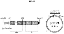
- genes encoding a human CH1 region and a region from an IgG2 hinge to a human IgG1 hybrid CH2-CH3 region were subcloned into a mammalian expression vector (see FIG. 10 ).
- FIG. 10 is a genetic map of an expression vector according to one embodiment of the present invention.
- Transfection was performed on an overexpressed recombinant protein.
- 2 ⁇ g of a mammalian expression vector and 4 ⁇ g of PEI (Polysciences, Warrington, PA, USA) per mL of a culture volume were mixed in 150 mM sodium chloride (NaCl, Merck) corresponding to 1/10 of the cell culture volume, and maintained at room temperature for 15 minutes.
- PEI Polysciences, Warrington, PA, USA
- the mixture was added to mammalian cells, HEK293F (1 ⁇ 10 6 cells/mL, Invitrogen) used for a protein overexpression system, and cultured in a Freestyle TM 293 expression medium (Invitrogen) containing 100 U/mL penicillin and streptomycin (Invitrogen) for 6 days at 37 °C and 7% CO 2 while stirring at 135 rpm.
- HEK293F 1 ⁇ 10 6 cells/mL, Invitrogen
- Freestyle TM 293 expression medium Invitrogen
- penicillin and streptomycin Invitrogen
- a supernatant of the cell culture was harvested, and to purify an Fc-fusion protein, affinity gel chromatography using protein A was used. After isolated scFv was purified through dialysis using PBS buffer, SDS-PAGE was performed to confirm a molecular weight of approximately 55 kDa.
- CCSP-2 protein was prepared per domain, denaturation buffer was added thereto, followed by heating for 10 minutes. Then, SDS-PAGE electrophoresis was performed by loading the denatured sample on a prepared gel. After electrophoresis, the gel was not stained, and for an immunological assay, the protein was transferred to Trans-Blot Turbo TM Mini PVDF Transfer Packs (Bio-Rad) by an electro-transfer method. To inhibit a non-specific reaction, the gel was immersed in a 5% skim milk/TBST solution and treated on a stirrer for 1 hour.
- the scFv obtained according to Example 4-2 diluted to a predetermined concentration was used, and as a secondary antibody, a HRP-binding anti-human immunoglobulin Fc secondary antibody (Invitrogen) was used. Each antibody reaction was performed for 1 hour, and after each step, washing was performed three times with 0.05% Tween 20-containing PBS buffer. Finally, to confirm an antigen-antibody reaction, an enhanced chemiluminescent (ECL) substrate was used (see FIG. 11 ).
- ECL enhanced chemiluminescent
- FIG. 11 is a western blotting result for confirming the binding ability per antigen (CCSP-2) domain of CH-1, CH-2 and CH-3 in scFv produced according to the present invention.
- a 96-well plate was coated with the scFv obtained according to Example 4-2 at a concentration of 10 ⁇ g/mL, and blocking was performed with 5% skim milk/TBST for 1 hour.
- the antigen-immobilized wells were then washed with 0.05% Tween 20-containing PBS buffer three times, and CCSP-2 protein was dispensed at 900 ng/mL 300 ng/mL, 100 ng/mL, 33.3 ng/mL or 11.1 ng/mL to induce an antigen-antibody reaction for 1 hour.
- a reaction was performed for 1 hour sequentially using an anti-CCSP-2 (Cloud-Clone Corp., TX, USA) primary antibody diluted at a ratio of 1:500 and a HRP conjugated anti-Rabbit IgG secondary antibody (Cell Signaling) diluted at a ratio of 1:2000.
- a tetramethylbenzidine (TMB) substrate was dispensed into the wells to induce color development at room temperature for 20 minutes, and then absorbance was measured at 450 nm using a microplate reader (see FIG. 12 ).
- FIG. 12 is a set of graphs showing the ELISA results obtained by analyzing the antigen (CCSP-2) binding ability of CH-1, CH-2 and CH-3 in scFv of the present invention.
- the scFv antibody according to the present invention exhibits dose-dependent reactivity for the CCSP-2 protein.
- the scFv antibody constructed according to the present invention has a size of approximately 55 kDa, and has a function as an antibody having binding ability to an antigen.
- the antibodies according to the present invention were compared with the conventional antibodies against CCSP-2 to examine the effect caused by specific binding to the EGF2 domain of CCSP-2.
- a recombinant protein of each domain EGF1, VWFA2, VWFA3 or EGF2 of CCSP-2-CT or CCSP-2 was prepared.
- the recombinant proteins were purified.
- the purified recombinant proteins were loaded on a 12% SDS-PAGE gel at 100 ng/well, and CCSP-2 and domains of the CCSP-2 were detected with various CCSP-2 antibodies.
- the CCSP-2 antibodies used in the experiment were 1 an antibody provided by Case Western Reserve University (not commercially available; hereinafter, CWRU antibody), 2 Proteintech antibody, and 3 Cloud-Clone antibody.
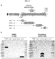
- FIG. 13A is a schematic diagram illustrating the locations of CCSP2 antibody per domain, and which domain is recognized by each CCSP-2 antibody.
- FIG. 13B is western blotting results showing that the CWRU antibody specifically recognizes the EGF2 domain, and the Proteintech antibody specifically recognizes the VWFA2 domain.
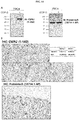
- CCSP-2 protein overexpression was induced in HeLa cells, and the CCSP-2-specific detection ability between the CWRU antibody and the Proteintech antibody was compared and analyzed by western blotting using the cell lysate thereof. While CCSP-2-specific detection of the CWRU antibody was possible according to the presence or absence of CCSP-2 expression, a non-specific signal was confirmed in the case of the Proteintech antibody regardless of CCSP-2 expression.
- paraffin blocks were manufactured by taking cancer tissue and normal colorectal tissue from a colorectal cancer patient, and analyzed by immunostaining using CCSP-2 expression-specific antibodies such as the CWRU antibody and the Proteintech antibody.
- the immunostaining method was carried out using an automated apparatus (Ventana Medical System), and the antibodies were reacted at a concentration of 1:100 for 32 minutes and then detected using the Nentana OptiView DAB kit.
- the CWRU antibody was not stained in normal tissue, but only stained in a lesion, whereas 50% of the Proteintech antibodies were stained even in normal tissue so that the Proteintech antibody did not clearly distinguish normal tissue from a lesion.
- the recognition of the amino acid sequence downstream the EGF2 domain specifically recognized by the CWRU antibody is effective for the purpose of distinguishing diseases, that is, normal tissue and a lesion.
- the inventors conducted antibody screening using a protein consisting of a sequence from the start of the EGF2 domain to the end of CCSP2 (the sequence of CCSP2 amino acids 712 ⁇ 755).
- FIG. 14A shows western blotting results for comparatively analyzing the antigen (CCSP-2)-specific detection ability of the CWRU antibody and the Proteintech antibody in a cell lysate.
- FIG. 14B shows immunohistochemical results for comparatively analyzing the antigen (CCSP-2)-specific detection ability of the CWRU antibody and the Proteintech antibody in normal colorectal tissue and colorectal cancer tissue.
- FIG. 15 is a schematic diagram for comparing the binding (detection) sites of anti-CCSP-2 antibodies which are currently available.
- the Proteintech antibody binds to the sequence of amino acids 341-517 of CCSP-2
- the Abcam antibody binds to the sequence of amino acids 201-373
- the Cloud-Clone antibody binds to the sequence of amino acids 457-517
- the Proteintech, Abcam and Cloud-Clone antibodies are unable to detect the EGF2 domain, which is the sequence of amino acids 712-748 of CCSP-2, which is meaningful for distinguishing normal and lesion sites.
- Affinity was compared between the antibody (scFv) according to the example of the present invention and a conventional peptide specifically binding to CCSP-2 ( Korean Patent Application No. 10-2018-0060184 ).
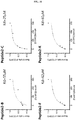
- FIG. 16 is a set of graphs showing the affinity of conventional peptides specifically binding to CCSP-2 ( Korean Patent Application No. 10-2018-0060184 ).
- the KD value (affinity) for the binding of the conventional peptide and CCSP-2 is expressed in ⁇ M units.
Landscapes
- Health & Medical Sciences (AREA)
- Chemical & Material Sciences (AREA)
- Life Sciences & Earth Sciences (AREA)
- Immunology (AREA)
- Molecular Biology (AREA)
- Engineering & Computer Science (AREA)
- Organic Chemistry (AREA)
- Biochemistry (AREA)
- General Health & Medical Sciences (AREA)
- Medicinal Chemistry (AREA)
- Hematology (AREA)
- Biomedical Technology (AREA)
- Urology & Nephrology (AREA)
- Cell Biology (AREA)
- Genetics & Genomics (AREA)
- Biophysics (AREA)
- Proteomics, Peptides & Aminoacids (AREA)
- Microbiology (AREA)
- General Physics & Mathematics (AREA)
- Pathology (AREA)
- Food Science & Technology (AREA)
- Analytical Chemistry (AREA)
- Physics & Mathematics (AREA)
- Biotechnology (AREA)
- Oncology (AREA)
- Peptides Or Proteins (AREA)
- Preparation Of Compounds By Using Micro-Organisms (AREA)
- Hospice & Palliative Care (AREA)
Abstract
Description
- The present invention relates to a monoclonal antibody specifically binding to an EGF2 domain of human colon cancer secreted protein-2 (CCSP-2), or an antigen-binding fragment thereof.
- The large intestine belongs to the digestive system and is an organ located between the small intestine and the anus. The total length is approximately 1.5 meters on average, and classified into the cecum, the ascending colon, the transverse colon, the descending colon, the sigmoid colon and the rectum from the right. Colorectal cancer is a malignant tumor that occurs in the colon and rectum, and malignant tumor cells (cancer cells) mainly arise from epithelial cells. Depending on a region where cancer occurs, cancer that occurs in the colon is called colon cancer, cancer that occurs in the rectum is called rectal cancer, and these are collectively called colorectal cancer.
- According to national cancer registration statistics, a total of 28,112 cases of colorectal cancer occurred in 2011, accounting for 12.9% of the total cancer incidence (218,017 cases), ranking 3rd in cancer incidence and 4th in cancer mortality.
- Another reason for a continuous increase in colorectal cancer mortality is that there are few initial symptoms of colorectal cancer, and therefore, the early detection rate of colorectal cancer in Korea is low as less than 10%. Many colorectal cancer patients are diagnosed with metastatic colorectal cancer in
stages Stage 3 colorectal cancer is quite low, 28%, and the survival rate ofStage 4 colorectal cancer is 6%, which is lower than the survival rate ofStage 3 colorectal cancer. Accordingly, the early diagnosis of colorectal cancer is very important. - Currently, the most prevalently used methods for colorectal cancer diagnosis are a double contrast barium-enema and colonoscopy. A double contrast barium-enema is an examination method of inserting a small tube through the anus after cleaning the large intestine to inject a contrast agent called barium, coating the wall of the large intestine with a thin layer of barium while expanding the large intestine by injecting air, and taking images using an X-ray fluoroscope. Advantages of this method are identification of the overall contour and shape of the large intestine, easy identification of the overall location of colorectal cancer, and easy testing for the changes in the large intestinal wall such as inflammatory or ischemic changes and the distal end of the small intestine. However, there are disadvantages that tissue cannot be obtained with testing, and accuracy for small polyps is inferior to colonoscopy.
- As a testing method for directly examining the large intestine with light and a flexible tube, colonoscopy is the most accurate method for diagnosing a colorectal disease, and this is because a doctor can directly observe the area of bleeding and the surface of a lesion, and determine the condition of tissue. A biopsy is also possible at the same time as an endoscopy, and when a conscious sedation endoscopy is performed while a patient is sleeping by intravenously injecting a sedative acting only for a short period of time, testing can be done without discomfort.
- Colonoscopy is the best diagnostic tool for finding colorectal cancer, but the frequency at which polyps missed in the testing are detected as having advanced to cancer in the patient's next test is high. Since polyps larger than 1 cm have a relatively and significantly increasing possibility of progression to cancer over time, it is necessary to improve the technology so as not to miss a small polyp during an endoscopy, and to attempt to avoid missing lesions behind flexures and mucosal folds with sufficient margin during withdrawal inspection. As one of these attempts, research is being conducted on the development of a biomarker and a marker, which are used in molecular imaging, to improve a tumor detection rate.
- Meanwhile, a colon cancer secreted protein (e.g., CCSP-2) is a colorectal cancer-specific marker (biomarker), and information on the gene encoding the same is well known in the related art. For example, the CCSP-2 may be human CCSP-2, and the information on the human CCSP-2 protein is registered at National Center for Biotechnology Information (NCBI) under Accession No. AAT77225.1, and the information on a gene encoding the same is registered at NCBI under Accession No. AY572972.1. The function and role of the CCSP-2 are not well known, but at the RNA level, it is known that the average expression level thereof in colorectal cancer cells and/or tissue is 78-fold higher than in normal tissue (colorectal cancer cell-free tissue). In addition, the CCSP-2 is a secretory protein and an extracellular matrix (ECM) protein, which is increased in colorectal cancer-specific expression, and has advantages of labeling colorectal cancer tissue through targeting CCSP-2 protein accumulated in colorectal cancer tissue as well as diagnosis of colorectal cancer using blood. Accordingly, when the expression and/or expression level of CCSP-2 is(are) measured at the gene and/or protein level(s), the presence of colorectal cancer and/or the degree of progression of colorectal cancer may be accurately and easily confirmed.
- There is a problem that a currently commercially available antibody specifically binding to CCSP-2 has low specificity, and thus binds to approximately 50% normal cells (or tissue) rather than colorectal cancer cells (or tissue) for labeling.
- In this regard, the inventors developed a peptide specifically binding to CCSP-2, confirmed the effect of specifically detecting CCSP-2 at the protein level, and proved that colorectal cancer and/or colorectal cancer cells and/or tissue can be detected, which is disclosed in
Korean Patent Application Publication No. 10-2018-0060184 - While continuing research on the technology of specifically detecting the CCSP-2, the inventors developed a monoclonal antibody specifically binding to the EGF2 domain of CCSP-2, and confirmed that the antibody is significantly improved in affinity and particularly more improved in specificity, compared with a conventional peptide specifically binding to CCSP-2, and thus the present invention was completed.
- The present invention is directed to providing a monoclonal antibody specifically binding to colon cancer secreted protein-2 (CCSP-2), or an antigen-binding fragment thereof.
- The present invention is also directed to providing a polynucleotide encoding the monoclonal antibody or an antigen-binding fragment thereof, an expression vector including the polynucleotide and a transformant into which the vector is introduced.
- The present invention is also directed to providing a composition for diagnosing cancer, which includes the monoclonal antibody or the antigen-binding fragment thereof.
- The present invention is also directed to providing a kit for diagnosing cancer, which includes the monoclonal antibody or the antigen-binding fragment thereof.
- The present invention is also directed to providing a method of providing information for cancer diagnosis, which includes detecting a CCSP-2 protein in a biological sample isolated from a subject suspected of having cancer using the monoclonal antibody or the antigen-binding fragment thereof through an antigen-antibody reaction.
- To achieve the above-described objects, the present invention provides a monoclonal antibody specifically binding to the EGF2 domain of human CCSP-2, which includes a heavy chain variable region and a light chain variable region selected from the group consisting of 1) to 4) below, or an antigen-binding fragment thereof:
- 1) a heavy chain variable region including a CDR1 region represented by SEQ ID NO: 1, a CDR2 region represented by SEQ ID NO: 2 and a CDR3 region represented by SEQ ID NO: 3, and a light chain variable region including a CDR1 region represented by SEQ ID NO: 4, a CDR2 region represented by SEQ ID NO: 5 and a CDR3 region SEQ ID NO: 6;
- 2) a heavy chain variable region including a CDR1 region represented by SEQ ID NO: 7, a CDR2 region represented by SEQ ID NO: 8 and a CDR3 region represented by SEQ ID NO: 9, and a light chain variable region including a CDR1 region represented by SEQ ID NO: 10, a CDR2 region represented by SEQ ID NO: 11 and a CDR3 region represented by SEQ ID NO: 12;
- 3) a heavy chain variable region including a CDR1 region represented by SEQ ID NO: 13, a CDR2 region represented by SEQ ID NO: 14 and a CDR3 region represented by SEQ ID NO: 15, and a light chain variable region including a CDR1 region represented by SEQ ID NO: 16, a CDR2 region represented by SEQ ID NO: 17 and a CDR3 region represented by SEQ ID NO: 18; and
- 4) a heavy chain variable region including a CDR1 region represented by SEQ ID NO: 19, a CDR2 region represented by SEQ ID NO: 20 and a CDR3 region represented by SEQ ID NO: 21, and a light chain variable region including a CDR1 region represented by SEQ ID NO: 22, a CDR2 region represented by SEQ ID NO: 23 and a CDR3 region represented by SEQ ID NO: 24.
- In one embodiment of the present invention, the antigen-binding fragment may be selected from the group consisting of Fab, Fab', F(ab')2, scFv, Fv, dsFv, a diabody, Fd and Fd'.
- In one embodiment of the present invention, the monoclonal antibody or the antigen-binding fragment thereof may include a heavy chain variable region and a light chain variable region, which are selected from the group consisting of a heavy chain variable region including a polypeptide sequence represented by SEQ ID NO: 25 and a light chain variable region including a polypeptide sequence represented by SEQ ID NO: 26; a heavy chain variable region including a polypeptide sequence represented by SEQ ID NO: 27 and a light chain variable region including a polypeptide sequence represented by SEQ ID NO: 28; a heavy chain variable region including a polypeptide sequence represented by SEQ ID NO: 29 and a light chain variable region including a polypeptide sequence represented by SEQ ID NO: 30; and a heavy chain variable region including a polypeptide sequence represented by SEQ ID NO: 31 and a light chain variable region including a polypeptide sequence represented by SEQ ID NO: 32.
- The present invention also provides a polynucleotide encoding the monoclonal antibody, or an antigen-binding fragment thereof.
- The present invention also provides an expression vector including the polynucleotide.
- The present invention also provides a transformant into which the expression vector is introduced, excluding a human.
- The present invention also provides a composition for diagnosing cancer, which includes the monoclonal antibody or the antigen-binding fragment thereof.
- In one embodiment of the present invention, the cancer may be selected from the group consisting of esophageal cancer, gastric cancer, colorectal cancer, rectal cancer, oral cancer, pharyngeal cancer, laryngeal cancer, lung cancer, colon cancer, breast cancer, cervical cancer, endometrial cancer, ovarian cancer, prostate cancer, testicular cancer, bladder cancer, kidney cancer, liver cancer, pancreatic cancer, bone cancer, connective tissue cancer, skin cancer, brain cancer, thyroid cancer, leukemia, Hodgkin's disease, soft tissue sarcoma, lymphoma and multiple myeloma hematologic cancer.
- The present invention also provides a kit for diagnosing cancer, which includes the composition.
- The present invention also provides a method of providing information for cancer diagnosis, which includes detecting CCSP-2 protein in a biological sample isolated from a subject suspected of having cancer using the monoclonal antibody or the antigen-binding fragment thereof through an antigen-antibody reaction.
- As a monoclonal antibody of the present invention or an antigen-binding fragment thereof specifically binds to the EGF2 domain of CCSP-2, compared with a conventional peptide specifically binding to CCSP-2, affinity can be significantly improved, and compared with a conventional antibody against CCSP-2, specificity can be more improved. Accordingly, CCSP-2 is specifically detected at the protein level and thus enables accurate diagnosis in the future.
-
-
FIG. 1 is a schematic diagram illustrating the strategy of cloning a variable region of a scFv antibody according to one embodiment of the present invention. -
FIG. 2 is a schematic diagram illustrating antigen CCSP-2-NT, CCSP-2-CT and CCSP-2-EGF2 sites of CCSP-2. -
FIG. 3 is a schematic diagram illustrating respective expression vectors of CCSP-2-CT and CCSP-2-EGF2 antigens. -
FIGS. 4A and 4B show phage ELISA results obtained by ELISA on 48 clones from produced phages (output) of third and fourth bio-panning, respectively. -
FIG. 5 is a genetic map of an expression vector according to one embodiment of the present invention. -
FIG. 6 is an SDS-PAGE gel image showing the molecular weight of HN-1 of scFv produced by the present invention. -
FIG. 7 shows a western blotting result for confirming the binding ability per antigen (CCSP-2) domain of HN-1 in scFv of the present invention. -
FIG. 8 is a graph showing an ELISA result obtained by analyzing the antigen (CCSP-2) binding ability of HN-1 in scFv of the present invention. -
FIGS. 9A, 9B and 9C show antigen (CCSP-2)-binding phage ELISA results on 96 clones from the produced phages (output) of the fourth bio-panning ofchicken libraries -
FIG. 10 is a genetic map representing an expression vector according to one embodiment of the present invention. -
FIG. 11 shows western blotting results for confirming the binding ability per antigen (CCSP-2) domain of CH-1, CH-2 and CH-3 in scFv produced by the present invention. -
FIG. 12 is a set of graphs showing the ELISA results obtained by analyzing the antigen (CCSP-2) binding ability of CH-1, CH-2 and CH-3 in scFv of the present invention. -
FIG. 13A is a schematic diagram illustrating the locations of CCSP2 antibodies per domain, and which domain is recognized by each CCSP-2 antibody.FIG. 13B is western blotting results showing that the CWRU antibody specifically recognizes the EGF2 domain, and the Proteintech antibody specifically recognizes the VWFA2 domain. -
FIG. 14A shows western blotting results for comparatively analyzing the antigen (CCSP-2)-specific detection ability of the CWRU antibody and the Proteintech antibody in a cell lysate.FIG. 14B shows immunohistochemical results for comparatively analyzing the antigen (CCSP-2)-specific detection ability of the CWRU antibody and the Proteintech antibody in normal colorectal tissue and colorectal cancer tissue. -
FIG. 15 is a schematic diagram for comparing the binding (detection) sites of anti-CCSP-2 antibodies which are currently available. -
FIG. 16 is a set of graphs showing the affinity of the conventional peptides specifically binding to CCSP-2 (Korean Patent Application No. 10-2018-0060184 - Hereinafter, the present invention will be described in detail.
- The present invention provides a monoclonal antibody specifically binding to the EGF2 domain of human colon cancer secreted protein-2 (CCSP-2), or an antigen-binding fragment. The CCSP-2 is specific for colorectal cancer (e.g., expressed specifically in colorectal cancer).
- The inventors identified antibodies specifically recognizing CCSP-2, and the amino acid sequence of a heavy chain variable region thereof, the amino acid sequence of a light chain variable region thereof and coding nucleotide sequences thereof.
- In addition, according to one exemplary embodiment of the present invention, colorectal cancer and/or colorectal cancer cells and/or tissue can be accurately diagnosed by specifically detecting CCSP-2 by providing a monoclonal antibody or an antigen-binding fragment thereof specifically binding to CCSP-2, and particularly, it may be effectively applied to the endoscopic diagnosis of colorectal cancer by visualizing colorectal tissue with the antibody conjugated with a visible labeling material.
- The term "antibody" used herein refers to a protein molecule serving as a receptor specifically recognizing an antigen, which includes an immunoglobulin molecule immunologically having reactivity to a specific antigen, and includes a polyclonal antibody, a monoclonal antibody, and an antigen-binding fragment (antibody fragment) of an antibody molecule as well as a complete antibody form. In addition, the term includes a chimeric antibody, a humanized antibody, a bivalent or bispecific molecule (e.g., bispecific antibody), a diabody, a triabody, and a tetrabody.
- The term "complete antibody" has a structure having two full-length light chains and two full-length heavy chains, in which each light chain is connected with a heavy chain by a disulfide bond. The complete antibody includes IgA, IgD, IgE, IgM and IgG, in which IgG has subtypes, for example, IgG1, IgG2, IgG3 and IgG4.
- The term "antigen-binding fragment of an antibody" used herein refers to a fragment that retains an antigen-antibody binding function in a complete antibody molecule, and includes Fab, Fab', F(ab')2, scFv, Fv, dsFv, a diabody, Fd and Fd'. The Fab has a structure having light and heavy chain variable regions, and a constant region of the light chain and the first constant region (CHI domain) of the heavy chain, and has one antigen-binding site. An Fab' is distinguished from Fab in that there is a hinge region having one or more cysteine residues at the C terminus of the CH1 domain of the heavy chain. An F(ab')2 antibody is generated by forming a disulfide bond between cysteine residues of the hinge region of Fab'. A variable fragment (Fv) refers to the smallest antibody fragment only having a heavy chain variable region and a light chain variable region. Double chain Fv (dsFv) is formed by connecting a heavy chain variable region and a light chain variable region using a disulfide bond, and single chain Fv (scFv) generally has the same structure as a dimer such as dsFv since a heavy chain variable region is connected with a light chain variable region by a covalent bond using a peptide linker, or may be directly connected to the C-terminus. A diabody is a complex of two or more polypeptide chains or proteins, and refers to a complex including at least one VL domain and at least one VH domain or fragments thereof, and having both domains in a single polypeptide chain. In one embodiment, the diabody includes a molecule including an Fc or hinge-Fc domain. The polypeptide chains of such a complex may be the same or different, in other words, the diabody may be a mono polymer or a hetero polymer.
- The antigen-binding fragment may be obtained using proteases (e.g., Fab may be obtained by restriction of a complete antibody by papain, and a F(ab')2 fragment may be obtained by cleavage by a pepsin), and preferably is manufactured using gene recombination technology.
- The term "heavy chain" used herein refers to both a full-length heavy chain which consists of a variable region domain VH and three constant region domains CH1, CH2 and CH3, and a fragment thereof, which includes an amino acid sequence having a sufficient variable region sequence imparting specificity to an antigen. In addition, the term "light chain" used herein refers to both a full-length light chain which consists of a variable region domain VL and a constant region domain CL, or a fragment thereof, which includes an amino acid sequence having a sufficient variable region sequence imparting specificity to an antigen.
- The term "monoclonal antibody" used herein refers to an antibody molecule of single molecular composition obtained from substantially the same antibody population, and exhibits single binding specificity and affinity for a specific epitope. Typically, an immunoglobulin has a heavy chain and a light chain, in which each of the heavy chain and the light chain includes a constant region and a variable region (these regions are also known as domains). The variable regions of the light chain and heavy chain includes three variable regions called complementarity-determining regions (hereinafter, referred to as "CDRs") and four framework regions (FRs). The CDRs mainly serve to bind to an epitope of an antigen. The CDRs of each chain are typically called CDR1, CDR2 and CDR3 sequentially from the N-terminus, and also distinguished by a chain in which a specific CDR is located.
- The term "complementarity determining region (CDR)" refers to the amino acid sequence of a variable region (hypervariable region) of an immunoglobulin heavy or light chain (Kabat et al. Sequences of Proteins of Immunological Interest, 4th Ed., U.S. Department of Health and Human Services, National Institutes of Health (1987)). Three CDRs are included in each heavy chain (CDRH1, CDRH2 and CDRH3) and each light chain (CDRL1, CDRL2 and CDRL3) and provide major contact residues for an antibody to bind to an antigen or epitope.
- Meanwhile, the monoclonal antibody may be in the form of a chimeric antibody or humanized antibody which has reduced immunogenicity to be applied to a human as described above.
- The term "chimeric antibody" is an antibody formed by combining a variable region of a heterogeneous antibody which is derived from a mouse or chicken and a constant region of a human antibody through DNA recombination technology, and the immune reaction of the chimeric antibody is considerably improved compared with a heterogeneous antibody derived from a mouse or chicken, and thus can be used clinically.
- The term "humanized antibody" used herein refers to an antibody formed by transplanting all or a part of the CDR sequence of a heterogeneous monoclonal antibody derived from a mouse or chicken into a human antibody, a humanized variable region may be formed by recombining CDRs of a chicken or mouse monoclonal antibody with a human antibody-derived FR, and the human antibody may be formed by recombining the variable region with a constant region of a preferred human antibody, but the present invention is not limited thereto. In addition, since the affinity of a humanized antibody is decreased when only the chicken or mouse-derived CDRs are inserted, the affinity may be improved by substituting an FR amino acid residue, which may be considered to affect the three-dimensional structure of a CDR, with an amino acid of a chicken or mouse antibody, but the present invention is not limited thereto.
- The term "monoclonal antibody specifically binding to the EGF2 domain of colon cancer secreted protein-2 (CCSP-2)" refers to an antibody capable of specifically binding to CCSP-2, and may be interchangeably used with an anti-CCSP-2 antibody. The monoclonal antibody specifically binding to the CCSP-2 protein may be, without limitation, any monoclonal antibody that binds to CCSP-2 to inhibit the biological activity of CCSP-2.
- In addition, the form of monoclonal antibody may include both of a complete antibody and an antigen-binding fragment as described above, and may be a chimeric antibody or a humanized antibody, but the present invention is not limited thereto. In addition, the monoclonal antibody of the present invention may specifically bind to the EGF2 domain of CCSP-2 to inhibit signaling by CCSP-2, resulting in suppression of biological activity, and thus may be effectively used for prevention or treatment of a disease such as cancer mediated by CCSP-2. In addition, since CCSP-2 overexpression is reported as a specific phenomenon in cancer, and the antibody of the present invention capable of specifically binding to CCSP-2 has diagnosis ability with high sensitivity and specificity in the diagnosis of cancer, the antibody may be effectively used in diagnosis of cancer. In one embodiment of the present invention, an antigen-binding fragment specifically binding to CCSP-2 of the present invention was formed using the EGF2 site of CCSP-2 as an antigen protein.
- The monoclonal antibody specifically binding to the EGF2 domain of the human CCSP-2, or an antigen-binding fragment thereof may include any one pair of a heavy chain variable region and a light chain variable region selected from the group consisting of 1) to 4) below, but the present invention is not limited thereto:
- 1) a heavy chain variable region including a CDR1 region represented by SEQ ID NO: 1, a CDR2 region represented by SEQ ID NO: 2 and a CDR3 region represented by SEQ ID NO: 3, and a light chain variable region including a CDR1 region represented by SEQ ID NO: 4, a CDR2 region represented by SEQ ID NO: 5 and a CDR3 region SEQ ID NO: 6;
- 2) a heavy chain variable region including a CDR1 region represented by SEQ ID NO: 7, a CDR2 region represented by SEQ ID NO: 8 and a CDR3 region represented by SEQ ID NO: 9, and a light chain variable region including a CDR1 region represented by SEQ ID NO: 10, a CDR2 region represented by SEQ ID NO: 11 and a CDR3 region represented by SEQ ID NO: 12;
- 3) a heavy chain variable region including a CDR1 region represented by SEQ ID NO: 13, a CDR2 region represented by SEQ ID NO: 14 and a CDR3 region represented by SEQ ID NO: 15, and a light chain variable region including a CDR1 region represented by SEQ ID NO: 16, a CDR2 region represented by SEQ ID NO: 17 and a CDR3 region represented by SEQ ID NO: 18; and
- 4) a heavy chain variable region including a CDR1 region represented by SEQ ID NO: 19, a CDR2 region represented by SEQ ID NO: 20 and a CDR3 region represented by SEQ ID NO: 21, and a light chain variable region including a CDR1 region represented by SEQ ID NO: 22, a CDR2 region represented by SEQ ID NO: 23 and a CDR3 region represented by SEQ ID NO: 24.
- In one embodiment of the present invention, the antigen-binding fragment is preferably Fab, Fab', F(ab')2, scFv, Fv, dsFv, a diabody, Fd, Fd', a light chain or heavy chain including a CDR region of the present invention, or a variable domain including a CDR region of the present invention, but the present invention is not limited thereto.
- In another embodiment of the present invention, the monoclonal antibody or an antigen-binding fragment thereof preferably includes one pair of a heavy chain variable region and a light chain variable region selected from the group consisting of a heavy chain variable region including a polypeptide sequence represented by SEQ ID NO: 25 and a light chain variable region including a polypeptide sequence represented by SEQ ID NO: 26; a heavy chain variable region including a polypeptide sequence represented by SEQ ID NO: 27 and a light chain variable region including polypeptide sequence represented by SEQ ID NO: 28; a heavy chain variable region including a polypeptide sequence represented by SEQ ID NO: 29 and a light chain variable region including a polypeptide sequence represented by SEQ ID NO: 30; and a heavy chain variable region including a polypeptide sequence represented by SEQ ID NO: 31 and a light chain variable region including a polypeptide sequence represented by SEQ ID NO: 32.
- The antibody of the present invention or an antigen-binding fragment thereof also includes all mutants achieving the effects of the present invention, which are obtained through one or more mutations such as substitution, deletion, inversion or translocation of an antibody limited by the above-described sequence.
- In addition, the present invention provides a polynucleotide encoding the monoclonal antibody or the antigen-binding fragment thereof.
- The polynucleotide may be selected from the group consisting of SEQ ID NOs: 33 to 36, but the present invention is not limited thereto.
- The present invention also provides an expression vector including the polynucleotide and a transformant into which the vector is introduced.
- The expression vector including the polynucleotide encoding the monoclonal antibody according to the present invention may be, but is not particularly limited to, a vector capable of replicating and/or expressing the polynucleotide in eukaryotic or prokaryotic cells including mammalian cells (e.g., human, monkey, rabbit, rat, hamster, and mouse cells), plant cells, yeast cells or bacterial cells (e.g., Escherichia coli), and preferably, a vector that is operably linked to a suitable promoter such that the nucleotide can be expressed in host cells and includes at least one selection marker. As an example, the expression vector may be a form in which the polynucleotide is introduced into a phage, a plasmid, a cosmid, a mini-chromosome, a virus or a retrovirus vector.
- The expression vector including a polynucleotide encoding the monoclonal antibody may be an expression vector which includes a polynucleotide encoding a heavy or light chain of the monoclonal antibody or an expression vector which includes both of polynucleotides encoding a heavy chain and a light chain.
- A transformant into which the expression vector of the present invention is introduced may be, but is not particularly limited to, bacterial cells, such as E. coli, Streptomyces or Salmonella typhimurium; yeast cells; fungal cells such as Pichia pastoris; insect cells such as Drozophila or Spodoptera Sf9 cells; animal cells such as Chinese hamster ovary (CHO) cells, SP2/0 (mouse myeloma), human lymphoblastoid, COS, NSO (mouse myeloma), 293T, Bowes melanoma cells, HT-1080, baby hamster kidney (BHK) cells, human embryonic kidney (HEK) cells or PERC.6 (human retinal cells); or plant cells, which are transformed by introducing the expression vector.
- The term "introduction" used herein refers to a method of delivering the vector including a polynucleotide encoding the monoclonal antibody to host cells. The introduction may be performed by various methods known in the art, such as calcium phosphate-DNA co-precipitation, DEAE-dextran-mediated transfection, polybrene-mediated transfection, electroporation, microinjection, liposome fusion, Lipofectamine and protoplast fusion. In addition, transduction refers to the delivery of a desired material into cells using a viral particle through infection. Additionally, transduction may be introduction of a vector into host cells via gene bombardment. In the present invention, the introduction may be interchangeably used with transformation.
- The present invention also provides a composition for diagnosing cancer, which includes the monoclonal antibody.
- The term "cancer" used herein may be any cancer expressing CCSP-2, and examples include, but are not limited to, esophageal cancer, gastric cancer, colorectal cancer, rectal cancer, oral cancer, pharyngeal cancer, laryngeal cancer, lung cancer, colon cancer, breast cancer, cervical cancer, endometrial cancer, ovarian cancer, prostate cancer, testicular cancer, bladder cancer, kidney cancer, liver cancer, pancreatic cancer, bone cancer, connective tissue cancer, skin cancer, brain cancer, thyroid cancer, leukemia, Hodgkin's disease, soft tissue sarcoma, lymphoma and multiple myeloma hematologic cancer, and is preferably colorectal cancer or pancreatic cancer.
- The term "diagnosis" used herein refers to confirmation of the presence, severity (symptoms) and/or characteristics of a pathological state. As used in the specification, the "diagnosis of colorectal cancer" is confirmation of the pathological state of colorectal cancer, for example, the onset of colorectal cancer, the progression of a colorectal cancer lesion, the location of colorectal cancer (colorectal cancer cells), or the stage of colorectal cancer. For more accurate diagnosis, it is important to accurately and rapidly distinguish colorectal cancer cells and/or tissue from normal cells or tissue.
- As an exemplary embodiment of the present invention, the present invention provides a monoclonal antibody specifically binding to CCSP-2, thereby specifically detecting CCSP-2 at the protein level, and thus it is possible to accurately diagnose colorectal cancer and/or colorectal cancer cells and/or tissue. Particularly, colorectal tissue may be visualized using a labeling material for visualization, conjugated to the antibody, and thus can be effectively applied to endoscopic diagnosis of colorectal cancer.
- The monoclonal antibody and cancer have been described above. A disease associated with the expression or the progression of the expression of CCSP-2 or a CCSP-2-mediated disease, for example, cancer may be diagnosed using a diagnostic composition including the CCSP-2-specific monoclonal antibody of the present invention.
- The present invention also provides a kit for diagnosing cancer, which includes the composition for diagnosing cancer.
- The composition and cancer have been described above. In addition, the kit for diagnosing cancer may be configured to further include a composition, solution or apparatus, which has one or more types of components suitable for an analysis method.
- In the specification, by providing an antibody specifically binding to CCSP-2, which is a biomarker specifically expressed in colorectal cancer, a means useful for the measurement of CCSP-2 expression and/or a CCSP-2 expression level is provided, and therefore, this means may be effectively applied to the diagnosis of cancer, particularly, colorectal cancer. In addition, the antibody may be used in the visualization of cancer when used with various labeling materials, and applied in colorectal cancer endoscopy to provide more accurate information on the confirmation of the presence of colorectal cancer and lesions, and morphological observation of cancer sites, and may reduce a misdiagnosis rate related to colorectal cancer and contribute to early diagnosis. In addition, when a bioactive material such as a drug is bound to the antibody, the bioactive material may be specifically delivered to colorectal cancer, and therefore it may be used as a composition for the colorectal cancer-targeted delivery of the bioactive material.
- The present invention provides a method of providing information for cancer diagnosis, which includes detecting a CCSP-2 protein in a biological sample isolated from a subject suspected of having cancer using the monoclonal antibody through an antigen-antibody reaction.
- The monoclonal antibody, cancer, a subject and CCSP-2 protein have been described above. In the method of providing information for cancer diagnosis, the CCSP-2 protein may be detected by reacting a monoclonal antibody specific for CCSP-2 of the present invention with a biological sample isolated from a subject suspected of having cancer and detecting the formation of an antigen-antibody complex, thereby providing information for cancer diagnosis. Since the CCSP-2 is overexpressed in various cancer cells such as colorectal cancer or pancreatic cancer, cancer may be diagnosed by comparing the expression level with that of a control such as normal cells or tissue, but the present invention is not limited thereto.
- The term "biological sample" used herein may refer to tissue, cells, whole blood, serum, plasma, a tissue autopsy sample (brain, skin, lymph node, spinal cord, etc.), cell culture supernatant, ruptured eukaryotic cells and a bacterial expression system, but the present invention is not limited thereto. These biological samples may be reacted with the antibody of the present invention with or without manipulation, thereby confirming the presence of a CCSP-2 protein or the presence or absence of cancer.
- The term "antigen-antibody complex" used herein refers to a conjugate of a CCSP-2 protein antigen in a sample and the monoclonal antibody according to the present invention recognizing the same, and the formation of such an antigen-antibody complex may be detected by any method selected from the group consisting of a colorimetric method, an electrochemical method, a fluorometric method, luminometry, a particle counting method, visual assessment and a scintillation counting method. However, the method is not particularly limited thereto, and various applications may be used.
- In the present invention, to detect the antigen-antibody complex, various markers may be used. As a specific example, the marker may be selected from the group consisting of an enzyme, a fluorescent material, a ligand, a luminescent material, a microparticle, and a radioactive isotope, but the present invention is not limited thereto.
- Enzymes used as a detection marker include acetylcholinesterase, alkaline phosphatase, β-D-galactosidase, horseradish peroxidase, and β-lactamase, fluorescent materials used as a detection marker include fluorescein, Eu3+, Eu3+ chelate and cryptate, ligands used as a detection marker include a biotin derivative, luminescent materials used as a detection marker include acridinium ester and an isoluminol derivative, microparticles used as a detection marker include colloidal gold and colored latex, and radioactive isotopes used as a detection marker include 57Co, 3H, 1251, 125I-Bonton and Hunter reagents.
- Preferably, the antigen-antibody complex may be detected by ELISA. ELISA includes various ELISA method, for example, direct ELISA using a labeled antibody recognizing an antigen attached to a solid support, indirect ELISA using a labeled secondary antibody recognizing a capture antibody in an antibody complex recognizing an antigen attached to a solid support, direct sandwich ELISA using another labeled antibody recognizing an antigen in an antibody-antigen complex attached to a solid support, and indirect sandwich ELISA reacting with another antibody recognizing an antigen in an antibody-antigen complex attached to a solid support and using a labeled secondary antibody recognizing the antibody.
- The monoclonal antibody may have a detection marker, and when there is no detection marker, these monoclonal antibodies may be captured and detected by treating another antibody having a detection marker.
- In one embodiment of the present invention, by confirming that the anti-CCSP-2 antibody of the present invention specifically recognizes CCSP-2 using an antigen-antibody reaction, it was shown that the antibody of the present invention can be effectively used for diagnosis of various types of cancer such as colorectal cancer.
- Hereinafter, the monoclonal antibody according to the present invention, or an antigen-binding fragment thereof will be described in detail with reference to examples.
-
FIG. 1 is a schematic diagram illustrating the strategy of cloning a variable region of a scFv antibody according to one embodiment of the present invention. - In addition,
FIG. 2 is a schematic diagram illustrating CCSP-2 antigens (NT, CT and EGF2) according to one embodiment of the present invention. - Among the antigens, CCSP-2-CT and CCSP-2-EGF2 contain the EGF2 domain.
- Specifically, CCSP-2-CT (268 aa - 755 aa of AY57297231) was inserted into a pBT7-C-his vector (Bioneer, Korea) to be transduced into Origami2 (DE3) (Novagen, MA, USA).
- CCSP-2-EGF2 (712 aa - 755 aa of AY572972.1) was inserted into a pBT7-N-His vector (Bioneer, Korea) to be transduced into Origami2 (DE3) (Novagen, MA, USA).
-
FIG. 3 is a schematic diagram illustrating respective expression vectors of CCSP-2-CT and CCSP-2-EGF2. - The CCSP-2-CT and the CCSP-2-EGF2 were purified with Ni-NTA agarose beads (Qiagen, USA).
- Peripheral blood mononuclear cells were collected by treating 16 samples of human tonsil tissue with a Ficoll-Paque solution (GE Healthcare), and total RNA was extracted using a TRI reagent (Invitrogen). First strand cDNA was synthesized using oligo-dT primers and the Superscript® III First-Strand Synthesis System (Invitrogen).
- A scFv library was constructed from cDNA obtained from the human mononuclear cells using primers in the following Table 1, specific for a heavy chain variable region and a light chain variable region of an immunoglobulin, and the Expand High Fidelity PCR system (Roche Molecular Systems).
[Table 1] Vκ Primers Forward HSCK1-F GGGCCCAGGCGGCCGAGCTCCAGATGACCCAGTCTCC HSCK24-F GGGCCCAGGCGGCCGAGCTCGTGATGACYCAGTCTCC HSCK3-F GGGCCCAGGCGGCCGAGCTCGTGWTGACRCAGTCTCC HSCK5-F GGGCCCAGGCGGCCGAGCTCACACTCACGCAGTCTCC Reverse HSCJK14o-B GGAAGATCTAGAGGAACCACCTTTGATYTCCACCTTGGTCCC HSCJK2o-B GGAAGATCTAGAGGAACCACCTTTGATCTCCAGCTTGGTCCC HSCJK3o-B GGAAGATCTAGAGGAACCACCTTTGATATCCACTTTGGTCCC HSCJK5o-B GGAAGATCTAGAGGAACCACCTTTAATCTCCAGTCGTGTCCC Vλ Primers Forward HSCLamla GGGCCCAGGCGGCCGAGCTCGTGBTGACGCAGCCGCCCTC HSCLamlb GGGCCCAGGCGGCCGAGCTCGTGCTGACTCAGCCACCCTC HSCLam2 GGGCCCAGGCGGCCGAGCTCGCCCTGACTCAGCCTCCCTCCGT HSCLam3 GGGCCCAGGCGGCCGAGCTCGAGCTGACTCAGCCACCCTCAGTGTC HSCLam4 GGGCCCAGGCGGCCGAGCTCGTGCTGACTCAATCGCCCTC HSCLam6 GGGCCCAGGCGGCCGAGCTCATGCTGACTCAGCCCCACTC HSCLam78 GGGCCCAGGCGGCCGAGCTCGTGGTGACYCAGGAGCCMTC HSCLam9 GGGCCCAGGCGGCCGAGCTCGTGCTGACTCAGCCACCTTC Reverse HSCJLam1236 GGAAGATCTAGAGGAACCACCGCCTAGGACGGTCASCTTGGTSCC HSCJLam57 GGAAGATCTAGAGGAACCACCGCCGAGGACGGTCAGCTSGGTSCC VH Primers Forward HSCVH1-FL 
HSCVH2-FL 
HSCVH35-FL 
HSCVH3a-FL 
HSCVH4-FL 
HSCVH4a-FL 
Reverse HSCG1234-B CCTGGCCGGCCTGGCCACTAGTGACCGATGGGCCCTTGGTGGARGC HSCM-B CCTGGCCGGCCTGGCCACTAGTAAGGGTTGGGGCGGATGCACTCCC Overlap Extension Primers RSC-F GAGGAGGAGGAGGAGGAGGCGGGGCCCAGGCGGCCGAGCTC RSC-B GAGGAGGAGGAGGAGGAGCCTGGCCGGCCTGGCCACTAGTG - In each reaction, 2 µL of cDNA was mixed with 60 pmol of each primer, 10 µL of 10X reaction buffer, 8 µL of 2.5 mM dNTP (Promega), 0.5 µL of Tap DNA polymerase and water so that a final volume became 100 µL. PCR was performed under the following conditions: 30 cycles, 15 sec at 94°C, 30 sec at 56 °C, 90 sec at 72 °C, and 10 min at 72 °C (final extension). A fragment with a length of approximately 350 bp was loaded on a 1.5% agarose gel for electrophoresis, and purified using a QIAGEN II Gel Extraction Kit (QIAGEN). The purified PCR product was read at OD 260 nm and quantified (1 OD unit = 50 µg /mL).
- In second PCR, first VL and VH products were randomly linked by overlap extension PCR. Each PCR was performed with a mixture having a final concentration of 100 µL, which was prepared by mixing 100 ng of the purified VL and VH products, 60 pmol of each primer, 10 µL of 10X reaction buffer, 8 µL of 2.5 mM dNTP and 0.5 µL of Taq DNA polymerase with water. PCR was performed under the following conditions: 25 cycles, 15 sec at 94 °C, 30 sec at 56 °C, 2 min at 72 °C, and 10 min at 72 °C (final extension). A scFv fragment having a length of approximately 700 bp was loaded on a 1.5% agarose gel for electrophoresis, and then purified using a QIAGEN II Gel Extraction Kit (QIAGEN). The purified PCR product was read at OD 260 nm and quantified (1 OD unit = 50 µg /mL).
- For cloning, a scFv fragment, which is the PCR product, and a pComb3X-SS vector (The Scripps Research Institute) were cleaved using a SfiI restriction enzyme.
- Specifically, 10 µg of the purified overlap PCR product was mixed with 360 units of SifI (16 units per µg DNA, Roche Molecular Systems), 20 µL of 10X reaction buffer and water, and adjusted to have a final volume of 200 µL. 20 µg of the pComb3X-SS vector was mixed with 120 units of SfiI (6 units per µg DNA), 20 µL of 10X reaction buffer and water, and adjusted to have a final volume of 200 µL. The mixture was cleaved for 8 hours at 50 °C. A scFv fragment having a length of approximately 700 bp and a vector having a length of 3,400 bp were loaded on a 1% agarose gel for electrophoresis, and purified using a QIAGEN II Gel Extraction Kit (QIAGEN). 1,400 ng of the SfiI-cleaved pComb3X vector and 700 ng of a scFv fragment were mixed with 40 µL of 5X ligase buffer, 10 µL of T4 DNA ligase (Invitrogen) and water so that a final volume became 200 µL, and incubated at 16 °C for 16 hours, followed by ligation. Afterward, the ligated product was precipitated with ethanol, and only the DNA pellet was dissolved in 15 µL of water.
- The ligated library sample was transformed into an E. coli strain, ER2738 (New England Biolabs, Inc.) through electroporation using a gene pulser (Bio-Rad Laboratories). The cells were mixed in 5 mL of a Super Broth (SB) medium at 37 °C, and incubated while stirring for 1 hour at 250 rpm. Afterward, 10 mL of SB medium and 3 µL of 100 mg/mL carbenicillin were added to the culture medium. 0.1 µL, 1 µL or 10 µL of the culture was plated on a Luria Broth (LB) agar plate containing 50 µg/mL of carbenicillin to determine a library size. The cell culture was further stirred for 1 hour, and then stirred for one more hour after 4.5 µL of 100 mg/mL carbenicillin was added. 2 mL of VCM13 helper phage (> 1,011 cfu/mL), 183 mL of pre-warmed SB and 92.5 µL of 100 mg/mL carbenicillin were added to the cell culture, and stirred at 250 rpm and 37 °C for 2 hours. 280 µL (50 mg/mL) of kanamycin was added to the cell culture, and stirred at 250 rpm and 37 °C overnight.
- The next day, the cell culture was centrifuged at 3,000 g and 4 °C using a highspeed centrifuge (Beckman, JA-10 rotor). Afterward, a bacterial pellet was stored to prepare phagemid DNA, and a supernatant was transferred to a sterilized centrifuge tube. Subsequently, 8 g of polyethylene glycol-8000 (PEG-8000, Sigma) and 6 g of sodium chloride (NaCl, Merck) were added and stored on ice for 30 minutes, and the supernatant was centrifuged at 15,000 g and 4 °C for 15 minutes. The supernatant was discarded, and the phage pellet was resuspended in Tris-buffered saline (TBS) containing 1% BSA.
- Bio-panning was performed using magnetic beads (Dynabeads M-270 Epoxy, Invitrogen). 3 µg of a C-terminus fragment EGF2 recombinant protein was agitated with 1×10⁷ beads at room temperature for 20 hours to coat an antigen. The coated beads were washed with PBS four times, blocked with 3% BSA-containing phosphate-buffered saline (PBS) at room temperature for 1 hour, and incubated with the phage-displayed scFv obtained in Example 3-3 at room temperature for 2 hours. To remove a phage not binding to the bead-coated antigen, after washing with 0.05% Tween20/PBS, the bound phage was eluted using 50 µL of 0.1M glycine/hydrogen chloride (0.1 M Glycine-HCl, pH 2.2), and neutralized with 3 µL of 2 M Tris-HCl (pH 9.1). E. coli ER2738 was transfected with the phage-containing supernatant, and rescued using a VCSM13 helper phage for overnight amplification of phages. In addition, a cell culture infected by the phage was plated on a LB agar plate containing 50 µg/mL of carbenicillin to determine input and output phage titers. The next day, PEG-8000 and NaCl were added to precipitate only phages, and the precipitated phages were used for the next round of bio-panning.
- Panning was performed up to four times by repeating the above-described process. In addition, phages having high affinity were selected and enriched by gradually increasing the number of washings, for example, once in the first round and eventually three times in the fourth round.
- To analyze clones selected from bio-panning, ELISA was performed to confirm whether a randomly-selected phage-displayed scFv individual clone has binding ability to a CCSP-2-EGF2 recombinant protein (monomer).
- The CCSP-2-EGF2 recombinant protein (monomer) was diluted in 0.1 M NaHCO3 buffer, and coated on a 96-well microtiter plate at 100 ng/well at 4 °C for 16 hours, and the next day, blocked with 3% BSA/PBS at 37 °C for 1 hour. Subsequently, a phage supernatant was mixed with 6% BSA/PBS at an equal amount, and incubated at 37 °C for 2 hours. After washing with 0.05% Tween20/PBS, a HRP conjugated anti-M13 antibody (a-M13-HRP, Pierce Chemical Co.) was diluted at a ratio of 1/5000 and added at 50 µL each to the plate, followed by incubation for 1 hour at 37 °C. After incubation, for a color reaction after washing, 1 µg/mL of 2,2'-azino-bis(3-ethylbenzothiazoline-6-sulphonic acid) (ABTS, Amresco) in 0.05 M citrate buffer and 0.1% H2O2 were added to each well, followed by color development. And then, absorbance was measured at 405 nm. The result is shown in
FIGS. 4A and 4B . -
FIGS. 4A and 4B show phage ELISA results obtained by ELISA on 48 clones from produced phages (output) of third and fourth bio-panning, respectively. A total of 11 types of scFv clones with different sequences were obtained by analyzing the genetic sequences of 24 clones with high absorbance among clones binding to the CCSP-2-EGF2 recombinant protein. - Genes encoding IgG2 hinge and human IgG1 hybrid CH2-CH3 were inserted into a pCEP4 vector (Invitrogen) using restriction enzymes HindIII (New England Biolabs) and XhoI (New England Biolabs). A gene encoding anti-CCSP-2-EGF2 scFv was subcloned into the 5' end of the Fc region by two SfiI restriction sites. For a light chain, a human immunoglobulin Fc gene was subcloned into a mammalian expression vector. For a heavy chain, genes encoding a human CH1 region and a region from an IgG2 hinge to a human IgG1 hybrid CH2-CH3 region were subcloned into a mammalian expression vector (see
FIG. 5 ). -
FIG. 5 is a genetic map of an expression vector according to one embodiment of the present invention. - Transfection was performed on an overexpressed recombinant protein. 2 µg of a mammalian expression vector and 4 µg of polyethyleneimine (PEI, Polysciences, Warrington, PA, USA) per mL of a culture volume were mixed in 150 mM sodium chloride (NaCl, Merck) corresponding to 1/10 of the cell culture volume, and maintained at room temperature for 15 minutes. The mixture was added to mammalian cells, HEK293F (1×10⁶ cells/mL, Invitrogen) used for a protein overexpression system, and cultured in a Freestyle™ 293 expression medium (Invitrogen) containing 100 U/mL penicillin and streptomycin (Invitrogen) for 6 days at 37 °C and 7% CO2 while stirring at 135 rpm.
- A supernatant of the cell culture was harvested, and to purify an Fc-fusion protein, an affinity gel chromatography method using protein A was used. After isolated scFv was purified through dialysis using PBS buffer, SDS-PAGE was performed to confirm a molecular weight of approximately 55 kDa (see
FIG. 6 ). -
FIG. 6 is an SDS-PAGE gel image showing the molecular weight of HN-1 of scFv produced by the present invention. - CCSP-2 protein was prepared per domain, denaturation buffer was added thereto, followed by heating for 10 minutes. Then, SDS-PAGE electrophoresis was performed by loading a denatured sample on a prepared gel. After electrophoresis, the gel was not stained, and for an immunological assay, the protein was transferred to Trans-Blot Turbo™ Mini PVDF Transfer Packs (Bio-Rad) through an electro-transfer method. To inhibit a non-specific reaction, the gel was immersed in a 5% skim milk/TBST solution and treated on a stirrer for 1 hour. As a primary antibody, scFv obtained in Example 1-4-2 was diluted to a predetermined concentration, and as a secondary antibody, a HRP-conjugated anti-human immunoglobulin Fc secondary antibody (Invitrogen) was used. Each antibody reaction was performed for 1 hour, and after each step, the reaction product was washed with a 0.1% Tween 20-containing TBST solution three times. Finally, to confirm an antigen-antibody reaction, an enhanced chemiluminescent (ECL) substrate was used (see
FIG. 7 ). -
FIG. 7 shows a western blotting result for confirming the binding ability per antigen (CCSP-2) domain of HN-1 in scFv of the present invention. - Referring to
FIG. 7 , it can be confirmed that the scFv antibody according to the present invention specifically binds to the EGF2 domain. - The scFv obtained according to Example 4-2 was coated on a 96-well plate at 10 µg/mL, followed by blocking with 5% skim milk/TBST for 1 hour. The antigen-immobilized wells were then washed with a 0.05% Tween 20-containing PBS buffer three times, the CCSP-2 protein was dispensed at 900 ng/mL, 300 ng/mL, 100 ng/mL, 33.3 ng/mL or 11.1 ng/mL to induce an antigen-antibody reaction for 1 hour. To measure the amount of antibodies reacting with the antigen, a reaction was performed for 1 hour by sequentially using an anti-CCSP-2 primary antibody (Cloud-Clone Corp., TX, USA) diluted at a ratio of 1:500 and a HRP-conjugated anti-rabbit IgG secondary antibody (Cell Signaling) diluted at a ratio of 1:2000. Afterward, color development was induced at room temperature for 20 minutes by dispensing a tetramethyl benzidine (TMB) substrate into the wells, and absorbance was then measured at 450 nm using a microplate reader (see
FIG. 8 ). -
FIG. 8 is a graph showing an ELISA result obtained by analyzing the antigen (CCSP-2) binding ability of HN-1 in scFv of the present invention. - Referring to
FIG. 8 , it can be confirmed that the scFv antibody according to the present invention exhibited dose-dependent reactivity for the CCSP-2 protein. - As described above, the scFv antibody constructed by the present invention has a size of approximately 55 kDa, and a function as an antibody with binding ability to an antigen.
- For sequencing of a variable region of a selected antibody library, a random transformant was cultured to obtain a plasmid, followed by sequencing. The result is shown in Table 2 below. The sequences of the complementarity-determining site and skeletal part of VL-VH were confirmed by referencing the Chothia numbering scheme at abYsis (http://www.abysis.org/).
[Table 2] VL VH Clone CDR1 CDR2 CDR3 CDR1 CDR2 CDR3 HN-1 SGSSRWY SNDKRPS GSGDSSSNPGI GFTFSDR SNAGGY GASYDAIDA - 30 µg of the CCSP-2-EGF2 protein prepared in Example 1, as an antigen, was emulsified in a RIBI+MPL+TDM+CWS adjuvant (Sigma, St. Louis, Mo, USA), which is a water-in-oil emulsion containing MPL (monophosphorylate lipid A species), which is a toxin-free endotoxin, TDW and CWS, which are cell wall components of mycobacteria, in 2% squalene, and then subcutaneously injected into three chickens. The resulting antigen mixture was additionally injected by the same method three weeks later and then two weeks thereafter, resulting in immunization being performed a total of three times. The titer of immunized chicken antibody was determined by enzyme-linked immunosorbent assay (ELISA) using a HRP-conjugated anti-chicken IgG(Y) monoclonal antibody (rabbit anti-chicken IgG(Y)-HRP, Millipore Corporation, Billeria, MA, USA) as a secondary antibody.
- Total RNA was extracted with a TRI reagent (Invitrogen, Carlsbad, CA, USA) from the spleen, bursa and bone marrow of the immunized chicken prepared in Example 2-1-1. First strand cDNA was synthesized using oligo-dT primers and Superscript™ III First-Strand Synthesis System (Invitrogen).
- A scFv library for cDNA obtained from the immune system of a chicken was constructed using primers in Table 3 below, specific for a heavy chain variable region and a light chain variable region of an immunoglobulin, using an Expand High Fidelity PCR system (Roche Molecular Systems, IN, USA).
[Table 3] Vλ Primers CSCVK GTG GCC CAG GCG GCC CTG ACT CAG CCG TCC TCG GTG TC CKJo-B GGA AGA TCT AGA GGA CTG ACC TAG GAC GGT CAG G VH Primers CSCVHo-FL 
CSCG-B CTG GCC GGC CTG GCC ACT AGT GGA GGA GAC GAT GAC TTC GGT CC Overlap Extension Primers CSC-F GAG GAG GAG GAG GAG GAG GTG GCC CAG GCG GCC CTG ACT CAG CSC-B GAG GAG GAG GAG GAG GAG GAG CTG GCC GGC CTG GCC ACT AGT GGA GG - In each reaction, 2 µL of cDNA was mixed with 60 pmol each of primers, 10 µL of 10X reaction buffer, 8 µL of 2.5 mM dNTP (Promega, Madison, WI, USA), 0.5 µL of Tap DNA polymerase and water so that a final volume became 100 µL. PCR was performed under the following conditions: 30 cycles, 15 sec at 94 °C, 30 sec at 56 °C and 90 sec at 72 °C, followed by final extension for 10 min at 72 °C. A fragment with a length of approximately 350 bp was loaded on a 1.5% agarose gel for electrophoresis, and then purified using a QIAGEN II Gel Extraction Kit (QIAGEN, Valencia, CA, USA). The purified PCR product was read at OD 260 nm for quantification (1 OD unit = 50 µg/mL).
- In second PCR, the first VL and VH products were randomly linked by overlap extension PCR. Each PCR was performed with a mixture having a final volume of 100 µL prepared by mixing 100 ng of the purified VL and VH products, 60 pmol of each primer, 10 µL of 10X reaction buffer, 8 µL of 2.5 mM dNTP and 0.5 µL of Taq DNA polymerase with water. PCR was performed under the following conditions: 25 cycles, 15 sec at 94 °C, 30 sec at 56 °C, and 2 min at 72 °C, followed by final extension for 10 min at 72 °C. A scFv fragment with a length of approximately 700 bp was loaded on a 1.5% agarose gel for electrophoresis, and then purified using a QIAGEN II Gel Extraction Kit (QIAGEN). The purified PCR product was read at OD 260 nm for quantification (1 OD unit = 50 µg/mL).
- For cloning, a PCR product, that is, a scFv fragment, and a pComb3X-SS vector (The Scripps Research Institute, CA, USA) were cleaved with a SfiI restriction enzyme. 10 µg of the purified overlap PCR product was mixed with 360 units of SifI (16 units per µg DNA, Roche Molecular Systems, Pleasanton, CA, USA), 20 µL of 10X reaction buffer and water so that a final volume became 200 µL. 20 µg of a pComb3X-SS vector was mixed with 120 units of SfiI (6 units per µg DNA), 20 µL of 10X reaction buffer and water, and adjusted to have a final volume to be 200 µL. The mixture was digested for 5 hours at 50 °C. A scFv fragment with a length of approximately 700 bp and a vector with a length of 3,400 bp were loaded on a 1% agarose gel for electrophoresis, and purified using a QIAGEN II Gel Extraction Kit (QIAGEN, Valencia, CA, USA). 1,400 ng of SfiI-cleaved pComb3X vector and 700 ng of the scFv fragment were mixed with 40 µL of 5X ligase buffer, 10 µL of T4 DNA ligase (Invitrogen, Carlsbad, CA, USA) and water so that a final volume became 200 µL, and incubated at 16 °C for 16 hours for ligation. Afterward, the resulting product was precipitated with ethanol, thereby obtaining a DNA pellet, and then the pellet was dissolved in 15 µL of water.
- The ligated library sample was transformed into an E. coli strain, ER2738 (New England Biolabs Inc, Hitchin, Hertfordshine, SG4 OTY, England, UK) through electroporation using a gene pulser (Bio-Rad Laboratories, Hercules, CA, USA). The cells were mixed at 37 °C in a 5 ml Super Broth (SB) medium, and incubated while stirring for 1 hour at 250 rpm. Subsequently, 10 mL SB medium and 3 µL of 100 mg/mL carbenicillin were added to the cell culture. 0.1 µL, 1 µL or 10 µL of the cell culture was plated on a Luria Broth (LB) agar plate containing 50 µg/mL of carbenicillin to determine a library size. The cell culture was further stirred for 1 hour, and then stirred for one more hour by adding 4.5 µL of 100 mg/mL carbenicillin thereto. 2 mL of VCM13 helper phage (> 1,011 cfu/mL), 183 mL of pre-warmed SB and 92.5 µL of 100 mg/mL carbenicillin were added to the resulting cell culture, and then stirred at 250 rpm and 37 °C for 2 hours. 280 µL (50 mg/mL) of kanamycin was added to the resulting cell culture, and stirred at 250 rpm and 37 °C overnight.
- The next day, the cell culture was centrifuged at 3,000 g and 4 °C using a highspeed centrifuge (Beckman, JA-10 rotor). Subsequently, a bacterial pellet was stored for preparation of phagemid DNA, and a supernatant was transferred to a sterilized centrifuge tube. Afterward, 8 g of PEG-8000 (Sigma) and 6 g of sodium chloride (NaCl, Merck) were added and stored on ice for 30 minutes, and a supernatant was centrifuged at 15,000 g and 4 °C for 15 minutes. The supernatant was discarded, and the phage pellet was resuspended in 1% BSA-containing TBS.
- Bio-panning was performed using magnetic beads (Dynabeads M-270 Epoxy, Invitrogen). An antigen was coated by stirring 3 µg of CCSP-2-EGF2 recombinant protein with 1×10⁷ beads at room temperature for 20 hours. The coated beads were washed with PBS four times, and the reaction was blocked with 3% BSA-containing PBS at room temperature for 1 hour, followed by incubation with the phage-displayed scFv obtained in Example 2-3 at room temperature for 2 hours. To remove a phage not binding to the antigen coated on the bead, the beads were washed with 0.05% Tween20/PBS, the bound phage was eluted with 50 µL of 0.1 M glycine-HCl (pH 2.2), and neutralized with 3 µL of 2 M Tris-HCl (pH 9.1). E. coli (ER2738) was transfected using the phage-containing supernatant, and rescued using a VCSM13 helper phage for overnight amplification of the phages. In addition, the cell culture transfected by the phages were plated on a LB agar plate containing 50 µg/mL of carbenicillin to determine the titers of input and output phages. The next day, only phages were precipitated by adding PEG-8000 and NaCl as described in Example 2-3, and the precipitated phages were used for the next round of bio-panning.
- The panning was performed four times by repeating the above-described processes. In addition, washing was performed once in the first round, and phages with high affinity were selected and enriched by gradually increasing the number of washings, for example, one in the first round, and eventually four times in the fourth round.
- To analyze clones selected from the bio-panning, ELISA was performed to confirm whether a randomly-selected phage-displayed scFv individual clone has a binding ability to a recombinant protein (monomer) at the end of the EGF2-containing CCSP2-C.
- The recombinant protein (monomer) at the end of the EGF2-containing CCSP2-C was diluted in 0.1 M NaHCO3 buffer and applied to coat a 96-well microtiter plate at 100 ng/well and 4 °C for 16 hours, and the next day, the reaction was blocked with 3% BSA/PBS at 37 °C for 1 hour. Afterward, a phage supernatant was mixed with 6% BSA/PBS at the same amount, and incubated at 37 °C for 2 hours. The resulting product was washed with 0.05% Tween20/PBS, and a HRP conjugated anti-M13 antibody (a-M13-HRP, Pierce Chemical Co, Rockford, IL, USA) diluted at a ratio of 1/5000 was added at 50 µL each to the plate, followed by incubation for 1 hour at 37 °C. After incubation, washing was performed, 1 µg/mL of 2,2'-azino-bis(3-ethylbenzothiazoline-6-sulphonic acid) (ABTS, Amresco, Solon, OH, USA) and 0.1% H2O2 in a 0.05 M citrate buffer solution were added to each well for color development, and color development was performed, followed by measurement of absorbance at 405 nm.
FIGS. 9A, 9B and 9C show the results of analyzing 96 clones from the produced phages (output) by bio-panning of eachchicken library - Genes encoding an IgG2 hinge and human IgG1 hybrid CH2-CH3 were inserted into a pCEP4 vector (Invitrogen) by HindIII (New England Biolabs) and XhoI (New England Biolabs) restriction enzymes. A gene encoding anti-CCSP-2-EGF2 scFv was subcloned into the 5' end of the Fc region by two SfiI restriction sites. For a light chain, a human immunoglobulin Fc gene was subcloned into a mammalian expression vector. For a heavy chain, genes encoding a human CH1 region and a region from an IgG2 hinge to a human IgG1 hybrid CH2-CH3 region were subcloned into a mammalian expression vector (see
FIG. 10 ). -
FIG. 10 is a genetic map of an expression vector according to one embodiment of the present invention. - Transfection was performed on an overexpressed recombinant protein. 2 µg of a mammalian expression vector and 4 µg of PEI (Polysciences, Warrington, PA, USA) per mL of a culture volume were mixed in 150 mM sodium chloride (NaCl, Merck) corresponding to 1/10 of the cell culture volume, and maintained at room temperature for 15 minutes. The mixture was added to mammalian cells, HEK293F (1×10⁶ cells/mL, Invitrogen) used for a protein overexpression system, and cultured in a Freestyle™ 293 expression medium (Invitrogen) containing 100 U/mL penicillin and streptomycin (Invitrogen) for 6 days at 37 °C and 7% CO2 while stirring at 135 rpm.
- A supernatant of the cell culture was harvested, and to purify an Fc-fusion protein, affinity gel chromatography using protein A was used. After isolated scFv was purified through dialysis using PBS buffer, SDS-PAGE was performed to confirm a molecular weight of approximately 55 kDa.
- CCSP-2 protein was prepared per domain, denaturation buffer was added thereto, followed by heating for 10 minutes. Then, SDS-PAGE electrophoresis was performed by loading the denatured sample on a prepared gel. After electrophoresis, the gel was not stained, and for an immunological assay, the protein was transferred to Trans-Blot Turbo™ Mini PVDF Transfer Packs (Bio-Rad) by an electro-transfer method. To inhibit a non-specific reaction, the gel was immersed in a 5% skim milk/TBST solution and treated on a stirrer for 1 hour. As a primary antibody, the scFv obtained according to Example 4-2 diluted to a predetermined concentration was used, and as a secondary antibody, a HRP-binding anti-human immunoglobulin Fc secondary antibody (Invitrogen) was used. Each antibody reaction was performed for 1 hour, and after each step, washing was performed three times with 0.05% Tween 20-containing PBS buffer. Finally, to confirm an antigen-antibody reaction, an enhanced chemiluminescent (ECL) substrate was used (see
FIG. 11 ). -
FIG. 11 is a western blotting result for confirming the binding ability per antigen (CCSP-2) domain of CH-1, CH-2 and CH-3 in scFv produced according to the present invention. - A 96-well plate was coated with the scFv obtained according to Example 4-2 at a concentration of 10 µg/mL, and blocking was performed with 5% skim milk/TBST for 1 hour. The antigen-immobilized wells were then washed with 0.05% Tween 20-containing PBS buffer three times, and CCSP-2 protein was dispensed at 900 ng/
mL 300 ng/mL, 100 ng/mL, 33.3 ng/mL or 11.1 ng/mL to induce an antigen-antibody reaction for 1 hour. To measure an amount of the antibodies reacting with the antigen, a reaction was performed for 1 hour sequentially using an anti-CCSP-2 (Cloud-Clone Corp., TX, USA) primary antibody diluted at a ratio of 1:500 and a HRP conjugated anti-Rabbit IgG secondary antibody (Cell Signaling) diluted at a ratio of 1:2000. Afterward, a tetramethylbenzidine (TMB) substrate was dispensed into the wells to induce color development at room temperature for 20 minutes, and then absorbance was measured at 450 nm using a microplate reader (seeFIG. 12 ). -
FIG. 12 is a set of graphs showing the ELISA results obtained by analyzing the antigen (CCSP-2) binding ability of CH-1, CH-2 and CH-3 in scFv of the present invention. - Referring to
FIG. 12 , it can be confirmed that the scFv antibody according to the present invention exhibits dose-dependent reactivity for the CCSP-2 protein. - As described above, the scFv antibody constructed according to the present invention has a size of approximately 55 kDa, and has a function as an antibody having binding ability to an antigen.
- For sequencing of a variable region in a selected antibody library, a random transformant was cultured to obtain a plasmid, and then sequencing was performed. The result is shown in Table 4 below. The sequences of the complementarity-determining site and skeletal part of VL-VH were confirmed by referencing the Chothia numbering scheme at abYsis (http://www.abysis.org/).
[Table 4] VL VH Clone CDR1 CDR2 CDR3 CDR1 CDR2 CDR3 1 SGGSNSYYG DNTNRPS GSTDSSYVDI GFSFSGV SSTGSG DAYGYRISGSWSYGYSIDA 2 SGGGSYYG DNTNRPS GSADSSAEPV GFTFSSV SNTGSG DAYGYTISGSWSYGYSIDA 3 SGDNSWYG DSDQRPS CGSYDSSAGYIGI GFSFSDR RSDGSS DYGNGGWIAGDIDA - The antibodies according to the present invention were compared with the conventional antibodies against CCSP-2 to examine the effect caused by specific binding to the EGF2 domain of CCSP-2.
- First, the recognition site of each CCSP2 antibody per domain was intended to be confirmed.
- A recombinant protein of each domain EGF1, VWFA2, VWFA3 or EGF2 of CCSP-2-CT or CCSP-2 was prepared. In addition, taking advantage of the affinity of His-tag and the Ni-NTA resin, the recombinant proteins were purified. The purified recombinant proteins were loaded on a 12% SDS-PAGE gel at 100 ng/well, and CCSP-2 and domains of the CCSP-2 were detected with various CCSP-2 antibodies. The CCSP-2 antibodies used in the experiment were ① an antibody provided by Case Western Reserve University (not commercially available; hereinafter, CWRU antibody), ② Proteintech antibody, and ③ Cloud-Clone antibody.
-
FIG. 13A is a schematic diagram illustrating the locations of CCSP2 antibody per domain, and which domain is recognized by each CCSP-2 antibody. -
FIG. 13B is western blotting results showing that the CWRU antibody specifically recognizes the EGF2 domain, and the Proteintech antibody specifically recognizes the VWFA2 domain. - Referring to
FIG. 13 , it can be specifically confirmed that the CWRU antibody among the anti-CCSP-2 antibodies including the CWRU antibody, the Proteintech antibody and the Cloud-Clone antibody, specifically recognized the EGF2 domain (including up toamino acid 755 of CCSP-2), and the Proteintech antibody and the Cloud-Clone antibody specifically recognized the VWFA2 domain. - In addition, by applying an anti-CCSP2 antibody recognizing the EGF2 domain of CCSP2 and an anti-CCSP2 antibody not recognizing the EGF2 domain of CCSP2 to an immunostaining method, it was intended to confirm whether normal tissue can be clearly distinguished from a lesion.
- To this end, first, CCSP-2 protein overexpression was induced in HeLa cells, and the CCSP-2-specific detection ability between the CWRU antibody and the Proteintech antibody was compared and analyzed by western blotting using the cell lysate thereof. While CCSP-2-specific detection of the CWRU antibody was possible according to the presence or absence of CCSP-2 expression, a non-specific signal was confirmed in the case of the Proteintech antibody regardless of CCSP-2 expression. In addition, paraffin blocks were manufactured by taking cancer tissue and normal colorectal tissue from a colorectal cancer patient, and analyzed by immunostaining using CCSP-2 expression-specific antibodies such as the CWRU antibody and the Proteintech antibody. The immunostaining method was carried out using an automated apparatus (Ventana Medical System), and the antibodies were reacted at a concentration of 1:100 for 32 minutes and then detected using the Nentana OptiView DAB kit. The CWRU antibody was not stained in normal tissue, but only stained in a lesion, whereas 50% of the Proteintech antibodies were stained even in normal tissue so that the Proteintech antibody did not clearly distinguish normal tissue from a lesion.
- From this result, it can be presumed that the recognition of the amino acid sequence downstream the EGF2 domain specifically recognized by the CWRU antibody is effective for the purpose of distinguishing diseases, that is, normal tissue and a lesion. Based on this assumption, the inventors conducted antibody screening using a protein consisting of a sequence from the start of the EGF2 domain to the end of CCSP2 (the sequence of CCSP2
amino acids 712―755). -
FIG. 14A shows western blotting results for comparatively analyzing the antigen (CCSP-2)-specific detection ability of the CWRU antibody and the Proteintech antibody in a cell lysate.FIG. 14B shows immunohistochemical results for comparatively analyzing the antigen (CCSP-2)-specific detection ability of the CWRU antibody and the Proteintech antibody in normal colorectal tissue and colorectal cancer tissue. - Referring to
FIG. 14 , in the case treated with the antibody provided from Case Western Reserve University, it can be confirmed that none of the antibodies were stained in paired normal cells, and in the case treated with antibodies of the comparative example, it can be confirmed that approximately 50% of the antibodies were stained in paired normal cells. - That is, since the antibodies specifically binding to the EGF2 domain of CCSP-2 more clearly distinguish normal tissue from a lesion, compared with an anti-CCSP-2 antibody that cannot detect the conventional EGF2 domain, which is meaningful for distinguishing between the normal site and lesion, more accurate diagnosis is possible.
-
FIG. 15 is a schematic diagram for comparing the binding (detection) sites of anti-CCSP-2 antibodies which are currently available. - Referring to
FIG. 15 , since the Proteintech antibody binds to the sequence of amino acids 341-517 of CCSP-2, the Abcam antibody binds to the sequence of amino acids 201-373, and the Cloud-Clone antibody binds to the sequence of amino acids 457-517, the Proteintech, Abcam and Cloud-Clone antibodies are unable to detect the EGF2 domain, which is the sequence of amino acids 712-748 of CCSP-2, which is meaningful for distinguishing normal and lesion sites. - Affinity was compared between the antibody (scFv) according to the example of the present invention and a conventional peptide specifically binding to CCSP-2 (
Korean Patent Application No. 10-2018-0060184 - However, due to the characteristics of materials for the peptide and the antibody, affinity was not able to be compared using the same experimental method. Therefore, for both the peptide and the antibody, the concentration range of the CCSP-2 protein was set, the binding ability between the peptide and the CCSP-2 protein was measured by FCS (
Korean Patent Application No. 10-2018-0060184 -
FIG. 16 is a set of graphs showing the affinity of conventional peptides specifically binding to CCSP-2 (Korean Patent Application No. 10-2018-0060184 - Referring to
FIG. 16 , it can be confirmed that the KD value (affinity) for the binding of the conventional peptide and CCSP-2 is expressed in µM units. - Since the KD value (affinity) for the binding of the HN-1 (scFv) according to Example 1 of the present invention and CCSP-2 is expressed in nM units (see
FIG. 1 ), it can be confirmed that the affinity of the antibody according to the present invention for CCSP-2 is considerably high. - Regarding the above description, it should be understood by those of ordinary skill in the art that the above description of the present application is exemplary, and the exemplary embodiments disclosed herein can be easily modified into other specific forms without changing the technical spirit or essential features of the present application. Therefore, it should be interpreted that the exemplary embodiments described above are exemplary in all aspects, and are not limitative. The scope of the present application is defined by the appended claims and encompasses all modifications and alterations derived from meanings, the scope and equivalents of the appended claims.
-
- SEQ ID NO: 1: VH CDR1 of HN-1
- SEQ ID NO: 2: VH CDR2 of HN-1
- SEQ ID NO: 3: VH CDR3 of HN-1
- SEQ ID NO: 4: VL CDR1 of HN-1
- SEQ ID NO: 5: VL CDR2 of HN-1
- SEQ ID NO: 6: VL CDR3 of HN-1
- SEQ ID NO: 7: VH CDR1 of CH-1
- SEQ ID NO: 8: VH CDR2 of CH-1
- SEQ ID NO: 9: VH CDR3 of CH-1
- SEQ ID NO: 10: VL CDR1 of CH-1
- SEQ ID NO: 11: VL CDR2 of CH-1
- SEQ ID NO: 12: VL CDR3 of CH-1
- SEQ ID NO: 13: VH CDR1 of CH-2
- SEQ ID NO: 14: VH CDR2 of CH-2
- SEQ ID NO: 15: VH CDR3 of CH-2
- SEQ ID NO: 16: VL CDR1 of CH-2
- SEQ ID NO: 17: VL CDR2 of CH-2
- SEQ ID NO: 18: VL CDR3 of CH-2
- SEQ ID NO: 19: VH CDR1 of CH-3
- SEQ ID NO: 20: VH CDR2 of CH-3
- SEQ ID NO: 21: VH CDR3 of CH-3
- SEQ ID NO: 22: VL CDR1 of CH-3
- SEQ ID NO: 23: VL CDR2 of CH-3
- SEQ ID NO: 24: VL CDR3 of CH-3
- SEQ ID NO: 25: VH of HN-1
- SEQ ID NO: 26: VL of HN-1
- SEQ ID NO: 27: VH of CH-1
- SEQ ID NO: 28: VL of CH-1
- SEQ ID NO: 29: VH of CH-2
- SEQ ID NO: 30: VL of CH-2
- SEQ ID NO: 31: VH of CH-3
- SEQ ID NO: 32: VL of CH-3
- SEQ ID NO: 33: HN-1
- SEQ ID NO: 34: CH-1
- SEQ ID NO: 35: CH-2
- SEQ ID NO: 36: CH-3



Claims (10)
- A monoclonal antibody specifically binding to the EGF2 domain of colon cancer secreted protein-2 (CCSP-2), which comprises a heavy chain variable region and a light chain variable region, selected from the group consisting of 1) to 4) as follows, or an antigen-binding fragment thereof:1) a heavy chain variable region including a CDR1 region represented by SEQ ID NO: 1, a CDR2 region represented by SEQ ID NO: 2 and a CDR3 region represented by SEQ ID NO: 3, and a light chain variable region including a CDR1 region represented by SEQ ID NO: 4, a CDR2 region represented by SEQ ID NO: 5 and a CDR3 region SEQ ID NO: 6;2) a heavy chain variable region including a CDR1 region represented by SEQ ID NO: 7, a CDR2 region represented by SEQ ID NO: 8 and a CDR3 region represented by SEQ ID NO: 9, and a light chain variable region including a CDR1 region represented by SEQ ID NO: 10, a CDR2 region represented by SEQ ID NO: 11 and a CDR3 region represented by SEQ ID NO: 12;3) a heavy chain variable region including a CDR1 region represented by SEQ ID NO: 13, a CDR2 region represented by SEQ ID NO: 14 and a CDR3 region represented by SEQ ID NO: 15, and a light chain variable region including a CDR1 region represented by SEQ ID NO: 16, a CDR2 region represented by SEQ ID NO: 17 and a CDR3 region represented by SEQ ID NO: 18; and4) a heavy chain variable region including a CDR1 region represented by SEQ ID NO: 19, a CDR2 region represented by SEQ ID NO: 20 and a CDR3 region represented by SEQ ID NO: 21, and a light chain variable region including a CDR1 region represented by SEQ ID NO: 22, a CDR2 region represented by SEQ ID NO: 23 and a CDR3 region represented by SEQ ID NO: 24.
- The monoclonal antibody or the antigen-binding fragment thereof according to claim 1, wherein the antigen-binding fragment is selected from the group consisting of Fab, Fab', F(ab')2, scFv, Fv, dsFv, a diabody, Fd and Fd'.
- The monoclonal antibody or the antigen-binding fragment thereof according to claim 1, wherein the monoclonal antibody or the antigen-binding fragment thereof comprises a heavy chain variable region and a light chain variable region selected from the group consisting of:a heavy chain variable region including a polypeptide sequence represented by SEQ ID NO: 25 and a light chain variable region including a polypeptide sequence represented by SEQ ID NO: 26;a heavy chain variable region including a polypeptide sequence represented by SEQ ID NO: 27 and a light chain variable region including polypeptide sequence represented by SEQ ID NO: 28;a heavy chain variable region including a polypeptide sequence represented by SEQ ID NO: 29 and a light chain variable region including a polypeptide sequence represented by SEQ ID NO: 30; anda heavy chain variable region including a polypeptide sequence represented by SEQ ID NO: 31 and a light chain variable region including a polypeptide sequence represented by SEQ ID NO: 32.
- A polynucleotide encoding the monoclonal antibody or the antigen-binding fragment thereof according to any one of claims 1 to 3.
- An expression vector comprising the polynucleotide of claim 4.
- A transformant excluding a human, into which the expression vector of claim 5 is introduced.
- A composition for diagnosing cancer, comprising the monoclonal antibody or the antigen-binding fragment thereof according to any one of claims 1 to 3.
- The composition of claim 7, wherein the cancer is selected from the group consisting of esophageal cancer, gastric cancer, colorectal cancer, rectal cancer, oral cancer, pharyngeal cancer, laryngeal cancer, lung cancer, colon cancer, breast cancer, cervical cancer, endometrial cancer, ovarian cancer, prostate cancer, testicular cancer, bladder cancer, kidney cancer, liver cancer, pancreatic cancer, bone cancer, connective tissue cancer, skin cancer, brain cancer, thyroid cancer, leukemia, Hodgkin's disease, soft tissue sarcoma, lymphoma and multiple myeloma hematologic cancer.
- A kit for cancer diagnosis, comprising the composition of claim 7.
- A method of providing information for cancer diagnosis, comprising:
detecting colon cancer secreted protein-2 (CCSP-2) in a biological sample isolated from a subject suspected of having cancer using the monoclonal antibody or the antigen-binding fragment thereof according to any one of claims 1 to 3 through an antigen-antibody reaction.
Applications Claiming Priority (2)
| Application Number | Priority Date | Filing Date | Title |
|---|---|---|---|
| KR1020190011172A KR102157813B1 (en) | 2019-01-29 | 2019-01-29 | CCSP-2 Specific Monoclonal Antibody and Use thereof |
| PCT/KR2020/001370 WO2020159229A1 (en) | 2019-01-29 | 2020-01-29 | Monoclonal antibody specifically binding to ccsp-2 and use thereof |
Publications (2)
| Publication Number | Publication Date |
|---|---|
| EP3919519A1 true EP3919519A1 (en) | 2021-12-08 |
| EP3919519A4 EP3919519A4 (en) | 2022-09-28 |
Family
ID=71841561
Family Applications (1)
| Application Number | Title | Priority Date | Filing Date |
|---|---|---|---|
| EP20747642.5A Pending EP3919519A4 (en) | 2019-01-29 | 2020-01-29 | MONOCLONAL ANTIBODY SPECIFICALLY BINDING TO CCSP-2 AND USE THEREOF |
Country Status (5)
| Country | Link |
|---|---|
| US (1) | US12227592B2 (en) |
| EP (1) | EP3919519A4 (en) |
| KR (1) | KR102157813B1 (en) |
| CN (2) | CN113784988B (en) |
| WO (1) | WO2020159229A1 (en) |
Families Citing this family (1)
| Publication number | Priority date | Publication date | Assignee | Title |
|---|---|---|---|---|
| KR102551185B1 (en) * | 2021-01-20 | 2023-07-05 | 재단법인 아산사회복지재단 | CCSP-2 antibody and fluorescent label agent conjugate for colorectal cancer diagnosis and use thereof |
Family Cites Families (7)
| Publication number | Priority date | Publication date | Assignee | Title |
|---|---|---|---|---|
| CN100342034C (en) | 2005-10-11 | 2007-10-10 | 山东省医药生物技术研究中心 | Liquichip for parallel detection of colorectal cancer protein marker, preparation and application thereof |
| US20100009905A1 (en) * | 2006-03-24 | 2010-01-14 | Macina Roberto A | Compositions and Methods for Detection, Prognosis and Treatment of Colon Cancer |
| EP2198306A1 (en) * | 2007-09-06 | 2010-06-23 | Case Western Reserve University | Methods for diagnosing and treating cancers |
| US20100092470A1 (en) * | 2008-09-22 | 2010-04-15 | Icb International, Inc. | Antibodies, analogs and uses thereof |
| KR20150105505A (en) | 2014-03-06 | 2015-09-17 | (주)노바셀테크놀로지 | Novel bio-maker for colorectal cancer and anticancer agent targeting the same |
| KR101906447B1 (en) | 2016-07-20 | 2018-10-11 | 한국과학기술연구원 | Electric-field colorectal sensor |
| KR102259726B1 (en) | 2016-11-28 | 2021-06-02 | 재단법인 아산사회복지재단 | CCSP-2 Specific Peptide and Use thereof |
-
2019
- 2019-01-29 KR KR1020190011172A patent/KR102157813B1/en active Active
-
2020
- 2020-01-29 CN CN202080011664.1A patent/CN113784988B/en active Active
- 2020-01-29 WO PCT/KR2020/001370 patent/WO2020159229A1/en not_active Ceased
- 2020-01-29 CN CN202411151421.4A patent/CN118772281A/en active Pending
- 2020-01-29 EP EP20747642.5A patent/EP3919519A4/en active Pending
- 2020-01-29 US US17/310,296 patent/US12227592B2/en active Active
Also Published As
| Publication number | Publication date |
|---|---|
| EP3919519A4 (en) | 2022-09-28 |
| CN113784988B (en) | 2024-09-10 |
| KR20200093894A (en) | 2020-08-06 |
| WO2020159229A1 (en) | 2020-08-06 |
| CN118772281A (en) | 2024-10-15 |
| US20220089774A1 (en) | 2022-03-24 |
| US12227592B2 (en) | 2025-02-18 |
| KR102157813B1 (en) | 2020-09-18 |
| CN113784988A (en) | 2021-12-10 |
Similar Documents
| Publication | Publication Date | Title |
|---|---|---|
| CN113166250B (en) | Anti-4-1BB antibodies and their uses | |
| AU2009234389B2 (en) | Compositions and methods for detecting EGFR mutations in cancer | |
| JP7018885B2 (en) | Compositions and Methods for Detection and Treatment of Esophageal Cancer | |
| AU2022263503B2 (en) | Compositions and methods for detecting and treating gastric cancer | |
| JP7539536B2 (en) | Serine protease inhibitor kazal (spik) compositions and methods | |
| EP3954999B9 (en) | Compositions and methods for detecting and treating ovarian cancer | |
| US12377162B2 (en) | Anti-serine protease inhibitor kazal (SPIK) antibodies, immunoconjugates, and methods of use | |
| AU2009331178A1 (en) | Diagnosis and treatment of cancer using anti-LGR7 antibody | |
| US12227592B2 (en) | Monoclonal antibody specifically binding to CCSP-2 and use thereof | |
| CN104837867A (en) | Novel monoclonal antibody which is specifically bound to TM4SF5 protein and use thereof | |
| WO2010008051A1 (en) | Anti-human clcp1 antibody and use thereof | |
| US20240043512A1 (en) | Epitopes of anti-serine protease inhibitor kazal (spik) antibodies | |
| WO2011010717A1 (en) | Anti-psk antibody | |
| EP4382536A2 (en) | Anti-gm2ap antibody and applications thereof | |
| HK40109511A (en) | Anti-gm2ap antibody and applications thereof | |
| KR101482165B1 (en) | Rna-specific binding antibody and selecting method for the same | |
| JP2024544961A (en) | Anti-CLDN18.2 monoclonal antibodies and uses thereof | |
| AU2014265106A1 (en) | Compositions and methods for detecting EGFR mutations in cancer |
Legal Events
| Date | Code | Title | Description |
|---|---|---|---|
| STAA | Information on the status of an ep patent application or granted ep patent |
Free format text: STATUS: THE INTERNATIONAL PUBLICATION HAS BEEN MADE |
|
| PUAI | Public reference made under article 153(3) epc to a published international application that has entered the european phase |
Free format text: ORIGINAL CODE: 0009012 |
|
| STAA | Information on the status of an ep patent application or granted ep patent |
Free format text: STATUS: REQUEST FOR EXAMINATION WAS MADE |
|
| 17P | Request for examination filed |
Effective date: 20210721 |
|
| AK | Designated contracting states |
Kind code of ref document: A1 Designated state(s): AL AT BE BG CH CY CZ DE DK EE ES FI FR GB GR HR HU IE IS IT LI LT LU LV MC MK MT NL NO PL PT RO RS SE SI SK SM TR |
|
| DAV | Request for validation of the european patent (deleted) | ||
| DAX | Request for extension of the european patent (deleted) | ||
| A4 | Supplementary search report drawn up and despatched |
Effective date: 20220829 |
|
| RIC1 | Information provided on ipc code assigned before grant |
Ipc: C07K 16/22 20060101ALI20220823BHEP Ipc: G01N 33/574 20060101ALI20220823BHEP Ipc: C07K 16/32 20060101ALI20220823BHEP Ipc: C07K 16/30 20060101AFI20220823BHEP |
|
| STAA | Information on the status of an ep patent application or granted ep patent |
Free format text: STATUS: EXAMINATION IS IN PROGRESS |
|
| 17Q | First examination report despatched |
Effective date: 20230721 |