EP3916389A1 - Method of diagnosis of infection by the sars-cov-2 virus - Google Patents
Method of diagnosis of infection by the sars-cov-2 virus Download PDFInfo
- Publication number
- EP3916389A1 EP3916389A1 EP20305553.8A EP20305553A EP3916389A1 EP 3916389 A1 EP3916389 A1 EP 3916389A1 EP 20305553 A EP20305553 A EP 20305553A EP 3916389 A1 EP3916389 A1 EP 3916389A1
- Authority
- EP
- European Patent Office
- Prior art keywords
- apolipoprotein
- patient
- level
- covid
- patients
- Prior art date
- Legal status (The legal status is an assumption and is not a legal conclusion. Google has not performed a legal analysis and makes no representation as to the accuracy of the status listed.)
- Withdrawn
Links
- 238000000034 method Methods 0.000 title claims description 32
- 241000700605 Viruses Species 0.000 title claims description 10
- 208000015181 infectious disease Diseases 0.000 title abstract description 11
- 238000003745 diagnosis Methods 0.000 title description 21
- 102000005666 Apolipoprotein A-I Human genes 0.000 claims abstract description 112
- 108010059886 Apolipoprotein A-I Proteins 0.000 claims abstract description 112
- 241001678559 COVID-19 virus Species 0.000 claims abstract description 25
- 208000025721 COVID-19 Diseases 0.000 claims description 81
- 101710107035 Gamma-glutamyltranspeptidase Proteins 0.000 claims description 38
- 101710173228 Glutathione hydrolase proenzyme Proteins 0.000 claims description 38
- 102000006640 gamma-Glutamyltransferase Human genes 0.000 claims description 38
- 102000014702 Haptoglobin Human genes 0.000 claims description 27
- 108050005077 Haptoglobin Proteins 0.000 claims description 27
- 210000002966 serum Anatomy 0.000 claims description 26
- 238000011282 treatment Methods 0.000 claims description 8
- 229960004171 hydroxychloroquine Drugs 0.000 claims description 5
- XXSMGPRMXLTPCZ-UHFFFAOYSA-N hydroxychloroquine Chemical compound ClC1=CC=C2C(NC(C)CCCN(CCO)CC)=CC=NC2=C1 XXSMGPRMXLTPCZ-UHFFFAOYSA-N 0.000 claims description 5
- 108010074051 C-Reactive Protein Proteins 0.000 claims description 4
- 102100032752 C-reactive protein Human genes 0.000 claims description 4
- 108700028909 Serum Amyloid A Proteins 0.000 claims description 4
- 102000054727 Serum Amyloid A Human genes 0.000 claims description 4
- MQTOSJVFKKJCRP-BICOPXKESA-N azithromycin Chemical compound O([C@@H]1[C@@H](C)C(=O)O[C@@H]([C@@]([C@H](O)[C@@H](C)N(C)C[C@H](C)C[C@@](C)(O)[C@H](O[C@H]2[C@@H]([C@H](C[C@@H](C)O2)N(C)C)O)[C@H]1C)(C)O)CC)[C@H]1C[C@@](C)(OC)[C@@H](O)[C@H](C)O1 MQTOSJVFKKJCRP-BICOPXKESA-N 0.000 claims description 3
- 229960004099 azithromycin Drugs 0.000 claims description 3
- 239000003154 D dimer Substances 0.000 claims description 2
- OFFWOVJBSQMVPI-RMLGOCCBSA-N Kaletra Chemical compound N1([C@@H](C(C)C)C(=O)N[C@H](C[C@H](O)[C@H](CC=2C=CC=CC=2)NC(=O)COC=2C(=CC=CC=2C)C)CC=2C=CC=CC=2)CCCNC1=O.N([C@@H](C(C)C)C(=O)N[C@H](C[C@H](O)[C@H](CC=1C=CC=CC=1)NC(=O)OCC=1SC=NC=1)CC=1C=CC=CC=1)C(=O)N(C)CC1=CSC(C(C)C)=N1 OFFWOVJBSQMVPI-RMLGOCCBSA-N 0.000 claims description 2
- 108010052295 fibrin fragment D Proteins 0.000 claims description 2
- 238000000338 in vitro Methods 0.000 claims description 2
- 229940113983 lopinavir / ritonavir Drugs 0.000 claims description 2
- RWWYLEGWBNMMLJ-MEUHYHILSA-N remdesivir Drugs C([C@@H]1[C@H]([C@@H](O)[C@@](C#N)(O1)C=1N2N=CN=C(N)C2=CC=1)O)OP(=O)(N[C@@H](C)C(=O)OCC(CC)CC)OC1=CC=CC=C1 RWWYLEGWBNMMLJ-MEUHYHILSA-N 0.000 claims description 2
- RWWYLEGWBNMMLJ-YSOARWBDSA-N remdesivir Chemical compound NC1=NC=NN2C1=CC=C2[C@]1([C@@H]([C@@H]([C@H](O1)CO[P@](=O)(OC1=CC=CC=C1)N[C@H](C(=O)OCC(CC)CC)C)O)O)C#N RWWYLEGWBNMMLJ-YSOARWBDSA-N 0.000 claims description 2
- 102000010781 Interleukin-6 Receptors Human genes 0.000 claims 1
- 108010038501 Interleukin-6 Receptors Proteins 0.000 claims 1
- 229960003989 tocilizumab Drugs 0.000 claims 1
- 238000012360 testing method Methods 0.000 abstract description 43
- 230000035945 sensitivity Effects 0.000 description 45
- 230000007423 decrease Effects 0.000 description 44
- 201000010099 disease Diseases 0.000 description 34
- 208000037265 diseases, disorders, signs and symptoms Diseases 0.000 description 34
- 208000008964 Chemical and Drug Induced Liver Injury Diseases 0.000 description 19
- 206010072268 Drug-induced liver injury Diseases 0.000 description 19
- 230000006870 function Effects 0.000 description 19
- 201000003176 Severe Acute Respiratory Syndrome Diseases 0.000 description 17
- 208000019423 liver disease Diseases 0.000 description 17
- 239000003550 marker Substances 0.000 description 14
- 239000000523 sample Substances 0.000 description 13
- 102100036475 Alanine aminotransferase 1 Human genes 0.000 description 12
- 108010082126 Alanine transaminase Proteins 0.000 description 12
- 210000004369 blood Anatomy 0.000 description 11
- 239000008280 blood Substances 0.000 description 11
- 208000019425 cirrhosis of liver Diseases 0.000 description 11
- 208000008338 non-alcoholic fatty liver disease Diseases 0.000 description 11
- 230000003612 virological effect Effects 0.000 description 11
- 230000003247 decreasing effect Effects 0.000 description 9
- 208000023445 Congenital pulmonary airway malformation Diseases 0.000 description 8
- 206010016654 Fibrosis Diseases 0.000 description 8
- 238000001514 detection method Methods 0.000 description 8
- 210000004185 liver Anatomy 0.000 description 8
- 108020004707 nucleic acids Proteins 0.000 description 8
- 102000039446 nucleic acids Human genes 0.000 description 8
- 150000007523 nucleic acids Chemical class 0.000 description 8
- 230000000694 effects Effects 0.000 description 7
- 230000004761 fibrosis Effects 0.000 description 7
- 230000001965 increasing effect Effects 0.000 description 7
- 208000024891 symptom Diseases 0.000 description 7
- 230000002123 temporal effect Effects 0.000 description 7
- 206010019728 Hepatitis alcoholic Diseases 0.000 description 6
- 206010061218 Inflammation Diseases 0.000 description 6
- 208000002353 alcoholic hepatitis Diseases 0.000 description 6
- 238000004458 analytical method Methods 0.000 description 6
- 239000000090 biomarker Substances 0.000 description 6
- 239000003814 drug Substances 0.000 description 6
- 230000004054 inflammatory process Effects 0.000 description 6
- 238000007449 liver function test Methods 0.000 description 6
- RZVAJINKPMORJF-UHFFFAOYSA-N Acetaminophen Chemical compound CC(=O)NC1=CC=C(O)C=C1 RZVAJINKPMORJF-UHFFFAOYSA-N 0.000 description 5
- 102000011767 Acute-Phase Proteins Human genes 0.000 description 5
- 108010062271 Acute-Phase Proteins Proteins 0.000 description 5
- 208000037847 SARS-CoV-2-infection Diseases 0.000 description 5
- HVYWMOMLDIMFJA-DPAQBDIFSA-N cholesterol Chemical compound C1C=C2C[C@@H](O)CC[C@]2(C)[C@@H]2[C@@H]1[C@@H]1CC[C@H]([C@H](C)CCCC(C)C)[C@@]1(C)CC2 HVYWMOMLDIMFJA-DPAQBDIFSA-N 0.000 description 5
- 238000002591 computed tomography Methods 0.000 description 5
- 238000012552 review Methods 0.000 description 5
- 102100033312 Alpha-2-macroglobulin Human genes 0.000 description 4
- 102000007592 Apolipoproteins Human genes 0.000 description 4
- 108010071619 Apolipoproteins Proteins 0.000 description 4
- BPYKTIZUTYGOLE-IFADSCNNSA-N Bilirubin Chemical compound N1C(=O)C(C)=C(C=C)\C1=C\C1=C(C)C(CCC(O)=O)=C(CC2=C(C(C)=C(\C=C/3C(=C(C=C)C(=O)N\3)C)N2)CCC(O)=O)N1 BPYKTIZUTYGOLE-IFADSCNNSA-N 0.000 description 4
- 206010050685 Cytokine storm Diseases 0.000 description 4
- 108010010234 HDL Lipoproteins Proteins 0.000 description 4
- 102000015779 HDL Lipoproteins Human genes 0.000 description 4
- 108010015078 Pregnancy-Associated alpha 2-Macroglobulins Proteins 0.000 description 4
- 230000001154 acute effect Effects 0.000 description 4
- 238000004590 computer program Methods 0.000 description 4
- 206010052015 cytokine release syndrome Diseases 0.000 description 4
- 231100000594 drug induced liver disease Toxicity 0.000 description 4
- 210000000936 intestine Anatomy 0.000 description 4
- 230000007246 mechanism Effects 0.000 description 4
- 235000018102 proteins Nutrition 0.000 description 4
- 102000004169 proteins and genes Human genes 0.000 description 4
- 108090000623 proteins and genes Proteins 0.000 description 4
- 206010057573 Chronic hepatic failure Diseases 0.000 description 3
- 208000006154 Chronic hepatitis C Diseases 0.000 description 3
- 208000010334 End Stage Liver Disease Diseases 0.000 description 3
- 108010023302 HDL Cholesterol Proteins 0.000 description 3
- 208000005176 Hepatitis C Diseases 0.000 description 3
- 206010019799 Hepatitis viral Diseases 0.000 description 3
- 206010035664 Pneumonia Diseases 0.000 description 3
- 230000015572 biosynthetic process Effects 0.000 description 3
- 230000001684 chronic effect Effects 0.000 description 3
- 208000011444 chronic liver failure Diseases 0.000 description 3
- 238000002405 diagnostic procedure Methods 0.000 description 3
- 229940079593 drug Drugs 0.000 description 3
- 238000013213 extrapolation Methods 0.000 description 3
- 208000010710 hepatitis C virus infection Diseases 0.000 description 3
- 238000012417 linear regression Methods 0.000 description 3
- 238000005259 measurement Methods 0.000 description 3
- 238000012986 modification Methods 0.000 description 3
- 230000004048 modification Effects 0.000 description 3
- 229960005489 paracetamol Drugs 0.000 description 3
- 238000012545 processing Methods 0.000 description 3
- 230000000241 respiratory effect Effects 0.000 description 3
- 210000000813 small intestine Anatomy 0.000 description 3
- 238000013517 stratification Methods 0.000 description 3
- 201000001862 viral hepatitis Diseases 0.000 description 3
- -1 A2M Proteins 0.000 description 2
- 206010001052 Acute respiratory distress syndrome Diseases 0.000 description 2
- 206010011224 Cough Diseases 0.000 description 2
- 102000004127 Cytokines Human genes 0.000 description 2
- 108090000695 Cytokines Proteins 0.000 description 2
- WQZGKKKJIJFFOK-GASJEMHNSA-N Glucose Natural products OC[C@H]1OC(O)[C@H](O)[C@@H](O)[C@@H]1O WQZGKKKJIJFFOK-GASJEMHNSA-N 0.000 description 2
- 208000008589 Obesity Diseases 0.000 description 2
- 206010037660 Pyrexia Diseases 0.000 description 2
- 206010040047 Sepsis Diseases 0.000 description 2
- 208000007536 Thrombosis Diseases 0.000 description 2
- 238000003556 assay Methods 0.000 description 2
- 230000008859 change Effects 0.000 description 2
- 239000003153 chemical reaction reagent Substances 0.000 description 2
- 235000012000 cholesterol Nutrition 0.000 description 2
- 230000002596 correlated effect Effects 0.000 description 2
- 230000007812 deficiency Effects 0.000 description 2
- 210000002889 endothelial cell Anatomy 0.000 description 2
- 238000000605 extraction Methods 0.000 description 2
- 239000008103 glucose Substances 0.000 description 2
- 230000036541 health Effects 0.000 description 2
- 238000011835 investigation Methods 0.000 description 2
- 210000000265 leukocyte Anatomy 0.000 description 2
- 210000004072 lung Anatomy 0.000 description 2
- 238000005399 mechanical ventilation Methods 0.000 description 2
- 235000020824 obesity Nutrition 0.000 description 2
- NGVDGCNFYWLIFO-UHFFFAOYSA-N pyridoxal 5'-phosphate Chemical compound CC1=NC=C(COP(O)(O)=O)C(C=O)=C1O NGVDGCNFYWLIFO-UHFFFAOYSA-N 0.000 description 2
- 230000009467 reduction Effects 0.000 description 2
- 238000003757 reverse transcription PCR Methods 0.000 description 2
- 230000001359 rheumatologic effect Effects 0.000 description 2
- 208000026425 severe pneumonia Diseases 0.000 description 2
- 238000010561 standard procedure Methods 0.000 description 2
- 238000003786 synthesis reaction Methods 0.000 description 2
- 150000003626 triacylglycerols Chemical class 0.000 description 2
- 238000010200 validation analysis Methods 0.000 description 2
- 230000009385 viral infection Effects 0.000 description 2
- WHTVZRBIWZFKQO-AWEZNQCLSA-N (S)-chloroquine Chemical compound ClC1=CC=C2C(N[C@@H](C)CCCN(CC)CC)=CC=NC2=C1 WHTVZRBIWZFKQO-AWEZNQCLSA-N 0.000 description 1
- KJLPSBMDOIVXSN-UHFFFAOYSA-N 4-[4-[2-[4-(3,4-dicarboxyphenoxy)phenyl]propan-2-yl]phenoxy]phthalic acid Chemical compound C=1C=C(OC=2C=C(C(C(O)=O)=CC=2)C(O)=O)C=CC=1C(C)(C)C(C=C1)=CC=C1OC1=CC=C(C(O)=O)C(C(O)=O)=C1 KJLPSBMDOIVXSN-UHFFFAOYSA-N 0.000 description 1
- VHRSUDSXCMQTMA-PJHHCJLFSA-N 6alpha-methylprednisolone Chemical compound C([C@@]12C)=CC(=O)C=C1[C@@H](C)C[C@@H]1[C@@H]2[C@@H](O)C[C@]2(C)[C@@](O)(C(=O)CO)CC[C@H]21 VHRSUDSXCMQTMA-PJHHCJLFSA-N 0.000 description 1
- 208000010470 Ageusia Diseases 0.000 description 1
- 208000022309 Alcoholic Liver disease Diseases 0.000 description 1
- 102000053723 Angiotensin-converting enzyme 2 Human genes 0.000 description 1
- 108090000975 Angiotensin-converting enzyme 2 Proteins 0.000 description 1
- 206010002653 Anosmia Diseases 0.000 description 1
- 108010003415 Aspartate Aminotransferases Proteins 0.000 description 1
- 102000004625 Aspartate Aminotransferases Human genes 0.000 description 1
- 108020000946 Bacterial DNA Proteins 0.000 description 1
- 102000004506 Blood Proteins Human genes 0.000 description 1
- 108010017384 Blood Proteins Proteins 0.000 description 1
- 241000701083 Bovine alphaherpesvirus 1 Species 0.000 description 1
- 208000017667 Chronic Disease Diseases 0.000 description 1
- 206010008909 Chronic Hepatitis Diseases 0.000 description 1
- 208000035473 Communicable disease Diseases 0.000 description 1
- 208000032928 Dyslipidaemia Diseases 0.000 description 1
- 208000000059 Dyspnea Diseases 0.000 description 1
- 206010013975 Dyspnoeas Diseases 0.000 description 1
- 208000010201 Exanthema Diseases 0.000 description 1
- 238000001134 F-test Methods 0.000 description 1
- 208000004930 Fatty Liver Diseases 0.000 description 1
- 108010058861 Fibrin Fibrinogen Degradation Products Proteins 0.000 description 1
- 208000000857 Hepatic Insufficiency Diseases 0.000 description 1
- 206010019663 Hepatic failure Diseases 0.000 description 1
- HEFNNWSXXWATRW-UHFFFAOYSA-N Ibuprofen Chemical compound CC(C)CC1=CC=C(C(C)C(O)=O)C=C1 HEFNNWSXXWATRW-UHFFFAOYSA-N 0.000 description 1
- 208000017170 Lipid metabolism disease Diseases 0.000 description 1
- 208000004852 Lung Injury Diseases 0.000 description 1
- 208000019693 Lung disease Diseases 0.000 description 1
- 102100040944 Lysophospholipid acyltransferase 5 Human genes 0.000 description 1
- 101710163717 Lysophospholipid acyltransferase 5 Proteins 0.000 description 1
- 208000002720 Malnutrition Diseases 0.000 description 1
- 241001293418 Mannheimia haemolytica Species 0.000 description 1
- 208000034486 Multi-organ failure Diseases 0.000 description 1
- 206010028980 Neoplasm Diseases 0.000 description 1
- 208000013616 Respiratory Distress Syndrome Diseases 0.000 description 1
- 208000004756 Respiratory Insufficiency Diseases 0.000 description 1
- 206010040070 Septic Shock Diseases 0.000 description 1
- 238000008050 Total Bilirubin Reagent Methods 0.000 description 1
- 108090000340 Transaminases Proteins 0.000 description 1
- 102000003929 Transaminases Human genes 0.000 description 1
- 206010069363 Traumatic lung injury Diseases 0.000 description 1
- 108020005202 Viral DNA Proteins 0.000 description 1
- 238000010521 absorption reaction Methods 0.000 description 1
- 208000038016 acute inflammation Diseases 0.000 description 1
- 230000006022 acute inflammation Effects 0.000 description 1
- 201000000028 adult respiratory distress syndrome Diseases 0.000 description 1
- 235000019666 ageusia Nutrition 0.000 description 1
- 235000019558 anosmia Nutrition 0.000 description 1
- 229940124599 anti-inflammatory drug Drugs 0.000 description 1
- 230000000840 anti-viral effect Effects 0.000 description 1
- 239000003443 antiviral agent Substances 0.000 description 1
- 230000001580 bacterial effect Effects 0.000 description 1
- 238000007681 bariatric surgery Methods 0.000 description 1
- 238000002306 biochemical method Methods 0.000 description 1
- 239000012472 biological sample Substances 0.000 description 1
- 244000309466 calf Species 0.000 description 1
- 201000011510 cancer Diseases 0.000 description 1
- 230000015556 catabolic process Effects 0.000 description 1
- 210000004027 cell Anatomy 0.000 description 1
- 238000006243 chemical reaction Methods 0.000 description 1
- 229960003677 chloroquine Drugs 0.000 description 1
- WHTVZRBIWZFKQO-UHFFFAOYSA-N chloroquine Natural products ClC1=CC=C2C(NC(C)CCCN(CC)CC)=CC=NC2=C1 WHTVZRBIWZFKQO-UHFFFAOYSA-N 0.000 description 1
- 208000037976 chronic inflammation Diseases 0.000 description 1
- 230000006020 chronic inflammation Effects 0.000 description 1
- 230000007882 cirrhosis Effects 0.000 description 1
- 230000001955 cumulated effect Effects 0.000 description 1
- 206010012601 diabetes mellitus Diseases 0.000 description 1
- 239000000539 dimer Substances 0.000 description 1
- 230000003292 diminished effect Effects 0.000 description 1
- 230000006806 disease prevention Effects 0.000 description 1
- 238000010494 dissociation reaction Methods 0.000 description 1
- 230000005593 dissociations Effects 0.000 description 1
- 230000002526 effect on cardiovascular system Effects 0.000 description 1
- 230000003511 endothelial effect Effects 0.000 description 1
- 201000005884 exanthem Diseases 0.000 description 1
- 230000007717 exclusion Effects 0.000 description 1
- 230000005567 fecaloral disease transmission Effects 0.000 description 1
- 239000000208 fibrin degradation product Substances 0.000 description 1
- 230000002440 hepatic effect Effects 0.000 description 1
- 208000006454 hepatitis Diseases 0.000 description 1
- 210000003494 hepatocyte Anatomy 0.000 description 1
- 231100000304 hepatotoxicity Toxicity 0.000 description 1
- 229960001680 ibuprofen Drugs 0.000 description 1
- 230000028993 immune response Effects 0.000 description 1
- 230000001771 impaired effect Effects 0.000 description 1
- 230000001939 inductive effect Effects 0.000 description 1
- 230000002757 inflammatory effect Effects 0.000 description 1
- 238000011081 inoculation Methods 0.000 description 1
- 230000031891 intestinal absorption Effects 0.000 description 1
- 238000011813 knockout mouse model Methods 0.000 description 1
- 201000007270 liver cancer Diseases 0.000 description 1
- 230000003908 liver function Effects 0.000 description 1
- 230000007056 liver toxicity Effects 0.000 description 1
- 238000007477 logistic regression Methods 0.000 description 1
- 231100000515 lung injury Toxicity 0.000 description 1
- 230000001071 malnutrition Effects 0.000 description 1
- 235000000824 malnutrition Nutrition 0.000 description 1
- 238000002483 medication Methods 0.000 description 1
- 238000010197 meta-analysis Methods 0.000 description 1
- 208000030159 metabolic disease Diseases 0.000 description 1
- 229960004584 methylprednisolone Drugs 0.000 description 1
- 208000029744 multiple organ dysfunction syndrome Diseases 0.000 description 1
- 208000015380 nutritional deficiency disease Diseases 0.000 description 1
- 230000001717 pathogenic effect Effects 0.000 description 1
- 230000007170 pathology Effects 0.000 description 1
- WTJKGGKOPKCXLL-RRHRGVEJSA-N phosphatidylcholine Chemical compound CCCCCCCCCCCCCCCC(=O)OC[C@H](COP([O-])(=O)OCC[N+](C)(C)C)OC(=O)CCCCCCCC=CCCCCCCCC WTJKGGKOPKCXLL-RRHRGVEJSA-N 0.000 description 1
- 210000004043 pneumocyte Anatomy 0.000 description 1
- 238000004393 prognosis Methods 0.000 description 1
- ZYHMJXZULPZUED-UHFFFAOYSA-N propargite Chemical compound C1=CC(C(C)(C)C)=CC=C1OC1C(OS(=O)OCC#C)CCCC1 ZYHMJXZULPZUED-UHFFFAOYSA-N 0.000 description 1
- 235000007682 pyridoxal 5'-phosphate Nutrition 0.000 description 1
- 239000011589 pyridoxal 5'-phosphate Substances 0.000 description 1
- 229960001327 pyridoxal phosphate Drugs 0.000 description 1
- 238000002601 radiography Methods 0.000 description 1
- 206010037844 rash Diseases 0.000 description 1
- 238000007634 remodeling Methods 0.000 description 1
- 201000004193 respiratory failure Diseases 0.000 description 1
- 230000036303 septic shock Effects 0.000 description 1
- 208000013220 shortness of breath Diseases 0.000 description 1
- 231100000046 skin rash Toxicity 0.000 description 1
- 238000001228 spectrum Methods 0.000 description 1
- 230000007480 spreading Effects 0.000 description 1
- 238000007619 statistical method Methods 0.000 description 1
- 230000007863 steatosis Effects 0.000 description 1
- 231100000240 steatosis hepatitis Toxicity 0.000 description 1
- 230000009897 systematic effect Effects 0.000 description 1
- 230000009885 systemic effect Effects 0.000 description 1
- 208000001072 type 2 diabetes mellitus Diseases 0.000 description 1
- 210000002700 urine Anatomy 0.000 description 1
Images




Classifications
-
- G—PHYSICS
- G01—MEASURING; TESTING
- G01N—INVESTIGATING OR ANALYSING MATERIALS BY DETERMINING THEIR CHEMICAL OR PHYSICAL PROPERTIES
- G01N33/00—Investigating or analysing materials by specific methods not covered by groups G01N1/00 - G01N31/00
- G01N33/48—Biological material, e.g. blood, urine; Haemocytometers
- G01N33/50—Chemical analysis of biological material, e.g. blood, urine; Testing involving biospecific ligand binding methods; Immunological testing
- G01N33/53—Immunoassay; Biospecific binding assay; Materials therefor
- G01N33/569—Immunoassay; Biospecific binding assay; Materials therefor for microorganisms, e.g. protozoa, bacteria, viruses
- G01N33/56983—Viruses
-
- G—PHYSICS
- G01—MEASURING; TESTING
- G01N—INVESTIGATING OR ANALYSING MATERIALS BY DETERMINING THEIR CHEMICAL OR PHYSICAL PROPERTIES
- G01N2333/00—Assays involving biological materials from specific organisms or of a specific nature
- G01N2333/005—Assays involving biological materials from specific organisms or of a specific nature from viruses
- G01N2333/08—RNA viruses
- G01N2333/165—Coronaviridae, e.g. avian infectious bronchitis virus
Definitions
- the invention relates to a new non-invasive test making it possible to detect infection by the SARS-CoV-2 virus in a patient.
- Coronavirus disease 2019 (COVID-19) is an infectious disease caused by severe acute respiratory syndrome coronavirus 2 (SARS-CoV-2). The disease was first identified in 2019 in Wuhan (China) and has quickly spread globally.
- SARS-CoV-2 severe acute respiratory syndrome coronavirus 2
- COVID-19 may affect the lungs causing pneumonia. In those most severely affected, COVID-19 may rapidly progress to acute respiratory distress syndrome (ARDS) causing respiratory failure, septic shock, or multi-organ failure.
- ARDS acute respiratory distress syndrome
- Such failure may result from a cytokine storm, when large numbers of white blood cells are activated and release inflammatory cytokines, which in turn activate yet more white blood cells. The result is an immune response that becomes ubiquitous and not focused on infected cells.
- anti-inflammatory drugs such as corticoids, for example methylprednisolone, or anti-IL6 or anti-IL6 receptor drugs
- Hydroxychloroquine may also be useful for treating cytokine storm ( Yao et al, Clin Infect Dis. 2020 Mar 9. pii: ciaa237. doi: 10.1093/cid/ciaa237 ).
- Apolipoprotein-A1 (Apo-A1) as a marker of SARS-CoV-2 infection, alone or in combination with other markers.
- Apolipoprotein-A1 is a very sensitive, specific and early marker of SARS-CoV-2 infection, but may also be linked to specific liver diseases. When used alone, it can be a surrogate marker for PCR (in particular when PCR is negative) and should be used within the complete clinical picture for posing the COVID-19 diagnosis.
- measuring the level (or amount) of Apolipoprotein-A1 in the blood or serum of a patient will help the physician to determine whether such patient is infected with the SARS-CoV-2 virus.
- Using other markers makes it possible to increase the sensitivity and specificity of the test, in particular by excluding other diseases that are susceptible to modify the amount of Apolipoprotein-A1.
- the bias mentioned by Wynants et al have been lowered by the use of data from the general population, that made it possible to obtain a robust diagnosis method.
- the quality of a diagnosis test is generally determined by drawing a Receiving Operating Characteristic (ROC) curve and measuring the Area Under Receiving Operating Characteristic curve (AUROC).
- the ROC curve is drawn by plotting the sensitivity versus (1-specificity), after classification of the patients, according to the result obtained for the diagnosis test, for different thresholds (from 0 to 1). It is usually acknowledged that the area under a ROC curve which has a value superior to 0.7 is a good predictive curve for diagnosis.
- the ROC curve has to be acknowledged as a curve allowing prediction of the quality of a diagnosis test. It is best for the AUROC to be as closed as 1 as possible, this value describing a test which is 100 % specific and sensitive.
- a diagnosis (or diagnostic) method comprises
- Some methods may also include a step i.a), which comprise the steps of modifying the information obtained from the patient in order to obtain a new type of information, which is the one that is then compared to the standards in step ii.
- Such modification is the combination of the values of variables in a function, and obtaining an end value.
- tests herein disclosed are not “gold-standard” tests, in the sense that the output (index calculated by the formulas herein disclosed) isn't a direct answer as to the state of the patient. Indeed, these tests are based on statistics and there may thus be false-positive or false-negative results, which is the reason why the specific experience of the physician in interpreting the index is of importance for reaching a diagnosis.
- step iii as disclosed above is not direct and immediate from step ii, as the physician must interpret the result from the clinical and general context to be able to reach a diagnosis.
- the invention thus relates to a method for detecting whether a patient is infected with the SARS-CoV-2 virus, comprising:
- the method also provides information as to whether a patient is susceptible to be infected by SARS-CoV-2. Indeed, all tests of diagnosis present false positive (and false negative). However, in the present case, the examples demonstrate a high specificity and a high sensitivity, thereby giving a high classification accuracy of the patients.
- the method is very appropriate to detect whether a patient is not infected with the SARS-CoV-2 virus. Indeed, as disclosed below, the sensitivity and specificity of the methods are very high. However, depending on the prevalence of the SARS-Cov-2 virus infection (it is expected to be below 10%), it may be sometimes more appropriate to rely on the negative predictive value (although the positive predictive value remains high in view when the specificity is high). It may thus be preferable to use the methods herein disclosed to identify patients that are not infected by the SARS-Cov-2 virus (negative patients), and to perform other appropriate tests on patients that are positive to the disclosed method (such as PCR tests, or lung scanner) to confirm the SARS-Cov-2 virus infection.
- the above method is performed in vitro or ex vivo on samples of the patients that are already held by the person performing the method.
- the inventors have engineered a test where the value of Apo-A1 alone can be used, and when the diagnosis may be performed when it is below a given threshold (preferably 1.25 g/l). When it is combined with other markers (such as the ones disclosed below) such combination will provide an end value. Depending on the sign of the coefficients for each markers, the diagnosis of infection with the SARS-Cov-2 virus may be linked to an end value above or below a given threshold.
- the amount of Apo-A1 alone is enough to allow the physician to conclude on the infection by the SARS-CoV-2 virus, especially during an outbreak and when the patient has other signs of infection.
- other clinical markers from the patient, in particular selected from the group consisting of age of the patient (Age), gender (Sex), a2-macroglobulin (A2M), gammaglutamyl transpeptidase (GGT), and haptoglobin (Hapto).
- inflammation markers such as CRP (C Reactive protein), Serum amyloid A, or D-dimers.
- a positive D-dimer result may indicate the presence of an abnormally high level of fibrin degradation products and indicates that there may be significant blood clot (thrombus) formation and breakdown in the body, which is consistent with the Covd-19 pathology.
- Serum amyloid A (SAA) proteins are a family of apolipoproteins associated with high-density lipoprotein (HDL) in plasma and secreted during the acute phase of inflammation.
- the amount of such markers in the patient's serum is used.
- the marker is used as a binary marker (0 for male, 1 for female).
- These markers can be used alone or in combination.
- multiple markers are used in addition of the level of Apo-A1, one shall combine all markers in a function (in particular a logistic regression function) in order to get an end result, from the combination of the marker's values. The end result is then compared to a threshold in order to have the information necessary to pose the diagnosis.
- the present application provides various thresholds. It is however understood that other threshold can be selected.
- the thresholds proposed herein have been chosen as to classify the patients in three classes. When the end result is lower than a first threshold, the patient can be classified as SARS-Cov-2 positive (i.e. the threshold has been chosen so as to have a very low or even nil risk of false positive (high sensitivity)); when the end result is higher than a second threshold, the patient can be classified as SARS-Cov-2 negative (i.e. the threshold has been chosen so as to have a very low or even nil risk of false negative (high NPV)). Between the first and second thresholds, the physician can assess the infection by taking into account the whole clinical picture and perform other tests.
- the present application describes two different thresholds, so as to provide assistance to the physician. It is however understood that other thresholds can be chosen by the one performing the methods herein disclosed, and that modifications of the thresholds will also lead to modifications of the specificity, sensitivity, NPV and PPV of the methods then used with these other thresholds.
- the test combines, in a function, the value of apolipoprotein A-1 (Apo-A1) (g/l) and one or more of the following components:
- the function preferably doesn't use the value of bilirubin or of transaminases (alanine aminotransferase (ALT) or aspartate aminotransferase, AST)).
- A2M is interesting to discard false positive patients (with severe liver diseases such as severe acute alcoholic hepatitis (ASH) or Drug-Induced Liver Injury (DILI)).
- ASH severe acute alcoholic hepatitis
- DILI Drug-Induced Liver Injury
- Apo-A1 is used alone and the cut-off is about 1.25 g/L (Sensitivity: 0.9063; specificity: 0.9614; NPV: 0.9983).
- This simple threshold makes it possible to easily detect patients that are not infected by SARS-CoV-2 (i.e. patients for which the Apo-A1 amount is higher than 1.25 g/L), as the NPV is very high.
- the AUROC is 0.978.
- Apo-A1 is used together with the age and the sex of the patient. Use of these three markers made it possible to obtain an AUROC of 0.962.
- one uses a function that combines the level of apolipoprotein A1 (apoA1), the level of gammaglutamyl transpeptidase (GGT), the level of haptoglobin (Hapto), the age and the sex of the patient. Use of these five markers made it possible to obtain an AUROC of 0.975.
- apoA1 apolipoprotein A1
- GTT gammaglutamyl transpeptidase
- Hapo haptoglobin
- apoA1 apolipoprotein A1
- GTT gammaglutamyl transpeptidase
- Hapto haptoglobin
- A2M a2-macroglobulin
- the patient can be classified as SAS-CoV-2 negative.
- the AUROC is 0.9937.for a 1.7% prevalence, this would give a PPV of 0.9561 and a NPV of 0.9975. For a 7% prevalence, the PPV would be 0.9897 and the NPV would be 0.9889.
- the very high specificity allows a high PPV even with a low prevalence.
- cut-off values can be chosen. Increasing the cut-off would increase the specificity but decrease the sensitivity, while decreasing the cut-off would lead to the opposite result.
- the invention also includes a device for diagnosis of SARS-CoV-2 infection in a patient, comprising a first means, wherein the first means provides a first index by performing a method as described above, either by using the Apo-A1 amount alone or in combination with other markers.
- Said first index can be later used to determine SARS-CoV-2 infection in said patient and whether it is necessary to initiate treatment or follow-up, or perform further investigations.
- the first mean is computerized. It may be an electronic spreadsheet with the formula recorded within, that provides the first index as an output when entering the various elements mentioned above. It can also be a computer program that provides the first index as an output after receipt of the various elements mentioned above.
- the first means can present one or more of the following, in either combination:
- the pre-inclusion criteria were consecutive patients examined in the internal medicine department of APHP-PSL hospital between 24/01/2020 and 21/04/2020, with a suspicion of covid-19, severe enough for admission, but not requiring intensive care unit or mechanical ventilation.
- Apolipoprotein-A1 and haptoglobin two biomarkers of acute phase, alpha-2 macroglobulin (A2M) which is not an acute phase protein in human but a marker of liver fibrosis, gammaglutamyl transpeptidase (GGT) and alanine aminotransferase (ALT) as standard sensitive "liver function tests" were performed.
- A2M alpha-2 macroglobulin
- GTT gammaglutamyl transpeptidase
- ALT alanine aminotransferase
- the covid-19 diagnostic was defined as a SARS-CoV2 PCR-positive in upper respiratory samples, or in blood. All patients with PCR-negative despite symptoms of covid-19 and chest computed tomography signs of covid-19, or patients having a spouse SARS-CoV2 PCR-positive, were adjudicated (PC, MR, TP, OL). Exclusion criteria were non signed informed consent, missing data for blood markers and not adjudicated as clinical covid-19 in case of absence of positive PCR. No prospective controls were included, as it was impossible in the context of the pandemic to have a large sample representative of the general population.
- Apolipoprotein-A1, haptoglobin, A2M, GGT and ALT components assays were performed on an automatic analyzers Cobas-c701 and Cobas-c501 from Roche Diagnostics (Mannheim, Germany). Proteins concentrations were measured according to turbidimetric analytical methods using manufacturer reagents for haptoglobin and apolipoprotein-A1, and Diagam (Ghislenghien, Belgium) reagents for A2M. ALT was determined according to IFCC method with pyridoxal phosphate, and GGT using Szasz method and calibrator value given for the IFCC. Total bilirubin was assessed according to a diazoreaction.
- liver diseases risk mostly chronic hepatitis C in France (17,778 sera in 2019 and 12,500 in 2020) and patients at risk of NAFLD in USA (136,688 sera in 2019, and 111,782 sera in 2020), were analyzed for the same period in the first 16 weeks of 2019 and 2020, following the BioPredictive (Paris, France) analytical recommendations (10).
- the linear regression curves with 95% confidence interval between the daily mean serum level of apolipoprotein-A1 were compared by F-test, and a significant difference was defined as P value ⁇ 0.001.
- Regression curves were also used to assess possible confounding factors, such as country, gender and age, not only on apolipoproteinA1 but also on haptoglobin a marker of systemic inflammation, A2M a marker of liver fibrosis, and GGT and ALT, two standard liver function tests. Total serum bilirubin was also assessed, but as no changes were observed, not included in the results and discussion.
- the sensitivity was assessed in patients with covid-19 as previously defined. To be able to use an early detection test in large populations, the specificity was assessed in six databases, including three at risk of false positive.
- the core analysis for the specificity assessment was the general population study in healthy volunteers' representative of the French population aged 40 years or older. The performance of apolipoprotein-A1, haptoglobin, A2M, GGT and ALT were assessed using non-parametric AUROCs. The best cutoff was chosen as the serum apolipoprotein value giving the highest Youden index (sensitivity + specificity -1).
- Example 2 Apolipoprotein-A1 as a sentinel marker of infection with SARS-CoV-2
- apolipoprotein-A1 as a predictor of hospital admission for covid-19 in general population was not assessed, neither its ability to detect covid-19 in patients with symptoms, or for constructing a multivariable covid-19 related prediction model.
- apolipoprotein-A1 To assess the diagnostic performances of apolipoprotein-A1, its sensitivity in a prospective non-interventional study in covid-19 patients was assessed, and its specificity was assessed in representative controls of a general population.
- Apolipoprotein-A1 at the 1.25 g/L cutoff had a sensitivity of 90.6% (95%CI 84.2-95.1) and a specificity of 96.1% (95.7-96.6%) for the diagnostic of covid-19.
- the area under the characteristics curve was 0.978 (0.957-0.988), and outperformed haptoglobin and the liver function tests.
- Apolipoprotein-A1 could be used both as a sentinel of the pandemic in existing routine surveillance of the general population, as well as a candidate predictor of suspected covid-19 in multivariable model, for cases with negative virologic test.
- Apolipoprotein-A1 means values (95%CI) decreased significantly (linear regression; P ⁇ 0.001) both in France (upper panel) and in the USA (lower panel) in 2020, more rapidly in France since the 11 th week. In the USA the decrease was already significant since the 2 nd week of January ( fig 1 A) . Mean values were higher in the USA than in France in 2019 and 2020. In the USA, subjects were all at risk of non-alcoholic fatty liver disease. In France, the subjects were mostly patients with chronic hepatitis C.
- apolipoprotein-A1 is one component of FibroTest
- one confounding factor in such patients at risk of liver fibrosis could be a higher prevalence of severe fibrosis in the 2020 population. Indeed, the inverse was observed.
- the prevalence of not severe fibrosis was 20.3% vs 22.0% in 2019 in line with the decrease of A2M, and GTT.
- ApoA1 decrease could be also explained by a severe DILI, such as an acetaminophen misuse or by a decompensation of end-stage liver disease in patients with previous chronic viral hepatitis, or as severe NASH.
- a severe DILI such as an acetaminophen misuse or by a decompensation of end-stage liver disease in patients with previous chronic viral hepatitis, or as severe NASH.
- the prevalence of such cases seems to rare to explain the significant decrease in ApoA1 observed already in January 2020. Therefore, other another explanation compatible with these facts could be an impact of the SARS-CoV2 virus on synthesis of apoA1, in patients still asymptomatic.
- Temporal association between apolipoprotein-A1 and covid-19 can be due to many confounding factors.
- the possible variability related to country, gender, age, and severe liver fibrosis stage were assessed.
- the decrease of serum apolipoprotein-A1 was similar according to gender, and to age both in France and in the USA (not shown). Only the sample size of the USA database permitted to assess the kinetics of apolipoprotein-A1 according to stratifications by both gender and age ( fig 1 B) .
- Apolipoprotein-A1 was assessed in covid-19.
- Apolipoprotein-A1 at the 1.25 g/L cutoff, had the best Youden index (sensitivity + specificity - 1 86.7%) with a sensitivity of 90.6% (95%CI 84.2-95.1) and a specificity of 96.1% (95%CI 95.7-96.6%) for the diagnostic of covid-19.
- Table 1 The area under the characteristics curve (AUROC) in 128 covid-19 cases and 7,482 controls representative of the French population 40 years and older (7), was 0.978 (0.957-0.988), and outperformed haptoglobin and the liver function tests ( fig 3 , Table 1).
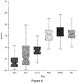
- apolipoprotein-A1 cutoff For specificity assessment and the choice of the best apolipoprotein-A1 cutoff, the study of 7,482 healthy volunteers aged 40 years and older, representative of the French population (7) was chosen as the core control population. To assess the impact on the specificity of apolipoprotein-A1 three populations at risk of false positive ( fig 4 , Table 2) were added successively: a population of 100 patients with rheumatologic diseases who had chronic inflammation and possible variability of acute phase proteins (18), and two populations where it was known that apolipoprotein-A1 had dramatically decreased, 238 patients with severe drug induced liver disease (DILI) (18-19), and 123 patients with severe alcoholic hepatitis (20).
- DILI drug induced liver disease
- ASH severe acute alcoholic hepatitis
- DILI Drug induced liver disease
- RHE Rheumatology patients
- BD blood donors
- the proportion of low apolipoprotein-A1 was linearly increasing since the first week 2020 in USA vs 2019 reaching 10% more low apolipoprotein-A1 in April.
- the proportion of low apolipoprotein-A1 reached 50% on the 16th week of surveillance in France and on the 8th week in APHP-PSL.
- apolipoprotein-A1 had a significant negative daily temporal association with the number of confirmed covid-19 cases, had a high sensitivity in prospective hospitalized patients, and had a high specificity in retrospective controls. The strength and the limitations of these results must be discussed successively.
- This stable A2M value as well as the 2020 lower prevalence of severe fibrosis stages eliminate a decrease of apolipoprotein-A1 due to an increase of severe fibrosis in the two surveillance populations, such as chronic viral hepatitis, alcoholic hepatitis, or non-alcohol-steatohepatitis (NASH).
- ApoA1 decrease could be also explained by a severe DILI, such as an acetaminophen misuse or by a decompensation of end-stage liver disease in patients with severe NASH. The prevalence of such cases seems too rare to explain a significant decrease in ApoA1 observed already in January 2020.
- the other serum biomarkers did not change during the first 3 months including, haptoglobin, GGT, ALT.
- the ideal population for assessing the specificity was a prospective sample representative of the general population, the absence of covid-19 being assessed by negative viral nucleic acid testing and normal chest computed tomography. Due to the context of the covid-19 pandemic, it was impossible to perform such a study.
- our databases in severe liver diseases permitted to identify the major risks of significant decrease of apolipoprotein-A1 (false negative) which were mainly the severe fibrosis of end-stage liver disease of chronic hepatitis, acute alcoholic hepatitis, and DILI. It is acknowledged that the possible impact of malnutrition on serum apolipoprotein-A1 value has not been assessed here. It is however considered that this factor was not relevant in the study.
- apolipoprotein-A1 The simple measure of apolipoprotein-A1 is therefore useful for clinicians according to the high percentage of false negative of available viral nucleic acid testing (25).
- apolipoprotein-A1 When apolipoprotein-A1 is expressed using 1.25 g/L the cutoff identified by the core control population the sensitivity of 90% is retrieved in all patients of the APHP-PSL hospital which was expected. This cutoff permitted an extrapolation for predicting the proportion of patients infected the large surveillance populations.
- the spread of the pandemic could be around 5 to 10% in this population of mixed liver disease, in line with the recent prediction modelling of 3 to 5% at the same date in France (26). For the USA the extrapolation in the population with NAFLD is around 9-11%.
- Apolipoprotein-A1 or HDL-cholesterol are already assessed in many ongoing large surveillances, of patients with chronic diseases such as cardiovascular, cancer and metabolic diseases, and could rapidly validate our results.
- SARS-CoV2 virus could inhibit the activity of lysophosphatidylcholine acyltransferase 3 (Lpcat) which is involved in phosphatidylcholine remodeling in the small intestine and liver (27). Indeed, in experimental calf pneumonia, the activity of Lpcat is reduced as early as one day after inoculation of Pasteurella haemolytica of bovine herpes virus-1, inducing a decrease in serum HDL concentration, and therefore of apolipoprotein-A1 its transporter, before the haptoglobin increase (12).
- Lpcat lysophosphatidylcholine acyltransferase 3
- Lpcat deficiency causes about 90% reduction of total Lpcat activity in the small intestine, whereas the deficiency only causes about a 70% reduction of total activity in the liver (27, 28).
- SARS-CoV2 on the small intestine could explained the decrease of serum apoA1, due to a decrease of its absorption (29). Indeed, they are evidence of direct SARS-CoV2 infection of the endothelial cell and diffuse endothelial inflammation in the intestine (30).
- the virus uses ACE2 receptor expressed by pneumocytes to infect the host, causing lung injury.
- the ACE-2 receptor is also widely expressed on endothelial cells, including intestine and liver.
- SARS-CoV2 is also present (14), and the ACE-2 receptor seems to be more expressed in the cholangiocytes than in the hepatocytes (31, 32). They are so far no data on cases infected by SARS-CoV2, without severe symptoms, on the prevalence of the virus in the liver and in the intestine. From the results in NAFLD patients, apo-A1 was decreasing in the first 16 weeks of 2020, without increase of ALT or GGT, in comparison with the same weeks of 2019.
- apolipoprotein-A1 can serve as a very early marker of the covid-19 pandemic in France and USA in the general population, with high specificity and sensitivity. It can also be used without delay in surveillance by dosing in routine. It could help immediately to manage patients with a clinical suspicion of covid-19 and a negative virologic test (25).
- the role of SARS-CoV2 in the possible "asymptomatic" decrease of apoliprotein-A1 could be related to intestine or liver infection without or before overt pulmonary disease.
- apolipoprotein-A1 hundred years after surrogate sentinel HDL-cholesterol (2) which had a reasonable temporal stability, could be included as "part of the early warning systems that alert the world to potential outbreaks" (35).
- a function that combines the level of apolipoprotein A1 (apoA1), the age (Log) and the sex of the patient was designed. Use of these three markers in this function made it possible to obtain an AUROC of 0.962.
- one uses a function that combines the level of apolipoprotein A1 (apoA1), the level of gammaglutamyl transpeptidase (GGT), the level of haptoglobin (Hapto), the age and the sex of the patient. Use of these five markers in this function made it possible to obtain an AUROC of 0.994. ( Figure 5 ).
- a function that combines the level of apolipoprotein A1 (apoA1), the amount of alpha-2 macroglobulin (A2M), the level of gammaglutamyl transpeptidase (GGT), the level of haptoglobin (Hapto), the age and the sex of the patient was designed.
- F6 -21.12957 - 9.80509 ⁇ ApoA1 + 6.14688 ⁇ Log(hapto) + 3.29395 ⁇ Log(GGT) - 0.52093 ⁇ Log(A2M) + 13.64505 ⁇ Log(AGE) - 1.15945 ⁇ SEX (0 for male; 1 for female)
- the Area Under Operating Curve (AUROC) was 0.9937( Figure 6 ). Different thresholds were assessed. For a cutoff at 0.5000, the function has
- the PPV and NPV values have been calculated with a prevalence of the disease of 1.7%, in the population, which is likely lower than the actual prevalence (which is estimated to be around 4-10%). With a 7% prevalence, using the 0.5000 cutoff, the PPV would be 0.9897 and the NPV would be 0.9889.
Landscapes
- Health & Medical Sciences (AREA)
- Life Sciences & Earth Sciences (AREA)
- Immunology (AREA)
- Engineering & Computer Science (AREA)
- Virology (AREA)
- Chemical & Material Sciences (AREA)
- Biomedical Technology (AREA)
- Urology & Nephrology (AREA)
- Molecular Biology (AREA)
- Hematology (AREA)
- Microbiology (AREA)
- Cell Biology (AREA)
- Biotechnology (AREA)
- Tropical Medicine & Parasitology (AREA)
- Food Science & Technology (AREA)
- Medicinal Chemistry (AREA)
- Physics & Mathematics (AREA)
- Analytical Chemistry (AREA)
- Biochemistry (AREA)
- General Health & Medical Sciences (AREA)
- General Physics & Mathematics (AREA)
- Pathology (AREA)
- Investigating Or Analysing Biological Materials (AREA)
Abstract
The invention relates to a new non-invasive test making it possible to detect infection by the SARS-CoV-2 virus in a patient, using values of Apolipoprotein A1 alone or in combination with other markers.
Description
- The invention relates to a new non-invasive test making it possible to detect infection by the SARS-CoV-2 virus in a patient.
- Coronavirus disease 2019 (COVID-19) is an infectious disease caused by severe acute respiratory syndrome coronavirus 2 (SARS-CoV-2). The disease was first identified in 2019 in Wuhan (China) and has quickly spread globally.
- Common symptoms generally include fever, cough, and shortness of breath. While most people have little symptoms, in some cases, COVID-19 may affect the lungs causing pneumonia. In those most severely affected, COVID-19 may rapidly progress to acute respiratory distress syndrome (ARDS) causing respiratory failure, septic shock, or multi-organ failure. Such failure may result from a cytokine storm, when large numbers of white blood cells are activated and release inflammatory cytokines, which in turn activate yet more white blood cells. The result is an immune response that becomes ubiquitous and not focused on infected cells.
- There are currently a few treatments that are investigated to treat COVID-19 patients. Hydroxychloroquine, alone or in combination with azithromycin, seem to have an antiviral effect for patients when treated early. Other antiviral drugs (remdesivir or lopinavir/ritonavir) may also be useful.
- When patients have acute respiratory distress syndrome, it is often noticed that the viral charge has diminished. Using anti-inflammatory drugs (such as corticoids, for example methylprednisolone, or anti-IL6 or anti-IL6 receptor drugs) can make it possible to control the cytokine storm. Hydroxychloroquine may also be useful for treating cytokine storm (Yao et al, Clin Infect Dis. 2020 Mar 9. pii: ciaa237. doi: 10.1093/cid/ciaa237).
- When faced with a patient with a suspicion of infection by the SARS-CoV-2 virus it is important to confirm such diagnosis. It is possible to perform a RT-PCR on a nasal sample from the patient. However, such test is not always accurate, as giving many false-negative results.
- There is thus a need for a novel and rapid test to identify SARS-CoV-2 positive patients, with good specificity and sensitivity. Such test should be applicable specifically in the context of a SARS-CoV-2 epidemic.
- There is thus an urgent need for efficient early detection of covid-19 in general population. Viral nucleic acid testing and chest computed tomography are standard methods for diagnosing covid-19 in patients with symptoms but are time consuming. In healthy general population, several predictors were identified such as age, sex, previous hospital admissions, comorbidity data, and social determinants of health. A recent review suggests that the proposed models are at high risk of bias, and that their predictive performance when used in practice is probably lower than that reported (Wynants et al. Prediction models for diagnosis and prognosis of covid-19 infection: systematic review and critical appraisal. BMJ. 2020;369:m1328).
- The inventors propose to use Apolipoprotein-A1 (Apo-A1) as a marker of SARS-CoV-2 infection, alone or in combination with other markers. In the context of a SARS-CoV-2 outbreak, i.e. when the disease is spreading among the population. As shown in the examples, Apolipoprotein-A1 is a very sensitive, specific and early marker of SARS-CoV-2 infection, but may also be linked to specific liver diseases. When used alone, it can be a surrogate marker for PCR (in particular when PCR is negative) and should be used within the complete clinical picture for posing the COVID-19 diagnosis. Hence, in an epidemic context, facing a patient with any of the COVID-19 signs (such as cough, fever, as well as skin rash, ageusia anosmia...), measuring the level (or amount) of Apolipoprotein-A1 in the blood or serum of a patient will help the physician to determine whether such patient is infected with the SARS-CoV-2 virus. Using other markers makes it possible to increase the sensitivity and specificity of the test, in particular by excluding other diseases that are susceptible to modify the amount of Apolipoprotein-A1. The bias mentioned by Wynants et al have been lowered by the use of data from the general population, that made it possible to obtain a robust diagnosis method.
- The quality of a diagnosis test is generally determined by drawing a Receiving Operating Characteristic (ROC) curve and measuring the Area Under Receiving Operating Characteristic curve (AUROC). The ROC curve is drawn by plotting the sensitivity versus (1-specificity), after classification of the patients, according to the result obtained for the diagnosis test, for different thresholds (from 0 to 1). It is usually acknowledged that the area under a ROC curve which has a value superior to 0.7 is a good predictive curve for diagnosis. The ROC curve has to be acknowledged as a curve allowing prediction of the quality of a diagnosis test. It is best for the AUROC to be as closed as 1 as possible, this value describing a test which is 100 % specific and sensitive.
- It is reminded that
- (1) sensitivity is the probability that the diagnosis is positive in individuals suffering from the disease sought (detection of true positives): the test is positive if the patient is suffering from the disease. The sensitivity is low when the number of false negatives is high. The sensitivity is calculated by the formula SE = (number of individuals suffering from the disease in whom the sign is present)/(number of individuals suffering from the disease in whom the sign is present + number of individuals suffering from the disease in whom the sign is absent).
- (2) specificity is the probability that the diagnosis is negative in the individuals not suffering from the disease sought (non-detection of true negatives): the test is negative if the patient is not suffering from the disease. The specificity is low when the number of false positives is high. The specificity is calculated by the formula SP = (number of individuals not suffering from the disease in whom the sign is absent)/(number of individuals not suffering from the disease in whom the sign is absent + number of individuals not suffering from the disease in whom the sign is present).
- (3) Positive predictive value (PPV): is the probability of having the disease if the diagnostic test is positive (i.e. that the patient is not a false positive): the patient is suffering from the disease if the test is positive. The positive predictive value is calculated by the formula PPV = (number of individuals suffering from the disease in whom the sign is present)/(number of individuals suffering from the disease in whom the sign is present + number of individuals not suffering from the disease in whom the sign is present).
- (4) Negative predictive value (NPV): is the probability of not having the disease if the diagnostic test is negative (that the patient is not a false negative): the patient is not suffering from the disease if the test is negative. The negative predictive value is calculated by the formula NPV = (number of individuals not suffering from the disease in whom the sign is absent)/(number of individuals not suffering from the disease in whom the sign is absent + number of individuals suffering from the disease in whom the sign is absent)
- In order to obtain a good diagnostic test, it is important to both increase specificity and sensitivity.
- In developing the assays and tests as herein disclosed, although the AUROC was very high when assessing Apo-A1 alone, it was increased when using other markers, such as age, sex or amount of other circulating proteins.
- Specificity was increased and verified by using controls such as patients who have a liver disease (but not COVID-19).
- Generally, a diagnosis (or diagnostic) method comprises
- i. a step of gathering information from the patient
- ii. a step of comparing said information with regards to thresholds
- iii. a step of deducing, from the difference between the patient's information and the threshold, whether the patient has a specific disease or the stage of the patient's disease.
- As a matter of illustration
- i. the information that can be gathered from the patient can be gathered directly from the patient (such as images from NMR, scanner, radiography, contrast-enhanced computed tomography), or indirectly from the patient, such as from a biological sample that has been obtained from a patient (such as urine, blood sample..). The information can be presence (or absence) and/or level of specific biological markers, whether specific from the pathogenic determinant (bacterial or viral DNA), or elevated levels of patient's markers.
- ii. once the information is obtained, it is compared to different values / standards and the deviation with regards to these standards is assessed. As a matter of illustration, the level of some biomarkers shall be compared to the level usually observed in healthy patients and to the levels usually observed in patients with the disease. Thresholds may exist, where 95 % of patients having passed the threshold have the disease and 95 % of the patients not having passed the threshold do not have the disease. For diseases where multiple clinical stages can be determined, such thresholds can discriminate the different stages. In this step ii, one may compare various types of information to their respective standards, in order to be able to reach a diagnostic in step iii (as a matter of illustration, one can use the values and information obtained from measurement of various blood or plasma markers, images from scanner and of Body Mass Index).
- iii. the last step is actually making the diagnosis i.e. deciding whether or not the patient has the condition sought, taking, in particular, into account the information gathered from the patient, the thresholds as described above. The physician may also take into account other elements (such as the consistency of the information gathered or the like) to make the diagnostic.
- Some methods, such as the ones disclosed in the present application, may also include a step i.a), which comprise the steps of modifying the information obtained from the patient in order to obtain a new type of information, which is the one that is then compared to the standards in step ii. Such modification is the combination of the values of variables in a function, and obtaining an end value.
- It is further to be noted that the mere measurement of the values of levels of markers in the plasma or serum of a patient and the combination thereof in an algorithm or function as herein disclosed is part of a method but only provides an intermediate result (an index) that would then to be compared to a reference index, in order to actually be able to pose the diagnostic.
- It is also to be noted that the tests herein disclosed are not "gold-standard" tests, in the sense that the output (index calculated by the formulas herein disclosed) isn't a direct answer as to the state of the patient. Indeed, these tests are based on statistics and there may thus be false-positive or false-negative results, which is the reason why the specific experience of the physician in interpreting the index is of importance for reaching a diagnosis.
- However, due to the specificity, sensitivity, positive predictive value and negative predictive value of the tests, herein provided for various thresholds of the index, these tests are of great interest in provided a help to the physician when investigating a clinical case. Consequently, step iii as disclosed above is not direct and immediate from step ii, as the physician must interpret the result from the clinical and general context to be able to reach a diagnosis.
- The invention thus relates to a method for detecting whether a patient is infected with the SARS-CoV-2 virus, comprising:
- a) Determining the level of apolipoprotein A1 (apoA1) in a serum sample from said patient and optionally of other clinical markers
- b) when other markers are used, combining the values obtained in a) through a function in order to obtain an end result
- c) Comparing the level of apolipoprotein A1 measured in step a) or of the end value obtained in step b) to a predetermined threshold
- It is to be noted that one can say that the method also provides information as to whether a patient is susceptible to be infected by SARS-CoV-2. Indeed, all tests of diagnosis present false positive (and false negative). However, in the present case, the examples demonstrate a high specificity and a high sensitivity, thereby giving a high classification accuracy of the patients.
- Alternatively, the method is very appropriate to detect whether a patient is not infected with the SARS-CoV-2 virus. Indeed, as disclosed below, the sensitivity and specificity of the methods are very high. However, depending on the prevalence of the SARS-Cov-2 virus infection (it is expected to be below 10%), it may be sometimes more appropriate to rely on the negative predictive value (although the positive predictive value remains high in view when the specificity is high). It may thus be preferable to use the methods herein disclosed to identify patients that are not infected by the SARS-Cov-2 virus (negative patients), and to perform other appropriate tests on patients that are positive to the disclosed method (such as PCR tests, or lung scanner) to confirm the SARS-Cov-2 virus infection.
- The above method is performed in vitro or ex vivo on samples of the patients that are already held by the person performing the method.
- It is to be noted that the inventors have engineered a test where the value of Apo-A1 alone can be used, and when the diagnosis may be performed when it is below a given threshold (preferably 1.25 g/l). When it is combined with other markers (such as the ones disclosed below) such combination will provide an end value. Depending on the sign of the coefficients for each markers, the diagnosis of infection with the SARS-Cov-2 virus may be linked to an end value above or below a given threshold.
- As shown in the examples, the amount of Apo-A1 alone is enough to allow the physician to conclude on the infection by the SARS-CoV-2 virus, especially during an outbreak and when the patient has other signs of infection. However, it is also envisaged to use other clinical markers from the patient, in particular selected from the group consisting of age of the patient (Age), gender (Sex), a2-macroglobulin (A2M), gammaglutamyl transpeptidase (GGT), and haptoglobin (Hapto). It is also possible to use inflammation markers, such as CRP (C Reactive protein), Serum amyloid A, or D-dimers. A positive D-dimer result may indicate the presence of an abnormally high level of fibrin degradation products and indicates that there may be significant blood clot (thrombus) formation and breakdown in the body, which is consistent with the Covd-19 pathology. Serum amyloid A (SAA) proteins are a family of apolipoproteins associated with high-density lipoprotein (HDL) in plasma and secreted during the acute phase of inflammation.
- For the biological markers, the amount of such markers in the patient's serum is used. For sex, the marker is used as a binary marker (0 for male, 1 for female). These markers can be used alone or in combination. When multiple markers are used in addition of the level of Apo-A1, one shall combine all markers in a function (in particular a logistic regression function) in order to get an end result, from the combination of the marker's values. The end result is then compared to a threshold in order to have the information necessary to pose the diagnosis.
- The present application provides various thresholds. It is however understood that other threshold can be selected. The thresholds proposed herein have been chosen as to classify the patients in three classes. When the end result is lower than a first threshold, the patient can be classified as SARS-Cov-2 positive (i.e. the threshold has been chosen so as to have a very low or even nil risk of false positive (high sensitivity)); when the end result is higher than a second threshold, the patient can be classified as SARS-Cov-2 negative (i.e. the threshold has been chosen so as to have a very low or even nil risk of false negative (high NPV)). Between the first and second thresholds, the physician can assess the infection by taking into account the whole clinical picture and perform other tests. Consequently, the present application describes two different thresholds, so as to provide assistance to the physician. It is however understood that other thresholds can be chosen by the one performing the methods herein disclosed, and that modifications of the thresholds will also lead to modifications of the specificity, sensitivity, NPV and PPV of the methods then used with these other thresholds.
- In a specific embodiment, the test combines, in a function, the value of apolipoprotein A-1 (Apo-A1) (g/l) and one or more of the following components:
- (1) alpha2-macroglobulin (A2M) (g/l)
- (2) Age (years)
- (3) Gender (0 for women, 1 for men)
- (4) Haptoglobin (Hapto) (g/l)
- (5) gamma-glutamyl transpeptidase (GGT) (IU/l).
- The function preferably doesn't use the value of bilirubin or of transaminases (alanine aminotransferase (ALT) or aspartate aminotransferase, AST)).
- Using GGT makes it possible to increase sensitivity (the Receiving Operating Curve is moved to the upper left) and increase AUROC.
- The use of A2M is interesting to discard false positive patients (with severe liver diseases such as severe acute alcoholic hepatitis (ASH) or Drug-Induced Liver Injury (DILI)).
- In a specific embodiment, though, Apo-A1 is used alone and the cut-off is about 1.25 g/L (Sensitivity: 0.9063; specificity: 0.9614; NPV: 0.9983). this simple threshold makes it possible to easily detect patients that are not infected by SARS-CoV-2 (i.e. patients for which the Apo-A1 amount is higher than 1.25 g/L), as the NPV is very high. The AUROC is 0.978.
- In another embodiment, Apo-A1 is used together with the age and the sex of the patient. Use of these three markers made it possible to obtain an AUROC of 0.962.
- In another embodiment, one uses a function that combines the level of apolipoprotein A1 (apoA1), the level of gammaglutamyl transpeptidase (GGT), the level of haptoglobin (Hapto), the age and the sex of the patient. Use of these five markers made it possible to obtain an AUROC of 0.975.
- In a preferred embodiment, one uses a function that combines the level of apolipoprotein A1 (apoA1), the level of gammaglutamyl transpeptidase (GGT), the level of haptoglobin (Hapto), the level of a2-macroglobulin (A2M), the age and the sex of the patient.
- One can use a function F6 = a0 + a1∗ApoA1 (g/L) + a2∗ Log(hapto (g/L)) + a3 Log(∗GGT (IU/L)) + a4∗ Log(A2M (g/L)) + a5∗ Log(AGE (years))+a6∗SEX (0 for women, 1 for men), with
- -23 ≤ a0 ≤ -19
- -11.2 ≤ a1 ≤ -8.4
- 4.5 ≤ a2 ≤7.7
- 2.3 ≤ a3 ≤ 4.2
- -0.75 ≤ a4 ≤ -0.25
- 11.5 ≤ a5 ≤ 15.5
- -1.8 ≤ a6 ≤ -0.5
- In particular, F6 = -21.12957 - 9.80509∗ApoA1 + 6.14688∗ Log(hapto) + 3.29395∗ Log(GGT) - 0.52093∗ Log(A2M) + 13.64505∗ Log(AGE) - 1.15945∗SEX (0=male; 1=female)
- For this function, if the end results is lower than 0.500 (sensitivity: 0.8516; specificity: 0.99933), the patient can be classified as SAS-CoV-2 negative. The AUROC is 0.9937.for a 1.7% prevalence, this would give a PPV of 0.9561 and a NPV of 0.9975. For a 7% prevalence, the PPV would be 0.9897 and the NPV would be 0.9889. The very high specificity allows a high PPV even with a low prevalence.
- It is to be noted that other cut-off values can be chosen. Increasing the cut-off would increase the specificity but decrease the sensitivity, while decreasing the cut-off would lead to the opposite result. One can also take into account the prevalence to determine the PPV. The choice of the cut-off (threshold) is thus determined by the physician according to its choice to increase or not the sensitivity or selectivity.
- The invention also includes a device for diagnosis of SARS-CoV-2 infection in a patient, comprising a first means, wherein the first means provides a first index by performing a method as described above, either by using the Apo-A1 amount alone or in combination with other markers.
- Said first index can be later used to determine SARS-CoV-2 infection in said patient and whether it is necessary to initiate treatment or follow-up, or perform further investigations.
- In a specific embodiment, the first mean is computerized. It may be an electronic spreadsheet with the formula recorded within, that provides the first index as an output when entering the various elements mentioned above. It can also be a computer program that provides the first index as an output after receipt of the various elements mentioned above.
- The first means can present one or more of the following, in either combination:
- Operate within a private or public network
- Receive the inputs (values of the various elements mentioned above) from a sender that is in a remote place (i.e. they are sent to the first means from a different location that where the first means is located)
- Require the sender to identify himself before sending the inputs
- Receive the inputs (values of the various elements mentioned above) from a secure manner
- Send the output (first index) to the sender of the inputs
- Store the output in a database (possibly with a unique identifier, making it possible to assign inputs, outputs to this identifier)
- Provides the first index with further information (such as sensitivity and/or specificity and/or positive predictive value and/or negative predictive value linked to the prevalence of the condition in the population to which belongs the patient)
- Is also foreseen a non-transitory computer readable storage medium, having stored thereon a computer program comprising program instructions, the computer program being loadable into a data-processing unit and adapted to cause the data-processing unit to carry out a method for calculating a first index by combining the values as measured from markers present in the serum or plasma of a patient through a method as disclosed above, when the computer program is run by the data-processing device.
- The following examples are meant to describe an aspect of invention, but shall not be limiting the invention.
-
-
Figure 1 . Daily apolipoprotein-A1 values during the first 16 weeks of the year. (A) Stratified by country. Apolipoprotein-A1 means values (95%CI) decreased significantly (linear regression; P<0.001) both in France (upper panel) than in USA (lower panel) in 2020, more rapidly in France since the 11th week. In USA the decrease was already significant since the 2nd week. Means values were higher in USA than in France in 2019 and 2020. In USA the subjects were at risk of non-alcoholic fatty liver disease. In France, the subjects were mostly patients with chronic hepatitis C. (B) Stratified by gender and age in USA. (C) Proportion of serum with low apolipoprotein-A1, in the APHP-PSL covid-19 center, in France and in USA. Low apolipoprotein-A1 was defined as below 1.25 g/L, the cutoff defined having the higher Youden index. These three populations had respectively 4109, 26170, and 248 470 sera. -
Figure 2 . Number of confirmed Covid-19 cases (per day, bars) and decrease of apolipoprotein-A1 (apoA1), absolute value and smoothed mean with 95% confidence interval. (A) Upper panel, data for France. (B) Lower panel, data for USA. In order to display the linear decrease of apoA1 along with exponential increase of covid-19 cases, the Y-axis for the decrease of apoA1 was expressed as 1.5- apoA1 value. For covid-19 cases we used a logarithmic scale divided by 20. i.e. on March2nd (first green dotted line) 30 cases occurred in France and the mean value of apoA1was 1.40 g/L. In USA 20 cases occurred in USA and the mean value of apoA1was 1.48 g/L. i.e. on April1st (second green dotted line) 7,578 cases occurred in France and the mean value of apoA1was 1.27 g/L. In USA 24,998 cases occurred in USA and the mean value of apoA1was 1.39 g/L. -
Figure 3 . Diagnostic performance of apolipoprotein-A1 for the diagnostic of Covid-19 in 128 Covid-19 cases and 7,482 healthy volunteers controls. Apolipoprotein-A1 at the 1.25 g/L cutoff, had a sensitivity of 90.6% (95%CI 84.2-95.1) and a specificity of 96.1% (95.7-96.6%) for the diagnostic of covid-19. -
Figure 4 . Serum apolipoprotein levels (IQR) between the 6 populations assessing sensitivity (covid-19), and specificity (CPAM: general population used for the core analysis) liver diseases (ASH: severe acute alcoholic hepatitis, DILI: Drug induced liver disease, RHE: Rheumatology patients) and blood donors (BD). The total number is 8,336 subjects. (8,336 subjects) -
Figure 5 . Receiving Operating Curve for a function combining the level of apolipoprotein A1 (apoA1), the level of gammaglutamyl transpeptidase (GGT), the level of haptoglobin (Hapto), the age and the sex of the patient. -
Figure 6 . Receiving Operating Curve for a function combining the level of apolipoprotein A1 (apoA1), the level of gammaglutamyl transpeptidase (GGT), the level of haptoglobin (Hapto), the level of Alpha-2-Macroglobulin (A2M) the age and the sex of the patient. - The prospective observational study in covid-19 patients was approved by CER-Sorbonne University IRB, N° 2020 - CER-2020-14, with a signed informed consent. Clinical investigation was conducted according to the principles of the Declaration of Helsinki. The components of FibroTest (FibroSure in USA) and Nash-FibroTest (Nash-FibroSure in USA) were measured in routine in patients with a risk of liver disease in France and USA. The serum databases used anonymous results. All the retrospective databases of patients analyses previously published were non-interventional study, without supplementary blood sample, and exempt from institutional review board (IRB) review (ethical committee of 'Comite de Protection des Personnes of Paris, Ile-de-France' FIBROFRANCE project. CPP-IDF-VI, 10- 1996-DR-964, DR-2012-222 and USA-NCT01927133).
- The pre-inclusion criteria were consecutive patients examined in the internal medicine department of APHP-PSL hospital between 24/01/2020 and 21/04/2020, with a suspicion of covid-19, severe enough for admission, but not requiring intensive care unit or mechanical ventilation.
- After signed informed consent, no supplementary tests were performed. The serum proteins of interest to assess the diagnostic and prognostic performances were routinely performed in all patients with a risk of liver disease. Apolipoprotein-A1 and haptoglobin, two biomarkers of acute phase, alpha-2 macroglobulin (A2M) which is not an acute phase protein in human but a marker of liver fibrosis, gammaglutamyl transpeptidase (GGT) and alanine aminotransferase (ALT) as standard sensitive "liver function tests" were performed.
- The covid-19 diagnostic was defined as a SARS-CoV2 PCR-positive in upper respiratory samples, or in blood. All patients with PCR-negative despite symptoms of covid-19 and chest computed tomography signs of covid-19, or patients having a spouse SARS-CoV2 PCR-positive, were adjudicated (PC, MR, TP, OL). Exclusion criteria were non signed informed consent, missing data for blood markers and not adjudicated as clinical covid-19 in case of absence of positive PCR. No prospective controls were included, as it was impossible in the context of the pandemic to have a large sample representative of the general population.
- Apolipoprotein-A1, haptoglobin, A2M, GGT and ALT components assays were performed on an automatic analyzers Cobas-c701 and Cobas-c501 from Roche Diagnostics (Mannheim, Germany). Proteins concentrations were measured according to turbidimetric analytical methods using manufacturer reagents for haptoglobin and apolipoprotein-A1, and Diagam (Ghislenghien, Belgium) reagents for A2M. ALT was determined according to IFCC method with pyridoxal phosphate, and GGT using Szasz method and calibrator value given for the IFCC. Total bilirubin was assessed according to a diazoreaction.
- The consecutive anonymous sera of populations routinely followed for liver diseases risk, mostly chronic hepatitis C in France (17,778 sera in 2019 and 12,500 in 2020) and patients at risk of NAFLD in USA (136,688 sera in 2019, and 111,782 sera in 2020), were analyzed for the same period in the first 16 weeks of 2019 and 2020, following the BioPredictive (Paris, France) analytical recommendations (10).
- To diagnose SARS-CoV2 in upper respiratory samples, we used the fully automated sample-to-result two-target test Cobas 6800 SARS-CoV-2 (Roche molecular systems, Branchburg, NJ,USA) (11). The presence of SARS-CoV-2 in the lower respiratory samples and blood were tested by Altona RealStar® RT-PCR Kit 1.0 (Altona Diagnostics, Hamburg, Germany). Plasma samples were subjected to total nucleic acid (TNA) extraction by using the NucliSENS easyMAG extraction system (BioMerieux, France) and PCR was performed using LightCycler® 480 Instrument II (Roche, Switzerland)
- The number of confirmed covid-19 cases in France and in the US was assessed according to the published data from the European Centre for Disease Prevention and Control (https://ourworldindata.org/coronavirus-data).
- The linear regression curves with 95% confidence interval between the daily mean serum level of apolipoprotein-A1 were compared by F-test, and a significant difference was defined as P value <0.001. Regression curves were also used to assess possible confounding factors, such as country, gender and age, not only on apolipoproteinA1 but also on haptoglobin a marker of systemic inflammation, A2M a marker of liver fibrosis, and GGT and ALT, two standard liver function tests. Total serum bilirubin was also assessed, but as no changes were observed, not included in the results and discussion.
- The sensitivity was assessed in patients with covid-19 as previously defined. To be able to use an early detection test in large populations, the specificity was assessed in six databases, including three at risk of false positive. The core analysis for the specificity assessment, was the general population study in healthy volunteers' representative of the French population aged 40 years or older. The performance of apolipoprotein-A1, haptoglobin, A2M, GGT and ALT were assessed using non-parametric AUROCs. The best cutoff was chosen as the serum apolipoprotein value giving the highest Youden index (sensitivity + specificity -1).
- Statistical software used R and NCSS-2020.
- There is an urgent need for efficient early detection of covid-19 in general population. Viral nucleic acid testing and chest computed tomography are standard methods for diagnosing covid-19 in patients with symptoms but are time consuming. In healthy general population, several predictors were identified such as age, sex, previous hospital admissions, comorbidity data, and social determinants of health. A recent review suggests that the proposed models are at high risk of bias, and that their predictive performance when used in practice is probably lower than that reported (1).
- In the meantime, several early detection tests of cytokine burst could be immediately available. In 1920, Harold A. Kipp found that a decrease in serum cholesterol was a marker of severe pneumonia (2). One hundred years later, a meta-analysis showed some association of high-density lipoprotein cholesterol (HDL) and apolipoprotein-A1 and an increased (twice) mortality rate (odds ratio= 2.0; 95% CI 1.2-3.2) in 791 patients with severe sepsis (fig. S1 which is
Figure 3 of Liu, 2020) (3). Apolipoprotein-A1 has also been studied in relationship with inflammation (4, 5). - It was hypothesized first that apolipoprotein-A1 and haptoglobin could help to detect the spread of SARS-CoV-2, and the databases of FibroTest (FibroSure in USA), a multi-analytes liver diagnosis which includes these proteins (6) and which is widely used in large surveillance of subjects at risk of cirrhosis and primary liver cancer (7-9) were analyzed. Two unexpected results suggested new mechanisms for the decrease of ApoA1. Firstly, the decrease was observed several weeks before the significant increase of confirmed Covi-19 cases and secondly, it was not associated with an increase of haptoglobin which appeared later in Covid-19 centers. These facts open speculations on the role of SARS-CoV-2 on the decrease of intestinal absorption of apoA1 and to a potential fecal-oral transmission.
- Therefore, it was aimed to illustrate that other existing surveillances in the world using apolipoprotein-A1 could also validate and participate to such sentinel networks for viral pandemic. In the present study, the performance of apolipoprotein-A1 as a predictor of hospital admission for covid-19 in general population was not assessed, neither its ability to detect covid-19 in patients with symptoms, or for constructing a multivariable covid-19 related prediction model.
- To assess the daily association between the serum apolipoprotein-A1 and the spread of covid-19, the consecutive results of three populations were used. Two were large surveillance cohorts of subjects at risk of mixed chronic liver disease in France, and at risk of non-alcoholic fatty liver disease (NAFLD) in USA. The third was the patients of Pitié-Salpêtrière hospital (APHP-PSL, Paris, France), a reference center for both covid-19 and liver disease.
- To assess the diagnostic performances of apolipoprotein-A1, its sensitivity in a prospective non-interventional study in covid-19 patients was assessed, and its specificity was assessed in representative controls of a general population.
- The mean of serum apolipoprotein-A1 daily during the first 16 weeks of 2020 in a population routinely followed for liver diseases risk, was compared in France (12,500 sera) and in USA (111,782 sera), in parallel with the local raise of confirmed cases, and in comparison with the same days of 2019 (respectively 17,779 and 136,688 sera). Its sensitivity was assessed prospectively in an observational study of 128 consecutive adjudicated hospitalized cases and retrospectively its specificity in 7,482 controls representative of general population.
- The mean serum level of apolipoprotein-A1 in these populations was already decreasing since January 2020, in comparison with the similar 16 weeks in 2019. This decrease was highly correlated and parallel with the daily increase of confirmed covid-19 cases, in the following 16 weeks, in France and USA. Apolipoprotein-A1 at the 1.25 g/L cutoff had a sensitivity of 90.6% (95%CI 84.2-95.1) and a specificity of 96.1% (95.7-96.6%) for the diagnostic of covid-19. The area under the characteristics curve was 0.978 (0.957-0.988), and outperformed haptoglobin and the liver function tests.
- Apolipoprotein-A1 could be used both as a sentinel of the pandemic in existing routine surveillance of the general population, as well as a candidate predictor of suspected covid-19 in multivariable model, for cases with negative virologic test.
- It is reported here that the mean serum level of apolipoprotein-A1 in these populations had already been decreasing since January 2020, both in France and in the USA (
fig 1 ), in comparison with the 16 first weeks of 2019, when the same surveillance populations were not exposed to SARS-CoV-2. Apolipoprotein-A1 means values (95%CI) decreased significantly (linear regression; P<0.001) both in France (upper panel) and in the USA (lower panel) in 2020, more rapidly in France since the 11th week. In the USA the decrease was already significant since the 2nd week of January (fig 1 A) . Mean values were higher in the USA than in France in 2019 and 2020. In the USA, subjects were all at risk of non-alcoholic fatty liver disease. In France, the subjects were mostly patients with chronic hepatitis C. - From January to March 2020, before the start of the pandemic awareness, there was no variability in the daily number of sera analyzed in France and USA in comparison with January to March 2019, and the decrease of apolipoprotein-A1 was already significant, whatever the age and gender. There was a delay of 10 days between the start of the pandemic between France and USA. The demographic and biochemical characteristics of the anonymous sera included in the US surveillance analysis for the first 16 weeks on 2020 and 2019 were very similar during the 16 weeks. Due to the sample size 111,782 and 136,688 sera in 2020 and 2019 respectively, most of the characteristics were significantly different, but reassuring for the risk of bias related to confounding factors associated with apolipoprotein A1 decrease, without more severe fibrosis stage. As apolipoprotein-A1 is one component of FibroTest, one confounding factor in such patients at risk of liver fibrosis could be a higher prevalence of severe fibrosis in the 2020 population. Indeed, the inverse was observed. In the 2020 population the prevalence of not severe fibrosis (stage F0 to F2) was 20.3% vs 22.0% in 2019 in line with the decrease of A2M, and GTT.
- This apolipoprotein-A1 decrease since January 2020 was highly correlated and followed the same trend than the daily increase of confirmed covid-19 cases, in the following 16 weeks, in France and in the USA. Apolipoprotein-A1 mean values (95%CI) decreased significantly both in France (upper panel) and in the USA (lower panel) in 2020. In the USA, the decrease was already significant since the 2nd week of January, earlier than in France where a decrease was only significant since the 11th week. The later and dramatic increase of haptoglobin is in line with the studies of acute phase proteins in severe sepsis and in covid-19 series (3-5, 12). The kinetics of apolipoprotein-A1 were not associated with those of haptoglobin (not shown), which is also in line with different possible mechanisms occurring at the early acute phase (12).
- ApoA1 decrease could be also explained by a severe DILI, such as an acetaminophen misuse or by a decompensation of end-stage liver disease in patients with previous chronic viral hepatitis, or as severe NASH. The prevalence of such cases seems to rare to explain the significant decrease in ApoA1 observed already in January 2020. Therefore, other another explanation compatible with these facts could be an impact of the SARS-CoV2 virus on synthesis of apoA1, in patients still asymptomatic.
- Temporal association between apolipoprotein-A1 and covid-19 can be due to many confounding factors. The possible variability related to country, gender, age, and severe liver fibrosis stage were assessed. The decrease of serum apolipoprotein-A1 was similar according to gender, and to age both in France and in the USA (not shown). Only the sample size of the USA database permitted to assess the kinetics of apolipoprotein-A1 according to stratifications by both gender and age (
fig 1 B) . - An overview estimated that one third of patients with severe covid-19 had an impaired hepatic function, based on standard liver function tests (13). The causes can be due to the SARS-CoV-2 direct liver toxicity (14), but also to drug induced liver disease (DILI) caused by medications such as paracetamol, ibuprofen, clavulinate or chloroquine, or to a previous liver disease. To assess if the changes in apolipoprotein-A1 were associated with liver fibrosis, the daily kinetics of A2M which is not an acute phase protein in human but a marker of liver fibrosis were also analyzed (6, 15), and no changes were observed. To assess if the changes were not associated with other liver features (steatosis and hepatic inflammation) the daily kinetics of GGT, and ALT as standard sensitive "liver function tests" were analyzed. There was a significant increase of GGT during 2020 versus 2019, starting earlier in France and of ALT in April 2020, only significant in the USA. A very significant "center effect" on the regression curves was observed in France, probably due to an increase of the proportion of sera coming from the covid-19 centers. In the USA, the much bigger sample size probably prevented such a center effect.
- The first patient with covid-19 was admitted in the hospital on the 30/01/2020. The study started on the 19/02/2020 and the last patient of the present report was included on the 21/04/2020. A total of 128 consecutive patients with severe covid-19 (PCR- SARS-CoV-2 positive), but who did not require intensive care, were hospitalized in the department of internal medicine. Characteristics of patients' PCR covid-19 positive were similar to those published in such severity profiles (16).
- For the first time apolipoprotein-A1 was assessed in covid-19. The area under the characteristics curve (AUROC) in 128 covid-19 cases and 7,482 controls representative of the
French population 40 years and older (7), was 0.978 (0.957-0.988), and outperformed haptoglobin and the liver function tests (fig 3 , Table 1). Apolipoprotein-A1 at the 1.25 g/L cutoff, had the best Youden index (sensitivity + specificity - 1 = 86.7%) with a sensitivity of 90.6% (95%CI 84.2-95.1) and a specificity of 96.1% (95%CI 95.7-96.6%) for the diagnostic of covid-19.Table 1. Diagnostic performance of apolipoprotein-A1 for the diagnostic of covid-19 in 128 covid-19 cases and 7,482 healthy volunteers' controls. Criterion Count AUC Standard Error P-Value vs ApoA1 Lower 95%Cl Upper 95%Cl ApoA1neg 7610 0.9779 0.0075 NA 0.9572 0.9887 Lhapto 7610 0.9435 0.0123 0.009 0.9138 0.9631 LGGT 7610 0.7667 0.0213 <0.001 0.7217 0.8053 LA2M 7610 0.5084 0.0300 <0.001 0.4472 0.5648 LbiliNeg 7610 0.7442 0.0262 <0.001 0.6883 0.7913 LALT 7610 0.6802 0.0269 <0.001 0.6238 0.7294 LAGE 7610 0.7436 0.0289 <0.001 0.6815 0.7950 - In the same department of internal medicine during the 16-weeks period, 43 patients with mixed severe diseases were consecutively pre-included, but not adjudicated as covid-19. They could not be used for assessing the specificity of apolipoprotein-A1, according to the small sample size and as they were not representative of the context of use of an early detection test in general population.
- However, data on apolipoprotein-A1 was prospectively cumulated since a first cohort of alcoholic liver disease in 1982 (17), then in the FibroFrance cohort (6-10). More recently six databases prior to the pandemic were collected, which allowed to validate retrospectively the specificity in a large spectrum of subjects. The measurements have all been performed on fresh serum collected prospectively and analyzed in the biochemistry unit of APHP-PSL hospital, with the same biochemical methods than for the covid-19 cases.
- For specificity assessment and the choice of the best apolipoprotein-A1 cutoff, the study of 7,482 healthy volunteers aged 40 years and older, representative of the French population (7) was chosen as the core control population. To assess the impact on the specificity of apolipoprotein-A1 three populations at risk of false positive (
fig 4 , Table 2) were added successively: a population of 100 patients with rheumatologic diseases who had chronic inflammation and possible variability of acute phase proteins (18), and two populations where it was known that apolipoprotein-A1 had dramatically decreased, 238 patients with severe drug induced liver disease (DILI) (18-19), and 123 patients with severe alcoholic hepatitis (20). The decrease was related to liver fibrosis stage (21) and hepatic insufficiency (18-19). 393 blood donors were also included to assess the specificity in younger controls than in the core control population (22).Table 2. Serum apolipoprotein levels between the 6 populations assessing sensitivity (covid-19), and specificity (CPAM: general population used for the core analysis) liver diseases (ASH: severe acute alcoholic hepatitis, DILI: Drug induced liver disease, RHE: Rheumatology patients) and blood donors (BD). Control subjects Count Median Dunnets' test difference (P<0.05) ASH 123 0.31 DILI, covid, RHE, CPAM, BD DILI 238 0.49 ASH, covid, RHE, CPAM, BD covid-19 128 0.86 ASH, DILI, RHE, CPAM, BD RHE 100 1.58 ASH, DILI, covid, CPAM CPAM 7482 1.67 ASH, DILI, covid, RHE, BD BD 393 1.59 ASH, DILI, covid, CPAM - When using the same 1.25 g/L cutoff the specificity in 393 blood donors was similar (94.2%; 95%CI 83.5-93.8%, AUC 0.9697), lower (84.0%; 95%CI 75.3-90.6%, AUC 0.9505) for 100 patients with rheumatologic disease. Finally, this database integrated six databases with a total of 8,464 subjects: 128 covid-19 (prevalence 1.51%) and 8,336 controls. Despite the higher risk of false positive and using the pre-defined 1.25 g/L cutoff of the core control population, a reassuring specificity of apolipoprotein-A1 of 92.3% (95%CI 91.7-92.8%), and an AUROC of 0.946 (95%CI 0.929-0.959) are reported.
- The sensitivity with cutoff <1.25 g/L was 90.6% (98/116). Interestingly the sensitivity among the 19 patients with negative viral nucleic acid testing was similar 18/19 (94.7%) than in patients positive, 98/109 (89.9%; exact Fisher test P=1). Thereafter, the 1.25 g/L cutoff was used to assess the proportion of low apolipoprotein-A1 among the surveillance populations during the 16 weeks of January (
fig 1 C) . In the APHP-PSL hospital, at the peak of the pandemic in Paris (first week of April) the proportion of low apolipoprotein-A1 in serum was 90% (upper panel) as expected by the sensitivity observed at admission of covid-19 patients. According to the serum sample size of each population, the proportion of low apolipoprotein-A1 was linearly increasing since thefirst week 2020 in USA vs 2019 reaching 10% more low apolipoprotein-A1 in April. The proportion of low apolipoprotein-A1 reached 50% on the 16th week of surveillance in France and on the 8th week in APHP-PSL. - It is reported here that apolipoprotein-A1 had a significant negative daily temporal association with the number of confirmed covid-19 cases, had a high sensitivity in prospective hospitalized patients, and had a high specificity in retrospective controls. The strength and the limitations of these results must be discussed successively.
- The intriguing fact was the very early decrease observed since January 2020 in the USA while the number of covid-19 cases was unknown, and only in March in France. The first known covid-19 patient was detected on 27/12/2019 and 19/01/2020 in France (23) and USA (24) respectively. One simple explanation could be due to the much larger sample size of the US surveillance population, in comparison with the French surveillance, which permitted to detect in January a 1% significant increase in the proportion of subject possibly infected using the 1.25 g/L cutoff (
fig 1 C) . There was no obvious bias in age or sex or other event which could change the characteristics of patients followed for NAFLD or chronic viral hepatitis. Therefore, these suggested that the spread of the pandemic in the USA could be around 10%, the plateau reached since April in the population followed for NAFLD. In France the proportion can be only estimated reasonably up to 14th of March before the knockdown and the dramatic decrease of sera collected. Therefore, the spread of the pandemic could be around 5 to 10% in this population of mixed liver disease. These extrapolations from the proportion of low apolipoprotein-A1 are not so different than the recent French modelling that predicted 3% to 5% on March 15th. They are very different from the unpublished controversial UK modelling study (BMJ 2020;368:m1216). These data indicated that by March 19th (15 days after the first reported UK death, and 45 days after the first confirmed case), between 36% and 68% of the UK population would have been infected, depending on the assumptions used. Applying the same modelling for France, 45 days after the first case on December 2019 27th, that is February 18th, the proportion of infected cases in France would be between 36% to 68%. - Such temporal association between apolipoprotein-A1 and covid-19 can be due to many confounding factors. In the context of the pandemic it was not possible to perform an ideal study including a prospective population representative of the general population of the country, but only of the patients requiring surveillance by liver fibrosis biomarkers. Sera were analyzed anonymously and therefore there was an unknown percentage of duplicated subject. However, the decrease of apolipoprotein-A1 in 2020 vs 2019 could not beexplained, at least by gender, age, and severity of liver disease. The decrease of serum apolipoprotein-A1 was similar according to gender, and to age both in France and in the USA. Only the sample size of the USA database permitted to assess the kinetics of apolipoprotein-A1 according to stratifications by both gender and age (
fig 1 B) . The two large surveillance populations were patients at risk of liver diseases and were not representative of the general population of France and USA. Therefore, it was mandatory to check that A2M, a marker of liver fibrosis (6), had no changes during the 16 weeks of 2019 and 2020. In case of severe fibrosis A2M is very significantly increased (6-9). This stable A2M value as well as the 2020 lower prevalence of severe fibrosis stages eliminate a decrease of apolipoprotein-A1 due to an increase of severe fibrosis in the two surveillance populations, such as chronic viral hepatitis, alcoholic hepatitis, or non-alcohol-steatohepatitis (NASH). ApoA1 decrease could be also explained by a severe DILI, such as an acetaminophen misuse or by a decompensation of end-stage liver disease in patients with severe NASH. The prevalence of such cases seems too rare to explain a significant decrease in ApoA1 observed already in January 2020. Moreover, the other serum biomarkers did not change during the first 3 months including, haptoglobin, GGT, ALT. No active drug for NAFLD or NASH appeared between 2019 and 2020. Possible changes in the treatment of diabetes, dyslipidemia and obesity (bariatric surgery) could also be related to apoA1-decrease. However, after stratification on age and sex, no significant changes were observed for total cholesterol, triglycerides, fasting glucose, weight and height. - Using the data of the APHP-PSL hospital, the risk of other confounding factors was assessed in more details, both in patients with covid-19 and in controls. After the admission of the first patient with covid-19 the 30/01/2020, several units of the hospital were transformed into one covid-19 reference center in March 2020. Due to the country-wide lockdown decisions, there was a dramatic decrease in the throughput of FibroTest prescription, decreasing to 12.5 % in April 2020 in France and in the USA. In APHP-PSL, the throughput of prescriptions in April 2020 was still between 35% to 50% versus a mean of 12.5% in France. This center-effect can be explained by the conversion of the APHP-PSL hospital into a covid-19 center with a dramatic increase in severe cases. In USA, the decrease of apolipoprotein-A1 was almost linear, and haptoglobin remained stable without such reference-center effects (
fig 1 C) . - According to the context of use of an early detection test in asymptomatic subjects, the ideal population for assessing the specificity was a prospective sample representative of the general population, the absence of covid-19 being assessed by negative viral nucleic acid testing and normal chest computed tomography. Due to the context of the covid-19 pandemic, it was impossible to perform such a study. However, our databases in severe liver diseases permitted to identify the major risks of significant decrease of apolipoprotein-A1 (false negative) which were mainly the severe fibrosis of end-stage liver disease of chronic hepatitis, acute alcoholic hepatitis, and DILI. It is acknowledged that the possible impact of malnutrition on serum apolipoprotein-A1 value has not been assessed here. It is however considered that this factor was not relevant in the study.
- It is acknowledged that the population included for sensitivity assessment may not be the ideal one, as there may be a risk of overestimation of the sensitivity estimated in severe disease, for a context of use in asymptomatic infected subject. However even if these patients were severe enough to justify their admission with a median of 70 years of age, none of them needed mechanical ventilation, 86% survived, 6% were transferred in intensive care unit, and 9% died. To validate the sensitivity of apolipoprotein-A1, a large number of asymptomatic subjects positive for SARS-CoV-2 viral nucleic acid testing was needed. At least, the sensitivity in the prospective study was similar among the 19 patients with negative viral nucleic acid testing (94.7%) to in patients positive (89.9%). The simple measure of apolipoprotein-A1 is therefore useful for clinicians according to the high percentage of false negative of available viral nucleic acid testing (25). When apolipoprotein-A1 is expressed using 1.25 g/L the cutoff identified by the core control population the sensitivity of 90% is retrieved in all patients of the APHP-PSL hospital which was expected. This cutoff permitted an extrapolation for predicting the proportion of patients infected the large surveillance populations. The spread of the pandemic could be around 5 to 10% in this population of mixed liver disease, in line with the recent prediction modelling of 3 to 5% at the same date in France (26). For the USA the extrapolation in the population with NAFLD is around 9-11%. These predicted proportion are challenging, due to the absence of validations using SARS-CoV-2 -antibodies. However, this surveillance population is particularly at risk of covid-19, according to the prevalence of obesity and type-2 diabetes (15). External and independent validations of the results in other large samples of general population shall be performed. Apolipoprotein-A1 or HDL-cholesterol are already assessed in many ongoing large surveillances, of patients with chronic diseases such as cardiovascular, cancer and metabolic diseases, and could rapidly validate our results.
- No major confounding factors, (age, gender, weight, fasting glucose, total cholesterol, triglycerides, and severe liver fibrosis) were found, which could explain the very early temporal association between the decrease of apo-A1 and the spread of the pandemic in the US population of patients followed for NAFLD. The mechanisms explaining the decrease of apo-A1 in late severe covid-19 pneumonia are well known, but not those explaining the early decrease before the acute phase. In patients with severe pneumonia, published evidence-based results demonstrated that apo-A1 decrease was associated with acute inflammation, the "cytokine storm" with an increase of IL6 and acute phase proteins such as CRP and haptoglobin. Here in the US population, the decrease of apolipoprotein-A1 was not associated with an increase of haptoglobin. This dissociation suggested different mechanisms for the early impact of SARS-CoV2 virus on synthesis of apoA1.
- It is speculated that SARS-CoV2 virus could inhibit the activity of lysophosphatidylcholine acyltransferase 3 (Lpcat) which is involved in phosphatidylcholine remodeling in the small intestine and liver (27). Indeed, in experimental calf pneumonia, the activity of Lpcat is reduced as early as one day after inoculation of Pasteurella haemolytica of bovine herpes virus-1, inducing a decrease in serum HDL concentration, and therefore of apolipoprotein-A1 its transporter, before the haptoglobin increase (12). In knock-out mice, Lpcat deficiency causes about 90% reduction of total Lpcat activity in the small intestine, whereas the deficiency only causes about a 70% reduction of total activity in the liver (27, 28). The impact of SARS-CoV2 on the small intestine could explained the decrease of serum apoA1, due to a decrease of its absorption (29). Indeed, they are evidence of direct SARS-CoV2 infection of the endothelial cell and diffuse endothelial inflammation in the intestine (30). The virus uses ACE2 receptor expressed by pneumocytes to infect the host, causing lung injury. The ACE-2 receptor is also widely expressed on endothelial cells, including intestine and liver. In the liver, SARS-CoV2 is also present (14), and the ACE-2 receptor seems to be more expressed in the cholangiocytes than in the hepatocytes (31, 32). They are so far no data on cases infected by SARS-CoV2, without severe symptoms, on the prevalence of the virus in the liver and in the intestine. From the results in NAFLD patients, apo-A1 was decreasing in the first 16 weeks of 2020, without increase of ALT or GGT, in comparison with the same weeks of 2019.
- These results indicate that apolipoprotein-A1 can serve as a very early marker of the covid-19 pandemic in France and USA in the general population, with high specificity and sensitivity. It can also be used without delay in surveillance by dosing in routine. It could help immediately to manage patients with a clinical suspicion of covid-19 and a negative virologic test (25). The role of SARS-CoV2 in the possible "asymptomatic" decrease of apoliprotein-A1 could be related to intestine or liver infection without or before overt pulmonary disease. Finally, apolipoprotein-A1 hundred years after surrogate sentinel HDL-cholesterol (2), which had a reasonable temporal stability, could be included as "part of the early warning systems that alert the world to potential outbreaks" (35).
- A function that combines the level of apolipoprotein A1 (apoA1), the age (Log) and the sex of the patient was designed. Use of these three markers in this function made it possible to obtain an AUROC of 0.962.
- In another embodiment, one uses a function that combines the level of apolipoprotein A1 (apoA1), the level of gammaglutamyl transpeptidase (GGT), the level of haptoglobin (Hapto), the age and the sex of the patient. Use of these five markers in this function made it possible to obtain an AUROC of 0.994. (
Figure 5 ). - A function that combines the level of apolipoprotein A1 (apoA1), the amount of alpha-2 macroglobulin (A2M), the level of gammaglutamyl transpeptidase (GGT), the level of haptoglobin (Hapto), the age and the sex of the patient was designed. F6 = -21.12957 - 9.80509∗ApoA1 + 6.14688∗ Log(hapto) + 3.29395∗ Log(GGT) - 0.52093∗ Log(A2M) + 13.64505∗ Log(AGE) - 1.15945∗SEX (0 for male; 1 for female)
The Area Under Operating Curve (AUROC) was 0.9937(Figure 6 ).
Different thresholds were assessed.
For a cutoff at 0.5000, the function has - Sensitivity: 0.8516
- Specificity: 0.9993
- NPV: 0.9975
- PPV: 0.9561
- For a cutoff at 0.3000, the sensitivity increases, but the specificity decreases.
- Sensitivity: 0.8984
- Specificity: 0.9985
- NPV: 0.9983
- PPV: 0.9127
- The accuracy for this cut-off would be similar to the one for 0.500 cutoff.
- For a cutoff at 0.7000, the sensitivity decreases, but the specificity increases.
- Sensitivity: 0.7734
- Specificity: 0.9996
- NPV: 0.9961
- PPV: 0.9706
- The PPV and NPV values have been calculated with a prevalence of the disease of 1.7%, in the population, which is likely lower than the actual prevalence (which is estimated to be around 4-10%).
With a 7% prevalence, using the 0.5000 cutoff, the PPV would be 0.9897 and the NPV would be 0.9889. -
- 1. Wynants L, Van Calster B, Bonten MMJ, et al. Prediction models for diagnosis and prognosis of covid-19 infection: systematic review and critical appraisal. .
- 2. Kipp HA. Variation in the cholesterol content of the serum in pneumonia. J Biol Chem 1920; 44:215-237.
- 3. Liu SH, Liang HY, Li HY, et al. Effect of low high-density lipoprotein levels on mortality of septic patients: A systematic review and meta-analysis of cohort studies. World .
- 4. Delanghe JR, Langlois MR, Boelaert JR, et al. J. Plum. Haptoglobin polymorphism, iron metabolism and mortality in HIV infection. AIDS 1998; 12:1027-32.
- 5. Yang H, Wang H, Wang Y, et al. The haptoglobin beta subunit sequesters HMGB1 toxicity in sterile and infectious inflammation. J Intern Med 2017; 282:76-93.
- 6. Imbert-Bismut F, Ratziu V, Pieroni L, et al. Biochemical markers of liver fibrosis in patients with hepatitis C virus infection: a prospective study. Lancet 2001; 357:1069-75.
- 7. Poynard T, Lebray P, Ingiliz P, et al. Prevalence of liver fibrosis and risk factors in a general population using non-invasive biomarkers (FibroTest). BMC Gastroenterol 2010; 10:40.
- 8. Poynard T, Deckmyn O, Munteanu M, et al. Awareness of the severity of liver disease re-examined using software-combined biomarkers of liver fibrosis and necroinflammatory activity. BMJ open 2015; 5:e010017.
- 9. Poynard T, Munteanu M, Charlotte F, et al. Diagnostic performance of a new noninvasive test for nonalcoholic steatohepatitis using a simplified histological reference. Eur J Gastroenterol Hepatol 2018; 30: 569-577.
- 10. Poynard T, Munteanu M, Deckmyn O, et al. Applicability and precautions of use of liver injury biomarker FT. A reappraisal at7 years of age. BMC Gastroenterol 2011;11:39.
- 11. Poljak M, Korva M, Knap Gaš̌̌per N, et al. Clinical evaluation of the Cobas SARS-CoV-2 test and a diagnostic platform switch during 48 hours in the midst of the COVID-19 pandemic. .
- 12. Nakagawa H, Katoh N. Reduced serum lecithin:cholesterol acyltransferase activity and cholesteryl ester concentration in calves experimentally inoculated with Pasteurella haemolytica and bovine herpes virus-1. J Vet Med Sci 1999; 6:1101-1106.
- 13. Zhang C, Shi L, Wang FS. Liver injury in COVID-19: management and challenges. .
- 14. Zhou P, Yang XL, Wang XG, et al. A pneumonia outbreak associated with a new coronavirus of probable bat origin. Nature. 2020; 579:270-273.
- 15. Kelly BJ, Lautenbach E, Nachamkin I, et al. Combined biomarkers predict acute mortality among critically ill patients with suspected sepsis. Crit Care Med 2018; 46:1106-1113.
- 16. Chen N, Zhou M, Dong X, et al. Epidemiological and clinical characteristics of 99 cases of 2019 novel coronavirus pneumonia in Wuhan, China: a descriptive study. .
- 17. Poynard T, Abella A, Pignon JP, et al. Apolipoprotein Al and alcoholic liver disease. Hepatology 1986; 6:1391-1395.
- 18. Church RJ, Kullak-Ublick GA, Aubrecht J, et al. Candidate biomarkers for the diagnosis and prognosis of drug-induced liver injury: An international collaborative effort. .
- 19. Peta V, Tse C, Perazzo H, et al. Serum apolipoprotein A1 and haptoglobin, in patients with suspected drug-induced liver injury (DILI) as biomarkers of recovery. PloS One 2017; 12:e0189436.
- 20. Rudler M, Mouri S, Charlotte F, et al. Validation of AshTest as a non-invasive alternative to transjugular liver biopsy in patients with suspected severe acute alcoholic hepatitis. PLoS One 2015; 10:e0134302.
- 21. Bedossa P, Poynard T, Abella A, et al. Apolipoprotein Al is a serum and tissue marker of liver fibrosis in alcoholic patients. Alcohol Clin Exp Res. 1989; 13:829-833.
- 22. Perazzo H, Pais R, Munteanu M, et al. Variability in definitions of transaminase upper limit of the normal impacts the APRI performance as a biomarker of fibrosis in patients with chronic hepatitis C: "APRI c'est fini ?". Clin Res Hepatol Gastroenterol. 2014; 38:432-439.
- 23. Deslandes A, Berti V, Tandjaoui-Lambotte Y, et al. SARS-COV-2 was already spreading in France in late December 2019. International Journal of .
- 24. Holshue ML, DeBolt C, Lindquist S, et al. First Case of 2019 novel coronavirus in the United States. N Engl J Med. 2020; 382:929-936.
- 25. Li Y, Yao L, Li J, et al. Stability issues of RT-PCR testing of SARS-CoV-2 for hospitalized patients clinically diagnosed with COVID-19. Journal of .
- 26. Salje H, Tran Kiem C, Lefrancq N, et al. Estimating the burden of SARS-CoV-2 in France. .
- 27. Kabir I, Li Z, Bui HH, Kuo MS, Gao G, Jiang XC. Small Intestine but Not Liver Lysophosphatidylcholine Acyltransferase 3 (Lpcat3) Deficiency Has a Dominant Effect on Plasma Lipid Metabolism. J Biol Chem. 2016;291(14):7651-7660.
- 28. Li, Z., Jiang, H., Ding, T., Lou, C., Bui, H. H., Kuo, M. S., and Jiang, X. C. (2015) Deficiency in lysophosphatidylcholine acyltransferase 3 reduces plasma levels of lipids by reducing lipid absorption in mice. Gastroenterology 149, 1519-1529
- 29. Glickman, R. M., and Green, P. H. (1977) The intestine as a source of apolipoprotein A1. Proc. Natl. Acad. Sci. U.S.A. 74, 2569-2573
- 30. Gu J, Han B, Wang J. COVID-19: Gastrointestinal Manifestations and Potential Fecal-Oral Transmission. Gastroenterology. 2020;158(6):1518-1519. doi: 10.1053/j.gastro.2020.02.054
- 31. Xu H, Zhong L, Deng J, et al. High expression of ACE2 receptor of 2019-nCoV on the epithelial cells of oral mucosa. Int J Oral Sci. 2020;12(1):8. Published 2020 Feb 24.
- 32. Zhang H, Kang ZJ, Gong HY, et al. The digestive system is a potential route of 2019-nCov infection: a bioinformatics analysis based on single-cell transcriptomes. Preprint. Posted online January 30, 2020. bioRxiv 927806. doi: 10.1101/2020.01.30.927806
- 33. Chai XQ, Hu LF, Zhang Y, et al. Specific ACE2 Expression in Cholangiocytes May Cause Liver Damage After 2019-nCoV Infection. Preprint. Posted online February 03, 2020. bioRxiv 931766. doi: 10.1101/2020.02.03.931766
- 34. Epstein MM, Breen EC, Magpantay L, et al. Temporal stability of serum concentrations of cytokines and soluble receptors measured across two years in low-risk HIV-seronegative men. Cancer Epidemiol Biomarkers Prev. 2013;22(11):2009-2015.
- 35. Gates B. Responding to covid-19 - A Once-in-a-Century Pandemic? New .
Claims (12)
- An in vitro method for detecting whether a patient is infected with the SARS-CoV-2 virus, comprising:a) Determining the level of apolipoprotein A1 (apoA1) in a serum sample from said patient and optionally of other clinical markersb) when other markers are used, combining the values obtained in a) through a function in order to obtain an end resultc) Comparing the level of apolipoprotein A1 measured in step a) or of the end value obtained in step b) to a predetermined thresholdwherein the patient is infected with the SARS-Cov-2 virus if the level of apolipoprotein A1 measured in step a) is lower than the threshold, or of the end value obtained in step b) is higher than the threshold.
- The method of claim 1, wherein other clinical markers are used and wherein the markers are selected from the group consisting of age of the patient (Age), gender (Sex), amount of a2-macroglobulin (A2M), amount of gammaglutamyl transpeptidase (GGT), amount of haptoglobin (Hapto), amount of C Reactive Protein, amount of Serum Amyloid A and amount of D-dimer.
- The method of claim 1 or 2, wherein other clinical markers are used, and wherein the function includes the level of apolipoprotein A1 (apoA1), the age and the sex of the patient.
- The method of claim 1 or 2, wherein other clinical markers are used, and wherein the function includes the level of apolipoprotein A1 (apoA1), the level of gammaglutamyl transpeptidase (GGT), the level of haptoglobin (Hapto), the age and the sex of the patient.
- The method of claim 1 or 2, wherein the other clinical markers are used, and wherein the function includes the level of apolipoprotein A1 (apoA1), the level of gammaglutamyl transpeptidase (GGT), the level of haptoglobin (Hapto), the level of a2-macroglobulin (A2M), the age and the sex of the patient.
- The method of claim 5, wherein the function is F6 = a0 + a1∗ApoA1 (g/L) + a2∗ Log(hapto (g/L)) + a3 Log(∗GGT (IU/L)) + a4∗ Log(A2M (g/L)) + a5∗ Log(AGE (years)) +a6∗SEX (0 for women, 1 for men), withi. -23 ≤ a0 ≤ -19ii. -11.2 ≤ a1 ≤ -8.4iii. 4.5 ≤a2 ≤ 77iv. 2.3 ≤ a3 ≤ 4.2v. -0.75 ≤ a4 ≤ -0.25vi. 11.5 ≤ a5 ≤ 15.5vii. -1.8 ≤ a6 ≤ -0.5
- The method of claim 6, wherein the function is F6 = -21.12957 - 9.80509∗ApoA1 + 6.14688∗ Log(hapto) + 3.29395∗ Log(GGT) - 0.52093∗ Log(A2M) + 13.64505∗ Log(AGE) - 1.15945∗SEX.
- Hydroxychloroquine for use thereof for the treatment of a COVID-19 patient, for which the method of any one of claims 1 and 7 has been performed and wherein the end value is lower than 1.25 g/l when level of Apo-A1 is measured alone or higher than 0.5000 when the function of claim 7 is used.
- Hydroxychloroquine and azithromycin for use thereof for the treatment of a COVID-19 patient, for which the method of any one of claims 1 and 7 has been performed and wherein the end value is lower than 1.25 g/l when level of Apo-A1 is measured alone or higher than 0.5000 when the function of claim 7 is used.
- Hydroxychloroquine and azithromycin for use thereof for the treatment of a COVID-19 patient, for which the method of any one of claims 1 and 7 has been performed and wherein the end value is lower than 1.25 g/l when level of Apo-A1 is measured alone or higher than 0.5000 when the function of claim 7 is used.
- Remdesivir or lopinavir/ritonavir for use thereof for the treatment of a COVID-19 patient, for which the method of any one of claims 1 and 7 has been performed and wherein the end value is lower than 1.25 g/l when level of Apo-A1 is measured alone or higher than 0.5000 when the function of claim 7 is used.
- Anti IL6 or anti IL6 receptor antibody, in particular tocilizumab for use thereof for the treatment of a COVID-19 patient, for which the method of any one of claims 1 and 7 has been performed and wherein the end value is lower than 1.25 g/l when level of Apo-A1 is measured alone or higher than 0.5000 when the function of claim 7 is used.
Priority Applications (4)
| Application Number | Priority Date | Filing Date | Title |
|---|---|---|---|
| EP20305553.8A EP3916389A1 (en) | 2020-05-27 | 2020-05-27 | Method of diagnosis of infection by the sars-cov-2 virus |
| PCT/EP2021/064034 WO2021239803A1 (en) | 2020-05-27 | 2021-05-26 | Method of diagnosis of infection by the sars-cov-2 virus |
| US17/999,922 US12487243B2 (en) | 2020-05-27 | 2021-05-26 | Method of diagnosis of infection by the SARS-CoV-2 virus |
| EP21726946.3A EP4158344A1 (en) | 2020-05-27 | 2021-05-26 | Method of diagnosis of infection by the sars-cov-2 virus |
Applications Claiming Priority (1)
| Application Number | Priority Date | Filing Date | Title |
|---|---|---|---|
| EP20305553.8A EP3916389A1 (en) | 2020-05-27 | 2020-05-27 | Method of diagnosis of infection by the sars-cov-2 virus |
Publications (1)
| Publication Number | Publication Date |
|---|---|
| EP3916389A1 true EP3916389A1 (en) | 2021-12-01 |
Family
ID=71465261
Family Applications (1)
| Application Number | Title | Priority Date | Filing Date |
|---|---|---|---|
| EP20305553.8A Withdrawn EP3916389A1 (en) | 2020-05-27 | 2020-05-27 | Method of diagnosis of infection by the sars-cov-2 virus |
Country Status (1)
| Country | Link |
|---|---|
| EP (1) | EP3916389A1 (en) |
Cited By (2)
| Publication number | Priority date | Publication date | Assignee | Title |
|---|---|---|---|---|
| WO2023091587A1 (en) * | 2021-11-17 | 2023-05-25 | Ampel Biosolutions, Llc | Systems and methods for targeting covid-19 therapies |
| CN117547596A (en) * | 2023-11-20 | 2024-02-13 | 吉林越洋血浆蛋白研究院有限责任公司 | Application of human blood apolipoprotein A1 product in preparation of anti-enveloped virus medicine |
-
2020
- 2020-05-27 EP EP20305553.8A patent/EP3916389A1/en not_active Withdrawn
Non-Patent Citations (44)
| Title |
|---|
| ANDREANI JULIEN ET AL: "In vitro testing of combined hydroxychloroquine and azithromycin on SARS-CoV-2 shows synergistic effect", MICROBIAL PATHOGENESIS, ACADEMIC PRESS LIMITED, NEW YORK, NY, US, vol. 145, 25 April 2020 (2020-04-25), XP086183213, ISSN: 0882-4010, [retrieved on 20200425], DOI: 10.1016/J.MICPATH.2020.104228 * |
| BEDOSSA PPOYNARD TABELLA A ET AL.: "Apolipoprotein Al is a serum and tissue marker of liver fibrosis in alcoholic patients", ALCOHOL CLIN EXP RES., vol. 13, 1989, pages 829 - 833, XP009068725, DOI: 10.1111/j.1530-0277.1989.tb00431.x |
| BMJ, 2020 |
| CHAI XQHU LFZHANG Y ET AL.: "Specific ACE2 Expression in Cholangiocytes May Cause Liver Damage After 2019-nCoV Infection", PREPRINT. POSTED ONLINE, 3 February 2020 (2020-02-03) |
| CHEN NZHOU MDONG X ET AL.: "Epidemiological and clinical characteristics of 99 cases of 2019 novel coronavirus pneumonia in Wuhan, China: a descriptive study", LANCET, vol. 395, 2020, pages 507 - 513, XP086050323, DOI: 10.1016/S0140-6736(20)30211-7 |
| CHRISTOPH B. MESSNER ET AL: "Clinical classifiers of COVID-19 infection from novel ultra-high-throughput proteomics", MEDRXIV, 3 May 2020 (2020-05-03), XP055743167, Retrieved from the Internet <URL:https://www.medrxiv.org/content/10.1101/2020.04.27.20081810v1.full.pdf> DOI: 10.1101/2020.04.27.20081810 * |
| CHURCH RJKULLAK-UBLICK GAAUBRECHT J ET AL.: "Candidate biomarkers for the diagnosis and prognosis of drug-induced liver injury: An international collaborative effort", HEPATOLOGY, vol. 69, 2019, pages 760 - 773 |
| DELANGHE JRLANGLOIS MRBOELAERT JR ET AL.: "J. Plum. Haptoglobin polymorphism, iron metabolism and mortality in HIV infection", AIDS, vol. 12, 1998, pages 1027 - 32 |
| DESLANDES ABERTI VTANDJAOUI-LAMBOTTE Y ET AL.: "SARS-COV-2 was already spreading in France in late December 2019", INTERNATIONAL JOURNAL OF ANTIMICROBIAL AGENTS, vol. 3, 2020, pages 106006 |
| EPSTEIN MMBREEN ECMAGPANTAY L ET AL.: "Temporal stability of serum concentrations of cytokines and soluble receptors measured across two years in low-risk HIV-seronegative men", CANCER EPIDEMIOL BIOMARKERS PREV., vol. 22, no. 11, 2013, pages 2009 - 2015, XP055314466, DOI: 10.1158/1055-9965.EPI-13-0379 |
| GATES B.: "Responding to covid-19 — A Once-in-a-Century Pandemic?", NEW ENGL J MED, vol. 382, 2020, pages 1677 - 1679 |
| GLICKMAN, R. M.GREEN, P. H.: "The intestine as a source of apolipoprotein A1", PROC. NATL. ACAD. SCI. U.S.A., vol. 74, 1977, pages 2569 - 2573 |
| GU JHAN BWANG J.: "COVID-19: Gastrointestinal Manifestations and Potential Fecal-Oral Transmission", GASTROENTEROLOGY, vol. 158, no. 6, 2020, pages 1518 - 1519, XP086145573, DOI: 10.1053/j.gastro.2020.02.054 |
| HOLSHUE MLDEBOLT CLINDQUIST S ET AL.: "First Case of 2019 novel coronavirus in the United States", N ENGL J MED., vol. 382, 2020, pages 929 - 936 |
| IMBERT-BISMUT FRATZIU VPIERONI L ET AL.: "Biochemical markers of liver fibrosis in patients with hepatitis C virus infection: a prospective study", LANCET, vol. 357, 2001, pages 1069 - 75, XP005061312, DOI: 10.1016/S0140-6736(00)04258-6 |
| KABIR ILI ZBUI HHKUO MSGAO GJIANG XC: "Small Intestine but Not Liver Lysophosphatidylcholine Acyltransferase 3 (Lpcat3) Deficiency Has a Dominant Effect on Plasma Lipid Metabolism", J BIOL CHEM., vol. 291, no. 14, 2016, pages 7651 - 7660 |
| KELLY BJLAUTENBACH ENACHAMKIN I ET AL.: "Combined biomarkers predict acute mortality among critically ill patients with suspected sepsis", CRIT CARE MED, vol. 46, 2018, pages 1106 - 1113 |
| KIPP HA: "Variation in the cholesterol content of the serum in pneumonia", J BIOL CHEM, vol. 44, 1920, pages 215 - 237 |
| LAU GEORGE ET AL: "Clinical practice guidance for hepatology and liver transplant providers during the COVID-19 pandemic: APASL expert panel consensus recommendations", HEPATOLOGY INTERNATIONAL, SPRINGER INDIA, INDIA, vol. 14, no. 4, 23 May 2020 (2020-05-23), pages 415 - 428, XP037194799, ISSN: 1936-0533, [retrieved on 20200523], DOI: 10.1007/S12072-020-10054-W * |
| LI YYAO LLI J ET AL.: "Stability issues of RT-PCR testing of SARS-CoV-2 for hospitalized patients clinically diagnosed with COVID-19", JOURNAL OF MEDICAL VIROLOGY, 2020 |
| LI, Z.JIANG, H.DING, T.LOU, C.BUI, H. H.KUO, M. S.JIANG, X. C.: "Deficiency in lysophosphatidylcholine acyltransferase 3 reduces plasma levels of lipids by reducing lipid absorption in mice", GASTROENTEROLOGY, vol. 149, 2015, pages 1519 - 1529, XP029308230, DOI: 10.1053/j.gastro.2015.07.012 |
| LIU SHLIANG HYLI HY ET AL.: "Effect of low high-density lipoprotein levels on mortality of septic patients: A systematic review and meta-analysis of cohort studies", WORLD J EMERG MED, vol. 11, 2020, pages 109 - 116 |
| NAKAGAWA HKATOH N.: "Reduced serum lecithin:cholesterol acyltransferase activity and cholesteryl ester concentration in calves experimentally inoculated with Pasteurella haemolytica and bovine herpes virus-1", J VET MED SCI, vol. 6, 1999, pages 1101 - 1106 |
| PERAZZO HPAIS RMUNTEANU M ET AL.: "Variability in definitions of transaminase upper limit of the normal impacts the APRI performance as a biomarker of fibrosis in patients with chronic hepatitis C: ''APRI c'est fini ?", CLIN RES HEPATOL GASTROENTEROL, vol. 38, 2014, pages 432 - 439 |
| PETA VTSE CPERAZZO H ET AL.: "Serum apolipoprotein A1 and haptoglobin, in patients with suspected drug-induced liver injury (DILI) as biomarkers of recovery", PLOS ONE, vol. 12, 2017, pages e0189436, XP055469291, DOI: 10.1371/journal.pone.0189436 |
| POLJAK MKORVA MKNAP GASPER N ET AL.: "Clinical evaluation of the Cobas SARS-CoV-2 test and a diagnostic platform switch during 48 hours in the midst of the COVID-19 pandemic", J CLIN MICROBIOL 2020 |
| POYNARD TABELLA APIGNON JP ET AL.: "Apolipoprotein Al and alcoholic liver disease", HEPATOLOGY, vol. 6, 1986, pages 1391 - 1395, XP009072971, DOI: 10.1002/hep.1840060628 |
| POYNARD TDECKMYN OMUNTEANU M ET AL.: "Awareness of the severity of liver disease re-examined using software-combined biomarkers of liver fibrosis and necroinflammatory activity", BMJ, vol. 5, 2015, pages e010017 |
| POYNARD TLEBRAY PINGILIZ P ET AL.: "Prevalence of liver fibrosis and risk factors in a general population using non-invasive biomarkers (FibroTest", BMC GASTROENTEROL, vol. 10, 2010, pages 40, XP021074066, DOI: 10.1186/1471-230X-10-40 |
| POYNARD TMUNTEANU MCHARLOTTE F ET AL.: "Diagnostic performance of a new noninvasive test for nonalcoholic steatohepatitis using a simplified histological reference", EUR J GASTROENTEROL HEPATOL, vol. 30, 2018, pages 569 - 577 |
| POYNARD TMUNTEANU MDECKMYN O ET AL.: "Applicability and precautions of use of liver injury biomarker FT. A reappraisal at7 years of age", BMC GASTROENTEROL, vol. 11, 2011, pages 39, XP021098125, DOI: 10.1186/1471-230X-11-39 |
| RUDLER MMOURI SCHARLOTTE F ET AL.: "Validation of AshTest as a non-invasive alternative to transjugular liver biopsy in patients with suspected severe acute alcoholic hepatitis", PLOS ONE, vol. 10, 2015, pages e0134302 |
| SALJE HTRAN KIEM CLEFRANCQ N ET AL.: "Estimating the burden of SARS-CoV-2 in France", SCIENCE, 2020, pages eabc3517 |
| THIBAULT FIOLET ET AL: "Effect of hydroxychloroquine with or without azithromycin on the mortality of coronavirus disease 2019 (COVID-19) patients: a systematic review and meta-analysis", CLINICAL MICROBIOLOGY AND INFECTION., 1 August 2020 (2020-08-01), United Kingdom, Switzerland, XP055743077, ISSN: 1198-743X, DOI: 10.1016/j.cmi.2020.08.022 * |
| THIERRY POYNARD ET AL: "Performance of serum apolipoprotein-A1 as a sentinel of Covid-19", MEDRXIV, 3 September 2020 (2020-09-03), pages 1 - 38, XP055743004, Retrieved from the Internet <URL:https://www.medrxiv.org/content/10.1101/2020.09.01.20186213v1.full.pdf> [retrieved on 20201022], DOI: 10.1101/2020.09.01.20186213 * |
| WYNANTS LVAN CALSTER BBONTEN MMJ ET AL.: "Prediction models for diagnosis and prognosis of covid-19 infection: systematic review and critical appraisal", BMJ, vol. 369, 2020, pages 1328 |
| XIAOLING XU ET AL: "Effective treatment of severe COVID-19 patients with tocilizumab", PROCEEDINGS OF THE NATIONAL ACADEMY OF SCIENCES, vol. 117, no. 20, 29 April 2020 (2020-04-29), pages 10970 - 10975, XP055740697, ISSN: 0027-8424, DOI: 10.1073/pnas.2005615117 * |
| XU HZHONG LDENG J ET AL.: "High expression of ACE2 receptor of 2019-nCoV on the epithelial cells of oral mucosa", INT J ORAL SCI., vol. 12, no. 1, 24 February 2020 (2020-02-24), pages 8 |
| YANG HWANG HWANG Y ET AL.: "The haptoglobin beta subunit sequesters HMGB1 toxicity in sterile and infectious inflammation", J INTERN MED, vol. 282, 2017, pages 76 - 93 |
| YAO ET AL., CLIN INFECT DIS., 9 March 2020 (2020-03-09) |
| YAYUN YANG ET AL: "Abstract", 25 May 2020 (2020-05-25), XP055743006, Retrieved from the Internet <URL:https://www.researchgate.net/publication/341744714_Low_serum_level_of_apolipoprotein_A1_is_an_indicator_of_severity_in_patients_with_coronavirus_disease_2019/fulltext/5ed1de6a45851529451bd5e6/Low-serum-level-of-apolipoprotein-A1-is-an-indicator-of-severity-in-patients-with-coronavirus-disease-2019.p> DOI: 10.21203/rs.3.rs-31251/v1 * |
| ZHANG CSHI LWANG FS: "Liver injury in COVID-19: management and challenges", LANCET GASTROENTEROL HEPATOL, vol. 5, 2020, pages 428 - 430 |
| ZHANG HKANG ZJGONG HY ET AL.: "The digestive system is a potential route of 2019-nCov infection: a bioinformatics analysis based on single-cell transcriptomes", PREPRINT. POSTED ONLINE, 30 January 2020 (2020-01-30) |
| ZHOU PYANG XLWANG XG ET AL.: "A pneumonia outbreak associated with a new coronavirus of probable bat origin", NATURE, vol. 579, 2020, pages 270 - 273, XP037099570, DOI: 10.1038/s41586-020-2012-7 |
Cited By (2)
| Publication number | Priority date | Publication date | Assignee | Title |
|---|---|---|---|---|
| WO2023091587A1 (en) * | 2021-11-17 | 2023-05-25 | Ampel Biosolutions, Llc | Systems and methods for targeting covid-19 therapies |
| CN117547596A (en) * | 2023-11-20 | 2024-02-13 | 吉林越洋血浆蛋白研究院有限责任公司 | Application of human blood apolipoprotein A1 product in preparation of anti-enveloped virus medicine |
Similar Documents
| Publication | Publication Date | Title |
|---|---|---|
| Kraemer et al. | Effect of allergic bronchopulmonary aspergillosis on lung function in children with cystic fibrosis | |
| Bueno‐Campaña et al. | Lung ultrasound for prediction of respiratory support in infants with acute bronchiolitis: A cohort study | |
| Akdogan et al. | Diagnostic and early prognostic value of serum CRP and LDH levels in patients with possible COVID-19 at the first admission | |
| Schellekens et al. | Evaluation of the diagnostic accuracy of plasma markers for early diagnosis in patients suspected for acute appendicitis | |
| Dragon-Durey et al. | Differential association between inflammatory cytokines and multiorgan dysfunction in COVID-19 patients with obesity | |
| Ucan et al. | Pneumonia severity indices predict prognosis in coronavirus disease-2019 | |
| Salari et al. | An investigation of risk factors of in-hospital death due to COVID-19: a case-control study in Rasht, Iran | |
| Malavazos et al. | Abdominal obesity phenotype is associated with COVID-19 chest X-ray severity score better than BMI-based obesity | |
| Xiang et al. | Early elevation of fibrosis-4 liver fibrosis score is associated with adverse outcomes among patients with coronavirus disease 2019 | |
| Mendizabal et al. | Comparison of different prognostic scores for patients with cirrhosis hospitalized with SARS-CoV-2 infection | |
| Gao et al. | Predictive criteria of severe cases in COVID‐19 patients of early stage: a retrospective observational study | |
| EP3916389A1 (en) | Method of diagnosis of infection by the sars-cov-2 virus | |
| US12487243B2 (en) | Method of diagnosis of infection by the SARS-CoV-2 virus | |
| Fabbian et al. | Sex and acute oesophageal variceal bleeding-related in-hospital mortality: a 15-year retrospective study | |
| Moneer et al. | Do CRP levels predict severity in COVID-19 hospitalized Egyptian patients? | |
| Azijli et al. | Predicting poor outcome in patients with suspected COVID-19 presenting to the Emergency Department (COVERED)-development, internal and external validation of a prediction model | |
| HASSAN et al. | Prediction of In-Hospital Mortality in Spontaneous Bacterial Peritonitis Patients with Advanced Liver Disease | |
| Holton et al. | Mediators of monocyte chemotaxis and matrix remodeling are associated with the development of fibrosis in patients with COVID-19 | |
| Patel et al. | Association of CRP and d-dimer with CT severity scoring in COVID-19 patients | |
| Oussalah et al. | Follow-up of multi-organ dysfunction and inflammation using biomarker kinetics in patients with severe COVID-19 disease and association with disease outcomes: results from a referral center cohort in the north east of France | |
| Ergenc et al. | Predictors of disease severity, clinical course, and therapeutic outcome in covid-19 patients: our experience with 1700 patients | |
| Kshirsagar et al. | Clinico-etiological profile of COVID-19 positive paediatrics patients in a tertiary care hospital: A retrospective study | |
| RU2789426C1 (en) | Method for rapid diagnosis of the severity of pneumonia in covid-19 | |
| Peris et al. | Evolution of radiologic alterations in patients unvaccinated with covid-19 pneumonia and prediction of lung fibrosis development at 6 months according to predictive model | |
| Abd-Elfattah et al. | Comparison of Presepsin (CD14), Procalcitonin (PCT) and C-reactive protein (CRP) at different SOFA and APACHE II scores in sepsis patients |
Legal Events
| Date | Code | Title | Description |
|---|---|---|---|
| PUAI | Public reference made under article 153(3) epc to a published international application that has entered the european phase |
Free format text: ORIGINAL CODE: 0009012 |
|
| STAA | Information on the status of an ep patent application or granted ep patent |
Free format text: STATUS: THE APPLICATION HAS BEEN PUBLISHED |
|
| AK | Designated contracting states |
Kind code of ref document: A1 Designated state(s): AL AT BE BG CH CY CZ DE DK EE ES FI FR GB GR HR HU IE IS IT LI LT LU LV MC MK MT NL NO PL PT RO RS SE SI SK SM TR |
|
| B565 | Issuance of search results under rule 164(2) epc |
Effective date: 20201106 |
|
| STAA | Information on the status of an ep patent application or granted ep patent |
Free format text: STATUS: THE APPLICATION IS DEEMED TO BE WITHDRAWN |
|
| 18D | Application deemed to be withdrawn |
Effective date: 20220602 |