EP3915464A1 - Annotation assistance device, annotation assistance method, and annotation assistance program - Google Patents
Annotation assistance device, annotation assistance method, and annotation assistance program Download PDFInfo
- Publication number
- EP3915464A1 EP3915464A1 EP20744432.4A EP20744432A EP3915464A1 EP 3915464 A1 EP3915464 A1 EP 3915464A1 EP 20744432 A EP20744432 A EP 20744432A EP 3915464 A1 EP3915464 A1 EP 3915464A1
- Authority
- EP
- European Patent Office
- Prior art keywords
- medical image
- annotation
- node
- information
- hierarchy
- Prior art date
- Legal status (The legal status is an assumption and is not a legal conclusion. Google has not performed a legal analysis and makes no representation as to the accuracy of the status listed.)
- Pending
Links
Images




Classifications
-
- G—PHYSICS
- G16—INFORMATION AND COMMUNICATION TECHNOLOGY [ICT] SPECIALLY ADAPTED FOR SPECIFIC APPLICATION FIELDS
- G16H—HEALTHCARE INFORMATICS, i.e. INFORMATION AND COMMUNICATION TECHNOLOGY [ICT] SPECIALLY ADAPTED FOR THE HANDLING OR PROCESSING OF MEDICAL OR HEALTHCARE DATA
- G16H30/00—ICT specially adapted for the handling or processing of medical images
- G16H30/20—ICT specially adapted for the handling or processing of medical images for handling medical images, e.g. DICOM, HL7 or PACS
-
- A—HUMAN NECESSITIES
- A61—MEDICAL OR VETERINARY SCIENCE; HYGIENE
- A61B—DIAGNOSIS; SURGERY; IDENTIFICATION
- A61B5/00—Measuring for diagnostic purposes; Identification of persons
- A61B5/08—Measuring devices for evaluating the respiratory organs
-
- A—HUMAN NECESSITIES
- A61—MEDICAL OR VETERINARY SCIENCE; HYGIENE
- A61B—DIAGNOSIS; SURGERY; IDENTIFICATION
- A61B5/00—Measuring for diagnostic purposes; Identification of persons
- A61B5/72—Signal processing specially adapted for physiological signals or for diagnostic purposes
- A61B5/7235—Details of waveform analysis
- A61B5/7264—Classification of physiological signals or data, e.g. using neural networks, statistical classifiers, expert systems or fuzzy systems
- A61B5/7267—Classification of physiological signals or data, e.g. using neural networks, statistical classifiers, expert systems or fuzzy systems involving training the classification device
-
- A—HUMAN NECESSITIES
- A61—MEDICAL OR VETERINARY SCIENCE; HYGIENE
- A61B—DIAGNOSIS; SURGERY; IDENTIFICATION
- A61B5/00—Measuring for diagnostic purposes; Identification of persons
- A61B5/74—Details of notification to user or communication with user or patient; User input means
- A61B5/742—Details of notification to user or communication with user or patient; User input means using visual displays
- A61B5/743—Displaying an image simultaneously with additional graphical information, e.g. symbols, charts, function plots
-
- A—HUMAN NECESSITIES
- A61—MEDICAL OR VETERINARY SCIENCE; HYGIENE
- A61B—DIAGNOSIS; SURGERY; IDENTIFICATION
- A61B5/00—Measuring for diagnostic purposes; Identification of persons
- A61B5/74—Details of notification to user or communication with user or patient; User input means
- A61B5/742—Details of notification to user or communication with user or patient; User input means using visual displays
- A61B5/7435—Displaying user selection data, e.g. icons in a graphical user interface
-
- G—PHYSICS
- G06—COMPUTING OR CALCULATING; COUNTING
- G06F—ELECTRIC DIGITAL DATA PROCESSING
- G06F3/00—Input arrangements for transferring data to be processed into a form capable of being handled by the computer; Output arrangements for transferring data from processing unit to output unit, e.g. interface arrangements
- G06F3/01—Input arrangements or combined input and output arrangements for interaction between user and computer
- G06F3/048—Interaction techniques based on graphical user interfaces [GUI]
- G06F3/0484—Interaction techniques based on graphical user interfaces [GUI] for the control of specific functions or operations, e.g. selecting or manipulating an object, an image or a displayed text element, setting a parameter value or selecting a range
-
- G—PHYSICS
- G06—COMPUTING OR CALCULATING; COUNTING
- G06F—ELECTRIC DIGITAL DATA PROCESSING
- G06F3/00—Input arrangements for transferring data to be processed into a form capable of being handled by the computer; Output arrangements for transferring data from processing unit to output unit, e.g. interface arrangements
- G06F3/01—Input arrangements or combined input and output arrangements for interaction between user and computer
- G06F3/048—Interaction techniques based on graphical user interfaces [GUI]
- G06F3/0484—Interaction techniques based on graphical user interfaces [GUI] for the control of specific functions or operations, e.g. selecting or manipulating an object, an image or a displayed text element, setting a parameter value or selecting a range
- G06F3/04845—Interaction techniques based on graphical user interfaces [GUI] for the control of specific functions or operations, e.g. selecting or manipulating an object, an image or a displayed text element, setting a parameter value or selecting a range for image manipulation, e.g. dragging, rotation, expansion or change of colour
-
- G—PHYSICS
- G06—COMPUTING OR CALCULATING; COUNTING
- G06F—ELECTRIC DIGITAL DATA PROCESSING
- G06F3/00—Input arrangements for transferring data to be processed into a form capable of being handled by the computer; Output arrangements for transferring data from processing unit to output unit, e.g. interface arrangements
- G06F3/01—Input arrangements or combined input and output arrangements for interaction between user and computer
- G06F3/048—Interaction techniques based on graphical user interfaces [GUI]
- G06F3/0484—Interaction techniques based on graphical user interfaces [GUI] for the control of specific functions or operations, e.g. selecting or manipulating an object, an image or a displayed text element, setting a parameter value or selecting a range
- G06F3/0486—Drag-and-drop
-
- G—PHYSICS
- G16—INFORMATION AND COMMUNICATION TECHNOLOGY [ICT] SPECIALLY ADAPTED FOR SPECIFIC APPLICATION FIELDS
- G16H—HEALTHCARE INFORMATICS, i.e. INFORMATION AND COMMUNICATION TECHNOLOGY [ICT] SPECIALLY ADAPTED FOR THE HANDLING OR PROCESSING OF MEDICAL OR HEALTHCARE DATA
- G16H30/00—ICT specially adapted for the handling or processing of medical images
- G16H30/40—ICT specially adapted for the handling or processing of medical images for processing medical images, e.g. editing
-
- G—PHYSICS
- G16—INFORMATION AND COMMUNICATION TECHNOLOGY [ICT] SPECIALLY ADAPTED FOR SPECIFIC APPLICATION FIELDS
- G16H—HEALTHCARE INFORMATICS, i.e. INFORMATION AND COMMUNICATION TECHNOLOGY [ICT] SPECIALLY ADAPTED FOR THE HANDLING OR PROCESSING OF MEDICAL OR HEALTHCARE DATA
- G16H50/00—ICT specially adapted for medical diagnosis, medical simulation or medical data mining; ICT specially adapted for detecting, monitoring or modelling epidemics or pandemics
- G16H50/70—ICT specially adapted for medical diagnosis, medical simulation or medical data mining; ICT specially adapted for detecting, monitoring or modelling epidemics or pandemics for mining of medical data, e.g. analysing previous cases of other patients
-
- A—HUMAN NECESSITIES
- A61—MEDICAL OR VETERINARY SCIENCE; HYGIENE
- A61B—DIAGNOSIS; SURGERY; IDENTIFICATION
- A61B2576/00—Medical imaging apparatus involving image processing or analysis
- A61B2576/02—Medical imaging apparatus involving image processing or analysis specially adapted for a particular organ or body part
-
- A—HUMAN NECESSITIES
- A61—MEDICAL OR VETERINARY SCIENCE; HYGIENE
- A61B—DIAGNOSIS; SURGERY; IDENTIFICATION
- A61B5/00—Measuring for diagnostic purposes; Identification of persons
- A61B5/05—Detecting, measuring or recording for diagnosis by means of electric currents or magnetic fields; Measuring using microwaves or radio waves
- A61B5/055—Detecting, measuring or recording for diagnosis by means of electric currents or magnetic fields; Measuring using microwaves or radio waves involving electronic [EMR] or nuclear [NMR] magnetic resonance, e.g. magnetic resonance imaging
-
- G—PHYSICS
- G16—INFORMATION AND COMMUNICATION TECHNOLOGY [ICT] SPECIALLY ADAPTED FOR SPECIFIC APPLICATION FIELDS
- G16H—HEALTHCARE INFORMATICS, i.e. INFORMATION AND COMMUNICATION TECHNOLOGY [ICT] SPECIALLY ADAPTED FOR THE HANDLING OR PROCESSING OF MEDICAL OR HEALTHCARE DATA
- G16H50/00—ICT specially adapted for medical diagnosis, medical simulation or medical data mining; ICT specially adapted for detecting, monitoring or modelling epidemics or pandemics
- G16H50/20—ICT specially adapted for medical diagnosis, medical simulation or medical data mining; ICT specially adapted for detecting, monitoring or modelling epidemics or pandemics for computer-aided diagnosis, e.g. based on medical expert systems
Definitions
- the present invention relates to an annotation support apparatus, an annotation support method, and an annotation support program.
- Patent Document 1 discloses a technique that acquires and stores annotations in response to the instruction operation of a user for a displayed medical image.
- Patent Document 1 JP-A 2013-132514
- an object of the invention is to provide an annotation support apparatus, an annotation support method, and an annotation support program that can give annotation information having a hierarchy and a relationship to a medical image.
- an annotation support apparatus that supports generation of annotation information related to a medical image.
- the medical image is managed on the basis of an attribute of each medical image by a hierarchical structure that has one medical image as a node and includes at least one of a medical image hierarchy to which the node of the medical image belongs, an upper hierarchy higher than the medical image hierarchy, or a lower hierarchy lower than the medical image hierarchy.
- the annotation support apparatus comprises: an image acquisition unit that acquires the medical image; a display unit that displays the medical image; an annotation input receiving unit that receives designation of any one of the node of the medical image or a node associated with the node of the medical image in the hierarchical structure and an input of annotation information which is information related to the designated node; and an annotation storage unit that stores the annotation information to be associated with the designated node.
- an annotation support method performed in an annotation support apparatus that supports generation of annotation information related to a medical image.
- the medical image is managed on the basis of an attribute of each medical image by a hierarchical structure that has one medical image as a node and includes at least one of a medical image hierarchy to which the node of the medical image belongs, an upper hierarchy higher than the medical image hierarchy, or a lower hierarchy lower than the medical image hierarchy.
- the annotation support method comprises: an image acquisition step of acquiring the medical image; a display step of displaying the medical image; an annotation input receiving step of receiving designation of any one of the node of the medical image or a node associated with the node of the medical image in the hierarchical structure and an input of annotation information which is information related to the designated node; and an annotation storage step of storing the annotation information to be associated with the designated node.
- an annotation support program that causes a computer to function as an annotation support apparatus supporting generation of annotation information related to a medical image.
- the medical image is managed on the basis of an attribute of each medical image by a hierarchical structure that has one medical image as a node and includes at least one of a medical image hierarchy to which the node of the medical image belongs, an upper hierarchy higher than the medical image hierarchy, or a lower hierarchy lower than the medical image hierarchy.
- the annotation support program causes the computer to implement: an image acquisition function of acquiring the medical image; a display function of displaying the medical image; an annotation input receiving function of receiving designation of any one of the node of the medical image or a node associated with the node of the medical image in the hierarchical structure and an input of annotation information which is information related to the designated node; and an annotation storage function of storing the annotation information to be associated with the designated node.
- the medical image is managed by the hierarchical structure including the medical image hierarchy to which the node of the medical image belongs and at least one of the upper hierarchy higher than the medical image hierarchy or the lower hierarchy lower than the medical image hierarchy.
- the annotation information is received together with the designation of any one of the node of the displayed medical image or any node associated with the node, and the received annotation information is stored so as to be associated with the designated node. Therefore, a data group of the hierarchical structure in which annotation information is appropriately associated with the node of the medical image and a node associated with the node is obtained. Learning data is extracted from the data group to generate a model for achieving an automatic diagnosis in which the reference process of various kinds of information by the doctor is reflected.
- the hierarchical structure may include the medical image hierarchy and at least one of the upper hierarchy or the lower hierarchy.
- the annotation support apparatus may further comprise a region designation receiving unit that receives designation of a finding region which is a portion of the medical image displayed by the display unit and is a region having a finding attached thereto.
- the hierarchical structure may include the medical image hierarchy and the lower hierarchy.
- the annotation input receiving unit may receive an input of annotation information for the finding region.
- the annotation storage unit may store finding information indicating the finding region as a node associated with the medical image in the hierarchy lower than the medical image hierarchy and store the input annotation information to be associated with the node.
- the input of the annotation information for the finding region designated as a portion of the medical image is received, and the finding information and the annotation information are stored as nodes in the hierarchy lower than the medical image. Therefore, the medical image, the finding information, and the annotation information for the finding are managed while having an appropriate hierarchical relationship therebetween.
- the region designation receiving unit may receive designation of a region having any shape in the medical image as the designation of the finding region.
- the designation of the finding region having any shape is received. Since the designation of the finding region is not limited to the designation by, for example, a specific marker, it is possible to obtain the finding information in which a lesion region is appropriately designated.
- the annotation input receiving unit may receive the input of the annotation information for the finding region, on the basis of reception of an operation of dragging and dropping an object indicating the annotation information to the finding region in the medical image displayed by the display unit.
- the hierarchical structure may include patient information that identifies a patient as a node in the upper hierarchy.
- the annotation input receiving unit may receive an input of annotation information for the patient information indicating the patient which is associated with the medical image.
- the annotation storage unit may store the input annotation information to be associated with the node of the patient information.
- the patient information for identifying the patient corresponding to the medical image is managed as a node in the hierarchy higher than the medical image by the hierarchical structure, and the annotation information related to the patient is stored so as to be associated with the patient information. Therefore, the medical image, the patient information, and the annotation information related to the patient are managed while having an appropriate hierarchical relationship therebetween.
- the annotation information may include relationship information indicating a relationship with a node other than the node associated with the annotation information.
- each node can include, for example, the association between the medical images and the association between the finding information items as the annotation information items. Therefore, in a case in which the information associated with a certain node has a relationship with the information associated with another node, an information group including the relationship information is used for training a model for automatic diagnosis, which makes it possible to perform automatic diagnosis in which the relationship has been reflected.
- the annotation support apparatus may further comprise: a learning data acquisition unit that acquires, as a learning data set, a data set including at least one medical image and annotation information associated with a node of the one medical image in information managed by the hierarchical structure; a model generation unit that generates an annotation guide model, which receives a medical image as an input and outputs annotation information related to the medical image, with machine learning using the learning data set acquired by the learning data acquisition unit; and an annotation guide unit that displays an output obtained by inputting the medical image, which is acquired by the image acquisition unit and is to be annotated, to the annotation guide model to be associated with the medical image to be annotated.
- the annotation guide model is generated by the machine learning using the learning data set extracted from the information group managed by the hierarchical structure.
- a medical image is input to the generated annotation guide model, and candidates of the annotation information to be associated with the medical image are output. Then, the candidates of the annotation information are displayed so as to be associated with the medical image and are presented to the user. Therefore, it is possible to promote the annotation work of the user.
- the learning data acquisition unit may acquire, as the learning data set, a data set including at least one medical image and annotation information associated with at least one of a node of the one medical image or another node associated with the node of the one medical image in the information managed by the hierarchical structure.
- the hierarchical structure managing the medical images consists of a plurality of hierarchies, it is possible to acquire a learning data set that appropriately includes necessary information according to the setting of a learning task in model generation.
- an annotation support apparatus that supports generation of annotation information related to a medical image.
- the medical image associated with annotation information in advance is capable of being acquired.
- the annotation support apparatus comprises: a learning data acquisition unit that acquires, as a learning data set, a data set including the medical image and the annotation information associated with the medical image; a model generation unit that generates an annotation guide model, which receives a medical image as an input and outputs annotation information related to the medical image, with machine learning using the learning data set acquired by the learning data acquisition unit; an image acquisition unit that acquires the medical image to which the annotation information is to be given; a display unit that displays the medical image acquired by the image acquisition unit; an annotation guide unit that displays an output obtained by inputting the medical image acquired by the image acquisition unit to the annotation guide model to be associated with the medical image to be annotated which is displayed by the display unit; an annotation input receiving unit that receives an input of annotation information related to the medical image to be annotated; and an annotation storage unit that stores the annotation information received by the
- annotation information having a hierarchy and a relationship to a medical image.
- Fig. 1 is a diagram illustrating a functional configuration of an annotation support apparatus according to this embodiment.
- An annotation support apparatus 1 is a device that supports the generation of annotation information related to medical images.
- the annotation support apparatus 1 comprises an image acquisition unit 11, a display unit 12, a region designation receiving unit 13, an annotation input receiving unit 14, an annotation storage unit 15, a learning data acquisition unit 16, a model generation unit 17, and an annotation guide unit 18.
- Each functional unit included in the annotation support apparatus 1 according to this embodiment may be distributed and configured in a plurality of devices.
- each functional unit included in the annotation support apparatus 1 is implemented by hardware or software, or any combination thereof.
- the annotation support apparatus 1 can access storage units such as a medical image storage unit 20 and a model storage unit 30.
- the storage units 20 and 30 are configured to be accessible by the annotation support apparatus 1 through a network or a predetermined communication unit. Further, the storage units 20 and 30 may be provided in the annotation support apparatus 1.
- the medical image storage unit 20 is a storage unit that stores the medical images acquired in various examinations performed on patients.
- the medical image is, for example, a DICOM image which is one of the standard formats of the medical images.
- Examples of the medical image may include images indicating the results of various examinations such as an X-ray examination, a CT examination, an MRI examination, a PET examination, an ultrasound examination, and an endoscopic examination.
- the medical images may include the information of, for example, a JPEG image, a BMP image, a PNG image, and an MP4 moving image as photographs and moving images recorded in the process of a medical treatment, in addition to these images.
- the medical image storage unit 20 manages the medical images using a hierarchical structure on the basis of the attributes of each medical image, which will be described in detail below.
- the model storage unit 30 is a storage unit that stores an annotation guide model which will be described in detail below.
- the annotation guide model is a model that is generated by machine learning, receives a medical image as an input, and outputs annotation information related to the medical image.
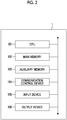
- Fig. 2 is a diagram illustrating a hardware configuration of the annotation support apparatus 1.
- the annotation support apparatus 1 is configured as a computer system that includes, for example, a CPU 101, a main memory 102 composed of memories, such as a RAM and a ROM, an auxiliary memory 103 composed of a hard disk, a memory, and the like, and a communication control device 104.
- the annotation support apparatus 1 may further include an input device 105, such as a keyboard, a touch panel, or a mouse, which is an input device and an output device 106 such as a display.
- the annotation support apparatus 1 may be configured to include a GPU.
- Each function illustrated in Fig. 1 is implemented by reading predetermined computer software to hardware, such as the CPU 101 and the main memory 102 illustrated in Fig. 2 , operating the communication control device 104 and the like under the control of the CPU 101, and performing the reading and writing of data from and to the main memory 102 or the auxiliary memory 103.
- Data or a database required for processing is stored in the main memory 102 and the auxiliary memory 103.
- the image acquisition unit 11 acquires medical images. Specifically, the image acquisition unit 11 acquires the medical images stored in the medical image storage unit 20.
- the medical images are images acquired in various examinations performed on the patients as described above.
- the medical image can have, as attribute information, patient information for identifying the patient, the type of examination, and other information.
- attribute information For example, in a case in which a plurality of images are acquired in one examination, information for grouping the plurality of images may be included as the attribute information.
- the medical image storage unit 20 manages the medical images using the hierarchical structure on the basis of the attributes of each medical image.
- the hierarchical structure related to the medical images has the medical image as a node and includes at least one of a medical image hierarchy to which the node of the medical image belongs, an upper hierarchy which is higher than the medical image hierarchy, or a lower hierarchy which is lower than the medical image hierarchy.
- the upper hierarchy can include, for example, patient information as a node.
- the lower hierarchy can include, for example, finding information attached to the medical image as a node.
- the hierarchical structure in which the medical images are managed may include the medical image hierarchy and at least one of the upper hierarchy or the lower hierarchy.
- the management of the medical images by the hierarchical structure makes it possible to appropriately associate the information of the upper hierarchy, such as patient information, and the information of the lower hierarchy, such as finding information, with the information of the medical images.
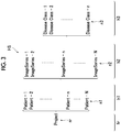
- Fig. 3 is a diagram schematically illustrating an example of a hierarchical structure HS in which medical images are managed.
- the hierarchical structure HS includes a medical image hierarchy, an upper hierarchy which is higher than the medical image hierarchy, and a lower hierarchy which is lower than the medical image hierarchy.
- the hierarchical structure HS includes a root hierarchy hr, a first hierarchy hi, a second hierarchy h2, and a third hierarchy h3.
- the hierarchical structure HS illustrated in Fig. 3 has a project nr represented by "Project" as a root and manages various kinds of information in the lower hierarchies.
- Project a project represented by "Project” as a root
- the hierarchical structure may be configured without having the project as the root node.
- a medical image n2 which is represented by "ImageSeries" constitutes a node belonging to the second hierarchy h2.
- the medical image has an aspect as examination information of each examination acquired in each examination among various examinations performed on a certain a patient.
- the first hierarchy h1 which is higher than the node of the medical image n2 has patient information n1 represented by "Patient" as a node.
- patient information n1 a plurality of medical images related to the same patient are grouped on the basis of the patient information that each medical image n2 has as the attribute information.
- the third hierarchy h3 which is lower than the node of the medical image n2 has finding information n3 represented by "Disease Class" as a node.
- the finding information n3 is information that is defined to have regionality for a portion of the medical image. As illustrated in Fig. 3 , a plurality of finding information items can be defined for one medical image.
- the display unit 12 displays the medical image. Specifically, the display unit 12 displays the medical image acquired by the image acquisition unit 11 on the display.
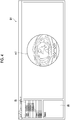
- Fig. 4 is a diagram illustrating an example of the medical image displayed on the display by the display unit 12. As illustrated in Fig. 4 , a medical image mi1 as one of the examination results is displayed on a display screen D1.
- the display screen D1 includes a browse list IB and an interpretation report display portion IR.
- the browse list IB is a portion in which a medical image list is displayed and which is used for selecting the medical image to be displayed on the display screen D1.
- the display unit 12 displays the selected medical image on the display screen D1.
- the list displayed in the browse list IB reflects the configuration of the hierarchical structure HS described with reference to Fig. 3 .
- the interpretation report display portion IR is a portion that presents the data of the interpretation report stored so as to be association with the medical image displayed on the display screen D1 to the user as reference information in a case in which the user inputs an annotation. Further, any clinical information stored in a text format, such as a medical record, can be displayed in the interpretation report display portion IR so as to be associated with the displayed medical image.
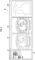
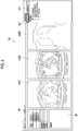
- Fig. 5 is a diagram illustrating an example in which the display unit displays a plurality of medical images on the display.
- a plurality of medical images mi21, mi22, and mi23 are displayed on a display screen D2.
- the medical image mi21 is a medical image obtained by a CT examination.
- the medical image mi22 is a medical image obtained by a PET examination.
- the medical image mi23 is a medical image obtained by an X-ray examination.
- the annotation support apparatus 1 can display medical images as the results of a plurality of related examinations side by side.
- the region designation receiving unit 13 receives the designation of a finding region on the medical image displayed by the display unit 12.
- the finding region is a portion of the medical image and is a region to which a finding is attached.
- the designation of the finding region can be received in various aspects.
- the region designation receiving unit 13 can receive the designation of the finding region by the input of the designation operation of the user for a portion of the displayed medical image. Specifically, for example, in a case in which the user places a pointer at any location in the medical image and performs an operation such as clicking, the region designation receiving unit 13 receives the location as the finding region.
- the region designation receiving unit 13 may receive the designation of any region in the medical image as the designation of the finding region. Specifically, for example, in a case in which the user operates various pointing devices to input a figure having a shape that surrounds any region in the medical image, the region designation receiving unit 13 can receive the region having any shape as the designation of the finding region. Therefore, since the designation of the finding region is not limited to the designation using, for example, a specific marker, it is possible to obtain the finding information in which a lesion region is appropriately designated.
- the annotation input receiving unit 14 receives the designation of the node of the medical image and any node associated with the node of the medical image in the hierarchical structure and the input of annotation information which is information related to the designated nodes. In this embodiment, the annotation input receiving unit 14 receives the designation of any node in the hierarchical structure HS described with reference to Fig. 3 . Then, the annotation input receiving unit 14 receives the input of the information to be associated with the designated node as the input of the annotation information.
- the annotation storage unit 15 stores the annotation information received by the annotation input receiving unit 14 so as to be associated with the designated node. Specifically, the annotation storage unit 15 stores the input annotation information so as to be associated with the designated node among the nodes managed in the hierarchical structure HS described with reference to Fig. 3 .
- Fig. 6 is a diagram illustrating an example of the input of the annotation information for patient information.
- the display unit 12 displays a plurality of medical images mi21, mi22, and mi23 on a display screen D3.
- the medical images mi21, mi22, and mi23 are medical images indicating the examination results of the patient identified by "Patient1".
- Each of the medical images mi21, mi22, and mi23 constitutes each node belonging to the second hierarchy h2 in the hierarchical structure HS.
- the nodes of the medical images are associated with the node of the patient information "Patient1" in the first hierarchy h1.
- the annotation input receiving unit 14 receives the input as the designation of the node of the patient information "Patient1".
- annotation input receiving unit 14 receives the input as the annotation information to be associated with the node of the patient information "Patient1 ".
- annotation information to be managed for each patient include the type of the main disease of the patient and the sex of the patient.
- information "Diseases: Lung cancer” is input as the annotation information to be associated with the patient "Patient1".
- the annotation storage unit 15 stores the input annotation information "Diseases: Lung cancer" so as to be associated with the node of the patient information "Patient1".
- the patient information for identifying the patient related to the medical image is managed as the node in the hierarchy higher than the medical image in the hierarchical structure, and the annotation information related to the patient is stored so as to be associated with the patient information. Therefore, the medical image, the patient information, and the annotation information related to the patient are managed while having an appropriate hierarchical relationship therebetween.
- Fig. 7 is a diagram illustrating an example of the input of annotation information for a medical image. As illustrated in Fig. 7 , the display unit 12 displays a plurality of medical images mi21, mi22, and mi23 on a display screen D4.
- the medical images mi21, mi22, and mi23 are medical images indicating the examination results of the patient identified by "Patient1".
- Each of the medical images mi21, mi22, and mi23 constitutes each node belonging to the second hierarchy h2 in the hierarchical structure HS.
- the nodes of the medical images are associated with the node of the patient information "Patient1" in the first hierarchy h1.
- the annotation input receiving unit 14 receives the input of the designation as the designation of the node of a medical image "Image Series 1-2".
- annotation input receiving unit 14 receives the input as the annotation information to be associated with the node of the medical image "Image Series 1-2".
- the annotation information to be managed for each medical image is information to be managed for each examination related to the acquisition of the image.
- the annotation information include a clinical situation, such as "before surgery” or "after surgery", and finding information to be defined on the basis of the information of the entire image (for example, tumor staging or the like).
- the relationship between the medical images may be defined as the annotation information of the medical images.
- another medical image may be explicitly designated as the annotation information of one medical image to define the content of the relationship.
- the relationship may be implicitly defined on the basis of the association with the node of the same patient information.
- the annotation storage unit 15 stores the annotation information input in the annotation information input field ai2 so as to be associated with the node of the medical image "Image Series 1-2".
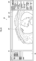
- Fig. 8 is a diagram illustrating an example of the designation of a region related to a finding in the medical image and the input of annotation information for the finding information. As illustrated in Fig. 8 , the display unit 12 displays the medical image mi21 on a display screen D5.
- the medical image mi21 is a medical image indicating the examination result of the patient identified by "Patient1".
- the medical image mi21 constitutes a node belonging to the second hierarchy h2 in the hierarchical structure HS, and the node is associated with the node of the patient information "Patient1" in the first hierarchy h1.
- the region designation receiving unit 13 receives the designation of a finding region or3 on the basis of the input of the designation of the region by the user in a state in which the medical image "Image Series 1-2" is selected by the designation of a tab on the display screen D5.
- the finding region is a portion of the medical image and is used for defining a region such as a lesion.
- the region designation receiving unit 13 can receive the designation of a region having any shape in the medical image as the designation of the finding region.
- the designation of the finding region is not limited to the designation by a specific marker or the like, it is possible to obtain the finding information in which a lesion region is appropriately designated.
- the annotation input receiving unit 14 receives the input as the annotation information to be associated with the finding region or3.
- the annotation information for each finding is information that is defined to have regionality for a predetermined region of the image and is, for example, information indicating image characteristics that are expressed in words by a clinician.
- the annotation information is input by labeling the finding region with qualitative information indicated by the finding region.
- the annotation storage unit 15 stores the finding information indicating the finding region as a node associated with the medical image in the hierarchy lower than the hierarchy of the medical image and stores the input annotation information so as to be associated with the node. Specifically, the annotation storage unit 15 stores the finding information indicating the finding region or3 as a node associated with the medical image "Image Series 1-2" in the third hierarchy h3 which is lower than the second hierarchy h2 in the hierarchical structure HS. Further, the annotation storage unit 15 stores the annotation information input to the annotation information input field ai3 so as to be associated with the node of the finding information indicating the finding region or3.
- the region designation receiving unit 13 can receive the designation of a plurality of finding regions in the one medical image.
- the designation of a plurality of finding regions may be received by receiving labeling with a plurality of different colors or line types on the display screen.
- the annotation input receiving unit 14 receives the input of annotation information for each finding region.
- the annotation storage unit 15 generates a node corresponding to each finding region as a node in a hierarchy that is lower than the node of one medical image and stores the input annotation information so as to be associated with each node.
- annotation information can be input in various aspects.
- an object indicating the annotation information input to the annotation information input field ai3 may be displayed in, for example, the annotation information input field ai3, and the annotation storage unit 15 may associate the input annotation information with the node of the finding region on the basis of the reception of an operation of dragging and-dropping the object to the finding region. This makes it possible to easily associate the annotation information with the finding region.
- the relationship between the findings in the image may be defined as the annotation information of the finding information.
- another finding information item may be explicitly designated as the annotation information of the one finding information item to define the content of the relationship.
- the relationship may be implicitly defined on the basis of the association with the hierarchy lower than the node of the same medical image.
- Fig. 9 is a diagram illustrating an example of the annotation information associated with each information item of each hierarchy managed in the hierarchical structure.
- the annotation storage unit 15 stores the input annotation information so as to be associated with the node designated in a case in which the annotation information is input.
- annotation information A1 is annotation information for each patient associated with the node of the patient information represented by "Patient" which belongs to the first hierarchy h1.
- the annotation information A1 can include, for example, information, such as the presence or absence of a disease, age, sex, the date of the first medical examination, staging at the time of the first medical examination, the content of an initial treatment, the date of recurrence, and the final outcome.
- Annotation information A2 is annotation information for each medical image (each examination) associated with the node of the medical image represented by "ImageSeries" which belongs to the second hierarchy h2.
- the annotation information A2 can include the clinical context (for example, before surgery, after surgery, and at the time of recurrence) of examinations and information (for example, tumor staging) obtained from the entire image.
- the setting information can be set randomly according to the input of the user for the purpose of machine learning and the model to be generated.
- the medical image is included in the learning data set.
- the predetermined setting information is not limited to be explicitly defined, and whether or not the medical image is included in the learning data set may be determined on the basis of whether or not specific annotation information is included.
- the node of the medical image can have information to be associated with each medical image as the annotation information.
- the finding information associated with the node of the medical image in the hierarchy lower than the node of the medical image can be regarded as the annotation information of the medical image. That is, the finding information can be regarded as a node in the hierarchy lower than the medical image and the annotation information of the medical image.
- Annotation information A3 is annotation information for each finding associated with the node of finding information represented by "DiseaseClass" which belongs to the third hierarchy h3.
- the annotation information A3 can include information indicating a finding region and the qualitative information of a finding.
- the information indicating the finding region can include coordinate information corresponding to the format (for example, a DICOM format) of the medical image to define a lesion region.
- the qualitative information of the finding can include qualitative label information (for example, image characteristics, a location, and a classification) for each finding region.
- the node of the finding information corresponds to notation "Structure" in Fig. 8 and Fig. 10 which will be referred to below.
- the learning data acquisition unit 16 acquires, as a learning data set, a data set including at least one medical image and annotation information associated with the node of the one medical image in the information managed by the hierarchical structure. Further, in a case in which the hierarchical structure managing the medical images consists of a plurality of hierarchies, the learning data acquisition unit 16 may acquire, as the learning data set, a data set including at least one medical image and annotation information associated with at least one of the node of the one medical image or another node associated with the node of the one medical image in the information managed by the hierarchical structure. The content of the data set is determined according to a learning task in model generation which will be described below.
- the model generation unit 17 generates an annotation guide model, which receives a medical image as an input and outputs annotation information related to the medical image with machine learning using the learning data set acquired by the learning data acquisition unit 16.
- the model generation unit 17 stores the generated annotation guide model in the model storage unit 30.
- the annotation guide model is generated by performing so-called supervised learning using a learning data set including a medical image as a learning target and annotation information as correct answer data associated with the learning result.
- the medical image included in the learning data set can be designated in, for example, the annotation information of the medical image as described above.
- the content of the learning task can include the classification of medical images, the detection of a specific finding, such as a lesion, included in the medical image, and the segmentation of the specific finding (the extraction of a region).
- other learning tasks may be used according to the design.
- the data included in the learning data set is not limited to the annotation information of one medical image, but may include, for example, information of other nodes related to the one medical image, annotation information associated with other nodes, and information (relationship information) indicating the relationship between nodes in the same hierarchy or different hierarchies for the purpose of the model and according to the setting of tasks.
- the annotation guide model is configured to include a neural network.
- the neural network may be configured by, for example, a convolution neural network (CNN).
- CNN convolution neural network
- the annotation guide model which is a model including a trained neural network can be regarded as a program that is read or referred to by a computer, causes the computer to perform a predetermined process, and causes the computer to implement a predetermined function.
- the annotation guide model according to this embodiment is used in a computer comprising a CPU (or/and a GPU) and a memory.
- the CPU (or/and the GPU) of the computer is operated so as to perform calculation based on, for example, a learned weighting coefficient and a response function corresponding to each layer for the input data input to an input layer of the neural network and to output results from an output layer, in response to commands from the annotation guide model stored in the memory.
- the annotation guide unit 18 displays the output obtained by inputting the medical image, which is acquired by the image acquisition unit 11 and is to be annotated, to the annotation guide model so as to be associated with the medical image to be annotated.
- the model generation unit 17 can perform machine learning, using a medical image and finding information input as annotation information for a lesion portion in the medical image as a learning data set, to generate an annotation guide model that receives the medical image as an input and outputs the finding information of the lesion portion to be associated with the medical image.
- the finding information used for learning and the finding information output from the model include information indicating the finding region and information such as characteristics related to the finding region and the type of lesion.
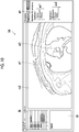
- Fig. 10 is a diagram illustrating an example of the display of candidates of annotation information to be associated with a medical image. As illustrated in Fig. 10 , the display unit 12 displays a medical image mi3 to be annotated on a display screen D6.
- the annotation guide unit 18 inputs the medical image mi3 to the annotation guide model and acquires the candidates of finding information as the annotation information to be associated with the medical image mi3 which has been output from the model. Specifically, as illustrated in Fig. 10 , the annotation guide unit 18 acquires a finding region candidate ar1 and a finding region candidate ar2 as the candidates of the finding information which is the annotation information to be associated with the medical image mi3 and displays the candidates so as to be superimposed on the medical image mi3. Further, the annotation guide unit 18 acquires a finding information candidate ac1 to be associated with the finding region candidate ar1 and a finding information candidate ac2 to be associated with the finding region candidate ar2 and displays the finding information candidates in an annotation candidate display portion gw.
- the model generation unit 17 can generate the annotation guide model on the basis of the accumulated medical image and annotation information. Then, the annotation guide unit 18 can acquire and output the candidates of the annotation information to be associated with the medical image to be annotated, on the basis of the generated annotation guide model.
- the output candidates of the annotation information are displayed so as to be associated with the medical image and are presented to the user, which makes it possible to assist the annotation work of the user and to promote the annotation work.
- the annotation guide model can be sequentially generated according to the accumulation of the medical image and the annotation information. Therefore, it is possible to improve the validity of the candidates of the annotation information presented to the user.
- Fig. 11 is a flowchart illustrating the processing content of an annotation support method performed in the annotation support apparatus 1.
- Step S1 the image acquisition unit 11 acquires the medical image stored in the medical image storage unit 20.
- Step S2 the display unit 12 displays the medical image acquired by the image acquisition unit 11 on a display device.
- the annotation guide unit 18 may display the candidates of the annotation information to be associated with the displayed medical image so as to be associated with the displayed medical image.
- Step S3 the annotation input receiving unit 14 receives the designation of any one of the node of the medical image or a node associated with the node of the medical image in the hierarchical structure. Further, in Step S4, the annotation input receiving unit 14 receives the input of the annotation information to be associated with the designated node.
- Step S5 the annotation storage unit 15 stores the annotation information received by the annotation input receiving unit 14 so as to be associated with the designated node. Specifically, the annotation storage unit 15 stores the input annotation information so as to be associated with the designated node among the nodes managed in the hierarchical structure HS.
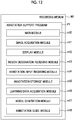
- An annotation support program PI illustrated in Fig. 12 includes a main module m10, an image acquisition module m11, a display module m12, a region designation receiving module m13, an annotation input receiving module m14, an annotation storage module m15, a learning data extraction module m16, a model generation module m17, and an annotation guide module m18.
- the main module m10 comprehensively controls an annotation support process.
- the image acquisition module m11, the display module m12, the region designation receiving module m13, the annotation input receiving module m14, the annotation storage module m15, the learning data extraction module m16, the model generation module m17, and the annotation guide module m18 are executed to implement the functions of the image acquisition unit 11, the display unit 12, the region designation receiving unit 13, the annotation input receiving unit 14, the annotation storage unit 15, the learning data acquisition unit 16, the model generation unit 17, and the annotation guide unit 18 of the annotation support apparatus 1 illustrated in Fig. 1 , respectively.
- the annotation support program PI may be provided by a storage medium M1 such as a magnetic disk, an optical disk, or a semiconductor memory. Further, the annotation support program P1 may be provided as a computer data signal superimposed on a carrier wave through a communication network.
- the medical image is managed by the hierarchical structure including the medical image hierarchy to which the node of the medical image belongs and at least one of the upper hierarchy higher than the medical image hierarchy or the lower hierarchy lower than the medical image hierarchy.
- the annotation information is received together with the designation of any one of the node of the displayed medical image or any node associated with the node, and the received annotation information is stored so as to be associated with the designated node. Therefore, a data group of the hierarchical structure in which annotation information is appropriately associated with the node of the medical image and a node associated with the node is obtained. Learning data is extracted from the data group to generate a model for achieving an automatic diagnosis in which the reference process of various kinds of information by the doctor is reflected.
- annotation support apparatus 11: image acquisition unit, 12: display unit, 13: region designation receiving unit, 14: annotation input receiving unit, 15: annotation storage unit, 16: learning data acquisition unit, 17: model generation unit, 18: annotation guide unit, 20: medical image storage unit, 30: model storage unit, M1: storage medium, m10: main module, m11: image acquisition module, m12: display module, m13: region designation receiving module, m14: annotation input receiving module, m15: annotation storage module, m16: learning data extraction module, m17: model generation module, m18: annotation guide module, P1: annotation support program.
Landscapes
- Engineering & Computer Science (AREA)
- Health & Medical Sciences (AREA)
- Public Health (AREA)
- Medical Informatics (AREA)
- Life Sciences & Earth Sciences (AREA)
- General Health & Medical Sciences (AREA)
- Physics & Mathematics (AREA)
- Biomedical Technology (AREA)
- Epidemiology (AREA)
- Primary Health Care (AREA)
- General Engineering & Computer Science (AREA)
- Theoretical Computer Science (AREA)
- Pathology (AREA)
- Radiology & Medical Imaging (AREA)
- Nuclear Medicine, Radiotherapy & Molecular Imaging (AREA)
- Human Computer Interaction (AREA)
- Veterinary Medicine (AREA)
- Animal Behavior & Ethology (AREA)
- Surgery (AREA)
- Molecular Biology (AREA)
- Heart & Thoracic Surgery (AREA)
- Biophysics (AREA)
- Data Mining & Analysis (AREA)
- General Physics & Mathematics (AREA)
- Artificial Intelligence (AREA)
- Databases & Information Systems (AREA)
- Physiology (AREA)
- Signal Processing (AREA)
- Psychiatry (AREA)
- Computer Vision & Pattern Recognition (AREA)
- Mathematical Physics (AREA)
- Fuzzy Systems (AREA)
- Evolutionary Computation (AREA)
- Pulmonology (AREA)
- Medical Treatment And Welfare Office Work (AREA)
- Measuring And Recording Apparatus For Diagnosis (AREA)
Abstract
Description
- The present invention relates to an annotation support apparatus, an annotation support method, and an annotation support program.
- In recent years, research has been conducted on automatic diagnosis support for medical images using machine learning. A large amount of learning data used for supervised learning is generally required in order to achieve a high-performance model. However, for that purpose, it is necessary to prepare annotations defined to be paired with medical images. Here, the annotations indicate information of the location or region of a lesion in the medical image and classification information related to the property and diagnosis of the lesion. For example,
Patent Document 1 discloses a technique that acquires and stores annotations in response to the instruction operation of a user for a displayed medical image. - Patent Document 1:
JP-A 2013-132514 - In the interpretation of medical images by a doctor, in a case in which a specific lesion is present in the medical image, the results of other examination on the lesion and the background information of the patient are referred to in addition to the location and region of the lesion, the nature of the lesion. In order to generate a high-quality model for diagnosis support, it is desirable to supply learning data which is appropriately constructed to reflect the hierarchy and relationship of annotation information as much as possible so as to reproduce the reference process of information by the doctor. In addition, in many cases, annotations have been acquired by the instruction operation of the user. However, since the labor required for this manual process is large, it is necessary to incorporate an appropriate model to save labor.
- Therefore, an object of the invention is to provide an annotation support apparatus, an annotation support method, and an annotation support program that can give annotation information having a hierarchy and a relationship to a medical image.
- According to an aspect of the invention, there is provided an annotation support apparatus that supports generation of annotation information related to a medical image. The medical image is managed on the basis of an attribute of each medical image by a hierarchical structure that has one medical image as a node and includes at least one of a medical image hierarchy to which the node of the medical image belongs, an upper hierarchy higher than the medical image hierarchy, or a lower hierarchy lower than the medical image hierarchy. The annotation support apparatus comprises: an image acquisition unit that acquires the medical image; a display unit that displays the medical image; an annotation input receiving unit that receives designation of any one of the node of the medical image or a node associated with the node of the medical image in the hierarchical structure and an input of annotation information which is information related to the designated node; and an annotation storage unit that stores the annotation information to be associated with the designated node.
- According to another aspect of the invention, there is provided an annotation support method performed in an annotation support apparatus that supports generation of annotation information related to a medical image. The medical image is managed on the basis of an attribute of each medical image by a hierarchical structure that has one medical image as a node and includes at least one of a medical image hierarchy to which the node of the medical image belongs, an upper hierarchy higher than the medical image hierarchy, or a lower hierarchy lower than the medical image hierarchy. The annotation support method comprises: an image acquisition step of acquiring the medical image; a display step of displaying the medical image; an annotation input receiving step of receiving designation of any one of the node of the medical image or a node associated with the node of the medical image in the hierarchical structure and an input of annotation information which is information related to the designated node; and an annotation storage step of storing the annotation information to be associated with the designated node.
- According to yet another aspect of the invention, there is provided an annotation support program that causes a computer to function as an annotation support apparatus supporting generation of annotation information related to a medical image. The medical image is managed on the basis of an attribute of each medical image by a hierarchical structure that has one medical image as a node and includes at least one of a medical image hierarchy to which the node of the medical image belongs, an upper hierarchy higher than the medical image hierarchy, or a lower hierarchy lower than the medical image hierarchy. The annotation support program causes the computer to implement: an image acquisition function of acquiring the medical image; a display function of displaying the medical image; an annotation input receiving function of receiving designation of any one of the node of the medical image or a node associated with the node of the medical image in the hierarchical structure and an input of annotation information which is information related to the designated node; and an annotation storage function of storing the annotation information to be associated with the designated node.
- According to the above-described aspects, the medical image is managed by the hierarchical structure including the medical image hierarchy to which the node of the medical image belongs and at least one of the upper hierarchy higher than the medical image hierarchy or the lower hierarchy lower than the medical image hierarchy. Then, the annotation information is received together with the designation of any one of the node of the displayed medical image or any node associated with the node, and the received annotation information is stored so as to be associated with the designated node. Therefore, a data group of the hierarchical structure in which annotation information is appropriately associated with the node of the medical image and a node associated with the node is obtained. Learning data is extracted from the data group to generate a model for achieving an automatic diagnosis in which the reference process of various kinds of information by the doctor is reflected.
- According to still yet another aspect, in the annotation support apparatus, the hierarchical structure may include the medical image hierarchy and at least one of the upper hierarchy or the lower hierarchy.
- According to the above-described aspect, it is possible to appropriately associate the information of the upper hierarchy, such as patient information, and the information of the lower hierarchy, such as finding information, with the information of the medical image.
- According to yet still another aspect, the annotation support apparatus may further comprise a region designation receiving unit that receives designation of a finding region which is a portion of the medical image displayed by the display unit and is a region having a finding attached thereto. The hierarchical structure may include the medical image hierarchy and the lower hierarchy. The annotation input receiving unit may receive an input of annotation information for the finding region. The annotation storage unit may store finding information indicating the finding region as a node associated with the medical image in the hierarchy lower than the medical image hierarchy and store the input annotation information to be associated with the node.
- According to the above-described aspect, the input of the annotation information for the finding region designated as a portion of the medical image is received, and the finding information and the annotation information are stored as nodes in the hierarchy lower than the medical image. Therefore, the medical image, the finding information, and the annotation information for the finding are managed while having an appropriate hierarchical relationship therebetween.
- According to still yet another aspect, in the annotation support apparatus, the region designation receiving unit may receive designation of a region having any shape in the medical image as the designation of the finding region.
- According to the above-described aspect, the designation of the finding region having any shape is received. Since the designation of the finding region is not limited to the designation by, for example, a specific marker, it is possible to obtain the finding information in which a lesion region is appropriately designated.
- According to yet still another aspect, in the annotation support apparatus, the annotation input receiving unit may receive the input of the annotation information for the finding region, on the basis of reception of an operation of dragging and dropping an object indicating the annotation information to the finding region in the medical image displayed by the display unit.
- According to the above-described aspect, it is possible to easily associate the annotation information with the finding region.
- According to still yet another aspect, in the annotation support apparatus, the hierarchical structure may include patient information that identifies a patient as a node in the upper hierarchy. The annotation input receiving unit may receive an input of annotation information for the patient information indicating the patient which is associated with the medical image. The annotation storage unit may store the input annotation information to be associated with the node of the patient information.
- According to the above-described aspect, the patient information for identifying the patient corresponding to the medical image is managed as a node in the hierarchy higher than the medical image by the hierarchical structure, and the annotation information related to the patient is stored so as to be associated with the patient information. Therefore, the medical image, the patient information, and the annotation information related to the patient are managed while having an appropriate hierarchical relationship therebetween.
- According to yet still another aspect, in the annotation support apparatus, the annotation information may include relationship information indicating a relationship with a node other than the node associated with the annotation information.
- According to the above-described aspect, each node can include, for example, the association between the medical images and the association between the finding information items as the annotation information items. Therefore, in a case in which the information associated with a certain node has a relationship with the information associated with another node, an information group including the relationship information is used for training a model for automatic diagnosis, which makes it possible to perform automatic diagnosis in which the relationship has been reflected.
- According to still yet another aspect, the annotation support apparatus may further comprise: a learning data acquisition unit that acquires, as a learning data set, a data set including at least one medical image and annotation information associated with a node of the one medical image in information managed by the hierarchical structure; a model generation unit that generates an annotation guide model, which receives a medical image as an input and outputs annotation information related to the medical image, with machine learning using the learning data set acquired by the learning data acquisition unit; and an annotation guide unit that displays an output obtained by inputting the medical image, which is acquired by the image acquisition unit and is to be annotated, to the annotation guide model to be associated with the medical image to be annotated.
- According to the above-described aspect, the annotation guide model is generated by the machine learning using the learning data set extracted from the information group managed by the hierarchical structure. A medical image is input to the generated annotation guide model, and candidates of the annotation information to be associated with the medical image are output. Then, the candidates of the annotation information are displayed so as to be associated with the medical image and are presented to the user. Therefore, it is possible to promote the annotation work of the user.
- According to yet still another aspect, in the annotation support apparatus, the learning data acquisition unit may acquire, as the learning data set, a data set including at least one medical image and annotation information associated with at least one of a node of the one medical image or another node associated with the node of the one medical image in the information managed by the hierarchical structure.
- According to the above-described aspect, in a case in which the hierarchical structure managing the medical images consists of a plurality of hierarchies, it is possible to acquire a learning data set that appropriately includes necessary information according to the setting of a learning task in model generation.
- According to still yet another aspect of the invention, there is provided an annotation support apparatus that supports generation of annotation information related to a medical image. The medical image associated with annotation information in advance is capable of being acquired. The annotation support apparatus comprises: a learning data acquisition unit that acquires, as a learning data set, a data set including the medical image and the annotation information associated with the medical image; a model generation unit that generates an annotation guide model, which receives a medical image as an input and outputs annotation information related to the medical image, with machine learning using the learning data set acquired by the learning data acquisition unit; an image acquisition unit that acquires the medical image to which the annotation information is to be given; a display unit that displays the medical image acquired by the image acquisition unit; an annotation guide unit that displays an output obtained by inputting the medical image acquired by the image acquisition unit to the annotation guide model to be associated with the medical image to be annotated which is displayed by the display unit; an annotation input receiving unit that receives an input of annotation information related to the medical image to be annotated; and an annotation storage unit that stores the annotation information received by the input receiving unit to be associated with the medical image to be annotated.
- According to one aspect of the invention, it is possible to add annotation information having a hierarchy and a relationship to a medical image.
-
-
Fig. 1 is a block diagram illustrating a functional configuration of an annotation support apparatus according to an embodiment. -
Fig. 2 is a diagram illustrating a hardware configuration of the annotation support apparatus. -
Fig. 3 is a diagram illustrating a hierarchical structure in which medical images are managed, patient information is included in a hierarchy higher than the medical images, and finding information is included in a hierarchy lower than the medical images. -
Fig. 4 is a diagram illustrating an example of the medical image displayed on a display device. -
Fig. 5 is a diagram illustrating an example in which a plurality of medical images are displayed on the display device. -
Fig. 6 is a diagram illustrating an example of the input of annotation information for patient information. -
Fig. 7 is a diagram illustrating an example of the input of annotation information for the medical image. -
Fig. 8 is a diagram illustrating an example of the designation of a region related to a finding in the medical image and the input of annotation information for the finding information. -
Fig. 9 is a diagram illustrating an example of annotation information associated with each information item of each hierarchy managed in the hierarchical structure. -
Fig. 10 is a diagram illustrating an example of the display of candidates of annotation information to be associated with a medical image. -
Fig. 11 is a flowchart illustrating the processing content of an annotation support method performed in the annotation support apparatus. -
Fig. 12 is a diagram illustrating a configuration of an annotation support program. - Hereinafter, an embodiment of the invention will be described in detail with reference to the accompanying drawings. In addition, in the description of the drawings, the same or equivalent elements are denoted by the same reference numerals, and duplicate description will be omitted.
-
Fig. 1 is a diagram illustrating a functional configuration of an annotation support apparatus according to this embodiment. Anannotation support apparatus 1 is a device that supports the generation of annotation information related to medical images. - As illustrated in
Fig. 1 , theannotation support apparatus 1 comprises an image acquisition unit 11, adisplay unit 12, a regiondesignation receiving unit 13, an annotationinput receiving unit 14, anannotation storage unit 15, a learningdata acquisition unit 16, amodel generation unit 17, and anannotation guide unit 18. Each functional unit included in theannotation support apparatus 1 according to this embodiment may be distributed and configured in a plurality of devices. In addition, each functional unit included in theannotation support apparatus 1 is implemented by hardware or software, or any combination thereof. - As illustrated in
Fig. 1 , theannotation support apparatus 1 can access storage units such as a medicalimage storage unit 20 and amodel storage unit 30. Thestorage units annotation support apparatus 1 through a network or a predetermined communication unit. Further, thestorage units annotation support apparatus 1. - Before each functional unit of the
annotation support apparatus 1 is described, thestorage units image storage unit 20 is a storage unit that stores the medical images acquired in various examinations performed on patients. - The medical image is, for example, a DICOM image which is one of the standard formats of the medical images. Examples of the medical image may include images indicating the results of various examinations such as an X-ray examination, a CT examination, an MRI examination, a PET examination, an ultrasound examination, and an endoscopic examination. Further, the medical images may include the information of, for example, a JPEG image, a BMP image, a PNG image, and an MP4 moving image as photographs and moving images recorded in the process of a medical treatment, in addition to these images. In addition, the medical
image storage unit 20 manages the medical images using a hierarchical structure on the basis of the attributes of each medical image, which will be described in detail below. - The
model storage unit 30 is a storage unit that stores an annotation guide model which will be described in detail below. The annotation guide model is a model that is generated by machine learning, receives a medical image as an input, and outputs annotation information related to the medical image. -
Fig. 2 is a diagram illustrating a hardware configuration of theannotation support apparatus 1. Physically, as illustrated inFig. 2 , theannotation support apparatus 1 is configured as a computer system that includes, for example, aCPU 101, amain memory 102 composed of memories, such as a RAM and a ROM, anauxiliary memory 103 composed of a hard disk, a memory, and the like, and acommunication control device 104. Theannotation support apparatus 1 may further include aninput device 105, such as a keyboard, a touch panel, or a mouse, which is an input device and anoutput device 106 such as a display. In addition, theannotation support apparatus 1 may be configured to include a GPU. - Each function illustrated in
Fig. 1 is implemented by reading predetermined computer software to hardware, such as theCPU 101 and themain memory 102 illustrated inFig. 2 , operating thecommunication control device 104 and the like under the control of theCPU 101, and performing the reading and writing of data from and to themain memory 102 or theauxiliary memory 103. Data or a database required for processing is stored in themain memory 102 and theauxiliary memory 103. - The functional units of the
annotation support apparatus 1 will be described again with reference toFig. 1 . The image acquisition unit 11 acquires medical images. Specifically, the image acquisition unit 11 acquires the medical images stored in the medicalimage storage unit 20. - The medical images are images acquired in various examinations performed on the patients as described above. The medical image can have, as attribute information, patient information for identifying the patient, the type of examination, and other information. For example, in a case in which a plurality of images are acquired in one examination, information for grouping the plurality of images may be included as the attribute information.
- The medical
image storage unit 20 manages the medical images using the hierarchical structure on the basis of the attributes of each medical image. Specifically, the hierarchical structure related to the medical images has the medical image as a node and includes at least one of a medical image hierarchy to which the node of the medical image belongs, an upper hierarchy which is higher than the medical image hierarchy, or a lower hierarchy which is lower than the medical image hierarchy. The upper hierarchy can include, for example, patient information as a node. The lower hierarchy can include, for example, finding information attached to the medical image as a node. - In addition, the hierarchical structure in which the medical images are managed may include the medical image hierarchy and at least one of the upper hierarchy or the lower hierarchy. The management of the medical images by the hierarchical structure makes it possible to appropriately associate the information of the upper hierarchy, such as patient information, and the information of the lower hierarchy, such as finding information, with the information of the medical images.
-
Fig. 3 is a diagram schematically illustrating an example of a hierarchical structure HS in which medical images are managed. In the example illustrated inFig. 3 , the hierarchical structure HS includes a medical image hierarchy, an upper hierarchy which is higher than the medical image hierarchy, and a lower hierarchy which is lower than the medical image hierarchy. As illustrated inFig. 3 , the hierarchical structure HS includes a root hierarchy hr, a first hierarchy hi, a second hierarchy h2, and a third hierarchy h3. - The hierarchical structure HS illustrated in
Fig. 3 has a project nr represented by "Project" as a root and manages various kinds of information in the lower hierarchies. In the example illustrated inFig. 3 , a case in which the project nr is provided as a root node is described. However, the hierarchical structure may be configured without having the project as the root node. - A medical image n2 which is represented by "ImageSeries" constitutes a node belonging to the second hierarchy h2. The medical image has an aspect as examination information of each examination acquired in each examination among various examinations performed on a certain a patient.
- The first hierarchy h1 which is higher than the node of the medical image n2 has patient information n1 represented by "Patient" as a node. In the patient information n1, a plurality of medical images related to the same patient are grouped on the basis of the patient information that each medical image n2 has as the attribute information.
- The third hierarchy h3 which is lower than the node of the medical image n2 has finding information n3 represented by "Disease Class" as a node. The finding information n3 is information that is defined to have regionality for a portion of the medical image. As illustrated in
Fig. 3 , a plurality of finding information items can be defined for one medical image. - Referring to
Fig. 1 again, thedisplay unit 12 displays the medical image. Specifically, thedisplay unit 12 displays the medical image acquired by the image acquisition unit 11 on the display.Fig. 4 is a diagram illustrating an example of the medical image displayed on the display by thedisplay unit 12. As illustrated inFig. 4 , a medical image mi1 as one of the examination results is displayed on a display screen D1. - In addition, the display screen D1 includes a browse list IB and an interpretation report display portion IR. The browse list IB is a portion in which a medical image list is displayed and which is used for selecting the medical image to be displayed on the display screen D1. In a case in which the input of the user's selection for a plurality of medical images displayed in the browse list IB is acquired, the
display unit 12 displays the selected medical image on the display screen D1. In addition, the list displayed in the browse list IB reflects the configuration of the hierarchical structure HS described with reference toFig. 3 . The interpretation report display portion IR is a portion that presents the data of the interpretation report stored so as to be association with the medical image displayed on the display screen D1 to the user as reference information in a case in which the user inputs an annotation. Further, any clinical information stored in a text format, such as a medical record, can be displayed in the interpretation report display portion IR so as to be associated with the displayed medical image. -
Fig. 5 is a diagram illustrating an example in which the display unit displays a plurality of medical images on the display. As illustrated inFig. 5 , a plurality of medical images mi21, mi22, and mi23 are displayed on a display screen D2. The medical image mi21 is a medical image obtained by a CT examination. The medical image mi22 is a medical image obtained by a PET examination. The medical image mi23 is a medical image obtained by an X-ray examination. As illustrated inFig. 5 , theannotation support apparatus 1 according to this embodiment can display medical images as the results of a plurality of related examinations side by side. - Referring to
Fig. 1 again, the regiondesignation receiving unit 13 receives the designation of a finding region on the medical image displayed by thedisplay unit 12. The finding region is a portion of the medical image and is a region to which a finding is attached. The designation of the finding region can be received in various aspects. - For example, the region
designation receiving unit 13 can receive the designation of the finding region by the input of the designation operation of the user for a portion of the displayed medical image. Specifically, for example, in a case in which the user places a pointer at any location in the medical image and performs an operation such as clicking, the regiondesignation receiving unit 13 receives the location as the finding region. - In addition, the region
designation receiving unit 13 may receive the designation of any region in the medical image as the designation of the finding region. Specifically, for example, in a case in which the user operates various pointing devices to input a figure having a shape that surrounds any region in the medical image, the regiondesignation receiving unit 13 can receive the region having any shape as the designation of the finding region. Therefore, since the designation of the finding region is not limited to the designation using, for example, a specific marker, it is possible to obtain the finding information in which a lesion region is appropriately designated. - The annotation
input receiving unit 14 receives the designation of the node of the medical image and any node associated with the node of the medical image in the hierarchical structure and the input of annotation information which is information related to the designated nodes. In this embodiment, the annotationinput receiving unit 14 receives the designation of any node in the hierarchical structure HS described with reference toFig. 3 . Then, the annotationinput receiving unit 14 receives the input of the information to be associated with the designated node as the input of the annotation information. - The
annotation storage unit 15 stores the annotation information received by the annotationinput receiving unit 14 so as to be associated with the designated node. Specifically, theannotation storage unit 15 stores the input annotation information so as to be associated with the designated node among the nodes managed in the hierarchical structure HS described with reference toFig. 3 . - Hereinafter, a specific example of the reception of the annotation information will be described with reference to
Figs. 6 to 8 .Fig. 6 is a diagram illustrating an example of the input of the annotation information for patient information. As illustrated inFig. 6 , thedisplay unit 12 displays a plurality of medical images mi21, mi22, and mi23 on a display screen D3. - The medical images mi21, mi22, and mi23 are medical images indicating the examination results of the patient identified by "Patient1". Each of the medical images mi21, mi22, and mi23 constitutes each node belonging to the second hierarchy h2 in the hierarchical structure HS. The nodes of the medical images are associated with the node of the patient information "Patient1" in the first hierarchy h1.
- For example, in a case in which the designation of a tab at1 on the display screen D3 is input, the annotation
input receiving unit 14 receives the input as the designation of the node of the patient information "Patient1". - Further, in a case in which annotation information is input to an annotation information input field ai1, the annotation
input receiving unit 14 receives the input as the annotation information to be associated with the node of the patient information "Patient1 ". Examples of the annotation information to be managed for each patient include the type of the main disease of the patient and the sex of the patient. In the example illustrated inFig. 6 , information "Diseases: Lung cancer" is input as the annotation information to be associated with the patient "Patient1". - The
annotation storage unit 15 stores the input annotation information "Diseases: Lung cancer" so as to be associated with the node of the patient information "Patient1". - According to the example described with reference to
Fig. 6 , the patient information for identifying the patient related to the medical image is managed as the node in the hierarchy higher than the medical image in the hierarchical structure, and the annotation information related to the patient is stored so as to be associated with the patient information. Therefore, the medical image, the patient information, and the annotation information related to the patient are managed while having an appropriate hierarchical relationship therebetween. -
Fig. 7 is a diagram illustrating an example of the input of annotation information for a medical image. As illustrated inFig. 7 , thedisplay unit 12 displays a plurality of medical images mi21, mi22, and mi23 on a display screen D4. - The medical images mi21, mi22, and mi23 are medical images indicating the examination results of the patient identified by "Patient1". Each of the medical images mi21, mi22, and mi23 constitutes each node belonging to the second hierarchy h2 in the hierarchical structure HS. The nodes of the medical images are associated with the node of the patient information "Patient1" in the first hierarchy h1.
- For example, in a case in which the designation of a tab at2 on the display screen D4 is input, the annotation
input receiving unit 14 receives the input of the designation as the designation of the node of a medical image "Image Series 1-2". - Further, in a case in which annotation information is input to an annotation information input field ai2, the annotation
input receiving unit 14 receives the input as the annotation information to be associated with the node of the medical image "Image Series 1-2". - The annotation information to be managed for each medical image is information to be managed for each examination related to the acquisition of the image. Examples of the annotation information include a clinical situation, such as "before surgery" or "after surgery", and finding information to be defined on the basis of the information of the entire image (for example, tumor staging or the like). Further, the relationship between the medical images may be defined as the annotation information of the medical images. For the relationship of one medical image with another medical image, another medical image may be explicitly designated as the annotation information of one medical image to define the content of the relationship. In addition, the relationship may be implicitly defined on the basis of the association with the node of the same patient information.
- The
annotation storage unit 15 stores the annotation information input in the annotation information input field ai2 so as to be associated with the node of the medical image "Image Series 1-2". -
Fig. 8 is a diagram illustrating an example of the designation of a region related to a finding in the medical image and the input of annotation information for the finding information. As illustrated inFig. 8 , thedisplay unit 12 displays the medical image mi21 on a display screen D5. - The medical image mi21 is a medical image indicating the examination result of the patient identified by "Patient1". The medical image mi21 constitutes a node belonging to the second hierarchy h2 in the hierarchical structure HS, and the node is associated with the node of the patient information "Patient1" in the first hierarchy h1.
- For example, the region
designation receiving unit 13 receives the designation of a finding region or3 on the basis of the input of the designation of the region by the user in a state in which the medical image "Image Series 1-2" is selected by the designation of a tab on the display screen D5. The finding region is a portion of the medical image and is used for defining a region such as a lesion. - As illustrated in
Fig. 8 , the regiondesignation receiving unit 13 can receive the designation of a region having any shape in the medical image as the designation of the finding region. As such, since the designation of the finding region is not limited to the designation by a specific marker or the like, it is possible to obtain the finding information in which a lesion region is appropriately designated. - In addition, in a case in which annotation information is input to an annotation information input field ai3 in a state in which a tab at3 associated with the finding region or3 is designated, the annotation
input receiving unit 14 receives the input as the annotation information to be associated with the finding region or3. The annotation information for each finding is information that is defined to have regionality for a predetermined region of the image and is, for example, information indicating image characteristics that are expressed in words by a clinician. In this embodiment, the annotation information is input by labeling the finding region with qualitative information indicated by the finding region. - The
annotation storage unit 15 stores the finding information indicating the finding region as a node associated with the medical image in the hierarchy lower than the hierarchy of the medical image and stores the input annotation information so as to be associated with the node. Specifically, theannotation storage unit 15 stores the finding information indicating the finding region or3 as a node associated with the medical image "Image Series 1-2" in the third hierarchy h3 which is lower than the second hierarchy h2 in the hierarchical structure HS. Further, theannotation storage unit 15 stores the annotation information input to the annotation information input field ai3 so as to be associated with the node of the finding information indicating the finding region or3. - For example, in a case in which there are a plurality of lesions in one medical image, the region
designation receiving unit 13 can receive the designation of a plurality of finding regions in the one medical image. For example, the designation of a plurality of finding regions may be received by receiving labeling with a plurality of different colors or line types on the display screen. In this case, the annotationinput receiving unit 14 receives the input of annotation information for each finding region. Then, theannotation storage unit 15 generates a node corresponding to each finding region as a node in a hierarchy that is lower than the node of one medical image and stores the input annotation information so as to be associated with each node. - In addition, the annotation information can be input in various aspects. For example, an object indicating the annotation information input to the annotation information input field ai3 may be displayed in, for example, the annotation information input field ai3, and the
annotation storage unit 15 may associate the input annotation information with the node of the finding region on the basis of the reception of an operation of dragging and-dropping the object to the finding region. This makes it possible to easily associate the annotation information with the finding region. - Further, the relationship between the findings in the image may be defined as the annotation information of the finding information. For the relationship of one finding information item with another finding information item, another finding information item may be explicitly designated as the annotation information of the one finding information item to define the content of the relationship. In addition, the relationship may be implicitly defined on the basis of the association with the hierarchy lower than the node of the same medical image.
-
Fig. 9 is a diagram illustrating an example of the annotation information associated with each information item of each hierarchy managed in the hierarchical structure. As described above, theannotation storage unit 15 stores the input annotation information so as to be associated with the node designated in a case in which the annotation information is input. - In
Fig. 9 , annotation information A1 is annotation information for each patient associated with the node of the patient information represented by "Patient" which belongs to the first hierarchy h1. The annotation information A1 can include, for example, information, such as the presence or absence of a disease, age, sex, the date of the first medical examination, staging at the time of the first medical examination, the content of an initial treatment, the date of recurrence, and the final outcome. - Annotation information A2 is annotation information for each medical image (each examination) associated with the node of the medical image represented by "ImageSeries" which belongs to the second hierarchy h2. The annotation information A2 can include the clinical context (for example, before surgery, after surgery, and at the time of recurrence) of examinations and information (for example, tumor staging) obtained from the entire image. In addition, the annotation information associated with the medical image can include setting information (Add to Dataset = True/False) which is set randomly and indicates whether or not the medical image (examination) is included in a learning data set of machine learning which will be described below. The setting information can be set randomly according to the input of the user for the purpose of machine learning and the model to be generated. In a case in which "True" is input, the medical image is included in the learning data set. In addition, the predetermined setting information is not limited to be explicitly defined, and whether or not the medical image is included in the learning data set may be determined on the basis of whether or not specific annotation information is included.
- Further, the node of the medical image can have information to be associated with each medical image as the annotation information. In addition, the finding information associated with the node of the medical image in the hierarchy lower than the node of the medical image can be regarded as the annotation information of the medical image. That is, the finding information can be regarded as a node in the hierarchy lower than the medical image and the annotation information of the medical image.
- Annotation information A3 is annotation information for each finding associated with the node of finding information represented by "DiseaseClass" which belongs to the third hierarchy h3. The annotation information A3 can include information indicating a finding region and the qualitative information of a finding. The information indicating the finding region can include coordinate information corresponding to the format (for example, a DICOM format) of the medical image to define a lesion region. The qualitative information of the finding can include qualitative label information (for example, image characteristics, a location, and a classification) for each finding region. In addition, the node of the finding information corresponds to notation "Structure" in
Fig. 8 andFig. 10 which will be referred to below. - Referring to
Fig. 1 again, the learningdata acquisition unit 16 acquires, as a learning data set, a data set including at least one medical image and annotation information associated with the node of the one medical image in the information managed by the hierarchical structure. Further, in a case in which the hierarchical structure managing the medical images consists of a plurality of hierarchies, the learningdata acquisition unit 16 may acquire, as the learning data set, a data set including at least one medical image and annotation information associated with at least one of the node of the one medical image or another node associated with the node of the one medical image in the information managed by the hierarchical structure. The content of the data set is determined according to a learning task in model generation which will be described below. - The
model generation unit 17 generates an annotation guide model, which receives a medical image as an input and outputs annotation information related to the medical image with machine learning using the learning data set acquired by the learningdata acquisition unit 16. Themodel generation unit 17 stores the generated annotation guide model in themodel storage unit 30. - In this embodiment, the annotation guide model is generated by performing so-called supervised learning using a learning data set including a medical image as a learning target and annotation information as correct answer data associated with the learning result. The medical image included in the learning data set can be designated in, for example, the annotation information of the medical image as described above. In addition, the content of the learning task can include the classification of medical images, the detection of a specific finding, such as a lesion, included in the medical image, and the segmentation of the specific finding (the extraction of a region). However, other learning tasks may be used according to the design.
- The data included in the learning data set is not limited to the annotation information of one medical image, but may include, for example, information of other nodes related to the one medical image, annotation information associated with other nodes, and information (relationship information) indicating the relationship between nodes in the same hierarchy or different hierarchies for the purpose of the model and according to the setting of tasks.
- The annotation guide model is configured to include a neural network. The neural network may be configured by, for example, a convolution neural network (CNN). The annotation guide model which is a model including a trained neural network can be regarded as a program that is read or referred to by a computer, causes the computer to perform a predetermined process, and causes the computer to implement a predetermined function.
- That is, the annotation guide model according to this embodiment is used in a computer comprising a CPU (or/and a GPU) and a memory. Specifically, the CPU (or/and the GPU) of the computer is operated so as to perform calculation based on, for example, a learned weighting coefficient and a response function corresponding to each layer for the input data input to an input layer of the neural network and to output results from an output layer, in response to commands from the annotation guide model stored in the memory.
- The
annotation guide unit 18 displays the output obtained by inputting the medical image, which is acquired by the image acquisition unit 11 and is to be annotated, to the annotation guide model so as to be associated with the medical image to be annotated. - In this embodiment, for example, the
model generation unit 17 can perform machine learning, using a medical image and finding information input as annotation information for a lesion portion in the medical image as a learning data set, to generate an annotation guide model that receives the medical image as an input and outputs the finding information of the lesion portion to be associated with the medical image. Here, the finding information used for learning and the finding information output from the model include information indicating the finding region and information such as characteristics related to the finding region and the type of lesion. -
Fig. 10 is a diagram illustrating an example of the display of candidates of annotation information to be associated with a medical image. As illustrated inFig. 10 , thedisplay unit 12 displays a medical image mi3 to be annotated on a display screen D6. - The
annotation guide unit 18 inputs the medical image mi3 to the annotation guide model and acquires the candidates of finding information as the annotation information to be associated with the medical image mi3 which has been output from the model. Specifically, as illustrated inFig. 10 , theannotation guide unit 18 acquires a finding region candidate ar1 and a finding region candidate ar2 as the candidates of the finding information which is the annotation information to be associated with the medical image mi3 and displays the candidates so as to be superimposed on the medical image mi3. Further, theannotation guide unit 18 acquires a finding information candidate ac1 to be associated with the finding region candidate ar1 and a finding information candidate ac2 to be associated with the finding region candidate ar2 and displays the finding information candidates in an annotation candidate display portion gw. - In a case in which the accumulated amount of annotation information associated with the medical image is equal to or greater than the value at which machine learning can be performed using the medical image and the annotation information as a learning data set, the
model generation unit 17 can generate the annotation guide model on the basis of the accumulated medical image and annotation information. Then, theannotation guide unit 18 can acquire and output the candidates of the annotation information to be associated with the medical image to be annotated, on the basis of the generated annotation guide model. The output candidates of the annotation information are displayed so as to be associated with the medical image and are presented to the user, which makes it possible to assist the annotation work of the user and to promote the annotation work. In addition, the annotation guide model can be sequentially generated according to the accumulation of the medical image and the annotation information. Therefore, it is possible to improve the validity of the candidates of the annotation information presented to the user. - Next, the operation of the
annotation support apparatus 1 according to this embodiment will be described with reference toFig. 11. Fig. 11 is a flowchart illustrating the processing content of an annotation support method performed in theannotation support apparatus 1. - In Step S1, the image acquisition unit 11 acquires the medical image stored in the medical
image storage unit 20. In Step S2, thedisplay unit 12 displays the medical image acquired by the image acquisition unit 11 on a display device. In a case in which the medical image is displayed in Step S2, theannotation guide unit 18 may display the candidates of the annotation information to be associated with the displayed medical image so as to be associated with the displayed medical image. - In Step S3, the annotation
input receiving unit 14 receives the designation of any one of the node of the medical image or a node associated with the node of the medical image in the hierarchical structure. Further, in Step S4, the annotationinput receiving unit 14 receives the input of the annotation information to be associated with the designated node. - In Step S5, the
annotation storage unit 15 stores the annotation information received by the annotationinput receiving unit 14 so as to be associated with the designated node. Specifically, theannotation storage unit 15 stores the input annotation information so as to be associated with the designated node among the nodes managed in the hierarchical structure HS. - Next, an annotation support program that causes a computer to function as the annotation support apparatus will be described with reference to
Fig. 12 . An annotation support program PI illustrated inFig. 12 includes a main module m10, an image acquisition module m11, a display module m12, a region designation receiving module m13, an annotation input receiving module m14, an annotation storage module m15, a learning data extraction module m16, a model generation module m17, and an annotation guide module m18. - The main module m10 comprehensively controls an annotation support process. The image acquisition module m11, the display module m12, the region designation receiving module m13, the annotation input receiving module m14, the annotation storage module m15, the learning data extraction module m16, the model generation module m17, and the annotation guide module m18 are executed to implement the functions of the image acquisition unit 11, the
display unit 12, the regiondesignation receiving unit 13, the annotationinput receiving unit 14, theannotation storage unit 15, the learningdata acquisition unit 16, themodel generation unit 17, and theannotation guide unit 18 of theannotation support apparatus 1 illustrated inFig. 1 , respectively. - The annotation support program PI may be provided by a storage medium M1 such as a magnetic disk, an optical disk, or a semiconductor memory. Further, the annotation support program P1 may be provided as a computer data signal superimposed on a carrier wave through a communication network.
- In the
annotation support apparatus 1, the annotation support method, and the annotation support program PI according to this embodiment described above, the medical image is managed by the hierarchical structure including the medical image hierarchy to which the node of the medical image belongs and at least one of the upper hierarchy higher than the medical image hierarchy or the lower hierarchy lower than the medical image hierarchy. Then, the annotation information is received together with the designation of any one of the node of the displayed medical image or any node associated with the node, and the received annotation information is stored so as to be associated with the designated node. Therefore, a data group of the hierarchical structure in which annotation information is appropriately associated with the node of the medical image and a node associated with the node is obtained. Learning data is extracted from the data group to generate a model for achieving an automatic diagnosis in which the reference process of various kinds of information by the doctor is reflected. - The invention has been described in detail above on the basis of the embodiment. However, the invention is not limited to the above-described embodiment. The invention can be modified in various ways without departing from the scope and spirit thereof.
- 1: annotation support apparatus, 11: image acquisition unit, 12: display unit, 13: region designation receiving unit, 14: annotation input receiving unit, 15: annotation storage unit, 16: learning data acquisition unit, 17: model generation unit, 18: annotation guide unit, 20: medical image storage unit, 30: model storage unit, M1: storage medium, m10: main module, m11: image acquisition module, m12: display module, m13: region designation receiving module, m14: annotation input receiving module, m15: annotation storage module, m16: learning data extraction module, m17: model generation module, m18: annotation guide module, P1: annotation support program.
Claims (12)
- An annotation support apparatus that supports generation of annotation information related to a medical image, wherein the medical image is managed on the basis of an attribute of each medical image by a hierarchical structure that has one medical image as a node and includes at least one of a medical image hierarchy to which the node of the medical image belongs, an upper hierarchy higher than the medical image hierarchy, or a lower hierarchy lower than the medical image hierarchy, the annotation support apparatus comprises:an image acquisition unit that acquires the medical image;a display unit that displays the medical image;an annotation input receiving unit that receives designation of any one of the node of the medical image or a node associated with the node of the medical image in the hierarchical structure and an input of annotation information which is information related to the designated node; andan annotation storage unit that stores the annotation information to be associated with the designated node.
- The annotation support apparatus according to claim 1, wherein the hierarchical structure includes the medical image hierarchy and at least one of the upper hierarchy or the lower hierarchy.
- The annotation support apparatus according to claim 1 or 2, further comprising a region designation receiving unit that receives designation of a finding region which is a portion of the medical image displayed by the display unit and is a region having a finding attached thereto,wherein the hierarchical structure includes the medical image hierarchy and the lower hierarchy,the annotation input receiving unit receives an input of annotation information for the finding region, andthe annotation storage unit stores finding information indicating the finding region as a node associated with the medical image in the lower hierarchy and stores the input annotation information to be associated with the node.
- The annotation support apparatus according to claim 3, wherein the region designation receiving unit receives designation of a region having any shape in the medical image as the designation of the finding region.
- The annotation support apparatus according to claim 3 or 4, wherein the annotation input receiving unit receives the input of the annotation information for the finding region, on the basis of reception of an operation of dragging and dropping an object indicating the annotation information to the finding region in the medical image displayed by the display unit.
- The annotation support apparatus according to any one of claims 1 to 5, wherein:the hierarchical structure includes patient information that identifies a patient as a node in the upper hierarchy,the annotation input receiving unit receives an input of annotation information for the patient information indicating the patient which is associated with the medical image, andthe annotation storage unit stores the input annotation information to be associated with the node of the patient information.
- The annotation support apparatus according to any one of claims 1 to 6, wherein the annotation information includes relationship information indicating a relationship with a node other than the node associated with the annotation information.
- The annotation support apparatus according to any one of claims 1 to 7, further comprising:a learning data acquisition unit that acquires, as a learning data set, a data set including at least one medical image and annotation information associated with a node of the one medical image in information managed by the hierarchical structure;a model generation unit that generates an annotation guide model, which receives a medical image as an input and outputs annotation information related to the medical image, with machine learning using the learning data set acquired by the learning data acquisition unit; andan annotation guide unit that displays an output obtained by inputting the medical image, which is acquired by the image acquisition unit and is to be annotated, to the annotation guide model to be associated with the medical image to be annotated.
- The annotation support apparatus according to claim 8, wherein the learning data acquisition unit acquires, as the learning data set, a data set including at least one medical image and annotation information associated with at least one of a node of the one medical image or another node associated with the node of the one medical image in the information managed by the hierarchical structure.
- An annotation support apparatus that supports generation of annotation information related to a medical image, wherein the medical image associated with annotation information in advance is capable of being acquired, the annotation support apparatus comprises:a learning data acquisition unit that acquires, as a learning data set, a data set including the medical image and the annotation information associated with the medical image;a model generation unit that generates an annotation guide model, which receives a medical image as an input and outputs annotation information related to the medical image, with machine learning using the learning data set acquired by the learning data acquisition unit;an image acquisition unit that acquires the medical image to which the annotation information is to be given;a display unit that displays the medical image acquired by the image acquisition unit;an annotation guide unit that displays an output obtained by inputting the medical image acquired by the image acquisition unit to the annotation guide model to be associated with the medical image to be annotated which is displayed by the display unit;an annotation input receiving unit that receives an input of annotation information related to the medical image to be annotated; andan annotation storage unit that stores the annotation information received by the input receiving unit to be associated with the medical image to be annotated.
- An annotation support method performed in an annotation support apparatus that supports generation of annotation information related to a medical image, wherein the medical image is managed on the basis of an attribute of each medical image by a hierarchical structure that has one medical image as a node and includes at least one of a medical image hierarchy to which the node of the medical image belongs, an upper hierarchy higher than the medical image hierarchy, or a lower hierarchy lower than the medical image hierarchy, the annotation support method comprises:an image acquisition step of acquiring the medical image;a display step of displaying the medical image;an annotation input receiving step of receiving designation of any one of the node of the medical image or a node associated with the node of the medical image in the hierarchical structure and an input of annotation information which is information related to the designated node; andan annotation storage step of storing the annotation information to be associated with the designated node.
- An annotation support program that causes a computer to function as an annotation support apparatus supporting generation of annotation information related to a medical image, wherein the medical image is managed on the basis of an attribute of each medical image by a hierarchical structure that has one medical image as a node and includes at least one of a medical image hierarchy to which the node of the medical image belongs, an upper hierarchy higher than the medical image hierarchy, or a lower hierarchy lower than the medical image hierarchy, the annotation support program causes the computer to implement;an image acquisition function of acquiring the medical image;a display function of displaying the medical image;an annotation input receiving function of receiving designation of any one of the node of the medical image or a node associated with the node of the medical image in the hierarchical structure and an input of annotation information which is information related to the designated node; andan annotation storage function of storing the annotation information to be associated with the designated node.
Applications Claiming Priority (2)
| Application Number | Priority Date | Filing Date | Title |
|---|---|---|---|
| JP2019010287 | 2019-01-24 | ||
| PCT/JP2020/002603 WO2020153493A1 (en) | 2019-01-24 | 2020-01-24 | Annotation assistance device, annotation assistance method, and annotation assistance program |
Publications (2)
| Publication Number | Publication Date |
|---|---|
| EP3915464A1 true EP3915464A1 (en) | 2021-12-01 |
| EP3915464A4 EP3915464A4 (en) | 2022-03-23 |
Family
ID=71736974
Family Applications (1)
| Application Number | Title | Priority Date | Filing Date |
|---|---|---|---|
| EP20744432.4A Pending EP3915464A4 (en) | 2019-01-24 | 2020-01-24 | Annotation assistance device, annotation assistance method, and annotation assistance program |
Country Status (5)
| Country | Link |
|---|---|
| US (1) | US12106847B2 (en) |
| EP (1) | EP3915464A4 (en) |
| JP (1) | JPWO2020153493A1 (en) |
| CN (1) | CN113329684B (en) |
| WO (1) | WO2020153493A1 (en) |
Families Citing this family (4)
| Publication number | Priority date | Publication date | Assignee | Title |
|---|---|---|---|---|
| JP7382240B2 (en) * | 2020-01-30 | 2023-11-16 | 富士フイルムヘルスケア株式会社 | Medical image processing device and medical image processing method |
| CN115398454A (en) | 2021-03-08 | 2022-11-25 | 奥林巴斯株式会社 | Estimation learning device and estimation learning method |
| USD1078783S1 (en) * | 2021-11-09 | 2025-06-10 | Taboola.Com Ltd. | Display screen or portion thereof with a graphic user interface |
| JPWO2024247991A1 (en) * | 2023-05-31 | 2024-12-05 |
Family Cites Families (16)
| Publication number | Priority date | Publication date | Assignee | Title |
|---|---|---|---|---|
| US20080235055A1 (en) * | 2003-07-17 | 2008-09-25 | Scott Mattingly | Laboratory instrumentation information management and control network |
| JP5226370B2 (en) * | 2008-04-18 | 2013-07-03 | 株式会社東芝 | Interpretation report creation support system |
| JP2010279542A (en) * | 2009-06-04 | 2010-12-16 | Toshiba Corp | Image display device |
| US8630975B1 (en) * | 2010-12-06 | 2014-01-14 | The Research Foundation For The State University Of New York | Knowledge discovery from citation networks |
| JP5725950B2 (en) * | 2011-04-12 | 2015-05-27 | 株式会社東芝 | Medical image display device and medical image storage system |
| JP5972570B2 (en) | 2011-12-27 | 2016-08-17 | 東芝メディカルシステムズ株式会社 | Medical image display device and medical image storage system |
| EP2987144B1 (en) * | 2013-04-19 | 2019-02-06 | Koninklijke Philips N.V. | Grouping image annotations |
| JP5707440B2 (en) * | 2013-05-13 | 2015-04-30 | 株式会社東芝 | Medical information display device |
| WO2017057180A1 (en) * | 2015-09-29 | 2017-04-06 | 日本電気株式会社 | Estimation device, estimation method, and program |
| JP2017118913A (en) * | 2015-12-28 | 2017-07-06 | キヤノンマーケティングジャパン株式会社 | Information processing device, control method and program thereof, server, control method and program thereof, information processing system, and control method and program thereof |
| US10592820B2 (en) * | 2016-06-09 | 2020-03-17 | International Business Machines Corporation | Sequential learning technique for medical image segmentation |
| JP6629678B2 (en) | 2016-06-16 | 2020-01-15 | 株式会社日立製作所 | Machine learning device |
| JP6908977B2 (en) * | 2016-07-22 | 2021-07-28 | 株式会社トプコン | Medical information processing system, medical information processing device and medical information processing method |
| US20180060488A1 (en) * | 2016-08-31 | 2018-03-01 | International Business Machines Corporation | Customizing annotations on medical images |
| US20180060535A1 (en) * | 2016-08-31 | 2018-03-01 | International Business Machines Corporation | Updating probabilities of conditions based on annotations on medical images |
| KR102119136B1 (en) * | 2018-12-26 | 2020-06-05 | 인천대학교 산학협력단 | Method for intelligent image classification |
-
2020
- 2020-01-24 JP JP2020567727A patent/JPWO2020153493A1/en active Pending
- 2020-01-24 EP EP20744432.4A patent/EP3915464A4/en active Pending
- 2020-01-24 WO PCT/JP2020/002603 patent/WO2020153493A1/en not_active Ceased
- 2020-01-24 CN CN202080010815.1A patent/CN113329684B/en active Active
-
2021
- 2021-07-21 US US17/381,200 patent/US12106847B2/en active Active
Also Published As
| Publication number | Publication date |
|---|---|
| US12106847B2 (en) | 2024-10-01 |
| CN113329684B (en) | 2024-11-05 |
| EP3915464A4 (en) | 2022-03-23 |
| CN113329684A (en) | 2021-08-31 |
| WO2020153493A1 (en) | 2020-07-30 |
| JPWO2020153493A1 (en) | 2021-12-02 |
| US20210350908A1 (en) | 2021-11-11 |
Similar Documents
| Publication | Publication Date | Title |
|---|---|---|
| US12106847B2 (en) | Annotation support apparatus, annotation support method, and annotation support program | |
| JP5317716B2 (en) | Information processing apparatus and information processing method | |
| US10628476B2 (en) | Information processing apparatus, information processing method, information processing system, and storage medium | |
| US9852272B1 (en) | Automated report generation | |
| JP5288866B2 (en) | Document creation support apparatus, document creation support method, and document creation support program | |
| US10783633B2 (en) | Automatically linking entries in a medical image report to an image | |
| JP5431924B2 (en) | Clinician-driven, example-based computer-aided diagnosis | |
| JP5677521B2 (en) | Information processing apparatus, information processing method, program, and storage medium | |
| JP2017533522A (en) | Picture archiving system with text image linking based on text recognition | |
| CN109192261B (en) | Information processing method and device, electronic device and storage medium | |
| EP2657866A1 (en) | Creating a radiology report | |
| US10825173B2 (en) | Automatically linking a description of pathology in a medical image report to an image | |
| US12216695B2 (en) | Information processing apparatus, information processing method, and program for analyzing text | |
| JP2022525350A (en) | Automatic creation of cancer registry records | |
| JP7473314B2 (en) | Medical information management device and method for adding metadata to medical reports | |
| WO2023205179A1 (en) | Development of medical imaging ai analysis algorithms leveraging image segmentation | |
| WO2025155564A1 (en) | Consistent finding item categorization | |
| CN116259396A (en) | Treatment cost prediction method, system, device and storage medium based on machine learning | |
| US11354007B2 (en) | Diagram based visual procedure note writing tool | |
| JP7832807B2 (en) | Information processing device, information processing method, and program | |
| US8005790B2 (en) | Object cloning management system and method | |
| US20120191720A1 (en) | Retrieving radiological studies using an image-based query | |
| JP7748454B2 (en) | Document creation support device, document creation support method, and document creation support program | |
| JP7792101B2 (en) | AI-based medical image analysis method and system | |
| US20240127917A1 (en) | Method and system for providing a document model structure for producing a medical findings report |
Legal Events
| Date | Code | Title | Description |
|---|---|---|---|
| STAA | Information on the status of an ep patent application or granted ep patent |
Free format text: STATUS: THE INTERNATIONAL PUBLICATION HAS BEEN MADE |
|
| PUAI | Public reference made under article 153(3) epc to a published international application that has entered the european phase |
Free format text: ORIGINAL CODE: 0009012 |
|
| STAA | Information on the status of an ep patent application or granted ep patent |
Free format text: STATUS: REQUEST FOR EXAMINATION WAS MADE |
|
| 17P | Request for examination filed |
Effective date: 20210723 |
|
| AK | Designated contracting states |
Kind code of ref document: A1 Designated state(s): AL AT BE BG CH CY CZ DE DK EE ES FI FR GB GR HR HU IE IS IT LI LT LU LV MC MK MT NL NO PL PT RO RS SE SI SK SM TR |
|
| REG | Reference to a national code |
Ref country code: DE Ref legal event code: R079 Free format text: PREVIOUS MAIN CLASS: A61B0005000000 Ipc: G16H0030200000 |
|
| A4 | Supplementary search report drawn up and despatched |
Effective date: 20220217 |
|
| RIC1 | Information provided on ipc code assigned before grant |
Ipc: G16H 50/20 20180101ALN20220215BHEP Ipc: G16H 50/70 20180101ALI20220215BHEP Ipc: A61B 5/00 20060101ALI20220215BHEP Ipc: G16H 30/40 20180101ALI20220215BHEP Ipc: G16H 30/20 20180101AFI20220215BHEP |
|
| DAV | Request for validation of the european patent (deleted) | ||
| DAX | Request for extension of the european patent (deleted) | ||
| STAA | Information on the status of an ep patent application or granted ep patent |
Free format text: STATUS: EXAMINATION IS IN PROGRESS |
|
| 17Q | First examination report despatched |
Effective date: 20250321 |