EP3895723A1 - Scd83 for wound healing, hair growth, and skin and hair care - Google Patents
Scd83 for wound healing, hair growth, and skin and hair care Download PDFInfo
- Publication number
- EP3895723A1 EP3895723A1 EP20169899.0A EP20169899A EP3895723A1 EP 3895723 A1 EP3895723 A1 EP 3895723A1 EP 20169899 A EP20169899 A EP 20169899A EP 3895723 A1 EP3895723 A1 EP 3895723A1
- Authority
- EP
- European Patent Office
- Prior art keywords
- scd83
- protein
- wound healing
- composition
- amino acid
- Prior art date
- Legal status (The legal status is an assumption and is not a legal conclusion. Google has not performed a legal analysis and makes no representation as to the accuracy of the status listed.)
- Withdrawn
Links
- 230000029663 wound healing Effects 0.000 title claims abstract description 80
- 230000003779 hair growth Effects 0.000 title claims abstract description 29
- 210000004209 hair Anatomy 0.000 title claims abstract description 18
- 108090000623 proteins and genes Proteins 0.000 claims abstract description 92
- 208000027418 Wounds and injury Diseases 0.000 claims abstract description 90
- 102000004169 proteins and genes Human genes 0.000 claims abstract description 87
- 206010052428 Wound Diseases 0.000 claims abstract description 82
- 102100035793 CD83 antigen Human genes 0.000 claims abstract description 57
- 101000946856 Homo sapiens CD83 antigen Proteins 0.000 claims abstract description 54
- 238000000034 method Methods 0.000 claims abstract description 48
- 239000003814 drug Substances 0.000 claims abstract description 30
- 229940079593 drug Drugs 0.000 claims abstract description 27
- 230000035617 depilation Effects 0.000 claims abstract description 16
- 230000009885 systemic effect Effects 0.000 claims abstract description 16
- 230000000699 topical effect Effects 0.000 claims abstract description 15
- 230000001225 therapeutic effect Effects 0.000 claims abstract description 11
- 208000037265 diseases, disorders, signs and symptoms Diseases 0.000 claims abstract description 10
- 201000010099 disease Diseases 0.000 claims abstract description 8
- 239000002537 cosmetic Substances 0.000 claims abstract description 6
- 235000018102 proteins Nutrition 0.000 claims description 86
- 239000000203 mixture Substances 0.000 claims description 33
- 230000000968 intestinal effect Effects 0.000 claims description 24
- 241001465754 Metazoa Species 0.000 claims description 20
- 125000000539 amino acid group Chemical group 0.000 claims description 20
- 150000001413 amino acids Chemical class 0.000 claims description 19
- 239000012634 fragment Substances 0.000 claims description 17
- 235000001014 amino acid Nutrition 0.000 claims description 16
- 238000011282 treatment Methods 0.000 claims description 16
- 238000002560 therapeutic procedure Methods 0.000 claims description 15
- 125000000151 cysteine group Chemical group N[C@@H](CS)C(=O)* 0.000 claims description 14
- 238000011200 topical administration Methods 0.000 claims description 10
- 230000001684 chronic effect Effects 0.000 claims description 7
- 239000000710 homodimer Substances 0.000 claims description 5
- 238000007910 systemic administration Methods 0.000 claims description 5
- DHMQDGOQFOQNFH-UHFFFAOYSA-N Glycine Chemical compound NCC(O)=O DHMQDGOQFOQNFH-UHFFFAOYSA-N 0.000 claims description 4
- MTCFGRXMJLQNBG-UHFFFAOYSA-N Serine Natural products OCC(N)C(O)=O MTCFGRXMJLQNBG-UHFFFAOYSA-N 0.000 claims description 4
- 206010040882 skin lesion Diseases 0.000 claims description 4
- 231100000444 skin lesion Toxicity 0.000 claims description 4
- 125000003607 serino group Chemical group [H]N([H])[C@]([H])(C(=O)[*])C(O[H])([H])[H] 0.000 claims description 3
- MTCFGRXMJLQNBG-REOHCLBHSA-N (2S)-2-Amino-3-hydroxypropansäure Chemical compound OC[C@H](N)C(O)=O MTCFGRXMJLQNBG-REOHCLBHSA-N 0.000 claims description 2
- QNAYBMKLOCPYGJ-REOHCLBHSA-N L-alanine Chemical compound C[C@H](N)C(O)=O QNAYBMKLOCPYGJ-REOHCLBHSA-N 0.000 claims description 2
- 230000001133 acceleration Effects 0.000 claims description 2
- 235000004279 alanine Nutrition 0.000 claims description 2
- 230000003834 intracellular effect Effects 0.000 claims description 2
- 239000013638 trimer Substances 0.000 claims description 2
- 239000000539 dimer Substances 0.000 claims 1
- 239000003018 immunosuppressive agent Substances 0.000 abstract description 9
- 206010012601 diabetes mellitus Diseases 0.000 abstract description 7
- 229960003444 immunosuppressant agent Drugs 0.000 abstract description 7
- 230000001861 immunosuppressant effect Effects 0.000 abstract description 6
- 241000699670 Mus sp. Species 0.000 description 64
- 210000004027 cell Anatomy 0.000 description 58
- 150000007523 nucleic acids Chemical class 0.000 description 41
- 102000039446 nucleic acids Human genes 0.000 description 36
- 108020004707 nucleic acids Proteins 0.000 description 36
- 230000014509 gene expression Effects 0.000 description 34
- 238000011084 recovery Methods 0.000 description 30
- 210000003491 skin Anatomy 0.000 description 26
- 210000001519 tissue Anatomy 0.000 description 25
- 238000004458 analytical method Methods 0.000 description 24
- 239000013598 vector Substances 0.000 description 23
- 230000001965 increasing effect Effects 0.000 description 22
- 102000004127 Cytokines Human genes 0.000 description 21
- 108090000695 Cytokines Proteins 0.000 description 21
- 210000001072 colon Anatomy 0.000 description 21
- 230000004054 inflammatory process Effects 0.000 description 21
- 206010061218 Inflammation Diseases 0.000 description 20
- 230000000694 effects Effects 0.000 description 20
- 239000013604 expression vector Substances 0.000 description 20
- 206010009887 colitis Diseases 0.000 description 19
- 230000003659 hair regrowth Effects 0.000 description 18
- 230000006698 induction Effects 0.000 description 18
- 210000003289 regulatory T cell Anatomy 0.000 description 18
- 238000002054 transplantation Methods 0.000 description 18
- 238000002474 experimental method Methods 0.000 description 17
- 230000008569 process Effects 0.000 description 17
- 230000000770 proinflammatory effect Effects 0.000 description 17
- TVZRAEYQIKYCPH-UHFFFAOYSA-N 3-(trimethylsilyl)propane-1-sulfonic acid Chemical compound C[Si](C)(C)CCCS(O)(=O)=O TVZRAEYQIKYCPH-UHFFFAOYSA-N 0.000 description 14
- 102100039019 Nuclear receptor subfamily 0 group B member 1 Human genes 0.000 description 14
- 230000006378 damage Effects 0.000 description 14
- 201000004384 Alopecia Diseases 0.000 description 13
- 238000009396 hybridization Methods 0.000 description 12
- 230000002757 inflammatory effect Effects 0.000 description 12
- 238000007390 skin biopsy Methods 0.000 description 12
- 230000035876 healing Effects 0.000 description 11
- 238000011740 C57BL/6 mouse Methods 0.000 description 10
- 239000000017 hydrogel Substances 0.000 description 10
- 230000035755 proliferation Effects 0.000 description 10
- 230000007704 transition Effects 0.000 description 10
- 108010052382 CD83 antigen Proteins 0.000 description 9
- 108020004414 DNA Proteins 0.000 description 9
- 102000053602 DNA Human genes 0.000 description 9
- 208000022559 Inflammatory bowel disease Diseases 0.000 description 9
- 101150000187 PTGS2 gene Proteins 0.000 description 9
- 230000003111 delayed effect Effects 0.000 description 9
- 210000000720 eyelash Anatomy 0.000 description 9
- 238000007912 intraperitoneal administration Methods 0.000 description 9
- 210000002540 macrophage Anatomy 0.000 description 9
- 108010074328 Interferon-gamma Proteins 0.000 description 8
- 102000004889 Interleukin-6 Human genes 0.000 description 8
- 108090001005 Interleukin-6 Proteins 0.000 description 8
- 102100035071 Vimentin Human genes 0.000 description 8
- 108010065472 Vimentin Proteins 0.000 description 8
- 230000003698 anagen phase Effects 0.000 description 8
- 230000000112 colonic effect Effects 0.000 description 8
- 230000003676 hair loss Effects 0.000 description 8
- 208000014674 injury Diseases 0.000 description 8
- 230000001404 mediated effect Effects 0.000 description 8
- 239000013612 plasmid Substances 0.000 description 8
- 239000000243 solution Substances 0.000 description 8
- 210000005048 vimentin Anatomy 0.000 description 8
- 102100037850 Interferon gamma Human genes 0.000 description 7
- 108091028043 Nucleic acid sequence Proteins 0.000 description 7
- 240000004808 Saccharomyces cerevisiae Species 0.000 description 7
- 102000004002 Secretory Leukocyte Peptidase Inhibitor Human genes 0.000 description 7
- 108010082545 Secretory Leukocyte Peptidase Inhibitor Proteins 0.000 description 7
- 102100040247 Tumor necrosis factor Human genes 0.000 description 7
- 230000001413 cellular effect Effects 0.000 description 7
- 238000011156 evaluation Methods 0.000 description 7
- 210000002950 fibroblast Anatomy 0.000 description 7
- 108020004201 indoleamine 2,3-dioxygenase Proteins 0.000 description 7
- 102000006639 indoleamine 2,3-dioxygenase Human genes 0.000 description 7
- 230000001939 inductive effect Effects 0.000 description 7
- 230000007246 mechanism Effects 0.000 description 7
- 238000011002 quantification Methods 0.000 description 7
- 238000007634 remodeling Methods 0.000 description 7
- 229920002477 rna polymer Polymers 0.000 description 7
- 238000010186 staining Methods 0.000 description 7
- 238000013518 transcription Methods 0.000 description 7
- 230000035897 transcription Effects 0.000 description 7
- CSCPPACGZOOCGX-UHFFFAOYSA-N Acetone Chemical compound CC(C)=O CSCPPACGZOOCGX-UHFFFAOYSA-N 0.000 description 6
- 208000023275 Autoimmune disease Diseases 0.000 description 6
- LFQSCWFLJHTTHZ-UHFFFAOYSA-N Ethanol Chemical compound CCO LFQSCWFLJHTTHZ-UHFFFAOYSA-N 0.000 description 6
- WZUVPPKBWHMQCE-UHFFFAOYSA-N Haematoxylin Chemical compound C12=CC(O)=C(O)C=C2CC2(O)C1C1=CC=C(O)C(O)=C1OC2 WZUVPPKBWHMQCE-UHFFFAOYSA-N 0.000 description 6
- 108050003558 Interleukin-17 Proteins 0.000 description 6
- 102000013691 Interleukin-17 Human genes 0.000 description 6
- 241000699666 Mus <mouse, genus> Species 0.000 description 6
- 229930040373 Paraformaldehyde Natural products 0.000 description 6
- 238000002123 RNA extraction Methods 0.000 description 6
- 102000005789 Vascular Endothelial Growth Factors Human genes 0.000 description 6
- 108010019530 Vascular Endothelial Growth Factors Proteins 0.000 description 6
- 241000700605 Viruses Species 0.000 description 6
- 230000009286 beneficial effect Effects 0.000 description 6
- 238000001574 biopsy Methods 0.000 description 6
- 239000000872 buffer Substances 0.000 description 6
- 238000006243 chemical reaction Methods 0.000 description 6
- -1 dicarboxylic acid compound Chemical class 0.000 description 6
- 230000006870 function Effects 0.000 description 6
- 208000024963 hair loss Diseases 0.000 description 6
- 230000001771 impaired effect Effects 0.000 description 6
- 210000002490 intestinal epithelial cell Anatomy 0.000 description 6
- 229920002866 paraformaldehyde Polymers 0.000 description 6
- 108090000765 processed proteins & peptides Proteins 0.000 description 6
- 230000002829 reductive effect Effects 0.000 description 6
- 239000006228 supernatant Substances 0.000 description 6
- 230000004083 survival effect Effects 0.000 description 6
- 241000894006 Bacteria Species 0.000 description 5
- 108091026890 Coding region Proteins 0.000 description 5
- 229920000663 Hydroxyethyl cellulose Polymers 0.000 description 5
- 241001529936 Murinae Species 0.000 description 5
- 238000011529 RT qPCR Methods 0.000 description 5
- 108060008682 Tumor Necrosis Factor Proteins 0.000 description 5
- 230000003110 anti-inflammatory effect Effects 0.000 description 5
- 210000004443 dendritic cell Anatomy 0.000 description 5
- 229960004039 finasteride Drugs 0.000 description 5
- DBEPLOCGEIEOCV-WSBQPABSSA-N finasteride Chemical compound N([C@@H]1CC2)C(=O)C=C[C@]1(C)[C@@H]1[C@@H]2[C@@H]2CC[C@H](C(=O)NC(C)(C)C)[C@@]2(C)CC1 DBEPLOCGEIEOCV-WSBQPABSSA-N 0.000 description 5
- 210000003780 hair follicle Anatomy 0.000 description 5
- 235000019447 hydroxyethyl cellulose Nutrition 0.000 description 5
- 210000002865 immune cell Anatomy 0.000 description 5
- 230000001976 improved effect Effects 0.000 description 5
- 210000002510 keratinocyte Anatomy 0.000 description 5
- 238000004519 manufacturing process Methods 0.000 description 5
- 239000003550 marker Substances 0.000 description 5
- 210000000440 neutrophil Anatomy 0.000 description 5
- 238000000746 purification Methods 0.000 description 5
- 230000028327 secretion Effects 0.000 description 5
- 239000008399 tap water Substances 0.000 description 5
- 235000020679 tap water Nutrition 0.000 description 5
- 230000003797 telogen phase Effects 0.000 description 5
- 230000017423 tissue regeneration Effects 0.000 description 5
- 238000013519 translation Methods 0.000 description 5
- 241000701161 unidentified adenovirus Species 0.000 description 5
- IJGRMHOSHXDMSA-UHFFFAOYSA-N Atomic nitrogen Chemical compound N#N IJGRMHOSHXDMSA-UHFFFAOYSA-N 0.000 description 4
- 239000004354 Hydroxyethyl cellulose Substances 0.000 description 4
- 241000235058 Komagataella pastoris Species 0.000 description 4
- ZFMITUMMTDLWHR-UHFFFAOYSA-N Minoxidil Chemical compound NC1=[N+]([O-])C(N)=CC(N2CCCCC2)=N1 ZFMITUMMTDLWHR-UHFFFAOYSA-N 0.000 description 4
- 210000001744 T-lymphocyte Anatomy 0.000 description 4
- 102000002689 Toll-like receptor Human genes 0.000 description 4
- 108020000411 Toll-like receptor Proteins 0.000 description 4
- 238000009825 accumulation Methods 0.000 description 4
- 210000003719 b-lymphocyte Anatomy 0.000 description 4
- 230000001580 bacterial effect Effects 0.000 description 4
- 238000010804 cDNA synthesis Methods 0.000 description 4
- 238000005119 centrifugation Methods 0.000 description 4
- 150000001875 compounds Chemical class 0.000 description 4
- OPTASPLRGRRNAP-UHFFFAOYSA-N cytosine Chemical compound NC=1C=CNC(=O)N=1 OPTASPLRGRRNAP-UHFFFAOYSA-N 0.000 description 4
- 230000004069 differentiation Effects 0.000 description 4
- 210000002919 epithelial cell Anatomy 0.000 description 4
- 238000000684 flow cytometry Methods 0.000 description 4
- MHMNJMPURVTYEJ-UHFFFAOYSA-N fluorescein-5-isothiocyanate Chemical compound O1C(=O)C2=CC(N=C=S)=CC=C2C21C1=CC=C(O)C=C1OC1=CC(O)=CC=C21 MHMNJMPURVTYEJ-UHFFFAOYSA-N 0.000 description 4
- 210000001035 gastrointestinal tract Anatomy 0.000 description 4
- 239000000499 gel Substances 0.000 description 4
- UYTPUPDQBNUYGX-UHFFFAOYSA-N guanine Chemical compound O=C1NC(N)=NC2=C1N=CN2 UYTPUPDQBNUYGX-UHFFFAOYSA-N 0.000 description 4
- 238000000265 homogenisation Methods 0.000 description 4
- 208000015181 infectious disease Diseases 0.000 description 4
- 230000028709 inflammatory response Effects 0.000 description 4
- 230000003902 lesion Effects 0.000 description 4
- 230000007774 longterm Effects 0.000 description 4
- 239000012528 membrane Substances 0.000 description 4
- 229960003632 minoxidil Drugs 0.000 description 4
- 239000002773 nucleotide Substances 0.000 description 4
- 125000003729 nucleotide group Chemical group 0.000 description 4
- 230000001717 pathogenic effect Effects 0.000 description 4
- 230000007170 pathology Effects 0.000 description 4
- 229920001184 polypeptide Polymers 0.000 description 4
- 102000004196 processed proteins & peptides Human genes 0.000 description 4
- 230000001105 regulatory effect Effects 0.000 description 4
- 150000003839 salts Chemical class 0.000 description 4
- 230000035882 stress Effects 0.000 description 4
- 208000024891 symptom Diseases 0.000 description 4
- 230000003614 tolerogenic effect Effects 0.000 description 4
- 230000009466 transformation Effects 0.000 description 4
- 238000007492 two-way ANOVA Methods 0.000 description 4
- 238000001262 western blot Methods 0.000 description 4
- 230000037314 wound repair Effects 0.000 description 4
- MZOFCQQQCNRIBI-VMXHOPILSA-N (3s)-4-[[(2s)-1-[[(2s)-1-[[(1s)-1-carboxy-2-hydroxyethyl]amino]-4-methyl-1-oxopentan-2-yl]amino]-5-(diaminomethylideneamino)-1-oxopentan-2-yl]amino]-3-[[2-[[(2s)-2,6-diaminohexanoyl]amino]acetyl]amino]-4-oxobutanoic acid Chemical compound OC[C@@H](C(O)=O)NC(=O)[C@H](CC(C)C)NC(=O)[C@H](CCCN=C(N)N)NC(=O)[C@H](CC(O)=O)NC(=O)CNC(=O)[C@@H](N)CCCCN MZOFCQQQCNRIBI-VMXHOPILSA-N 0.000 description 3
- NVKAWKQGWWIWPM-ABEVXSGRSA-N 17-β-hydroxy-5-α-Androstan-3-one Chemical compound C1C(=O)CC[C@]2(C)[C@H]3CC[C@](C)([C@H](CC4)O)[C@@H]4[C@@H]3CC[C@H]21 NVKAWKQGWWIWPM-ABEVXSGRSA-N 0.000 description 3
- 102100022900 Actin, cytoplasmic 1 Human genes 0.000 description 3
- 108010085238 Actins Proteins 0.000 description 3
- 241000701822 Bovine papillomavirus Species 0.000 description 3
- 241000238631 Hexapoda Species 0.000 description 3
- 101000897480 Homo sapiens C-C motif chemokine 2 Proteins 0.000 description 3
- 101000738771 Homo sapiens Receptor-type tyrosine-protein phosphatase C Proteins 0.000 description 3
- 101000611183 Homo sapiens Tumor necrosis factor Proteins 0.000 description 3
- FBOZXECLQNJBKD-ZDUSSCGKSA-N L-methotrexate Chemical compound C=1N=C2N=C(N)N=C(N)C2=NC=1CN(C)C1=CC=C(C(=O)N[C@@H](CCC(O)=O)C(O)=O)C=C1 FBOZXECLQNJBKD-ZDUSSCGKSA-N 0.000 description 3
- KDXKERNSBIXSRK-UHFFFAOYSA-N Lysine Natural products NCCCCC(N)C(O)=O KDXKERNSBIXSRK-UHFFFAOYSA-N 0.000 description 3
- 241000829100 Macaca mulatta polyomavirus 1 Species 0.000 description 3
- OKKJLVBELUTLKV-UHFFFAOYSA-N Methanol Chemical compound OC OKKJLVBELUTLKV-UHFFFAOYSA-N 0.000 description 3
- 101100167135 Mus musculus Chil3 gene Proteins 0.000 description 3
- 102100037422 Receptor-type tyrosine-protein phosphatase C Human genes 0.000 description 3
- 230000004913 activation Effects 0.000 description 3
- 230000002411 adverse Effects 0.000 description 3
- 206010068168 androgenetic alopecia Diseases 0.000 description 3
- 201000002996 androgenic alopecia Diseases 0.000 description 3
- 229960003473 androstanolone Drugs 0.000 description 3
- 210000004082 barrier epithelial cell Anatomy 0.000 description 3
- 230000015572 biosynthetic process Effects 0.000 description 3
- 239000000969 carrier Substances 0.000 description 3
- 230000004663 cell proliferation Effects 0.000 description 3
- 238000010367 cloning Methods 0.000 description 3
- 210000004953 colonic tissue Anatomy 0.000 description 3
- 230000009089 cytolysis Effects 0.000 description 3
- 230000007423 decrease Effects 0.000 description 3
- 238000011161 development Methods 0.000 description 3
- 230000018109 developmental process Effects 0.000 description 3
- YQGOJNYOYNNSMM-UHFFFAOYSA-N eosin Chemical compound [Na+].OC(=O)C1=CC=CC=C1C1=C2C=C(Br)C(=O)C(Br)=C2OC2=C(Br)C(O)=C(Br)C=C21 YQGOJNYOYNNSMM-UHFFFAOYSA-N 0.000 description 3
- 230000004890 epithelial barrier function Effects 0.000 description 3
- 210000000981 epithelium Anatomy 0.000 description 3
- 210000000744 eyelid Anatomy 0.000 description 3
- 239000003102 growth factor Substances 0.000 description 3
- 238000004128 high performance liquid chromatography Methods 0.000 description 3
- RAXXELZNTBOGNW-UHFFFAOYSA-N imidazole Natural products C1=CNC=N1 RAXXELZNTBOGNW-UHFFFAOYSA-N 0.000 description 3
- 230000002519 immonomodulatory effect Effects 0.000 description 3
- 238000011534 incubation Methods 0.000 description 3
- 238000005259 measurement Methods 0.000 description 3
- 210000004379 membrane Anatomy 0.000 description 3
- 229960000485 methotrexate Drugs 0.000 description 3
- 238000012544 monitoring process Methods 0.000 description 3
- 238000001543 one-way ANOVA Methods 0.000 description 3
- 230000003287 optical effect Effects 0.000 description 3
- 210000000056 organ Anatomy 0.000 description 3
- 239000008188 pellet Substances 0.000 description 3
- 239000000546 pharmaceutical excipient Substances 0.000 description 3
- 102000040430 polynucleotide Human genes 0.000 description 3
- 108091033319 polynucleotide Proteins 0.000 description 3
- 239000002157 polynucleotide Substances 0.000 description 3
- 230000001737 promoting effect Effects 0.000 description 3
- 230000010473 stable expression Effects 0.000 description 3
- 238000007619 statistical method Methods 0.000 description 3
- 238000006467 substitution reaction Methods 0.000 description 3
- 230000008093 supporting effect Effects 0.000 description 3
- 239000000725 suspension Substances 0.000 description 3
- 238000005406 washing Methods 0.000 description 3
- 230000004580 weight loss Effects 0.000 description 3
- DIGQNXIGRZPYDK-WKSCXVIASA-N (2R)-6-amino-2-[[2-[[(2S)-2-[[2-[[(2R)-2-[[(2S)-2-[[(2R,3S)-2-[[2-[[(2S)-2-[[2-[[(2S)-2-[[(2S)-2-[[(2R)-2-[[(2S,3S)-2-[[(2R)-2-[[(2S)-2-[[(2S)-2-[[(2S)-2-[[2-[[(2S)-2-[[(2R)-2-[[2-[[2-[[2-[(2-amino-1-hydroxyethylidene)amino]-3-carboxy-1-hydroxypropylidene]amino]-1-hydroxy-3-sulfanylpropylidene]amino]-1-hydroxyethylidene]amino]-1-hydroxy-3-sulfanylpropylidene]amino]-1,3-dihydroxypropylidene]amino]-1-hydroxyethylidene]amino]-1-hydroxypropylidene]amino]-1,3-dihydroxypropylidene]amino]-1,3-dihydroxypropylidene]amino]-1-hydroxy-3-sulfanylpropylidene]amino]-1,3-dihydroxybutylidene]amino]-1-hydroxy-3-sulfanylpropylidene]amino]-1-hydroxypropylidene]amino]-1,3-dihydroxypropylidene]amino]-1-hydroxyethylidene]amino]-1,5-dihydroxy-5-iminopentylidene]amino]-1-hydroxy-3-sulfanylpropylidene]amino]-1,3-dihydroxybutylidene]amino]-1-hydroxy-3-sulfanylpropylidene]amino]-1,3-dihydroxypropylidene]amino]-1-hydroxyethylidene]amino]-1-hydroxy-3-sulfanylpropylidene]amino]-1-hydroxyethylidene]amino]hexanoic acid Chemical compound C[C@@H]([C@@H](C(=N[C@@H](CS)C(=N[C@@H](C)C(=N[C@@H](CO)C(=NCC(=N[C@@H](CCC(=N)O)C(=NC(CS)C(=N[C@H]([C@H](C)O)C(=N[C@H](CS)C(=N[C@H](CO)C(=NCC(=N[C@H](CS)C(=NCC(=N[C@H](CCCCN)C(=O)O)O)O)O)O)O)O)O)O)O)O)O)O)O)N=C([C@H](CS)N=C([C@H](CO)N=C([C@H](CO)N=C([C@H](C)N=C(CN=C([C@H](CO)N=C([C@H](CS)N=C(CN=C(C(CS)N=C(C(CC(=O)O)N=C(CN)O)O)O)O)O)O)O)O)O)O)O)O DIGQNXIGRZPYDK-WKSCXVIASA-N 0.000 description 2
- 102100026926 60S ribosomal protein L4 Human genes 0.000 description 2
- QTBSBXVTEAMEQO-UHFFFAOYSA-N Acetic acid Chemical compound CC(O)=O QTBSBXVTEAMEQO-UHFFFAOYSA-N 0.000 description 2
- 229930024421 Adenine Natural products 0.000 description 2
- GFFGJBXGBJISGV-UHFFFAOYSA-N Adenine Chemical compound NC1=NC=NC2=C1N=CN2 GFFGJBXGBJISGV-UHFFFAOYSA-N 0.000 description 2
- 108020004705 Codon Proteins 0.000 description 2
- 206010009900 Colitis ulcerative Diseases 0.000 description 2
- 208000011231 Crohn disease Diseases 0.000 description 2
- 239000006144 Dulbecco’s modified Eagle's medium Substances 0.000 description 2
- KCXVZYZYPLLWCC-UHFFFAOYSA-N EDTA Chemical compound OC(=O)CN(CC(O)=O)CCN(CC(O)=O)CC(O)=O KCXVZYZYPLLWCC-UHFFFAOYSA-N 0.000 description 2
- YQYJSBFKSSDGFO-UHFFFAOYSA-N Epihygromycin Natural products OC1C(O)C(C(=O)C)OC1OC(C(=C1)O)=CC=C1C=C(C)C(=O)NC1C(O)C(O)C2OCOC2C1O YQYJSBFKSSDGFO-UHFFFAOYSA-N 0.000 description 2
- 206010051814 Eschar Diseases 0.000 description 2
- 108700039887 Essential Genes Proteins 0.000 description 2
- 241000206602 Eukaryota Species 0.000 description 2
- PEDCQBHIVMGVHV-UHFFFAOYSA-N Glycerine Chemical compound OCC(O)CO PEDCQBHIVMGVHV-UHFFFAOYSA-N 0.000 description 2
- 108010091358 Hypoxanthine Phosphoribosyltransferase Proteins 0.000 description 2
- SIKJAQJRHWYJAI-UHFFFAOYSA-N Indole Chemical compound C1=CC=C2NC=CC2=C1 SIKJAQJRHWYJAI-UHFFFAOYSA-N 0.000 description 2
- 102100030703 Interleukin-22 Human genes 0.000 description 2
- 239000004472 Lysine Substances 0.000 description 2
- 208000002720 Malnutrition Diseases 0.000 description 2
- 102000003792 Metallothionein Human genes 0.000 description 2
- 108090000157 Metallothionein Proteins 0.000 description 2
- 102000052812 Ornithine decarboxylases Human genes 0.000 description 2
- 108700005126 Ornithine decarboxylases Proteins 0.000 description 2
- 102000010780 Platelet-Derived Growth Factor Human genes 0.000 description 2
- 108010038512 Platelet-Derived Growth Factor Proteins 0.000 description 2
- 108020004511 Recombinant DNA Proteins 0.000 description 2
- FAPWRFPIFSIZLT-UHFFFAOYSA-M Sodium chloride Chemical compound [Na+].[Cl-] FAPWRFPIFSIZLT-UHFFFAOYSA-M 0.000 description 2
- 238000000692 Student's t-test Methods 0.000 description 2
- 108700012920 TNF Proteins 0.000 description 2
- 241000723873 Tobacco mosaic virus Species 0.000 description 2
- 108091023040 Transcription factor Proteins 0.000 description 2
- 102000040945 Transcription factor Human genes 0.000 description 2
- 102000004887 Transforming Growth Factor beta Human genes 0.000 description 2
- 108090001012 Transforming Growth Factor beta Proteins 0.000 description 2
- 238000010162 Tukey test Methods 0.000 description 2
- 201000006704 Ulcerative Colitis Diseases 0.000 description 2
- ISAKRJDGNUQOIC-UHFFFAOYSA-N Uracil Chemical compound O=C1C=CNC(=O)N1 ISAKRJDGNUQOIC-UHFFFAOYSA-N 0.000 description 2
- 241000700618 Vaccinia virus Species 0.000 description 2
- 238000007792 addition Methods 0.000 description 2
- 229960000643 adenine Drugs 0.000 description 2
- 238000001042 affinity chromatography Methods 0.000 description 2
- 231100000360 alopecia Toxicity 0.000 description 2
- 150000003862 amino acid derivatives Chemical class 0.000 description 2
- 230000033115 angiogenesis Effects 0.000 description 2
- 210000004102 animal cell Anatomy 0.000 description 2
- 238000010171 animal model Methods 0.000 description 2
- 239000000427 antigen Substances 0.000 description 2
- 108091007433 antigens Proteins 0.000 description 2
- 102000036639 antigens Human genes 0.000 description 2
- 230000001640 apoptogenic effect Effects 0.000 description 2
- 230000004888 barrier function Effects 0.000 description 2
- 230000008901 benefit Effects 0.000 description 2
- 239000003124 biologic agent Substances 0.000 description 2
- 238000010805 cDNA synthesis kit Methods 0.000 description 2
- 125000003178 carboxy group Chemical group [H]OC(*)=O 0.000 description 2
- 230000003915 cell function Effects 0.000 description 2
- 238000002512 chemotherapy Methods 0.000 description 2
- 210000000349 chromosome Anatomy 0.000 description 2
- 239000002299 complementary DNA Substances 0.000 description 2
- 238000011109 contamination Methods 0.000 description 2
- 238000007796 conventional method Methods 0.000 description 2
- 239000003246 corticosteroid Substances 0.000 description 2
- 229960001334 corticosteroids Drugs 0.000 description 2
- 238000005520 cutting process Methods 0.000 description 2
- 229940104302 cytosine Drugs 0.000 description 2
- 230000001419 dependent effect Effects 0.000 description 2
- 230000008021 deposition Effects 0.000 description 2
- 238000011033 desalting Methods 0.000 description 2
- 208000035475 disorder Diseases 0.000 description 2
- 238000009826 distribution Methods 0.000 description 2
- 238000001647 drug administration Methods 0.000 description 2
- 239000012149 elution buffer Substances 0.000 description 2
- 238000001839 endoscopy Methods 0.000 description 2
- 210000002889 endothelial cell Anatomy 0.000 description 2
- 239000002158 endotoxin Substances 0.000 description 2
- 230000008556 epithelial cell proliferation Effects 0.000 description 2
- 231100000333 eschar Toxicity 0.000 description 2
- 210000003527 eukaryotic cell Anatomy 0.000 description 2
- 230000001747 exhibiting effect Effects 0.000 description 2
- 238000013401 experimental design Methods 0.000 description 2
- 230000013595 glycosylation Effects 0.000 description 2
- 238000006206 glycosylation reaction Methods 0.000 description 2
- 210000002175 goblet cell Anatomy 0.000 description 2
- 230000003660 hair regeneration Effects 0.000 description 2
- 238000007490 hematoxylin and eosin (H&E) staining Methods 0.000 description 2
- 230000023597 hemostasis Effects 0.000 description 2
- 230000008105 immune reaction Effects 0.000 description 2
- 230000028993 immune response Effects 0.000 description 2
- 229940125721 immunosuppressive agent Drugs 0.000 description 2
- 238000002650 immunosuppressive therapy Methods 0.000 description 2
- 238000000338 in vitro Methods 0.000 description 2
- 238000001727 in vivo Methods 0.000 description 2
- 238000010348 incorporation Methods 0.000 description 2
- 230000005764 inhibitory process Effects 0.000 description 2
- 238000002347 injection Methods 0.000 description 2
- 239000007924 injection Substances 0.000 description 2
- 238000003780 insertion Methods 0.000 description 2
- 230000037431 insertion Effects 0.000 description 2
- 230000010354 integration Effects 0.000 description 2
- 230000003993 interaction Effects 0.000 description 2
- 210000004347 intestinal mucosa Anatomy 0.000 description 2
- 238000011835 investigation Methods 0.000 description 2
- YGPSJZOEDVAXAB-UHFFFAOYSA-N kynurenine Chemical compound OC(=O)C(N)CC(=O)C1=CC=CC=C1N YGPSJZOEDVAXAB-UHFFFAOYSA-N 0.000 description 2
- 210000000265 leukocyte Anatomy 0.000 description 2
- 239000007788 liquid Substances 0.000 description 2
- 239000006166 lysate Substances 0.000 description 2
- 230000001071 malnutrition Effects 0.000 description 2
- 235000000824 malnutrition Nutrition 0.000 description 2
- 210000004962 mammalian cell Anatomy 0.000 description 2
- 210000003519 mature b lymphocyte Anatomy 0.000 description 2
- 238000002483 medication Methods 0.000 description 2
- 239000002609 medium Substances 0.000 description 2
- 108020004999 messenger RNA Proteins 0.000 description 2
- 230000000813 microbial effect Effects 0.000 description 2
- 230000005012 migration Effects 0.000 description 2
- 238000013508 migration Methods 0.000 description 2
- 210000001616 monocyte Anatomy 0.000 description 2
- 238000010172 mouse model Methods 0.000 description 2
- 210000004877 mucosa Anatomy 0.000 description 2
- 229910052757 nitrogen Inorganic materials 0.000 description 2
- 235000015097 nutrients Nutrition 0.000 description 2
- 208000015380 nutritional deficiency disease Diseases 0.000 description 2
- 239000012188 paraffin wax Substances 0.000 description 2
- 244000052769 pathogen Species 0.000 description 2
- YBYRMVIVWMBXKQ-UHFFFAOYSA-N phenylmethanesulfonyl fluoride Chemical compound FS(=O)(=O)CC1=CC=CC=C1 YBYRMVIVWMBXKQ-UHFFFAOYSA-N 0.000 description 2
- 238000002360 preparation method Methods 0.000 description 2
- 230000002265 prevention Effects 0.000 description 2
- 230000002035 prolonged effect Effects 0.000 description 2
- 239000012460 protein solution Substances 0.000 description 2
- 230000010076 replication Effects 0.000 description 2
- 238000011160 research Methods 0.000 description 2
- 230000004044 response Effects 0.000 description 2
- 230000001177 retroviral effect Effects 0.000 description 2
- 230000002441 reversible effect Effects 0.000 description 2
- 108090000893 ribosomal protein L4 Proteins 0.000 description 2
- 230000036573 scar formation Effects 0.000 description 2
- 230000019491 signal transduction Effects 0.000 description 2
- 239000007787 solid Substances 0.000 description 2
- 210000000130 stem cell Anatomy 0.000 description 2
- 238000001356 surgical procedure Methods 0.000 description 2
- 238000012353 t test Methods 0.000 description 2
- RWQNBRDOKXIBIV-UHFFFAOYSA-N thymine Chemical compound CC1=CNC(=O)NC1=O RWQNBRDOKXIBIV-UHFFFAOYSA-N 0.000 description 2
- 230000000451 tissue damage Effects 0.000 description 2
- 231100000827 tissue damage Toxicity 0.000 description 2
- 239000013603 viral vector Substances 0.000 description 2
- 230000003612 virological effect Effects 0.000 description 2
- DGVVWUTYPXICAM-UHFFFAOYSA-N β‐Mercaptoethanol Chemical compound OCCS DGVVWUTYPXICAM-UHFFFAOYSA-N 0.000 description 2
- LOGFVTREOLYCPF-KXNHARMFSA-N (2s,3r)-2-[[(2r)-1-[(2s)-2,6-diaminohexanoyl]pyrrolidine-2-carbonyl]amino]-3-hydroxybutanoic acid Chemical compound C[C@@H](O)[C@@H](C(O)=O)NC(=O)[C@H]1CCCN1C(=O)[C@@H](N)CCCCN LOGFVTREOLYCPF-KXNHARMFSA-N 0.000 description 1
- UKAUYVFTDYCKQA-UHFFFAOYSA-N -2-Amino-4-hydroxybutanoic acid Natural products OC(=O)C(N)CCO UKAUYVFTDYCKQA-UHFFFAOYSA-N 0.000 description 1
- NHJVRSWLHSJWIN-UHFFFAOYSA-N 2,4,6-trinitrobenzenesulfonic acid Chemical compound OS(=O)(=O)C1=C([N+]([O-])=O)C=C([N+]([O-])=O)C=C1[N+]([O-])=O NHJVRSWLHSJWIN-UHFFFAOYSA-N 0.000 description 1
- FWMNVWWHGCHHJJ-SKKKGAJSSA-N 4-amino-1-[(2r)-6-amino-2-[[(2r)-2-[[(2r)-2-[[(2r)-2-amino-3-phenylpropanoyl]amino]-3-phenylpropanoyl]amino]-4-methylpentanoyl]amino]hexanoyl]piperidine-4-carboxylic acid Chemical compound C([C@H](C(=O)N[C@H](CC(C)C)C(=O)N[C@H](CCCCN)C(=O)N1CCC(N)(CC1)C(O)=O)NC(=O)[C@H](N)CC=1C=CC=CC=1)C1=CC=CC=C1 FWMNVWWHGCHHJJ-SKKKGAJSSA-N 0.000 description 1
- 229940117976 5-hydroxylysine Drugs 0.000 description 1
- 101150094949 APRT gene Proteins 0.000 description 1
- 108010024223 Adenine phosphoribosyltransferase Proteins 0.000 description 1
- 208000019901 Anxiety disease Diseases 0.000 description 1
- 0 CC(C1)C1(CC1N(*)C(*)CC1*)C(*)* Chemical compound CC(C1)C1(CC1N(*)C(*)CC1*)C(*)* 0.000 description 1
- 108700013048 CCL2 Proteins 0.000 description 1
- 101150052909 CCL2 gene Proteins 0.000 description 1
- 241000701489 Cauliflower mosaic virus Species 0.000 description 1
- 102000000018 Chemokine CCL2 Human genes 0.000 description 1
- 101150074155 DHFR gene Proteins 0.000 description 1
- 229940123736 Decarboxylase inhibitor Drugs 0.000 description 1
- 241000702421 Dependoparvovirus Species 0.000 description 1
- BWGNESOTFCXPMA-UHFFFAOYSA-N Dihydrogen disulfide Chemical compound SS BWGNESOTFCXPMA-UHFFFAOYSA-N 0.000 description 1
- 206010061818 Disease progression Diseases 0.000 description 1
- 241000196324 Embryophyta Species 0.000 description 1
- 102000004190 Enzymes Human genes 0.000 description 1
- 108090000790 Enzymes Proteins 0.000 description 1
- 208000010228 Erectile Dysfunction Diseases 0.000 description 1
- 206010015548 Euthanasia Diseases 0.000 description 1
- 206010063560 Excessive granulation tissue Diseases 0.000 description 1
- 229940124602 FDA-approved drug Drugs 0.000 description 1
- 102000009123 Fibrin Human genes 0.000 description 1
- 108010073385 Fibrin Proteins 0.000 description 1
- BWGVNKXGVNDBDI-UHFFFAOYSA-N Fibrin monomer Chemical compound CNC(=O)CNC(=O)CN BWGVNKXGVNDBDI-UHFFFAOYSA-N 0.000 description 1
- 206010016654 Fibrosis Diseases 0.000 description 1
- 102000013446 GTP Phosphohydrolases Human genes 0.000 description 1
- 108091006109 GTPases Proteins 0.000 description 1
- 108700028146 Genetic Enhancer Elements Proteins 0.000 description 1
- 208000034826 Genetic Predisposition to Disease Diseases 0.000 description 1
- 239000004471 Glycine Substances 0.000 description 1
- 206010048462 Growth of eyelashes Diseases 0.000 description 1
- 108091006054 His-tagged proteins Proteins 0.000 description 1
- 101001046686 Homo sapiens Integrin alpha-M Proteins 0.000 description 1
- 101001057504 Homo sapiens Interferon-stimulated gene 20 kDa protein Proteins 0.000 description 1
- 101001055144 Homo sapiens Interleukin-2 receptor subunit alpha Proteins 0.000 description 1
- 101000831567 Homo sapiens Toll-like receptor 2 Proteins 0.000 description 1
- 101000669447 Homo sapiens Toll-like receptor 4 Proteins 0.000 description 1
- 101100321817 Human parvovirus B19 (strain HV) 7.5K gene Proteins 0.000 description 1
- PMMYEEVYMWASQN-DMTCNVIQSA-N Hydroxyproline Chemical compound O[C@H]1CN[C@H](C(O)=O)C1 PMMYEEVYMWASQN-DMTCNVIQSA-N 0.000 description 1
- 102100029098 Hypoxanthine-guanine phosphoribosyltransferase Human genes 0.000 description 1
- 101150106931 IFNG gene Proteins 0.000 description 1
- 102100022338 Integrin alpha-M Human genes 0.000 description 1
- 102000008070 Interferon-gamma Human genes 0.000 description 1
- 102100027268 Interferon-stimulated gene 20 kDa protein Human genes 0.000 description 1
- 102000003777 Interleukin-1 beta Human genes 0.000 description 1
- 108090000193 Interleukin-1 beta Proteins 0.000 description 1
- 108091092195 Intron Proteins 0.000 description 1
- ONIBWKKTOPOVIA-BYPYZUCNSA-N L-Proline Chemical compound OC(=O)[C@@H]1CCCN1 ONIBWKKTOPOVIA-BYPYZUCNSA-N 0.000 description 1
- UKAUYVFTDYCKQA-VKHMYHEASA-N L-homoserine Chemical compound OC(=O)[C@@H](N)CCO UKAUYVFTDYCKQA-VKHMYHEASA-N 0.000 description 1
- AYFVYJQAPQTCCC-GBXIJSLDSA-N L-threonine Chemical compound C[C@@H](O)[C@H](N)C(O)=O AYFVYJQAPQTCCC-GBXIJSLDSA-N 0.000 description 1
- QIVBCDIJIAJPQS-VIFPVBQESA-N L-tryptophane Chemical compound C1=CC=C2C(C[C@H](N)C(O)=O)=CNC2=C1 QIVBCDIJIAJPQS-VIFPVBQESA-N 0.000 description 1
- KZSNJWFQEVHDMF-BYPYZUCNSA-N L-valine Chemical compound CC(C)[C@H](N)C(O)=O KZSNJWFQEVHDMF-BYPYZUCNSA-N 0.000 description 1
- 108010052014 Liberase Proteins 0.000 description 1
- 208000007466 Male Infertility Diseases 0.000 description 1
- 241000124008 Mammalia Species 0.000 description 1
- 238000000585 Mann–Whitney U test Methods 0.000 description 1
- 101100261636 Methanothermobacter marburgensis (strain ATCC BAA-927 / DSM 2133 / JCM 14651 / NBRC 100331 / OCM 82 / Marburg) trpB2 gene Proteins 0.000 description 1
- 241000713333 Mouse mammary tumor virus Species 0.000 description 1
- 206010028116 Mucosal inflammation Diseases 0.000 description 1
- 229910020700 Na3VO4 Inorganic materials 0.000 description 1
- 241001045988 Neogene Species 0.000 description 1
- 229930193140 Neomycin Natural products 0.000 description 1
- 208000036110 Neuroinflammatory disease Diseases 0.000 description 1
- 239000000020 Nitrocellulose Substances 0.000 description 1
- 206010067482 No adverse event Diseases 0.000 description 1
- CTQNGGLPUBDAKN-UHFFFAOYSA-N O-Xylene Chemical compound CC1=CC=CC=C1C CTQNGGLPUBDAKN-UHFFFAOYSA-N 0.000 description 1
- 108010042215 OX40 Ligand Proteins 0.000 description 1
- 102000016387 Pancreatic elastase Human genes 0.000 description 1
- 108010067372 Pancreatic elastase Proteins 0.000 description 1
- 108091005804 Peptidases Proteins 0.000 description 1
- 206010057249 Phagocytosis Diseases 0.000 description 1
- 101100124346 Photorhabdus laumondii subsp. laumondii (strain DSM 15139 / CIP 105565 / TT01) hisCD gene Proteins 0.000 description 1
- 208000012641 Pigmentation disease Diseases 0.000 description 1
- 208000035965 Postoperative Complications Diseases 0.000 description 1
- ONIBWKKTOPOVIA-UHFFFAOYSA-N Proline Natural products OC(=O)C1CCCN1 ONIBWKKTOPOVIA-UHFFFAOYSA-N 0.000 description 1
- 239000004365 Protease Substances 0.000 description 1
- 102000014128 RANK Ligand Human genes 0.000 description 1
- 108010025832 RANK Ligand Proteins 0.000 description 1
- 241000700159 Rattus Species 0.000 description 1
- 102100037486 Reverse transcriptase/ribonuclease H Human genes 0.000 description 1
- 108091078243 Rho family Proteins 0.000 description 1
- 102000042463 Rho family Human genes 0.000 description 1
- 241000702208 Shigella phage SfX Species 0.000 description 1
- 241000700584 Simplexvirus Species 0.000 description 1
- 208000028990 Skin injury Diseases 0.000 description 1
- 206010072170 Skin wound Diseases 0.000 description 1
- 108020004459 Small interfering RNA Proteins 0.000 description 1
- FKNQFGJONOIPTF-UHFFFAOYSA-N Sodium cation Chemical compound [Na+] FKNQFGJONOIPTF-UHFFFAOYSA-N 0.000 description 1
- 108091081024 Start codon Proteins 0.000 description 1
- 230000006044 T cell activation Effects 0.000 description 1
- 101150033527 TNF gene Proteins 0.000 description 1
- AYFVYJQAPQTCCC-UHFFFAOYSA-N Threonine Natural products CC(O)C(N)C(O)=O AYFVYJQAPQTCCC-UHFFFAOYSA-N 0.000 description 1
- 239000004473 Threonine Substances 0.000 description 1
- 108020004440 Thymidine kinase Proteins 0.000 description 1
- 108010060818 Toll-Like Receptor 9 Proteins 0.000 description 1
- 102100024333 Toll-like receptor 2 Human genes 0.000 description 1
- 102100039360 Toll-like receptor 4 Human genes 0.000 description 1
- 102100033117 Toll-like receptor 9 Human genes 0.000 description 1
- YXFVVABEGXRONW-UHFFFAOYSA-N Toluene Chemical compound CC1=CC=CC=C1 YXFVVABEGXRONW-UHFFFAOYSA-N 0.000 description 1
- 102000046299 Transforming Growth Factor beta1 Human genes 0.000 description 1
- 102000009618 Transforming Growth Factors Human genes 0.000 description 1
- 108010009583 Transforming Growth Factors Proteins 0.000 description 1
- 101800002279 Transforming growth factor beta-1 Proteins 0.000 description 1
- 239000007983 Tris buffer Substances 0.000 description 1
- QIVBCDIJIAJPQS-UHFFFAOYSA-N Tryptophan Natural products C1=CC=C2C(CC(N)C(O)=O)=CNC2=C1 QIVBCDIJIAJPQS-UHFFFAOYSA-N 0.000 description 1
- 102000000852 Tumor Necrosis Factor-alpha Human genes 0.000 description 1
- 102100026890 Tumor necrosis factor ligand superfamily member 4 Human genes 0.000 description 1
- 208000025865 Ulcer Diseases 0.000 description 1
- KZSNJWFQEVHDMF-UHFFFAOYSA-N Valine Natural products CC(C)C(N)C(O)=O KZSNJWFQEVHDMF-UHFFFAOYSA-N 0.000 description 1
- 102000009524 Vascular Endothelial Growth Factor A Human genes 0.000 description 1
- 108010073929 Vascular Endothelial Growth Factor A Proteins 0.000 description 1
- 238000002679 ablation Methods 0.000 description 1
- 229960000583 acetic acid Drugs 0.000 description 1
- 230000001154 acute effect Effects 0.000 description 1
- 208000038016 acute inflammation Diseases 0.000 description 1
- 230000006022 acute inflammation Effects 0.000 description 1
- 208000037894 acute intestinal inflammation Diseases 0.000 description 1
- 230000032683 aging Effects 0.000 description 1
- 208000004631 alopecia areata Diseases 0.000 description 1
- 125000003277 amino group Chemical group 0.000 description 1
- 229940126575 aminoglycoside Drugs 0.000 description 1
- 230000031016 anaphase Effects 0.000 description 1
- 238000004873 anchoring Methods 0.000 description 1
- 230000003042 antagnostic effect Effects 0.000 description 1
- 230000000340 anti-metabolite Effects 0.000 description 1
- 230000000845 anti-microbial effect Effects 0.000 description 1
- 230000000692 anti-sense effect Effects 0.000 description 1
- 238000009175 antibody therapy Methods 0.000 description 1
- 230000007234 antiinflammatory process Effects 0.000 description 1
- 229940100197 antimetabolite Drugs 0.000 description 1
- 239000002256 antimetabolite Substances 0.000 description 1
- 230000036506 anxiety Effects 0.000 description 1
- 238000013459 approach Methods 0.000 description 1
- 238000003556 assay Methods 0.000 description 1
- QVGXLLKOCUKJST-UHFFFAOYSA-N atomic oxygen Chemical compound [O] QVGXLLKOCUKJST-UHFFFAOYSA-N 0.000 description 1
- 230000002238 attenuated effect Effects 0.000 description 1
- 230000001363 autoimmune Effects 0.000 description 1
- 230000001588 bifunctional effect Effects 0.000 description 1
- 230000033228 biological regulation Effects 0.000 description 1
- 229960000074 biopharmaceutical Drugs 0.000 description 1
- 210000004369 blood Anatomy 0.000 description 1
- 239000008280 blood Substances 0.000 description 1
- 230000017531 blood circulation Effects 0.000 description 1
- 230000037396 body weight Effects 0.000 description 1
- WHLPIOPUASGRQN-UHFFFAOYSA-N butyl 2-methylprop-2-enoate;methyl 2-methylprop-2-enoate Chemical compound COC(=O)C(C)=C.CCCCOC(=O)C(C)=C WHLPIOPUASGRQN-UHFFFAOYSA-N 0.000 description 1
- 239000001506 calcium phosphate Substances 0.000 description 1
- 229910000389 calcium phosphate Inorganic materials 0.000 description 1
- 235000011010 calcium phosphates Nutrition 0.000 description 1
- 238000004364 calculation method Methods 0.000 description 1
- 230000000747 cardiac effect Effects 0.000 description 1
- 230000010261 cell growth Effects 0.000 description 1
- 239000006285 cell suspension Substances 0.000 description 1
- 230000008614 cellular interaction Effects 0.000 description 1
- 230000005754 cellular signaling Effects 0.000 description 1
- 229920002678 cellulose Polymers 0.000 description 1
- 239000001913 cellulose Substances 0.000 description 1
- 230000008859 change Effects 0.000 description 1
- 239000003153 chemical reaction reagent Substances 0.000 description 1
- 239000003795 chemical substances by application Substances 0.000 description 1
- 238000004587 chromatography analysis Methods 0.000 description 1
- 210000003690 classically activated macrophage Anatomy 0.000 description 1
- 230000015271 coagulation Effects 0.000 description 1
- 238000005345 coagulation Methods 0.000 description 1
- 230000000295 complement effect Effects 0.000 description 1
- 210000004087 cornea Anatomy 0.000 description 1
- 239000012228 culture supernatant Substances 0.000 description 1
- 230000016396 cytokine production Effects 0.000 description 1
- 239000003954 decarboxylase inhibitor Substances 0.000 description 1
- 230000003247 decreasing effect Effects 0.000 description 1
- 230000007547 defect Effects 0.000 description 1
- 230000018044 dehydration Effects 0.000 description 1
- 238000006297 dehydration reaction Methods 0.000 description 1
- YSMODUONRAFBET-UHFFFAOYSA-N delta-DL-hydroxylysine Natural products NCC(O)CCC(N)C(O)=O YSMODUONRAFBET-UHFFFAOYSA-N 0.000 description 1
- 238000001514 detection method Methods 0.000 description 1
- 150000004985 diamines Chemical class 0.000 description 1
- 230000003292 diminished effect Effects 0.000 description 1
- 230000005750 disease progression Effects 0.000 description 1
- PMMYEEVYMWASQN-UHFFFAOYSA-N dl-hydroxyproline Natural products OC1C[NH2+]C(C([O-])=O)C1 PMMYEEVYMWASQN-UHFFFAOYSA-N 0.000 description 1
- 239000006196 drop Substances 0.000 description 1
- 230000004064 dysfunction Effects 0.000 description 1
- 230000002500 effect on skin Effects 0.000 description 1
- 239000012636 effector Substances 0.000 description 1
- VLCYCQAOQCDTCN-UHFFFAOYSA-N eflornithine Chemical compound NCCCC(N)(C(F)F)C(O)=O VLCYCQAOQCDTCN-UHFFFAOYSA-N 0.000 description 1
- 238000004520 electroporation Methods 0.000 description 1
- 238000010828 elution Methods 0.000 description 1
- 239000003623 enhancer Substances 0.000 description 1
- 230000002708 enhancing effect Effects 0.000 description 1
- 230000007613 environmental effect Effects 0.000 description 1
- 230000002327 eosinophilic effect Effects 0.000 description 1
- 239000006167 equilibration buffer Substances 0.000 description 1
- YSMODUONRAFBET-UHNVWZDZSA-N erythro-5-hydroxy-L-lysine Chemical compound NC[C@H](O)CC[C@H](N)C(O)=O YSMODUONRAFBET-UHNVWZDZSA-N 0.000 description 1
- 125000004494 ethyl ester group Chemical group 0.000 description 1
- 238000010195 expression analysis Methods 0.000 description 1
- 238000000605 extraction Methods 0.000 description 1
- 239000003889 eye drop Substances 0.000 description 1
- 229940012356 eye drops Drugs 0.000 description 1
- 230000001815 facial effect Effects 0.000 description 1
- 229950003499 fibrin Drugs 0.000 description 1
- 230000004761 fibrosis Effects 0.000 description 1
- 238000001914 filtration Methods 0.000 description 1
- 230000004927 fusion Effects 0.000 description 1
- 238000001502 gel electrophoresis Methods 0.000 description 1
- 238000001415 gene therapy Methods 0.000 description 1
- 230000002068 genetic effect Effects 0.000 description 1
- 238000010353 genetic engineering Methods 0.000 description 1
- 210000004392 genitalia Anatomy 0.000 description 1
- 239000012362 glacial acetic acid Substances 0.000 description 1
- 239000003862 glucocorticoid Substances 0.000 description 1
- 235000011187 glycerol Nutrition 0.000 description 1
- 101150106093 gpt gene Proteins 0.000 description 1
- 239000008187 granular material Substances 0.000 description 1
- 210000001126 granulation tissue Anatomy 0.000 description 1
- 201000000079 gynecomastia Diseases 0.000 description 1
- 230000031774 hair cycle Effects 0.000 description 1
- 230000003803 hair density Effects 0.000 description 1
- 230000036541 health Effects 0.000 description 1
- 101150113423 hisD gene Proteins 0.000 description 1
- HNDVDQJCIGZPNO-UHFFFAOYSA-N histidine Natural products OC(=O)C(N)CC1=CN=CN1 HNDVDQJCIGZPNO-UHFFFAOYSA-N 0.000 description 1
- 230000013632 homeostatic process Effects 0.000 description 1
- 230000006801 homologous recombination Effects 0.000 description 1
- 238000002744 homologous recombination Methods 0.000 description 1
- 230000003054 hormonal effect Effects 0.000 description 1
- 125000002887 hydroxy group Chemical group [H]O* 0.000 description 1
- 229960002591 hydroxyproline Drugs 0.000 description 1
- 210000003405 ileum Anatomy 0.000 description 1
- 230000001900 immune effect Effects 0.000 description 1
- 230000001506 immunosuppresive effect Effects 0.000 description 1
- 229940124589 immunosuppressive drug Drugs 0.000 description 1
- 201000001881 impotence Diseases 0.000 description 1
- PZOUSPYUWWUPPK-UHFFFAOYSA-N indole Natural products CC1=CC=CC2=C1C=CN2 PZOUSPYUWWUPPK-UHFFFAOYSA-N 0.000 description 1
- RKJUIXBNRJVNHR-UHFFFAOYSA-N indolenine Natural products C1=CC=C2CC=NC2=C1 RKJUIXBNRJVNHR-UHFFFAOYSA-N 0.000 description 1
- 239000012678 infectious agent Substances 0.000 description 1
- 230000008595 infiltration Effects 0.000 description 1
- 238000001764 infiltration Methods 0.000 description 1
- 230000002401 inhibitory effect Effects 0.000 description 1
- 229960003130 interferon gamma Drugs 0.000 description 1
- 108010027445 interleukin-22 receptor Proteins 0.000 description 1
- 210000003963 intermediate filament Anatomy 0.000 description 1
- 210000005027 intestinal barrier Anatomy 0.000 description 1
- 230000007358 intestinal barrier function Effects 0.000 description 1
- 210000000936 intestine Anatomy 0.000 description 1
- 230000009545 invasion Effects 0.000 description 1
- 230000002147 killing effect Effects 0.000 description 1
- 239000003446 ligand Substances 0.000 description 1
- 230000000670 limiting effect Effects 0.000 description 1
- 239000002502 liposome Substances 0.000 description 1
- 210000004185 liver Anatomy 0.000 description 1
- 238000011866 long-term treatment Methods 0.000 description 1
- 230000035168 lymphangiogenesis Effects 0.000 description 1
- 210000004698 lymphocyte Anatomy 0.000 description 1
- 239000012139 lysis buffer Substances 0.000 description 1
- 238000007433 macroscopic evaluation Methods 0.000 description 1
- 238000012423 maintenance Methods 0.000 description 1
- 239000000463 material Substances 0.000 description 1
- 239000011159 matrix material Substances 0.000 description 1
- 230000035800 maturation Effects 0.000 description 1
- 230000008099 melanin synthesis Effects 0.000 description 1
- 230000008018 melting Effects 0.000 description 1
- 238000002844 melting Methods 0.000 description 1
- 210000002901 mesenchymal stem cell Anatomy 0.000 description 1
- 125000002496 methyl group Chemical group [H]C([H])([H])* 0.000 description 1
- HPNSFSBZBAHARI-UHFFFAOYSA-N micophenolic acid Natural products OC1=C(CC=C(C)CCC(O)=O)C(OC)=C(C)C2=C1C(=O)OC2 HPNSFSBZBAHARI-UHFFFAOYSA-N 0.000 description 1
- 244000005700 microbiome Species 0.000 description 1
- 244000005706 microflora Species 0.000 description 1
- 238000000520 microinjection Methods 0.000 description 1
- 238000010369 molecular cloning Methods 0.000 description 1
- 230000004001 molecular interaction Effects 0.000 description 1
- 239000000178 monomer Substances 0.000 description 1
- 229960000951 mycophenolic acid Drugs 0.000 description 1
- HPNSFSBZBAHARI-RUDMXATFSA-N mycophenolic acid Chemical compound OC1=C(C\C=C(/C)CCC(O)=O)C(OC)=C(C)C2=C1C(=O)OC2 HPNSFSBZBAHARI-RUDMXATFSA-N 0.000 description 1
- 101150091879 neo gene Proteins 0.000 description 1
- 229960004927 neomycin Drugs 0.000 description 1
- 230000003959 neuroinflammation Effects 0.000 description 1
- 230000003448 neutrophilic effect Effects 0.000 description 1
- 229920001220 nitrocellulos Polymers 0.000 description 1
- 230000001473 noxious effect Effects 0.000 description 1
- 238000007899 nucleic acid hybridization Methods 0.000 description 1
- 230000035764 nutrition Effects 0.000 description 1
- 235000016709 nutrition Nutrition 0.000 description 1
- 230000009437 off-target effect Effects 0.000 description 1
- 229910052760 oxygen Inorganic materials 0.000 description 1
- 239000001301 oxygen Substances 0.000 description 1
- 229940094443 oxytocics prostaglandins Drugs 0.000 description 1
- 230000036961 partial effect Effects 0.000 description 1
- 230000008506 pathogenesis Effects 0.000 description 1
- 230000035515 penetration Effects 0.000 description 1
- 210000003819 peripheral blood mononuclear cell Anatomy 0.000 description 1
- 210000001539 phagocyte Anatomy 0.000 description 1
- 230000008782 phagocytosis Effects 0.000 description 1
- 230000008488 polyadenylation Effects 0.000 description 1
- 229920001296 polysiloxane Polymers 0.000 description 1
- 239000013641 positive control Substances 0.000 description 1
- 230000008092 positive effect Effects 0.000 description 1
- 230000003389 potentiating effect Effects 0.000 description 1
- 239000002244 precipitate Substances 0.000 description 1
- 238000012545 processing Methods 0.000 description 1
- 239000000047 product Substances 0.000 description 1
- 230000000750 progressive effect Effects 0.000 description 1
- 230000000644 propagated effect Effects 0.000 description 1
- XJMOSONTPMZWPB-UHFFFAOYSA-M propidium iodide Chemical compound [I-].[I-].C12=CC(N)=CC=C2C2=CC=C(N)C=C2[N+](CCC[N+](C)(CC)CC)=C1C1=CC=CC=C1 XJMOSONTPMZWPB-UHFFFAOYSA-M 0.000 description 1
- 150000003180 prostaglandins Chemical class 0.000 description 1
- 230000009993 protective function Effects 0.000 description 1
- 230000009979 protective mechanism Effects 0.000 description 1
- 238000000751 protein extraction Methods 0.000 description 1
- 239000003531 protein hydrolysate Substances 0.000 description 1
- 238000000164 protein isolation Methods 0.000 description 1
- 238000001959 radiotherapy Methods 0.000 description 1
- ZAHRKKWIAAJSAO-UHFFFAOYSA-N rapamycin Natural products COCC(O)C(=C/C(C)C(=O)CC(OC(=O)C1CCCCN1C(=O)C(=O)C2(O)OC(CC(OC)C(=CC=CC=CC(C)CC(C)C(=O)C)C)CCC2C)C(C)CC3CCC(O)C(C3)OC)C ZAHRKKWIAAJSAO-UHFFFAOYSA-N 0.000 description 1
- 238000010188 recombinant method Methods 0.000 description 1
- 230000000306 recurrent effect Effects 0.000 description 1
- 230000009467 reduction Effects 0.000 description 1
- 230000001172 regenerating effect Effects 0.000 description 1
- 230000007363 regulatory process Effects 0.000 description 1
- 230000008263 repair mechanism Effects 0.000 description 1
- 239000011347 resin Substances 0.000 description 1
- 229920005989 resin Polymers 0.000 description 1
- 230000000717 retained effect Effects 0.000 description 1
- 101150009248 rpl4 gene Proteins 0.000 description 1
- 210000004761 scalp Anatomy 0.000 description 1
- 230000037390 scarring Effects 0.000 description 1
- 230000003248 secreting effect Effects 0.000 description 1
- 230000035945 sensitivity Effects 0.000 description 1
- 230000035939 shock Effects 0.000 description 1
- 238000004904 shortening Methods 0.000 description 1
- 229960002930 sirolimus Drugs 0.000 description 1
- QFJCIRLUMZQUOT-HPLJOQBZSA-N sirolimus Chemical compound C1C[C@@H](O)[C@H](OC)C[C@@H]1C[C@@H](C)[C@H]1OC(=O)[C@@H]2CCCCN2C(=O)C(=O)[C@](O)(O2)[C@H](C)CC[C@H]2C[C@H](OC)/C(C)=C/C=C/C=C/[C@@H](C)C[C@@H](C)C(=O)[C@H](OC)[C@H](O)/C(C)=C/[C@@H](C)C(=O)C1 QFJCIRLUMZQUOT-HPLJOQBZSA-N 0.000 description 1
- 238000001542 size-exclusion chromatography Methods 0.000 description 1
- 230000008591 skin barrier function Effects 0.000 description 1
- 230000000391 smoking effect Effects 0.000 description 1
- 239000011780 sodium chloride Substances 0.000 description 1
- 238000002415 sodium dodecyl sulfate polyacrylamide gel electrophoresis Methods 0.000 description 1
- 229910001415 sodium ion Inorganic materials 0.000 description 1
- 230000000392 somatic effect Effects 0.000 description 1
- 150000003431 steroids Chemical class 0.000 description 1
- 239000000126 substance Substances 0.000 description 1
- 230000009469 supplementation Effects 0.000 description 1
- 238000003786 synthesis reaction Methods 0.000 description 1
- ZRKFYGHZFMAOKI-QMGMOQQFSA-N tgfbeta Chemical compound C([C@H](NC(=O)[C@H](C(C)C)NC(=O)CNC(=O)[C@H](CCC(O)=O)NC(=O)[C@H](CCCNC(N)=N)NC(=O)[C@H](CC(N)=O)NC(=O)[C@H](CC(C)C)NC(=O)[C@H]([C@@H](C)O)NC(=O)[C@H](CCC(O)=O)NC(=O)[C@H]([C@@H](C)O)NC(=O)[C@H](CC(C)C)NC(=O)CNC(=O)[C@H](C)NC(=O)[C@H](CO)NC(=O)[C@H](CCC(N)=O)NC(=O)[C@@H](NC(=O)[C@H](C)NC(=O)[C@H](C)NC(=O)[C@@H](NC(=O)[C@H](CC(C)C)NC(=O)[C@@H](N)CCSC)C(C)C)[C@@H](C)CC)C(=O)N[C@@H]([C@@H](C)O)C(=O)N[C@@H](C(C)C)C(=O)N[C@@H](CC=1C=CC=CC=1)C(=O)N[C@@H](C)C(=O)N1[C@@H](CCC1)C(=O)N[C@@H]([C@@H](C)O)C(=O)N[C@@H](CC(N)=O)C(=O)N[C@@H](CCC(O)=O)C(=O)N[C@@H](C)C(=O)N[C@@H](CC=1C=CC=CC=1)C(=O)N[C@@H](CCCNC(N)=N)C(=O)N[C@@H](C)C(=O)N[C@@H](CC(C)C)C(=O)N1[C@@H](CCC1)C(=O)N1[C@@H](CCC1)C(=O)N[C@@H](CCCNC(N)=N)C(=O)N[C@@H](CCC(O)=O)C(=O)N[C@@H](CCCNC(N)=N)C(=O)N[C@@H](CO)C(=O)N[C@@H](CCCNC(N)=N)C(=O)N[C@@H](CC(C)C)C(=O)N[C@@H](CC(C)C)C(O)=O)C1=CC=C(O)C=C1 ZRKFYGHZFMAOKI-QMGMOQQFSA-N 0.000 description 1
- 229940124597 therapeutic agent Drugs 0.000 description 1
- 230000008719 thickening Effects 0.000 description 1
- 125000003396 thiol group Chemical group [H]S* 0.000 description 1
- 229940113082 thymine Drugs 0.000 description 1
- 230000007838 tissue remodeling Effects 0.000 description 1
- 238000012546 transfer Methods 0.000 description 1
- 230000023750 transforming growth factor beta production Effects 0.000 description 1
- QORWJWZARLRLPR-UHFFFAOYSA-H tricalcium bis(phosphate) Chemical compound [Ca+2].[Ca+2].[Ca+2].[O-]P([O-])([O-])=O.[O-]P([O-])([O-])=O QORWJWZARLRLPR-UHFFFAOYSA-H 0.000 description 1
- LENZDBCJOHFCAS-UHFFFAOYSA-N tris Chemical compound OCC(N)(CO)CO LENZDBCJOHFCAS-UHFFFAOYSA-N 0.000 description 1
- IHIXIJGXTJIKRB-UHFFFAOYSA-N trisodium vanadate Chemical compound [Na+].[Na+].[Na+].[O-][V]([O-])([O-])=O IHIXIJGXTJIKRB-UHFFFAOYSA-N 0.000 description 1
- 101150081616 trpB gene Proteins 0.000 description 1
- 101150111232 trpB-1 gene Proteins 0.000 description 1
- 230000036269 ulceration Effects 0.000 description 1
- 241000701447 unidentified baculovirus Species 0.000 description 1
- 241001515965 unidentified phage Species 0.000 description 1
- 241001430294 unidentified retrovirus Species 0.000 description 1
- 230000003827 upregulation Effects 0.000 description 1
- 229940035893 uracil Drugs 0.000 description 1
- VBEQCZHXXJYVRD-GACYYNSASA-N uroanthelone Chemical compound C([C@@H](C(=O)N[C@H](C(=O)N[C@@H](CS)C(=O)N[C@@H](CC(N)=O)C(=O)N[C@@H](CS)C(=O)N[C@H](C(=O)N[C@@H]([C@@H](C)CC)C(=O)NCC(=O)N[C@@H](CC=1C=CC(O)=CC=1)C(=O)N[C@@H](CO)C(=O)NCC(=O)N[C@@H](CC(O)=O)C(=O)N[C@@H](CCCNC(N)=N)C(=O)N[C@@H](CS)C(=O)N[C@@H](CCC(N)=O)C(=O)N[C@@H]([C@@H](C)O)C(=O)N[C@@H](CCCNC(N)=N)C(=O)N[C@@H](CC(O)=O)C(=O)N[C@@H](CC(C)C)C(=O)N[C@@H](CCCNC(N)=N)C(=O)N[C@@H](CC=1C2=CC=CC=C2NC=1)C(=O)N[C@@H](CC=1C2=CC=CC=C2NC=1)C(=O)N[C@@H](CCC(O)=O)C(=O)N[C@@H](CC(C)C)C(=O)N[C@@H](CCCNC(N)=N)C(O)=O)C(C)C)[C@@H](C)O)NC(=O)[C@H](CO)NC(=O)[C@H](CC(O)=O)NC(=O)[C@H](CC(C)C)NC(=O)[C@H](CO)NC(=O)[C@H](CCC(O)=O)NC(=O)[C@@H](NC(=O)[C@H](CC=1NC=NC=1)NC(=O)[C@H](CCSC)NC(=O)[C@H](CS)NC(=O)[C@@H](NC(=O)CNC(=O)CNC(=O)[C@H](CC(N)=O)NC(=O)[C@H](CC(C)C)NC(=O)[C@H](CS)NC(=O)[C@H](CC=1C=CC(O)=CC=1)NC(=O)CNC(=O)[C@H](CC(O)=O)NC(=O)[C@H](CC=1C=CC(O)=CC=1)NC(=O)[C@H](CO)NC(=O)[C@H](CO)NC(=O)[C@H]1N(CCC1)C(=O)[C@H](CS)NC(=O)CNC(=O)[C@H]1N(CCC1)C(=O)[C@H](CC=1C=CC(O)=CC=1)NC(=O)[C@H](CO)NC(=O)[C@@H](N)CC(N)=O)C(C)C)[C@@H](C)CC)C1=CC=C(O)C=C1 VBEQCZHXXJYVRD-GACYYNSASA-N 0.000 description 1
- 239000004474 valine Substances 0.000 description 1
- 230000000304 vasodilatating effect Effects 0.000 description 1
- 239000003981 vehicle Substances 0.000 description 1
- XLYOFNOQVPJJNP-UHFFFAOYSA-N water Substances O XLYOFNOQVPJJNP-UHFFFAOYSA-N 0.000 description 1
Images


Classifications
-
- A—HUMAN NECESSITIES
- A61—MEDICAL OR VETERINARY SCIENCE; HYGIENE
- A61K—PREPARATIONS FOR MEDICAL, DENTAL OR TOILETRY PURPOSES
- A61K38/00—Medicinal preparations containing peptides
- A61K38/16—Peptides having more than 20 amino acids; Gastrins; Somatostatins; Melanotropins; Derivatives thereof
- A61K38/17—Peptides having more than 20 amino acids; Gastrins; Somatostatins; Melanotropins; Derivatives thereof from animals; from humans
- A61K38/177—Receptors; Cell surface antigens; Cell surface determinants
- A61K38/1774—Immunoglobulin superfamily (e.g. CD2, CD4, CD8, ICAM molecules, B7 molecules, Fc-receptors, MHC-molecules)
-
- A—HUMAN NECESSITIES
- A61—MEDICAL OR VETERINARY SCIENCE; HYGIENE
- A61K—PREPARATIONS FOR MEDICAL, DENTAL OR TOILETRY PURPOSES
- A61K38/00—Medicinal preparations containing peptides
- A61K38/16—Peptides having more than 20 amino acids; Gastrins; Somatostatins; Melanotropins; Derivatives thereof
- A61K38/17—Peptides having more than 20 amino acids; Gastrins; Somatostatins; Melanotropins; Derivatives thereof from animals; from humans
-
- A—HUMAN NECESSITIES
- A61—MEDICAL OR VETERINARY SCIENCE; HYGIENE
- A61K—PREPARATIONS FOR MEDICAL, DENTAL OR TOILETRY PURPOSES
- A61K8/00—Cosmetics or similar toiletry preparations
- A61K8/18—Cosmetics or similar toiletry preparations characterised by the composition
- A61K8/30—Cosmetics or similar toiletry preparations characterised by the composition containing organic compounds
- A61K8/64—Proteins; Peptides; Derivatives or degradation products thereof
-
- A—HUMAN NECESSITIES
- A61—MEDICAL OR VETERINARY SCIENCE; HYGIENE
- A61P—SPECIFIC THERAPEUTIC ACTIVITY OF CHEMICAL COMPOUNDS OR MEDICINAL PREPARATIONS
- A61P17/00—Drugs for dermatological disorders
- A61P17/02—Drugs for dermatological disorders for treating wounds, ulcers, burns, scars, keloids, or the like
-
- A—HUMAN NECESSITIES
- A61—MEDICAL OR VETERINARY SCIENCE; HYGIENE
- A61P—SPECIFIC THERAPEUTIC ACTIVITY OF CHEMICAL COMPOUNDS OR MEDICINAL PREPARATIONS
- A61P17/00—Drugs for dermatological disorders
- A61P17/14—Drugs for dermatological disorders for baldness or alopecia
-
- A—HUMAN NECESSITIES
- A61—MEDICAL OR VETERINARY SCIENCE; HYGIENE
- A61Q—SPECIFIC USE OF COSMETICS OR SIMILAR TOILETRY PREPARATIONS
- A61Q19/00—Preparations for care of the skin
-
- A—HUMAN NECESSITIES
- A61—MEDICAL OR VETERINARY SCIENCE; HYGIENE
- A61Q—SPECIFIC USE OF COSMETICS OR SIMILAR TOILETRY PREPARATIONS
- A61Q7/00—Preparations for affecting hair growth
Definitions
- sCD83 soluble form of a member of the CD83 family of proteins
- sCD83 for wound healing, including hard-to-heal wounds of elderly-, diabetic-, or patients under immunosuppressant medication.
- therapeutic use of the sCD83 for the systemic promotion of hair growth, after physical stress/medication, disease and/or depilation by application of systemic sCD83, and cosmetic methods for skin and hair care comprising the topical application of sCD83.
- Injuries are not just a painful event, but also disrupt the protective function of the skin-barrier and enhance the susceptibility to infections. Therefore a fast wound healing is crucial for maintaining the physical integrity of the skin.
- aging and health conditions are described to hamper the regenerative capacities of skin tissue, e.g. diabetes mellitus ( Swift, M.E. et al., J Invest Dermatol 117:1027-1035 (2001 ); Goodson, W.H.3rd and Hung, T.K., J Surg Res 22:221-227 (1977 )).
- Smoking and malnutrition contribute to wound healing complications ( Janis, J.E. and Harrison, B., Plast Reconstr Surg 133:199e-207e (2014 )).
- wound healing is negatively affected by the medication with immunosuppressive drugs and glucocorticoid steroids, used upon transplantations or the treatment of autoimmune disorders.
- radiation therapy leads to ineffective wound repair, thereby prolonging the phase of pain and susceptibility ( Guo, S. and Dipietro, L.A.., J Dent Res 89:219-229 (2010 )).
- enhanced wound healing is of major medical interests to avoid discomfort and post invasive complications.
- wound healing is a complex multifactorial event, which can be divided into four major phases: (I) hemostasis, (II) inflammation, (III) reepithelization and (IV) remodeling, which are characterized by distinct cell compositions and cytokine expression profiles ( Eming, S.A. et al., Sci Transl Med 6:265sr266 (2014 )).
- platelets Upon injury, platelets attenuate blood flow by coagulation and induce the inflammatory phase by the release of a-granules ( Zarbock, A. et al., Blood Rev 21:99-111 (2007 )).
- Neutrophils classically activated macrophages and lymphocytes are recruited to the site of lesion to sterilize the wound area and promote inflammation.
- Proinflammatory cytokines are the key regulators and dominate within the wound tissue, since the depletion of various proinflammatory cytokines (e.g. TNFa and IL-6) during wound healing result in an impaired resolution of cutaneous wounds ( Ritsu, M. et al., J Dermatology & Dermatologic Surgery 21:14-19 (2017 ); Lin, Z.Q. et al., J Leukoc Biol 73:713-721 (2003 )).
- AAM tissue-repair and/or tolerogenic alternatively activated macrophages
- VEGF vascular endothelial growth factor
- lymph-angiogenesis The secretion of VEGF induces and stabilizes both angiogenesis and lymph-angiogenesis to ensure a sufficient supply of nutrients and cellular compounds (e.g. keratinocytes and fibroblasts) to restore damaged tissue by enhanced migration and proliferation. Wound healing is completed when tissue integrity and function is restored within the remodeling phase.
- nutrients and cellular compounds e.g. keratinocytes and fibroblasts
- IBD inflammatory bowel diseases
- UC Ulcerative Colitis
- CD Crohn's Disease
- Intestinal wound healing involves various time-coordinated events that are initiated immediately upon mucosal injury. Within a few minutes after the injury, a necessary and crucial step for wound healing, the so-called epithelial restitution, occurs. This process stimulates the migration of intestinal epithelial cells (IECs) into the injured tissue and thus acts independently to processes of cell proliferation ( Neurath, M.F., Mucosal Immunol 7:6-19 (2014 )). Within the epithelial restitution process, various molecules such as the transforming growth factor a (TGF-a), interleukin-1 beta (IL-1 ⁇ ), the epidermal growth factor (EGF), interferon gamma (IFN- ⁇ ) ( Dignass, A.U.
- TGF-a transforming growth factor a
- IL-1 ⁇ interleukin-1 beta
- EGF epidermal growth factor
- IFN- ⁇ interferon gamma
- TLR Toll-like-receptor
- the CD83 molecule once just known as a maturation marker expressed on mature dendritic cells (DCs), has revealed promising immunomodulatory properties in the context of prevention and treatment of autoimmune disease and transplantation models ( Royzman, D. et al., Front Immunol 10:633 (2019 ); Zinser, E. et al., J Exp Med 200:345-351 (2004 ); Ge, W. et al., Transplantation 90:1145-1156 (2010 ); Lan, Z. et al., Transplantation 90:1286-1293 (2010 ); WO04/046182 ).
- sCD83 binds to the MD2-TLR4 complex and represents an interaction partner for sCD83 on PBMCs. This interaction, in the long run, inhibits the pro-inflammatory TLR-mediated signaling cascade and induces an alternative anti-inflammatory environment ( Horvatinovich, J.M. et al., J Immunol 198:2286-2301 (2017 )). Interestingly, upon sCD83 administration, IL-6 expression is strongly induced in monocytes, which as mentioned above is crucial for cutaneous wounds. Further Bock et al. demonstrated that the systemic, and therapeutically even more important, also the topical application of sCD83 enhances organ acceptance in high risk transplantation models in a Treg- an IDO-mediated mechanism ( Bock, F.
- Effluvium can be divided into a variety of medical conditions and triggers, e.g. scarring alopecia, androgenetic alopecia, alopecia areata and a few more ( Phillips, T.G. et al., Am Fam Physician 96:371-378 (2017 )).
- methotrexate cause severe effluvium in a great number of patients ( Trueb, R.M., Skin Therapy Lett 15:5-7 (2010 ); Lukasik, A. et al., Pol Merkur Lekarski 46:77-79 (2019 )).
- finasteride prevents the formation of dihydrotestosterone, by selectively antagonizing the Steroid-5 ⁇ -Reduktase type 2 ( Finn, D.A. et al., CNS Drug Rev 12:53-76 (2006 )).
- dihydrotestosterone stimulates the hair growth of the facial and genital area, while promoting hair loss on the scalp in patients with genetically-determined sensitivity of the hair follicles ( Urysiak-Czubatka, I. et al., Postepy Dermatol Alergol 31:207-215 (2014 )).
- the reduction of dihydrotestosterone levels by finasteride slows the progression of male androgenetic alopecia along with a partial hair regrowth in two third of male patients ( Van Neste, D. et al., Br J Dermatol 143:804-810 (2000 )).
- finasteride-therapy is associated with severe side effects as erectile dysfunction, male infertility and gynecomastia ( Traish, A.M. et al., J Sex Med 8:872-884 (2011 ); Ramot, Y. et al., Int J Trichology 1:27-29 (2009 )).
- both medications minoxidil and finasteride
- wound healing both cutaneous and intestinal, is a highly delicate and multifactorial process, where slight changes may result in delayed wound closure, scar formation or fibrosis.
- wound healing can be divided into four major phases i.e. hemostasis, inflammation, reepithelization and remodeling, each characterized by a unique cell- and cytokine profile, respectively.
- medication e.g. corticosteroids or cytokine-inhibiting biologicals, such as anti-IL-6 and anti-TNFa antibody therapies, suppress the inflammatory response and adversely affect the progression of wound closure, thereby providing discomfort and enhanced vulnerability to infections.
- sCD83 soluble CD83
- IL-1 ⁇ , IFN ⁇ , IL-17A and TNF ⁇ was strongly increased during the inflammation phase, and reduced at later stages, which correlates with a better wound healing.
- the cellular composition within the skin revealed an increased accumulation of B- and T cells in cutaneous wounds of sCD83-treated mice, which as reported, contribute to better cell survival and proliferation of e.g. fibroblasts, during reepithelization and remodeling phase.
- the data presented in the following provide the basis for future treatment options of e.g. hard-to-heal wounds of elderly-, diabetic-, or patients under immunosuppressant medication by application of sCD83.
- the data further provide the promotion of hair growth after physical stress and/or depilation by application of systemic sCD83, and also by its topical application.
- the invention thus provides:
- the soluble form of a member of the CD83 family of proteins, a fragment thereof, or a functional derivative thereof may be used for the for use in wound healing.
- the CD83 protein may be a sCD83 protein as disclosed in the WO 04/046182 referred to above, namely a sCD83 protein that comprises at least amino acid residues 20 to 144, or 20 to 145 of SEQ ID NO: 2.
- Suitable fragments are those having the same activity and conformation as natural CD83.
- Suitable derivatives include, but are not limited to, those proteins having additional sequences attached to its C- or N-terminus, e.g. those carrying part of a transmembrane domain at their C-terminus or carrying at their N-terminus a short functional peptide (Gly-Ser-Pro-Gly) may be used.
- nucleic acids or vectors coding for these proteins or fragments thereof may be used for wound healing.
- DNA sequences comprising nucleotides 58 to 432, more preferably 58 to 435 of SEQ ID NO: 1 may be used.
- the term "soluble form" of the CD83 family of proteins is used here to define a proteinaceous molecule that has at least a portion of the extracellular domain of a member of the CD83 family of proteins, but does not have an amino acid sequence that is capable of anchoring said molecule to the membrane of a cell in which it is expressed.
- the nucleic acid sequence encoding human CD83 protein as well as the amino acid sequence of CD83 are described in Zhou, L.J. et al. (1992) J. Immunol. 149(2):735-742 (Genbank accession number Z11697) and are provided in SEQ ID NO:1 and SEQ ID NO:2, respectively.
- a member of the CD83 family of proteins includes any naturally occurring protein that has at least 70%, preferably 80%, and more preferably 90% or more amino acid identity to the human CD83 as depicted in SEQ ID NO:2.
- members of the CD83 family of proteins include the mouse HB15 protein that is encoded by the nucleic acid sequence of SEQ ID NO:3 and is represented by the amino acid sequence provided in SEQ ID NO:4, (Genbank accession number NM_009856 (Berchthold et al.)).
- nucleic acid comprising, for example, all or the extracellular portion of the human CD83 coding region or mouse HB15 coding region to various sources of nucleic acids (genomic DNA, cDNA, RNA) from other animals, preferably mammals, or from other tissues of the same organism.
- Hybridization refers to the binding between complementary nucleic acid sequences (e.g., sense/antisense, siRNA, etc.).
- T m melting temperature
- selective hybridization refers to hybridization under moderately stringent or highly stringent conditions, which can distinguish CD83 related nucleotide sequences from unrelated sequences.
- the conditions used in order to achieve a particular level of stringency will vary, depending on the nature of the nucleic acids being hybridized. For example, the length, degree of sequence complementarity, sequence composition (e.g., the GC v. AT content), and type (e.g., RNA v. DNA) of the hybridizing regions can be considered in selecting particular hybridization conditions. An additional consideration is whether one of the nucleic acids is immobilized, for example, on a filter.
- the stability of a nucleic acid hybrid decreases as the sodium ion decreases and the temperature of the hybridization reaction increases.
- An example of moderate stringency hybridization reaction is as follows: 2 x SSC/0.1 SDS at about 37°C or 42°C (hybridization conditions); 0.5 x SSC/0.1% SDS at about room temperature (low stringency wash conditions); 0.5 x SSC/0.1 % SDS at about 42 ° C (moderate stringency wash conditions).
- high stringency hybridization conditions is as follows: 2 x SSC/0.1% SDS at about room temperature (hybridization conditions); 0.5 x SSC/0.1% SDS at about room temperature (low stringency wash conditions); 0.5 x SSC/0.1% SDS at about 42°C (moderate stringency wash conditions); and 0.1 x SSC/0.1% SDS at about 65°C (high stringency conditions).
- hybridization stringency can be determined, for example, by washing at a particular condition, e.g., at low stringency conditions or high stringency conditions, or by using each of the conditions, e.g., for 10-15 min each, in the order listed above, repeating any or all of the steps listed.
- Optimal conditions for selective hybridization will vary depending on the particular hybridization reaction involved, and can be determined empirically.
- the extracellular domain can be determined by comparison of the extracellular domain of known CD83 molecules with that of the cloned CD83 sequence.
- a soluble form of a given naturally occurring CD83 protein can then be expressed recombinantly using the techniques as described herein.
- a nucleic acid encoding a soluble form of CD83 can be produced, inserted into a vector and transformed into prokaryotic or eukaryotic host cells using well known techniques described herein and further known in the art ( Sambrook et al. Molecular Cloning: A Laboratory Manual, Cold Spring Harbor Laboratory, N.Y.,1989 ).
- constitutive promoters such as T7 and the like, as well as inducible promoters such as pi, of bacteriophage X, plac, ptrp, ptac (ptrp-lac hybrid promoter) may be used.
- constitutive promoters such as SV40, RSV, CMV including CMV-IE, and the like or inducible promoters derived from the genome of mammalian cells (e.g., metallothionein promoter) or from mammalian viruses (e.g., the mouse mammary tumor virus long terminal repeat; the adenovirus late promoter) may be used.
- Promoters produced by recombinant DNA or synthetic techniques may also be used to provide for transcription of the nucleic acid sequences of the invention.
- Mammalian expression systems which utilize recombinant viruses or viral elements to direct expression may be engineered.
- nucleic acid of interest may be ligated to an adenovirus transcription/translation control complex, e.g., the late promoter and tripartite leader sequence.
- the vaccinia virus 7.5K promoter may be used.
- vectors based on bovine papilloma virus (BPV) which have the ability to replicate, as extrachromosomal elements.
- the vector Shortly after entry of an extrachromosomal vector into mouse cells, the vector replicates to about 100 to 200 copies per cell. Because transcription of the inserted cDNA does not require integration of the plasmid into the host's chromosome, a high level of expression occurs.
- These vectors can be used for stable expression by including a selectable marker in the plasmid, such as the neo gene, for example.
- the retroviral genome can be modified for use as a vector capable of introducing and directing the expression of the nucleic acid of interest in host cells. High level expression may also be achieved using inducible promoters, including, but not limited to, the metallothionein RA promoter and heat shock promoters.
- yeast a number of vectors containing constitutive or inducible promoters, e.g. pPIC9K, may be used.
- a constitutive yeast promoter such as ADH or LEU2 or an inducible promoter such as GAL may be used.
- vectors that facilitate integration of foreign nucleic acid sequences into a yeast chromosome, via homologous recombination for example, are known in the art and can be used.
- a nucleic acid of interest encoding a soluble form of a member of the CD83 family of proteins for use according to the present invention may be inserted into an expression vector for expression in vitro (e.g., using in vitro transcription/ translation assays or commercially available kits), or may be inserted into an expression vector that contains a promoter sequence which facilitates transcription and/or translation in either prokaryotes or eukaryotes (e.g., an insect cell) by transfer of an appropriate nucleic acid into a suitable cell.
- a cell into which a vector can be propagated and its nucleic acid transcribed, or encoded polypeptide expressed, is referred to herein as a "host cell".
- a nucleic acid of interest according to the present invention may be inserted into an expression vector for expression in vivo for somatic gene therapy.
- these vectors for example, retroviral vectors, Adenovirus vectors, Adeno-associated virus vectors, plasmid expression vectors
- the nucleic acids of the invention are expressed upon infection/introduction of the vector into specific cells such as immune cells, stem cells, endothelial cells, epithelial cells, fibroblasts as well as keratinocytes and others.
- Host cells include but are not limited to microorganisms such as bacteria, yeast, insect and mammalian organisms.
- bacteria transformed with recombinant bacteriophage nucleic acid, plasmid nucleic acid or cosmid nucleic acid expression vectors containing a nucleic acid of interest yeast transformed with recombinant yeast expression vectors containing a nucleic acid of interest
- plant cell systems infected with recombinant virus expression vectors e.g., cauliflower mosaic virus, CaMV; tobacco mosaic virus, TMV
- plant cell systems infected with recombinant virus expression vectors e.g., cauliflower mosaic virus, CaMV; tobacco mosaic virus, TMV
- recombinant plasmid expression vectors e.g., Ti plasmid
- insect cell systems infected with recombinant virus expression vectors e.g., baculovirus
- animal cell systems infected with recombinant virus expression vectors e.g., retro
- expression vectors which contain viral origins of replication for example, cells can be transformed with a nucleic acid of interest controlled by appropriate control elements (e.g., promoter/enhancer sequences, transcription terminators, polyadenylation sites, etc.).
- the expression vector also can contain a nucleic acid encoding a selectable or identifiable marker conferring resistance to a selective pressure thereby allowing cells having the vector to be identified, grown and expanded.
- the selectable marker can be on a second vector that is cotransfected into a host cell with a first vector containing an invention polynucleotide.
- a number of selection systems may be used, including, but not limited to the herpes simplex virus thymidine kinase gene, hypoxanthine-guanine phosphoribosyltransferase gene, and the adenine phosphoribosyltransferase genes can be employed in tk-, hgprt or aprt cells respectively.
- antimetabolite resistance can be used as the basis of selection for dhfr, which confers resistance to methotrexate; the gpt gene, which confers resistance to mycophenolic acid; the neomycin gene, which confers resistance to the aminoglycoside G-418; and the hygromycin gene, which confers resistance to hygromycin.
- trpB which allows cells to utilize indole in place of tryptophan
- hisD which allows cells to utilize histinol in place of histidine
- ODC ornithine decarboxylase
- a “transformed cell” is a cell into which (or a progeny of which) a DNA molecule has been introduced by means of recombinant DNA techniques.
- Transformation of a host cell with DNA may be carried out by conventional techniques known to those skilled in the art.
- methods of DNA transformation include, for example, calcium phosphate co-precipitates, conventional mechanical procedures such as microinjection, electroporation, insertion of a plasmid encased in liposomes, and viral vectors.
- Eukaryotic cells also can be cotransformed with DNA sequences encoding a nucleic acid of interest, and a second foreign DNA molecule encoding a selectable phenotype, such as the those described herein.
- a eukaryotic viral vector such as simian virus 40 (SV40) or bovine papilloma virus
- SV40 simian virus 40
- bovine papilloma virus bovine papilloma virus
- the soluble form of CD83 may be isolated and purified in accordance with conventional methods.
- lysate prepared from an expression host e.g., bacteria
- HPLC size-exclusion chromatography
- gel electrophoresis e.g. chromatography
- affinity chromatography e.g. chromatography
- Substantially pure proteins can also be obtained by chemical synthesis using a peptide synthesizer (e.g. Applied Biosystems, Inc., Foster City, CA; Model 430A or the like).
- the sCD83 protein for use in the medicament of the present invention may be a dimeric or trimeric structure of the soluble form of CD83 as described in WO04/046182 and Heilingloh, C.S. et al., JMB J Mol Biol. 429(8): 1227-1243 (2017 ), respectively.
- the dimeric or trimeric structure is a homodimer/- trimer. Dimerisation/trimerisation may be achieved through formation of one or more disulfide bonds between the cysteine residues present within the monomeric form of the soluble CD83 protein (which are present at aa 12, 27, 35, 100, 107, 129, 163 in SEQ ID NO:2), or by means of a bifunctional linker molecule (e.g.
- a diamine, a dicarboxylic acid compound or the like connecting same or different functional moieties (e.g. carboxy groups, amino groups, hydroxy groups, mercapto groups, etc.) within the monomeric form of the soluble CD83 protein.
- the latter also includes the use of polypeptide linkers (e.g. out of small polar amino acid residues such as -[(Gly) x Ser] y - (where x is e.g. 3 or 4 and y is e.g. 1 to 5)) to yield dimeric or trimeric structures which can directly be produced by recombinant techniques.
- a homodimer such as a homodimer comprising amino acid residues 20 to 144 of SEQ ID NO:2 or 1 to 130 of SEQ ID NO:8) connected via a disulfide bond between the fifth cysteine residue of the soluble CD83 (i. e., the cysteine residue corresponding to aa 129 in SEQ ID NO:2 and aa 114 in SEQ ID NO:8).
- the sCD83 protein for use in the present invention also include derivatives of soluble forms of members of the CD83 family of proteins according to the invention as mentioned above in which one or more amino acids has been added, deleted, substituted, inserted or inverted as long as these derivatives remain soluble as defined above and are capable of exhibiting a healing function. It also includes splice variants of the CD83 compounds mentioned hereinbefore.
- the soluble CD83 protein as defined hereinbefore has one or more amino acid residues derived from the neighbouring intracellular domain at its C-terminus, preferably the soluble CD83 protein comprises amino acid residues 20 to 145 of SEQ ID NO:2; and/or has functional sequences attached to its N-terminus, preferably functional sequences of up to 10 amino acid residues, and most preferably carries at the N-terminus the additional amino acids Gly-Ser-Pro-Gly or 6HIS.
- conservative substitutions include substitutions in which aliphatic amino acid residues such as Met, Ile, Val, Leu or Ala are substituted for one other.
- polar amino acid residues can be substituted for each other such as Lys and Arg, Glu and Asp or Gln and Asn.
- substitution muteins of the sCD83 protein for use in the invention are those, where the sCD83 protein is a monomer CD83 protein where one or more of the cysteine residues have been substituted by same or different short and/or polar amino acid residue(s) as described in WO04/046182 .
- the small and/or polar amino acid residues are selected from serine, alanine, glycine, valine, threonine, etc., preferably is serine.
- one cysteine residue, more preferably the fifth cysteine residue has been replaced.
- the soluble CD83 protein comprises amino acid residues 20 to 144 of SEQ ID NO:2, where the cysteine residue at position 129 has been replaced by a serine residue, or amino acid residues 1 to 130 of SEQ ID NO:10.
- Such defined monomeric molecules possess particular importance for pharmaceutical application.
- derivatives of an CD83 protein also include derivatives in which one or more of the amino acids therein has an altered side chain.
- derivatized polypeptides include, for example, those comprising amino acids in which free amino groups form amine hydrochlorides, p-toluene sulfonyl groups, carobenzoxy groups; the free carboxy groups form salts, methyl and ethyl esters; free hydroxl groups that form 0-acyl or 0-alkyl derivatives as well as naturally occurring amino acid derivatives, for example, 4-hydroxyproline, for proline, 5-hydroxylysine for lysine, homoserine for serine, omithine for lysine etc.
- amino acid derivatives that can alter covalent bonding, for example, the disulfide linkage that forms between two cysteine residues that produces a cyclized polypeptide.
- An sCD83 protein or derivative thereof can have a native glycosylation pattern of a CD83 molecule or an altered glycosylation pattern or can be non-glycosylated as long as these molecules are soluble as defined above and are capable of exhibiting a wound healing function and/or promote hair regrowth.
- the sCD83 for use in the present invention comprises amino acids 20 to amino acids 144, more preferably amino acids 20 to 145, of the human CD83 protein as depicted in SEQ ID NO:2 or amino acids 1 to 130 of SEQ ID NO:8.
- the sCD83 for use in the present invention comprises amino acids 22 to 135 of the mouse HB15 protein as depicted in SEQ ID NO:4.
- the sCD83 for use in the present invention is a human SCD83 that comprises amino acids 8 to 133 of the N6HIS-sCD83 protein as prepared according to Example 3 below and as depicted in SEQ ID NO:12.
- the present invention also relates to the use of a nucleic acid or an expression vector encoding a soluble form of a member of the CD83 family of proteins or a derivative of such a protein for the production of a medicament for the treatment or prevention of a disease or medical condition caused by the dysfunction or undesired function of specific cells such as immune cells, stem cells, endothelial cells, epithelial cells, fibroblasts as well as keratinocytes and others.
- nucleic acids for use in the present invention as described above can be in the form of DNA (deoxyribonucleic acid) which contains the bases adenine, thymine, guanine and cytosine or RNA (ribonucleic acid) which contains the bases adenine, uracil, guanine and cytosine or mixtures of the two.
- DNA deoxyribonucleic acid
- RNA ribonucleic acid
- the portion of the coding region is preferably from nucleotide 58 to 432 of the sequence in SEQ ID NO:1.
- the portion of the coding region is from nucleotide 58 to 435 of the sequence in SEQ ID NO:1.
- the portion of the coding region is preferably from about nucleotide 76 to 418 of the sequence in SEQ ID NO:3.
- a nucleic acid that encodes a protein for use according to the invention may be inserted into a vector.
- vector refers to a plasmid, virus or other vehicle known in the art that can be manipulated by insertion or incorporation of a polynucleotide. Such vectors can be used for genetic manipulation (i.e., "cloning vectors") or can be used to transcribe or translate the inserted polynucleotide ("expression vectors").
- a vector generally contains at least an origin of replication for propagation in a cell and a promoter.
- Control elements including expression control elements as set forth herein, present within an expression vector are included to facilitate proper transcription and translation (e.g., splicing signal for introns, maintenance of the correct reading frame of the gene to permit in-frame translation of mRNA and, stop codons etc.).
- control element is intended to include, at a minimum, one or more components whose presence can influence expression, and can also include additional components, for example, leader sequences and fusion partner sequences.
- composition for use in wound healing may comprise - apart from the sCD83 protein, or derivative thereof, or nucleic acid coding therefore - pharmaceutically acceptable salts, buffers, carriers, excipients, etc. It may also comprise one or more additional pharmaceutically active compounds.
- the sCD83 protein or the composition comprising said sCD83 for use in wound healing of aspect (1) of the invention comprises, but is not limited to, systemic administration of the sCD83 for accelerated wound closure of cutaneous and intestinal wounds, including hard-to-heal wounds of elderly-, diabetic-, or patients under immunosuppressant medication. It also comprises topical administration, preferably in form of a gel-based sCD83 solution, for wound closure of skin lesions, including locally restricted wounds, chronic wounds (hard-to-heal wounds of elderly-, diabetic-, or patients under immunosuppressant medication) and transplantation induced wounds.
- any of the sCD83 proteins as defined above or nucleic acids encoding said sCD83 proteins may be administered to the patient.
- the systemic administration of the sCD83 composition to the patient provides for accelerated wound closure of cutaneous and intestinal wounds, whereas the topical administration, preferably in form of a gel-based sCD83 solution, for wound closure of skin lesions, including locally restricted wounds and chronic wounds.
- the discussion provided hereinbefore equally applies to the sCD83 protein, fragment, dimeric or trimeric form, and/or functional derivative thereof, or the composition comprising such sCD83 protein, for use in therapeutic hair growth of aspect (2) of the invention.
- the therapeutic hair growth comprises systemic or topical sCD83 applications after physical stress/medication, disease and/or depilation. Any of the sCD83 proteins as defined above may be used for the topical and systemic applications, for systemic applications also nucleic acids encoding said sCD83 proteins may be used.
- the "composition" for use in therapeutic hair growth may comprise - apart from the sCD83 protein, or derivative thereof, or nucleic acids coding therefore - pharmaceutically acceptable salts, buffers, carriers, excipients, etc. It may also comprise one or more additional pharmaceutically active compounds.
- composition utilized in the method of aspect (3) may comprise - apart from the sCD83 protein, or derivative thereof, or nucleic acids coding therefore - cosmetically/dermatologically or pharmaceutically acceptable salts, buffers, carriers, excipients, etc., respectively.
- mice Female C57BL/6 mice (8-10 weeks old) were purchased from Charles River Laboratories (Sulzfeld) and maintained under pathogen free conditions according to the institutional and national guidelines for the care and use of laboratory animals.
- Biopsies were obtained as 8 mm punches (pfm medicals) around the former wound area on day 3, 6, 7 and 12, respectively and samples were either fixed in 4% paraformaldehyde (PFA) for histological analyses, snap frozen in liquid nitrogen for protein extraction or stored in RNAlater (Thermo Fisher Scientific) at -80 °C for subsequent RNA-analyses.
- PFA paraformaldehyde
- RNAlater Thermo Fisher Scientific
- mice Female C57BL/6 mice, at an age of 8 to 10 weeks, were randomly divided into three groups: control-, PBS-, and sCD83 group. Each group was subdivided into a maximum of three more groups representing the different time points of analyses, i.e. day 9, day 16 and day 22. Symptoms were induced by giving tap water containing 3% DSS for 5 d. Additionally in the sCD83 groups, each mouse received an intraperitoneal (i.p.) injection of 100 ⁇ g sCD83/d for 6 d. Accordingly, in the PBS groups mice received i.p. injections of sterile 100 ⁇ l PBS/d.
- i.p. intraperitoneal
- Clinical monitoring included the daily measurement of body weight, after reaching the different time points of analyses, a high-resolution video endoscopic monitoring of the colitis activity in the anesthetized mice, and measurement of the colon length after veterinary euthanasia of the animals.
- Animals were dissected to use the mLNs and the colon for further analyses. Therefor an approximately 1 cm section of the distal colonic part, was fixed in 4% PFA for hematoxylin and eosin (HE) staining, and was assessed under a light microscope.
- HE hematoxylin and eosin
- a small piece of the ensuing colon segment as well as mLNs were snap frozen in RNALater solution using liquid nitrogen, and stored at -80 °C.
- sCD83 hydrogels were prepared by the addition of 4% (w/v) hydroxyethylcellulose (Natrosol® 250 HX Pharm; Caelo) to sCD83 solutions. The hydroxyethylcellulose was dispersed on the surface of the aqueous SD83 solutions and allowed to gel overnight.
- the mock (PBS) samples were prepared identically.
- chemical penetration enhancers (CPE) could also be used.
- Biopsies of 8 mm were isolated on day 3 and 6, cut into small pieces using a surgical scalpel and transferred into a 1.5 ml tube containing 1 ml DMEM/F12 (Thermo Fisher Scientific), mixed with Liberase TL (Sigma Aldrich) in a final concentration of 0.26 WU/ml.
- the samples were incubated for 30 min at 37 °C and 700 rpm on a shaking incubator. Afterwards, the suspension was filtered through a 70 ⁇ m cell strainer using the back-site of a 5 ml syringe plunger.
- the strainer was flushed with 5 ml DMEM/F12 and the flow through was centrifuged for 5 min (300 g at 4 °C). After an additional washing step with 5 ml PBS (5 min, 300 g at 4 °C), the pellet was resuspended in 1 ml PBS, filtered through a 35 ⁇ m cell strainer and used for subsequent surface staining for flow cytometry.
- Protein isolation from skin tissue Proteins were isolated from murine skin biopsies, which were cut by a surgical scalpel into small pieces. Next, the skin tissue was transferred into innuSPEED lysis tubes W (Analytic Jena), treated with 40 mg/ml lysis buffer (10% glycerin, 2 mM EDTA, 137 mM NaCl, 50 mM Tris, 0.5% NP40, PMSF, NaF1M, Na 3 VO 4 ) and shredded using the SpeedMill PLUS (Analytic Jena), until the tissue was homogenized. Afterwards, lysates were centrifuged (16.000 g, 3 min, 4 °C) and transferred into 1.5 ml Eppendorf tubes. Upon a second centrifugation step (16.000 g, 20 min, 4 °C), supernatants were collected, and protein concentration was determined using BCA according to the manufacturer's instructions (Thermo Fisher Scientific).
- Flow cytometry Cell samples from skin biopsies were centrifuged for 3 min (500 g at 4 °C) and the cell pellet was resuspended in 50 ⁇ l PBS (supplemented with 1 mM EDTA), containing fluorescence-coupled antibodies against CD45 (PerCP (clone 30-F11)), CD3 (BV421(clone 17A2), CD11b (PE-Cy7 (clone M1/70), B220 (PE (clone RA3-6B2), F4/80 (FITC (clone BM8), Ly6C (APC-Cy7 (clone HK1.4), Ly6G (APC (clone 1A8) and LIVE/DEAD Fixable Aqua DEAD Cell stain (Thermo Fisher Scientific).
- RNA extraction, cDNA synthesis and qPCR analyses were collected and stored in RNA later (Thermo Fisher Scientific) at -80 °C.
- RNA isolation the samples were thawed gently on ice and homogenization was performed in 400 ⁇ l RLT buffer (supplemented with 1% ⁇ -mercaptoethanol), using innuSPEED Lysis Tube I and SpeedMill PLUS (both from AnalytikJena). After three homogenization steps, each 20 s with 5 min cooldown at -20 °C, the suspension was centrifuged for 3 min at 16.000 g and the supernatant was collected for RNA extraction using the RNeasy Plus Mini Kit (Qiagen), according to manufacturer's protocol.
- cDNA synthesis was performed according to the First Strand cDNA Synthesis Kit manual (Thermo Fisher). Ribosomal protein L4 was used as housekeeping gene. The reaction and quantification were performed using the CFX96 Touch (Bio-Rad). Nucleotide sequences of the murine primer pairs are provided in the following table.
- Histology For preparation of skin and colon tissue samples for histological analyses, skin biopsies were obtained as 8 mm punches (pfm medicals) around the former wound area on day 3, 6 and 7, respectively and samples were fixed in 4% paraformaldehyde (PFA) for histological analyses. Small colon samples were prepared by cutting approximately 0.5 cm of the distal part of the colon, then fixed in 4 % PFA at room temperature overnight, and washed in PBS. Afterwards, they underwent a dehydration process via increasing alcohol concentrations and a last acetone incubation step. Next, the colonic samples were embedded in paraffin and tissue sections were prepared by cutting 1 ⁇ m to 5 ⁇ m thick slices with the microtome, layed onto coated slides, dried and stored at RT for later staining procedures.
- PFA paraformaldehyde
- Histological staining procedure for tissue structures of the colon and skin samples using Mayer's hematoxylin and eosin was performed as follows: For the complete removal of paraffin, the sections were initially incubated at 60 °C overnight and afterwards washed in xylol (2 times for 2-5 min). Then, the sections underwent a rehydration process through decreasing alcohol concentrations and a final washing step.
- the sections were incubated in freshly filtered hematoxylin, followed by the incubation in eosin-g-solution (1 g eosin in 100 ml VE-water and 1 droplet of glacial acetic acid), to stain eosinophilic tissue structures. After each staining step, the sections were rinsed in running tap water, to remove excess dye. Finally, the sections were dehydrated, mounted in entellan (Merck), and harden overnight.
- eosin-g-solution 1 g eosin in 100 ml VE-water and 1 droplet of glacial acetic acid
- H&E staining The degree of intestinal tissue damage was evaluated using H&E stained colonic sections. Three consecutive colonic sections were analyzed regarding specific clinically relevant parameters under the light microscope. Considered parameters were the following: thickening of the colon wall, destruction of the intestinal tissue architecture, destruction of the mucosal epithelium, infiltration of leukocytes, and depletion of goblet cells. The different parameters were assessed by optical appraisal and the first 4 parameters categorized between 0 and 3, while goblet cell depletion was categorized between 0 and 1. The overall histological score was determined by summing up the individual scores of each parameter.
- mice Female C57BL/6 mice (7 weeks old) were purchased from Charles River Laboratories (Sulzfeld) and maintained under pathogen free conditions according to the institutional and national guidelines for the care and use of laboratory animals.
- sCD83 (7.5 ⁇ g) was topically administered dropwise thrice a day on 7 w old female C57BL/6 mice, over a period of 14 days. After day 14, mice were sacrificed by cervical dislocation and eyelids were removed and stored in 70% EtOH for the quantification of eyelash-numbers using a microscope.
- RNA extraction, cDNA synthesis and qPCR analyses were collected and stored in RNAlater (Thermo Fisher Scientific) at -80 °C.
- RNAlater Thermo Fisher Scientific
- cDNA synthesis was performed according to First Strand cDNA Synthesis Kit manual (Thermo Fisher Scientific). Ribosomal protein L4 was used as a housekeeping gene. The reaction and quantification was performed using a CFX96 Touch (Bio-Rad). Nucleotide sequences of the primer pairs are those provided in the respective section above.
- Multi-Epitope-Ligand-Cartography The histological MELC-technique has been applied as described before ( Eckhardt, J. et al., J Histochem Cytochem 61:125-133 (2013 )). Briefly, 6 mm skin biopsies were stored in Tissue-Tek® O.C.T. (Sakura) at -80 °C. 5 ⁇ m slides were prepared using the CryotomeTM device and the fixation step was performed in acetone for 10 min at -20 °C. The sample was allowed to dry, before the residual tissue-tek® was removed with 70% EtOH and the tissue sample was surrounded by a silicone ring and was washed in PBS.
- Staining procedure was performed using fluorescence-coupled antibodies against Foxp3 (PE), CD4 (PE), CD3e (FITC), CD45 (FITC) and Cytokeratine-14 (FITC).
- Propidium-iodide was used as LIVE-DEAD marker. Data were acquired using an inverted wide-field fluorescence microscope and evaluated using Image J software.
- vimentin is an intermediate filament, mainly expressed in mesenchymal stem cells and thus enhances the reconstitution of wound area ( Walker, J.L. et al., Proc Natl Acad Sci U S A 107:13730-13735 (2010 )).
- vimentin provides crucial stimuli for fibroblast proliferation and the differentiation of keratinocytes; thereby further enhancing the healing capacities of cutaneous tissue in the course of reepithelization and remodeling ( Cheng, F. et al., Proc Natl Acad Sci U S A 113:E4320-4327 (2016 )).
- IL-17A-expression was enhanced within the colonic tissue of sCD83-treated DSS-mice on day 9, indicating ameliorating effects of sCD83 via Th17-mediated immune responses.
- pathogenesis is based on excessive T cell activity and cytokine secretion ( McInnes, I.B. and Schett, G., N Engl J Med 365:2205-2219 (2011 )
- a temporary upregulation of proinflammatory mediators is crucial for proper wound closure ( Hubner, G. et al., Cytokine 8:548-556 (1996 )).
- wound healing is initiated by a strong local inflammation, which, as represented by increased levels of proinflammatory cytokines, is strikingly enhanced in sCD83-treated mice.
- IL-1 ⁇ and TNF a play a pivot role in the course of wound healing and their blockade, either by knockout or depletion results in an impaired inflammation phase and poor clearance of infectious agents ( Ritsu, M. et al., J Dermatology & Dermatologic Surgery 21:14-19 (2017 ); Ishida, Y. et al., J Immunol 176:5598-5606 (2006 )).
- SLPI knockout experiments revealed a drastically impaired wound closure due to impaired resolution of inflammation and wound chronification ( Ashcroft, G.S. et al., Nat Med 6:1147-1153 (2000 )).
- the major role of SLPI lies in the inhibition of elastase activity upon transition to the proliferation / remodeling phases, since excessive protease activity is associated with age-related complications of wound healing ( Herrick, S. et al., Lab Invest 77:281-288 (1997 )). Therefore, we hypothesize that the administration of sCD83 overwrites this age-related setting and restores the healing capacities, e.g. in elderly or diabetic patients, at least temporary.
- topical administration of sCD83 potently improved cutaneous wound closure.
- a locally-restricted medication provides crucial advantages over the systemic administration, namely an optimal drug supply and a restricted risk of systemic side-effects.
- topical sCD83 uptake is ensured by the mechanical disruption of epithelial barriers and can therefore be proposed as a first aid wound care medication, which sterilizes the wound area by enhanced proinflammatory cytokine production, and subsequently increased frequencies of immune cells which accelerate would healing and tissue remodeling.
- sCD83 remained stable and active for at least 10 days at 4 °C, in the used hydrogel matrix and can therefore most likely be stored for long-term treatment. This will be evaluated in further studies.
- a balanced release of pro- and anti-inflammatory cytokines and mediators is the main source of regulating intestinal immune reactions against potentially noxious microbial components as well as innocuous commensal bacteria.
- Intensive research has shown that in IBD pathologies intestinal immune reactions become decontrolled upon activation by luminal antigens, which are able to cross an attenuated epithelial barrier. In return, this causes severe mucosal inflammation which goes along with continuous structural destruction of the epithelial tissue ( Zundler, S. et al., Gut 68:1688-1700 (2019 ); Strober, W. and Fuss, I.J., Gastroenterology 140:1756-1767 (2011 ).
- recent therapy goals of IBD have also focused on improving the healing process of the inflamed mucosa upon disruption.
- sCD83 also has a beneficial effect on the repair mechanisms of the intestinal epithelium, thereby favoring an accelerated recovery from colitis symptoms.
- a potential mechanistic effect of sCD83 may be the induction of an increased expression of inflammatory mediators at the initial recovery phase, which not only positively modulates the mucosal epithelium itself, but also the bacterial killing efficiency of phagocytic cell populations. In return, this provides a basis for a faster transition into following wound healing processes. Supporting our conclusion, analyses of colon and ileum samples of IBD patients showed that the inducible enzyme Cox2 is, besides its expression in cell types such as macrophages and fibroblasts during intestinal inflammation, also expressed by IECs, whereas it is not detectable in healthy patients ( Neurath, M., Nat Rev Gastroenterol Hepatol 14:688 (2017 )).
- sCD83 can support a quicker intestinal wound closure by mediating increased epithelial cell proliferation.
- studies have also shown that the cytokines IL-6 and IL-17A when tightly controlled are suggested to mediate tissue repair and protective mechanisms, as they boost epithelial cell proliferation and survival at certain recovery phases ( Neurath, M.F., Mucosal Immunol 7:6-19 (2014 ); Brockmann, L. et al., Int J Mol Sci 18 (2017 )).
- sCD83 is an interesting biological agent for the treatment of e.g. hard-to-heal wounds in the context of cutaneous and mucosal injuries and/ or individuals receiving immunosuppressive therapy, which is accompanied with delayed wound closure and an increased susceptibility to infections.
- the soluble CD83 molecule induces hair growth.
- an accelerated hair growth in sCD83-treated mice in four independent experimental settings was observed.
- sCD83 represents a highly interesting biological agent to promote hair regrowth, e.g. in individuals suffering from age or disease related hair loss or patients treated with chemotherapy, which also results in pronounced baldness (effluvium).
- Yeast culture was incubated for additional 72 h/30 °C and 150 rpm with a daily supplementation of 1% methanol. Finally, the cell suspension was harvested by centrifugation (5.000xg; 10 min; 4 °C). The supernatant was filtered through 0.22 ⁇ m stericups for the subsequent purification of his-tagged proteins by highpressure liquid chromatography (HPLC) using HisPurTM Ni-NTA chromatography cartridges. A maximal volume of 150 ml supernatant was loaded at once onto a superloop column and transferred to the 5 ml HisPurTM Ni-NTA column at a flow rate of 5 ml/min (pressure must not exceed a maximal value of 0.3 MPa).
- HPLC highpressure liquid chromatography
- His-tagged sCD83 was retained within the HisPurTM Ni-NTA column, while other molecules passed the resin and proceeded with the flow-through. Afterwards, the HisPurTM Ni-NTA column was rinsed with 60 ml Equilibration Buffer. For sCD83 elution, elution buffer was used and the protein solution was collected in a clean syringe as soon as the OD has reached a value of 1.000 on the ⁇ -axis. The sCD83 solution was stored overnight at 4 °C. The next day, imidazole was removed by HiPrep desalting procedure. For this purpose, the protein solution was again loaded onto the superloop and transferred to the HiPrep column with a flow rate of 3 ml/min.
- the Start codon is provided by the pPIC9K-expression vector. More details about the expression vector and the processing of the sCD83 are provided in Fig. 12 .
Landscapes
- Health & Medical Sciences (AREA)
- Life Sciences & Earth Sciences (AREA)
- Veterinary Medicine (AREA)
- Animal Behavior & Ethology (AREA)
- General Health & Medical Sciences (AREA)
- Public Health (AREA)
- Medicinal Chemistry (AREA)
- Pharmacology & Pharmacy (AREA)
- Engineering & Computer Science (AREA)
- Bioinformatics & Cheminformatics (AREA)
- Chemical & Material Sciences (AREA)
- Dermatology (AREA)
- Epidemiology (AREA)
- Chemical Kinetics & Catalysis (AREA)
- General Chemical & Material Sciences (AREA)
- Nuclear Medicine, Radiotherapy & Molecular Imaging (AREA)
- Organic Chemistry (AREA)
- Gastroenterology & Hepatology (AREA)
- Zoology (AREA)
- Birds (AREA)
- Immunology (AREA)
- Proteomics, Peptides & Aminoacids (AREA)
- Cell Biology (AREA)
- Medicines That Contain Protein Lipid Enzymes And Other Medicines (AREA)
- Pharmaceuticals Containing Other Organic And Inorganic Compounds (AREA)
- Cosmetics (AREA)
- Peptides Or Proteins (AREA)
Abstract
Description
- Provided herein is the use of a soluble form of a member of the CD83 family of proteins (sCD83) for wound healing, including hard-to-heal wounds of elderly-, diabetic-, or patients under immunosuppressant medication. Further provided is the therapeutic use of the sCD83 for the systemic promotion of hair growth, after physical stress/medication, disease and/or depilation by application of systemic sCD83, and cosmetic methods for skin and hair care comprising the topical application of sCD83.
- Injuries are not just a painful event, but also disrupt the protective function of the skin-barrier and enhance the susceptibility to infections. Therefore a fast wound healing is crucial for maintaining the physical integrity of the skin. However aging and health conditions are described to hamper the regenerative capacities of skin tissue, e.g. diabetes mellitus (Swift, M.E. et al., J Invest Dermatol 117:1027-1035 (2001); Goodson, W.H.3rd and Hung, T.K., J Surg Res 22:221-227 (1977)). Smoking and malnutrition contribute to wound healing complications (Janis, J.E. and Harrison, B., Plast Reconstr Surg 133:199e-207e (2014)). Furthermore, wound healing is negatively affected by the medication with immunosuppressive drugs and glucocorticoid steroids, used upon transplantations or the treatment of autoimmune disorders. Finally, radiation therapy leads to ineffective wound repair, thereby prolonging the phase of pain and susceptibility (Guo, S. and Dipietro, L.A.., J Dent Res 89:219-229 (2010)). Thus, enhanced wound healing is of major medical interests to avoid discomfort and post invasive complications.
- Mechanistically, wound healing is a complex multifactorial event, which can be divided into four major phases: (I) hemostasis, (II) inflammation, (III) reepithelization and (IV) remodeling, which are characterized by distinct cell compositions and cytokine expression profiles (Eming, S.A. et al., Sci Transl Med 6:265sr266 (2014)). Upon injury, platelets attenuate blood flow by coagulation and induce the inflammatory phase by the release of a-granules (Zarbock, A. et al., Blood Rev 21:99-111 (2007)). Neutrophils, classically activated macrophages and lymphocytes are recruited to the site of lesion to sterilize the wound area and promote inflammation. Proinflammatory cytokines are the key regulators and dominate within the wound tissue, since the depletion of various proinflammatory cytokines (e.g. TNFa and IL-6) during wound healing result in an impaired resolution of cutaneous wounds (Ritsu, M. et al., J Dermatology & Dermatologic Surgery 21:14-19 (2017); Lin, Z.Q. et al., J Leukoc Biol 73:713-721 (2003)). Subsequently to TLR-stimulation and the phagocytosis of apoptotic cells (mainly neutrophils), macrophages undergo a phenotypic switch towards tissue-repair and/or tolerogenic alternatively activated macrophages (AAM) (Daley, J.M. et al., J Immunol 174:2265-2272 (2005)). Anti-inflammatory mediators as IL-10 and growth factors like TGF-β and VEGF, which are released by AAMs play a crucial role for the transition towards the third phase, i.e. the proliferation phase (Mosser, D.M. and Edwards, J.P., Nat Rev Immunol 8:958-969 (2008)). The secretion of VEGF induces and stabilizes both angiogenesis and lymph-angiogenesis to ensure a sufficient supply of nutrients and cellular compounds (e.g. keratinocytes and fibroblasts) to restore damaged tissue by enhanced migration and proliferation. Wound healing is completed when tissue integrity and function is restored within the remodeling phase.
- Analogous to cutaneous wounding, intestinal epithelial injuries as typically observed in patients suffering from inflammatory bowel diseases (IBD), such as Ulcerative Colitis (UC) and Crohn's Disease (CD), cause physical stress by breaking the intestinal immune homeostasis and inducing severe intestinal inflammation. It is currently assumed that in these pathologies initial trigger for the loss of intestinal barrier integrity are environmental factors (Galvez, J., ISRN Inflamm 2014, 928461 (2014); Kiesler, P. et al., Cell Mol Gastroenterol Hepatol 1:154-170 (2015)). Furthermore, it is well documented that the invasion of luminal organisms or their products into the mucosa trigger effector cytokine driven chronic and recurrent inflammation of the gastrointestinal (GI) tract (Galvez, J., ISRN Inflamm 2014, 928461 (2014); Kiesler, P. et al., Cell Mol Gastroenterol Hepatol 1:154-170 (2015); Nenci, A. et al., Nature 446:557-561 (2007); Friswell, M. et al., Gut Liver 4:295-306 (2010)). Extensive research also revealed that one reason for this is a defect in the mucosal regulation of pro- and anti-inflammatory processes as a response to unknown components of the normal microflora (Galvez, J., ISRN Inflamm 2014, 928461 (2014)). During the course of inflammation, infiltrating immune cells continue to damage the inflamed tissue, as they are more susceptible to bacterial antigens than the resident macrophages and epithelial cells (Nenci, A. et al., Nature 446:557-561 (2007); Sartor, R.B., Nat Clin Pract Gastroenterol Hepatol 3:390-407 (2006); Reaves, T.A. et al., Mem Inst Oswaldo Cruz 100(Suppl 1):191-198 (2005)). Additionally, microbial factors and deranged epithelial cell functions can support these excessive immunologic responses. Therefore, the pathogenetic concept of IBD has changed from the classical autoimmune disease to a complex barrier disorder with a progressive and disruptive character (Kiesler, P. et al., Cell Mol Gastroenterol Hepatol 1:154-170 (2015); Neurath, M., Nat Rev Gastroenterol Hepatol 14:688 (2017)).
- Intestinal wound healing involves various time-coordinated events that are initiated immediately upon mucosal injury. Within a few minutes after the injury, a necessary and crucial step for wound healing, the so-called epithelial restitution, occurs. This process stimulates the migration of intestinal epithelial cells (IECs) into the injured tissue and thus acts independently to processes of cell proliferation (Neurath, M.F., Mucosal Immunol 7:6-19 (2014)). Within the epithelial restitution process, various molecules such as the transforming growth factor a (TGF-a), interleukin-1 beta (IL-1β), the epidermal growth factor (EGF), interferon gamma (IFN-γ) (Dignass, A.U. and Podolsky, D.K., Gastroenterology 105:1323-1332 (1993)) as well as GTPases of the Rho family (Hall, A., Biochem Soc Trans 33:891-895 (2005)), have already been identified to have a beneficial regulatory effect by promoting the expression of TGF-β1 in the IECs (Neurath, M.F., Mucosal Immunol 7:6-19 (2014)). Interestingly, subsequent regulatory processes of IEC proliferation and differentiation are strongly dependent on molecules such as IL-6, TNF-a and platelet-derived growth factor (PDGF), as these mechanisms need to be strictly controlled. This following phase starts within hours or days after initial disruption and is essential for the mucosal wound closure. In this regard, different growth factors and cytokines, such as IL-6 and IL-22 as well as Toll-like-receptor (TLR) ligands are sufficient to boost cell proliferation, expansion and survival in IECs by the induction of specific transcription factors (TFs), whereas pro-inflammatory cytokines, suchas IFN-γ and TNFs are essential to counter-regulate these signaling cascades. In addition, different TLRs, such as TLR2, TLR4 and TLR9 have been identified by multiple studies to reflect decisive regulators of wound healing (Jin, F.Y. et al., Cell 88:417-426 (1997)).
- The CD83 molecule, once just known as a maturation marker expressed on mature dendritic cells (DCs), has revealed promising immunomodulatory properties in the context of prevention and treatment of autoimmune disease and transplantation models (Royzman, D. et al., Front Immunol 10:633 (2019); Zinser, E. et al., J Exp Med 200:345-351 (2004); Ge, W. et al., Transplantation 90:1145-1156 (2010); Lan, Z. et al., Transplantation 90:1286-1293 (2010);
WO04/046182 - Nowadays, several immune cell types, including T- and B cells, regulatory T cells (Tregs), macrophages and others have been described to express CD83 (Li, Z. et al., Front Immunol 10:1312 (2019), and Grosche, L. et al., Front Immunol doi: 10.3389/fimmu.2020.00721 (2020)), which reportedly is crucial for cell function. For instance, Doebbeler et al. have reported that CD83 expression is essential for Treg development and stability and that mice with a Treg-specific conditional CD83 knockout are hampered in their tolerogenic function and provide rather an inflammatory milieu (Doebbeler, M. et al., JCI Insight 3 (2018)). On DCs, the absence of CD83 resulted in an significantly increased expression of co-stimulatory molecules CD25 and OX40L and subsequently enhanced T cell activation (Wild, A.B. et al., JCI Insight 4 (2019)). Noteworthy, both mouse models revealed an aggravated EAE-severity along with a delayed resolution of neuro-inflammation (Doebbeler, M. et al., JCI Insight 3 (2018); Wild, A.B. et al., JCI Insight 4 (2019)). However, the underlying mechanism(s) remained unknown until Horvatinovitsch et al. described that sCD83 binds to the MD2-TLR4 complex and represents an interaction partner for sCD83 on PBMCs. This interaction, in the long run, inhibits the pro-inflammatory TLR-mediated signaling cascade and induces an alternative anti-inflammatory environment (Horvatinovich, J.M. et al., J Immunol 198:2286-2301 (2017)). Interestingly, upon sCD83 administration, IL-6 expression is strongly induced in monocytes, which as mentioned above is crucial for cutaneous wounds. Further Bock et al. demonstrated that the systemic, and therapeutically even more important, also the topical application of sCD83 enhances organ acceptance in high risk transplantation models in a Treg- an IDO-mediated mechanism (Bock, F. et al., J Immunol 191:1965-1975 (2013)). Indeed, transplantations are often accompanied by severe tissue damage and wounding, where complications can massively affect the course of acceptance and healing. Medication with immunosuppressives prevent organ rejection on the one side, but lead to impaired wound healing on the other side, affecting transplant acceptance and function in the long-term (Roine, E. et al., Transplant Proc 42:2542-2546 (2010)). Besides the reportedly beneficial effects on transplantation experiments, whereby soluble CD83 was reported to induce tolerogenic DCs that prevent e.g. cardiac allograft rejection, the aim of this study was to elucidate the therapeutic potential of sCD83 regarding wound healing using two mouse models. This included (i) the DSS-induced colitis model and (ii) studies on mechanically induced skin injuries.
- In addition, data regarding the impact of sCD83 on hair regeneration are provided. Although hair loss is not a life-threatening disorder, it can strikingly affect patient's quality of life and is associated with higher prevalence of anxiety and depression (Baghestani, S. et al., Dermatol Reports 7:6063 (2015)). Effluvium can be divided into a variety of medical conditions and triggers, e.g. scarring alopecia, androgenetic alopecia, alopecia areata and a few more (Phillips, T.G. et al., Am Fam Physician 96:371-378 (2017)). The most prevalent form of hair loss (androgenetic alopecia) is associated with genetic predisposition and hormonal imbalance, which subsequently result in a shortened phase of hair growth (anagen), while the length of telogen (hair follicle quiescence) is rather prolonged, thereby reducing hair length with every single hair cycle ((Ellis, J.A. et al., Expert Rev Mol Med 4:1-11 (2002); Courtois, M. et al., Skin Pharmacol 7:84-89 (1994)). Moreover, strong medication, such as chemotherapy and immunosuppressants (e.g. methotrexate) cause severe effluvium in a great number of patients (Trueb, R.M., Skin Therapy Lett 15:5-7 (2010); Lukasik, A. et al., Pol Merkur Lekarski 46:77-79 (2019)).
- To date, medical treatment of hair loss is limited to two FDA-approved drugs, i.e. minoxidil and finasteride (Lee, S.W. et al., J Drugs Dermatol 17:457-463 (2018)) with annual sales of 860 million US$ in 2019 for minoxidil (https://www.marketwatch.com/press-release/at-47-cagr-minoxidil-market-size-is-expected-to-reach-1130-million-by-2024-04-29) It is hypothesized that the effects of minoxidil are based on its enhanced vasodilatory properties and the induction of VEGF in dermal papilla cells, which subsequently increase local vascularization and the nutrient supply (Messenger, A.G. and Rundegren, J., Br J Dermatol 150:186-194 (2004)). In contrast, the administration of finasteride increases the anagen to telogen ratio, by reversing the abbreviation of the anagen phase in alopecia patients (Shapiro, J., and Kaufman, K.D., J Investig Dermatol Symp Proc 8:20-23 (2003)). Mechanistically, finasteride prevents the formation of dihydrotestosterone, by selectively antagonizing the Steroid-5α-Reduktase type 2 (Finn, D.A. et al., CNS Drug Rev 12:53-76 (2006)). Herein, the role of dihydrotestosterone is highly controversial, since it stimulates the hair growth of the facial and genital area, while promoting hair loss on the scalp in patients with genetically-determined sensitivity of the hair follicles (Urysiak-Czubatka, I. et al., Postepy Dermatol Alergol 31:207-215 (2014)). The reduction of dihydrotestosterone levels by finasteride slows the progression of male androgenetic alopecia along with a partial hair regrowth in two third of male patients (Van Neste, D. et al., Br J Dermatol 143:804-810 (2000)). However, according to various studies, finasteride-therapy is associated with severe side effects as erectile dysfunction, male infertility and gynecomastia (Traish, A.M. et al., J Sex Med 8:872-884 (2011); Ramot, Y. et al., Int J Trichology 1:27-29 (2009)). However, both medications (minoxidil and finasteride) require life-long administration and do not completely reverse preceding effluvium, but rather prevent further baldness, if the medication is successful at all.
- Wound healing, both cutaneous and intestinal, is a highly delicate and multifactorial process, where slight changes may result in delayed wound closure, scar formation or fibrosis. Generally, wound healing can be divided into four major phases i.e. hemostasis, inflammation, reepithelization and remodeling, each characterized by a unique cell- and cytokine profile, respectively. Particularly, medication by e.g. corticosteroids or cytokine-inhibiting biologicals, such as anti-IL-6 and anti-TNFa antibody therapies, suppress the inflammatory response and adversely affect the progression of wound closure, thereby providing discomfort and enhanced vulnerability to infections. Noteworthy, also immunosuppressive therapies, which are standard approaches to prevent transplanted organ rejection, impaire wound-healing and subsequently negatively affect transplant survival. Thus, new and more effective therapeutic wound care strategies are mandatory. In sharp contrast to commonly used immunosuppressive agents, the soluble CD83 (sCD83) molecule has immunomodulatory properties and has been shown to be very efficacious in numerous transplantation models, including renal-, skin-, heart- and high-risk cornea transplantation, without any notable adverse effects to date. Herein, the administration of sCD83 induced an immunomodulatory environment, characterized by the induction of regulatory T cells (Tregs), in an indoleamine-2,3-dioxygenase (IDO) dependent manner. Since among other cells, Tregs play a crucial role in wound healing processes, we investigated the role of sCD83 during these events. Strikingly, here we show for the first time, the systemic and, most notably also topical administration of sCD83 promotes/ improves wound healing in a pre-clinical skin wound model. In addition, using the DSS-induced colitis model, we could improve wound healing also in this specific intestinal wound healing model. In both cases, wound closure was significantly accelerated in sCD83-treated mice. Mechanistically, in sCD83 treated animals, the temporary expression of proinflammatory cytokines, e.g. IL-1β, IFNγ, IL-17A and TNFα, was strongly increased during the inflammation phase, and reduced at later stages, which correlates with a better wound healing. Interestingly, the cellular composition within the skin, revealed an increased accumulation of B- and T cells in cutaneous wounds of sCD83-treated mice, which as reported, contribute to better cell survival and proliferation of e.g. fibroblasts, during reepithelization and remodeling phase. Thus, the data presented in the following provide the basis for future treatment options of e.g. hard-to-heal wounds of elderly-, diabetic-, or patients under immunosuppressant medication by application of sCD83. The data further provide the promotion of hair growth after physical stress and/or depilation by application of systemic sCD83, and also by its topical application. The invention thus provides:
- (1) A soluble form of a member of the CD83 family of proteins (hereinafter shortly referred to as "sCD83 protein" or "sCD83"), a fragment, a dimeric or trimeric form, and/or a functional derivative thereof, or a composition comprising said sCD83 protein, fragment, dimeric or trimeric form, and/or functional derivative thereof, for use in wound healing;
- (2) an sCD83 protein, a fragment, a dimeric or trimeric form and/or a functional derivative thereof, or a composition comprising said sCD83 protein, fragment, dimeric form, and/or functional derivative thereof, for use in therapeutic hair growth;
- (3) a method comprising systemically or topically applying a composition to the human or animal skin or hair, said composition comprising an sCD83 protein, a fragment, a dimeric or trimeric form and/or a functional derivative thereof; and
- (4) a therapeutic method for wound healing comprising administering an sCD83 protein, a fragment, a dimeric or trimeric form and/or a functional derivative thereof, or a composition comprising an sCD83 protein, a fragment, a dimeric or trimeric form, and/or a functional derivative thereof, to a patient in need of such treatment
-
-
Figure 1 - Cutaneous wound healing is enhanced in systemically sCD83-treated mice: (A) Wound lesions were induced using 6 mm punches and the wound area was assessed by caliper at indicated time points. (B) Percentage of wound closure was set relatively to theday 0 value, with sCD83 and mock n ≥ 12. (C) Vimentin expression was analyzed onday 3 andday 6 by WB and quantified relatively to β-actin controls using ImageJ with n ≥ 10 for each group (left-hand side). Representative vimentin and β-actin bands from a WB-analysis of sCD83- and mock-treatedday 6 skin samples (right-hand side). (D) Flow cytometric analyses of cellular composition within the wound margins onday 3 andday 6 with n=3 for each group. Data are illustrated as mean ± SEM and statistically analyzed using two-way ANOVA (B) and Mann-Whitney (C) and unpaired multiple t-test (D). Asterisks mark statistically significant difference (*p < 0.05 and **p < 0.01). The absence of asterisks indicates that there is no statistical significance. -
Figure 2 - sCD83 modulates the inflammatory and wound healing phase: RNA was isolated from wound tissue at indicated time points and used for qPCR-analyses. n ≥ 5 for each group. Data are illustrated as mean ± SEM and statistically analyzed using two-way ANOVA. Asterisks mark statistically significant difference (*p < 0.05 and **p < 0.01). The absence of asterisks indicates that there is no statistical significance. -
Figure 3 - Topic sCD83 application induces local wound healing: Wound lesions were set using 6 mm punches and diameters were measured by caliper at indicated time points. (A+B) Percentage of wound closure was set relatively to theday 0 value, with each group n=10. (C) Representative histological analyses ofday 7 samples. (D) Western blot analyses of sCD83-hydrogel matrices after 10 days at 4 °C, demonstrate the stability of the sCD83 protein. Data are illustrated as mean ± SEM and statistically analyzed using two-way ANOVA (B). Asterisks mark statistically significant difference (*p < 0.05, **p < 0.01, ***p < 0.001, and ****p < 0.0001). The absence of asterisks indicates that there is no statistical significance. -
Figure 4 - Intestinal wound healing is accelerated in sCD83-treated mice during the recovery phase of DSS-induced colitis: (A) Average percentage of weight loss over a time period of 22 days. Shown is one representative experiment out of four independent ones. (B) Measurement of the colon length in cm onday day day -
Figure 5 - sCD83 treatment of DSS-challenged mice accelerates their recovery via an enhanced inflammatory response and a faster intestinal wound healing process: RNA was isolated from colon tissue on (A)day 9 and (B)day 16 for the analysis of specific gene transcripts, including Ifng, I/1b, I/6, Tnfa, Cox2, Ccl2, Chil3, and I/17a using real-time qPCR. Shown is the Mean ± SEM; PBS group n = 13 / 11, sCD83 group n = 13 / 9, control group n = 8 / 4 mice per group (day 9 / 16); pooled data from four independent experiments; data are normalized to HPRT and relativized to the mean of the control group. Statistical analysis: Ordinary One-Way ANOVA, Tukey's multiple comparison test; significant outliers were determined by the R -
Figure 6 - Hair regrowth is boosted in sCD83-treated mice: (A) Representative pictures from sCD83- (n=4) and mock-treated (n=5) mice. (B) Quantification of bald area relatively to the whole back area of the depicted mice using ImageJ. Data are illustrated as mean ± SEM and statistically analyzed by Mann Whitney. The asterisk represents a statistically significant difference of p < 0.05.OUT method (Q = 0.1%), if necessary. -
Figure 7 - Tregs play a pivote role in the sCD83-induced hair regrowth: (A) Microscopical analyses using the MELC technique (Eckhardt, J. et al., J Histochem Cytochem 61:125-133 (2013)) to assess CD4+/Foxp3+ Treg cells and (B) qPCR analyses (sCD83 n=10 and mock n=9), were performed using dorsal skin biopsies, 31 days after depilation. Data are illustrated as mean ± SEM and statistically analyzed using Mann Whitney. Asterisks mark statistically significant difference (*p < 0.05). -
Figure 8 - Hair regrowth was boosted in sCD83-treated mice during cutaneous wound healing: Before the induction of wound lesions, dorsal hair was removed by depilation. Wounds were inflicted using 8 mm skin biopsy punches. Enhanced hair regrowth in sCD83-treated mice is represented by the dark skin color onday 7, as summarized by the hair growth scheme (left-handed). Quantification of skin pigmentation was performed using ImageJ software and the RBG measure plugin. A low RBG-value represents a darker skin tone. -
Figure 9 - sCD83 promotes hair growth upon telogen to anagen transition: Dorsal skin hair was depilated in sCD83- and mock-treated mice and images were recorded on day 14. Red circles highlight area of light hair in mock mice, which are not present in the sCD83-group (left-handed). Quantification of hair density was performed using ImageJ software and the RBG measure plugin. Dense dorsal hair covers the pale murine skin in sCD83-treated mice, as represented by the lower RBG-value (right-handed). -
Figure 10 - increases the numbers of eyelashes upon topical administration: In order to assess the effect of sCD83 regarding the growth of eyelashes, 7 µg sCD83 were applied topically thrice a day for a time period of two weeks. Analogous volume of PBS was applied as control. On day 14 eyelids were removed, and the number of eyelashes was determined by two independent investigators (sCD83 n=9 and mock n=14). Data are illustrated as mean ± SEM and statistically analyzed using Mann Whitney. Asterisks mark the statistically significant difference, wherein ** represents a p value of < 0.01. -
Figure 11 - Flowchart of production and purification of sCD83 from P. pastoris flask cultures: A single colony of N6HIS-sCD83-pPIC9K transformed P. pastoris was cultured in buffered Glycerol-complex Medium for 72 h. Thereafter, cells were transferred into buffered Methanol-complex expression-medium for further 72 h. Onday 6, culture supernatants were collected by centrifugation and filtration for subsequent His-tagged sCD83-purification via Ni-NTA affinity chromatography. sCD83 was eluted in 12 ml elution buffer and stored o.n. at 4 °C. The next day, desalting procedure was performed by HiPrep purification and endotoxin contaminations were removed by vivapure systems. Finally, the protein was sterile filtered and protein yields were determined by BCA method. sCD83 aliqiots were stored at - 80 °C. -
Figure 12 - pPIC9K-expression vector: Funktional sequences and elements of the pPIC9K-expression vector used in Example 3. - According to embodiment (1) of the invention the soluble form of a member of the CD83 family of proteins, a fragment thereof, or a functional derivative thereof may be used for the for use in wound healing. The CD83 protein may be a sCD83 protein as disclosed in the
WO 04/046182 amino acid residues 20 to 144, or 20 to 145 of SEQ ID NO: 2. Suitable fragments are those having the same activity and conformation as natural CD83. Suitable derivatives include, but are not limited to, those proteins having additional sequences attached to its C- or N-terminus, e.g. those carrying part of a transmembrane domain at their C-terminus or carrying at their N-terminus a short functional peptide (Gly-Ser-Pro-Gly) may be used. - In a similar manner, nucleic acids or vectors coding for these proteins or fragments thereof may be used for wound healing. In particular, DNA sequences comprising nucleotides 58 to 432, more preferably 58 to 435 of SEQ ID NO: 1 may be used. The term "soluble form" of the CD83 family of proteins is used here to define a proteinaceous molecule that has at least a portion of the extracellular domain of a member of the CD83 family of proteins, but does not have an amino acid sequence that is capable of anchoring said molecule to the membrane of a cell in which it is expressed. The nucleic acid sequence encoding human CD83 protein as well as the amino acid sequence of CD83 are described in Zhou, L.J. et al. (1992) J. Immunol. 149(2):735-742 (Genbank accession number Z11697) and are provided in SEQ ID NO:1 and SEQ ID NO:2, respectively.
- As defined herein, a member of the CD83 family of proteins includes any naturally occurring protein that has at least 70%, preferably 80%, and more preferably 90% or more amino acid identity to the human CD83 as depicted in SEQ ID NO:2. Thus, aside from human CD83 itself, members of the CD83 family of proteins include the mouse HB15 protein that is encoded by the nucleic acid sequence of SEQ ID NO:3 and is represented by the amino acid sequence provided in SEQ ID NO:4, (Genbank accession number NM_009856 (Berchthold et al.)).
- Other naturally occurring members of the CD83 family of proteins can be obtained by hybridizing a nucleic acid comprising, for example, all or the extracellular portion of the human CD83 coding region or mouse HB15 coding region to various sources of nucleic acids (genomic DNA, cDNA, RNA) from other animals, preferably mammals, or from other tissues of the same organism.
- Hybridization refers to the binding between complementary nucleic acid sequences (e.g., sense/antisense, siRNA, etc.). As is known to those skilled in the art, the Tm (melting temperature) refers to the temperature at which the binding between sequences is no longer stable. As used herein, the term "selective hybridization" refers to hybridization under moderately stringent or highly stringent conditions, which can distinguish CD83 related nucleotide sequences from unrelated sequences.
- In nucleic acid hybridization reactions, the conditions used in order to achieve a particular level of stringency will vary, depending on the nature of the nucleic acids being hybridized. For example, the length, degree of sequence complementarity, sequence composition (e.g., the GC v. AT content), and type (e.g., RNA v. DNA) of the hybridizing regions can be considered in selecting particular hybridization conditions. An additional consideration is whether one of the nucleic acids is immobilized, for example, on a filter.
- In general, the stability of a nucleic acid hybrid decreases as the sodium ion decreases and the temperature of the hybridization reaction increases. An example of moderate stringency hybridization reaction is as follows: 2 x SSC/0.1 SDS at about 37°C or 42°C (hybridization conditions); 0.5 x SSC/0.1% SDS at about room temperature (low stringency wash conditions); 0.5 x SSC/0.1 % SDS at about 42 ° C (moderate stringency wash conditions). An example of high stringency hybridization conditions is as follows: 2 x SSC/0.1% SDS at about room temperature (hybridization conditions); 0.5 x SSC/0.1% SDS at about room temperature (low stringency wash conditions); 0.5 x SSC/0.1% SDS at about 42°C (moderate stringency wash conditions); and 0.1 x SSC/0.1% SDS at about 65°C (high stringency conditions).
- Typically, the wash conditions are adjusted so as to attain the desired degree of stringency. Thus, hybridization stringency can be determined, for example, by washing at a particular condition, e.g., at low stringency conditions or high stringency conditions, or by using each of the conditions, e.g., for 10-15 min each, in the order listed above, repeating any or all of the steps listed. Optimal conditions for selective hybridization will vary depending on the particular hybridization reaction involved, and can be determined empirically.
- Once a nucleic acid encoding a naturally occurring CD83 protein has been cloned, the extracellular domain can be determined by comparison of the extracellular domain of known CD83 molecules with that of the cloned CD83 sequence. A soluble form of a given naturally occurring CD83 protein can then be expressed recombinantly using the techniques as described herein. For example, a nucleic acid encoding a soluble form of CD83 can be produced, inserted into a vector and transformed into prokaryotic or eukaryotic host cells using well known techniques described herein and further known in the art (Sambrook et al. Molecular Cloning: A Laboratory Manual, Cold Spring Harbor Laboratory, N.Y.,1989).
- Thus, when cloning in bacterial systems, constitutive promoters such as T7 and the like, as well as inducible promoters such as pi, of bacteriophage X, plac, ptrp, ptac (ptrp-lac hybrid promoter) may be used. When cloning in mammalian cell systems, constitutive promoters such as SV40, RSV, CMV including CMV-IE, and the like or inducible promoters derived from the genome of mammalian cells (e.g., metallothionein promoter) or from mammalian viruses (e.g., the mouse mammary tumor virus long terminal repeat; the adenovirus late promoter) may be used. Promoters produced by recombinant DNA or synthetic techniques may also be used to provide for transcription of the nucleic acid sequences of the invention. Mammalian expression systems which utilize recombinant viruses or viral elements to direct expression may be engineered. For example, when using adenovirus expression vectors, nucleic acid of interest may be ligated to an adenovirus transcription/translation control complex, e.g., the late promoter and tripartite leader sequence. Alternatively, the vaccinia virus 7.5K promoter may be used. Of particular interest are vectors based on bovine papilloma virus (BPV) which have the ability to replicate, as extrachromosomal elements. Shortly after entry of an extrachromosomal vector into mouse cells, the vector replicates to about 100 to 200 copies per cell. Because transcription of the inserted cDNA does not require integration of the plasmid into the host's chromosome, a high level of expression occurs. These vectors can be used for stable expression by including a selectable marker in the plasmid, such as the neo gene, for example. Alternatively, the retroviral genome can be modified for use as a vector capable of introducing and directing the expression of the nucleic acid of interest in host cells. High level expression may also be achieved using inducible promoters, including, but not limited to, the metallothionein RA promoter and heat shock promoters.
- In yeast, a number of vectors containing constitutive or inducible promoters, e.g. pPIC9K, may be used. A constitutive yeast promoter such as ADH or LEU2 or an inducible promoter such as GAL may be used. Alternatively, vectors that facilitate integration of foreign nucleic acid sequences into a yeast chromosome, via homologous recombination for example, are known in the art and can be used.
- A nucleic acid of interest encoding a soluble form of a member of the CD83 family of proteins for use according to the present invention may be inserted into an expression vector for expression in vitro (e.g., using in vitro transcription/ translation assays or commercially available kits), or may be inserted into an expression vector that contains a promoter sequence which facilitates transcription and/or translation in either prokaryotes or eukaryotes (e.g., an insect cell) by transfer of an appropriate nucleic acid into a suitable cell. A cell into which a vector can be propagated and its nucleic acid transcribed, or encoded polypeptide expressed, is referred to herein as a "host cell".
- The term also includes any progeny of the subject host cell. Moreover, a nucleic acid of interest according to the present invention may be inserted into an expression vector for expression in vivo for somatic gene therapy. With these vectors, for example, retroviral vectors, Adenovirus vectors, Adeno-associated virus vectors, plasmid expression vectors, the nucleic acids of the invention are expressed upon infection/introduction of the vector into specific cells such as immune cells, stem cells, endothelial cells, epithelial cells, fibroblasts as well as keratinocytes and others.
- Host cells include but are not limited to microorganisms such as bacteria, yeast, insect and mammalian organisms. For example, bacteria transformed with recombinant bacteriophage nucleic acid, plasmid nucleic acid or cosmid nucleic acid expression vectors containing a nucleic acid of interest; yeast transformed with recombinant yeast expression vectors containing a nucleic acid of interest; plant cell systems infected with recombinant virus expression vectors (e.g., cauliflower mosaic virus, CaMV; tobacco mosaic virus, TMV) or transformed with recombinant plasmid expression vectors (e.g., Ti plasmid) containing a nucleic acid of interest; insect cell systems infected with recombinant virus expression vectors (e.g., baculovirus) containing a nucleic acid of interest; or animal cell systems infected with recombinant virus expression vectors (e.g., retroviruses, adenovirus, vaccinia virus) containing a nucleic acid of interest, or transformed animal cell systems engineered for stable expression.
- For long-term expression of the soluble forms of members of the CD83 family of proteins in host cells, stable expression is preferred. Thus, using expression vectors which contain viral origins of replication, for example, cells can be transformed with a nucleic acid of interest controlled by appropriate control elements (e.g., promoter/enhancer sequences, transcription terminators, polyadenylation sites, etc.). Optionally, the expression vector also can contain a nucleic acid encoding a selectable or identifiable marker conferring resistance to a selective pressure thereby allowing cells having the vector to be identified, grown and expanded. Alternatively, the selectable marker can be on a second vector that is cotransfected into a host cell with a first vector containing an invention polynucleotide.
- A number of selection systems may be used, including, but not limited to the herpes simplex virus thymidine kinase gene, hypoxanthine-guanine phosphoribosyltransferase gene, and the adenine phosphoribosyltransferase genes can be employed in tk-, hgprt or aprt cells respectively. Additionally, antimetabolite resistance can be used as the basis of selection for dhfr, which confers resistance to methotrexate; the gpt gene, which confers resistance to mycophenolic acid; the neomycin gene, which confers resistance to the aminoglycoside G-418; and the hygromycin gene, which confers resistance to hygromycin. Additional selectable genes have been described, namely trpB, which allows cells to utilize indole in place of tryptophan; hisD, which allows cells to utilize histinol in place of histidine; and ODC (ornithine decarboxylase) which confers resistance to the omithine decarboxylase inhibitor, 2-(difluoromethyl)-DL-onithine, DFMO.
- As used herein, the term "transformation" means a genetic change in a cell following incorporation of DNA exogenous to the cell. Thus, a "transformed cell" is a cell into which (or a progeny of which) a DNA molecule has been introduced by means of recombinant DNA techniques.
- Transformation of a host cell with DNA may be carried out by conventional techniques known to those skilled in the art. For example, when the host cell is a eukaryote, methods of DNA transformation include, for example, calcium phosphate co-precipitates, conventional mechanical procedures such as microinjection, electroporation, insertion of a plasmid encased in liposomes, and viral vectors. Eukaryotic cells also can be cotransformed with DNA sequences encoding a nucleic acid of interest, and a second foreign DNA molecule encoding a selectable phenotype, such as the those described herein. Another method is to use a eukaryotic viral vector, such as simian virus 40 (SV40) or bovine papilloma virus, to transiently infect or transform eukaryotic cells and express the protein. Following transformation, the soluble form of CD83 may be isolated and purified in accordance with conventional methods. For example, lysate prepared from an expression host (e.g., bacteria) can be purified using HPLC, size-exclusion chromatography, gel electrophoresis, affinity chromatography, or other purification technique. Substantially pure proteins can also be obtained by chemical synthesis using a peptide synthesizer (e.g. Applied Biosystems, Inc., Foster City, CA; Model 430A or the like).
- Furthermore, the sCD83 protein for use in the medicament of the present invention may be a dimeric or trimeric structure of the soluble form of CD83 as described in
WO04/046182 aa - Particularly preferred is a homodimer (such as a homodimer comprising
amino acid residues 20 to 144 of SEQ ID NO:2 or 1 to 130 of SEQ ID NO:8) connected via a disulfide bond between the fifth cysteine residue of the soluble CD83 (i. e., the cysteine residue corresponding to aa 129 in SEQ ID NO:2 and aa 114 in SEQ ID NO:8). - The sCD83 protein for use in the present invention also include derivatives of soluble forms of members of the CD83 family of proteins according to the invention as mentioned above in which one or more amino acids has been added, deleted, substituted, inserted or inverted as long as these derivatives remain soluble as defined above and are capable of exhibiting a healing function. It also includes splice variants of the CD83 compounds mentioned hereinbefore.
- Particular preferred additions are those where the soluble CD83 protein as defined hereinbefore has one or more amino acid residues derived from the neighbouring intracellular domain at its C-terminus, preferably the soluble CD83 protein comprises
amino acid residues 20 to 145 of SEQ ID NO:2; and/or has functional sequences attached to its N-terminus, preferably functional sequences of up to 10 amino acid residues, and most preferably carries at the N-terminus the additional amino acids Gly-Ser-Pro-Gly or 6HIS. - When one or more amino acids of a soluble form of a member of the CD83 family of proteins is substituted, it is preferred that the one or more amino acids are conservatively substituted. For example, conservative substitutions include substitutions in which aliphatic amino acid residues such as Met, Ile, Val, Leu or Ala are substituted for one other. Likewise, polar amino acid residues can be substituted for each other such as Lys and Arg, Glu and Asp or Gln and Asn.
- Particular substitution muteins of the sCD83 protein for use in the invention are those, where the sCD83 protein is a monomer CD83 protein where one or more of the cysteine residues have been substituted by same or different short and/or polar amino acid residue(s) as described in
WO04/046182 amino acid residues 20 to 144 of SEQ ID NO:2, where the cysteine residue at position 129 has been replaced by a serine residue, oramino acid residues 1 to 130 of SEQ ID NO:10. Such defined monomeric molecules possess particular importance for pharmaceutical application. - According to the invention, derivatives of an CD83 protein also include derivatives in which one or more of the amino acids therein has an altered side chain. Such derivatized polypeptides include, for example, those comprising amino acids in which free amino groups form amine hydrochlorides, p-toluene sulfonyl groups, carobenzoxy groups; the free carboxy groups form salts, methyl and ethyl esters; free hydroxl groups that form 0-acyl or 0-alkyl derivatives as well as naturally occurring amino acid derivatives, for example, 4-hydroxyproline, for proline, 5-hydroxylysine for lysine, homoserine for serine, omithine for lysine etc. Also included are amino acid derivatives that can alter covalent bonding, for example, the disulfide linkage that forms between two cysteine residues that produces a cyclized polypeptide.
- An sCD83 protein or derivative thereof can have a native glycosylation pattern of a CD83 molecule or an altered glycosylation pattern or can be non-glycosylated as long as these molecules are soluble as defined above and are capable of exhibiting a wound healing function and/or promote hair regrowth.
- In a preferred embodiment, the sCD83 for use in the present invention comprises
amino acids 20 to amino acids 144, more preferablyamino acids 20 to 145, of the human CD83 protein as depicted in SEQ ID NO:2 oramino acids 1 to 130 of SEQ ID NO:8. - In a further preferred embodiment, the sCD83 for use in the present invention comprises
amino acids 22 to 135 of the mouse HB15 protein as depicted in SEQ ID NO:4. - In a further preferred embodiment, the sCD83 for use in the present invention is a human SCD83 that comprises amino acids 8 to 133 of the N6HIS-sCD83 protein as prepared according to Example 3 below and as depicted in SEQ ID NO:12. The present invention also relates to the use of a nucleic acid or an expression vector encoding a soluble form of a member of the CD83 family of proteins or a derivative of such a protein for the production of a medicament for the treatment or prevention of a disease or medical condition caused by the dysfunction or undesired function of specific cells such as immune cells, stem cells, endothelial cells, epithelial cells, fibroblasts as well as keratinocytes and others.
- The nucleic acids for use in the present invention as described above can be in the form of DNA (deoxyribonucleic acid) which contains the bases adenine, thymine, guanine and cytosine or RNA (ribonucleic acid) which contains the bases adenine, uracil, guanine and cytosine or mixtures of the two.
- When the nucleic acid molecule for use in the invention is derived from human CD83 protein, the portion of the coding region is preferably from nucleotide 58 to 432 of the sequence in SEQ ID NO:1. Alternatively, the portion of the coding region is from nucleotide 58 to 435 of the sequence in SEQ ID NO:1.
- When the nucleic acid molecule for use in the invention is derived from the mouse HB15 protein, the portion of the coding region is preferably from about nucleotide 76 to 418 of the sequence in SEQ ID NO:3.
- A nucleic acid that encodes a protein for use according to the invention may be inserted into a vector. The term "vector" refers to a plasmid, virus or other vehicle known in the art that can be manipulated by insertion or incorporation of a polynucleotide. Such vectors can be used for genetic manipulation (i.e., "cloning vectors") or can be used to transcribe or translate the inserted polynucleotide ("expression vectors"). A vector generally contains at least an origin of replication for propagation in a cell and a promoter. Control elements, including expression control elements as set forth herein, present within an expression vector are included to facilitate proper transcription and translation (e.g., splicing signal for introns, maintenance of the correct reading frame of the gene to permit in-frame translation of mRNA and, stop codons etc.). The term "control element" is intended to include, at a minimum, one or more components whose presence can influence expression, and can also include additional components, for example, leader sequences and fusion partner sequences.
- The "composition" for use in wound healing may comprise - apart from the sCD83 protein, or derivative thereof, or nucleic acid coding therefore - pharmaceutically acceptable salts, buffers, carriers, excipients, etc. It may also comprise one or more additional pharmaceutically active compounds.
- The sCD83 protein or the composition comprising said sCD83 for use in wound healing of aspect (1) of the invention comprises, but is not limited to, systemic administration of the sCD83 for accelerated wound closure of cutaneous and intestinal wounds, including hard-to-heal wounds of elderly-, diabetic-, or patients under immunosuppressant medication. It also comprises topical administration, preferably in form of a gel-based sCD83 solution, for wound closure of skin lesions, including locally restricted wounds, chronic wounds (hard-to-heal wounds of elderly-, diabetic-, or patients under immunosuppressant medication) and transplantation induced wounds.
- The discussion provided hereinbefore equally applies to the therapeutic method for wound healing of aspect (4) of the invention, which comprises administering an sCD83 protein or a composition comprising an sCD83 protein to a patient in need of such treatment. Notably, any of the sCD83 proteins as defined above or nucleic acids encoding said sCD83 proteins may be administered to the patient. In particular, the systemic administration of the sCD83 composition to the patient provides for accelerated wound closure of cutaneous and intestinal wounds, whereas the topical administration, preferably in form of a gel-based sCD83 solution, for wound closure of skin lesions, including locally restricted wounds and chronic wounds.
- The discussion provided hereinbefore equally applies to the sCD83 protein, fragment, dimeric or trimeric form, and/or functional derivative thereof, or the composition comprising such sCD83 protein, for use in therapeutic hair growth of aspect (2) of the invention. The therapeutic hair growth comprises systemic or topical sCD83 applications after physical stress/medication, disease and/or depilation. Any of the sCD83 proteins as defined above may be used for the topical and systemic applications, for systemic applications also nucleic acids encoding said sCD83 proteins may be used. The "composition" for use in therapeutic hair growth may comprise - apart from the sCD83 protein, or derivative thereof, or nucleic acids coding therefore - pharmaceutically acceptable salts, buffers, carriers, excipients, etc. It may also comprise one or more additional pharmaceutically active compounds.
- The discussion provided hereinbefore equally applies to the method comprising systemically or topically applying a composition to the human or animal skin or hair of aspect (3) of the invention. The sCD83 protein of the composition is as defined hereinbefore. In a preferred embodiment, the method is a cosmetic and non-therapeutic method, respectively. Such cosmetic method is preferably for the acceleration of hair growth, and skin and hair care. In another preferred embodiment, where the method is a therapeutic method for promoting hair growth, the above discussion of aspect (2) equally applies. The "composition" utilized in the method of aspect (3) may comprise - apart from the sCD83 protein, or derivative thereof, or nucleic acids coding therefore - cosmetically/dermatologically or pharmaceutically acceptable salts, buffers, carriers, excipients, etc., respectively.
- The invention is further explained by the following examples which are, however, not to be construed as limiting the invention.
- Mice: Female C57BL/6 mice (8-10 weeks old) were purchased from Charles River Laboratories (Sulzfeld) and maintained under pathogen free conditions according to the institutional and national guidelines for the care and use of laboratory animals.
- Experimental design of the wound healing model: At
day 0, 8 to 10 weeks old female CD57BL/6 mice were anesthetized and wounds were induced using 6 mm punches (pfm medicals). These skin samples were used asday 0 control for subsequent analyses. A daily dose of 100 µg sCD83 or the corresponding volume of PBS was administered i.p. fromday 0 untilday 7. Before scoring, the eschar was removed and wound area was determined by caliper (length and width). Biopsies were obtained as 8 mm punches (pfm medicals) around the former wound area onday - Experimental design for the DSS-induced colitis model: Female C57BL/6 mice, at an age of 8 to 10 weeks, were randomly divided into three groups: control-, PBS-, and sCD83 group. Each group was subdivided into a maximum of three more groups representing the different time points of analyses, i.e.
day 9,day 16 andday 22. Symptoms were induced by giving tap water containing 3% DSS for 5 d. Additionally in the sCD83 groups, each mouse received an intraperitoneal (i.p.) injection of 100 µg sCD83/d for 6 d. Accordingly, in the PBS groups mice received i.p. injections of sterile 100 µl PBS/d. Clinical monitoring included the daily measurement of body weight, after reaching the different time points of analyses, a high-resolution video endoscopic monitoring of the colitis activity in the anesthetized mice, and measurement of the colon length after veterinary euthanasia of the animals. Animals were dissected to use the mLNs and the colon for further analyses. Therefor an approximately 1 cm section of the distal colonic part, was fixed in 4% PFA for hematoxylin and eosin (HE) staining, and was assessed under a light microscope. For the analysis of inflammatory and anti-inflammatory parameters on mRNA level, a small piece of the ensuing colon segment as well as mLNs were snap frozen in RNALater solution using liquid nitrogen, and stored at -80 °C. - Evaluation of endoscopic score: Colitis severity was assessed using a miniature endoscope for high-resolution colon monitoring, as described before (Becker, C. et al., Nat Protoc 1:2900-2904 (2006); Becker, C. et al., Gut 54:950-954 (2005)) and the help of Dr. E. Martini (Department of
Medicine 1, University Hospital Erlangen). Mice were analyzed regarding their stool consistency, translucency of the colon wall, vascularity, granularity, and fibrin deposit. The overall endoscopic severity score ranged from a minimum of 0 to a maximum of 15 and was determined by adding up the singly scores of the individual parameters, each ranking from 0 to 3. Control animals with an overall endoscopic score ranging between 0 and 3 were considered as healthy. - Preparation of Hydrogel: sCD83 hydrogels were prepared by the addition of 4% (w/v) hydroxyethylcellulose (Natrosol® 250 HX Pharm; Caelo) to sCD83 solutions. The hydroxyethylcellulose was dispersed on the surface of the aqueous SD83 solutions and allowed to gel overnight. The mock (PBS) samples were prepared identically. In addition, chemical penetration enhancers (CPE) could also be used.
- Cell extraction from skin biopsies: Biopsies of 8 mm were isolated on
day - Protein isolation from skin tissue: Proteins were isolated from murine skin biopsies, which were cut by a surgical scalpel into small pieces. Next, the skin tissue was transferred into innuSPEED lysis tubes W (Analytic Jena), treated with 40 mg/ml lysis buffer (10% glycerin, 2 mM EDTA, 137 mM NaCl, 50 mM Tris, 0.5% NP40, PMSF, NaF1M, Na3VO4) and shredded using the SpeedMill PLUS (Analytic Jena), until the tissue was homogenized. Afterwards, lysates were centrifuged (16.000 g, 3 min, 4 °C) and transferred into 1.5 ml Eppendorf tubes. Upon a second centrifugation step (16.000 g, 20 min, 4 °C), supernatants were collected, and protein concentration was determined using BCA according to the manufacturer's instructions (Thermo Fisher Scientific).
- Western blot analyses: Protein lysates were denatured in reducing Laemmli buffer (95°C, 5min), separated via 10 % SDS PAGE and blotted onto a nitrocellulose membrane (GE Healthcare). The membrane was blocked in 1x Rotiblock (Carl Roth) and subsequently probed with the following primary antibodies: rabbit-anti-Vimentin (Clone D21H3, Cell Signaling) and mouse-anti-β-Aktin (Clone AC-74, Sigma), diluted 1:1000 in 1x Rotiblock and incubated overnight at 4 °C. Afterwards, membranes were washed and the appropriate HRP-labelled secondary antibody (diluted 1:5000 in 1x Rotiblock), was added. Signals were detected using the ECL Prime Detection reagent (GE Healthcare) and digitally quantified using the Image J software.
- Flow cytometry: Cell samples from skin biopsies were centrifuged for 3 min (500 g at 4 °C) and the cell pellet was resuspended in 50 µl PBS (supplemented with 1 mM EDTA), containing fluorescence-coupled antibodies against CD45 (PerCP (clone 30-F11)), CD3 (BV421(clone 17A2), CD11b (PE-Cy7 (clone M1/70), B220 (PE (clone RA3-6B2), F4/80 (FITC (clone BM8), Ly6C (APC-Cy7 (clone HK1.4), Ly6G (APC (clone 1A8) and LIVE/DEAD Fixable Aqua DEAD Cell stain (Thermo Fisher Scientific). All antibodies (except for B220, from BD) were purchased from BioLegend. After incubation for 30 min at 4 °C, cells were washed with 500 µl PBS (500 g, 3 min at 4 °C), resuspended in 100 µl PBS and assessed using a FACSCantoII flow cytometer (BD).
- RNA extraction, cDNA synthesis and qPCR analyses (for Example 1): Biopsies were collected and stored in RNA later (Thermo Fisher Scientific) at -80 °C. For RNA isolation the samples were thawed gently on ice and homogenization was performed in 400 µl RLT buffer (supplemented with 1% β-mercaptoethanol), using innuSPEED Lysis Tube I and SpeedMill PLUS (both from AnalytikJena). After three homogenization steps, each 20 s with 5 min cooldown at -20 °C, the suspension was centrifuged for 3 min at 16.000 g and the supernatant was collected for RNA extraction using the RNeasy Plus Mini Kit (Qiagen), according to manufacturer's protocol. cDNA synthesis was performed according to the First Strand cDNA Synthesis Kit manual (Thermo Fisher). Ribosomal protein L4 was used as housekeeping gene. The reaction and quantification were performed using the CFX96 Touch (Bio-Rad). Nucleotide sequences of the murine primer pairs are provided in the following table.
Name Forward (SEQ ID NO) Reverse (SEQ ID NO) IL-17A TCCAGAAGGCCCTCAGACTA (13) AGCATCTTCTCGACCCTGAA (14) FoxP3 CCCAGGAAAGACAGCAACCTT (15) CCTTGCCTTTCTCATCCAGGA (16) RANKL CAGCCATTTGCACACCTCAC (17) CCCGATGTTTCATGATGCCG (18) TNF-α GTGATCGGTCCCCAAAGGG (19) CCAGCTGCTCCTCCACTTG (20) IL-1β TGCCACCTTTTGACAGTGATG (21) ATGTGCTGCTGCGAGATTTG (22) Rpl4 GCTGAACCCTTACGCCAAGA (23) TCTCGGATTTGGTTGCCAGT (24) IFN-γ GCTTTGCAGCTCTTCCTCAT (25) GTCACCATCCTTTTGCCAGT (26) IL-10 CCAAGCCTTATCGGAAATGA (27) TTTTCACAGGGGAGAAATCG (28) TGFβ TGGAGCAACATGTGGAACTCTA (29) AGACAGCCACTCAGGCGTATC (30) IFN-γ GCTTTGCAGCTCTTCCTCAT (31) GTCACCATCCTTTTGCCAGT (32) IL-6 ACAAAGCCAGAGTCCTTCAGAG (33) GAGCATTGGAAATTGGGGTAGG (34) Cox2 GCTGTACAAGCAGTGGCAAA (35) CCCCAAAGATAGCATCTGGA (36) CCL2 GAGAGCTACAAGAGGATCACC (37) GATCTCATTTGGTTCCGATCC (38) Chil3 GACTTGCGTGACTATGAAGC (39) TGAATATCTGACGGTTCTGAGG (40) VEGF ACATTGGCTCACTTCCAGAAACA (41) GGTTGGAACCGGCATCTTTATC (42) SLPI GGCCTTTTACCTTTCACGGTG (43) TACGGCATTGTGGCTTCTCAA (44) - Histology: For preparation of skin and colon tissue samples for histological analyses, skin biopsies were obtained as 8 mm punches (pfm medicals) around the former wound area on
day - Histological staining procedure for tissue structures of the colon and skin samples using Mayer's hematoxylin and eosin (H&E) was performed as follows:
For the complete removal of paraffin, the sections were initially incubated at 60 °C overnight and afterwards washed in xylol (2 times for 2-5 min). Then, the sections underwent a rehydration process through decreasing alcohol concentrations and a final washing step. For the staining of basophilic tissue structures, the sections were incubated in freshly filtered hematoxylin, followed by the incubation in eosin-g-solution (1 g eosin in 100 ml VE-water and 1 droplet of glacial acetic acid), to stain eosinophilic tissue structures. After each staining step, the sections were rinsed in running tap water, to remove excess dye. Finally, the sections were dehydrated, mounted in entellan (Merck), and harden overnight. - Evaluation of the H&E staining: The degree of intestinal tissue damage was evaluated using H&E stained colonic sections. Three consecutive colonic sections were analyzed regarding specific clinically relevant parameters under the light microscope. Considered parameters were the following: thickening of the colon wall, destruction of the intestinal tissue architecture, destruction of the mucosal epithelium, infiltration of leukocytes, and depletion of goblet cells. The different parameters were assessed by optical appraisal and the first 4 parameters categorized between 0 and 3, while goblet cell depletion was categorized between 0 and 1. The overall histological score was determined by summing up the individual scores of each parameter.
- Statistical analysis: All data are expressed as mean ± SEM. Statistical significance was calculated using unpaired multiple t-test (n=3) or the Mann-Whitney U test (n>3) for nonparametric distribution. Grouped data were analyzed using One- or Two-way ANOVA. All calculations were performed using GraphPad Prism 7 (GraphPad). P-values < 0.05 were considered significant.
- Experimental setup for the hair growth experiments: Female C57BL/6 mice (7 weeks old) were purchased from Charles River Laboratories (Sulzfeld) and maintained under pathogen free conditions according to the institutional and national guidelines for the care and use of laboratory animals.
- Removal by shaving: Dorsal neck hair of 7 w female old C57BL/6 mice was removed using a shaver. A daily dose of 100 µg sCD83 was applied i.p. from
day 1 over a period of four weeks. At the end of the experiment, mice were sacrificed by cervical dislocation and dorsal skin biopsies were removed using 8 mm punches. - Removal by depilation: Dorsal neck hair of 7 w female old C57BL/6 mice was removed by depilation. A daily dose of 100 µg sCD83 was applied i.p. every second day, from
day 2 until day 14. At the end of the experiment, mice were sacrificed by cervical dislocation and dorsal skin biopsies were removed using 8 mm punches. - Analyses of eyelashes: sCD83 (7.5 µg) was topically administered dropwise thrice a day on 7 w old female C57BL/6 mice, over a period of 14 days. After day 14, mice were sacrificed by cervical dislocation and eyelids were removed and stored in 70% EtOH for the quantification of eyelash-numbers using a microscope.
- RNA extraction, cDNA synthesis and qPCR analyses (for Example 2): Biopsies were collected and stored in RNAlater (Thermo Fisher Scientific) at -80 °C. For RNA isolation the samples were thawed gently on ice and homogenization was performed in 400 µl RLT buffer (supplemented with 1% β-mercaptoethanol) using innuSPEED Lysis Tube I and SpeedMill PLUS (both from AnalytikJena). After three homogenization steps, each 20 s with 5 min cooldown at -20 °C, the suspension was centrifuged for 3 min at 16.000 g and the supernatant was collected for RNA extraction with the RNeasy Plus Mini Kit (Qiagen) according to manufacturer's protocol. cDNA synthesis was performed according to First Strand cDNA Synthesis Kit manual (Thermo Fisher Scientific). Ribosomal protein L4 was used as a housekeeping gene. The reaction and quantification was performed using a CFX96 Touch (Bio-Rad). Nucleotide sequences of the primer pairs are those provided in the respective section above.
- Multi-Epitope-Ligand-Cartography (MELC): The histological MELC-technique has been applied as described before (Eckhardt, J. et al., J Histochem Cytochem 61:125-133 (2013)). Briefly, 6 mm skin biopsies were stored in Tissue-Tek® O.C.T. (Sakura) at -80 °C. 5 µm slides were prepared using the Cryotome™ device and the fixation step was performed in acetone for 10 min at -20 °C. The sample was allowed to dry, before the residual tissue-tek® was removed with 70% EtOH and the tissue sample was surrounded by a silicone ring and was washed in PBS. Staining procedure was performed using fluorescence-coupled antibodies against Foxp3 (PE), CD4 (PE), CD3e (FITC), CD45 (FITC) and Cytokeratine-14 (FITC). Propidium-iodide was used as LIVE-DEAD marker. Data were acquired using an inverted wide-field fluorescence microscope and evaluated using Image J software.
-
- A. Systemic sCD83 administration promotes cutaneous wound healing: Wounds were induced in 8 weeks old C57BL/6 mice using 6 mm skin biopsy punches and wound area was assessed by caliper on
day day 0. Mice that were treated with sCD83 (i.p.) showed a significantly enhanced wound closure onday Fig. 1 A and B ). These optical evaluations were supported by vimentin-specific Western blot analyses and a subsequent quantification (relatively to the β-actin control). As shown inFig. 1 C (left-hand side), vimentin reconstitution was accelerated in sCD83-treated mice, which is associated with advanced tissue repair and wound closure. Moreover, vimentin is regarded as a regulator of fibroblast proliferation and keratinocyte differentiation (Cheng, F. et al., Proc Natl Acad Sci U S A 113:E4320-4327 (2016)), both essential cellular components of the remodeling phase.
Analyses of the cellular composition within the wound area onday Fig. 1 D) revealed that neutrophil granulocytes represent the major fraction of CD45+ cells in sCD83- and mock-treated mice. Notoriously, neutrophils play a crucial role during wound healing by three means: (I) clearance of debris, (II) secretion of growth factors (e.g. VEGF-A) and (III) the transition towards resolution of inflammation, which is mediated by the induction of AAMs and the subsequent production of anti-inflammatory cytokines, such as IL-10 (Wang, J., Cell Tissue Res 371:531-539 (2018)). Application of sCD83 had no detectable impact on the cellular distribution onday 3. However, the decrease in neutrophilic cells numbers of sCD83-treated mice was slightly accelerated onday 6 when compared to mock controls (Fig. 1 D; day 6). In contrast,day 6 samples from sCD83-mice revealed significantly increased numbers of B lymphocytes, which were not detectable onday 3. According to the data reported by Sîrbulescu et al., the presence of mature B cells positively contributes to accelerated wound healing in acute and chronic wounds (Sîrbulescu, R.F. et al., Wound Repair Regen 25:774-791 (2017)). Likewise, the frequencies of T cells were significantly increased within the wound biopsies of sCD83-treated mice. Lastly, a trend towards increased monocyte and macrophage frequencies was detectable withinday 6 wound biopsies of sCD83-treated mice (Fig. 1 D) .
Interestingly, although no difference was detectable on cellular levels withinday 3 samples of sCD83- and mock treated mice, RNA-analyses revealed a modulation of numerous wound-healing-related cytokines (Fig 2 ). Proinflammatory cytokines, such as IL-1β and TNFa were strikingly increased in sCD83-treated mice within the inflammatory phase onday 3. Also, VEGF was slightly upregulated onday 3. In contrast, IL-10 expression peaked onday 6 in sCD83-treated mice, thereby accelerating the transition towards resolution of inflammation and the proliferation phase. These data further support our hypothesis, that sCD83 induces IL-10-secreting AAMs. Interestingly, expression of the secretory leukocyte protease inhibitor (SLPI) was drastically upregulated in sCD83-treated mice onday 3 and moderately on day 6 (Fig. 2 ). As reported by Ashcroft et al., SLPI is involved in the control of excessive inflammation and is therefore an important mediator for the transition towards resolution of inflammation and tissue regeneration (Ashcroft, G.S. et al., Nat Med 6:1147-1153 (2000)). - B. Topical sCD83 application also induces wound closure: Systemic drug administration may go along with several disadvantages when compared to a local - topical administration. On the one hand one cannot ensure the delivery of an intended drug concentration to the specific site of interest, thereby requiring an increased dose of medication. On the other hand, systemic drug administration can result in unexpected off-target effects, undesired cellular/ molecular interactions which subsequently could lead to adverse complications and unwanted side effects (Wen, H. et al., AAPS J 17:1327-1340 (2015)). Thus, to assess the potential topical would healing effects of sCD83, sCD83 was stabilized in a hydroxyethyl cellulose hydrogel. To ensure a uniform dosage of this sCD83-hydrogel, dorsal hair was removed by depilation. Three treatment groups were included into these in vivo experiments, namely: (I) topic sCD83-hydrogel or (II) saline-hydrogel applications and (III) a systemic sCD83-application, as a positive control. As expected and shown in
Fig. 3 A and B , wound closure was again significantly accelerated in systemically sCD83-treated mice onday 3 andday 6. Interestingly, onday 3, wound healing was also significantly improved upon topical sCD83-admnistration, and onday 6, the topically-treated mice reached a wound closure, comparable to the systemically-treated group. These data represent the first prove, that topical sCD83 administration induces indeed an accelerated would closure.
Representative H&E stainings from day 8 skin biopsies demonstrate a restored cutaneous architecture in the systemic- but also in the topically treated sCD83 group (Fig. 3 C) . Only hair regeneration, which is represented by the enlarged fat layer including numerous hair follicles, was still ongoing in the topical group. In sharp contrast, at this particular time point, granulation tissue was detectable in mock-treated mice, which is associated with an ongoing and delayed process of wound healing. Finally, in order to analyze the stability of sCD83 within the hydroxyethyl cellulose hydrogel, Western blot analyses were performed, and as shown inFig. 3 D the sCD83 molecule was stable for at least 10 d at 4 °C. This indicates that sCD83 could be applied topically using such cellulose gels. This of course is of great interest for the development and practical application of sCD83 in future clinical trials. - C. Soluble CD83 accelerates recovery from DSS-induced colitis and fastens epithelial healing processes: Here we focused on the detailed investigation whether and how sCD83 modulates acute intestinal inflammation and induces intestinal wound healing processes as they occur in IBD pathologies. For this purpose we used the murine DSS-colitis model, whereby DSS is known to cause mucosal injuries as well as inflammation of the colon accompanied by the destruction of the colonic barrier leading to intestinal wounds (Kiesler, P. et al., Cell Mol Gastroenterol Hepatol 1:154-170 (2015); Neurath, M.F., Mucosal Immunol 7:6-19 (2014); Wirtz, S. et al., Nat Protoc 12:1295-1309 (2017)).
Acute inflammation in the colonic intestine was established in 10-12 week old C57BL/6 mice by challenging them with 3 % DSS, supplemented in tap water, for a period of 5 days. Simultaneously, mice either received i.p. injected sCD83 or PBS for a period of 6 days. After the challenging phase, the recovery phase was initiated by replacing the DSS supplemented tap water with regular tap water. Additionally, a control group receiving neither DSS nor any other treatment regiments. Evaluating the average daily weight loss, it became evident that sCD83-treated animals showed a better regaining of weight during the recovery phase, fromday 10 onwards, when compared to PBS-treated animals. Importantly, those mice receiving sCD83 reaches their initial weight by the end of the experiment, whereas the PBS group still showed a weight loss, compared to their initial weight (Fig. 4 A ). To investigate possible sCD83 mediated effects on healing processes in the intestinal mucosa, different time points (d 9,d 16, d 22) were assessed. Interestingly, better recovery from colitis symptoms, within the sCD83-treated mice, was obvious on a macroscopic level, since colon shortening was reduced (Fig. 4 B) . During the main recovery phase (i.e. d 16), a slightly enhanced colon length was observed in sCD83-treated mice (6.34 cm ± 0.17 cm), compared to PBS-treated mice (5.91 cm ± 0.20 cm), which was even clearer in the late recovery phase (i.e. d 22) (Fig. 1 B) . The data, regarding a better wound healing during the recovery phase, were further supported by high-resolution endoscopy (Fig. 4 C) . Onday 16, a significantly improved recovery from colitis manifestations was observed in sCD83-treated mice, characterized by a reduced average endoscopic disease score of 7.00 ± 0.82, in comparison to PBS-treated animals having an average endoscopic disease score of 10.14 ± 0.54. In line with the above described macroscopic findings, histological evaluation of H&E stained colonic samples (d 9, d 16) provided clear evidence that the sCD83 treatment had an ameliorating effect onday 16, during the recovery phase of DSS-induced colitis (Fig. 4 D, E ). At this time point sCD83-treated animals showed clearly reduced numbers of infiltrating leukocytes as well as better restitution of the epithelial barrier and the entire mucosal colonic architecture (Fig. 4 D) .
Next, the RNA-expression profiles of different inflammatory cytokines, such as IFN-γ, IL-1β, IL-6, and TNF-α, in the colonic tissue of sCD83-treated mice were analyzed. These investigations revealed clearly enhanced inflammatory processes within the initial recovery phase onday 9, when sCD83 was applied (Fig. 5 A) . In line with these results, also factors that are known to mediate beneficial effects in intestinal wound healing processes, such as IL-17A, Cox2, CCL2, and YM-1 (gene Chil3), were significantly upregulated in the colonic tissue of sCD83-treated mice during the initial recovery phase (Fig. 5 A) . Interestingly, during the main recovery phase onday 16, significantly reduced IL-1β expression levels were observed, in sCD83-treated animals (Fig. 5 B) . A trend towards reduced expression levels were observed for all other transcripts. - D. Discussion - sCD83 boosts the inflammation phase of cutaneous wound healing: The modulatory potential of sCD83 in terms of autoimmune diseases and transplantation experiments has been reported by numerous groups. Administration of sCD83 ameliorates disease progression in murine autoimmune models e.g. EAE, SLE, IBD and AIA (Royzman, D. et al., Front Immunol 10:633 (2019); Zinser, E. et al., J Exp Med 200:345-351 (2004); Eckhardt, J. et al., Mucosal Immunol 7:1006-1018 (2014); Starke, C. et al., Immunobiology 218:1411-1415 (2013)). Analogously, sCD83 has significantly improved graft survival in cornea-, cardiac-, renal-, and skin transplantation models (Ge, W. et al., Transplantation 90:1145-1156 (2010); Xu, J.F. et al., Transpl Int 20:266-276 (2007); Lan, Z. et al., Transplantation 90:1278-1285 (2010)). Mechanistically by the IDO-mediated induction of Treg differentiation and proliferation [30]. Within the present study we report for the very first time the striking effects of sCD83-treatment on cutaneous and intestinal wound healing.
- Cutaneous wound healing was significantly enhanced in systemically sCD83-treated mice, which was accompanied by an increased deposition of vimentin within the 8 mm punches on
day 3 andday 6. On the one hand vimentin is an intermediate filament, mainly expressed in mesenchymal stem cells and thus enhances the reconstitution of wound area (Walker, J.L. et al., Proc Natl Acad Sci U S A 107:13730-13735 (2010)). On the other hand, vimentin provides crucial stimuli for fibroblast proliferation and the differentiation of keratinocytes; thereby further enhancing the healing capacities of cutaneous tissue in the course of reepithelization and remodeling (Cheng, F. et al., Proc Natl Acad Sci U S A 113:E4320-4327 (2016)). - On
day 6, but not onday 3, a different cell composition was detected between sCD83- and mock-treated mice. A prominent accumulation of B220+ B cells was observed in sCD83-treated mice, and although, little is known regarding the role of B cells during the course of wound healing, it has been reported, that wound closure is accelerated in the presence of mature B cells (Sirbulescu, R.F. et al., Wound Repair Regen 25:774-791 (2017)). In addition, the presence of CD3+ T cells onday 6 is associated with enhanced tissue regeneration, e.g. via the secretion of IL-22, particularly by Th17 (Brockmann, L. et al., Int J Mol Sci 18 (2017)). Noteworthy, IL-17A-expression was enhanced within the colonic tissue of sCD83-treated DSS-mice onday 9, indicating ameliorating effects of sCD83 via Th17-mediated immune responses. In this respect it is important to know that, in sharp contrast to inflammatory autoimmune disorders, where the pathogenesis is based on excessive T cell activity and cytokine secretion (McInnes, I.B. and Schett, G., N Engl J Med 365:2205-2219 (2011)), a temporary upregulation of proinflammatory mediators is crucial for proper wound closure (Hubner, G. et al., Cytokine 8:548-556 (1996)). Generally spoken, wound healing is initiated by a strong local inflammation, which, as represented by increased levels of proinflammatory cytokines, is strikingly enhanced in sCD83-treated mice. Herein, IL-1β and TNF a play a pivot role in the course of wound healing and their blockade, either by knockout or depletion results in an impaired inflammation phase and poor clearance of infectious agents (Ritsu, M. et al., J Dermatology & Dermatologic Surgery 21:14-19 (2017); Ishida, Y. et al., J Immunol 176:5598-5606 (2006)). Additionally, the observed increased secretion of SLPI in sCD83-treated mice, has been described to be associated with anti-microbial properties and the amelioration of excessive inflammation (Jin, F.Y. et al., Cell 88:417-426 (1997)). SLPI knockout experiments revealed a drastically impaired wound closure due to impaired resolution of inflammation and wound chronification (Ashcroft, G.S. et al., Nat Med 6:1147-1153 (2000)). However, the major role of SLPI lies in the inhibition of elastase activity upon transition to the proliferation / remodeling phases, since excessive protease activity is associated with age-related complications of wound healing (Herrick, S. et al., Lab Invest 77:281-288 (1997)). Therefore, we hypothesize that the administration of sCD83 overwrites this age-related setting and restores the healing capacities, e.g. in elderly or diabetic patients, at least temporary. - Interestingly, although sCD83-treatment was continued until the end of the experiment, the expression of proinflammatory cytokines was diminished around
day 6, thereby indicating a manifold regulated and long-term modulation by sCD83 throughout the healing process, and does not simply rely on boosting IL-1β and TNF a expression. This critical modulation by sCD83 is further supported by the delayed induction of IL-10 onday 6, which represents the induction of alternatively activated macrophages and the subsequent transition towards proliferation and reepithelization. The shift towards AAMs was most likely mediated by the clearance process of apoptotic neutrophils, which initiates a so called "feed-forward pro-resolution program" in macrophages and the subsequent production of IL-10 (Wang, J., Cell Tissue Res 371:531-539 (2018)). Finally, the slightly increased VEGF levels may positively contribute to angiogenesis and sufficient supply of the wound area with oxygen and nutritions, since malnutrition of the wound area is associated with delayed wound closure for instance in elderly patients (Sgonc, R. and Gruber, J., Gerontology 59:159-164 (2013)). - All these data indicate that the role of sCD83 does not just rely on a unique activation/ modulation on a single cell level but modifies wound healing by multifactorial steps. Initially it boosts the inflammatory phase and thereby bacterial clearance without disturbing the natural kinetics of wound healing. Herein we see a profound advantage of our sCD83-therapy in comparison to commonly used medications (some of them immunosuppressive), since wound healing is a tightly regulated process, where mechanistic deviations can result in delayed wound closure or the development of chronic wounds. This is one reason, why numerous therapeutic agents reportedly provide a negative impact on wound healing (Guo, S. and Dipietro, L.A.., J Dent Res 89:219-229 (2010); Krischak, G.D. et al., J Wound Care 16:76-78 (2007)). In this context, especially the medication of autoimmune diseases is highly controversial, as the administration of e.g. methotrexate or corticosteroids is often associated with delayed wound healing, scar formation, postoperative complications and ulcerations (Tekur, V.K., Indian Dermatol Online J 7:418-420 (2016)). This is most likely due to the interference with at least one of the manifold mechanisms of wound healing, e.g. inflammatory response or cell proliferation (Anstead, G.M., Adv Wound Care 11:277-285 (1998)). This is obviously not the case when sCD83 is used, since our previous transplantation studies revealed no negative effects on wound healing. On the contrary, transplant survival was prolonged and wound healing accelerated (Ge, W. et al., Transplantation 90:1145-1156 (2010); Bock, F. et al., J Immunol 191:1965-1975 (2013)). Thus, in contrast to currently approved immunosuppressant drugs, such as rapamycin, no adverse events were observed during sCD83-therapy in transplantation experiments, regarding wound healing. Since delayed wound healing is a major cause of transplant morbidity (Ueno, P. et al., Transplantation 101:844-850 (2017); Mehrabi, A. et al., Clin Transplant 20(Suppl 17):97-110 (2006)), sCD83 therapy opens new strategies to overcome these intrinsic problems of currently available and administered drugs.
- Of major practical interest is also the finding, that topical administration of sCD83 potently improved cutaneous wound closure. Although no severe side effects of sCD83 therapy were reported to date, a locally-restricted medication provides crucial advantages over the systemic administration, namely an optimal drug supply and a restricted risk of systemic side-effects. Within our cutaneous wound healing experiments, topical sCD83 uptake is ensured by the mechanical disruption of epithelial barriers and can therefore be proposed as a first aid wound care medication, which sterilizes the wound area by enhanced proinflammatory cytokine production, and subsequently increased frequencies of immune cells which accelerate would healing and tissue remodeling. Noteworthy is also the fact, that sCD83 remained stable and active for at least 10 days at 4 °C, in the used hydrogel matrix and can therefore most likely be stored for long-term treatment. This will be evaluated in further studies.
- A balanced release of pro- and anti-inflammatory cytokines and mediators is the main source of regulating intestinal immune reactions against potentially noxious microbial components as well as innocuous commensal bacteria. Intensive research has shown that in IBD pathologies intestinal immune reactions become decontrolled upon activation by luminal antigens, which are able to cross an attenuated epithelial barrier. In return, this causes severe mucosal inflammation which goes along with continuous structural destruction of the epithelial tissue (Zundler, S. et al., Gut 68:1688-1700 (2019); Strober, W. and Fuss, I.J., Gastroenterology 140:1756-1767 (2011). In addition to the fundamental goal of preventing onset of colitis, recent therapy goals of IBD have also focused on improving the healing process of the inflamed mucosa upon disruption.
- In addition to cutaneous wound healing, findings within the DSS-induced colitis experiments of this study have shown that sCD83 also has a beneficial effect on the repair mechanisms of the intestinal epithelium, thereby favoring an accelerated recovery from colitis symptoms. When analyzing clinically relevant parameters for the IBD pathologies, results revealed that in sCD83-treated animals the main recovery phase (d 16) was characterized by clearly accelerated regaining of weight combined with a marginally improved colon length, which became explicitly obvious towards the late recovery phase. Corresponding endoscopic and histological analyses confirmed the positive properties of sCD83 specifically during the main recovery phase in the DSS-induced colitis model.
- Further gene expression analyses of colon tissue focused either on pro-inflammatory mediators such as IFN-γ, IL1-β, IL-6, and TNF-α or factors which are classically associated with inflammation but also provide beneficial effects in healing processes such as IL-17A, Cox2, CCL2, and YM-1. In colon samples from sCD83-treated mice expression levels of the pro-inflammatory mediators were highly elevated during the initial recovery phase and almost rebounded to their underlying by the time the mice reached the main recovery phase. Contrary to that, PBS-treated mice exhibited only slightly elevated levels of those factors in the initial recovery phase, which either became stagnant or continued to increase during the main recovery phase. These results suggest that sCD83 affects certain components of the mucosal environment to induce higher levels of pro-inflammatory cytokines in the initial recovery phase. This establishment of an exceptionally intense pro-inflammatory status might in turn result in faster induction and more effective wound healing processes in the later recovery phase which also correlates with the results from macroscopic and endoscopic evaluation of the colon. Supporting our conclusion, other groups already showed an essential role of specific cytokines such as IFN-γ and IL-1β right upon a mucosal injury as those factors regulate the crucial step of epithelial restitution by positively promote TGF-β production in IECs (Neurath, M.F., Mucosal Immunol 7:6-19 (2014); Dignass, A.U. and Podolsky, D.K., Gastroenterology 105:1323-1332 (1993)). Results from former studies also suggest a correlation between the release of IFN-y and IDO expression in DCs, followed by the induction of tolerogenic mechanisms (Pallotta, M.T. et al., Nat Immunol 12:870-878 (2011); Chen, W., Nat Immunol 12:809-811 (2011)). In agreement with these reports we observed highly elevated levels of IDO expression in sCD83-treated animals during the initial recovery phase of the DSS-induced colitis model (data not shown). Supporting our results IDO-mediated kynurenine is proposed to contribute to the induction of long term tolerance and mucosal wound repair mechanisms (Chen, W., Nat Immunol 12:809-811 (2011); Sugihara, K. et al., Front Immunol 9:3183 (2018)).
- Comparable to the analyzed pro-inflammatory mediators, interestingly also the factors IL-17A, Cox2, CCL2 and YM-1 showed this striking increased expression levels during the initial recovery phase. When focusing on the main recovery phase, in which intestinal wound healing processes mainly occur, a similar picture as for the pro-inflammatory cytokines was given. In sCD83-treated mice the expression of most factors drop back towards their basis level whereas in PBS-treated animals almost no modulation was detectable. As already mentioned earlier, most of the analyzed factors are not known for its beneficial effects in regard to wound healing processes in a classical way and, indeed, are controversially discussed in this regard. However, it is further known that a solid initial inflammatory phase is necessary to eliminate the pathogenic agents for efficient wound healing. Thus, a potential mechanistic effect of sCD83 may be the induction of an increased expression of inflammatory mediators at the initial recovery phase, which not only positively modulates the mucosal epithelium itself, but also the bacterial killing efficiency of phagocytic cell populations. In return, this provides a basis for a faster transition into following wound healing processes. Supporting our conclusion, analyses of colon and ileum samples of IBD patients showed that the inducible enzyme Cox2 is, besides its expression in cell types such as macrophages and fibroblasts during intestinal inflammation, also expressed by IECs, whereas it is not detectable in healthy patients (Neurath, M., Nat Rev Gastroenterol Hepatol 14:688 (2017)). Moreover, it has been reported that the selective inhibition of Cox2 in TNBS-induced colitis in rats leads to exacerbated colitis symptoms and epithelial destruction, which has been associated with the absence of prostaglandins that are normally produced by Cox2 (Singer, II et al., Gastroenterology 115:297-306 (1998); Reuter, B.K. et al., J Clin Invest 98:2076-2085 (1996)). Besides Cox2, a recent study showed that the chemokine CCL2 supports the activation of inflammatory macrophages and thereby mediates inflammatory responses (Carson, W.F.t. et al., Cell Immunol 314:63-72 (2017)). Simultaneously to the induction of a solid inflammatory immune response, sCD83 can support a quicker intestinal wound closure by mediating increased epithelial cell proliferation. In this regard, studies have also shown that the cytokines IL-6 and IL-17A when tightly controlled are suggested to mediate tissue repair and protective mechanisms, as they boost epithelial cell proliferation and survival at certain recovery phases (Neurath, M.F., Mucosal Immunol 7:6-19 (2014); Brockmann, L. et al., Int J Mol Sci 18 (2017)).
- In summary, our results provide strong evidence that sCD83 is an interesting biological agent for the treatment of e.g. hard-to-heal wounds in the context of cutaneous and mucosal injuries and/ or individuals receiving immunosuppressive therapy, which is accompanied with delayed wound closure and an increased susceptibility to infections.
- The soluble CD83 molecule induces hair growth. In addition to the impact of sCD83 on wound healing described in Example 1 above, an accelerated hair growth in sCD83-treated mice in four independent experimental settings was observed.
- A. sCD83 accelerated hair regrowth after excessive barbering: in sCD83-treated C57BL/6 mice hair regrowth was significantly boosted within the dorsal region at day 21 (
Fig. 6A ). The area of bald sites was quantified relatively to the whole body area and revealed a significant recovery from hair loss (Fig. 6B ).
In order to get further insights regarding the underlying mechanisms of sCD83 induced hair regrowth, we have analyzed the presence of regulatory T cells (Tregs) in dorsal skin biopsies, 31 days after depilation. As reported by the group of Rosenblum, the presence of Tregs within the sites of hair follicles in the skin, is associated with enhanced hair growth. In sharp contrast, the ablation of Tregs resulted in impaired anagen induction and hair regrowth (Ali, N. et al., Cell 169:1119-1129 e1111 (2017)) Since it is known, that sCD83 induces Tregs (Bock, F. et al., J Immunol 191:1965-1975 (2013)), we hypothesized that sCD83 increases the numbers of Tregs in treated animals and that this would lead to a better hair growth. Indeed, MELC-analyses of skin samples from sCD83 treated animals revealed an accumulation of CD4+/Foxp3+ Tregs in sCD83-treated mice when compared to the mock-group (Fig. 7 A; 20 Tregs vs. 11 Tregs). These data were further supported by qPCR analyses of the skin, which revealed a significantly increased expression of Foxp3 in the sCD83 group (Fig. 7 B) . - B. sCD83 promotes hair regrowth after wound induction: in order to investigate the effects of sCD83 on hair regrowth after wound infliction, dorsal neck region was depilated right before the induction of 6 mm wounds. In the context of hair growth, depilation is a potent stimulus for telogen to anagen transition and the subsequent induction of hair growth (Muller-Rover, S. et al., J Invest Dermatol 117:3-15 (2001)). Interestingly, mice that received sCD83 i.p., revealed an enhanced coloration of the dorsal skin, which according to Muller-Rover et al. is associated with a progressed phase of hair growth within the anaphase (Muller-Rover, S. et al., J Invest Dermatol 117:3-15 (2001)) (
Fig. 8 ). Thereby, the dark shape of dorsal skin is associated with increased melanin production within the hair follicles (Plikus, M.V. and Chuong, C.M., J Invest Dermatol 128:1071-1080 (2008)), which was obviously not the case in mock-treated mice. - C. sCD83 promotes hair regrowth after depilation: in order to substantiate the effect of sCD83 on hair growth, the dorsal neck hair of healthy C57BL/6 mice was either removed by depilation or shaving, without any additional experimental procedures. As mentioned afore, depilation induces a telogen to anagen transition. Thus, depilated mice were systemically treated with sCD83 and hair growth was assessed by macroscopic evaluation until day 14. As depicted in
Fig. 9 hair regrowth was induced in depilated sCD83- as well as in mock-treated mice, however, hair regrowth was denser in sCD83-mice when compared to mock-treated mice (indicated by red circles). - D. sCD83 increases the number and length of eyelashes after topical sCD83 application: Finally, we investigated if local administration of sCD83 modulates the number of eyelashes. Therefore, sCD83 was topically administered in the form of eye drops (7 µg sCD83) thrice a day for a time period of 14 days. Eyelids were removed on day 14 and the number of eyelashes was determined under magnification by two independent investigators. Surprisingly and noteworthy, the sCD83-treated group revealed increased numbers of eyelashes with a longer and thicker shape, when compared with mock-controls (
Fig. 10 ). - E. Discussion: Taken together, the above described independent experiments clearly support the effect of sCD83 on hair growth. Our results further demonstrate that the accumulation of Tregs in the skin tissue is significantly increased by sCD83-treatment. As reported by Ali et al., Tregs positively contribute to the induction of the anagen phase and subsequent hair regrowth (Ali, N. et al., Cell 169:1119-1129 e1111 (2017)). Noteworthy, topical administration of sCD83 onto the eye boosted the formation of eyelashes. In summary, the above described experimental data show for the first that sCD83 improves hair regrowth and that topical administration increases the numbers of eyelashes.
- Thus, sCD83 represents a highly interesting biological agent to promote hair regrowth, e.g. in individuals suffering from age or disease related hair loss or patients treated with chemotherapy, which also results in pronounced baldness (effluvium).
- Expression of sCD83 in Pichia pastoris flask cultures: A flowchart of sCD83 production is depicted in
Fig. 11 . Briefly, N6HIS-sCD83-pPIC9K transformed P. pastoris (strain: GS115) cells were cultured for 72 h/30 °C and 150 rpm in a shaking incubator (the sequence of N6HIS-sCD83 being shown below). After 72 h, OD600 was determined by photometer and centrifuged for 5 min at 3.000xg/4 °C. The supernatant was completely decanted and the pellet was divided into four shaking flasks, each with 400 ml BMMY and a final optical density of OD600 = 10. Yeast culture was incubated for additional 72 h/30 °C and 150 rpm with a daily supplementation of 1% methanol. Finally, the cell suspension was harvested by centrifugation (5.000xg; 10 min; 4 °C). The supernatant was filtered through 0.22 µm stericups for the subsequent purification of his-tagged proteins by highpressure liquid chromatography (HPLC) using HisPur™ Ni-NTA chromatography cartridges. A maximal volume of 150 ml supernatant was loaded at once onto a superloop column and transferred to the 5 ml HisPur™ Ni-NTA column at a flow rate of 5 ml/min (pressure must not exceed a maximal value of 0.3 MPa). His-tagged sCD83 was retained within the HisPur™ Ni-NTA column, while other molecules passed the resin and proceeded with the flow-through. Afterwards, the HisPur™ Ni-NTA column was rinsed with 60 ml Equilibration Buffer. For sCD83 elution, elution buffer was used and the protein solution was collected in a clean syringe as soon as the OD has reached a value of 1.000 on the γ-axis. The sCD83 solution was stored overnight at 4 °C. The next day, imidazole was removed by HiPrep desalting procedure. For this purpose, the protein solution was again loaded onto the superloop and transferred to the HiPrep column with a flow rate of 3 ml/min. Afterwards the protein was eluted in commercially available PBS. Lastly, possible LPS contaminations were removed by centrifugation in Vivapure Q Maxi H columns for 5 min at 1500 rpm/4 °C, before sCD83 solution was filtered (0.22 µm). Aliquots were stored at - 80 °C. Concentration of sCD83 was determination by BCA method according to manufacturer's instructions. - 6xHIS-tag TACCATCATCACCACCACCATACGCCGGAGGTGAAGGTGGCTTGCTCCGAAGATGTGG ACTTGCCCTGCACCGCCCCCTGGGATCCGCAGGTTCCCTACACGGTCTCCTGGGTCAA GTTATTGGAGGGTGGTGAAGAGAGGATGGAGACACCCCAGGAAGACCACCTCAGGGG ACAGCACTATCATCAGAAGGGGCAAAATGGTTCTTTCGACGCCCCCAATGAAAGGCCCT ATTCCCTGAAGATCCGAAACACTACCAGCTGCAACTCGGGGACATACAGGTGCACTCTG CAGGACCCGGATGGGCAGAGAAACCTAAGTGGCAAGGTGATCTTGAGAGTGACAGGAT GCCCTGCACAGCGTAAAGAAGAGACTTTTAAGAAATACAGAGCGGAGATTTGA Stop codon
- The Start codon is provided by the pPIC9K-expression vector. More details about the expression vector and the processing of the sCD83 are provided in
Fig. 12 .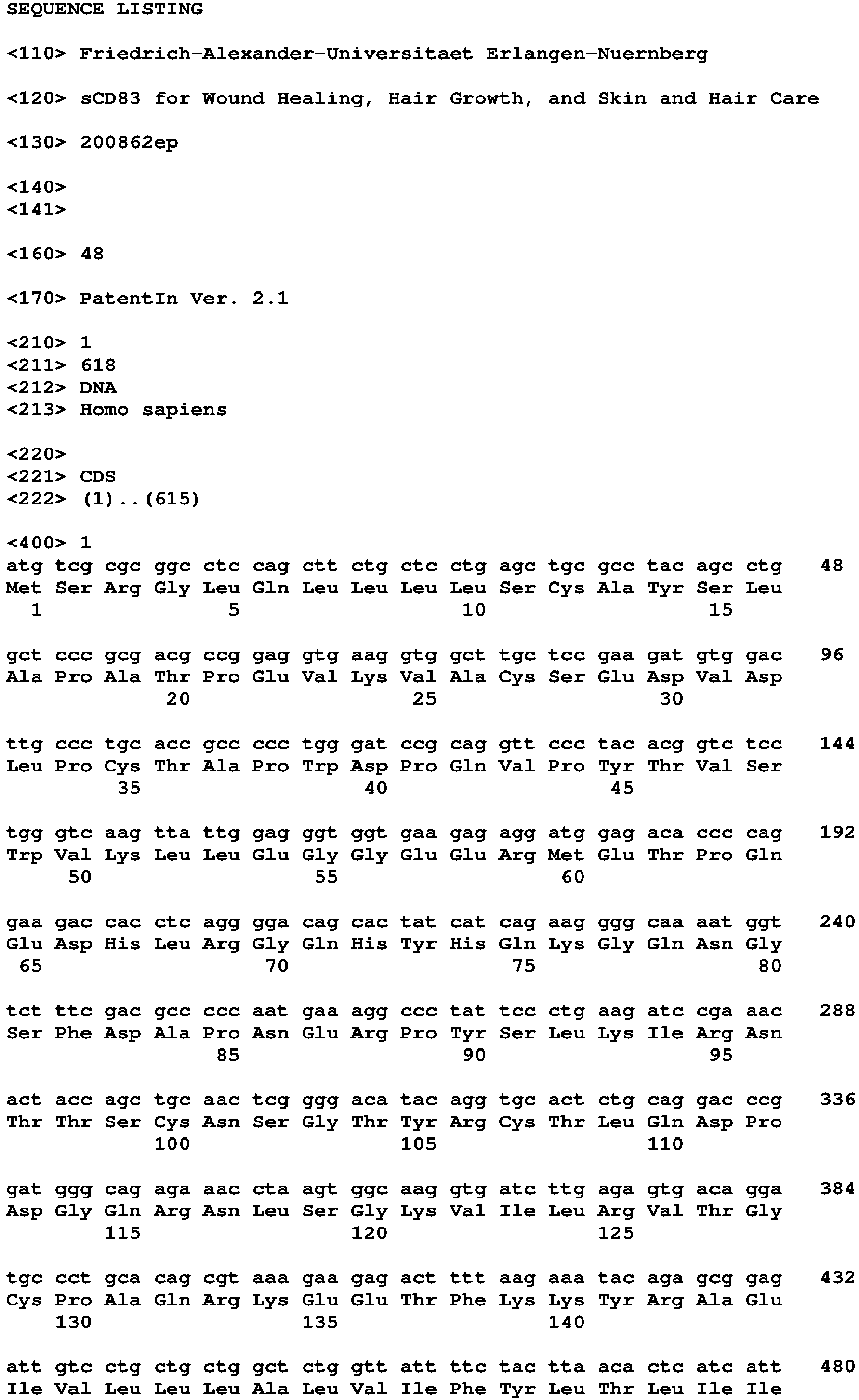


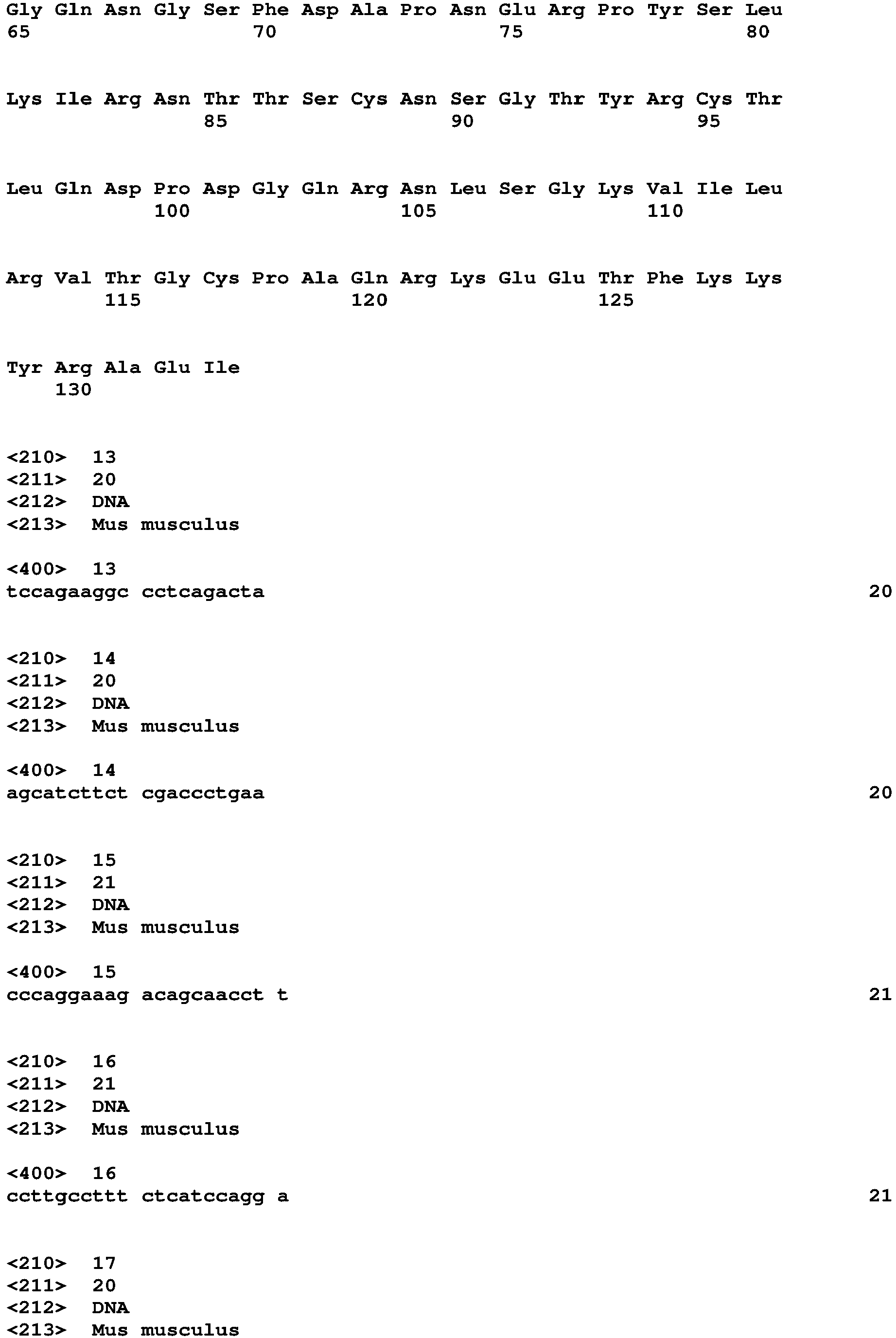


Claims (14)
Priority Applications (13)
| Application Number | Priority Date | Filing Date | Title |
|---|---|---|---|
| EP20169899.0A EP3895723A1 (en) | 2020-04-16 | 2020-04-16 | Scd83 for wound healing, hair growth, and skin and hair care |
| HRP20250640TT HRP20250640T1 (en) | 2020-04-16 | 2021-04-16 | SCD83 FOR WOUND HEALING, HAIR GROWTH, AND SKIN AND HAIR CARE |
| EP25168764.6A EP4591944A3 (en) | 2020-04-16 | 2021-04-16 | Scd83 for wound healing, hair growth, and skin and hair care |
| SM20250330T SMT202500330T1 (en) | 2020-04-16 | 2021-04-16 | Scd83 for wound healing, hair growth, and skin and hair care |
| RS20250521A RS66881B1 (en) | 2020-04-16 | 2021-04-16 | Scd83 for wound healing, hair growth, and skin and hair care |
| US17/996,366 US20230270820A1 (en) | 2020-04-16 | 2021-04-16 | Scd83 for wound healing, hair growth, and skin and hair care |
| PCT/EP2021/059923 WO2021209607A1 (en) | 2020-04-16 | 2021-04-16 | Scd83 for wound healing, hair growth, and skin and hair care |
| HUE21717933A HUE072389T2 (en) | 2020-04-16 | 2021-04-16 | sCD83 for wound healing, hair growth, and skin and hair care |
| JP2022562631A JP2023522022A (en) | 2020-04-16 | 2021-04-16 | sCD83 for wound healing, hair growth, and skin and hair care |
| CA3175640A CA3175640A1 (en) | 2020-04-16 | 2021-04-16 | Scd83 for wound healing, hair growth, and skin and hair care |
| EP21717933.2A EP4135745B1 (en) | 2020-04-16 | 2021-04-16 | Scd83 for wound healing, hair growth, and skin and hair care |
| ES21717933T ES3028665T3 (en) | 2020-04-16 | 2021-04-16 | Scd83 for wound healing, hair growth, and skin and hair care |
| PL21717933.2T PL4135745T3 (en) | 2020-04-16 | 2021-04-16 | Scd83 for wound healing, hair growth, and skin and hair care |
Applications Claiming Priority (1)
| Application Number | Priority Date | Filing Date | Title |
|---|---|---|---|
| EP20169899.0A EP3895723A1 (en) | 2020-04-16 | 2020-04-16 | Scd83 for wound healing, hair growth, and skin and hair care |
Publications (1)
| Publication Number | Publication Date |
|---|---|
| EP3895723A1 true EP3895723A1 (en) | 2021-10-20 |
Family
ID=70294945
Family Applications (3)
| Application Number | Title | Priority Date | Filing Date |
|---|---|---|---|
| EP20169899.0A Withdrawn EP3895723A1 (en) | 2020-04-16 | 2020-04-16 | Scd83 for wound healing, hair growth, and skin and hair care |
| EP25168764.6A Pending EP4591944A3 (en) | 2020-04-16 | 2021-04-16 | Scd83 for wound healing, hair growth, and skin and hair care |
| EP21717933.2A Active EP4135745B1 (en) | 2020-04-16 | 2021-04-16 | Scd83 for wound healing, hair growth, and skin and hair care |
Family Applications After (2)
| Application Number | Title | Priority Date | Filing Date |
|---|---|---|---|
| EP25168764.6A Pending EP4591944A3 (en) | 2020-04-16 | 2021-04-16 | Scd83 for wound healing, hair growth, and skin and hair care |
| EP21717933.2A Active EP4135745B1 (en) | 2020-04-16 | 2021-04-16 | Scd83 for wound healing, hair growth, and skin and hair care |
Country Status (11)
| Country | Link |
|---|---|
| US (1) | US20230270820A1 (en) |
| EP (3) | EP3895723A1 (en) |
| JP (1) | JP2023522022A (en) |
| CA (1) | CA3175640A1 (en) |
| ES (1) | ES3028665T3 (en) |
| HR (1) | HRP20250640T1 (en) |
| HU (1) | HUE072389T2 (en) |
| PL (1) | PL4135745T3 (en) |
| RS (1) | RS66881B1 (en) |
| SM (1) | SMT202500330T1 (en) |
| WO (1) | WO2021209607A1 (en) |
Cited By (1)
| Publication number | Priority date | Publication date | Assignee | Title |
|---|---|---|---|---|
| WO2025219050A1 (en) * | 2024-04-18 | 2025-10-23 | Christian Arnold | Pharmaceutical composition for healing a wound |
Citations (3)
| Publication number | Priority date | Publication date | Assignee | Title |
|---|---|---|---|---|
| WO2004046182A2 (en) | 2002-11-19 | 2004-06-03 | Argos Therapeutics, Inc. | Use of soluble forms of cd83 and nucleic acids encoding them for the treatment or prevention of diseases |
| US20110182903A1 (en) * | 2008-05-23 | 2011-07-28 | Stephen Brand | Novel soluble cd83 polypeptides, formulations and methods of use |
| WO2013006505A1 (en) * | 2011-07-01 | 2013-01-10 | Genentech, Inc. | Use of anti-cd83 agonist antibodies for treating autoimmune diseases |
-
2020
- 2020-04-16 EP EP20169899.0A patent/EP3895723A1/en not_active Withdrawn
-
2021
- 2021-04-16 ES ES21717933T patent/ES3028665T3/en active Active
- 2021-04-16 SM SM20250330T patent/SMT202500330T1/en unknown
- 2021-04-16 US US17/996,366 patent/US20230270820A1/en active Pending
- 2021-04-16 PL PL21717933.2T patent/PL4135745T3/en unknown
- 2021-04-16 JP JP2022562631A patent/JP2023522022A/en active Pending
- 2021-04-16 HR HRP20250640TT patent/HRP20250640T1/en unknown
- 2021-04-16 HU HUE21717933A patent/HUE072389T2/en unknown
- 2021-04-16 EP EP25168764.6A patent/EP4591944A3/en active Pending
- 2021-04-16 WO PCT/EP2021/059923 patent/WO2021209607A1/en not_active Ceased
- 2021-04-16 CA CA3175640A patent/CA3175640A1/en active Pending
- 2021-04-16 RS RS20250521A patent/RS66881B1/en unknown
- 2021-04-16 EP EP21717933.2A patent/EP4135745B1/en active Active
Patent Citations (3)
| Publication number | Priority date | Publication date | Assignee | Title |
|---|---|---|---|---|
| WO2004046182A2 (en) | 2002-11-19 | 2004-06-03 | Argos Therapeutics, Inc. | Use of soluble forms of cd83 and nucleic acids encoding them for the treatment or prevention of diseases |
| US20110182903A1 (en) * | 2008-05-23 | 2011-07-28 | Stephen Brand | Novel soluble cd83 polypeptides, formulations and methods of use |
| WO2013006505A1 (en) * | 2011-07-01 | 2013-01-10 | Genentech, Inc. | Use of anti-cd83 agonist antibodies for treating autoimmune diseases |
Non-Patent Citations (82)
| Title |
|---|
| "Genbank", Database accession no. NM_009856 |
| ALI, N. ET AL., CELL, vol. 169, 2017, pages 1119 - 1129 ellll |
| ANSTEAD, G.M., ADV WOUND CARE, vol. 11, 1998, pages 277 - 285 |
| ASHCROFT, G.S. ET AL., NAT MED, vol. 6, 2000, pages 1147 - 1153 |
| BAGHESTANI, S. ET AL., DERMATOL REPORTS, vol. 7, 2015, pages 6063 |
| BECKER, C. ET AL., GUT, vol. 54, 2005, pages 950 - 954 |
| BECKER, C. ET AL., NAT PROTOC, vol. 1, 2006, pages 2900 - 2904 |
| BOCK, F. ET AL., J IMMUNOL, vol. 191, 2013, pages 1965 - 1975 |
| BROCKMANN, L. ET AL., INT J MOL SCI, vol. 18, 2017 |
| CARSON, W.F.T. ET AL., CELL IMMUNOL, vol. 314, 2017, pages 63 - 72 |
| CHENG, F. ET AL., PROC NATL ACAD SCI U S A, vol. 113, 2016, pages E4320 - 4327 |
| COURTOIS, M. ET AL., SKIN PHARMACOL, vol. 7, 1994, pages 84 - 89 |
| DALEY, J.M. ET AL., J IMMUNOL, vol. 174, 2005, pages 2265 - 2272 |
| DIGNASS, A.U.PODOLSKY, D.K., GASTROENTEROLOGY, vol. 105, 1993, pages 1323 - 1332 |
| DOEBBELER, M. ET AL., JCI INSIGHT, vol. 3, 2018 |
| ECKHARDT, J. ET AL., J HISTOCHEM CYTOCHEM, vol. 61, 2013, pages 125 - 133 |
| ECKHARDT, J. ET AL., MUCOSAL IMMUNOL, vol. 7, 2014, pages 1006 - 1018 |
| ELLIS, J.A. ET AL., EXPERT REV MOL MED, vol. 4, 2002, pages 1 - 11 |
| EMING, S.A. ET AL., SCI TRANSL MED 6:265SR266, 2014 |
| FINN, D.A. ET AL., CNS DRUG REV, vol. 12, 2006, pages 53 - 76 |
| FRISWELL, M. ET AL., GUT LIVER, vol. 4, 2010, pages 295 - 306 |
| GALVEZ, J., ISRN INFLAMM 2014, vol. 928461, 2014 |
| GOODSON, W.H.3RDHUNG, T.K., J SURG RES, vol. 22, 1977, pages 221 - 227 |
| GROSCHE, L. ET AL., FRONT IMMUNOL, 2020 |
| GUO, S.DIPIETRO, L.A., J DENT RES, vol. 89, 2010, pages 219 - 229 |
| HALL, A., BIOCHEM SOC TRANS, vol. 33, 2005, pages 891 - 895 |
| HEILINGLOH, C.S. ET AL., JMB J MOL BIOL., vol. 429, no. 8, 2017, pages 1227 - 1243 |
| HERRICK, S. ET AL., LAB INVEST, vol. 77, 1997, pages 281 - 288 |
| HORVATINOVICH, J.M. ET AL., J IMMUNOL, vol. 198, 2017, pages 2286 - 2301 |
| HUBNER, G. ET AL., CYTOKINE, vol. 8, 1996, pages 548 - 556 |
| ISHIDA, Y. ET AL., J IMMUNOL, vol. 176, 2006, pages 5598 - 5606 |
| JANIS, J.E.HARRISON, B., PLAST RECONSTR SURG, vol. 133, 2014, pages 199e - 207e |
| JIN, F.Y. ET AL., CELL, vol. 88, 1997, pages 417 - 426 |
| JOE M. HORVATINOVICH ET AL: "Soluble CD83 Inhibits T Cell Activation by Binding to the TLR4/MD-2 Complex on CD14 + Monocytes", THE JOURNAL OF IMMUNOLOGY, vol. 198, no. 6, 13 February 2017 (2017-02-13), US, pages 2286 - 2301, XP055731233, ISSN: 0022-1767, DOI: 10.4049/jimmunol.1600802 * |
| KIESLER, P. ET AL., CELL MOL GASTROENTEROL HEPATOL, vol. 1, 2015, pages 154 - 170 |
| KRISCHAK, G.D. ET AL., J WOUND CARE, vol. 16, 2007, pages 76 - 78 |
| LAN, Z. ET AL., TRANSPLANTATION, vol. 90, 2010, pages 1278 - 1285 |
| LEE, S.W. ET AL., J DRUGS DERMATOL, vol. 17, 2018, pages 457 - 463 |
| LIN, Z.Q. ET AL., J LEUKOC BIOL, vol. 73, 2003, pages 713 - 721 |
| LUKASIK, A. ET AL., POL MERKUR LEKARSKI, vol. 46, 2019, pages 77 - 79 |
| MCINNES, I.B.SCHETT, G., N ENGL J MED, vol. 365, 2011, pages 2205 - 2219 |
| MEHRABI, A. ET AL., CLIN TRANSPLANT, vol. 20, no. 17, 2006, pages 97 - 110 |
| MESSENGER, A.G.RUNDEGREN, J., BR J DERMATOL, vol. 150, 2004, pages 186 - 194 |
| MOSSER, D.M.EDWARDS, J.P., NAT REV IMMUNOL, vol. 8, 2008, pages 958 - 969 |
| MULLER-ROVER, S. ET AL., J INVEST DERMATOL, vol. 117, 2001, pages 1027 - 1035 |
| NENCI, A. ET AL., NATURE, vol. 446, 2007, pages 557 - 561 |
| NEURATH, M., NAT REV GASTROENTEROL HEPATOL, vol. 14, 2017, pages 688 |
| PALLOTTA, M.T. ET AL., NAT IMMUNOL, vol. 12, 2011, pages 809 - 811 |
| PHILLIPS, T.G. ET AL., AM FAM PHYSICIAN, vol. 96, 2017, pages 371 - 378 |
| PLIKUS, M.V.CHUONG, C.M., J INVEST DERMATOL, vol. 128, 2008, pages 1071 - 1080 |
| RAMOT, Y. ET AL., INT J TRICHOLOGY, vol. 1, 2009, pages 27 - 29 |
| REAVES, T.A. ET AL., MEM INST OSWALDO CRUZ, vol. 100, no. 1, 2005, pages 191 - 198 |
| REUTER, B.K. ET AL., J CLIN INVEST, vol. 98, 1996, pages 2076 - 2085 |
| RITSU, M. ET AL., J DERMATOLOGY & DERMATOLOGIC SURGERY, vol. 21, 2017, pages 14 - 19 |
| ROINE, E. ET AL., TRANSPLANT PROC, vol. 42, 2010, pages 2542 - 2546 |
| ROYZMAN, D. ET AL., FRONT IMMUNOL, vol. 10, 2019, pages 1312 |
| SAMBROOK ET AL.: "Molecular Cloning: A Laboratory Manual", 1989, COLD SPRING HARBOR LABORATORY |
| SARTOR, R.B., NAT CLIN PRACT GASTROENTEROL HEPATOL, vol. 3, 2006, pages 390 - 407 |
| SGONC, R.GRUBER, J., GERONTOLOGY, vol. 59, 2013, pages 159 - 164 |
| SHAPIRO, J.KAUFMAN, K.D., J INVESTIG DERMATOL SYMP PROC, vol. 8, 2003, pages 20 - 23 |
| SINGER, II ET AL., GASTROENTEROLOGY, vol. 115, 1998, pages 297 - 306 |
| SIRBULESCU, R.F. ET AL., WOUND REPAIR REGEN, vol. 25, 2017, pages 774 - 791 |
| STARKE, C. ET AL., IMMUNOBIOLOGY, vol. 218, 2013, pages 1411 - 1415 |
| STROBER, W.FUSS, I.J., GASTROENTEROLOGY, vol. 140, 2011, pages 1756 - 1767 |
| SUGIHARA, K. ET AL., FRONT IMMUNOL, vol. 9, 2018, pages 3183 |
| TEKUR, V.K., INDIAN DERMATOL ONLINE J, vol. 7, 2016, pages 418 - 420 |
| TRAISH, A.M. ET AL., J SEX MED, vol. 8, 2011, pages 872 - 884 |
| TRUEB, R.M., SKIN THERAPY LETT, vol. 15, 2010, pages 5 - 7 |
| UENO, P. ET AL., TRANSPLANTATION, vol. 101, 2017, pages 844 - 850 |
| URYSIAK-CZUBATKA, I. ET AL., POSTEPY DERMATOL ALERGOL, vol. 31, 2014, pages 207 - 215 |
| VAN NESTE, D. ET AL., BR J DERMATOL, vol. 143, 2000, pages 804 - 810 |
| WALKER, J.L. ET AL., PROC NATL ACAD SCI U S A, vol. 107, 2010, pages 13730 - 13735 |
| WANG, J., CELL TISSUE RES, vol. 371, 2018, pages 531 - 539 |
| WEN, H. ET AL., AAPS J, vol. 17, 2015, pages 1327 - 1340 |
| WILD, A.B. ET AL., JCI INSIGHT, vol. 4, 2019 |
| WIRTZ, S. ET AL., NAT PROTOC, vol. 12, 2017, pages 1295 - 1309 |
| XU, J.F. ET AL., TRANSPL INT, vol. 20, 2007, pages 266 - 276 |
| ZARBOCK, A. ET AL., BLOOD REV, vol. 21, 2007, pages 99 - 111 |
| ZHOU, L.J. ET AL., J. IMMUNOL., vol. 149, no. 2, 1992, pages 735 - 742 |
| ZIDUO LI ET AL: "CD83: Activation Marker for Antigen Presenting Cells and Its Therapeutic Potential", FRONTIERS IN IMMUNOLOGY, vol. 10, 1 January 2019 (2019-01-01), pages 1312, XP055731281, DOI: 10.3389/fimmu.2019.01312 * |
| ZINSER, E. ET AL., J EXP MED, vol. 200, 2004, pages 345 - 351 |
| ZUNDLER, S. ET AL., GUT, vol. 68, 2019, pages 1688 - 1700 |
Cited By (2)
| Publication number | Priority date | Publication date | Assignee | Title |
|---|---|---|---|---|
| WO2025219050A1 (en) * | 2024-04-18 | 2025-10-23 | Christian Arnold | Pharmaceutical composition for healing a wound |
| DE102024110866A1 (en) | 2024-04-18 | 2025-10-23 | Christian Arnold | Pharmaceutical composition for healing a wound |
Also Published As
| Publication number | Publication date |
|---|---|
| EP4135745A1 (en) | 2023-02-22 |
| EP4591944A2 (en) | 2025-07-30 |
| PL4135745T3 (en) | 2025-09-15 |
| EP4135745C0 (en) | 2025-05-07 |
| SMT202500330T1 (en) | 2025-11-10 |
| RS66881B1 (en) | 2025-07-31 |
| WO2021209607A1 (en) | 2021-10-21 |
| ES3028665T3 (en) | 2025-06-19 |
| EP4591944A3 (en) | 2025-10-01 |
| EP4135745B1 (en) | 2025-05-07 |
| US20230270820A1 (en) | 2023-08-31 |
| HUE072389T2 (en) | 2025-11-28 |
| JP2023522022A (en) | 2023-05-26 |
| HRP20250640T1 (en) | 2025-09-12 |
| CA3175640A1 (en) | 2021-10-21 |
Similar Documents
| Publication | Publication Date | Title |
|---|---|---|
| JP6661530B2 (en) | Interleukin-4 receptor binding fusion protein and use thereof | |
| KR20010006534A (en) | Type ii tgf-beta receptor/immunoglobulin constant region fusion proteins | |
| ES2398039T3 (en) | Use of anti-TGF-beta antagonist antibodies to treat or prevent loss of renal function | |
| CN113194978B (en) | Peptides with mesenchymal stem cell mobilization activity | |
| JPH09509825A (en) | Methods and compositions for stimulating bone cells | |
| US9351914B2 (en) | Methods for modulating hair growth using truncated laminin-511 | |
| JP2003534363A (en) | Cosmetic composition containing human serum albumin derived from transgenic non-human animal | |
| EP4135745B1 (en) | Scd83 for wound healing, hair growth, and skin and hair care | |
| JP7672636B2 (en) | Peptides with mesenchymal stem cell mobilization activity | |
| US12006343B2 (en) | C4BP-based compounds for treating immunological diseases | |
| HK40129498A (en) | Scd83 for wound healing, hair growth, and skin and hair care | |
| AU2001247825B2 (en) | Angiogenic onchocerca volvulus proteins and uses thereof | |
| HUP0401323A2 (en) | Use of il-18 inhibitors in hypersensitivity disorders | |
| KR20010075060A (en) | Chemokine receptor antagonist and cyclosporin in combined therapy | |
| US11524091B2 (en) | GILZ formulations for wound healing | |
| AU2001247825A1 (en) | Angiogenic onchocerca volvulus proteins and uses thereof | |
| Class et al. | Patent application title: Methods for Modulating Hair Growth Using Truncated Laminin-511 Inventors: M. Peter Marinkovich (Redwood City, CA, US) Jing Gao (Mountain View, CA, US) Xiaoyu Xu (Foster City, CA, US) Jayakumar Rajadas (Cupertino, CA, US) Jayakumar Rajadas (Cupertino, CA, US) | |
| TW201822813A (en) | Therapeutic agent for cerebral infarction | |
| Wang | Molecular and cell biology of pig skin: a model for wound healing | |
| Cruz | Altered gene expression of IL-1 Alpha, IL-1 Beta, and KGF-1 in early cutaneous wounds of restrained Mice |
Legal Events
| Date | Code | Title | Description |
|---|---|---|---|
| PUAI | Public reference made under article 153(3) epc to a published international application that has entered the european phase |
Free format text: ORIGINAL CODE: 0009012 |
|
| STAA | Information on the status of an ep patent application or granted ep patent |
Free format text: STATUS: THE APPLICATION HAS BEEN PUBLISHED |
|
| AK | Designated contracting states |
Kind code of ref document: A1 Designated state(s): AL AT BE BG CH CY CZ DE DK EE ES FI FR GB GR HR HU IE IS IT LI LT LU LV MC MK MT NL NO PL PT RO RS SE SI SK SM TR |
|
| B565 | Issuance of search results under rule 164(2) epc |
Effective date: 20200928 |
|
| RIN1 | Information on inventor provided before grant (corrected) |
Inventor name: ZINSER, ELISABETH Inventor name: ROYZMAN, DMYTRO Inventor name: STEINKASSERER, ALEXANDER |
|
| STAA | Information on the status of an ep patent application or granted ep patent |
Free format text: STATUS: THE APPLICATION IS DEEMED TO BE WITHDRAWN |
|
| 18D | Application deemed to be withdrawn |
Effective date: 20220421 |