EP3889610B1 - Biomarker composition for predicting prognosis of gastric cancer induced by exposure to polystyrene microplastics - Google Patents
Biomarker composition for predicting prognosis of gastric cancer induced by exposure to polystyrene microplastics Download PDFInfo
- Publication number
- EP3889610B1 EP3889610B1 EP21165421.5A EP21165421A EP3889610B1 EP 3889610 B1 EP3889610 B1 EP 3889610B1 EP 21165421 A EP21165421 A EP 21165421A EP 3889610 B1 EP3889610 B1 EP 3889610B1
- Authority
- EP
- European Patent Office
- Prior art keywords
- cadherin
- expression
- microplastics
- polystyrene
- gastric cancer
- Prior art date
- Legal status (The legal status is an assumption and is not a legal conclusion. Google has not performed a legal analysis and makes no representation as to the accuracy of the status listed.)
- Active
Links
Images






Classifications
-
- G—PHYSICS
- G01—MEASURING; TESTING
- G01N—INVESTIGATING OR ANALYSING MATERIALS BY DETERMINING THEIR CHEMICAL OR PHYSICAL PROPERTIES
- G01N33/00—Investigating or analysing materials by specific methods not covered by groups G01N1/00 - G01N31/00
- G01N33/48—Biological material, e.g. blood, urine; Haemocytometers
- G01N33/50—Chemical analysis of biological material, e.g. blood, urine; Testing involving biospecific ligand binding methods; Immunological testing
- G01N33/68—Chemical analysis of biological material, e.g. blood, urine; Testing involving biospecific ligand binding methods; Immunological testing involving proteins, peptides or amino acids
- G01N33/6893—Chemical analysis of biological material, e.g. blood, urine; Testing involving biospecific ligand binding methods; Immunological testing involving proteins, peptides or amino acids related to diseases not provided for elsewhere
-
- G—PHYSICS
- G01—MEASURING; TESTING
- G01N—INVESTIGATING OR ANALYSING MATERIALS BY DETERMINING THEIR CHEMICAL OR PHYSICAL PROPERTIES
- G01N33/00—Investigating or analysing materials by specific methods not covered by groups G01N1/00 - G01N31/00
- G01N33/48—Biological material, e.g. blood, urine; Haemocytometers
- G01N33/50—Chemical analysis of biological material, e.g. blood, urine; Testing involving biospecific ligand binding methods; Immunological testing
- G01N33/53—Immunoassay; Biospecific binding assay; Materials therefor
- G01N33/575—Immunoassay; Biospecific binding assay; Materials therefor for cancer
- G01N33/5758—Immunoassay; Biospecific binding assay; Materials therefor for cancer involving compounds serving as markers for tumours, cancers or neoplasias, e.g. cellular determinants, receptors, heat shock/stress proteins, A-protein, oligosaccharides or metabolites
-
- G—PHYSICS
- G01—MEASURING; TESTING
- G01N—INVESTIGATING OR ANALYSING MATERIALS BY DETERMINING THEIR CHEMICAL OR PHYSICAL PROPERTIES
- G01N33/00—Investigating or analysing materials by specific methods not covered by groups G01N1/00 - G01N31/00
- G01N33/48—Biological material, e.g. blood, urine; Haemocytometers
- G01N33/50—Chemical analysis of biological material, e.g. blood, urine; Testing involving biospecific ligand binding methods; Immunological testing
- G01N33/53—Immunoassay; Biospecific binding assay; Materials therefor
- G01N33/575—Immunoassay; Biospecific binding assay; Materials therefor for cancer
- G01N33/5753—Immunoassay; Biospecific binding assay; Materials therefor for cancer of the stomach or small intestine
-
- C—CHEMISTRY; METALLURGY
- C12—BIOCHEMISTRY; BEER; SPIRITS; WINE; VINEGAR; MICROBIOLOGY; ENZYMOLOGY; MUTATION OR GENETIC ENGINEERING
- C12Q—MEASURING OR TESTING PROCESSES INVOLVING ENZYMES, NUCLEIC ACIDS OR MICROORGANISMS; COMPOSITIONS OR TEST PAPERS THEREFOR; PROCESSES OF PREPARING SUCH COMPOSITIONS; CONDITION-RESPONSIVE CONTROL IN MICROBIOLOGICAL OR ENZYMOLOGICAL PROCESSES
- C12Q1/00—Measuring or testing processes involving enzymes, nucleic acids or microorganisms; Compositions therefor; Processes of preparing such compositions
- C12Q1/68—Measuring or testing processes involving enzymes, nucleic acids or microorganisms; Compositions therefor; Processes of preparing such compositions involving nucleic acids
- C12Q1/6876—Nucleic acid products used in the analysis of nucleic acids, e.g. primers or probes
- C12Q1/6883—Nucleic acid products used in the analysis of nucleic acids, e.g. primers or probes for diseases caused by alterations of genetic material
- C12Q1/6886—Nucleic acid products used in the analysis of nucleic acids, e.g. primers or probes for diseases caused by alterations of genetic material for cancer
-
- G—PHYSICS
- G01—MEASURING; TESTING
- G01N—INVESTIGATING OR ANALYSING MATERIALS BY DETERMINING THEIR CHEMICAL OR PHYSICAL PROPERTIES
- G01N33/00—Investigating or analysing materials by specific methods not covered by groups G01N1/00 - G01N31/00
- G01N33/48—Biological material, e.g. blood, urine; Haemocytometers
- G01N33/50—Chemical analysis of biological material, e.g. blood, urine; Testing involving biospecific ligand binding methods; Immunological testing
- G01N33/53—Immunoassay; Biospecific binding assay; Materials therefor
- G01N33/575—Immunoassay; Biospecific binding assay; Materials therefor for cancer
-
- C—CHEMISTRY; METALLURGY
- C12—BIOCHEMISTRY; BEER; SPIRITS; WINE; VINEGAR; MICROBIOLOGY; ENZYMOLOGY; MUTATION OR GENETIC ENGINEERING
- C12Q—MEASURING OR TESTING PROCESSES INVOLVING ENZYMES, NUCLEIC ACIDS OR MICROORGANISMS; COMPOSITIONS OR TEST PAPERS THEREFOR; PROCESSES OF PREPARING SUCH COMPOSITIONS; CONDITION-RESPONSIVE CONTROL IN MICROBIOLOGICAL OR ENZYMOLOGICAL PROCESSES
- C12Q2600/00—Oligonucleotides characterized by their use
- C12Q2600/112—Disease subtyping, staging or classification
-
- C—CHEMISTRY; METALLURGY
- C12—BIOCHEMISTRY; BEER; SPIRITS; WINE; VINEGAR; MICROBIOLOGY; ENZYMOLOGY; MUTATION OR GENETIC ENGINEERING
- C12Q—MEASURING OR TESTING PROCESSES INVOLVING ENZYMES, NUCLEIC ACIDS OR MICROORGANISMS; COMPOSITIONS OR TEST PAPERS THEREFOR; PROCESSES OF PREPARING SUCH COMPOSITIONS; CONDITION-RESPONSIVE CONTROL IN MICROBIOLOGICAL OR ENZYMOLOGICAL PROCESSES
- C12Q2600/00—Oligonucleotides characterized by their use
- C12Q2600/118—Prognosis of disease development
-
- C—CHEMISTRY; METALLURGY
- C12—BIOCHEMISTRY; BEER; SPIRITS; WINE; VINEGAR; MICROBIOLOGY; ENZYMOLOGY; MUTATION OR GENETIC ENGINEERING
- C12Q—MEASURING OR TESTING PROCESSES INVOLVING ENZYMES, NUCLEIC ACIDS OR MICROORGANISMS; COMPOSITIONS OR TEST PAPERS THEREFOR; PROCESSES OF PREPARING SUCH COMPOSITIONS; CONDITION-RESPONSIVE CONTROL IN MICROBIOLOGICAL OR ENZYMOLOGICAL PROCESSES
- C12Q2600/00—Oligonucleotides characterized by their use
- C12Q2600/158—Expression markers
-
- G—PHYSICS
- G01—MEASURING; TESTING
- G01N—INVESTIGATING OR ANALYSING MATERIALS BY DETERMINING THEIR CHEMICAL OR PHYSICAL PROPERTIES
- G01N2333/00—Assays involving biological materials from specific organisms or of a specific nature
- G01N2333/435—Assays involving biological materials from specific organisms or of a specific nature from animals; from humans
- G01N2333/705—Assays involving receptors, cell surface antigens or cell surface determinants
- G01N2333/70503—Immunoglobulin superfamily, e.g. VCAMs, PECAM, LFA-3
- G01N2333/70532—B7 molecules, e.g. CD80, CD86
-
- G—PHYSICS
- G01—MEASURING; TESTING
- G01N—INVESTIGATING OR ANALYSING MATERIALS BY DETERMINING THEIR CHEMICAL OR PHYSICAL PROPERTIES
- G01N2333/00—Assays involving biological materials from specific organisms or of a specific nature
- G01N2333/435—Assays involving biological materials from specific organisms or of a specific nature from animals; from humans
- G01N2333/705—Assays involving receptors, cell surface antigens or cell surface determinants
- G01N2333/70585—CD44
-
- G—PHYSICS
- G01—MEASURING; TESTING
- G01N—INVESTIGATING OR ANALYSING MATERIALS BY DETERMINING THEIR CHEMICAL OR PHYSICAL PROPERTIES
- G01N2333/00—Assays involving biological materials from specific organisms or of a specific nature
- G01N2333/435—Assays involving biological materials from specific organisms or of a specific nature from animals; from humans
- G01N2333/705—Assays involving receptors, cell surface antigens or cell surface determinants
- G01N2333/70596—Molecules with a "CD"-designation not provided for elsewhere in G01N2333/705
-
- G—PHYSICS
- G01—MEASURING; TESTING
- G01N—INVESTIGATING OR ANALYSING MATERIALS BY DETERMINING THEIR CHEMICAL OR PHYSICAL PROPERTIES
- G01N2333/00—Assays involving biological materials from specific organisms or of a specific nature
- G01N2333/435—Assays involving biological materials from specific organisms or of a specific nature from animals; from humans
- G01N2333/705—Assays involving receptors, cell surface antigens or cell surface determinants
- G01N2333/72—Assays involving receptors, cell surface antigens or cell surface determinants for hormones
- G01N2333/726—G protein coupled receptor, e.g. TSHR-thyrotropin-receptor, LH/hCG receptor, FSH
-
- G—PHYSICS
- G01—MEASURING; TESTING
- G01N—INVESTIGATING OR ANALYSING MATERIALS BY DETERMINING THEIR CHEMICAL OR PHYSICAL PROPERTIES
- G01N2333/00—Assays involving biological materials from specific organisms or of a specific nature
- G01N2333/90—Enzymes; Proenzymes
- G01N2333/91—Transferases (2.)
- G01N2333/91005—Transferases (2.) transferring one-carbon groups (2.1)
- G01N2333/91011—Methyltransferases (general) (2.1.1.)
- G01N2333/91017—Methyltransferases (general) (2.1.1.) with definite EC number (2.1.1.-)
-
- G—PHYSICS
- G01—MEASURING; TESTING
- G01N—INVESTIGATING OR ANALYSING MATERIALS BY DETERMINING THEIR CHEMICAL OR PHYSICAL PROPERTIES
- G01N2800/00—Detection or diagnosis of diseases
- G01N2800/52—Predicting or monitoring the response to treatment, e.g. for selection of therapy based on assay results in personalised medicine; Prognosis
Definitions
- the present invention relates to an in vitro use of a biomarker composition for predicting the prognosis of gastric cancer induced by the exposure to microplastics.
- Gastric cancer is a very common cancer in Asia and is one of the leading causes of cancer deaths worldwide.
- many attempts have been made to better elucidate the biological processes involved in the development of gastric cancer, in order to improve understanding of the cancer mechanisms, as well as the early diagnosis and treatment of gastric cancer, but the molecular mechanisms involved in carcinogenesis of gastric cancer still remain the subject of intensive research.
- gastric cancer is the most frequent among all cancers, and the second most common cause of death.
- the stage of gastric cancer is determined by the degree of invasion of the surrounding area of the tumor, the metastasis to lymph nodes and metastasis to other organs, and accordingly, treatment and prognosis depend thereon.
- the prognosis gradually worsens, and especially the prognosis in the terminal stage (Stage 4) is so poor that the 5-year survival rate is only 3%.
- Plastics have contributed greatly to the abundant daily life of modern people and industrial development due to their excellent functions and low prices, and recently, discarded plastics has been reported to decompose into microscopic sizes that are difficult to visually identify over time, thereby polluting the environment and threatening human health, but the effect of microplastics on cancer disease has not been studied.
- Korean Patent Publication No. 10-2013-0109600 (published on October 08, 2013 )
- WO 2019/016422 A1 relates to a method for predicting the response of a subject suffering from lung cancer to treatment with FGFR inhibitors.
- Kahtan Al-Bayaty Mustafa ET AL "E-Cadherin Protein as a Potential Marker for Gastric Cancer and Its Association with Helicobacter Pylori - Induced Gastritis and Gastric Ulcer", Reports of biochemistry and molecular biology, Iran, (20191001), pages 269 - 277 is a study about E-Cadherin protein as a potential marker for gastric cancer and its association with helicobacter pylori - induced gastritis and gastric ulcer.
- AZADI SHOHREH ET AL "Upregulation of PD-L1 expression in breast cancer cells through the formation of 3D multicellular cancer aggregates under different chemical and mechanical conditions
- BIOCHIMICA ET BIOPHYSICA ACTA, ELSEVIER SCIENCE PUBLISHERS, AMSTERDAM, NL, vol. 1866, no. 12 is a study about the upregulation of PD-L1 expression in breast cancer cells.
- WU Z. ET AL "Claudin-7 (CLDN7) is overexpressed in gastric cancer and promotes gastric cancer cell proliferation, invasion and maintains mesenchymal state”
- NEOPLASMA vol. 65, no. 03, pages 349 - 359 is a study about Claudin-7 (CLDN7) and gastric cancer. Chen H.
- F. ET AL "Protocadherin 7 inhibits cell migration and invasion through E-cadherin in gastric cancer", Tumor Biology, London, England, pages 1-11 is a study about Protocadherin 7 and gastric cancer.
- URAOKA NAOHIRO ET AL "NRD1, which encodes nardilysin protein, promotes esophageal cancer cell invasion through induction of MMP2 and MMP3 expression", CANCER SCIENCE, JP, vol. 105, no. 1 , is a study about NRD1 and esophageal cancer.
- WO 2017/203444 A1 relates to a method of prognosing and predicting breast cancer recurrence in patients.
- STOCK VALERIE ET AL "Uptake and effects of orally ingested polystyrene microplastic particles in vitro and in vivo", ARCHIVES OF TOXICOLOGY, SPRINGER, DE, vol. 93, no. 7, pages 1817 - 1833 is a study about orally ingested polystyrene microplastic particles.
- DENG YONGFENG ET AL "Tissue accumulation of microplastics in mice and biomarker responses suggest widespread health risks of exposure", SCIENTIFIC REPORTS, vol. 7, no. 1 is a study about Tissue accumulation of microplastics in mice.
- An object of the present invention is to provide a biomarker composition for predicting the prognosis of cancer malignancy induced by the exposure to microplastics and a method of providing information for predicting the prognosis of cancer malignancy using the same.
- the present invention is defined in the claims.
- the present invention makes use of a biomarker composition, as defined in claim 1, for predicting prognosis of gastric cancer induced by exposure to microplastics, comprising the proteins CD44, E-cadherin, N-cadherin, PD-L1 (programmed death-ligand 1), NPAS2 (Neuronal PAS domain protein 2), NR1D1 (nuclear receptor subfamily 1 group D member 1), DNMT1 (DNA (cytosine-5)-methyltransferase 1), SLC7A2 (Solute Carrier Family 7 Member 2), PCDH7 (Protocadherin-7) and CLDN7 (Claudin 7), wherein the CD44, N-cadherin, PD-L1, NR1D1, SLC7A2 and PCDH7 increase in expression of cancer cells exposed to the polystyrene microplastics, and the E-cadherin, NPAS2, DNMT1 and CLDN7 reduce in expression of cancer cells
- the present invention provides a method, as defined in claim 2, of providing information for predicting prognosis of gastric cancer induced by exposure to microplastics comprising detecting expression or activity level of the proteins CD44, E-cadherin, N-cadherin, PD-L1, NPAS2, NR1D1, DNMT1, SLC7A2, PCDH7 and CLDN7, or expression level of gene encoding the protein in a sample isolated from specimen, wherein the CD44, N-cadherin, PD-L1, NR1D1, SLC7A2 and PCDH7 increase in expression of cancer cells exposed to the polystyrene microplastics, the E-cadherin, NPAS2, DNMT1 and CLDN7 reduce in expression of cancer cells exposed to the polystyrene microplastics, and the cancer gastric cancer is induced due to an increase in proliferation, migration and invasion of cancer cells and an decrease in 5-year overall survival rates in gastric cancer by the change in the expression level of genes encoding the proteins.
- microplastics with diameters less than 5 mm are being recognized as a new environmental treat and human health risk
- the present inventors investigated the effects of the microplastic polystyrene (PS) on the hallmarks of cancer changes and 5-year overall survival (OS) to study the risks that may occur when PS, a type of microplastic, are ingested in vivo and as a result, they confirmed that when PS is ingested in vivo, the gastric cancer was exacerbated through the change in the expression level of the gene in the stomach tissue, which is the primary site of exposure and completed the present invention.
- PS microplastic polystyrene
- OS 5-year overall survival
- the present invention is the proteins CD44, E-cadherin, N-cadherin, PD-L1, NPAS2, NR1D1, DNMT1, SLC7A2, PCDH7 and CLDN7, and it is possible to provide a biomarker composition for predicting prognosis of gastric cancer induced by exposure to microplastics.
- the biomarker composition is CD44 (NCBI No. 960), E-cadherin (NCBI No. 999), N-cadherin (NCBI No. 1000), PD-L1 (programmed death-ligand 1, NCBI No. 29126), NPAS2 (Neuronal PAS domain protein 2, NCBI No. 4862), NR1D1 (nuclear receptor subfamily 1 group D member 1, NCBI No. 9572), DNMT1 (DNA (cytosine-5)-methyltransferase 1, NCBI No. 1786), SLC7A2 (Solute Carrier Family 7 Member 2, NCBI No. 6542), PCDH7 (Protocadherin-7, NCBI No. 5099) and CLDN7 (Claudin 7, NCBI No. 1366).
- the microplastics is polystyrene.
- the CD44, N-cadherin, PD-L1, NR1D1, SLC7A2 and PCDH7 increase in the expression in cancer cells exposed to the microplastics.
- the E-cadherin, NPAS2, DNMT1 and CLDN7 reduce in the expression in cancer cells exposed to the microplastics.
- the cancer is gastric cancer.
- a diagnostic kit can be provided for predicting prognosis of cancer malignancy induced by exposure to microplastics comprising the proteins CD44, E-cadherin, N-cadherin, PD-L1, NPAS2, NR1D1, DNMT1, SLC7A2, PCDH7 and CLDN7, and an agent for detecting expression or activity level of the protein, or expression level of gene encoding the protein as an active ingredient.
- the agent may be selected from the group consisting of primers, probes, antibodies, peptides and aptamers.
- the present invention may provide a method of providing information for predicting prognosis of gastric cancer induced by exposure to microplastics comprising detecting expression or activity level of the proteins CD44, E-cadherin, N-cadherin, PD-L1, NPAS2, NR1D1, DNMT1, SLC7A2, PCDH7 and CLDN7, or expression level of gene encoding the protein in a sample isolated from specimen wherein the CD44, N-cadherin, PD-L1, NR1D1, SLC7A2 and PCDH7 increase in expression of cancer cells exposed to the polystyrene microplastics, the E-cadherin, NPAS2, DNMT1 and CLDN7 reduce in expression of cancer cells exposed to the polystyrene microplastics, and the gastric cancer is induced due to an increase in proliferation, migration and invasion of cancer cells and an decrease in 5-year overall survival rates in gastric cancer by the change in the expression level of genes encoding the proteins.
- biomarker refers to a substance capable of diagnosing a tissue or cell of a subject with gastric cancer by distinguishing it from a tissue or cell of a normal control group and includes organic biomolecules such as proteins, nucleic acids, lipids, glycolipids, glycoproteins, and the like, which show an increase or decrease in tissues or cells of a diseased object compared to the normal control group.
- prognosis includes determining whether an object for a specific disease or symptom, that is, the subject to be tested will have the autologous disease in the future, or determining the responsiveness of the subject to be tested to the treatment.
- fluorescent green polyethylene microspheres 100 ppm/100 ⁇ L; 10-20 ⁇ m; Cospheric, USA was used. Fluorescent green polyethylene microspheres were orally administered to 5-week-old BALB/c nude mice daily for 4 weeks.
- mice were euthanized using CO 2 .
- the stomach was isolated and fixed overnight with 4% PFA at 4°C.
- Frozen slides (8 ⁇ m thick) were generated using optimal cutting temperature (OCT) compound, and the slides were attached to slide glass.
- OCT optimal cutting temperature
- Cell nuclei were stained with DAPI (ThermoFisher Scientific, USA), and images were obtained using an IN cell analyzer 2200 (GE Healthcare, USA)
- PS polystyrene microspheres
- Ultrasonic grinding and vortexing were repeatedly performed to create a 5% PS suspension, which was then aliquoted to cell culture media to the desired concentration during subculture.
- the PS was removed by washing three times with PBS.
- EDTA-trypsin was used during subculture.
- the human gastric cancer cell lines, AGS, MKN1, MKN45, KATO III, were obtained from the Korea Cell-Line Bank (KLCB, Korea).
- NCI-N87 was obtained from the American Type Culture Collection (ATCC, USA).
- Cells (1 ⁇ 10 5 cells per mL) were suspended in 0.2 mL of serum-free RPMI for invasion and motility assays.
- the cells were loaded in the upper well of the Transwell chamber (8-um pore size; SPL, Korea) that was pre-coated with Matrigel (Corning, USA) on an upper side of the chamber; the lower well was filled with 0.75 mL of RPMI with 20% serum.
- Invasiveness was determined by counting cells in four microscopic fields per well, and the extent of invasion was expressed as an average number of cells per microscopic field.
- permeabilization buffer 0.02% Triton-X in distilled water
- cells were incubated with blocking buffer containing 1% BSA with 10% normal goat serum for 1 h at room temperature. Cells were then incubated with primary antibody (anti-N-cadherin, anti-CD44, or anti-PD-L1 antibody) overnight at 4°C. After washing 3 times by PBS, cells were incubated with Alexa Fluor-647 goat anti-rabbit IgG (H + L) at room temperature in the dark for 60 min.
- primary antibody anti-N-cadherin, anti-CD44, or anti-PD-L1 antibody
- cells were seeded onto a 2-well chamber slide (Falcon, USA) with RPMI 1640 medium (Corning, USA) supplemented with 10% FBS at a density of 1 ⁇ 10 5 cells/mL and treated with PS at 500 ppm.
- the samples were permeabilized by incubation with 0.02% Triton-X in distilled water for 20 min at room temperature. After the buffer was removed, cells were incubated with blocking buffer (1% BSA and 10% normal goat serum) for 1 h at room temperature and cells were incubated with primary antibody overnight at 4°C.
- blocking buffer 1% BSA and 10% normal goat serum
- N-cadherin and E-cadherin were detected with Alexa Fluor-488 goat anti-rabbit IgG (H + L) and Alexa Fluor-647 goat anti-mouse IgG (H + L), respectively.
- PD-L1 was detected with Alexa Fluor-647 goat anti-rabbit IgG (H + L)
- CD44 was detected with Alexa Fluor-488 goat anti-mouse IgG (H + L).
- the slides were treated with DAPI and analyzed by confocal microscopy (LSM-710, Zeiss, Germany).
- a Superscript III cDNA Synthesis Kit (Invitrogen, USA) was used to generate cDNA from 5 ⁇ g of mRNA following the manufacturer's instructions.
- qPCR was performed on an ABI qPCR system (Applied Biosystems, USA) using SYBR Green PCR Master Mix (Applied Biosystems, USA) following the manufacturer's instructions.
- the reaction conditions were: 10 min at 95°C followed by 50 cycles of 95°C for 15 s, 60°C for 60 s.
- cell proliferation was measured by the alamarBlue ® assay (ThermoFisher Scientific, USA) following the manufacturer's instructions.
- cells were seeded onto 96-well culture plates with RPMI-1640 medium supplemented with 10% FBS at a density of 5000 cells/well and treated PS microspheres.
- the emission values are stated as percentages of controls, yielding the percent cell proliferation after 72 h of seeding the plate.
- cytotoxicity was measured using bortezomib or cisplatin. Cytotoxicity was determined by the alamarBlue ® assay (Thermo Fisher Scientific, Waltham, MA) following the manufacturer's instructions.
- Cytotoxicity was measured at an emission of 590 nm using a microplate reader.
- the emission values are stated as percentages of vehicle controls, yielding percentage cell viability after 72 h of treatment.
- ⁇ cytotoxicity cytotoxicity with PS ⁇ cytotoxicity without PS
- mice were orally administered 10- ⁇ m PS microspheres (100 ppm/100 ⁇ L PBS) for 4 weeks.
- RNA was randomly fragmented for sequencing with short reads, which were then reverse transcribed to cDNA with different adapters attached to the 5' and 3' ends.
- PCR amplification was performed to the extent that sequencing was possible, and size selection was performed to obtain an insert size of 200-400 bp, which was used for NovaSeq 6000 Sequencing System (illumina, USA) with the manufacturer's instructions.
- the expression profile was extracted with FPKM (Fragments Per Kilobase of transcript per Million mapped reads), which is a normalization value that accounts for read count, transcript length, and depth of coverage.
- FPKM Frragments Per Kilobase of transcript per Million mapped reads
- the best expression cutoff value in the Protein ATLAS web-toolkit was used.
- the prognosis of each group of patients was examined by Kaplan-Meier survival estimators, and the survival outcome of the two groups were compared by log-rank test.
- OS overall survival
- FPKM values above 1 which was recommended by The Human Protein ATLAS, and p ⁇ 0.0001, indicated a significant OS.
- [Table 3] Variables Counts (n 354) Age(years) ⁇ 60 241 68.1% >60 107 30.2% N/A 6 1.7% Gender Female 125 35.3% mate 229 64.7% Pathologic stage Stage I 48 13.6% Stage II 110 31.1% Stage III 146 41.2% Stage IV 35 9.9%. N/A 15 4.2% N/A: not available.
- the interaction network was analyzed with STRING software.
- Microplastics with diameters less than 5 mm are being recognized as a new environmental threat and human health risk.
- Polystyrene microspheres (PS) are the one type of microplastic.
- PS polystyrene microspheres
- Fluorescent green polyethylene signals were observed in the gastric tissue ( FIG. 1A and FIG. 2 ).
- PS was exposed to human gastric cancer cell lines (AGS, MKN1, MKN45, NCI-N87, and KATOIII) for 4 weeks. As a result, it was confirmed that increases of proliferation were observed (* P ⁇ 0.05, ** P ⁇ 0.005) as shown in FIG. 1D .
- FIG. 3B shows immunohistochemistry results of N-cadherin and E-cadherin after PS exposure and in parallel to this result, flow cytometry experiments showed elevated levels of N-cadherin was 1.51, 1.59, 1.35, 2.05, 3.07-fold increases in AGS, MKN1, NCI-N87, and KATOIII cells ( FIG. 3C ).
- TCGA-STAD Cancer Genome Atlas Stomach Adenocarcinoma
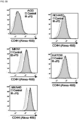
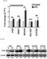
- FIG. 5A Upregulated CD44 expression was found by immunohistochemistry ( FIG. 5A ). mRNA level and protein level of CD44 increased in five cell lines ( FIG. 5B to FIG. 5D ).
- FIG. 6A demonstrated increase of ⁇ cytotoxicity (* P ⁇ 0.05, ** P ⁇ 0.005).
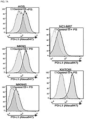
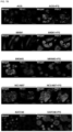
- FIG. 7A Flow cytometry results indicated that PD-L1 expression was elevated for AGS, MKN1, MKN45, NCI-N87 and KATOIII cell lines. These results were confirmed by immunohistochemistry ( FIG. 7B ) and qPCR ( FIG. 7C ) and the fold increases were 1.67, 3.70, 4.23, 1.69, and 1.98 in AGS, MKN1, MKN45, NCI-N87, and KATOIII cells, respectively.
- RNAseq was performed using mouse stomach tissue 4 weeks after exposure of PS ( FIG. 8A ) and 194 (cut off fold change > 2, P ⁇ 0.05) significantly altered genes were identified in the PS exposure group compared to the control group ( FIG. 8B ).
- Gene set enrichment analysis using MSigDB identified the genes in cancer hallmark (H) and oncogenic signatures (C6) set.
- H cancer hallmark
- C6 oncogenic signatures
- the expression level of CD44, E-cadherin, N-cadherin, PD-L1, NPAS2, NR1D1, DNMT1, SLC7A2, PCDH7 and CLDN7 was changed in cancer cell lines and animal models treated with polystyrene microspheres which are the one type of microplastic for 4 weeks, and malignancy was induced due to an increase in proliferation, migration and invasion of cancer cells by the change in the expression level of the gene, and 5-year overall survival rates in gastric cancer patients decreased and thus the genes may be provided as a biomarker composition for predicting the prognosis of cancer malignancies by exposure to microplastics.
Landscapes
- Health & Medical Sciences (AREA)
- Life Sciences & Earth Sciences (AREA)
- Chemical & Material Sciences (AREA)
- Engineering & Computer Science (AREA)
- Immunology (AREA)
- Molecular Biology (AREA)
- Pathology (AREA)
- Analytical Chemistry (AREA)
- Hematology (AREA)
- Urology & Nephrology (AREA)
- Biomedical Technology (AREA)
- Proteomics, Peptides & Aminoacids (AREA)
- Organic Chemistry (AREA)
- Physics & Mathematics (AREA)
- Microbiology (AREA)
- Biotechnology (AREA)
- Biochemistry (AREA)
- General Health & Medical Sciences (AREA)
- Wood Science & Technology (AREA)
- Genetics & Genomics (AREA)
- Zoology (AREA)
- Cell Biology (AREA)
- General Physics & Mathematics (AREA)
- Food Science & Technology (AREA)
- Medicinal Chemistry (AREA)
- Hospice & Palliative Care (AREA)
- Oncology (AREA)
- Biophysics (AREA)
- Bioinformatics & Cheminformatics (AREA)
- General Engineering & Computer Science (AREA)
- Measuring Or Testing Involving Enzymes Or Micro-Organisms (AREA)
Description
- The present invention relates to an in vitro use of a biomarker composition for predicting the prognosis of gastric cancer induced by the exposure to microplastics.
- Gastric cancer is a very common cancer in Asia and is one of the leading causes of cancer deaths worldwide. In the past few years, many attempts have been made to better elucidate the biological processes involved in the development of gastric cancer, in order to improve understanding of the cancer mechanisms, as well as the early diagnosis and treatment of gastric cancer, but the molecular mechanisms involved in carcinogenesis of gastric cancer still remain the subject of intensive research.
- In Korea, gastric cancer is the most frequent among all cancers, and the second most common cause of death. The stage of gastric cancer is determined by the degree of invasion of the surrounding area of the tumor, the metastasis to lymph nodes and metastasis to other organs, and accordingly, treatment and prognosis depend thereon. As the stage progresses, the prognosis gradually worsens, and especially the prognosis in the terminal stage (Stage 4) is so poor that the 5-year survival rate is only 3%.
- Plastics have contributed greatly to the abundant daily life of modern people and industrial development due to their excellent functions and low prices, and recently, discarded plastics has been reported to decompose into microscopic sizes that are difficult to visually identify over time, thereby polluting the environment and threatening human health, but the effect of microplastics on cancer disease has not been studied.
-
Korean Patent Publication No. 10-2013-0109600 (published on October 08, 2013 -
WO 2019/016422 A1 relates to a method for predicting the response of a subject suffering from lung cancer to treatment with FGFR inhibitors. Kahtan Al-Bayaty Mustafa ET AL, "E-Cadherin Protein as a Potential Marker for Gastric Cancer and Its Association with Helicobacter Pylori - Induced Gastritis and Gastric Ulcer", Reports of biochemistry and molecular biology, Iran, (20191001), pages 269 - 277 is a study about E-Cadherin protein as a potential marker for gastric cancer and its association with helicobacter pylori - induced gastritis and gastric ulcer. AZADI SHOHREH ET AL, "Upregulation of PD-L1 expression in breast cancer cells through the formation of 3D multicellular cancer aggregates under different chemical and mechanical conditions", BIOCHIMICA ET BIOPHYSICA ACTA, ELSEVIER SCIENCE PUBLISHERS, AMSTERDAM, NL, vol. 1866, no. 12 is a study about the upregulation of PD-L1 expression in breast cancer cells. WU Z. ET AL, "Claudin-7 (CLDN7) is overexpressed in gastric cancer and promotes gastric cancer cell proliferation, invasion and maintains mesenchymal state", NEOPLASMA, vol. 65, no. 03, pages 349 - 359 is a study about Claudin-7 (CLDN7) and gastric cancer. Chen H. F. ET AL, "Protocadherin 7 inhibits cell migration and invasion through E-cadherin in gastric cancer", Tumor Biology, London, England, pages 1-11 is a study about Protocadherin 7 and gastric cancer. URAOKA NAOHIRO ET AL, "NRD1, which encodes nardilysin protein, promotes esophageal cancer cell invasion through induction of MMP2 and MMP3 expression", CANCER SCIENCE, JP, vol. 105, no. 1, is a study about NRD1 and esophageal cancer.WO 2017/203444 A1 relates to a method of prognosing and predicting breast cancer recurrence in patients. STOCK VALERIE ET AL, "Uptake and effects of orally ingested polystyrene microplastic particles in vitro and in vivo", ARCHIVES OF TOXICOLOGY, SPRINGER, DE, vol. 93, no. 7, pages 1817 - 1833 is a study about orally ingested polystyrene microplastic particles. DENG YONGFENG ET AL, "Tissue accumulation of microplastics in mice and biomarker responses suggest widespread health risks of exposure", SCIENTIFIC REPORTS, vol. 7, no. 1 is a study about Tissue accumulation of microplastics in mice. FORTE MAURIZIO ET AL, "Polystyrene nanoparticles internalization in human gastric adenocarcinoma cells", TOXICOLOGY IN VITRO, vol. 31, pages 126 - 136 is a study about polystyrene nanoparticles internalization in human gastric adenocarcinoma cells. - An object of the present invention is to provide a biomarker composition for predicting the prognosis of cancer malignancy induced by the exposure to microplastics and a method of providing information for predicting the prognosis of cancer malignancy using the same.
- The present invention is defined in the claims. The present invention makes use of a biomarker composition, as defined in
claim 1, for predicting prognosis of gastric cancer induced by exposure to microplastics, comprising the proteins CD44, E-cadherin, N-cadherin, PD-L1 (programmed death-ligand 1), NPAS2 (Neuronal PAS domain protein 2), NR1D1 (nuclear receptor subfamily 1 group D member 1), DNMT1 (DNA (cytosine-5)-methyltransferase 1), SLC7A2 (Solute Carrier Family 7 Member 2), PCDH7 (Protocadherin-7) and CLDN7 (Claudin 7), wherein the CD44, N-cadherin, PD-L1, NR1D1, SLC7A2 and PCDH7 increase in expression of cancer cells exposed to the polystyrene microplastics, and the E-cadherin, NPAS2, DNMT1 and CLDN7 reduce in expression of cancer cells exposed to the polystyrene microplastics. - In addition, the present invention provides a method, as defined in
claim 2, of providing information for predicting prognosis of gastric cancer induced by exposure to microplastics comprising detecting expression or activity level of the proteins CD44, E-cadherin, N-cadherin, PD-L1, NPAS2, NR1D1, DNMT1, SLC7A2, PCDH7 and CLDN7, or expression level of gene encoding the protein in a sample isolated from specimen, wherein the CD44, N-cadherin, PD-L1, NR1D1, SLC7A2 and PCDH7 increase in expression of cancer cells exposed to the polystyrene microplastics, the E-cadherin, NPAS2, DNMT1 and CLDN7 reduce in expression of cancer cells exposed to the polystyrene microplastics, and the cancer gastric cancer is induced due to an increase in proliferation, migration and invasion of cancer cells and an decrease in 5-year overall survival rates in gastric cancer by the change in the expression level of genes encoding the proteins.. -
-
FIG. 1 shows the microplastic accumulation and increased proliferation.FIG. 1A is the representative microscopy images showing accumulation of fluorescent green polyethylene in gastric tissue (magnification: 20×, scale bar, 100 µm);FIG. 1B shows a result of cultured cells exposed to 500 ppm of polystyrene (PS) (10 µm diameter) for 4 weeks;FIG. 1C shows microscopy images of AGS, MKN1, MKN45, NCI-N87, and KATO III cells with/without PS exposure (Magnification: 20×, scale bar, 100 µm.); andFIG. 1D shows PS exposure induced increased proliferation of AGS, MKN45, NCI-M87, and KATOIII compared to the control (mean ± SD. *P < 0.05, **P < 0.005, n.s., not significant. student's t-test). -
FIG. 2 shows the fluorescent green polyethylene (FG-PS) accumulation in gastric tissue. 100 ppm/100 µL of FG-PS was exposed to BALB/c nude mice were exposed for 4 weeks and after sacrifice of mice, the accumulation of FG-PS was imaged by confocal microscopy. FG-PS was deposited at gastric mucosal layers and sub-mucosal layers. FG-MS was indicated by red arrow in bright field image (magnification: 20×, scale bar, 100 µm). -
FIG. 3 shows PS exposure increased N-cadherin and decreased E-cadherin expression.FIG. 3A shows in vitro migration and invasion assays and bar graphs represent the average number of cells on the underside of the membrane, normalized to the control condition. PS promoted invasion and migration of every gastric cancer cell (Magnification: 20×, mean ± SD. *P < 0.05, ** P < 0.005, n.s., not significant. Student's t-test);FIG. 3B shows immunocytochemistry images showing gastric cancer cells stained for E-cadherin and N-cadherin and PS exposure (10 µm diameter, 500 ppm, 4 weeks) decreased E-cadherin levels in AGS and KATOIII cells and increased N-cadherin levels in AGS, NCI-N87, and KATOIII cells (Magnification: 40×, Scale bar: 20 µm);FIG. 3C shows flow cytometry histograms of N-cadherin expression in gastric cancer cell lines with/without PS exposure and PS exposure upregulates N-cadherin expression in gastric cancer cell after exposure to PS;FIG. 3D shows qPCR analysis of N-cadherin mRNA expression. mRNA expression of N-cadherin increased after PS exposure (mean ± SD, *P < 0.05, ** P < 0.005, Student's t-test); andFIG. 3E shows western blot analysis of N-cadherin and E-cadherin expressions with/without PS. -
FIG. 4 shows 5-year overall survival (OS) rate of Cancer Genome Atlas Stomach Adenocarcinoma (TCGA-STAD) datasets. Kaplan-Meier plots showed 5-year OS was correlated with expression of E-cadherin and N-cadherin. The Cancer Genome Atlas Stomach Adenocarcinoma (TCGA-STAD) datasets identified that high N-cadherin expression showed poor 5-years overall survival rate (OS) in stage II (P =0.079), III (P =0.00055), IV (P =0.00069), and low E-cadherin expression showed poor 5-years OS in stage II (P =0.0081) (P-value: log rank t-test). -
FIG. 5 shows a result that PS promotes the upregulation of CD44 expression.FIG. 5A shows CD44 expression in gastric cancer cells by immunofluorescence staining (magnification: 40×, scale bar: 20 µm);FIG. 5B shows flow cytometry analysis of CD44 expression in gastric cancer cell lines and PS exposure for 4 weeks increased CD44 expression;FIG. 5C shows qPCR analysis of CD44 mRNA expression (mean ± SD, *P < 0.05, **P < 0.005, student's t-test n.s., not significant); andFIG. 5D shows western blotting analysis of CD44 expression in cells with/without PS. -
FIG. 6 shows the effect of CD44 in terms of drug resistance and 5-year overall survival rate in the Cancer Genome Atlas Stomach Adenocarcinoma (TCGA-STAD) datasets.FIG. 6A shows results of measuring cytotoxicity using bortezomib or cisplatin to confirm the drug resistance induced by CD44 after PS exposure (10 µm diameter, 500 ppm, 4 weeks) and confirming the increase of Δ cytotoxicity in AGS, MKN1, MKN45, NCI-N87 and KATOIII cell lines; andFIG. 6B shows results of confirming that the TCGA-STAD datasets with high CD44 expression showed a tendency of poor 5-years OS (P=0.064) (P-value: log rank t-test). -
FIG. 7 shows results that PS promotes the upregulation of PD-L1 expression.FIG. 7A shows flow cytometry histograms of PD-L1 expression in gastric cancer cell-lines and PS exposure for 4 weeks dramatically increased PD-L1 expression in all cell lines;FIG. 7B shows immunofluorescence staining of PD-L1 in gastric cancer cells with/without PS (Magnification 40×, Scale bar: 20 µm); andFIG. 7C shows qPCR results of mRNA level of PD-L1 expression (mean ± SD, *P < 0.05, **P < 0.005, student's t-test). -
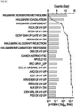
FIG. 8 shows results that PS induced malignant effects on the overall survival of gastric cancer patients.FIG. 8A shows schematic of the experimental procedures and BALB/c nude mice were exposed to 100 ppm/100 µL of PS for 4 weeks, and RNAseq was performed on the extracted stomach tissues;FIG. 8B shows heat map showing the RNA-seq analysis results and PS exposure induced gene expression changes in normal stomach tissues;FIG. 8C shows gene set enrichment analysis using MSigDB and the experimental gene set overlap plenty of Hallmark and Oncogenic Signatures set;FIG. 8D shows schematic diagram showing the comparison of genes identified by RNAseq of PS-exposed mice and the TCGA-STAD RNAseq gastric cancer patient dataset (354 patients) and six candidate genes were found by correlating the overall survival values to the 145 genes examined;FIG. 8E shows the hazard ratio and among the six gene candidates, the fold changes in gene expression in the gastric 10 cancer patients were consistent with the murine RNAseq results, and three candidate genes with malignant effects on overall survival were found to be significant: CLDN7, PCDH7, and SLC7A2, and error bars represent the 95% confidence interval (CI) of the hazard ratio (HR); andFIG. 8F shows Kaplan-Meier plots showing the overall survival of patients expressing the three major candidate genes from the TCGA-STAD patient data and increased SLC7A2 and PCDH7 and decreased CLDN7 expression dramatically correlated with the overall survival of gastric cancer patients (p-value: log rank t-test). -
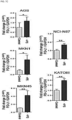
FIG. 9 shows results that PS interfered circadian clock-related gene and methylation.FIG. 9A shows that the qPCR analysis of NPAS2 mRNA expression and NPAS2 decreased following PS exposure (mean ± SD *P < 0.05, *P < 0.005, Student's t-test, n.s., not significant);FIG. 9B shows that in TCGA-STAD datasets, low expression of NPAS2 showed poor 5-years overall survival (OS) rates;FIG. 9C shows the qPCR analysis of NR1D1 mRNA expression and NR1D1 increased following PS exposure (mean ± SD, *P < 0.05, ** P < 0.005, student's t-test, n.s., not significant);FIG. 9D shows that in TCGA-STAD datasets, high expression of NR1D1 showed poor 5-years OS (P-value: log rank t-test); andFIG. 9E shows western blot analysis of DNMT1 expression in cells with/without PS and DNMT1 decreased following PS exposure (P-value: log rank t-test). - Hereinafter, the present invention will be described in more detail.
- As microplastics with diameters less than 5 mm are being recognized as a new environmental treat and human health risk, the present inventors investigated the effects of the microplastic polystyrene (PS) on the hallmarks of cancer changes and 5-year overall survival (OS) to study the risks that may occur when PS, a type of microplastic, are ingested in vivo and as a result, they confirmed that when PS is ingested in vivo, the gastric cancer was exacerbated through the change in the expression level of the gene in the stomach tissue, which is the primary site of exposure and completed the present invention.
- In the present invention, the present invention is the proteins CD44, E-cadherin, N-cadherin, PD-L1, NPAS2, NR1D1, DNMT1, SLC7A2, PCDH7 and CLDN7, and it is possible to provide a biomarker composition for predicting prognosis of gastric cancer induced by exposure to microplastics.
- In more detail, the biomarker composition is CD44 (NCBI No. 960), E-cadherin (NCBI No. 999), N-cadherin (NCBI No. 1000), PD-L1 (programmed death-
ligand 1, NCBI No. 29126), NPAS2 (NeuronalPAS domain protein 2, NCBI No. 4862), NR1D1 (nuclear receptor subfamily 1group D member 1, NCBI No. 9572), DNMT1 (DNA (cytosine-5)-methyltransferase 1, NCBI No. 1786), SLC7A2 (Solute Carrier Family 7Member 2, NCBI No. 6542), PCDH7 (Protocadherin-7, NCBI No. 5099) and CLDN7 (Claudin 7, NCBI No. 1366). - The microplastics is polystyrene.
- The CD44, N-cadherin, PD-L1, NR1D1, SLC7A2 and PCDH7 increase in the expression in cancer cells exposed to the microplastics.
- The E-cadherin, NPAS2, DNMT1 and CLDN7 reduce in the expression in cancer cells exposed to the microplastics.
- The cancer is gastric cancer.
- A diagnostic kit can be provided for predicting prognosis of cancer malignancy induced by exposure to microplastics comprising the proteins CD44, E-cadherin, N-cadherin, PD-L1, NPAS2, NR1D1, DNMT1, SLC7A2, PCDH7 and CLDN7, and an agent for detecting expression or activity level of the protein, or expression level of gene encoding the protein as an active ingredient.
- The agent may be selected from the group consisting of primers, probes, antibodies, peptides and aptamers.
- In addition, the present invention may provide a method of providing information for predicting prognosis of gastric cancer induced by exposure to microplastics comprising detecting expression or activity level of the proteins CD44, E-cadherin, N-cadherin, PD-L1, NPAS2, NR1D1, DNMT1, SLC7A2, PCDH7 and CLDN7, or expression level of gene encoding the protein in a sample isolated from specimen wherein the CD44, N-cadherin, PD-L1, NR1D1, SLC7A2 and PCDH7 increase in expression of cancer cells exposed to the polystyrene microplastics, the E-cadherin, NPAS2, DNMT1 and CLDN7 reduce in expression of cancer cells exposed to the polystyrene microplastics, and the gastric cancer is induced due to an increase in proliferation, migration and invasion of cancer cells and an decrease in 5-year overall survival rates in gastric cancer by the change in the expression level of genes encoding the proteins..
- As used herein, "biomarker" refers to a substance capable of diagnosing a tissue or cell of a subject with gastric cancer by distinguishing it from a tissue or cell of a normal control group and includes organic biomolecules such as proteins, nucleic acids, lipids, glycolipids, glycoproteins, and the like, which show an increase or decrease in tissues or cells of a diseased object compared to the normal control group.
- As used herein, "prognosis" includes determining whether an object for a specific disease or symptom, that is, the subject to be tested will have the autologous disease in the future, or determining the responsiveness of the subject to be tested to the treatment.
- Hereinafter, the present invention will be described in more detail through examples. These examples are only intended to illustrate the present invention in more detail, and it will be apparent to those skilled in the art that the scope of the present invention is not limited by these examples according to the gist of the present invention.
- The following experimental examples are intended to provide experimental examples commonly applied to each of the examples according to the present invention.
- To identify the microplastic accumulation in gastric tissue, fluorescent green polyethylene microspheres (100 ppm/100 µL; 10-20 µm; Cospheric, USA) was used. Fluorescent green polyethylene microspheres were orally administered to 5-week-old BALB/c nude mice daily for 4 weeks.
- After 4 weeks, mice were euthanized using CO2. The stomach was isolated and fixed overnight with 4% PFA at 4°C.
- Frozen slides (8 µm thick) were generated using optimal cutting temperature (OCT) compound, and the slides were attached to slide glass. Cell nuclei were stained with DAPI (ThermoFisher Scientific, USA), and images were obtained using an IN cell analyzer 2200 (GE Healthcare, USA)
- To identify the effect of microplastic to gastric cancer cells, polystyrene microspheres (PS) (500 mg, 9.5-11.5 µm, Cospheric, USA) was used. PS were mixed with 10 mL of normal RPMI culture medium containing 10% FBS and 1% antibiotics.
- Ultrasonic grinding and vortexing were repeatedly performed to create a 5% PS suspension, which was then aliquoted to cell culture media to the desired concentration during subculture.
- The PS was removed by washing three times with PBS. EDTA-trypsin was used during subculture.
- The human gastric cancer cell lines, AGS, MKN1, MKN45, KATO III, were obtained from the Korea Cell-Line Bank (KLCB, Korea). NCI-N87 was obtained from the American Type Culture Collection (ATCC, USA).
- All cell lines maintained in RPMI 1640 medium (Corning, USA) supplemented with 10% fetal bovine serum (FBS) and 1% penicillin/streptomycin at 37°C in 5% CO2.
- Cells (1 × 10⁵ cells per mL) were suspended in 0.2 mL of serum-free RPMI for invasion and motility assays.
- For the invasion assay, the cells were loaded in the upper well of the Transwell chamber (8-um pore size; SPL, Korea) that was pre-coated with Matrigel (Corning, USA) on an upper side of the chamber; the lower well was filled with 0.75 mL of RPMI with 20% serum.
- After incubation for 48 h at 37°C, non-invaded cells on the upper surface of the filter were removed with a cotton swab, and migrated cells on the lower surface of the filter were fixed and stained with a Crystal violet menthol solution (Sigma-Aldrich, USA) and photographed at ×20 magnification.
- Invasiveness was determined by counting cells in four microscopic fields per well, and the extent of invasion was expressed as an average number of cells per microscopic field.
- For migration studies, we used invasion chambers with control inserts that contained Matrigel. Cells (5 × 10⁴ cells per mL) in 0.2 mL of serum-free RPMI were added to the apical side of each insert, and 0.75 mL of RPMI with 20% serum was added to the basal side of each insert. The inserts were processed as described above for the invasion assay.
- To detect N-cadherin, CD44, and PD-L1 expression, cells were exposed to PS for 4 weeks, dislodged with EDTA-trypsin, and then collected in 5-mL FACS tubes (1 × 10⁶ cells/tube).
- Cells were centrifuged, the supernatant was aspirated, and 4% paraformaldehyde was added and allowed to incubate for 30 min at room temperature. After removing the fixation buffer, permeabilization buffer (0.02% Triton-X in distilled water) was added to the cells.
- After removing the buffer, cells were incubated with blocking buffer containing 1% BSA with 10% normal goat serum for 1 h at room temperature. Cells were then incubated with primary antibody (anti-N-cadherin, anti-CD44, or anti-PD-L1 antibody) overnight at 4°C. After washing 3 times by PBS, cells were incubated with Alexa Fluor-647 goat anti-rabbit IgG (H + L) at room temperature in the dark for 60 min.
- After, washing 3 times by PBS, antibody signals were detected by flow cytometry (BD Accuri™ C6 Plus, Becton Dickinson Biosciences, USA).
- Briefly, cells were seeded onto a 2-well chamber slide (Falcon, USA) with RPMI 1640 medium (Corning, USA) supplemented with 10% FBS at a density of 1 × 10⁵ cells/mL and treated with PS at 500 ppm.
- Cells were incubated overnight at 37°C in 5% CO2, the culture medium was removed, and cells were washed 3 times with PBS and fixed with 4% paraformaldehyde for 20 min at room temperature.
- After fixation, the samples were permeabilized by incubation with 0.02% Triton-X in distilled water for 20 min at room temperature. After the buffer was removed, cells were incubated with blocking buffer (1% BSA and 10% normal goat serum) for 1 h at room temperature and cells were incubated with primary antibody overnight at 4°C.
- N-cadherin and E-cadherin were detected with Alexa Fluor-488 goat anti-rabbit IgG (H + L) and Alexa Fluor-647 goat anti-mouse IgG (H + L), respectively. PD-L1 was detected with Alexa Fluor-647 goat anti-rabbit IgG (H + L), and CD44 was detected with Alexa Fluor-488 goat anti-mouse IgG (H + L).
- The slides were treated with DAPI and analyzed by confocal microscopy (LSM-710, Zeiss, Germany).
- Total RNA was extracted from cells using TRIzol (Molecular Research Center, USA) in accordance with the manufacturer's instructions.
- RNA was quantified by spectrophotometry and then diluted in RNasefree dH2O (ThermoFisher Scientific, USA).
- For the PCR reaction, a Superscript III cDNA Synthesis Kit (Invitrogen, USA) was used to generate cDNA from 5 µg of mRNA following the manufacturer's instructions.
- qPCR was performed on an ABI qPCR system (Applied Biosystems, USA) using SYBR Green PCR Master Mix (Applied Biosystems, USA) following the manufacturer's instructions. The reaction conditions were: 10 min at 95°C followed by 50 cycles of 95°C for 15 s, 60°C for 60 s.
- The primer sequences are described in Supplementary Table 1. mRNA values were normalized to GAPDH, relative quantity values of mRNA (RQ value) were calculated with the following equation:

[Table 1] Primers Target Sequence GAPDH Forward GCCTCAAGATCATCAGCAATGCCT N.A Reverse TGTGGTCATGAGTCCTTCCACGAT N.A N-cadherin Forward AGAGGCTTCTGGTGAAATCGC N.A Reverse TGGAAAGCTTCTCACGGCAT N.A CD44 Forward CCAATGCCTTTGATGGACC N.A Reverse TCTGTCTGTGCTGTCGGTGAT N.A PD-L1 Forward TTGGGAAATGGAGGATAAGA N.A Reverse GGATGTGCCAGAGGTAGTTCT N.A NR1D1 Forward CTGGACTCCAACAACAACACAG N.A Reverse GGGGATGGTGGGAAGTAGGT N.A NPAS2 Forward GTATCACGCCTCTCCTTGGG N.A Reverse ATTACAGGAGGGGCTAGGCA N.A ACE2 Forward CATTGGAGCAAGTGTTGGATCTT N.A Reverse GAGCTAATGCATGCCATTCTCA N.A - Cell extracts were obtained using Extraction
Cell Lysis Buffer 1× (Abcam, UK) containing Extraction Enhancer Buffer (Abcam, UK), and protein concentration was determined by BCA assay (ThermoFisher Scientific, USA). - Western blot analysis was performed using the antibodies listed in Table 2.
[Table 2] Material or Reagent Source Cat. No. Antibody N-cadherin Abcam, UK, ab76057(for ICC, Flow cytometer) ab76011(for WB) E-cadherin Abcam, UK, ab1416 CD44 Abcam, UK, ab1416 (for ICC/IF, Flow cytometer) ab157107 (for WB) PD-L1 Abcam, UK, ab205921 DNMT1 Abcam, UK, ab134148 AGTR1 Abcam, UK, ab124734 Beta Actin Abcam, UK, ab6276 Anti-Rabbit/-Mouse-HRP Abcam, UK, ab205718ab205719 Anti-Rabbit/Mouse-Alexa488 Abcam, UK, ab150077ab150113 Anti-Rabbit/Mouse-Alexa647 Abcam, UK, ab150079ab150115 - To investigate PS cytotoxicity, cell proliferation was measured by the alamarBlue® assay (ThermoFisher Scientific, USA) following the manufacturer's instructions.
- Briefly, cells were seeded onto 96-well culture plates with RPMI-1640 medium supplemented with 10% FBS at a density of 5000 cells/well and treated PS microspheres.
- Cell viability was measured at λex = 560 nm and λem = 590 nm using a microplate reader (i3X, Molecular Devices, USA).
- The emission values are stated as percentages of controls, yielding the percent cell proliferation after 72 h of seeding the plate.
- To assess the drug resistance due to CD44, cytotoxicity was measured using bortezomib or cisplatin. Cytotoxicity was determined by the alamarBlue® assay (Thermo Fisher Scientific, Waltham, MA) following the manufacturer's instructions.
- Briefly, Cells were exposed to PS for 4 weeks, collected in tube. After, cells were seeded onto 96-well culture plates with medium supplemented with 10% FBS at a density of 1 × 10⁵ cells/mL concertation with 500 ppm PS.
- After overnights, removed culture media and treated with Bortezomib (Takeda Oncology, USA, 10 nM in PBS), or Cisplatin (United states Pharmacopeia, USA, 1.5 ug/mL in DMSO) with RPMI-1640 medium supplemented with 5% FBS and/or
PS 500 ppm. - Cytotoxicity was measured at an emission of 590 nm using a microplate reader.
- The emission values are stated as percentages of vehicle controls, yielding percentage cell viability after 72 h of treatment.
- Then, Δcytotoxicity was calculated as followings:

- To confirm changes in gene expression in gastric tissues due to PS exposure, mice were orally administered 10-µm PS microspheres (100 ppm/100 µL PBS) for 4 weeks.
- Total RNA was extracted from the stomach tissues.
- The RNA was randomly fragmented for sequencing with short reads, which were then reverse transcribed to cDNA with different adapters attached to the 5' and 3' ends.
- PCR amplification was performed to the extent that sequencing was possible, and size selection was performed to obtain an insert size of 200-400 bp, which was used for NovaSeq 6000 Sequencing System (illumina, USA) with the manufacturer's instructions.
- The expression profile was extracted with FPKM (Fragments Per Kilobase of transcript per Million mapped reads), which is a normalization value that accounts for read count, transcript length, and depth of coverage.
- Overall survival analysis was performed using The Human Protein ATLAS web toolkit (https://www.proteinatlas.org/) using the RNAseq data from 354 patients in The Cancer Genome Atlas-Stomach Adenocarcinoma (TCGA-STAD) dataset.
- The demographic data was listed in Table 3.
- Based on the FPKM value of each gene, patients were classified into two expression groups (high and low), and the correlation between expression level and patient survival was examined.
- To classify the two expression categories, the best expression cutoff value in the Protein ATLAS web-toolkit was used. The prognosis of each group of patients was examined by Kaplan-Meier survival estimators, and the survival outcome of the two groups were compared by log-rank test.
- The overall survival (OS) results included all Stage I-IV and N/A patients. FPKM values above 1, which was recommended by The Human Protein ATLAS, and p < 0.0001, indicated a significant OS.
[Table 3] Variables Counts (n=354) Age(years) ≤60 241 68.1% >60 107 30.2% N/ A 6 1.7% Gender Female 125 35.3% mate 229 64.7% Pathologic stage Stage I 48 13.6% Stage II 110 31.1% Stage III 146 41.2% Stage IV 35 9.9%. N/ A 15 4.2% N/A: not available. - Gene sets were downloaded from the MSigDB database of the Broad Institute (http://software/broadinstitute.org/gsea/msigdb), and hallmark gene signature (H) sets and oncogene signatures(C6) gene sets were selected to perform quantification of RNAseq gene lists (Fold change>2, p<0.05).
- All top-ranked gene sets were manually curated to confirm their accurate functional and pathway categorizations (p < 0.05, top 20).
- The interaction network was analyzed with STRING software.
- Briefly, Cytocape String App (Version 1.4.0) were downloaded from the official website. In this study, we selected significant RNAseq gene lists for network analysis (Fold change>2, p<0.05).
- Microplastics with diameters less than 5 mm are being recognized as a new environmental threat and human health risk. Polystyrene microspheres (PS) are the one type of microplastic. In vivo studies using mice reported that deposition of PS in the liver, kidney, and gut was observed after oral administration of PS particles sized 1-10 µm. Moreover, exposure of mice to PS led to disturbed intestinal microbiome
- However, it has not been revealed whether PS induce changes in cancer hallmarks and the effect of 5-year overall survival (OS). The stomach is the primary site of exposure when PS are ingested in vivo and given the deleterious effects of PS exposure, it was hypothesized that it may exacerbate gastric cancer.
- The important hallmarks of cancer include cancer cell proliferation and metastasis, circadian clock, immunosuppressive environment, and expression of stem cell markers. This study aimed to evaluate the effects of PS on gastric cancer. To confirm accumulation of PS into the digestive tract, BALB/nude mice were fed daily with 100 ppm/100 µL of 10-20-µm sized fluorescent green polyethylene for 4 weeks.
- Fluorescent green polyethylene signals were observed in the gastric tissue (
FIG. 1A andFIG. 2 ). - After confirming PS can directly interact with the stomach, PS was exposed to human gastric cancer cell lines (AGS, MKN1, MKN45, NCI-N87, and KATOIII) for 4 weeks. As a result, it was confirmed that increases of proliferation were observed (*P < 0.05, **P< 0.005) as shown in
FIG. 1D . - Next, to determine whether PS exposure induces changes in invasion, migration, and epithelial mesenchymal transition, the migration and invasion rates were measured
- As a result, it was confirmed that 2.12, 3.24, 1.74, 3.78, and 3.71-fold increases of migration rate were observed in AGS, MKN1, MKN45, NCI-N87, and KATOIII cells, respectively and invasion rates were elevated by 1.33, 2.47, 3.48, and 11.4-fold in AGS, MKN1, MKN45, and KATOIII cells, respectively (*P < 0.05, **P< 0.005) as shown in
FIG. 3A . - In addition,
FIG. 3B shows immunohistochemistry results of N-cadherin and E-cadherin after PS exposure and in parallel to this result, flow cytometry experiments showed elevated levels of N-cadherin was 1.51, 1.59, 1.35, 2.05, 3.07-fold increases in AGS, MKN1, NCI-N87, and KATOIII cells (FIG. 3C ). - Quantitatively, qPCR results showed 3.84, 1,47, 4,40, 1.61, and 1.71-fold increases of N-cadherin gene expression after PS exposure in AGS, MKN1, MKN45, NCI-N87 and KATOIII cells, respectively (
FIG. 3D ). Western blot results showed the decreased E-cadherin and increased N-cadherin following PS exposure except MKN1 cells (FIG. 3E ). - The correlated gene expression was investigated in Cancer Genome Atlas Stomach Adenocarcinoma (TCGA-STAD) datasets using finding of RNAseq in mouse model. In the TCGA-STAD datasets, it is confirmed that high N-cadherin expression showed poor 5-year overall survival rate (OS) in stage II (P =0.079), III (
P 5 =0.00055), IV (P =0.00069), and low E-cadherin expression showed poor 5-year OS in stage II (P =0.0081) as shown inFIG. 4 . - Upregulated CD44 expression was found by immunohistochemistry (
FIG. 5A ). mRNA level and protein level of CD44 increased in five cell lines (FIG. 5B to FIG. 5D ). - To assess the drug resistance due to CD44, cytotoxicity was measured using bortezomib or cisplatin. Then Δ cytotoxicity was calculated as followings; "Δ cytotoxicity = cytotoxicity with PS - cytotoxicity without PS".
FIG. 6A demonstrated increase of Δ cytotoxicity (* P < 0.05, ** P < 0.005). The TCGA-STAD datasets with high CD44 expression showed a tendency of poor 5-year OS (P=0.064) (FIG. 6B ). - As upregulation of programmed death-ligand 1 (PD-L1) is reported to mediate potent immunosuppressive effects in tumor microenvironments, flow cytometry analysis was performed to confirm the change in PD-L1 expression in gastric cancer cells treated with PS for 4 weeks.
- Flow cytometry results indicated that PD-L1 expression was elevated for AGS, MKN1, MKN45, NCI-N87 and KATOIII cell lines (
FIG. 7A ). These results were confirmed by immunohistochemistry (FIG. 7B ) and qPCR (FIG. 7C ) and the fold increases were 1.67, 3.70, 4.23, 1.69, and 1.98 in AGS, MKN1, MKN45, NCI-N87, and KATOIII cells, respectively. - RNAseq was performed using
mouse stomach tissue 4 weeks after exposure of PS (FIG. 8A ) and 194 (cut off fold change > 2, P <0.05) significantly altered genes were identified in the PS exposure group compared to the control group (FIG. 8B ). - Gene set enrichment analysis using MSigDB identified the genes in cancer hallmark (H) and oncogenic signatures (C6) set. To identify the effect of PS exposure to OS in patients, 145 genes that were common between the PS-exposed mouse model and the 354 patients in the TCGA-STAD dataset were identified (
FIG. 8D ). - To examine whether the change in OS correlates with changes in expression of these genes, the significantly expressed prognostic factors were identified based on a FPKM value > 1 and log-rank p value <0.001, and gene overlapped by MSigDB H and C6.
- Finally, 3 genes, SLC7A2, PCDH7, and CLDN7, were identified (
FIG. 8D ). - Next, it was confirmed hazard ratio for 3 common genes; SLC7A2 (HR = 1.4250-3.281, P = 0.00031), PCDH7 (HR = 1.284-2.487, P= 0.00064), and CLDN7 (HR = 0.3497-0.7396, P = 0.00041) (
FIG. 8E ). - An increase of 2.42-fold SLC7A2, 2.08-fold PCDH expression, and decrease of CLDN7 were observed in mouse RNAseq datasets. In the TCGA-STAD datasets, high expression of SLC7A2 (P = 0.00031) or PCDH (P = 0.00064), and low expression of CLDN7(P = 0.00041) showed poor 5-year OS.
- Disruption of the circadian clock was known as cancer hallmarks and in fact, as shown in
FIG. 8B , it was confirmed the decrease of NPAS2 expression (FC: -2.02) and the increase of NR1D1 expression (FC: 2.80) in RNAseq data. - Decrease of mRNA expression of NPAS2 in MKN45, NCI-N87, and KATOIII cells following PS exposure was also found (**P < 0.005, * P < 0.05) (
FIG. 9A ). The mRNA expression of NR1D1 increased for AGS, MKN45, KATOIII cell lines (** P < 0.005) (FIG. 9C ). - It was confirmed that low expression of NPAS2 (P=0.070) and high expression of NR1D1 (P=0.0045) showed poor 5-year OS in TCGA-STAD datasets.
- Next, it was confirmed whether PS exposure induces changes of methylation.
- The western blot results indicated protein level of DNMT1 decreased in AGS, MKN1, N45, and KATOIII cells (
FIG. 9E ) and as shown inFIG. 9F , in TCGA-STAD datasets, low expression of DNMT1 showed poor 5-year OS. - According to the present invention, it was confirmed that the expression level of CD44, E-cadherin, N-cadherin, PD-L1, NPAS2, NR1D1, DNMT1, SLC7A2, PCDH7 and CLDN7 was changed in cancer cell lines and animal models treated with polystyrene microspheres which are the one type of microplastic for 4 weeks, and malignancy was induced due to an increase in proliferation, migration and invasion of cancer cells by the change in the expression level of the gene, and 5-year overall survival rates in gastric cancer patients decreased and thus the genes may be provided as a biomarker composition for predicting the prognosis of cancer malignancies by exposure to microplastics.
Claims (2)
- In vitro use of a biomarker composition for predicting prognosis of gastric cancer induced by exposure to polystyrene microplastics, comprising CD44, E-cadherin, N-cadherin, PD-L1 (programmed death-ligand 1), NPAS2 (Neuronal PAS domain protein 2), NR1D1 (nuclear receptor subfamily 1 group D member 1), DNMT1 (DNA (cytosine-5)-methyltransferase 1), SLC7A2 (Solute Carrier Family 7 Member 2), PCDH7 (Protocadherin-7) and CLDN7 (Claudin 7),
wherein the CD44, N-cadherin, PD-L1, NR1D1, SLC7A2 and PCDH7 increase in expression of cancer cells exposed to the polystyrene microplastics, and the E-cadherin, NPAS2, DNMT1 and CLDN7 reduce in expression of cancer cells exposed to the polystyrene microplastics. - A method of providing information for predicting prognosis of gastric cancer induced by exposure to polystyrene microplastics comprising detecting expression or activity level of CD44, E-cadherin, N-cadherin, PD-L1, NPAS2, NR1D1, DNMT1, SLC7A2, PCDH7 and CLDN7, or expression level of gene encoding the proteins in a sample isolated from specimen,
wherein the CD44, N-cadherin, PD-L1, NR1D1, SLC7A2 and PCDH7 increase in expression of cancer cells exposed to the polystyrene microplastics, the E-cadherin, NPAS2, DNMT1 and CLDN7 reduce in expression of cancer cells exposed to the polystyrene microplastics, and the gastric cancer is induced due to an increase in proliferation, migration and invasion of cancer cells and an decrease in 5-year overall survival rates in gastric cancer by the change in the expression level of genes encoding the proteins.
Applications Claiming Priority (1)
| Application Number | Priority Date | Filing Date | Title |
|---|---|---|---|
| KR1020200038278A KR102315706B1 (en) | 2020-03-30 | 2020-03-30 | Biomarker composition for predicting cancer malignant prognosis induced by microplastic exposure |
Publications (2)
| Publication Number | Publication Date |
|---|---|
| EP3889610A1 EP3889610A1 (en) | 2021-10-06 |
| EP3889610B1 true EP3889610B1 (en) | 2024-02-07 |
Family
ID=75252528
Family Applications (1)
| Application Number | Title | Priority Date | Filing Date |
|---|---|---|---|
| EP21165421.5A Active EP3889610B1 (en) | 2020-03-30 | 2021-03-27 | Biomarker composition for predicting prognosis of gastric cancer induced by exposure to polystyrene microplastics |
Country Status (4)
| Country | Link |
|---|---|
| US (1) | US12228575B2 (en) |
| EP (1) | EP3889610B1 (en) |
| KR (2) | KR102315706B1 (en) |
| CN (1) | CN113466457A (en) |
Families Citing this family (2)
| Publication number | Priority date | Publication date | Assignee | Title |
|---|---|---|---|---|
| KR102725581B1 (en) | 2021-12-02 | 2024-11-05 | 주식회사 바이나리 | Method for monitoring microplastic accumulated in a body using biological tissue clearing |
| CN120254237B (en) * | 2024-10-15 | 2025-11-21 | 武汉纺织大学 | A one-step precipitation and colorimetric detection method for micro/nanoplastics using NC membranes for non-diagnostic purposes |
Family Cites Families (8)
| Publication number | Priority date | Publication date | Assignee | Title |
|---|---|---|---|---|
| JP2006526998A (en) * | 2003-06-12 | 2006-11-30 | コリア リサーチ インスティテュート オブ バイオサイエンス アンド バイオテクノロジー | Gastric cancer and metastatic gastric cancer diagnostic kit |
| EP1961825A1 (en) | 2007-02-26 | 2008-08-27 | INSERM (Institut National de la Santé et de la Recherche Medicale) | Method for predicting the occurrence of metastasis in breast cancer patients |
| KR101370108B1 (en) | 2012-03-28 | 2014-03-20 | 가톨릭대학교 산학협력단 | Use of HDAC2 as a diagnostic marker for gastic cancer |
| WO2017010949A1 (en) * | 2015-07-15 | 2017-01-19 | National University Of Singapore | Biomarkers for the diagnosis and treatment of gastric cancer |
| WO2017203444A1 (en) * | 2016-05-24 | 2017-11-30 | Oncostem Diagnostics Pvt. Ltd. | Method of prognosing and predicting breast cancer recurrence, markers employed therein and kit thereof |
| KR101900428B1 (en) | 2016-12-29 | 2018-09-20 | 순천향대학교 산학협력단 | Biomarker for identification of exposure to air pollutants and method of identifying exposure to air pollutants using the same |
| KR101933695B1 (en) | 2017-06-30 | 2018-12-28 | 동국대학교 산학협력단 | Diagnostic kit comprising biomarkers for bisphenol A exposure diagnosis |
| ES2696798B2 (en) * | 2017-07-13 | 2019-07-08 | Fundacion De Investig Hospital 12 De Octubre | N-CADHERINA AND FGFR1 AND / OR FGFR4 FOR USE IN THE PREDICTION OF PATIENTS 'RESPONSE TO TREATMENT OF LUNG AND METHOD CANCER AND KIT BASED ON SUCH USE |
-
2020
- 2020-03-30 KR KR1020200038278A patent/KR102315706B1/en active Active
- 2020-03-30 KR KR1020200038278D patent/KR20210121505A/en active Granted
-
2021
- 2021-03-27 EP EP21165421.5A patent/EP3889610B1/en active Active
- 2021-03-29 US US17/214,966 patent/US12228575B2/en active Active
- 2021-03-30 CN CN202110340439.9A patent/CN113466457A/en active Pending
Also Published As
| Publication number | Publication date |
|---|---|
| US20210301351A1 (en) | 2021-09-30 |
| US12228575B2 (en) | 2025-02-18 |
| EP3889610A1 (en) | 2021-10-06 |
| CN113466457A (en) | 2021-10-01 |
| KR20210121505A (en) | 2021-10-08 |
| KR102315706B1 (en) | 2021-10-21 |
Similar Documents
| Publication | Publication Date | Title |
|---|---|---|
| Huang et al. | The lncRNA BDNF-AS/WDR5/FBXW7 axis mediates ferroptosis in gastric cancer peritoneal metastasis by regulating VDAC3 ubiquitination | |
| Rotinen et al. | ONECUT2 is a targetable master regulator of lethal prostate cancer that suppresses the androgen axis | |
| Zhou et al. | Effect of microRNA-135a on cell proliferation, migration, invasion, apoptosis and tumor angiogenesis through the IGF-1/PI3K/Akt signaling pathway in non-small cell lung cancer | |
| Liu et al. | Keratin 17 activates AKT signalling and induces epithelial-mesenchymal transition in oesophageal squamous cell carcinoma | |
| Cortes-Sempere et al. | IGFBP-3 methylation-derived deficiency mediates the resistance to cisplatin through the activation of the IGFIR/Akt pathway in non-small cell lung cancer | |
| Huang et al. | BICC1 drives pancreatic cancer progression by inducing VEGF-independent angiogenesis | |
| Chi et al. | DOCK6 promotes chemo-and radioresistance of gastric cancer by modulating WNT/β-catenin signaling and cancer stem cell traits | |
| EP2529223B1 (en) | Biomarkers for circulating tumor cells | |
| Jones et al. | Nuclear localization of Kaiso promotes the poorly differentiated phenotype and EMT in infiltrating ductal carcinomas | |
| US9880171B2 (en) | iASPP phosphorylation and metastatic potential | |
| Aggarwal et al. | The calcium-sensing receptor suppresses epithelial-to-mesenchymal transition and stem cell-like phenotype in the colon | |
| WO2014120265A1 (en) | Method of isolating circulating tumor cells | |
| Lei et al. | Senescent lung fibroblasts in idiopathic pulmonary fibrosis facilitate non-small cell lung cancer progression by secreting exosomal MMP1 | |
| Islam et al. | FAM134B promotes esophageal squamous cell carcinoma in vitro and its correlations with clinicopathologic features | |
| Zhang et al. | Exosomal circEZH2_005, an intestinal injury biomarker, alleviates intestinal ischemia/reperfusion injury by mediating Gprc5a signaling | |
| JP5224309B2 (en) | Proteins specifically expressed in ovarian clear cell adenocarcinoma and their applications | |
| Lan et al. | FAT1 upregulates in oral squamous cell carcinoma and promotes cell proliferation via cell cycle and DNA repair | |
| He et al. | HOXA5 is amplified in glioblastoma stem cells and promotes tumor progression by transcriptionally activating PTPRZ1 | |
| Wu et al. | B-cell lymphoma/leukemia 10 promotes oral cancer progression through STAT1/ATF4/S100P signaling pathway | |
| US12228575B2 (en) | Biomarker composition for predicting cancer malignant prognosis induced by microplastic exposure | |
| US20130011496A1 (en) | Cancer testis antigens as biomarkers in non-small cell lung cancer | |
| Mo et al. | IGFBP7 regulates cell proliferation and migration through JAK/STAT pathway in gastric cancer and is regulated by DNA and RNA methylation | |
| Calvo et al. | High CD44 expression and enhanced E-selectin binding identified as biomarkers of chemoresistant leukemic cells in human T-ALL | |
| US20220017971A1 (en) | Composition for predicting chemotherapy resistance of ovarian cancer and use thereof | |
| JP2025537735A (en) | Spatial transcriptome interpretation-based biomarkers for predicting immunotherapy response and their applications |
Legal Events
| Date | Code | Title | Description |
|---|---|---|---|
| PUAI | Public reference made under article 153(3) epc to a published international application that has entered the european phase |
Free format text: ORIGINAL CODE: 0009012 |
|
| STAA | Information on the status of an ep patent application or granted ep patent |
Free format text: STATUS: REQUEST FOR EXAMINATION WAS MADE |
|
| 17P | Request for examination filed |
Effective date: 20210512 |
|
| AK | Designated contracting states |
Kind code of ref document: A1 Designated state(s): AL AT BE BG CH CY CZ DE DK EE ES FI FR GB GR HR HU IE IS IT LI LT LU LV MC MK MT NL NO PL PT RO RS SE SI SK SM TR |
|
| STAA | Information on the status of an ep patent application or granted ep patent |
Free format text: STATUS: EXAMINATION IS IN PROGRESS |
|
| 17Q | First examination report despatched |
Effective date: 20220822 |
|
| GRAP | Despatch of communication of intention to grant a patent |
Free format text: ORIGINAL CODE: EPIDOSNIGR1 |
|
| STAA | Information on the status of an ep patent application or granted ep patent |
Free format text: STATUS: GRANT OF PATENT IS INTENDED |
|
| INTG | Intention to grant announced |
Effective date: 20231122 |
|
| RIN1 | Information on inventor provided before grant (corrected) |
Inventor name: ZAHEER, JAVERIA Inventor name: KIM, HYEONGI Inventor name: KIM, JIN SU |
|
| GRAS | Grant fee paid |
Free format text: ORIGINAL CODE: EPIDOSNIGR3 |
|
| GRAA | (expected) grant |
Free format text: ORIGINAL CODE: 0009210 |
|
| STAA | Information on the status of an ep patent application or granted ep patent |
Free format text: STATUS: THE PATENT HAS BEEN GRANTED |
|
| AK | Designated contracting states |
Kind code of ref document: B1 Designated state(s): AL AT BE BG CH CY CZ DE DK EE ES FI FR GB GR HR HU IE IS IT LI LT LU LV MC MK MT NL NO PL PT RO RS SE SI SK SM TR |
|
| REG | Reference to a national code |
Ref country code: GB Ref legal event code: FG4D |
|
| REG | Reference to a national code |
Ref country code: CH Ref legal event code: EP |
|
| REG | Reference to a national code |
Ref country code: IE Ref legal event code: FG4D |
|
| REG | Reference to a national code |
Ref country code: DE Ref legal event code: R096 Ref document number: 602021009138 Country of ref document: DE |
|
| REG | Reference to a national code |
Ref country code: LT Ref legal event code: MG9D |
|
| REG | Reference to a national code |
Ref country code: NL Ref legal event code: MP Effective date: 20240207 |
|
| PG25 | Lapsed in a contracting state [announced via postgrant information from national office to epo] |
Ref country code: IS Free format text: LAPSE BECAUSE OF FAILURE TO SUBMIT A TRANSLATION OF THE DESCRIPTION OR TO PAY THE FEE WITHIN THE PRESCRIBED TIME-LIMIT Effective date: 20240607 |
|
| PG25 | Lapsed in a contracting state [announced via postgrant information from national office to epo] |
Ref country code: LT Free format text: LAPSE BECAUSE OF FAILURE TO SUBMIT A TRANSLATION OF THE DESCRIPTION OR TO PAY THE FEE WITHIN THE PRESCRIBED TIME-LIMIT Effective date: 20240207 |
|
| PG25 | Lapsed in a contracting state [announced via postgrant information from national office to epo] |
Ref country code: GR Free format text: LAPSE BECAUSE OF FAILURE TO SUBMIT A TRANSLATION OF THE DESCRIPTION OR TO PAY THE FEE WITHIN THE PRESCRIBED TIME-LIMIT Effective date: 20240508 |
|
| REG | Reference to a national code |
Ref country code: AT Ref legal event code: MK05 Ref document number: 1655688 Country of ref document: AT Kind code of ref document: T Effective date: 20240207 |
|
| PG25 | Lapsed in a contracting state [announced via postgrant information from national office to epo] |
Ref country code: HR Free format text: LAPSE BECAUSE OF FAILURE TO SUBMIT A TRANSLATION OF THE DESCRIPTION OR TO PAY THE FEE WITHIN THE PRESCRIBED TIME-LIMIT Effective date: 20240207 Ref country code: RS Free format text: LAPSE BECAUSE OF FAILURE TO SUBMIT A TRANSLATION OF THE DESCRIPTION OR TO PAY THE FEE WITHIN THE PRESCRIBED TIME-LIMIT Effective date: 20240507 Ref country code: NL Free format text: LAPSE BECAUSE OF FAILURE TO SUBMIT A TRANSLATION OF THE DESCRIPTION OR TO PAY THE FEE WITHIN THE PRESCRIBED TIME-LIMIT Effective date: 20240207 |
|
| PG25 | Lapsed in a contracting state [announced via postgrant information from national office to epo] |
Ref country code: ES Free format text: LAPSE BECAUSE OF FAILURE TO SUBMIT A TRANSLATION OF THE DESCRIPTION OR TO PAY THE FEE WITHIN THE PRESCRIBED TIME-LIMIT Effective date: 20240207 |
|
| PG25 | Lapsed in a contracting state [announced via postgrant information from national office to epo] |
Ref country code: AT Free format text: LAPSE BECAUSE OF FAILURE TO SUBMIT A TRANSLATION OF THE DESCRIPTION OR TO PAY THE FEE WITHIN THE PRESCRIBED TIME-LIMIT Effective date: 20240207 |
|
| PG25 | Lapsed in a contracting state [announced via postgrant information from national office to epo] |
Ref country code: RS Free format text: LAPSE BECAUSE OF FAILURE TO SUBMIT A TRANSLATION OF THE DESCRIPTION OR TO PAY THE FEE WITHIN THE PRESCRIBED TIME-LIMIT Effective date: 20240507 Ref country code: NO Free format text: LAPSE BECAUSE OF FAILURE TO SUBMIT A TRANSLATION OF THE DESCRIPTION OR TO PAY THE FEE WITHIN THE PRESCRIBED TIME-LIMIT Effective date: 20240507 Ref country code: NL Free format text: LAPSE BECAUSE OF FAILURE TO SUBMIT A TRANSLATION OF THE DESCRIPTION OR TO PAY THE FEE WITHIN THE PRESCRIBED TIME-LIMIT Effective date: 20240207 Ref country code: LT Free format text: LAPSE BECAUSE OF FAILURE TO SUBMIT A TRANSLATION OF THE DESCRIPTION OR TO PAY THE FEE WITHIN THE PRESCRIBED TIME-LIMIT Effective date: 20240207 Ref country code: IS Free format text: LAPSE BECAUSE OF FAILURE TO SUBMIT A TRANSLATION OF THE DESCRIPTION OR TO PAY THE FEE WITHIN THE PRESCRIBED TIME-LIMIT Effective date: 20240607 Ref country code: HR Free format text: LAPSE BECAUSE OF FAILURE TO SUBMIT A TRANSLATION OF THE DESCRIPTION OR TO PAY THE FEE WITHIN THE PRESCRIBED TIME-LIMIT Effective date: 20240207 Ref country code: GR Free format text: LAPSE BECAUSE OF FAILURE TO SUBMIT A TRANSLATION OF THE DESCRIPTION OR TO PAY THE FEE WITHIN THE PRESCRIBED TIME-LIMIT Effective date: 20240508 Ref country code: FI Free format text: LAPSE BECAUSE OF FAILURE TO SUBMIT A TRANSLATION OF THE DESCRIPTION OR TO PAY THE FEE WITHIN THE PRESCRIBED TIME-LIMIT Effective date: 20240207 Ref country code: ES Free format text: LAPSE BECAUSE OF FAILURE TO SUBMIT A TRANSLATION OF THE DESCRIPTION OR TO PAY THE FEE WITHIN THE PRESCRIBED TIME-LIMIT Effective date: 20240207 Ref country code: BG Free format text: LAPSE BECAUSE OF FAILURE TO SUBMIT A TRANSLATION OF THE DESCRIPTION OR TO PAY THE FEE WITHIN THE PRESCRIBED TIME-LIMIT Effective date: 20240207 Ref country code: AT Free format text: LAPSE BECAUSE OF FAILURE TO SUBMIT A TRANSLATION OF THE DESCRIPTION OR TO PAY THE FEE WITHIN THE PRESCRIBED TIME-LIMIT Effective date: 20240207 |
|
| PG25 | Lapsed in a contracting state [announced via postgrant information from national office to epo] |
Ref country code: PT Free format text: LAPSE BECAUSE OF FAILURE TO SUBMIT A TRANSLATION OF THE DESCRIPTION OR TO PAY THE FEE WITHIN THE PRESCRIBED TIME-LIMIT Effective date: 20240607 Ref country code: PL Free format text: LAPSE BECAUSE OF FAILURE TO SUBMIT A TRANSLATION OF THE DESCRIPTION OR TO PAY THE FEE WITHIN THE PRESCRIBED TIME-LIMIT Effective date: 20240207 |
|
| PG25 | Lapsed in a contracting state [announced via postgrant information from national office to epo] |
Ref country code: SE Free format text: LAPSE BECAUSE OF FAILURE TO SUBMIT A TRANSLATION OF THE DESCRIPTION OR TO PAY THE FEE WITHIN THE PRESCRIBED TIME-LIMIT Effective date: 20240207 Ref country code: PT Free format text: LAPSE BECAUSE OF FAILURE TO SUBMIT A TRANSLATION OF THE DESCRIPTION OR TO PAY THE FEE WITHIN THE PRESCRIBED TIME-LIMIT Effective date: 20240607 Ref country code: PL Free format text: LAPSE BECAUSE OF FAILURE TO SUBMIT A TRANSLATION OF THE DESCRIPTION OR TO PAY THE FEE WITHIN THE PRESCRIBED TIME-LIMIT Effective date: 20240207 Ref country code: LV Free format text: LAPSE BECAUSE OF FAILURE TO SUBMIT A TRANSLATION OF THE DESCRIPTION OR TO PAY THE FEE WITHIN THE PRESCRIBED TIME-LIMIT Effective date: 20240207 |
|
| PG25 | Lapsed in a contracting state [announced via postgrant information from national office to epo] |
Ref country code: DK Free format text: LAPSE BECAUSE OF FAILURE TO SUBMIT A TRANSLATION OF THE DESCRIPTION OR TO PAY THE FEE WITHIN THE PRESCRIBED TIME-LIMIT Effective date: 20240207 |
|
| PG25 | Lapsed in a contracting state [announced via postgrant information from national office to epo] |
Ref country code: SM Free format text: LAPSE BECAUSE OF FAILURE TO SUBMIT A TRANSLATION OF THE DESCRIPTION OR TO PAY THE FEE WITHIN THE PRESCRIBED TIME-LIMIT Effective date: 20240207 |
|
| PG25 | Lapsed in a contracting state [announced via postgrant information from national office to epo] |
Ref country code: CZ Free format text: LAPSE BECAUSE OF FAILURE TO SUBMIT A TRANSLATION OF THE DESCRIPTION OR TO PAY THE FEE WITHIN THE PRESCRIBED TIME-LIMIT Effective date: 20240207 Ref country code: EE Free format text: LAPSE BECAUSE OF FAILURE TO SUBMIT A TRANSLATION OF THE DESCRIPTION OR TO PAY THE FEE WITHIN THE PRESCRIBED TIME-LIMIT Effective date: 20240207 |
|
| PG25 | Lapsed in a contracting state [announced via postgrant information from national office to epo] |
Ref country code: SK Free format text: LAPSE BECAUSE OF FAILURE TO SUBMIT A TRANSLATION OF THE DESCRIPTION OR TO PAY THE FEE WITHIN THE PRESCRIBED TIME-LIMIT Effective date: 20240207 |
|
| PG25 | Lapsed in a contracting state [announced via postgrant information from national office to epo] |
Ref country code: SM Free format text: LAPSE BECAUSE OF FAILURE TO SUBMIT A TRANSLATION OF THE DESCRIPTION OR TO PAY THE FEE WITHIN THE PRESCRIBED TIME-LIMIT Effective date: 20240207 Ref country code: SK Free format text: LAPSE BECAUSE OF FAILURE TO SUBMIT A TRANSLATION OF THE DESCRIPTION OR TO PAY THE FEE WITHIN THE PRESCRIBED TIME-LIMIT Effective date: 20240207 Ref country code: RO Free format text: LAPSE BECAUSE OF FAILURE TO SUBMIT A TRANSLATION OF THE DESCRIPTION OR TO PAY THE FEE WITHIN THE PRESCRIBED TIME-LIMIT Effective date: 20240207 Ref country code: EE Free format text: LAPSE BECAUSE OF FAILURE TO SUBMIT A TRANSLATION OF THE DESCRIPTION OR TO PAY THE FEE WITHIN THE PRESCRIBED TIME-LIMIT Effective date: 20240207 Ref country code: DK Free format text: LAPSE BECAUSE OF FAILURE TO SUBMIT A TRANSLATION OF THE DESCRIPTION OR TO PAY THE FEE WITHIN THE PRESCRIBED TIME-LIMIT Effective date: 20240207 Ref country code: CZ Free format text: LAPSE BECAUSE OF FAILURE TO SUBMIT A TRANSLATION OF THE DESCRIPTION OR TO PAY THE FEE WITHIN THE PRESCRIBED TIME-LIMIT Effective date: 20240207 |
|
| REG | Reference to a national code |
Ref country code: CH Ref legal event code: PL |
|
| REG | Reference to a national code |
Ref country code: DE Ref legal event code: R097 Ref document number: 602021009138 Country of ref document: DE |
|
| PG25 | Lapsed in a contracting state [announced via postgrant information from national office to epo] |
Ref country code: LU Free format text: LAPSE BECAUSE OF NON-PAYMENT OF DUE FEES Effective date: 20240327 |
|
| PG25 | Lapsed in a contracting state [announced via postgrant information from national office to epo] |
Ref country code: MC Free format text: LAPSE BECAUSE OF FAILURE TO SUBMIT A TRANSLATION OF THE DESCRIPTION OR TO PAY THE FEE WITHIN THE PRESCRIBED TIME-LIMIT Effective date: 20240207 |
|
| PG25 | Lapsed in a contracting state [announced via postgrant information from national office to epo] |
Ref country code: MC Free format text: LAPSE BECAUSE OF FAILURE TO SUBMIT A TRANSLATION OF THE DESCRIPTION OR TO PAY THE FEE WITHIN THE PRESCRIBED TIME-LIMIT Effective date: 20240207 Ref country code: LU Free format text: LAPSE BECAUSE OF NON-PAYMENT OF DUE FEES Effective date: 20240327 |
|
| PG25 | Lapsed in a contracting state [announced via postgrant information from national office to epo] |
Ref country code: IT Free format text: LAPSE BECAUSE OF FAILURE TO SUBMIT A TRANSLATION OF THE DESCRIPTION OR TO PAY THE FEE WITHIN THE PRESCRIBED TIME-LIMIT Effective date: 20240207 |
|
| PLBE | No opposition filed within time limit |
Free format text: ORIGINAL CODE: 0009261 |
|
| STAA | Information on the status of an ep patent application or granted ep patent |
Free format text: STATUS: NO OPPOSITION FILED WITHIN TIME LIMIT |
|
| REG | Reference to a national code |
Ref country code: BE Ref legal event code: MM Effective date: 20240331 |
|
| PG25 | Lapsed in a contracting state [announced via postgrant information from national office to epo] |
Ref country code: IT Free format text: LAPSE BECAUSE OF FAILURE TO SUBMIT A TRANSLATION OF THE DESCRIPTION OR TO PAY THE FEE WITHIN THE PRESCRIBED TIME-LIMIT Effective date: 20240207 |
|
| 26N | No opposition filed |
Effective date: 20241108 |
|
| PG25 | Lapsed in a contracting state [announced via postgrant information from national office to epo] |
Ref country code: BE Free format text: LAPSE BECAUSE OF NON-PAYMENT OF DUE FEES Effective date: 20240331 |
|
| PG25 | Lapsed in a contracting state [announced via postgrant information from national office to epo] |
Ref country code: FR Free format text: LAPSE BECAUSE OF NON-PAYMENT OF DUE FEES Effective date: 20240407 |
|
| PG25 | Lapsed in a contracting state [announced via postgrant information from national office to epo] |
Ref country code: IE Free format text: LAPSE BECAUSE OF NON-PAYMENT OF DUE FEES Effective date: 20240327 |
|
| PG25 | Lapsed in a contracting state [announced via postgrant information from national office to epo] |
Ref country code: IE Free format text: LAPSE BECAUSE OF NON-PAYMENT OF DUE FEES Effective date: 20240327 Ref country code: FR Free format text: LAPSE BECAUSE OF NON-PAYMENT OF DUE FEES Effective date: 20240407 Ref country code: BE Free format text: LAPSE BECAUSE OF NON-PAYMENT OF DUE FEES Effective date: 20240331 Ref country code: CH Free format text: LAPSE BECAUSE OF NON-PAYMENT OF DUE FEES Effective date: 20240331 |
|
| PG25 | Lapsed in a contracting state [announced via postgrant information from national office to epo] |
Ref country code: SI Free format text: LAPSE BECAUSE OF FAILURE TO SUBMIT A TRANSLATION OF THE DESCRIPTION OR TO PAY THE FEE WITHIN THE PRESCRIBED TIME-LIMIT Effective date: 20240207 |
|
| PG25 | Lapsed in a contracting state [announced via postgrant information from national office to epo] |
Ref country code: CY Free format text: LAPSE BECAUSE OF FAILURE TO SUBMIT A TRANSLATION OF THE DESCRIPTION OR TO PAY THE FEE WITHIN THE PRESCRIBED TIME-LIMIT; INVALID AB INITIO Effective date: 20210327 |
|
| PG25 | Lapsed in a contracting state [announced via postgrant information from national office to epo] |
Ref country code: HU Free format text: LAPSE BECAUSE OF FAILURE TO SUBMIT A TRANSLATION OF THE DESCRIPTION OR TO PAY THE FEE WITHIN THE PRESCRIBED TIME-LIMIT; INVALID AB INITIO Effective date: 20210327 |
|
| GBPC | Gb: european patent ceased through non-payment of renewal fee |
Effective date: 20250327 |
|
| REG | Reference to a national code |
Ref country code: DE Ref legal event code: R079 Ref document number: 602021009138 Country of ref document: DE Free format text: PREVIOUS MAIN CLASS: G01N0033574000 Ipc: G01N0033575000 |
|
| PG25 | Lapsed in a contracting state [announced via postgrant information from national office to epo] |
Ref country code: TR Free format text: LAPSE BECAUSE OF FAILURE TO SUBMIT A TRANSLATION OF THE DESCRIPTION OR TO PAY THE FEE WITHIN THE PRESCRIBED TIME-LIMIT Effective date: 20240207 |
|
| PG25 | Lapsed in a contracting state [announced via postgrant information from national office to epo] |
Ref country code: GB Free format text: LAPSE BECAUSE OF NON-PAYMENT OF DUE FEES Effective date: 20250327 |
|
| PGFP | Annual fee paid to national office [announced via postgrant information from national office to epo] |
Ref country code: DE Payment date: 20251222 Year of fee payment: 6 |