EP3889606A1 - Biosensor and manufacturing method therefor - Google Patents
Biosensor and manufacturing method therefor Download PDFInfo
- Publication number
- EP3889606A1 EP3889606A1 EP19890467.4A EP19890467A EP3889606A1 EP 3889606 A1 EP3889606 A1 EP 3889606A1 EP 19890467 A EP19890467 A EP 19890467A EP 3889606 A1 EP3889606 A1 EP 3889606A1
- Authority
- EP
- European Patent Office
- Prior art keywords
- support layer
- biosensor
- antibody
- target substance
- base material
- Prior art date
- Legal status (The legal status is an assumption and is not a legal conclusion. Google has not performed a legal analysis and makes no representation as to the accuracy of the status listed.)
- Pending
Links
Images

Classifications
-
- G—PHYSICS
- G01—MEASURING; TESTING
- G01N—INVESTIGATING OR ANALYSING MATERIALS BY DETERMINING THEIR CHEMICAL OR PHYSICAL PROPERTIES
- G01N33/00—Investigating or analysing materials by specific methods not covered by groups G01N1/00 - G01N31/00
- G01N33/48—Biological material, e.g. blood, urine; Haemocytometers
- G01N33/50—Chemical analysis of biological material, e.g. blood, urine; Testing involving biospecific ligand binding methods; Immunological testing
- G01N33/53—Immunoassay; Biospecific binding assay; Materials therefor
- G01N33/543—Immunoassay; Biospecific binding assay; Materials therefor with an insoluble carrier for immobilising immunochemicals
- G01N33/54393—Improving reaction conditions or stability, e.g. by coating or irradiation of surface, by reduction of non-specific binding, by promotion of specific binding
-
- G—PHYSICS
- G01—MEASURING; TESTING
- G01N—INVESTIGATING OR ANALYSING MATERIALS BY DETERMINING THEIR CHEMICAL OR PHYSICAL PROPERTIES
- G01N33/00—Investigating or analysing materials by specific methods not covered by groups G01N1/00 - G01N31/00
- G01N33/48—Biological material, e.g. blood, urine; Haemocytometers
- G01N33/50—Chemical analysis of biological material, e.g. blood, urine; Testing involving biospecific ligand binding methods; Immunological testing
- G01N33/52—Use of compounds or compositions for colorimetric, spectrophotometric or fluorometric investigation, e.g. use of reagent paper and including single- and multilayer analytical elements
-
- G—PHYSICS
- G01—MEASURING; TESTING
- G01N—INVESTIGATING OR ANALYSING MATERIALS BY DETERMINING THEIR CHEMICAL OR PHYSICAL PROPERTIES
- G01N33/00—Investigating or analysing materials by specific methods not covered by groups G01N1/00 - G01N31/00
- G01N33/48—Biological material, e.g. blood, urine; Haemocytometers
- G01N33/50—Chemical analysis of biological material, e.g. blood, urine; Testing involving biospecific ligand binding methods; Immunological testing
- G01N33/53—Immunoassay; Biospecific binding assay; Materials therefor
- G01N33/543—Immunoassay; Biospecific binding assay; Materials therefor with an insoluble carrier for immobilising immunochemicals
- G01N33/54366—Apparatus specially adapted for solid-phase testing
-
- G—PHYSICS
- G01—MEASURING; TESTING
- G01N—INVESTIGATING OR ANALYSING MATERIALS BY DETERMINING THEIR CHEMICAL OR PHYSICAL PROPERTIES
- G01N33/00—Investigating or analysing materials by specific methods not covered by groups G01N1/00 - G01N31/00
- G01N33/48—Biological material, e.g. blood, urine; Haemocytometers
- G01N33/50—Chemical analysis of biological material, e.g. blood, urine; Testing involving biospecific ligand binding methods; Immunological testing
- G01N33/53—Immunoassay; Biospecific binding assay; Materials therefor
- G01N33/543—Immunoassay; Biospecific binding assay; Materials therefor with an insoluble carrier for immobilising immunochemicals
- G01N33/551—Immunoassay; Biospecific binding assay; Materials therefor with an insoluble carrier for immobilising immunochemicals the carrier being inorganic
- G01N33/552—Glass or silica
-
- G—PHYSICS
- G01—MEASURING; TESTING
- G01N—INVESTIGATING OR ANALYSING MATERIALS BY DETERMINING THEIR CHEMICAL OR PHYSICAL PROPERTIES
- G01N2610/00—Assays involving self-assembled monolayers [SAMs]
Definitions
- the present application relates to a biosensor and a method for preparing the same.
- Immunoanalytical methods are analytical methods based on antigen-antibody binding reactions.
- a representative form of the immunoanaytical methods is an enzyme-linked immunosorbent assay (ELISA), which is the most commonly used immunodiagnostic method.
- ELISA enzyme-linked immunosorbent assay
- the enzyme-linked immunosorbent assay generally detects a target substance by immobilizing an antibody in a surface-treated substrate.
- a liquid phase process is used, in which the substrate is immersed in a solution containing a substance to be treated on the surface.
- the liquid phase process has an advantage of being easy to perform, whereas it has disadvantages that the surface treatment of the substrate is somewhat uneven and the treated substance has a low density.
- the density of the antibody immobilized in the substrate is lowered and uneven, thereby lowering detection sensitivity of the target substance of the biosensor.
- room temperature is a natural temperature without being heated or cooled, which may be, for example, any temperature in a range of 10°C to 30°C, or about 23°C or about 25°C or so.
- the unit of temperature is °C.
- normal pressure is a natural pressure without being pressurized or depressurized, where usually about 1 atm or so is referred to as the normal pressure.
- the present application relates to a biosensor for detecting a target substance.
- the biosensor of the present application comprises a base material; a support layer formed on one side of the base material; and a first antibody immobilized in the support layer, wherein the support layer formed on one side of the base material is a self-assembled monolayer formed by a vapor deposition method.
- the base material is not particularly limited, which may be, for example, a polymer substrate, a metal substrate, a metal oxide substrate or a glass substrate.
- the polymer substrate for example, a polycarbonate, polymethyl methacrylate (PMMA), or polypropylene substrate may be used, without being limited thereto.
- the metal substrate for example, an aluminum, iron or gold substrate may be used, without being limited thereto.
- the metal oxide substrate an Al 2 O 3 , ZnO, Fe 3 O 4 , ZrO 2 , TiO 2 , SnO or ITO (indium tin oxide) substrate may be used, without being limited thereto.
- the glass substrate a quartz glass, soda-lime glass, borosilicate glass, crystal glass, aluminosilicate glass or germanium-oxide glass substrate may be used, without being limited thereto.
- the support layer may be formed on one side of the base material.
- the method of forming the support layer on one side of the base material may be by a vapor deposition method, and specifically, may be formed by a molecular layer deposition (MLD) method to be described below.
- MLD molecular layer deposition
- the first antibody immobilized in the support layer is not particularly limited, where an appropriate antibody may be selected in consideration of the target substance to be detected.
- an antibody having a binding site capable of binding to a target substance may be selected as the first antibody. Therefore, the first antibody may vary depending on the target substance to be detected. On the other hand, the type of the first antibody is not particularly limited, which may be, for example, IgE, IgD or IgG.
- target substance may mean an antigen as a substance capable of binding to a binding site of an antibody.
- the support layer formed on one side of the base material may be a self-assembled monolayer formed by a vapor deposition method.
- the self-assembled monolayer means a molecular assembly spontaneously formed on a surface by adsorption without the help of specific enzymes or factors. Therefore, in the present application, the self-assembled monolayer may mean a molecular assembly spontaneously formed on one side of a base material by adsorption.
- SAM self-assembled monolayer
- the biosensor of the present application may have absorbance satisfying the following equation 1. Ab ⁇ 0.15
- Ab is the absorbance at the maximum absorption wavelength measured using a UV-Vis spectrophotometer after contacting 100 ⁇ L of a solution containing 20ng/mL to 40ng/mL of a target substance with the support layer in which the first antibody is immobilized, and then contacting a second antibody capable of binding to the target substance and bound by an enzyme reacting with TMB (3,3',5,5'-tetramethylbenzidine), and TMB (3,3',5,5'-tetramethylbenzidine) therewith.
- an area of the support layer, in which the first antibody is immobilized may be about 8 cm wide and about 2 cm long.
- the 20ng/mL to 40ng/mL may mean, for example, about 20ng/mL 22ng/mL, 24ng/mL, 26ng/mL, 28ng/mL, 30ng/mL, 32ng/mL, 34ng/mL, 36ng/mL, 38ng/mL or about 40ng/mL.
- PBS phosphate-buffered saline
- Equation 1 after reaction from the time when a target substance has been contacted to any one time when about 50 minutes to about 70 minutes have elapsed, for example, from the time when a test substance has been contacted to the time when about 55 minutes, about 60 minutes or about 65 minutes have elapsed, the second antibody capable of binding to the target substance and bound by an enzyme reacting with TMB (3,3',5,5'-tetramethylbenzidine) may be contacted.
- a washing process may be performed before contacting the second antibody.
- the washing process is not particularly limited, where it may be washed using a known washing solution.
- PBS phosphate-buffered saline
- Tween may be used as the washing solution.
- the second antibody capable of binding to a target substance and bound by an enzyme reacting with TMB is not particularly limited, where an appropriate antibody may be selected in consideration of the target substance to be detected.
- an antibody having a binding site capable of binding to the target substance bound to the first antibody may be selected as the second antibody.
- the binding site of the second antibody may be bound by reacting with the target substance bound to the first antibody.
- the second antibody may be contacted in an amount to be capable of sufficiently reacting with the target substance bound to the first antibody. For example, about 100 ⁇ L of a solution containing 1 ⁇ g/mL of the second antibody may be contacted.
- the type of the second antibody is not particularly limited, which may be, for example, IgE, IgD or IgG.
- the enzyme bound to the second antibody a known enzyme capable of reacting with TMB (3,3',5,5'-tetramethylbenzidine) may be used.
- the enzyme may be horseradish peroxidase (HRP).
- TMB (3,3',5,5'-tetramethylbenzidine) may be contacted.
- the TMB (3,3',5,5'-tetramethylbenzidine) may be contacted in an amount of about 100 ⁇ L (0.1 mg/mL).
- Equation 1 in the measurement of absorbance, it may be measured using a UV-Vis spectrophotometer after reaction from the time when TMB (3,3',5,5'-tetramethylbenzidine) has been contacted to any one time when 10 minutes to 20 minutes have elapsed, for example, from the time when TMB (3,3',5,5'-tetramethylbenzidine) has been contacted to the time when about 12 minutes, 13 minutes, 14 minutes, 15 minutes, 16 minutes, 17 minutes or 18 minutes have elapsed.
- the absorbance (Ab) may be about 0.16 or more, 0.17 or more, or about 0.18 or more, and the upper limit is not particularly limited, but may be about 0.3 or less, 0.29 or less, 0.28 or less, or about 0.27 or less.
- the absorbance in such a range means that the amount of the enzyme immobilized in the second antibody reacting with TMB is high, which means that the amount of the target substance bound to the first antibody is high. In addition, this may mean that the first antibody immobilized in the support layer has a high density. Therefore, the biosensor having absorbance in the above range has high detection sensitivity of the target substance.
- the absorbance of the biosensor may be dependent on the target substance concentration in a range of 0.6ng/mL to 1.0ng/mL. Specifically, when the concentration of the target substance increases in the range of 0.6ng/mL to 1.0ng/mL, the absorbance also increases.
- the biosensor of the present application can also detect quantitatively a target substance in such a very low concentration range.
- the forming material of the support layer forming the support layer formed on one side of the base material may comprise a compound represented by the following formula 1.
- A is a carbon atom, a silicon atom or a germanium atom
- R 1 and R 2 are each independently an alkyl group having 1 to 5 carbon atoms
- R 3 is an alkyl group having 1 to 12 carbon atoms or a phenyl group
- p is an integer of 0 to 2
- r and q are each independently a natural number of 1 to 3 and the sum of p, q and r satisfies 4.
- R 1 and R 2 may be each independently an alkyl group having 1 to 4 carbon atoms or an alkyl group having 1 to 3 carbon atoms.
- R 3 may be an alkyl group having 1 to 10 carbon atoms, 1 to 8 carbon atoms, 1 to 6 carbon atoms or 1 to 4 carbon atoms, or a phenyl group.
- p may be an integer of 0, 1 or 2.
- r and q may be each independently a natural number of 1, 2 or 3.
- the kind of the compound represented by Formula 1 above is not particularly limited, but it may be, for example, (4-aminobutyl)triethoxysilane, (4-aminobutyl)trimethoxysilane, aminophenyltriethoxysilane, aminophenyltrimethoxysilane, (3-aminopropyl)trimethoxysilane, (3-aminopropyl)triethoxysilane or (3-aminopropyl) dimethylethoxysilane.
- the support layer is formed using the compound represented by Formula 1 above, it is advantageous to form a self-assembled monolayer on one side of the support layer.
- the density of the support layer formed on one side of the base material may be 800pieces/ ⁇ m 2 to 2,000pieces/ ⁇ m 2 . As another example, it may be about 850pieces/ ⁇ m 2 or more, 900pieces/ ⁇ m 2 or more, 950pieces/ ⁇ m 2 or more, 1,000pieces/ ⁇ m 2 or more, or about 1,100pieces/ ⁇ m 2 or more.
- the upper limit is not particularly limited, but may be about 1,900pieces/ ⁇ m 2 or less, or 1,800pieces/ ⁇ m 2 or less, 1,700pieces/ ⁇ m 2 or less, or about 1,900pieces/ ⁇ m 2 or less.
- the support layer having a density in the above range can immobilize the first antibody in the support layer at a high density. Therefore, the biosensor comprising the support layer having such a density range can immobilize the first antibody in the support layer at a high density, thereby improving the measurement sensitivity of the biosensor.
- the method of measuring the density of the support layer formed on one side of the base material is not particularly limited, and for example, the adsorption density of the support layer can be measured with SEM by binding biotin to the support layer with EDC coupling, and using selectively adsorbing properties of streptavidin, to which 20nm gold nanoparticles are bonded, and the biotin.
- the present application also relates to a method for preparing a biosensor. Furthermore, as a base material, a forming material of a support layer for forming the support layer and a first antibody immobilized in the support layer, which are used for the method for preparing a biosensor of the present application, the base material, the forming material of the support layer and the first antibody as described above may be used.
- the biosensor preparation method of the present application comprises a first step of forming a support layer, which is a self-assembled monolayer, on one side of a base material by a vapor deposition method; and a second step of immobilizing a first antibody in the support layer.
- a method of forming a support layer, which is a self-assembled monolayer, on one side of a base material may be performed by a vapor deposition method.
- the support layer may be formed on one side of a base material by molecular layer deposition (MLD).
- the molecular layer deposition may be performed using molecular layer deposition equipment (1) as in Figure 1 .
- the molecular layer deposition equipment (1) may comprise a reaction chamber (10), a reaction tank (30), a gas connecting pipe (40) for connecting the reaction chamber and the reaction tank, a vacuum pump (50) for adjusting the pressure of the reaction chamber, a first heat source (20) for adjusting the temperature of the reaction tank, a second heat source (60) for adjusting the temperature of the reaction chamber, a first valve (70), provided in the gas connecting pipe (40), for adjusting the flow rate of the vaporized forming material of the support layer flowing into the reaction chamber (10), a second valve (80), provided in the vacuum pump (50), for adjusting the pressure of the reaction chamber (10), and a third valve (90) for adjusting the pressure of the reaction chamber (10) to normal pressure.
- a base material may be safely placed in the reaction chamber of the molecular layer deposition equipment (1), and a forming material of a support layer may be charged in the reaction tank.
- the temperature of the reaction tank (30) may be adjusted to a temperature higher than the boiling point of the forming material of the support layer by the first heat source (20) provided in the reaction tank (30), and for example, the temperature of the reaction tank (30) may be adjusted to a temperature range of 50°C to 500°C.
- the first heat source (20) is not particularly limited, but may be an electric heater as one example.
- the first heat source (20) may vaporize the forming material of the support layer by adjusting the temperature of the reaction tank (30) to a temperature higher than the boiling point of the forming material of the support layer charged in the reaction tank (30).
- the vaporized forming material of the support layer may be introduced into the reaction chamber (10) through the gas connecting pipe (40).
- the first valve (70) may adjust the opening and closing time of the valve to adjust the flow rate of the vaporized forming material of the support layer.
- the flow rate of the vaporized forming material of the support layer may be adjusted by opening the first valve for about 40 seconds to about 80 seconds so that the vaporized forming material of the support layer in the reaction tank is introduced into the reaction chamber through the gas connecting pipe. If the opening time of the first valve is short, the support layer with a sufficient density may not be deposited on the base material, and if the opening time is long, the forming material of the support layer may be wasted because the vaporized forming material of the support layer flowing into the reaction chamber increases.
- the temperature of the reaction chamber may be adjusted to a range of about 40°C to about 200°C by the second heat source (60) provided in the reaction chamber.
- the second heat source (60) may be an electric heater of the same type as the above-described electric heater.
- the second heat source (60) serves to provide the reaction chamber (10) with a high temperature while the vaporized forming material of the support layer is deposited in the reaction chamber (10), where the high temperature may be appropriately selected within the above temperature range in consideration of the type of the forming material of the support layer and the desired adsorption density of the support layer.
- the deposition of the forming material of the support layer may be performed for about 10 minutes to about 60 minutes. When the deposition is performed in the above range, the support layer may be efficiently deposited as a self-assembled monolayer without wasting time while having a high density on the base material.
- the pressure of the reaction chamber (10) may be adjusted by the vacuum pump (50) and the second valve (80) provided in the vacuum pump.
- a negative pressure may be applied to the inside of the reaction chamber (10).
- the second valve (80) may be opened so that the inside of the reaction chamber (10) becomes a vacuum atmosphere before the forming material of the support layer is deposited on the base material (11).
- the second valve (80) may be closed while the first valve is opened and the vaporized forming material of the support layer flows into the reaction chamber (10).
- the pressure of the reaction chamber (10) may also be adjusted by the third valve (90).
- the third valve (90) may be closed in order to maintain the inside of the reaction chamber (10) in a vacuum atmosphere.
- the third valve (90) when the third valve (90) is opened, the pressure of the reaction chamber (10) may be adjusted to normal pressure.
- the base material (11), on which the support layer is formed is taken out of the reaction chamber (10)
- the third valve (90) may be opened to adjust the pressure of the reaction chamber (10) to normal pressure.
- the forming material of the support layer remaining without being deposited on the base material (11) may be effectively removed from the reaction chamber (10).
- the support layer When the support layer is deposited on one side of the base material by the molecular layer deposition as above, the support layer, which is a self-assembled monolayer having a high density, may be deposited. Therefore, the biosensor comprising the support layer may improve the detection sensitivity of the target substance.
- the first antibody in the support layer in the second step may be immobilized in the support layer via a linking medium.
- the first antibody may be immobilized in the support layer by binding a linking medium to the support layer, and then binding the first antibody to the linking medium.
- the linking medium is not particularly limited, but may be, as one example, glutaraldehyde 1,4-butanediol diglycidyl ether, N,N'-disuccinimidyl carbonate or p-phenylene diisocyanate.
- the method of binding the linking medium to the support layer is not particularly limited, where it may be bound by a known method.
- the linking medium may be bound to the support layer by reacting with a solution containing p-phenylene diisocyanate.
- the method of binding the first antibody to the linking medium is not particularly limited, where it may be attached by a known method.
- the first antibody may be attached to the linking medium by contacting the solution containing the first antibody with the base material, on which the linking medium is formed, so that the linking medium and the first antibody react.
- the present application also relates to a method for detecting a target substance.
- the method for detecting a target substance of the present application comprises a first step of contacting a test subject substance comprising a target substance, a second antibody in which an enzyme reacting with a substrate is immobilized and a substrate with a biosensor, and a second step of measuring changes in the substrate in contact with the biosensor.
- the biosensor the above-described biosensor may be used as the biosensor. Therefore, the detection sensitivity of the target substance may be improved. That is, a trace amount of the target substance may also be detected with high sensitivity.
- the test subject substance comprising a target substance in the first step is not particularly limited, which may be, for example, derived from a human body.
- the test subject substance may be sweat, blood, serum, plasma, cerebrospinal fluid, saliva, tears, nasal discharge or urine, and the like.
- Such a test subject substance may be appropriately selected in consideration of the concentration of the target substance included in the test subject substance.
- the target substance may mean an antigen that is a material capable of being bound to the binding site of the antibody.
- the second antibody in which an enzyme reacting with a substrate is immobilized, and the substrate in the first step may be the same as the second antibody and the substrate as described above.
- the test subject substance comprising a target substance, the second antibody, in which an enzyme reacting with a substrate is immobilized, and the substrate may be sequentially contacted with the biosensor.
- the test subject substance comprising a target substance may be contacted with a biosensor, and then the second antibody, in which an enzyme reacting with a substrate is immobilized, may be contacted with the biosensor, and finally, the substrate may be contacted with the biosensor.
- a washing process of washing the biosensor may be included.
- a washing process for washing the biosensor may be included.
- the method of measuring changes in the substrate in the second step is not particularly limited, but for example, the change of the substrate may be measured by an optical method.
- the change of the substrate by the enzyme immobilized in the second antibody may be measured by measuring the absorbance of the chromogen.
- the measurement of the absorbance is not particularly limited, but it may be measured using a known spectrophotometer.
- data may be constructed by measuring absorbance (hereinafter, may be referred to as 'standard absorbance') according to the content of the target substance.
- 'standard absorbance' absorbance
- a biosensor having improved detection sensitivity of a target substance is provided. Also, a method for preparing a biosensor with improved detection sensitivity of a target substance is provided. In addition, a method for detecting a target substance using the biosensor is provided.
- biosensors prepared through examples and comparative examples were washed four times with 1% PBS and 0.05% Tween 20, blocked with 1% BSA (bovine serum albumin) for 1 hour, and then washed four times.
- BSA bovine serum albumin
- IgE IgE
- NIBSC IgE
- HRP anti-human IgE antibody
- TMB (3,3',5,5'-tetramethylbenzidine) (0.1 mg/mL) as a substrate was added thereto, reacted for 15 minutes, and then about 100 ⁇ L of 2M H 2 SO 4 was added thereto.
- Base material A glass substrate (8cm ⁇ 2cm) (Sewon Tech, Soda-lime glass) was used as the base material.
- Forming material of support layer (3-aminopropyl)trimethoxysilane (APTMS) (Sigma-Aldrich, 281778) was used.
- Linking medium p-phenylene diisocyanate (Sigma-Aldrich, 258555) was used.
- Anti-human IgE antibody (ThermoFisher Scientific, A18797) was used.
- the glass substrate was safely placed in the reaction chamber of the vapor deposition equipment configured as shown in Figure 1 , the second valve was opened to form the reaction chamber in a vacuum atmosphere, and the temperature of the reaction chamber was maintained at about 100°C using the second heat source. Furthermore, (3-aminopropyl)trimethoxysilane (APTMS), which was a forming material of the support layer, was charged in the reaction tank, and then APTMS was vaporized by heating the temperature of the reaction tank to 100°C using the first heat source.
- APTMS (3-aminopropyl)trimethoxysilane
- the first valve was opened for about 60 seconds so that the vaporized APTMS flowed into the reaction chamber, and again, the first valve was closed and then held for 10 minutes to deposit APTMS on the glass substrate, thereby forming a support layer as a self-assembled monolayer.
- the second valve was opened and the temperature of the reaction chamber was lowered to room temperature. Thereafter, the second valve was closed and the third valve was opened to convert the pressure of the reaction chamber to normal pressure, and then the base material, on which the support layer was formed, was collected from the reaction chamber.
- FIG. 2 is an exemplary conceptual diagram showing a series of processes for manufacturing a biosensor of an example. The absorbance was measured using the biosensor of Example prepared by the same method as above.
- the support layer was formed on the glass substrate. Specifically, the glass substrate was immersed in ethanol containing 1% (3-aminopropyl)triethoxysilane (APTMS), which was a forming material of a support layer, for about 2 hours to form a support layer on the base material. Thereafter, the base material, on which the support layer was formed, was collected by washing it with ethanol for about 10 seconds.
- ATMS (3-aminopropyl)triethoxysilane
- the first antibody was immobilized in the support layer in the same manner as in Example.
- the absorbance was measured using the biosensor of Comparative Example prepared in the same method as above.
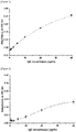
- Figure 3 is a graph showing absorbance at the maximum absorption wavelength (wavelength of about 450nm) measured with a UV-Vis spectrophotometer (path length: 1 cm) according to concentrations of a target substance using the biosensor prepared through Example
- Figure 4 is a graph showing absorbance at the maximum absorption wavelength (wavelength of about 450nm) measured with a UV-Vis spectrophotometer (path length: 1 cm) according to concentrations of a target substance using the biosensor prepared through Comparative Example.
- the absorbance was found to be high as about 0.175 at 20ng/mL of the target substance.
- the absorbance was found to be low as about 0.08 at 20ng/mL of the target substance.
Landscapes
- Health & Medical Sciences (AREA)
- Life Sciences & Earth Sciences (AREA)
- Immunology (AREA)
- Engineering & Computer Science (AREA)
- Chemical & Material Sciences (AREA)
- Hematology (AREA)
- Urology & Nephrology (AREA)
- Biomedical Technology (AREA)
- Molecular Biology (AREA)
- Physics & Mathematics (AREA)
- Analytical Chemistry (AREA)
- Cell Biology (AREA)
- Pathology (AREA)
- Food Science & Technology (AREA)
- Medicinal Chemistry (AREA)
- Biotechnology (AREA)
- Microbiology (AREA)
- Biochemistry (AREA)
- General Health & Medical Sciences (AREA)
- General Physics & Mathematics (AREA)
- Chemical Kinetics & Catalysis (AREA)
- Inorganic Chemistry (AREA)
- Apparatus Associated With Microorganisms And Enzymes (AREA)
- Investigating Or Analysing Materials By The Use Of Chemical Reactions (AREA)
- Investigating Or Analysing Materials By Optical Means (AREA)
Abstract
Description
- This application claims the benefit of priority based on
Korean Patent Application No. 10-2018-0147292 filed on November 26, 2018 - The present application relates to a biosensor and a method for preparing the same.
- Immunoanalytical methods are analytical methods based on antigen-antibody binding reactions. A representative form of the immunoanaytical methods is an enzyme-linked immunosorbent assay (ELISA), which is the most commonly used immunodiagnostic method.
- The enzyme-linked immunosorbent assay generally detects a target substance by immobilizing an antibody in a surface-treated substrate. On the other hand, as the surface treatment method of the substrate, a liquid phase process is used, in which the substrate is immersed in a solution containing a substance to be treated on the surface. However, the liquid phase process has an advantage of being easy to perform, whereas it has disadvantages that the surface treatment of the substrate is somewhat uneven and the treated substance has a low density. As a result, there has been a problem that the density of the antibody immobilized in the substrate is lowered and uneven, thereby lowering detection sensitivity of the target substance of the biosensor.
- Accordingly, there is a need for a biosensor having improved detection sensitivity of a target substance.
- It is an object of the present application to provide a biosensor with improved detection sensitivity of a target substance. It is another object of the present application to provide a method for preparing a biosensor with improved detection sensitivity.
- Among physical properties mentioned in this specification, when the measured temperature affects the results, the relevant physical properties are physical properties measured at room temperature, unless otherwise specified. The term room temperature is a natural temperature without being heated or cooled, which may be, for example, any temperature in a range of 10°C to 30°C, or about 23°C or about 25°C or so. In addition, unless otherwise specified herein, the unit of temperature is °C.
- Among physical properties mentioned in this specification, when the measured pressure affects the results, the relevant physical properties are physical properties measured at room pressure, unless otherwise specified. The term normal pressure is a natural pressure without being pressurized or depressurized, where usually about 1 atm or so is referred to as the normal pressure.
- In one example of the present application, the present application relates to a biosensor for detecting a target substance.
- The biosensor of the present application comprises a base material; a support layer formed on one side of the base material; and a first antibody immobilized in the support layer, wherein the support layer formed on one side of the base material is a self-assembled monolayer formed by a vapor deposition method.
- As one example, the base material is not particularly limited, which may be, for example, a polymer substrate, a metal substrate, a metal oxide substrate or a glass substrate.
- As the polymer substrate, for example, a polycarbonate, polymethyl methacrylate (PMMA), or polypropylene substrate may be used, without being limited thereto. As the metal substrate, for example, an aluminum, iron or gold substrate may be used, without being limited thereto. As the metal oxide substrate, an Al2O3, ZnO, Fe3O4, ZrO2, TiO2, SnO or ITO (indium tin oxide) substrate may be used, without being limited thereto. As the glass substrate, a quartz glass, soda-lime glass, borosilicate glass, crystal glass, aluminosilicate glass or germanium-oxide glass substrate may be used, without being limited thereto.
- As one example, the support layer may be formed on one side of the base material.
- The method of forming the support layer on one side of the base material may be by a vapor deposition method, and specifically, may be formed by a molecular layer deposition (MLD) method to be described below.
- As one example, the first antibody immobilized in the support layer is not particularly limited, where an appropriate antibody may be selected in consideration of the target substance to be detected.
- Specifically, an antibody having a binding site capable of binding to a target substance may be selected as the first antibody. Therefore, the first antibody may vary depending on the target substance to be detected. On the other hand, the type of the first antibody is not particularly limited, which may be, for example, IgE, IgD or IgG.
- The term target substance may mean an antigen as a substance capable of binding to a binding site of an antibody.
- As one example, the support layer formed on one side of the base material may be a self-assembled monolayer formed by a vapor deposition method. In general, the self-assembled monolayer means a molecular assembly spontaneously formed on a surface by adsorption without the help of specific enzymes or factors. Therefore, in the present application, the self-assembled monolayer may mean a molecular assembly spontaneously formed on one side of a base material by adsorption. When the support layer of the biosensor is a self-assembled monolayer (SAM), it is advantageous to uniformly attach the first antibody to the support layer.
- As one example, the biosensor of the present application may have absorbance satisfying the following equation 1.

- In Equation 1 above, Ab is the absorbance at the maximum absorption wavelength measured using a UV-Vis spectrophotometer after contacting 100µL of a solution containing 20ng/mL to 40ng/mL of a target substance with the support layer in which the first antibody is immobilized, and then contacting a second antibody capable of binding to the target substance and bound by an enzyme reacting with TMB (3,3',5,5'-tetramethylbenzidine), and TMB (3,3',5,5'-tetramethylbenzidine) therewith.
- In Equation 1 above, an area of the support layer, in which the first antibody is immobilized, may be about 8 cm wide and about 2 cm long.
- In Equation 1 above, the 20ng/mL to 40ng/mL may mean, for example, about 20ng/mL 22ng/mL, 24ng/mL, 26ng/mL, 28ng/mL, 30ng/mL, 32ng/mL, 34ng/mL, 36ng/mL, 38ng/mL or about 40ng/mL.
- In Equation 1 above, as the solution containing a target substance, phosphate-buffered saline (PBS) may be used.
- In Equation 1 above, after reaction from the time when a target substance has been contacted to any one time when about 50 minutes to about 70 minutes have elapsed, for example, from the time when a test substance has been contacted to the time when about 55 minutes, about 60 minutes or about 65 minutes have elapsed, the second antibody capable of binding to the target substance and bound by an enzyme reacting with TMB (3,3',5,5'-tetramethylbenzidine) may be contacted.
- On the other hand, in order to remove a solution containing a target substance not bound to the first antibody, a washing process may be performed before contacting the second antibody. The washing process is not particularly limited, where it may be washed using a known washing solution. For example, as the washing solution, PBS (phosphate-buffered saline)-Tween may be used.
- In Equation 1 above, the second antibody capable of binding to a target substance and bound by an enzyme reacting with TMB (3,3',5,5'-tetramethylbenzidine) is not particularly limited, where an appropriate antibody may be selected in consideration of the target substance to be detected. Specifically, an antibody having a binding site capable of binding to the target substance bound to the first antibody may be selected as the second antibody. Thus, the binding site of the second antibody may be bound by reacting with the target substance bound to the first antibody. On the other hand, the second antibody may be contacted in an amount to be capable of sufficiently reacting with the target substance bound to the first antibody. For example, about 100µL of a solution containing 1µg/mL of the second antibody may be contacted. The type of the second antibody is not particularly limited, which may be, for example, IgE, IgD or IgG.
- On the other hand, as the enzyme bound to the second antibody, a known enzyme capable of reacting with TMB (3,3',5,5'-tetramethylbenzidine) may be used. For example, the enzyme may be horseradish peroxidase (HRP).
- In Equation 1 above, after reaction from the time when the second antibody has been contacted to any one time when about 50 minutes to about 70 minutes have elapsed, for example, from the time when the second antibody has been contacted to the time when about 55 minutes, about 60 minutes or about 65 minutes have elapsed, TMB (3,3',5,5'-tetramethylbenzidine) may be contacted. On the other hand, the TMB (3,3',5,5'-tetramethylbenzidine) may be contacted in an amount of about 100µL (0.1 mg/mL).
- In Equation 1 above, in the measurement of absorbance, it may be measured using a UV-Vis spectrophotometer after reaction from the time when TMB (3,3',5,5'-tetramethylbenzidine) has been contacted to any one time when 10 minutes to 20 minutes have elapsed, for example, from the time when TMB (3,3',5,5'-tetramethylbenzidine) has been contacted to the time when about 12 minutes, 13 minutes, 14 minutes, 15 minutes, 16 minutes, 17 minutes or 18 minutes have elapsed.
- In Equation 1 above, in another example, the absorbance (Ab) may be about 0.16 or more, 0.17 or more, or about 0.18 or more, and the upper limit is not particularly limited, but may be about 0.3 or less, 0.29 or less, 0.28 or less, or about 0.27 or less.
- The absorbance in such a range means that the amount of the enzyme immobilized in the second antibody reacting with TMB is high, which means that the amount of the target substance bound to the first antibody is high. In addition, this may mean that the first antibody immobilized in the support layer has a high density. Therefore, the biosensor having absorbance in the above range has high detection sensitivity of the target substance.
- As one example, the absorbance of the biosensor may be dependent on the target substance concentration in a range of 0.6ng/mL to 1.0ng/mL. Specifically, when the concentration of the target substance increases in the range of 0.6ng/mL to 1.0ng/mL, the absorbance also increases. Thus, the biosensor of the present application can also detect quantitatively a target substance in such a very low concentration range.
- As one example, the forming material of the support layer forming the support layer formed on one side of the base material may comprise a compound represented by the following formula 1.
[Formula 1] A(R1)p(OR2)q(R3NH2)r
- In Formula 1 above, A is a carbon atom, a silicon atom or a germanium atom, R1 and R2 are each independently an alkyl group having 1 to 5 carbon atoms, R3 is an alkyl group having 1 to 12 carbon atoms or a phenyl group, p is an integer of 0 to 2, r and q are each independently a natural number of 1 to 3, and the sum of p, q and r satisfies 4.
- In Formula 1 above, in another example, R1 and R2 may be each independently an alkyl group having 1 to 4 carbon atoms or an alkyl group having 1 to 3 carbon atoms.
- In Formula 1 above, in another example, R3 may be an alkyl group having 1 to 10 carbon atoms, 1 to 8 carbon atoms, 1 to 6 carbon atoms or 1 to 4 carbon atoms, or a phenyl group.
- In Formula 1 above, p may be an integer of 0, 1 or 2.
- In Formula 1 above, r and q may be each independently a natural number of 1, 2 or 3.
- In Formula 1 above, the sum of p, q and r satisfies 4. That is, it satisfies p+q+r = 4.
- As one example, the kind of the compound represented by Formula 1 above is not particularly limited, but it may be, for example, (4-aminobutyl)triethoxysilane, (4-aminobutyl)trimethoxysilane, aminophenyltriethoxysilane, aminophenyltrimethoxysilane, (3-aminopropyl)trimethoxysilane, (3-aminopropyl)triethoxysilane or (3-aminopropyl) dimethylethoxysilane.
- When the support layer is formed using the compound represented by Formula 1 above, it is advantageous to form a self-assembled monolayer on one side of the support layer.
- As one example, the density of the support layer formed on one side of the base material may be 800pieces/µm2 to 2,000pieces/µm2. As another example, it may be about 850pieces/µm2 or more, 900pieces/µm2 or more, 950pieces/µm2 or more, 1,000pieces/µm2 or more, or about 1,100pieces/µm2 or more. The upper limit is not particularly limited, but may be about 1,900pieces/µm2 or less, or 1,800pieces/µm2 or less, 1,700pieces/µm2 or less, or about 1,900pieces/µm2 or less. The support layer having a density in the above range can immobilize the first antibody in the support layer at a high density. Therefore, the biosensor comprising the support layer having such a density range can immobilize the first antibody in the support layer at a high density, thereby improving the measurement sensitivity of the biosensor.
- The method of measuring the density of the support layer formed on one side of the base material is not particularly limited, and for example, the adsorption density of the support layer can be measured with SEM by binding biotin to the support layer with EDC coupling, and using selectively adsorbing properties of streptavidin, to which 20nm gold nanoparticles are bonded, and the biotin.
- The present application also relates to a method for preparing a biosensor. Furthermore, as a base material, a forming material of a support layer for forming the support layer and a first antibody immobilized in the support layer, which are used for the method for preparing a biosensor of the present application, the base material, the forming material of the support layer and the first antibody as described above may be used.
- The biosensor preparation method of the present application comprises a first step of forming a support layer, which is a self-assembled monolayer, on one side of a base material by a vapor deposition method; and a second step of immobilizing a first antibody in the support layer.
- As one example, in the first step, a method of forming a support layer, which is a self-assembled monolayer, on one side of a base material may be performed by a vapor deposition method. Specifically, the support layer may be formed on one side of a base material by molecular layer deposition (MLD).
- The molecular layer deposition may be performed using molecular layer deposition equipment (1) as in
Figure 1 . As shown inFigure 1 , the molecular layer deposition equipment (1) may comprise a reaction chamber (10), a reaction tank (30), a gas connecting pipe (40) for connecting the reaction chamber and the reaction tank, a vacuum pump (50) for adjusting the pressure of the reaction chamber, a first heat source (20) for adjusting the temperature of the reaction tank, a second heat source (60) for adjusting the temperature of the reaction chamber, a first valve (70), provided in the gas connecting pipe (40), for adjusting the flow rate of the vaporized forming material of the support layer flowing into the reaction chamber (10), a second valve (80), provided in the vacuum pump (50), for adjusting the pressure of the reaction chamber (10), and a third valve (90) for adjusting the pressure of the reaction chamber (10) to normal pressure. - First, a base material may be safely placed in the reaction chamber of the molecular layer deposition equipment (1), and a forming material of a support layer may be charged in the reaction tank.
- Thereafter, the temperature of the reaction tank (30) may be adjusted to a temperature higher than the boiling point of the forming material of the support layer by the first heat source (20) provided in the reaction tank (30), and for example, the temperature of the reaction tank (30) may be adjusted to a temperature range of 50°C to 500°C. The first heat source (20) is not particularly limited, but may be an electric heater as one example. The first heat source (20) may vaporize the forming material of the support layer by adjusting the temperature of the reaction tank (30) to a temperature higher than the boiling point of the forming material of the support layer charged in the reaction tank (30). The vaporized forming material of the support layer may be introduced into the reaction chamber (10) through the gas connecting pipe (40). At this time, the first valve (70) may adjust the opening and closing time of the valve to adjust the flow rate of the vaporized forming material of the support layer. For example, the flow rate of the vaporized forming material of the support layer may be adjusted by opening the first valve for about 40 seconds to about 80 seconds so that the vaporized forming material of the support layer in the reaction tank is introduced into the reaction chamber through the gas connecting pipe. If the opening time of the first valve is short, the support layer with a sufficient density may not be deposited on the base material, and if the opening time is long, the forming material of the support layer may be wasted because the vaporized forming material of the support layer flowing into the reaction chamber increases.
- On the other hand, the temperature of the reaction chamber may be adjusted to a range of about 40°C to about 200°C by the second heat source (60) provided in the reaction chamber. The second heat source (60) may be an electric heater of the same type as the above-described electric heater. The second heat source (60) serves to provide the reaction chamber (10) with a high temperature while the vaporized forming material of the support layer is deposited in the reaction chamber (10), where the high temperature may be appropriately selected within the above temperature range in consideration of the type of the forming material of the support layer and the desired adsorption density of the support layer. On the other hand, the deposition of the forming material of the support layer may be performed for about 10 minutes to about 60 minutes. When the deposition is performed in the above range, the support layer may be efficiently deposited as a self-assembled monolayer without wasting time while having a high density on the base material.
- The pressure of the reaction chamber (10) may be adjusted by the vacuum pump (50) and the second valve (80) provided in the vacuum pump. When the second valve is opened, a negative pressure may be applied to the inside of the reaction chamber (10). The second valve (80) may be opened so that the inside of the reaction chamber (10) becomes a vacuum atmosphere before the forming material of the support layer is deposited on the base material (11). In addition, the second valve (80) may be closed while the first valve is opened and the vaporized forming material of the support layer flows into the reaction chamber (10).
- Furthermore, the pressure of the reaction chamber (10) may also be adjusted by the third valve (90). For example, the third valve (90) may be closed in order to maintain the inside of the reaction chamber (10) in a vacuum atmosphere. On the other hand, when the third valve (90) is opened, the pressure of the reaction chamber (10) may be adjusted to normal pressure. Specifically, when the base material (11), on which the support layer is formed, is taken out of the reaction chamber (10), the third valve (90) may be opened to adjust the pressure of the reaction chamber (10) to normal pressure. As described above, by opening the third valve (90) to adjust the pressure of the reaction chamber (10) to normal pressure, the forming material of the support layer remaining without being deposited on the base material (11) may be effectively removed from the reaction chamber (10).
- When the support layer is deposited on one side of the base material by the molecular layer deposition as above, the support layer, which is a self-assembled monolayer having a high density, may be deposited. Therefore, the biosensor comprising the support layer may improve the detection sensitivity of the target substance.
- In one example, in the method of immobilizing the first antibody in the support layer in the second step, it may be immobilized in the support layer via a linking medium. Specifically, the first antibody may be immobilized in the support layer by binding a linking medium to the support layer, and then binding the first antibody to the linking medium.
- The linking medium is not particularly limited, but may be, as one example, glutaraldehyde 1,4-butanediol diglycidyl ether, N,N'-disuccinimidyl carbonate or p-phenylene diisocyanate.
- The method of binding the linking medium to the support layer is not particularly limited, where it may be bound by a known method. In one embodiment, the linking medium may be bound to the support layer by reacting with a solution containing p-phenylene diisocyanate.
- On the other hand, the method of binding the first antibody to the linking medium is not particularly limited, where it may be attached by a known method. In one embodiment, the first antibody may be attached to the linking medium by contacting the solution containing the first antibody with the base material, on which the linking medium is formed, so that the linking medium and the first antibody react.
- The present application also relates to a method for detecting a target substance. The method for detecting a target substance of the present application comprises a first step of contacting a test subject substance comprising a target substance, a second antibody in which an enzyme reacting with a substrate is immobilized and a substrate with a biosensor, and a second step of measuring changes in the substrate in contact with the biosensor. In one example, as the biosensor, the above-described biosensor may be used. Therefore, the detection sensitivity of the target substance may be improved. That is, a trace amount of the target substance may also be detected with high sensitivity.
- As one example, the test subject substance comprising a target substance in the first step is not particularly limited, which may be, for example, derived from a human body. In one embodiment, the test subject substance may be sweat, blood, serum, plasma, cerebrospinal fluid, saliva, tears, nasal discharge or urine, and the like. Such a test subject substance may be appropriately selected in consideration of the concentration of the target substance included in the test subject substance. On the other hand, the target substance may mean an antigen that is a material capable of being bound to the binding site of the antibody.
- As one example, the second antibody, in which an enzyme reacting with a substrate is immobilized, and the substrate in the first step may be the same as the second antibody and the substrate as described above.
- As one example, in the first step, the test subject substance comprising a target substance, the second antibody, in which an enzyme reacting with a substrate is immobilized, and the substrate may be sequentially contacted with the biosensor. Specifically, the test subject substance comprising a target substance may be contacted with a biosensor, and then the second antibody, in which an enzyme reacting with a substrate is immobilized, may be contacted with the biosensor, and finally, the substrate may be contacted with the biosensor.
- On the other hand, after contacting the test subject substance comprising a target substance with a biosensor in the first step, a washing process of washing the biosensor may be included. In addition, after contacting the second antibody, in which an enzyme reacting with a substrate is immobilized, with the biosensor, a washing process for washing the biosensor may be included. Through such a series of washing processes, it is possible to improve the detection accuracy of the target substance.
- The method of measuring changes in the substrate in the second step is not particularly limited, but for example, the change of the substrate may be measured by an optical method. Specifically, in the case of using a chromogen reacting with an enzyme bound to a second antibody to develop colors as a substrate, the change of the substrate by the enzyme immobilized in the second antibody may be measured by measuring the absorbance of the chromogen. Furthermore, the measurement of the absorbance is not particularly limited, but it may be measured using a known spectrophotometer.
- By measuring the change of the substrate, it is possible to quantitatively analyzing the target substance. Specifically, data may be constructed by measuring absorbance (hereinafter, may be referred to as 'standard absorbance') according to the content of the target substance. By comparing the standard absorbance of the constructed data with the absorbance of the test subject substance, it is possible to determine the content of the target substance contained in the test subject substance.
- According to one example of the present application, a biosensor having improved detection sensitivity of a target substance is provided. Also, a method for preparing a biosensor with improved detection sensitivity of a target substance is provided. In addition, a method for detecting a target substance using the biosensor is provided.
-
-
Figure 1 is a conceptual diagram illustrating exemplary vapor deposition equipment that may be used in a method for preparing a biosensor. -
Figure 2 is an exemplary conceptual diagram showing a series of processes for manufacturing the biosensor of Example. -
Figure 3 is a graph showing absorbance at the maximum absorption wavelength (wavelength of about 450nm) measured with a UV-Vis spectrophotometer according to concentrations of a target substance using the biosensor prepared through Example. -
Figure 4 is a graph showing absorbance at the maximum absorption wavelength (wavelength of about 450nm) measured with a UV-Vis spectrophotometer according to concentrations of a target substance using the biosensor prepared through Comparative Example. - Hereinafter, the present application will be described in detail with reference to examples, but the scope of the present application is not limited by the following examples.
- The biosensors prepared through examples and comparative examples were washed four times with 1% PBS and 0.05
% Tween 20, blocked with 1% BSA (bovine serum albumin) for 1 hour, and then washed four times. - Thereafter, about 100µL of IgE (NIBSC, 11/234) (20 ng/mL) as a target substance was added thereto, reacted for 1 hour, and then washed four times, and about 100µL of an anti-human IgE antibody (manufacturer: Goma Biotech, product name: IgE, Anti-Human, HRP) (1µg/mL), in which HRP (horseradish peroxidase) was immobilized, as a second antibody was added thereto, reacted for 1 hour, and then washed four times. Then, about 100µL of TMB (3,3',5,5'-tetramethylbenzidine) (0.1 mg/mL) as a substrate was added thereto, reacted for 15 minutes, and then about 100µL of 2M H2SO4 was added thereto.
- Thereafter, the absorbance was measured using a UV-Vis spectrophotometer (Agilent, Cary 8454).
- Base material: A glass substrate (8cm × 2cm) (Sewon Tech, Soda-lime glass) was used as the base material.
- Forming material of support layer: (3-aminopropyl)trimethoxysilane (APTMS) (Sigma-Aldrich, 281778) was used.
- Linking medium: p-phenylene diisocyanate (Sigma-Aldrich, 258555) was used.
- First antibody: Anti-human IgE antibody (ThermoFisher Scientific, A18797) was used.
- The glass substrate was safely placed in the reaction chamber of the vapor deposition equipment configured as shown in
Figure 1 , the second valve was opened to form the reaction chamber in a vacuum atmosphere, and the temperature of the reaction chamber was maintained at about 100°C using the second heat source. Furthermore, (3-aminopropyl)trimethoxysilane (APTMS), which was a forming material of the support layer, was charged in the reaction tank, and then APTMS was vaporized by heating the temperature of the reaction tank to 100°C using the first heat source. The first valve was opened for about 60 seconds so that the vaporized APTMS flowed into the reaction chamber, and again, the first valve was closed and then held for 10 minutes to deposit APTMS on the glass substrate, thereby forming a support layer as a self-assembled monolayer. - The second valve was opened and the temperature of the reaction chamber was lowered to room temperature. Thereafter, the second valve was closed and the third valve was opened to convert the pressure of the reaction chamber to normal pressure, and then the base material, on which the support layer was formed, was collected from the reaction chamber.
- Immobilization of first antibody in support layer:
- The glass substrate, on which the support layer was formed, was supported on a 0.2% p-phenylene diisocyanate solution (in DMF/pyridine 9:1) for 2 hours.
- Thereafter, about 100µL of the first antibody (1µg/mL) was reacted for 1 hour to immobilize the antibody in the support layer.
-
Figure 2 is an exemplary conceptual diagram showing a series of processes for manufacturing a biosensor of an example. The absorbance was measured using the biosensor of Example prepared by the same method as above. - The base material, the forming material of the support layer and the first antibody, which were the same as those of Example 1, were used.
- Using a liquid phase deposition method of a glass substrate, the support layer was formed on the glass substrate. Specifically, the glass substrate was immersed in ethanol containing 1% (3-aminopropyl)triethoxysilane (APTMS), which was a forming material of a support layer, for about 2 hours to form a support layer on the base material. Thereafter, the base material, on which the support layer was formed, was collected by washing it with ethanol for about 10 seconds.
- The first antibody was immobilized in the support layer in the same manner as in Example.
- The absorbance was measured using the biosensor of Comparative Example prepared in the same method as above.
-
Figure 3 is a graph showing absorbance at the maximum absorption wavelength (wavelength of about 450nm) measured with a UV-Vis spectrophotometer (path length: 1 cm) according to concentrations of a target substance using the biosensor prepared through Example, andFigure 4 is a graph showing absorbance at the maximum absorption wavelength (wavelength of about 450nm) measured with a UV-Vis spectrophotometer (path length: 1 cm) according to concentrations of a target substance using the biosensor prepared through Comparative Example. - As shown in
Figures 3 and 4 , as a result of analyzing the absorbance of the biosensor prepared in Example, the absorbance was found to be high as about 0.175 at 20ng/mL of the target substance. However, as a result of analyzing the absorbance of the biosensor prepared in Comparative Example, the absorbance was found to be low as about 0.08 at 20ng/mL of the target substance. Through this, it could be confirmed that the detection sensitivity of the biosensor prepared in Example was significantly higher than the detection sensitivity of the biosensor prepared in Comparative Example.
Claims (10)
- A biosensor comprising a base material; a support layer formed on one side of the base material; and a first antibody immobilized in the support layer,
wherein the support layer formed on one side of the base material is a self-assembled monolayer formed by a vapor deposition method. - The biosensor according to claim 1, wherein absorbance satisfies the following equation 1:

- The biosensor according to claim 2, wherein the absorbance of the biosensor is dependent on the target substance concentration in a range of 0.6ng/mL to 1.0ng/m.
- The biosensor according to claim 1, wherein the base material is a polymer substrate, a metal substrate, a metal oxide substrate or a glass substrate.
- The biosensor according to claim 1, wherein the forming material of the support layer formed on one side of the base material is a compound represented by the following formula 1:
[Formula 1] A(R1)p(OR2)q(R3NH2)r
wherein, A is a carbon atom, a silicon atom or a germanium atom, R1 and R2 are each independently an alkyl group having 1 to 5 carbon atoms, R3 is an alkyl group having 1 to 12 carbon atoms or a phenyl group, p is an integer of 0 to 2, r and q are each independently a natural number of 1 to 3, and the sum of p, q and r satisfies 4. - The biosensor according to claim 5, wherein the compound represented by Formula 1 above is (4-aminobutyl)triethoxysilane, (4-aminobutyl)trimethoxysilane, aminophenyltriethoxysilane, aminophenyltrimethoxysilane, (3-aminopropyl)trimethoxysilane, (3-aminopropyl)triethoxysilane or (3-aminopropyl) dimethylethoxysilane.
- The biosensor according to claim 1, wherein the support layer formed on one side of the base material has a density of 800pieces/µm2 to 2,000pieces/µm2.
- A method for preparing a biosensor comprising a first step of forming a support layer, which is a self-assembled monolayer, on one side of a base material by a vapor deposition method; and
a second step of immobilizing a first antibody in the support layer. - The method for preparing a biosensor according to claim 8, wherein the formation of the support layer in the first step is performed at a temperature of 40°C to 200°C.
- A method for detecting a target substance comprising a first step of contacting a test subject substance comprising a target substance, a second antibody in which an enzyme reacting with a substrate is immobilized and a substrate with the biosensor according to any one of claims 1 to 7; and a second step of measuring changes in the substrate in contact with the biosensor.
Applications Claiming Priority (2)
| Application Number | Priority Date | Filing Date | Title |
|---|---|---|---|
| KR1020180147292A KR102550375B1 (en) | 2018-11-26 | 2018-11-26 | Biosensor and Preparation Method There of |
| PCT/KR2019/016333 WO2020111712A1 (en) | 2018-11-26 | 2019-11-26 | Biosensor and manufacturing method therefor |
Publications (2)
| Publication Number | Publication Date |
|---|---|
| EP3889606A1 true EP3889606A1 (en) | 2021-10-06 |
| EP3889606A4 EP3889606A4 (en) | 2022-03-02 |
Family
ID=70852048
Family Applications (1)
| Application Number | Title | Priority Date | Filing Date |
|---|---|---|---|
| EP19890467.4A Pending EP3889606A4 (en) | 2018-11-26 | 2019-11-26 | BIOSENSOR AND CORRESPONDING METHOD OF MANUFACTURING |
Country Status (6)
| Country | Link |
|---|---|
| US (1) | US12474335B2 (en) |
| EP (1) | EP3889606A4 (en) |
| JP (1) | JP7259193B2 (en) |
| KR (1) | KR102550375B1 (en) |
| CN (1) | CN112955746A (en) |
| WO (1) | WO2020111712A1 (en) |
Family Cites Families (10)
| Publication number | Priority date | Publication date | Assignee | Title |
|---|---|---|---|---|
| US4689310A (en) | 1984-09-21 | 1987-08-25 | Ortho Diagnostic Systems Inc. | Methods for attaching ligands or anti-ligands to a solid phase |
| EP0651253A1 (en) | 1993-11-02 | 1995-05-03 | Abbott Laboratories | Immunoassay for the detection of human autoantibodies |
| JPH11242031A (en) * | 1998-02-26 | 1999-09-07 | Dainippon Printing Co Ltd | Measurement chip for O-157 detection |
| US6897073B2 (en) * | 1998-07-14 | 2005-05-24 | Zyomyx, Inc. | Non-specific binding resistant protein arrays and methods for making the same |
| KR100558185B1 (en) * | 2001-09-21 | 2006-03-10 | 주식회사 바이오메드랩 | Method of quantitatively measuring protein using surface plasmon resonance |
| US8571805B2 (en) * | 2009-04-10 | 2013-10-29 | Pharmaco-Kinesis Corporation | Method and apparatus for detecting and regulating vascular endothelial growth factor (VEGF) by forming a homeostatic loop employing a half-antibody biosensor |
| KR101100380B1 (en) * | 2009-06-10 | 2011-12-30 | 도레이첨단소재 주식회사 | Surface treatment method for treating the surface of the substrate with high hydrophobicity |
| WO2014056896A2 (en) * | 2012-10-08 | 2014-04-17 | Albert-Ludwigs-Universität Freiburg | One-step biomolecular immobilisation procedure and products thereof |
| KR101596287B1 (en) * | 2014-04-24 | 2016-02-23 | 한국과학기술원 | A surface treatment method for bio-material deposition and an immunoassay chip using the same |
| KR101687950B1 (en) | 2014-11-14 | 2016-12-21 | 연세대학교 산학협력단 | Surface modified micropate, menufacturing method for the surface modified micropate and immunoassays using the surface modified micropate |
-
2018
- 2018-11-26 KR KR1020180147292A patent/KR102550375B1/en active Active
-
2019
- 2019-11-26 EP EP19890467.4A patent/EP3889606A4/en active Pending
- 2019-11-26 JP JP2021523065A patent/JP7259193B2/en active Active
- 2019-11-26 CN CN201980071329.8A patent/CN112955746A/en active Pending
- 2019-11-26 US US17/295,587 patent/US12474335B2/en active Active
- 2019-11-26 WO PCT/KR2019/016333 patent/WO2020111712A1/en not_active Ceased
Also Published As
| Publication number | Publication date |
|---|---|
| JP7259193B2 (en) | 2023-04-18 |
| US20220018834A1 (en) | 2022-01-20 |
| KR102550375B1 (en) | 2023-07-04 |
| WO2020111712A1 (en) | 2020-06-04 |
| KR20200061693A (en) | 2020-06-03 |
| EP3889606A4 (en) | 2022-03-02 |
| JP2022508037A (en) | 2022-01-19 |
| US12474335B2 (en) | 2025-11-18 |
| CN112955746A (en) | 2021-06-11 |
Similar Documents
| Publication | Publication Date | Title |
|---|---|---|
| Funari et al. | Detection of antibodies against SARS-CoV-2 spike protein by gold nanospikes in an opto-microfluidic chip | |
| Nakamura et al. | A plasma-polymerized film for surface plasmon resonance immunosensing | |
| Čavić et al. | Acoustic waves and the study of biochemical macromolecules and cells at the sensor–liquid interface | |
| JP2004117345A (en) | Method for mass transport assisting optical assay and its device | |
| Le et al. | A goat-anti-human IgG modified piezoimmunosensor for Staphylococcus aureus detection | |
| Wu et al. | A novel approach of antibody immobilization based on n-butyl amine plasma-polymerized films for immunosensors | |
| Lee et al. | Sensitive and reproducible detection of cardiac troponin I in human plasma using a surface acoustic wave immunosensor | |
| WO2008134511A1 (en) | Detection of cancer markers | |
| EP1145006B1 (en) | Detection of small molecules by use of a piezoelectric sensor | |
| JP4996579B2 (en) | Methods for biomolecule immobilization | |
| JPWO2013002193A1 (en) | Sensor chip for intermolecular interaction measurement method subjected to non-specific adsorption prevention treatment using surfactant, method for producing the sensor chip, and intermolecular interaction measurement method using the sensor chip | |
| Encarnação et al. | Piezoelectric biosensors for biorecognition analysis: Application to the kinetic study of HIV-1 Vif protein binding to recombinant antibodies | |
| KR20120006591A (en) | Sensor for fluids with no bubbles | |
| Paddeu et al. | LB-based PAB immunosystem: activity of an immobilized urease monolayer | |
| EP3889606A1 (en) | Biosensor and manufacturing method therefor | |
| Yin et al. | Plasma polymer surfaces compatible with a CMOS process for direct covalent enzyme immobilization | |
| CN113376124A (en) | Surface plasma resonance sensing chip and preparation method and application thereof | |
| Wessa et al. | Immunosensing of photoimmobilized proteins on surface acoustic wave sensors | |
| JP4011159B2 (en) | Laminated body for optical analyzer measuring chip | |
| Kwak et al. | Sensitivity and reproducibility improvements in a human plasma immunoassay with removal of clotting factors | |
| Dastider et al. | Impedance biosensor based on interdigitated electrode array for detection of E. coli O157: H7 in food products | |
| KR102857516B1 (en) | Biosensor and Preparation Method There of | |
| Fertier et al. | Love wave immunosensor for antibody recognition using an innovative semicarbazide surface functionalization | |
| KR102469279B1 (en) | Vapor deposition apparatus and deposition method using the same | |
| KR101687950B1 (en) | Surface modified micropate, menufacturing method for the surface modified micropate and immunoassays using the surface modified micropate |
Legal Events
| Date | Code | Title | Description |
|---|---|---|---|
| STAA | Information on the status of an ep patent application or granted ep patent |
Free format text: STATUS: THE INTERNATIONAL PUBLICATION HAS BEEN MADE |
|
| PUAI | Public reference made under article 153(3) epc to a published international application that has entered the european phase |
Free format text: ORIGINAL CODE: 0009012 |
|
| STAA | Information on the status of an ep patent application or granted ep patent |
Free format text: STATUS: REQUEST FOR EXAMINATION WAS MADE |
|
| 17P | Request for examination filed |
Effective date: 20210426 |
|
| AK | Designated contracting states |
Kind code of ref document: A1 Designated state(s): AL AT BE BG CH CY CZ DE DK EE ES FI FR GB GR HR HU IE IS IT LI LT LU LV MC MK MT NL NO PL PT RO RS SE SI SK SM TR |
|
| A4 | Supplementary search report drawn up and despatched |
Effective date: 20220127 |
|
| DAV | Request for validation of the european patent (deleted) | ||
| DAX | Request for extension of the european patent (deleted) | ||
| RIC1 | Information provided on ipc code assigned before grant |
Ipc: G01N 33/543 20060101AFI20220121BHEP |
|
| STAA | Information on the status of an ep patent application or granted ep patent |
Free format text: STATUS: EXAMINATION IS IN PROGRESS |
|
| 17Q | First examination report despatched |
Effective date: 20240220 |