EP3888715B1 - Composition for transplantation of organoid - Google Patents
Composition for transplantation of organoid Download PDFInfo
- Publication number
- EP3888715B1 EP3888715B1 EP19890365.0A EP19890365A EP3888715B1 EP 3888715 B1 EP3888715 B1 EP 3888715B1 EP 19890365 A EP19890365 A EP 19890365A EP 3888715 B1 EP3888715 B1 EP 3888715B1
- Authority
- EP
- European Patent Office
- Prior art keywords
- organoid
- organoids
- colon
- transplantation
- transplanted
- Prior art date
- Legal status (The legal status is an assumption and is not a legal conclusion. Google has not performed a legal analysis and makes no representation as to the accuracy of the status listed.)
- Active
Links
Images



Classifications
-
- A—HUMAN NECESSITIES
- A61—MEDICAL OR VETERINARY SCIENCE; HYGIENE
- A61L—METHODS OR APPARATUS FOR STERILISING MATERIALS OR OBJECTS IN GENERAL; DISINFECTION, STERILISATION OR DEODORISATION OF AIR; CHEMICAL ASPECTS OF BANDAGES, DRESSINGS, ABSORBENT PADS OR SURGICAL ARTICLES; MATERIALS FOR BANDAGES, DRESSINGS, ABSORBENT PADS OR SURGICAL ARTICLES
- A61L27/00—Materials for grafts or prostheses or for coating grafts or prostheses
- A61L27/14—Macromolecular materials
- A61L27/22—Polypeptides or derivatives thereof, e.g. degradation products
- A61L27/222—Gelatin
-
- A—HUMAN NECESSITIES
- A61—MEDICAL OR VETERINARY SCIENCE; HYGIENE
- A61L—METHODS OR APPARATUS FOR STERILISING MATERIALS OR OBJECTS IN GENERAL; DISINFECTION, STERILISATION OR DEODORISATION OF AIR; CHEMICAL ASPECTS OF BANDAGES, DRESSINGS, ABSORBENT PADS OR SURGICAL ARTICLES; MATERIALS FOR BANDAGES, DRESSINGS, ABSORBENT PADS OR SURGICAL ARTICLES
- A61L27/00—Materials for grafts or prostheses or for coating grafts or prostheses
- A61L27/14—Macromolecular materials
- A61L27/22—Polypeptides or derivatives thereof, e.g. degradation products
- A61L27/225—Fibrin; Fibrinogen
-
- A—HUMAN NECESSITIES
- A61—MEDICAL OR VETERINARY SCIENCE; HYGIENE
- A61L—METHODS OR APPARATUS FOR STERILISING MATERIALS OR OBJECTS IN GENERAL; DISINFECTION, STERILISATION OR DEODORISATION OF AIR; CHEMICAL ASPECTS OF BANDAGES, DRESSINGS, ABSORBENT PADS OR SURGICAL ARTICLES; MATERIALS FOR BANDAGES, DRESSINGS, ABSORBENT PADS OR SURGICAL ARTICLES
- A61L27/00—Materials for grafts or prostheses or for coating grafts or prostheses
- A61L27/14—Macromolecular materials
- A61L27/22—Polypeptides or derivatives thereof, e.g. degradation products
- A61L27/24—Collagen
-
- A—HUMAN NECESSITIES
- A61—MEDICAL OR VETERINARY SCIENCE; HYGIENE
- A61L—METHODS OR APPARATUS FOR STERILISING MATERIALS OR OBJECTS IN GENERAL; DISINFECTION, STERILISATION OR DEODORISATION OF AIR; CHEMICAL ASPECTS OF BANDAGES, DRESSINGS, ABSORBENT PADS OR SURGICAL ARTICLES; MATERIALS FOR BANDAGES, DRESSINGS, ABSORBENT PADS OR SURGICAL ARTICLES
- A61L27/00—Materials for grafts or prostheses or for coating grafts or prostheses
- A61L27/14—Macromolecular materials
- A61L27/26—Mixtures of macromolecular compounds
-
- A—HUMAN NECESSITIES
- A61—MEDICAL OR VETERINARY SCIENCE; HYGIENE
- A61L—METHODS OR APPARATUS FOR STERILISING MATERIALS OR OBJECTS IN GENERAL; DISINFECTION, STERILISATION OR DEODORISATION OF AIR; CHEMICAL ASPECTS OF BANDAGES, DRESSINGS, ABSORBENT PADS OR SURGICAL ARTICLES; MATERIALS FOR BANDAGES, DRESSINGS, ABSORBENT PADS OR SURGICAL ARTICLES
- A61L27/00—Materials for grafts or prostheses or for coating grafts or prostheses
- A61L27/36—Materials for grafts or prostheses or for coating grafts or prostheses containing ingredients of undetermined constitution or reaction products thereof, e.g. transplant tissue, natural bone, extracellular matrix
- A61L27/38—Materials for grafts or prostheses or for coating grafts or prostheses containing ingredients of undetermined constitution or reaction products thereof, e.g. transplant tissue, natural bone, extracellular matrix containing added animal cells
- A61L27/3804—Materials for grafts or prostheses or for coating grafts or prostheses containing ingredients of undetermined constitution or reaction products thereof, e.g. transplant tissue, natural bone, extracellular matrix containing added animal cells characterised by specific cells or progenitors thereof, e.g. fibroblasts, connective tissue cells, kidney cells
-
- A—HUMAN NECESSITIES
- A61—MEDICAL OR VETERINARY SCIENCE; HYGIENE
- A61L—METHODS OR APPARATUS FOR STERILISING MATERIALS OR OBJECTS IN GENERAL; DISINFECTION, STERILISATION OR DEODORISATION OF AIR; CHEMICAL ASPECTS OF BANDAGES, DRESSINGS, ABSORBENT PADS OR SURGICAL ARTICLES; MATERIALS FOR BANDAGES, DRESSINGS, ABSORBENT PADS OR SURGICAL ARTICLES
- A61L27/00—Materials for grafts or prostheses or for coating grafts or prostheses
- A61L27/36—Materials for grafts or prostheses or for coating grafts or prostheses containing ingredients of undetermined constitution or reaction products thereof, e.g. transplant tissue, natural bone, extracellular matrix
- A61L27/38—Materials for grafts or prostheses or for coating grafts or prostheses containing ingredients of undetermined constitution or reaction products thereof, e.g. transplant tissue, natural bone, extracellular matrix containing added animal cells
- A61L27/3895—Materials for grafts or prostheses or for coating grafts or prostheses containing ingredients of undetermined constitution or reaction products thereof, e.g. transplant tissue, natural bone, extracellular matrix containing added animal cells using specific culture conditions, e.g. stimulating differentiation of stem cells, pulsatile flow conditions
-
- A—HUMAN NECESSITIES
- A61—MEDICAL OR VETERINARY SCIENCE; HYGIENE
- A61L—METHODS OR APPARATUS FOR STERILISING MATERIALS OR OBJECTS IN GENERAL; DISINFECTION, STERILISATION OR DEODORISATION OF AIR; CHEMICAL ASPECTS OF BANDAGES, DRESSINGS, ABSORBENT PADS OR SURGICAL ARTICLES; MATERIALS FOR BANDAGES, DRESSINGS, ABSORBENT PADS OR SURGICAL ARTICLES
- A61L2400/00—Materials characterised by their function or physical properties
- A61L2400/06—Flowable or injectable implant compositions
Definitions
- Epithelial organoids can be formed from human colon, adenoma, and adenocarcinoma tissue and cultured from personalized medicine, patient-derived crypt or ex vivo and opened up possibilities for autologous transplantation using stem cells culture and proliferated in ex vivo.
- stem cell-derived organoids from the gastrointestinal tract GI tract
- Matrigel as a 3D scaffolo, as for example shown in US 2014/243227 A1 .
- Matrigel is a widely used commercial product to provide a 3D scaffold for the growth of organoids of all cell types. It is used to grow intestine, retina, kidney, liver, stomach, prostate, breast, inner ear, cardiac muscle fibers, hepatic endothelium, pancreas, fallopian tubes, and cerebral organoids. It is also used for growing organoids from a variety of species including chickens, rats and humans.
- reliance on Matrigel or similar naturally derived biopolymer matrices as scaffolds for organoid growth poses several significant limitations to the study and use of the resulting organoids.
- Matrigel is derived from basement membrane of ECM-rich mouse sarcoma and therefore carries a high risk of transmitting immunogens or pathogens when given to a patient, particularly problematic in the field of serious patient death and morbidity related to infection following immunosuppression and it is known to promote angiogenesis and cancer development.
- Matrigel is a crucial element of the current organoid culture model, its role in organoid formation has not been elucidated.
- One aspect of the present invention is to provide a composition for biotransplantation comprising an intestinal organoid, and fibrin glue, wherein the fibrin glue is comprised in an amount of 10 to 15 % (v/v) based on the total weight of the composition.
- Another aspect of the present invention is to provide the use of a fibrin glue in the manufacture of a composition for biotransplantation comprising an intestinal organoid wherein the fibrin glue is comprised in an amount of 10 to 15% (v/v) based on the total weight of the composition.
- One aspect of the present invention provides a composition for biotransplantation comprising intestinal organoid, and fibrin glue, wherein the fibrin glue is comprised in an amount of 10 to 15 % (v/v) based on the total weight of the composition.
- collagen refers to a protein that is most widely distributed in the human body, and although it is a main component of connective tissues, mainly in bones and skin, it is a component distributed throughout our body, such as joints, membranes of each organ, and hair.
- a hard protein may also be called 'collagen'.
- tissue glue refers to a tissue adhesive composed of fibrinogen, thrombin, calcium chloride, and inhibitors of anti-fibrinolytic enzyme, and is used for hemostasis in patients with lacerations.
- tissue When a tissue is wounded, it refers to a substance that forms fibrin by leaking fibrinogen together with blood components from the capillaries around the cut.
- tissue glue refers to a tissue adhesive composed of fibrinogen, thrombin, calcium chloride, and inhibitors of anti-fibrinolytic enzyme, that is used for suturing peripheral nerves, suturing microvessels, cranial nerve surgery, orthopedic surgery such as bone adhesion, and hemostasis of patients with lacerations.
- tissue refers to a substance that forms fibrin by leaking fibrinogen together with blood components from the capillaries around the cut.
- the gelatin, collagen, and fibrin glue all have excellent biocompatibility and stability in the body.
- biotransplanting or “bio implanting”, as used herein refers to a phenomenon in which a composition for biotransplantation is administered to a subject and settle at the implantation site.
- transplantation it is required that the transplanted site is not affected, and to have a high survival rate and transplantation rate.
- the organoid can be implanted using gelatin, collagen, fibrin glue, or a combination thereof as a scaffold.
- the organoids may be selected from a group of intestinal organoids, retinal organoids, kidney organoids, liver organoids, gastric organoids, prostate organoids, breast organoids, inner ear organoids, cardiac muscle fiber organoids, liver endothelial organoids, pancreas organoids, fallopian tube organoids, and cerebral organoids.
- Gelatin may be comprised in an amount of about 2.5 to 10% (w/v) based on the total weight of the composition. If the gelatin is more than about 10% (w/v), cytotoxicity may occur to the transplanted organoids due to the endotoxicity of the gelatin.
- Collagen may be comprised in an amount of about 10 to 20% (v/v) based on the total weight of the composition.
- Fibrin glue may be comprised in an amount of about 10 to 15% (v/v) based on the total weight of the composition. If the fibrin glue is more than about 15% (v/v), the composition may harden too quickly and the transplantation rate of the organoid may be reduced.
- gelatin, collagen, or fibrin glue may be used by purchasing commercially available ones, separating those existing in nature, or synthesizing them.
- composition may further comprise a material that may be conventionally included when transplanting and culturing organoids.
- the organoid or fibrin glue may be in a form attached to a medical device for transplantation.
- the medical device may be selected from the group consisting of stents, pins, stitches, splits, pacemakers, artificial skin, and rods.
- composition according to one embodiment of the present invention may include a pharmaceutically acceptable carrier and/or additive which are sterile water, physiological saline, conventional buffers (phosphoric acid, citric acid, other organic acids), stabilizers, salts, antioxidants (ascorbic acid), surfactants, suspending agents, isotonic agents, and/or preservatives.
- a pharmaceutically acceptable carrier and/or additive which are sterile water, physiological saline, conventional buffers (phosphoric acid, citric acid, other organic acids), stabilizers, salts, antioxidants (ascorbic acid), surfactants, suspending agents, isotonic agents, and/or preservatives.
- organic materials such as biopolymers, inorganic materials such as hydroxyapatite, specifically collagen matrix, polylactic acid polymers or copolymers, polyethylene glycol polymers or copolymers and chemical derivatives thereof. may also be combined for topical administration.
- cell therapy product or “cell therapy agent” as used herein refers to a pharmaceutical manufactured through separation, culture, and special manipulation of human cells and tissues used for the purpose of treatment, diagnosis, and prevention through a series of actions such as proliferating and selecting allogeneic or xenogeneic cells in vitro, or changing the biological properties of cells in other ways.
- the cell therapy agent may be used to treat a condition in which the mucosa itself is lost, such as inflammatory bowel disease or damage to the mucous membrane.
- Gelatin, collagen, fibrin glue, and organoids are as described above.
- the subject may be a subject that needs to be formed by transplanting an organoid.
- the subject includes humans and mammals, and specifically includes humans, monkeys, mice, rats, rabbits, sheep, cattle, dogs, horses, pigs.
- administering can be used interchangeably and, according to one embodiment of the present invention may refer to a method that results in at least partial localization of the composition to a desired site or a placement of a composition according to a route into a subject in one embodiment.
- Administration may be by any suitable route that delivers at least a portion of a cell or cellular component of a composition according to one embodiment to a desired location in a living subj ect.
- administration may be administered to a lesion site requiring transplantation of an organoid.
- Endoscopy equipment may be used for administration.
- Administration to the esophagus, stomach, duodenum, large intestine, or colon using an endoscope is typical, and in addition, all organs in the body through surgical operations, such as salivary glands, lacrimal glands, muscles, lungs, liver, pancreas, kidney, uterus, prostate, can be administered.
- the mucous membrane itself is lost, such as inflammatory bowel disease or damage to the mucous membrane, it can be used to replace the damaged mucosa by transplanting organoids.
- the method uses collagen, gelatin, or fibrin glue as a scaffold for organoid transplantation, thereby exhibiting transplant stability and transplant efficiency similar to those of conventionally used matrigel, and is also clinically applicable safely.
- organoids can be differentiated into organoids with adult-like characteristics even in vitro, and above all, because they utilize the patient's own adult cells, there are no technical issues such as immunogenicity, and ethical problems which become obstacles in future use as a tissue therapy.
- the transplantation rate and survival rate of organoids are high, and stability is also excellent.
- Colon tissue was separated from EGFP mice, and colonic crypt was isolated using an enzyme, and matrigel and medium for colon organoids were mixed in a 1:1 ratio and inoculated into an uncoated 48-well plate. It was placed in an incubator, and after 20 minutes, it was confirmed that the matrigel was hardened. And then, a medium for colon organoids was added and cultured for 5 days to prepare colon organoids for biotransplantation to be used below.
- a colon tissue damage model to produce organoids was prepared as follows. Wild-type mice to be transplanted with colon organoids were exposed to 0.5M EDTA for 5 minutes, and physical damage was applied to remove the crypts of the colonic lining for 2 minutes with an electric toothbrush.
- a colon organoid was prepared so that the colon organoid prepared above expresses GFP. All colon organoids were treated with 10 ⁇ M of Y-27632 in the organoid medium one day before transplantation to maximize the survival rate. In addition, the matrix used during culture was completely removed by treatment with a cell recovery solution.
- Matrigel was used as a control group.
- Matrigel was transplanted by mixing it in a colonoid culture medium containing organoids at a concentration of 10%. At this time, the medium containing the organoids contains 10 ⁇ M of Y-27632.
- the anus was closed using 10 ⁇ l of 3M Vetbond, and the suture was released 14 hours later to induce normal bowel activity.
- the colon tissue was autopsied to measure GFP signals released from the transplanted organoids. Specifically, the organoid-implanted colon tissue was vertically incised to create a planar structure, and this tissue was spread thinly on a slide glass and a glass cover was covered thereon. At this time, the crypt was directed downward. The prepared slide glass was placed on the stage of a fluorescence microscope to observe the region where GFP fluorescence was expressed.
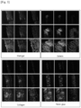
- FIG. 1 is a photograph of measuring the GFP signal emitted from the organoid transplanted into the colon tissue on the 7th day after transplantation of the colon organoid for each scaffold.
- FIG. 2 is a graph showing the area of the organoid transplanted into the colon tissue on the 7th day after transplantation of the colon organoid.
- the survival rate and engraftment ratio according to the scaffold of the colon organoid transplanted in Example 1 it was carried out as follows. 10 to 13 animals were used for each scaffold experimental group, and tissue autopsy was performed one week after transplantation of GFP organoids into the colon. The final engraftment ratio was calculated by calculating the percentage of the number of transplanted animals out of the total number of animals by classifying those expressing GFP in the colon as those with engraftment successes, and those without GFP expression as those with engraftment failed. And the final survival rate was calculated by counting the animals who died within 7 days after transplantation.
- FIG. 3 is a photograph showing the results of observing the colon tissue section on the 7th day after transplantation of colon organoids.
- Example 1 In order to evaluate the stability of the colon organoid transplanted in Example 1, an autopsy was performed on the 7th day after transplantation, and the shape of the autopsied tissue and the occurrence of lesions were checked.
- FIG. 4 is a photograph showing the shape of the colon transplanted with the organoid on the 7th day after transplantation of the colon organoid.
- FIG. 5 is a graph showing the result of calculating the weight/length of the colon transplanted with the organoid on the 7th day after transplantation of the colon organoid.
- the volume of edema that may appear when inflammation occurred was checked by calculating the weight/length of the colon transplanted with organoid. As a result, there was no significant difference compared with the control group using Matrigel.
- Example 2 In order to evaluate whether normal organoids are formed from the colonic organoids transplanted in Example 1, a second colonic organoid (secondary organoid) was formed from the transplanted colonic tissue.
- a medium supplemented with Y-27632 was added and cultured for 5 days. On the 5th day of culture, colonic organoids expressing GFP were followed up.
- FIG. 6 is a photograph of the result of confirming the GFP signal after forming a secondary organoid after transplantation of the colon organoid.
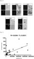
- FIG. 7 is a graph showing the results of measuring the correlation with the engraftment area by measuring the number of GFP-expressing organoids after forming a secondary organoid after transplantation of colonic organoids.
- gelatin, collagen, and fibrin glue do not significantly affect normal organoid formation.
- the transplantation rate and survival rate of the organoid is high and the stability is also excellent, therefore the composition comprising the same can be used usefully for biotransplantation in vivo.
Landscapes
- Health & Medical Sciences (AREA)
- Life Sciences & Earth Sciences (AREA)
- Chemical & Material Sciences (AREA)
- Veterinary Medicine (AREA)
- Epidemiology (AREA)
- Public Health (AREA)
- General Health & Medical Sciences (AREA)
- Animal Behavior & Ethology (AREA)
- Dermatology (AREA)
- Medicinal Chemistry (AREA)
- Oral & Maxillofacial Surgery (AREA)
- Transplantation (AREA)
- Engineering & Computer Science (AREA)
- Biomedical Technology (AREA)
- Chemical Kinetics & Catalysis (AREA)
- Botany (AREA)
- Zoology (AREA)
- Cell Biology (AREA)
- Urology & Nephrology (AREA)
- Biophysics (AREA)
- Developmental Biology & Embryology (AREA)
- Materials For Medical Uses (AREA)
- Micro-Organisms Or Cultivation Processes Thereof (AREA)
- Medicinal Preparation (AREA)
Description
- The present invention relates to a composition for transplantation of an organoid, and a use of same.
- Organoids are considered basic experimental models, sources of implantable tissue, and physiologically relevant platforms for drug screening. In contrast to a culture of immortalized cells, intestinal organoids, for example, contain viable stem cells residing in crypt-like lobes, undergo a continuous cycle of regeneration and differentiation to produce multiple functional cell types and repeat key aspects of gut development and homeostasis.
- Epithelial organoids can be formed from human colon, adenoma, and adenocarcinoma tissue and cultured from personalized medicine, patient-derived crypt or ex vivo and opened up possibilities for autologous transplantation using stem cells culture and proliferated in ex vivo. Despite an unmatched histological accuracy of the original tissues, stem cell-derived organoids from the gastrointestinal tract (GI tract) have several limitations, a main one of which is depending on Matrigel as a 3D scaffolo, as for example shown in
US 2014/243227 A1 . - Matrigel is a widely used commercial product to provide a 3D scaffold for the growth of organoids of all cell types. It is used to grow intestine, retina, kidney, liver, stomach, prostate, breast, inner ear, cardiac muscle fibers, hepatic endothelium, pancreas, fallopian tubes, and cerebral organoids. It is also used for growing organoids from a variety of species including chickens, rats and humans. However, reliance on Matrigel or similar naturally derived biopolymer matrices as scaffolds for organoid growth poses several significant limitations to the study and use of the resulting organoids. Matrigel is derived from basement membrane of ECM-rich mouse sarcoma and therefore carries a high risk of transmitting immunogens or pathogens when given to a patient, particularly problematic in the field of serious patient death and morbidity related to infection following immunosuppression and it is known to promote angiogenesis and cancer development.
- In addition, the batch-to-batch variability of Matrigel can lead to inconsistent cell behavior introducing unknown and potentially confounding variables that complicate the interpretation of both basic and translational research. Moreover, although Matrigel is a crucial element of the current organoid culture model, its role in organoid formation has not been elucidated.
- Therefore, a development of a scaffold for human injection that is effective enough to replace Matrigel in the field of organoids and suitable for in vivo transplantation is needed.
- One aspect of the present invention is to provide a composition for biotransplantation comprising an intestinal organoid, and fibrin glue, wherein the fibrin glue is comprised in an amount of 10 to 15 % (v/v) based on the total weight of the composition.
- Another aspect of the present invention is to provide the use of a fibrin glue in the manufacture of a composition for biotransplantation comprising an intestinal organoid wherein the fibrin glue is comprised in an amount of 10 to 15% (v/v) based on the total weight of the composition.
- One aspect of the present invention provides a composition for biotransplantation comprising intestinal organoid, and fibrin glue, wherein the fibrin glue is comprised in an amount of 10 to 15 % (v/v) based on the total weight of the composition.
- The term "organoid" as used herein refers to a cell mass having a 3D three-dimensional structure and refers to a miniaturized and simplified version of an organ prepared through an artificial culture process that is not collected or acquired from animals. Organoids can be derived from tissues, stem cells, embryonic stem cells or induced pluripotent stem cells, and may be cultured in three dimensions from their self-renewal and differentiation ability. The organoid may have an environment that is allowed to interact with the surrounding environment during the cell growth process. Accordingly, the 3D organoid in the present invention almost completely mimics the organs that interact in vivo and can be an excellent model for observing the development of therapeutic agents for diseases. It can similarly reproduce the physiologically active function of the human body, and by constructing an organ analogue from the patient's tissue, disease modeling based on the patient's genetic information and drug screening through repeated tests are possible. For this function, it is required to have excellent transplantation rate, transplantation efficiency, survival rate and stability when transplanted into a living body. The term "gelatin" as used herein refers to a kind of a protein obtained by decomposing and purifying natural proteins composed in animal skin, cartilage, tendon.
- The term "collagen" as used herein refers to a protein that is most widely distributed in the human body, and although it is a main component of connective tissues, mainly in bones and skin, it is a component distributed throughout our body, such as joints, membranes of each organ, and hair. A hard protein may also be called 'collagen'.
- The term "fibrin glue" as used herein refers to a tissue adhesive composed of fibrinogen, thrombin, calcium chloride, and inhibitors of anti-fibrinolytic enzyme, and is used for hemostasis in patients with lacerations. When a tissue is wounded, it refers to a substance that forms fibrin by leaking fibrinogen together with blood components from the capillaries around the cut.
- The term "fibrin glue" as used herein refers to a tissue adhesive composed of fibrinogen, thrombin, calcium chloride, and inhibitors of anti-fibrinolytic enzyme, that is used for suturing peripheral nerves, suturing microvessels, cranial nerve surgery, orthopedic surgery such as bone adhesion, and hemostasis of patients with lacerations. When a tissue is wounded, it refers to a substance that forms fibrin by leaking fibrinogen together with blood components from the capillaries around the cut.
- The gelatin, collagen, and fibrin glue all have excellent biocompatibility and stability in the body.
- The term "biotransplanting" or "bio implanting", as used herein refers to a phenomenon in which a composition for biotransplantation is administered to a subject and settle at the implantation site.
- For biotransplantation, it is required that the transplanted site is not affected, and to have a high survival rate and transplantation rate.
- The organoid can be implanted using gelatin, collagen, fibrin glue, or a combination thereof as a scaffold. Specifically, the organoids may be selected from a group of intestinal organoids, retinal organoids, kidney organoids, liver organoids, gastric organoids, prostate organoids, breast organoids, inner ear organoids, cardiac muscle fiber organoids, liver endothelial organoids, pancreas organoids, fallopian tube organoids, and cerebral organoids.
- Gelatin may be comprised in an amount of about 2.5 to 10% (w/v) based on the total weight of the composition. If the gelatin is more than about 10% (w/v), cytotoxicity may occur to the transplanted organoids due to the endotoxicity of the gelatin.
- Collagen may be comprised in an amount of about 10 to 20% (v/v) based on the total weight of the composition.
- Fibrin glue may be comprised in an amount of about 10 to 15% (v/v) based on the total weight of the composition. If the fibrin glue is more than about 15% (v/v), the composition may harden too quickly and the transplantation rate of the organoid may be reduced.
- The gelatin, collagen, or fibrin glue may be used by purchasing commercially available ones, separating those existing in nature, or synthesizing them.
- The composition may further comprise a material that may be conventionally included when transplanting and culturing organoids.
- In the composition, the organoid or fibrin glue may be in a form attached to a medical device for transplantation. The medical device may be selected from the group consisting of stents, pins, stitches, splits, pacemakers, artificial skin, and rods.
- The composition according to one embodiment of the present invention may include a pharmaceutically acceptable carrier and/or additive which are sterile water, physiological saline, conventional buffers (phosphoric acid, citric acid, other organic acids), stabilizers, salts, antioxidants (ascorbic acid), surfactants, suspending agents, isotonic agents, and/or preservatives. For topical administration, organic materials such as biopolymers, inorganic materials such as hydroxyapatite, specifically collagen matrix, polylactic acid polymers or copolymers, polyethylene glycol polymers or copolymers and chemical derivatives thereof. may also be combined for topical administration.
- The term "cell therapy product" or "cell therapy agent" as used herein refers to a pharmaceutical manufactured through separation, culture, and special manipulation of human cells and tissues used for the purpose of treatment, diagnosis, and prevention through a series of actions such as proliferating and selecting allogeneic or xenogeneic cells in vitro, or changing the biological properties of cells in other ways.
- According to one aspect of the invention, the cell therapy agent may be used to treat a condition in which the mucosa itself is lost, such as inflammatory bowel disease or damage to the mucous membrane.
- Gelatin, collagen, fibrin glue, and organoids are as described above.
- The subject may be a subject that needs to be formed by transplanting an organoid.
- The subject includes humans and mammals, and specifically includes humans, monkeys, mice, rats, rabbits, sheep, cattle, dogs, horses, pigs.
- The terms "administering," "introducing," and "transplanting" as used herein can be used interchangeably and, according to one embodiment of the present invention may refer to a method that results in at least partial localization of the composition to a desired site or a placement of a composition according to a route into a subject in one embodiment. Administration may be by any suitable route that delivers at least a portion of a cell or cellular component of a composition according to one embodiment to a desired location in a living subj ect.
- In the above method, administration may be administered to a lesion site requiring transplantation of an organoid. Endoscopy equipment may be used for administration. Administration to the esophagus, stomach, duodenum, large intestine, or colon using an endoscope is typical, and in addition, all organs in the body through surgical operations, such as salivary glands, lacrimal glands, muscles, lungs, liver, pancreas, kidney, uterus, prostate, can be administered. When the mucous membrane itself is lost, such as inflammatory bowel disease or damage to the mucous membrane, it can be used to replace the damaged mucosa by transplanting organoids.
- The method uses collagen, gelatin, or fibrin glue as a scaffold for organoid transplantation, thereby exhibiting transplant stability and transplant efficiency similar to those of conventionally used matrigel, and is also clinically applicable safely.
- As mentioned above, organoids can be differentiated into organoids with adult-like characteristics even in vitro, and above all, because they utilize the patient's own adult cells, there are no technical issues such as immunogenicity, and ethical problems which become obstacles in future use as a tissue therapy.
- According to one aspect of the present invention, when collagen, gelatin, or fibrin glue is used as a scaffold for organoid transplantation, the transplantation rate and survival rate of organoids are high, and stability is also excellent.
-
-
FIG. 1 is a photograph of measuring the GFP signal emitted from an organoid transplanted into the colon tissue on the 7th day after transplanting the colon organoid for each scaffold. -
FIG. 2 is a graph showing the area of an organoid transplanted to the colon tissue on the 7th day after transplanting the colon organoid. -
FIG. 3 is a photograph showing the result of observing a colon tissue section on the 7th day after transplanting colon organoids. -
FIG. 4 is a photograph showing the morphology of a colon transplanted with an organoid on the 7th day after transplanting the colon organoid. -
FIG. 5 is a graph showing the result of calculating the weight/length of a colon transplanted with an organoid on the 7th day after transplanting the colon organoid. -
FIG. 6 is a photograph of the result of confirming the GFP signal after forming a secondary organoid after transplanting the colon organoid. -
FIG. 7 is a graph showing the results of measuring a correlation with an engraftment area by measuring a number of GFP-expressing organoids after forming a secondary organoid after transplanting a colonic organoid. -
FIG. 8a is a schematic diagram showing the manufacturing process of the acute colon injury model induced by EDTA. -
FIG. 8b is a schematic diagram illustrating a transplantation process of an organoid according to one embodiment. -
FIG. 8c is a result showing the tissue analysis result after transplantation of the organoid according to one embodiment. - Hereinafter, it will be described in more detail through examples.
- Colon tissue was separated from EGFP mice, and colonic crypt was isolated using an enzyme, and matrigel and medium for colon organoids were mixed in a 1:1 ratio and inoculated into an uncoated 48-well plate. It was placed in an incubator, and after 20 minutes, it was confirmed that the matrigel was hardened. And then, a medium for colon organoids was added and cultured for 5 days to prepare colon organoids for biotransplantation to be used below.
- A colon tissue damage model to produce organoids was prepared as follows. Wild-type mice to be transplanted with colon organoids were exposed to 0.5M EDTA for 5 minutes, and physical damage was applied to remove the crypts of the colonic lining for 2 minutes with an electric toothbrush.
- A colon organoid was prepared so that the colon organoid prepared above expresses GFP. All colon organoids were treated with 10 µM of Y-27632 in the organoid medium one day before transplantation to maximize the survival rate. In addition, the matrix used during culture was completely removed by treatment with a cell recovery solution.
- Thereafter, the following three scaffolds were mixed under the conditions described below and transplanted in a volume of 50 µl into the anus of the colon tissue damage mouse model prepared in 1.1.
- 1) Gelatin: GFP+ colon organoids were mixed in PBS with 5% gelatin dissolved in gelatin : organoid = 1 : 2 ratio of medium and transplanted. At this time, the medium containing the organoids contains 10 µM of Y-27632.
- 2) Collagen: GFP+ colon organoids were mixed in 100% collagen stock solution dissolved in collagen : organoid = 1 : 9 ratio of medium and transplanted. At this time, the medium containing the organoids contains 10 µM of Y-27632.
- 3) Fibrin Glue: a colonoid solution was prepared by mixing GFP+ colon organoids with 45 µl of colonoid culture medium containing 10 µM of Y-27632, and fibrin was mixed with a ratio of 1:1 in a solution in which thrombin is diluted 1:100 in PBS to make 5 µl of the solution totally. And then, 5 µl of the solution were mixed with the prepared 45 µl of colonoid solution and transplanted.
- Matrigel was used as a control group.
- Matrigel was transplanted by mixing it in a colonoid culture medium containing organoids at a concentration of 10%. At this time, the medium containing the organoids contains 10 µM of Y-27632.
- After transplantation, the anus was closed using 10 µl of 3M Vetbond, and the suture was released 14 hours later to induce normal bowel activity.
- In order to evaluate the transplantation rate according to the scaffold of the colon organoid transplanted in Example 1, it was carried out as follows.
- On the 7th day after transplantation of colon organoids, the colon tissue was autopsied to measure GFP signals released from the transplanted organoids. Specifically, the organoid-implanted colon tissue was vertically incised to create a planar structure, and this tissue was spread thinly on a slide glass and a glass cover was covered thereon. At this time, the crypt was directed downward. The prepared slide glass was placed on the stage of a fluorescence microscope to observe the region where GFP fluorescence was expressed.
-
FIG. 1 is a photograph of measuring the GFP signal emitted from the organoid transplanted into the colon tissue on the 7th day after transplantation of the colon organoid for each scaffold. -
FIG. 2 is a graph showing the area of the organoid transplanted into the colon tissue on the 7th day after transplantation of the colon organoid. - As shown in
FIG. 1 and 2 , in the absence of a scaffold, it was confirmed that the GFP signal was the weakest and the transplantation rate was low. On the other hand, it was confirmed that when gelatin, collagen, and fibrin glue were used as scaffolds, the transplantation rate and transplanted area were similar to those when Matrigel, a positive control, was used as a scaffold. Therefore, when gelatin, collagen, or fibrin glue is used as a scaffold for organoid transplantation, the transplantation rate of organoids is significantly increased. - In order to evaluate the survival rate and engraftment ratio according to the scaffold of the colon organoid transplanted in Example 1, it was carried out as follows. 10 to 13 animals were used for each scaffold experimental group, and tissue autopsy was performed one week after transplantation of GFP organoids into the colon. The final engraftment ratio was calculated by calculating the percentage of the number of transplanted animals out of the total number of animals by classifying those expressing GFP in the colon as those with engraftment successes, and those without GFP expression as those with engraftment failed. And the final survival rate was calculated by counting the animals who died within 7 days after transplantation.
- The results are shown in Table 1 below.
[Table 1] Number of transplantation Engraftment successes Engraftment failed Dead Survival rate (%) Engraftment ratio (%) No scaffold 13 8 1 2 84.62 61.54 Matrigel 10 9 0 1 90.00 90.00 Gelatin 13 9 1 3 76.92 69.23 Collagen 10 9 1 0 100.00 90.00 Fibrin glue 10 9 0 1 90.00 90.00 - As shown in Table 1, when gelatin, collagen, and fibrin glue were used as scaffolds, the survival rates were 76.92, 100, and 90%, respectively, and the engraftment ratio was confirmed as high as 69.23, 90, and 90%, respectively. Therefore, it was confirmed that the survival rate and engraftment ratio were excellent. This was at a level similar to that in the case of using Matrigel, a positive control.
FIG. 3 is a photograph showing the results of observing the colon tissue section on the 7th day after transplantation of colon organoids. - As shown in
FIG. 3 , when gelatin, collagen, and fibrin glue were used as scaffolds, it was confirmed that the organoids were successfully settled to form colonic crypts. - In order to evaluate the stability of the colon organoid transplanted in Example 1, an autopsy was performed on the 7th day after transplantation, and the shape of the autopsied tissue and the occurrence of lesions were checked.
-
FIG. 4 is a photograph showing the shape of the colon transplanted with the organoid on the 7th day after transplantation of the colon organoid. - As shown in
FIG. 4 , when gelatin, collagen, or fibrin glue was used as a scaffold, an edema or bloody stool that may appear in the colon transplanted with organoid was not observed in the autopsy tissue. -
FIG. 5 is a graph showing the result of calculating the weight/length of the colon transplanted with the organoid on the 7th day after transplantation of the colon organoid. - As shown in
FIG. 5 , the volume of edema that may appear when inflammation occurred was checked by calculating the weight/length of the colon transplanted with organoid. As a result, there was no significant difference compared with the control group using Matrigel. - Therefore, it was confirmed that when the organoid was transplanted using gelatin, collagen, or fibrin glue as a scaffold, the stability was excellent.
- In order to evaluate whether normal organoids are formed from the colonic organoids transplanted in Example 1, a second colonic organoid (secondary organoid) was formed from the transplanted colonic tissue.
- Specifically, after checking the GFP of the transplanted colon tissue with a fluorescence microscope, it was cut with surgical scissors, put in a tube containing crypt chelating buffer, and reacted in a shaking incubator at 37 °C for 20 minutes. And then, it was put in a 10 ml syringe equipped with an 18 gage needle, and crypt is separated by grinding 20 times. The separated crypt was centrifuged, collected, filtered through a 70 µm filter, mixed with Y-27632-added medium and matrigel at a 1:1 ratio, and inoculated at a concentration of 20 µl/well in a 48-well plate. After hardening the matrigel by putting it in an incubator at 37 °C for 30 minutes, a medium supplemented with Y-27632 was added and cultured for 5 days. On the 5th day of culture, colonic organoids expressing GFP were followed up.
-
FIG. 6 is a photograph of the result of confirming the GFP signal after forming a secondary organoid after transplantation of the colon organoid. - As shown in
FIG. 6 , when gelatin, collagen, or fibrin glue was used as a scaffold, secondary organoids expressing GFP were effectively formed. This was confirmed to be at a level similar to that of the positive control using matrigel. -
FIG. 7 is a graph showing the results of measuring the correlation with the engraftment area by measuring the number of GFP-expressing organoids after forming a secondary organoid after transplantation of colonic organoids. - As shown in
FIG. 7 , it was confirmed that the number and area of secondary organoids increased in proportion. - Therefore, it indicates that gelatin, collagen, and fibrin glue do not significantly affect normal organoid formation.
- According to one aspect of the present invention, when collagen, gelatin, or fibrin glue is used as a scaffold for organoid transplantation, the transplantation rate and survival rate of the organoid is high and the stability is also excellent, therefore the composition comprising the same can be used usefully for biotransplantation in vivo.
Claims (2)
- A composition for biotransplantation comprising an intestinal organoid and fibrin glue, wherein the fibrin glue is comprised in an amount of 10 to 15% (v/v) based on the total weight of the composition.
- Use of fibrin in the manufacture of a composition for biotransplantation comprising an intestinal organoid, wherein the fibrin glue is comprised in an amount of 10 to 15% (v/v) based on the total weight of the composition.
Applications Claiming Priority (2)
| Application Number | Priority Date | Filing Date | Title |
|---|---|---|---|
| KR1020180152804A KR20200065892A (en) | 2018-11-30 | 2018-11-30 | A composition for bio transplanting of organoid |
| PCT/KR2019/013527 WO2020111507A1 (en) | 2018-11-30 | 2019-10-15 | Composition for transplantation of organoid |
Publications (4)
| Publication Number | Publication Date |
|---|---|
| EP3888715A1 EP3888715A1 (en) | 2021-10-06 |
| EP3888715A4 EP3888715A4 (en) | 2022-08-17 |
| EP3888715B1 true EP3888715B1 (en) | 2024-12-04 |
| EP3888715C0 EP3888715C0 (en) | 2024-12-04 |
Family
ID=70852032
Family Applications (1)
| Application Number | Title | Priority Date | Filing Date |
|---|---|---|---|
| EP19890365.0A Active EP3888715B1 (en) | 2018-11-30 | 2019-10-15 | Composition for transplantation of organoid |
Country Status (6)
| Country | Link |
|---|---|
| US (1) | US12623000B2 (en) |
| EP (1) | EP3888715B1 (en) |
| JP (2) | JP2022510982A (en) |
| KR (2) | KR20200065892A (en) |
| CN (1) | CN113660962A (en) |
| WO (1) | WO2020111507A1 (en) |
Families Citing this family (6)
| Publication number | Priority date | Publication date | Assignee | Title |
|---|---|---|---|---|
| KR102939575B1 (en) | 2020-11-17 | 2026-03-19 | 주식회사 세라트젠 | Esophagus extracellular matrix-derived scaffold for culture and transplantation of esophagus organoid and preparing method thereof |
| EP4230726A4 (en) | 2020-11-17 | 2025-02-12 | Cellartgen Inc. | PANCREATIC EXTRACELLULAR MATRIX-DERIVED SUPPORT FOR PANCREATIC ORGANOIDS CULTURE AND TRANSPLANTATION, AND PREPARATION METHOD THEREOF |
| KR102939688B1 (en) | 2020-11-17 | 2026-03-17 | 주식회사 세라트젠 | Kidney extracellular matrix-derived scaffold for culture and transplantation of kidney organoid and preparing method thereof |
| KR102792540B1 (en) | 2020-11-17 | 2025-04-09 | 주식회사 세라트젠 | Pancreas extracellular matrix-derived scaffold for culture and transplantation of pancreas organoid and preparing method thereof |
| CN114317413A (en) * | 2022-01-17 | 2022-04-12 | 中山大学 | Culture methods of intestinal organoids and their application in intestinal toxicity testing |
| KR102909085B1 (en) * | 2022-10-07 | 2026-01-07 | 한림대학교 산학협력단 | Conjugation adjuvant composition for brain organoid implantation and method for preparing same |
Family Cites Families (16)
| Publication number | Priority date | Publication date | Assignee | Title |
|---|---|---|---|---|
| AU2002367580A1 (en) * | 2001-05-16 | 2003-09-22 | Tracy C. Grikscheit | Tissue-engineered organs |
| MXPA04004534A (en) * | 2001-11-16 | 2004-08-11 | Childrens Medical Center | Augmentation of organ function. |
| US9314545B2 (en) * | 2008-04-15 | 2016-04-19 | Life Science Enterprises, Inc. | Minimally invasive treatment of vertebra (MITV) using a calcium phosphate combination bone cement |
| US9752124B2 (en) * | 2009-02-03 | 2017-09-05 | Koninklijke Nederlandse Akademie Van Wetenschappen | Culture medium for epithelial stem cells and organoids comprising the stem cells |
| GB201111244D0 (en) | 2011-06-30 | 2011-08-17 | Konink Nl Akademie Van Wetenschappen Knaw | Culture media for stem cells |
| CN102198022B (en) * | 2011-05-23 | 2013-04-17 | 西安交通大学 | Solid forming method of active cell-hydrogel organ structure |
| ES2661587T3 (en) * | 2011-08-10 | 2018-04-02 | Depuy Synthes Products Llc | Treatment of peripheral vascular disease using cells derived from umbilical cord tissue |
| US20150284689A1 (en) * | 2012-10-26 | 2015-10-08 | The Regents Of The University Of California | Strategy for engineering various 3d tissues, organoids and vasculature |
| KR102349183B1 (en) * | 2013-05-08 | 2022-01-07 | 인리젠 | Organoids comprising isolated renal cells and uses thereof |
| KR101613478B1 (en) * | 2014-09-22 | 2016-04-19 | (주)안트로젠 | Composition comprising mesenchymal stem cell-hydrogel and preparation method thereof |
| CA2963704A1 (en) * | 2014-10-17 | 2016-04-21 | Children's Hospital Medical Center | In vivo model of human small intestine using pluripotent stem cells and methods of making and using same |
| WO2017036533A1 (en) | 2015-09-03 | 2017-03-09 | Ecole Polytechnique Federale De Lausanne (Epfl) | Three-dimensional hydrogels for culturing adult epithelial stem cells and organoids |
| LU92845B1 (en) | 2015-10-08 | 2017-05-02 | Univ Du Luxembourg Campus Belval | Means and methods for generating midbrain organoids |
| GB201603569D0 (en) * | 2016-03-01 | 2016-04-13 | Koninklijke Nederlandse Akademie Van Wetenschappen | Improved differentiation method |
| GB201610748D0 (en) | 2016-06-20 | 2016-08-03 | Koninklijke Nederlandse Akademie Van Wetenschappen | Improved diffrentation method |
| EP3323882A1 (en) * | 2016-11-18 | 2018-05-23 | Ecole Polytechnique Fédérale de Lausanne | Organoid tissue engineering |
-
2018
- 2018-11-30 KR KR1020180152804A patent/KR20200065892A/en not_active Ceased
-
2019
- 2019-10-15 CN CN201980090843.6A patent/CN113660962A/en active Pending
- 2019-10-15 US US17/298,833 patent/US12623000B2/en active Active
- 2019-10-15 EP EP19890365.0A patent/EP3888715B1/en active Active
- 2019-10-15 JP JP2021531269A patent/JP2022510982A/en active Pending
- 2019-10-15 WO PCT/KR2019/013527 patent/WO2020111507A1/en not_active Ceased
-
2021
- 2021-06-04 KR KR1020210072919A patent/KR102597594B1/en active Active
-
2023
- 2023-02-20 JP JP2023023950A patent/JP7542880B2/en active Active
Also Published As
| Publication number | Publication date |
|---|---|
| CN113660962A (en) | 2021-11-16 |
| WO2020111507A1 (en) | 2020-06-04 |
| KR20200065892A (en) | 2020-06-09 |
| US12623000B2 (en) | 2026-05-12 |
| JP2023054120A (en) | 2023-04-13 |
| EP3888715C0 (en) | 2024-12-04 |
| JP7542880B2 (en) | 2024-09-02 |
| KR20210069619A (en) | 2021-06-11 |
| EP3888715A1 (en) | 2021-10-06 |
| KR102597594B1 (en) | 2023-11-02 |
| EP3888715A4 (en) | 2022-08-17 |
| US20220226538A1 (en) | 2022-07-21 |
| JP2022510982A (en) | 2022-01-28 |
Similar Documents
| Publication | Publication Date | Title |
|---|---|---|
| US12623000B2 (en) | Composition for transplantation of organoid | |
| KR102339700B1 (en) | Hybrid gel containing particulate decellularized tissue | |
| ES2703785T3 (en) | Method and composition to treat an inflammatory bowel disease without colectomy | |
| ES2546734T3 (en) | Rapid preparation and use of tissues and structures obtained by tissue engineering as individual implants | |
| US11801331B2 (en) | Composition for cartilage regeneration and preparing thereof | |
| KR20130067247A (en) | Process, tube and device for the preparation of wound healant composition | |
| JPH08509356A (en) | Cell transplantation system and method | |
| KR20180134897A (en) | Methods and compositions for treating inflammatory bowel disease | |
| KR20190055088A (en) | Extracellular matrix for tissue remodeling of mucosal tissue | |
| CN110476892A (en) | A kind of construction method of osteosarcoma xenograft mouse model and application | |
| BR112020015616B1 (en) | BIOINK COMPOSITION FOR DERMIS REGENERATION SHEET, METHOD FOR MANUFACTURING CUSTOM DERMIS REGENERATION SHEET USING THE SAME AND CUSTOM DERMIS REGENERATION SHEET MANUFACTURED USING THE MANUFACTURING METHOD | |
| JPH03503167A (en) | Site-directed angioplasty device and its usage | |
| KR102104120B1 (en) | 3D bioprinting construct using human nasal inferior turbinate derived mesenchymal stem cell and uses thereof | |
| KR20170108325A (en) | Composition and method for differentiation adult stem cells into chondrocytes | |
| JP7587866B2 (en) | Osteoblasts differentiated from mesenchymal stem cells and composition for treating bone diseases containing the same | |
| CN111450119A (en) | Perinatal tissue-derived extracellular matrix hydrogel preparation for promoting organ injury repair | |
| WO2004045666A1 (en) | Method of organ regeneration | |
| CN110624112A (en) | Hydrogel connected with prostaglandin E2, and preparation method and application thereof | |
| CN117414349A (en) | Cell porous microneedle patch for sustainable supply of exosomes and preparation method thereof | |
| CN115287257A (en) | Toxoplasma gondii exocrine protein-based osteogenic induced differentiation culture medium, preparation method and osteogenic induced differentiation method | |
| KR102332926B1 (en) | Pharmaceutical composition for treating cartilage damage comprising nasal septum cartilage cell | |
| TWI611805B (en) | Pharmaceutical composition for treating sciatic nerve injury | |
| US12427230B2 (en) | Human nasal turbinate-derived mesenchymal stem cell-based, 3D bioprinted construct, and use thereof | |
| RU2195940C2 (en) | Method for regeneration of an articular cartilage | |
| Jaramillo et al. | Odontogenic cell culture in PEGDA hydrogel scaffolds for use in tooth regeneration protocols |
Legal Events
| Date | Code | Title | Description |
|---|---|---|---|
| STAA | Information on the status of an ep patent application or granted ep patent |
Free format text: STATUS: THE INTERNATIONAL PUBLICATION HAS BEEN MADE |
|
| PUAI | Public reference made under article 153(3) epc to a published international application that has entered the european phase |
Free format text: ORIGINAL CODE: 0009012 |
|
| STAA | Information on the status of an ep patent application or granted ep patent |
Free format text: STATUS: REQUEST FOR EXAMINATION WAS MADE |
|
| 17P | Request for examination filed |
Effective date: 20210630 |
|
| AK | Designated contracting states |
Kind code of ref document: A1 Designated state(s): AL AT BE BG CH CY CZ DE DK EE ES FI FR GB GR HR HU IE IS IT LI LT LU LV MC MK MT NL NO PL PT RO RS SE SI SK SM TR |
|
| DAV | Request for validation of the european patent (deleted) | ||
| DAX | Request for extension of the european patent (deleted) | ||
| A4 | Supplementary search report drawn up and despatched |
Effective date: 20220715 |
|
| RIC1 | Information provided on ipc code assigned before grant |
Ipc: A61L 27/24 20060101ALI20220711BHEP Ipc: A61L 27/22 20060101ALI20220711BHEP Ipc: A61L 27/26 20060101ALI20220711BHEP Ipc: A61L 27/38 20060101AFI20220711BHEP |
|
| GRAP | Despatch of communication of intention to grant a patent |
Free format text: ORIGINAL CODE: EPIDOSNIGR1 |
|
| STAA | Information on the status of an ep patent application or granted ep patent |
Free format text: STATUS: GRANT OF PATENT IS INTENDED |
|
| INTG | Intention to grant announced |
Effective date: 20240207 |
|
| GRAJ | Information related to disapproval of communication of intention to grant by the applicant or resumption of examination proceedings by the epo deleted |
Free format text: ORIGINAL CODE: EPIDOSDIGR1 |
|
| STAA | Information on the status of an ep patent application or granted ep patent |
Free format text: STATUS: REQUEST FOR EXAMINATION WAS MADE |
|
| GRAP | Despatch of communication of intention to grant a patent |
Free format text: ORIGINAL CODE: EPIDOSNIGR1 |
|
| STAA | Information on the status of an ep patent application or granted ep patent |
Free format text: STATUS: GRANT OF PATENT IS INTENDED |
|
| INTC | Intention to grant announced (deleted) | ||
| INTG | Intention to grant announced |
Effective date: 20240627 |
|
| GRAS | Grant fee paid |
Free format text: ORIGINAL CODE: EPIDOSNIGR3 |
|
| GRAA | (expected) grant |
Free format text: ORIGINAL CODE: 0009210 |
|
| STAA | Information on the status of an ep patent application or granted ep patent |
Free format text: STATUS: THE PATENT HAS BEEN GRANTED |
|
| AK | Designated contracting states |
Kind code of ref document: B1 Designated state(s): AL AT BE BG CH CY CZ DE DK EE ES FI FR GB GR HR HU IE IS IT LI LT LU LV MC MK MT NL NO PL PT RO RS SE SI SK SM TR |
|
| REG | Reference to a national code |
Ref country code: CH Ref legal event code: EP |
|
| REG | Reference to a national code |
Ref country code: DE Ref legal event code: R096 Ref document number: 602019063194 Country of ref document: DE |
|
| REG | Reference to a national code |
Ref country code: IE Ref legal event code: FG4D |
|
| U01 | Request for unitary effect filed |
Effective date: 20241219 |
|
| U07 | Unitary effect registered |
Designated state(s): AT BE BG DE DK EE FI FR IT LT LU LV MT NL PT RO SE SI Effective date: 20250110 |
|
| PG25 | Lapsed in a contracting state [announced via postgrant information from national office to epo] |
Ref country code: HR Free format text: LAPSE BECAUSE OF FAILURE TO SUBMIT A TRANSLATION OF THE DESCRIPTION OR TO PAY THE FEE WITHIN THE PRESCRIBED TIME-LIMIT Effective date: 20241204 |
|
| PG25 | Lapsed in a contracting state [announced via postgrant information from national office to epo] |
Ref country code: ES Free format text: LAPSE BECAUSE OF FAILURE TO SUBMIT A TRANSLATION OF THE DESCRIPTION OR TO PAY THE FEE WITHIN THE PRESCRIBED TIME-LIMIT Effective date: 20241204 |
|
| PG25 | Lapsed in a contracting state [announced via postgrant information from national office to epo] |
Ref country code: NO Free format text: LAPSE BECAUSE OF FAILURE TO SUBMIT A TRANSLATION OF THE DESCRIPTION OR TO PAY THE FEE WITHIN THE PRESCRIBED TIME-LIMIT Effective date: 20250304 |
|
| PG25 | Lapsed in a contracting state [announced via postgrant information from national office to epo] |
Ref country code: GR Free format text: LAPSE BECAUSE OF FAILURE TO SUBMIT A TRANSLATION OF THE DESCRIPTION OR TO PAY THE FEE WITHIN THE PRESCRIBED TIME-LIMIT Effective date: 20250305 |
|
| PG25 | Lapsed in a contracting state [announced via postgrant information from national office to epo] |
Ref country code: RS Free format text: LAPSE BECAUSE OF FAILURE TO SUBMIT A TRANSLATION OF THE DESCRIPTION OR TO PAY THE FEE WITHIN THE PRESCRIBED TIME-LIMIT Effective date: 20250304 |
|
| PG25 | Lapsed in a contracting state [announced via postgrant information from national office to epo] |
Ref country code: SM Free format text: LAPSE BECAUSE OF FAILURE TO SUBMIT A TRANSLATION OF THE DESCRIPTION OR TO PAY THE FEE WITHIN THE PRESCRIBED TIME-LIMIT Effective date: 20241204 |
|
| PG25 | Lapsed in a contracting state [announced via postgrant information from national office to epo] |
Ref country code: PL Free format text: LAPSE BECAUSE OF FAILURE TO SUBMIT A TRANSLATION OF THE DESCRIPTION OR TO PAY THE FEE WITHIN THE PRESCRIBED TIME-LIMIT Effective date: 20241204 |
|
| PG25 | Lapsed in a contracting state [announced via postgrant information from national office to epo] |
Ref country code: IS Free format text: LAPSE BECAUSE OF FAILURE TO SUBMIT A TRANSLATION OF THE DESCRIPTION OR TO PAY THE FEE WITHIN THE PRESCRIBED TIME-LIMIT Effective date: 20250404 |
|
| PG25 | Lapsed in a contracting state [announced via postgrant information from national office to epo] |
Ref country code: SK Free format text: LAPSE BECAUSE OF FAILURE TO SUBMIT A TRANSLATION OF THE DESCRIPTION OR TO PAY THE FEE WITHIN THE PRESCRIBED TIME-LIMIT Effective date: 20241204 |
|
| PG25 | Lapsed in a contracting state [announced via postgrant information from national office to epo] |
Ref country code: CZ Free format text: LAPSE BECAUSE OF FAILURE TO SUBMIT A TRANSLATION OF THE DESCRIPTION OR TO PAY THE FEE WITHIN THE PRESCRIBED TIME-LIMIT Effective date: 20241204 |
|
| PLBE | No opposition filed within time limit |
Free format text: ORIGINAL CODE: 0009261 |
|
| STAA | Information on the status of an ep patent application or granted ep patent |
Free format text: STATUS: NO OPPOSITION FILED WITHIN TIME LIMIT |
|
| 26N | No opposition filed |
Effective date: 20250905 |
|
| U20 | Renewal fee for the european patent with unitary effect paid |
Year of fee payment: 7 Effective date: 20251027 |