EP3882275B1 - Anti-pd-1 and anti-vegfa bifunctional antibody, pharmaceutical composition thereof and use thereof - Google Patents
Anti-pd-1 and anti-vegfa bifunctional antibody, pharmaceutical composition thereof and use thereof Download PDFInfo
- Publication number
- EP3882275B1 EP3882275B1 EP19853809.2A EP19853809A EP3882275B1 EP 3882275 B1 EP3882275 B1 EP 3882275B1 EP 19853809 A EP19853809 A EP 19853809A EP 3882275 B1 EP3882275 B1 EP 3882275B1
- Authority
- EP
- European Patent Office
- Prior art keywords
- antibody
- seq
- amino acid
- variable region
- chain variable
- Prior art date
- Legal status (The legal status is an assumption and is not a legal conclusion. Google has not performed a legal analysis and makes no representation as to the accuracy of the status listed.)
- Active
Links
Images






Classifications
-
- A—HUMAN NECESSITIES
- A61—MEDICAL OR VETERINARY SCIENCE; HYGIENE
- A61K—PREPARATIONS FOR MEDICAL, DENTAL OR TOILETRY PURPOSES
- A61K47/00—Medicinal preparations characterised by the non-active ingredients used, e.g. carriers or inert additives; Targeting or modifying agents chemically bound to the active ingredient
- A61K47/50—Medicinal preparations characterised by the non-active ingredients used, e.g. carriers or inert additives; Targeting or modifying agents chemically bound to the active ingredient the non-active ingredient being chemically bound to the active ingredient, e.g. polymer-drug conjugates
- A61K47/51—Medicinal preparations characterised by the non-active ingredients used, e.g. carriers or inert additives; Targeting or modifying agents chemically bound to the active ingredient the non-active ingredient being chemically bound to the active ingredient, e.g. polymer-drug conjugates the non-active ingredient being a modifying agent
- A61K47/68—Medicinal preparations characterised by the non-active ingredients used, e.g. carriers or inert additives; Targeting or modifying agents chemically bound to the active ingredient the non-active ingredient being chemically bound to the active ingredient, e.g. polymer-drug conjugates the non-active ingredient being a modifying agent the modifying agent being an antibody, an immunoglobulin or a fragment thereof, e.g. an Fc-fragment
- A61K47/6835—Medicinal preparations characterised by the non-active ingredients used, e.g. carriers or inert additives; Targeting or modifying agents chemically bound to the active ingredient the non-active ingredient being chemically bound to the active ingredient, e.g. polymer-drug conjugates the non-active ingredient being a modifying agent the modifying agent being an antibody, an immunoglobulin or a fragment thereof, e.g. an Fc-fragment the modifying agent being an antibody or an immunoglobulin bearing at least one antigen-binding site
- A61K47/6849—Medicinal preparations characterised by the non-active ingredients used, e.g. carriers or inert additives; Targeting or modifying agents chemically bound to the active ingredient the non-active ingredient being chemically bound to the active ingredient, e.g. polymer-drug conjugates the non-active ingredient being a modifying agent the modifying agent being an antibody, an immunoglobulin or a fragment thereof, e.g. an Fc-fragment the modifying agent being an antibody or an immunoglobulin bearing at least one antigen-binding site the antibody targeting a receptor, a cell surface antigen or a cell surface determinant
-
- A—HUMAN NECESSITIES
- A61—MEDICAL OR VETERINARY SCIENCE; HYGIENE
- A61P—SPECIFIC THERAPEUTIC ACTIVITY OF CHEMICAL COMPOUNDS OR MEDICINAL PREPARATIONS
- A61P35/00—Antineoplastic agents
-
- A—HUMAN NECESSITIES
- A61—MEDICAL OR VETERINARY SCIENCE; HYGIENE
- A61P—SPECIFIC THERAPEUTIC ACTIVITY OF CHEMICAL COMPOUNDS OR MEDICINAL PREPARATIONS
- A61P35/00—Antineoplastic agents
- A61P35/02—Antineoplastic agents specific for leukemia
-
- C—CHEMISTRY; METALLURGY
- C07—ORGANIC CHEMISTRY
- C07K—PEPTIDES
- C07K16/00—Immunoglobulins [IG], e.g. monoclonal or polyclonal antibodies
- C07K16/18—Immunoglobulins [IG], e.g. monoclonal or polyclonal antibodies against material from animals or humans
- C07K16/22—Immunoglobulins [IG], e.g. monoclonal or polyclonal antibodies against material from animals or humans against growth factors ; against growth regulators
-
- C—CHEMISTRY; METALLURGY
- C07—ORGANIC CHEMISTRY
- C07K—PEPTIDES
- C07K16/00—Immunoglobulins [IG], e.g. monoclonal or polyclonal antibodies
- C07K16/18—Immunoglobulins [IG], e.g. monoclonal or polyclonal antibodies against material from animals or humans
- C07K16/28—Immunoglobulins [IG], e.g. monoclonal or polyclonal antibodies against material from animals or humans against receptors, cell surface antigens or cell surface determinants
- C07K16/2803—Immunoglobulins [IG], e.g. monoclonal or polyclonal antibodies against material from animals or humans against receptors, cell surface antigens or cell surface determinants against the immunoglobulin superfamily
- C07K16/2818—Immunoglobulins [IG], e.g. monoclonal or polyclonal antibodies against material from animals or humans against receptors, cell surface antigens or cell surface determinants against the immunoglobulin superfamily against CD28 or CD152
-
- C—CHEMISTRY; METALLURGY
- C12—BIOCHEMISTRY; BEER; SPIRITS; WINE; VINEGAR; MICROBIOLOGY; ENZYMOLOGY; MUTATION OR GENETIC ENGINEERING
- C12N—MICROORGANISMS OR ENZYMES; COMPOSITIONS THEREOF; PROPAGATING, PRESERVING, OR MAINTAINING MICROORGANISMS; MUTATION OR GENETIC ENGINEERING; CULTURE MEDIA
- C12N15/00—Mutation or genetic engineering; DNA or RNA concerning genetic engineering, vectors, e.g. plasmids, or their isolation, preparation or purification; Use of hosts therefor
- C12N15/09—Recombinant DNA-technology
- C12N15/11—DNA or RNA fragments; Modified forms thereof; Non-coding nucleic acids having a biological activity
- C12N15/62—DNA sequences coding for fusion proteins
-
- G—PHYSICS
- G01—MEASURING; TESTING
- G01N—INVESTIGATING OR ANALYSING MATERIALS BY DETERMINING THEIR CHEMICAL OR PHYSICAL PROPERTIES
- G01N33/00—Investigating or analysing materials by specific methods not covered by groups G01N1/00 - G01N31/00
- G01N33/48—Biological material, e.g. blood, urine; Haemocytometers
- G01N33/50—Chemical analysis of biological material, e.g. blood, urine; Testing involving biospecific ligand binding methods; Immunological testing
- G01N33/53—Immunoassay; Biospecific binding assay; Materials therefor
- G01N33/531—Production of immunochemical test materials
- G01N33/532—Production of labelled immunochemicals
-
- G—PHYSICS
- G01—MEASURING; TESTING
- G01N—INVESTIGATING OR ANALYSING MATERIALS BY DETERMINING THEIR CHEMICAL OR PHYSICAL PROPERTIES
- G01N33/00—Investigating or analysing materials by specific methods not covered by groups G01N1/00 - G01N31/00
- G01N33/48—Biological material, e.g. blood, urine; Haemocytometers
- G01N33/50—Chemical analysis of biological material, e.g. blood, urine; Testing involving biospecific ligand binding methods; Immunological testing
- G01N33/68—Chemical analysis of biological material, e.g. blood, urine; Testing involving biospecific ligand binding methods; Immunological testing involving proteins, peptides or amino acids
-
- G—PHYSICS
- G01—MEASURING; TESTING
- G01N—INVESTIGATING OR ANALYSING MATERIALS BY DETERMINING THEIR CHEMICAL OR PHYSICAL PROPERTIES
- G01N33/00—Investigating or analysing materials by specific methods not covered by groups G01N1/00 - G01N31/00
- G01N33/48—Biological material, e.g. blood, urine; Haemocytometers
- G01N33/50—Chemical analysis of biological material, e.g. blood, urine; Testing involving biospecific ligand binding methods; Immunological testing
- G01N33/68—Chemical analysis of biological material, e.g. blood, urine; Testing involving biospecific ligand binding methods; Immunological testing involving proteins, peptides or amino acids
- G01N33/6854—Immunoglobulins
- G01N33/6857—Antibody fragments
-
- G—PHYSICS
- G01—MEASURING; TESTING
- G01N—INVESTIGATING OR ANALYSING MATERIALS BY DETERMINING THEIR CHEMICAL OR PHYSICAL PROPERTIES
- G01N33/00—Investigating or analysing materials by specific methods not covered by groups G01N1/00 - G01N31/00
- G01N33/48—Biological material, e.g. blood, urine; Haemocytometers
- G01N33/50—Chemical analysis of biological material, e.g. blood, urine; Testing involving biospecific ligand binding methods; Immunological testing
- G01N33/68—Chemical analysis of biological material, e.g. blood, urine; Testing involving biospecific ligand binding methods; Immunological testing involving proteins, peptides or amino acids
- G01N33/6872—Intracellular protein regulatory factors and their receptors, e.g. including ion channels
-
- G—PHYSICS
- G01—MEASURING; TESTING
- G01N—INVESTIGATING OR ANALYSING MATERIALS BY DETERMINING THEIR CHEMICAL OR PHYSICAL PROPERTIES
- G01N33/00—Investigating or analysing materials by specific methods not covered by groups G01N1/00 - G01N31/00
- G01N33/48—Biological material, e.g. blood, urine; Haemocytometers
- G01N33/50—Chemical analysis of biological material, e.g. blood, urine; Testing involving biospecific ligand binding methods; Immunological testing
- G01N33/74—Chemical analysis of biological material, e.g. blood, urine; Testing involving biospecific ligand binding methods; Immunological testing involving hormones or other non-cytokine intercellular protein regulatory factors such as growth factors, including receptors to hormones and growth factors
-
- A—HUMAN NECESSITIES
- A61—MEDICAL OR VETERINARY SCIENCE; HYGIENE
- A61K—PREPARATIONS FOR MEDICAL, DENTAL OR TOILETRY PURPOSES
- A61K39/00—Medicinal preparations containing antigens or antibodies
- A61K2039/505—Medicinal preparations containing antigens or antibodies comprising antibodies
-
- C—CHEMISTRY; METALLURGY
- C07—ORGANIC CHEMISTRY
- C07K—PEPTIDES
- C07K2317/00—Immunoglobulins specific features
- C07K2317/20—Immunoglobulins specific features characterized by taxonomic origin
- C07K2317/24—Immunoglobulins specific features characterized by taxonomic origin containing regions, domains or residues from different species, e.g. chimeric, humanized or veneered
-
- C—CHEMISTRY; METALLURGY
- C07—ORGANIC CHEMISTRY
- C07K—PEPTIDES
- C07K2317/00—Immunoglobulins specific features
- C07K2317/30—Immunoglobulins specific features characterized by aspects of specificity or valency
- C07K2317/31—Immunoglobulins specific features characterized by aspects of specificity or valency multispecific
-
- C—CHEMISTRY; METALLURGY
- C07—ORGANIC CHEMISTRY
- C07K—PEPTIDES
- C07K2317/00—Immunoglobulins specific features
- C07K2317/50—Immunoglobulins specific features characterized by immunoglobulin fragments
- C07K2317/56—Immunoglobulins specific features characterized by immunoglobulin fragments variable (Fv) region, i.e. VH and/or VL
- C07K2317/565—Complementarity determining region [CDR]
-
- C—CHEMISTRY; METALLURGY
- C07—ORGANIC CHEMISTRY
- C07K—PEPTIDES
- C07K2317/00—Immunoglobulins specific features
- C07K2317/60—Immunoglobulins specific features characterized by non-natural combinations of immunoglobulin fragments
- C07K2317/62—Immunoglobulins specific features characterized by non-natural combinations of immunoglobulin fragments comprising only variable region components
- C07K2317/622—Single chain antibody (scFv)
-
- C—CHEMISTRY; METALLURGY
- C07—ORGANIC CHEMISTRY
- C07K—PEPTIDES
- C07K2317/00—Immunoglobulins specific features
- C07K2317/60—Immunoglobulins specific features characterized by non-natural combinations of immunoglobulin fragments
- C07K2317/64—Immunoglobulins specific features characterized by non-natural combinations of immunoglobulin fragments comprising a combination of variable region and constant region components
-
- C—CHEMISTRY; METALLURGY
- C07—ORGANIC CHEMISTRY
- C07K—PEPTIDES
- C07K2317/00—Immunoglobulins specific features
- C07K2317/70—Immunoglobulins specific features characterized by effect upon binding to a cell or to an antigen
-
- C—CHEMISTRY; METALLURGY
- C07—ORGANIC CHEMISTRY
- C07K—PEPTIDES
- C07K2317/00—Immunoglobulins specific features
- C07K2317/70—Immunoglobulins specific features characterized by effect upon binding to a cell or to an antigen
- C07K2317/73—Inducing cell death, e.g. apoptosis, necrosis or inhibition of cell proliferation
-
- C—CHEMISTRY; METALLURGY
- C07—ORGANIC CHEMISTRY
- C07K—PEPTIDES
- C07K2317/00—Immunoglobulins specific features
- C07K2317/70—Immunoglobulins specific features characterized by effect upon binding to a cell or to an antigen
- C07K2317/76—Antagonist effect on antigen, e.g. neutralization or inhibition of binding
-
- C—CHEMISTRY; METALLURGY
- C07—ORGANIC CHEMISTRY
- C07K—PEPTIDES
- C07K2317/00—Immunoglobulins specific features
- C07K2317/90—Immunoglobulins specific features characterized by (pharmaco)kinetic aspects or by stability of the immunoglobulin
- C07K2317/92—Affinity (KD), association rate (Ka), dissociation rate (Kd) or EC50 value
-
- C—CHEMISTRY; METALLURGY
- C07—ORGANIC CHEMISTRY
- C07K—PEPTIDES
- C07K2319/00—Fusion polypeptide
-
- G—PHYSICS
- G01—MEASURING; TESTING
- G01N—INVESTIGATING OR ANALYSING MATERIALS BY DETERMINING THEIR CHEMICAL OR PHYSICAL PROPERTIES
- G01N2333/00—Assays involving biological materials from specific organisms or of a specific nature
- G01N2333/435—Assays involving biological materials from specific organisms or of a specific nature from animals; from humans
- G01N2333/475—Assays involving growth factors
-
- G—PHYSICS
- G01—MEASURING; TESTING
- G01N—INVESTIGATING OR ANALYSING MATERIALS BY DETERMINING THEIR CHEMICAL OR PHYSICAL PROPERTIES
- G01N2333/00—Assays involving biological materials from specific organisms or of a specific nature
- G01N2333/435—Assays involving biological materials from specific organisms or of a specific nature from animals; from humans
- G01N2333/705—Assays involving receptors, cell surface antigens or cell surface determinants
- G01N2333/70503—Immunoglobulin superfamily, e.g. VCAMs, PECAM, LFA-3
- G01N2333/70521—CD28, CD152
-
- G—PHYSICS
- G01—MEASURING; TESTING
- G01N—INVESTIGATING OR ANALYSING MATERIALS BY DETERMINING THEIR CHEMICAL OR PHYSICAL PROPERTIES
- G01N2333/00—Assays involving biological materials from specific organisms or of a specific nature
- G01N2333/435—Assays involving biological materials from specific organisms or of a specific nature from animals; from humans
- G01N2333/705—Assays involving receptors, cell surface antigens or cell surface determinants
- G01N2333/70596—Molecules with a "CD"-designation not provided for elsewhere in G01N2333/705
Definitions
- the present invention relates to the fields of tumor treatment and immunobiology, particularly to an anti-PD-1/VEGFA bifunctional antibody, a pharmaceutical composition thereof and use thereof. Specifically, the present invention relates to an anti-human PD-1/human VEGFA bifunctional antibody, a pharmaceutical composition thereof and use thereof.
- Tumor especially a malignant tumor
- Tumor is a serious health-threatening disease in the world today, and it is the second leading cause of death among various diseases.
- the incidence of the disease has been increasing remarkably.
- Malignant tumor is characterized by poor treatment response, high late metastasis rate and poor prognosis.
- conventional treatment methods such as radiotherapy, chemotherapy and surgical treatment
- the methods have great limitations, and it is difficult to further improve their efficacy.
- the angiogenesis enables the tumor to acquire enough nutrition to complete the blood vessel switching stage, and if there is no angiogenesis, the primary tumor will be no more than 1-2 mm, and thus the metastasis cannot be realized.
- VEGF Vascular Endothelial Growth Factor
- VEGF vascular endothelial growth factor
- the VEGF family includes: VEGFA, VEGFB, VEGFC, VEGFD and PIGF.
- Vascular Endothelial Growth Factor Receptors include VEGFR1 (also known as Flt1), VEGFR2 (also known as KDR or Flk1), VEGFR3 (also known as Flt4), and Neuropilin-1 (NRP-1).
- the first three receptors are similar in structure, belong to a tyrosine kinase superfamily, and are composed of an extramembrane region, a transmembrane segment and an intramembrane region, where the extramembrane region is composed of an immunoglobulin-like domain, and the intramembrane region is a tyrosine kinase region.
- VEGFR1 and VEGFR2 are located primarily on the surface of vascular endothelial cells
- VEGFR3 is located primarily on the surface of lymphatic endothelial cells.
- VEGFA mainly acts in combination with VEGFR1, VEGFR2 and NRP-1.
- VEGFR1 is the earliest found receptor and has a higher affinity for VEGFA than VEGFR2 under normal physiological conditions, but it has a lower tyrosinase activity in intracellular segment than VEGFR2 ( Ma Li, J. Chinese Journal of birth Health and Heredity, 24 (5): 146-148 (2016 )).
- VEGFR2 is the primary regulator of angiogenesis and vascular engineering, and has a much higher tyrosine kinase activity than VEGFR1.
- VEGFR2 after binding to ligand VEGFA, mediates the proliferation, differentiation and the like of vascular endothelial cells, as well as the formation process of blood vessels and the permeability of blood vessels ( Roskoski R Jr. et al., CritRev Oncol Hematol, 62(3): 179-213 (2007 )).
- VEGFA after binding to VEGFR2, mediates the transcriptional expression of intracellular related protein genes through the downstream PLC- ⁇ -PKC-Raf-MEK-MAPK signaling pathway, and thus promotes the proliferation of vascular endothelial cells ( Takahashi T et al., Oncogene, 18(13): 2221-2230 (1999 )).
- VEGFR3 is one of the tyrosine kinase family members, and mainly expresses embryonic vascular endothelial cell and adult lymphatic endothelial cells, and VEGFC and VEGFD bind to VEGFR3 to stimulate proliferation and migration of lymphatic endothelial cells and promote neogenesis of lymphatic vessels; NRP-1 is a non-tyrosine kinase transmembrane protein and is incapable of independently transducing biological signals, and it is able to mediate signaling only after forming a complex with a VEGF tyrosine kinase receptor. ( Ma Li, Chinese Journal of birth Health and Heredity, 24(5): 146-148 (2016 )).
- VEGFA and VEGFR2 are mainly involved in regulation of angiogenesis, where before and after the binding of VEGFA to VEGFR2, a cascade reaction of numerous intermediate signals in upstream and downstream pathways is formed, and finally the physiological functions are changed by proliferation, survival, migration, permeability increase and infiltration to peripheral tissues, etc. of endothelial cells ( Dong Hongchao et al., Sep. 2014, Journal of Modern Oncology, 22(9): 2231-3 ).
- VEGFA humanized monoclonal antibodies targeting human VEGF, particularly VEGFA, such as bevacizumab, which has been approved by the U.S. Food and Drug Administration for the treatment of various tumors such as non-small cell lung cancer, renal cell carcinoma, cervical cancer, and metastatic colorectal cancer in succession during 2004.
- the programmed cell death receptor-1 also known as CD279, is a type I transmembrane glycoprotein membrane surface receptor, belongs to the CD28 immunoglobulin superfamily, and is commonly expressed in T cells, B cells, and myeloid cells.
- PD-1 has two natural ligands, PD-L1 and PD-L2. Both PD-L1 and PD-L2 belong to the B7 superfamily and are expressed constitutively or inducibly on the membrane surface a variety of cells, including nonhematopoietic cells and a variety of tumor cells.
- PD-L1 is mainly expressed on T cells, B cells, DC and microvascular endothelial cells and a variety of tumor cells, while PD-L2 is expressed only on antigen presenting cells such as dendritic cells and macrophages.
- the interaction between PD-1 and its ligands can inhibit the activation of lymph, the proliferation of T cells, and the secretion of cytokines such as IL-2 and IFN- ⁇ .
- a tumor microenvironment can protect tumor cells from being damaged by immune cells
- expression of PD-1 in lymphocytes infiltrated in the tumor microenvironment is up-regulated
- various primary tumor tissues are PD-L1 positive in immunohistochemical analysis, such as lung cancer, liver cancer, ovarian cancer, skin cancer, colon cancer and glioma.
- the expression of PD-L1 in the tumor is significantly correlated with poor prognosis of cancer patients. Blocking the interaction between PD-1 and its ligands can promote the tumor-specific T cell immunity and enhance the immune elimination efficiency of tumor cells.
- antibodies targeting PD-1 or PD-L1 can promote infiltration of CD8 + T cells into tumor tissues and up-regulate anti-tumor immune effector factors such as IL-2, IFN- ⁇ , granzyme B and perforin, thereby effectively inhibiting the growth of tumors.
- anti-PD-1 antibodies may also be used in the treatment of viral chronic infections.
- Viral chronic infections are often accompanied by a loss of function of virus-specific effector T cells and a reduction in its number.
- the interaction between PD-1 and PD-L1 can be blocked by injecting a PD-1 antibody, thereby effectively inhibiting the exhaustion of effector T cells in viral chronic infection.
- the bifunctional antibody also known as bispecific antibody, is a specific medicament that targets two different antigens simultaneously, and can be produced by immunoselection purification.
- the bispecific antibody can also be produced by genetic engineering, which has certain advantages due to corresponding flexibility in aspects such as the optimization of binding sites, consideration of synthetic form, and yield.
- the bispecific antibody has been demonstrated to exist in over 45 forms ( Müller D, Kontermann RE. Bispecific antibodies for cancer immunotherapy: current perspectives. BioDrugs 2010; 24: 89-98 ).
- a number of bispecific antibodies have been developed in the form of IgG-ScFv, namely the Morrison form ( Coloma M. J., Morrison S. L. Design and production of novel tetravalent bispecific antibodies.
- US 2017/275353 A1 describes trispecific inhibitors for cancer treatment.
- WO2013/181452 A1 describes methods of treating cancer using PD-L1 axis binding antagonists and VEGF antagonists.
- US 2017/275375 A1 describes combination therapy with t-cell redirecting bispecific antibodies and checkpoint inhibitors.
- WO2018/036472 A1 describes an anti-PD1 monoclonal antibody, pharmaceutical composition thereof and use thereof.
- VEGFA monoclonal antibody Avastin (bevacizumab) and 14C12H1L1 acquired before see Chinese patent publication No. CN106977602A .
- the inventors have acquired a humanized bifunctional antibody named VP101, which is capable of simultaneously binding to VEGFA and PD-1, and blocking the binding of VEGFA to VEGFR2 and that of PD-1 to PD-L1.
- VP101 is capable of:
- references in the description to methods of treatment refer to the compounds, pharmaceutical compositions and medicaments of the present invention for use in a method for treatment of the human (or animal) body by therapy (or for diagnosis).
- bispecific antibody which comprises:
- the bispecific antibody is provided, wherein,
- the bispecific antibody is in IgG-scFv form.
- the first protein functional region is an immunoglobulin, a heavy chain variable region of the immunoglobulin comprising HCDR1-HCDR3 with amino acid sequences set forth in SEQ ID NOs: 15-17 respectively, and a light chain variable region of the immunoglobulin comprising LCDR1-LCDR3 with amino acid sequences set forth in SEQ ID NOs: 18-20 respectively; and the second protein functional region is a scFv, a heavy chain variable region of the scFv comprising HCDR1-HCDR3 with amino acid sequences set forth in SEQ ID NOs: 21-23 respectively, and a light chain variable region of the scFv comprising LCDR1-LCDR3 with amino acid sequences set forth in SEQ ID NOs: 24-26 respectively; or, the first protein functional region is a scFv, a heavy chain variable region of the scFv comprising HCDR1-HCDR3 with amino acid sequences set forth in SEQ ID NOs: 15-17 respectively;
- a bispecific antibody which comprises:
- the bispecific antibody is provided, wherein,
- the bispecific antibody is provided, wherein,
- each heavy chain constant region is Ig gamma-1 chain C region, ACCESSION: P01857, and each light chain constant region is Ig kappa chain C region, ACCESSION: P01834.
- the bispecific antibody is provided, wherein the first protein functional region and the second protein functional region are linked directly or via a linker fragment; preferably, the linker fragment is (GGGGS)m, wherein m is a positive integer such as 1, 2, 3, 4, 5, or 6, and GGGGS (SEQ ID NO: 14) is a constituent unit of the linker.
- the linker fragment is (GGGGS)m, wherein m is a positive integer such as 1, 2, 3, 4, 5, or 6, and GGGGS (SEQ ID NO: 14) is a constituent unit of the linker.
- the bispecific antibody is provided, wherein the numbers of the first protein functional region and the second protein functional region are each independently 1, 2 or more.
- the bispecific antibody is provided, wherein 1 immunoglobulin and 2 scFvs, preferably two identical scFvs, are present.
- the bispecific antibody is provided, wherein the immunoglobulin is an IgG, IgA, IgD, IgE, or IgM, preferably an IgG, such as an IgG1, IgG2, IgG3 or IgG4.
- the bispecific antibody is provided, wherein the scFv is linked to the C-terminus of the heavy chain of the immunoglobulin. Since an immunoglobulin has two heavy chains, two scFvs are linked to one immunoglobulin molecule. Preferably, the two scFvs are identical.
- the bispecific antibody is provided, wherein two scFvs are present, and one terminus of each scFv is linked to the C-terminus or the N-terminus of one of the two heavy chains of the immunoglobulin.
- a disulfide bond is present between the V H and the V L of the single chain antibody.
- Methods for introducing a disulfide bond between the VH and VL of an antibody are well known in the art, see, for example, US 5,747,654 ; Rajagopal et al., Prot. Engin. 10(1997)1453-1459 ; Reiter et al., Nat. Biotechnol.
- the bispecific antibody is provided, wherein the bispecific antibody binds to a VEGFA protein and/or a PD-1 protein with a K D of less than 10 -5 M, such as less than 10 -6 M, 10 -7 M, 10 -8 M, 10 -9 M or 10 -10 M or less; preferably, the K D is measured by a Fortebio molecular interaction instrument.
- the bispecific antibody is provided, wherein,
- Another aspect of the present invention relates to an isolated nucleic acid molecule encoding the bispecific antibody as claimed.
- the present invention also relates to a vector comprising the isolated nucleic acid molecule of the present invention.
- Another aspect of the present invention relates to a method for preparing the bispecific antibody as claimed, which comprises culturing the host cell of the present invention in a suitable condition and isolating the bispecific antibody from the cell cultures.
- Another aspect of the present invention relates to a conjugate, comprising a bispecific antibody and a conjugated moiety, wherein the bispecific antibody is the bispecific antibody as claimed and the conjugated moiety is a detectable label; preferably, the conjugated moiety is a radioisotope, a fluorescent substance, a luminescent substance, a colored substance, or an enzyme.
- kits comprising the bispecific antibody as claimed or comprising the conjugate of the present invention; wherein the kit further comprises a second antibody capable of specifically binding to the bispecific antibody; optionally, the second antibody further comprises a detectable label, such as a radioisotope, a fluorescent substance, a luminescent substance, a colored substance, or an enzyme.
- a detectable label such as a radioisotope, a fluorescent substance, a luminescent substance, a colored substance, or an enzyme.
- Another aspect of the present invention relates to use of the bispecific antibody as claimed in preparing a kit for detecting the presence or level of VEGFA and/or PD-1 in a sample.
- Another aspect of the present invention relates to a pharmaceutical composition
- a pharmaceutical composition comprising the bispecific antibody as claimed or comprising the conjugate of the present invention; and, it further comprises a pharmaceutically acceptable excipient.
- the bispecific antibody or the pharmaceutical composition may be formulated into any dosage form known in the pharmaceutical field, such as tablet, pill, suspension, emulsion, solution, gel, capsule, powder, granule, elixir, troche, suppository, injection (including injection solution, sterile powder for injection and concentrated solution for injection), inhalant, and spray.
- the preferred dosage form depends on the intended mode of administration and therapeutic use.
- the pharmaceutical composition of the present invention should be sterile and stable under the conditions of manufacture and storage.
- One preferred dosage form is an injection. Such injections may be sterile injection solutions.
- sterile injection solutions can be prepared by the following method: a necessary amount of the bispecific antibody of the present invention is added in an appropriate solvent, and optionally, other desired ingredients (including, but not limited to, pH regulators, surfactants, adjuvants, ionic strength enhancers, isotonic agents, preservatives, diluents, or any combination thereof) are added at the same time, followed by filtration and sterilization.
- sterile injection solutions can be prepared as sterile lyophilized powders (e.g ., by vacuum drying or lyophilizing) for convenient storage and use. Such sterile lyophilized powders may be dispersed in a suitable carrier (e.g ., sterile pyrogen-free water) prior to use.
- the bispecific antibody may be present in a pharmaceutical composition in unit dose form for ease of administration.
- the unit dose is at least 1 mg, at least 5 mg, at least 10 mg, at least 15 mg, at least 20 mg, at least 25 mg, at least 30 mg, at least 45 mg, at least 50 mg, at least 75 mg, or at least 100 mg.
- the pharmaceutical composition may comprise the bispecific antibody of the present invention at a concentration of at least 0.1 mg/mL, such as at least 0.25 mg/mL, at least 0.5 mg/mL, at least 1 mg/mL, at least 2.5 mg/mL, at least 5 mg/mL, at least 8 mg/mL, at least 10 mg/mL, at least 15 mg/mL, at least 25 mg/mL, at least 50 mg/mL, at least 75 mg/mL, or at least 100 mg/mL.
- a concentration of at least 0.1 mg/mL such as at least 0.25 mg/mL, at least 0.5 mg/mL, at least 1 mg/mL, at least 2.5 mg/mL, at least 5 mg/mL, at least 8 mg/mL, at least 10 mg/mL, at least 15 mg/mL, at least 25 mg/mL, at least 50 mg/mL, at least 75 mg/mL, or at least 100 mg/mL.
- the bispecific antibody or the pharmaceutical composition may be administered by any suitable method known in the art, including, but not limited to, oral, buccal, sublingual, ocular, topical, parenteral, rectal, intrathecal, intracisternal, inguinal, intravesical, topical (e.g ., powder, ointment, or drop), or nasal route.
- the preferred route/mode of administration is parenteral (such as intravenous injection, subcutaneous injection, intraperitoneal injection, and intramuscular injection).
- the bispecific antibody or the pharmaceutical composition of the present invention may be administered by intravenous infusion or injection.
- bispecific antibody or the pharmaceutical composition provided herein can be used alone or in combination, or used in combination with additional pharmaceutically active agents (e.g ., a tumor chemotherapeutic drug).
- additional pharmaceutically active agent e.g ., a tumor chemotherapeutic drug.
- Such an additional pharmaceutically active agent may be administered prior to, concurrently with, or subsequent to the administration of the bispecific antibody of the present invention or the pharmaceutical composition of the present invention.
- the administration regimen may be adjusted to achieve the optimal desired response (e.g., a therapeutic or prophylactic response). For example, it may be a single administration, may be multiple administrations over a period of time, or may be characterized by reducing or increasing the dose proportionally with the emergency degree of the treatment.
- An in vivo or in vitro method may comprise administering to a cell an effective amount of the bispecific antibody according to any embodiment of the present invention or the conjugate of the present invention, and the method is selected from:
- An in vitro method may be non-therapeutic and/or non-diagnostic.
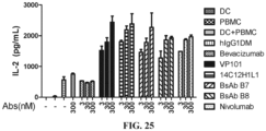
- the anti-VEGFA antibody and the anti-VEGFA/PD-1 bifunctional antibody both can inhibit HUVEC cell proliferation, and the anti-PD-1 antibody and the anti-VEGFA/PD-1 bifunctional antibody both can promote the secretion of IFN- ⁇ and/or IL-2 and activate immune reaction.
- a method for preventing and/or treating a malignant tumor may comprise administering to a subject in need an effective amount of the bispecific antibody according to any embodiment of the present invention or the conjugate of the present invention, wherein preferably, the malignant tumor is selected from colon cancer, rectal cancer, lung cancer such as non-small cell lung cancer, liver cancer, ovarian cancer, skin cancer, glioma, melanoma, renal tumor, prostate cancer, bladder cancer, gastrointestinal cancer, breast cancer, brain cancer and leukemia.
- lung cancer such as non-small cell lung cancer, liver cancer, ovarian cancer, skin cancer, glioma, melanoma, renal tumor, prostate cancer, bladder cancer, gastrointestinal cancer, breast cancer, brain cancer and leukemia.
- a typical non-limiting range of a therapeutically or prophylactically effective amount of the bispecific antibody of the present invention is 0.02-50 mg/kg, such as 0.1-50 mg/kg, 0.1-25 mg/kg, or 1-10 mg/kg. It should be noted that the dose may vary with the type and severity of the symptom to be treated. Furthermore, those skilled in the art will appreciate that for any particular patient, the particular administration regimen will be adjusted over time according to the needs of the patient and the professional judgment of the physician; the dose ranges given herein are for illustrative purpose only and do not limit the use or scope of the pharmaceutical composition of the present invention.
- a subject may be a mammal, such as a human.
- the bispecific antibody or the conjugate as claimed for use in preventing and/or treating a malignant tumor wherein preferably, the malignant tumor is selected from colon cancer, rectal cancer, lung cancer such as non-small cell lung cancer, liver cancer, ovarian cancer, skin cancer, glioma, melanoma, renal tumor, prostate cancer, bladder cancer, gastrointestinal cancer, breast cancer, brain cancer and leukemia.
- lung cancer such as non-small cell lung cancer, liver cancer, ovarian cancer, skin cancer, glioma, melanoma, renal tumor, prostate cancer, bladder cancer, gastrointestinal cancer, breast cancer, brain cancer and leukemia.
- a bispecific antibody or conjugate may be for use in:
- Antibody drugs especially monoclonal antibodies, have achieved good efficacy in the treatment of various diseases.
- Traditional experimental methods for acquiring these therapeutic antibodies are to immunize animals with the antigen and acquire antibodies targeting the antigen in the immunized animals, or to improve those antibodies with lower affinity for the antigen by affinity maturation.
- variable regions of the light chain and the heavy chain determine the binding of the antigen; the variable region of each chain contains three hypervariable regions called Complementarity Determining Regions (CDRs)
- CDRs of the heavy chain (H Chain) comprise HCDR1, HCDR2, and HCDR3
- CDRs of the light chain (L Chain) comprise LCDR1, LCDR2, and LCDR3, which are named by Kabat et al., see Bethesda M.d., Sequences of Proteins of Immunological Interest, Fifth Edition, NIH Publication (1-3) 1991: 91-3242 ).
- CDRs may also be defined by the IMGT numbering system, see Ehrenmann, Francois, Quentin Kaas, and Marie-Paule Lefranc. "IMGT/3Dstructure-DB and IMGT/DomainGapAlign: a database and a tool for immunoglobulins or antibodies, T cell receptors, MHC, IgSF and MhcSF.” Nucleic acids research 38.suppl_1 (2009): D301-D307 .
- amino acid sequence of the heavy chain variable region is set forth in SEQ ID NO: 5
- amino acid sequence of the light chain variable region is set forth in SEQ ID NO: 7.
- amino acid sequences of the 3 CDR regions of its heavy chain variable region are as follows:
- amino acid sequences of the 3 CDR regions of its light chain variable region are as follows:
- amino acid sequence of the heavy chain variable region is set forth in SEQ ID NO: 9, and the amino acid sequence of the light chain variable region is set forth in SEQ ID NO: 11.
- amino acid sequences of the 3 CDR regions of its light chain variable region are as follows:
- amino acid sequences of the 9 CDR regions of its heavy chains are as follows:
- amino acid sequences of the 3 CDR regions of its light chain variable region are as follows:
- VEGFA protein As used herein, when referring to the amino acid sequence of VEGFA protein (GenBank ID: NP_001165097.1), it includes the full length of the VEGFA protein, as well as a fusion protein of VEGFA, such as a fragment fused to an Fc protein fragment of mouse or human IgG (mFc or hFc).
- mFc or hFc fragment fused to an Fc protein fragment of mouse or human IgG
- VEGFA protein should include all such sequences, including their natural or artificial variants.
- the amino acid sequence of the VEGFA protein is shown as the underlined part of SEQ ID NO: 1 (without the last 6 His, a total of 165 amino acids).
- VEGFR2 protein when referring to the amino acid sequence of VEGFR2 protein (also known as KDR, GenBank ID: NP_002244), it includes the full length of the VEGFR2 protein, or the extracellular fragment VEGFR2-ECD of VEGFR2, or a fragment comprising VEGFR2-ECD, and it also includes a fusion protein of VEGFR2-ECD, such as a fragment fused to an Fc protein fragment of mouse or human IgG (mFc or hFc).
- mFc or hFc Fc protein fragment of mouse or human IgG
- VEGFR2 protein should include all such sequences, including their natural or artificial variants.
- sequence fragment of the VEGFR2 protein when describing the sequence fragment of the VEGFR2 protein, it also includes the corresponding sequence fragments in its natural or artificial variants.
- amino acid sequence of the extracellular fragment VEGFR2-ECD of VEGFR2 is shown as the wavy-underlined part of SEQ ID NO: 3 (766 amino acids).
- VEGFR is VEGFR1 and/or VEGFR2; specific protein sequence thereof is a sequence known in the prior art, and reference may be made to the sequence disclosed in the existing literature or GenBank.
- VEGFR1 (VEGFR1, NCBI Gene ID: 2321); VEGFR2 (VEGFR2, NCBI Gene ID: 3791).
- PD-1 protein when referring to the amino acid sequence of PD-1 protein (Programmed cell death protein 1, NCBI GenBank: NM_005018), it includes the full length of the PD-1 protein, or the extracellular fragment PD-1ECD of PD-1 or a fragment comprising PD-1ECD, and it also includes a fusion protein of PD-1ECD, such as a fragment fused to an Fc protein fragment of a mouse or human IgG (mFc or hFc).
- mFc or hFc Fc protein fragment of a mouse or human IgG
- the term "PD-1 protein” should include all such sequences, including their natural or artificial variants.
- sequence fragment of the PD-1 protein when describing the sequence fragment of the PD-1 protein, it also includes the corresponding sequence fragments in its natural or artificial variants.
- the term "antibody” refers to an immunoglobulin molecule that generally consists of two pairs of polypeptide chains (each pair with one "light” (L) chain and one "heavy” (H) chain).
- the heavy chain can be interpreted as a polypeptide chain with a larger molecular weight in an antibody
- the light chain refers to a polypeptide chain with a smaller molecular weight in an antibody.
- Light chains are classified as ⁇ and ⁇ light chains.
- Heavy chains are generally classified as ⁇ , ⁇ , ⁇ , ⁇ , or ⁇ , and isotypes of antibodies are defined as IgM, IgD, IgG, IgA, and IgE, respectively.
- variable region and constant region are linked by a "J" region of about 12 or more amino acids, and the heavy chain also comprises a "D" region of about 3 or more amino acids.
- Each heavy chain consists of a heavy chain variable region (V H ) and a heavy chain constant region (C H ).
- the heavy chain constant region consists of 3 domains (C H1 , C H2 , and C H3 ).
- Each light chain consists of a light chain variable region (V L ) and a light chain constant region (C L ).
- the light chain constant region consists of one domain C L .
- the constant region of the antibody can mediate the binding of immunoglobulins to host tissues or factors, including the binding of various cells of the immune system (e.g ., effector cells) to the first component (C1q) of classical complement system.
- the V H and V L regions can be further subdivided into highly variable regions (called Complementarity Determining Regions (CDRs)), between which conservative regions called framework regions (FRs) are distributed.
- CDRs Complementarity Determining Regions
- FRs framework regions
- Each V H and V L consists of 3 CDRs and 4 FRs arranged from amino terminus to carboxyl terminus in the following order: FR1, CDR1, FR2, CDR2, FR3, CDR3, FR4.
- the variable regions (V H and V L ) of each heavy chain/light chain pair form antibody binding sites, respectively.
- the assignment of amino acids to the regions or domains may be based on Kabat Sequences of Proteins of Immunological Interest (National Institutes of Health, Bethesda, Md. (1987 and 1991 )), or Chothia & Lesk J. Mol. Biol. 196(1987): 901-917 ; Chothia et al. Nature 342(1989): 878-883 or the definition of IMGT numbering system, see Ehrenmann, Francois, Quentin Kaas, and Marie-Paule Lefranc.
- the heavy chain may also comprise more than 3 CDRs, such as 6, 9, or 12.
- the heavy chain may be a ScFv with the C-terminus of the heavy chain of IgG antibody linked to another antibody, and in this case, the heavy chain comprises 9 CDRs.
- antibody is not limited by any specific method for producing antibody.
- the antibody includes, in particular, a recombinant antibody, a monoclonal antibody, and a polyclonal antibody.
- Antibodies can be different isotypes, such as antibody IgG ( e.g ., subtype IgG1, IgG2, IgG3 or IgG4), IgA1, IgA2, IgD, IgE or IgM.
- the term "antigen binding fragment”, also known as the "antigen binding portion”, refers to a polypeptide comprising the fragment of a full-length antibody, which maintains the ability to specifically bind to the same antigen to which the full-length antibody binds, and/or competes with the full-length antibody for the specific binding to an antigen. See generally, Fundamental Immunology, Ch. 7 (Paul, W., ed., 2nd edition, Raven Press, N.Y. (1989 ).
- An antigen-binding fragment of an antibody can be produced by recombinant DNA technique or by enzymatic or chemical cleavage of an intact antibody.
- the antigen binding fragment includes Fab, Fab', F (ab') 2 , Fv, a scFv, a chimeric antibody, and a diabody.
- the term “Fd fragment” refers to an antibody fragment consisting of V H and C H1 domains;
- the term “Fv fragment” refers to an antibody fragment consisting of the V L and V H domains of a single arm of an antibody;
- the term “dAb fragment” refers to an antibody fragment consisting of a V H domain ( Ward et al., Nature 341 (1989):544-546 );
- the term “Fab fragment” refers to an antibody fragment consisting of V L , V H , C L and C H1 domains;
- the term “F(ab') 2 fragment” refers to an antibody fragment comprising two Fab fragments linked by the disulfide bridge on a hinge region.
- the antigen binding fragment of the antibody is a scFv in which the V L and V H domains are paired to form a monovalent molecule via a linker that enables them to produce a single polypeptide chain (see, e.g., Bird et al., Science 242 (1988):423-426 and Huston et al., Proc. Natl. Acad. Sci. USA 85 (1988):5879-5883 ).
- Such scFv molecules may have a general structure: NH 2 -V L -linker-V H -COOH or NH 2 -V H -linker-V L -COOH.
- An appropriate linker in prior art consists of a repeating GGGGS amino acid sequence or a variant thereof.
- a linker having the amino acid sequence (GGGGS) 4 can be used, and variants thereof can also be used ( Holliger et al., Proc. Natl. Acad. Sci. USA 90 (1993): 6444-6448 ).
- Other linkers useful in the present invention are described by Alfthan et al., Protein Eng. 8 (1995): 725-731 , Choi et al., Eur. J. Immunol. 31 (2001): 94-106 , Hu et al., Cancer Res. 56 (1996): 3055-3061 , Kipriyanov et al., J. Mol. Biol. 293 (1999): 41-56 , and Roovers et al., Cancer Immunol. (2001 ).
- the antigen binding fragment of the antibody is a diabody, that is, a bivalent antibody, in which the V H and V L domains are expressed on a single polypeptide chain.
- the linker used is too short to allow the pairing of the two domains on the same chain, thereby the domains are forced to pair with the complementary domains on the other chain and two antigen binding sites are generated (see, e.g., Holliger P. et al., Proc. Natl. Acad. Sci. USA 90 (1993):6444-6448 , and Poljak RJ et al., Structure 2 (1994):1121-1123 ).
- Antigen binding fragments e.g ., the above mentioned antibody fragments
- Antigen binding fragments of antibodies can be obtained from given antibodies by using conventional techniques known to those skilled in the art (e.g ., recombinant DNA technique or enzymatic or chemical cleavage), and the antigen binding fragments of the antibodies are screened for specificity in the same way as for intact antibodies.
- antibody As used herein, unless otherwise clearly defined in the context, when referring to the term “antibody”, it includes not only intact antibodies but also antigen binding fragments of antibodies.
- mAb and “monoclonal antibody” refer to an antibody or a fragment thereof that is derived from a group of highly homologous antibodies, i.e. from a group of identical antibody molecules, except for natural mutations that may occur spontaneously.
- the monoclonal antibody has a high specificity for a single epitope on an antigen.
- the polyclonal antibody, relative to the monoclonal antibody generally comprises at least two or more different antibodies which generally recognize different epitopes on an antigen.
- Monoclonal antibodies can generally be obtained by hybridoma technique first reported by Kohler et al. (Nature, 256:495, 1975 ), and can also be obtained by recombinant DNA technique (for example, see U.S. Patent 4,816,567 ).
- chimeric antibody refers to an antibody of which a part of the light or/and heavy chains is derived from an antibody (which may be derived from a specific species or belong to a specific antibody class or subclass), and the other part of the light or/and heavy chains are derived from another antibody (which may be derived from the same or different species or belong to the same or different antibody class or subclass). But in any case, it retains the binding activity for the target antigen ( U.S. Patent 4,816,567 to Cabilly et al. ; Morrison et al., Proc. Natl. Acad. Sci. USA, 81 (1984):6851-6855 ).
- humanized antibody refers to an antibody or antibody fragment obtained when all or a part of CDR regions of a human immunoglobulin (receptor antibody) are replaced by the CDR regions of a non-human antibody (donor antibody), wherein the donor antibody may be a non-human (e.g., mouse, rat or rabbit) antibody having expected specificity, affinity or reactivity.
- donor antibody may be a non-human (e.g., mouse, rat or rabbit) antibody having expected specificity, affinity or reactivity.
- some amino acid residues in the framework regions (FRs) of the receptor antibody can also be replaced by the amino acid residues of corresponding non-human antibodies or by the amino acid residues of other antibodies to further improve or optimize the performance of the antibody.
- epitope refers to a site on the antigen that an immunoglobulin or antibody specifically binds to.
- epitope is also called in the art as an "antigenic determinant”.
- the epitope or antigenic determinant generally consists of chemically active surface groups of a molecule such as amino acids or carbohydrates or sugar side chains, and usually has specific three-dimensional structural characteristics and specific charge characteristics.
- the epitope generally includes at least 3, 4, 5, 6, 7, 8, 9, 10, 11, 12, 13, 14, or 15 consecutive or non-consecutive amino acids in a unique spatial conformation, which can be "linear” or “conformational”. See, for example, Epitope Mapping Protocols in Methods in Molecular Biology, Vol. 66, G. E.
- isolated refers to obtained by artificial means from natural state. If a certain "isolated” substance or component appears in nature, it may be that change occurs in its natural environment, or that it is isolated from the natural environment, or both. For example, a certain non-isolated polynucleotide or polypeptide naturally exists in a certain living animal, and the same polynucleotide or polypeptide with a high purity isolated from such a natural state is called isolated polynucleotide or polypeptide.
- isolated does not exclude the existence of artificial or synthetic substances or other impurities that do not affect the activity of the substance.
- vector refers to a nucleic acid vehicle into which a polynucleotide can be inserted.
- a vector allows for the expression of the protein encoded by the inserted polynucleotide
- the vector is called an expression vector.
- a vector can be introduced into a host cell by transformation, transduction, or transfection so that the genetic substance elements carried by the vector can be expressed in the host cell.
- Vectors are well known to those skilled in the art, including but not limited to: plasmids; phagemids; cosmids; artificial chromosomes, such as yeast artificial chromosome (YAC), bacterial artificial chromosome (BAC), or P1-derived artificial chromosome (PAC); phages such as lambda phages or M13 phages, and animal viruses.
- artificial chromosomes such as yeast artificial chromosome (YAC), bacterial artificial chromosome (BAC), or P1-derived artificial chromosome (PAC)
- phages such as lambda phages or M13 phages, and animal viruses.
- Animal viruses that can be used as vectors include, but are not limited to, retroviruses (including lentiviruses), adenoviruses, adeno-associated viruses, herpes viruses (such as herpes simplex virus), poxviruses, baculoviruses, papillomaviruses, and papovaviruses (such as SV40).
- retroviruses including lentiviruses
- adenoviruses such as lentiviruses
- adeno-associated viruses such as herpes viruses
- herpes viruses such as herpes simplex virus
- poxviruses such as herpes simplex virus
- baculoviruses such as baculoviruses
- papillomaviruses papillomaviruses
- papovaviruses such as SV40
- a vector can contain a variety of elements that control expression, including, but not limited to, promoter sequences, transcription initiation sequence
- the term "host cell” refers to cells to which the vector can be introduced, including but not limited to prokaryotic cells such as E. coli or bacillus subtilis, fungal cells such as yeast cells or aspergillus, insect cells such as S2 drosophila cells or Sf9, or animal cells such as fibroblast, CHO cells, COS cells, NSO cells, HeLa cells, BHK cells, HEK 293 cells, or human cells.
- prokaryotic cells such as E. coli or bacillus subtilis
- fungal cells such as yeast cells or aspergillus
- insect cells such as S2 drosophila cells or Sf9
- animal cells such as fibroblast, CHO cells, COS cells, NSO cells, HeLa cells, BHK cells, HEK 293 cells, or human cells.
- the term “specifically bind” refers to a non-random binding reaction between two molecules, such as a reaction between an antibody and an antigen it targets.
- an antibody that specifically binds to an antigen means that the antibody binds to the antigen with an affinity (K D ) of less than about 10 -5 M, such as less than about 10 -6 M, 10 -7 M, 10 -8 M, 10 -9 M or 10 -10 M or less.
- affinity K D
- target refers to specific binding.
- K D refers to a dissociation equilibrium constant for a specific antibody-antigen interaction, which is used to describe the binding affinity between the antibody and the antigen.
- K D dissociation equilibrium constant
- antibodies bind to antigens with a dissociation equilibrium constant (K D ) of less than about 10 -5 M, such as less than about 10 -6 M, 10 -7 M, 10 -8 M, 10 -9 M or 10 -10 M or less, for example, as determined in a BIACORE instrument using Surface Plasmon Resonance (SPR).
- amino acids are generally represented by single-letter and three-letter abbreviations known in the art.
- alanine can be represented by A or Ala.
- adjuvant refers to a non-specific immune enhancer, which can enhance the immune response of an organism to antigens or change the type of immune response when delivered into the organism together with the antigens or delivered into the organism in advance.
- adjuvants including but not limited to aluminum adjuvant (such as aluminum hydroxide), Freund's adjuvant (such as complete Freund's adjuvant and incomplete Freund's adjuvant), corynebacterium parvum, lipopolysaccharide, cytokine, etc.
- the Freund's adjuvant is the most commonly used adjuvant in animal experiments.
- the aluminum hydroxide adjuvant is used more in clinical trials.
- the term "effective amount" refers to an amount sufficient to obtain or at least partially obtain desired effect.
- a prophylactically effective amount e.g ., for a disease associated with PD-1 binding to PD-L1 or overexpression of VEGF, such as a tumor
- a prophylactically effective amount is an amount sufficient to prevent, stop, or delay the onset of the disease (e.g ., a disease associated with PD-L1 binding to PD-L1 or overexpression of VEGF, such as a tumor)
- a therapeutically effective amount is an amount sufficient to cure or at least partially stop the disease and its complications in a patient suffering from the disease. It is undoubtedly within the ability of those skilled in the art to determine such an effective amount.
- the amount effective for therapeutic use will depend on the severity of the disease to be treated, the overall state of the immune system of the patient, the general condition of the patient such as age, weight and sex, the mode of drug administration, and other treatments administered concurrently, etc.
- the bispecific antibody VP101 can specifically bind to VEGFA well, effectively block the binding of VEGFA to VEGFR2, and specifically relieve the immunosuppression of VEGFA in an organism and the promoting effect of VEGFA on angiogenesis; VP101 can specifically bind to PD-1 well, effectively block the binding of PD-1 to PD-L1, and specifically relieve the immunosuppression of PD-1 in an organism, and activate the immune response.
- the marketed antibody bevacizumab (trade name Avastin ® ) for the same target was purchased from Roche as a control antibody, or was prepared according to Preparation Example 4.
- the marketed antibody nivolumab for the same target (trade name Opdivo ® ) was purchased from BMS as a control antibody.
- control antibodies BsAbB7 and BsAbB8 were identical to the amino acid sequences of BsAbB7 and BsAbB8 respectively in Chinese Patent Publication CN105175545A .
- amino acid sequences and the encoding nucleotide sequences of the fusion proteins PD-1-mFc, PD-1-hFc and PD-L1-hFc in this preparation example are the same as those of PD-1-mFc, PD-1-hFc and PDL-1-hFc respectively in the Preparation Example 1 of Chinese Patent Publication CN106632674A .
- Fusion proteins PD-1-mFc, PD-1-hFc and PD-L1-hFc were thus obtained.
- PCR amplification was performed using VEGFA human cDNA (purchased from Origene) as a template and the hVEGFA-His fragment was purified and isolated using an ordinary DNA product purification kit.
- the isolated hVEGFA-His fragment and an expression vector pcDNA3.1 were enzyme-digested with XbaI&HindIII-HF, and a target gene fragment was isolated by gel extraction and ligated with a linear expression vector by T4 ligase. Then all the ligation products were transformed into DH5a chemically competent cells and coated on an Agar plate with Amp.
- the culture medium was subjected to high-speed centrifugation, supernatant concentration and buffer exchange into Binding Buffer A, and then loaded onto a HisTrap column, and proteins were linearly eluted with Elution Buffer A.
- the primary pure sample was subjected to buffer exchange into Binding Buffer B with a HiTrap Desalting column and loaded onto a HiTrap Q column, proteins were linearly eluted with Elution Buffer B, and the target sample was isolated and buffer exchanged into PBS.
- the purified sample was added to a reduced protein electrophoresis loading buffer for SDS-PAGE electrophoresis detection.
- the fusion protein VEGFA-His was thus obtained.
- VEGFA-His The amino acid sequence of VEGFA-His is as follows (171 aa): wherein, the underlined part without the 6-His is the amino acid sequence of VEGFA.
- VEGFR2 ECD of gene VEGFR2 Vascular Endothelial Growth Factor Receptor 2, NCBI GenBank: NP_002244
- TEV Transcription Factor Receptor 2
- Fc Fc protein fragment of human IgG
- the wavy-underlined part is the ECD part of VEGFR2
- the framed part is TEV enzyme digestion site
- the solid-underlined part is hFc part.
- the wavy-underlined part is the ECD part of VEGFR2
- the framed part is TEV enzyme digestion site
- the solid-underlined part is hFc part.
- VEGFR2-hFc encoding gene synthesized by Genscript was cloned into an expression vector pUC57simple (provided by Genscript), and a pUC57simple-VEGFR2-hFc plasmid was obtained.
- the plasmid pUC57simple-VEGFR2-hFc was enzyme-digested (Xba I and BamH I), and the fusion gene fragment VEGFR2-hFc isolated by electrophoresis was ligated with expression vector pcDNA3.1 (purchased from Invitrogen) to give pcDNA3.1-VEGFR2-hFc, which was transfected into competent E. coli cell DH5a (purchased from TIANGEN); the transfection and culture were performed according to the manual.
- the positive pcDNA3.1-VEGFR2-hFc colonies were screened, E.
- coli was amplified according to a conventional method, and a kit (purchased from Tiangen Biotech (Beijing) Co., Ltd., DP103-03) was then used and a recombinant plasmid pcDNA3.1-VEGFR2-hFc was extracted according to the manual of the kit.
- a kit purchased from Tiangen Biotech (Beijing) Co., Ltd., DP103-03
- a recombinant plasmid pcDNA3.1-VEGFR2-hFc was extracted according to the manual of the kit.
- the recombinant plasmid pcDNA3.1-VEGFR2-hFc was transfected into 293F cells (purchased from Invitrogen) according to the lipofectamin transfection kit (purchased from Invitrogen).
- the culture medium was subjected to high-speed centrifugation, microporous membrane vacuum filtration and purification in a Mabselect SuRe column to obtain a VEGFR2-hFc fusion protein sample, and a part of the sample was added into a reduced protein electrophoresis loading buffer for SDS-PAGE electrophoresis detection.
- the fusion protein VEGFR2-hFc was thus obtained.
- Chinese Patent Publication CN1259962A is referred to for the amino acid sequences of the heavy chain variable region and the light chain variable region of the marketed VEGFA monoclonal antibody Avastin (bevacizumab). Genscript was entrusted to synthesize nucleotide sequences encoding the heavy chain variable region and the light chain variable region.
- Amino acid sequence of the heavy chain variable region of bevacizumab (123 aa)
- Amino acid sequence of the light chain variable region of bevacizumab (107 aa)
- the heavy chain constant regions were all Ig gamma-1 chain C region, ACCESSION: P01857; the light chain constant regions were all Ig kappa chain C region, ACCESSION: P01834.
- the heavy chain cDNA and the light chain cDNA of bevacizumab were cloned into vector pcDNA3.1, and the recombinant expression plasmid of the antibody bevacizumab was obtained.
- the recombinant plasmid was transfected into 293F cells.
- the 293F cell culture medium was purified and then detected.
- the anti-VEGFA monoclonal antibody Avastin (bevacizumab) was thus obtained.
- amino acid sequences of the heavy chain variable region and the light chain variable region of humanized antibody 14C12H1L1, and the nucleotide sequence encoding the same are also the same as those described in Examples 3-4 of Chinese Patent Publication CN106977602A , and are also provided herein as follows:
- the anti-PD-1 humanized antibody 14C12H1L1 was thus obtained.
- anti-HEL Human Anti-Hen Egg Lysozyme IgG
- hIgG human IgG
- the sequence of Human Anti-Hen Egg Lysozyme IgG is derived from a variable region sequence of the Fab F10.6.6 sequence in the research published by Acierno et al., which is entitled " Affinity maturation increases the stability and plasticity of the Fv domain of anti-protein antibodies" (Acierno et al., J Mol Biol. 2007; 374(1): 130-46 ).
- the preparation method is as follows: Nanjing Genscript Biology was entrusted to carry out codon optimization of amino acids and gene synthesis on heavy and light chain (complete sequence or variable region) genes of human IgG antibody, and by referring to the standard technologies introduced in the "Guide to Molecular Cloning Experiments (Third Edition)" and using standard molecular cloning technologies such as PCR, enzyme digestion, DNA gel extraction, ligation transformation, colony PCR or enzyme digestion identification, the heavy and light chain genes were respectively subcloned into the antibody heavy chain expression vector and antibody light chain expression vector of the mammalian expression system, and the heavy and light chain genes of the recombinant expression vector were further sequenced and analyzed.
- endotoxin-free expression plasmids were prepared in a large scale, and the heavy and light chain plasmids were transiently co-transfected into HEK293 cells for expression of recombinant antibody. After 7 days of culture, the cell culture medium was collected and affinity purified using an rProtein A column (GE), and the quality of the resulting antibody sample was determined using SDS-PAGE and SEC-HPLC standard analysis techniques.
- GE rProtein A column
- the hIgG was thus obtained, and used in Examples 8-9 below.
- Example 1 Sequence Design. Preparation and Detection of Heavy and Light Chains of Bifunctional Antibody VP101
- the structure of the bifunctional antibody VP101 of the present invention is in the Morrison form (IgG-scFv), i.e. C-termini of two heavy chains of an IgG antibody are each linked to a scFv fragment of another antibody, and the main composition design of the heavy and light chains is as shown in Table 1 below.
- Table 1 Composition design of the heavy and light chains of VP101 Bifunctional antibody No. Heavy chain Light chain IgG part Linker fragment scFv part VP101 Bevacizum ab-H Linker 1 14C12H1 V -Linker1-14C12L1v Bevacizumab-L
- the heavy chain cDNA sequence and the light chain cDNA sequence of VP101 were each cloned into vector pUC57simple (provided by Genscript) to obtain plasmids pUC57simple-VP101H and pUC57simple- VP101L, respectively.
- Plasmids pUC57simple-VP101H and pUC57simple-VP101L were enzyme-digested (HindIII&EcoRI), and heavy and light chains isolated by electrophoresis were subcloned into vector pcDNA3.1, and recombinant plasmids were extracted to co-transfect 293F cells. After 7 days of cell culture, the culture medium was centrifuged at high speed, and the supernatant was concentrated and loaded onto a HiTrap MabSelect SuRe column. The protein was further eluted in one step with Elution Buffer, and the target sample antibody VP101 was isolated and buffer exchanged into PBS.
- the purified sample was added to both a reduced protein electrophoresis loading buffer and a non-reduced protein electrophoresis loading buffer, and then boiled for SDS-PAGE electrophoresis detection.
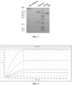
- the electropherogram of VP101 is shown in FIG. 1 .
- the target protein of the reduced protein sample is at 75 kD and 25 kD, and the target protein of the non-reduced protein sample (single antibody) is at 200 kD.
- the humanized antibody VP101 used in the following experiments was prepared by the method of this example.
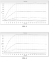
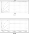
- the sample dilution buffer was PBS (0.02% Tween-20, 0.1% BSA, pH7.4). 5 ⁇ g/mL antibody was immobilized to an AHC sensor with the immobilization height being about 0.4 nM.
- the sensor was equilibrated in a buffer for 60 s, and the antibody immobilized to the sensor bound to PD-1-mFc at a concentration of 0.62-50 nM (three-fold gradient dilution) for 120 s, and then the antigen and antibody dissociated in the buffer for 300 s.
- the data were analyzed by 1: 1 model fitting to obtain affinity constants.
- the data acquisition software was Fortebio Data Acquisition 7.0, and the data analysis software was Fortebio Data Analysis 7.0.
- the method is specified as follows: The microplate was coated with VEGFA-His and incubated at 37 °C for 2 hours. After being washed, the microplate was blocked with 1% BSA for 2 hours. After being washed, the microplate was added with the gradiently diluted antibody and incubated at 37 °C for 30 minutes. After being washed, the microplate was added with the enzyme-labeled goat anti-human IgG secondary antibody working solution and incubated for 30 minutes at 37 °C. After being washed, the microplate was added with TMB chromogenic solution for color developing for 5 minutes in the absence of light, and then stop solution was added to terminate the chromogenic reaction. Then the microplate was put into a microplate reader immediately, and the OD value of each well in the microplate was read at 450 nm. SoftMax Pro 6.2.1 was used to analyze and process the data.
- the detection result of the binding of antibody VP101 to antigen VEGFA-His is shown in FIG. 11 .
- the absorbance intensities at each dose are shown in Table 4.
- the binding EC 50 of antibody was calculated by curve fitting using antibody concentration as the abscissa and absorbance value as the ordinate, and the results are shown in Table 4 below.
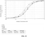
- the method is specified as follows: The microplate was coated with VEGFA-His and incubated overnight at 4 °C. After being washed, the microplate was blocked with 1% BSA (dissolved in PBS) for 2 hours. After being washed, the microplate was added with the gradiently diluted antibody and incubated at 37 °C for 30 minutes. After being washed, the microplate was added with the horseradish peroxidase-labeled goat anti-human IgG Fc (Jackson, 109-035-098) working solution and incubated for 30 minutes at 37 °C.
- the microplate After being washed, the microplate was added with TMB (Neogen, 308177) for color developing for 5 minutes in the absence of light, and then stop solution was added to terminate the chromogenic reaction. Then the microplate was put into a microplate reader immediately, and the OD value of each well in the microplate was read at 450 nm. SoftMax Pro 6.2.1 was used to analyze and process the data.
- TMB Neogen, 308177
- the result of the binding of antibody VP101 to antigen VEGFA-His is shown in FIG. 12 .
- the absorbance intensities at each dose are shown in Table 5.
- the binding EC 50 of antibody was calculated by curve fitting using antibody concentration as the abscissa and absorbance value as the ordinate, and the results are shown in Table 5 below.
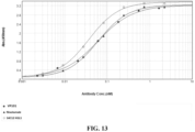
- the method is specified as follows: The microplate was coated with human PD-1-mFc and incubated overnight at 4 °C. After being blocked with 1% BSA at 37 °C for 2 hours, the microplate was added with antibody, and then incubated at 37 °C for 30 minutes. After the microplate was washed and patted dry, the HRP-labeled goat anti-human IgG (H+L) secondary antibody (Jackson, 109-035-088) was added, and the microplate was incubated at 37 °C for 30 minutes. After the microplate was washed and patted dry, TMB (Neogen, 308177) was added for color developing for 5 minutes, and then stop solution was added to terminate the color development. Then the microplate was put into a microplate reader immediately, and the OD value of each well in the microplate was read at 450 nm. SoftMax Pro 6.2.1 was used to analyze and process the data.
- HRP-labeled goat anti-human IgG (H+L) secondary antibody
- the detection result of the binding of antibody VP101 to antigen PD-1 is shown in FIG. 13 .
- the absorbance intensities at each dose are shown in Table 6.
- the curve simulation was performed to obtain the binding efficiency EC 50 of the antibody, which is shown in Table 6 below.
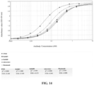
- the method is specified as follows: The microplate was coated with human PD-1-mFc and incubated overnight at 4 °C. After being blocked with 1% BSA at 37 °C for 2 hours, the microplate was added with antibody, and then incubated at 37 °C for 30 minutes. After the microplate was washed and patted dry, the horseradish peroxidase-labeled goat anti-human IgG Fc (Jackson, 109-035-098) was added, and the microplate was incubated at 37 °C for 30 minutes. After the microplate was washed and patted dry, TMB (Neogen, 308177) was added for color developing for 5 minutes, and then stop solution was added to terminate the color development. Then the microplate was put into a microplate reader immediately, and the OD value of each well in the microplate was read at 450 nm. SoftMax Pro 6.2.1 was used to analyze and process the data.
- the detection result of the binding of antibody VP101 to antigen PD-1 is shown in FIG. 14 .
- the absorbance intensities at each dose are shown in Table 7.
- the curve simulation was performed to give the binding efficiency EC 50 of the antibody, which is shown in Table 7 below.
- Table 7 Respective binding activities of antibodies VP101, BsAbB7, BsAbB8, 14C12H1L1 and nivolumab to PD-1 (Indirect ELISA) Antibody concentration ( ⁇ g/mL) Antigen coating: PD-1-mFc 0.5 ⁇ g/mL VP101 BsAbB7 BsAbB8 14C12 H1L1 Nivolumab 0.333 2.717 2.709 2.732 2.755 2.716 2.715 2.947 2.966 2.823 2.824 0.111 2.507 2.381 2.318 2.321 2.377 2.409 2.923 2.967 2.747 2.758 0.037 1.709 1.616 1.491 1.457 1.522 1.549 2.656 2.694 2.208 2.293 0.012 0.916 0.822 0.732 0.711 0.797 0.775 2.049 2.060 1.348 1.389 0.004 0.413 0.394 0.333 0.321 0.368 0.351 1.139 1.132 0.629
- the results show that the antibody VP101 can bind to the PD-1 protein efficiently and its binding efficiency is dose-dependent, and antibody VP101 has a higher binding activity to human PD-1 than BsAbB7 and BsAbB8.
- the method is specifically as follows: The microplate was coated with VEGF-His and incubated at 37 °C for 2 hours. After being washed, the microplate was blocked with 1% BSA for 1 hour at 37 °C. After being washed, the microplate was added with the gradiently diluted antibodies and human VEGFR2 ECD-mFc-bio (final concentration: 0.02 ⁇ g/mL) and incubated at room temperature for 2 hours. After being washed, the microplate was added with HRP-labeled streptavidin SA-HRP (1:4000) working solution and incubated at 37 °C for 30 minutes.
- the microplate After being washed, the microplate was added with TMB chromogenic solution for color developing for 5 minutes in the absence of light, and then stop solution was added to terminate the chromogenic reaction. Then the microplate was put into a microplate reader immediately, and the OD value of each well in the microplate was read at 450 nm. SoftMax Pro 6.2.1 was used to analyze and process the data.
- the results show that the antibody VP101 can effectively bind to the antigen VEGFA and inhibit the binding of VEGFR2 to VEGFA, and its efficiency in inhibiting the binding of VEGFR2 to VEGFA is dose-dependent.
- the method is specifically as follows: The microplate was coated with PD-1-hFc and incubated overnight at 4 °C. After the microplate was blocked with 1% BSA for 2 hours, antibodies at different concentrations were each mixed with PD-L1-hFc for 10 minutes (see Table 10 for the dilution concentrations). After incubation at 37 °C for 30 minutes, the microplate was washed and patted dry. Then enzyme-labeled secondary antibody was added, and the microplate was incubated at 37 °C for 30 minutes. After the microplate was washed and patted dry, TMB was added for color developing for 5 minutes, and then stop solution was added to terminate the color development. Then the microplate was put into a microplate reader immediately, and the OD value of each well in the microplate was read at 450 nm (see Table 10). SoftMax Pro 6.2.1 was used to analyze and process the data.
- Example 4 Binding of antibody VP101 to cell membrane surface antigen
- 293T cells expressing PD-1 antigen was constructed, and then the specific binding capacity of the antibody to the cell membrane surface antigen was analyzed and verified by flow cytometry.
- the vector pLenti6.3-PD-1 of PD-1 (the vector pLenti6.3 was purchased from Invitrogen) was transfected into 293T cells, and clone group 293T-PD-1 cells which stably express PD-1 were obtained by screening.
- the 293T-PD-1 expressing antigen obtained in the previous step was digested with pancreatin by a conventional pancreatin digestion method, and the number of cells in each collection tube was made to be 2 ⁇ 10 5 .
- Antibody diluting solutions with concentration gradiently diluted with PBSA (1% BSA) were each incubated with 293T-PD-1 cells on ice for 2 hours, and then each tube was added with 100 ⁇ L of FITC goat anti-human IgG (1:500) and incubated on ice for 1 hour. Then PBS was used for washing, and 300 ⁇ L of PBSA was used to resuspend the cells, and fluorescence signals (MFI) were detected with FITC channel on a flow cytometer.
- MFI fluorescence signals
- Table 10 Analysis of fluorescence intensity of the binding of VP101 to 293T-PD-1 surface antigen detected by FACS Antibody (nM) 0.14 0.41 1.23 3.70 11.11 33.33 100 EC 50 Bevacizumab 3.2 2.2 2.0 2.3 2.7 3.8 5.7 - Nivolumab 33.3 74.9 171.9 357.9 481.9 498.3 478.4 2.1 14C12H1L1 48.1 99.7 201.5 409.0 600.2 655.4 670.8 2.9 VP101 30.8 61.8 135.7 286.9 487.7 534.0 528.6 3.5
- the results show that the VP101 antibody can effectively bind to the PD-1 antigen on the 293T-PD-1 host cell surface, and its binding efficiency is dose-dependent, and bevacizumab has no binding activity to 293T-PD-1, which indicates that the binding of VP101 to 293T-PD-1 is specific.
- the binding EC 50 values of nivolumab, 14C12H1L1, VP101, BsAbB7 and BsAbB8 were calculated to be 7.853 nM, 3.607 nM, 7.896 nM, 9.943 nM and 10.610 nM, respectively.
- Table 11 Analysis of fluorescence intensities of the binding of VP101, BsAbB7 and BsAbB8 to 293T-PD-1 surface antigen detected by FACS Antibody (nM) 0.014 0.14 0.41 1.23 3.7 11 30 100 EC50(nM) Bevacizumab 1.89 1.90 2.20 1.92 2.04 2.48 2.80 2.43 - Nivolumab 3.91 15.30 34.69 94.04 234.34 533.63 640.15 804.69 7.853 14C12H1L1 7.40 29.55 69.16 175.54 422.53 868.45 831.27 813.58 3.607 VP101 3.47 16.16 38.75 93.08 216.76 509.23 810.37 783.58 7.896 BsAbB7 3.85 14.86 37.45 83.78 202.40 465.10 837.61 846.80 9.943 BsAbB8 4.41 16.77 36.86 89.89 210.40 457.91 8
- VP101 antibody can bind to the membrane surface PD-1 of 293T-PD1 in a dose-dependent manner.
- Bevacizumab has no binding activity to 293T-PD-1, which indicates that the binding of VP101 to 293T-PD-1 is specific.
- Example 5 Competitive binding of antibody VP101 to cell membrane surface antigen
- a competitive flow cytometry method was adopted to detect the EC 50 of the VP101 in competing with PD-L1 for binding to the cell membrane surface antigen PD-1, and the method is specified as follows:
- the 293T-PD-1 cells was digested in a conventional way, and divided into several samples with 300,000 cells for each, which were then subjected to centrifugation and washing. Then each tube was added with 100 ⁇ L of corresponding gradiently diluted antibody and incubated on ice for 30 minutes; 100 ⁇ L of PD-L1-mFc was then added to each tube, and the mixture was mixed well to reach a final concentration of 20 nM, and then incubated on ice for 1 hour.
- the binding EC 50 values of the antibodies VP101 and 14C12H1L1 were calculated to be 8.33 nM and 4.37 nM, respectively.
- Table 12 Analysis of fluorescence intensities of 14C12H1L1 and VP101 in competing for binding to 293T-PD-1 surface antigen detected by FACS Antibody (nM) 0.05 0.14 0.41 1.23 3.70 11.11 33.33 100.00 EC 50 R 2 14C12H1L1 288.17 287.29 277.09 237.22 177.80 12.04 10.32 9.87 4.37 0.988 VP101 272.66 264.39 279.11 272.26 239.18 99.29 17.05 14.91 8.33 0.999 The results show that the VP101 antibody can effectively block the binding of PDL-1 to PD-1 on the surface of 293T-PD-1 host cells in a dose-dependent manner.
- EC 50 values of VP101, BsAbB7, BsAbB8, 14C12H1L1 and nivolumab in competing with PD-L1 for binding to the cell membrane surface antigen PD-1 were detected by using competitive flow cytometry and referring to the experimental procedure described in step 1 of this example.
- the results are shown in FIG. 20 , and the MFI values at each concentration are shown in Table 13.
- the competitive binding EC50 values of antibodies VP101, BsAbB7, BsAbB8, 14C12H1L1 and nivolumab were calculated to be 15.04 nM, 22.25 nM, 19.25 nM, 9.21 nM and 9.72 nM, respectively.
- Table 13 Analysis of fluorescence intensities of VP101, BsAbB7, BsAbB8, 14C12H1L1 and nivolumab in competing for binding to 293T-PD-1 surface antigen detected by FACS Antibody (nM) 0.14 0.41 1.23 3.7 11.11 33.33 100 300 EC 50 R 2 VP101 1441.94 1380.62 1368.15 1288.34 982.69 112.90 8.49 8.21 15.04 0.9971 BsAbB7 1412.62 1377.27 1339.60 1341.27 1094.35 417.70 9.23 9.18 22.25 0.9985 BsAbB8 1578.36 1521.50 1427.20 1429.85 1137.74 359.69 9.73 9.68 19.25 0.9962 14C12H1L1 1384.08 1551.05 1462.85 1296.64 580.45 12.93 13.37 14.99 9.21 0.9950 Nivolumab 1539.58 1552.37 1483.84 1300.81 7
- the results show that the activity of the antibody 14C12H1L1 is equivalent to that of the marketed antibody nivolumab targeting PD-1, and is superior to that of the bifunctional antibody VP101.
- the activity of the antibody VP101 is superior that of to BsAbB7 and BsAbB8.
- KDR (VEGFR2) vector pCDH-KDRFL(OPV)-GFP-Puro (Vector pCDH-GFP-Puro is purchased from Youbio) and NFAT vector pNFAT-luc-hygro (vector pGL4-luc2P-hygro is purchased from Promega) were transfected into 293T cells, and a clone group 293T-NFAT-(opv) KDR(C7) cells stably expressing KDR and NFAT luciferase reporter genes were obtained by screening.
- 293T-NFAT-(opv)KDR(C7) cells were collected and centrifuged for 5 minutes to remove the supernatant; DMEM+10%FBS medium was used to resuspend the cells, and the cell number was counted and the cell viability was detected; then the cell concentration was adjusted to be in a proper range, and 50000 cells/50 ⁇ L cell suspension was added into each well of a black 96-well plate;
- Corresponding antibodies (final concentrations being 300, 100, 10, 2, 0.2, 0.02, 0.002 nM) and VEGF (final concentration being 30 ng/mL) were diluted according to the experimental design, and the antibodies targeting VEGF were preincubated with VEGF for 1 hour at room temperature before being added into the cells.
- Blank and isotype controls (final volume of each well being 100 ⁇ L) were designed and incubated in a carbon dioxide incubator at 37 °C, 5% CO 2 for 4 hours; 50 ⁇ L of Luciferase Assay System was added to each well, and Relative Fluorescence Units (RLUs) were detected by a multi-label microplate tester within 5 minutes.
- RLUs Relative Fluorescence Units
- the results show that the EC 50 of VP101 is 1.240 nM, the EC 50 of BsAbB7 is 1.217 nM, the EC 50 of BsAbB8 is 1.728 nM, and the EC 50 of bevacizumab is 0.773 nM, and the experimental results show that the activity of VP101 and BsAbB7 in blocking VEGF to activate NFAT signaling pathway is better than that of BsAbB8.
- Example 7 Experiment of VP101 Antibody Inhibiting VEGFA-Induced HUVEC Cell Proliferation
- HUVEC cells purchased from Allcell in a good growth state, after the cell concentration was adjusted to be 1.5 ⁇ 10 4 /mL, were inoculated into a 96-well plate at 200 ⁇ L/well, and then incubated in an incubator at 37 °C, 5% CO 2 for 24 hours. Then it was observed that the cells adhered well, and then culture medium was discarded. 20 nM VEGFA prepared by using 1640 containing 2% FBS was then added into the 96-well plate at 200 ⁇ L/well, and antibodies at different concentrations were added, followed by incubation for 72 hours. 72 hours later, the culture medium was discarded and MTT was added. 4 hours later, the MTT was discarded and DMSO was added, and then a microplate reader was used to measure the OD value at 490 nm.
- the results are shown in FIG. 22 .
- the results show that the humanized antibodies VP101 and bevacizumab both can effectively inhibit VEGFA-induced HUVEC cell proliferation in a dose-dependent manner, and the pharmacological activity of VP101 in inhibiting VEGFA-induced HUVEC cell proliferation is higher than that of bevacizumab at the same dose.
- PBMCs were isolated by Ficoll-Paque Plus (GE Healthcare) and added to IL-4 (Peprotech 200-04, 1000 U/mL) and GM-CSF (Peprotech 300-03, 1000 U/mL) for 6 days of induction, and then TNF- ⁇ (Peprotech 300-01A, 200 U/mL) was additionally added for 3 days of induction to obtain mature DC cells.
- IL-4 Peprotech 200-04, 1000 U/mL
- GM-CSF Peprotech 300-03, 1000 U/mL
- TNF- ⁇ Peprotech 300-01A, 200 U/mL
- VP101 can effectively promote secretion of IFN-y in a dose-dependent manner.
- VP101 has greater activity in promoting secretion of IFN- ⁇ than equivalent 14C12H1L1, and at dose level of 30 nM, it has greater activity in promoting secretion of IFN-y than equivalent nivolumab.
- Step 1 in this example was referred to for the experimental method, namely PBMCs were isolated by Ficoll-Paque Plus (GE Healthcare) and added to IL-4 (Peprotech 200-04, 1000 U/mL) and GM-CSF (Peprotech 300-03, 1000 U/mL) for 6 days of induction, and then TNF- ⁇ (Peprotech 300-01A, 200 U/mL) was additionally added for 3 days of induction to obtain mature DC cell.
- IL-4 Peprotech 200-04, 1000 U/mL
- GM-CSF Peprotech 300-03, 1000 U/mL
- VP101 The effect of VP101 on secretion of IFN-y in mixed culture system of DC and PBMC cells is shown in FIG. 24 .
- VP101 can effectively promote secretion of IFN-y in a dose-dependent manner.
- the pharmacological activity of VP101 in promoting secretion of IFN- ⁇ is significantly better than that of BsAbB7 and BsAbB8.
- VP101 The effect of VP101 on secretion of IL-2 in mixed culture system of DC and PBMC cells is shown in FIG. 25 .
- VP101 can effectively promote secretion of IL-2 in a dose-dependent manner, and the pharmacological activity of VP101 in promoting secretion of IL-2 is better than that of BsAbB7 and BsAbB8.
- PD-L1 was stably transfected into Raji cells through lentivirus infection, and Raji-PD-L1 cells stably expressing PD-L1 were obtained after dosing and screening; PBMCs, after two days of stimulation by SEB, were cultured in together with mitomycin C-treated Raji-PD-L1.
- the results are shown in FIGs. 26 and 27 .
- the results show that VP101 can effectively promote secretion of IL-2 and IFN-y, and at dose level of 300 nM, the activity of VP101 in promoting secretion of IL-2 is significantly better than that of equivalent 14C12H1L1 and nivolumab.
- the isotype control antibody to be studied was Human Anti-Hen Lysozyme (anti-HEL, i.e., human IgG, abbreviated as hIgG), and it was prepared as described in Preparation Example 6 above.
- anti-HEL Human Anti-Hen Lysozyme
- the results of secretion of IFN-y are shown in FIG. 28 .
- the results show that VP101 can effectively promote secretion of IFN-y in a dose-dependent manner.
- VP101 is significantly better than BsAbB7 at the same dose, while the pharmacological activity of VP101 is significantly better than that of BsAbB8 at dose levels of 3 nM and 30 nM.
- the results of secretion of IL-2 are shown in FIG. 29 .
- the results show that VP101 can effectively promote secretion of IL-2 in a dose-dependent manner.
- the pharmacological activity of VP101 is significantly better than that of BsAbB7 at doses of 3 nM and 300 nM, while VP101 is equivalent to BsAbB8 at the same dose.
- U87MG cells human glioma cells, purchased from ATCC
- Scid Beige mice purchased from Vital River
- Table 15 Dosing regimen of treating U87MG tumor xenograft Scid Beige mouse model with VP101 Grouping n Tumor xenograft Condition of administration Isotype control 40 mg/kg 7 U-87MG, 5 million cells/mouse subcutaneously Isotype control antibody, hIgG, 40 mg/kg, injected intravenously on days 0, 7 and 13 Bevacizumab 30mg/kg 8 Bevacizumab 30 mg/kg, injected intravenously on days 0, 7 and 13 VP101 40mg/kg 7 VP101 40 mg/kg, injected intravenously on days 0, 7 and 13 VP101 4mg/kg 7 VP101 4 mg/kg, injected intravenously on days 0, 7 and 13
- the results are shown in FIG. 30 .
- the results show that compared with an isotype control antibody hIgG (the preparation method is the same as that of the Preparation Example 6), bevacizumab and VP101 at different doses can effectively inhibit the growth of mouse tumors, and the high-dose VP101 is better than that of low-dose VP 101 in inhibiting tumors.
- VP 101 does not affect the body weight of tumor-bearing mouse.
Landscapes
- Health & Medical Sciences (AREA)
- Life Sciences & Earth Sciences (AREA)
- Chemical & Material Sciences (AREA)
- Immunology (AREA)
- Organic Chemistry (AREA)
- Molecular Biology (AREA)
- General Health & Medical Sciences (AREA)
- Medicinal Chemistry (AREA)
- Engineering & Computer Science (AREA)
- Biochemistry (AREA)
- Biomedical Technology (AREA)
- Hematology (AREA)
- Genetics & Genomics (AREA)
- Proteomics, Peptides & Aminoacids (AREA)
- Urology & Nephrology (AREA)
- Biophysics (AREA)
- Cell Biology (AREA)
- Animal Behavior & Ethology (AREA)
- Veterinary Medicine (AREA)
- Public Health (AREA)
- Pharmacology & Pharmacy (AREA)
- Biotechnology (AREA)
- Nuclear Medicine, Radiotherapy & Molecular Imaging (AREA)
- Microbiology (AREA)
- Physics & Mathematics (AREA)
- Chemical Kinetics & Catalysis (AREA)
- General Chemical & Material Sciences (AREA)
- General Physics & Mathematics (AREA)
- Pathology (AREA)
- Analytical Chemistry (AREA)
- Food Science & Technology (AREA)
- Bioinformatics & Cheminformatics (AREA)
- Oncology (AREA)
- Zoology (AREA)
- General Engineering & Computer Science (AREA)
- Wood Science & Technology (AREA)
- Epidemiology (AREA)
- Plant Pathology (AREA)
- Endocrinology (AREA)
- Peptides Or Proteins (AREA)
Description
- The present invention relates to the fields of tumor treatment and immunobiology, particularly to an anti-PD-1/VEGFA bifunctional antibody, a pharmaceutical composition thereof and use thereof. Specifically, the present invention relates to an anti-human PD-1/human VEGFA bifunctional antibody, a pharmaceutical composition thereof and use thereof.
- Tumor, especially a malignant tumor, is a serious health-threatening disease in the world today, and it is the second leading cause of death among various diseases. In recent years, the incidence of the disease has been increasing remarkably. Malignant tumor is characterized by poor treatment response, high late metastasis rate and poor prognosis. Although conventional treatment methods (such as radiotherapy, chemotherapy and surgical treatment) adopted clinically at present alleviate the pain to a great extent and prolong the survival time, the methods have great limitations, and it is difficult to further improve their efficacy.
- There are two distinct stages of tumor growth, namely, from a slow growth stage without blood vessels to a rapid proliferation stage with blood vessels. The angiogenesis enables the tumor to acquire enough nutrition to complete the blood vessel switching stage, and if there is no angiogenesis, the primary tumor will be no more than 1-2 mm, and thus the metastasis cannot be realized.
- Vascular Endothelial Growth Factor (VEGF) is a growth factor which can promote division and proliferation of endothelial cells, promote formation of new blood vessels and improve blood vessel permeability, and it binds to vascular endothelial growth factor receptors on the cell surface and plays a role by activating tyrosine kinase signal transduction pathways. In tumor tissues, tumor cells, and macrophages and mast cells invading into tumors can secrete high-level VEGF, stimulate tumor vascular endothelial cells in a paracrine form, promote proliferation and migration of endothelial cells, induce angiogenesis, promote continuous growth of tumor, improve vascular permeability, cause fibrin deposition in surrounding tissues, and promote infiltration of mononuclear cells, fibroblast and endothelial cells, which facilitates formation of tumor stroma and entry of tumor cells into new blood vessels, and promote tumor metastasis. Therefore, inhibiting tumor angiogenesis is considered to be one of the most promising tumor treatment methods at present. The VEGF family includes: VEGFA, VEGFB, VEGFC, VEGFD and PIGF. Vascular Endothelial Growth Factor Receptors (VEGFRs) include VEGFR1 (also known as Flt1), VEGFR2 (also known as KDR or Flk1), VEGFR3 (also known as Flt4), and Neuropilin-1 (NRP-1). The first three receptors are similar in structure, belong to a tyrosine kinase superfamily, and are composed of an extramembrane region, a transmembrane segment and an intramembrane region, where the extramembrane region is composed of an immunoglobulin-like domain, and the intramembrane region is a tyrosine kinase region. VEGFR1 and VEGFR2 are located primarily on the surface of vascular endothelial cells, and VEGFR3 is located primarily on the surface of lymphatic endothelial cells.
- Molecules of the VEGF family have different affinities for these receptors. VEGFA mainly acts in combination with VEGFR1, VEGFR2 and NRP-1. VEGFR1 is the earliest found receptor and has a higher affinity for VEGFA than VEGFR2 under normal physiological conditions, but it has a lower tyrosinase activity in intracellular segment than VEGFR2 (Ma Li, J. Chinese Journal of Birth Health and Heredity, 24 (5): 146-148 (2016)).
- VEGFR2 is the primary regulator of angiogenesis and vascular engineering, and has a much higher tyrosine kinase activity than VEGFR1. VEGFR2, after binding to ligand VEGFA, mediates the proliferation, differentiation and the like of vascular endothelial cells, as well as the formation process of blood vessels and the permeability of blood vessels (Roskoski R Jr. et al., CritRev Oncol Hematol, 62(3): 179-213 (2007)). VEGFA, after binding to VEGFR2, mediates the transcriptional expression of intracellular related protein genes through the downstream PLC-γ-PKC-Raf-MEK-MAPK signaling pathway, and thus promotes the proliferation of vascular endothelial cells (Takahashi T et al., Oncogene, 18(13): 2221-2230 (1999)).
- VEGFR3 is one of the tyrosine kinase family members, and mainly expresses embryonic vascular endothelial cell and adult lymphatic endothelial cells, and VEGFC and VEGFD bind to VEGFR3 to stimulate proliferation and migration of lymphatic endothelial cells and promote neogenesis of lymphatic vessels; NRP-1 is a non-tyrosine kinase transmembrane protein and is incapable of independently transducing biological signals, and it is able to mediate signaling only after forming a complex with a VEGF tyrosine kinase receptor. (Ma Li, Chinese Journal of Birth Health and Heredity, 24(5): 146-148 (2016)).
- VEGFA and VEGFR2 are mainly involved in regulation of angiogenesis, where before and after the binding of VEGFA to VEGFR2, a cascade reaction of numerous intermediate signals in upstream and downstream pathways is formed, and finally the physiological functions are changed by proliferation, survival, migration, permeability increase and infiltration to peripheral tissues, etc. of endothelial cells (Dong Hongchao et al., Sep. 2014, Journal of Modern Oncology, 22(9): 2231-3).
- Currently, there are several humanized monoclonal antibodies targeting human VEGF, particularly VEGFA, such as bevacizumab, which has been approved by the U.S. Food and Drug Administration for the treatment of various tumors such as non-small cell lung cancer, renal cell carcinoma, cervical cancer, and metastatic colorectal cancer in succession during 2004.
- The programmed cell death receptor-1 (PD-1), also known as CD279, is a type I transmembrane glycoprotein membrane surface receptor, belongs to the CD28 immunoglobulin superfamily, and is commonly expressed in T cells, B cells, and myeloid cells. PD-1 has two natural ligands, PD-L1 and PD-L2. Both PD-L1 and PD-L2 belong to the B7 superfamily and are expressed constitutively or inducibly on the membrane surface a variety of cells, including nonhematopoietic cells and a variety of tumor cells. PD-L1 is mainly expressed on T cells, B cells, DC and microvascular endothelial cells and a variety of tumor cells, while PD-L2 is expressed only on antigen presenting cells such as dendritic cells and macrophages. The interaction between PD-1 and its ligands can inhibit the activation of lymph, the proliferation of T cells, and the secretion of cytokines such as IL-2 and IFN-γ.
- A large number of researches show that a tumor microenvironment can protect tumor cells from being damaged by immune cells, expression of PD-1 in lymphocytes infiltrated in the tumor microenvironment is up-regulated, and various primary tumor tissues are PD-L1 positive in immunohistochemical analysis, such as lung cancer, liver cancer, ovarian cancer, skin cancer, colon cancer and glioma. Meanwhile, the expression of PD-L1 in the tumor is significantly correlated with poor prognosis of cancer patients. Blocking the interaction between PD-1 and its ligands can promote the tumor-specific T cell immunity and enhance the immune elimination efficiency of tumor cells. A large number of clinical trials show that antibodies targeting PD-1 or PD-L1 can promote infiltration of CD8+ T cells into tumor tissues and up-regulate anti-tumor immune effector factors such as IL-2, IFN-γ, granzyme B and perforin, thereby effectively inhibiting the growth of tumors.
- In addition, anti-PD-1 antibodies may also be used in the treatment of viral chronic infections. Viral chronic infections are often accompanied by a loss of function of virus-specific effector T cells and a reduction in its number. The interaction between PD-1 and PD-L1 can be blocked by injecting a PD-1 antibody, thereby effectively inhibiting the exhaustion of effector T cells in viral chronic infection.
- Due to the broad anti-tumor prospect and surprising efficacy of PD-1 antibodies, it is widely accepted in the industry that antibodies targeting the PD-1 pathway will bring about breakthroughs in the treatment of a variety of tumors: for the treatment of non-small cell lung cancer, renal cell carcinoma, ovarian cancer and melanoma (Hornet M. B., Parisi G., et al., Anti-PD-1 therapy in melanoma. Semin Oncol. 2015 Jun; 42(3): 466-473), and lymphoma and anemia (Held SA, Heine A, et al., Advances in immunotherapy of chronic myeloid leukemia CML. Curr Cancer Drug Targets 2013 Sep; 13(7): 768-74).
- The bifunctional antibody, also known as bispecific antibody, is a specific medicament that targets two different antigens simultaneously, and can be produced by immunoselection purification. In addition, the bispecific antibody can also be produced by genetic engineering, which has certain advantages due to corresponding flexibility in aspects such as the optimization of binding sites, consideration of synthetic form, and yield. Currently, the bispecific antibody has been demonstrated to exist in over 45 forms (Müller D, Kontermann RE. Bispecific antibodies for cancer immunotherapy: current perspectives. BioDrugs 2010; 24: 89-98). A number of bispecific antibodies have been developed in the form of IgG-ScFv, namely the Morrison form (Coloma M. J., Morrison S. L. Design and production of novel tetravalent bispecific antibodies. NatBiotechnol., 1997; 15: 159-163), which has been demonstrated to be one of the ideal forms of the bispecific antibodies because of its similarity to the naturally existing IgG form and advantages in antibody engineering, expression and purification (Miller B. R., Demarest S. J., et al., Stability engineering of scFvs for the development of bispecific and multivalent antibodies. Protein Eng Des Sel 2010; 23: 549-57; Fitzgerald J, Lugovskoy A. Rational engineering of antibody therapeutics targeting multiple oncogene pathways. MAbs 2011; 3: 299-309).
- Currently, there is a need to develop a bifunctional antibody medicament targeting both PD-1 and VEGF (e.g., VEGFA).
-
US 2017/275353 A1 describes trispecific inhibitors for cancer treatment.WO2013/181452 A1 describes methods of treating cancer using PD-L1 axis binding antagonists and VEGF antagonists.US 2017/275375 A1 describes combination therapy with t-cell redirecting bispecific antibodies and checkpoint inhibitors.WO2018/036472 A1 describes an anti-PD1 monoclonal antibody, pharmaceutical composition thereof and use thereof. - The invention is defined by the claims and any other aspects, configurations or embodiments set forth herein not falling within the scope of the claims are for information only.
- Through in-depth research and creative efforts, and based on commercially available VEGFA monoclonal antibody Avastin (bevacizumab) and 14C12H1L1 acquired before (see Chinese patent publication No.
CN106977602A ), the inventors have acquired a humanized bifunctional antibody named VP101, which is capable of simultaneously binding to VEGFA and PD-1, and blocking the binding of VEGFA to VEGFR2 and that of PD-1 to PD-L1. - The inventors have surprisingly found that VP101 is capable of:
- effectively binding to PD-1 on the surface of human immune cells, relieving immunosuppression mediated by PD-L1 and PD-1, and promoting secretion of IFN-γ and IL-2 by human immune cells;
- effectively inhibiting VEGFA-induced proliferation of vascular endothelial cells, and thereby inhibiting tumor-induced angiogenesis; and/or
- having the potential of being used for preparing medicaments for preventing and treating malignant tumors such as liver cancer, lung cancer, melanoma, renal tumor, ovarian cancer and lymphoma.
- Any references in the description to methods of treatment refer to the compounds, pharmaceutical compositions and medicaments of the present invention for use in a method for treatment of the human (or animal) body by therapy (or for diagnosis).
- The present invention is detailed below.
- One aspect relates to a bispecific antibody, which comprises:
- a first protein functional region targeting VEGFA, and
- a second protein functional region targeting PD-1;
- preferably,
- the first protein functional region is an anti-VEGFA antibody or an antigen-binding fragment thereof, a heavy chain variable region of the anti-VEGFA antibody comprising HCDR1-HCDR3 with amino acid sequences set forth in SEQ ID NOs: 15-17 respectively, and a light chain variable region of the anti-VEGFA antibody comprising LCDR1-LCDR3 with amino acid sequences set forth in SEQ ID NOs: 18-20 respectively; and
- the second protein functional region is an anti-PD-1 antibody or an antigen-binding fragment thereof, a heavy chain variable region of the anti-PD-1 antibody comprising HCDR1-HCDR3 with amino acid sequences set forth in SEQ ID NOs: 21-23 respectively, and a light chain variable region of the anti-PD-1 antibody comprising LCDR1-LCDR3 with amino acid sequences set forth in SEQ ID NOs: 24-26 respectively.
- In some embodiments of the present invention, the bispecific antibody is provided, wherein,
- the anti-VEGFA antibody or the antigen-binding fragment thereof is selected from Fab, Fab', F(ab')2, Fv, a scFv, a humanized antibody, a chimeric antibody, and a diabody;
and/or, - the anti-PD-1 antibody or the antigen-binding fragment thereof is selected from Fab, Fab', F(ab')2, Fv, a scFv, a humanized antibody, a chimeric antibody, and a diabody.
- In some embodiments the bispecific antibody is in IgG-scFv form.
- In some embodiments of the present invention, the first protein functional region is an immunoglobulin, a heavy chain variable region of the immunoglobulin comprising HCDR1-HCDR3 with amino acid sequences set forth in SEQ ID NOs: 15-17 respectively, and a light chain variable region of the immunoglobulin comprising LCDR1-LCDR3 with amino acid sequences set forth in SEQ ID NOs: 18-20 respectively; and the second protein functional region is a scFv, a heavy chain variable region of the scFv comprising HCDR1-HCDR3 with amino acid sequences set forth in SEQ ID NOs: 21-23 respectively, and a light chain variable region of the scFv comprising LCDR1-LCDR3 with amino acid sequences set forth in SEQ ID NOs: 24-26 respectively;
or,
the first protein functional region is a scFv, a heavy chain variable region of the scFv comprising HCDR1-HCDR3 with amino acid sequences set forth in SEQ ID NOs: 15-17 respectively, and a light chain variable region of the scFv comprising LCDR1-LCDR3 with amino acid sequences set forth in SEQ ID NOs: 18-20 respectively; and the second protein functional region is an immunoglobulin, a heavy chain variable region of the immunoglobulin comprising HCDR1-HCDR3 with amino acid sequences set forth in SEQ ID NOs: 21-23 respectively, and a light chain variable region of the immunoglobulin comprising LCDR1-LCDR3 with amino acid sequences set forth in SEQ ID NOs: 24-26 respectively. - In a specific embodiment of the present invention, a bispecific antibody is provided, which comprises:
- a first protein functional region targeting VEGFA, and
- a second protein functional region targeting PD-1;
- wherein,
- the first protein functional region is an immunoglobulin, a heavy chain variable region of the immunoglobulin comprising HCDR1-HCDR3 with amino acid sequences set forth in SEQ ID NOs: 15-17 respectively, and a light chain variable region of the immunoglobulin comprising LCDR1-LCDR3 with amino acid sequences set forth in SEQ ID NOs: 18-20 respectively; and the second protein functional region is a scFv, a heavy chain variable region of the scFv comprising HCDR1-HCDR3 with amino acid sequences set forth in SEQ ID NOs: 21-23 respectively, and a light chain variable region of the scFv comprising LCDR1-LCDR3 with amino acid sequences set forth in SEQ ID NOs: 24-26 respectively;
or, - the first protein functional region is a scFv, a heavy chain variable region of the scFv comprising HCDR1-HCDR3 with amino acid sequences set forth in SEQ ID NOs: 15-17 respectively, and a light chain variable region of the scFv comprising LCDR1-LCDR3 with amino acid sequences set forth in SEQ ID NOs: 18-20 respectively; and the second protein functional region is an immunoglobulin, a heavy chain variable region of the immunoglobulin comprising HCDR1-HCDR3 with amino acid sequences set forth in SEQ ID NOs: 21-23 respectively, and a light chain variable region of the immunoglobulin comprising LCDR1-LCDR3 with amino acid sequences set forth in SEQ ID NOs: 24-26 respectively.
- In some embodiments the bispecific antibody is provided, wherein,
- the amino acid sequence of the heavy chain variable region of the immunoglobulin is set forth in SEQ ID NO: 5, and the amino acid sequence of the light chain variable region of the immunoglobulin is set forth in SEQ ID NO: 7; and the amino acid sequence of the heavy chain variable region of the scFv is set forth in SEQ ID NO: 9, and the amino acid sequence of the light chain variable region of the scFv is set forth in SEQ ID NO: 11;
or, - the amino acid sequence of the heavy chain variable region of the scFv is set forth in SEQ ID NO: 5, and the amino acid sequence of the light chain variable region of the scFv is set forth in SEQ ID NO: 7; and the amino acid sequence of the heavy chain variable region of the immunoglobulin is set forth in SEQ ID NO: 9, and the amino acid sequence of the light chain variable region of the immunoglobulin is set forth in SEQ ID NO: 11.
- In some embodiments the bispecific antibody is provided, wherein
the immunoglobulin comprises a non-CDR region derived from a human antibody. - In some embodiments the bispecific antibody is provided, wherein,
- the immunoglobulin comprises constant regions derived from a human antibody;
- preferably, the constant regions of the immunoglobulin are selected from constant regions of human IgG1, IgG2, IgG3, and IgG4.
- In some embodiments the bispecific antibody is provided, wherein,
the heavy chain constant region of the immunoglobulin is human Ig gamma-1 chain C region or human Ig gamma-4 chain C region, and its light chain constant region is human Ig kappa chain C region. - In some embodiments the constant regions of the immunoglobulin are humanized. For example, each heavy chain constant region is Ig gamma-1 chain C region, ACCESSION: P01857, and each light chain constant region is Ig kappa chain C region, ACCESSION: P01834.
- In some embodiments the bispecific antibody is provided, wherein the first protein functional region and the second protein functional region are linked directly or via a linker fragment;
preferably, the linker fragment is (GGGGS)m, wherein m is a positive integer such as 1, 2, 3, 4, 5, or 6, and GGGGS (SEQ ID NO: 14) is a constituent unit of the linker. - In some embodiments of the present invention, the bispecific antibody is provided, wherein the numbers of the first protein functional region and the second protein functional region are each independently 1, 2 or more.
- In some embodiments the bispecific antibody is provided, wherein 1 immunoglobulin and 2 scFvs, preferably two identical scFvs, are present.
- In some embodiments of the present invention, the bispecific antibody is provided, wherein the immunoglobulin is an IgG, IgA, IgD, IgE, or IgM, preferably an IgG, such as an IgG1, IgG2, IgG3 or IgG4.
- In some embodiments the bispecific antibody is provided, wherein the scFv is linked to the C-terminus of the heavy chain of the immunoglobulin. Since an immunoglobulin has two heavy chains, two scFvs are linked to one immunoglobulin molecule. Preferably, the two scFvs are identical.
- In some embodiments of the present invention, the bispecific antibody is provided, wherein two scFvs are present, and one terminus of each scFv is linked to the C-terminus or the N-terminus of one of the two heavy chains of the immunoglobulin.
- In some embodiments a disulfide bond is present between the VH and the VL of the single chain antibody. Methods for introducing a disulfide bond between the VH and VL of an antibody are well known in the art, see, for example,
US 5,747,654 ; Rajagopal et al., Prot. Engin. 10(1997)1453-1459; Reiter et al., Nat. Biotechnol. 14(1996)1239-1245; Reiter et al., Protein Engineering 8(1995)1323-1331; Webber et al., Molecular Immunology 32(1995)249-258; Reiter et al., Immunity 2(1995)281-287; Reiter et al., JBC 269(1994)18327-18331; Reiter et al., Inter. J. of Cancer 58(1994)142-149; or Reiter et al., Cancer Res. 54(1994)2714-2718. - In some embodiments of the present invention, the bispecific antibody is provided, wherein the bispecific antibody binds to a VEGFA protein and/or a PD-1 protein with a KD of less than 10-5 M, such as less than 10-6 M, 10-7 M, 10-8 M, 10-9 M or 10-10 M or less; preferably, the KD is measured by a Fortebio molecular interaction instrument.
- In some embodiments of the present invention, the bispecific antibody is provided, wherein,
- the bispecific antibody binds to the VEGFA protein with an EC50 of less than 1 nM, less than 0.5 nM, less than 0.2 nM, less than 0.15 nM, or less than 0.14 nM; wherein the EC50 is detected by indirect ELISA;
and/or, - the bispecific antibody binds to the PD-1 protein with an EC50 of less than 1 nM, less than 0.5 nM, less than 0.2 nM, less than 0.17 nM, less than 0.16 nM, or less than 0.15 nM; wherein the EC50 is detected by indirect ELISA.
- Another aspect of the present invention relates to an isolated nucleic acid molecule encoding the bispecific antibody as claimed.
- The present invention also relates to a vector comprising the isolated nucleic acid molecule of the present invention.
- The present invention also relates to a host cell comprising the isolated nucleic acid molecule of the present invention or comprising the vector of the present invention.
- Another aspect of the present invention relates to a method for preparing the bispecific antibody as claimed, which comprises culturing the host cell of the present invention in a suitable condition and isolating the bispecific antibody from the cell cultures.
- Another aspect of the present invention relates to a conjugate, comprising a bispecific antibody and a conjugated moiety, wherein the bispecific antibody is the bispecific antibody as claimed and the conjugated moiety is a detectable label; preferably, the conjugated moiety is a radioisotope, a fluorescent substance, a luminescent substance, a colored substance, or an enzyme.
- Another aspect of the present invention relates to a kit comprising the bispecific antibody as claimed or comprising the conjugate of the present invention;
wherein the kit further comprises a second antibody capable of specifically binding to the bispecific antibody; optionally, the second antibody further comprises a detectable label, such as a radioisotope, a fluorescent substance, a luminescent substance, a colored substance, or an enzyme. - Another aspect of the present invention relates to use of the bispecific antibody as claimed in preparing a kit for detecting the presence or level of VEGFA and/or PD-1 in a sample.
- Another aspect of the present invention relates to a pharmaceutical composition comprising the bispecific antibody as claimed or comprising the conjugate of the present invention; and, it further comprises a pharmaceutically acceptable excipient.
- The bispecific antibody or the pharmaceutical composition may be formulated into any dosage form known in the pharmaceutical field, such as tablet, pill, suspension, emulsion, solution, gel, capsule, powder, granule, elixir, troche, suppository, injection (including injection solution, sterile powder for injection and concentrated solution for injection), inhalant, and spray. The preferred dosage form depends on the intended mode of administration and therapeutic use. The pharmaceutical composition of the present invention should be sterile and stable under the conditions of manufacture and storage. One preferred dosage form is an injection. Such injections may be sterile injection solutions. For example, sterile injection solutions can be prepared by the following method: a necessary amount of the bispecific antibody of the present invention is added in an appropriate solvent, and optionally, other desired ingredients (including, but not limited to, pH regulators, surfactants, adjuvants, ionic strength enhancers, isotonic agents, preservatives, diluents, or any combination thereof) are added at the same time, followed by filtration and sterilization. In addition, sterile injection solutions can be prepared as sterile lyophilized powders (e.g., by vacuum drying or lyophilizing) for convenient storage and use. Such sterile lyophilized powders may be dispersed in a suitable carrier (e.g., sterile pyrogen-free water) prior to use.
- In addition, the bispecific antibody may be present in a pharmaceutical composition in unit dose form for ease of administration. In some embodiments, the unit dose is at least 1 mg, at least 5 mg, at least 10 mg, at least 15 mg, at least 20 mg, at least 25 mg, at least 30 mg, at least 45 mg, at least 50 mg, at least 75 mg, or at least 100 mg. Where the pharmaceutical composition is in a liquid (e.g., injection) dosage form, it may comprise the bispecific antibody of the present invention at a concentration of at least 0.1 mg/mL, such as at least 0.25 mg/mL, at least 0.5 mg/mL, at least 1 mg/mL, at least 2.5 mg/mL, at least 5 mg/mL, at least 8 mg/mL, at least 10 mg/mL, at least 15 mg/mL, at least 25 mg/mL, at least 50 mg/mL, at least 75 mg/mL, or at least 100 mg/mL.
- The bispecific antibody or the pharmaceutical composition may be administered by any suitable method known in the art, including, but not limited to, oral, buccal, sublingual, ocular, topical, parenteral, rectal, intrathecal, intracisternal, inguinal, intravesical, topical (e.g., powder, ointment, or drop), or nasal route. However, for many therapeutic uses, the preferred route/mode of administration is parenteral (such as intravenous injection, subcutaneous injection, intraperitoneal injection, and intramuscular injection). Those skilled in the art will appreciate that the route and/or mode of administration will vary depending on the intended purpose. The bispecific antibody or the pharmaceutical composition of the present invention may be administered by intravenous infusion or injection.
- The bispecific antibody or the pharmaceutical composition provided herein can be used alone or in combination, or used in combination with additional pharmaceutically active agents (e.g., a tumor chemotherapeutic drug). Such an additional pharmaceutically active agent may be administered prior to, concurrently with, or subsequent to the administration of the bispecific antibody of the present invention or the pharmaceutical composition of the present invention.
- The administration regimen may be adjusted to achieve the optimal desired response (e.g., a therapeutic or prophylactic response). For example, it may be a single administration, may be multiple administrations over a period of time, or may be characterized by reducing or increasing the dose proportionally with the emergency degree of the treatment.
- An in vivo or in vitro method may comprise administering to a cell an effective amount of the bispecific antibody according to any embodiment of the present invention or the conjugate of the present invention, and the method is selected from:
- (1)
- a method for detecting the level of VEGFA in a sample,
- a method for blocking the binding of VEGFA to VEGFR2,
- a method for down-regulating the activity or level of VEGFA,
- a method for relieving the stimulation of VEGFA on vascular endothelial cell proliferation,
- a method for inhibiting vascular endothelial cell proliferation, or
- a method for blocking tumor angiogenesis;
- (2)
- a method for blocking the binding of PD-1 to PD-L1,
- a method for down-regulating the activity or level of PD-1,
- a method for relieving the immunosuppression of PD-1 in an organism,
- a method for promoting IFN-γ secretion in T lymphocytes, or
- a method for promoting IL-2 secretion in T lymphocytes.
- An in vitro method may be non-therapeutic and/or non-diagnostic.
- In the in vitro experiment, the anti-VEGFA antibody and the anti-VEGFA/PD-1 bifunctional antibody both can inhibit HUVEC cell proliferation, and the anti-PD-1 antibody and the anti-VEGFA/PD-1 bifunctional antibody both can promote the secretion of IFN-γ and/or IL-2 and activate immune reaction.
- A method for preventing and/or treating a malignant tumor may comprise administering to a subject in need an effective amount of the bispecific antibody according to any embodiment of the present invention or the conjugate of the present invention, wherein preferably, the malignant tumor is selected from colon cancer, rectal cancer, lung cancer such as non-small cell lung cancer, liver cancer, ovarian cancer, skin cancer, glioma, melanoma, renal tumor, prostate cancer, bladder cancer, gastrointestinal cancer, breast cancer, brain cancer and leukemia.
- A typical non-limiting range of a therapeutically or prophylactically effective amount of the bispecific antibody of the present invention is 0.02-50 mg/kg, such as 0.1-50 mg/kg, 0.1-25 mg/kg, or 1-10 mg/kg. It should be noted that the dose may vary with the type and severity of the symptom to be treated. Furthermore, those skilled in the art will appreciate that for any particular patient, the particular administration regimen will be adjusted over time according to the needs of the patient and the professional judgment of the physician; the dose ranges given herein are for illustrative purpose only and do not limit the use or scope of the pharmaceutical composition of the present invention.
- A subject may be a mammal, such as a human.
- Provided is the bispecific antibody or the conjugate as claimed for use in preventing and/or treating a malignant tumor, wherein preferably, the malignant tumor is selected from colon cancer, rectal cancer, lung cancer such as non-small cell lung cancer, liver cancer, ovarian cancer, skin cancer, glioma, melanoma, renal tumor, prostate cancer, bladder cancer, gastrointestinal cancer, breast cancer, brain cancer and leukemia.
- A bispecific antibody or conjugate may be for use in:
- (1)
- detecting the level of VEGFA in a sample,
- blocking the binding of VEGFA to VEGFR2,
- down-regulating the activity or level of VEGFA,
- relieving the stimulation of VEGFA on vascular endothelial cell proliferation,
- inhibiting vascular endothelial cell proliferation, or
- blocking tumor angiogenesis;
- (2)
- blocking the binding of PD-1 to PD-L1,
- down-regulating the activity or level of PD-1,
- relieving the immunosuppression of PD-1 in an organism,
- promoting IFN-γ secretion in T lymphocytes, or
- promoting IL-2 secretion in T lymphocytes.
- Antibody drugs, especially monoclonal antibodies, have achieved good efficacy in the treatment of various diseases. Traditional experimental methods for acquiring these therapeutic antibodies are to immunize animals with the antigen and acquire antibodies targeting the antigen in the immunized animals, or to improve those antibodies with lower affinity for the antigen by affinity maturation.
- The variable regions of the light chain and the heavy chain determine the binding of the antigen; the variable region of each chain contains three hypervariable regions called Complementarity Determining Regions (CDRs) (CDRs of the heavy chain (H Chain) comprise HCDR1, HCDR2, and HCDR3, and CDRs of the light chain (L Chain) comprise LCDR1, LCDR2, and LCDR3, which are named by Kabat et al., see Bethesda M.d., Sequences of Proteins of Immunological Interest, Fifth Edition, NIH Publication (1-3) 1991: 91-3242).
- Preferably, CDRs may also be defined by the IMGT numbering system, see Ehrenmann, Francois, Quentin Kaas, and Marie-Paule Lefranc. "IMGT/3Dstructure-DB and IMGT/DomainGapAlign: a database and a tool for immunoglobulins or antibodies, T cell receptors, MHC, IgSF and MhcSF." Nucleic acids research 38.suppl_1 (2009): D301-D307.
- The amino acid sequences of the CDR regions of the monoclonal antibody sequences in (1) to (13) below were analyzed by technical means well known to those skilled in the art, for example by VBASE2 database and according to the IMGT definition, and the results are as follows:
- The amino acid sequence of the heavy chain variable region is set forth in SEQ ID NO: 5, and the amino acid sequence of the light chain variable region is set forth in SEQ ID NO: 7.
- The amino acid sequences of the 3 CDR regions of its heavy chain variable region are as follows:
- HCDR1: GYTFTNYG (SEQ ID NO: 15)
- HCDR2: INTYTGEP (SEQ ID NO: 16)
- HCDR3: AKYPHYYGSSHWYFDV (SEQ ID NO: 17)
- The amino acid sequences of the 3 CDR regions of its light chain variable region are as follows:
- LCDR1: QDISNY (SEQ ID NO: 18)
- LCDR2: FTS (SEQ ID NO: 19)
- LCDR3: QQYSTVPWT (SEQ ID NO: 20)
- The amino acid sequence of the heavy chain variable region is set forth in SEQ ID NO: 9, and the amino acid sequence of the light chain variable region is set forth in SEQ ID NO: 11.
- The amino acid sequences of the 3 CDR regions of its heavy chain variable region are as follows:
- HCDR1: GFAFSSYD (SEQ ID NO: 21)
- HCDR2: ISGGGRYT (SEQ ID NO: 22)
- HCDR3: ANRYGEAWFAY (SEQ ID NO: 23)
- The amino acid sequences of the 3 CDR regions of its light chain variable region are as follows:
- LCDR1: QDINTY (SEQ ID NO: 24)
- LCDR2: RAN (SEQ ID NO: 25)
- LCDR3: LQYDEFPLT (SEQ ID NO: 26)
- The amino acid sequences of the 9 CDR regions of its heavy chains are as follows:
- HCDR1: GYTFTNYG (SEQ ID NO: 15)
- HCDR2: INTYTGEP (SEQ ID NO: 16)
- HCDR3: AKYPHYYGSSHWYFDV (SEQ ID NO: 17)
- HCDR4: GFAFSSYD (SEQ ID NO: 21)
- HCDR5: ISGGGRYT (SEQ ID NO: 22)
- HCDR6: ANRYGEAWFAY (SEQ ID NO: 23)
- HCDR7: QDINTY (SEQ ID NO: 24)
- HCDR8: RAN (SEQ ID NO: 25)
- HCDR9: LQYDEFPLT (SEQ ID NO: 26)
- The amino acid sequences of the 3 CDR regions of its light chain variable region are as follows:
- LCDR1: QDISNY (SEQ ID NO: 18)
- LCDR2: FTS (SEQ ID NO: 19)
- LCDR3: QQYSTVPWT (SEQ ID NO: 20)
- In the present invention, unless otherwise defined, the scientific and technical terms used herein have the meanings generally understood by those skilled in the art. In addition, the laboratory operations of cell culture, molecular genetics, nucleic acid chemistry and immunology used herein are the routine procedures widely used in the corresponding fields. Meanwhile, in order to better understand the present invention, the definitions and explanations of the relevant terms are provided below.
- As used herein, when referring to the amino acid sequence of VEGFA protein (GenBank ID: NP_001165097.1), it includes the full length of the VEGFA protein, as well as a fusion protein of VEGFA, such as a fragment fused to an Fc protein fragment of mouse or human IgG (mFc or hFc). However, those skilled in the art will appreciate that in the amino acid sequence of the VEGFA protein, mutations or variations (including but not limited to, substitutions, deletions and/or additions) can be naturally generated or artificially introduced without affecting biological functions thereof. Therefore, in the present invention, the term "VEGFA protein" should include all such sequences, including their natural or artificial variants. In addition, when describing the sequence fragment of the VEGFA protein, it also includes the corresponding sequence fragments in its natural or artificial variants. In one embodiment of the present invention, the amino acid sequence of the VEGFA protein is shown as the underlined part of SEQ ID NO: 1 (without the last 6 His, a total of 165 amino acids).
- As used herein, when referring to the amino acid sequence of VEGFR2 protein (also known as KDR, GenBank ID: NP_002244), it includes the full length of the VEGFR2 protein, or the extracellular fragment VEGFR2-ECD of VEGFR2, or a fragment comprising VEGFR2-ECD, and it also includes a fusion protein of VEGFR2-ECD, such as a fragment fused to an Fc protein fragment of mouse or human IgG (mFc or hFc). However, those skilled in the art will appreciate that in the amino acid sequence of the VEGFR2 protein, mutations or variations (including but not limited to, substitutions, deletions and/or additions) can be naturally generated or artificially introduced without affecting biological functions thereof. Therefore, in the present invention, the term "VEGFR2 protein" should include all such sequences, including their natural or artificial variants. In addition, when describing the sequence fragment of the VEGFR2 protein, it also includes the corresponding sequence fragments in its natural or artificial variants. In one embodiment of the present invention, the amino acid sequence of the extracellular fragment VEGFR2-ECD of VEGFR2 is shown as the wavy-underlined part of SEQ ID NO: 3 (766 amino acids).
- As used herein, unless otherwise specified, the VEGFR is VEGFR1 and/or VEGFR2; specific protein sequence thereof is a sequence known in the prior art, and reference may be made to the sequence disclosed in the existing literature or GenBank. For example, VEGFR1 (VEGFR1, NCBI Gene ID: 2321); VEGFR2 (VEGFR2, NCBI Gene ID: 3791).
- As used herein, when referring to the amino acid sequence of PD-1 protein (Programmed
cell death protein 1, NCBI GenBank: NM_005018), it includes the full length of the PD-1 protein, or the extracellular fragment PD-1ECD of PD-1 or a fragment comprising PD-1ECD, and it also includes a fusion protein of PD-1ECD, such as a fragment fused to an Fc protein fragment of a mouse or human IgG (mFc or hFc). However, it will be appreciated by those skilled in the art that in the amino acid sequence of PD-1 protein, mutations or variations (including but not limited to substitutions, deletions and/or additions) can be naturally generated or artificially introduced without affecting biological functions thereof. Therefore, in the present invention, the term "PD-1 protein" should include all such sequences, including their natural or artificial variants. In addition, when describing the sequence fragment of the PD-1 protein, it also includes the corresponding sequence fragments in its natural or artificial variants. - As used herein, the term EC50 refers to the concentration for 50% of maximal effect, i.e. the concentration that can cause 50% of the maximal effect.
- As used herein, the term "antibody" refers to an immunoglobulin molecule that generally consists of two pairs of polypeptide chains (each pair with one "light" (L) chain and one "heavy" (H) chain). In a general sense, the heavy chain can be interpreted as a polypeptide chain with a larger molecular weight in an antibody, and the light chain refers to a polypeptide chain with a smaller molecular weight in an antibody. Light chains are classified as κ and λ light chains. Heavy chains are generally classified as µ, δ, γ, α, or ε, and isotypes of antibodies are defined as IgM, IgD, IgG, IgA, and IgE, respectively. In light chains and heavy chains, the variable region and constant region are linked by a "J" region of about 12 or more amino acids, and the heavy chain also comprises a "D" region of about 3 or more amino acids. Each heavy chain consists of a heavy chain variable region (VH) and a heavy chain constant region (CH). The heavy chain constant region consists of 3 domains (CH1, CH2, and CH3). Each light chain consists of a light chain variable region (VL) and a light chain constant region (CL). The light chain constant region consists of one domain CL. The constant region of the antibody can mediate the binding of immunoglobulins to host tissues or factors, including the binding of various cells of the immune system (e.g., effector cells) to the first component (C1q) of classical complement system. The VH and VL regions can be further subdivided into highly variable regions (called Complementarity Determining Regions (CDRs)), between which conservative regions called framework regions (FRs) are distributed. Each VH and VL consists of 3 CDRs and 4 FRs arranged from amino terminus to carboxyl terminus in the following order: FR1, CDR1, FR2, CDR2, FR3, CDR3, FR4. The variable regions (VH and VL) of each heavy chain/light chain pair form antibody binding sites, respectively. The assignment of amino acids to the regions or domains may be based on Kabat Sequences of Proteins of Immunological Interest (National Institutes of Health, Bethesda, Md. (1987 and 1991)), or Chothia & Lesk J. Mol. Biol. 196(1987): 901-917; Chothia et al. Nature 342(1989): 878-883 or the definition of IMGT numbering system, see Ehrenmann, Francois, Quentin Kaas, and Marie-Paule Lefranc. "IMGT/3Dstructure-DB and IMGT/DomainGapAlign: a database and a tool for immunoglobulins or antibodies, T cell receptors, MHC, IgSF and MhcSF." Nucleic acids research 38.suppl_1 (2009): D301-D307. In particular, the heavy chain may also comprise more than 3 CDRs, such as 6, 9, or 12. For example, in the bispecific antibody of the present disclosure, the heavy chain may be a ScFv with the C-terminus of the heavy chain of IgG antibody linked to another antibody, and in this case, the heavy chain comprises 9 CDRs. The term "antibody" is not limited by any specific method for producing antibody. For example, the antibody includes, in particular, a recombinant antibody, a monoclonal antibody, and a polyclonal antibody. Antibodies can be different isotypes, such as antibody IgG (e.g., subtype IgG1, IgG2, IgG3 or IgG4), IgA1, IgA2, IgD, IgE or IgM.
- As used herein, the term "antigen binding fragment", also known as the "antigen binding portion", refers to a polypeptide comprising the fragment of a full-length antibody, which maintains the ability to specifically bind to the same antigen to which the full-length antibody binds, and/or competes with the full-length antibody for the specific binding to an antigen. See generally, Fundamental Immunology, Ch. 7 (Paul, W., ed., 2nd edition, Raven Press, N.Y. (1989). An antigen-binding fragment of an antibody can be produced by recombinant DNA technique or by enzymatic or chemical cleavage of an intact antibody. In some cases, the antigen binding fragment includes Fab, Fab', F (ab')2, Fv, a scFv, a chimeric antibody, and a diabody.
- As used herein, the term "Fd fragment" refers to an antibody fragment consisting of VH and CH1 domains; the term "Fv fragment" refers to an antibody fragment consisting of the VL and VH domains of a single arm of an antibody; the term "dAb fragment" refers to an antibody fragment consisting of a VH domain (Ward et al., Nature 341 (1989):544-546); the term "Fab fragment" refers to an antibody fragment consisting of VL, VH, CL and CH1 domains; and the term "F(ab')2 fragment" refers to an antibody fragment comprising two Fab fragments linked by the disulfide bridge on a hinge region.
- In some aspects the antigen binding fragment of the antibody is a scFv in which the VL and VH domains are paired to form a monovalent molecule via a linker that enables them to produce a single polypeptide chain (see, e.g., Bird et al., Science 242 (1988):423-426 and Huston et al., Proc. Natl. Acad. Sci. USA 85 (1988):5879-5883). Such scFv molecules may have a general structure: NH2-VL-linker-VH-COOH or NH2-VH-linker-VL-COOH. An appropriate linker in prior art consists of a repeating GGGGS amino acid sequence or a variant thereof. For example, a linker having the amino acid sequence (GGGGS)4 can be used, and variants thereof can also be used (Holliger et al., Proc. Natl. Acad. Sci. USA 90 (1993): 6444-6448). Other linkers useful in the present invention are described by Alfthan et al., Protein Eng. 8 (1995): 725-731, Choi et al., Eur. J. Immunol. 31 (2001): 94-106, Hu et al., Cancer Res. 56 (1996): 3055-3061, Kipriyanov et al., J. Mol. Biol. 293 (1999): 41-56, and Roovers et al., Cancer Immunol. (2001).
- In some aspects the antigen binding fragment of the antibody is a diabody, that is, a bivalent antibody, in which the VH and VL domains are expressed on a single polypeptide chain. However, the linker used is too short to allow the pairing of the two domains on the same chain, thereby the domains are forced to pair with the complementary domains on the other chain and two antigen binding sites are generated (see, e.g., Holliger P. et al., Proc. Natl. Acad. Sci. USA 90 (1993):6444-6448, and Poljak RJ et al., Structure 2 (1994):1121-1123).
- Antigen binding fragments (e.g., the above mentioned antibody fragments) of antibodies can be obtained from given antibodies by using conventional techniques known to those skilled in the art (e.g., recombinant DNA technique or enzymatic or chemical cleavage), and the antigen binding fragments of the antibodies are screened for specificity in the same way as for intact antibodies.
- As used herein, unless otherwise clearly defined in the context, when referring to the term "antibody", it includes not only intact antibodies but also antigen binding fragments of antibodies.
- As used herein, the terms "mAb" and "monoclonal antibody" refer to an antibody or a fragment thereof that is derived from a group of highly homologous antibodies, i.e. from a group of identical antibody molecules, except for natural mutations that may occur spontaneously. The monoclonal antibody has a high specificity for a single epitope on an antigen. The polyclonal antibody, relative to the monoclonal antibody, generally comprises at least two or more different antibodies which generally recognize different epitopes on an antigen. Monoclonal antibodies can generally be obtained by hybridoma technique first reported by Kohler et al. (Nature, 256:495, 1975), and can also be obtained by recombinant DNA technique (for example, see
U.S. Patent 4,816,567 ). - As used herein, the term "chimeric antibody" refers to an antibody of which a part of the light or/and heavy chains is derived from an antibody (which may be derived from a specific species or belong to a specific antibody class or subclass), and the other part of the light or/and heavy chains are derived from another antibody (which may be derived from the same or different species or belong to the same or different antibody class or subclass). But in any case, it retains the binding activity for the target antigen (
U.S. Patent 4,816,567 to Cabilly et al. ; Morrison et al., Proc. Natl. Acad. Sci. USA, 81 (1984):6851-6855). - As used herein, the term "humanized antibody" refers to an antibody or antibody fragment obtained when all or a part of CDR regions of a human immunoglobulin (receptor antibody) are replaced by the CDR regions of a non-human antibody (donor antibody), wherein the donor antibody may be a non-human (e.g., mouse, rat or rabbit) antibody having expected specificity, affinity or reactivity. In addition, some amino acid residues in the framework regions (FRs) of the receptor antibody can also be replaced by the amino acid residues of corresponding non-human antibodies or by the amino acid residues of other antibodies to further improve or optimize the performance of the antibody. For more details on humanized antibodies, see, e.g., Jones et al., Nature, 321 (1986): 522-525; Reichmann et al., Nature, 332:323 329 (1988); Presta, Curr. Op. Struct. Biol., 2 (1992): 593-596, and Clark, Immunol. Today 21 (2000): 397-402.
- As used herein, the term "epitope" refers to a site on the antigen that an immunoglobulin or antibody specifically binds to. "Epitope" is also called in the art as an "antigenic determinant". The epitope or antigenic determinant generally consists of chemically active surface groups of a molecule such as amino acids or carbohydrates or sugar side chains, and usually has specific three-dimensional structural characteristics and specific charge characteristics. For example, the epitope generally includes at least 3, 4, 5, 6, 7, 8, 9, 10, 11, 12, 13, 14, or 15 consecutive or non-consecutive amino acids in a unique spatial conformation, which can be "linear" or "conformational". See, for example, Epitope Mapping Protocols in Methods in Molecular Biology, Vol. 66, G. E. Morris, Ed. (1996). In a linear epitope, all interacting sites between a protein and an interacting molecule (e.g., an antibody) exist linearly along the primary amino acid sequence of the protein. In a conformational epitope, the interacting sites exist across the protein amino acid residues that are separated from each other.
- As used herein, the term "isolated" refers to obtained by artificial means from natural state. If a certain "isolated" substance or component appears in nature, it may be that change occurs in its natural environment, or that it is isolated from the natural environment, or both. For example, a certain non-isolated polynucleotide or polypeptide naturally exists in a certain living animal, and the same polynucleotide or polypeptide with a high purity isolated from such a natural state is called isolated polynucleotide or polypeptide. The term "isolated" does not exclude the existence of artificial or synthetic substances or other impurities that do not affect the activity of the substance.
- As used herein, the term "vector" refers to a nucleic acid vehicle into which a polynucleotide can be inserted. When a vector allows for the expression of the protein encoded by the inserted polynucleotide, the vector is called an expression vector. A vector can be introduced into a host cell by transformation, transduction, or transfection so that the genetic substance elements carried by the vector can be expressed in the host cell. Vectors are well known to those skilled in the art, including but not limited to: plasmids; phagemids; cosmids; artificial chromosomes, such as yeast artificial chromosome (YAC), bacterial artificial chromosome (BAC), or P1-derived artificial chromosome (PAC); phages such as lambda phages or M13 phages, and animal viruses. Animal viruses that can be used as vectors include, but are not limited to, retroviruses (including lentiviruses), adenoviruses, adeno-associated viruses, herpes viruses (such as herpes simplex virus), poxviruses, baculoviruses, papillomaviruses, and papovaviruses (such as SV40). A vector can contain a variety of elements that control expression, including, but not limited to, promoter sequences, transcription initiation sequences, enhancer sequences, selection elements, and reporter genes. In addition, the vector may further contain a replication initiation site.
- As used herein, the term "host cell" refers to cells to which the vector can be introduced, including but not limited to prokaryotic cells such as E. coli or bacillus subtilis, fungal cells such as yeast cells or aspergillus, insect cells such as S2 drosophila cells or Sf9, or animal cells such as fibroblast, CHO cells, COS cells, NSO cells, HeLa cells, BHK cells, HEK 293 cells, or human cells.
- As used herein, the term "specifically bind" refers to a non-random binding reaction between two molecules, such as a reaction between an antibody and an antigen it targets. In some embodiments, an antibody that specifically binds to an antigen (or an antibody that is specific for an antigen) means that the antibody binds to the antigen with an affinity (KD) of less than about 10-5 M, such as less than about 10-6 M, 10-7 M, 10-8 M, 10-9 M or 10-10 M or less. In some embodiments of the present invention, the term "target" refers to specific binding.
- As used herein, the term "KD" refers to a dissociation equilibrium constant for a specific antibody-antigen interaction, which is used to describe the binding affinity between the antibody and the antigen. The smaller the equilibrium dissociation constant, the tighter the antibody-antigen binding, and the higher the affinity between the antibody and the antigen. Generally, antibodies bind to antigens with a dissociation equilibrium constant (KD) of less than about 10-5 M, such as less than about 10-6 M, 10-7 M, 10-8 M, 10-9 M or 10-10 M or less, for example, as determined in a BIACORE instrument using Surface Plasmon Resonance (SPR).
- As used herein, the terms "monoclonal antibody" and "mAb" have the same meaning and can be used interchangeably; the terms "polyclonal antibody" and "PcAb" have the same meaning and can be used interchangeably; the terms "polypeptide" and "protein" have the same meaning and can be used interchangeably. Besides, in the present invention, amino acids are generally represented by single-letter and three-letter abbreviations known in the art. For example, alanine can be represented by A or Ala.
- As used herein, the term "pharmaceutically acceptable excipient" refers to a carrier and/or vehicle that is pharmacologically and/or physiologically compatible with the subject and the active ingredient, which is well known in the art (see, e.g., Remington's Pharmaceutical Sciences. Edited by Gennaro AR, 19th ed. Pennsylvania: Mack Publishing Company, 1995) and includes, but is not limited to, pH regulators, surfactants, adjuvants, and ionic strength enhancers. For example, the pH regulators include, but are not limited to, phosphate buffer; the surfactants include, but are not limited to, cationic, anionic, or non-ionic surfactants, such as Tween-80; the ionic strength enhancers include, but are not limited to, sodium chloride.
- As used herein, the term "adjuvant" refers to a non-specific immune enhancer, which can enhance the immune response of an organism to antigens or change the type of immune response when delivered into the organism together with the antigens or delivered into the organism in advance.
- There are various adjuvants, including but not limited to aluminum adjuvant (such as aluminum hydroxide), Freund's adjuvant (such as complete Freund's adjuvant and incomplete Freund's adjuvant), corynebacterium parvum, lipopolysaccharide, cytokine, etc. The Freund's adjuvant is the most commonly used adjuvant in animal experiments. The aluminum hydroxide adjuvant is used more in clinical trials.
- As used herein, the term "effective amount" refers to an amount sufficient to obtain or at least partially obtain desired effect. For example, a prophylactically effective amount (e.g., for a disease associated with PD-1 binding to PD-L1 or overexpression of VEGF, such as a tumor) is an amount sufficient to prevent, stop, or delay the onset of the disease (e.g., a disease associated with PD-L1 binding to PD-L1 or overexpression of VEGF, such as a tumor); a therapeutically effective amount is an amount sufficient to cure or at least partially stop the disease and its complications in a patient suffering from the disease. It is undoubtedly within the ability of those skilled in the art to determine such an effective amount. For example, the amount effective for therapeutic use will depend on the severity of the disease to be treated, the overall state of the immune system of the patient, the general condition of the patient such as age, weight and sex, the mode of drug administration, and other treatments administered concurrently, etc.
- Advantages of the present invention:
The bispecific antibody VP101 can specifically bind to VEGFA well, effectively block the binding of VEGFA to VEGFR2, and specifically relieve the immunosuppression of VEGFA in an organism and the promoting effect of VEGFA on angiogenesis; VP101 can specifically bind to PD-1 well, effectively block the binding of PD-1 to PD-L1, and specifically relieve the immunosuppression of PD-1 in an organism, and activate the immune response. -
-
FIG. 1 shows the SDS-PAGE detection results of bifunctional antibody VP101. The samples of the four lanes from left to right and their respective loading amounts are: antibody in non-reduced protein electrophoresis loading buffer, 1 µg; antibody in reduced protein electrophoresis loading buffer, 1 µg; Marker, 5 µL; BSA, 1 µg. -
FIG. 2 shows the detection results of kinetic characteristic parameters of the binding of antibody VP101 to PD-1. -
FIG. 3 shows the detection results of kinetic characteristic parameters of the binding of antibody BsAbB7 to PD-1. -
FIG. 4 shows the detection results of kinetic characteristic parameters of the binding of antibody BsAbB8 to PD-1. -
FIG. 5 shows the detection results of kinetic characteristic parameters of the binding of antibody 14C12H1L1 to PD-1. -
FIG. 6 shows the detection results of kinetic characteristic parameters of the binding of antibody nivolumab to PD-1. -
FIG. 7 shows the detection results of kinetic characteristic parameters of the binding of antibody VP101 to VEGF. -
FIG. 8 shows the detection results of kinetic characteristic parameters of the binding of antibody BsAbB7 to VEGF. -
FIG. 9 shows the detection results of kinetic characteristic parameters of the binding of antibody BsAbB8 to VEGF. -
FIG. 10 shows the detection results of kinetic characteristic parameters of the binding of antibody bevacizumab to VEGF. -
FIG. 11 shows the binding activity of antibody VP101 to VEGFA detected by indirect ELISA. -
FIG. 12 shows the respective binding activities of antibodies VP101, BsAbB7, BsAbB8 and bevacizumab to VEGFA-his detected by indirect ELISA. -
FIG. 13 shows the binding activity of antibody VP101 to PD-1 detected by indirect ELISA. -
FIG. 14 shows the respective binding activities of antibodies VP101, BsAbB7, BsAbB8, 14C12H1L1 and nivolumab to PD-1 detected by indirect ELISA. -
FIG. 15 shows the activity of antibody VP101 in competing with VEGFR2 for binding to VEGFA detected by competitive ELISA. -
FIG. 16 shows the activity of antibody VP101 in competing with PD-L1 for binding to PD-1 detected by competitive ELISA. -
FIG. 17 shows the binding EC50 of antibodies 14C12H1L1 and VP101 to 293T-PD-1 cell surface protein PD-1 detected by FACS. -
FIG. 18 shows the binding EC50 of antibodies VP101, BsAbB7 and BsAbB8 to 293T-PD-1 cell surface protein PD-1 detected by FACS. -
FIG. 19 shows the activity of antibodies VP101 and 14C12H1L1 in competing with PD-L1 for binding to 293T-PD-1 cell surface protein PD-1 detected by FACS. -
FIG. 20 shows the activity of antibodies 14C12H1L1, VP101, BsAbB7, BsAbB8, 14C12H1L and nivolumab in competing with PD-L1 for binding to 293T-PD-1 cell surface protein PD-1 detected by FACS. -
FIG. 21 shows the neutralization bioactivity of antibodies VP101, BsAbB7 and BsAbB8 in blocking VEGF to activate NFAT signaling pathway. -
FIG. 22 shows the effect of bevacizumab and VP101 on HUVEC cell proliferation. -
FIG. 23 shows the effect of VP101 on secretion of IFN-γ in mixed culture system of DC and PBMC cells. -
FIG. 24 shows the effect of VP101, BsAbB7 and BsAbB8 on secretion of IFN-γ in mixed culture system of DC and PBMC cells. -
FIG. 25 shows the effect of VP101, BsAbB7 and BsAbB8 on secretion of IL-2 in mixed culture system of DC and PBMC cells. -
FIG. 26 shows the effect of antibodies 14C12H1L1 and VP101 on secretion of the cytokine IL-2 induced by mixed culture of PBMC and Raji-PD-L1 cells detected by ELISA. -
FIG. 27 shows effect of antibodies 14C12H1L1 and VP101 on secretion of the cytokine IFN-γ induced by mixed culture of PBMC and Raji-PD-L1 cells detected by ELISA. -
FIG. 28 shows effect of antibodies 14C12H1L1, VP101, BsAbB7 and BsAbB8 on secretion of the cytokine IFN-γ induced by mixed culture of PBMC and Raji-PD-L1 cells detected by ELISA. -
FIG. 29 shows effect of antibodies 14C12H1L1, VP101, BsAbB7 and BsAbB8 on secretion of the cytokine IL-2 induced by mixed culture of PBMC and Raji-PD-L1 cells detected by ELISA. -
FIG. 30 shows the inhibition of tumor growth by VP101 at different concentrations. -
FIG. 31 shows effect of VP101 at different concentrations on body weight of mouse. - The embodiments of the present invention will be described in detail below with reference to the examples. Those skilled in the art will understand that the following examples are only used to illustrate the present invention, and should not be regarded as limiting the scope of the present invention. The cases without the specific descriptions of techniques or conditions were carried out according to the technologies or conditions described in the literature in the art (e.g., see, Guide to Molecular Cloning Experiments, authored by J. Sambrook et al., and translated by Huang Peitang et al., third edition, Science Press) or according to the product manual. Reagents or instruments used are all commercially available conventional products if the manufacturers thereof are not specified.
- In the following examples of the present invention, the marketed antibody bevacizumab (trade name Avastin®) for the same target was purchased from Roche as a control antibody, or was prepared according to Preparation Example 4.
- In the following examples of the present invention, the marketed antibody nivolumab for the same target (trade name Opdivo®) was purchased from BMS as a control antibody.
- In the following examples of the present invention, the amino acid sequences of the control antibodies BsAbB7 and BsAbB8 were identical to the amino acid sequences of BsAbB7 and BsAbB8 respectively in Chinese Patent Publication
CN105175545A . - The preparation of fusion proteins PD-1-mFc, PD-1-hFc and PD-L1-hFc and the SDS-PAGE electrophoresis detection are carried out by fully referring to Preparation Example 1 of Chinese Patent Publication
CN106632674A . - The amino acid sequences and the encoding nucleotide sequences of the fusion proteins PD-1-mFc, PD-1-hFc and PD-L1-hFc in this preparation example are the same as those of PD-1-mFc, PD-1-hFc and PDL-1-hFc respectively in the Preparation Example 1 of Chinese Patent Publication
CN106632674A . - PCR amplification was performed using VEGFA human cDNA (purchased from Origene) as a template and the hVEGFA-His fragment was purified and isolated using an ordinary DNA product purification kit. The isolated hVEGFA-His fragment and an expression vector pcDNA3.1 were enzyme-digested with XbaI&HindIII-HF, and a target gene fragment was isolated by gel extraction and ligated with a linear expression vector by T4 ligase. Then all the ligation products were transformed into DH5a chemically competent cells and coated on an Agar plate with Amp. Well separated single colonies were selected for colony PCR identification, PCR positive clones were inoculated to an LB culture medium for culture, and a bacteria solution was taken and sent to Guangzhou Invitrogen Biotechnology for sequencing verification. The alignment of the sequencing results showed that the insertion sequence of the positive recon was completely correct.
- After the recombinant plasmid VEGFA-his was transfected into 293F cells (purchased from Invitrogen) for 7 days according to the manual in lipofectamin transfection kit (purchased from Invitrogen), the culture medium was subjected to high-speed centrifugation, supernatant concentration and buffer exchange into Binding Buffer A, and then loaded onto a HisTrap column, and proteins were linearly eluted with Elution Buffer A. The primary pure sample was subjected to buffer exchange into Binding Buffer B with a HiTrap Desalting column and loaded onto a HiTrap Q column, proteins were linearly eluted with Elution Buffer B, and the target sample was isolated and buffer exchanged into PBS. The purified sample was added to a reduced protein electrophoresis loading buffer for SDS-PAGE electrophoresis detection.
- The fusion protein VEGFA-His was thus obtained.
- The amino acid sequence of VEGFA-His is as follows (171 aa):wherein, the underlined part without the 6-His is the amino acid sequence of VEGFA.

-
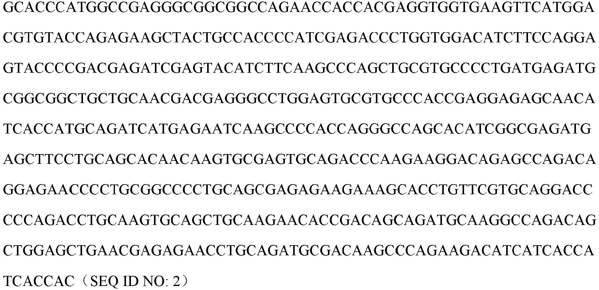
- The amino acids corresponding to the extracellular fragment VEGFR2 ECD of gene VEGFR2 (Vascular Endothelial
Growth Factor Receptor 2, NCBI GenBank: NP_002244) were fused with TEV and the Fc protein fragment of human IgG (hFc) respectively (SEQ ID NO: 3). Genscript was entrusted to synthesize corresponding encoding nucleotide sequence (SEQ ID NO: 4). - VEGFR2, Vascular Endothelial
Growth Factor Receptor 2, NCBI GenBank NP_002244; - hFc: Ig gamma-1 chain C region, ACCESSION: P01857, 106-330;
- wherein, the wavy-underlined part is the ECD part of VEGFR2, the framed part is TEV enzyme digestion site, and the solid-underlined part is hFc part.

-

 wherein, the wavy-underlined part is the ECD part of VEGFR2, the framed part is TEV enzyme digestion site, and the solid-underlined part is hFc part.
- The VEGFR2-hFc encoding gene synthesized by Genscript was cloned into an expression vector pUC57simple (provided by Genscript), and a pUC57simple-VEGFR2-hFc plasmid was obtained.
- The plasmid pUC57simple-VEGFR2-hFc was enzyme-digested (Xba I and BamH I), and the fusion gene fragment VEGFR2-hFc isolated by electrophoresis was ligated with expression vector pcDNA3.1 (purchased from Invitrogen) to give pcDNA3.1-VEGFR2-hFc, which was transfected into competent E. coli cell DH5a (purchased from TIANGEN); the transfection and culture were performed according to the manual. The positive pcDNA3.1-VEGFR2-hFc colonies were screened, E. coli was amplified according to a conventional method, and a kit (purchased from Tiangen Biotech (Beijing) Co., Ltd., DP103-03) was then used and a recombinant plasmid pcDNA3.1-VEGFR2-hFc was extracted according to the manual of the kit.
- The recombinant plasmid pcDNA3.1-VEGFR2-hFc was transfected into 293F cells (purchased from Invitrogen) according to the lipofectamin transfection kit (purchased from Invitrogen).
- After transfecting the recombinant plasmid pcDNA3.1-VEGFR2-hFc into 293F cells for 7 days, the culture medium was subjected to high-speed centrifugation, microporous membrane vacuum filtration and purification in a Mabselect SuRe column to obtain a VEGFR2-hFc fusion protein sample, and a part of the sample was added into a reduced protein electrophoresis loading buffer for SDS-PAGE electrophoresis detection.
- The fusion protein VEGFR2-hFc was thus obtained.
- Chinese Patent Publication
CN1259962A is referred to for the amino acid sequences of the heavy chain variable region and the light chain variable region of the marketed VEGFA monoclonal antibody Avastin (bevacizumab). Genscript was entrusted to synthesize nucleotide sequences encoding the heavy chain variable region and the light chain variable region. -


-

-

-
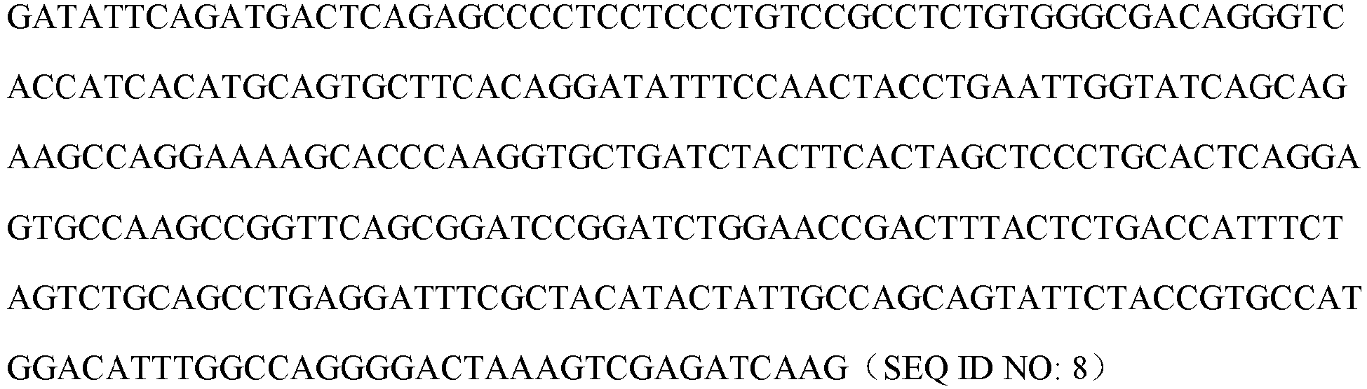
- The heavy chain constant regions were all Ig gamma-1 chain C region, ACCESSION: P01857; the light chain constant regions were all Ig kappa chain C region, ACCESSION: P01834.
- The heavy chain cDNA and the light chain cDNA of bevacizumab were cloned into vector pcDNA3.1, and the recombinant expression plasmid of the antibody bevacizumab was obtained. The recombinant plasmid was transfected into 293F cells. The 293F cell culture medium was purified and then detected.
- The anti-VEGFA monoclonal antibody Avastin (bevacizumab) was thus obtained.
- The preparation was carried out according to the Examples 3-4 described in Chinese Patent Publication
CN106977602A . - The amino acid sequences of the heavy chain variable region and the light chain variable region of humanized antibody 14C12H1L1, and the nucleotide sequence encoding the same are also the same as those described in Examples 3-4 of Chinese Patent Publication
CN106977602A , and are also provided herein as follows: - Amino acid sequence of the heavy chain variable region of humanized antibody 14C12H1L1: (118 aa)

- Nucleotide sequence encoding the heavy chain variable region of humanized antibody 14C12H1L1: (354 bp)

- Amino acid sequence of the light chain variable region of humanized antibody 14C12H1L1: (107 aa)

- Nucleotide sequence encoding the light chain variable region of humanized antibody 14C12H1L1: (321 bp)

- The anti-PD-1 humanized antibody 14C12H1L1 was thus obtained.
- The sequence of Human Anti-Hen Egg Lysozyme IgG (anti-HEL, i.e., human IgG, abbreviated as hIgG) is derived from a variable region sequence of the Fab F10.6.6 sequence in the research published by Acierno et al., which is entitled "Affinity maturation increases the stability and plasticity of the Fv domain of anti-protein antibodies" (Acierno et al., J Mol Biol. 2007; 374(1): 130-46). The preparation method is as follows:
Nanjing Genscript Biology was entrusted to carry out codon optimization of amino acids and gene synthesis on heavy and light chain (complete sequence or variable region) genes of human IgG antibody, and by referring to the standard technologies introduced in the "Guide to Molecular Cloning Experiments (Third Edition)" and using standard molecular cloning technologies such as PCR, enzyme digestion, DNA gel extraction, ligation transformation, colony PCR or enzyme digestion identification, the heavy and light chain genes were respectively subcloned into the antibody heavy chain expression vector and antibody light chain expression vector of the mammalian expression system, and the heavy and light chain genes of the recombinant expression vector were further sequenced and analyzed. After the sequence was verified to be correct, endotoxin-free expression plasmids were prepared in a large scale, and the heavy and light chain plasmids were transiently co-transfected into HEK293 cells for expression of recombinant antibody. After 7 days of culture, the cell culture medium was collected and affinity purified using an rProtein A column (GE), and the quality of the resulting antibody sample was determined using SDS-PAGE and SEC-HPLC standard analysis techniques. - The hIgG was thus obtained, and used in Examples 8-9 below.
- The structure of the bifunctional antibody VP101 of the present invention is in the Morrison form (IgG-scFv), i.e. C-termini of two heavy chains of an IgG antibody are each linked to a scFv fragment of another antibody, and the main composition design of the heavy and light chains is as shown in Table 1 below.
Table 1: Composition design of the heavy and light chains of VP101 Bifunctional antibody No. Heavy chain Light chain IgG part Linker fragment scFv part VP101 Bevacizum ab- H Linker 1 14C12H1V-Linker1-14C12L1v Bevacizumab-L - In the Table 1 above:
- (1) Those with "V" labeled at lower right corner refer to the variable region of corresponding heavy chain or the variable region of corresponding light chain. For those without "V" label, the corresponding heavy or light chain is the full length comprising the constant region. The corresponding sequences described in the above preparation examples are referred to for the amino acid sequences of these variable regions or the full length and the nucleotide sequences encoding them.
- (2) The amino acid sequence of
linker 1 is GGGGSGGGGSGG GGSGGGGS (SEQ ID NO: 13) 2. Expression and purification of antibody VP101 - The heavy chain cDNA sequence and the light chain cDNA sequence of VP101 were each cloned into vector pUC57simple (provided by Genscript) to obtain plasmids pUC57simple-VP101H and pUC57simple- VP101L, respectively.
- Plasmids pUC57simple-VP101H and pUC57simple-VP101L were enzyme-digested (HindIII&EcoRI), and heavy and light chains isolated by electrophoresis were subcloned into vector pcDNA3.1, and recombinant plasmids were extracted to co-transfect 293F cells. After 7 days of cell culture, the culture medium was centrifuged at high speed, and the supernatant was concentrated and loaded onto a HiTrap MabSelect SuRe column. The protein was further eluted in one step with Elution Buffer, and the target sample antibody VP101 was isolated and buffer exchanged into PBS.
- The purified sample was added to both a reduced protein electrophoresis loading buffer and a non-reduced protein electrophoresis loading buffer, and then boiled for SDS-PAGE electrophoresis detection. The electropherogram of VP101 is shown in
FIG. 1 . The target protein of the reduced protein sample is at 75 kD and 25 kD, and the target protein of the non-reduced protein sample (single antibody) is at 200 kD. - Unless otherwise specified, the humanized antibody VP101 used in the following experiments was prepared by the method of this example.
- The sample dilution buffer was PBS (0.02% Tween-20, 0.1% BSA, pH7.4). 5 µg/mL antibody was immobilized to an AHC sensor with the immobilization height being about 0.4 nM. The sensor was equilibrated in a buffer for 60 s, and the antibody immobilized to the sensor bound to PD-1-mFc at a concentration of 0.62-50 nM (three-fold gradient dilution) for 120 s, and then the antigen and antibody dissociated in the buffer for 300 s. The data were analyzed by 1: 1 model fitting to obtain affinity constants. The data acquisition software was Fortebio Data Acquisition 7.0, and the data analysis software was Fortebio Data Analysis 7.0. Kinetic parameters of the binding of antibodies VP101, BsAbB7, BsAbB8, 14C12H1L1 and the control antibody nivolumab to PD-1-mFc are shown in Table 2, and the detection results of the kinetic characteristic parameters are shown in
FIG. 2 ,FIG. 3 ,FIG. 4 ,FIG. 5 and FIG. 6 , respectively.Table 2: Kinetic parameters of the binding of humanized antibody VP101, BsAbB7, BsAbB8, 14C12H1L1 and the control antibody nivolumab to PD-1-mFc Sample ID KD (M) Kon (1/Ms) S E (kon) Kdis (1/s) S E (kdis) Rmax (nm) VP101 1.68E-10 3.22E+05 1.44E+04 5.40E-05 3.16E-05 0.14-0.28 BsAbB7 1.62E-10 3.27E+05 2.60E+04 5.30E-05 6.24E-05 0.01-0.11 BsAbB8 4.06E-10 3.39E+05 2.04E+04 1.37E-04 4.61E-05 0.01-0.13 14C12H1L1 1.64E-10 4.55E+05 1.61E+04 7.47E-05 2.98E-05 0.24-0.28 Nivolumab 2.32E-10 5.85E+05 2.03E+04 1.36E-04 3.47E-05 0.02-0.14 - KD is affinity constant; kon is binding rate of antigen and antibody; kdis is dissociation rate of antigen and antibody; KD = kdis/kon.
- The results show that the antibodies VP101 and BsAbB7 are equivalent in terms of affinity for PD-1-mFc; the affinity constant of VP101 for PD-1-mFc is significantly smaller than that of BsAbB8, suggesting that VP101 has better binding activity; the dissociation rate constant for VP101 and PD-1-mFc was significantly smaller than BsAbB8 and 14C12H1L1, suggesting that VP101 binds to antigen more stably with a dissociation rate slower than that of 14C12H1L1 and BsAbB8.
- The sample dilution buffer was PBS (0.02% Tween-20, 0.1% BSA, pH7.4). 1 µg/mL VEGF-His was immobilized to the HIS1K sensor for 20 s, then the sensor was equilibrated in a buffer for 60 s, and the VEGF immobilized on the sensor bound to the antibody at a concentration of 12.34-1000 nM (three-fold gradient dilution) for 120 s, and then the antigen and antibody dissociated in the buffer for 300 s. The data were analyzed by 1:1 model fitting to obtain affinity constants. The data acquisition software was Fortebio Data Acquisition 7.0, and the data analysis software was Fortebio Data Analysis 7.0.
- Kinetic parameters of the binding of antibodies VP101, BsAbB7, BsAbB8 and the control antibody bevacizumab to VEGF-His are shown in Table 3, and the detection results of kinetic characteristic parameters are shown in
FIG. 7, FIG. 8 ,FIG. 9 andFIG. 10 respectively.Table 3: Kinetic parameters of the binding of antibodies VP101, BsAbB7, BsAbB8 and the control antibody bevacizumab to VEGF-His Sample ID KD (M) Kon (1/Ms) S E (kon) Kdis (1/s) S E (kdis) Rmax (nm) VP101 5.21E-10 1.55E+05 9.67E+03 8.05E-05 4.66E-05 0.39-0.60 BsAbB7 5.14E-10 1.57E+05 9.67E+03 8.05E-05 4.83E-05 0.36-0.53 BsAbB8 6.33E-10 1.71E+05 1.07E+04 1.08E-04 4.64E-05 0.39-0.56 Bevacizumab 7.24E-10 1.23E+05 7.09E+03 8.90E-05 4.53E-05 0.29-0.41 - The results show that the antibodies VP101 and BsAbB7 are equivalent in terms of affinity for the antigen, and the affinity constant of VP101 is significantly smaller than that of BsAbB8 and the control antibody bevacizumab, suggesting that VP101 has better binding activity; the dissociation rate constant of VP101 for VEGF-His is significantly smaller than that of BsAbB8, suggesting that VP101 binds to antigen more stably with a slower dissociation rate than that of BsAbB8.
- The method is specified as follows:
The microplate was coated with VEGFA-His and incubated at 37 °C for 2 hours. After being washed, the microplate was blocked with 1% BSA for 2 hours. After being washed, the microplate was added with the gradiently diluted antibody and incubated at 37 °C for 30 minutes. After being washed, the microplate was added with the enzyme-labeled goat anti-human IgG secondary antibody working solution and incubated for 30 minutes at 37 °C. After being washed, the microplate was added with TMB chromogenic solution for color developing for 5 minutes in the absence of light, and then stop solution was added to terminate the chromogenic reaction. Then the microplate was put into a microplate reader immediately, and the OD value of each well in the microplate was read at 450 nm. SoftMax Pro 6.2.1 was used to analyze and process the data. - The detection result of the binding of antibody VP101 to antigen VEGFA-His is shown in
FIG. 11 . The absorbance intensities at each dose are shown in Table 4. The binding EC50 of antibody was calculated by curve fitting using antibody concentration as the abscissa and absorbance value as the ordinate, and the results are shown in Table 4 below.Table 4: Binding of bifunctional antibody to VEGFA-his (Indirect ELISA) Antibody concentration (µg/mL) Coating: VEGFA-His (1 µg/mL) VP101 Bevacizumab 1.0000 3.045 2.943 2.798 2.974 0.3333 3.037 2.861 2.816 2.993 0.1111 2.901 2.689 2.653 2.700 0.0370 2.597 2.460 2.445 2.555 0.0123 2.013 1.914 1.998 2.074 0.0041 1.115 1.086 1.446 1.363 0.0014 0.524 0.496 0.640 0.729 0.0000 0.099 0.091 0.094 0.083 Secondary antibody Goat anti-human IgG (H+L), HRP (1:5000) EC50(nM) 0.036 0.035 - The results show that antibody VP101 is able to bind to VEGFA protein efficiently and its binding efficiency is dose-dependent, and the two antibodies are equivalent in terms of binding activity to human VEGFA.
- The method is specified as follows:
The microplate was coated with VEGFA-His and incubated overnight at 4 °C. After being washed, the microplate was blocked with 1% BSA (dissolved in PBS) for 2 hours. After being washed, the microplate was added with the gradiently diluted antibody and incubated at 37 °C for 30 minutes. After being washed, the microplate was added with the horseradish peroxidase-labeled goat anti-human IgG Fc (Jackson, 109-035-098) working solution and incubated for 30 minutes at 37 °C. After being washed, the microplate was added with TMB (Neogen, 308177) for color developing for 5 minutes in the absence of light, and then stop solution was added to terminate the chromogenic reaction. Then the microplate was put into a microplate reader immediately, and the OD value of each well in the microplate was read at 450 nm. SoftMax Pro 6.2.1 was used to analyze and process the data. - The result of the binding of antibody VP101 to antigen VEGFA-His is shown in
FIG. 12 . The absorbance intensities at each dose are shown in Table 5. The binding EC50 of antibody was calculated by curve fitting using antibody concentration as the abscissa and absorbance value as the ordinate, and the results are shown in Table 5 below.Table 5: Respective binding activities of VP101, BsAbB7, BsAbB8 and bevacizumab to VEGFA-His (Indirect ELISA) Antibody concentration (µg/mL) Antibody coating: VEGFA-His 1 µg/mL VP101 BsAbB7 BsAbB8 Bevacizumab 1.000 3.112 3.090 3.074 3.081 3.070 3.093 3.137 3.138 0.333 3.026 2.961 2.954 2.941 2.946 2.968 3.075 3.086 0.111 2.802 2.684 2.575 2.621 2.631 2.618 2.965 2.999 0.037 1.972 1.876 1.656 1.668 1.756 1.709 2.504 2.503 0.012 0.994 0.915 0.754 0.764 0.809 0.814 1.476 1.454 0.004 0.436 0.391 0.317 0.332 0.347 0.339 0.711 0.700 0.001 0.197 0.177 0.151 0.155 0.159 0.155 0.318 0.311 0 0.083 0.063 0.086 0.076 0.095 0.072 0.066 0.064 Secondary antibody Horseradish peroxidase-labeled goat anti-human IgG Fc, HRP (1:5000) EC50(nM) 0.130 0.171 0.159 0.092 - The results show that the antibodies VP101, BsAbB7, BsAbB8 and bevacizumab all can bind to the VEGF protein efficiently and their binding efficiency is dose-dependent, and antibody VP101 has a higher binding activity to human VEGF than BsAbB7 and BsAbB8.
- The method is specified as follows:
The microplate was coated with human PD-1-mFc and incubated overnight at 4 °C. After being blocked with 1% BSA at 37 °C for 2 hours, the microplate was added with antibody, and then incubated at 37 °C for 30 minutes. After the microplate was washed and patted dry, the HRP-labeled goat anti-human IgG (H+L) secondary antibody (Jackson, 109-035-088) was added, and the microplate was incubated at 37 °C for 30 minutes. After the microplate was washed and patted dry, TMB (Neogen, 308177) was added for color developing for 5 minutes, and then stop solution was added to terminate the color development. Then the microplate was put into a microplate reader immediately, and the OD value of each well in the microplate was read at 450 nm. SoftMax Pro 6.2.1 was used to analyze and process the data. - The detection result of the binding of antibody VP101 to antigen PD-1 is shown in
FIG. 13 . The absorbance intensities at each dose are shown in Table 6. By quantitative analysis of the bound antibody VP101, the curve simulation was performed to obtain the binding efficiency EC50 of the antibody, which is shown in Table 6 below.Table 6: Binding of bifunctional antibody to PD-1 (Indirect ELISA) Antibody dilution gradient Antibody coating: PD-1-mFc 0.5 µg/mL VP101 Nivolumab 14C12H1L1 0.333µg/ml 3.109 3.063 3.137 3.130 3.298 3.278 1:3 3.016 2.926 3.139 3.140 3.245 3.352 1:9 2.461 2.513 2.802 2.758 3.104 3.155 1:27 1.638 1.675 1.949 1.810 2.352 2.549 1:81 0.787 0.791 0.933 0.990 1.382 1.421 1:243 0.301 0.656 0.348 0.375 0.612 0.596 1:729 0.136 0.145 0.159 0.162 0.253 0.247 0 0.068 0.056 0.053 0.053 0.053 0.053 EC50(nM) 0.06 0.061 0.037 - The results show that antibody VP101 is able to bind to PD-1 protein efficiently and its binding efficiency is dose-dependent.
- The method is specified as follows:
The microplate was coated with human PD-1-mFc and incubated overnight at 4 °C. After being blocked with 1% BSA at 37 °C for 2 hours, the microplate was added with antibody, and then incubated at 37 °C for 30 minutes. After the microplate was washed and patted dry, the horseradish peroxidase-labeled goat anti-human IgG Fc (Jackson, 109-035-098) was added, and the microplate was incubated at 37 °C for 30 minutes. After the microplate was washed and patted dry, TMB (Neogen, 308177) was added for color developing for 5 minutes, and then stop solution was added to terminate the color development. Then the microplate was put into a microplate reader immediately, and the OD value of each well in the microplate was read at 450 nm. SoftMax Pro 6.2.1 was used to analyze and process the data. - The detection result of the binding of antibody VP101 to antigen PD-1 is shown in
FIG. 14 . The absorbance intensities at each dose are shown in Table 7. By quantitative analysis of the bound antibody VP101, the curve simulation was performed to give the binding efficiency EC50 of the antibody, which is shown in Table 7 below.Table 7: Respective binding activities of antibodies VP101, BsAbB7, BsAbB8, 14C12H1L1 and nivolumab to PD-1 (Indirect ELISA) Antibody concentration (µg/mL) Antigen coating: PD-1-mFc 0.5 µg/mL VP101 BsAbB7 BsAbB8 14C12 H1L1 Nivolumab 0.333 2.717 2.709 2.732 2.755 2.716 2.715 2.947 2.966 2.823 2.824 0.111 2.507 2.381 2.318 2.321 2.377 2.409 2.923 2.967 2.747 2.758 0.037 1.709 1.616 1.491 1.457 1.522 1.549 2.656 2.694 2.208 2.293 0.012 0.916 0.822 0.732 0.711 0.797 0.775 2.049 2.060 1.348 1.389 0.004 0.413 0.394 0.333 0.321 0.368 0.351 1.139 1.132 0.629 0.638 0.001 0.195 0.191 0.167 0.174 0.181 0.174 0.552 0.541 0.295 0.295 0.000 0.140 0.123 0.110 0.103 0.117 0.118 0.254 0.248 0.152 0.157 0.000 0.099 0.095 0.089 0.074 0.100 0.081 0.083 0.075 0.078 0.084 Secondary antibody Horseradish peroxidase-labeled goat anti-human IgG Fc, HRP (1:5000) EC50(nM) 0.146 0.199 0.173 0.045 0.095 - The results show that the antibody VP101 can bind to the PD-1 protein efficiently and its binding efficiency is dose-dependent, and antibody VP101 has a higher binding activity to human PD-1 than BsAbB7 and BsAbB8.
- The method is specifically as follows:
The microplate was coated with VEGF-His and incubated at 37 °C for 2 hours. After being washed, the microplate was blocked with 1% BSA for 1 hour at 37 °C. After being washed, the microplate was added with the gradiently diluted antibodies and human VEGFR2 ECD-mFc-bio (final concentration: 0.02 µg/mL) and incubated at room temperature for 2 hours. After being washed, the microplate was added with HRP-labeled streptavidin SA-HRP (1:4000) working solution and incubated at 37 °C for 30 minutes. After being washed, the microplate was added with TMB chromogenic solution for color developing for 5 minutes in the absence of light, and then stop solution was added to terminate the chromogenic reaction. Then the microplate was put into a microplate reader immediately, and the OD value of each well in the microplate was read at 450 nm. SoftMax Pro 6.2.1 was used to analyze and process the data. - The detection results are shown in
FIG 15 . The absorbance intensities at each dose are shown in Table 8. By quantitative analysis of the absorbance intensities of the bound antibodies, the curve simulation was performed to give the binding efficiency EC50 of the antibodies (Table 8).Table 8: Detection of antibody in competing with VEGFR2-mFc for binding to the antigen VEGFA-His by competitive ELISA Coating: VEGF-His (2 µg/mL) Antibody concentration (µg/mL) VP101 Bevacizumab 10.000 0.133 0.133 0.103 0.104 3.333 0.161 0.149 0.114 0.109 1.111 0.624 0.563 0.374 0.351 0.370 1.055 1.051 0.905 0.982 0.123 1.059 1.075 0.964 1.049 0.041 1.137 1.068 1.062 1.141 0.014 1.106 1.138 1.010 1.169 0.000 1.155 1.131 1.173 1.153 Receptor VEGFR2 ECD-mFc-bio, 0.02µg/ml Secondary antibody SA-HRP ( 1:4000) EC50 (nM) 5.324 5.086 - The results show that the antibody VP101 can effectively bind to the antigen VEGFA and inhibit the binding of VEGFR2 to VEGFA, and its efficiency in inhibiting the binding of VEGFR2 to VEGFA is dose-dependent.
- The method is specifically as follows:
The microplate was coated with PD-1-hFc and incubated overnight at 4 °C. After the microplate was blocked with 1% BSA for 2 hours, antibodies at different concentrations were each mixed with PD-L1-hFc for 10 minutes (see Table 10 for the dilution concentrations). After incubation at 37 °C for 30 minutes, the microplate was washed and patted dry. Then enzyme-labeled secondary antibody was added, and the microplate was incubated at 37 °C for 30 minutes. After the microplate was washed and patted dry, TMB was added for color developing for 5 minutes, and then stop solution was added to terminate the color development. Then the microplate was put into a microplate reader immediately, and the OD value of each well in the microplate was read at 450 nm (see Table 10). SoftMax Pro 6.2.1 was used to analyze and process the data. - The detection results are shown in
FIG 16 . The absorbance intensities at each dose are shown in Table 9. By quantitative analysis of the bound antibody VP101, the curve simulation was performed to give the binding efficiency EC50 of the antibody (Table 9).Table 9: Detection of bifunctional antibody competing with PD-L1 for binding to PD-1 by competitive ELISA Antibody dilution gradient Antigen coating: PD-1-hFc 0.5 µg/mL VP101 Nivolumab 14C12H1L1 5µg/ml 0.096 0.063 0.058 0.058 0.062 0.063 1:3 0.064 0.077 0.059 0.059 0.061 0.064 1:9 0.166 0.160 0.061 0.062 0.066 0.071 1:27 0.867 0.848 0.284 0.335 0.262 0.193 1:81 1.217 1.149 0.973 1.007 0.968 0.882 1:243 1.196 1.949 1.139 1.144 1.122 1.051 1:729 1.183 1.250 1.127 1.185 1.052 1.059 0 1.153 1.276 0.960 1.071 1.027 1.024 Receptor PD-L1-mFc 0.3µg/ml Secondary antibody Goat anti-mouse IgG (H+L), HRP conjugated (1:5000) EC50(nM) 1.216 0.842 0.745 - The results show that antibody VP101 can effectively bind to antigen PD-1 and inhibit the binding of ligand PD-L1 to PD-1, and its efficiency in inhibiting the binding of PD-L1 to PD-1 is dose-dependent.
- Firstly, 293T cells expressing PD-1 antigen was constructed, and then the specific binding capacity of the antibody to the cell membrane surface antigen was analyzed and verified by flow cytometry.
- The vector pLenti6.3-PD-1 of PD-1 (the vector pLenti6.3 was purchased from Invitrogen) was transfected into 293T cells, and clone group 293T-PD-1 cells which stably express PD-1 were obtained by screening.
- The 293T-PD-1 expressing antigen obtained in the previous step was digested with pancreatin by a conventional pancreatin digestion method, and the number of cells in each collection tube was made to be 2×10⁵. Antibody diluting solutions with concentration gradiently diluted with PBSA (1% BSA) were each incubated with 293T-PD-1 cells on ice for 2 hours, and then each tube was added with 100 µL of FITC goat anti-human IgG (1:500) and incubated on ice for 1 hour. Then PBS was used for washing, and 300 µL of PBSA was used to resuspend the cells, and fluorescence signals (MFI) were detected with FITC channel on a flow cytometer.
- The results are shown in
FIG. 17 , and the MFI values at each concentration are shown in Table 10. By fluorescence quantification analysis and curve fitting of the bound 14C12H1L1 antibody, the binding EC50 of the VP101 antibody was calculated to be 3.5 nM.Table 10: Analysis of fluorescence intensity of the binding of VP101 to 293T-PD-1 surface antigen detected by FACS Antibody (nM) 0.14 0.41 1.23 3.70 11.11 33.33 100 EC50 Bevacizumab 3.2 2.2 2.0 2.3 2.7 3.8 5.7 - Nivolumab 33.3 74.9 171.9 357.9 481.9 498.3 478.4 2.1 14C12H1L1 48.1 99.7 201.5 409.0 600.2 655.4 670.8 2.9 VP101 30.8 61.8 135.7 286.9 487.7 534.0 528.6 3.5 - The results show that the VP101 antibody can effectively bind to the PD-1 antigen on the 293T-PD-1 host cell surface, and its binding efficiency is dose-dependent, and bevacizumab has no binding activity to 293T-PD-1, which indicates that the binding of VP101 to 293T-PD-1 is specific.
- The results are shown in
FIG. 18 , and the MFI values at each concentration are shown in Table 11. By fluorescence quantification analysis and curve fitting of the bound antibody, the binding EC50 values of nivolumab, 14C12H1L1, VP101, BsAbB7 and BsAbB8 were calculated to be 7.853 nM, 3.607 nM, 7.896 nM, 9.943 nM and 10.610 nM, respectively.Table 11: Analysis of fluorescence intensities of the binding of VP101, BsAbB7 and BsAbB8 to 293T-PD-1 surface antigen detected by FACS Antibody (nM) 0.014 0.14 0.41 1.23 3.7 11 30 100 EC50(nM) Bevacizumab 1.89 1.90 2.20 1.92 2.04 2.48 2.80 2.43 - Nivolumab 3.91 15.30 34.69 94.04 234.34 533.63 640.15 804.69 7.853 14C12H1L1 7.40 29.55 69.16 175.54 422.53 868.45 831.27 813.58 3.607 VP101 3.47 16.16 38.75 93.08 216.76 509.23 810.37 783.58 7.896 BsAbB7 3.85 14.86 37.45 83.78 202.40 465.10 837.61 846.80 9.943 BsAbB8 4.41 16.77 36.86 89.89 210.40 457.91 804.43 863.35 10.610 - The results show that the VP101 antibody can bind to the membrane surface PD-1 of 293T-PD1 in a dose-dependent manner. Bevacizumab has no binding activity to 293T-PD-1, which indicates that the binding of VP101 to 293T-PD-1 is specific.
-
1. A competitive flow cytometry method was adopted to detect the EC50 of the VP101 in competing with PD-L1 for binding to the cell membrane surface antigen PD-1, and the method is specified as follows:
The 293T-PD-1 cells was digested in a conventional way, and divided into several samples with 300,000 cells for each, which were then subjected to centrifugation and washing. Then each tube was added with 100 µL of corresponding gradiently diluted antibody and incubated on ice for 30 minutes; 100 µL of PD-L1-mFc was then added to each tube, and the mixture was mixed well to reach a final concentration of 20 nM, and then incubated on ice for 1 hour. Then 500 µL of 1% PBSA was added, and the mixture was centrifuged at 5600 rpm for 5 minutes to remove the supernatant. 100 µL of FITC coat anti mouse antibody diluted at a ratio of 1:500 was then added into each tube, and the mixture was incubated on ice for 40 minutes in the absence of light after being mixed well. Then the mixture was centrifuged, washed and resuspended, and then transferred to a loading tube for testing.
The results are shown inFIG. 19 , and the MFI values at each concentration are shown in Table 12. By fluorescence quantification analysis and curve fitting, the binding EC50 values of the antibodies VP101 and 14C12H1L1 were calculated to be 8.33 nM and 4.37 nM, respectively.Table 12: Analysis of fluorescence intensities of 14C12H1L1 and VP101 in competing for binding to 293T-PD-1 surface antigen detected by FACS Antibody (nM) 0.05 0.14 0.41 1.23 3.70 11.11 33.33 100.00 EC50 R2 14C12H1L1 288.17 287.29 277.09 237.22 177.80 12.04 10.32 9.87 4.37 0.988 VP101 272.66 264.39 279.11 272.26 239.18 99.29 17.05 14.91 8.33 0.999
The results show that the VP101 antibody can effectively block the binding of PDL-1 to PD-1 on the surface of 293T-PD-1 host cells in a dose-dependent manner.
2. EC50 values of VP101, BsAbB7, BsAbB8, 14C12H1L1 and nivolumab in competing with PD-L1 for binding to the cell membrane surface antigen PD-1 were detected by using competitive flow cytometry and referring to the experimental procedure described instep 1 of this example. - The results are shown in
FIG. 20 , and the MFI values at each concentration are shown in Table 13. By fluorescence quantification analysis and curve fitting, the competitive binding EC50 values of antibodies VP101, BsAbB7, BsAbB8, 14C12H1L1 and nivolumab were calculated to be 15.04 nM, 22.25 nM, 19.25 nM, 9.21 nM and 9.72 nM, respectively.Table 13: Analysis of fluorescence intensities of VP101, BsAbB7, BsAbB8, 14C12H1L1 and nivolumab in competing for binding to 293T-PD-1 surface antigen detected by FACS Antibody (nM) 0.14 0.41 1.23 3.7 11.11 33.33 100 300 EC50 R2 VP101 1441.94 1380.62 1368.15 1288.34 982.69 112.90 8.49 8.21 15.04 0.9971 BsAbB7 1412.62 1377.27 1339.60 1341.27 1094.35 417.70 9.23 9.18 22.25 0.9985 BsAbB8 1578.36 1521.50 1427.20 1429.85 1137.74 359.69 9.73 9.68 19.25 0.9962 14C12H1L1 1384.08 1551.05 1462.85 1296.64 580.45 12.93 13.37 14.99 9.21 0.9950 Nivolumab 1539.58 1552.37 1483.84 1300.81 713.56 70.92 60.77 56.92 9.72 0.9969 - The results show that the activity of the antibody 14C12H1L1 is equivalent to that of the marketed antibody nivolumab targeting PD-1, and is superior to that of the bifunctional antibody VP101. The activity of the antibody VP101 is superior that of to BsAbB7 and BsAbB8.
- KDR (VEGFR2) vector pCDH-KDRFL(OPV)-GFP-Puro (Vector pCDH-GFP-Puro is purchased from Youbio) and NFAT vector pNFAT-luc-hygro (vector pGL4-luc2P-hygro is purchased from Promega) were transfected into 293T cells, and a clone group 293T-NFAT-(opv) KDR(C7) cells stably expressing KDR and NFAT luciferase reporter genes were obtained by screening.
- 2. 293T-NFAT-(opv)KDR(C7) cells were collected and centrifuged for 5 minutes to remove the supernatant; DMEM+10%FBS medium was used to resuspend the cells, and the cell number was counted and the cell viability was detected; then the cell concentration was adjusted to be in a proper range, and 50000 cells/50 µL cell suspension was added into each well of a black 96-well plate;
- Corresponding antibodies (final concentrations being 300, 100, 10, 2, 0.2, 0.02, 0.002 nM) and VEGF (final concentration being 30 ng/mL) were diluted according to the experimental design, and the antibodies targeting VEGF were preincubated with VEGF for 1 hour at room temperature before being added into the cells. Blank and isotype controls (final volume of each well being 100 µL) were designed and incubated in a carbon dioxide incubator at 37 °C, 5% CO2 for 4 hours; 50 µL of Luciferase Assay System was added to each well, and Relative Fluorescence Units (RLUs) were detected by a multi-label microplate tester within 5 minutes.
- The experimental results are shown in
FIG. 21 , and the EC50 values for each antibody are shown in Table 14.Table 14: Detection of neutralization bioactivity of antibodies VP101, BsAbB7 and BsAbB8 in blocking VEGF to activate NFAT signaling pathway by reporter assay Sample VP101 BsAbB7 BsAbB8 Bevacizumab EC50(nM) 1.2400 1.2170 1.7280 0.7730 - The results show that the EC50 of VP101 is 1.240 nM, the EC50 of BsAbB7 is 1.217 nM, the EC50 of BsAbB8 is 1.728 nM, and the EC50 of bevacizumab is 0.773 nM, and the experimental results show that the activity of VP101 and BsAbB7 in blocking VEGF to activate NFAT signaling pathway is better than that of BsAbB8.
- HUVEC cells (purchased from Allcell) in a good growth state, after the cell concentration was adjusted to be 1.5 × 10⁴/mL, were inoculated into a 96-well plate at 200 µL/well, and then incubated in an incubator at 37 °C, 5% CO2 for 24 hours. Then it was observed that the cells adhered well, and then culture medium was discarded. 20 nM VEGFA prepared by using 1640 containing 2% FBS was then added into the 96-well plate at 200 µL/well, and antibodies at different concentrations were added, followed by incubation for 72 hours. 72 hours later, the culture medium was discarded and MTT was added. 4 hours later, the MTT was discarded and DMSO was added, and then a microplate reader was used to measure the OD value at 490 nm.
- The results are shown in
FIG. 22 . The results show that the humanized antibodies VP101 and bevacizumab both can effectively inhibit VEGFA-induced HUVEC cell proliferation in a dose-dependent manner, and the pharmacological activity of VP101 in inhibiting VEGFA-induced HUVEC cell proliferation is higher than that of bevacizumab at the same dose. - 1. Promotion of secretion of IFN-γ by VP101, 14C12H1L1 and nivolumab in mixed culture system of DC and PBMC cells
- PBMCs were isolated by Ficoll-Paque Plus (GE Healthcare) and added to IL-4 (Peprotech 200-04, 1000 U/mL) and GM-CSF (Peprotech 300-03, 1000 U/mL) for 6 days of induction, and then TNF-α (Peprotech 300-01A, 200 U/mL) was additionally added for 3 days of induction to obtain mature DC cells.
- On the day of co-culture, fresh PBMCs were isolated from peripheral blood of another donor, and the obtained mature DC cells were mixed with the freshly isolated PBMCs of another donor at a ratio of 1: 10, and meanwhile antibodies at different concentrations (hIgG as a control) were added. After co-culture for 5-6 days, cell supernatant was collected and assayed for IFN-y content using an ELISA kit (purchased from Dakewe).
- The effect of VP101 on secretion of IFN-y in mixed culture system of DC and PBMC cells is shown in
FIG. 23 . As can be seen inFIG. 23 , VP101 can effectively promote secretion of IFN-y in a dose-dependent manner. In addition, at doses of 30 nM and 300 nM, VP101 has greater activity in promoting secretion of IFN-γ than equivalent 14C12H1L1, and at dose level of 30 nM, it has greater activity in promoting secretion of IFN-y than equivalent nivolumab. - 2. Promotion of secretion of IL-2 and IFN-y by VP101, BsAbB7 and BsAbB8b in mixed culture system of DC and PBMC cells
-
Step 1 in this example was referred to for the experimental method, namely PBMCs were isolated by Ficoll-Paque Plus (GE Healthcare) and added to IL-4 (Peprotech 200-04, 1000 U/mL) and GM-CSF (Peprotech 300-03, 1000 U/mL) for 6 days of induction, and then TNF-α (Peprotech 300-01A, 200 U/mL) was additionally added for 3 days of induction to obtain mature DC cell. - On the day of co-culture, fresh PBMCs were isolated from peripheral blood of another donor, and the obtained mature DC cells were mixed with the freshly isolated PBMCs of another donor at a ratio of 1: 10, and meanwhile antibodies at different concentrations (hIgG as a control) were added; after co-culture for 5-6 days, cell supernatant was collected and assayed for IL-2 and IFN-y content using an ELISA kit (purchased from Dakewe).
- The effect of VP101 on secretion of IFN-y in mixed culture system of DC and PBMC cells is shown in
FIG. 24 . As can be seen fromFIG. 24 , VP101 can effectively promote secretion of IFN-y in a dose-dependent manner. The pharmacological activity of VP101 in promoting secretion of IFN-γ is significantly better than that of BsAbB7 and BsAbB8. - The effect of VP101 on secretion of IL-2 in mixed culture system of DC and PBMC cells is shown in
FIG. 25 . As can be seen fromFIG. 25 , VP101 can effectively promote secretion of IL-2 in a dose-dependent manner, and the pharmacological activity of VP101 in promoting secretion of IL-2 is better than that of BsAbB7 and BsAbB8. - 3. Promotion of secretion of IL-2 and IFN-y by VP101, 14C12H1L1 and nivolumab in mixed culture system of PBMC and Raji-PD-L1 cells
- PD-L1 was stably transfected into Raji cells through lentivirus infection, and Raji-PD-L1 cells stably expressing PD-L1 were obtained after dosing and screening; PBMCs, after two days of stimulation by SEB, were cultured in together with mitomycin C-treated Raji-PD-L1.
- The results are shown in
FIGs. 26 and 27 . The results show that VP101 can effectively promote secretion of IL-2 and IFN-y, and at dose level of 300 nM, the activity of VP101 in promoting secretion of IL-2 is significantly better than that of equivalent 14C12H1L1 and nivolumab. - The isotype control antibody to be studied was Human Anti-Hen Lysozyme (anti-HEL, i.e., human IgG, abbreviated as hIgG), and it was prepared as described in Preparation Example 6 above.
- 4. The promotion of secretion of IL-2 and IFN-y by VP101, BsAbB7 and BsAbB8 in mixed culture system of PBMC and Raji-PD-L1 cells was studied by referring to the experimental method described in
step 3 of this example. - The results of secretion of IFN-y are shown in
FIG. 28 . The results show that VP101 can effectively promote secretion of IFN-y in a dose-dependent manner. At the same time, VP101 is significantly better than BsAbB7 at the same dose, while the pharmacological activity of VP101 is significantly better than that of BsAbB8 at dose levels of 3 nM and 30 nM. - The results of secretion of IL-2 are shown in
FIG. 29 . The results show that VP101 can effectively promote secretion of IL-2 in a dose-dependent manner. At the same time, the pharmacological activity of VP101 is significantly better than that of BsAbB7 at doses of 3 nM and 300 nM, while VP101 is equivalent to BsAbB8 at the same dose. - To detect the in vivo tumor-inhibiting activity of VP101, U87MG cells (human glioma cells, purchased from ATCC) were first inoculated subcutaneously into 5-7 week old female Scid Beige mice (purchased from Vital River), and the modeling and specific mode of administration were shown in Table 15. After the administration, the length and width of each group of tumors were measured, and the tumor volume was calculated.
Table 15: Dosing regimen of treating U87MG tumor xenograft Scid Beige mouse model with VP101 Grouping n Tumor xenograft Condition of administration Isotype control 40 mg/ kg 7 U-87MG, 5 million cells/mouse subcutaneously Isotype control antibody, hIgG, 40 mg/kg, injected intravenously on days Bevacizumab 30mg/ kg 8 Bevacizumab 30 mg/kg, injected intravenously ondays VP101 40mg/ kg 7 VP101 40 mg/kg, injected intravenously ondays VP101 4mg/ kg 7 VP101 4 mg/kg, injected intravenously ondays - The results are shown in
FIG. 30 . The results show that compared with an isotype control antibody hIgG (the preparation method is the same as that of the Preparation Example 6), bevacizumab and VP101 at different doses can effectively inhibit the growth of mouse tumors, and the high-dose VP101 is better than that of low-dose VP 101 in inhibiting tumors. - Furthermore, as shown in
FIG. 31 , VP 101 does not affect the body weight of tumor-bearing mouse. - The invention is defined by the claims and any other aspects, configurations or embodiments set forth herein not falling within the scope of the claims are for information only.
Claims (20)
- A bispecific antibody, comprising:a first protein functional region targeting VEGFA, anda second protein functional region targeting PD-1;wherein:the first protein functional region is an anti-VEGFA antibody or an antigen-binding fragment thereof, wherein the anti-VEGFA antibody or an antigen-binding fragment thereof comprises a heavy chain variable region comprising HCDR1-HCDR3 with amino acid sequences set forth in SEQ ID NOs: 15-17 respectively, and comprises a light chain variable region comprising LCDR1-LCDR3 with amino acid sequences set forth in SEQ ID NOs: 18-20 respectively; andthe second protein functional region is an anti-PD-1 antibody or an antigen-binding fragment thereof, wherein the anti-PD-1 antibody or an antigen-binding fragment thereof comprises a heavy chain variable region comprising HCDR1-HCDR3 with amino acid sequences set forth in SEQ ID NOs: 21-23 respectively, and comprises a light chain variable region comprising LCDR1-LCDR3 with amino acid sequences set forth in SEQ ID NOs: 24-26 respectively.
- The bispecific antibody according to claim 1, wherein,the anti-VEGFA antibody or the antigen-binding fragment thereof is selected from Fab, Fab', F(ab')2, Fv, a scFv, a humanized antibody, a chimeric antibody, and a diabody;
and/or,the anti-PD-1 antibody or the antigen-binding fragment thereof is selected from Fab, Fab', F(ab')2, Fv, a scFv, a humanized antibody, a chimeric antibody, and a diabody. - The bispecific antibody according to claim 1, wherein:(i) the first protein functional region is an immunoglobulin, wherein the immunoglobulin comprises a heavy chain variable region comprising HCDR1-HCDR3 with amino acid sequences set forth in SEQ ID NOs: 15-17 respectively, and comprises a light chain variable region comprising LCDR1-LCDR3 with amino acid sequences set forth in SEQ ID NOs: 18-20 respectively; andthe second protein functional region is a scFv, wherein the scFv comprises a heavy chain variable region comprising HCDR1-HCDR3 with amino acid sequences set forth in SEQ ID NOs: 21-23 respectively, and comprises a light chain variable region comprising LCDR1-LCDR3 with amino acid sequences set forth in SEQ ID NOs: 24-26 respectively,optionally wherein the amino acid sequence of the heavy chain variable region of the immunoglobulin is set forth in SEQ ID NO: 5, and the amino acid sequence of the light chain variable region of the immunoglobulin is set forth in SEQ ID NO: 7; andthe amino acid sequence of the heavy chain variable region of the scFv is set forth in SEQ ID NO: 9, and the amino acid sequence of the light chain variable region of the scFv is set forth in SEQ ID NO: 11;or,(ii) the first protein functional region is a scFv, wherein the scFv comprises a heavy chain variable region comprising HCDR1-HCDR3 with amino acid sequences set forth in SEQ ID NOs: 15-17 respectively, and comprises a light chain variable region comprising LCDR1-LCDR3 with amino acid sequences set forth in SEQ ID NOs: 18-20 respectively; andthe second protein functional region is an immunoglobulin, wherein the immunoglobulin comprises a heavy chain variable region comprising HCDR1-HCDR3 with amino acid sequences set forth in SEQ ID NOs: 21-23 respectively, and comprises a light chain variable region comprising LCDR1-LCDR3 with amino acid sequences set forth in SEQ ID NOs: 24-26 respectively,optionally wherein the amino acid sequence of the heavy chain variable region of the scFv is set forth in SEQ ID NO: 5, and the amino acid sequence of the light chain variable region of the scFv is set forth in SEQ ID NO: 7; andthe amino acid sequence of the heavy chain variable region of the immunoglobulin is set forth in SEQ ID NO: 9, and the amino acid sequence of the light chain variable region of the immunoglobulin is set forth in SEQ ID NO: 11.
- The bispecific antibody according to claim 3, wherein:(i) the immunoglobulin is an IgG, IgA, IgD, IgE, or IgM;(ii) the bispecific antibody comprises two scFvs, and one terminus of each scFv is linked to the C-terminus or the N-terminus of each of the two heavy chain polypeptides of the immunoglobulin; and/or(iii) wherein the immunoglobulin comprises two pairs of polypeptide chains, wherein each pair comprises a heavy chain polypeptide and a light chain polypeptide.
- The bispecific antibody according to claim 3 or 4, wherein the immunoglobulin comprises a non-CDR region derived from a human antibody.
- The bispecific antibody according to any one of claims 3-5, wherein,
the immunoglobulin comprises constant regions derived from a human antibody, optionally, wherein the constant regions of the immunoglobulin are selected from constant regions of human IgG1, IgG2, IgG3, and IgG4. - The bispecific antibody according to any one of claims 3-6, wherein,
the immunoglobulin comprises a heavy chain constant region and a light chain constant region, wherein the heavy chain constant region is human Ig gamma-1 chain C region or human Ig gamma-4 chain C region, and the light chain constant region is human Ig kappa chain C region. - The bispecific antibody according to any one of claims 1-7, wherein the first and second protein functional regions are linked directly or via a linker fragment;
optionally, wherein the linker fragment is (GGGGS)m, wherein m is a positive integer such as 1, 2, 3, 4, 5, or 6. - The bispecific antibody according to claim 3, wherein,the first protein functional region targeting VEGFA is an anti-VEGFA IgG1 immunoglobulin and comprises two pairs of polypeptide chains, wherein each pair comprises a heavy chain polypeptide and a light chain polypeptide, wherein:(i) the heavy chain polypeptide comprises a heavy chain variable region comprising HCDR1-HCDR3 with amino acid sequences set forth in SEQ ID NOs: 15-17, respectively, and a human Ig gamma-1 chain C region; and(ii) the light chain polypeptide comprises a light chain variable region comprising LCDR1-LCDR3 with amino acid sequences set forth in SEQ ID NOs: 18-20, respectively, and a human Ig kappa C region; andthe second protein functional region targeting PD1 is two anti-PD-1 scFvs, wherein each scFv comprises:(i) a heavy chain variable region comprising HCDR1-HCDR3 with amino acid sequences set forth in SEQ ID NOs: 21-23, respectively; and(ii) a light chain variable region comprising LCDR1-LCDR3 with amino acid sequences set forth in SEQ ID NOs: 24-26, respectively;wherein the heavy chain variable region and light chain variable region of each of the anti-PD-1 scFvs are linked by a first linker fragment having the amino acid sequence set forth in SEQ ID NO: 13; andwherein one terminus of each anti-PD-1 scFv is linked to the C-terminus of each heavy chain polypeptide of the anti-VEGFA IgG1 immunoglobulin via a second linker fragment having the amino acid sequence set forth in SEQ ID NO:13.
- The bispecific antibody according to claim 1 or 2, wherein the numbers of the first and second protein functional regions are each independently 1, 2 or more.
- The bispecific antibody according to claim 1 or 2, wherein,the bispecific antibody binds to the VEGFA protein with an EC50 of less than 1 nM, less than 0.5 nM, less than 0.2 nM, less than 0.15 nM, or less than 0.14 nM; wherein, the EC50 is detected by indirect ELISA;
and/or,the bispecific antibody binds to the PD-1 protein with an EC50 of less than 1 nM, less than 0.5 nM, less than 0.2 nM, less than 0.17 nM, less than 0.16 nM, or less than 0.15 nM; wherein, the EC50 is detected by indirect ELISA. - An isolated nucleic acid molecule, encoding the bispecific antibody according to any one of claims 1-11.
- A vector, comprising the isolated nucleic acid molecule according to claim 12.
- A host cell, comprising the isolated nucleic acid molecule according to claim 12 or the vector according to claim 13.
- A method for preparing the bispecific antibody according to any one of claims 1-11, comprising: culturing the host cell according to claim 14 in a suitable condition, and isolating the bispecific antibody from the cell cultures.
- A conjugate, comprising a bispecific antibody and a conjugated moiety, wherein the bispecific antibody is the bispecific antibody according to any one of claims 1-11, and the conjugated moiety is a detectable label; optionally, the conjugated moiety is a radioisotope, a fluorescent substance, a luminescent substance, a colored substance, or an enzyme.
- A kit, comprising the bispecific antibody according to any one of claims 1-11 or the conjugate according to claim 16;wherein the kit further comprises a second antibody capable of specifically binding to the bispecific antibody; optionally,wherein the second antibody further comprises a detectable label, such as a radioisotope, a fluorescent substance, a luminescent substance, a colored substance, or an enzyme.
- Use of the bispecific antibody according to any one of claims 1-11 in preparing a kit for detecting the presence or level of VEGFA and/or PD-1 in a sample.
- A pharmaceutical composition, comprising the bispecific antibody according to any one of claims 1-11 or the conjugate according to claim 16, and a pharmaceutically acceptable excipient.
- The bispecific antibody according to any one of claims 1-11 or the conjugate according to claim 16 for use in preventing and/or treating a malignant tumor, wherein optionally, the malignant tumor is selected from colon cancer, rectal cancer, lung cancer such as non-small cell lung cancer, liver cancer, ovarian cancer, skin cancer, glioma, melanoma, renal tumor, prostate cancer, bladder cancer, gastrointestinal cancer, breast cancer, brain cancer and leukemia.
Priority Applications (4)
| Application Number | Priority Date | Filing Date | Title |
|---|---|---|---|
| EP24192172.5A EP4461746A3 (en) | 2018-08-30 | 2019-08-30 | Anti-pd-1/vegfa bifunctional antibody, pharmaceutical composition thereof and use thereof |
| SI201930851T SI3882275T1 (en) | 2018-08-30 | 2019-08-30 | Anti-pd-1 and anti-vegfa bifunctional antibody, pharmaceutical composition thereof and use thereof |
| HRP20241417TT HRP20241417T1 (en) | 2018-08-30 | 2019-08-30 | ANTI-PD-1 AND ANTI-VEGFA BIFUNCTIONAL ANTIBODY, ITS PHARMACEUTICAL PREPARATION AND ITS USE |
| RS20241104A RS66073B1 (en) | 2018-08-30 | 2019-08-30 | Anti-pd-1 and anti-vegfa bifunctional antibody, pharmaceutical composition thereof and use thereof |
Applications Claiming Priority (2)
| Application Number | Priority Date | Filing Date | Title |
|---|---|---|---|
| CN201811002548.4A CN109053895B (en) | 2018-08-30 | 2018-08-30 | Anti-PD-1-anti-VEGFA bifunctional antibody, pharmaceutical composition and use thereof |
| PCT/CN2019/103618 WO2020043184A1 (en) | 2018-08-30 | 2019-08-30 | Anti-pd-1 and anti-vegfa bifunctional antibody, pharmaceutical composition thereof and use thereof |
Related Child Applications (2)
| Application Number | Title | Priority Date | Filing Date |
|---|---|---|---|
| EP24192172.5A Division EP4461746A3 (en) | 2018-08-30 | 2019-08-30 | Anti-pd-1/vegfa bifunctional antibody, pharmaceutical composition thereof and use thereof |
| EP24192172.5A Division-Into EP4461746A3 (en) | 2018-08-30 | 2019-08-30 | Anti-pd-1/vegfa bifunctional antibody, pharmaceutical composition thereof and use thereof |
Publications (3)
| Publication Number | Publication Date |
|---|---|
| EP3882275A1 EP3882275A1 (en) | 2021-09-22 |
| EP3882275A4 EP3882275A4 (en) | 2022-08-10 |
| EP3882275B1 true EP3882275B1 (en) | 2024-09-18 |
Family
ID=64758699
Family Applications (2)
| Application Number | Title | Priority Date | Filing Date |
|---|---|---|---|
| EP19853809.2A Active EP3882275B1 (en) | 2018-08-30 | 2019-08-30 | Anti-pd-1 and anti-vegfa bifunctional antibody, pharmaceutical composition thereof and use thereof |
| EP24192172.5A Pending EP4461746A3 (en) | 2018-08-30 | 2019-08-30 | Anti-pd-1/vegfa bifunctional antibody, pharmaceutical composition thereof and use thereof |
Family Applications After (1)
| Application Number | Title | Priority Date | Filing Date |
|---|---|---|---|
| EP24192172.5A Pending EP4461746A3 (en) | 2018-08-30 | 2019-08-30 | Anti-pd-1/vegfa bifunctional antibody, pharmaceutical composition thereof and use thereof |
Country Status (25)
| Country | Link |
|---|---|
| US (3) | US20210340239A1 (en) |
| EP (2) | EP3882275B1 (en) |
| JP (3) | JP7374995B2 (en) |
| KR (2) | KR20260036304A (en) |
| CN (1) | CN109053895B (en) |
| AU (2) | AU2019332708B2 (en) |
| BR (1) | BR112021003559A2 (en) |
| CA (1) | CA3110749A1 (en) |
| DK (1) | DK3882275T5 (en) |
| EA (1) | EA202190535A1 (en) |
| ES (1) | ES2989584T3 (en) |
| FI (1) | FI3882275T3 (en) |
| HR (1) | HRP20241417T1 (en) |
| HU (1) | HUE068809T2 (en) |
| IL (1) | IL281116A (en) |
| LT (1) | LT3882275T (en) |
| MX (1) | MX2021002312A (en) |
| PH (1) | PH12021550407A1 (en) |
| PL (1) | PL3882275T3 (en) |
| PT (1) | PT3882275T (en) |
| RS (1) | RS66073B1 (en) |
| SG (1) | SG11202101985TA (en) |
| SI (1) | SI3882275T1 (en) |
| WO (1) | WO2020043184A1 (en) |
| ZA (1) | ZA202101894B (en) |
Families Citing this family (35)
| Publication number | Priority date | Publication date | Assignee | Title |
|---|---|---|---|---|
| US9840553B2 (en) | 2014-06-28 | 2017-12-12 | Kodiak Sciences Inc. | Dual PDGF/VEGF antagonists |
| KR20250057128A (en) | 2015-12-30 | 2025-04-28 | 코디악 사이언시스 인코포레이티드 | Antibodies and conjugates thereof |
| US12071476B2 (en) | 2018-03-02 | 2024-08-27 | Kodiak Sciences Inc. | IL-6 antibodies and fusion constructs and conjugates thereof |
| CN109053895B (en) | 2018-08-30 | 2020-06-09 | 中山康方生物医药有限公司 | Anti-PD-1-anti-VEGFA bifunctional antibody, pharmaceutical composition and use thereof |
| CN111423512B (en) * | 2019-01-10 | 2024-01-05 | 北京比洋生物技术有限公司 | Multi-targeting fusion protein for blocking vascular endothelial cell growth and activating T cells and pharmaceutical composition comprising same |
| CN109942712B (en) * | 2019-04-01 | 2022-12-20 | 华博生物医药技术(上海)有限公司 | anti-PD-L1/VEGF bifunctional antibody and application thereof |
| KR20220062503A (en) * | 2019-08-02 | 2022-05-17 | 씨티티큐-아케소 (상하이) 바이오메드. 테크. 컴퍼니, 리미티드 | Anti-PD-1 Antibodies and Pharmaceutical Uses Thereof |
| CN115947846A (en) | 2019-09-12 | 2023-04-11 | 普米斯生物技术(珠海)有限公司 | An anti-PD-L1 nanobody and its derivatives and uses |
| AU2020364071A1 (en) | 2019-10-10 | 2022-05-26 | Kodiak Sciences Inc. | Methods of treating an eye disorder |
| GB201914747D0 (en) * | 2019-10-11 | 2019-11-27 | Ultrahuman Eight Ltd | PD1 and vegfr2 dual-binding agents |
| BR112022010069A2 (en) * | 2019-11-25 | 2022-09-06 | Akeso Biopharma Inc | BIESPECIFIC ANTIBODY, ISOLATED NUCLEIC ACID MOLECULE, VECTOR, HOST CELL, CONJUGATE, KIT, USE OF BIESPECIFIC ANTIBODY AND PHARMACEUTICAL COMPOSITION |
| AU2021210025A1 (en) * | 2020-01-21 | 2022-08-25 | Wuxi Biologics (Shanghai) Co. Ltd. | A bispecific anti-PD-L1/VEGF antibody and uses thereof |
| KR20230004726A (en) * | 2020-04-22 | 2023-01-06 | 아케소 바이오파마, 인크. | Anti-CD73/Anti-PD-1 Bispecific Antibodies and Uses Thereof |
| KR20230003178A (en) * | 2020-04-29 | 2023-01-05 | 치아타이 티안큉 파마수티컬 그룹 주식회사 | Bifunctional proteins for PD-1 and TGF-β |
| CN114057883B (en) * | 2020-07-31 | 2024-04-16 | 江苏恒瑞医药股份有限公司 | Bispecific antigen binding molecules and their medical uses |
| CN114106190B (en) | 2020-08-31 | 2025-04-08 | 普米斯生物技术(珠海)有限公司 | An anti-VEGF/PD-L1 bispecific antibody and its use |
| WO2022068894A1 (en) * | 2020-09-30 | 2022-04-07 | 上海齐鲁制药研究中心有限公司 | Bifunctional molecule simultaneously targeting pd-l1 and vegf and medical use thereof |
| CN114316025B (en) * | 2020-12-25 | 2025-01-03 | 百奥赛图(北京)医药科技股份有限公司 | Non-human animals with humanized VEGFR2 gene and construction method and application thereof |
| US20250114450A1 (en) * | 2021-03-12 | 2025-04-10 | Akeso Biopharma, Inc. | Pharmaceutical combination containing anti-pd-1-anti-vegfa bispecific antibody, and use thereof |
| WO2022206843A1 (en) * | 2021-03-31 | 2022-10-06 | Wuxi Biologics (Shanghai) Co., Ltd. | A bispecific anti-pd-l1/vegf antibody and uses thereof |
| JP2024515396A (en) * | 2021-04-14 | 2024-04-10 | 中山康方生物医▲藥▼有限公司 | Anti-CD47 monoclonal antibodies and uses thereof |
| MX2023012182A (en) * | 2021-04-14 | 2023-10-25 | Akeso Pharmaceuticals Inc | Use of antibody in anti-tumor treatment. |
| CN116143935A (en) * | 2021-11-22 | 2023-05-23 | 杭州翰思生物医药有限公司 | Recombinant antibody and its application |
| CN114181310B (en) * | 2022-02-14 | 2022-07-05 | 中山康方生物医药有限公司 | Anti-TIGIT antibody, its pharmaceutical composition and use |
| WO2024017281A1 (en) * | 2022-07-20 | 2024-01-25 | 明慧医药(杭州)有限公司 | Multispecific antibody and use thereof |
| CN119654343A (en) * | 2022-08-09 | 2025-03-18 | 上海济煜医药科技有限公司 | An antibody targeting PD-1 and VEGF and its application |
| WO2024051223A1 (en) * | 2022-09-09 | 2024-03-14 | 中山康方生物医药有限公司 | Pharmaceutical composition and use thereof |
| JP2025534252A (en) * | 2022-09-14 | 2025-10-15 | 尋済生物科技(北京)有限公司 | Anti-VEGFA antibodies or antigen-binding fragments thereof, and uses thereof |
| CN116003619B (en) * | 2022-12-12 | 2023-08-04 | 三门峡市眼科医院 | Humanized anti-Ang-2 and VEGF bifunctional antibody and medical application thereof |
| KR20250054821A (en) * | 2022-12-30 | 2025-04-23 | 레메젠 (상하이) 씨오., 엘티디. | Bispecific antibodies and their applications |
| CN119039450A (en) * | 2023-05-22 | 2024-11-29 | 上海君实生物医药科技股份有限公司 | Bispecific antibodies against PD-1-anti-VEGF, pharmaceutical compositions thereof and uses thereof |
| WO2025006794A1 (en) * | 2023-06-29 | 2025-01-02 | Promab Biotechnologies, Inc. | Her-2 -cd3-epsilon bispecific antibodies |
| WO2025056056A1 (en) * | 2023-09-14 | 2025-03-20 | 中山康方生物医药有限公司 | Pharmaceutical combination and use |
| WO2025157267A1 (en) * | 2024-01-25 | 2025-07-31 | 中山康方生物医药有限公司 | Pharmaceutical use of anti-pd-1-anti-vegfa bispecific antibody |
| WO2025193962A1 (en) * | 2024-03-15 | 2025-09-18 | OncoC4, Inc. | Anti-vegf and pd-1 bispecific antibodies and uses thereof |
Citations (6)
| Publication number | Priority date | Publication date | Assignee | Title |
|---|---|---|---|---|
| WO2013181452A1 (en) | 2012-05-31 | 2013-12-05 | Genentech, Inc. | Methods of treating cancer using pd-l1 axis binding antagonists and vegf antagonists |
| CN105175545A (en) | 2015-10-20 | 2015-12-23 | 安徽瀚海博兴生物技术有限公司 | VEGF-resistant and PD-1-resistant difunctional antibody and application thereof |
| WO2017165681A1 (en) | 2016-03-24 | 2017-09-28 | Gensun Biopharma Inc. | Trispecific inhibitors for cancer treatment |
| WO2018036472A1 (en) | 2016-08-23 | 2018-03-01 | 中山康方生物医药有限公司 | Anti-pd1 monoclonal antibody, pharmaceutical composition thereof and use thereof |
| WO2018036473A1 (en) | 2016-08-23 | 2018-03-01 | 中山康方生物医药有限公司 | Anti-ctla4 and anti-pd-1 bifunctional antibody, pharmaceutical composition thereof and use thereof |
| CN109053895A (en) | 2018-08-30 | 2018-12-21 | 中山康方生物医药有限公司 | Bifunctional antibody, its medical composition and its use of anti-PD-1- anti-vegf A |
Family Cites Families (46)
| Publication number | Priority date | Publication date | Assignee | Title |
|---|---|---|---|---|
| US4816567A (en) | 1983-04-08 | 1989-03-28 | Genentech, Inc. | Recombinant immunoglobin preparations |
| US5747654A (en) | 1993-06-14 | 1998-05-05 | The United States Of America As Represented By The Department Of Health And Human Services | Recombinant disulfide-stabilized polypeptide fragments having binding specificity |
| IL132240A0 (en) | 1997-04-07 | 2001-03-19 | Genentech Inc | Anti-vegf antibodies |
| US6884879B1 (en) | 1997-04-07 | 2005-04-26 | Genentech, Inc. | Anti-VEGF antibodies |
| US20040132101A1 (en) | 2002-09-27 | 2004-07-08 | Xencor | Optimized Fc variants and methods for their generation |
| CN105315323A (en) | 2007-10-30 | 2016-02-10 | 健泰科生物技术公司 | Antibody purification by cation exchange chromatography |
| ES2545609T3 (en) * | 2008-08-25 | 2015-09-14 | Amplimmune, Inc. | PD-1 antagonist compositions and methods of use |
| BRPI1012321A8 (en) | 2009-06-17 | 2016-09-27 | Abbott Biotherapeutics Corp | ANTI-VEGF ANTIBODIES AND THEIR USES |
| PT2542590T (en) | 2010-03-05 | 2017-08-31 | Univ Johns Hopkins | COMPOSITIONS AND METHODS FOR DIRECTED IMMUNOMODULATOR ANTIBODIES AND FUSION PROTEINS |
| AU2011290480B2 (en) | 2010-08-16 | 2015-07-30 | Novimmune S.A. | Methods for the generation of multispecific and multivalent antibodies |
| US20150086584A1 (en) | 2012-03-22 | 2015-03-26 | University Of Miami | Multi-specific binding agents |
| US10131712B2 (en) * | 2012-08-14 | 2018-11-20 | Ibc Pharmaceuticals, Inc. | Combination therapy with T-cell redirecting bispecific antibodies and checkpoint inhibitors |
| US20140154255A1 (en) | 2012-11-30 | 2014-06-05 | Abbvie Biotherapeutics Inc. | Anti-vegf antibodies and their uses |
| US9840553B2 (en) | 2014-06-28 | 2017-12-12 | Kodiak Sciences Inc. | Dual PDGF/VEGF antagonists |
| MX379491B (en) | 2014-06-28 | 2025-03-10 | Kodiak Sciences Inc | Platelet-derived growth factor (PDGF)/vascular endothelial growth factor (VEGF) dual antagonists |
| AR103477A1 (en) * | 2015-01-28 | 2017-05-10 | Lilly Co Eli | VEGFA / ANG2 COMPOUNDS |
| CN105175544B (en) | 2015-10-20 | 2021-04-09 | 安徽瀚海博兴生物技术有限公司 | An anti-PD-1 humanized monoclonal antibody and its application |
| CN106632674B (en) | 2015-10-30 | 2018-11-16 | 泽达生物医药有限公司 | A kind of anti-PD-1 monoclonal antibody, its medical composition and its use |
| CN108779182A (en) * | 2015-12-28 | 2018-11-09 | 麻省理工学院 | Bispecific antibody and application thereof with constant region mutation |
| CN109152831A (en) | 2015-12-29 | 2019-01-04 | 安口生物公司 | The buffer preparation of Avastin |
| EP3309177B1 (en) | 2016-03-04 | 2020-05-13 | Sichuan Kelun-Biotech Biopharmaceutical Co., Ltd. | Pdl-1 antibody, pharmaceutical composition thereof, and uses thereof |
| MX384141B (en) * | 2016-04-15 | 2025-03-14 | Genentech Inc | METHODS FOR MONITORING AND THERAPIES FOR USE IN TREATING CANCER. |
| CN109195992A (en) | 2016-04-29 | 2019-01-11 | 本康生物制药(深圳)有限公司 | Multispecific binding conjugates, related pharmaceutical compositions and applications |
| CN106519034B (en) * | 2016-12-22 | 2020-09-18 | 鲁南制药集团股份有限公司 | anti-PD-1 antibodies and uses thereof |
| CN110291109B (en) | 2017-01-20 | 2023-01-31 | 大有华夏生物医药集团有限公司 | Monoclonal antibodies to human programmed death receptor PD-1 and fragments thereof |
| TW202444417A (en) | 2017-05-09 | 2024-11-16 | 美商提薩羅有限公司 | Combination therapies for treating cancer |
| WO2018223923A1 (en) | 2017-06-05 | 2018-12-13 | 江苏恒瑞医药股份有限公司 | Use of pd-1 antibody combined with vegf ligand or vegf receptor inhibitor in preparing drug for treating tumor |
| WO2019148412A1 (en) | 2018-02-01 | 2019-08-08 | Merck Sharp & Dohme Corp. | Anti-pd-1/lag3 bispecific antibodies |
| JP2021512624A (en) | 2018-02-11 | 2021-05-20 | ベイジン・ハンミ・ファーマシューティカル・カンパニー・リミテッドBeijing Hanmi Pharmaceutical Co., Ltd. | Anti-PD-1 / anti-VEGF natural antibody structure-like heterodimer type bispecific antibody and its production |
| EP3758735B1 (en) | 2018-02-28 | 2023-12-13 | AP Biosciences, Inc. | Bifunctional proteins combining checkpoint blockade for targeted therapy |
| WO2020083277A1 (en) | 2018-10-22 | 2020-04-30 | 上海一宸医药科技有限公司 | Bispecific antibody |
| CN113272330B (en) | 2019-03-02 | 2024-07-26 | 南通壹宸生物医药科技有限公司 | Bispecific antibody |
| WO2021007428A2 (en) | 2019-07-09 | 2021-01-14 | The Johns Hopkins University | Molecules, compositions and methods for treatment of cancer |
| AU2019456113A1 (en) | 2019-07-11 | 2022-03-03 | Wuhan Yzy Biopharma Co., Ltd. | Tetravalent symmetric bispecific antibody |
| CN112300286A (en) | 2019-08-02 | 2021-02-02 | 康方药业有限公司 | anti-CTLA 4-anti-PD-1 bispecific antibodies and uses thereof |
| KR20220062503A (en) | 2019-08-02 | 2022-05-17 | 씨티티큐-아케소 (상하이) 바이오메드. 테크. 컴퍼니, 리미티드 | Anti-PD-1 Antibodies and Pharmaceutical Uses Thereof |
| CN110563849B (en) | 2019-08-09 | 2022-09-09 | 安徽瀚海博兴生物技术有限公司 | An anti-VEGF-anti-PD1 bispecific antibody |
| CN110498857B (en) | 2019-08-09 | 2022-09-09 | 安徽瀚海博兴生物技术有限公司 | An anti-VEGF-anti-PD1 bispecific antibody |
| US11661454B2 (en) | 2019-08-09 | 2023-05-30 | Anhui Biox Vision Biological Technology Co., Ltd. | Anti-VEGF-PD1 bispecific antibody with novel structure and use thereof |
| GB201914747D0 (en) | 2019-10-11 | 2019-11-27 | Ultrahuman Eight Ltd | PD1 and vegfr2 dual-binding agents |
| BR112022010069A2 (en) | 2019-11-25 | 2022-09-06 | Akeso Biopharma Inc | BIESPECIFIC ANTIBODY, ISOLATED NUCLEIC ACID MOLECULE, VECTOR, HOST CELL, CONJUGATE, KIT, USE OF BIESPECIFIC ANTIBODY AND PHARMACEUTICAL COMPOSITION |
| CN110960543A (en) | 2019-12-10 | 2020-04-07 | 安徽瀚海博兴生物技术有限公司 | anti-VEGF-anti-PD 1 combined bispecific antibody for preparing anti-cancer drug |
| KR20230003178A (en) | 2020-04-29 | 2023-01-05 | 치아타이 티안큉 파마수티컬 그룹 주식회사 | Bifunctional proteins for PD-1 and TGF-β |
| KR20220001106A (en) | 2020-06-29 | 2022-01-05 | (주)메디톡스 | High concentration anti-VEGF antibody formulation and anti-VEGF antibody for use in the same |
| US20240076411A1 (en) | 2020-12-30 | 2024-03-07 | WuXi Biologics Ireland Limited | Multispecific antigen binding proteins |
| JP2024515396A (en) * | 2021-04-14 | 2024-04-10 | 中山康方生物医▲藥▼有限公司 | Anti-CD47 monoclonal antibodies and uses thereof |
-
2018
- 2018-08-30 CN CN201811002548.4A patent/CN109053895B/en active Active
-
2019
- 2019-08-30 EP EP19853809.2A patent/EP3882275B1/en active Active
- 2019-08-30 EA EA202190535A patent/EA202190535A1/en unknown
- 2019-08-30 US US17/272,121 patent/US20210340239A1/en active Pending
- 2019-08-30 PL PL19853809.2T patent/PL3882275T3/en unknown
- 2019-08-30 JP JP2021510912A patent/JP7374995B2/en active Active
- 2019-08-30 KR KR1020267003279A patent/KR20260036304A/en active Pending
- 2019-08-30 PT PT198538092T patent/PT3882275T/en unknown
- 2019-08-30 KR KR1020217008115A patent/KR102924276B1/en active Active
- 2019-08-30 CA CA3110749A patent/CA3110749A1/en active Pending
- 2019-08-30 LT LTEPPCT/CN2019/103618T patent/LT3882275T/en unknown
- 2019-08-30 RS RS20241104A patent/RS66073B1/en unknown
- 2019-08-30 DK DK19853809.2T patent/DK3882275T5/en active
- 2019-08-30 HU HUE19853809A patent/HUE068809T2/en unknown
- 2019-08-30 AU AU2019332708A patent/AU2019332708B2/en active Active
- 2019-08-30 EP EP24192172.5A patent/EP4461746A3/en active Pending
- 2019-08-30 SG SG11202101985TA patent/SG11202101985TA/en unknown
- 2019-08-30 HR HRP20241417TT patent/HRP20241417T1/en unknown
- 2019-08-30 MX MX2021002312A patent/MX2021002312A/en unknown
- 2019-08-30 BR BR112021003559-1A patent/BR112021003559A2/en unknown
- 2019-08-30 SI SI201930851T patent/SI3882275T1/en unknown
- 2019-08-30 FI FIEP19853809.2T patent/FI3882275T3/en active
- 2019-08-30 ES ES19853809T patent/ES2989584T3/en active Active
- 2019-08-30 WO PCT/CN2019/103618 patent/WO2020043184A1/en not_active Ceased
-
2021
- 2021-02-25 IL IL281116A patent/IL281116A/en unknown
- 2021-02-26 PH PH12021550407A patent/PH12021550407A1/en unknown
- 2021-03-19 ZA ZA2021/01894A patent/ZA202101894B/en unknown
-
2023
- 2023-05-12 US US18/316,999 patent/US12195527B2/en active Active
- 2023-10-25 JP JP2023183148A patent/JP7589314B2/en active Active
-
2024
- 2024-11-13 JP JP2024198037A patent/JP2025037880A/en active Pending
- 2024-11-21 US US18/955,907 patent/US20250197487A1/en active Pending
-
2026
- 2026-02-26 AU AU2026201467A patent/AU2026201467A1/en active Pending
Patent Citations (6)
| Publication number | Priority date | Publication date | Assignee | Title |
|---|---|---|---|---|
| WO2013181452A1 (en) | 2012-05-31 | 2013-12-05 | Genentech, Inc. | Methods of treating cancer using pd-l1 axis binding antagonists and vegf antagonists |
| CN105175545A (en) | 2015-10-20 | 2015-12-23 | 安徽瀚海博兴生物技术有限公司 | VEGF-resistant and PD-1-resistant difunctional antibody and application thereof |
| WO2017165681A1 (en) | 2016-03-24 | 2017-09-28 | Gensun Biopharma Inc. | Trispecific inhibitors for cancer treatment |
| WO2018036472A1 (en) | 2016-08-23 | 2018-03-01 | 中山康方生物医药有限公司 | Anti-pd1 monoclonal antibody, pharmaceutical composition thereof and use thereof |
| WO2018036473A1 (en) | 2016-08-23 | 2018-03-01 | 中山康方生物医药有限公司 | Anti-ctla4 and anti-pd-1 bifunctional antibody, pharmaceutical composition thereof and use thereof |
| CN109053895A (en) | 2018-08-30 | 2018-12-21 | 中山康方生物医药有限公司 | Bifunctional antibody, its medical composition and its use of anti-PD-1- anti-vegf A |
Non-Patent Citations (5)
| Title |
|---|
| D1 - KEGG Drug listing of Ivonescimab including sequences |
| D2 - Inxight Drugs listing of Penpulimab including sequences |
| D3 - Patent Proprietor’s submission dated 31 December 2021 in EP3505535 proceedings (belonging to the patent family of D7/D7a) |
| D5 - CDRs of claim 1 shown in the sequences of BsAbB7 and BsAbB8 |
| D6 - Ivonescimab HCDR3 of scFv portion |
Also Published As
Similar Documents
| Publication | Publication Date | Title |
|---|---|---|
| US12195527B2 (en) | Anti-PD-1/VEGFA bifunctional antibody, pharmaceutical composition thereof | |
| US20250041409A1 (en) | Anti-pd1 monoclonal antibody, pharmaceutical composition thereof and use thereof | |
| KR102340832B1 (en) | Anti-PD-1 antibodies and uses thereof | |
| JP2022130393A (en) | Anti-CTLA4-anti-PD-1 bifunctional antibodies, pharmaceutical compositions and uses thereof | |
| US20180305464A1 (en) | Pdl-1 antibody, pharmaceutical composition thereof, and uses thereof | |
| EP4273170A1 (en) | Pharmaceutical composition comprising anti-tigit antibody and anti-pd-1-anti-vegfa bispecific antibody, and use | |
| US20250114450A1 (en) | Pharmaceutical combination containing anti-pd-1-anti-vegfa bispecific antibody, and use thereof | |
| EP4541821A1 (en) | Bispecific antibody containing anti-cldn18.2 antibody, and pharmaceutical composition and use thereof | |
| US20230257465A1 (en) | ANTI-PDL1 x EGFR BISPECIFIC ANTIBODY | |
| WO2023246247A1 (en) | Pharmaceutical composition and use thereof | |
| KR20250060271A (en) | Pharmaceutical combinations and their uses | |
| HK40118237A (en) | Anti-pd-1/vegfa bifunctional antibody, pharmaceutical composition thereof and use thereof | |
| CN119613559B (en) | Drug combinations and uses | |
| EA049135B1 (en) | BIFUNCTIONAL ANTI-PD-1 AND ANTI-VEGFA ANTIBODY, ITS PHARMACEUTICAL COMPOSITION AND ITS APPLICATION | |
| HK40091003A (en) | Pharmaceutical combination containing anti-pd-1-anti-vegfa bispecific antibody, and use thereof | |
| KR20250160510A (en) | Anti-LAG3 antibodies, pharmaceutical compositions and uses | |
| HK40100877A (en) | Pharmaceutical composition comprising anti-tigit antibody and anti-pd-1-anti-vegfa bispecific antibody, and use | |
| HK40096182A (en) | Pharmaceutical composition and use thereof | |
| HK40115876A (en) | Anti-lag3 antibody, pharmaceutical composition and use | |
| HK40109390A (en) | Pharmaceutical combination and use thereof |
Legal Events
| Date | Code | Title | Description |
|---|---|---|---|
| REG | Reference to a national code |
Ref country code: HR Ref legal event code: TUEP Ref document number: P20241417T Country of ref document: HR |
|
| STAA | Information on the status of an ep patent application or granted ep patent |
Free format text: STATUS: THE INTERNATIONAL PUBLICATION HAS BEEN MADE |
|
| PUAI | Public reference made under article 153(3) epc to a published international application that has entered the european phase |
Free format text: ORIGINAL CODE: 0009012 |
|
| STAA | Information on the status of an ep patent application or granted ep patent |
Free format text: STATUS: REQUEST FOR EXAMINATION WAS MADE |
|
| 17P | Request for examination filed |
Effective date: 20210330 |
|
| AK | Designated contracting states |
Kind code of ref document: A1 Designated state(s): AL AT BE BG CH CY CZ DE DK EE ES FI FR GB GR HR HU IE IS IT LI LT LU LV MC MK MT NL NO PL PT RO RS SE SI SK SM TR |
|
| DAV | Request for validation of the european patent (deleted) | ||
| DAX | Request for extension of the european patent (deleted) | ||
| A4 | Supplementary search report drawn up and despatched |
Effective date: 20220708 |
|
| RIC1 | Information provided on ipc code assigned before grant |
Ipc: C07K 16/22 20060101ALI20220704BHEP Ipc: A61K 39/00 20060101ALI20220704BHEP Ipc: G01N 33/74 20060101ALI20220704BHEP Ipc: C07K 16/28 20060101ALI20220704BHEP Ipc: A61P 35/02 20060101ALI20220704BHEP Ipc: A61P 35/00 20060101ALI20220704BHEP Ipc: A61K 39/395 20060101ALI20220704BHEP Ipc: G01N 33/68 20060101ALI20220704BHEP Ipc: C12N 15/13 20060101ALI20220704BHEP Ipc: C07K 16/46 20060101AFI20220704BHEP |
|
| STAA | Information on the status of an ep patent application or granted ep patent |
Free format text: STATUS: EXAMINATION IS IN PROGRESS |
|
| 17Q | First examination report despatched |
Effective date: 20230621 |
|
| P01 | Opt-out of the competence of the unified patent court (upc) registered |
Effective date: 20230717 |
|
| GRAP | Despatch of communication of intention to grant a patent |
Free format text: ORIGINAL CODE: EPIDOSNIGR1 |
|
| STAA | Information on the status of an ep patent application or granted ep patent |
Free format text: STATUS: GRANT OF PATENT IS INTENDED |
|
| INTG | Intention to grant announced |
Effective date: 20231114 |
|
| GRAJ | Information related to disapproval of communication of intention to grant by the applicant or resumption of examination proceedings by the epo deleted |
Free format text: ORIGINAL CODE: EPIDOSDIGR1 |
|
| STAA | Information on the status of an ep patent application or granted ep patent |
Free format text: STATUS: EXAMINATION IS IN PROGRESS |
|
| INTC | Intention to grant announced (deleted) | ||
| GRAP | Despatch of communication of intention to grant a patent |
Free format text: ORIGINAL CODE: EPIDOSNIGR1 |
|
| STAA | Information on the status of an ep patent application or granted ep patent |
Free format text: STATUS: GRANT OF PATENT IS INTENDED |
|
| INTG | Intention to grant announced |
Effective date: 20240613 |
|
| GRAS | Grant fee paid |
Free format text: ORIGINAL CODE: EPIDOSNIGR3 |
|
| GRAA | (expected) grant |
Free format text: ORIGINAL CODE: 0009210 |
|
| STAA | Information on the status of an ep patent application or granted ep patent |
Free format text: STATUS: THE PATENT HAS BEEN GRANTED |
|
| RAP3 | Party data changed (applicant data changed or rights of an application transferred) |
Owner name: AKESO BIOPHARMA, INC. |
|
| AK | Designated contracting states |
Kind code of ref document: B1 Designated state(s): AL AT BE BG CH CY CZ DE DK EE ES FI FR GB GR HR HU IE IS IT LI LT LU LV MC MK MT NL NO PL PT RO RS SE SI SK SM TR |
|
| REG | Reference to a national code |
Ref country code: GB Ref legal event code: FG4D |
|
| REG | Reference to a national code |
Ref country code: CH Ref legal event code: EP |
|
| REG | Reference to a national code |
Ref country code: DE Ref legal event code: R096 Ref document number: 602019059225 Country of ref document: DE |
|
| REG | Reference to a national code |
Ref country code: IE Ref legal event code: FG4D |
|
| REG | Reference to a national code |
Ref country code: DK Ref legal event code: T3 Effective date: 20241009 |
|
| REG | Reference to a national code |
Ref country code: PT Ref legal event code: SC4A Ref document number: 3882275 Country of ref document: PT Date of ref document: 20241015 Kind code of ref document: T Free format text: AVAILABILITY OF NATIONAL TRANSLATION Effective date: 20241010 |
|
| REG | Reference to a national code |
Ref country code: NL Ref legal event code: FP |
|
| REG | Reference to a national code |
Ref country code: FI Ref legal event code: FGE |
|
| REG | Reference to a national code |
Ref country code: DK Ref legal event code: T5 Effective date: 20241024 |
|
| REG | Reference to a national code |
Ref country code: SE Ref legal event code: TRGR |
|
| REG | Reference to a national code |
Ref country code: ES Ref legal event code: FG2A Ref document number: 2989584 Country of ref document: ES Kind code of ref document: T3 Effective date: 20241127 |
|
| REG | Reference to a national code |
Ref country code: SK Ref legal event code: T3 Ref document number: E 45148 Country of ref document: SK |
|
| REG | Reference to a national code |
Ref country code: GR Ref legal event code: EP Ref document number: 20240402455 Country of ref document: GR Effective date: 20241209 |
|
| REG | Reference to a national code |
Ref country code: HR Ref legal event code: T1PR Ref document number: P20241417 Country of ref document: HR |
|
| PG25 | Lapsed in a contracting state [announced via postgrant information from national office to epo] |
Ref country code: BG Free format text: LAPSE BECAUSE OF FAILURE TO SUBMIT A TRANSLATION OF THE DESCRIPTION OR TO PAY THE FEE WITHIN THE PRESCRIBED TIME-LIMIT Effective date: 20240918 |
|
| REG | Reference to a national code |
Ref country code: HU Ref legal event code: AG4A Ref document number: E068809 Country of ref document: HU |
|
| PG25 | Lapsed in a contracting state [announced via postgrant information from national office to epo] |
Ref country code: BG Free format text: LAPSE BECAUSE OF FAILURE TO SUBMIT A TRANSLATION OF THE DESCRIPTION OR TO PAY THE FEE WITHIN THE PRESCRIBED TIME-LIMIT Effective date: 20240918 |
|
| PG25 | Lapsed in a contracting state [announced via postgrant information from national office to epo] |
Ref country code: IS Free format text: LAPSE BECAUSE OF FAILURE TO SUBMIT A TRANSLATION OF THE DESCRIPTION OR TO PAY THE FEE WITHIN THE PRESCRIBED TIME-LIMIT Effective date: 20250118 |
|
| PG25 | Lapsed in a contracting state [announced via postgrant information from national office to epo] |
Ref country code: SM Free format text: LAPSE BECAUSE OF FAILURE TO SUBMIT A TRANSLATION OF THE DESCRIPTION OR TO PAY THE FEE WITHIN THE PRESCRIBED TIME-LIMIT Effective date: 20240918 |
|
| PG25 | Lapsed in a contracting state [announced via postgrant information from national office to epo] |
Ref country code: EE Free format text: LAPSE BECAUSE OF FAILURE TO SUBMIT A TRANSLATION OF THE DESCRIPTION OR TO PAY THE FEE WITHIN THE PRESCRIBED TIME-LIMIT Effective date: 20240918 |
|
| REG | Reference to a national code |
Ref country code: DE Ref legal event code: R026 Ref document number: 602019059225 Country of ref document: DE |
|
| PLBI | Opposition filed |
Free format text: ORIGINAL CODE: 0009260 |
|
| PLAB | Opposition data, opponent's data or that of the opponent's representative modified |
Free format text: ORIGINAL CODE: 0009299OPPO |
|
| PLAX | Notice of opposition and request to file observation + time limit sent |
Free format text: ORIGINAL CODE: EPIDOSNOBS2 |
|
| 26 | Opposition filed |
Opponent name: STRAWMAN LIMITED Effective date: 20250618 |
|
| R26 | Opposition filed (corrected) |
Opponent name: STRAWMAN LIMITED Effective date: 20250618 |
|
| PGFP | Annual fee paid to national office [announced via postgrant information from national office to epo] |
Ref country code: LU Payment date: 20250822 Year of fee payment: 7 Ref country code: NL Payment date: 20250821 Year of fee payment: 7 |
|
| PGFP | Annual fee paid to national office [announced via postgrant information from national office to epo] |
Ref country code: HU Payment date: 20250825 Year of fee payment: 7 |
|
| REG | Reference to a national code |
Ref country code: HR Ref legal event code: ODRP Ref document number: P20241417 Country of ref document: HR Payment date: 20250821 Year of fee payment: 7 |
|
| PLAF | Information modified related to communication of a notice of opposition and request to file observations + time limit |
Free format text: ORIGINAL CODE: EPIDOSCOBS2 |
|
| PGFP | Annual fee paid to national office [announced via postgrant information from national office to epo] |
Ref country code: FI Payment date: 20250822 Year of fee payment: 7 Ref country code: ES Payment date: 20250926 Year of fee payment: 7 Ref country code: PT Payment date: 20250821 Year of fee payment: 7 |
|
| PGFP | Annual fee paid to national office [announced via postgrant information from national office to epo] |
Ref country code: DE Payment date: 20250820 Year of fee payment: 7 Ref country code: LT Payment date: 20250723 Year of fee payment: 7 Ref country code: DK Payment date: 20250825 Year of fee payment: 7 |
|
| PGFP | Annual fee paid to national office [announced via postgrant information from national office to epo] |
Ref country code: MC Payment date: 20250822 Year of fee payment: 7 Ref country code: NO Payment date: 20250826 Year of fee payment: 7 Ref country code: GR Payment date: 20250822 Year of fee payment: 7 |
|
| PGFP | Annual fee paid to national office [announced via postgrant information from national office to epo] |
Ref country code: PL Payment date: 20250821 Year of fee payment: 7 Ref country code: TR Payment date: 20250825 Year of fee payment: 7 Ref country code: IT Payment date: 20250825 Year of fee payment: 7 |
|
| REG | Reference to a national code |
Ref country code: AT Ref legal event code: UEP Ref document number: 1724639 Country of ref document: AT Kind code of ref document: T Effective date: 20240918 |
|
| PGFP | Annual fee paid to national office [announced via postgrant information from national office to epo] |
Ref country code: BE Payment date: 20250820 Year of fee payment: 7 Ref country code: GB Payment date: 20250820 Year of fee payment: 7 |
|
| PGFP | Annual fee paid to national office [announced via postgrant information from national office to epo] |
Ref country code: HR Payment date: 20250821 Year of fee payment: 7 |
|
| PGFP | Annual fee paid to national office [announced via postgrant information from national office to epo] |
Ref country code: FR Payment date: 20250828 Year of fee payment: 7 Ref country code: AT Payment date: 20250821 Year of fee payment: 7 |
|
| PGFP | Annual fee paid to national office [announced via postgrant information from national office to epo] |
Ref country code: MT Payment date: 20250723 Year of fee payment: 7 Ref country code: CH Payment date: 20250901 Year of fee payment: 7 Ref country code: SE Payment date: 20250820 Year of fee payment: 7 |
|
| PGFP | Annual fee paid to national office [announced via postgrant information from national office to epo] |
Ref country code: IE Payment date: 20250820 Year of fee payment: 7 Ref country code: CZ Payment date: 20250827 Year of fee payment: 7 Ref country code: RS Payment date: 20250821 Year of fee payment: 7 |
|
| PGFP | Annual fee paid to national office [announced via postgrant information from national office to epo] |
Ref country code: RO Payment date: 20250825 Year of fee payment: 7 |
|
| PGFP | Annual fee paid to national office [announced via postgrant information from national office to epo] |
Ref country code: SK Payment date: 20250828 Year of fee payment: 7 |
|
| PGFP | Annual fee paid to national office [announced via postgrant information from national office to epo] |
Ref country code: LV Payment date: 20250818 Year of fee payment: 7 |
|
| PGFP | Annual fee paid to national office [announced via postgrant information from national office to epo] |
Ref country code: SI Payment date: 20250821 Year of fee payment: 7 |
|
| PLBB | Reply of patent proprietor to notice(s) of opposition received |
Free format text: ORIGINAL CODE: EPIDOSNOBS3 |
|
| RAP4 | Party data changed (patent owner data changed or rights of a patent transferred) |
Owner name: AKESO BIOPHARMA CO., LTD. |
|
| PLAB | Opposition data, opponent's data or that of the opponent's representative modified |
Free format text: ORIGINAL CODE: 0009299OPPO |
|
| REG | Reference to a national code |
Ref country code: CH Ref legal event code: L10 Free format text: ST27 STATUS EVENT CODE: U-0-0-L10-L00 (AS PROVIDED BY THE NATIONAL OFFICE) Effective date: 20260304 |
|
| R26 | Opposition filed (corrected) |
Opponent name: STRAWMAN LIMITED Effective date: 20250618 |