EP3879582A1 - System of graphene transistors for measuring electrophysiological signals - Google Patents
System of graphene transistors for measuring electrophysiological signals Download PDFInfo
- Publication number
- EP3879582A1 EP3879582A1 EP19881753.8A EP19881753A EP3879582A1 EP 3879582 A1 EP3879582 A1 EP 3879582A1 EP 19881753 A EP19881753 A EP 19881753A EP 3879582 A1 EP3879582 A1 EP 3879582A1
- Authority
- EP
- European Patent Office
- Prior art keywords
- graphene
- transistor
- signals
- band
- gsgfet
- Prior art date
- Legal status (The legal status is an assumption and is not a legal conclusion. Google has not performed a legal analysis and makes no representation as to the accuracy of the status listed.)
- Granted
Links
Images




Classifications
-
- A—HUMAN NECESSITIES
- A61—MEDICAL OR VETERINARY SCIENCE; HYGIENE
- A61B—DIAGNOSIS; SURGERY; IDENTIFICATION
- A61B5/00—Measuring for diagnostic purposes; Identification of persons
- A61B5/24—Detecting, measuring or recording bioelectric or biomagnetic signals of the body or parts thereof
- A61B5/25—Bioelectric electrodes therefor
- A61B5/263—Bioelectric electrodes therefor characterised by the electrode materials
-
- H—ELECTRICITY
- H10—SEMICONDUCTOR DEVICES; ELECTRIC SOLID-STATE DEVICES NOT OTHERWISE PROVIDED FOR
- H10D—INORGANIC ELECTRIC SEMICONDUCTOR DEVICES
- H10D30/00—Field-effect transistors [FET]
-
- A—HUMAN NECESSITIES
- A61—MEDICAL OR VETERINARY SCIENCE; HYGIENE
- A61B—DIAGNOSIS; SURGERY; IDENTIFICATION
- A61B5/00—Measuring for diagnostic purposes; Identification of persons
- A61B5/24—Detecting, measuring or recording bioelectric or biomagnetic signals of the body or parts thereof
- A61B5/30—Input circuits therefor
-
- A—HUMAN NECESSITIES
- A61—MEDICAL OR VETERINARY SCIENCE; HYGIENE
- A61B—DIAGNOSIS; SURGERY; IDENTIFICATION
- A61B5/00—Measuring for diagnostic purposes; Identification of persons
-
- A—HUMAN NECESSITIES
- A61—MEDICAL OR VETERINARY SCIENCE; HYGIENE
- A61B—DIAGNOSIS; SURGERY; IDENTIFICATION
- A61B5/00—Measuring for diagnostic purposes; Identification of persons
- A61B5/24—Detecting, measuring or recording bioelectric or biomagnetic signals of the body or parts thereof
- A61B5/25—Bioelectric electrodes therefor
- A61B5/279—Bioelectric electrodes therefor specially adapted for particular uses
- A61B5/291—Bioelectric electrodes therefor specially adapted for particular uses for electroencephalography [EEG]
- A61B5/293—Invasive
-
- A—HUMAN NECESSITIES
- A61—MEDICAL OR VETERINARY SCIENCE; HYGIENE
- A61B—DIAGNOSIS; SURGERY; IDENTIFICATION
- A61B5/00—Measuring for diagnostic purposes; Identification of persons
- A61B5/24—Detecting, measuring or recording bioelectric or biomagnetic signals of the body or parts thereof
- A61B5/316—Modalities, i.e. specific diagnostic methods
- A61B5/318—Heart-related electrical modalities, e.g. electrocardiography [ECG]
- A61B5/367—Electrophysiological study [EPS], e.g. electrical activation mapping or electro-anatomical mapping
-
- A—HUMAN NECESSITIES
- A61—MEDICAL OR VETERINARY SCIENCE; HYGIENE
- A61B—DIAGNOSIS; SURGERY; IDENTIFICATION
- A61B5/00—Measuring for diagnostic purposes; Identification of persons
- A61B5/24—Detecting, measuring or recording bioelectric or biomagnetic signals of the body or parts thereof
- A61B5/316—Modalities, i.e. specific diagnostic methods
- A61B5/369—Electroencephalography [EEG]
- A61B5/37—Intracranial electroencephalography [IC-EEG], e.g. electrocorticography [ECoG]
-
- G—PHYSICS
- G01—MEASURING; TESTING
- G01N—INVESTIGATING OR ANALYSING MATERIALS BY DETERMINING THEIR CHEMICAL OR PHYSICAL PROPERTIES
- G01N27/00—Investigating or analysing materials by the use of electric, electrochemical, or magnetic means
- G01N27/26—Investigating or analysing materials by the use of electric, electrochemical, or magnetic means by investigating electrochemical variables; by using electrolysis or electrophoresis
- G01N27/403—Cells and electrode assemblies
- G01N27/414—Ion-sensitive or chemical field-effect transistors, i.e. ISFETS or CHEMFETS
-
- A—HUMAN NECESSITIES
- A61—MEDICAL OR VETERINARY SCIENCE; HYGIENE
- A61B—DIAGNOSIS; SURGERY; IDENTIFICATION
- A61B2562/00—Details of sensors; Constructional details of sensor housings or probes; Accessories for sensors
- A61B2562/02—Details of sensors specially adapted for in-vivo measurements
- A61B2562/0209—Special features of electrodes classified in A61B5/24, A61B5/25, A61B5/283, A61B5/291, A61B5/296, A61B5/053
- A61B2562/0217—Electrolyte containing
-
- A—HUMAN NECESSITIES
- A61—MEDICAL OR VETERINARY SCIENCE; HYGIENE
- A61B—DIAGNOSIS; SURGERY; IDENTIFICATION
- A61B2562/00—Details of sensors; Constructional details of sensor housings or probes; Accessories for sensors
- A61B2562/04—Arrangements of multiple sensors of the same type
- A61B2562/046—Arrangements of multiple sensors of the same type in a matrix array
-
- A—HUMAN NECESSITIES
- A61—MEDICAL OR VETERINARY SCIENCE; HYGIENE
- A61B—DIAGNOSIS; SURGERY; IDENTIFICATION
- A61B5/00—Measuring for diagnostic purposes; Identification of persons
- A61B5/24—Detecting, measuring or recording bioelectric or biomagnetic signals of the body or parts thereof
- A61B5/242—Detecting biomagnetic fields, e.g. magnetic fields produced by bioelectric currents
- A61B5/245—Detecting biomagnetic fields, e.g. magnetic fields produced by bioelectric currents specially adapted for magnetoencephalographic [MEG] signals
Definitions
- the object of the invention belongs to the technical field of physics, more precisely to measuring electrical signals.
- the object of the invention is aimed to a device and a method using said device, for measuring and recording certain electrophysiological signals.
- Electrophysiological signals exist in a wide range of frequencies and amplitudes: from minute-long, high-amplitude signals such as cortical spreading depression to millisecond-long microvolt spikes. Recording the full range of electrophysiological signals with high-spatiotemporal resolution would be beneficial to unravel its relation and interactions and to ensure that no meaningful information is lost.
- microelectrode arrays suffer from voltage drift and oscillations that affect its recording performance of infraslow signals, which frequency is below 0.1 Hz. This is so widely known, that most recording systems include high-pass filters to solve saturation issues that may arise due to baseline drift, at the cost of excluding potential physiological and pathological information from being recorded.
- ISA very slow, ultraslow or infraslow activity
- CSD cortical spreading depression
- EEG electroencephalography
- MEG magnetoencephalography
- microelectrode technology is based on the relation between the microelectrode impedance and the input impedance of the recording equipment ( Z e ′ and Z a ′ , respectively).
- Eq. (1) implies that when Z a ′ , is not substantially larger than Z e ′ , the recorded signals will be attenuated and delayed with respect to V sig . Even using high-input impedance amplifiers, for 50 ⁇ m diameter gold microelectrodes, an attenuation of more than 50% is expected. It is important to highlight that the Z a ′ > > Z e ′ requirement to achieve a voltage gain equal to 1 is compromised when the electrode area is scaled down, due to the inverse relation between electrode impedance and its area leading to high-pass filtering of the recorded signals.
- miniaturization of electrode size to achieve higher spatial resolution causes intrinsic high-pass filtering of ISA due to the associated electrode impedance increase.
- recording electrophysiological signals with field-effect transistors offers several advantages including that they are less sensitive to environmental noise thanks to their intrinsic voltage-to-current amplification, and that they can be easily multiplexed 26 . Nonetheless, the difficulties to combine high gate capacitance and carrier mobility silicon FETs with flexible materials has historically hampered its use for in vivo recordings.
- Graphene solution-gated field-effect transistors gSGFETs have been proposed to potentially overcome most previous drawbacks.
- Graphene flexibility allows gSGFETs to be embedded in ultra-soft and flexible substrates without loss of performance, while its wide electrochemical window and biocompatibility allows direct contact with biological fluids and tissues and ensures a safe operation in in vivo conditions.
- the two-dimensional nature of graphene provides the highest surface-to-volume ratio possible, making graphene very sensitive to charges at its surface.
- the frequency response of the transconductance of a gSGFET is flat in a wide bandwidth including inflalow frequencies.
- G-SgFETs graphene based solution-gated field effect transistors
- a sensor for detecting the presence of at least one biological molecule and a method for the production of such a sensor which comprises a patterned graphene structure, at least two electric contacts arranged in contact with the patterned graphene structure for determining a conductivity; and at least one linker attached to at least a portion of the patterned graphene structure, wherein the at least one linker has a binding affinity for the at least one biological molecule.
- the present invention addresses the need for flexible, large-scale and high-density arrays with a wide electrophysiological recording bandwidth.
- the object of the invention is based on a graphene solution-gated field-effect transistor (gSGFETs) preferably an array of graphene solution-gated field-effect transistor (gSGFETs) which are able to record infraslow signals alongside with signals in the typical local field potential bandwidth.
- the graphene solution-gated field-effect transistor (gSGFETs) preferably are preferably placed on epicortical and intracortical positions.
- the present invention simultaneously overcomes the challenges remaining in the prior art by providing vastly increased baseline stability that arises from the electrochemical inertness of graphene, as also surpasses the signal attenuation due to the impedance divisor present in electrode recording systems by using a transistor as a recording element.
- the graphene solution-gated field-effect transistor (gSGFETs) of the present invention is fabricated using flexible substrate to overcome the difficulty in conforming to the geometry of different biological structures, hence the graphene solution-gated field-effect transistor (gSGFETs) is preferably flexible. Also, when arranged in an array, said array is designed in an extensible manner such that transistors can be scaled up from micro to macro scale as needed while different kinds of electrical contacts, such as those that sit on the surface of (as oppose to penetrating) tissue.
- the graphene transistor system for measuring electrophysiological signal of the invention encompasses a processing unit, and at least one graphene transistor (gSGFET) comprising graphene as channel material contacted by two terminals, to which a tunable voltage source at the drain and source terminals of the transistor (gSGFET) referred to the gate voltage, and at least one filter (a low-pass filter (LPF) with of 104 [V/A] configured to generate a low-pass filtered band with a frequency set between 0Hz and 0.16 Hz or a band-pass filter (BPF) with a gain of 106 [V/A] configured to generate a band-filtered band with a frequency comprised between 0.16 Hz and10 kHz) configured to acquire and split the signal from the transistor into at least two frequency bands, low frequency band and high frequency band, are connected; being the first and second signals respectively amplified with a gain value.
- LPF low-pass filter
- BPF band-pass filter
- the method and associated apparatus of the invention address the above-mentioned needs in the art by providing an amplification of the signals as also the capability to measure the transistor transfer curve at the site of recording. That allows both choosing the best operation point of the transistor and to apply a calibration methodology (current-to-voltage conversion of the recorded signal) that ensures a high-fidelity recording in a wide-bandwidth.
- a first aspect of the invention is aimed to a system for registering electrofysiological infraslow signals like cortical spreading depression (CSD) signals i. e. those with a frequency value below 0.1Hz; the device comprising a processing unit associated or embedded in the very device, and at least one graphene transistor (gSGFET), preferably an array of graphene transistors, comprising graphene as channel material contacted by source and drain terminals, with a reference as gate terminal.
- Said graphene transistor is connected to at least a filter like a low pass filter (LPF).
- the recorded current signal is transformed into a voltage signal using the transistor transfer curve I ds - V gs recorded to the start of the recordings).
- At least one band pass filter is arranged in either a sequential or a cascade arrangement along with the low pass filter (LPF). but both filters (LPF, BPF) being configured so that the respective cutting points have the same value.
- An gSGFETS is a device in which graphene is used as channel material, contacted by two metal leads (source and drain terminals), and is immersed in an electrolyte solution where a reference electrode is used as gate terminal ( Fig. 1a ).
- Flexible probes containing arrays of gSGFETs in both epicortical and intracortical designs were produced.
- a 4x4 array of 100 ⁇ m wide by 50 ⁇ m long graphene channels were designed for epicortical recordings while a design consisting of a linear array of 15 graphene channels (80 ⁇ m width, 30 ⁇ m length) was used for intracortical recordings ( Fig. 1b ).
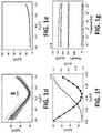
- Figure 1e shows the sum of leakage current (I_gs) for all gSGFETs in the array, which is in the nA range throughout the voltage sweep, demonstrating the good insulation of the passivation layer and the negligible reactivity of the graphene.
- I_gs leakage current
- gm the frequency response of the transconductance of a gSGFET, which indicates the efficiency of the signal coupling (( ⁇ I_ds)/( ⁇ V_gs)), obtaining constant values in a wide bandwidth including inflalow frequencies ( Fig. 1f-g ).
- the negative gm for Vgs values lower than the CNP results in an inversion (180o phase) of the signals measured at such bias; for Vgs values higher than the CNP the signal phase is preserved.
- the device of the invention was compared to conventional high-pass filtered recordings, to do so the propagation of cortical spreading depression (CSD) events using a 4x4 epicortical gSGFET array was mapped and then compared with what is observed in conventional high-pass filtered recordings ( Fig. 5a-b ).
- the recording of the whole CSD event with the gSGFET array reveals that while the onset of the negative shift is similar for all gSGFETs, there is much more variety in the subsequent recovery, with some transistors exhibiting a second negative shift with higher amplitude than the first one. This effect can also be observed in the last frames (corresponding to 80 s and 90 s) of the spatial maps of gSGFET recordings ( Fig.
- Cortical spreading depression was chosen to illustrate the capabilities of the object of the invention to record in a wide bandwidth.
- two craniotomies were performed over the left hemisphere of isoflurane-anaesthetized Wistar rats: a larger craniotomy over the primary somatosensory cortex, where the epicortical probe was placed, and a smaller one in the frontal cortex, where 5 mM KCI was applied locally to induce CSD ( Fig. 2b ).
- a custom electronic circuit allowed us to simultaneously record at two frequency bands: low-pass filtered band (LPF, ⁇ 0-0.16 Hz) and band-pass filtered band (BPF, 0.16 Hz-10 kHz) with different gains (10 4 , and 10 6 respectively) to avoid amplifier saturation due to the high-amplitude CSD signal.
- LPF low-pass filtered band
- BPF band-pass filtered band
- the graphene transistors were polarized in the hole conduction regime, i.e. V gs ⁇ CNP (negative g m ); therefore, the recorded LPF and BPF current signals are inverted with respect to the voltage signal occurring at the gate.
- the LPF signal shows the very slow CSD event whereas the BPF signal corresponds to the local field potential, revealing the silencing of activity typical of cortical spreading depression.
- the wide-band electrophysiological signal can be obtained (see Fig. 2a , c ).
- a small positive shift of 1-2 mV generally precedes the depression, immediately after which a steep negative change ( ⁇ -20 mV) can be observed, which slowly recovers during the next minute or so.
- the CSD-associated silencing of high-frequency activity and its progressive recovery is shown in the voltage wave and spectrogram of Fig. 2d .
Landscapes
- Health & Medical Sciences (AREA)
- Life Sciences & Earth Sciences (AREA)
- Molecular Biology (AREA)
- Physics & Mathematics (AREA)
- Pathology (AREA)
- Engineering & Computer Science (AREA)
- General Health & Medical Sciences (AREA)
- Veterinary Medicine (AREA)
- Heart & Thoracic Surgery (AREA)
- Surgery (AREA)
- Animal Behavior & Ethology (AREA)
- Biomedical Technology (AREA)
- Public Health (AREA)
- Biophysics (AREA)
- Medical Informatics (AREA)
- Chemical & Material Sciences (AREA)
- Electrochemistry (AREA)
- General Physics & Mathematics (AREA)
- Immunology (AREA)
- Biochemistry (AREA)
- Microelectronics & Electronic Packaging (AREA)
- Analytical Chemistry (AREA)
- Chemical Kinetics & Catalysis (AREA)
- Neurosurgery (AREA)
- Psychiatry (AREA)
- Cardiology (AREA)
- Psychology (AREA)
- Physiology (AREA)
- Investigating Or Analyzing Materials By The Use Of Electric Means (AREA)
- Measurement And Recording Of Electrical Phenomena And Electrical Characteristics Of The Living Body (AREA)
Abstract
Description
- The object of the invention belongs to the technical field of physics, more precisely to measuring electrical signals.
- The object of the invention is aimed to a device and a method using said device, for measuring and recording certain electrophysiological signals.
- There is a great need for flexible, large-scale and high-density arrays with a wide electrophysiological recording bandwidth. Flexible, large-scale and high-density electrode arrays are state-of-the-art. However, those arrays do not provide high-fidelity recordings in the entire frequency bandwidth of electrophysiological signals.
- Electrophysiological signals exist in a wide range of frequencies and amplitudes: from minute-long, high-amplitude signals such as cortical spreading depression to millisecond-long microvolt spikes. Recording the full range of electrophysiological signals with high-spatiotemporal resolution would be beneficial to unravel its relation and interactions and to ensure that no meaningful information is lost.
- Most microelectrode arrays suffer from voltage drift and oscillations that affect its recording performance of infraslow signals, which frequency is below 0.1 Hz. This is so widely known, that most recording systems include high-pass filters to solve saturation issues that may arise due to baseline drift, at the cost of excluding potential physiological and pathological information from being recorded.
- In the last years, there has been a particular resurgence of interest in fluctuations of brain activity at frequencies below 0.1 Hz, commonly referred to as very slow, ultraslow or infraslow activity (ISA). They are suggested to be indicative of brain states (e.g. sleep, anesthesia, coma, wakefulness) and were found to be correlated with resting-state networks in functional magnetic resonance imaging. They also may significantly contribute to the high variability observed in the time course of physiological signals.
- There are some reported infraslow signals, such as cortical propagation waves called "cortical spreading depression (CSD)" that are only registered at very low frequencies and therefore it is very difficult to study them in a habitual way due to the impediment of the current electrodes. CSDs are defined as a wave of slow propagation of depolarization of neurons and astrocytes followed by a period of suppression of brain activity and are often triggered when there is a brain episode as in patients who have a vascular or traumatic brain stroke as well as in migraines and other brain pathologies. To monitor or detect them could improve the diagnosis but above all influence on therapeutic changes.
- Full-band recordings including infralow frequencies have been traditionally performed with non-invasive techniques such as electroencephalography (EEG) and magnetoencephalography (MEG). However, their limited spatial resolution and averaged signal impose serious limitations; e.g. EEG alone has not yet been sufficient for non-invasive CSD detection. For these reason, invasive electrophysiological techniques are the most commonly used to record infraslow brainwaves.
- The proper recording of ISA requires the use of direct-coupled amplifiers and extremely stable and low-impedance invasive electrodes. Traditionally, liquid-filled glass micropipettes are used, which allow only one or few-point measurements. For higher spatial resolution and mapping, non-polarizable silver/silver chloride (Ag/AgCI) electrodes could be used, which prevent charge accumulation at the interface and therefore voltage drift. However, due to the toxicity of silver, the use of such electrodes for human or chronic animal in vivo monitoring is not an option. This has fostered the search for alternative microelectrode materials with low impedance and drift although none has yet been found capable of offering comparable performance as Ag/AgCl electrodes. So, ISA recordings in humans are currently performed with platinum electrodes, which challenge CSD detection due to artifacts and transients. Importantly, baseline drift in the form of baseline oscillations in the infralow frequencies, hamper the determination of its "true" characteristics such as amplitude or waveform as any high-pass filter used to remove such effects will alter the signal shape.
- Another intrinsic limitation of microelectrode technology is based on the relation between the microelectrode impedance and the input impedance of the recording equipment (


- The recorded signal (Vin ) is determined by the voltage divider formed by both impedances:

- Eq. (1) implies that when


- Therefore, miniaturization of electrode size to achieve higher spatial resolution causes intrinsic high-pass filtering of ISA due to the associated electrode impedance increase.
- Invasive optical techniques such as calcium imaging are also used to monitor ISA, but still nowadays have serious challenges to resolve high-frequency activity for a large number of neurons24,25 and their intrinsic need of indicators limits the translation to the clinics. Therefore, a technique which allows for measuring large-scale, high-spatiotemporal resolution recordings including infraslow frequencies in a potentially fully implantable, nontoxic, clinical-scale system is still missing.
- Alternatively to the commonly used microelectrode technology, recording electrophysiological signals with field-effect transistors (FET) offers several advantages including that they are less sensitive to environmental noise thanks to their intrinsic voltage-to-current amplification, and that they can be easily multiplexed26. Nonetheless, the difficulties to combine high gate capacitance and carrier mobility silicon FETs with flexible materials has historically hampered its use for in vivo recordings. Graphene solution-gated field-effect transistors (gSGFETs) have been proposed to potentially overcome most previous drawbacks. Graphene flexibility allows gSGFETs to be embedded in ultra-soft and flexible substrates without loss of performance, while its wide electrochemical window and biocompatibility allows direct contact with biological fluids and tissues and ensures a safe operation in in vivo conditions. In addition, the two-dimensional nature of graphene provides the highest surface-to-volume ratio possible, making graphene very sensitive to charges at its surface. Importantly, the frequency response of the transconductance of a gSGFET, is flat in a wide bandwidth including inflalow frequencies.
- On the other hand ,graphene based solution-gated field effect transistors (G-SgFETs) have been extensively investigated as potential biosensors for various analytes, like in
WO2011004136A1 where it is disclosed a sensor for detecting the presence of at least one biological molecule and a method for the production of such a sensor which comprises a patterned graphene structure, at least two electric contacts arranged in contact with the patterned graphene structure for determining a conductivity; and at least one linker attached to at least a portion of the patterned graphene structure, wherein the at least one linker has a binding affinity for the at least one biological molecule. - The present invention addresses the need for flexible, large-scale and high-density arrays with a wide electrophysiological recording bandwidth. The object of the invention is based on a graphene solution-gated field-effect transistor (gSGFETs) preferably an array of graphene solution-gated field-effect transistor (gSGFETs) which are able to record infraslow signals alongside with signals in the typical local field potential bandwidth. The graphene solution-gated field-effect transistor (gSGFETs) preferably are preferably placed on epicortical and intracortical positions.
- The present invention simultaneously overcomes the challenges remaining in the prior art by providing vastly increased baseline stability that arises from the electrochemical inertness of graphene, as also surpasses the signal attenuation due to the impedance divisor present in electrode recording systems by using a transistor as a recording element.
- The graphene solution-gated field-effect transistor (gSGFETs) of the present invention is fabricated using flexible substrate to overcome the difficulty in conforming to the geometry of different biological structures, hence the graphene solution-gated field-effect transistor (gSGFETs) is preferably flexible. Also, when arranged in an array, said array is designed in an extensible manner such that transistors can be scaled up from micro to macro scale as needed while different kinds of electrical contacts, such as those that sit on the surface of (as oppose to penetrating) tissue.
- Hence, the graphene transistor system for measuring electrophysiological signal of the invention encompasses a processing unit, and at least one graphene transistor (gSGFET) comprising graphene as channel material contacted by two terminals, to which a tunable voltage source at the drain and source terminals of the transistor (gSGFET) referred to the gate voltage, and
at least one filter (a low-pass filter (LPF) with of 104 [V/A] configured to generate a low-pass filtered band with a frequency set between 0Hz and 0.16 Hz or a band-pass filter (BPF) with a gain of 106 [V/A] configured to generate a band-filtered band with a frequency comprised between 0.16 Hz and10 kHz) configured to acquire and split the signal from the transistor into at least two frequency bands, low frequency band and high frequency band, are connected; being the first and second signals respectively amplified with a gain value. - The method and associated apparatus of the invention address the above-mentioned needs in the art by providing an amplification of the signals as also the capability to measure the transistor transfer curve at the site of recording. That allows both choosing the best operation point of the transistor and to apply a calibration methodology (current-to-voltage conversion of the recorded signal) that ensures a high-fidelity recording in a wide-bandwidth.
- Of the main applications of the object of the invention is that of monitoring full-band brain signals; in either research or clinic implementations like in neurology. These same advantages exist for applications to other biological systems outside of the brain, such as heart, kidneys, stomach, cranial nerves, and other regions. The flexibility and versatility of graphene-transistor arrays allows several applications and deployments which range from subdural, epidural and intracortical devices to other placements in the brain, peripheral and cranial nerves, heart, blood vessels, spinal cord and other biological structures or non-invasive placement similar to an electroencephalogram.
- To complement the description being made and in order to aid towards a better understanding of the characteristics of the invention, in accordance with a preferred example of practical embodiment thereof, a set of drawings is attached as an integral part of said description wherein, with illustrative and non-limiting character, the following has been represented:
-
Figures 1a-1g .- Show: a representation of flexible graphene solution-gated field-effect transistor array technology and characterization 1a A schematic of a graphene transistor polarized in common gate mode. 1b: Optical microscope images of the active area of the 4x4 gSGFET array and the 15 channel intracortical array. 1c: a photograph of the neural probe. 1d: Steady-state characterization of a 100x50-µm2 gSGFET array in 10 mM phosphate buffered saline (PBS) and with a drain-source voltage bias (Vds) of 50 mV. 1d A graph showing gSGFET transfer curves, drain-source current (Ids) vs gate-source voltage (Vgs), together with the mean (dark curves) and standard deviation (lighter curves). Boxplot inset shows charge neutrality point dispersion (center line, median; box limits, upper and lower quartiles). 1e A graph for the leakage current (Igs) of all gSGFETs in the array. 1f: A graph for the transfer curve (blue squares and line) and its first derivative (transconductance (gm), black line) of a gSGFET. 1g A graph for the frequency response of the transconductance at two different points of the transfer curve (e): Vgs lower than the CNP (green), where gm is negative resulting in a signal inversion (180º phase); and Vgs higher than the CNP (orange), where gm is positive and thus results in no inversion (0º phase) . Independently of the branch of the transfer curve where a gSGFET is polarized, the module of gm is similar to the steady-state value for a wide bandwidth (≈ 0 - 1 kHz). -
Figures 2a-2d .- Show an exemplary embodiment of the invention incorporating a gSGFET, the custom electronic circuit and post-processing methodology as also examples of the recorded signals: infraslow, local field potential, and wide-band in vivo gSGFET recordings of neural signals. 2a, Schematic of the gSGFET recording setup and signal post processing methodology. The custom electronic circuit is used to perform the in vivo characterization (transfer curve) and record the transistor current in the low-pass-filtered (LPF) band and the band-pass-filtered (BPF) band. From the combination of both signals and taking into account the current-to-voltage conversion, the wide-band signal (Vsig ) is obtained. 2b, Electrophysiological recordings obtained with a gSGFET epicortical array during the induction of four CSD events (blue shade). From top to bottom: current LPF signal, current BPF and voltage-converted wide-band signal. -
Figure 3 .- Shows an exemplary embodiment of the custom electronic circuit. a, Schematic of the custom electronic instrumentation which controls the polarization of the gSGFETS (Vgs, Vds) and amplifies differently the two previously mentioned bands: LPF (≈0-0.16 Hz, gain=104) and BPF (0.16 Hz-10 kHz, gain=106). We use the custom electronic instrumentation to characterize the steady-state behaviour of the gSGFETs as well as the AC modulation of graphene transistors. -
Figure 4 .- Shows the calibration procedure of gSGFET current recordings to recover the voltage signal at the gate. a, gSGFET current recordings of a 10 Hz, 0.85 mV-peak sinusoidal gate signal applied through a reference electrode. Graphene transistors are biased at Vds =50mV and Vgs =250mV. b, Transfer curves of the same graphene transistors at Vds =50mV. Dotted line indicates the Vgs bias voltage used in a. c, Voltage signal as obtained by interpolation of the current signal (a) of each transistor into its corresponding transfer curve and removal of the Vgs offset. -
Figure 5 .- Shows the mapping cortical spreading depression with graphene transistors. a, Infralow frequency signals recorded by a 4x4, 400 µm grid spacing, gSGFET array (black lines) during the occurrence of a CSD event as illustrated in the top left schematic. The contour plot shows the time delays of the onset of CSD with respect to the mean time illustrating the spatiotemporal course of the CSD. b, Interpolated spatial voltage maps showing the propagation of the same CSD event as measured by the gSGFET array. a,b High pass filtered recordings at 0.1Hz (red lines in a and bottom spatial voltage maps in b) are included to illustrate the loss of signal information in conventional microelectrode recordings. -
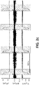
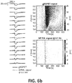
Figures 6a ,6b .- Depict the depth profile of the infralow-frequency voltage variations induced by cortical spreading depression in a rat cortex. a, Layout of the fabricated 15-channel graphene intracortical probe and ordered local field potential recordings. Infralow-frequency recordings (black lines) during the occurrence of a CSD event. Dashed lines, have been interpolated from nearby transistors. b, Colour maps of the temporal course of the infraslow changes during a CSD event across the depth of a rat cortex. a-b, Same signal high-pass filtered at 0.1 Hz (red lines) and their spatiotemporal colour map are included to illustrate the loss of information in conventional microelectrode recordings. - A first aspect of the invention is aimed to a system for registering electrofysiological infraslow signals like cortical spreading depression (CSD) signals i. e. those with a frequency value below 0.1Hz; the device comprising a processing unit associated or embedded in the very device, and at least one graphene transistor (gSGFET), preferably an array of graphene transistors, comprising graphene as channel material contacted by source and drain terminals, with a reference as gate terminal. Said graphene transistor is connected to at least a filter like a low pass filter (LPF). The recorded current signal is transformed into a voltage signal using the transistor transfer curve Ids - Vgs recorded to the start of the recordings).
- In an alternative embodiment of the invention, at least one band pass filter (BPF) is arranged in either a sequential or a cascade arrangement along with the low pass filter (LPF). but both filters (LPF, BPF) being configured so that the respective cutting points have the same value.
- An gSGFETS is a device in which graphene is used as channel material, contacted by two metal leads (source and drain terminals), and is immersed in an electrolyte solution where a reference electrode is used as gate terminal (
Fig. 1a ). Flexible probes containing arrays of gSGFETs in both epicortical and intracortical designs were produced. In particular, a 4x4 array of 100 µm wide by 50 µm long graphene channels were designed for epicortical recordings while a design consisting of a linear array of 15 graphene channels (80 µm width, 30 µm length) was used for intracortical recordings (Fig. 1b ). Both array designs were fabricated on a 10 µm thick polyimide layer coated on a 4-inch silicon wafer. Flexible gSGFET arrays were placed in zero insertion force connectors for interfacing with recording electronics (Fig. 1c ). The transfer curve, drain current (I_ds) vs gate-source voltage (V_gs), of all gSGFETs in each array was measured with a fixed drain-source voltage (V_ds). The dispersion of the charge neutrality point (CNP=243.6 ± 6.1 mV), which is the minimum of the transfer curve, indicates the homogeneity of the transistors (Fig. 1d ). Importantly, since the V_gs and V_ds bias are shared, the small CNP dispersion allows near-optimal recording performance for all gSGFETs in the same array.Figure 1e shows the sum of leakage current (I_gs) for all gSGFETs in the array, which is in the nA range throughout the voltage sweep, demonstrating the good insulation of the passivation layer and the negligible reactivity of the graphene. Furthermore, we measured the frequency response of the transconductance (gm) of a gSGFET, which indicates the efficiency of the signal coupling ((∂I_ds)/(∂V_gs)), obtaining constant values in a wide bandwidth including inflalow frequencies (Fig. 1f-g ). The negative gm for Vgs values lower than the CNP results in an inversion (180º phase) of the signals measured at such bias; for Vgs values higher than the CNP the signal phase is preserved. - The device of the invention was compared to conventional high-pass filtered recordings, to do so the propagation of cortical spreading depression (CSD) events using a 4x4 epicortical gSGFET array was mapped and then compared with what is observed in conventional high-pass filtered recordings (
Fig. 5a-b ). The recording of the whole CSD event with the gSGFET array reveals that while the onset of the negative shift is similar for all gSGFETs, there is much more variety in the subsequent recovery, with some transistors exhibiting a second negative shift with higher amplitude than the first one. This effect can also be observed in the last frames (corresponding to 80 s and 90 s) of the spatial maps of gSGFET recordings (Fig. 5b ) where recovered and still depressed brain areas coexist. Importantly, this information is lost in conventional microelectrode recordings, where only the CSD onset is observed due to the high pass filter in the recording electronics. The following results are referred to a sample of 10 CSDs collected from two different subjects in the somatosensory cortex: we found that the mean duration of CSD events is 47.24 ± 7.65 s and a speed of propagation of 7.68 ± 1.35 mm/min, in agreement with the literature defining CSDs as infraslow brainwaves. - To further illustrate the potential of the device of the invention and taking advantage of the design versatility offered by this technology, a linear array of 15 gSGFETs spanning the entire depth of the cortex (
Fig. 6a ) was arranged. From either the ordered recording or the spatiotemporal voltage map (Fig. 6b ), it can be seen how CSD occurs in the whole cortex depth. These results highlight the capability of the device of the invention to reveal the rich pattern of infraslow signals in the cortex; in this particular case, a transition from a superficial long depolarization to a shorter one preceded and followed by a hyperpolarization in the deeper layers is clearly observed. The origin of such depth-dependent effect is not well understood and will be the target of further investigations, taking advantage of the demonstrated capability of gSGFET technology to monitor ISA with high spatial resolution. - In a second aspect of the invention a method for recording infraslow brain signals, being infraslow signals those with a frequency value below 0.1Hz is provided, Cortical spreading depression (CSD) was chosen to illustrate the capabilities of the object of the invention to record in a wide bandwidth. Experimentally, two craniotomies were performed over the left hemisphere of isoflurane-anaesthetized Wistar rats: a larger craniotomy over the primary somatosensory cortex, where the epicortical probe was placed, and a smaller one in the frontal cortex, where 5 mM KCI was applied locally to induce CSD (
Fig. 2b ). A custom electronic circuit allowed us to simultaneously record at two frequency bands: low-pass filtered band (LPF, ≈0-0.16 Hz) and band-pass filtered band (BPF, 0.16 Hz-10 kHz) with different gains (104, and 106 respectively) to avoid amplifier saturation due to the high-amplitude CSD signal. In a first set of experiments, we recorded the LPF and BPF current signals with an epicortical gSGFET array during the induction of CSD events (Fig. 2c ). The graphene transistors were polarized in the hole conduction regime, i.e. Vgs < CNP (negative gm); therefore, the recorded LPF and BPF current signals are inverted with respect to the voltage signal occurring at the gate. The LPF signal shows the very slow CSD event whereas the BPF signal corresponds to the local field potential, revealing the silencing of activity typical of cortical spreading depression. After summation of the LPF and BPF signals and then transforming the current into a voltage signal (using the transistor transfer curve Ids - Vgs recorded in vivo prior to the start of the recordings), the wide-band electrophysiological signal can be obtained (seeFig. 2a ,c ). In each CSD event a small positive shift of 1-2 mV generally precedes the depression, immediately after which a steep negative change (≈ -20 mV) can be observed, which slowly recovers during the next minute or so. The CSD-associated silencing of high-frequency activity and its progressive recovery is shown in the voltage wave and spectrogram ofFig. 2d .
Claims (7)
- Graphene transistor system for measuring electrophysiological signals, comprising:a. a processing unit, andb. at least one graphene transistor (gSGFET) comprising graphene as channel material contacted by two terminals,being the device characterized by comprising connected to the graphene transistor (gSGFET):a. a tunable voltage source at the drain and source terminals of the transistor (gSGFET) referred to the gate voltage, andb. at least one filter configured to acquire and split the signal from the transistor into at least two frequency bands, low frequency band and high frequency band, wherein the first and second signals are respectively amplified with a gain value.
- Graphene transistor system for measuring electrophysiological according to claim 1 wherein the filter is configured to generate one of:a. a low-pass filtered band with a frequency set between 0Hz and 0.16 Hz, andb. a band-filtered band with a frequency comprised between 0.16 Hz and10 kHz.
- Graphene transistor system for measuring full-band electrophysiological according to claim 2 wherein the low-pass filter (LPF) and the band-pass filter (BPF) have different gains; 104 [V/A] and 106 [V/A] respectively.
- Method for measuring electrophysiological signals, using the graphene transistor system of any one of claims 1-3, the method being characterized by comprising:a. splitting an input signal into frequency and high frequency signals by means of the filter,b. merging the low frequency and high frequency signals weighted by the corresponding gain, andc. transforming the signal into voltage according with the intrinsic gain of the transistor.
- Method according to claim 4, wherein the gain value of the amplification is different for each signal.
- Method according to claim 4 wherein the transformation into a voltage signal is carried out by means of interpolation using the graphene transistor transfer curve Ids - Vgs
- Method according to claim 6, wherein the transistor transfer curve Ids - Vgs is generated with a fixed drain-source voltage (V_ds).
Applications Claiming Priority (2)
| Application Number | Priority Date | Filing Date | Title |
|---|---|---|---|
| ES201831068A ES2759053B2 (en) | 2018-11-06 | 2018-11-06 | Graphene transistor system for measuring electrophysiological signals. |
| PCT/ES2019/070728 WO2020094898A1 (en) | 2018-11-06 | 2019-10-28 | System of graphene transistors for measuring electrophysiological signals |
Publications (3)
| Publication Number | Publication Date |
|---|---|
| EP3879582A1 true EP3879582A1 (en) | 2021-09-15 |
| EP3879582A4 EP3879582A4 (en) | 2022-11-02 |
| EP3879582B1 EP3879582B1 (en) | 2025-07-23 |
Family
ID=70483270
Family Applications (1)
| Application Number | Title | Priority Date | Filing Date |
|---|---|---|---|
| EP19881753.8A Active EP3879582B1 (en) | 2018-11-06 | 2019-10-28 | System of graphene transistors for measuring electrophysiological signals |
Country Status (6)
| Country | Link |
|---|---|
| US (1) | US20210345930A1 (en) |
| EP (1) | EP3879582B1 (en) |
| JP (1) | JP7621958B2 (en) |
| CN (1) | CN113366653B (en) |
| ES (1) | ES2759053B2 (en) |
| WO (1) | WO2020094898A1 (en) |
Families Citing this family (2)
| Publication number | Priority date | Publication date | Assignee | Title |
|---|---|---|---|---|
| JP7048972B2 (en) * | 2018-11-09 | 2022-04-06 | 国立研究開発法人理化学研究所 | Cortical EEG electrodes, brain activity processing system, and brain activity processing method |
| EP3970614A1 (en) | 2020-09-17 | 2022-03-23 | Consejo Superior De Investigaciones Científicas (CSIC) | Acquisition device to limit leakage current in electrophysiological signal recording devices |
Family Cites Families (19)
| Publication number | Priority date | Publication date | Assignee | Title |
|---|---|---|---|---|
| US3826243A (en) * | 1972-07-10 | 1974-07-30 | W Anderson | Brainwave analysis and feedback method and apparatus |
| US4417590A (en) * | 1978-06-09 | 1983-11-29 | Beckman Instruments, Inc. | Electroencephalograph |
| JPH0628647B2 (en) * | 1985-08-05 | 1994-04-20 | 三菱電機株式会社 | EEG measuring device |
| US7187968B2 (en) * | 2003-10-23 | 2007-03-06 | Duke University | Apparatus for acquiring and transmitting neural signals and related methods |
| US7346312B2 (en) * | 2004-10-23 | 2008-03-18 | Triangle Biosystems, Inc. | Wireless neural data acquisition system |
| US20090093687A1 (en) * | 2007-03-08 | 2009-04-09 | Telfort Valery G | Systems and methods for determining a physiological condition using an acoustic monitor |
| EP2231006A1 (en) * | 2008-01-11 | 2010-09-29 | Koninklijke Philips Electronics N.V. | Method and support system for presenting electrophysiological measurements |
| JP2010257343A (en) | 2009-04-27 | 2010-11-11 | Niigata Univ | Communication support device |
| GB2471672B (en) * | 2009-07-07 | 2015-12-09 | Swansea Innovations Ltd | Graphene biosensor |
| WO2012126003A1 (en) * | 2011-03-17 | 2012-09-20 | Brown University | Implantable wireless neural device |
| JP5711056B2 (en) | 2011-06-21 | 2015-04-30 | アイシン精機株式会社 | EEG interface system |
| US10492702B2 (en) * | 2012-01-31 | 2019-12-03 | The Regents Of The University Of California | Real-time cortical mapping |
| US20150038870A1 (en) * | 2012-02-17 | 2015-02-05 | Massachusetts Institute Of Technology | Multi-Channel Scalable EEG Acquisition System on a Chip with Integrated Patient Specific Seizure Classification and Recording Processor |
| US9622672B2 (en) * | 2013-06-07 | 2017-04-18 | Indiana University Research And Technology Corporation | Digitally invertible universal amplifier for recording and processing of bioelectric signals |
| CN103901089B (en) * | 2014-04-16 | 2016-08-24 | 国家纳米科学中心 | The detection sensor of neurocyte electricity physiological signal and manufacture method and detection method |
| KR102273650B1 (en) * | 2016-02-22 | 2021-07-07 | 한국전자통신연구원 | A circuit for both bio-stimulating and measuring biological signal |
| US11103168B2 (en) | 2016-08-08 | 2021-08-31 | New York University | Systems and methods for in vivo detection of electrophysiological and electrochemical signals |
| US10206602B2 (en) * | 2017-04-05 | 2019-02-19 | LR Technologies, Inc. | Hybrid electromagnetic field signal detection system for human bioelectrical signal monitoring |
| GB201801828D0 (en) * | 2018-02-05 | 2018-03-21 | Imperial Innovations Ltd | Analysis of spreading depolarization waves |
-
2018
- 2018-11-06 ES ES201831068A patent/ES2759053B2/en active Active
-
2019
- 2019-10-28 JP JP2021549923A patent/JP7621958B2/en active Active
- 2019-10-28 CN CN201980072941.7A patent/CN113366653B/en active Active
- 2019-10-28 EP EP19881753.8A patent/EP3879582B1/en active Active
- 2019-10-28 WO PCT/ES2019/070728 patent/WO2020094898A1/en not_active Ceased
-
2021
- 2021-05-06 US US17/313,469 patent/US20210345930A1/en active Pending
Also Published As
| Publication number | Publication date |
|---|---|
| EP3879582A4 (en) | 2022-11-02 |
| EP3879582B1 (en) | 2025-07-23 |
| US20210345930A1 (en) | 2021-11-11 |
| ES2759053B2 (en) | 2020-10-08 |
| JP2022511125A (en) | 2022-01-28 |
| ES2759053A1 (en) | 2020-05-07 |
| WO2020094898A1 (en) | 2020-05-14 |
| JP7621958B2 (en) | 2025-01-27 |
| CN113366653B (en) | 2025-11-04 |
| CN113366653A (en) | 2021-09-07 |
Similar Documents
| Publication | Publication Date | Title |
|---|---|---|
| Bonaccini Calia et al. | Full-bandwidth electrophysiology of seizures and epileptiform activity enabled by flexible graphene microtransistor depth neural probes | |
| Masvidal-Codina et al. | High-resolution mapping of infraslow cortical brain activity enabled by graphene microtransistors | |
| KR101007558B1 (en) | Method for measuring laboratory animal EBE using thin-film multi-channel microelectrode and microelectrode for laboratory animal EB measurement | |
| Li et al. | Novel passive ceramic based semi-dry electrodes for recording electroencephalography signals from the hairy scalp | |
| Day | Important factors in surface EMG measurement | |
| KR20130022542A (en) | Apparatus and method for measuring bioelectic signals | |
| WO2005065544A1 (en) | Active dry sensor module for measurement of bioelectricity | |
| KR20110135296A (en) | Apparatus and method for measuring bio signals | |
| US20130338529A1 (en) | Bioelectric signal measurement apparatus | |
| Garcia-Casado et al. | Enhancement of non-invasive recording of electroenterogram by means of a flexible array of concentric ring electrodes | |
| de Camp et al. | Light-cured polymer electrodes for non-invasive EEG recordings | |
| US20140378859A1 (en) | Method of Multichannel Galvanic Skin Response Detection for Improving Measurement Accuracy and Noise/Artifact Rejection | |
| US20210345930A1 (en) | Graphene transistor system for measuring electrophysiological signals | |
| Pazzini et al. | An ultra-compact integrated system for brain activity recording and stimulation validated over cortical slow oscillations in vivo and in vitro | |
| Fara et al. | Robust, ultra low-cost mmg system with brain-machine-interface applications | |
| Ahmad et al. | A review of EMG recording technique | |
| Howells et al. | Tracking small sensory nerve action potentials in human axonal excitability studies | |
| WO2018088400A1 (en) | Sensor connection device for brain wave spectrum analysis device | |
| US20060224077A1 (en) | Analysis of a skin reactivity and hypersensitivity | |
| Schulz-Juergensen et al. | Simultaneous EMG–fMRI during startle inhibition in monosymptomatic enuresis—an exploratory study | |
| US20180140214A1 (en) | System for measuring electromyographic signals of deep muscles based on surface electromyography | |
| Svärd et al. | Design and evaluation of a capacitively coupled sensor readout circuit, toward contact-less ECG and EEG | |
| Journée et al. | Extramuscular recording of spontaneous EMG activity and transcranial electrical elicited motor potentials in horses: characteristics of different subcutaneous and surface electrode types and practical guidelines | |
| Williams et al. | A Novel μECoG Electrode Interface for Comparison of Local and Common Averaged Referenced Signals | |
| Guimerà-Brunet et al. | Neural interfaces based on flexible graphene transistors: A new tool for electrophysiology |
Legal Events
| Date | Code | Title | Description |
|---|---|---|---|
| STAA | Information on the status of an ep patent application or granted ep patent |
Free format text: STATUS: THE INTERNATIONAL PUBLICATION HAS BEEN MADE |
|
| PUAI | Public reference made under article 153(3) epc to a published international application that has entered the european phase |
Free format text: ORIGINAL CODE: 0009012 |
|
| STAA | Information on the status of an ep patent application or granted ep patent |
Free format text: STATUS: REQUEST FOR EXAMINATION WAS MADE |
|
| 17P | Request for examination filed |
Effective date: 20210512 |
|
| AK | Designated contracting states |
Kind code of ref document: A1 Designated state(s): AL AT BE BG CH CY CZ DE DK EE ES FI FR GB GR HR HU IE IS IT LI LT LU LV MC MK MT NL NO PL PT RO RS SE SI SK SM TR |
|
| DAV | Request for validation of the european patent (deleted) | ||
| DAX | Request for extension of the european patent (deleted) | ||
| A4 | Supplementary search report drawn up and despatched |
Effective date: 20220929 |
|
| RIC1 | Information provided on ipc code assigned before grant |
Ipc: A61B 5/00 20060101ALI20220923BHEP Ipc: H01L 29/772 20060101AFI20220923BHEP |
|
| REG | Reference to a national code |
Ref country code: DE Free format text: PREVIOUS MAIN CLASS: H01L0029772000 Ipc: A61B0005000000 Ref legal event code: R079 Ref document number: 602019073055 Country of ref document: DE |
|
| GRAP | Despatch of communication of intention to grant a patent |
Free format text: ORIGINAL CODE: EPIDOSNIGR1 |
|
| STAA | Information on the status of an ep patent application or granted ep patent |
Free format text: STATUS: GRANT OF PATENT IS INTENDED |
|
| RIC1 | Information provided on ipc code assigned before grant |
Ipc: A61B 5/00 20060101AFI20250221BHEP |
|
| INTG | Intention to grant announced |
Effective date: 20250306 |
|
| GRAS | Grant fee paid |
Free format text: ORIGINAL CODE: EPIDOSNIGR3 |
|
| GRAA | (expected) grant |
Free format text: ORIGINAL CODE: 0009210 |
|
| STAA | Information on the status of an ep patent application or granted ep patent |
Free format text: STATUS: THE PATENT HAS BEEN GRANTED |
|
| AK | Designated contracting states |
Kind code of ref document: B1 Designated state(s): AL AT BE BG CH CY CZ DE DK EE ES FI FR GB GR HR HU IE IS IT LI LT LU LV MC MK MT NL NO PL PT RO RS SE SI SK SM TR |
|
| REG | Reference to a national code |
Ref country code: GB Ref legal event code: FG4D |
|
| REG | Reference to a national code |
Ref country code: CH Ref legal event code: EP |
|
| REG | Reference to a national code |
Ref country code: IE Ref legal event code: FG4D |
|
| REG | Reference to a national code |
Ref country code: DE Ref legal event code: R096 Ref document number: 602019073055 Country of ref document: DE |
|
| REG | Reference to a national code |
Ref country code: CH Ref legal event code: U11 Free format text: ST27 STATUS EVENT CODE: U-0-0-U10-U11 (AS PROVIDED BY THE NATIONAL OFFICE) Effective date: 20251101 |
|
| REG | Reference to a national code |
Ref country code: NL Ref legal event code: MP Effective date: 20250723 |
|
| PG25 | Lapsed in a contracting state [announced via postgrant information from national office to epo] |
Ref country code: PT Free format text: LAPSE BECAUSE OF FAILURE TO SUBMIT A TRANSLATION OF THE DESCRIPTION OR TO PAY THE FEE WITHIN THE PRESCRIBED TIME-LIMIT Effective date: 20251124 |
|
| PG25 | Lapsed in a contracting state [announced via postgrant information from national office to epo] |
Ref country code: NL Free format text: LAPSE BECAUSE OF FAILURE TO SUBMIT A TRANSLATION OF THE DESCRIPTION OR TO PAY THE FEE WITHIN THE PRESCRIBED TIME-LIMIT Effective date: 20250723 |
|
| REG | Reference to a national code |
Ref country code: AT Ref legal event code: MK05 Ref document number: 1815522 Country of ref document: AT Kind code of ref document: T Effective date: 20250723 |
|
| PG25 | Lapsed in a contracting state [announced via postgrant information from national office to epo] |
Ref country code: IS Free format text: LAPSE BECAUSE OF FAILURE TO SUBMIT A TRANSLATION OF THE DESCRIPTION OR TO PAY THE FEE WITHIN THE PRESCRIBED TIME-LIMIT Effective date: 20251123 |
|
| PGFP | Annual fee paid to national office [announced via postgrant information from national office to epo] |
Ref country code: DE Payment date: 20251028 Year of fee payment: 7 |
|
| PGFP | Annual fee paid to national office [announced via postgrant information from national office to epo] |
Ref country code: GB Payment date: 20251022 Year of fee payment: 7 |
|
| PG25 | Lapsed in a contracting state [announced via postgrant information from national office to epo] |
Ref country code: NO Free format text: LAPSE BECAUSE OF FAILURE TO SUBMIT A TRANSLATION OF THE DESCRIPTION OR TO PAY THE FEE WITHIN THE PRESCRIBED TIME-LIMIT Effective date: 20251023 |
|
| REG | Reference to a national code |
Ref country code: LT Ref legal event code: MG9D |
|
| PG25 | Lapsed in a contracting state [announced via postgrant information from national office to epo] |
Ref country code: AT Free format text: LAPSE BECAUSE OF FAILURE TO SUBMIT A TRANSLATION OF THE DESCRIPTION OR TO PAY THE FEE WITHIN THE PRESCRIBED TIME-LIMIT Effective date: 20250723 |
|
| PG25 | Lapsed in a contracting state [announced via postgrant information from national office to epo] |
Ref country code: FI Free format text: LAPSE BECAUSE OF FAILURE TO SUBMIT A TRANSLATION OF THE DESCRIPTION OR TO PAY THE FEE WITHIN THE PRESCRIBED TIME-LIMIT Effective date: 20250723 |
|
| PG25 | Lapsed in a contracting state [announced via postgrant information from national office to epo] |
Ref country code: HR Free format text: LAPSE BECAUSE OF FAILURE TO SUBMIT A TRANSLATION OF THE DESCRIPTION OR TO PAY THE FEE WITHIN THE PRESCRIBED TIME-LIMIT Effective date: 20250723 |
|
| PGFP | Annual fee paid to national office [announced via postgrant information from national office to epo] |
Ref country code: FR Payment date: 20251030 Year of fee payment: 7 |
|
| PGFP | Annual fee paid to national office [announced via postgrant information from national office to epo] |
Ref country code: BE Payment date: 20251021 Year of fee payment: 7 |
|
| PGFP | Annual fee paid to national office [announced via postgrant information from national office to epo] |
Ref country code: CH Payment date: 20251101 Year of fee payment: 7 |
|
| PG25 | Lapsed in a contracting state [announced via postgrant information from national office to epo] |
Ref country code: SE Free format text: LAPSE BECAUSE OF FAILURE TO SUBMIT A TRANSLATION OF THE DESCRIPTION OR TO PAY THE FEE WITHIN THE PRESCRIBED TIME-LIMIT Effective date: 20250723 |
|
| PG25 | Lapsed in a contracting state [announced via postgrant information from national office to epo] |
Ref country code: LV Free format text: LAPSE BECAUSE OF FAILURE TO SUBMIT A TRANSLATION OF THE DESCRIPTION OR TO PAY THE FEE WITHIN THE PRESCRIBED TIME-LIMIT Effective date: 20250723 |
|
| PG25 | Lapsed in a contracting state [announced via postgrant information from national office to epo] |
Ref country code: BG Free format text: LAPSE BECAUSE OF FAILURE TO SUBMIT A TRANSLATION OF THE DESCRIPTION OR TO PAY THE FEE WITHIN THE PRESCRIBED TIME-LIMIT Effective date: 20250723 Ref country code: PL Free format text: LAPSE BECAUSE OF FAILURE TO SUBMIT A TRANSLATION OF THE DESCRIPTION OR TO PAY THE FEE WITHIN THE PRESCRIBED TIME-LIMIT Effective date: 20250723 |
|
| PG25 | Lapsed in a contracting state [announced via postgrant information from national office to epo] |
Ref country code: RS Free format text: LAPSE BECAUSE OF FAILURE TO SUBMIT A TRANSLATION OF THE DESCRIPTION OR TO PAY THE FEE WITHIN THE PRESCRIBED TIME-LIMIT Effective date: 20251023 |
|
| PG25 | Lapsed in a contracting state [announced via postgrant information from national office to epo] |
Ref country code: ES Free format text: LAPSE BECAUSE OF FAILURE TO SUBMIT A TRANSLATION OF THE DESCRIPTION OR TO PAY THE FEE WITHIN THE PRESCRIBED TIME-LIMIT Effective date: 20250723 |
|
| PG25 | Lapsed in a contracting state [announced via postgrant information from national office to epo] |
Ref country code: RO Free format text: LAPSE BECAUSE OF FAILURE TO SUBMIT A TRANSLATION OF THE DESCRIPTION OR TO PAY THE FEE WITHIN THE PRESCRIBED TIME-LIMIT Effective date: 20250723 |
|
| PG25 | Lapsed in a contracting state [announced via postgrant information from national office to epo] |
Ref country code: SM Free format text: LAPSE BECAUSE OF FAILURE TO SUBMIT A TRANSLATION OF THE DESCRIPTION OR TO PAY THE FEE WITHIN THE PRESCRIBED TIME-LIMIT Effective date: 20250723 |
|
| PG25 | Lapsed in a contracting state [announced via postgrant information from national office to epo] |
Ref country code: DK Free format text: LAPSE BECAUSE OF FAILURE TO SUBMIT A TRANSLATION OF THE DESCRIPTION OR TO PAY THE FEE WITHIN THE PRESCRIBED TIME-LIMIT Effective date: 20250723 |
|
| PG25 | Lapsed in a contracting state [announced via postgrant information from national office to epo] |
Ref country code: IT Free format text: LAPSE BECAUSE OF FAILURE TO SUBMIT A TRANSLATION OF THE DESCRIPTION OR TO PAY THE FEE WITHIN THE PRESCRIBED TIME-LIMIT Effective date: 20250723 |
|
| PG25 | Lapsed in a contracting state [announced via postgrant information from national office to epo] |
Ref country code: CZ Free format text: LAPSE BECAUSE OF FAILURE TO SUBMIT A TRANSLATION OF THE DESCRIPTION OR TO PAY THE FEE WITHIN THE PRESCRIBED TIME-LIMIT Effective date: 20250723 |
|
| PG25 | Lapsed in a contracting state [announced via postgrant information from national office to epo] |
Ref country code: EE Free format text: LAPSE BECAUSE OF FAILURE TO SUBMIT A TRANSLATION OF THE DESCRIPTION OR TO PAY THE FEE WITHIN THE PRESCRIBED TIME-LIMIT Effective date: 20250723 Ref country code: SK Free format text: LAPSE BECAUSE OF FAILURE TO SUBMIT A TRANSLATION OF THE DESCRIPTION OR TO PAY THE FEE WITHIN THE PRESCRIBED TIME-LIMIT Effective date: 20250723 |