EP3872754A1 - Method and system for automated processing of images when using a contrast agent in mri - Google Patents
Method and system for automated processing of images when using a contrast agent in mri Download PDFInfo
- Publication number
- EP3872754A1 EP3872754A1 EP20160012.9A EP20160012A EP3872754A1 EP 3872754 A1 EP3872754 A1 EP 3872754A1 EP 20160012 A EP20160012 A EP 20160012A EP 3872754 A1 EP3872754 A1 EP 3872754A1
- Authority
- EP
- European Patent Office
- Prior art keywords
- contrast
- post
- images
- image
- mri
- Prior art date
- Legal status (The legal status is an assumption and is not a legal conclusion. Google has not performed a legal analysis and makes no representation as to the accuracy of the status listed.)
- Withdrawn
Links
Images


Classifications
-
- G—PHYSICS
- G06—COMPUTING OR CALCULATING; COUNTING
- G06T—IMAGE DATA PROCESSING OR GENERATION, IN GENERAL
- G06T7/00—Image analysis
- G06T7/0002—Inspection of images, e.g. flaw detection
- G06T7/0012—Biomedical image inspection
-
- G—PHYSICS
- G06—COMPUTING OR CALCULATING; COUNTING
- G06T—IMAGE DATA PROCESSING OR GENERATION, IN GENERAL
- G06T11/00—Two-dimensional [2D] image generation
-
- G—PHYSICS
- G06—COMPUTING OR CALCULATING; COUNTING
- G06T—IMAGE DATA PROCESSING OR GENERATION, IN GENERAL
- G06T7/00—Image analysis
- G06T7/10—Segmentation; Edge detection
- G06T7/11—Region-based segmentation
-
- G—PHYSICS
- G06—COMPUTING OR CALCULATING; COUNTING
- G06T—IMAGE DATA PROCESSING OR GENERATION, IN GENERAL
- G06T2207/00—Indexing scheme for image analysis or image enhancement
- G06T2207/10—Image acquisition modality
- G06T2207/10072—Tomographic images
- G06T2207/10088—Magnetic resonance imaging [MRI]
-
- G—PHYSICS
- G06—COMPUTING OR CALCULATING; COUNTING
- G06T—IMAGE DATA PROCESSING OR GENERATION, IN GENERAL
- G06T2207/00—Indexing scheme for image analysis or image enhancement
- G06T2207/20—Special algorithmic details
- G06T2207/20081—Training; Learning
-
- G—PHYSICS
- G06—COMPUTING OR CALCULATING; COUNTING
- G06T—IMAGE DATA PROCESSING OR GENERATION, IN GENERAL
- G06T2207/00—Indexing scheme for image analysis or image enhancement
- G06T2207/20—Special algorithmic details
- G06T2207/20084—Artificial neural networks [ANN]
Definitions
- the present disclosure is directed, in general, to imaging techniques for imaging biological objects, like tissues, using Magnetic Resonance Imaging (MRI) techniques. More specifically, the present invention is directed to methods and systems for automated processing of those images when using a contrast agent in MRI.
- MRI Magnetic Resonance Imaging
- MRI is an established imaging technique able to deliver a wide variety of contrasts among soft tissues that contain information of crucial importance in daily clinical practice.
- several international consortiums provide guidelines recommending the use of specific MRI protocols.
- MAGNIMS European research network for MR imaging in multiple sclerosis
- T1w gadolinium
- pre-Gd gadolinium
- pre-Gd T1w images provide added value for the quantitative assessment of additional neuroimaging biomarkers (e.g. morphometry, automated localization and counting of brain lesions, among others).
- additional neuroimaging biomarkers e.g. morphometry, automated localization and counting of brain lesions, among others.
- WM white matter
- GM grey matter
- CSF cerebrospinal fluid
- T1w images [2] [3]
- Siemens Healthcare Brain Morphometry software [4] for syngo.via and AI-Rad Companion Unfortunately, it has also been shown by previous studies that the application of such tools on post-Gd T1w images results in significant biases given the sensitivity of brain volumetric assessments to changes in image intensities [5], introducing for instance errors in tissue classification.
- the tissue segmentation achieved with this existing solution might present the disadvantage of relying on suboptimal contrasts with poor information regarding tissues boundaries that might result in poor segmentation quality.
- the quality of the segmentation strongly depends on the ground truth labels used for the training and does not open to the possibility of using eventually other available tools for the tissue segmentation or perform any other processing that requires the missing contrast (e.g., lesion counting and localization).
- the present invention proposes notably to generate pre-contrast images (e.g. pre-Gd images, i.e. images representing a biological object before or without an administration of a contrast agent) from post-contrast imaging data (e.g. from post-Gd images, i.e. images of the biological object taken after the administration of said contrast agent) based on machine learning, e.g. based on a deep learning network.
- the pre-contrast images synthetized from the deep learning network are then used for an automated process configured for characterizing a biological tissue, like an automated biological tissue segmentation (typically brain tissue segmentation), or for any biological tissue characterization process that requires pre-contrast T1w images.
- the method and system according to the invention do not increase the acquisition time since the processing of brain tissues in exams is realized from post-contrast image acquisition only and does not require a pre-contrast image acquisition for this purpose, keeping therefore the acquisition time as short as possible.
- the present invention concerns a method for an automated biological tissue characterization process, like an automated biological tissue segmentation, of a biological object to be imaged by MRI, the method comprising:
- the present invention concerns also a computer-implemented method for providing a trained function, the method comprising:
- the present invention concerns also a training system comprising:
- the present invention concerns also a system for an automated characterization of biological object tissues, said system comprising:
- the present invention proposes thus to use machine learning, more precisely deep learning generative models, and preferentially a cycleGAN, for the synthesis of pre-contrast T1w images from post-contrast T1w images to achieve a biological object tissue characterization free of an acquisition of said pre-contrast T1w images.
- the present invention enables to perform a whole-brain segmentation from synthetic pre-Gd T1w images generated from post-Gd data.
- the present invention enables an accurate post-processing of synthetic pre-contrast T1w images after an acquisition of post-contrast images of said biological object, which until now was not possible without the acquisition of said pre-contrast T1w images.
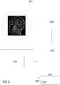
- Figure 1 describes the different steps of the method 100 carried out by a preferred embodiment of the system according to the invention which is illustrated by Figure 2 .
- a control unit 203 of the system for automated characterization (e.g. automated segmentation and/or abnormality detection) of biological tissues acquires or receives, for instance via a first interface 202 configured for enabling an exchange of data between an imaging system, e.g. the imaging system 201 of a MRI apparatus, and said control unit 203, post-contrast MRI data, e.g. post-contrast images, for a biological object to be imaged.
- the post-contrast data or images are data or images acquired after administration of a contrast agent to the biological object.
- Said control unit 203 may comprise one or several processors configured for processing MRI data.
- the control unit 203 might be part of the MRI apparatus or might be a separated independent device.
- the control unit 203 applies a trained function to the acquired or received post-contrast MRI data in order to output a synthetic pre-contrast image from the post-contrast MRI data, e.g. a synthetic pre-contrast T1w image.
- the synthetic pre-contrast image is an image representing the biological object as it would be obtained when acquiring said image before or without the administration of the contrast agent.
- a training system 205 has been used for training said function.
- the training system 205 comprises notably a first interface 206, a training computation unit 207, a second and a third interface which can be the same or different from the first interface 206.
- the training system 205 is notably configured for acquiring or receiving via its first interface 206 an input dataset and another input dataset.
- the input dataset and said another input dataset are paired, i.e. to each data of the input dataset corresponds a data of said another input dataset which is closely related or linked to the data of the input dataset.
- the input dataset typically comprises post-contrast MRI data or images of the biological object and said another input dataset comprises pre-contrast T1w data or images of said biological object, wherein each image or data of the input dataset is preferentially paired to an image or data of said another input dataset in that they have been acquired for a same voxel or set of voxels, e.g. they represent a same part of the biological object.
- the datasets are used by the training computation unit 207 for training the function.
- the trained function is able to generate realistic and accurate synthetic pre-contrast T1w images from acquired post-contrast MRI data, e.g. from the input dataset and consequently from any other post-contrast MRI data or image.
- the trained function might be outputted via the third interface.
- the system according to the invention automatically performs an automated process (e.g., segmentation) for characterizing the biological tissues of said biological object from the synthetic pre-contrast T1w image and may display the obtained results of the characterization on a display 204.
- an automated process e.g., segmentation
- technique known in the art might be used.
- the training computation unit 207 uses a machine learning technique for training said function, notably based on a training of deep convolutional networks.
- a generative adversarial algorithm is used by said training computation unit 207, like a GAN, a cycleGAN, or a conditional GAN (hereafter cGAN) algorithm for generating synthetic pre-contrast T1w images of the biological object.
- a preferred embodiment illustrating the use of the cycleGAN is described hereafter.
- the cycleGAN learn to translate images from one domain to the other (domain is denoted as a set of images sharing features that are inherent in the images of the set).
- a first image domain x comprises post-contrast MRI images from said input dataset
- a second image domain y comprises pre-contrast T1w images from said another input dataset.
- the cycleGAN architecture involves then a pair of generators (G x , G y ) that are simultaneously trained to obtain synthetic images G x (y) similar to images of the first image domain x for G x and G y (x) similar to images of the second image domain y for G y .
- the synthetic images G x (y) and G y (x) are then fed to a pair of corresponding discriminators (D x , D y ), which try to discern between fake and real images.
- G x (y) outputs from an input image y an output image X in the x domain.
- this output image X is used as input for the generator G y , wherein G y (X) outputs an image Y in the y domain, wherein Y is similar to the original image y.
- the discriminator compares then y to Y.
- the architecture of the generators follows the conventional shape of a U-Net with skip connections as described by Ronneberger et al. in the paper entitled "U-Net: Convolutional Networks for Biomedical Image Segmentation” [8]: an encoder is configured for extracting a latent representation (i.e. some information, features that are shared by the input images) of an input image and a decoder is configured for constructing an output image from the extracted latent representation. Said output image is for instance the synthetic pre-contrast T1w image.
- the discriminators consist of a convolutional neural network that returns the probability of the input image of being sampled from the original image domain.
- the discriminator receives for instance a pre-contrast T1w image and determines whether the latter is a fake or was truly part of said another input dataset.
- Generators and discriminators are trained simultaneously including preferentially a cycle-consistency loss between the target (i.e.
- loss function e.g. a Wasserstein function
- other architecture than the U-net architecture might be used for the generator: e.g. a ResNet or any other deep learning architecture able to achieve image-to-image translation.
- the generator that learned the mapping transformation from post-contrast MRI image domain to pre-contrast T1w image domain can be used to retrieve pre-contrast T1w images from post-contrast MRI images, allowing the use of a conventional tool for the characterization, e.g. segmentation, of brain tissues from the synthetic pre-contrast T1w images.
- Figures 3 and 4 show concrete applications of the method according to the invention with respect to a brain as biological object and gadolinium (Gd) as contrast agent for MRI imaging.
- Gd gadolinium
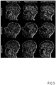
- Original pre-Gd and post-Gd T1w images in three example subjects are shown together with synthetic pre-Gd images generated from a cycleGAN according to the present invention.
- whole-brain T1w images pre- and post-Gd enhancement were collected from 42 patients with multiple sclerosis scanned at 3T.
- synthetic pre-Gd T1w images were computed in the remaining ten test patients.
- the Siemens Healthcare MorphoBox prototype software was employed to automatically segment brain regions from the original pre-Gd T1w and the synthetic pre-Gd T1w images.
- the synthetic T1w pre-Gd images retained comparable contrast between gray and white matter to the original images (see Figure 3 ).
- Figure 4 shows that estimated regional volumes from synthetic images are very similar to those derived from original T1w images (see Figure 4 ). Good correlation (r>0.99, p ⁇ 0.001) and an average bias of -0.04 ml (limits of agreement at 95% of confidence: [-6.2; 6.2] ml) was observed between volumes computed in original and cycleGAN-generated images.
- the proposed cycleGAN could be replaced by any generative model able to reliably synthetized pre-contrast image intensities from post-contrast data.
- One representative example which is not based on deep learning architectures, is Replica, a supervised random forest algorithm that learns a nonlinear regression between multiresolution image patches of a target and input domain.
- Replica a supervised random forest algorithm that learns a nonlinear regression between multiresolution image patches of a target and input domain.
- the method suffers from the blurring of small anatomical details due to the average of predictions from the different random forest tree, which in turn may affect morphometry performances.
- the present invention makes it possible to use existing reliable and validated tools for brain image processing in prospective studies where pre-contrast T1w images were not acquired or in current studies where pre-contrast T1w images are not acquired to keep short the exam duration.
- the here described method can be also easily applied to different pairs of images and/or adapted to consider more than one single input and/or target image domain.
- the generation of the synthetic contrast, i.e. synthetic image is computationally cheap. Compared to the work of Ceccaldi et al.
- the present invention focus notably on a deep learning network to synthetize the missing pre-Gd T1w contrast in order to subsequently use more established and validated tools when pre-contrast T1w images are missing.
- the present invention proposes to use a deep learning-based generative model that reliably estimates pre-contrast image intensities from post-contrast data together with a tool for characterizing tissues of said biological object from the obtained synthetic pre-contrast images of the biological object.
- the present invention allows notably an accurate automated segmentation of brain tissues, otherwise not obtainable in studies without a pre-contrast T1w acquisition.
Landscapes
- Engineering & Computer Science (AREA)
- Physics & Mathematics (AREA)
- General Physics & Mathematics (AREA)
- Theoretical Computer Science (AREA)
- Computer Vision & Pattern Recognition (AREA)
- Health & Medical Sciences (AREA)
- General Health & Medical Sciences (AREA)
- Medical Informatics (AREA)
- Nuclear Medicine, Radiotherapy & Molecular Imaging (AREA)
- Radiology & Medical Imaging (AREA)
- Quality & Reliability (AREA)
- Magnetic Resonance Imaging Apparatus (AREA)
Abstract
The present invention concerns a system and a method for automated biological tissue characterization of a biological object to be imaged using contrast agent enhanced MRI, the method comprising:- acquiring (101) post-contrast MRI data for the biological object;- applying (102) a function trained by a training system (205) to the received post-contrast MRI data in order to output a synthetic pre-contrast image from said post-contrast MRI data;- automatically characterizing (103) biological tissues of said biological object from the synthetic pre-contrast image.
Description
- The present disclosure is directed, in general, to imaging techniques for imaging biological objects, like tissues, using Magnetic Resonance Imaging (MRI) techniques. More specifically, the present invention is directed to methods and systems for automated processing of those images when using a contrast agent in MRI.
- MRI is an established imaging technique able to deliver a wide variety of contrasts among soft tissues that contain information of crucial importance in daily clinical practice. According both to the body part and the disease under analysis, several international consortiums provide guidelines recommending the use of specific MRI protocols. For instance, the European research network for MR imaging in multiple sclerosis (MAGNIMS) recommends the acquisition of T2 FLAIR and gadolinium (Gd)-enhanced T1-weighted (T1w) images for the clinical diagnosis at baseline, whereas T1w images without administration of gadolinium (pre-Gd) are only presented as optional [1]. Even though the former acquisitions are within the most relevant for clinical diagnosis (Gd-enhanced T1w acquisitions are essential to assess the disruption of the blood-brain-barrier in active lesions), pre-Gd T1w images provide added value for the quantitative assessment of additional neuroimaging biomarkers (e.g. morphometry, automated localization and counting of brain lesions, among others). For instance, given the high tissue contrast between white matter (WM), grey matter (GM) and cerebrospinal fluid (CSF) offered by pre-contrast T1w, a variety of tools have been developed to automatically assess brain volumetry from T1w images [2] [3], including the Siemens Healthcare Brain Morphometry software [4] for syngo.via and AI-Rad Companion. Unfortunately, it has also been shown by previous studies that the application of such tools on post-Gd T1w images results in significant biases given the sensitivity of brain volumetric assessments to changes in image intensities [5], introducing for instance errors in tissue classification.
- A potential solution with respect to this problematic of biases resulting from the application of tools tailored for pre-Gd T1w images to post-contrast images might be found in a paper of Ceccaldi et al. [6], wherein a deep learning network is trained to achieve brain tissue segmentation independently from the employed protocol.
- Unfortunately, in the absence of the main contrast from which ground truth labels are derived, the tissue segmentation achieved with this existing solution might present the disadvantage of relying on suboptimal contrasts with poor information regarding tissues boundaries that might result in poor segmentation quality. In addition, the quality of the segmentation strongly depends on the ground truth labels used for the training and does not open to the possibility of using eventually other available tools for the tissue segmentation or perform any other processing that requires the missing contrast (e.g., lesion counting and localization).
- It is therefore desirable to find a method and a system for improving an automated characterization process of biological tissues, like an automated segmentation of biological tissues, when using a contrast agent during MRI, so that errors are avoided (e.g. errors in tissue classification in the case of automated segmentation) and image quality is improved while keeping the total acquisition time as short as possible for realistic clinical application.
- Said objective is achieved according to the present invention by a method and a system for automated processing of images according to the object of the independent claims. Dependent claims present further advantages of the invention.
- The present invention proposes notably to generate pre-contrast images (e.g. pre-Gd images, i.e. images representing a biological object before or without an administration of a contrast agent) from post-contrast imaging data (e.g. from post-Gd images, i.e. images of the biological object taken after the administration of said contrast agent) based on machine learning, e.g. based on a deep learning network. The pre-contrast images synthetized from the deep learning network are then used for an automated process configured for characterizing a biological tissue, like an automated biological tissue segmentation (typically brain tissue segmentation), or for any biological tissue characterization process that requires pre-contrast T1w images. Advantageously, the method and system according to the invention do not increase the acquisition time since the processing of brain tissues in exams is realized from post-contrast image acquisition only and does not require a pre-contrast image acquisition for this purpose, keeping therefore the acquisition time as short as possible.
- More specifically, the present invention concerns a method for an automated biological tissue characterization process, like an automated biological tissue segmentation, of a biological object to be imaged by MRI, the method comprising:
- receiving with a first interface post-contrast MRI data or images, e.g. post-contrast T1w images, for the biological object;
- applying a function trained by a training system to the received post-contrast MRI data in order to output a synthetic pre-contrast image from said post-contrast MRI data (e.g. a pre-contrast T1w image from the post-contrast T1w image), wherein the training system is preferentially based on a generative adversarial network (hereafter GAN);
- automatically applying a characterization process (e.g. applying an automated segmentation process) to the biological tissues of said biological object from the synthetic pre-contrast image.
- The present invention concerns also a computer-implemented method for providing a trained function, the method comprising:
- receiving via a first interface an input dataset, wherein the input dataset comprises post-contrast MRI images of a biological object;
- receiving via a second interface another input dataset, wherein said another input dataset comprises pre-contrast MRI images of said biological object, wherein each pre-contrast MRI image is preferentially paired with one of said post-contrast MRI image, i.e. they represent or show a same part of the biological object, said pre-and/or post-contrast images being for instance T1w images;
- training a function based on the input dataset and said another input dataset with a training computation unit, wherein the training computation unit is configured for using a machine learning algorithm, preferentially based on a GAN, i.e. preferentially based on a generative adversarial algorithm, for training said function. The GAN comprises for instance a generative neural network for generating synthetic pre-contrast images from said post-contrast MRI images and a discriminative neural network for evaluating the generated synthetic pre-contrast images. The training aims to increase an error rate of the discriminative neural network in determining whether a pre-contrast MRI image has been generated by the generative neural network or was part of the received another input dataset (i.e. it aims to determine whether synthetized pre-contrast images are realistic enough for being evaluated by the discriminative neural network as belonging to said another input dataset). Preferentially, the training computation unit uses a cycle generative adversarial network, also called "cycleGAN";
- providing the trained function with a third interface.
- The present invention concerns also a training system comprising:
- a first interface configured for receiving an input dataset, wherein the input dataset comprises post-contrast MRI images of the biological object;
- a second interface configured for receiving another input dataset, wherein said another input dataset comprises pre-contrast MRI images of a biological object, wherein each post-contrast MRI image is preferentially paired to one of said pre-contrast images, wherein pairing refers to the fact that they represent or show a same part of the biological object. Depending on the technique used, pairing is not mandatory;
- a training computation unit configured for training a function based on said input dataset and said another input dataset, wherein the training uses a machine learning algorithm configured for generating synthetic pre-contrast image from post contrast images, e.g. a GAN algorithm, preferentially a cycleGAN algorithm;
- a third interface for providing the trained function.
- Finally, the present invention concerns also a system for an automated characterization of biological object tissues, said system comprising:
- a connection to an imaging system, like the imaging system of an MRI apparatus, configured for acquiring post-contrast MRI data for a biological object;
- a memory for storing each of the acquired post-contrast MRI data;
- a control unit comprising a processor, the control unit being configured for automatically characterizing biological object tissues from images of said biological object. By characterizing, it has to be understood a process that enables the control unit to determine a type of tissue, a property of a tissue, that may differentiate one tissue from another one, etc. Such process encompasses notably the tissue segmentation (e.g. for brain morphometry), and/or the detection and segmentation of biological object abnormalities (e.g., multiple sclerosis lesions, tumors, metastasis, among others);
- optionally a display for displaying the obtained biological tissue characterization of the biological object;
- In particular, the present invention proposes thus to use machine learning, more precisely deep learning generative models, and preferentially a cycleGAN, for the synthesis of pre-contrast T1w images from post-contrast T1w images to achieve a biological object tissue characterization free of an acquisition of said pre-contrast T1w images. For instance, the present invention enables to perform a whole-brain segmentation from synthetic pre-Gd T1w images generated from post-Gd data.
- The foregoing has broadly outlined the features and technical advantages of the present disclosure so that those skilled in the art may better understand the detailed description that follows. In particular, the present invention enables an accurate post-processing of synthetic pre-contrast T1w images after an acquisition of post-contrast images of said biological object, which until now was not possible without the acquisition of said pre-contrast T1w images.
- Additional features and advantages of the disclosure will be described hereinafter that form the object of the claims. Those skilled in the art will appreciate that they may readily use the concept and the specific embodiment disclosed as a basis for modifying or designing other structures for carrying out the same purposes of the present disclosure. Those skilled in the art will also realize that such equivalent constructions do not depart from the spirit and scope of the disclosure as defined by the claims.
- For a more complete understanding of the present disclosure, and the advantages thereof, reference is now made to the following descriptions taken in conjunction with the accompanying drawings, wherein like numbers designate like objects, and in which:
-
Figure 1 illustrates a flowchart of a method for an automated characterization of tissues of a biological object according to the invention; -
Figure 2 illustrates a system for implementing the claimed method; -
Figure 3 illustrates a comparison between biological object images obtained according to the present invention and obtained according to prior art techniques; -
Figure 4 shows examples of automated segmentations obtained according to the claimed method. -
Figures 1 to 4 , discussed below, and the various embodiments used to describe the principles of the present disclosure in this patent document are by way of illustration only and should not be construed in any way to limit the scope of the disclosure. Those skilled in the art will understand that the principles of the present disclosure may be implemented in any suitably arranged device. The numerous innovative teachings of the present application will be described with reference to exemplary non-limiting embodiments. - We will now describe in more details the method according to the invention through
Figure 1 andFigure 2 , whereinFigure 1 describes the different steps of themethod 100 carried out by a preferred embodiment of the system according to the invention which is illustrated byFigure 2 . - At
step 101, acontrol unit 203 of the system for automated characterization (e.g. automated segmentation and/or abnormality detection) of biological tissues according to the invention acquires or receives, for instance via afirst interface 202 configured for enabling an exchange of data between an imaging system, e.g. theimaging system 201 of a MRI apparatus, and saidcontrol unit 203, post-contrast MRI data, e.g. post-contrast images, for a biological object to be imaged. The post-contrast data or images are data or images acquired after administration of a contrast agent to the biological object. Saidcontrol unit 203 may comprise one or several processors configured for processing MRI data. Thecontrol unit 203 might be part of the MRI apparatus or might be a separated independent device. - At
step 102, thecontrol unit 203 applies a trained function to the acquired or received post-contrast MRI data in order to output a synthetic pre-contrast image from the post-contrast MRI data, e.g. a synthetic pre-contrast T1w image. The synthetic pre-contrast image is an image representing the biological object as it would be obtained when acquiring said image before or without the administration of the contrast agent. - Preferentially, a
training system 205 has been used for training said function. Thetraining system 205 comprises notably afirst interface 206, atraining computation unit 207, a second and a third interface which can be the same or different from thefirst interface 206. Thetraining system 205 is notably configured for acquiring or receiving via itsfirst interface 206 an input dataset and another input dataset. In particular, the input dataset and said another input dataset are paired, i.e. to each data of the input dataset corresponds a data of said another input dataset which is closely related or linked to the data of the input dataset. In the present case, the input dataset typically comprises post-contrast MRI data or images of the biological object and said another input dataset comprises pre-contrast T1w data or images of said biological object, wherein each image or data of the input dataset is preferentially paired to an image or data of said another input dataset in that they have been acquired for a same voxel or set of voxels, e.g. they represent a same part of the biological object. The datasets are used by thetraining computation unit 207 for training the function. At the end of the training, the trained function is able to generate realistic and accurate synthetic pre-contrast T1w images from acquired post-contrast MRI data, e.g. from the input dataset and consequently from any other post-contrast MRI data or image. The trained function might be outputted via the third interface. - At
step 103, the system according to the invention automatically performs an automated process (e.g., segmentation) for characterizing the biological tissues of said biological object from the synthetic pre-contrast T1w image and may display the obtained results of the characterization on adisplay 204. For said automated process for characterizing the biological tissues, technique known in the art might be used. - According to the present invention, the
training computation unit 207 uses a machine learning technique for training said function, notably based on a training of deep convolutional networks. Preferably, a generative adversarial algorithm is used by saidtraining computation unit 207, like a GAN, a cycleGAN, or a conditional GAN (hereafter cGAN) algorithm for generating synthetic pre-contrast T1w images of the biological object. - A preferred embodiment illustrating the use of the cycleGAN is described hereafter. Given training datasets from two different domains, the cycleGAN learn to translate images from one domain to the other (domain is denoted as a set of images sharing features that are inherent in the images of the set). In the present case, a first image domain x comprises post-contrast MRI images from said input dataset and a second image domain y comprises pre-contrast T1w images from said another input dataset. The cycleGAN architecture involves then a pair of generators (Gx, Gy) that are simultaneously trained to obtain synthetic images Gx(y) similar to images of the first image domain x for Gx and Gy(x) similar to images of the second image domain y for Gy. The synthetic images Gx(y) and Gy(x) are then fed to a pair of corresponding discriminators (Dx, Dy), which try to discern between fake and real images. For instance, Gx(y) outputs from an input image y an output image X in the x domain. Then this output image X is used as input for the generator Gy, wherein Gy(X) outputs an image Y in the y domain, wherein Y is similar to the original image y. The discriminator compares then y to Y. The same applies mutatis mutandis to Gy(x) to compare an original input image x to an output image X. Preferentially, the architecture of the generators follows the conventional shape of a U-Net with skip connections as described by Ronneberger et al. in the paper entitled "U-Net: Convolutional Networks for Biomedical Image Segmentation" [8]: an encoder is configured for extracting a latent representation (i.e. some information, features that are shared by the input images) of an input image and a decoder is configured for constructing an output image from the extracted latent representation. Said output image is for instance the synthetic pre-contrast T1w image. The discriminators consist of a convolutional neural network that returns the probability of the input image of being sampled from the original image domain. The discriminator receives for instance a pre-contrast T1w image and determines whether the latter is a fake or was truly part of said another input dataset. Generators and discriminators are trained simultaneously including preferentially a cycle-consistency loss between the target (i.e. original input image) and the synthetic image in the adversarial loss function JcycleGAN:

- Of course, another loss function might be used (e.g. a Wasserstein function) or any other metric that describes the degree of similarity between the original and fake (synthetic) images. Similarly, other architecture than the U-net architecture might be used for the generator: e.g. a ResNet or any other deep learning architecture able to achieve image-to-image translation.
- After completing the training, the generator that learned the mapping transformation from post-contrast MRI image domain to pre-contrast T1w image domain can be used to retrieve pre-contrast T1w images from post-contrast MRI images, allowing the use of a conventional tool for the characterization, e.g. segmentation, of brain tissues from the synthetic pre-contrast T1w images.
-
Figures 3 and4 show concrete applications of the method according to the invention with respect to a brain as biological object and gadolinium (Gd) as contrast agent for MRI imaging. InFigure 3 , original pre-Gd and post-Gd T1w images in three example subjects are shown together with synthetic pre-Gd images generated from a cycleGAN according to the present invention. In order to train the generative adversarial algorithm, whole-brain T1w images pre- and post-Gd enhancement were collected from 42 patients with multiple sclerosis scanned at 3T. A cycleGAN was trained to learn the mapping transformation from post-Gd T1w images to pre-Gd images over 100 epochs (Adam optimizer, learning rate of 0.0002, β1=0.5, β2=0.999, λx = λy = 100) using sagittal slices from 32 randomly selected patients after performing an affine registration of pre-Gd images onto post-Gd images. After training, synthetic pre-Gd T1w images were computed in the remaining ten test patients. The Siemens Healthcare MorphoBox prototype software was employed to automatically segment brain regions from the original pre-Gd T1w and the synthetic pre-Gd T1w images. According to the present invention, the synthetic T1w pre-Gd images retained comparable contrast between gray and white matter to the original images (seeFigure 3 ). Additionally,Figure 4 shows that estimated regional volumes from synthetic images are very similar to those derived from original T1w images (seeFigure 4 ). Good correlation (r>0.99, p<0.001) and an average bias of -0.04 ml (limits of agreement at 95% of confidence: [-6.2; 6.2] ml) was observed between volumes computed in original and cycleGAN-generated images. - Alternatively, the proposed cycleGAN could be replaced by any generative model able to reliably synthetized pre-contrast image intensities from post-contrast data. One representative example, which is not based on deep learning architectures, is Replica, a supervised random forest algorithm that learns a nonlinear regression between multiresolution image patches of a target and input domain. However, although proved to be effective in the synthesis of T2w and FLAIR images form T1w, the method suffers from the blurring of small anatomical details due to the average of predictions from the different random forest tree, which in turn may affect morphometry performances.
- Compared to existing solutions, the present invention makes it possible to use existing reliable and validated tools for brain image processing in prospective studies where pre-contrast T1w images were not acquired or in current studies where pre-contrast T1w images are not acquired to keep short the exam duration. Given the availability of the proper datasets required for the training of the mapping transformation, the here described method can be also easily applied to different pairs of images and/or adapted to consider more than one single input and/or target image domain. In addition, the generation of the synthetic contrast, i.e. synthetic image, is computationally cheap. Compared to the work of Ceccaldi et al. [6] proposing a network to achieve brain tissue segmentation independently from the employed protocol used, the present invention focus notably on a deep learning network to synthetize the missing pre-Gd T1w contrast in order to subsequently use more established and validated tools when pre-contrast T1w images are missing.
- To summarize, the present invention proposes to use a deep learning-based generative model that reliably estimates pre-contrast image intensities from post-contrast data together with a tool for characterizing tissues of said biological object from the obtained synthetic pre-contrast images of the biological object. For example, the present invention allows notably an accurate automated segmentation of brain tissues, otherwise not obtainable in studies without a pre-contrast T1w acquisition.
-
- [1] Wattjes MP, Rovira À, Tintore M, et al. Evidence-based guidelines: MAGNIMS consensus guidelines on the use of MRI in multiple sclerosis-clinical implementation in the diagnostic process. Nat Rev Neurol. 2015;11(8):471-482.
- [2] Ashburner J. Computational anatomy with the SPM software. Magn Reson Imaging. 2009;27(8):1163-1174.
- [3] Fischl B. FreeSurfer. Neuroimage. 2012;62(2):774-781.
- [4] Schmitter D, Roche A, Marechal B, et al. An evaluation of volume-based morphometry for prediction of mild cognitive impairment and Alzheimer's disease.NeuroImage Clin. 2015;7:7-17.
- [5] Hannoun S, Baalbaki M, Haddad R, et al. Gadolinium effect on thalamus and whole brain tissue segmentation. Neuroradiology. 2018;60(11):1167-1173.
- [6] Ceccaldi et al., PROTOCOL INDEPENDENT IMAGE PROCESSING WITH ADVERSARIAL NETWORKS, Pub. No.:
US 2019 / 0046068 A1. 2019 . - [7] Liu, J., Gong, E., Christen, T., Zaharchuk, G., Contrast-free MRI Contrast Enhancement with Deep Attention Generative Adversarial Network, ISMRM 2019.
- [8] Ronneberger O, Fischer P, Brox T. U-net: Convolutional networks for biomedical image segmentation. In: Lecture Notes in Computer Science. Vol 9351. ; 2015:234-241.
Claims (14)
- A method (100) for automated characterization of tissues of a biological object to be imaged using contrast agent enhanced MRI, the method comprising:- acquiring (101) post-contrast MRI data for the biological object;- applying (102) a function trained by a training system (205) to the received post-contrast MRI data in order to output a synthetic pre-contrast image from said post-contrast MRI data;- automatically characterizing the (103) biological tissues of said biological object from the synthetic pre-contrast image.
- The method of claim 1, wherein the post-contrast MRI data are post-contrast T1-weighted images and the synthetic pre-contrast image is a synthetic pre-contrast T1-weighted image.
- The method of claim 1 or 2, wherein the contrast agent is gadolinium.
- The method according to one of the claims 1 to 3, wherein a generative adversarial network is used for training said function.
- The method according to claim 4, wherein the generative adversarial network is a cycle generative adversarial network - hereafter cycleGAN.
- Method according to one of the claims 1 to 5, wherein automatically characterizing the biological tissues comprises automatically segmenting said biological tissues.
- Computer-implemented method for providing a trained function, the method comprising:- receiving via a first interface an input dataset, wherein the input dataset comprises post-contrast MRI images of a biological object;- receiving via a second interface another input dataset, wherein said another input dataset comprises pre-contrast MRI images of said biological object;- training a function based on the input dataset and said another input dataset with a training computation unit (207), wherein the training computation unit (207) is configured for using a machine learning algorithm for training said function;- providing the trained function with a third interface.
- Computer-implemented method according to claim 7, wherein each pre-contrast MRI image is paired with one of said post-contrast MRI image.
- Computer-implemented method according to claim 7 or 8, wherein the machine learning algorithm is a generative adversarial algorithm, preferentially a cycleGAN algorithm.
- Computer-implemented method according to one of the claims 7 to 9, wherein the post-contrast and pre-contrast images are T1-weighted MRI images.
- Training system (205) comprising:- a first interface (206) configured for receiving an input dataset, wherein the input dataset comprises post-contrast MRI images of the biological object;- a second interface configured for receiving another input dataset, wherein said another input dataset comprises pre-contrast MRI images of a biological object;- a training computation unit (203) configured for training a function based on said input dataset and said another input dataset, wherein the training uses a machine learning algorithm configured for generating synthetic pre-contrast image from post contrast images;- a third interface for providing the trained function.
- System for automated characterization of biological object tissues from MRI data acquired using a contrast agent, the system comprising:- a connection to an imaging system (201) of an MRI apparatus, said connection being configured for acquiring post-contrast MRI data for a biological object;- a memory for storing each of the acquired post-contrast MRI data;- a control unit (203) comprising a processor, the control unit (203) being configured for automatically characterizing biological object tissues from images of said biological object;the system (200) being characterized in that the control unit (203) is configured for applying a function trained by a machine learning algorithm to the post-contrast MRI data in order to output a synthetic pre-contrast image from said post-contrast MRI data, wherein the control unit (203) is configured for automatically characterizing the biological tissues of said biological object from the obtained synthetic pre-contrast image.
- System according to claim 12, wherein the control unit (203) is configured for automatically segmenting the biological tissues.
- System according to claim 12 or 13, wherein the machine learning algorithm is a cycleGAN algorithm.
Priority Applications (1)
| Application Number | Priority Date | Filing Date | Title |
|---|---|---|---|
| EP20160012.9A EP3872754A1 (en) | 2020-02-28 | 2020-02-28 | Method and system for automated processing of images when using a contrast agent in mri |
Applications Claiming Priority (1)
| Application Number | Priority Date | Filing Date | Title |
|---|---|---|---|
| EP20160012.9A EP3872754A1 (en) | 2020-02-28 | 2020-02-28 | Method and system for automated processing of images when using a contrast agent in mri |
Publications (1)
| Publication Number | Publication Date |
|---|---|
| EP3872754A1 true EP3872754A1 (en) | 2021-09-01 |
Family
ID=69742898
Family Applications (1)
| Application Number | Title | Priority Date | Filing Date |
|---|---|---|---|
| EP20160012.9A Withdrawn EP3872754A1 (en) | 2020-02-28 | 2020-02-28 | Method and system for automated processing of images when using a contrast agent in mri |
Country Status (1)
| Country | Link |
|---|---|
| EP (1) | EP3872754A1 (en) |
Citations (4)
| Publication number | Priority date | Publication date | Assignee | Title |
|---|---|---|---|---|
| US20190046068A1 (en) | 2017-08-10 | 2019-02-14 | Siemens Healthcare Gmbh | Protocol independent image processing with adversarial networks |
| US20190108634A1 (en) * | 2017-10-09 | 2019-04-11 | The Board Of Trustees Of The Leland Stanford Junior University | Contrast Dose Reduction for Medical Imaging Using Deep Learning |
| US20190259153A1 (en) * | 2018-02-22 | 2019-08-22 | Siemens Healthcare Gmbh | Cross domain medical image segmentation |
| CN110827232A (en) * | 2019-11-14 | 2020-02-21 | 四川大学 | Cross-modal MRI (magnetic resonance imaging) synthesis method based on morphological feature GAN (gain) |
-
2020
- 2020-02-28 EP EP20160012.9A patent/EP3872754A1/en not_active Withdrawn
Patent Citations (4)
| Publication number | Priority date | Publication date | Assignee | Title |
|---|---|---|---|---|
| US20190046068A1 (en) | 2017-08-10 | 2019-02-14 | Siemens Healthcare Gmbh | Protocol independent image processing with adversarial networks |
| US20190108634A1 (en) * | 2017-10-09 | 2019-04-11 | The Board Of Trustees Of The Leland Stanford Junior University | Contrast Dose Reduction for Medical Imaging Using Deep Learning |
| US20190259153A1 (en) * | 2018-02-22 | 2019-08-22 | Siemens Healthcare Gmbh | Cross domain medical image segmentation |
| CN110827232A (en) * | 2019-11-14 | 2020-02-21 | 四川大学 | Cross-modal MRI (magnetic resonance imaging) synthesis method based on morphological feature GAN (gain) |
Non-Patent Citations (10)
| Title |
|---|
| ASHBURNER J: "Computational anatomy with the SPM software", MAGN RESON IMAGING, vol. 27, no. 8, 2009, pages 1163 - 1174, XP026601968, DOI: 10.1016/j.mri.2009.01.006 |
| DAR SALMAN UH ET AL: "Image Synthesis in Multi-Contrast MRI With Conditional Generative Adversarial Networks", IEEE TRANSACTIONS ON MEDICAL IMAGING, IEEE SERVICE CENTER, PISCATAWAY, NJ, US, vol. 38, no. 10, 1 October 2019 (2019-10-01), pages 2375 - 2388, XP011748194, ISSN: 0278-0062, [retrieved on 20191001], DOI: 10.1109/TMI.2019.2901750 * |
| FISCHL B, FREESURFER. NEUROIMAGE, vol. 62, no. 2, 2012, pages 774 - 781 |
| HANNOUN SBAALBAKI MHADDAD R ET AL.: "Gadolinium effect on thalamus and whole brain tissue segmentation", NEURORADIOLOGY, vol. 60, no. 11, 2018, pages 1167 - 1173, XP036619576, DOI: 10.1007/s00234-018-2082-5 |
| LIU, J.GONG, E.CHRISTEN, T.ZAHARCHUK, G.: "Contrast-free MRI Contrast Enhancement with Deep Attention Generative Adversarial Network", ISMRM, 2019 |
| RONNEBERGER 0FISCHER PBROX T: "U-net: Convolutional networks for biomedical image segmentation", LECTURE NOTES IN COMPUTER SCIENCE, vol. 9351, 2015, pages 234 - 241, XP055561106, DOI: 10.1007/978-3-319-24574-4_28 |
| SAHIN OLUT ET AL: "Generative Adversarial Training for MRA Image Synthesis Using Multi-Contrast MRI", ARXIV.ORG, CORNELL UNIVERSITY LIBRARY, 201 OLIN LIBRARY CORNELL UNIVERSITY ITHACA, NY 14853, 12 April 2018 (2018-04-12), XP080870088 * |
| SCHMITTER DROCHE AMARECHAL B ET AL.: "An evaluation of volume-based morphometry for prediction of mild cognitive impairment and Alzheimer's disease", NEUROIMAGE CLIN, vol. 7, 2015, pages 7 - 17, XP055592522, DOI: 10.1016/j.nicl.2014.11.001 |
| WATTJES MPROVIRA ATINTORE M ET AL.: "Evidence-based guidelines: MAGNIMS consensus guidelines on the use of MRI in multiple sclerosis-clinical implementation in the diagnostic process", NAT REV NEUROL, vol. 11, no. 8, 2015, pages 471 - 482 |
| WU KUN ET AL: "Image synthesis in contrast MRI based on super resolution reconstruction with multi-refinement cycle-consistent generative adversarial networks", JOURNAL OF INTELLIGENT MANUFACTURING, CHAPMAN AND HALL, LONDON, GB, vol. 31, no. 5, 1 November 2019 (2019-11-01), pages 1215 - 1228, XP037140954, ISSN: 0956-5515, [retrieved on 20191101], DOI: 10.1007/S10845-019-01507-7 * |
Similar Documents
| Publication | Publication Date | Title |
|---|---|---|
| Spieker et al. | Deep learning for retrospective motion correction in MRI: a comprehensive review | |
| Wee et al. | Enriched white matter connectivity networks for accurate identification of MCI patients | |
| CN111640118B (en) | Tumor tissue characterization using multiparametric magnetic resonance imaging | |
| EP2790575B1 (en) | Method and apparatus for the assessment of medical images | |
| US20170238879A1 (en) | Method of Analyzing the Brain Activity of a Subject | |
| Shen et al. | VBM lesion detection depends on the normalization template: a study using simulated atrophy | |
| JP2022539063A (en) | An Improved Medical Scan Protocol for In-Scanner Patient Data Acquisition Analysis | |
| US12456197B2 (en) | Lesion detection and segmentation | |
| Mohebbian et al. | Classifying MRI motion severity using a stacked ensemble approach | |
| Reid et al. | How many streamlines are required for reliable probabilistic tractography? Solutions for microstructural measurements and neurosurgical planning | |
| Weiss et al. | Automated multiclass tissue segmentation of clinical brain MRIs with lesions | |
| Basukala et al. | Automated segmentation of substantia nigra and red nucleus using quantitative susceptibility mapping images: Application to Parkinson's disease | |
| Sanchez et al. | FetMRQC: Automated quality control for fetal brain MRI | |
| Chen | Functional brain network estimation based on Weighted BOLD signals for MCI identification | |
| Moschetto et al. | Benchmarking GANs, Diffusion Models, and Flow Matching for T1w-to-T2w MRI Translation | |
| Elliott | A Bayesian framework for 4-D segmentation of Multiple Sclerosis lesions in serial MRI in the brain | |
| Mehdipour Ghazi et al. | FAST-AID Brain: Fast and accurate segmentation tool using artificial intelligence developed for brain | |
| Bernstein et al. | MRI brain imagery processing software in data analysis. | |
| US20230316716A1 (en) | Systems and methods for automated lesion detection using magnetic resonance fingerprinting data | |
| Tivari et al. | Enhancing Diagnostic Accuracy of Brain MRI Using Super-Resolution AttentionGAN | |
| Lan et al. | Fiber Microstructure Quantile (FMQ) regression: A novel statistical approach for analyzing white matter bundles from periphery to core | |
| Commowick et al. | Diffusion MRI abnormalities detection with orientation distribution functions: A multiple sclerosis longitudinal study | |
| Jafrasteh et al. | WASABI: A Metric for Evaluating Morphometric Plausibility of Synthetic Brain MRIs | |
| WO2025055849A1 (en) | Detection and forecasting of neurocognitive decline using functional neuroimaging and machine learning | |
| Ashburner et al. | Spatial registration of images |
Legal Events
| Date | Code | Title | Description |
|---|---|---|---|
| PUAI | Public reference made under article 153(3) epc to a published international application that has entered the european phase |
Free format text: ORIGINAL CODE: 0009012 |
|
| STAA | Information on the status of an ep patent application or granted ep patent |
Free format text: STATUS: THE APPLICATION HAS BEEN PUBLISHED |
|
| AK | Designated contracting states |
Kind code of ref document: A1 Designated state(s): AL AT BE BG CH CY CZ DE DK EE ES FI FR GB GR HR HU IE IS IT LI LT LU LV MC MK MT NL NO PL PT RO RS SE SI SK SM TR |
|
| STAA | Information on the status of an ep patent application or granted ep patent |
Free format text: STATUS: THE APPLICATION IS DEEMED TO BE WITHDRAWN |
|
| 18D | Application deemed to be withdrawn |
Effective date: 20220302 |