EP3869199B1 - A method and reagents for the diagnosis of sars-cov-2 - Google Patents
A method and reagents for the diagnosis of sars-cov-2 Download PDFInfo
- Publication number
- EP3869199B1 EP3869199B1 EP20215445.6A EP20215445A EP3869199B1 EP 3869199 B1 EP3869199 B1 EP 3869199B1 EP 20215445 A EP20215445 A EP 20215445A EP 3869199 B1 EP3869199 B1 EP 3869199B1
- Authority
- EP
- European Patent Office
- Prior art keywords
- seq
- sars
- cov
- antibody
- iga
- Prior art date
- Legal status (The legal status is an assumption and is not a legal conclusion. Google has not performed a legal analysis and makes no representation as to the accuracy of the status listed.)
- Active
Links
Classifications
-
- G—PHYSICS
- G01—MEASURING; TESTING
- G01N—INVESTIGATING OR ANALYSING MATERIALS BY DETERMINING THEIR CHEMICAL OR PHYSICAL PROPERTIES
- G01N33/00—Investigating or analysing materials by specific methods not covered by groups G01N1/00 - G01N31/00
- G01N33/48—Biological material, e.g. blood, urine; Haemocytometers
- G01N33/50—Chemical analysis of biological material, e.g. blood, urine; Testing involving biospecific ligand binding methods; Immunological testing
- G01N33/53—Immunoassay; Biospecific binding assay; Materials therefor
- G01N33/569—Immunoassay; Biospecific binding assay; Materials therefor for microorganisms, e.g. protozoa, bacteria, viruses
- G01N33/56983—Viruses
-
- C—CHEMISTRY; METALLURGY
- C07—ORGANIC CHEMISTRY
- C07K—PEPTIDES
- C07K14/00—Peptides having more than 20 amino acids; Gastrins; Somatostatins; Melanotropins; Derivatives thereof
- C07K14/005—Peptides having more than 20 amino acids; Gastrins; Somatostatins; Melanotropins; Derivatives thereof from viruses
-
- G—PHYSICS
- G01—MEASURING; TESTING
- G01N—INVESTIGATING OR ANALYSING MATERIALS BY DETERMINING THEIR CHEMICAL OR PHYSICAL PROPERTIES
- G01N33/00—Investigating or analysing materials by specific methods not covered by groups G01N1/00 - G01N31/00
- G01N33/48—Biological material, e.g. blood, urine; Haemocytometers
- G01N33/50—Chemical analysis of biological material, e.g. blood, urine; Testing involving biospecific ligand binding methods; Immunological testing
- G01N33/53—Immunoassay; Biospecific binding assay; Materials therefor
- G01N33/569—Immunoassay; Biospecific binding assay; Materials therefor for microorganisms, e.g. protozoa, bacteria, viruses
-
- C—CHEMISTRY; METALLURGY
- C12—BIOCHEMISTRY; BEER; SPIRITS; WINE; VINEGAR; MICROBIOLOGY; ENZYMOLOGY; MUTATION OR GENETIC ENGINEERING
- C12N—MICROORGANISMS OR ENZYMES; COMPOSITIONS THEREOF; PROPAGATING, PRESERVING, OR MAINTAINING MICROORGANISMS; MUTATION OR GENETIC ENGINEERING; CULTURE MEDIA
- C12N2770/00—MICROORGANISMS OR ENZYMES; COMPOSITIONS THEREOF; PROPAGATING, PRESERVING, OR MAINTAINING MICROORGANISMS; MUTATION OR GENETIC ENGINEERING; CULTURE MEDIA ssRNA viruses positive-sense
- C12N2770/00011—Details
- C12N2770/20011—Coronaviridae
- C12N2770/20022—New viral proteins or individual genes, new structural or functional aspects of known viral proteins or genes
-
- G—PHYSICS
- G01—MEASURING; TESTING
- G01N—INVESTIGATING OR ANALYSING MATERIALS BY DETERMINING THEIR CHEMICAL OR PHYSICAL PROPERTIES
- G01N2333/00—Assays involving biological materials from specific organisms or of a specific nature
- G01N2333/005—Assays involving biological materials from specific organisms or of a specific nature from viruses
- G01N2333/08—RNA viruses
- G01N2333/165—Coronaviridae, e.g. avian infectious bronchitis virus
Definitions
- the present invention relates to a method for aiding in the diagnosis of a SARS-CoV-2 infection comprising the step of detecting the presence or absence of an IgA class antibody to SEQ ID NO1 in a blood sample from a subject and a use of an IgA class antibody to SEQ ID NO1 for aiding in the diagnosis of a SARS-CoV-2 infection , wherein the use is for the early diagnosis of a SARS-CoV-2 infection.
- coronavirus Study Group recognized it as a sister to severe acute respiratory syndrome coronaviruses (SARS-CoVs) and labeled it as severe acute respiratory syndrome coronavirus 2 (SARS-CoV-2).
- SARS-CoV-2 is generally less pathogenic than SARS-CoV and Middle East respiratory syndrome coronavirus (MERS-CoV), it has a relatively high transmissibility. Since symptoms may be mild and may be confused with a cold, there is the danger that patients are unaware that they have been infected and may help the virus spread further.
- MERS-CoV Middle East respiratory syndrome coronavirus
- US2005/0112559 discloses reagents derived from SARS-CoV nucleocapsid protein for the diagnosis of SARS-CoV.
- WO2005118813 discloses immunogenic compositions associated with the SARS-CoV spike protein.
- US2006/0188519 discloses immunoreactive peptides derived from SARS-CoV.
- IgG could be detected as early as four days after the onset of a SARS infection, simultaneously or one day earlier than IgM and IgA ( Hsue, P. R., Huang, L. M., Chen, P. J., Kao, C. L., and Yang P. C. (2004) Chronological evolution of IgM, IgA, IgG and neutralization antibodies after infection with SARS-associated coronavirus, Clinical Microbiology and Infection, 10(12), 1062-1066 .
- PCR-based assays have several shortcomings.
- a sample from the upper respiratory tract of the patient is required. Improper recovery of such a sample may lead to false-negative results.
- the results may not reflect the disease status of the patient. Therefore, a serological assay would be desirable.
- a problem underlying the present invention is to provide an assay and reagents for the early serological detection of SARS-CoV-2.
- the problem is solved by a method for aiding in the diagnosis of a SARS-CoV-2 infection comprising the step of detecting the presence or absence of an IgA class antibody to SEQ ID NO1 in a blood sample from a subject.
- the sample is selected from the group comprising whole blood, serum or plasma.
- the antibody is detected using a labeled secondary antibody, preferably binding to IgA class antibodies.
- the IgA class antibody is detected using a method selected from the group comprising colorimetry, immunofluorescence, detection of enzymatic activity, chemiluminscence and radioactivity.
- the infection is detected at an early stage.
- the problem is solved by a use of an IgA class antibody to SEQ ID NO1, for aiding in the diagnosis of a SARS-CoV-2 infection, wherein the use is for the early diagnosis of a SARS-CoV-2 infection.
- the present invention is based on the inventors' surprising finding that antibodies against SEQ ID NO1 can be detected at an early stage of the infection, with IgA class antibodies becoming detectable earlier than IgG antibodies.
- diagnosis refers to any kind of procedure aiming to obtain information instrumental in the assessment whether a patient suffers or is likely or more likely than the average or a comparative subject, the latter preferably having similar symptoms, to suffer from certain a disease or disorder in the past, at the time of the diagnosis or in the future, to find out how the disease is progressing or is likely to progress in the future or to evaluate the responsiveness of a patient with regard to a treatment.
- diagnosis comprises not only diagnosing, but also prognosticating and/or monitoring the course of a disease or disorder.
- the subject is likely or more likely to suffer from a SARS-CoV-2 infection if an IgA and/or IgG and/or IgM antibody to SEQ ID NO1 is detected in samples from them.
- the sample is preferably a mammalian, more preferably a human sample.
- diagnosis does preferably not imply that the diagnostic methods or agents according to the present invention will be definitive and sufficient to finalize the diagnosis on the basis of a single test, let alone parameter, but may refer to a contribution to what is referred to as a "differential diagnosis", i.e. a systematic diagnostic procedure considering the likelihood of a range of possible conditions on the basis of a range of diagnostic parameters.
- SARS-CoV-2 refers to a virus characterized by the genome deposited on GenBank under accession code MN908947 and derivatives thereof having at least 80, preferably 85, preferably 88, preferably 90, preferably 91, preferably 92, preferably 93, preferably 94, preferably 95, preferably 96, preferably 97, preferably 98, preferably 99, preferably 99.5, preferably 99.8, preferably 99.9 or 99.99 percent sequence identity over the entire genome nucleotide sequence. All data base entries used herein correspond to the version online at the filing date of the application.
- diagnosis means that the method or product or use may be used for aiding in the diagnosis of a disease or identifying a subject a risk of suffering from a disease.
- diagnosis may also refer to a method or agent used to choose the most promising treatment regime for a patient. In other words, the method or agent may relate to selecting a treatment regimen for a subject.
- the method according to the present invention comprises the step providing the diagnostically useful carrier and a sample from a patient suspected of being infected, preferably a mammalian, more preferably a human patient.
- the carrier is coated with the polypeptide comprising SEQ ID NO1 or variant thereof.
- the carrier may then be contacted with the sample under conditions allowing for binding of any antibodies to the polypeptide comprising SEQ ID NO1 or variant thereof.
- the sample may then be removed and the carrier with the cell may be washed to remove any remaining sample.
- a secondary antibody or similar reagent or means binding to the antibody and carrying a detectable label may then be contacted with the carrier under conditions allowing formation of a complex between any bound antibody and the secondary antibody.
- the carrier may be washed then to remove non-bound secondary antibody. Finally, the presence of the antibody is detected by checking whether the secondary antibody may be detected.
- the method is used more than once to examine samples from the same patient, preferably on different days.
- the presence or absence of antibodies may be detected on a daily basis over one or two weeks.
- least 2, 3, 4, 5, 6, 7, 8, 9 or 10 samples are examined on different days.
- the method according to the present invention may also be used for screening the potency and the usefulness of an antiviral drug.
- the method according to the present invention may also be used for screening whether donated blood is contaminated with coronavirus.
- the antibody to be detected binds preferably specifically to SEQ ID NO1.
- Specific binding preferably means that the binding reaction is stronger than a binding reaction characterized by a dissociation constant of 1 ⁇ 10 -5 M, more preferably 1 ⁇ 10 -7 M, more preferably 1 ⁇ 10 -8 M, more preferably 1 ⁇ 10 -9 M, more preferably 1 ⁇ 10 -10 M, more preferably 1 ⁇ 10 -11 M, more preferably 1 ⁇ 10 -12 M, as determined by surface plasmon resonance using Biacore equipment at 25 °C in PBS buffer at pH 7.
- teachings of the present invention may not only be carried out using polypeptides having the exact sequences referred to in this application explicitly, for example by function, name, sequence or accession number, or implicitly, but also using variants of such polypeptides.
- variant may refer to at least one fragment of the full length sequence referred to or a polypeptide comprising said fragment, more specifically to one or more amino acid or nucleic acid sequences which are, relative to the full-length sequence, truncated at one or both termini by one or more amino acids.
- a fragment comprises or encodes for a peptide having at least 10, 15, 25, 50, 75, 100, 150, 200, 250, 300, 400, 500 or 600 successive amino acids of the original sequence or for a variant thereof.
- SEQ ID NO12 is an exemplary fragment that may be used.
- variant relates not only to at least one fragment, but also a polypeptide or a fragment thereof comprising amino acid sequences, preferably a fragment comprising at least 25, more preferably 50, more preferably 200 successive amino acids, that are at least 40, 50, 60, 70, 75, 80, 85, 90, 92, 94, 95, 96, 97, 98 or 99 % identical to the reference amino acid sequence referred to or the fragment thereof, wherein amino acids other than those essential for the biological activity, for example the ability to bind specifically to an antibody of interest, or the fold or structure of the polypeptide are deleted or substituted and/or one or more such essential amino acids are replaced in a conservative manner and/or amino acids are added or deleted such that the biological activity of the polypeptide is at least partially preserved.
- the state of the art comprises various methods that may be used to align two given nucleic acid or amino acid sequences and to calculate the degree of identity, see for example Arthur Lesk (2008), Introduction to bioinformatics, Oxford University Press, 2008, 3rd editi on.
- the ClustalW software Larkin, M. A., Blackshields, G., Brown, N. P., Chenna, R., McGettigan, P. A., McWilliam, H., Valentin, F., Wallace, I. M., Wilm, A., Lopez, R., Thompson, J. D., Gibson, T. J., Higgins, D. G. (2007): Clustal W and Clustal X version 2.0. Bioinformatics, 23, 2947-2948 ) is used applying default settings.
- SARS-CoV-2-related publications on specific amino acid sequences such as Beal et al. may aid the skilled one in designing variants ( Beal, J., Mitechell, T., Wyschogrod, W., Manthey, J, and Clore, A. (2020) Highly Distinguished Amino Acid Sequences of 2019-nCoV (Wuhan Coronavirus) doi: https://doi.org/10.1101/2020.01.31.929497 , as well as publications relating to SARS-CoV, for example Hua, R., Zhou, Y., Wang, Y., Hua, Y and Tong, T.
- SARS-CoV spike protein BBR 319, 929-935 , wherein homologous epitopes may be found and SARS-CoV-2 epitopes be identified on account of their homology.
- possible epitopes may be derived from SEQ ID NO5.
- Variants may, in addition, comprise chemical modifications, for example labels such as isotopic labels or detectable labels or covalent modifications such as glycosylation, phosphorylation, acetylation, decarboxylation, citrullination, hydroxylation and the like.
- labels such as isotopic labels or detectable labels
- covalent modifications such as glycosylation, phosphorylation, acetylation, decarboxylation, citrullination, hydroxylation and the like.
- the person skilled in the art is familiar with methods for the modification of polypeptides.
- variants may also be generated by way of fusion with other known polypeptides or variants thereof, for example artificial linkers, affinity tags, other antigens and the like.
- SEQ ID NO3 is a fusion protein.
- the variant of the polypeptide has biological activity. Such biological activity is the ability to bind to the respective antibody. It comprises an epitope having the ability or has itself the ability to bind to an IgA class antibody to SEQ ID NO1, preferably from a sample from a patient suffering from SARS-CoV-2, wherein more preferably the epitope comprises a sequence comprising at least 5, 6, 7 or 8 amino acid residues.
- SEQ ID NO1 from other coronaviruses, preferably from the group comprising MERS (SEQ ID NO6), NL63 (SEQ ID N010), 229E (SEQ ID NO7), OC43 (SEQ ID N08) and HKU1 (SEQ ID NO9), more preferably from the group comprising SARS-CoV-1 (SEQ ID NO11), MERS NL63, 229E, OC43 and HKU1.
- the detection of the antibody for the diagnosis or methods according to the present invention comprises the use of a method selected from the group comprising immunodiffusion techniques, immunoelectrophoretic techniques, light scattering immunoassays, agglutination techniques, labeled immunoassays such as those from the group comprising radiolabeled immunoassays, enzyme immunoassays such as colorimetric assays, chemiluminscence immunoassays and immunofluorescence techniques.
- the complex is detected using a method selected from the group comprising immunodiffusion techniques, immunoelectrophoretic techniques, light scattering immunoassays, agglutination techniques, labeled immunoassays from the group comprising radiolabeled immunoassays, chemiluminscence immunoassays and immunofluorescence techniques.
- a method selected from the group comprising immunodiffusion techniques, immunoelectrophoretic techniques, light scattering immunoassays, agglutination techniques, labeled immunoassays from the group comprising radiolabeled immunoassays, chemiluminscence immunoassays and immunofluorescence techniques.
- the test format is an ELISA and a microtiter plate comprising wells is used as a diagnostically useful carrier.
- a secondary antibody is an antibody binding to all antibodies from an antibody class, preferably a human antibody class, preferably IgA and IgG and IgM antibodies, preferably IgA. Secondary antibodies typically recognize the constant domain of said class. A wide range of them is commercially available.
- the diagnostically useful carrier is preferably selected from the group comprising a glass slide, preferably for microscopy, a biochip, a microtiter plate, a lateral flow device, a test strip, a membrane, preferably a line blot, a chromatography column and a bead, preferably a microtiter plate.
- a polypeptide preferably the polypeptide comprising SEQ ID NO1 or a variant thereof may be a recombinant protein, wherein the term "recombinant”, as used herein, refers to a polypeptide produced using genetic engineering approaches at any stage of the production process, for example by fusing a nucleic acid encoding the polypeptide to a strong promoter for overexpression in cells or tissues or by engineering the sequence of the polypeptide itself.
- the person skilled in the art is familiar with methods for engineering nucleic acids and polypeptides encoded (for example, described in Sambrook, J., Fritsch, E. F. and Maniatis, T. (1989), Molecular Cloning, CSH or in Brown T. A.
- the polypeptide is an isolated polypeptide, wherein the term "isolated” means that the polypeptide has been enriched compared to its state upon production using a biotechnological or synthetic approach and is preferably pure, i.e.
- polypeptide in the respective liquid consists of said polypeptide as judged by SDS polyacrylamide gel electrophoresis followed by Coomassie blue staining and visual inspection.
- any polypeptide on a carrier used as a means to capture an antibody is pure.
- a secondary antibody comprising a detectable label may be used to detect IgA and IgG and IgM, preferably IgA class antibodies to SEQ ID NO1.
- the label is selected from the group comprising a fluorescent, a radioactive or an enzymatically active label, preferably one catalyzing a colorimetric reaction.
- FITC or another fluorescein derivative may be used as a fluorescent label.
- a protein having peroxidase activity may be used as an enzymatically active label.
- the secondary antibody recognizes mammalian, more preferably human antibodies.
- the secondary antibody is a monoclonal antibody.
- kits comprising a polypeptide comprising SEQ ID NO1 or a variant thereof, preferably coated to a diagnostically useful carrier, more preferably a microtiter plate, and one or more, preferably all reagents from the antibodies, wherein the secondary antibody may comprise a detectable label, a sample buffer, a detection solution, preferably a chromogen/substrate solution, a stop solution and a protective foil.
- a calibrator is a reagent that binds to a polypeptide comprising SEQ ID NO1 or a variant thereof and is preferably recognized by secondary antibodies recognizing IgA class antibodies.
- the calibrator may be an IgA antibody to SEQ ID NO1
- a positive control is a solution comprising a compound such as antibody to SEQ ID NO1, preferably from the group comprising IgA, IgG and IgM class antibodies, more preferably IgA, from the sample of a patient suffering from SARS-coV-2 at an amount that a positive result is obtained using the method according to the present invention.
- a negative control is a reagent that lacks such a compound and could comprise serum from a healthy person.
- a wash buffer may be used to wash the microtiter plate after the incubation to remove unspecific antibodies and could be PBS.
- the means for detecting the presence of an antibody could be a secondary antibody binding to the antibody class to be detected, preferably human IgA class antibodies, and is labeled, preferably with an enzyme, more preferably with an enzyme having peroxidase activity.
- the sample buffer may be used to dilute patient sample and may be PBS.
- the detection solution may yield a signal in the presence of the labeled secondary antibody and is preferably a color-developing solution and more preferably 3,3',5,5' tetramethylbenzidine/H2O2.
- the stop solution may be added to a reaction to stop the reaction of the detection solution and may comprise a strong acid, preferably 0.5 M sulphuric acid.
- the protective foil may be place on top of the microtiter plate to avoid evaporation.
- the present specification comprises a range of novel nucleic acid and polypeptide sequences, more specifically
- samples containing various coronaviruses were available, including 18 samples from patients infected with MERS, three samples from patients infected with SARS-CoV-1, four patients with NL63, three patients with 229E, six patients with OC43 and three patients with HKU1.
- SEQID NO2 was expressed in HEK293T cells using standard cloning of SEQ ID NO4 into the pTriEx-1 plasmid with an artificial signal sequence and a C-terminal His tag, resulting in the expression of SEQ ID NO2 and, after removal of the signal peptide, SEQ ID NO3.
- Transfected cells were cultures at 37°C and 8.5% CO2 in Dulbecco's modified eagle's medium with 10% fetal calf serum, 100 U/ml penicillin and 0.1 mg/ml streptomycin for three to five days. Cells were harvested, resuspended in 20 mM Tris-HCI pH 7.4, 10% (w/v) sucrose, 5 mM EDTA, 1 mM PMSF and stored at -80°C until further use.
- SEQ ID NO3 cell culture supernatant was adjusted to 5 mmol/I tris chloride pH 8.0, 164 mmol/I sodium chloride, 50 mmol/I magnesium chloride, 20 mmol/I imidazole, 0,1% Triton X-100, cleared by centrifugation for 30 minutes at 17,600xg, 4°C, applied to Nickel Rapid Run (Agarose Bead Technologies, Miami, FL, USA) equilibrated with 5 mmol/I tris chloride pH 8.0, 300 mmol/I sodium chloride, 20 mmol/I imidazole and eluted by increasing the imidazole concentration to 150 mmol/I. All fractions containing SEQ ID NO3 were pooled and concentrated by ultrafiltration (VivaSpin, Sartorius, Gottingen, Germany). The final preparation was stored at -80°C until further use.
- SEQ ID NO3 The final protein preparation of SEQ ID NO3 was treated with or without 16 mmol/I dithiotreitol and incubated at 70°C or at room temperature for 10 minutes, followed by SDS gel electrophoresis and Coomassie staining.
- Protein identity was verified by mass spectrometry.
- the purified protein was diluted in PBS to final concentrations of approximately 1.5 ⁇ g/ml and used to coat ELISA microtiter plates (Nunc, Roskilde, Denmark) overnight.
- antibodies to SEQ ID NO1 may be used for aiding in the diagnosis of an SARS-CoV-2 infection in samples from human patients.
Landscapes
- Health & Medical Sciences (AREA)
- Life Sciences & Earth Sciences (AREA)
- Chemical & Material Sciences (AREA)
- Immunology (AREA)
- Engineering & Computer Science (AREA)
- Molecular Biology (AREA)
- Biomedical Technology (AREA)
- Hematology (AREA)
- Virology (AREA)
- Urology & Nephrology (AREA)
- Biochemistry (AREA)
- Medicinal Chemistry (AREA)
- General Health & Medical Sciences (AREA)
- Food Science & Technology (AREA)
- Organic Chemistry (AREA)
- Cell Biology (AREA)
- Physics & Mathematics (AREA)
- Analytical Chemistry (AREA)
- Biotechnology (AREA)
- Tropical Medicine & Parasitology (AREA)
- General Physics & Mathematics (AREA)
- Pathology (AREA)
- Microbiology (AREA)
- Biophysics (AREA)
- Genetics & Genomics (AREA)
- Proteomics, Peptides & Aminoacids (AREA)
- Gastroenterology & Hepatology (AREA)
- Peptides Or Proteins (AREA)
- Apparatus Associated With Microorganisms And Enzymes (AREA)
- Medicines Containing Antibodies Or Antigens For Use As Internal Diagnostic Agents (AREA)
Description
- The present invention relates to a method for aiding in the diagnosis of a SARS-CoV-2 infection comprising the step of detecting the presence or absence of an IgA class antibody to SEQ ID NO1 in a blood sample from a subject and a use of an IgA class antibody to SEQ ID NO1 for aiding in the diagnosis of a SARS-CoV-2 infection , wherein the use is for the early diagnosis of a SARS-CoV-2 infection.
- At the end of 2019, a rising number of pneumonia patients with unknown pathogen emerged from Wuhan, the capital of Hubei province, China, to nearly the entirety of China. A novel coronavirus was isolated and based on its phylogeny, taxonomy and established practice, the Coronavirus Study Group (CSG) recognized it as a sister to severe acute respiratory syndrome coronaviruses (SARS-CoVs) and labeled it as severe acute respiratory syndrome coronavirus 2 (SARS-CoV-2).
- Although SARS-CoV-2 is generally less pathogenic than SARS-CoV and Middle East respiratory syndrome coronavirus (MERS-CoV), it has a relatively high transmissibility. Since symptoms may be mild and may be confused with a cold, there is the danger that patients are unaware that they have been infected and may help the virus spread further.
- Corman et al. published a real time RT-PCR based assay for the detection of SARS-CoV-2 (Corman et al. (2020) Diagnostic detection of 2019-nCoV by real-time RT-PCR, https://www.who.int/docs/default-source/coronaviruse/protocol-v2-1.pdf?sfvrsn=a9ef618c 2 ).
WO14045254 -
US2005/0112559 discloses reagents derived from SARS-CoV nucleocapsid protein for the diagnosis of SARS-CoV. -
WO2005118813 discloses immunogenic compositions associated with the SARS-CoV spike protein. -
US2006/0188519 discloses immunoreactive peptides derived from SARS-CoV. - Jiang et al. (Jiang, S., Du, L., Shi, Z. Emerging Microbes and Infections, 2020 Vol. 9, 275-277) discuss therapeutic strategies related to SARS-CoV-2.
- Meyer et al. (Virus research 194 (2014), 175-183) discuss serological assays relating to emerging corona viruses.
- Hsueh et al., reported that IgG could be detected as early as four days after the onset of a SARS infection, simultaneously or one day earlier than IgM and IgA (Hsue, P. R., Huang, L. M., Chen, P. J., Kao, C. L., and Yang P. C. (2004) Chronological evolution of IgM, IgA, IgG and neutralization antibodies after infection with SARS-associated coronavirus, Clinical Microbiology and Infection, 10(12), 1062-1066.
- However, PCR-based assays have several shortcomings. In particular, a sample from the upper respiratory tract of the patient is required. Improper recovery of such a sample may lead to false-negative results. Moreover, the results may not reflect the disease status of the patient. Therefore, a serological assay would be desirable.
- Therefore, a problem underlying the present invention is to provide an assay and reagents for the early serological detection of SARS-CoV-2.
- The problems are solved by the subject matter of the independent and dependent claims.
- In a first aspect, the problem is solved by a method for aiding in the diagnosis of a SARS-CoV-2 infection comprising the step of detecting the presence or absence of an IgA class antibody to SEQ ID NO1 in a blood sample from a subject.
- In a preferred embodiment, the presence of an IgG and/or IgM class antibody to SEQ ID
- NO1 is detected in addition to an IgA class antibody to SEQ ID NO1.
- In a preferred embodiment, the sample is selected from the group comprising whole blood, serum or plasma.
- In a preferred embodiment, the antibody is detected using a labeled secondary antibody, preferably binding to IgA class antibodies.
- In a preferred embodiment, the IgA class antibody is detected using a method selected from the group comprising colorimetry, immunofluorescence, detection of enzymatic activity, chemiluminscence and radioactivity.
- In a preferred embodiment, the infection is detected at an early stage.
- In a 3rd aspect, the problem is solved by a use of an IgA class antibody to SEQ ID NO1, for aiding in the diagnosis of a SARS-CoV-2 infection, wherein the use is for the early diagnosis of a SARS-CoV-2 infection.
- The present invention is based on the inventors' surprising finding that antibodies against SEQ ID NO1 can be detected at an early stage of the infection, with IgA class antibodies becoming detectable earlier than IgG antibodies.
- The term "diagnosis", as used herein, refers to any kind of procedure aiming to obtain information instrumental in the assessment whether a patient suffers or is likely or more likely than the average or a comparative subject, the latter preferably having similar symptoms, to suffer from certain a disease or disorder in the past, at the time of the diagnosis or in the future, to find out how the disease is progressing or is likely to progress in the future or to evaluate the responsiveness of a patient with regard to a treatment. In other words, the term "diagnosis" comprises not only diagnosing, but also prognosticating and/or monitoring the course of a disease or disorder. The subject is likely or more likely to suffer from a SARS-CoV-2 infection if an IgA and/or IgG and/or IgM antibody to SEQ ID NO1 is detected in samples from them.
- The sample is preferably a mammalian, more preferably a human sample.
- Therefore, the term "diagnosis" does preferably not imply that the diagnostic methods or agents according to the present invention will be definitive and sufficient to finalize the diagnosis on the basis of a single test, let alone parameter, but may refer to a contribution to what is referred to as a "differential diagnosis", i.e. a systematic diagnostic procedure considering the likelihood of a range of possible conditions on the basis of a range of diagnostic parameters. In a preferred embodiment, the term "SARS-CoV-2", as used herein, refers to a virus characterized by the genome deposited on GenBank under accession code MN908947 and derivatives thereof having at least 80, preferably 85, preferably 88, preferably 90, preferably 91, preferably 92, preferably 93, preferably 94, preferably 95, preferably 96, preferably 97, preferably 98, preferably 99, preferably 99.5, preferably 99.8, preferably 99.9 or 99.99 percent sequence identity over the entire genome nucleotide sequence. All data base entries used herein correspond to the version online at the filing date of the application. In a preferred embodiment, the term "diagnosis" means that the method or product or use may be used for aiding in the diagnosis of a disease or identifying a subject a risk of suffering from a disease. The term "diagnosis" may also refer to a method or agent used to choose the most promising treatment regime for a patient. In other words, the method or agent may relate to selecting a treatment regimen for a subject.
- In a preferred embodiment, the method according to the present invention comprises the step providing the diagnostically useful carrier and a sample from a patient suspected of being infected, preferably a mammalian, more preferably a human patient. The carrier is coated with the polypeptide comprising SEQ ID NO1 or variant thereof. The carrier may then be contacted with the sample under conditions allowing for binding of any antibodies to the polypeptide comprising SEQ ID NO1 or variant thereof. The sample may then be removed and the carrier with the cell may be washed to remove any remaining sample. A secondary antibody or similar reagent or means binding to the antibody and carrying a detectable label may then be contacted with the carrier under conditions allowing formation of a complex between any bound antibody and the secondary antibody. The carrier may be washed then to remove non-bound secondary antibody. Finally, the presence of the antibody is detected by checking whether the secondary antibody may be detected.
- In a preferred embodiment the method is used more than once to examine samples from the same patient, preferably on different days. For example, the presence or absence of antibodies may be detected on a daily basis over one or two weeks. In a preferred embodiment, least 2, 3, 4, 5, 6, 7, 8, 9 or 10 samples are examined on different days.
- The method according to the present invention may also be used for screening the potency and the usefulness of an antiviral drug.
- The method according to the present invention may also be used for screening whether donated blood is contaminated with coronavirus.
- The antibody to be detected binds preferably specifically to SEQ ID NO1. Specific binding preferably means that the binding reaction is stronger than a binding reaction characterized by a dissociation constant of 1 × 10⁻⁵ M, more preferably 1 × 10⁻⁷ M, more preferably 1 × 10⁻⁸ M, more preferably 1 × 10⁻⁹ M, more preferably 1 × 10⁻¹⁰ M, more preferably 1 × 10-11 M, more preferably 1 × 10-12 M, as determined by surface plasmon resonance using Biacore equipment at 25 °C in PBS buffer at pH 7.
- The teachings of the present invention may not only be carried out using polypeptides having the exact sequences referred to in this application explicitly, for example by function, name, sequence or accession number, or implicitly, but also using variants of such polypeptides.
- The term "variant", as used herein, may refer to at least one fragment of the full length sequence referred to or a polypeptide comprising said fragment, more specifically to one or more amino acid or nucleic acid sequences which are, relative to the full-length sequence, truncated at one or both termini by one or more amino acids. Such a fragment comprises or encodes for a peptide having at least 10, 15, 25, 50, 75, 100, 150, 200, 250, 300, 400, 500 or 600 successive amino acids of the original sequence or for a variant thereof. For example, SEQ ID NO12 is an exemplary fragment that may be used.
- The term "variant" relates not only to at least one fragment, but also a polypeptide or a fragment thereof comprising amino acid sequences, preferably a fragment comprising at least 25, more preferably 50, more preferably 200 successive amino acids, that are at least 40, 50, 60, 70, 75, 80, 85, 90, 92, 94, 95, 96, 97, 98 or 99 % identical to the reference amino acid sequence referred to or the fragment thereof, wherein amino acids other than those essential for the biological activity, for example the ability to bind specifically to an antibody of interest, or the fold or structure of the polypeptide are deleted or substituted and/or one or more such essential amino acids are replaced in a conservative manner and/or amino acids are added or deleted such that the biological activity of the polypeptide is at least partially preserved. The state of the art comprises various methods that may be used to align two given nucleic acid or amino acid sequences and to calculate the degree of identity, see for example Arthur Lesk (2008), Introduction to bioinformatics, Oxford University Press, 2008, 3rd edition. In a preferred embodiment, the ClustalW software (Larkin, M. A., Blackshields, G., Brown, N. P., Chenna, R., McGettigan, P. A., McWilliam, H., Valentin, F., Wallace, I. M., Wilm, A., Lopez, R., Thompson, J. D., Gibson, T. J., Higgins, D. G. (2007): Clustal W and Clustal X version 2.0. Bioinformatics, 23, 2947-2948) is used applying default settings.
- SARS-CoV-2-related publications on specific amino acid sequences such as Beal et al. may aid the skilled one in designing variants (Beal, J., Mitechell, T., Wyschogrod, W., Manthey, J, and Clore, A. (2020) Highly Distinguished Amino Acid Sequences of 2019-nCoV (Wuhan Coronavirus) doi: https://doi.org/10.1101/2020.01.31.929497, as well as publications relating to SARS-CoV, for example Hua, R., Zhou, Y., Wang, Y., Hua, Y and Tong, T. (2004) Identification of two antigenic epitopes on SARS-CoV spike protein, BBR 319, 929-935, wherein homologous epitopes may be found and SARS-CoV-2 epitopes be identified on account of their homology. For example, possible epitopes may be derived from SEQ ID NO5.
- Variants may, in addition, comprise chemical modifications, for example labels such as isotopic labels or detectable labels or covalent modifications such as glycosylation, phosphorylation, acetylation, decarboxylation, citrullination, hydroxylation and the like. The person skilled in the art is familiar with methods for the modification of polypeptides. Moreover, variants may also be generated by way of fusion with other known polypeptides or variants thereof, for example artificial linkers, affinity tags, other antigens and the like. For example, SEQ ID NO3 is a fusion protein.
- The variant of the polypeptide has biological activity. Such biological activity is the ability to bind to the respective antibody. It comprises an epitope having the ability or has itself the ability to bind to an IgA class antibody to SEQ ID NO1, preferably from a sample from a patient suffering from SARS-CoV-2, wherein more preferably the epitope comprises a sequence comprising at least 5, 6, 7 or 8 amino acid residues. More preferably, it does not bind specifically to homologues of SEQ ID NO1 from other coronaviruses, preferably from the group comprising MERS (SEQ ID NO6), NL63 (SEQ ID N010), 229E (SEQ ID NO7), OC43 (SEQ ID N08) and HKU1 (SEQ ID NO9), more preferably from the group comprising SARS-CoV-1 (SEQ ID NO11), MERS NL63, 229E, OC43 and HKU1.
- The detection of the antibody for the diagnosis or methods according to the present invention comprises the use of a method selected from the group comprising immunodiffusion techniques, immunoelectrophoretic techniques, light scattering immunoassays, agglutination techniques, labeled immunoassays such as those from the group comprising radiolabeled immunoassays, enzyme immunoassays such as colorimetric assays, chemiluminscence immunoassays and immunofluorescence techniques. In a preferred embodiment, the complex is detected using a method selected from the group comprising immunodiffusion techniques, immunoelectrophoretic techniques, light scattering immunoassays, agglutination techniques, labeled immunoassays from the group comprising radiolabeled immunoassays, chemiluminscence immunoassays and immunofluorescence techniques. The person skilled in the art is familiar with these methods, which are also described in the state of the art, for example in Zane, H. D. (2001): Immunology - Theoretical & Practical Concepts in Laboratory Medicine, W. B. Saunders Company, in particular in Chapter 14. Preferably the test format is an ELISA and a microtiter plate comprising wells is used as a diagnostically useful carrier.
- A secondary antibody is an antibody binding to all antibodies from an antibody class, preferably a human antibody class, preferably IgA and IgG and IgM antibodies, preferably IgA. Secondary antibodies typically recognize the constant domain of said class. A wide range of them is commercially available.
- The diagnostically useful carrier is preferably selected from the group comprising a glass slide, preferably for microscopy, a biochip, a microtiter plate, a lateral flow device, a test strip, a membrane, preferably a line blot, a chromatography column and a bead, preferably a microtiter plate.
- A polypeptide, preferably the polypeptide comprising SEQ ID NO1 or a variant thereof may be a recombinant protein, wherein the term "recombinant", as used herein, refers to a polypeptide produced using genetic engineering approaches at any stage of the production process, for example by fusing a nucleic acid encoding the polypeptide to a strong promoter for overexpression in cells or tissues or by engineering the sequence of the polypeptide itself. The person skilled in the art is familiar with methods for engineering nucleic acids and polypeptides encoded (for example, described in Sambrook, J., Fritsch, E. F. and Maniatis, T. (1989), Molecular Cloning, CSH or in Brown T. A. (1986), Gene Cloning - an introduction, Chapman & Hall) and for producing and purifying native or recombinant polypeptides (for example Handbooks "Strategies for Protein Purification", "Antibody Purification", published by GE Healthcare Life Sciences, and in Burgess, R. R., Deutscher, M. P. (2009): Guide to Protein Purification). In another preferred embodiment, the polypeptide is an isolated polypeptide, wherein the term "isolated" means that the polypeptide has been enriched compared to its state upon production using a biotechnological or synthetic approach and is preferably pure, i.e. at least 60, 70, 80, 90, 95 or 99 percent of the polypeptide in the respective liquid consists of said polypeptide as judged by SDS polyacrylamide gel electrophoresis followed by Coomassie blue staining and visual inspection. Preferably any polypeptide on a carrier used as a means to capture an antibody is pure.
- A secondary antibody comprising a detectable label may be used to detect IgA and IgG and IgM, preferably IgA class antibodies to SEQ ID NO1. In a preferred embodiment, the label is selected from the group comprising a fluorescent, a radioactive or an enzymatically active label, preferably one catalyzing a colorimetric reaction. FITC or another fluorescein derivative may be used as a fluorescent label. A protein having peroxidase activity may be used as an enzymatically active label. Preferably the secondary antibody recognizes mammalian, more preferably human antibodies. Preferably the secondary antibody is a monoclonal antibody.
- Further described herein is a kit comprising a polypeptide comprising SEQ ID NO1 or a variant thereof, preferably coated to a diagnostically useful carrier, more preferably a microtiter plate, and one or more, preferably all reagents from the antibodies, wherein the secondary antibody may comprise a detectable label, a sample buffer, a detection solution, preferably a chromogen/substrate solution, a stop solution and a protective foil.
- A calibrator is a reagent that binds to a polypeptide comprising SEQ ID NO1 or a variant thereof and is preferably recognized by secondary antibodies recognizing IgA class antibodies. The calibrator may be an IgA antibody to SEQ ID NO1 A positive control is a solution comprising a compound such as antibody to SEQ ID NO1, preferably from the group comprising IgA, IgG and IgM class antibodies, more preferably IgA, from the sample of a patient suffering from SARS-coV-2 at an amount that a positive result is obtained using the method according to the present invention. A negative control is a reagent that lacks such a compound and could comprise serum from a healthy person. A wash buffer may be used to wash the microtiter plate after the incubation to remove unspecific antibodies and could be PBS. The means for detecting the presence of an antibody could be a secondary antibody binding to the antibody class to be detected, preferably human IgA class antibodies, and is labeled, preferably with an enzyme, more preferably with an enzyme having peroxidase activity. The sample buffer may be used to dilute patient sample and may be PBS. The detection solution may yield a signal in the presence of the labeled secondary antibody and is preferably a color-developing solution and more preferably 3,3',5,5' tetramethylbenzidine/H2O2. The stop solution may be added to a reaction to stop the reaction of the detection solution and may comprise a strong acid, preferably 0.5 M sulphuric acid. The protective foil may be place on top of the microtiter plate to avoid evaporation.
- The present specification comprises a range of novel nucleic acid and polypeptide sequences, more specifically
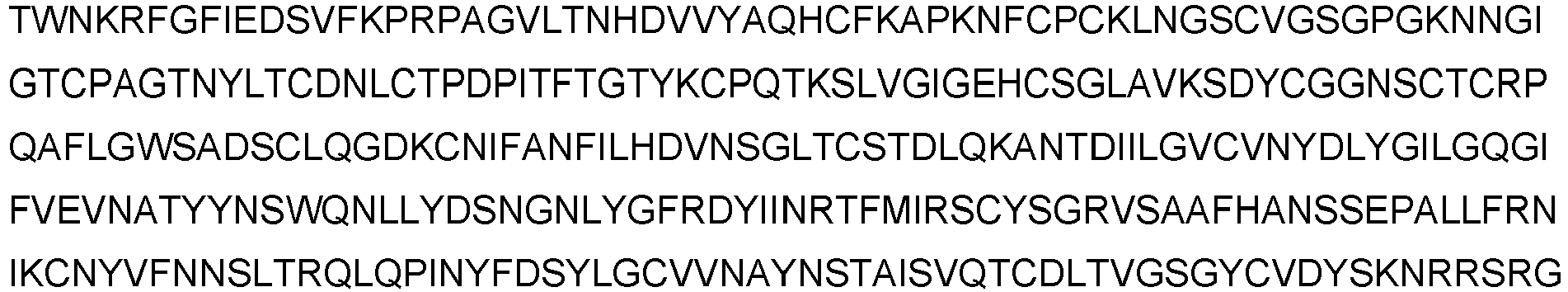
- SEQ ID NO1 (SARS-CoV-2 S1 Spike-Protein)

- SEQ ID NO2 (SARS-CoV-2 Spike-Protein, C-terminally his-tagged, as expressed in cells for the example)

- SEQ ID NO3 (SARS-CoV-2 Spike-Protein, C-terminally his-tagged, as after cleavage of signal peptide, used in the examples)


- SEQ ID NO4 (SARS-CoV-2 Spike-Protein, nucleotide sequence encoding SEQ ID NO2)


- SEQ ID NO5 (possible epitope)
NLKPFERDISTE - SEQ ID NO6 (S1[MERS_CoV])

- SEQ ID NO7 (S1[HCoV-229E])

- SEQ ID NO8 (S1[HCoV-OC43])

- SEQ ID NO9 (S1[HCoV-HKU1])

- SEQ ID NO10 (S1[HCoV-NL63])

- SEQ ID NO11 (S1[SARS_CoV])

- SEQ ID NO12 (fragment of SEQ ID NO1)

- The present invention is further illustrated by the following examples, sequences and figures from which further features, embodiments, aspects and advantages of the present invention may be taken. All methods and materials similar or equivalent to those described herein can be used in the practice or testing of the present invention, with suitable methods and materials being described herein.
- Eight samples from patients tested SARS-coV-2 positive by PCR as described by Corman et al. (Corman et al. (2020) Diagnostic detection of 2019-nCoV by real-time RT-PCR, https://www.who.int/docs/default-source/coronaviruse/protocol-v2-1.pdf?sfvrsn=a9ef618c 2 ), which were obtained 6 to 14 days after the infection and 14 samples from such patients obtained at an earlier time point after the infection were available.
- In addition, a range of samples containing various coronaviruses was available, including 18 samples from patients infected with MERS, three samples from patients infected with SARS-CoV-1, four patients with NL63, three patients with 229E, six patients with OC43 and three patients with HKU1.
- SEQID NO2 was expressed in HEK293T cells using standard cloning of SEQ ID NO4 into the pTriEx-1 plasmid with an artificial signal sequence and a C-terminal His tag, resulting in the expression of SEQ ID NO2 and, after removal of the signal peptide, SEQ ID NO3. Transfected cells were cultures at 37°C and 8.5% CO2 in Dulbecco's modified eagle's medium with 10% fetal calf serum, 100 U/ml penicillin and 0.1 mg/ml streptomycin for three to five days. Cells were harvested, resuspended in 20 mM Tris-HCI pH 7.4, 10% (w/v) sucrose, 5 mM EDTA, 1 mM PMSF and stored at -80°C until further use.
- To prepare SEQ ID NO3, cell culture supernatant was adjusted to 5 mmol/I tris chloride pH 8.0, 164 mmol/I sodium chloride, 50 mmol/I magnesium chloride, 20 mmol/I imidazole, 0,1% Triton X-100, cleared by centrifugation for 30 minutes at 17,600xg, 4°C, applied to Nickel Rapid Run (Agarose Bead Technologies, Miami, FL, USA) equilibrated with 5 mmol/I tris chloride pH 8.0, 300 mmol/I sodium chloride, 20 mmol/I imidazole and eluted by increasing the imidazole concentration to 150 mmol/I. All fractions containing SEQ ID NO3 were pooled and concentrated by ultrafiltration (VivaSpin, Sartorius, Gottingen, Germany). The final preparation was stored at -80°C until further use.
- The final protein preparation of SEQ ID NO3 was treated with or without 16 mmol/I dithiotreitol and incubated at 70°C or at room temperature for 10 minutes, followed by SDS gel electrophoresis and Coomassie staining.
- Protein identity was verified by mass spectrometry.
- For use in microtiter ELISA the purified protein was diluted in PBS to final concentrations of approximately 1.5 µg/ml and used to coat ELISA microtiter plates (Nunc, Roskilde, Denmark) overnight.
- Samples were diluted 1:101 in IgG sample buffer, applied to microtiter plates and incubated as described for commercial EUROIMMUN ELISA Test-Kits, using reagents commercially available (e.g. El 2260-9601 G/A). The manual of El 2260-9601 G/A was followed. In brief: 60 min at 37 °C; 3 washing steps using washing buffer; addition of 100 µl of peroxidase-labelled anti-human IgG conjugate (rabbit) or anti-human IgA conjugate (rabbit) per well; incubation for 30 min at 37 °C; 3 washing steps using EUROIMMUN washing buffer; addition of 100 µl of chromogen/substrate solution (TMB/H2O2) per well; incubation for 30 min at room temperature; addition of 100 µl stop-solution (0.5 M sulfuric acid); measurement of optical density at 450 nm against 630 nm as a reference.
- Calibration was carried out using commercially available calibrators (product number El 2606-9601 A, EUROIMMUN Medizinische Labordiagnostika AG). A ratio was calculated by dividing extinction of the control or patient sample by the extinction of the calibrator. Results below 0.8 were considered negative, results between 0.8 and 1.1 borderline, and results of more than 1.1 positive.
- The primary data are shown in Table 1:


- The results show that antibodies to SEQ ID NO1 may be used for aiding in the diagnosis of an SARS-CoV-2 infection in samples from human patients.
- Comparison of the data obtained with secondary antibodies recognizing IgG and IgA class antibodies shows that the detection of IgA antibodies is more sensitive: 4/14 patient samples taken at an earlier stage of the infection, before six days post onset of illness, could be correctly identified as positive when IgA class antibodies were detected, while the detection of IgG in the same samples gave negative results.
- Both assays showed cross-reactivity with samples from SARS-CoV-1 patients, but virtually none of the samples from patients infected with MERS, NL63, 229E, OC43 and HKU1.
-
- <110> EUROIMMUN Medizinische Labordiagnostika AG
- <120> A method and reagents for the diagnosis of SARS-CoV-2
- <130> 20PP007EP
- <160> 12
- <170> PatentIn version 3.5
- <210> 1
<211> 670
<212> PRT
<213> Artificial Sequence - <220>
<223> SARS-CoV-2 S1 Spike-Protein - <400> 1


- <210> 2
<211> 695
<212> PRT
<213> Artificial Sequence - <220>
<223> SARS-CoV-2 Spike-Protein, C-terminally his-tagged - <400> 2


- <210> 3
<211> 680
<212> PRT
<213> Artificial Sequence - <220>
<223> SARS-CoV-2 Spike-Protein, C-terminally his-tagged, as after cleavage of signal peptide - <400> 3


- <210> 4
<211> 2088
<212> DNA
<213> Artificial Sequence - <220>
<223> SARS-CoV-2 Spike-Protein, nucleotide sequence encoding SEQ ID NO2 - <400> 4


- <210> 5
<211> 12
<212> PRT
<213> Artificial Sequence - <220>
<223> possible epitope - <400> 5

- <210> 6
<211> 734
<212> PRT
<213> Artificial Sequence - <220>
<223> S1 [MERS_CoV] - <400> 6


- <210> 7
<211> 521
<212> PRT
<213> Artificial Sequence - <220>
<223> S1 [HCoV-229E] - <400> 7


- <210> 8
<211> 745
<212> PRT
<213> Artificial Sequence - <220>
<223> S1 [HCoV-OC43] - <400> 8


- <210> 9
<211> 744
<212> PRT
<213> Artificial Sequence - <220>
<223> S1 [HCoV-HKU1] - <400> 9


- <210> 10
<211> 702
<212> PRT
<213> Artificial Sequence - <220>
<223> S1 [HCoV-NL63] - <400> 10


- <210> 11
<211> 657
<212> PRT
<213> Artificial Sequence - <220>
<223> S1 [SARS_CoV] - <400> 11


- <210> 12
<211> 649
<212> PRT
<213> Artificial Sequence - <220>
<223> fragment of SEQ ID NO1 - <400> 12


Claims (8)
- A method for aiding in the diagnosis of a SARS-CoV-2 infection comprising the step of detecting the presence or absence of an IgA class antibody to SEQ ID NO1 in a blood sample from a subject.
- The method according to claim 1, wherein the presence of an IgG and/or IgM class antibody to SEQ ID NO1 is detected in addition to an IgA class antibody to SEQ ID NO1.
- The method according to any of claims 1 to 2, wherein the sample is selected from the group comprising whole blood, serum or plasma.
- The method according to any of claims 1 to 3, wherein an IgA class antibody to SEQ ID NO1 is detected using a labeled secondary antibody binding to IgA class antibodies, preferably human IgA class antibodies.
- The method according to any of claims 1 to 4, wherein the IgA antibody is detected using a method selected from the group comprising colorimetry, immunofluorescence, detection of enzymatic activity, chemiluminscence and radioactivity.
- The method according to any of claims 1 to 5, wherein the infection is detected at an early stage, which is 5 or fewer days after the onset of disease symptoms.
- A use of an IgA class antibody to SEQ ID NO1 for aiding in the diagnosis of a SARS-CoV-2 infection at an early stage, which is 5 or fewer days after the onset of disease symptoms,
wherein a subject is likely to suffer from a SARS-CoV-2 infection if an IgA class antibody to SEQ ID NO1 is detected in a blood sample from the subject. - The method according to any of claims 1 to 6, wherein the sample is a mammalian, preferably a human sample.
Priority Applications (9)
| Application Number | Priority Date | Filing Date | Title |
|---|---|---|---|
| PL20215445T PL3869199T3 (en) | 2020-02-20 | 2020-02-20 | A method and reagents for the diagnosis of sars-cov-2 |
| HUE20215445A HUE058338T2 (en) | 2020-02-20 | 2020-02-20 | A method and reagents for the diagnosis of sars-cov-2 |
| PT202154456T PT3869199T (en) | 2020-02-20 | 2020-02-20 | A method and reagents for the diagnosis of sars-cov-2 |
| EP20215445.6A EP3869199B1 (en) | 2020-02-20 | 2020-02-20 | A method and reagents for the diagnosis of sars-cov-2 |
| DK20215445.6T DK3869199T3 (en) | 2020-02-20 | 2020-02-20 | Method and reagents for diagnosing SARS-CoV-2 |
| HRP20220373TT HRP20220373T1 (en) | 2020-02-20 | 2020-02-20 | A method and reagents for the diagnosis of sars-cov-2 |
| SI202030035T SI3869199T1 (en) | 2020-02-20 | 2020-02-20 | A method and reagents for the diagnosis of sars-cov-2 |
| ES20215445T ES2910184T3 (en) | 2020-02-20 | 2020-02-20 | A procedure and reagents for the diagnosis of SARS-CoV-2 |
| LTEP20215445.6T LT3869199T (en) | 2020-02-20 | 2020-02-20 | A method and reagents for the diagnosis of sars-cov-2 |
Applications Claiming Priority (2)
| Application Number | Priority Date | Filing Date | Title |
|---|---|---|---|
| EP20158626.0A EP3715847A1 (en) | 2020-02-20 | 2020-02-20 | A method and reagents for the diagnosis of sars-cov-2 |
| EP20215445.6A EP3869199B1 (en) | 2020-02-20 | 2020-02-20 | A method and reagents for the diagnosis of sars-cov-2 |
Related Parent Applications (1)
| Application Number | Title | Priority Date | Filing Date |
|---|---|---|---|
| EP20158626.0A Division EP3715847A1 (en) | 2020-02-19 | 2020-02-20 | A method and reagents for the diagnosis of sars-cov-2 |
Publications (2)
| Publication Number | Publication Date |
|---|---|
| EP3869199A1 EP3869199A1 (en) | 2021-08-25 |
| EP3869199B1 true EP3869199B1 (en) | 2022-02-09 |
Family
ID=69726444
Family Applications (3)
| Application Number | Title | Priority Date | Filing Date |
|---|---|---|---|
| EP20215445.6A Active EP3869199B1 (en) | 2020-02-20 | 2020-02-20 | A method and reagents for the diagnosis of sars-cov-2 |
| EP20215513.1A Withdrawn EP3869200A1 (en) | 2020-02-20 | 2020-02-20 | A method and reagents for the diagnosis of sars-cov-2 |
| EP20158626.0A Pending EP3715847A1 (en) | 2020-02-19 | 2020-02-20 | A method and reagents for the diagnosis of sars-cov-2 |
Family Applications After (2)
| Application Number | Title | Priority Date | Filing Date |
|---|---|---|---|
| EP20215513.1A Withdrawn EP3869200A1 (en) | 2020-02-20 | 2020-02-20 | A method and reagents for the diagnosis of sars-cov-2 |
| EP20158626.0A Pending EP3715847A1 (en) | 2020-02-19 | 2020-02-20 | A method and reagents for the diagnosis of sars-cov-2 |
Country Status (10)
| Country | Link |
|---|---|
| EP (3) | EP3869199B1 (en) |
| DE (1) | DE20158626T1 (en) |
| DK (1) | DK3869199T3 (en) |
| ES (2) | ES2788459T1 (en) |
| HR (1) | HRP20220373T1 (en) |
| HU (1) | HUE058338T2 (en) |
| LT (1) | LT3869199T (en) |
| PL (1) | PL3869199T3 (en) |
| PT (1) | PT3869199T (en) |
| SI (1) | SI3869199T1 (en) |
Cited By (2)
| Publication number | Priority date | Publication date | Assignee | Title |
|---|---|---|---|---|
| EP3715847A1 (en) | 2020-02-20 | 2020-09-30 | Euroimmun Medizinische Labordiagnostika AG | A method and reagents for the diagnosis of sars-cov-2 |
| EP3809137A1 (en) | 2020-02-19 | 2021-04-21 | Euroimmun Medizinische Labordiagnostika AG | Methods and reagents for diagnosis of sars-cov-2 infection |
Families Citing this family (23)
| Publication number | Priority date | Publication date | Assignee | Title |
|---|---|---|---|---|
| WO2021186029A1 (en) * | 2020-03-20 | 2021-09-23 | Deutsches Krebsforschungszentrum | Sars spike peptides and uses thereof |
| GB202006815D0 (en) * | 2020-05-07 | 2020-06-24 | Senseutics Ltd | Method |
| WO2021222988A1 (en) * | 2020-05-07 | 2021-11-11 | Griffith University | Cell entry-modulating agents and uses therefor |
| EP3734286A1 (en) | 2020-05-15 | 2020-11-04 | Euroimmun Medizinische Labordiagnostika AG | A method for determining the efficacy of a sars-cov-2 vaccine |
| CN111551715B (en) * | 2020-05-18 | 2022-04-22 | 天津博奥赛斯生物科技股份有限公司 | Novel enzyme-linked immunosorbent assay detection kit for coronavirus IgM antibody |
| EP3913369A1 (en) * | 2020-05-20 | 2021-11-24 | Diesse Diagnostica Senese S.p.a. | Method for inactivating sars-cov-2 and uses thereof |
| CN111848753B (en) * | 2020-07-20 | 2022-03-15 | 中国科学院过程工程研究所 | Novel coronavirus epitope and application thereof |
| CN118206645A (en) * | 2020-08-19 | 2024-06-18 | 重庆医科大学 | Novel coronavirus RBD-specific monoclonal antibodies and their applications |
| DE102020125915B3 (en) | 2020-10-02 | 2022-03-03 | Institut für Molekulare Diagnostik und Bioanalytik | IMMUNDIAGNOSTIC MEANS AND METHODS FOR DETECTION AND DIFFERENTIATION OF CORONAVIRUS INFECTIONS |
| CN112251414A (en) * | 2020-10-12 | 2021-01-22 | 中国科学院苏州纳米技术与纳米仿生研究所 | A kind of hybridoma cell line, its preparation method and application |
| KR20220054080A (en) * | 2020-10-23 | 2022-05-02 | 주식회사 와이바이오로직스 | ANTIBODY SPECIFICALLY BINDING TO SARS-CoV-2 SPIKE PROTEIN AND USES THEREOF |
| AR124054A1 (en) * | 2020-11-12 | 2023-02-08 | Tscan Therapeutics Inc | CONSTRUCTIONS OF SARS-CoV-2 IMMUNODOMINANT PEPTIDES AND THEIR USES |
| WO2022109751A1 (en) * | 2020-11-27 | 2022-06-02 | The University Of Western Ontario | Point-of-care testing for sars-cov antibodies |
| EP4263576A4 (en) * | 2020-12-18 | 2025-08-13 | Scripps Research Inst | IMMUNOGENIC COMPOSITIONS |
| CN112611870B (en) * | 2020-12-21 | 2022-09-16 | 杭州宝临生物科技有限公司 | Method for predicting titer of neutralizing antibody of novel coronavirus and kit thereof |
| CN112379090B (en) * | 2021-01-07 | 2021-04-16 | 北京百普赛斯生物科技股份有限公司 | Novel coronavirus antibody detection kit and preparation method and application thereof |
| CN112946261A (en) * | 2021-01-14 | 2021-06-11 | 广州中医药大学顺德医院(佛山市顺德区中医院) | Novel coronavirus neutralizing antibody detection kit based on trimer S protein RBD-ACE2 binding competition |
| WO2022159834A1 (en) * | 2021-01-22 | 2022-07-28 | La Jolla Institute For Immunology | Chimeric anti-coronavirus spike protein antibodies |
| CN112415189B (en) * | 2021-01-22 | 2021-03-30 | 北京百普赛斯生物科技股份有限公司 | Magnetic bead coupled with novel coronavirus S2 protein, and preparation method and application thereof |
| CN113278067B (en) * | 2021-02-09 | 2022-06-28 | 武汉中生毓晋生物医药有限责任公司 | Preparation method of novel coronavirus porcine immunoglobulin |
| CN113238046A (en) * | 2021-04-30 | 2021-08-10 | 深圳迈瑞生物医疗电子股份有限公司 | Kit for detecting coronavirus antibody and detection method of coronavirus antibody |
| CN114371159B (en) * | 2021-05-19 | 2023-11-07 | 南京医科大学第二附属医院 | RNA biochip, preparation method and application thereof |
| CN114231497B (en) * | 2022-02-24 | 2022-05-20 | 广州伯尼兹生物科技有限公司 | A hybridoma cell line expressing SARS-CoV-2 S1 protein monoclonal antibody and neutralizing antibody |
Citations (1)
| Publication number | Priority date | Publication date | Assignee | Title |
|---|---|---|---|---|
| WO2006085933A2 (en) * | 2004-06-17 | 2006-08-17 | Becton, Dickinson And Company | Immunogenic domains of sars coronavirus |
Family Cites Families (8)
| Publication number | Priority date | Publication date | Assignee | Title |
|---|---|---|---|---|
| TW200510450A (en) | 2003-07-21 | 2005-03-16 | Nat Inst Health | Soluble fragments of the SARS-CoV spike glycoprotein |
| US20050112559A1 (en) * | 2003-09-29 | 2005-05-26 | The Chinese University Of Hong Kong | Compositions and methods for diagnosing and preventing severe acute respiratory syndrome (SARS) |
| US20050282154A1 (en) | 2003-10-06 | 2005-12-22 | The Brigham And Women's Hospital, Inc. | Angiotensin-converting enzyme-2 as a receptor for the SARS coronavirus |
| US7491397B2 (en) | 2004-01-09 | 2009-02-17 | National Health Research Institutes | Receptor binding polypeptides |
| EP1751178A2 (en) * | 2004-06-04 | 2007-02-14 | Institut Pasteur | Nucleic acids, polypeptides, methods of expression, and immunogenic compositions associated with sars corona virus spike protein |
| US20060188519A1 (en) * | 2004-06-14 | 2006-08-24 | To Cheung | Peptides, antibodies, and methods for the diagnosis of SARS |
| SG11201502189UA (en) | 2012-09-23 | 2015-04-29 | Univ Erasmus Medical Ct | Human betacoronavirus lineage c and identification of n-terminal dipeptidyl peptidase as its virus receptor |
| PT3869199T (en) | 2020-02-20 | 2022-03-02 | Euroimmun Medizinische Labordiagnostika Ag | A method and reagents for the diagnosis of sars-cov-2 |
-
2020
- 2020-02-20 PT PT202154456T patent/PT3869199T/en unknown
- 2020-02-20 DE DE20158626.0T patent/DE20158626T1/en active Pending
- 2020-02-20 ES ES20158626T patent/ES2788459T1/en active Pending
- 2020-02-20 LT LTEP20215445.6T patent/LT3869199T/en unknown
- 2020-02-20 EP EP20215445.6A patent/EP3869199B1/en active Active
- 2020-02-20 SI SI202030035T patent/SI3869199T1/en unknown
- 2020-02-20 HU HUE20215445A patent/HUE058338T2/en unknown
- 2020-02-20 PL PL20215445T patent/PL3869199T3/en unknown
- 2020-02-20 HR HRP20220373TT patent/HRP20220373T1/en unknown
- 2020-02-20 DK DK20215445.6T patent/DK3869199T3/en active
- 2020-02-20 EP EP20215513.1A patent/EP3869200A1/en not_active Withdrawn
- 2020-02-20 ES ES20215445T patent/ES2910184T3/en active Active
- 2020-02-20 EP EP20158626.0A patent/EP3715847A1/en active Pending
Patent Citations (1)
| Publication number | Priority date | Publication date | Assignee | Title |
|---|---|---|---|---|
| WO2006085933A2 (en) * | 2004-06-17 | 2006-08-17 | Becton, Dickinson And Company | Immunogenic domains of sars coronavirus |
Non-Patent Citations (36)
| Title |
|---|
| ANONYMOUS: "COVID-19 diagnostics - Comprehensive test and automation portfolio for direct and indirect detection of SARS-CoV-2 infections", EUROIMMUN MEDIZINISCHE LABORDIAGNOSTIKA AG, 1 December 2022 (2022-12-01), XP093138983, [retrieved on 20240307] |
| ANONYMOUS: "Wantai SARS-CoV-2 Diagnostics, WANTAI SARS-CoV-2 Ab ELISA / ELISA for Total Antibody to SARS-CoV-2.", BEIJING WANTAI BIOLOGICAL PHARMACY ENTERPRISE CO. LTD., 1 August 2020 (2020-08-01), pages 1 - 3, XP055801396 |
| CALLOW K. A., PARRY H. F., SERGEANT M., TYRRELL D. A. J.: "The time course of the immune response to experimental coronavirus infection of man", EPIDEMIOLOGY AND INFECTION, vol. 105, no. 2, 1 October 1990 (1990-10-01), GB , pages 435 - 446, XP093000698, ISSN: 0950-2688, DOI: 10.1017/S0950268800048019 |
| CHAN JASPER FUK-WOO, KOK KIN-HANG, ZHU ZHENG, CHU HIN, TO KELVIN KAI-WANG, YUAN SHUOFENG, YUEN KWOK-YUNG: "Genomic characterization of the 2019 novel human-pathogenic coronavirus isolated from a patient with atypical pneumonia after visiting Wuhan", EMERGING MICROBES & INFECTIONS, vol. 9, no. 1, 1 January 2020 (2020-01-01), pages 221 - 236, XP055785644, DOI: 10.1080/22221751.2020.1719902 |
| CHAN JF ET AL.: "A familial cluster of pneumonia associated with the 2019 novel coronavirus indicating person-to-person transmission: a study of a family cluster", LANCET, vol. 395, no. 10223, 15 February 2020 (2020-02-15) - 24 January 2020 (2020-01-24), pages 514 - 523, XP086050313, DOI: 10.1016/S0140-6736(20)30154-9 |
| DATABASE NUCLEOTIDE ANONYMOUS: "Severe acute respiratory syndrome coronavirus 2 isolate 2019-nCoV_HKU-SZ-005b_2020, complete genome", XP055840191, retrieved from GENBANK |
| DE SOUSA-PEREIRA PATRÍCIA, WOOF JENNY M.: "IgA: Structure, Function, and Developability", ANTIBODIES, vol. 8, no. 4, 5 December 2019 (2019-12-05), pages 57, XP093000973, DOI: 10.3390/antib8040057 |
| DU LANYING; HE YUXIAN; ZHOU YUSEN; LIU SHUWEN; ZHENG BO-JIAN; JIANG SHIBO: "The spike protein of SARS-CoV — a target for vaccine and therapeutic development", NATURE REVIEWS MICROBIOLOGY, NATURE PUBLISHING GROUP, GB, vol. 7, no. 3, 9 February 2009 (2009-02-09), GB , pages 226 - 236, XP037065603, ISSN: 1740-1526, DOI: 10.1038/nrmicro2090 |
| EUROPEAN CENTRE FOR DISEASE PREVENTION AND CONTROL: "Outbreak of acute respiratory syndrome associated with a novel coronavirus, China: first local transmission in the EU/EEA - third update", 31 January 2020 (2020-01-31), pages 1 - 10, XP055974895 |
| HSUEH P.-R., HUANG L.-M., CHEN P.-J., KAO C.-L., YANG P.-C.: "Chronological evolution of IgM, IgA, IgG and neutralisation antibodies after infection with SARS-associated coronavirus", CLINICAL MICROBIOLOGY AND INFECTION., WILEY-BLACKWELL PUBLISHING LTD, UNITED KINGDOM, SWITZERLAND, vol. 10, no. 12, 1 December 2004 (2004-12-01), United Kingdom, Switzerland , pages 1062 - 1066, XP055883607, ISSN: 1198-743X, DOI: 10.1111/j.1469-0691.2004.01009.x |
| JIANG SHIBO, DU LANYING, SHI ZHENGLI: "An emerging coronavirus causing pneumonia outbreak in Wuhan, China: calling for developing therapeutic and prophylactic strategies", EMERGING MICROBES & INFECTIONS, vol. 9, no. 1, 1 January 2020 (2020-01-01), pages 275 - 277, XP055785642, DOI: 10.1080/22221751.2020.1723441 |
| LABORINFORMATION 44 DES MEDIZINISCHEN LABORS WAHL, 9 April 2020 (2020-04-09) |
| LAN ET AL.: "Structure of the SARS-CoV-2 spike receptor-binding domain bound to the ACE2 receptor", NATURE, vol. 581, 30 March 2020 (2020-03-30), pages 215 - 220, XP037182122, Retrieved from the Internet <URL:https://doi.org/10.1038/s41586-020-2180-5> DOI: 10.1038/s41586-020-2180-5 |
| LASSAUNIÈRE RIA, FRISCHE ANDERS, HARBOE ZITTA B., NIELSEN ALEX C.Y., FOMSGAARD ANDERS, KROGFELT KAREN A., JØRGENSEN CHARLOTTE S.: "Evaluation of nine commercial SARS-CoV-2 immunoassays", MEDRXIV, 10 April 2020 (2020-04-10), XP093000992, [retrieved on 20221122], DOI: 10.1101/2020.04.09.20056325 |
| LEONG KA WAI, DING JEAK LING: "The Unexplored Roles of Human Serum IgA", DNA AND CELL BIOLOGY, vol. 33, no. 12, 1 December 2014 (2014-12-01), US , pages 823 - 829, XP093000658, ISSN: 1044-5498, DOI: 10.1089/dna.2014.2639 |
| MAACHE MIMOUN, KOMURIAN-PRADEL FLORENCE, RAJOHARISON ALAIN, PERRET MAGALI, BERLAND JEAN-LUC, POUZOL STÉPHANE, BAGNAUD AUDREY: "False-Positive Results in a Recombinant Severe Acute Respiratory Syndrome-Associated Coronavirus (SARS-CoV) Nucleocapsid-Based Western Blot Assay Were Rectified by the Use of Two Subunits (S1 and S2) of Spike for Detection of Antibody to SARS-CoV", CLINICAL AND VACCINE IMMUNOLOGY, vol. 13, no. 3, 1 March 2006 (2006-03-01), pages 409 - 414, XP055797674, ISSN: 1556-6811, DOI: 10.1128/CVI.13.3.409-414.2006 |
| MANOPO, I. LU, L. HE, Q. CHEE, L.L. CHAN, S.W. KWANG, J.: "Evaluation of a safe and sensitive Spike protein-based immunofluorescence assay for the detection of antibody responses to SARS-CoV", JOURNAL OF IMMUNOLOGICAL METHODS, vol. 296, no. 1-2, 1 January 2005 (2005-01-01), NL , pages 37 - 44, XP004725500, ISSN: 0022-1759, DOI: 10.1016/j.jim.2004.10.012 |
| MEYER BENJAMIN; DROSTEN CHRISTIAN; MÜLLER MARCEL A. : "Serological assays for emerging coronaviruses: Challenges and pitfalls", VIRUS RESEARCH, AMSTERDAM, NL, vol. 194, 23 March 2014 (2014-03-23), NL , pages 175 - 183, XP029105622, ISSN: 0168-1702, DOI: 10.1016/j.virusres.2014.03.018 |
| MEYER BENJAMIN; DROSTEN CHRISTIAN; MÜLLER MARCEL A. : "Serological assays for emerging coronaviruses: Challenges and pitfalls", VIRUS RESEARCH, vol. 194, 23 March 2014 (2014-03-23), NL , pages 175 - 183, XP029105622, ISSN: 0168-1702, DOI: 10.1016/j.virusres.2014.03.018 |
| PAULES CATHARINE I., MARSTON HILARY D., FAUCI ANTHONY S.: "Coronavirus Infections—More Than Just the Common Cold", JAMA THE JOURNAL OF THE AMERICAN MEDICAL ASSOCIATION, AMERICAN MEDICAL ASSOCIATION, US, vol. 323, no. 8, 25 February 2020 (2020-02-25), US , pages 707, XP093000978, ISSN: 0098-7484, DOI: 10.1001/jama.2020.0757 |
| PERLMAN STANLEY: "Another Decade, Another Coronavirus", THE NEW ENGLAND JOURNAL OF MEDICINE, MASSACHUSETTS MEDICAL SOCIETY, US, vol. 382, no. 8, 24 January 2020 (2020-01-24), US , pages 760 - 762, XP093000977, ISSN: 0028-4793, DOI: 10.1056/NEJMe1917479 |
| REUSKEN C ET AL: "Specific serology for emerging human coronaviruses by protein microarray", EUROSURVEILLANCE, vol. 18, no. 14, 4 April 2013 (2013-04-04), FR , XP055825179, ISSN: 1560-7917, DOI: 10.2807/1560-7917.ES2013.18.14.20441 |
| ROUJIAN LU ET AL: "Genomic characterisation and epidemiology of 2019 novel coronavirus: implications for virus origins and receptor binding", THE LANCET, vol. 395, no. 10224, 22 February 2020 (2020-02-22), AMSTERDAM, NL , pages 565 - 574, XP055740615, ISSN: 0140-6736, DOI: 10.1016/S0140-6736(20)30251-8 |
| SINO BIOLOGICAL: "SARS-CoV-2 (2019-nCoV) Spike S1-His Recombinant Protein (Cat# 40591-V08B1)", MATERIAL SAFETY DATA SHEET, 15 January 2020 (2020-01-15), pages 1 - 2, XP093000650 |
| STIBA KONSTANZE: "EUROIMMUN is developing new diagnostic assays for the diagnosis of infections with the novel coronavirus", EUROIMMUNBLOG, 30 January 2020 (2020-01-30), XP093000985, [retrieved on 20221122] |
| VICTOR M CORMAN ET AL: "Detection of 2019 novel coronavirus (2019-nCoV) by real-time RT-PCR", EUROSURVEILLANCE, vol. 25, no. 3, 23 January 2020 (2020-01-23), FR , XP055695049, ISSN: 1560-7917, DOI: 10.2807/1560-7917.ES.2020.25.3.2000045 |
| WRAPP D ET AL.: "Supplementary Materials for Cryo-EMstructure of the 2019-nCo Vspike in the prefusion conformation", 19 February 2020 (2020-02-19), pages 1 - 19, XP055829067 |
| WRAPP DANIEL, WANG NIANSHUANG, CORBETT KIZZMEKIA S., GOLDSMITH JORY A., HSIEH CHING-LIN, ABIONA OLUBUKOLA, GRAHAM BARNEY S., MCLEL: "Cryo-EM structure of the 2019-nCoV spike in the prefusion conformation", SCIENCE, vol. 367, no. 6483, 19 February 2020 (2020-02-19), US , pages 1260 - 1263, XP055829062, ISSN: 0036-8075, DOI: 10.1126/science.abb2507 |
| WU H-S, ET AL.: "EARLY DETECTION OF ANTIBODIES AGAINST VARIOUS STRUCTURAL PROTEINS OF THE SARS-ASSOCIATED CORONAVIRUS IN SARS PATIENTS", JOURNAL OF BIOMEDICAL SCIENCE, vol. 11, no. 01, 1 February 2004 (2004-02-01), CH , pages 117 - 126, XP009040395, ISSN: 1021-7770, DOI: 10.1159/000075294 |
| XIAO SHU-YUAN, WU YINGJIE, LIU HUAN: "Evolving status of the 2019 novel coronavirus infection: Proposal of conventional serologic assays for disease diagnosis and infection monitoring", JOURNAL OF MEDICAL VIROLOGY, vol. 92, no. 5, 1 May 2020 (2020-05-01), US , pages 464 - 467, XP055785764, ISSN: 0146-6615, DOI: 10.1002/jmv.25702 |
| XIAOLONG TIAN, LI CHENG, HUANG AILING, XIA SHUAI, LU SICONG, SHI ZHENGLI, LU LU, JIANG SHIBO, YANG ZHENLIN, WU YANLING, YING TIANL: "Potent binding of 2019 novel coronavirus spike protein by a SARS coronavirus-specific human monoclonal antibody", EMERGING MICROBES & INFECTIONS, vol. 9, no. 1, 3 February 2020 (2020-02-03), pages 382 - 385, XP055736759, DOI: 10.1080/22221751.2020.1729069 |
| ZHAO J ET AL: "Development and evaluation of an enzyme-linked immunosorbent assay for detection of antibodies against the spike protein of SARS-coronavirus", JOURNAL OF CLINICAL VIROLOGY, ELSEVIER, AMSTERDAM, NL, vol. 33, no. 1, 1 May 2005 (2005-05-01), pages 12 - 18, XP027704122, ISSN: 1386-6532, [retrieved on 20050501] * |
| ZHAO, J. WANG, W. WANG, G.-F. LI, Y. ZHUANG, H. XU, X. REN, F. ZHAO, Z. GAO, X.-M.: "Development and evaluation of an enzyme-linked immunosorbent assay for detection of antibodies against the spike protein of SARS-coronavirus", JOURNAL OF CLINICAL VIROLOGY, vol. 33, no. 1, 1 May 2005 (2005-05-01), NL , pages 12 - 18, XP004813076, ISSN: 1386-6532, DOI: 10.1016/j.jcv.2004.09.024 |
| ZHOU PENG ET AL: "A pneumonia outbreak associated with a new coronavirus of probable bat origin", NATURE, vol. 579, no. 7798, 3 February 2020 (2020-02-03), London, pages 270 - 273, XP037060207, ISSN: 0028-0836, DOI: 10.1038/s41586-020-2012-7 |
| ZHOU PENG; YANG XING-LOU; WANG XIAN-GUANG; HU BEN; ZHANG LEI; ZHANG WEI; SI HAO-RUI; ZHU YAN; LI BEI; HUANG CHAO-LIN; CHEN HUI-DON: "A pneumonia outbreak associated with a new coronavirus of probable bat origin", NATURE, vol. 579, no. 7798, 3 February 2020 (2020-02-03), London, pages 270 - 273, XP037060207, ISSN: 0028-0836, DOI: 10.1038/s41586-020-2012-7 |
| ZHU NA, ZHANG DINGYU, WANG WENLING, LI XINGWANG, YANG BO, SONG JINGDONG, ZHAO XIANG, HUANG BAOYING, SHI WEIFENG, LU ROUJIAN, NIU P: "A Novel Coronavirus from Patients with Pneumonia in China, 2019", THE NEW ENGLAND JOURNAL OF MEDICINE, vol. 382, no. 8, 20 February 2020 (2020-02-20), US , pages 727 - 733, XP055810616, ISSN: 0028-4793, DOI: 10.1056/NEJMoa2001017 |
Cited By (2)
| Publication number | Priority date | Publication date | Assignee | Title |
|---|---|---|---|---|
| EP3809137A1 (en) | 2020-02-19 | 2021-04-21 | Euroimmun Medizinische Labordiagnostika AG | Methods and reagents for diagnosis of sars-cov-2 infection |
| EP3715847A1 (en) | 2020-02-20 | 2020-09-30 | Euroimmun Medizinische Labordiagnostika AG | A method and reagents for the diagnosis of sars-cov-2 |
Also Published As
| Publication number | Publication date |
|---|---|
| PT3869199T (en) | 2022-03-02 |
| HRP20220373T1 (en) | 2022-05-13 |
| ES2788459T1 (en) | 2020-10-21 |
| SI3869199T1 (en) | 2022-05-31 |
| HUE058338T2 (en) | 2022-07-28 |
| DK3869199T3 (en) | 2022-03-07 |
| ES2910184T3 (en) | 2022-05-11 |
| EP3715847A1 (en) | 2020-09-30 |
| PL3869199T3 (en) | 2022-05-30 |
| EP3869200A1 (en) | 2021-08-25 |
| EP3869199A1 (en) | 2021-08-25 |
| DE20158626T1 (en) | 2020-12-10 |
| LT3869199T (en) | 2022-07-11 |
Similar Documents
| Publication | Publication Date | Title |
|---|---|---|
| EP3869199B1 (en) | A method and reagents for the diagnosis of sars-cov-2 | |
| EP3855186B1 (en) | A method for determining the efficacy of a sars-cov-2 vaccine | |
| CN114258490A (en) | Detection of SARSR-COV antibodies | |
| EP3420357B1 (en) | An immunoassay for the diagnosis of zika viral infection | |
| WO2021179371A1 (en) | Novel coronavirus n-s dominant epitope fusion protein, preparation method therefor and application thereof, expression protein, microorganism, application thereof and kit | |
| KR100847586B1 (en) | How to measure hepatitis C virus | |
| CN111978378B (en) | SARS-CoV-2 antigen polypeptide and its application | |
| EP2488872B1 (en) | Methods for prrsv detection | |
| TW201300421A (en) | Reagents and methods for PRRSV detection | |
| CN112505330B (en) | Kit for detecting novel coronavirus based on fusion protein of nucleocapsid protein | |
| EP1894005B1 (en) | Methods and compositions for detecting herpes simplex virus type 2 | |
| CN106397551A (en) | Treponema pallidum infection dependent antigens and kits and applications thereof | |
| US20220026428A1 (en) | Detection of antibodies to sarsr-cov | |
| CN111647055B (en) | A kind of N protein for novel coronavirus detection and its preparation and application | |
| CN111848748B (en) | A truncated protein of African swine fever virus and its application in the preparation of ELISA detection kit | |
| Tian et al. | Epitope mapping of severe acute respiratory syndrome-related coronavirus nucleocapsid protein with a rabbit monoclonal antibody | |
| CN107664694B (en) | A kind of ELISA kit based on E2 Protein Detection pig atypia pestivirus antibody | |
| CN101363856A (en) | Colloidal gold rapid detection test strip for measles and rubella virus IgG antibodies | |
| CN112300252A (en) | Prediction of 2019-nCoV coronavirus nucleocapsid protein epitope polypeptide and application of polypeptide in detection | |
| WO2019162454A1 (en) | Assay for the diagnosis of viral infections | |
| CN103146714A (en) | A tandem recombinant expression method of Mycobacterium tuberculosis CFP10 antigen protein and its application in tuberculosis detection | |
| CN113248578B (en) | Novel coronavirus (2019-nCoV) recombinant antigen and polyclonal antibody | |
| EP3530668A1 (en) | A novel assay for the diagnosis of viral infections | |
| WO2008034297A1 (en) | Method of detecting antibodies against a series of human immunodeficiency virus proteins | |
| CN102757971B (en) | Mycobacterium tuberculosis recombinant protein and preparation method thereof |
Legal Events
| Date | Code | Title | Description |
|---|---|---|---|
| REG | Reference to a national code |
Ref country code: HR Ref legal event code: TUEP Ref document number: P20220373T Country of ref document: HR |
|
| PUAI | Public reference made under article 153(3) epc to a published international application that has entered the european phase |
Free format text: ORIGINAL CODE: 0009012 |
|
| STAA | Information on the status of an ep patent application or granted ep patent |
Free format text: STATUS: EXAMINATION IS IN PROGRESS |
|
| 17P | Request for examination filed |
Effective date: 20210514 |
|
| AC | Divisional application: reference to earlier application |
Ref document number: 3715847 Country of ref document: EP Kind code of ref document: P |
|
| AK | Designated contracting states |
Kind code of ref document: A1 Designated state(s): AL AT BE BG CH CY CZ DE DK EE ES FI FR GB GR HR HU IE IS IT LI LT LU LV MC MK MT NL NO PL PT RO RS SE SI SK SM TR |
|
| GRAP | Despatch of communication of intention to grant a patent |
Free format text: ORIGINAL CODE: EPIDOSNIGR1 |
|
| STAA | Information on the status of an ep patent application or granted ep patent |
Free format text: STATUS: GRANT OF PATENT IS INTENDED |
|
| GRAJ | Information related to disapproval of communication of intention to grant by the applicant or resumption of examination proceedings by the epo deleted |
Free format text: ORIGINAL CODE: EPIDOSDIGR1 |
|
| STAA | Information on the status of an ep patent application or granted ep patent |
Free format text: STATUS: EXAMINATION IS IN PROGRESS |
|
| INTG | Intention to grant announced |
Effective date: 20211014 |
|
| INTC | Intention to grant announced (deleted) | ||
| GRAJ | Information related to disapproval of communication of intention to grant by the applicant or resumption of examination proceedings by the epo deleted |
Free format text: ORIGINAL CODE: EPIDOSDIGR1 |
|
| GRAP | Despatch of communication of intention to grant a patent |
Free format text: ORIGINAL CODE: EPIDOSNIGR1 |
|
| GRAP | Despatch of communication of intention to grant a patent |
Free format text: ORIGINAL CODE: EPIDOSNIGR1 |
|
| STAA | Information on the status of an ep patent application or granted ep patent |
Free format text: STATUS: GRANT OF PATENT IS INTENDED |
|
| GRAS | Grant fee paid |
Free format text: ORIGINAL CODE: EPIDOSNIGR3 |
|
| GRAA | (expected) grant |
Free format text: ORIGINAL CODE: 0009210 |
|
| STAA | Information on the status of an ep patent application or granted ep patent |
Free format text: STATUS: THE PATENT HAS BEEN GRANTED |
|
| RIN1 | Information on inventor provided before grant (corrected) |
Inventor name: CORMAN, VICTOR Inventor name: MUELLER, MARCEL Inventor name: NEUGEBAUER, EVA Inventor name: LINDHORST, FABIAN Inventor name: STIBA, KONSTANZE Inventor name: LATTWEIN, ERIK Inventor name: MESSING, CLAUDIA Inventor name: STEINHAGEN, KATJA |
|
| INTG | Intention to grant announced |
Effective date: 20211220 |
|
| RAP3 | Party data changed (applicant data changed or rights of an application transferred) |
Owner name: CHARITE - UNIVERSITAETSMEDIZIN BERLIN Owner name: EUROIMMUN MEDIZINISCHE LABORDIAGNOSTIKA AG |
|
| AC | Divisional application: reference to earlier application |
Ref document number: 3715847 Country of ref document: EP Kind code of ref document: P |
|
| AK | Designated contracting states |
Kind code of ref document: B1 Designated state(s): AL AT BE BG CH CY CZ DE DK EE ES FI FR GB GR HR HU IE IS IT LI LT LU LV MC MK MT NL NO PL PT RO RS SE SI SK SM TR |
|
| REG | Reference to a national code |
Ref country code: GB Ref legal event code: FG4D |
|
| REG | Reference to a national code |
Ref country code: CH Ref legal event code: EP Ref country code: AT Ref legal event code: REF Ref document number: 1467825 Country of ref document: AT Kind code of ref document: T Effective date: 20220215 |
|
| REG | Reference to a national code |
Ref country code: DE Ref legal event code: R096 Ref document number: 602020001811 Country of ref document: DE |
|
| REG | Reference to a national code |
Ref country code: IE Ref legal event code: FG4D Ref country code: PT Ref legal event code: SC4A Ref document number: 3869199 Country of ref document: PT Date of ref document: 20220302 Kind code of ref document: T Free format text: AVAILABILITY OF NATIONAL TRANSLATION Effective date: 20220224 |
|
| REG | Reference to a national code |
Ref country code: DK Ref legal event code: T3 Effective date: 20220303 |
|
| REG | Reference to a national code |
Ref country code: SE Ref legal event code: TRGR |
|
| REG | Reference to a national code |
Ref country code: RO Ref legal event code: EPE |
|
| REG | Reference to a national code |
Ref country code: HR Ref legal event code: ODRP Ref document number: P20220373T Country of ref document: HR Payment date: 20220316 Year of fee payment: 3 Ref country code: NL Ref legal event code: FP |
|
| REG | Reference to a national code |
Ref country code: FI Ref legal event code: FGE Ref country code: GR Ref legal event code: EP Ref document number: 20220400585 Country of ref document: GR Effective date: 20220418 |
|
| REG | Reference to a national code |
Ref country code: ES Ref legal event code: FG2A Ref document number: 2910184 Country of ref document: ES Kind code of ref document: T3 Effective date: 20220511 |
|
| REG | Reference to a national code |
Ref country code: HR Ref legal event code: T1PR Ref document number: P20220373 Country of ref document: HR |
|
| RAP4 | Party data changed (patent owner data changed or rights of a patent transferred) |
Owner name: CHARITE - UNIVERSITAETSMEDIZIN BERLIN Owner name: EUROIMMUN MEDIZINISCHE LABORDIAGNOSTIKA AG |
|
| REG | Reference to a national code |
Ref country code: NO Ref legal event code: T2 Effective date: 20220209 |
|
| REG | Reference to a national code |
Ref country code: EE Ref legal event code: FG4A Ref document number: E022266 Country of ref document: EE Effective date: 20220404 |
|
| REG | Reference to a national code |
Ref country code: HU Ref legal event code: AG4A Ref document number: E058338 Country of ref document: HU |
|
| PG25 | Lapsed in a contracting state [announced via postgrant information from national office to epo] |
Ref country code: RS Free format text: LAPSE BECAUSE OF FAILURE TO SUBMIT A TRANSLATION OF THE DESCRIPTION OR TO PAY THE FEE WITHIN THE PRESCRIBED TIME-LIMIT Effective date: 20220209 |
|
| PG25 | Lapsed in a contracting state [announced via postgrant information from national office to epo] |
Ref country code: IS Free format text: LAPSE BECAUSE OF FAILURE TO SUBMIT A TRANSLATION OF THE DESCRIPTION OR TO PAY THE FEE WITHIN THE PRESCRIBED TIME-LIMIT Effective date: 20220609 |
|
| REG | Reference to a national code |
Ref country code: DE Ref legal event code: R026 Ref document number: 602020001811 Country of ref document: DE |
|
| PLBI | Opposition filed |
Free format text: ORIGINAL CODE: 0009260 |
|
| PG25 | Lapsed in a contracting state [announced via postgrant information from national office to epo] |
Ref country code: SM Free format text: LAPSE BECAUSE OF FAILURE TO SUBMIT A TRANSLATION OF THE DESCRIPTION OR TO PAY THE FEE WITHIN THE PRESCRIBED TIME-LIMIT Effective date: 20220209 |
|
| PLBI | Opposition filed |
Free format text: ORIGINAL CODE: 0009260 |
|
| 26 | Opposition filed |
Opponent name: MIKROGEN GMBH Effective date: 20221010 |
|
| PLBI | Opposition filed |
Free format text: ORIGINAL CODE: 0009260 |
|
| PLAX | Notice of opposition and request to file observation + time limit sent |
Free format text: ORIGINAL CODE: EPIDOSNOBS2 |
|
| PG25 | Lapsed in a contracting state [announced via postgrant information from national office to epo] |
Ref country code: MC Free format text: LAPSE BECAUSE OF FAILURE TO SUBMIT A TRANSLATION OF THE DESCRIPTION OR TO PAY THE FEE WITHIN THE PRESCRIBED TIME-LIMIT Effective date: 20220209 |
|
| 26 | Opposition filed |
Opponent name: DIASORIN ITALIA S.P.A. Effective date: 20221109 |
|
| 26 | Opposition filed |
Opponent name: SIMMONS & SIMMONS LLP Effective date: 20221109 |
|
| REG | Reference to a national code |
Ref country code: HR Ref legal event code: ODRP Ref document number: P20220373 Country of ref document: HR Payment date: 20230213 Year of fee payment: 4 |
|
| PLBB | Reply of patent proprietor to notice(s) of opposition received |
Free format text: ORIGINAL CODE: EPIDOSNOBS3 |
|
| PLBP | Opposition withdrawn |
Free format text: ORIGINAL CODE: 0009264 |
|
| P01 | Opt-out of the competence of the unified patent court (upc) registered |
Effective date: 20230602 |
|
| PLAB | Opposition data, opponent's data or that of the opponent's representative modified |
Free format text: ORIGINAL CODE: 0009299OPPO |
|
| PLAB | Opposition data, opponent's data or that of the opponent's representative modified |
Free format text: ORIGINAL CODE: 0009299OPPO |
|
| R26 | Opposition filed (corrected) |
Opponent name: SIMMONS & SIMMONS LLP Effective date: 20221109 |
|
| R26 | Opposition filed (corrected) |
Opponent name: MIKROGEN GMBH Effective date: 20221010 |
|
| PLCK | Communication despatched that opposition was rejected |
Free format text: ORIGINAL CODE: EPIDOSNREJ1 |
|
| REG | Reference to a national code |
Ref country code: HR Ref legal event code: ODRP Ref document number: P20220373 Country of ref document: HR Payment date: 20240215 Year of fee payment: 5 |
|
| APBM | Appeal reference recorded |
Free format text: ORIGINAL CODE: EPIDOSNREFNO |
|
| APBP | Date of receipt of notice of appeal recorded |
Free format text: ORIGINAL CODE: EPIDOSNNOA2O |
|
| APAH | Appeal reference modified |
Free format text: ORIGINAL CODE: EPIDOSCREFNO |
|
| PG25 | Lapsed in a contracting state [announced via postgrant information from national office to epo] |
Ref country code: MK Free format text: LAPSE BECAUSE OF FAILURE TO SUBMIT A TRANSLATION OF THE DESCRIPTION OR TO PAY THE FEE WITHIN THE PRESCRIBED TIME-LIMIT Effective date: 20220209 Ref country code: CY Free format text: LAPSE BECAUSE OF FAILURE TO SUBMIT A TRANSLATION OF THE DESCRIPTION OR TO PAY THE FEE WITHIN THE PRESCRIBED TIME-LIMIT Effective date: 20220209 |
|
| APBQ | Date of receipt of statement of grounds of appeal recorded |
Free format text: ORIGINAL CODE: EPIDOSNNOA3O |
|
| REG | Reference to a national code |
Ref country code: AT Ref legal event code: UEP Ref document number: 1467825 Country of ref document: AT Kind code of ref document: T Effective date: 20220209 |
|
| PG25 | Lapsed in a contracting state [announced via postgrant information from national office to epo] |
Ref country code: MT Free format text: LAPSE BECAUSE OF FAILURE TO SUBMIT A TRANSLATION OF THE DESCRIPTION OR TO PAY THE FEE WITHIN THE PRESCRIBED TIME-LIMIT Effective date: 20220209 |
|
| REG | Reference to a national code |
Ref country code: HR Ref legal event code: ODRP Ref document number: P20220373 Country of ref document: HR Payment date: 20250212 Year of fee payment: 6 |
|
| PGFP | Annual fee paid to national office [announced via postgrant information from national office to epo] |
Ref country code: PT Payment date: 20250213 Year of fee payment: 6 Ref country code: HR Payment date: 20250212 Year of fee payment: 6 Ref country code: DE Payment date: 20250218 Year of fee payment: 6 |
|
| PGFP | Annual fee paid to national office [announced via postgrant information from national office to epo] |
Ref country code: DK Payment date: 20250219 Year of fee payment: 6 Ref country code: FI Payment date: 20250218 Year of fee payment: 6 Ref country code: RO Payment date: 20250213 Year of fee payment: 6 Ref country code: LT Payment date: 20250205 Year of fee payment: 6 |
|
| PGFP | Annual fee paid to national office [announced via postgrant information from national office to epo] |
Ref country code: BG Payment date: 20250214 Year of fee payment: 6 |
|
| PGFP | Annual fee paid to national office [announced via postgrant information from national office to epo] |
Ref country code: ES Payment date: 20250318 Year of fee payment: 6 |
|
| PGFP | Annual fee paid to national office [announced via postgrant information from national office to epo] |
Ref country code: IE Payment date: 20250219 Year of fee payment: 6 Ref country code: SE Payment date: 20250219 Year of fee payment: 6 |
|
| PGFP | Annual fee paid to national office [announced via postgrant information from national office to epo] |
Ref country code: NO Payment date: 20250218 Year of fee payment: 6 |
|
| PGFP | Annual fee paid to national office [announced via postgrant information from national office to epo] |
Ref country code: CH Payment date: 20250301 Year of fee payment: 6 Ref country code: BE Payment date: 20250218 Year of fee payment: 6 Ref country code: GR Payment date: 20250217 Year of fee payment: 6 Ref country code: EE Payment date: 20250217 Year of fee payment: 6 Ref country code: LV Payment date: 20250217 Year of fee payment: 6 Ref country code: AT Payment date: 20250217 Year of fee payment: 6 Ref country code: SI Payment date: 20250210 Year of fee payment: 6 |
|
| PGFP | Annual fee paid to national office [announced via postgrant information from national office to epo] |
Ref country code: FR Payment date: 20250219 Year of fee payment: 6 Ref country code: PL Payment date: 20250210 Year of fee payment: 6 Ref country code: CZ Payment date: 20250207 Year of fee payment: 6 |
|
| PGFP | Annual fee paid to national office [announced via postgrant information from national office to epo] |
Ref country code: IT Payment date: 20250228 Year of fee payment: 6 Ref country code: SK Payment date: 20250214 Year of fee payment: 6 Ref country code: GB Payment date: 20250220 Year of fee payment: 6 |
|
| PGFP | Annual fee paid to national office [announced via postgrant information from national office to epo] |
Ref country code: TR Payment date: 20250212 Year of fee payment: 6 |
|
| PGFP | Annual fee paid to national office [announced via postgrant information from national office to epo] |
Ref country code: AL Payment date: 20250214 Year of fee payment: 6 |
|
| REG | Reference to a national code |
Ref country code: CH Ref legal event code: U11 Free format text: ST27 STATUS EVENT CODE: U-0-0-U10-U11 (AS PROVIDED BY THE NATIONAL OFFICE) Effective date: 20260301 |
|
| REG | Reference to a national code |
Ref country code: HR Ref legal event code: ODRP Ref document number: P20220373 Country of ref document: HR Payment date: 20260211 Year of fee payment: 7 |
|
| PGFP | Annual fee paid to national office [announced via postgrant information from national office to epo] |
Ref country code: LU Payment date: 20260217 Year of fee payment: 7 Ref country code: NL Payment date: 20260218 Year of fee payment: 7 |
|
| PGFP | Annual fee paid to national office [announced via postgrant information from national office to epo] |
Ref country code: HU Payment date: 20260213 Year of fee payment: 7 |