EP3865571B1 - Synovium-derived mesenchymal stem cells and use thereof - Google Patents
Synovium-derived mesenchymal stem cells and use thereof Download PDFInfo
- Publication number
- EP3865571B1 EP3865571B1 EP19867525.8A EP19867525A EP3865571B1 EP 3865571 B1 EP3865571 B1 EP 3865571B1 EP 19867525 A EP19867525 A EP 19867525A EP 3865571 B1 EP3865571 B1 EP 3865571B1
- Authority
- EP
- European Patent Office
- Prior art keywords
- stem cells
- mesenchymal stem
- synovium
- synmscs
- derived mesenchymal
- Prior art date
- Legal status (The legal status is an assumption and is not a legal conclusion. Google has not performed a legal analysis and makes no representation as to the accuracy of the status listed.)
- Active
Links
Images






Classifications
-
- A—HUMAN NECESSITIES
- A61—MEDICAL OR VETERINARY SCIENCE; HYGIENE
- A61P—SPECIFIC THERAPEUTIC ACTIVITY OF CHEMICAL COMPOUNDS OR MEDICINAL PREPARATIONS
- A61P19/00—Drugs for skeletal disorders
- A61P19/02—Drugs for skeletal disorders for joint disorders, e.g. arthritis, arthrosis
-
- C—CHEMISTRY; METALLURGY
- C12—BIOCHEMISTRY; BEER; SPIRITS; WINE; VINEGAR; MICROBIOLOGY; ENZYMOLOGY; MUTATION OR GENETIC ENGINEERING
- C12N—MICROORGANISMS OR ENZYMES; COMPOSITIONS THEREOF; PROPAGATING, PRESERVING, OR MAINTAINING MICROORGANISMS; MUTATION OR GENETIC ENGINEERING; CULTURE MEDIA
- C12N5/00—Undifferentiated human, animal or plant cells, e.g. cell lines; Tissues; Cultivation or maintenance thereof; Culture media therefor
- C12N5/06—Animal cells or tissues; Human cells or tissues
- C12N5/0602—Vertebrate cells
- C12N5/0652—Cells of skeletal and connective tissues; Mesenchyme
- C12N5/0662—Stem cells
-
- A—HUMAN NECESSITIES
- A61—MEDICAL OR VETERINARY SCIENCE; HYGIENE
- A61K—PREPARATIONS FOR MEDICAL, DENTAL OR TOILETRY PURPOSES
- A61K35/00—Medicinal preparations containing materials or reaction products thereof with undetermined constitution
- A61K35/12—Materials from mammals; Compositions comprising non-specified tissues or cells; Compositions comprising non-embryonic stem cells; Genetically modified cells
- A61K35/24—Mucus; Mucous glands; Bursa; Synovial fluid; Arthral fluid; Excreta; Spinal fluid
-
- A—HUMAN NECESSITIES
- A61—MEDICAL OR VETERINARY SCIENCE; HYGIENE
- A61K—PREPARATIONS FOR MEDICAL, DENTAL OR TOILETRY PURPOSES
- A61K35/00—Medicinal preparations containing materials or reaction products thereof with undetermined constitution
- A61K35/12—Materials from mammals; Compositions comprising non-specified tissues or cells; Compositions comprising non-embryonic stem cells; Genetically modified cells
- A61K35/28—Bone marrow; Haematopoietic stem cells; Mesenchymal stem cells of any origin, e.g. adipose-derived stem cells
-
- C—CHEMISTRY; METALLURGY
- C12—BIOCHEMISTRY; BEER; SPIRITS; WINE; VINEGAR; MICROBIOLOGY; ENZYMOLOGY; MUTATION OR GENETIC ENGINEERING
- C12N—MICROORGANISMS OR ENZYMES; COMPOSITIONS THEREOF; PROPAGATING, PRESERVING, OR MAINTAINING MICROORGANISMS; MUTATION OR GENETIC ENGINEERING; CULTURE MEDIA
- C12N5/00—Undifferentiated human, animal or plant cells, e.g. cell lines; Tissues; Cultivation or maintenance thereof; Culture media therefor
- C12N5/06—Animal cells or tissues; Human cells or tissues
- C12N5/0602—Vertebrate cells
- C12N5/0652—Cells of skeletal and connective tissues; Mesenchyme
- C12N5/0662—Stem cells
- C12N5/0668—Mesenchymal stem cells from other natural sources
-
- C—CHEMISTRY; METALLURGY
- C12—BIOCHEMISTRY; BEER; SPIRITS; WINE; VINEGAR; MICROBIOLOGY; ENZYMOLOGY; MUTATION OR GENETIC ENGINEERING
- C12N—MICROORGANISMS OR ENZYMES; COMPOSITIONS THEREOF; PROPAGATING, PRESERVING, OR MAINTAINING MICROORGANISMS; MUTATION OR GENETIC ENGINEERING; CULTURE MEDIA
- C12N2501/00—Active agents used in cell culture processes, e.g. differentation
- C12N2501/10—Growth factors
- C12N2501/105—Insulin-like growth factors [IGF]
-
- C—CHEMISTRY; METALLURGY
- C12—BIOCHEMISTRY; BEER; SPIRITS; WINE; VINEGAR; MICROBIOLOGY; ENZYMOLOGY; MUTATION OR GENETIC ENGINEERING
- C12N—MICROORGANISMS OR ENZYMES; COMPOSITIONS THEREOF; PROPAGATING, PRESERVING, OR MAINTAINING MICROORGANISMS; MUTATION OR GENETIC ENGINEERING; CULTURE MEDIA
- C12N2501/00—Active agents used in cell culture processes, e.g. differentation
- C12N2501/10—Growth factors
- C12N2501/11—Epidermal growth factor [EGF]
-
- C—CHEMISTRY; METALLURGY
- C12—BIOCHEMISTRY; BEER; SPIRITS; WINE; VINEGAR; MICROBIOLOGY; ENZYMOLOGY; MUTATION OR GENETIC ENGINEERING
- C12N—MICROORGANISMS OR ENZYMES; COMPOSITIONS THEREOF; PROPAGATING, PRESERVING, OR MAINTAINING MICROORGANISMS; MUTATION OR GENETIC ENGINEERING; CULTURE MEDIA
- C12N2501/00—Active agents used in cell culture processes, e.g. differentation
- C12N2501/10—Growth factors
- C12N2501/115—Basic fibroblast growth factor (bFGF, FGF-2)
-
- C—CHEMISTRY; METALLURGY
- C12—BIOCHEMISTRY; BEER; SPIRITS; WINE; VINEGAR; MICROBIOLOGY; ENZYMOLOGY; MUTATION OR GENETIC ENGINEERING
- C12N—MICROORGANISMS OR ENZYMES; COMPOSITIONS THEREOF; PROPAGATING, PRESERVING, OR MAINTAINING MICROORGANISMS; MUTATION OR GENETIC ENGINEERING; CULTURE MEDIA
- C12N2501/00—Active agents used in cell culture processes, e.g. differentation
- C12N2501/10—Growth factors
- C12N2501/155—Bone morphogenic proteins [BMP]; Osteogenins; Osteogenic factor; Bone inducing factor
-
- C—CHEMISTRY; METALLURGY
- C12—BIOCHEMISTRY; BEER; SPIRITS; WINE; VINEGAR; MICROBIOLOGY; ENZYMOLOGY; MUTATION OR GENETIC ENGINEERING
- C12N—MICROORGANISMS OR ENZYMES; COMPOSITIONS THEREOF; PROPAGATING, PRESERVING, OR MAINTAINING MICROORGANISMS; MUTATION OR GENETIC ENGINEERING; CULTURE MEDIA
- C12N2502/00—Coculture with; Conditioned medium produced by
- C12N2502/13—Coculture with; Conditioned medium produced by connective tissue cells; generic mesenchyme cells, e.g. so-called "embryonic fibroblasts"
-
- C—CHEMISTRY; METALLURGY
- C12—BIOCHEMISTRY; BEER; SPIRITS; WINE; VINEGAR; MICROBIOLOGY; ENZYMOLOGY; MUTATION OR GENETIC ENGINEERING
- C12N—MICROORGANISMS OR ENZYMES; COMPOSITIONS THEREOF; PROPAGATING, PRESERVING, OR MAINTAINING MICROORGANISMS; MUTATION OR GENETIC ENGINEERING; CULTURE MEDIA
- C12N2509/00—Methods for the dissociation of cells, e.g. specific use of enzymes
-
- C—CHEMISTRY; METALLURGY
- C12—BIOCHEMISTRY; BEER; SPIRITS; WINE; VINEGAR; MICROBIOLOGY; ENZYMOLOGY; MUTATION OR GENETIC ENGINEERING
- C12N—MICROORGANISMS OR ENZYMES; COMPOSITIONS THEREOF; PROPAGATING, PRESERVING, OR MAINTAINING MICROORGANISMS; MUTATION OR GENETIC ENGINEERING; CULTURE MEDIA
- C12N2533/00—Supports or coatings for cell culture, characterised by material
- C12N2533/30—Synthetic polymers
- C12N2533/40—Polyhydroxyacids, e.g. polymers of glycolic or lactic acid (PGA, PLA, PLGA); Bioresorbable polymers
Definitions
- the present invention relates to a method of preparing synovium-derived mesenchymal stem cells using a synovial tissue and a hydrogel, and to a pharmaceutical composition for treating bone or cartilage damage, the pharmaceutical composition including the synovium-derived mesenchymal stem cells.
- Bones support the soft tissues of the body and body weight, and protect the internal organs from external impact by enclosing them. Moreover, bones are one of the important parts of the body that not only structurally support muscles or organs, but also store substances such as calcium or other essential minerals, i.e., phosphorus or magnesium, in the body.
- Joints exist between bones that constitute the body. Joints can be classified into immovable joints with little or no mobility between two bones or cartilages that are in contact with each other, such as skull or dental root, movable joints with many connective tissues and high mobility between two bones such as an animal's limb bones or jawbone, and slightly movable, amphiarthrodial joints. Joints generally refer to the movable joints, and the movable joints are classified into ligamentous joints, whereby both bones are connected only with ligaments, and synovial joints.
- the synovial joint refers to a joint surrounded by a connective tissue capsule (articular capsule). The inside of the articular capsule secretes a synovial fluid having lubricating property, and many ligaments are located outside the articular capsule to strengthen the joint.
- Stem cells which are the cells in the pre-differentiation stage before being differentiated into each cell constituting a tissue, refer to cells having an unlimited proliferation potential in the undifferentiated state, and having a potential for differentiation into cells of various tissues by a specific differentiation stimulus.
- Embryonic stem cells are stem cells isolated from inner cell mass (ICM), which is supposed to develop into a fetus, in the very early stage blastocyst before the fertilized egg is implanted in the endometrium. These embryonic stem cells are pluripotent cells that are able to differentiate into every tissue.
- tissue-specific stem cells are stem cells specific to each organ, which appear at the organ forming stage during embryonic development. Differentiation potential thereof is generally limited to tissue forming cells (multipotent).
- the representative tissue-specific stem cells are hematopoietic stem cells that exist in bone marrow and mesenchymal stem cells that differentiate into connective tissue cells except blood cells. Hematopoietic stem cells differentiate into various blood cells such as erythrocytes, leucocytes, etc., and mesenchymal stem cells differentiate into osteoblasts, chondroblasts, adipocytes, myoblasts, etc.
- stem cell application is to use stem cells as a cell supplier for cell replacement therapy.
- mesenchymal stem cells In the differentiation of mesenchymal stem cells into chondrogenic cells, and further into chondrocytes, that is, in the chondrogenic differentiation, cytokines and growth factors are involved. The exact mechanisms have not been identified, but transforming growth factor beta (TGF- ⁇ ), insulin-like growth factor (IGF), bone morphogenic protein (BMP), fibroblast growth factor (FGF), etc., is known to play an important role in differentiation into chondrocytes. Accordingly, mesenchymal stem cells are studied to use their ability to differentiate for regeneration of damaged joint tissue, treatment of inflammation, or the like ( US Patent No. 6835377 ). Also, bone marrow-derived stem cells ( Majumdar M. K. et al., J. Cell. Physiol.
- EP 3 028 722 A1 discloses synovial MSCs for tissue repair. Further described is the storage of synovial MSCs in hyaluronic acid.
- Carrion et al., Tissue Engineering Part C, Methods Dec 2008, Vol.20. no.3, 2014, p.252-263 describe a method to recover cells encapsulated within 3D fibrin hydrogelsbased on nattokinase.
- the method uses mesenchymal stem cell and not synovial tissue.
- the present inventors have conducted various studies to develop new stem cells that may be used for the treatment of cartilage damage, and as a result, they confirmed that synovial tissue-derived mesenchymal stem cells may effectively treat cartilage damage, thereby completing the present invention.
- An object of the present invention is to provide a method of preparing synovium-derived mesenchymal stem cells using a synovial tissue and a hydrogel as defined in claims 1 to 3.
- the present document also describes synovium-derived mesenchymal stem cells prepared by the above method.
- Another object of the present invention is to provide a pharmaceutical composition for treating bone or cartilage damage, the pharmaceutical composition including the synovium-derived mesenchymal stem cells as defined in claims 4 to 8.
- the present document also describes a composition for culturing mesenchymal stem cells, the composition including a synovial tissue and a hydrogel.
- Still another aspect of the present disclosure is a kit for culturing mesenchymal stem cells, the kit including the composition.
- Still another aspect of the present disclosure is the use of the synovium-derived mesenchymal stem cells for the preparation of a pharmaceutical composition for treating bone or cartilage damage.
- an aspect of the present invention defined in claims 1 to 3 provides a method of preparing synovium-derived mesenchymal stem cells using a synovial tissue and a hydrogel.
- the method of preparing synovium-derived mesenchymal stem cells includes the steps of (a) encapsulating the synovial tissue in the hydrogel and culturing the same to obtain a culture; and (b) degrading the hydrogel in the obtained culture to recover, from the synovial tissue, the mesenchymal stem cells that migrate to and proliferate in the hydrogel, wherein the synovium-derived mesenchymal stem cells have immunological characteristics of overexpressing CD29, CD44, CD73 and CD90 on the cell surface thereof.
- synovial membrane As used herein, the term "synovium”, which is also called synovial membrane or synovial stratum, refers to a loose connective tissue that constitutes the inner layer of articular capsules surrounding the joint cavity. It is known that capillary blood vessels are developed in the synovium to actively exchange synovial fluid. The synovium has a different volume depending on the joint, and in some cases, forms wrinkles to surround the fat pad in the joint cavity and fills the cavity of the joint.
- the synovium may be understood as a source tissue for isolating mesenchymal stem cells.
- hydrogel refers to a gel containing water as a dispersion medium.
- the hydrogel is mainly formed when fluidity is lost due to cooling or when a hydrophilic polymer having a three-dimensional network structure and a microcrystalline structure contains water to swell. Since a hydrogel containing polymer electrolytes exhibits high water absorption, it has been practically used as a water absorbent polymer in a variety of fields.
- the hydrophilic polymer constituting the hydrogel is not particularly limited, as long as it is used in the process of preparing the synovium-derived mesenchymal stem cells, and it may be, for example, collagen, gelatin, chondroitin, hyaluronic acid, alginic acid, MatrigelTM, chitosan, peptide, fibrin, polyglycolic acid (PGA), polylactic acid (PLA), polyethylene glycol (PEG), polyacrylamide, etc., and a mixture thereof.
- a method of encapsulating the synovial tissue in the hydrogel is not particularly limited, but it may be performed by a method of mixing the synovial tissue with the hydrophilic polymer constituting the hydrogel, and then transforming the hydrophilic polymer to the hydrogel; or a method of forming the hydrogel, and then physically injecting the synovial tissue inside the hydrogel, etc.
- the culturing of the hydrogel, in which the synovial tissue is encapsulated may be performed after immersing the hydrogel, in which the synovial tissue is encapsulated, in a common medium which is known in the art to be suitable for culturing stem cells.
- the medium is not particularly limited, but for one example, Dulbecco's modified Eagle medium (DMEM) or Keratinocyte serum free medium (Keratinocyte-SFM) may be used, and for another example, D-media (Gibco) may be used.
- DMEM Dulbecco's modified Eagle medium
- Keratinocyte-SFM Keratinocyte serum free medium
- Gibco D-media
- the medium may further include various kinds of additives, and for example, a neutral buffer (e.g., phosphate and/or high concentration bicarbonate) in isotonic solution, a protein nutrient (e.g., serum such as FBS, serum replacement, albumin, or essential and non-essential amino acids such as glutamine), lipids (fatty acids, cholesterol, an HDL or LDL extract of serum), and other ingredients (e.g., insulin or transferrin, nucleosides or nucleotides, pyruvate, a sugar source such as glucose, selenium in any ionized form or salt, a glucocorticoid such as hydrocortisone, and/or a reducing agent such as ⁇ -mercaptoethanol) may be used as the additive.
- a neutral buffer e.g., phosphate and/or high concentration bicarbonate
- a protein nutrient e.g., serum such as FBS, serum replacement, albumin, or essential and non-essential
- the degrading of the hydrogel is not particularly limited, as long as it is able to degrade the hydrogel without affecting the stem cells in the hydrogel.
- the degrading of the hydrogel may be performed by, for example, a method of using an enzymatic reaction, and for another example, a method of using an enzyme that cleaves binding of the hydrophilic polymer constituting the hydrogel, such as collagenase, gelatinase, urokinase, streptokinase, a tissue plasminogen activator (TPA), plasmin, hyaluronidase, etc.
- TPA tissue plasminogen activator
- the mesenchymal stem cells prepared by the above method of preparing synovium-derived mesenchymal stem cells exhibit immunological characteristics of expressing CD29, CD44, CD73, and CD90 on the cell surface thereof, and characteristics of differentiating into cells selected from the group consisting of adipocytes, osteocytes, chondrocytes, and combinations thereof.
- mesenchymal stem cells are differentiated into chondrocytes by treatment with BMP-7, differentiation efficiency into chondrocytes may be remarkably increased.
- proliferation of the mesenchymal stem cells may be promoted by using a PLGA scaffold.
- Another aspect of the present invention defined in claims 4 to 8 provides a pharmaceutical composition for treating bone or cartilage damage, the pharmaceutical composition including, as an active ingredient, the synovium-derived mesenchymal stem cells prepared by the method of the invention.
- treatment means all of the actions by which symptoms of bone or cartilage damage have taken a turn for the better or been modified favorably by the administration of the synovium-derived mesenchymal stem cells, the culture thereof, or the cells differentiated from the mesenchymal stem cells.
- synovium-derived mesenchymal stem cells were injected into the damaged site of the cartilage and femur of the knee joint, they exhibited a therapeutic effect of regenerating the damaged bone and cartilage, and this therapeutic effect was improved by BMP-7.
- the synovium-derived mesenchymal stem cells may exhibit a therapeutic effect on the damaged bone or cartilage site, and this therapeutic effect may be improved when the mesenchymal stem cells are used in the form of being mixed with a support. Specifically, when the synovium-derived mesenchymal stem cells are used after being cultured in the form of being adhered onto the support, the therapeutic effect may be improved. When the support, of which surface is loaded with BMP-7, is used, the therapeutic effect may be further improved.
- the support is not particularly limited, as long as it does not impair the therapeutic effect of the synovium-derived mesenchymal stem cells, and the support may be, for example, a PLGA scaffold.
- the level of the synovium-derived mesenchymal stem cells included in the pharmaceutical composition provided in the present invention is not particularly limited, and the pharmaceutical composition may include, for example, 1.0 ⁇ 10 5 cells to 1.0 ⁇ 10 9 cells per 1 ml, for another example, 1.0 ⁇ 10 6 cells to 1.0 ⁇ 10 8 cells per 1 ml, and for still another example, 1.0 ⁇ 10 7 cells per 1 ml.
- the pharmaceutical composition may be used unfrozen, or may be frozen for later use. If the pharmaceutical composition is required to be frozen, a standard cryopreservative (e.g., DMSO, glycerol, Epilife ® cell freezing medium (Cascade Biologics)) may be added to population of cells before being frozen.
- a standard cryopreservative e.g., DMSO, glycerol, Epilife ® cell freezing medium (Cascade Biologics)
- DMSO glycerol
- Epilife ® cell freezing medium (Cascade Biologics)
- a formulation for parenteral administration preferably includes an injection formulation such as an injection ampoule, an infusion formulation such as an infusion bag, and a spray formulation such as aerosol.
- the injection ampoule may be mixed with an injection solution immediately before administration, and as the injection solution, a saline solution, a glucose, mannitol, or ringer solution, etc. may be used.
- the infusion bag may be textured with polyvinyl chloride or polyethylene, and may be exemplified by a product of Baxter, Becton Dickinson, Medcep, National Hospital Products, or Terumo.
- the pharmaceutical composition may further include one or more pharmaceutically acceptable common inactive carriers, for example, a preservative, an analgesic controller, a solubilizer, a stabilizer, etc. for injection formulation, and a base, an excipient, a lubricant, a preservative, etc. for topical formulation, in addition to the synovium-derived mesenchymal stem cells.
- a pharmaceutically acceptable common inactive carriers for example, a preservative, an analgesic controller, a solubilizer, a stabilizer, etc. for injection formulation, and a base, an excipient, a lubricant, a preservative, etc. for topical formulation, in addition to the synovium-derived mesenchymal stem cells.
- the pharmaceutical composition provided in the present invention may include various components which may assist in the treatment of bone or cartilage damage, may maintain the activity of the synovium-derived mesenchymal stem cells, or may promote the differentiation of the synovium-derived mesenchymal stem cells, in addition to the synovium-derived mesenchymal stem cells.
- the pharmaceutical composition may further include, for example, an anti-inflammatory agent, a stem cell-mobilizing factor, a growth inducing factor, etc.
- the pharmaceutical composition of the present invention may be administered in accordance with any common method in the art, together with other stem cells used for transplantation and other purposes, in the form of a mixture therewith.
- Direct engraftment or transplantation to the lesion of a patient in need of treatment, or direct transplantation or injection into the peritoneal cavity is preferred, but is not limited thereto.
- both of a non-surgical administration using a catheter and a surgical administration such as injection or transplantation after incision are possible, but non-surgical administration using a catheter is more preferred.
- the composition may also be administered parenterally, for example, intravenous injection, which is one of the common methods for transplantation of stem cells of hematopoietic system, besides direct administration to the lesion.
- the daily dosage of the stem cells is not particularly limited, but the stem cells may be administered, for example, in an amount of 1.0 ⁇ 10 4 to 1.0 ⁇ 10 10 cells/kg (body weight), and for another example, 1.0 ⁇ 10 5 to 1.0 ⁇ 10 9 cells/kg (body weight) per day in a single dose or in divided doses.
- the amount of the active ingredient actually administered ought to be determined in light of various relevant factors including a disease to be treated, severity of the disease, a route of administration, and a patient's body weight, age, and sex, etc., and therefore, the above administration dose should not limit the scope of the present invention in any way.
- compositions for culturing mesenchymal stem cells including a synovial tissue and a hydrogel
- kit for culturing the mesenchymal stem cells the kit including the composition.
- the synovium-derived mesenchymal stem cells may be effectively prepared, and thus the composition including the synovial tissue and the hydrogel may be used as a composition for culturing the mesenchymal stem cells.
- the kit for culturing the mesenchymal stem cells may include the composition for culturing the mesenchymal stem cells and various components, such as a solution and a device, needed for culturing the mesenchymal stem cells.
- the components may be exemplified by, but are not particularly limited to, a test tube, an appropriate container, a reaction buffer (varies in pH and buffer concentration), a culture container, a culture medium, sterile water, etc.
- Still another aspect of the present disclosure is the use of the synovium-derived mesenchymal stem cells for the preparation of the pharmaceutical composition for treating bone or cartilage damage.
- synovium-derived mesenchymal stem cells for the preparation of the pharmaceutical composition may be mixed with an acceptable excipient, diluent, carrier, etc., and may be formulated into a complex preparation with other active agents to have a synergistic effect of the active ingredients.
- Synovium was obtained from the suprapatellar pouch of both knee joints of a rabbit (New Zealand white rabbit, 6-48 week-old, male). The obtained synovium was finely cut to obtain synovial fragments, which were then washed with PBS. The washed synovial fragments were suspended in a DMEM medium, in which 100 ug/ml of aminomethylbenzoic acid was contained and 1 unit/ml of thrombin was dissolved, and this was mixed with a DMEM medium, in which 40 mmol/L of calcium chloride was contained and 0.5% fibrinogen was dissolved, at 1:1 (v/v) to form a hydrogel.
- 10 ml of the hydrogel containing about 200 mg of the synovial fragments was put in a 100 mm culture vessel, and incubated in a humidification chamber at 37°C for 2 hours, and then 10 ml of a primary growth medium (90% DMEM-Ham's F12, 10% fetal bovine serum, 10 ng/ml of epidermal growth factor (EGF), 2 ng/ml of basic fibroblast growth factor (bFGF), 10 ng/ml of insulin-like growth factor (IGF), and 10 ug/ml of gentamycin) was added thereto, followed by incubation at 37°C for 14 days in the humidification chamber.
- a primary growth medium 90% DMEM-Ham's F12, 10% fetal bovine serum, 10 ng/ml of epidermal growth factor (EGF), 2 ng/ml of basic fibroblast growth factor (bFGF), 10 ng/ml of insulin-like growth factor (IGF), and 10 ug/m
- synovium-derived mesenchymal stem cells (synMSCs) ( FIG. 1A ).
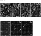
- FIG. 1A shows microscopic images showing the results of culturing synovial fragments for 14 days.
- the obtained synMSCs were washed with PBS twice, and suspended in a secondary growth medium (10% v/v FBS with 10 U/ml antibiotics, 10 ng/ml EGF, and 2 ng/ml bFGF in DMEM/Ham's F-12 (1,1) mixture), and then sub-cultured for 7 days. When the cells reached 80% to 90% confluency, the culturing was terminated. Then, the culture was treated with a 2.5% (w/v) trypsin-EDTA solution to detach the cells, and then centrifuged to obtain Passage 1 (P1) of synMSCs. Thereafter, the same method was repeated to obtain five different series of Passage 15 (P15) of synMSCs ( FIG. 1B ).
- a secondary growth medium (10% v/v FBS with 10 U/ml antibiotics, 10 ng/ml EGF, and 2 ng/ml bFGF in DMEM/Ham's F-12 (1,1) mixture
- FIG. 1B shows microscopic images showing the results of sub-culturing synMSCs for 15 passages, in which Passage 1 represents subculture passage 1 (P1), Passage 5 represents subculture passage 5 (P5), and Passage 15 represents subculture passage 15 (P15).
- synovium-derived mesenchymal stem cells As shown in FIG. 1B , it was observed that synovium-derived mesenchymal stem cells (synMSCs) maintained morphological characteristics even after subculturing 15 times.
- passage 3 (P3) of synMSCs was subjected to cell surface epitope profiling.
- CD29, CD34, CD44, CD45, CD73, and CD90 were targeted as epitopes.
- FIG. 2A shows images showing the results of immunostaining of positive epitopes of synMSCs with the monoclonal antibody
- FIG. 2B shows images showing the results of immunostaining of negative epitopes of synMSCs with the monoclonal antibody
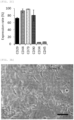
- FIG. 2C shows a graph showing the results of quantitative analysis of fluorescence developed during immunostaining of synMSCs.
- CD29, CD44, CD73, and CD90 were expressed, but CD34 and CD45 were not expressed on the cell surface of synMSCs.
- Example 3-1 Differentiation into adipocytes
- ⁇ 10 4 P3 synMSCs per well were dispensed into each well of a 48-well plate containing an adipogenic differentiation induction medium (0.5 mM isobutyl-methylxanthin, 1 ⁇ M dexamethasone, 10 ⁇ M insulin, 200 ⁇ M indomethacin, 1% antibiotic in DMEM), and cultured in a monolayer for 8 days to induce differentiation into adipocytes.
- an adipogenic differentiation induction medium 0.5 mM isobutyl-methylxanthin, 1 ⁇ M dexamethasone, 10 ⁇ M insulin, 200 ⁇ M indomethacin, 1% antibiotic in DMEM
- a basic medium 10% FBS in DMEM
- FIG. 3A shows fluorescence microscopic images showing the results of performing Oil Red O staining, after inducing differentiation of synMSCs into adipocytes
- FIG. 3B shows a graph showing the results of quantitative analysis of Oil Red O staining levels which were measured in adipocytes differentiated from synMSCs.
- synMSCs exhibited the differentiation potential into adipocytes.
- Example 3-2 Differentiation into osteocytes
- FIG. 3C shows fluorescence microscopic images showing the results of performing Alizarin Red S staining, after inducing differentiation of synMSCs into osteocytes
- FIG. 3D shows a graph showing the results of quantitative analysis of Alizarin Red S staining levels which were measured in osteocytes differentiated from synMSCs.
- FIG. 3E shows fluorescence microscopic images showing BCIP/NBT color development levels by ALP activity, after inducing differentiation of synMSCs into osteocytes
- FIG. 3F shows a graph showing the results of quantitative analysis of ALP activity which was measured in osteocytes differentiated from synMSCs.
- synMSCs exhibited the differentiation potential into osteocytes.
- Example 3-3 Differentiation into chondrocytes
- spheroids were then washed with PBS three times, and then the spheroids were dehydrated by sequentially treating with increasing concentrations of ethanol solutions from 50% to 100%. Then, the spheroids were embedded in paraffin, and 4 ⁇ m-thick sections were obtained, followed by H&E staining and alcian blue staining. Nuclear regions were counterstained with nuclear fast red (FIGS. 4A to 4C). At this time, as a control group, a product resulting from inducing differentiation of bone marrow-derived stem cells (BMSCs) under the same conditions was used.
- BMSCs bone marrow-derived stem cells
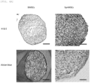
- FIG. 4A shows microscopic images showing the results of performing H&E staining and alcian blue staining of control groups, BMSCs and P3 synMSCs
- FIG. 4B shows a graph showing the results of quantitative analysis of H&E staining levels of BMSCs and P3 synMSCs
- FIG. 4C shows a graph showing the results of quantitative analysis of alcian blue staining levels of BMSCs and P3 synMSCs.
- synMSCs exhibited the differentiation potential into chondrocytes.
- Example 3-4-1 Induction of differentiation of synMSCs into chondrocytes using BMP-7
- the differentiation of chondrocytes from P3 synMSCs was induced in the same manner as in Example 3-3, except that a chondrogenic differentiation induction medium supplemented with BMP-7 (50 ng/ml) was used.
- a culture, obtained by using a basal medium (10% FBS in DMEM) instead of the chondrogenic differentiation induction medium was used as a control group, and a culture, obtained by inducing the differentiation of chondrocytes from P3 synMSCs in the same manner as in Example 3-3, was used as a comparative group.
- Example 3-4-2 Analysis of sulfated glycosaminoglycan (sGAGs) content
- Sulfated glycosaminoglycans were quantitatively analyzed for the control group, comparative group, and experimental group obtained in Example 3-4-1.
- blyscan dye was added to the cell extract of each sample, reacted for 30 minutes under shaking conditions, and then centrifuged (12,000 rpm, 10 minutes) to obtain a precipitate. A dissociation reagent was added to the obtained precipitate to obtain a suspension, and absorbance at 656 nm was measured ( FIG. 5A ) .
- FIG. 5A shows a graph showing the results of quantitative analysis of sGAGs (sulfated glycosaminoglycan) levels which were measured in P3 synMSCs (control group), chondrocytes (comparative group) differentiated from P3 synMSCs, and chondrocytes (experimental group) differentiated from P3 synMSCs using BMP-7.
- sGAGs sulfated glycosaminoglycan
- Example 3-4-3 Analysis of expression levels of chondrogenic markers
- chondrogenic marker proteins SOX-9, aggrecan, and type 2 collagen
- Trizol reagent Invitrogen, CA
- PCR was performed using each of the synthesized cDNAs as a template and the following primers to obtain respective amplification products, and levels thereof were quantitatively analyzed ( FIGS. 5B and 5C ).
- GAPDH was used as an internal control group.
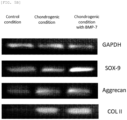
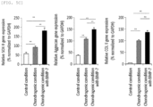
- FIG. 5B shows an electrophoresis image showing the results of measuring expression levels of chondrogenic marker proteins (SOX-9, aggrecan, and type 2 collagen) which were expressed in P3 synMSCs (control group), chondrocytes (comparative group) differentiated from P3 synMSCs, and chondrocytes (experimental group) differentiated from P3 synMSCs using BMP-7, and FIG.
- chondrogenic marker proteins SOX-9, aggrecan, and type 2 collagen
- 5C shows a graph showing the results of quantitative analysis of expression levels of chondrogenic marker proteins (SOX-9, aggrecan, and type 2 collagen) which were expressed in P3 synMSCs (control group), chondrocytes (comparative group) differentiated from P3 synMSCs, and chondrocytes (experimental group) differentiated from P3 synMSCs using BMP-7.
- chondrogenic marker proteins SOX-9, aggrecan, and type 2 collagen
- chondrogenic marker proteins SOX-9, aggrecan, and type 2 collagen
- a 20% (w/v) polymer solution obtained by dissolving PLGA in dichloromethane (DCM) was placed in a 10 ml syringe with a 17-gauge needle, and wet-spun, and then PLGA fibers were obtained using a roller.
- the obtained PLGA fibers were dried for 48 hours, and then vacuum-dried at -70°C for 3 days to remove the residual solvent.
- a three-dimensional PLGA scaffold was formed using the obtained PLGA fiber, immersed in 1.0 M PBS (pH 7.4), and stirred at 37°C at 60 rpm to remove acidic by-products resulting from degradation of PLGA.
- the PLGA scaffold prepared in Example 4-1 was wetted with ethanol, and 1 ⁇ 10 6 P3 synMSCs per the scaffold were inoculated, washed with PBS twice, and cultured for 7 days in a secondary growth medium. After the culturing was completed, the number of viable cells was analyzed using a colorimetric CCK-8 assay (Dojindo Molecular Technologies Inc., MD, USA), and the morphology was analyzed with a scanning electron microscope (S. 6A and 6B ). At this time, P3 synMSCs which were cultured in a 24-well plate under the same conditions were used as a control group.
- SEM scanning electron microscope
- FIG. 6A shows a graph showing the results of analyzing changes in the number of viable cells, after culturing P3 synMSCs using the PLGA scaffold
- FIG. 6B shows a scanning electron microscopic image showing morphological changes over the culture period, when P3 synMSCs were cultured using the PLGA scaffold.
- Recombinant human BMP-7 protein (3 ug) was dissolved in 10 ⁇ L of PBS, and then emulsified with 0.2% (w/v) PLGA in acetone/ethanol (9:1) solvent to obtain a water-in-oil suspension of BMP-7 (1:100 w/o ratio), which was placed in a 10 ml syringe with a 17-gauge needle, and spun onto PLGA fibers by electrospraying (10 kV voltage, 0.033 ml/min flow rate), thereby preparing a PLGA scaffold, in which encapsulated BMP-7 was loaded on the PLGA fibers.
- xylazine (5 mg/kg) was intramuscularly injected into 4-month old male New Zealand white rabbits with an average weight of 3.2 kg to induce general anesthesia, and the knee joint was incised to expose the patella. About 1 mm thick cartilage was removed from the exposed patella, and three osteochondral defects (diameter of 2 mm and depth of 3 mm) were created in in the groove of the femur using a dental drill. Each implant prepared previously was implanted into the created defect site to terminate the operation, tramadol (5 mg/kg) and oxytetracycline (20 mg/kg) were injected, and then raised under normal conditions.
- each rabbit was sacrificed, and the femur was removed from the knee thereof, and then fixed for 5 days using a 10% neutral buffered formalin (pH 7.4, BBC Biochemical, Mount Vernon, USA, WA) solution.
- the fixed femur was treated with a 0.5% EDTA solution to remove calcium deposits, and then embedded in paraffin.
- the embedded femur was finely cut to obtain 4 um-thick tissue sections, which were subjected to H&E staining and Safranin-O staining ( FIG. 7 ).
- tissue sections were immersed in 3% hydrogen peroxide in methanol for 30 minutes to inhibit the endogenous peroxidase activity, and then proteinase K (Sigma-Aldrich, MO, USA) was added thereto and reacted at 37°C for 10 minutes.
- proteinase K Sigma-Aldrich, MO, USA
- the tissue sections were stained with an antibody against type 2 collagen (Calbiochem, CA, USA, dilution 1:100), and immunostaining was performed using a Vectastain Elite ABC-Peroxidase kit (Vector Laboratories Inc., CA, USA).
- Antibody reaction was color-developed using Vector SG (Vector Laboratories Inc., CA, USA), and counterstaining was performed using nuclear fast solutions (Vector Laboratories Inc., CA, USA) ( FIG. 7 ).
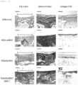
- FIG. 7 shows microscopic images showing the results of performing H&E staining, Safranin-O staining, and type 2 collagen antibody immunostaining of femurs of the rabbit joints, in which the PLGA scaffold, the culture of synMSCs on the PLGA scaffold (PLGA/SynMSC), or the culture of synMSCs on the BMP-7-loaded PLGA scaffold (PLGA/SynMSC/BMP-7) was implanted.
- synMSCs exhibited a therapeutic effect of regenerating damaged bone and cartilage, and this therapeutic effect was improved by BMP-7.
- stem cells may be effectively extracted and obtained from synovium, and the obtained synovium-derived mesenchymal stem cells may effectively treat bone or cartilage damage, and therefore, it will be able to greatly contribute to the development of a method of treating bone or cartilage damage.
Landscapes
- Health & Medical Sciences (AREA)
- Life Sciences & Earth Sciences (AREA)
- Engineering & Computer Science (AREA)
- Biomedical Technology (AREA)
- Developmental Biology & Embryology (AREA)
- Chemical & Material Sciences (AREA)
- Cell Biology (AREA)
- Zoology (AREA)
- Biotechnology (AREA)
- Immunology (AREA)
- General Health & Medical Sciences (AREA)
- Bioinformatics & Cheminformatics (AREA)
- Organic Chemistry (AREA)
- Wood Science & Technology (AREA)
- Genetics & Genomics (AREA)
- Medicinal Chemistry (AREA)
- Pharmacology & Pharmacy (AREA)
- Animal Behavior & Ethology (AREA)
- Public Health (AREA)
- Veterinary Medicine (AREA)
- Virology (AREA)
- Epidemiology (AREA)
- Rheumatology (AREA)
- Microbiology (AREA)
- Biochemistry (AREA)
- General Engineering & Computer Science (AREA)
- Hematology (AREA)
- Orthopedic Medicine & Surgery (AREA)
- Physical Education & Sports Medicine (AREA)
- Neurology (AREA)
- Chemical Kinetics & Catalysis (AREA)
- General Chemical & Material Sciences (AREA)
- Nuclear Medicine, Radiotherapy & Molecular Imaging (AREA)
- Micro-Organisms Or Cultivation Processes Thereof (AREA)
- Medicines Containing Material From Animals Or Micro-Organisms (AREA)
- Materials For Medical Uses (AREA)
- Enzymes And Modification Thereof (AREA)
- Peptides Or Proteins (AREA)
Description
- The present invention relates to a method of preparing synovium-derived mesenchymal stem cells using a synovial tissue and a hydrogel, and to a pharmaceutical composition for treating bone or cartilage damage, the pharmaceutical composition including the synovium-derived mesenchymal stem cells.
- Bones support the soft tissues of the body and body weight, and protect the internal organs from external impact by enclosing them. Moreover, bones are one of the important parts of the body that not only structurally support muscles or organs, but also store substances such as calcium or other essential minerals, i.e., phosphorus or magnesium, in the body.
- Joints exist between bones that constitute the body. Joints can be classified into immovable joints with little or no mobility between two bones or cartilages that are in contact with each other, such as skull or dental root, movable joints with many connective tissues and high mobility between two bones such as an animal's limb bones or jawbone, and slightly movable, amphiarthrodial joints. Joints generally refer to the movable joints, and the movable joints are classified into ligamentous joints, whereby both bones are connected only with ligaments, and synovial joints. The synovial joint refers to a joint surrounded by a connective tissue capsule (articular capsule). The inside of the articular capsule secretes a synovial fluid having lubricating property, and many ligaments are located outside the articular capsule to strengthen the joint.
- Stem cells, which are the cells in the pre-differentiation stage before being differentiated into each cell constituting a tissue, refer to cells having an unlimited proliferation potential in the undifferentiated state, and having a potential for differentiation into cells of various tissues by a specific differentiation stimulus.
- Stem cells are largely divided into embryonic stem cells (ES cells) and adult stem cells (tissue-specific stem cells) according to their differentiation potential. Embryonic stem cells are stem cells isolated from inner cell mass (ICM), which is supposed to develop into a fetus, in the very early stage blastocyst before the fertilized egg is implanted in the endometrium. These embryonic stem cells are pluripotent cells that are able to differentiate into every tissue.
- In contrast, tissue-specific stem cells are stem cells specific to each organ, which appear at the organ forming stage during embryonic development. Differentiation potential thereof is generally limited to tissue forming cells (multipotent). The representative tissue-specific stem cells are hematopoietic stem cells that exist in bone marrow and mesenchymal stem cells that differentiate into connective tissue cells except blood cells. Hematopoietic stem cells differentiate into various blood cells such as erythrocytes, leucocytes, etc., and mesenchymal stem cells differentiate into osteoblasts, chondroblasts, adipocytes, myoblasts, etc.
- Recently, human embryonic stem cells were successfully isolated and the clinical application thereof has been a major concern. The best interest of stem cell application is to use stem cells as a cell supplier for cell replacement therapy.
- In the differentiation of mesenchymal stem cells into chondrogenic cells, and further into chondrocytes, that is, in the chondrogenic differentiation, cytokines and growth factors are involved. The exact mechanisms have not been identified, but transforming growth factor beta (TGF-β), insulin-like growth factor (IGF), bone morphogenic protein (BMP), fibroblast growth factor (FGF), etc., is known to play an important role in differentiation into chondrocytes. Accordingly, mesenchymal stem cells are studied to use their ability to differentiate for regeneration of damaged joint tissue, treatment of inflammation, or the like (
US Patent No. 6835377 ). Also, bone marrow-derived stem cells (Majumdar M. K. et al., J. Cell. Physiol. 185: 98-106, 2000), cord blood(Gang E. J. et al., Biochem. Biophys. Res. Commun. 321: 102-108, 2004), and synovium (Fickert S. et al., Osteoarthritis Cartilage 11: 790-800, 2003) are studied as available cell source for treatment of damaged cartilage in addition to the patient's autologous chondrocytes. However, the treatment of bone disease or inflammation using stem cells is not so effective at present. Especially, the treatment of bone disease or inflammation using cartilage stem cells has never been reported. - European
patent application EP 3 028 722 A1 discloses synovial MSCs for tissue repair. Further described is the storage of synovial MSCs in hyaluronic acid. - International patent application
WO 2008/023829 discloses culturing ex vivo autologous synovium-derived mesenchymal stem cells and implanting the MSCs such that a cartilage defect site is covered by the MSCs and regeneration can occur. - Ferro et al., J. Cell. Physiol., Vol.234, no.4, 2018, p.3973-3984, describes the isolation and cultivation of synovial-derived MSCs that can be used in tissue engineering and cartilage repair.
- Fan et al., Acta Biomaterialia, Vol.6, no.3, 2010, p.1178-1186 disclose synovium-derived MSCs isolated from rabbit synovial tissue, amplified to
passage 4 in monolayer, and encapsulated in injectable gellan hydrogels. Injectable gels, when treated with TGF-beta1, TGF-beta3 or BMP-2 are deemed competent for in vitro engineered cartilage formation. - Carrion et al., Tissue Engineering Part C, Methods Dec 2008, Vol.20. no.3, 2014, p.252-263, describe a method to recover cells encapsulated within 3D fibrin hydrogelsbased on nattokinase. The method uses mesenchymal stem cell and not synovial tissue.
- In view of this technical background, the present inventors have conducted various studies to develop new stem cells that may be used for the treatment of cartilage damage, and as a result, they confirmed that synovial tissue-derived mesenchymal stem cells may effectively treat cartilage damage, thereby completing the present invention.
- The invention is defined by the appended claims. Additional aspects are provided for illustrative purposes only. It is understood that any references to methods of treatment are to be interpreted as references to the compounds, pharmaceutical compositions and medicaments for use in such methods.
- An object of the present invention is to provide a method of preparing synovium-derived mesenchymal stem cells using a synovial tissue and a hydrogel as defined in
claims 1 to 3. - The present document also describes synovium-derived mesenchymal stem cells prepared by the above method.
- Another object of the present invention is to provide a pharmaceutical composition for treating bone or cartilage damage, the pharmaceutical composition including the synovium-derived mesenchymal stem cells as defined in
claims 4 to 8. - The present document also describes a composition for culturing mesenchymal stem cells, the composition including a synovial tissue and a hydrogel.
- Still another aspect of the present disclosure is a kit for culturing mesenchymal stem cells, the kit including the composition.
- Still another aspect of the present disclosure is the use of the synovium-derived mesenchymal stem cells for the preparation of a pharmaceutical composition for treating bone or cartilage damage.
-
-
FIG. 1A shows microscopic images showing the results of culturing synovial fragments for 14 days; -
FIG. 1B shows microscopic images showing the results of sub-culturing synMSCs for 15 passages, in whichPassage 1 represents subculture passage 1 (P1), Passage 5 represents subculture passage 5 (P5), and Passage 15 represents subculture passage 15 (P15); -
FIG. 2A shows images showing the results of immunostaining of positive epitopes of synMSCs with a monoclonal antibody; -
FIG. 2B shows images showing the results of immunostaining of negative epitopes of synMSCs with a monoclonal antibody; -
FIG. 2C shows a graph showing the results of quantitative analysis of fluorescence developed during immunostaining of synMSCs; -
FIG. 3A shows fluorescence microscopic images showing the results of performing Oil Red O staining, after inducing differentiation of synMSCs into adipocytes; -
FIG. 3B shows a graph showing the results of quantitative analysis of Oil Red O staining levels which were measured in adipocytes differentiated from synMSCs; -
FIG. 3C shows fluorescence microscopic images showing the results of performing Alizarin Red S staining, after inducing differentiation of synMSCs into osteocytes; -
FIG. 3D shows a graph showing the results of quantitative analysis of Alizarin Red S staining levels which were measured in osteocytes differentiated from synMSCs; -
FIG. 3E shows fluorescence microscopic images showing BCIP/NBT color development levels by ALP activity, after inducing differentiation of synMSCs into osteocytes; -
FIG. 3F shows a graph showing the results of quantitative analysis of ALP activity which was measured in osteocytes differentiated from synMSCs; -
FIG. 4A shows microscopic images showing the results of performing H&E staining and alcian blue staining of control groups, BMSCs and P3 synMSCs; -
FIG. 4B shows a graph showing the results of quantitative analysis of H&E staining levels of BMSCs and P3 synMSCs; - FIG. 4C shows a graph showing the results of quantitative analysis of alcian blue staining levels of BMSCs and P3 synMSCs;
-
FIG. 5A shows a graph showing the results of quantitative analysis of sGAGs (sulfated glycosaminoglycan) levels which were measured in P3 synMSCs (control group), chondrocytes (comparative group) differentiated from P3 synMSCs, and chondrocytes (experimental group) differentiated from P3 synMSCs using BMP-7; -
FIG. 5B shows an electrophoresis image showing the results of measuring expression levels of chondrogenic marker proteins (SOX-9, aggrecan, andtype 2 collagen) which were expressed in P3 synMSCs (control group), chondrocytes (comparative group) differentiated from P3 synMSCs, and chondrocytes (experimental group) differentiated from P3 synMSCs using BMP-7; -
FIG. 5C shows a graph showing the results of quantitative analysis of expression levels of chondrogenic marker proteins (SOX-9, aggrecan, andtype 2 collagen) which were expressed in P3 synMSCs (control group), chondrocytes (comparative group) differentiated from P3 synMSCs, and chondrocytes (experimental group) differentiated from P3 synMSCs using BMP-7; -
FIG. 6A shows a graph showing the results of analyzing changes in the number of viable cells, after culturing P3 synMSCs using a PLGA scaffold; -
FIG. 6B shows a scanning electron microscopic image showing morphological changes over the culture period, when P3 synMSCs were cultured using a PLGA scaffold; and -
FIG. 7 shows microscopic images showing the results of performing H&E staining, Safranin-O staining, andtype 2 collagen antibody immunostaining of femurs of the rabbit joints, in which a PLGA scaffold, a culture of synMSCs on a PLGA scaffold (PLGA/SynMSC), or a culture of synMSCs on a BMP-7-loaded PLGA scaffold (PLGA/SynMSC/BMP-7) was implanted. - To achieve the above-described objects, an aspect of the present invention defined in
claims 1 to 3 provides a method of preparing synovium-derived mesenchymal stem cells using a synovial tissue and a hydrogel. The method of preparing synovium-derived mesenchymal stem cells includes the steps of (a) encapsulating the synovial tissue in the hydrogel and culturing the same to obtain a culture; and (b) degrading the hydrogel in the obtained culture to recover, from the synovial tissue, the mesenchymal stem cells that migrate to and proliferate in the hydrogel, wherein the synovium-derived mesenchymal stem cells have immunological characteristics of overexpressing CD29, CD44, CD73 and CD90 on the cell surface thereof. - As used herein, the term "synovium", which is also called synovial membrane or synovial stratum, refers to a loose connective tissue that constitutes the inner layer of articular capsules surrounding the joint cavity. It is known that capillary blood vessels are developed in the synovium to actively exchange synovial fluid. The synovium has a different volume depending on the joint, and in some cases, forms wrinkles to surround the fat pad in the joint cavity and fills the cavity of the joint.
- In the present invention, the synovium may be understood as a source tissue for isolating mesenchymal stem cells.
- As used herein, the term "hydrogel" refers to a gel containing water as a dispersion medium. The hydrogel is mainly formed when fluidity is lost due to cooling or when a hydrophilic polymer having a three-dimensional network structure and a microcrystalline structure contains water to swell. Since a hydrogel containing polymer electrolytes exhibits high water absorption, it has been practically used as a water absorbent polymer in a variety of fields.
- In the present invention, the hydrophilic polymer constituting the hydrogel is not particularly limited, as long as it is used in the process of preparing the synovium-derived mesenchymal stem cells, and it may be, for example, collagen, gelatin, chondroitin, hyaluronic acid, alginic acid, MatrigelTM, chitosan, peptide, fibrin, polyglycolic acid (PGA), polylactic acid (PLA), polyethylene glycol (PEG), polyacrylamide, etc., and a mixture thereof.
- In the step (a) of the method of preparing synovium-derived mesenchymal stem cells provided in the present invention, a method of encapsulating the synovial tissue in the hydrogel is not particularly limited, but it may be performed by a method of mixing the synovial tissue with the hydrophilic polymer constituting the hydrogel, and then transforming the hydrophilic polymer to the hydrogel; or a method of forming the hydrogel, and then physically injecting the synovial tissue inside the hydrogel, etc.
- In the step (a), the culturing of the hydrogel, in which the synovial tissue is encapsulated, may be performed after immersing the hydrogel, in which the synovial tissue is encapsulated, in a common medium which is known in the art to be suitable for culturing stem cells.
- The medium is not particularly limited, but for one example, Dulbecco's modified Eagle medium (DMEM) or Keratinocyte serum free medium (Keratinocyte-SFM) may be used, and for another example, D-media (Gibco) may be used.
- The medium may further include various kinds of additives, and for example, a neutral buffer (e.g., phosphate and/or high concentration bicarbonate) in isotonic solution, a protein nutrient (e.g., serum such as FBS, serum replacement, albumin, or essential and non-essential amino acids such as glutamine), lipids (fatty acids, cholesterol, an HDL or LDL extract of serum), and other ingredients (e.g., insulin or transferrin, nucleosides or nucleotides, pyruvate, a sugar source such as glucose, selenium in any ionized form or salt, a glucocorticoid such as hydrocortisone, and/or a reducing agent such as β-mercaptoethanol) may be used as the additive. Furthermore, to prevent cells from adhering to each other, an anti-clumping agent may be used as the additive.
- Moreover, in the step (b) of the method of preparing synovium-derived mesenchymal stem cells provided in the present invention, the degrading of the hydrogel is not particularly limited, as long as it is able to degrade the hydrogel without affecting the stem cells in the hydrogel. The degrading of the hydrogel may be performed by, for example, a method of using an enzymatic reaction, and for another example, a method of using an enzyme that cleaves binding of the hydrophilic polymer constituting the hydrogel, such as collagenase, gelatinase, urokinase, streptokinase, a tissue plasminogen activator (TPA), plasmin, hyaluronidase, etc.
- Meanwhile, the mesenchymal stem cells prepared by the above method of preparing synovium-derived mesenchymal stem cells exhibit immunological characteristics of expressing CD29, CD44, CD73, and CD90 on the cell surface thereof, and characteristics of differentiating into cells selected from the group consisting of adipocytes, osteocytes, chondrocytes, and combinations thereof.
- In particular, when the mesenchymal stem cells are differentiated into chondrocytes by treatment with BMP-7, differentiation efficiency into chondrocytes may be remarkably increased.
- Further, proliferation of the mesenchymal stem cells may be promoted by using a PLGA scaffold.
- Another aspect of the present invention defined in
claims 4 to 8 provides a pharmaceutical composition for treating bone or cartilage damage, the pharmaceutical composition including, as an active ingredient, the synovium-derived mesenchymal stem cells prepared by the method of the invention. - As used herein, the term "treatment" means all of the actions by which symptoms of bone or cartilage damage have taken a turn for the better or been modified favorably by the administration of the synovium-derived mesenchymal stem cells, the culture thereof, or the cells differentiated from the mesenchymal stem cells.
- According to one exemplary embodiment of the present invention, it was confirmed that when the synovium-derived mesenchymal stem cells were injected into the damaged site of the cartilage and femur of the knee joint, they exhibited a therapeutic effect of regenerating the damaged bone and cartilage, and this therapeutic effect was improved by BMP-7.
- In the present invention, the synovium-derived mesenchymal stem cells may exhibit a therapeutic effect on the damaged bone or cartilage site, and this therapeutic effect may be improved when the mesenchymal stem cells are used in the form of being mixed with a support. Specifically, when the synovium-derived mesenchymal stem cells are used after being cultured in the form of being adhered onto the support, the therapeutic effect may be improved. When the support, of which surface is loaded with BMP-7, is used, the therapeutic effect may be further improved.
- In the present invention, the support is not particularly limited, as long as it does not impair the therapeutic effect of the synovium-derived mesenchymal stem cells, and the support may be, for example, a PLGA scaffold.
- The level of the synovium-derived mesenchymal stem cells included in the pharmaceutical composition provided in the present invention is not particularly limited, and the pharmaceutical composition may include, for example, 1.0×10⁵ cells to 1.0×10⁹ cells per 1 ml, for another example, 1.0×10⁶ cells to 1.0×10⁸ cells per 1 ml, and for still another example, 1.0×10⁷ cells per 1 ml.
- The pharmaceutical composition may be used unfrozen, or may be frozen for later use. If the pharmaceutical composition is required to be frozen, a standard cryopreservative (e.g., DMSO, glycerol, Epilife® cell freezing medium (Cascade Biologics)) may be added to population of cells before being frozen.
- Furthermore, the pharmaceutical composition may be administered after formulating it into a unit dosage suitable for administering to a patient by common methods in the pharmaceutical field, in which the formulation contains an effective amount for a single dose or for divided doses. For this purpose, a formulation for parenteral administration preferably includes an injection formulation such as an injection ampoule, an infusion formulation such as an infusion bag, and a spray formulation such as aerosol. The injection ampoule may be mixed with an injection solution immediately before administration, and as the injection solution, a saline solution, a glucose, mannitol, or ringer solution, etc. may be used. Furthermore, the infusion bag may be textured with polyvinyl chloride or polyethylene, and may be exemplified by a product of Baxter, Becton Dickinson, Medcep, National Hospital Products, or Terumo.
- The pharmaceutical composition may further include one or more pharmaceutically acceptable common inactive carriers, for example, a preservative, an analgesic controller, a solubilizer, a stabilizer, etc. for injection formulation, and a base, an excipient, a lubricant, a preservative, etc. for topical formulation, in addition to the synovium-derived mesenchymal stem cells.
- Furthermore, the pharmaceutical composition provided in the present invention may include various components which may assist in the treatment of bone or cartilage damage, may maintain the activity of the synovium-derived mesenchymal stem cells, or may promote the differentiation of the synovium-derived mesenchymal stem cells, in addition to the synovium-derived mesenchymal stem cells. The pharmaceutical composition may further include, for example, an anti-inflammatory agent, a stem cell-mobilizing factor, a growth inducing factor, etc.
- The pharmaceutical composition of the present invention may be administered in accordance with any common method in the art, together with other stem cells used for transplantation and other purposes, in the form of a mixture therewith. Direct engraftment or transplantation to the lesion of a patient in need of treatment, or direct transplantation or injection into the peritoneal cavity is preferred, but is not limited thereto. Furthermore, both of a non-surgical administration using a catheter and a surgical administration such as injection or transplantation after incision are possible, but non-surgical administration using a catheter is more preferred. In addition, the composition may also be administered parenterally, for example, intravenous injection, which is one of the common methods for transplantation of stem cells of hematopoietic system, besides direct administration to the lesion.
- The daily dosage of the stem cells is not particularly limited, but the stem cells may be administered, for example, in an amount of 1.0×10⁴ to 1.0×10¹⁰ cells/kg (body weight), and for another example, 1.0×10⁵ to 1.0×10⁹ cells/kg (body weight) per day in a single dose or in divided doses. However, it should be understood that the amount of the active ingredient actually administered ought to be determined in light of various relevant factors including a disease to be treated, severity of the disease, a route of administration, and a patient's body weight, age, and sex, etc., and therefore, the above administration dose should not limit the scope of the present invention in any way.
- Still another aspect of the present disclosure is a composition for culturing mesenchymal stem cells, the composition including a synovial tissue and a hydrogel, and a kit for culturing the mesenchymal stem cells, the kit including the composition.
- As described above, when the synovial tissue is cultured after being encapsulated in the hydrogel, the synovium-derived mesenchymal stem cells may be effectively prepared, and thus the composition including the synovial tissue and the hydrogel may be used as a composition for culturing the mesenchymal stem cells.
- Furthermore, the kit for culturing the mesenchymal stem cells may include the composition for culturing the mesenchymal stem cells and various components, such as a solution and a device, needed for culturing the mesenchymal stem cells.
- The components may be exemplified by, but are not particularly limited to, a test tube, an appropriate container, a reaction buffer (varies in pH and buffer concentration), a culture container, a culture medium, sterile water, etc.
- Still another aspect of the present disclosure is the use of the synovium-derived mesenchymal stem cells for the preparation of the pharmaceutical composition for treating bone or cartilage damage.
- The synovium-derived mesenchymal stem cells for the preparation of the pharmaceutical composition may be mixed with an acceptable excipient, diluent, carrier, etc., and may be formulated into a complex preparation with other active agents to have a synergistic effect of the active ingredients.
- Those mentioned in the composition, use, and treatment method described herein are the same as long as they do not contradict each other.
- Hereinafter, the present invention will be described in more detail with reference to Examples. However, the following Examples are only for illustrating the present invention, and the scope of the present invention is not intended to be limited by the following Examples.
- Synovium was obtained from the suprapatellar pouch of both knee joints of a rabbit (New Zealand white rabbit, 6-48 week-old, male). The obtained synovium was finely cut to obtain synovial fragments, which were then washed with PBS. The washed synovial fragments were suspended in a DMEM medium, in which 100 ug/ml of aminomethylbenzoic acid was contained and 1 unit/ml of thrombin was dissolved, and this was mixed with a DMEM medium, in which 40 mmol/L of calcium chloride was contained and 0.5% fibrinogen was dissolved, at 1:1 (v/v) to form a hydrogel. 10 ml of the hydrogel containing about 200 mg of the synovial fragments was put in a 100 mm culture vessel, and incubated in a humidification chamber at 37°C for 2 hours, and then 10 ml of a primary growth medium (90% DMEM-Ham's F12, 10% fetal bovine serum, 10 ng/ml of epidermal growth factor (EGF), 2 ng/ml of basic fibroblast growth factor (bFGF), 10 ng/ml of insulin-like growth factor (IGF), and 10 ug/ml of gentamycin) was added thereto, followed by incubation at 37°C for 14 days in the humidification chamber. After the culturing was completed, the medium was removed, and fibrin forming the hydrogel was degraded by adding a DMEM medium containing 5000 units of urokinase and 30% calf serum, and then centrifuged (150 g, 5 minutes) to obtain synovium-derived mesenchymal stem cells (synMSCs) (
FIG. 1A ). -
FIG. 1A shows microscopic images showing the results of culturing synovial fragments for 14 days. - The obtained synMSCs were washed with PBS twice, and suspended in a secondary growth medium (10% v/v FBS with 10 U/ml antibiotics, 10 ng/ml EGF, and 2 ng/ml bFGF in DMEM/Ham's F-12 (1,1) mixture), and then sub-cultured for 7 days. When the cells reached 80% to 90% confluency, the culturing was terminated. Then, the culture was treated with a 2.5% (w/v) trypsin-EDTA solution to detach the cells, and then centrifuged to obtain Passage 1 (P1) of synMSCs. Thereafter, the same method was repeated to obtain five different series of Passage 15 (P15) of synMSCs (
FIG. 1B ). -
FIG. 1B shows microscopic images showing the results of sub-culturing synMSCs for 15 passages, in whichPassage 1 represents subculture passage 1 (P1),Passage 5 represents subculture passage 5 (P5), andPassage 15 represents subculture passage 15 (P15). - As shown in
FIG. 1B , it was observed that synovium-derived mesenchymal stem cells (synMSCs) maintained morphological characteristics even after subculturing 15 times. - Among the various passages of synMSCs obtained in Example 1, passage 3 (P3) of synMSCs was subjected to cell surface epitope profiling. At this time, CD29, CD34, CD44, CD45, CD73, and CD90 were targeted as epitopes.
- Briefly, 1 × 10⁴ P3 synMSCs per well were dispensed and cultured in a 96-well plate, and after completion of the culturing, PBST containing 1% BSA (PBS containing 0.05% Tween 20) was added to perform blocking. Then, each monoclonal antibody against the epitope (anti-mouse CD29, anti-rabbit CD44, anti-human CD73 PE-Cyanine7, anti-human CD90 PE-CyTM7, anti-CD34 PerCPCy5.5 and anti-rabbit CD45) was added and allowed to react. After the reaction was completed, the cells were washed three times with PBS, 1% BSA and a secondary antibody (goat anti-mouse IgG labeled with Alexa Fluor 488, 1:100) was added, and then further reacted in the dark. After the reaction was completed, the cells were washed three times with PBS again, DAPI (Molecular probes, OR, USA) was added, followed by counterstaining in a dark room at room temperature. After staining was completed, scanning was performed using Operetta®; High Content Imaging System (PerkinElmer, MA, USA), and fluorescence intensity was quantitatively analyzed (
FIGS. 2A to 2C ). At this time, as a control group, those treated with Alexa Fluor 488 goat anti-mouse IgG (H+L) instead of the monoclonal antibody against the epitope were used. -
FIG. 2A shows images showing the results of immunostaining of positive epitopes of synMSCs with the monoclonal antibody,FIG. 2B shows images showing the results of immunostaining of negative epitopes of synMSCs with the monoclonal antibody, andFIG. 2C shows a graph showing the results of quantitative analysis of fluorescence developed during immunostaining of synMSCs. - As shown in
FIGS. 2A to 2C , it was confirmed that CD29, CD44, CD73, and CD90 were expressed, but CD34 and CD45 were not expressed on the cell surface of synMSCs. - 1 × 10⁴ P3 synMSCs per well were dispensed into each well of a 48-well plate containing an adipogenic differentiation induction medium (0.5 mM isobutyl-methylxanthin, 1 µM dexamethasone, 10 µM insulin, 200 µM indomethacin, 1% antibiotic in DMEM), and cultured in a monolayer for 8 days to induce differentiation into adipocytes. As a control group, those cultured in a basic medium (10% FBS in DMEM) were used. After the culturing was completed, cells were fixed by treatment with 4% paraformaldehyde, and Oil Red O staining was performed to detect intracellular lipid droplets which are a feature of adipocytes, and then observed using a fluorescence microscope (
FIG. 3A ). Thereafter, in order to quantitatively analyze the content of the dye, isopropanol was added to the stained cells to extract the dye, and absorbance at 540 nm was measured (FIG. 3B ) . -
FIG. 3A shows fluorescence microscopic images showing the results of performing Oil Red O staining, after inducing differentiation of synMSCs into adipocytes, andFIG. 3B shows a graph showing the results of quantitative analysis of Oil Red O staining levels which were measured in adipocytes differentiated from synMSCs. - As shown in
FIGS. 3A and3B , when differentiation of synMSCs into adipocytes was induced, lipid droplets were detected in the cells after 8 days, which was demonstrated by Oil Red O staining. - Therefore, it was found that synMSCs exhibited the differentiation potential into adipocytes.
- 1 × 10⁴ P3 synMSCs per well were dispensed into each well of a 48-well plate containing an osteogenic differentiation induction medium (100 nM dexamethasone, 50 ug/ml ascorbate-2-phosphate, 10 mM β-glycerophosphate, 1% antibiotic suspended in DMEM), and cultured for 14 days to induce differentiation into osteocytes. As a control group, those cultured in a basic medium (10% FBS in DMEM) were used. After the culturing was completed, cells were fixed by treatment with 4% paraformaldehyde, and BCIP/NBT (Sigma Aldrich, MO, USA) which is a substrate of alkaline phosphatase (ALP) was added thereto and allowed to react. After the reaction was completed, Alizarin Red S staining was performed to detect calcium deposits which are a feature of osteocytes, and then observed using a fluorescence microscope, and staining levels thereof were quantitatively analyzed (
FIGS. 3C and3D ). -
FIG. 3C shows fluorescence microscopic images showing the results of performing Alizarin Red S staining, after inducing differentiation of synMSCs into osteocytes, andFIG. 3D shows a graph showing the results of quantitative analysis of Alizarin Red S staining levels which were measured in osteocytes differentiated from synMSCs. - As shown in
FIGS. 3C and3D , when differentiation of synMSCs into osteocytes was induced, calcium deposits were detected in the cells after 14 days, which was demonstrated by Alizarin Red S staining. -
FIG. 3E shows fluorescence microscopic images showing BCIP/NBT color development levels by ALP activity, after inducing differentiation of synMSCs into osteocytes, andFIG. 3F shows a graph showing the results of quantitative analysis of ALP activity which was measured in osteocytes differentiated from synMSCs. - As shown in
FIGS. 3E and3F , it was confirmed that when differentiation of synMSCs into osteocytes was induced, the activity of ALP, which is known as a bone differentiation marker, was increased by about 3 times after 14 days. - Therefore, it was found that synMSCs exhibited the differentiation potential into osteocytes.
- 1 × 10⁶ P3 synMSCs per well were dispensed into each well of a poly-D-lysine-coated 6-well culture plate containing a chondrogenic differentiation induction medium (1% calf serum, 1× ITS, 0.1 mM dexamethasone, 50 ug/ml ascorbate-2-phosphate suspended in DMEM), and cultured for 2 weeks to induce differentiation into chondrocytes. On the second day of culture, spheroids were formed from P3 synMSCs, and the medium was replaced every two days. After the culturing was completed, spheroids as culture products were obtained, and fixed by treatment with 4% paraformaldehyde for 2 hours. Subsequently, centrifugation (300 g, 5 min) was performed to obtain spheroids, which were then washed with PBS three times, and then the spheroids were dehydrated by sequentially treating with increasing concentrations of ethanol solutions from 50% to 100%. Then, the spheroids were embedded in paraffin, and 4 µm-thick sections were obtained, followed by H&E staining and alcian blue staining. Nuclear regions were counterstained with nuclear fast red (FIGS. 4A to 4C). At this time, as a control group, a product resulting from inducing differentiation of bone marrow-derived stem cells (BMSCs) under the same conditions was used.
-
FIG. 4A shows microscopic images showing the results of performing H&E staining and alcian blue staining of control groups, BMSCs and P3 synMSCs,FIG. 4B shows a graph showing the results of quantitative analysis of H&E staining levels of BMSCs and P3 synMSCs, and FIG. 4C shows a graph showing the results of quantitative analysis of alcian blue staining levels of BMSCs and P3 synMSCs. - As shown in FIGS. 4A to 4C, it was confirmed that when differentiation into chondrocytes was induced under the same conditions, the level of chondrocytes differentiated from P3 synMSCs was significantly higher than that of chondrocytes differentiated from the control group BMSCs. In particular, the result of H&E staining showed that chondrocytes differentiated from P3 synMSCs showed about 9 times higher level than chondrocytes differentiated from BMSCs, and the result of alcian blue staining showed that chondrocytes differentiated from P3 synMSCs showed about 13 times higher level than chondrocytes differentiated from BMSCs.
- Therefore, it was found that synMSCs exhibited the differentiation potential into chondrocytes.
- The differentiation of chondrocytes from P3 synMSCs was induced in the same manner as in Example 3-3, except that a chondrogenic differentiation induction medium supplemented with BMP-7 (50 ng/ml) was used. At this time, a culture, obtained by using a basal medium (10% FBS in DMEM) instead of the chondrogenic differentiation induction medium, was used as a control group, and a culture, obtained by inducing the differentiation of chondrocytes from P3 synMSCs in the same manner as in Example 3-3, was used as a comparative group.
- Sulfated glycosaminoglycans (SGAGs) were quantitatively analyzed for the control group, comparative group, and experimental group obtained in Example 3-4-1.
- Briefly, blyscan dye was added to the cell extract of each sample, reacted for 30 minutes under shaking conditions, and then centrifuged (12,000 rpm, 10 minutes) to obtain a precipitate. A dissociation reagent was added to the obtained precipitate to obtain a suspension, and absorbance at 656 nm was measured (
FIG. 5A ) . -
FIG. 5A shows a graph showing the results of quantitative analysis of sGAGs (sulfated glycosaminoglycan) levels which were measured in P3 synMSCs (control group), chondrocytes (comparative group) differentiated from P3 synMSCs, and chondrocytes (experimental group) differentiated from P3 synMSCs using BMP-7. - As shown in
FIG. 5A , it was confirmed that a significantly high level of sGAGs was detected in chondrocytes (experimental group) differentiated from P3 synMSCs using BMP-7, as compared with chondrocytes (comparative group) differentiated from P3 synMSCs. - The expression levels of chondrogenic marker proteins (SOX-9, aggrecan, and
type 2 collagen) were analyzed using RT-PCR for the control group, comparative group, and experimental group obtained in Example 3-4-1. - Briefly, cells of each sample were disrupted and total RNA was extracted using Trizol reagent (Invitrogen, CA), respectively. Each extracted total RNA was applied to a TOPscript™ One-step RT PCR kit (Enzynomics, Daejeon, Korea) to synthesize each cDNA. PCR was performed using each of the synthesized cDNAs as a template and the following primers to obtain respective amplification products, and levels thereof were quantitatively analyzed (
FIGS. 5B and5C ). At this time, GAPDH was used as an internal control group. - SOX-9 F: 5'-CCCGATCTGAAGAAGGAGAGC-3'(SEQ ID NO: 1)
- SOX-9 R: 5'-GTTCTTCACCGACTTCCTCCG-3'(SEQ ID NO: 2)
- aggrecan F: 5'-TGAGGAGGGCTGGAACAAGTACC-3'(SEQ ID NO: 3)
- aggrecan R: 5'-GGAGGTGGTAATTGCAGGGAACA-3'(SEQ ID NO: 4)
- T2 collagen F: 5'-TTCAGCTATGGAGATGACAATC-3'(SEQ ID NO: 5)
- T2 collagen R: 5'-AGAGTCCTAGAGTGACTGAG-3'(SEQ ID NO: 6)
- GAPDH F: 5'-ATTGTTGCCATCAATGACCC-3'(SEQ ID NO: 7)
- GAPDH R: 5'-AGTAGAGGCAGGGATGATGTT-3'(SEQ ID NO: 8)
-
FIG. 5B shows an electrophoresis image showing the results of measuring expression levels of chondrogenic marker proteins (SOX-9, aggrecan, andtype 2 collagen) which were expressed in P3 synMSCs (control group), chondrocytes (comparative group) differentiated from P3 synMSCs, and chondrocytes (experimental group) differentiated from P3 synMSCs using BMP-7, andFIG. 5C shows a graph showing the results of quantitative analysis of expression levels of chondrogenic marker proteins (SOX-9, aggrecan, andtype 2 collagen) which were expressed in P3 synMSCs (control group), chondrocytes (comparative group) differentiated from P3 synMSCs, and chondrocytes (experimental group) differentiated from P3 synMSCs using BMP-7. - As shown in
FIGS. 5B and5C , it was confirmed that significantly high levels of chondrogenic marker proteins (SOX-9, aggrecan, andtype 2 collagen) were detected in chondrocytes (experimental group) differentiated from P3 synMSCs using BMP-7, as compared with chondrocytes (comparative group) differentiated from P3 synMSCs. - A 20% (w/v) polymer solution obtained by dissolving PLGA in dichloromethane (DCM) was placed in a 10 ml syringe with a 17-gauge needle, and wet-spun, and then PLGA fibers were obtained using a roller. The obtained PLGA fibers were dried for 48 hours, and then vacuum-dried at -70°C for 3 days to remove the residual solvent.
- A three-dimensional PLGA scaffold was formed using the obtained PLGA fiber, immersed in 1.0 M PBS (pH 7.4), and stirred at 37°C at 60 rpm to remove acidic by-products resulting from degradation of PLGA. The PLGA scaffold, from which the acidic by-products were removed, was washed with PBS three times, and vacuum-dried for 48 hours to prepare a three-dimensional scaffold for P3 synMSC culture.
- The PLGA scaffold prepared in Example 4-1 was wetted with ethanol, and 1 × 10⁶ P3 synMSCs per the scaffold were inoculated, washed with PBS twice, and cultured for 7 days in a secondary growth medium. After the culturing was completed, the number of viable cells was analyzed using a colorimetric CCK-8 assay (Dojindo Molecular Technologies Inc., MD, USA), and the morphology was analyzed with a scanning electron microscope (SEM) (
FIGS. 6A and 6B ). At this time, P3 synMSCs which were cultured in a 24-well plate under the same conditions were used as a control group. -
FIG. 6A shows a graph showing the results of analyzing changes in the number of viable cells, after culturing P3 synMSCs using the PLGA scaffold, andFIG. 6B shows a scanning electron microscopic image showing morphological changes over the culture period, when P3 synMSCs were cultured using the PLGA scaffold. - As shown in
FIG. 6A and 6B , when the PLGA scaffold rather than the 24-well plate was used, the culture efficiency of P3 synMSC was confirmed to increase. This is because the adhesion rate of P3 synMSCs to the PLGA scaffold was increased over time, and as a result, the proliferation rate was increased. - Recombinant human BMP-7 protein (3 ug) was dissolved in 10 µL of PBS, and then emulsified with 0.2% (w/v) PLGA in acetone/ethanol (9:1) solvent to obtain a water-in-oil suspension of BMP-7 (1:100 w/o ratio), which was placed in a 10 ml syringe with a 17-gauge needle, and spun onto PLGA fibers by electrospraying (10 kV voltage, 0.033 ml/min flow rate), thereby preparing a PLGA scaffold, in which encapsulated BMP-7 was loaded on the PLGA fibers.
- The PLGA scaffold prepared in Example 4-1, the culture of synMSCs on the PLGA scaffold (PLGA/SynMSC), prepared in Example 4-2, and the culture of synMSCs on the BMP-7-loaded PLGA scaffold (PLGA/SynMSC/BMP-7), prepared in Example 4-3, were prepared, respectively, and used to prepare each implant having a thickness of 1 mm and a length of 10 mm.
- Meanwhile, xylazine (5 mg/kg) was intramuscularly injected into 4-month old male New Zealand white rabbits with an average weight of 3.2 kg to induce general anesthesia, and the knee joint was incised to expose the patella. About 1 mm thick cartilage was removed from the exposed patella, and three osteochondral defects (diameter of 2 mm and depth of 3 mm) were created in in the groove of the femur using a dental drill. Each implant prepared previously was implanted into the created defect site to terminate the operation, tramadol (5 mg/kg) and oxytetracycline (20 mg/kg) were injected, and then raised under normal conditions.
- After 6 weeks, each rabbit was sacrificed, and the femur was removed from the knee thereof, and then fixed for 5 days using a 10% neutral buffered formalin (pH 7.4, BBC Biochemical, Mount Vernon, USA, WA) solution. The fixed femur was treated with a 0.5% EDTA solution to remove calcium deposits, and then embedded in paraffin. The embedded femur was finely cut to obtain 4 um-thick tissue sections, which were subjected to H&E staining and Safranin-O staining (
FIG. 7 ). - In addition, the tissue sections were immersed in 3% hydrogen peroxide in methanol for 30 minutes to inhibit the endogenous peroxidase activity, and then proteinase K (Sigma-Aldrich, MO, USA) was added thereto and reacted at 37°C for 10 minutes. After the reaction was completed, the tissue sections were stained with an antibody against
type 2 collagen (Calbiochem, CA, USA, dilution 1:100), and immunostaining was performed using a Vectastain Elite ABC-Peroxidase kit (Vector Laboratories Inc., CA, USA). Antibody reaction was color-developed using Vector SG (Vector Laboratories Inc., CA, USA), and counterstaining was performed using nuclear fast solutions (Vector Laboratories Inc., CA, USA) (FIG. 7 ). -
FIG. 7 shows microscopic images showing the results of performing H&E staining, Safranin-O staining, andtype 2 collagen antibody immunostaining of femurs of the rabbit joints, in which the PLGA scaffold, the culture of synMSCs on the PLGA scaffold (PLGA/SynMSC), or the culture of synMSCs on the BMP-7-loaded PLGA scaffold (PLGA/SynMSC/BMP-7) was implanted. - As shown in
FIG. 7 , in the control group, in which the damaged site was not treated, the layer of cartilage was not regenerated, and the site was simply covered with the fibrous tissue, and Safranin-O staining did not occur. However, at the site where the PLGA scaffold implant was implanted, a cartilage layer was formed, but a thin cartilage layer containing a small number of chondrocytes was formed, and proteoglycan was also formed in a small amount. It was also confirmed that, at the site where the implant of synMSCs cultured on the PLGA scaffold was implanted, the cell level was increased and cartilage was regenerated on the surface. Finally, it was confirmed that, at the site where the implant of synMSCs cultured on the BMP-7-loaded PLGA scaffold was implanted, a thick cartilage layer was regenerated and a large amount oftype 2 collagen was present. - Accordingly, it was found that synMSCs exhibited a therapeutic effect of regenerating damaged bone and cartilage, and this therapeutic effect was improved by BMP-7.
- Based on the above description, it will be understood by those skilled in the art that the present disclosure may be implemented in a different specific form or essential characteristics thereof. In this regard, it should be understood that the above embodiment is not limitative, but illustrative in all aspects. The scope of the disclosure is defined by the appended claims rather than by the description preceding them, and therefore all changes and modifications that fall within metes and bounds of the claims, or equivalents of such metes and bounds are therefore intended to be embraced by the claims.
- When a preparation method of the present invention is used, stem cells may be effectively extracted and obtained from synovium, and the obtained synovium-derived mesenchymal stem cells may effectively treat bone or cartilage damage, and therefore, it will be able to greatly contribute to the development of a method of treating bone or cartilage damage.
Claims (8)
- A method of preparing synovium-derived mesenchymal stem cells, the method comprising the steps of:(a) encapsulating a synovial tissue in a hydrogel and culturing the same to obtain a culture; and(b) degrading the hydrogel in the obtained culture to recover, from the synovial tissue, mesenchymal stem cells that migrate to and proliferate in the hydrogel,
wherein the synovium-derived mesenchymal stem cells have immunological characteristics of expressing CD29, CD44, CD73, and CD90 on the cell surface thereof. - The method of claim 1, wherein the hydrogel consists of a material selected from the group consisting of collagen, gelatin, chondroitin, hyaluronic acid, alginic acid, MatrigelTM, chitosan, peptide, fibrin, polyglycolic acid (PGA), polylactic acid (PLA), polyethylene glycol (PEG), polyacrylamide, and combinations thereof.
- The method of claim 1, wherein the degrading of the hydrogel is performed by treatment with an enzyme selected from the group consisting of collagenase, gelatinase, urokinase, streptokinase, a tissue plasminogen activator (TPA), plasmin, hyaluronidase, and combinations thereof.
- A pharmaceutical composition for treating bone or cartilage damage, the pharmaceutical composition comprising, as an active ingredient, the synovium-derived mesenchymal stem cells prepared by the method of any one of claims 1 to 3, a culture thereof,wherein the mesenchymal stem cells are in a form where the cells are cultured on a three-dimensional PLGA scaffold support and adhered thereto; andthe PLGA scaffold support is in a form where BMP-7 are spun onto PLGA fibers and thereby encapsulated BMP-7 is bound to the PLGA fibers.
- The pharmaceutical composition of claim 4, wherein the synovium-derived mesenchymal stem cells have enhanced ability to regenerate cartilage by the addition of BMP-7.
- The pharmaceutical composition of claim 4, wherein the synovium-derived mesenchymal stem cells have increased GAG synthesis.
- The pharmaceutical composition of claim 4, wherein the synovium-derived mesenchymal stem cells have increased expression levels of SOX-9, aggrecan, and type 2 collagen.
- A pharmaceutical composition for treating bone or cartilage damage, the pharmaceutical composition comprising, as an active ingredient, the synovium-derived mesenchymal stem cells prepared by the method of any one of claims 1 to 3, a culture thereof,
wherein the synovium-derived mesenchymal stem cells have immunological characteristics of expressing CD29, CD44, CD73, and CD90 on the cell surface thereof.
Applications Claiming Priority (2)
| Application Number | Priority Date | Filing Date | Title |
|---|---|---|---|
| KR20180116496 | 2018-09-28 | ||
| PCT/KR2019/012602 WO2020067774A1 (en) | 2018-09-28 | 2019-09-27 | Synovium-derived mesenchymal stem cells and use thereof |
Publications (4)
| Publication Number | Publication Date |
|---|---|
| EP3865571A1 EP3865571A1 (en) | 2021-08-18 |
| EP3865571A4 EP3865571A4 (en) | 2022-08-10 |
| EP3865571B1 true EP3865571B1 (en) | 2024-12-25 |
| EP3865571C0 EP3865571C0 (en) | 2024-12-25 |
Family
ID=69953136
Family Applications (1)
| Application Number | Title | Priority Date | Filing Date |
|---|---|---|---|
| EP19867525.8A Active EP3865571B1 (en) | 2018-09-28 | 2019-09-27 | Synovium-derived mesenchymal stem cells and use thereof |
Country Status (7)
| Country | Link |
|---|---|
| US (1) | US20220112465A1 (en) |
| EP (1) | EP3865571B1 (en) |
| JP (1) | JP7230219B2 (en) |
| KR (1) | KR102223043B1 (en) |
| CN (1) | CN113454206A (en) |
| ES (1) | ES3014281T3 (en) |
| WO (1) | WO2020067774A1 (en) |
Families Citing this family (5)
| Publication number | Priority date | Publication date | Assignee | Title |
|---|---|---|---|---|
| JP7444267B2 (en) * | 2020-09-03 | 2024-03-06 | 株式会社ツーセル | Composition for treating central nervous system diseases, method for producing compositions for treating central nervous system diseases, and method for producing formulations for treating central nervous system diseases |
| CN112595698A (en) * | 2020-11-18 | 2021-04-02 | 上海中医药大学 | Application of oil red O and method for quantitatively detecting lipid in tissue or cell |
| WO2023121399A1 (en) * | 2021-12-24 | 2023-06-29 | 이화여자대학교 산학협력단 | Method for culturing skin-derived stem cells, and use thereof |
| CN115770326B (en) * | 2022-09-28 | 2024-06-25 | 中国人民解放军总医院第一医学中心 | Gel system for cartilage repair and regeneration in joint cavity and preparation method and use thereof |
| WO2024144250A1 (en) * | 2022-12-29 | 2024-07-04 | 가톨릭관동대학교산학협력단 | Cartilage regeneration composition comprising synovial fluid-derived stem cells and chondrocytes |
Family Cites Families (16)
| Publication number | Priority date | Publication date | Assignee | Title |
|---|---|---|---|---|
| US6835377B2 (en) | 1998-05-13 | 2004-12-28 | Osiris Therapeutics, Inc. | Osteoarthritis cartilage regeneration |
| JP4246994B2 (en) | 2001-01-30 | 2009-04-02 | オーソジーン, インコーポレイテッド | Compositions and methods for the treatment and repair of defects or lesions in articular cartilage using synovial tissue or cells |
| US7560432B2 (en) * | 2003-11-26 | 2009-07-14 | Histogenics Corporation | In situ method for treatment and repair of meniscal injuries |
| TW200817019A (en) * | 2006-07-10 | 2008-04-16 | Univ Columbia | De novo formation and regeneration of vascularized tissue from tissue progenitor cells and vascular progenitor cells |
| JP5656183B2 (en) * | 2006-08-22 | 2015-01-21 | 国立大学法人 東京医科歯科大学 | Application of synovial mesenchymal stem cells (MSCs) to cartilage and meniscal regeneration |
| KR100902569B1 (en) * | 2007-01-19 | 2009-06-11 | 재단법인서울대학교산학협력재단 | Human Umbilical Cord-derived Mesenchymal Stem Cells and Methods of Establishing the Same |
| KR101012869B1 (en) * | 2008-02-05 | 2011-02-08 | 동국대학교 산학협력단 | Differentiation of Adipose Mesenchymal Stem Cells into Chondrocytes |
| JP5373427B2 (en) | 2009-02-24 | 2013-12-18 | 国立大学法人 千葉大学 | Use of synovial cells and minced cartilage fragments in cartilage repair |
| US8546338B2 (en) * | 2010-12-08 | 2013-10-01 | Johnson & Johnson Consumer Companies, Inc. | Self-assembling hydrogels based on dicephalic peptide amphiphiles |
| WO2013025763A2 (en) * | 2011-08-15 | 2013-02-21 | President And Fellows Of Harvard College | Tissue engineering using injectable, oxidized alginate hydrogels |
| US9155607B2 (en) * | 2011-11-16 | 2015-10-13 | Purdue Research Foundation | Compositions and methods for repair or regeneration of soft tissue |
| KR101389849B1 (en) * | 2012-05-04 | 2014-04-29 | 이화여자대학교 산학협력단 | Method for culture of mesenchymal stem cell from gastrointestinal tissue, nerve tissue, or blood vessels and Uses Therefor |
| KR101851019B1 (en) * | 2012-12-03 | 2018-04-20 | 사회복지법인 삼성생명공익재단 | Cartilage matrix produced from benign chondroma and a preparation method thereof |
| CN103131675A (en) * | 2013-02-07 | 2013-06-05 | 中国人民解放军第二军医大学 | Method for differentiating synovium mesenchymal stem cells (SMSCs) to chondrocytes through in-vitro adenovirus mediated BMP-2/7 gene coexpression induction |
| AU2014297188B2 (en) * | 2013-08-01 | 2017-05-11 | Two Cells Co., Ltd. | Cartilage-damage treatment agent and method for producing same |
| KR101613478B1 (en) * | 2014-09-22 | 2016-04-19 | (주)안트로젠 | Composition comprising mesenchymal stem cell-hydrogel and preparation method thereof |
-
2019
- 2019-09-27 KR KR1020190119759A patent/KR102223043B1/en active Active
- 2019-09-27 EP EP19867525.8A patent/EP3865571B1/en active Active
- 2019-09-27 US US17/280,003 patent/US20220112465A1/en active Pending
- 2019-09-27 JP JP2021543107A patent/JP7230219B2/en active Active
- 2019-09-27 ES ES19867525T patent/ES3014281T3/en active Active
- 2019-09-27 WO PCT/KR2019/012602 patent/WO2020067774A1/en not_active Ceased
- 2019-09-27 CN CN201980078739.5A patent/CN113454206A/en active Pending
Also Published As
| Publication number | Publication date |
|---|---|
| CN113454206A (en) | 2021-09-28 |
| JP2022513329A (en) | 2022-02-07 |
| KR102223043B1 (en) | 2021-03-08 |
| EP3865571C0 (en) | 2024-12-25 |
| JP7230219B2 (en) | 2023-02-28 |
| EP3865571A1 (en) | 2021-08-18 |
| KR20200037101A (en) | 2020-04-08 |
| ES3014281T3 (en) | 2025-04-21 |
| EP3865571A4 (en) | 2022-08-10 |
| WO2020067774A1 (en) | 2020-04-02 |
| US20220112465A1 (en) | 2022-04-14 |
Similar Documents
| Publication | Publication Date | Title |
|---|---|---|
| EP3865571A1 (en) | Synovium-derived mesenchymal stem cells and use thereof | |
| Giovannini et al. | Multilineage differentiation potential of equine blood-derived fibroblast-like cells | |
| Hui et al. | Comparative study of the ability of mesenchymal stem cells derived from bone marrow, periosteum, and adipose tissue in treatment of partial growth arrest in rabbit | |
| Kuhbier et al. | Isolation, characterization, differentiation, and application of adipose-derived stem cells | |
| Hildner et al. | State of the art and future perspectives of articular cartilage regeneration: a focus on adipose‐derived stem cells and platelet‐derived products | |
| CN101589139B (en) | Artificial cartilage comprising chondrocytes obtained from costal cartilage and method for producing the same | |
| DE60028666T2 (en) | Use of adipose-derived stromal cells for differentiation into chondrocytes and their use to repair cartilage tissue | |
| Liu et al. | Characteristics of mesenchymal stem cells derived from Wharton's jelly of human umbilical cord and for fabrication of non-scaffold tissue-engineered cartilage | |
| US20100178274A1 (en) | Application of synovium-derived mesenchymal stem cells (mscs) for cartilage or meniscus regeneration | |
| US12163157B2 (en) | Method for preparing pellets of chondrocytes from human induced pluripotent stem cells, and use thereof | |
| EP2377542A2 (en) | Pharmaceutical composition for bone-disease treatment or countering inflammation, comprising cartilage stem cells as an active principle | |
| AU2011291537B2 (en) | Compositions comprising perivascular stem cells and Nell-1 protein | |
| US20230295572A1 (en) | Method and composition for inducing chondrogenesis or tenogenesis in mesenchymal stem cells | |
| Zheng et al. | Co-culture pellet of human Wharton’s jelly mesenchymal stem cells and rat costal chondrocytes as a candidate for articular cartilage regeneration: in vitro and in vivo study | |
| EP3479834B1 (en) | Complex comprising cartilage cell-free lysate and stem cell for promoting cartilage differentiation and use thereof | |
| Ghiasi et al. | Use of mesenchymal adult stem cell for cartilage regeneration by hydrogel | |
| Shukrimi et al. | Mesenchymal stem cell therapy for injured growth plate | |
| Saha et al. | Adult stem cells for articular cartilage tissue engineering | |
| Martinelli et al. | Current strategies of tissue engineering in talus chondral defects | |
| Pestehei et al. | Reconstruction of Trachea Defect by Composite of PLGA/FG Materials with Differentiated Human Adipose-Derived Stem Cells in a Dog Model | |
| Sadlik et al. | Early OA Treatment Using Allogeneic Human Umbilical Cord MSCs | |
| Toumi et al. | Stem Cell-Based Therapies for Osteoarthritis: From Pre-Clinical to Clinical Applications | |
| WO2022045996A1 (en) | Product-method containing mesenchymal stem cell, platelet-rich plasma and chondrocyte encapsulated with hyaluronic acid-collagen for the treatment of osteoarthritis | |
| HK40084780A (en) | Method for producing synovial membrane-derived mesenchymal stem cells and method for producing cell preparation for joint treatment | |
| Yang et al. | Extracellular matrix characteristics of co-cultured zonal chondrocytes and bone marrow mesenchymal stem cells |
Legal Events
| Date | Code | Title | Description |
|---|---|---|---|
| STAA | Information on the status of an ep patent application or granted ep patent |
Free format text: STATUS: THE INTERNATIONAL PUBLICATION HAS BEEN MADE |
|
| PUAI | Public reference made under article 153(3) epc to a published international application that has entered the european phase |
Free format text: ORIGINAL CODE: 0009012 |
|
| STAA | Information on the status of an ep patent application or granted ep patent |
Free format text: STATUS: REQUEST FOR EXAMINATION WAS MADE |
|
| 17P | Request for examination filed |
Effective date: 20210428 |
|
| AK | Designated contracting states |
Kind code of ref document: A1 Designated state(s): AL AT BE BG CH CY CZ DE DK EE ES FI FR GB GR HR HU IE IS IT LI LT LU LV MC MK MT NL NO PL PT RO RS SE SI SK SM TR |
|
| DAV | Request for validation of the european patent (deleted) | ||
| DAX | Request for extension of the european patent (deleted) | ||
| A4 | Supplementary search report drawn up and despatched |
Effective date: 20220711 |
|
| RIC1 | Information provided on ipc code assigned before grant |
Ipc: A61P 19/02 20060101ALI20220705BHEP Ipc: A61K 35/24 20150101ALI20220705BHEP Ipc: A61K 35/28 20150101ALI20220705BHEP Ipc: C12N 5/0775 20100101AFI20220705BHEP |
|
| STAA | Information on the status of an ep patent application or granted ep patent |
Free format text: STATUS: EXAMINATION IS IN PROGRESS |
|
| 17Q | First examination report despatched |
Effective date: 20230905 |
|
| RAP1 | Party data changed (applicant data changed or rights of an application transferred) |
Owner name: HIERABIO INC. |
|
| GRAP | Despatch of communication of intention to grant a patent |
Free format text: ORIGINAL CODE: EPIDOSNIGR1 |
|
| STAA | Information on the status of an ep patent application or granted ep patent |
Free format text: STATUS: GRANT OF PATENT IS INTENDED |
|
| INTG | Intention to grant announced |
Effective date: 20240724 |
|
| GRAS | Grant fee paid |
Free format text: ORIGINAL CODE: EPIDOSNIGR3 |
|
| GRAA | (expected) grant |
Free format text: ORIGINAL CODE: 0009210 |
|
| STAA | Information on the status of an ep patent application or granted ep patent |
Free format text: STATUS: THE PATENT HAS BEEN GRANTED |
|
| AK | Designated contracting states |
Kind code of ref document: B1 Designated state(s): AL AT BE BG CH CY CZ DE DK EE ES FI FR GB GR HR HU IE IS IT LI LT LU LV MC MK MT NL NO PL PT RO RS SE SI SK SM TR |
|
| REG | Reference to a national code |
Ref country code: GB Ref legal event code: FG4D |
|
| REG | Reference to a national code |
Ref country code: CH Ref legal event code: EP |
|
| REG | Reference to a national code |
Ref country code: DE Ref legal event code: R096 Ref document number: 602019064064 Country of ref document: DE |
|
| REG | Reference to a national code |
Ref country code: IE Ref legal event code: FG4D |
|
| REG | Reference to a national code |
Ref country code: CH Ref legal event code: PK Free format text: BERICHTIGUNGEN |
|
| U01 | Request for unitary effect filed |
Effective date: 20250124 |
|
| U07 | Unitary effect registered |
Designated state(s): AT BE BG DE DK EE FI FR IT LT LU LV MT NL PT RO SE SI Effective date: 20250131 |
|
| PG25 | Lapsed in a contracting state [announced via postgrant information from national office to epo] |
Ref country code: HR Free format text: LAPSE BECAUSE OF FAILURE TO SUBMIT A TRANSLATION OF THE DESCRIPTION OR TO PAY THE FEE WITHIN THE PRESCRIBED TIME-LIMIT Effective date: 20241225 |
|
| REG | Reference to a national code |
Ref country code: ES Ref legal event code: FG2A Ref document number: 3014281 Country of ref document: ES Kind code of ref document: T3 Effective date: 20250421 |
|
| PG25 | Lapsed in a contracting state [announced via postgrant information from national office to epo] |
Ref country code: NO Free format text: LAPSE BECAUSE OF FAILURE TO SUBMIT A TRANSLATION OF THE DESCRIPTION OR TO PAY THE FEE WITHIN THE PRESCRIBED TIME-LIMIT Effective date: 20250325 |
|
| PG25 | Lapsed in a contracting state [announced via postgrant information from national office to epo] |
Ref country code: GR Free format text: LAPSE BECAUSE OF FAILURE TO SUBMIT A TRANSLATION OF THE DESCRIPTION OR TO PAY THE FEE WITHIN THE PRESCRIBED TIME-LIMIT Effective date: 20250326 |
|
| PG25 | Lapsed in a contracting state [announced via postgrant information from national office to epo] |
Ref country code: RS Free format text: LAPSE BECAUSE OF FAILURE TO SUBMIT A TRANSLATION OF THE DESCRIPTION OR TO PAY THE FEE WITHIN THE PRESCRIBED TIME-LIMIT Effective date: 20250325 |
|
| PG25 | Lapsed in a contracting state [announced via postgrant information from national office to epo] |
Ref country code: SM Free format text: LAPSE BECAUSE OF FAILURE TO SUBMIT A TRANSLATION OF THE DESCRIPTION OR TO PAY THE FEE WITHIN THE PRESCRIBED TIME-LIMIT Effective date: 20241225 |
|
| PG25 | Lapsed in a contracting state [announced via postgrant information from national office to epo] |
Ref country code: PL Free format text: LAPSE BECAUSE OF FAILURE TO SUBMIT A TRANSLATION OF THE DESCRIPTION OR TO PAY THE FEE WITHIN THE PRESCRIBED TIME-LIMIT Effective date: 20241225 |
|
| PG25 | Lapsed in a contracting state [announced via postgrant information from national office to epo] |
Ref country code: IS Free format text: LAPSE BECAUSE OF FAILURE TO SUBMIT A TRANSLATION OF THE DESCRIPTION OR TO PAY THE FEE WITHIN THE PRESCRIBED TIME-LIMIT Effective date: 20250425 |
|
| PG25 | Lapsed in a contracting state [announced via postgrant information from national office to epo] |
Ref country code: SK Free format text: LAPSE BECAUSE OF FAILURE TO SUBMIT A TRANSLATION OF THE DESCRIPTION OR TO PAY THE FEE WITHIN THE PRESCRIBED TIME-LIMIT Effective date: 20241225 |
|
| PG25 | Lapsed in a contracting state [announced via postgrant information from national office to epo] |
Ref country code: CZ Free format text: LAPSE BECAUSE OF FAILURE TO SUBMIT A TRANSLATION OF THE DESCRIPTION OR TO PAY THE FEE WITHIN THE PRESCRIBED TIME-LIMIT Effective date: 20241225 |
|
| U20 | Renewal fee for the european patent with unitary effect paid |
Year of fee payment: 7 Effective date: 20250807 |
|
| REG | Reference to a national code |
Ref country code: CH Ref legal event code: U11 Free format text: ST27 STATUS EVENT CODE: U-0-0-U10-U11 (AS PROVIDED BY THE NATIONAL OFFICE) Effective date: 20251001 |
|
| PGFP | Annual fee paid to national office [announced via postgrant information from national office to epo] |
Ref country code: GB Payment date: 20250807 Year of fee payment: 7 |
|
| PLBE | No opposition filed within time limit |
Free format text: ORIGINAL CODE: 0009261 |
|
| STAA | Information on the status of an ep patent application or granted ep patent |
Free format text: STATUS: NO OPPOSITION FILED WITHIN TIME LIMIT |
|
| 26N | No opposition filed |
Effective date: 20250926 |
|
| PGFP | Annual fee paid to national office [announced via postgrant information from national office to epo] |
Ref country code: CH Payment date: 20251001 Year of fee payment: 7 |
|
| PGFP | Annual fee paid to national office [announced via postgrant information from national office to epo] |
Ref country code: ES Payment date: 20251015 Year of fee payment: 7 |