EP3854880A1 - Screening method for client protein-protecting protein, and physiologically active protein-stabilizing protein and pharmaceutical composition comprising said protein - Google Patents
Screening method for client protein-protecting protein, and physiologically active protein-stabilizing protein and pharmaceutical composition comprising said protein Download PDFInfo
- Publication number
- EP3854880A1 EP3854880A1 EP19862340.7A EP19862340A EP3854880A1 EP 3854880 A1 EP3854880 A1 EP 3854880A1 EP 19862340 A EP19862340 A EP 19862340A EP 3854880 A1 EP3854880 A1 EP 3854880A1
- Authority
- EP
- European Patent Office
- Prior art keywords
- protein
- amino acid
- proteins
- physiologically active
- client
- Prior art date
- Legal status (The legal status is an assumption and is not a legal conclusion. Google has not performed a legal analysis and makes no representation as to the accuracy of the status listed.)
- Granted
Links
Images





Classifications
-
- C—CHEMISTRY; METALLURGY
- C07—ORGANIC CHEMISTRY
- C07K—PEPTIDES
- C07K14/00—Peptides having more than 20 amino acids; Gastrins; Somatostatins; Melanotropins; Derivatives thereof
- C07K14/435—Peptides having more than 20 amino acids; Gastrins; Somatostatins; Melanotropins; Derivatives thereof from animals; from humans
- C07K14/46—Peptides having more than 20 amino acids; Gastrins; Somatostatins; Melanotropins; Derivatives thereof from animals; from humans from vertebrates
- C07K14/47—Peptides having more than 20 amino acids; Gastrins; Somatostatins; Melanotropins; Derivatives thereof from animals; from humans from vertebrates from mammals
- C07K14/4701—Peptides having more than 20 amino acids; Gastrins; Somatostatins; Melanotropins; Derivatives thereof from animals; from humans from vertebrates from mammals not used
- C07K14/4702—Regulators; Modulating activity
-
- G—PHYSICS
- G01—MEASURING; TESTING
- G01N—INVESTIGATING OR ANALYSING MATERIALS BY DETERMINING THEIR CHEMICAL OR PHYSICAL PROPERTIES
- G01N33/00—Investigating or analysing materials by specific methods not covered by groups G01N1/00 - G01N31/00
- G01N33/48—Biological material, e.g. blood, urine; Haemocytometers
- G01N33/50—Chemical analysis of biological material, e.g. blood, urine; Testing involving biospecific ligand binding methods; Immunological testing
- G01N33/68—Chemical analysis of biological material, e.g. blood, urine; Testing involving biospecific ligand binding methods; Immunological testing involving proteins, peptides or amino acids
- G01N33/6803—General methods of protein analysis not limited to specific proteins or families of proteins
- G01N33/6827—Total protein determination, e.g. albumin in urine
- G01N33/6839—Total protein determination, e.g. albumin in urine involving dyes, e.g. Coomassie blue, bromcresol green
-
- A—HUMAN NECESSITIES
- A61—MEDICAL OR VETERINARY SCIENCE; HYGIENE
- A61K—PREPARATIONS FOR MEDICAL, DENTAL OR TOILETRY PURPOSES
- A61K45/00—Medicinal preparations containing active ingredients not provided for in groups A61K31/00 - A61K41/00
- A61K45/06—Mixtures of active ingredients without chemical characterisation, e.g. antiphlogistics and cardiaca
-
- C—CHEMISTRY; METALLURGY
- C07—ORGANIC CHEMISTRY
- C07K—PEPTIDES
- C07K14/00—Peptides having more than 20 amino acids; Gastrins; Somatostatins; Melanotropins; Derivatives thereof
- C07K14/435—Peptides having more than 20 amino acids; Gastrins; Somatostatins; Melanotropins; Derivatives thereof from animals; from humans
- C07K14/43504—Peptides having more than 20 amino acids; Gastrins; Somatostatins; Melanotropins; Derivatives thereof from animals; from humans from invertebrates
- C07K14/43563—Peptides having more than 20 amino acids; Gastrins; Somatostatins; Melanotropins; Derivatives thereof from animals; from humans from invertebrates from insects
- C07K14/43577—Peptides having more than 20 amino acids; Gastrins; Somatostatins; Melanotropins; Derivatives thereof from animals; from humans from invertebrates from insects from flies
- C07K14/43581—Peptides having more than 20 amino acids; Gastrins; Somatostatins; Melanotropins; Derivatives thereof from animals; from humans from invertebrates from insects from flies from Drosophila
-
- C—CHEMISTRY; METALLURGY
- C07—ORGANIC CHEMISTRY
- C07K—PEPTIDES
- C07K14/00—Peptides having more than 20 amino acids; Gastrins; Somatostatins; Melanotropins; Derivatives thereof
- C07K14/435—Peptides having more than 20 amino acids; Gastrins; Somatostatins; Melanotropins; Derivatives thereof from animals; from humans
- C07K14/46—Peptides having more than 20 amino acids; Gastrins; Somatostatins; Melanotropins; Derivatives thereof from animals; from humans from vertebrates
- C07K14/47—Peptides having more than 20 amino acids; Gastrins; Somatostatins; Melanotropins; Derivatives thereof from animals; from humans from vertebrates from mammals
-
- C—CHEMISTRY; METALLURGY
- C12—BIOCHEMISTRY; BEER; SPIRITS; WINE; VINEGAR; MICROBIOLOGY; ENZYMOLOGY; MUTATION OR GENETIC ENGINEERING
- C12N—MICROORGANISMS OR ENZYMES; COMPOSITIONS THEREOF; PROPAGATING, PRESERVING, OR MAINTAINING MICROORGANISMS; MUTATION OR GENETIC ENGINEERING; CULTURE MEDIA
- C12N9/00—Enzymes; Proenzymes; Compositions thereof; Processes for preparing, activating, inhibiting, separating or purifying enzymes
- C12N9/96—Stabilising an enzyme by forming an adduct or a composition; Forming enzyme conjugates
-
- G—PHYSICS
- G01—MEASURING; TESTING
- G01N—INVESTIGATING OR ANALYSING MATERIALS BY DETERMINING THEIR CHEMICAL OR PHYSICAL PROPERTIES
- G01N33/00—Investigating or analysing materials by specific methods not covered by groups G01N1/00 - G01N31/00
- G01N33/15—Medicinal preparations ; Physical properties thereof, e.g. dissolubility
-
- G—PHYSICS
- G01—MEASURING; TESTING
- G01N—INVESTIGATING OR ANALYSING MATERIALS BY DETERMINING THEIR CHEMICAL OR PHYSICAL PROPERTIES
- G01N33/00—Investigating or analysing materials by specific methods not covered by groups G01N1/00 - G01N31/00
- G01N33/48—Biological material, e.g. blood, urine; Haemocytometers
- G01N33/50—Chemical analysis of biological material, e.g. blood, urine; Testing involving biospecific ligand binding methods; Immunological testing
- G01N33/68—Chemical analysis of biological material, e.g. blood, urine; Testing involving biospecific ligand binding methods; Immunological testing involving proteins, peptides or amino acids
- G01N33/6893—Chemical analysis of biological material, e.g. blood, urine; Testing involving biospecific ligand binding methods; Immunological testing involving proteins, peptides or amino acids related to diseases not provided for elsewhere
- G01N33/6896—Neurological disorders, e.g. Alzheimer's disease
-
- G—PHYSICS
- G16—INFORMATION AND COMMUNICATION TECHNOLOGY [ICT] SPECIALLY ADAPTED FOR SPECIFIC APPLICATION FIELDS
- G16B—BIOINFORMATICS, i.e. INFORMATION AND COMMUNICATION TECHNOLOGY [ICT] SPECIALLY ADAPTED FOR GENETIC OR PROTEIN-RELATED DATA PROCESSING IN COMPUTATIONAL MOLECULAR BIOLOGY
- G16B30/00—ICT specially adapted for sequence analysis involving nucleotides or amino acids
- G16B30/10—Sequence alignment; Homology search
-
- A—HUMAN NECESSITIES
- A61—MEDICAL OR VETERINARY SCIENCE; HYGIENE
- A61K—PREPARATIONS FOR MEDICAL, DENTAL OR TOILETRY PURPOSES
- A61K38/00—Medicinal preparations containing peptides
-
- G—PHYSICS
- G01—MEASURING; TESTING
- G01N—INVESTIGATING OR ANALYSING MATERIALS BY DETERMINING THEIR CHEMICAL OR PHYSICAL PROPERTIES
- G01N2333/00—Assays involving biological materials from specific organisms or of a specific nature
- G01N2333/435—Assays involving biological materials from specific organisms or of a specific nature from animals; from humans
- G01N2333/43504—Assays involving biological materials from specific organisms or of a specific nature from animals; from humans from invertebrates
- G01N2333/43552—Assays involving biological materials from specific organisms or of a specific nature from animals; from humans from invertebrates from insects
- G01N2333/43569—Assays involving biological materials from specific organisms or of a specific nature from animals; from humans from invertebrates from insects from flies
- G01N2333/43573—Assays involving biological materials from specific organisms or of a specific nature from animals; from humans from invertebrates from insects from flies from Drosophila
-
- G—PHYSICS
- G01—MEASURING; TESTING
- G01N—INVESTIGATING OR ANALYSING MATERIALS BY DETERMINING THEIR CHEMICAL OR PHYSICAL PROPERTIES
- G01N2333/00—Assays involving biological materials from specific organisms or of a specific nature
- G01N2333/435—Assays involving biological materials from specific organisms or of a specific nature from animals; from humans
- G01N2333/46—Assays involving biological materials from specific organisms or of a specific nature from animals; from humans from vertebrates
- G01N2333/47—Assays involving proteins of known structure or function as defined in the subgroups
- G01N2333/4701—Details
- G01N2333/4703—Regulators; Modulating activity
-
- G—PHYSICS
- G01—MEASURING; TESTING
- G01N—INVESTIGATING OR ANALYSING MATERIALS BY DETERMINING THEIR CHEMICAL OR PHYSICAL PROPERTIES
- G01N2500/00—Screening for compounds of potential therapeutic value
- G01N2500/04—Screening involving studying the effect of compounds C directly on molecule A (e.g. C are potential ligands for a receptor A, or potential substrates for an enzyme A)
-
- G—PHYSICS
- G01—MEASURING; TESTING
- G01N—INVESTIGATING OR ANALYSING MATERIALS BY DETERMINING THEIR CHEMICAL OR PHYSICAL PROPERTIES
- G01N2500/00—Screening for compounds of potential therapeutic value
- G01N2500/10—Screening for compounds of potential therapeutic value involving cells
-
- G—PHYSICS
- G01—MEASURING; TESTING
- G01N—INVESTIGATING OR ANALYSING MATERIALS BY DETERMINING THEIR CHEMICAL OR PHYSICAL PROPERTIES
- G01N2500/00—Screening for compounds of potential therapeutic value
- G01N2500/20—Screening for compounds of potential therapeutic value cell-free systems
Definitions
- the present invention relates to a screening method for a client protein-protecting protein.
- the present invention also relates to a physiologically active protein-stabilizing protein and a pharmaceutical composition comprising the protein.
- TDPs tardigrade disordered proteins
- LSA late embryogenic abundant proteins and associated proteins
- Artemia brine shrimps
- An object of the present invention is to provide a novel screening method for a client protein-protecting protein. Another object of the present invention is to provide a physiologically active protein-stabilizing protein and a pharmaceutical composition using the protein.
- the present inventors have now found that supernatants and boiled supernatants of cell lysates suppress nonspecific adhesion of proteins, promote expression of functions of the proteins, and protect the activity of the proteins from desiccation.
- the present inventors have also found that proteins having an intrinsically disordered structure are present abundantly in boiled supernatants of cell lysates by an intrinsically disordered region prediction method, and that the amino acid compositions of these proteins have tendencies different from those of conventionally-known typical intrinsically disordered regions.
- the present inventors have selected proteins having an intrinsically disordered structure from the human proteome using an intrinsically disordered region prediction method, found that these proteins have high heat resistance characteristics, and named these proteins Hero (HEat-Resistant Obscure) proteins.
- the present inventors have also found that the Hero proteins protect client proteins from denaturation due to various stress conditions and suppress aggregation of aggregate proteins in vitro and in vivo. Further, the present inventors have found that the physiological activity of the Hero proteins are maintained even when the proteins are fragmented and even when their amino acid sequences are shuffled while the composition ratio of amino acid residues is maintained. The present invention is based on these findings.
- the present invention provides the following inventions.
- the method of the present invention is advantageous in enabling selection of a client protein-protecting protein, especially, a protein useful for stabilization of a physiologically active protein or a protein effective in treatment, prevention or improvement of a disease which develops and/or progresses upon abnormal aggregation of the protein.
- the "client protein” refers to a protein to be protected from stress conditions.
- the client protein include physiologically active proteins.
- the “physiologically active protein” is not particularly limited, and examples thereof include antibodies, antibody fragments, enzymes, hormones, cytokines and interferons.
- the physiologically active protein can be an active ingredient of a medicine or a candidate therefor.
- the client protein examples include aggregate proteins.
- the "aggregate protein” can be selected from proteins causing development and/or progression of a disease.
- the "candidate protein” can be a protein of mammalian (preferably, human) origin.
- the "protein population” can be a protein population of mammalian (preferably, human) origin.
- the meaning of the "protein” includes peptides and polypeptides.
- the "intrinsically disordered structure” refers to a structure having no fixed three-dimensional structure in the native state, and is synonymous with “intrinsically irregular structure.”
- the "intrinsically disordered” is synonymous with “intrinsically unstructured”.
- proteins having an “intrinsically disordered structure” are referred to as intrinsically disordered proteins.
- the "polynucleotide” includes DNA and RNA, and further includes modified products thereof and artificial nucleic acids, but is preferably DNA.
- DNA also includes cDNA, genomic DNA and chemically synthesized DNA.
- the "screening method (method for screening)” means a method of selecting a substance having a predetermined property based on evaluation results of candidate substances, and the candidate substance may be one or more substances.
- the "screening method” of the present invention includes an “identification method” for identifying the candidate substance as a substance having a predetermined property.
- the “screening method” of the present invention can be an in vitro screening method.
- the screening method of the present invention is characterized by comprising the step of evaluating stability of a client protein in the presence of a protein having an intrinsically disordered structure as a candidate protein (sometimes referred to as “evaluation step” or “step (B)" herein).
- the candidate protein to be evaluated in step (B) is not particularly limited as long as it is a protein having an intrinsically disordered structure, and a protein predicted to have an intrinsically disordered structure by an intrinsically unstructured/disordered region/domain prediction method can be used.
- a protein predicted to have an intrinsically disordered structure by IUPred Z. Dosztanyi, V. Csizmok, P. Tompa, I. Simon, J. Mol. Biol. 347, 827-839 (2005 )
- a protein in which a median of IUPred scores of all amino acid residues is greater than 0.5 can be used as the candidate protein.
- a protein in which more than 50% (preferably, more than 60%, more than 70%, more than 75%, or more than 80%) of amino acid residues relative to the entire sequence have an IUPred score of greater than 0.3 (preferably, greater than 0.4, greater than 0.5, or greater than 0.6) can be subjected, as the candidate protein, to step (B).
- a protein predicted to have an intrinsically disordered structure by analysis based on heat solubility can be subjected, as the candidate protein, to step (B).
- a protein which remains heat-soluble after a heat treatment can be used as the candidate protein.
- Step (B) may be carried out in combination with the evaluation of the stability of the client protein in the absence of the candidate protein or the evaluation of the stability of the client protein in the presence of a control protein.
- the control protein can be selected from proteins which are not proteins that protect the client protein, and can be, for example, bovine serum albumin (BSA), glutathione S-transferase (GST) and other proteins, which are widely used as control proteins in the field of biochemistry.
- BSA bovine serum albumin
- GST glutathione S-transferase
- step (B) for example, when the degree of stability of the client protein in the presence of the candidate protein is above the degree of stability in the absence of the candidate protein or the degree of stability in the presence of the control protein (preferably, when the former degree is above the latter degree with a significant difference), it can be assessed that the candidate protein contributes to the stabilization of the client protein.
- the screening method of the present invention may further comprise, after step (B), the step of (C) determining that the candidate protein is a client-protein protecting protein, when the degree of stability of the client protein in the presence of the candidate protein is above the degree of stability in the absence of the candidate protein or the degree of stability in the presence of the control protein (preferably, when the former degree is above the latter degree with a significant difference).
- Step (B) is not limited in its embodiment as long as the effect of the candidate protein on the stability of the client protein can be evaluated.
- a recombinant protein of the candidate protein may be prepared according to a known method based on amino acid sequence information to carry out the evaluation step, or the candidate protein may be forcibly expressed or knocked down or out in cells or individuals to carry out the evaluation step.
- the effect of the candidate protein can be verified, for example, by inhibiting the function of the candidate protein with an antibody or the like.
- a lysate may be prepared from a sample in which the candidate protein is forcibly expressed or knocked down in cells or individuals based on base sequence information to carry out step (B) using the crude lysate itself without purification of the candidate protein as a recombinant protein.
- step (B) of evaluating the stability any of these methods may be used, or a combination of these methods may be used.
- the stability of the client protein in the evaluation step, can be evaluated using a degree of protection of the client protein from stress as an index.
- the protection of the client protein from stress can be protection of the client protein from denaturation under stress conditions.
- the screening method of the present invention can be a method for screening for a protein useful for stabilization of the physiologically active protein.
- the "stress" is stress on a protein, specifically stress to denature a protein, and examples of such stress include physical stresses and chemical stresses.
- physical stresses include heating, storage at room temperature, freezing, thawing, low temperature, heat shock, desiccation, pressure, adhesion, stirring, ultrasonic waves, dilution, enrichment, daily light rays, ultraviolet rays, and X-rays.
- chemical stresses include treatments with denaturing agents such as acids, alkalies, urea, guanidine hydrochloride, urea, organic solvents and surfactants, and treatments with enzymes such as protease.
- step (B) for example, when the degree of protection of the client protein from stress in the presence of the candidate protein is above the degree of protection in the absence of the candidate protein or the degree of prediction in the presence of the control protein (preferably, when the former degree is above the latter degree with a significant difference), it can be assessed that the candidate protein contributes to the protection of the client protein from stress.
- the screening method of the present invention may further comprise, after step (B), the step of (C1) determining that the candidate protein is a protein which protects the client protein from stress (or a protein useful for stabilization of the client protein), when the degree of protection of the client protein from stress in the presence of the candidate protein is above the degree of protection in the absence of the candidate protein or the degree of protection in the presence of the control protein (preferably, when the former degree is above the latter degree with a significant difference).
- the degree of protection of the client protein from stress can be evaluated using the activity of the physiologically active protein as an index.
- the degree of protection of the client protein from stress can be evaluated based on a proportion (percentage) of the residual physiological activity after loading of stress to the physiological activity before loading of stress. As a rate of the residual physiological activity after loading of stress is higher, the degree of protection from stress can be said to be higher.
- the stability for example, storage stability or light stability
- an evaluation test may be conducted in accordance with conditions for a stability test required for approval for manufacture of the medicine in step (B).
- a stability evaluation test can be conducted under conditions for a long-term storage test, an intermediate test, or an accelerated test required for the approval for manufacture of the medicine.
- the screening method may further comprise, after step (B), the step of (C2) determining that the candidate protein is a protein useful for stabilization of the physiologically active protein, when the degree of stability of the physiologically active protein (preferably, physiologically active protein which is an active ingredient of the medicine) in the presence of the candidate protein is above the degree of stability in the absence of the candidate protein or the degree of stability in the presence of the control protein (preferably, when the former degree is above the latter degree with a significant difference).
- the screening method of the present invention can be a method for identifying a protein useful for stabilization of the physiologically active protein (preferably, stabilization, as the medicine, of the physiologically active protein which is an active ingredient of the medicine).
- step (B) the stability of the client protein can be evaluated using a degree of aggregation of the protein as an index.
- the client protein when the stability of the client protein is evaluated using the degree of aggregation of the protein as an index, can be an aggregate protein, and, the aggregate protein can be selected from proteins causing development and/or progression of a disease, as described above.
- the screening method of the present invention can be a method for screening for a protein effective in treatment, prevention or improvement of a disease or a candidate therefor.
- the disease is, for example, a disease which develops and/or progresses upon abnormal aggregation of the protein, and specific examples thereof include neurodegenerative diseases, muscle diseases and diseases caused by amyloid deposition.
- neurodegenerative diseases include Alzheimer disease, Parkinson disease, Huntington chorea, polyglutamine disease, amyotrophic lateral sclerosis (ALS), frontotemporal dementia (FTD), and prion diseases.
- neurodegenerative diseases include ⁇ -synucleinopathy, serpinopathy, spinocerebellar deformity (e.g., spinocerebellar ataxia type 1 (SCA1), spinocerebellar ataxia type 2 (SCA2), spinocerebellar ataxia type 6 (SCA6), spinocerebellar ataxia type 7 (SCA7), spinocerebellar ataxia type 17 (SCA17)), tauopathy, secondary (AA) amyloidosis, spinal and bulbar muscular atrophy (SBMA or Kennedy disease), frontotemporal dementia associated with chromium 17/parkinsonism, Pick disease, diffuse Levi body dementia (DLBD), multiple system atrophy (MSA), dystrophia myotonica, dentato-rubro-pallido-luysian atrophy (DRPLA), Friedreich's ataxia, fragile X syndrome, fragile XE mental retardation, Machado-Joseph disease (MJD
- Examples of the disease which develops and/or progresses upon abnormal aggregation of the protein include cataract, cystic fibrosis, type 2 diabetes, chronic liver disease, hemolytic anemia, Marfan syndrome, emphysema, idiopathic pulmonary fibrosis, Down syndrome, age-related macular degeneration, light-chain (AL) amyloidosis (primary systemic amyloidosis), heavy-chain (AH) amyloidosis, aortic medial amyloidosis, ApoAI amyloidosis, ApoAII amyloidosis, ApoAIV amyloidosis, Finnish familial amyloidosis (FAF), lysozyme amyloidosis, fibrinogen amyloidosis, dialysis amyloidosis, inclusion body myositis/myopathy, retinitis pigmentosa with rhodopsin mutation, medullary thyroid cancer, atrial amyloidosis
- the client protein can be an aggregate protein causing development and/or progression of the disease.
- the aggregate protein causing development and/or progression of the disease include amyloid ⁇ which is a causative protein of Alzheimer disease; ⁇ -synuclein and parkin which are causative proteins of Parkinson disease; huntingtin which is a causative protein of Huntington chorea; a polyglutamine sequence which is a causative protein of polyglutamine disease; 43-kDa TAR DNA binding protein (TDP-43), superoxide dismutase, FUS (Fused in sarcoma), C9ORF72 and ubiquilin 2 which are causative proteins of amyotrophic lateral sclerosis (ALS) and frontotemporal dementia (FTD); prion, EWSR, hnRNPA1, A2, FUS and amyloid which are causative proteins of prion disease; ⁇ -synuclein which is a causative protein of ⁇ -synuclein which
- step (B) for example, when the degree of aggregation of the client protein in the presence of the candidate protein selected in step (A) is below the degree of aggregation in the absence of the candidate protein or the degree of aggregation in the presence of the control protein (preferably, when the former degree is below the latter degree with a significant difference), it can be assessed that the candidate protein contributes to the suppression of the aggregation of the client protein.
- the screening method of the present invention may further comprise, after step (B), the step of (C3) determining that the candidate protein is a protein which suppresses the aggregation of the client protein (or a protein effective in treatment, prevention or improvement of the disease or a candidate therefor), when the degree of aggregation of the client protein in the presence of the candidate protein is below the degree of stability in the absence of the candidate protein or the degree of stability in the presence of the control protein (preferably, when the former degree is below the latter degree with a significant difference).
- the degree of aggregation of the client protein can be measured by a known measuring means.
- aggregates can be measured quantitatively by a filter trap assay, analysis of protein localization in cells, pulse shape analysis (PulSA), or phenotype analysis.
- PulSA pulse shape analysis
- the degree of aggregation of the client protein can be evaluated based on the amount of aggregates produced, and it can be said that the effect for suppressing aggregation of the client protein is higher as the amount of aggregates produced is smaller.
- the screening method of the present invention may comprise, before step (B), the step of selecting a candidate protein from a protein population using an intrinsically disordered structure of the protein as an index (sometimes referred to as “selection step” or “step (A)” herein).
- Step (A) can be carried out using an intrinsically disordered region prediction method.
- the meaning of the "intrinsically unstructured/disordered region/domain prediction method” includes programs and systems using the intrinsically unstructured/disordered region/domain prediction method, and known programs and systems can be used. Examples of such programs and systems include IUPred ( Z. Dosztanyi, V. Csizmok, P. Tompa, I. Simon, J. Mol. Biol.
- the index for evaluating the intrinsically disordered structure of a protein by the intrinsically disordered region prediction methods can be appropriately determined based on the characteristics and criteria in each of the prediction methods.
- the index include the ratio of the intrinsically disordered region to the entire sequence (sequence length), the median of scores (e.g., mean value) indicating the intrinsically disordered structure, the amino acid composition of the intrinsically disordered region, the length of the intrinsically disordered region, the ratio between hydrophobic and hydrophilic amino acids in the amino acid composition, and the charge (PI value) of the intrinsically disordered structural region/domain.
- a score of an amino acid residue at a given position of 0.5 or greater in IUPred indicates that the amino acid residue is highly likely to be a part of a disordered region ( Z. Dosztanyi, V. Csizmok, P. Tompa, I. Simon, J. Mol. Biol. 347, 827-839 (2005 )). Accordingly, in the case where the intrinsically disordered structure of a protein is evaluated by IUPred, when a median of IUPred scores of all amino acid residues for a protein of interest is, for example, greater than 0.5, preferably greater than 0.6, the protein can be assessed as the candidate protein.
- the protein in the case where a protein of interest has high IUPred scores in the entire sequence, the protein can be assessed as the candidate protein.
- a protein in which more than 50% (preferably, more than 60%, more than 70%, more than 75%, or more than 80%) of amino acid residues relative to the entire sequence have an IUPred score of greater than 0.3 (preferably, greater than 0.4, greater than 0.5, or greater than 0.6) can be assessed as the candidate protein.
- the protein population can be the human proteome and its sequence data, such as a human genome gene product database and protein sequence data from a human protein database (UniProt human proteome; UP0000005640, human protein atlas).
- the protein of interest can be assessed as the candidate protein when the pI is 6 or less or 8 or more.
- the pI is 5 or less or 9 or more, more preferably when the pI is 4.5 or less or 10 or more, further preferably when the pI is 4 or less or 11 or more, particularly preferably when the pI is 3.5 or less or 12 or more, the protein of interest can be assessed as the candidate protein.
- step (A) of the screening method of the present invention can be carried out by analysis based on heat solubility, and a protein having an intrinsically disordered structure can be selected as the candidate protein.
- Step (A) can be carried out only by the intrinsically unstructured/disordered region/domain prediction method or only by the analysis based on heat solubility, but may be carried out by a combination of the intrinsically unstructured/disordered region/domain prediction method and the analysis based on heat solubility.
- the analysis based on heat solubility can be carried out using, as an index, a phenomenon in which the protein of interest is present in the supernatant without precipitation after heating. That is, when the protein of interest is heated (preferably, boiled) in an aqueous solution, the protein of interest, which is present in the supernatant without precipitation after heating, can be selected as the candidate protein. It is considered that proteins which are soluble in heated supernatants (i.e., heat-soluble proteins) are highly likely to have an intrinsically disordered structure under physiological conditions.
- the heating conditions are not particularly limited as long as proteins having a typical higher-order structure can be denatured, and can be, for example, 5 to 20 minutes at 80 to 100°C, preferably 10 to 20 minutes at 85°C to 100°C, more preferably 15 minutes at 90 to 97°C.
- a boiled supernatant (sometimes referred to simply as "boiled supernatant” herein) can be used as the heated supernatant.
- the protein population can be derived from human cells, tissues or body fluids, non-human animals, plants, bacteria, viruses or the like, and examples thereof include human epithelial cells and nervous tissues. Further, since the protein having the protein-protecting effect, which is identified by the screening method of the present invention, can be utilized as an agent for stabilizing a medicine as will be described later, the protein population can be derived from human normal cells, normal tissues or normal organs, body fluids of a healthy person, or the like.
- a heat-soluble protein can be prepared as follows. For example, when the protein population is derived from cells, the cells are lysed; a lysate (crude cell lysate) is heated; a supernatant is recovered by centrifugation; and proteins contained in the supernatant can be obtained as the heat-soluble proteins.
- a lysate crude cell lysate
- proteins contained in the supernatant can be obtained as the heat-soluble proteins.
- the protein population is present in various living tissues, crushing and suspending treatments are performed; a supernatant is recovered by centrifugation; the supernatant is heated; an additional supernatant is recovered by centrifugation; and proteins contained in the supernatant can be obtained as the heat-soluble proteins.
- the heat-soluble proteins present in the recovered supernatant can be identified, for example, by high-performance liquid chromatography/mass spectrometry (LC/MS), and these proteins can be selected as the candidate proteins.
- LC/MS high-performance liquid chromatography/mass spectrometry
- step (A) When step (A) is carried out using the intrinsically unstructured/disordered region/domain prediction method, the candidate protein can be subjected to step (B) of evaluating the stability, based on the amino acid sequence information and base sequence information on the candidate protein.
- step (A) When step (A) is carried out by the analysis based on heat solubility, the obtained candidate protein can be subjected, as it is, to step (B) of evaluating the stability.
- the individual candidate proteins are identified, for example, by mass spectrometry or the like from the viewpoint of distinguishing their respective characteristics, and, based on the identification information, the candidate proteins can be subjected to step (B) of evaluating the stability.
- an agent for stabilizing a physiologically active protein comprising, as an active ingredient, (a) a protein having any one of amino acid sequences of SEQ ID NOs: 1 to 6 (hereinafter referred to as "stabilizing protein of the present invention") is provided.
- the stabilizing agent of the present invention can be used as an agent for stabilizing a medicine comprising a physiologically active protein as an active ingredient.
- Hero proteins having a shuffled amino acid sequence while maintaining the amino acid residue composition ratio and fragments of the Hero proteins were observed to have an activity to protect the client protein, similarly to the original Hero proteins. Therefore, in the present invention, in addition to the (a) protein having any one of amino acid sequences of SEQ ID NOs: 1 to 6, the proteins (b), (c), (d) and (e) described above are encompassed in the stabilizing protein of the present invention.
- the proteins (b), (c), (d) and (e) described above may be non-naturally occurring proteins or proteins of mammalian (preferably, human) origin.
- the "shuffle" of an amino acid sequence means that the arrangement order is randomly changed while the composition ratio of amino acid residues constituting the original amino acid sequence is maintained.
- the (b) protein having an amino acid sequence obtained by shuffling any one of amino acid sequences of SEQ ID NOs: 1 to 6 means a protein having an amino acid sequence in which the arrangement order is randomly changed while maintaining the composition ratio of amino acid residues constituting the original amino acid sequence.
- Tables 1 to 6 which will be indicated below.
- the protein (b) maintains the number of amino acid residues of the original amino acid sequence, and thus, in other words, can be a protein having an amino acid sequence in which the arrangement order is randomly changed while maintaining the number and types of amino acid residues of any one of the amino acid sequences of SEQ ID NOs: 1 to 6.
- Amino acid sequence shuffling can be performed by using a known technique, and, for example, software for generating random protein sequences (for example, Random protein sequence generator, RandSeq, https://web.expasy.org/randseq/) can be used to obtain a shuffled amino acid sequence.
- a protein having the amino acid sequence can be synthesized according to a technique such as a known peptide synthesis method.
- the protein (b) can be defined as having the activity to stabilize a physiologically active protein.
- the protein (c) described above is a protein having a fragment sequence of any one of amino acid sequences of SEQ ID NOs: 1 to 6.
- the protein means a protein having an arbitrary fragment sequence of the original amino acid sequence.
- the amino acid residue length of the fragment sequence can be defined as one-third or more (33% or more) of the overall amino acid residue length of the original amino acid sequence, preferably two-fifths or more (40% or more), one-half or more (50% or more), two-thirds or more (67% or more), three-quarters or more (75% or more) or four-fifths or more (80% or more) of the overall amino acid residue length.
- the lower limit value of the amino acid residue length of the fragment sequence can be, for example, 43 amino acid residues, 50 amino acid residues, 60 amino acid residues, 70 amino acid residues, 80 amino acid residues, or 90 amino acid residues.
- the protein (c) can be defined as having the activity to stabilize a physiologically active protein.
- the protein (d) described above is a protein which satisfies an amino acid residue composition ratio of a Hero protein and has an amino acid sequence having a length of 43 amino acid residues or more.
- the arrangement order of the amino acid residues is not limited as long as the protein satisfies the amino acid residue composition ratio of the original amino acid sequence and has a predetermined amino acid residue length.
- the lower limit value of the amino acid residue length of the amino acid sequence can be, for example, 43 amino acid residues, 50 amino acid residues, 60 amino acid residues, 70 amino acid residues, 80 amino acid residues, or 90 amino acid residues
- the upper limit value thereof can be, for example, 5000 amino acid residues, 2000 amino acid residues, 1000 amino acid residues, 700 amino acid residues, 500 amino acid residues, 408 amino acid residues, 300 amino acid residues, 200 amino acid residues, or 110 amino acid residues.
- These lower limit values and upper limit values can be arbitrarily combined, and the amino acid sequence of the protein (d) described above can be, for example, a length of 50 to 1000 amino acid residues, 70 to 700 amino acid residues, or 90 to 500 amino acid residues.
- the amino acid residue composition ratios of Hero proteins defined in the protein (d) are as follows.
- the amino acid residue composition ratios indicated in Tables 1 to 6 below each include a range of ⁇ 5% (preferably, ⁇ 4%, ⁇ 3%, ⁇ 2%, ⁇ 1% or ⁇ 0.5%) thereof.
- the composition ratio of Arg can be in the range of ⁇ 5% of 11.9%, that is, 6.9% to 16.9%.
- the protein (d) can be defined as having the activity to stabilize a physiologically active protein. As a specific embodiment of the protein (d), the protein (b) is indicated.
- a protein substantially identical with the protein (a), (b), (c) or (d) (protein (e)) is also encompassed in the stabilizing protein of the present invention.
- the substantially identical protein is a protein selected from the group consisting of the above (i) to (v).
- the meaning of the "identity” includes “homology.”
- the “identity” is, for example, a degree of identity when sequences to be compared are appropriately aligned (alignment), and means an occurrence rate (%) of accurate match in amino acids between the sequences.
- the alignment can be performed, for example, by using an arbitrary algorithm, and, specifically, homology search software such as BLAST (Basic local alignment search tool) ( Altschul et al., J. Mol. Biol.
- the identity can be calculated, for example, by using a known homology search program as described above, and can be calculated, for example, by using the default parameters in the homology algorithm BLAST (https://blast.ncbi.nlm.nih.gov/Blast.cgi) of the National Center for Biotechnology Information (NCBI).
- the identity in the above (i) is, for example, 80% or more, 85% or more, 90% or more, 95% or more, 96% or more, 97% or more, 98% or more, or 99% or more.
- the phrase "one or more amino acid(s) is/are deleted, substituted, inserted and/or added in the amino acid sequence” means that a number of amino acids produced by a known method such as site mutagenesis or a number of naturally occurring amino acids have been modified by deletion, substitution, insertion and/or addition.
- the number of amino acids modified by the substitution and/or the like is, for example, 1 to 20, 1 to 10, 1 to 5, 1 to 4, 1 to 3, 1 to 2, or 1.
- the modifications may occur continuously or discontinuously, for example.
- Examples of the insertion of amino acids in the above (ii) include insertion into the amino acid sequence. Further, the addition of amino acids in the above (ii) may be, for example, addition to the N-terminal or C-terminal of the amino acid sequence, or addition to both the N-terminal and the C-terminal thereof.
- substitution of amino acids in the above (ii) means that amino acid residues constituting the amino acid sequence are replaced with other types of amino acid residues.
- the substitution of amino acids in the above (ii) may be, for example, conservative substitution.
- the "conservative substitution” means substituting one or more amino acid(s) with another/other amino acid(s) and/or amino acid derivative(s) so as not to substantially modify the function of the protein.
- the amino acid to be substituted and the amino acid after the substitution are preferably similar in nature and/or function, for example. Specifically, they are preferably similar in chemical properties such as hydrophobicity and hydrophilicity indexes, polarity and charge, or physical properties such as secondary structure.
- non-polar amino acids hydrophobic amino acids
- polar amino acids neutral amino acids
- positively charged amino acids basic amino acids
- negatively charged amino acids include aspartic acid and glutamic acid.
- the "base sequence encoding the amino acid sequence" in the above (iii), (iv) and (v) can be specified based on the genetic code (that is, codon) when a specific amino acid sequence is given.
- amino acid sequence encoded by a base sequence and the “amino acid sequence encoded by a polynucleotide” in the above (iii), (iv) and (v) can be specified based on the genetic code (that is, codon) when a specific base sequence or a specific polynucleotide having a specific base sequence is given.
- the meaning of the "identity” includes “homology.”
- the “identity” is, for example, a degree of identity when sequences to be compared are appropriately aligned (alignment), and means an occurrence rate (%) of accurate match in amino acids between the sequences, as described in the above (i).
- For the identity the presence of a gap and the natures of the bases in the sequences, for example, are taken into account ( Wilbur, Natl. Acad. Sci. U.S.A. 80:726-730(1983 )).
- the alignment can be performed, for example, by using an arbitrary algorithm, and, for example, homology search software such as BLAST, FASTA and Smith-Waterman can be used.
- the identity can be calculated, for example, by using a known homology search program as described above, and can be calculated, for example, by using the default parameters in the homology algorithm BLAST (http://www.ncbi.nlm.nih.gov/BLAST/).
- the identity in the above (iii) is, for example, 80% or more, 85% or more, 90% or more, 95% or more, 96% or more, 97% or more, 98% or more, or 99% or more.
- the phrase "one or more base(s) is/are deleted, substituted, inserted and/or added in the base sequence” means that a number of bases (nucleotides) produced by a known method such as site mutagenesis or a number of naturally occurring bases have been modified by deletion, substitution, insertion and/or addition.
- the number of bases modified by the substitution and/or the like is, for example, 1 to 50, 1 to 20, 1 to 10, 1 to 6, 1 to several, 1 to 3, 1 to 2, or 1.
- the bases to be deleted, inserted and/or added as described above are preferably a codon consisting of continuous three bases.
- the number of codons is, for example, 1 to 20, 1 to 10, 1 to 5, 1 to 4, 1 to 3, 1 to 2, or 1.
- the modifications may occur continuously or discontinuously.
- Examples of the insertion of bases in the above (iv) include insertion into the base sequence. Further, the addition of bases in the above (iv) may be, for example, addition to the 5'-terminal or 3'-terminal of the base sequence, or addition to both the 5'-terminal and the 3'-terminal thereof.
- hybridize means that a certain polynucleotide forms a double strand with the target polynucleotide by hydrogen bonds between their respective complementary bases.
- the “hybridize” can be detected by various hybridization assays. Examples of hybridization assays include known methods such as a Southern hybridization assay and a colony hybridization assay.
- the "hybridize” or “hybridization” can be performed under stringent conditions, and, for example, can be carried out by performing a hybridization reaction in a hybridization buffer, and then washing the resultant product with a washing buffer.
- the "stringent conditions” can be determined depending on Tm (°C) of the double strand of a certain base sequence and its complementary strand, the required salt concentration and the like, and it is a well-known technique to those skilled in the art to select a base sequence encoding an amino acid sequence and then to set stringent conditions corresponding to the selected base sequence (see, for example, J. Sambrook, E. F. Frisch, T.
- the stringent conditions are as follows: the hybridization reaction is performed in a suitable buffer commonly used for hybridization (e.g., an SSC solution) at a temperature slightly lower than Tm as determined by the base sequence (e.g., a temperature 0 to 5°C lower than Tm or a temperature 0 to 2°C lower than Tm). Further, the stringent conditions are, for example, as follows: washing after the hybridization reaction is performed with a high-concentration, low-salt-concentration solution.
- a suitable buffer commonly used for hybridization e.g., an SSC solution
- the "activity to stabilize a physiologically active protein” is an activity to stabilize a physiologically active protein which is the client protein. Therefore, whether a protein is the "protein having an activity to stabilize a physiologically active protein” can be determined, for example, by subjecting the candidate protein to a protective effect confirmation test and an aggregation suppressive effect confirmation test as will be described in the following Examples and assessing these effects of the candidate protein.
- a pharmaceutical composition comprising one or more stabilizing proteins of the present invention.
- the pharmaceutical composition of the present invention may comprise a physiologically active protein as an active ingredient, and the stabilizing protein of the present invention acts as an agent for stabilizing the active ingredient. That is, the pharmaceutical composition of the present invention can comprise the stabilizing protein of the present invention in an amount effective in stabilizing the active ingredient.
- the pharmaceutical composition of the present invention may further comprise a pharmaceutically acceptable additive for formulations.
- Additives for formulations suitable for a formulation form are obvious to those skilled in the art and can be appropriately selected according to the formulation form.
- the formulation form of the pharmaceutical composition of the present invention is not particularly limited, and can be an arbitrary formulation form such as a liquid formulation or a solid formulation, but is preferably a liquid formulation.
- the physiologically active protein may be provided in an embodiment conjugated to the stabilizing protein of the present invention.
- the physiologically active protein may be provided in a form such that it is fused to the stabilizing protein of the present invention by genetic engineering. Conjugation or genetic engineering fusion of the stabilizing protein of the present invention can be carried out by a known means. Further, only one stabilizing protein of the present invention may be conjugated or fused, or a plurality of stabilized proteins may be conjugated or fused (for example, in tandem or in a scattered form).
- the stabilizing protein(s) may be conjugated or fused to a terminal or the inside of the physiologically active protein.
- a method for stabilizing a physiologically active protein comprising the step of contacting a protein selected from the group consisting of the proteins (a), (b), (c), (d) and (e) described above and a physiologically active protein.
- the stabilizing method of the present invention can be carried out in accordance with the descriptions about the screening method of the present invention, the stabilizing agent of the present invention and the pharmaceutical composition of the present invention.
- a protein selected from the group consisting of the proteins (a), (b), (c), (d) and (e) described above for the manufacture of an agent for stabilizing a physiologically active protein, as an agent for stabilizing a physiologically active protein, or for stabilizing a physiologically active protein.
- the use of the present invention can be carried out in accordance with the descriptions about the screening method of the present invention, the stabilizing agent of the present invention and the pharmaceutical composition of the present invention.
- non-therapeutic means elimination of operating, treating or diagnosing activities to a human (i.e., medical activities to a human), and specifically means elimination of a method of performing operation or treatment of, or diagnosis involving, a human by a doctor or a person who receives an instruction from the doctor.
- Example 1 Analysis of influence of boiled supernatant on client protein
- Drosophila S2 cells were cultured at 27°C in Schneider's Drosophila Medium (manufactured by Gibco) supplemented with 10% fetal bovine serum (manufactured by Gibco) and Antibiotic-Antimycotic (manufactured by Gibco) (the same applies to the following examples).
- HEK293T cells were maintained in Dulbecco's Modified Eagle Medium (manufactured by Sigma) supplemented with 10% FBS in a 5% CO 2 atmosphere at 37°C (the same applies to the following examples).
- a lysis buffer (30 mM HEPES-KOH pH 7.4, 100 mM KOAc, and 2 mM Mg(OAc) 2 ), a wash buffer (a lysis buffer containing 1% Trition X-100 and 800 mM NaCl), and a hypotonic lysis buffer (10 mM HEPES-KOH pH 7.4, 10 mM KOAc, 1.5 mM Mg(OAc) 2 , 1 ⁇ complete EDTA-free Protease Inhibitor Cocktail (manufactured by Roche), and 5 mM DTT) were used to prepare a cell lysate (the same applies to the following examples).
- lysates from S2 cells or HEK293T cells were collected and washed once with PBS.
- the cell pellets were resuspended with an equal volume of a hypotonic lysis buffer, incubated for 15 minutes, vortexed for 15 seconds, and centrifuged at 17,000g for 20 minutes.
- the supernatant was collected into tubes, rapidly frozen in liquid nitrogen, and stored at -80°C.
- the lysates were heated at 95°C for 15 minutes, kept on ice for 1 minute, then centrifuged at 20,000g for 5 minutes to remove aggregation (the same applies to the following examples).
- pCAGEN-FlagTevSnap-DEST was prepared as follows: a DNA fragment containing an N-terminal FLAG tag, a TEV protease recognition sequence and a SNAP tag, followed by Gateway attR sites was inserted into pCAGEN using NEBuilder HiFi DNA Assembly Master Mix (manufactured by NEB).
- pCAGEN-FlagTevSnap-Ago2 wild type
- FLAG-TEV-SNAP-Ago2 expressed in HEK293T cells was labeled with SNAP-Surface Alexa Fluor 647 (manufactured by NEB) and immunopurified by an anti-FLAG antibody conjugated onto Dynabeads Protein G (manufactured by Invitrogen).
- the beads were washed three times with a wash buffer, three times with a wash buffer containing 2mM ATP, and rinsed with a lysis buffer.
- the beads were incubated with a lysis buffer, crude lysates, or their boiled supernatants containing 2 U/ ⁇ L TurboTEV protease (manufactured by Accelagen) at room temperature for 1 hour.
- the supernatants were mixed with 2 mg/mL proteinase K, incubated at 37°C for 1 hour, and reboiled at 95°C for 15 minutes to deactivate the proteinase K.
- the red fluorescent dye covalently bonded to the SNAP tag was detected by a chemiluminescence detection method using Lumine Forte Western HRP Substrate (manufactured by Millipore). An image was acquired by Amersham Imager 600 (manufactured by GE Healthcare).
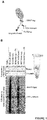
- Fig. 1B The results were as shown in Fig. 1B .
- Fig. IB a strange phenomenon was observed: when Drosophila Ago2 was fused with a TEV protease-cleavable FLAG tag and immunopurified using anti-FLAG magnetic beads (see Fig. 1A ), the Ago2 protein could not be eluted from the beads, even after the FLAG tag was removed using the TEV protease. From this, it was considered that purified, free Ago2 protein is unstable and tends to adhere nonspecifically to the beads. Strikingly, addition of the crude Drosophila S2 cell lysate during the TEV protease treatment promoted efficient elution of Ago2.
- the crude S2 cell lysate was boiled at 95°C for 15 minutes, and the precipitated proteins were removed by centrifugation.
- the supernatant of the heat-denatured lysate which contained only about 4.3% of the total protein concentration in the crude cell lysate, was as competent in eluting Ago2 as the original lysate. Digestion of the boiled supernatant with proteinase K (PK), followed by its inactivation by a second round of heating, halved its activity in Ago2 elution.
- PK proteinase K
- pCold-Hsc70-4, Hsp83, Hop, Droj2 and p23 are described in S. Iwasaki et al., Nature. 521, 533-536 (2015 ).
- pCAGEN-FlagTevSnap-Ago2 wild type was prepared in accordance with the description in the above (2)A.
- pCAGEN-Flag-GST was prepared by inserting a DNA fragment containing GST with an N-terminal FLAG tag into pCAGEN using NEBuilder HiFi DNA Assembly Master Mix (manufactured by NEB).
- RNA-induced silencing complex, RISC RNA-induced silencing complex, RISC
- RISC RNA-induced silencing complex
- FLAG-TEV-Halo-Ago2 expressed in S2 cells was immunopurified by an anti-FLAG antibody conjugated onto Dynabeads Protein G (manufactured by Invitrogen). The beads were washed three times with a wash buffer, three times with a wash buffer containing 2mM ATP, and finally rinsed with a lysis buffer.
- the Ago2-conjugated beads were then incubated with the reconstitution system, 32 P-radiolabeled small RNA duplex, ATP regeneration system (1 mM ATP, 25 mM creatine monophosphate (manufactured by Sigma), 0.03 U/ ⁇ L creatine kinase (manufactured by Calbiochem), and 0.1 U/ ⁇ L RNasin Plus RNase Inhibitor (manufactured by Promega)) and 3.5 ⁇ L of each boiled supernatant in 10 ⁇ L lysis buffer at 25°C for 60 minutes.
- ATP regeneration system (1 mM ATP, 25 mM creatine monophosphate (manufactured by Sigma), 0.03 U/ ⁇ L creatine kinase (manufactured by Calbiochem), and 0.1 U/ ⁇ L RNasin Plus RNase Inhibitor (manufactured by Promega)
- the reconstitution system contained 20 nM Dicer-2/R2D2, 600 nM Droj2, 1.8 ⁇ M Hsc70-4, 1.5 ⁇ M Hop, 3 ⁇ M Hsp83, and 3 ⁇ M p23.
- the total protein concentration was adjusted by the supplementation of glutathione S-transferase (referred to simply as "GST" herein). After incubation, beads were washed four times with a wash buffer, then bound RNAs were extracted by proteinase K treatment and analyzed by native PAGE.
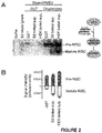
- the boiled supernatants from either Drosophila S2 or human HEK293T cells can promote the assembly of Ago2-RISC by Dicer-2/R2D2 and the chaperone machinery.
- LDH lactate dehydrogenase
- Example 2 Identification of soluble protein contained in boiled crude cell lysate
- LTQ-Orbitrap Velos mass spectrometer manufactured by Thermo Fisher Scientific
- Dina-2A nanoflow LC system manufactured by KYA Technologies
- nano liquid chromatography tandem mass spectrometry nano liquid chromatography tandem mass spectrometry (nano LC-MS/MS) analysis according to a routine method.
- the MS/MS signals were processed against the UniProt human proteome (Homo sapiens; UP000005640) and Drosophila proteome (Drosophila melanogaster; UP000000803) using the Mascot algorithm (version 2.5.1; Matrix Science).
- the proteins were identified based on a Mascot score beyond the threshold value (false discovery rate [FDR] ⁇ 0.01).
- FDR false discovery rate
- One (1) was added to all the peptide spectrum match (PSM) values (to avoid zeros), which correlate with the protein amount, and the ratio between (PSM + 1) in the original crude lysate and that in the boiled supernatants was calculated.
- the identified proteins were divided into five groups according to the degrees of depletion or enrichment by boiling (Group I [most depleted] to Group V [most enriched]), in such a way that each group contained virtually the same number of proteins.
- the respective groups contained virtually the same number of proteins according to the degrees of enrichment or depletion by boiling.
- IUPred Z. Dosztanyi, V. Csizmok, P. Tompa, I. Simon, J. Mol. Biol. 347, 827-839 (2005 )
- IDRs Intrinsically disordered Regions
- Amino acid sequences were collected from the UniProt human proteome (Homo Sapiens; UP000005640) and Drosophila proteome (Drosophila melanogaster; UP000000803). Also, the amino acid sequences of the proteins identified in the above (1) were used. These sequences were subjected to the IUPred method. The median of IUPred scores of all the amino acid residues was allocated for each protein, and their distributions were plotted as violin plots.
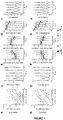
- the identified proteins were divided into five groups according to the degrees of depletion or enrichment by boiling (Group I [most depleted] to Group V [most enriched]), in such a way that each group contained virtually the same number of proteins ( Figs. 4A and 4B ).
- the proteins enriched by boiling showed much higher distributions of the median of IUPred scores than the proteins depleted by boiling in both Drosophila flies or humans ( Figs. 4C and 4D ). Moreover, hydrophilic amino acids were overrepresented, while hydrophobic amino acids were underrepresented, in the proteins enriched by boiling ( Figs. 4E and 4F ). Thus, structurally disordered, hydrophilic proteins were enriched in the boiled supernatant. Motifs associated with LEA proteins were not found among the proteins identified in the boiled supernatants. No common or conserved motifs were detected among the primary sequences of the identified proteins.
- the isoelectric points (pIs) of the proteins enriched by boiling exhibited a bimodal distribution, concentrating on the acidic (about pH 5) or basic (about pH 10) region and avoiding the neutral (about pH 7 to 8) region ( Fig 4I and 4J ). Thus, it was confirmed that heat-resistant proteins tend to be either highly negatively charged or highly positively charged under the physiological pH.
- the LC-MS/MS analysis has an intrinsic bias, and can only detect proteins expressed in HEK293T cells. Therefore, the IUPred score itself was utilized independently of the protein list identified by the LC-MS/MS analysis in Example 2, to select proteins from the entire human proteome for further functional analyses.
- the human protein atlas ( M. Uhlen et al., Science. 347, 1260419 (2015 )) was used as a population to select proteins having high IUPred scores across their entire sequences (median of IUPred scores of all residues of greater than 0.6, excluding locally structured proteins with more than 25% of residues having an IUPred score of smaller than 0.4) and high expression levels (median RPKM of greater than 25) from the human protein atlas ( M. Uhlen et al., Science. 347, 1260419 (2015 )).
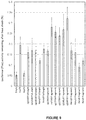
- Fig. 5A About 450 human proteins were identified (Fig. 5A ). These proteins also showed a bimodal isoelectric point (pI) distribution ( FIG 5B ). Among these proteins, six representative proteins, i.e., SERF2 (P84101), C9orf16 (Uniprot identifier; Q9BUW7), C19orf53 (Q9UNZ5), BEX3 (Q00994), C11orf58 [SMAP] (000193) and SERBP1 (Q8NC51) were chosen for further analysis ( Fig. 6A ). Four out of these proteins (C9orf16, C11orf58, SERBP1 and SERF2) were detected by the LC-MS/MS in Example 2.
- SERF2 P84101
- C9orf16 Uniprot identifier
- Q9UNZ5 C19orf53
- BEX3 Q00994
- C11orf58 [SMAP] 000193
- SERBP1 Q8NC51
- the intrinsically disordered structure was predicted using IUPred ( Fig. 6B ).
- the pI values for the six proteins were 4.13 (C9orf16), 4.57 (C11orf58), 5.31 (BEX3), 8.66 (SERBP1), 10.44 (SERF2), and 11.55 (C19orf53), respectively.
- pCAGEN-Flag-Hero9, -20, -13, -45, -7 or -11 was prepared as follows: a DNA fragment containing Hero9, -20, -13, -45, -7 or -11 (C9orf16, C11orf58, BEX3, SERBP1, SERF2 oro C19orf53) was amplified from HEK293T cell cDNAby PCR and cloned into pENTR/D-TOPO (manufactured by Invitrogen), followed by recombination with pCAGEN-Flag-DEST using Gateway LR Clonase (manufactured by Invitrogen).
- pCAGEN-Flag-GST and pCAGEN-Flag-GFP were prepared by inserting a DNA fragment containing GST or GFP with an N-terminal FLAG tag into pCAGEN by using NEBuilder HiFi DNA Assembly Master Mix (manufactured by NEB).
- the plasmids prepared in the above A were expressed as FLAG-tagged proteins in HEK293T cells, and the cell lysates were boiled at 95°C for 15 minutes. Then, the supernatants were analyzed by western blotting using an anti-DDDDK antibody (M185, manufactured by MBL) diluted at 1:10,000. Images were acquired by Amersham Imager 600 (manufactured by GE Healthcare).
- SERF2, C9orf16, C19orf53, BEX3, C11orf58, and SERBP1 as Hero7 (SEQ ID NO: 1), Hero9 (SEQ ID NO: 2), Hero11 (SEQ ID NO: 3), Hero13 (SEQ ID NO: 4), Hero20 (SEQ ID NO: 5), and Hero45 (SEQ ID NO: 6), respectively.
- pCold I-GFP GST, Hero9, Hero20, Hero13, Hero45, Hero7, or Hero11-FlagHis was prepared as follows: a DNA fragment containing CDS of each Hero protein and C-terminal FLAG- and His-tags was inserted into pCold I by NEBuilder HiFi DNA Assembly Master Mix (manufactured by NEB). For Hero13 and Hero45, a codon-optimized DNA fragment (manufactured by Eurofins) was synthesized and utilized as the template.
- Recombinant proteins of GST, Hero9, -20, -13, -45, -7, and -11 were expressed as C-terminal His-tagged proteins in the E. coli Rosetta 2 (DE3) strain.
- the cells were cultivated to an OD 600 of 0.4 to 0.6 at 37°C together with ampicillin and then allowed to grow at 15°C overnight together with 1 mM isopropyl- ⁇ -D-thiogalactoside (IPTG).
- the cell pellets were resuspended in a His A buffer (30 mM HEPES-KOH pH 7.4, 200 mM KOAc, 2 mM Mg(OAc) 2 , and 5% glycerol) containing 1 ⁇ EDTA-free Protease Inhibitor Cocktail (manufactured by Roche), sonicated, and centrifuged at 10,000g for 5 minutes twice. The supernatant was added to a slurry of cOmplete His-Tag Purification Resin (manufactured by Roche) and incubated for 1 hour at room temperature or for 2 hours at 4°C, and then eluted with a His B buffer (His A buffer containing 400 mM imidazole). The eluents were collected, and the buffer was exchanged for PBS with PD-10 (manufactured by GE Healthcare).
- His A buffer 30 mM HEPES-KOH pH 7.4, 200 mM KOAc, 2 mM Mg(
- LDH activity was measured according to the method described in Example 1, (4)A, except that 5 ⁇ g/mL purified lactate dehydrogenase (LDH) derived from rabbit muscle (manufactured by Roche) was diluted to 5 ⁇ g/mL with PBS containing 4 ⁇ g/mL BSA, GST or each Hero protein prepared in the above B, 400 ⁇ g/mL trehalose or 2 mg/mL arginine.
- LDH lactate dehydrogenase
- Protein purification was performed in accordance with the description in the above (1)B. Recombinant protein of EGFP was expressed and purified in accordance with the procedures described in S. Iwasaki et al., Nature. 521, 533-536 (2015 ).
- EGFP enhanced green fluorescent protein
- pCAGEN-Flag-Hero9, -20, -13, -45, -7 or -11 was prepared in accordance with the description in Example 4, (1)A.
- pCold-Hsc70-4 and Hsp83 are described in S. Iwasaki et al., Nature. 521, 533-536 (2015 ).
- pCAGEN-FLuc was prepared by inserting firefly luciferase into pCAGEN using NEBuilder HiFi DNA Assembly Master Mix (manufactured by NEB).
- pCAGEN-Flag-DEST is described in S. Iwasaki et al., Nature. 521, 533-536 (2015 ).
- Firefly luciferase was expressed together with each of the six Hero proteins, Hsp70, or Hsp90 in HEK293T cells, then the cells were subjected to heat shock.
- HEK293T cells in a 96-well plate were transfected with 10 ng pCAGEN-FLuc prepared in the above A and 90 ng empty pCAGEN or pCAGEN-Flag-Hero9, -20, -13, -45, -7, or -11 by using Lipofectamine 3000 (manufactured by Thermo Fisher Scientific).
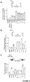
- the results were as shown in Fig. 8C .
- the luciferase activity was reduced to about 10% of the original level by heat shock.
- Overexpression of the conventional chaperones Hsp70 or Hsp90 maintained about 15 to 30% of the luciferase activity.
- the Hero proteins protected luciferase from heat shock as well as or better than the chaperones, with the Hero7 protein protecting about 50% of luciferase activity.
- a combination of the results of Examples 4 and 5 verified that the Hero proteins have the effect for stabilizing various proteins both in vitro and in cells.
- CG1943, CG12384 and CG14818 were cloned by the cDNAlibrary derived from fly S2 cells and inserted into a pCAGEN-F vector.
- pCAGEN-Flag-Hero9 or -20 was prepared in accordance with the description in Example 4, (1)A.
- the unnecessary sequence was eliminated by using inverse PCR.
- sequences of the respective proteins are: full-length protein (SEQ ID NO: 7), Fragment 1 (SEQ ID NO: 8) and Fragment 2 (SEQ ID NO: 9) of CG1943; full-length protein (SEQ ID NO: 10), Fragment 1 (SEQ ID NO: 11) and Fragment 2 (SEQ ID NO: 12) of CG12384; full-length protein (SEQ ID NO: 13), Fragment 1 (SEQ ID NO: 14) and Fragment 2 (SEQ ID NO: 15) of CG14818; Fragment 1 (SEQ ID NO: 16) and Fragment 2 (SEQ ID NO: 17) of Hero9; and Fragment 1 (SEQ ID NO: 18) and Fragment 2 (SEQ ID NO: 19) of Hero20.
- the results were as shown in Fig. 9 .
- the luciferase activity was reduced to about 10% of the original level by heat shock.
- Overexpression of the conventional heat shock protein Hsp70 or Hsp90 maintained about 15 to 30% of the luciferase activity. It was confirmed that the Hero protein fragments obtained by halving the Drosophila and human Hero proteins protected the luciferase activity from heat shock as well as or better than the respective full-length Hero proteins.
- the Hero proteins were confirmed to have the effect for stabilizing the protein even when fragmented.
- TDP-43 TAR DNA-binding protein of 43 kDa
- ALS amyotrophic lateral sclerosis
- FTD frontotemporal dementia
- pCold I-GstTev-TDP-43-HA was prepared as follows. A DNA fragment containing an N-terminal GST tag and a TEV protease recognition sequence was inserted into pCold I (manufactured by Takara), and, subsequently, a DNA fragment containing a C-terminal 2 ⁇ HA tag was inserted by NEBuilder HiFi DNA Assembly Master Mix (manufactured by NEB). Finally, a DNA fragment containing TDP-43 was inserted into pCold I-GstTev-HA by NEBuilder HiFi DNA Assembly Master Mix (manufactured by NEB) to prepare pCold I-GstTev-TDP-43-HA.
- pCold I-GST, Hero9, Hero20, Hero13, Hero45, Hero7 or Hero11-FlagHis was prepared in accordance with the description in Example 5, (2)A.
- Recombinant proteins of GST, Hero9, -20, -13, -45, -7, and -11 were purified in accordance with the description in Example 5, (2)B.
- Recombinant protein of GSTTEV-TDP-43-HA was expressed in the E. coli Rosetta 2 (DE3) strain.
- E. coli Rosetta 2 (DE3) strain.
- the cells in 1-L culture were cultivated to an OD 600 of 0.4 to 0.6 at 37°C together with ampicillin and then allowed to grow at 15°C overnight together with 1 mM isopropyl- ⁇ -D-thiogalactoside (IPTG).
- the cell pellets were resuspended in PBS containing 0.5 M NaCl, 0.1% Toriton, 10 mg/L RNase A (manufactured by Nacalai Tesque), and 1 ⁇ EDTA-free Protease Inhibitor Cocktail (manufactured by Roche) ( L. Guo et al., Cell. 173, 677-692 (2018 )), sonicated, and centrifuged at 10,000g for 5 minutes twice.
- the supernatant was added to a slurry of Glutathione Sepharose 4 Fast Flow (manufactured by GE Healthcare), incubated at 4°C for two hours, and then eluted with a GST elution buffer (100 mM Tris-HCl pH8.0, 0.20 mM glutathione, and 100 mM NaCl). The eluate was recovered, and the buffer was exchanged for PBS with PD-10 (manufactured by GE Healthcare).
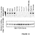
- the recombinant protein of GST-TEV-TDP-43-HA prepared in the above B was mixed with BSA (100 ⁇ g/mL), GST (100 ⁇ g/mL) and each Hero protein (100 ⁇ g/mL) prepared in the above B, arginine (2 mg/mL) or trehalose (500 ⁇ g/mL), and incubated at 37°C for 16 hours. Subsequently, PBS containing a 5-fold volume of 1% SDS was added, and loaded onto a cellulose acetate membrane with a 0.2- ⁇ m pore size (manufactured by GE Healthcare) that had been incubated in PBS containing 1% SDS. After washing with 1% SDS, aggregates were detected by using an anti-HA antibody (M180, manufactured by MBL) diluted at 1:10,000. Images were acquired by Amersham Imager 600 (manufactured by GE Healthcare).
- pCAGEN-GFP-TDP43 ⁇ NLS was prepared as follows: a DNA fragment containing TDP-43 was amplified from HEK293T cell cDNA by PCR and cloned into pENTR/D-TOPO (manufactured by Invitrogen), followed by recombination with pCAGEN-GFP-DEST using Gateway LR Clonase (manufactured by Invitrogen). NLS deletion (78 to 99) in TDP-43 was performed by PCR-based site-directed mutagenesis.
- pCAGEN-HTTQ103-GFP was prepared as follows: a DNA fragment containing HTT103Q-GFP was amplified from p426 103Q GPD (Addgene#1184; S. Krobitsch, S. Lindquist, Proc. Natl. Acad. Sci. U. S. A. 97, 1589-94 (2000 )), and inserted into pCAGEN using NEBuilder HiFi DNA Assembly Master Mix (manufactured by NEB).
- pCAGEN-GFP-GA50 was prepared as follows. A DNA fragment containing GA 50 was amplified from pAG303-Gal-GA 50 (Addgene#84907; A. Jovicic et al., Nat. Neurosci.
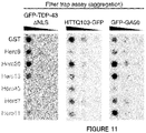
- Aggregation-prone proteins were expressed as GFP fusions, together with each of the six human Hero proteins or GST as a control, in HEK293T cells.
- HEK293T cells in a 12-well plate were transfected with 100 ng pCAGEN-GFP-HTTQ103, 100 ng pCAGEN-GFP-GA50 or 200 ng pCAGEN-GFP-TDP43 ⁇ NLS, and 900 ng (for HTTQ103 and GA50) or 800 ng (for TDP43 ⁇ NLS) pCAGEN-Flag-Hero9, - 20, -13, -45, -7 or -11 by using Lipofectamine 3000 (manufactured by Thermo Fisher Scientific).
- the cells were resuspended in 200 ⁇ L PBS containing 1 ⁇ cOmplete EDTA-free Protease Inhibitor Cocktail (manufactured by Roche) and sonicated by Bioruptor II (manufactured by BMBio). The total protein concentrations were adjusted and then 1% SDS was added. The samples were subjected to a filter trap assay in accordance with the description in the above (1)C, and aggregates were detected by using an anti-GFP antibody (sc-9996, manufactured by Santa Cruz) diluted at 1:10,000. Images were acquired by Amersham Imager 600 (manufactured by GE Healthcare).
- HTTQ103 consists of a stretch of 103 polyglutamine residues derived from an abnormal CAG expansion found in a Huntington disease-causing huntingtin mutant ( S. Krobitsch, S. Lindquist, Proc. Natl. Acad. Sci. U. S. A. 97, 1589-94 (2000 )).
- GA50 50 Glycine-Alanine repeats
- GGGGCC repeats found in the ALS-causing C9orf72 intron ( K. Mori et al., Science. 339, 1335-8 (2013 )).
- TDP-43 lacking the nuclear localization signal (TDP-43 ⁇ NLS) is more prone to aggregation than the wild type because of its forced cytoplasmic localization ( Nonaka T et al., Hum Mol Genet., 18(18): 3353-64(2009 )).
- a plasmid was constructed in accordance with the description in the above (2)A.
- HEK293T cells were transfected in accordance with the description in the above (2)B.
- Cell images were acquired with an inverted microscope (IX83, manufactured by Olympus) with a 20 ⁇ objective lens (UCPLFLN 20 ⁇ , 0.7 NA, manufactured by Olympus).
- pCAGEN-Flag-Shuffled100 (GST, Hero7, Hero11 or Hero45) was prepared by inserting a DNA fragment containing shuffled 100-amino acid residue sequences with the amino acid residue composition ratio of GST, Hero7, Hero11 or Hero45 into a pCAGEN-Flag-DEST vector.
- pCAGEN-superFlag-Shuffled42 (GST, Hero7, Hero11 or Hero45) was prepared by inserting a DNA fragment containing N-terminal superFlag (Layton et al., 2019) and shuffled 42-amino acid residue sequences with the amino acid residue composition ratio of GST, Hero7, Hero11 or Hero45 into a pCAGEN-superFlag-DEST vector.
- the amino acid sequences of the shuffled Hero proteins are: 100-amino acid residue sequences (SEQ ID NOs: 20 to 22) and 42-amino acid residue sequences (SEQ ID NOs: 23 and 24) of Hero7; 100-amino acid residue sequences (SEQ ID NOs: 25 to 27) and 42-amino acid residue sequences (SEQ ID NOs: 28 and 29) of Hero11; 100-amino acid residue sequences (SEQ ID NOs: 30 to 32) and 42-amino acid residue sequences (SEQ ID NOs: 33 and 34) of Hero45; and original sequence (SEQ ID NO: 35), 100-amino acid residue sequences (SEQ ID NOs: 36 to 38) and 42-amino acid residue sequences (SEQ ID NOs: 39 and 40) of GST.
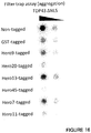
- the filter trap assay was conducted using pCAGEN-GFP-TDP43 ⁇ NLS in accordance with the description in the above (2)B.
- the amino acid sequences of Hero7, -11 and -45, and GST as a control were randomly shuffled, while maintaining the original amino acid residue composition ratios of the Hero proteins (i.e., proportions of the respective amino acid residues relative to the total number of amino acid sequences) and fixing the total number of amino acids at 100 or 42 amino acids.
- Three different shuffle sequences were prepared for 100 amino acid residues, and two different shuffle sequences were prepared for 42 amino acid residues.
- the 100-amino acid residue sequences obtained by shuffling the Hero proteins inhibited the aggregation of TDP-43 ⁇ NLS with comparable or even higher efficiencies than the original Hero proteins. Also, the 42-amino acid residue sequences obtained by shuffling the Hero proteins were confirmed not to inhibit the aggregation of TDP-43 ⁇ NLS.
- Drosophila eyes have been used as a model system useful for studying neurodegenerative diseases. Drosophila melanogaster eyes were used to evaluate the aggregation preventing effect of Hero proteins in vivo.
- pUASg-HA.attB-Hero9, -13, -45 or -11 was prepared as follows: a DNA fragment containing Hero9, -13, -45, or -11 in pENTR was inserted into pUASg-HA-attB (DGRC#1423) using Gateway LR Clonase (manufactured by Invitrogen). Drosophila culture and crosses were carried out at 25°C (the same applies hereinafter).
- Drosophila stocks were obtained from the Bloomington and Kyoto Drosophila Stock Center (the same applies hereinafter).
- Drosophila flies carrying pUASg.attB-Hero9, -13, -45 and -11 transgenes were prepared by standard phiC31 integrase-mediated transgenesis (manufactured by Bestgene Inc.).
- GMR-Gal4 was used to express the transgenes in the retina.
- UAS-MJDtr-Q78 J. M. Warrick et al., Cell. 93, 939-949 (1998 )
- UAS-TDP-43-YFP and UAS-YFP lines were provided by Dr. Nancy Bonini (the same applies hereinafter).
- Fig. 14 The results were as shown in Fig. 14 . From the results shown in Fig. 14A , when transgenic Drosophila flies expressing Hero9, -13, -45 or -11 in the retina were prepared, and the transgenes were introduced into strains expressing MJDtr-Q78 and TDP-43-YFP, neither YFP nor any of the transgenic Hero proteins alone caused a detectable change in the normal eye morphology. From the results shown in Fig. 14B (upper row), overexpression of human Ataxin 3 containing a pathogenic expansion of 78 glutamine repetitive sequences (MJDtr-Q78) in the differentiating photoreceptor neurons of the Drosophila retina caused strong retinal degeneration ( J. M.
- RNAi lines were obtained from the Vienna Drosophila Resource Center ( Barinova et al., Nature 448:151-156 (2007 )).
- the GMR-Gal4 line was used to express transgenes and/or induce RNAi by long hairpins in the retina.
- the RNAi lines used were UAS-KK(Piwi), KK(Hsc70-4), KK(CG17931), KK(CG14818), KK(Vig2), KK(CG12384) and KK(CG11444).
- a P2A peptide sequence and an mRuby3 sequence were added to the C-terminal of pCAGEN-GFP-TDP43De1NLS. Then, the GST, Hero9, -13, -20, -45, -9 or -11 sequence was added to the immediate upstream of the P2A sequence.
- Example 6 The same procedures as described in Example 6, (2)B were performed except that the plasmid prepared in the above A was used. Also, GST has an anti-aggregation effect, and solubilization of proteins fused with GST is promoted (https://www.ncbi.nlm.nih.gov/pubmed/16781139). GST-tagged TDP43 ⁇ NLS was used as a positive control.
Landscapes
- Health & Medical Sciences (AREA)
- Life Sciences & Earth Sciences (AREA)
- Chemical & Material Sciences (AREA)
- Engineering & Computer Science (AREA)
- Molecular Biology (AREA)
- General Health & Medical Sciences (AREA)
- Medicinal Chemistry (AREA)
- Organic Chemistry (AREA)
- Biochemistry (AREA)
- Physics & Mathematics (AREA)
- Biophysics (AREA)
- Proteomics, Peptides & Aminoacids (AREA)
- Biomedical Technology (AREA)
- Zoology (AREA)
- Bioinformatics & Cheminformatics (AREA)
- Urology & Nephrology (AREA)
- Immunology (AREA)
- Genetics & Genomics (AREA)
- Analytical Chemistry (AREA)
- Hematology (AREA)
- Biotechnology (AREA)
- General Physics & Mathematics (AREA)
- Pathology (AREA)
- Food Science & Technology (AREA)
- Microbiology (AREA)
- Insects & Arthropods (AREA)
- Gastroenterology & Hepatology (AREA)
- Toxicology (AREA)
- Bioinformatics & Computational Biology (AREA)
- Pharmacology & Pharmacy (AREA)
- Cell Biology (AREA)
- Wood Science & Technology (AREA)
- Medical Informatics (AREA)
- Spectroscopy & Molecular Physics (AREA)
- Theoretical Computer Science (AREA)
- Evolutionary Biology (AREA)
- General Engineering & Computer Science (AREA)
- Tropical Medicine & Parasitology (AREA)
- Veterinary Medicine (AREA)
- Neurology (AREA)
Abstract
Description
- The present application enjoys the benefit of priority from the prior
U.S. Patent Application No. 62/734,329 filed on September 21, 2018 - The present invention relates to a screening method for a client protein-protecting protein. The present invention also relates to a physiologically active protein-stabilizing protein and a pharmaceutical composition comprising the protein.
- Proteins exert their biological function by being folded into their intrinsic native-state three-dimensional structure. Due to various physical or chemical factors, the structures such as the secondary, tertiary and quaternary structures can be changed without change in primary structure of proteins, leading to changes in physical properties of the proteins. Such a phenomenon is referred to as protein denaturation. Changes associated with denaturation include decrease in protein solubility, loss or reduction in biological activity, and loss of crystallization tendency.
- It is generally considered that proteins can be denatured, but examples of highly hydrophilic and highly heat-resistant proteins found in extremophiles are known as exceptions. For example, tardigrade disordered proteins (TDPs), which are required for tardigrade, bear animalcule, to withstand desiccation, are known. Also, late embryogenic abundant (LEA) proteins and associated proteins have been identified in land plants, radiation-resistant bacteria, and desiccation-tolerant animals such as Artemia (brine shrimps) and nematodes, and expression of these proteins is known to be associated with their tolerance to extreme environments including dehydration, freezing and high salinity. In contrast, heat-soluble proteins in mesophiles including mammals have been reported (
Non-Patent Documents 1 and 2), but their functions have hardly been revealed. -
- Non-Patent Document 1: T. D. Kim, H. J. Ryu, H. Il Cho, C. H. Yang, J. Kim, Biochemistry. 39, 14839-14846 (2000)
- Non-Patent Document 2: C. A. Galea et al., J. Proteome Res. 8, 211-226 (2009)
- An object of the present invention is to provide a novel screening method for a client protein-protecting protein. Another object of the present invention is to provide a physiologically active protein-stabilizing protein and a pharmaceutical composition using the protein.
- The present inventors have now found that supernatants and boiled supernatants of cell lysates suppress nonspecific adhesion of proteins, promote expression of functions of the proteins, and protect the activity of the proteins from desiccation. The present inventors have also found that proteins having an intrinsically disordered structure are present abundantly in boiled supernatants of cell lysates by an intrinsically disordered region prediction method, and that the amino acid compositions of these proteins have tendencies different from those of conventionally-known typical intrinsically disordered regions. Also, the present inventors have selected proteins having an intrinsically disordered structure from the human proteome using an intrinsically disordered region prediction method, found that these proteins have high heat resistance characteristics, and named these proteins Hero (HEat-Resistant Obscure) proteins. The present inventors have also found that the Hero proteins protect client proteins from denaturation due to various stress conditions and suppress aggregation of aggregate proteins in vitro and in vivo. Further, the present inventors have found that the physiological activity of the Hero proteins are maintained even when the proteins are fragmented and even when their amino acid sequences are shuffled while the composition ratio of amino acid residues is maintained. The present invention is based on these findings.
- The present invention provides the following inventions.
- [1] A method for screening for a client protein-protecting protein, comprising the step (evaluation step) of evaluating stability of a client protein in the presence of a protein (candidate protein) having an intrinsically disordered structure.
- [2] The method according to [1], wherein, in the evaluation step, the stability of the client protein is evaluated using a degree of protection of the client protein from stress as an index.
- [3] The method according to [2], wherein the stress is one or more selected from the group consisting of physical stresses and chemical stresses.
- [4] The method according to any one of [1] to [3], wherein the client protein is a physiologically active protein.
- [5] The method according to [4], wherein the physiologically active protein is selected from the group consisting of antibodies, antibody fragments, enzymes, hormones, cytokines and interferons.
- [6] The method according to [4] or [5], which is a method for identifying a protein useful for stabilization of the physiologically active protein.
- [7] The method according to any one of [4] to [6], wherein the physiologically active protein is an active ingredient of a medicine.
- [8] The method according to any one of [4] to [7], wherein the stability of the physiologically active protein is evaluated in accordance with conditions for a stability test required for approval for manufacture of the medicine.
- [9] The method according to any one of [1] to [8], wherein the client protein is a non-aggregate protein.
- [10] The method according to [1], wherein, in the evaluation step, the stability of the client protein is evaluated using a degree of aggregation of the protein as an index.
- [11] The method according to [10], wherein the aggregation of the protein is measured by one or more selected from the group consisting of a filter trap assay or protein localization in a cell, pulse shape analysis (PulSA), phenotype analysis, detection using a protein aggregation detecting dye (thioflavin T (ThT)), and detection using a protein aggregation detecting reagent (ProteoStat).
- [12] The method according to any one of [1], [10] and [11], wherein the client protein is an aggregate protein.
- [13] The method according to [12], wherein the aggregate protein is a protein causing development and/or progression of a disease.
- [14] The method according to [13], which is a method for screening for a protein effective in treatment, prevention or improvement of the disease.
- [15] The method according to [14], wherein the disease is a disease which develops and/or progresses upon abnormal aggregation of the protein.
- [16] The method according to [15], wherein the disease which develops and/or progresses upon abnormal aggregation of the protein is a neurodegenerative disease.
- [17] The method according to any one of [1] to [16], wherein the candidate protein is of mammalian origin.
- [18] The method according to any one of [1] to [17], wherein the candidate protein is a protein in which a median of IUPred scores of all amino acid residues is greater than 0.5.
- [19] The method according to any one of [1] to [18], wherein the candidate protein is a protein in which more than 50% of amino acid residues relative to the entire sequence have an IUPred score of greater than 0.3.
- [20] The method according to any one of [1] to [19], wherein the candidate protein is a protein which remains heat-soluble after a heat treatment.
- [21] The method according to any one of [1] to [20], which further comprises the step (selection step) of selecting one or more candidate protein(s) from a protein population using the intrinsically disordered structure of the protein as an index.
- [22] The method according to [21], wherein the protein population is of mammalian origin.
- [23] The method according to [21] or [22], wherein, in the selection step, the intrinsically disordered structure of the protein is evaluated by an intrinsically disordered region prediction method.
- [24] The method according to [23], wherein the intrinsically disordered region prediction method is carried out using a program selected from the group consisting of IUPred, D2P2, GlobPlot, GLOBPLOT2, FoldIndex, IsUnstruct, PONDR VL-XT, DisEMBL, PONDR VL3, PONDR VL3H, RONN, PONDR VSL2B, PONDR VSL2P, Spritz and SLIDER.
- [25] The method according to any one of [21] to [24], wherein, in the selection step, the intrinsically disordered structure of the protein is evaluated by IUPred.
- [26] The method according to [25], wherein a protein of interest in which a median of IUPred scores of all amino acid residues is greater than 0.5 in IUPred is selected as the candidate protein.
- [27] The method according to [21] or [22], wherein, in the selection step, the intrinsically disordered structure of the protein is evaluated by analysis based on heat solubility.
- [28] The method according to [27], wherein a protein which remains heat-soluble after a heat treatment is selected as the candidate protein.
- [29] An agent for stabilizing a physiologically active protein comprising a protein selected from the group consisting of the following (a), (b), (c), (d) and (e):
- (a) a protein having any one of amino acid sequences of SEQ ID NOs: 1 to 6;
- (b) a protein having an amino acid sequence obtained by shuffling any one of amino acid sequences of SEQ ID NOs: 1 to 6;
- (c) a protein having a fragment sequence of any one of amino acid sequences of SEQ ID NOs: 1 to 6;
- (d) a protein which satisfies an amino acid residue composition ratio (the amino acid residue composition ratio including a range of ± 5%) indicated in Table 1, 2, 3, 4, 5 or 6, and has an amino acid sequence having a length of 43 amino acid residues or more; and
- (e) a protein substantially identical with the protein (a), (b), (c) or (d).
- [30] A pharmaceutical composition comprising: a protein selected from the group consisting of the proteins (a), (b), (c), (d) and (e) as defined in [29]; and a physiologically active protein.
- [31] The agent according to [29] and the pharmaceutical composition according to [30], wherein the physiologically active protein is an active ingredient of a medicine.
- [32] The agent according to [29] or [31] and the pharmaceutical composition according to [30] or [31], wherein the physiologically active protein is selected from the group consisting of antibodies, antibody fragments, enzymes, hormones, cytokines and interferons.
- [33] The agent according to [29], [31] or [32] and the pharmaceutical composition according to any one of [30] to [32], wherein the fragment sequence of the protein (c) has at least 43 amino acid residues.
- [34] The agent according to any one of [29] and [31] to [33] and the pharmaceutical composition according to any one of [30] to [33], wherein the protein (c) has an activity of stabilizing the physiologically active protein.
- [35] The agent according to [29], [31] or [32] and the pharmaceutical composition according to any one of [30] to [32], wherein the substantially identical protein (e) is a protein selected from the group consisting of the following (i), (ii), (iii), (iv), and (v):
- (i) a protein which comprises an amino acid sequence having 80% or more identity with the amino acid sequence as defined in any one of (a), (b), (c) and (d) of [29], and which has an activity of stabilizing the physiologically active protein;
- (ii) a protein which comprises an amino acid sequence in which one or more amino acid(s) is/are deleted, substituted, inserted and/or added in the amino acid sequence as defined in any one of (a), (b), (c) and (d) of [29], and which has an activity of stabilizing the physiologically active protein;
- (iii) a protein which comprises an amino acid sequence encoded by a base sequence having 80% or more identity with a base sequence encoding the amino acid sequence as defined in any one of (a), (b), (c) and (d) of [29], and which has an activity of stabilizing the physiologically active protein;
- (iv) a protein comprising an amino acid sequence encoded by a base sequence in which one or more base(s) is/are deleted, substituted, inserted and/or added in a base sequence encoding the amino acid sequence as defined in any one of (a), (b), (c) and (d) of [29], and which has an activity of stabilizing the physiologically active protein; and
- (v) a protein which comprises an amino acid sequence encoded by a polynucleotide that hybridizes, under stringent conditions, with a polynucleotide consisting of a sequence complementary to a base sequence encoding the amino acid sequence as defined in any one of (a), (b), (c) and (d) of [29], and which has an activity of stabilizing the physiologically active protein.
- [36] The pharmaceutical composition according to any one of [30] to [35], wherein the physiologically active protein is conjugated to the protein selected from the group consisting of the proteins (a), (b), (c), (d) and (e).
- The method of the present invention is advantageous in enabling selection of a client protein-protecting protein, especially, a protein useful for stabilization of a physiologically active protein or a protein effective in treatment, prevention or improvement of a disease which develops and/or progresses upon abnormal aggregation of the protein.
-
-
Fig. 1 shows that heat-soluble proteins improve the molecular behavior of Drosophila Argonaute 2 (Ago2) protein in vitro. (A) A schematic diagram of FLAG-TEV-SNAP-Ago2 immunopurified on magnetic beads. (B) FLAG-TEV-SNAP-Ago2 was immunopurified on magnetic beads via an anti-FLAG antibody, and then the FLAG tag was cleaved off by TEV protease in a buffer, a crude lysate from Drosophila S2 cells or human HEK293T cells, or its boiled supernatant. +PK indicates that the boiled supernatants were mostly deproteinized by proteinase K in advance. The eluted Ago2 (top panel) and the Ago2 remaining on the beads (middle panel) were visualized with a red fluorescent dye covalently bonded to the SNAP tag. All the proteins contained in the eluate were visualized by Coomassie Brilliant Blue (CBB) staining (bottom panel). The numerical values above the panels indicate relative amounts of the eluted Ago2 normalized to that with the crude S2 cell lysate. The proteins remaining in the boiled supernatants have an effect for promoting Ago2 elution. -
Fig. 2 shows that heat-soluble proteins improve the molecular behavior of Drosophila Argonaute 2 (Ago2) protein in vitro. (A) A small RNA pull-down assay for pre-Ago2-RISC and mature Ago2-RISC assembled in the reconstitution system, containing a 32P-radiolabeled small RNA duplex, Ago2, Dicer-2/R2D2, and Hsp70/Hsp90 chaperone machinery. Addition of the boiled supernatant from S2 cells or HEK293T cells promoted formation of both pre-Ago2-RISC and mature Ago2-RISC. (B) shows the quantified results of the assay (A). Data represent means ± SD from three independent experiments. -
Fig. 3 shows that heat-soluble proteins improve the activity of lactate dehydrogenase (LDH) when condensed after desiccation of Drosophila Argonaute 2 (Ago2) protein in vitro. The heat-soluble proteins in the boiled supernatants protect the lactate dehydrogenase (LDH) activity from desiccation. LDH mixed with the boiled supernatants or buffer was dried up overnight and the remaining LDH activities were measured. Data are normalized to the activity of LDH incubated on ice overnight and represent means ± SD from three independent experiments. -
Fig. 4 shows that hydrophilic and highly charged proteins are enriched in boiled supernatants. (A and B) show, for the proteins identified, by MS analysis, in Drosophila- and human-derived cell lysates, distributions of a ratio between the peptide spectrum match (PSM) + 1 in the original crude lysate and that in the boiled supernatants. The identified proteins were divided into five groups according to the degrees of depletion by boiling or enrichment by boiling (Groups I to V), in such a way that each group contained virtually the same number of proteins. (C and D) Distributions of medians of IUPred scores of the proteins belonging to Groups I to V. The width of each group indicates a proportion (or a relative value) of the protein. (E and F) Proportions of hydrophilic amino acids or hydrophobic amino acids of the proteins belonging to Groups I to V. (G and H) Distributions of DIOPT (DRSC Integrative Ortholog Prediction Tool) scores of the proteins belonging to Groups I to V. (I and J) Distributions of isoelectric points of the proteins of Groups I to V. The width of each group indicates a proportion (or a relative value) of the protein. -
Fig. 5 shows (A) distributions of the medians of IUPred scores and (B) distributions of the isoelectric points, in the human protein atlas population and the proteins with a high IUPred score selected from the population. The width of each group indicates a proportion (or a relative value) of the protein. -
Fig. 6 shows amino acid sequences of six representative proteins and prediction of disordered regions. (A) shows the amino acid sequences of the six representative proteins. (B) shows prediction of disordered regions by IUPred using the default settings (Z. Dosztanyi, V. Csizmok, P. Tompa, I. Simon, J. Mol. Biol. 347, 827-839 (2005)). As controls, green fluorescent protein (GFP) and glutathione S-transferase (GST) were used. -
Fig. 7 shows that Hero proteins protect the activities of various proteins under stress conditions. Heat solubility of the selected six Hero proteins. Each Hero protein, GFP or GST was fused with a FLAG tag and expressed in HEK293T cells. The cell lysates were prepared with (+) or without (-) boiling, and then the proteins in the lysates or their supernatants were detected by the western blotting method using an anti-FLAG antibody. The boiled supernatants were each loaded in an amount twice the amount of the non-boiled lysate. -
Fig. 8 shows that Hero proteins protect the activities of various proteins under stress conditions. (A) Hero proteins protect the LDH activity from desiccation. LDH mixed with each Hero protein or control was dried up overnight and the remaining LDH activities were measured. Note that much higher concentrations of arginine (500-fold) and trehalose (100-fold) were used, as compared to those of the proteins. Data are normalized to the activity of LDH incubated on ice overnight and represent means ± SD from three independent experiments. (B) Hero proteins protect GFP in an organic solvent. GFP mixed with each Hero protein or control was exposure to chloroform and the remaining GFP fluorescence was measured. Data were normalized to GFP without exposure to chloroform, and represent means ± SD from three independent experiments. (C) Hero proteins protect the luciferase activity from heat shock. Firefly luciferase was transfected into HEK293T cells, together with each Hero protein or chaperone factor, and then the cells were exposed to heat shock. The remaining luciferase activities after heat shock were measured. Data were normalized to the activity of luciferase in cells without exposure to heat shock, and represent means ± SD from five independent experiments. -
Fig. 9 shows that fragmented Hero proteins protect the luciferase activity from heat shock. -
Fig. 10 shows that Hero proteins protect aggregation of pathogenic proteins in vitro. A filter trap assay of TDP-43 aggregation formed in vitro. Recombinant GST-TDP-43-HA was incubated in the presence of each Hero protein or control. The original samples or 5-fold diluted samples were each loaded on a cellulose acetate membrane in the presence of 1% SDS, and the trapped aggregates were probed with an anti-HA antibody. The total amount of GST-TDP-43-HA remained unchanged, as detected by western blotting using an anti-HA antibody. -
Fig. 11 shows that Hero proteins prevent aggregation of pathogenic proteins in cells. A filter trap assay of aggregation-prone proteins expressed in cells. TDP-43ΔNLS, HTTQ103 or GA50 was expressed in the form of a GFP fusion, together with each Hero protein or the GST control, in HEK293T cells. A stock solution, 5-fold diluent and 25-fold diluent of the cell lysate were subjected to the filter trap assay and probed with an anti-GFP antibody. -
Fig. 12 shows that Hero proteins prevent aggregation of pathogenic proteins in cells. Representative microscopic images of GFP signals in the cells transfected with GFP-tagged TDP-43ΔNLS, HTTQ103 or GA50, together with GST (control) or each Hero protein. -
Fig. 13 shows that shuffled Hero proteins prevent pathogenic protein aggregation in cells. -
Fig. 14 shows that Hero proteins suppress the neurotoxicity of protein aggregates in Drosophila eyes. (A) Expression of Hero9, -13, -45, or -11 alone showed no phenotype in the external eye morphology, much as the control (YFP). The genotype of Oregon R was +/+;+/+, and that of the control (YFP), Hero9, -13, -45 or - 11 was GMR-GAL4;UAS-YFP, UAS-Hero9, -13, -45, or -11/SM6a-TM6B. (B) Expression of MJDtr-Q78 or TDP-43-YFP caused the external eye degeneration. Expression of Hero9 and Hero13 somewhat alleviated the eye degeneration by MJDtr-Q78 (upper row). The genotype of - was GMR-GAL4/+;UAS-MJDtr-Q78/TM3. The genotype of the control (YFP) was GMR-GAL4/+;UAS-MJDtr-Q78/UQS-YFP. The genotype of Hero9, -13, -45 or -11 was GMR-GAL4/+;UAS-MJDtr-Q78/UAS-Hero9, -13, -45 or -11. Expression of Hero9 and double copies of Hero45 strongly suppressed the eye degeneration by TDP-43-YFP (lower row). The genotype of - was GMR-GAL4, UAS-TDP-43-YFP/+;TM3/+. The genotype of the control (YFP) was GMR-GAL4, UAS-TDP-43-YFP/+;UAS-YFP/+. The genotype of Hero9, -13, -45 or - 11 was GMR-GAL4, UAS-TDP-43-YFP/+;UAS-Hero9, -13, -45 or -11/+. The genotype of Hero45 (×2) was GMR-GAL4, UAS-TDP-43-YFP/+;UAS-Hero45/UAS-Hero45. Eyes were imaged in 1-day-old adult flies. -
Fig. 15 shows that Hero proteins suppress the neurotoxicity of protein aggregates in Drosophila eyes. (Top row) The genotype of - was GMR-GAL4/+. The genotype of the control (Piwi) was GMR-GAL4/UAS-KK(Piwi). The genotype of Hsc70-4, CG17931, CG14818 or Vig2 was GMR-GAL4/UAS-KK(Hsc70-4), KK(CG17931), KK(CG14818) or KK(Vig2). The genotype of CG12384 or CG11444 was GMR-GAL4/UAS-KK(CG12384) or KK(CG11444). Knockdown of Hsc70-4 or CG17931 (Drosophila homolog of human Hero7) showed an eye degeneration phenotype. (Middle row) The genotype of - was GMR-GAL4/+;UAS-MJDtr-Q78/+. The genotype of the control (Piwi) was GMR-GAL4/UAS-KK(Piwi);UAS-MJDtr-Q78/+. The genotype of Hsc70-4, CG17931, CG14818 or Vig2 was GMR-GAL4/UAS-KK(Hsc70-4), KK(CG17931), KK(CG14818) or KK(Vig2);UAS-MJDtr-Q78/+. The genotype of CG12384 or CG11444 was GMR-GAL4/UAS-KK(CG12384) or KK(CG11444);UAS-MJDtr-Q78/+. The eye phenotype by overexpression of MJDtr-Q78 was exacerbated by depletion of CG17931, CG14818, Vig2, CG12384 and CG11444 through RNAi. (Bottom row) The genotype of - was GMR-GAL4;UAS-TDP43-YFP/+. The genotype of the control (Piwi) was GMR-GAL4;UAS-TDP43-YFP/UAS-KK(Piwi). The genotype of Hsc70-4, CG17931, CG14818 or Vig2was GMR-GAL4;UAS-TDP43-YFP/UAS-KK(Hsc70-4), KK(CG17931), KK(CG14818)or KK(Vig2). The genotype of CG12384 or CG11444 was GMR-GAL4;UAS-TDP43-YFP/UAS-KK(CG12384) or KK(CG11444). Eyes were imaged in 1-day-old female adult flies. The eye phenotype by overexpression of TDP-43-YFP was exacerbated by depletion of CG17931, CG14818, Vig2, CG12384 and CG11444 through RNAi. -
Fig. 16 shows the aggregation suppressive effect of the Hero proteins when a fusion of TDP43ΔNLS and each Hero protein was expressed. - In the present invention, the "client protein" refers to a protein to be protected from stress conditions. Examples of the client protein include physiologically active proteins. In the present invention, the "physiologically active protein" is not particularly limited, and examples thereof include antibodies, antibody fragments, enzymes, hormones, cytokines and interferons. In the present invention, the physiologically active protein can be an active ingredient of a medicine or a candidate therefor.
- Examples of the client protein include aggregate proteins. In the present invention, the "aggregate protein" can be selected from proteins causing development and/or progression of a disease.
- In the present invention, the "candidate protein" can be a protein of mammalian (preferably, human) origin. In the present invention, the "protein population" can be a protein population of mammalian (preferably, human) origin. As used herein, the meaning of the "protein" includes peptides and polypeptides.
- In the present invention, the "intrinsically disordered structure" refers to a structure having no fixed three-dimensional structure in the native state, and is synonymous with "intrinsically irregular structure." Here, the "intrinsically disordered" is synonymous with "intrinsically unstructured". Further, proteins having an "intrinsically disordered structure" are referred to as intrinsically disordered proteins.
- In the present invention, the "polynucleotide" includes DNA and RNA, and further includes modified products thereof and artificial nucleic acids, but is preferably DNA. DNA also includes cDNA, genomic DNA and chemically synthesized DNA.
- In the present invention, the "screening method (method for screening)" means a method of selecting a substance having a predetermined property based on evaluation results of candidate substances, and the candidate substance may be one or more substances. The "screening method" of the present invention includes an "identification method" for identifying the candidate substance as a substance having a predetermined property. The "screening method" of the present invention can be an in vitro screening method.
- The screening method of the present invention is characterized by comprising the step of evaluating stability of a client protein in the presence of a protein having an intrinsically disordered structure as a candidate protein (sometimes referred to as "evaluation step" or "step (B)" herein).
- The candidate protein to be evaluated in step (B) is not particularly limited as long as it is a protein having an intrinsically disordered structure, and a protein predicted to have an intrinsically disordered structure by an intrinsically unstructured/disordered region/domain prediction method can be used. For example, a protein predicted to have an intrinsically disordered structure by IUPred (Z. Dosztanyi, V. Csizmok, P. Tompa, I. Simon, J. Mol. Biol. 347, 827-839 (2005)) can be subjected, as the candidate protein, to step (B). Preferably, a protein in which a median of IUPred scores of all amino acid residues is greater than 0.5 can be used as the candidate protein. Alternatively, a protein in which more than 50% (preferably, more than 60%, more than 70%, more than 75%, or more than 80%) of amino acid residues relative to the entire sequence have an IUPred score of greater than 0.3 (preferably, greater than 0.4, greater than 0.5, or greater than 0.6) can be subjected, as the candidate protein, to step (B).
- Also, in the screening method of the present invention, a protein predicted to have an intrinsically disordered structure by analysis based on heat solubility can be subjected, as the candidate protein, to step (B). Preferably, a protein which remains heat-soluble after a heat treatment can be used as the candidate protein.
- Step (B) may be carried out in combination with the evaluation of the stability of the client protein in the absence of the candidate protein or the evaluation of the stability of the client protein in the presence of a control protein. The control protein can be selected from proteins which are not proteins that protect the client protein, and can be, for example, bovine serum albumin (BSA), glutathione S-transferase (GST) and other proteins, which are widely used as control proteins in the field of biochemistry.
- In step (B), for example, when the degree of stability of the client protein in the presence of the candidate protein is above the degree of stability in the absence of the candidate protein or the degree of stability in the presence of the control protein (preferably, when the former degree is above the latter degree with a significant difference), it can be assessed that the candidate protein contributes to the stabilization of the client protein. Specifically, the screening method of the present invention may further comprise, after step (B), the step of (C) determining that the candidate protein is a client-protein protecting protein, when the degree of stability of the client protein in the presence of the candidate protein is above the degree of stability in the absence of the candidate protein or the degree of stability in the presence of the control protein (preferably, when the former degree is above the latter degree with a significant difference).
- Step (B) is not limited in its embodiment as long as the effect of the candidate protein on the stability of the client protein can be evaluated. For example, a recombinant protein of the candidate protein may be prepared according to a known method based on amino acid sequence information to carry out the evaluation step, or the candidate protein may be forcibly expressed or knocked down or out in cells or individuals to carry out the evaluation step. Alternatively, the effect of the candidate protein can be verified, for example, by inhibiting the function of the candidate protein with an antibody or the like. In addition, a lysate may be prepared from a sample in which the candidate protein is forcibly expressed or knocked down in cells or individuals based on base sequence information to carry out step (B) using the crude lysate itself without purification of the candidate protein as a recombinant protein. In step (B) of evaluating the stability, any of these methods may be used, or a combination of these methods may be used.
- In the screening method of the present invention, in the evaluation step, the stability of the client protein can be evaluated using a degree of protection of the client protein from stress as an index. The protection of the client protein from stress can be protection of the client protein from denaturation under stress conditions.
- In the evaluation step, when the stability of the client protein is evaluated using the degree of protection of the client protein from stress as an index, a physiologically active protein or a non-aggregate protein can be selected as the client protein. In this case, the screening method of the present invention can be a method for screening for a protein useful for stabilization of the physiologically active protein.
- In the present invention, the "stress" is stress on a protein, specifically stress to denature a protein, and examples of such stress include physical stresses and chemical stresses. Examples of physical stresses include heating, storage at room temperature, freezing, thawing, low temperature, heat shock, desiccation, pressure, adhesion, stirring, ultrasonic waves, dilution, enrichment, daily light rays, ultraviolet rays, and X-rays. Examples of chemical stresses include treatments with denaturing agents such as acids, alkalies, urea, guanidine hydrochloride, urea, organic solvents and surfactants, and treatments with enzymes such as protease.
- In step (B), for example, when the degree of protection of the client protein from stress in the presence of the candidate protein is above the degree of protection in the absence of the candidate protein or the degree of prediction in the presence of the control protein (preferably, when the former degree is above the latter degree with a significant difference), it can be assessed that the candidate protein contributes to the protection of the client protein from stress. Specifically, the screening method of the present invention may further comprise, after step (B), the step of (C1) determining that the candidate protein is a protein which protects the client protein from stress (or a protein useful for stabilization of the client protein), when the degree of protection of the client protein from stress in the presence of the candidate protein is above the degree of protection in the absence of the candidate protein or the degree of protection in the presence of the control protein (preferably, when the former degree is above the latter degree with a significant difference).
- When the client protein is a physiologically active protein, the degree of protection of the client protein from stress can be evaluated using the activity of the physiologically active protein as an index. For example, the degree of protection of the client protein from stress can be evaluated based on a proportion (percentage) of the residual physiological activity after loading of stress to the physiological activity before loading of stress. As a rate of the residual physiological activity after loading of stress is higher, the degree of protection from stress can be said to be higher.
- In the screening for a protein having a protective effect on the physiologically active protein, the stability (for example, storage stability or light stability) of the physiologically active protein as a medicine in the presence of the candidate protein can be evaluated in step (B). In this case, an evaluation test may be conducted in accordance with conditions for a stability test required for approval for manufacture of the medicine in step (B). For example, for the storage stability, a stability evaluation test can be conducted under conditions for a long-term storage test, an intermediate test, or an accelerated test required for the approval for manufacture of the medicine. In the case where step (B) is carried out in accordance with the conditions for a stability test required for the approval for manufacture of the medicine, the screening method may further comprise, after step (B), the step of (C2) determining that the candidate protein is a protein useful for stabilization of the physiologically active protein, when the degree of stability of the physiologically active protein (preferably, physiologically active protein which is an active ingredient of the medicine) in the presence of the candidate protein is above the degree of stability in the absence of the candidate protein or the degree of stability in the presence of the control protein (preferably, when the former degree is above the latter degree with a significant difference). In this case, the screening method of the present invention can be a method for identifying a protein useful for stabilization of the physiologically active protein (preferably, stabilization, as the medicine, of the physiologically active protein which is an active ingredient of the medicine).
- In the screening method of the present invention, in step (B), the stability of the client protein can be evaluated using a degree of aggregation of the protein as an index.
- In step (B), when the stability of the client protein is evaluated using the degree of aggregation of the protein as an index, the client protein can be an aggregate protein, and, the aggregate protein can be selected from proteins causing development and/or progression of a disease, as described above.
- In step (B), when the stability of the client protein is evaluated using the degree of aggregation of the aggregate protein as an index, and the client protein is an aggregate protein causing development and/or progression of the disease, then the screening method of the present invention can be a method for screening for a protein effective in treatment, prevention or improvement of a disease or a candidate therefor.
- The disease is, for example, a disease which develops and/or progresses upon abnormal aggregation of the protein, and specific examples thereof include neurodegenerative diseases, muscle diseases and diseases caused by amyloid deposition. Examples of neurodegenerative diseases include Alzheimer disease, Parkinson disease, Huntington chorea, polyglutamine disease, amyotrophic lateral sclerosis (ALS), frontotemporal dementia (FTD), and prion diseases. Further, examples of neurodegenerative diseases include α-synucleinopathy, serpinopathy, spinocerebellar deformity (e.g., spinocerebellar ataxia type 1 (SCA1), spinocerebellar ataxia type 2 (SCA2), spinocerebellar ataxia type 6 (SCA6), spinocerebellar ataxia type 7 (SCA7), spinocerebellar ataxia type 17 (SCA17)), tauopathy, secondary (AA) amyloidosis, spinal and bulbar muscular atrophy (SBMA or Kennedy disease), frontotemporal dementia associated with chromium 17/parkinsonism, Pick disease, diffuse Levi body dementia (DLBD), multiple system atrophy (MSA), dystrophia myotonica, dentato-rubro-pallido-luysian atrophy (DRPLA), Friedreich's ataxia, fragile X syndrome, fragile XE mental retardation, Machado-Joseph disease (MJD or SCA3), familial encephalopathy with neuroserpin inclusion bodies (FENIB), corticobasal degeneration (CBD), progressive supranuclear palsy (PSP), amyotrophic lateral sclerosis/parkinsonism dementia complex, amyotrophic lateral sclerosis (ALS), Wilson disease, neurofibromatosis type 2, demyelinating peripheral neuropathy, retinitis pigmentosa, argyrophilic grain dementia, corticobasal degeneration, diffuse neurofibrillary tangles with calcification, Hallervorden-Spatz disease, Niemann-Pick disease type C, subacute sclerosing panencephalitis, transmissible spongiform encephalopathy (TSE), dialysis-related amyloidosis, cerebral amyloid angiopathy, inclusion body myositis, frontotemporal dementia-FUS (FTD-FUS), Creutzfeldt-Jakob disease, familial amyloidosis, cerebral β amyloid angiopathy, retinal ganglion cell degeneration in glaucoma, trinucleotide repeat disease, familial British dementia, familial Danish dementia, amyloidosis, cerebral autosomal dominant arteriopathy with subcortical infarct and leukoencephalopathy (CADASIL), Alexander disease, seipinopathy, familial amyloid neuropathy, and senile systemic amyloidosis. Examples of the disease which develops and/or progresses upon abnormal aggregation of the protein include cataract, cystic fibrosis,
type 2 diabetes, chronic liver disease, hemolytic anemia, Marfan syndrome, emphysema, idiopathic pulmonary fibrosis, Down syndrome, age-related macular degeneration, light-chain (AL) amyloidosis (primary systemic amyloidosis), heavy-chain (AH) amyloidosis, aortic medial amyloidosis, ApoAI amyloidosis, ApoAII amyloidosis, ApoAIV amyloidosis, Finnish familial amyloidosis (FAF), lysozyme amyloidosis, fibrinogen amyloidosis, dialysis amyloidosis, inclusion body myositis/myopathy, retinitis pigmentosa with rhodopsin mutation, medullary thyroid cancer, atrial amyloidosis, pituitary prolactinoma, hereditary lattice corneal dystrophy, cutaneous lichen amyloidosis, Mallory body, corneal lactoferrin amyloidosis, alveolar proteinosis, odontogenic (Pindborg) tumor amyloid, seminal vesicle amyloid, apolipoprotein C2 amyloidosis, apolipoprotein C3 amyloidosis, Lect2 amyloidosis, insulin amyloidosis, galectin-7 amyloidosis (primary localized cutaneous amyloidosis), corneodesmosin amyloidosis, enfuvirtide amyloidosis, and drepanocytosis. - As described above, the client protein can be an aggregate protein causing development and/or progression of the disease. Examples of the aggregate protein causing development and/or progression of the disease include amyloid β which is a causative protein of Alzheimer disease; α-synuclein and parkin which are causative proteins of Parkinson disease; huntingtin which is a causative protein of Huntington chorea; a polyglutamine sequence which is a causative protein of polyglutamine disease; 43-kDa TAR DNA binding protein (TDP-43), superoxide dismutase, FUS (Fused in sarcoma), C9ORF72 and ubiquilin 2 which are causative proteins of amyotrophic lateral sclerosis (ALS) and frontotemporal dementia (FTD); prion, EWSR, hnRNPA1, A2, FUS and amyloid which are causative proteins of prion disease; α-synuclein which is a causative protein of α-synucleinopathy; serpin which is a causative protein of serpinopathy; crystallin which is a causative protein of cataract; Ataxin 3 which is a causative protein of spinocerebellar ataxia; microtubule-associated protein tau which is a causative protein of tauopathy; amyloid A protein which is a causative protein of secondary (AA) amyloidosis; cystic fibrosis transmembrane conductance regulator (CFTR) protein which is a causative protein of cystic fibrosis; islet amyloid polypeptide (IAPP; amylin) which is a causative protein of type 2 diabetes; an androgen receptor which is a causative protein of spinal and bulbar muscular atrophy; FUS (Fused in sarcoma) which is a causative protein of frontotemporal dementia-FUS (FTD-FUS); prion which is a causative protein of Creutzfeldt-Jakob disease; transthyretin which is a causative protein of familial amyloidosis; amyloid β which is a causative protein of cerebral β amyloid angiopathy; amyloid β which is a causative protein of retinal ganglion cell degeneration in glaucoma; a protein with tandem glutamine elongation which is a causative protein of trinucleotide repeat disease; ABri which is a causative protein of familial British dementia; ADan which is a causative protein of familial Danish dementia; cystatin C which is a causative protein of hereditary cerebral hemorrhage with amyloidosis; Notch 3 which is a causative protein of cerebral autosomal dominant arteriopathy with subcortical infarct and leukoencephalopathy (CADASIL) with subcortical infarction and leukoencephalopathy; glial fibrous acidic protein (GFAP) which is a causative protein of Alexander disease; seipin which is a causative protein of seipinopathy; transthyretin which is a causative protein of familial amyloid neuropathy and senile systemic amyloidosis; monoclonal immunoglobulin light chain which is a causative protein of light-chain (AL) amyloidosis (primary systemic amyloidosis); monoclonal immunoglobulin heavy chain which is a causative protein of heavy-chain (AH) amyloidosis; medan (lactadherin) which is a causative protein of aortic medial amyloidosis; apolipoprotein AI which is a causative protein of ApoAI amyloidosis; apolipoprotein All which is a causative protein of ApoAII amyloidosis; apolipoprotein AIV which is a causative protein of ApoAIV amyloidosis; gelsolin which is a causative protein of Finnish familial amyloidosis (FAF); lysozyme which is a causative protein of lysozyme amyloidosis; fibrinogen which is a causative protein of fibrinogen amyloidosis; beta 2 microglobulin which is a causative protein of dialysis amyloidosis; amyloid β peptide (Aβ) which is a causative protein of inclusion body myositis/myopathy; rhodopsin which is a causative protein of retinitis pigmentosa with rhodopsin mutation; calcitonin which is a causative protein of medullary thyroid cancer; an atrial natriuretic factor which is a causative protein of atrial amyloidosis; prolactin which is a causative protein of pituitary prolactinoma; keratoepithelin which is a causative protein of hereditary lattice corneal dystrophy; keratin which is a causative protein of cutaneous lichen amyloidosis; keratin intermediate filament protein which is a causative protein of Mallory body; lactoferrin which is a causative protein of corneal lactoferrin amyloidosis; surfactant protein C (SP-C) which is a causative protein of alveolar proteinosis; odontogenic ameloblast-associated protein which is a causative protein of odontogenic (Pindborg) tumor amyloid; semenogelin I which is a causative protein of seminal vesicle amyloid; apolipoprotein C2 (ApoC2) which is a causative protein of apolipoprotein C2 amyloidosis; apolipoprotein C3 (ApoC3) which is a causative protein of apolipoprotein C3 amyloidosis; Leukocyte chemotactic factor-2 (Lect2), which is a causative protein of Lect2 amyloidosis; insulin which is a causative protein of insulin amyloidosis; galectin-7 (Gal7) which is a causative protein of galectin-7 amyloidosis (primary localized cutaneous amyloidosis); corneodesmosin which is a causative protein of corneodesmosin amyloidosis; enfuvirtide which is a causative protein of enfuvirtide amyloidosis; and hemoglobin which is a causative protein of drepanocytosis.
- In step (B), for example, when the degree of aggregation of the client protein in the presence of the candidate protein selected in step (A) is below the degree of aggregation in the absence of the candidate protein or the degree of aggregation in the presence of the control protein (preferably, when the former degree is below the latter degree with a significant difference), it can be assessed that the candidate protein contributes to the suppression of the aggregation of the client protein. Specifically, the screening method of the present invention may further comprise, after step (B), the step of (C3) determining that the candidate protein is a protein which suppresses the aggregation of the client protein (or a protein effective in treatment, prevention or improvement of the disease or a candidate therefor), when the degree of aggregation of the client protein in the presence of the candidate protein is below the degree of stability in the absence of the candidate protein or the degree of stability in the presence of the control protein (preferably, when the former degree is below the latter degree with a significant difference).
- When the client protein is an aggregate protein, the degree of aggregation of the client protein can be measured by a known measuring means. For example, aggregates can be measured quantitatively by a filter trap assay, analysis of protein localization in cells, pulse shape analysis (PulSA), or phenotype analysis. In the screening method of the present invention, the degree of aggregation of the client protein can be evaluated based on the amount of aggregates produced, and it can be said that the effect for suppressing aggregation of the client protein is higher as the amount of aggregates produced is smaller.
- The screening method of the present invention may comprise, before step (B), the step of selecting a candidate protein from a protein population using an intrinsically disordered structure of the protein as an index (sometimes referred to as "selection step" or "step (A)" herein). Step (A) can be carried out using an intrinsically disordered region prediction method.
- In the present invention, the meaning of the "intrinsically unstructured/disordered region/domain prediction method" includes programs and systems using the intrinsically unstructured/disordered region/domain prediction method, and known programs and systems can be used. Examples of such programs and systems include IUPred (Z. Dosztanyi, V. Csizmok, P. Tompa, I. Simon, J. Mol. Biol. 347, 827-839 (2005), URL: https://iupred2a.elte.hu/), D2P2 (URL: http://d2p2.pro/), GLOBPLOT (URL: http://globplot.embl.de), GLOBPLOT2 (URL http://globplot.embl.de), Foldlndex (URL: https://fold.weizmann.ac.il/), IsUnstruct (URL: http://bioinfo.protres.ru/IsUnstruct/), DisEMBL (URL: http://dis.embl.de/), PONDR VL-XT (URL: http://www.pondr.com), PONDR VL3 (URL http://www.dabi.temple.edu/disprot/predictor.php), PONDR VL3H (URL http://www.dabi.temple.edu/disprot/predictor.php), PONDR VSL2B (URL http://www.dabi.temple.edu/disprot/predictor.php), PONDR VSL2P (URL http://www.dabi.temple.edu/disprot/predictor.php), RONN (URL http://www.bioinformatics.n1/-berndb/ronn.html), ESpritz (URL http://protein.bio.unipd.it/espritz/), and SLIDER (URL http://biomine.cs.vcu.edu/servers/SLIDER/).
- As the index for evaluating the intrinsically disordered structure of a protein by the intrinsically disordered region prediction methods can be appropriately determined based on the characteristics and criteria in each of the prediction methods. Examples of the index include the ratio of the intrinsically disordered region to the entire sequence (sequence length), the median of scores (e.g., mean value) indicating the intrinsically disordered structure, the amino acid composition of the intrinsically disordered region, the length of the intrinsically disordered region, the ratio between hydrophobic and hydrophilic amino acids in the amino acid composition, and the charge (PI value) of the intrinsically disordered structural region/domain.
- For example, a score of an amino acid residue at a given position of 0.5 or greater in IUPred indicates that the amino acid residue is highly likely to be a part of a disordered region (Z. Dosztanyi, V. Csizmok, P. Tompa, I. Simon, J. Mol. Biol. 347, 827-839 (2005)). Accordingly, in the case where the intrinsically disordered structure of a protein is evaluated by IUPred, when a median of IUPred scores of all amino acid residues for a protein of interest is, for example, greater than 0.5, preferably greater than 0.6, the protein can be assessed as the candidate protein. Also, in the case where a protein of interest has high IUPred scores in the entire sequence, the protein can be assessed as the candidate protein. For example, a protein in which more than 50% (preferably, more than 60%, more than 70%, more than 75%, or more than 80%) of amino acid residues relative to the entire sequence have an IUPred score of greater than 0.3 (preferably, greater than 0.4, greater than 0.5, or greater than 0.6) can be assessed as the candidate protein.
- When step (A) is carried out using a structure prediction program as described above, the protein population can be the human proteome and its sequence data, such as a human genome gene product database and protein sequence data from a human protein database (UniProt human proteome; UP0000005640, human protein atlas).
- Further, in the case where the intrinsically disordered structure of a protein of interest is evaluated based on an isoelectric point (pI value), the protein of interest can be assessed as the candidate protein when the pI is 6 or less or 8 or more. Preferably when the pI is 5 or less or 9 or more, more preferably when the pI is 4.5 or less or 10 or more, further preferably when the pI is 4 or less or 11 or more, particularly preferably when the pI is 3.5 or less or 12 or more, the protein of interest can be assessed as the candidate protein.
- Also, step (A) of the screening method of the present invention can be carried out by analysis based on heat solubility, and a protein having an intrinsically disordered structure can be selected as the candidate protein. Step (A) can be carried out only by the intrinsically unstructured/disordered region/domain prediction method or only by the analysis based on heat solubility, but may be carried out by a combination of the intrinsically unstructured/disordered region/domain prediction method and the analysis based on heat solubility.
- For example, when the protein of interest is heated (preferably, boiled) in an aqueous solution, the analysis based on heat solubility can be carried out using, as an index, a phenomenon in which the protein of interest is present in the supernatant without precipitation after heating. That is, when the protein of interest is heated (preferably, boiled) in an aqueous solution, the protein of interest, which is present in the supernatant without precipitation after heating, can be selected as the candidate protein. It is considered that proteins which are soluble in heated supernatants (i.e., heat-soluble proteins) are highly likely to have an intrinsically disordered structure under physiological conditions. The heating conditions are not particularly limited as long as proteins having a typical higher-order structure can be denatured, and can be, for example, 5 to 20 minutes at 80 to 100°C, preferably 10 to 20 minutes at 85°C to 100°C, more preferably 15 minutes at 90 to 97°C. In a preferred embodiment of the present invention, a boiled supernatant (sometimes referred to simply as "boiled supernatant" herein) can be used as the heated supernatant.
- When step (A) is carried out by the analysis based on heat solubility, the protein population can be derived from human cells, tissues or body fluids, non-human animals, plants, bacteria, viruses or the like, and examples thereof include human epithelial cells and nervous tissues. Further, since the protein having the protein-protecting effect, which is identified by the screening method of the present invention, can be utilized as an agent for stabilizing a medicine as will be described later, the protein population can be derived from human normal cells, normal tissues or normal organs, body fluids of a healthy person, or the like.
- When step (A) is carried out by the analysis based on heat solubility, a heat-soluble protein can be prepared as follows. For example, when the protein population is derived from cells, the cells are lysed; a lysate (crude cell lysate) is heated; a supernatant is recovered by centrifugation; and proteins contained in the supernatant can be obtained as the heat-soluble proteins. When the protein population is present in various living tissues, crushing and suspending treatments are performed; a supernatant is recovered by centrifugation; the supernatant is heated; an additional supernatant is recovered by centrifugation; and proteins contained in the supernatant can be obtained as the heat-soluble proteins.
- The heat-soluble proteins present in the recovered supernatant can be identified, for example, by high-performance liquid chromatography/mass spectrometry (LC/MS), and these proteins can be selected as the candidate proteins.
- When step (A) is carried out using the intrinsically unstructured/disordered region/domain prediction method, the candidate protein can be subjected to step (B) of evaluating the stability, based on the amino acid sequence information and base sequence information on the candidate protein. When step (A) is carried out by the analysis based on heat solubility, the obtained candidate protein can be subjected, as it is, to step (B) of evaluating the stability. However, when two or more candidate proteins are obtained through the analysis based on heat solubility as performed in step (A), the individual candidate proteins are identified, for example, by mass spectrometry or the like from the viewpoint of distinguishing their respective characteristics, and, based on the identification information, the candidate proteins can be subjected to step (B) of evaluating the stability.
- As will be described in the following Examples, the screening method of the present invention was carried out, so that proteins having amino acid sequences of SEQ ID NOs: 1 to 6 were identified as proteins protecting a physiologically active protein from stress. Therefore, according to another aspect of the present invention, an agent for stabilizing a physiologically active protein comprising, as an active ingredient, (a) a protein having any one of amino acid sequences of SEQ ID NOs: 1 to 6 (hereinafter referred to as "stabilizing protein of the present invention") is provided. The stabilizing agent of the present invention can be used as an agent for stabilizing a medicine comprising a physiologically active protein as an active ingredient.
- As will be described in the following Examples, Hero proteins having a shuffled amino acid sequence while maintaining the amino acid residue composition ratio and fragments of the Hero proteins were observed to have an activity to protect the client protein, similarly to the original Hero proteins. Therefore, in the present invention, in addition to the (a) protein having any one of amino acid sequences of SEQ ID NOs: 1 to 6, the proteins (b), (c), (d) and (e) described above are encompassed in the stabilizing protein of the present invention. The proteins (b), (c), (d) and (e) described above may be non-naturally occurring proteins or proteins of mammalian (preferably, human) origin.
- In the present invention, the "shuffle" of an amino acid sequence means that the arrangement order is randomly changed while the composition ratio of amino acid residues constituting the original amino acid sequence is maintained. The (b) protein having an amino acid sequence obtained by shuffling any one of amino acid sequences of SEQ ID NOs: 1 to 6 means a protein having an amino acid sequence in which the arrangement order is randomly changed while maintaining the composition ratio of amino acid residues constituting the original amino acid sequence. For the composition ratios of the amino acid residues of the Hero proteins, reference can be made to Tables 1 to 6 which will be indicated below. The protein (b) maintains the number of amino acid residues of the original amino acid sequence, and thus, in other words, can be a protein having an amino acid sequence in which the arrangement order is randomly changed while maintaining the number and types of amino acid residues of any one of the amino acid sequences of SEQ ID NOs: 1 to 6. Amino acid sequence shuffling can be performed by using a known technique, and, for example, software for generating random protein sequences (for example, Random protein sequence generator, RandSeq, https://web.expasy.org/randseq/) can be used to obtain a shuffled amino acid sequence. When the obtained shuffled amino acid sequence is given, a protein having the amino acid sequence can be synthesized according to a technique such as a known peptide synthesis method. The protein (b) can be defined as having the activity to stabilize a physiologically active protein.
- The protein (c) described above is a protein having a fragment sequence of any one of amino acid sequences of SEQ ID NOs: 1 to 6. The protein means a protein having an arbitrary fragment sequence of the original amino acid sequence. The amino acid residue length of the fragment sequence can be defined as one-third or more (33% or more) of the overall amino acid residue length of the original amino acid sequence, preferably two-fifths or more (40% or more), one-half or more (50% or more), two-thirds or more (67% or more), three-quarters or more (75% or more) or four-fifths or more (80% or more) of the overall amino acid residue length. The lower limit value of the amino acid residue length of the fragment sequence can be, for example, 43 amino acid residues, 50 amino acid residues, 60 amino acid residues, 70 amino acid residues, 80 amino acid residues, or 90 amino acid residues. The protein (c) can be defined as having the activity to stabilize a physiologically active protein.
- The protein (d) described above is a protein which satisfies an amino acid residue composition ratio of a Hero protein and has an amino acid sequence having a length of 43 amino acid residues or more. The arrangement order of the amino acid residues is not limited as long as the protein satisfies the amino acid residue composition ratio of the original amino acid sequence and has a predetermined amino acid residue length. The lower limit value of the amino acid residue length of the amino acid sequence can be, for example, 43 amino acid residues, 50 amino acid residues, 60 amino acid residues, 70 amino acid residues, 80 amino acid residues, or 90 amino acid residues, and the upper limit value thereof can be, for example, 5000 amino acid residues, 2000 amino acid residues, 1000 amino acid residues, 700 amino acid residues, 500 amino acid residues, 408 amino acid residues, 300 amino acid residues, 200 amino acid residues, or 110 amino acid residues. These lower limit values and upper limit values can be arbitrarily combined, and the amino acid sequence of the protein (d) described above can be, for example, a length of 50 to 1000 amino acid residues, 70 to 700 amino acid residues, or 90 to 500 amino acid residues.
- The amino acid residue composition ratios of Hero proteins defined in the protein (d) are as follows. When the protein (d) is specified by the amino acid residue composition ratio, the amino acid residue composition ratios indicated in Tables 1 to 6 below each include a range of ± 5% (preferably, ± 4%, ± 3%, ± 2%, ± 1% or ± 0.5%) thereof. For example, in the amino acid residue composition ratio of Hero7 (Table 1), the composition ratio of Arg can be in the range of ± 5% of 11.9%, that is, 6.9% to 16.9%.
-
Table 1: Amino acid residue composition ratio of Hero7 (SEQ ID NO: 1) Amino acid Composition ratio (%) Amino acid Composition ratio (%) Basic amino acid Arg 11.9 Non-polar amino acid (hydrophobic amino acid) Ala 8.5 His 0.0 Ile 1.7 Lys 20.3 Leu 3.4 Acidic amino acid Asp 6.8 Met 5.1 Glu 8.5 Phe 0.0 Polar amino acid (neutral amino acid) Asn 5.1 Pro 1.7 Cys 0.0 Trp 0.0 Gln 11.9 Val 1.7 Gly 5.1 Ser 6.8 Thr 1.7 Tyr 0.0 -
Table 2: Amino acid residue composition ratio of Hero11 (SEQ ID NO: 3) Amino acid Composition ratio (%) Amino acid Composition ratio (%) Basic amino acid Arg 6.1 Non-polar amino acid (hydrophobic amino acid) Ala 17.2 His 2.0 Ile 3.0 Lys 21.2 Leu 6.1 Acidic amino acid Asp 1.0 Met 2.0 Glu 3.0 Phe 1.0 Polar amino acid (neutral amino acid) Asn 2.0 Pro 6.1 Cys 0.0 Trp 0.0 Gln 6.1 Val 6.1 Gly 6.1 Ser 8.1 Thr 3.0 Tyr 0.0 -
Table 3: Amino acid residue composition ratio of Hero45 (SEQ ID NO: 6) Amino acid Composition ratio (%) Amino acid Composition ratio (%) Basic amino acid Arg 9.8 Non-polar amino acid (hydrophobic amino acid) Ala 5.9 His 2.7 Ile 2.9 Lys 8.6 Leu 4.2 Acidic amino acid Asp 7.4 Met 0.7 Glu 10.3 Phe 3.4 Polar amino acid (neutral amino acid) Asn 3.9 Pro 6.4 Cys 0.2 Trp 0.7 Gln 3.9 Val 4.7 Gly 13.8 Ser 6.6 Thr 2.7 Tyr 1.0 -
Table 4: Amino acid residue composition ratio of Hero9 (SEQ ID NO: 2) Amino acid Composition ratio (%) Amino acid Composition ratio (%) Basic amino acid Arg 3.6 Non-polar amino acid (hydrophobic amino acid) Ala 9.6 His 3.6 Ile 2.4 Lys 1.2 Leu 12.0 Acidic amino acid Asp 7.2 Met 3.6 Glu 14.5 Phe 2.4 Polar amino acid (neutral amino acid) Asn 6.0 Pro 4.8 Cys 1.2 Trp 0.0 Gln 7.2 Val 1.2 Gly 9.6 Ser 7.2 Thr 1.2 Tyr 1.2 -
Table 5: Amino acid residue composition ratio of Hero13 (SEQ ID NO: 4) Amino acid Composition ratio (%) Amino acid Composition ratio (%) Basic amino acid Arg 4.9 Non-polar amino acid (hydrophobic amino acid) Ala 6.0 His 4.4 Ile 1.1 Lys 8.7 Leu 4.9 Acidic amino acid Asp 13.7 Met 2.7 Glu 13.1 Phe 2.2 Polar amino acid (neutral amino acid) Asn 2.7 Pro 3.8 Cys 0.5 Trp 0.5 Gln 2.7 Val 3.3 Gly 8.2 Ser 13.1 Thr 1.6 Tyr 1.6 -
Table 6: Amino acid residue composition ratio of Hero20 (SEQ ID NO: 5) Amino acid Composition ratio (%) Amino acid Composition ratio (%) Basic amino acid Arg 11.7 Non-polar amino acid (hydrophobic amino acid) Ala 4.5 His 5.4 Ile 5.4 Lys 0.9 Leu 8.1 Acidic amino acid Asp 6.3 Met 8.1 Glu 12.6 Phe 2.7 Polar amino acid (neutral amino acid) Asn 8.1 Pro 5.4 Cys 1.8 Trp 0.9 Gln 6.3 Val 0.0 Gly 10.8 Ser 0.9 Thr 0.0 Tyr 0.0 - The protein (d) can be defined as having the activity to stabilize a physiologically active protein. As a specific embodiment of the protein (d), the protein (b) is indicated.
- In the present invention, in addition to the proteins (a), (b), (c) and (d) described above, a protein substantially identical with the protein (a), (b), (c) or (d) (protein (e)) is also encompassed in the stabilizing protein of the present invention. The substantially identical protein is a protein selected from the group consisting of the above (i) to (v).
- In the above (i), the meaning of the "identity" includes "homology." The "identity" is, for example, a degree of identity when sequences to be compared are appropriately aligned (alignment), and means an occurrence rate (%) of accurate match in amino acids between the sequences. For the identity, the presence of a gap and the natures of the amino acids in the sequences, for example, are taken into account (Wilbur, Natl. Acad. Sci. U.S.A. 80:726-730(1983)). The alignment can be performed, for example, by using an arbitrary algorithm, and, specifically, homology search software such as BLAST (Basic local alignment search tool) (Altschul et al., J. Mol. Biol. 215:403-410 (1990)), FASTA (Peasron et al., Methods in Enzymology 183:63-69 (1990)), and Smith-Waterman (Meth. Enzym., 164, 765 (1988)) can be used. In addition, the identity can be calculated, for example, by using a known homology search program as described above, and can be calculated, for example, by using the default parameters in the homology algorithm BLAST (https://blast.ncbi.nlm.nih.gov/Blast.cgi) of the National Center for Biotechnology Information (NCBI).
- The identity in the above (i) is, for example, 80% or more, 85% or more, 90% or more, 95% or more, 96% or more, 97% or more, 98% or more, or 99% or more.
- In the above (ii), the phrase "one or more amino acid(s) is/are deleted, substituted, inserted and/or added in the amino acid sequence" means that a number of amino acids produced by a known method such as site mutagenesis or a number of naturally occurring amino acids have been modified by deletion, substitution, insertion and/or addition. The number of amino acids modified by the substitution and/or the like is, for example, 1 to 20, 1 to 10, 1 to 5, 1 to 4, 1 to 3, 1 to 2, or 1. In the amino acid sequence, the modifications may occur continuously or discontinuously, for example.
- Examples of the insertion of amino acids in the above (ii) include insertion into the amino acid sequence. Further, the addition of amino acids in the above (ii) may be, for example, addition to the N-terminal or C-terminal of the amino acid sequence, or addition to both the N-terminal and the C-terminal thereof.
- The substitution of amino acids in the above (ii) means that amino acid residues constituting the amino acid sequence are replaced with other types of amino acid residues. The substitution of amino acids in the above (ii) may be, for example, conservative substitution. The "conservative substitution" means substituting one or more amino acid(s) with another/other amino acid(s) and/or amino acid derivative(s) so as not to substantially modify the function of the protein. In the conservative substitution, the amino acid to be substituted and the amino acid after the substitution are preferably similar in nature and/or function, for example. Specifically, they are preferably similar in chemical properties such as hydrophobicity and hydrophilicity indexes, polarity and charge, or physical properties such as secondary structure. Such amino acids or amino acid derivatives similar in nature and/or function are known in the technical field. Examples of non-polar amino acids (hydrophobic amino acids) include alanine, valine, isoleucine, leucine, proline, tryptophan, phenylalanine, and methionine. Examples of polar amino acids (neutral amino acids) include glycine, serine, threonine, tyrosine, glutamine, asparagine, and cysteine. Examples of positively charged amino acids (basic amino acids) include arginine, histidine, and lysine, and examples of negatively charged amino acids (acidic amino acids) include aspartic acid and glutamic acid.
- The "base sequence encoding the amino acid sequence" in the above (iii), (iv) and (v) can be specified based on the genetic code (that is, codon) when a specific amino acid sequence is given.
- The "amino acid sequence encoded by a base sequence" and the "amino acid sequence encoded by a polynucleotide" in the above (iii), (iv) and (v) can be specified based on the genetic code (that is, codon) when a specific base sequence or a specific polynucleotide having a specific base sequence is given.
- In the above (iii), the meaning of the "identity" includes "homology." The "identity" is, for example, a degree of identity when sequences to be compared are appropriately aligned (alignment), and means an occurrence rate (%) of accurate match in amino acids between the sequences, as described in the above (i). For the identity, the presence of a gap and the natures of the bases in the sequences, for example, are taken into account (Wilbur, Natl. Acad. Sci. U.S.A. 80:726-730(1983)). The alignment can be performed, for example, by using an arbitrary algorithm, and, for example, homology search software such as BLAST, FASTA and Smith-Waterman can be used. In addition, the identity can be calculated, for example, by using a known homology search program as described above, and can be calculated, for example, by using the default parameters in the homology algorithm BLAST (http://www.ncbi.nlm.nih.gov/BLAST/).
- The identity in the above (iii) is, for example, 80% or more, 85% or more, 90% or more, 95% or more, 96% or more, 97% or more, 98% or more, or 99% or more.
- In the above (iv), the phrase "one or more base(s) is/are deleted, substituted, inserted and/or added in the base sequence" means that a number of bases (nucleotides) produced by a known method such as site mutagenesis or a number of naturally occurring bases have been modified by deletion, substitution, insertion and/or addition. The number of bases modified by the substitution and/or the like is, for example, 1 to 50, 1 to 20, 1 to 10, 1 to 6, 1 to several, 1 to 3, 1 to 2, or 1. For example, the bases to be deleted, inserted and/or added as described above are preferably a codon consisting of continuous three bases. The number of codons is, for example, 1 to 20, 1 to 10, 1 to 5, 1 to 4, 1 to 3, 1 to 2, or 1. In the base sequence, the modifications may occur continuously or discontinuously.
- Examples of the insertion of bases in the above (iv) include insertion into the base sequence. Further, the addition of bases in the above (iv) may be, for example, addition to the 5'-terminal or 3'-terminal of the base sequence, or addition to both the 5'-terminal and the 3'-terminal thereof.
- In the above (v), the "hybridize" means that a certain polynucleotide forms a double strand with the target polynucleotide by hydrogen bonds between their respective complementary bases. The "hybridize" can be detected by various hybridization assays. Examples of hybridization assays include known methods such as a Southern hybridization assay and a colony hybridization assay.
- In the above (v), the "hybridize" or "hybridization" can be performed under stringent conditions, and, for example, can be carried out by performing a hybridization reaction in a hybridization buffer, and then washing the resultant product with a washing buffer. Here, the "stringent conditions" can be determined depending on Tm (°C) of the double strand of a certain base sequence and its complementary strand, the required salt concentration and the like, and it is a well-known technique to those skilled in the art to select a base sequence encoding an amino acid sequence and then to set stringent conditions corresponding to the selected base sequence (see, for example, J. Sambrook, E. F. Frisch, T. Maniatis; Molecular Cloning 2nd edition, Cold Spring Harbor Laboratory(1989)). For example, the stringent conditions are as follows: the hybridization reaction is performed in a suitable buffer commonly used for hybridization (e.g., an SSC solution) at a temperature slightly lower than Tm as determined by the base sequence (e.g., a
temperature 0 to 5°C lower than Tm or atemperature 0 to 2°C lower than Tm). Further, the stringent conditions are, for example, as follows: washing after the hybridization reaction is performed with a high-concentration, low-salt-concentration solution. - In the present invention, the "activity to stabilize a physiologically active protein" is an activity to stabilize a physiologically active protein which is the client protein. Therefore, whether a protein is the "protein having an activity to stabilize a physiologically active protein" can be determined, for example, by subjecting the candidate protein to a protective effect confirmation test and an aggregation suppressive effect confirmation test as will be described in the following Examples and assessing these effects of the candidate protein.
- Moreover, according to still another aspect of the present invention, a pharmaceutical composition comprising one or more stabilizing proteins of the present invention is provided. The pharmaceutical composition of the present invention may comprise a physiologically active protein as an active ingredient, and the stabilizing protein of the present invention acts as an agent for stabilizing the active ingredient. That is, the pharmaceutical composition of the present invention can comprise the stabilizing protein of the present invention in an amount effective in stabilizing the active ingredient.
- The pharmaceutical composition of the present invention may further comprise a pharmaceutically acceptable additive for formulations. Additives for formulations suitable for a formulation form are obvious to those skilled in the art and can be appropriately selected according to the formulation form. The formulation form of the pharmaceutical composition of the present invention is not particularly limited, and can be an arbitrary formulation form such as a liquid formulation or a solid formulation, but is preferably a liquid formulation.
- In the pharmaceutical composition of the present invention, the physiologically active protein may be provided in an embodiment conjugated to the stabilizing protein of the present invention. Preferably, the physiologically active protein may be provided in a form such that it is fused to the stabilizing protein of the present invention by genetic engineering. Conjugation or genetic engineering fusion of the stabilizing protein of the present invention can be carried out by a known means. Further, only one stabilizing protein of the present invention may be conjugated or fused, or a plurality of stabilized proteins may be conjugated or fused (for example, in tandem or in a scattered form). The stabilizing protein(s) may be conjugated or fused to a terminal or the inside of the physiologically active protein.
- According to still another aspect of the present invention, there is provided a method for stabilizing a physiologically active protein, comprising the step of contacting a protein selected from the group consisting of the proteins (a), (b), (c), (d) and (e) described above and a physiologically active protein. The stabilizing method of the present invention can be carried out in accordance with the descriptions about the screening method of the present invention, the stabilizing agent of the present invention and the pharmaceutical composition of the present invention.
- According to yet another aspect of the present invention, there is provided use of a protein selected from the group consisting of the proteins (a), (b), (c), (d) and (e) described above for the manufacture of an agent for stabilizing a physiologically active protein, as an agent for stabilizing a physiologically active protein, or for stabilizing a physiologically active protein. The use of the present invention can be carried out in accordance with the descriptions about the screening method of the present invention, the stabilizing agent of the present invention and the pharmaceutical composition of the present invention.
- The methods and uses of the present invention can be carried out in a non-therapeutic mode. The "non-therapeutic" means elimination of operating, treating or diagnosing activities to a human (i.e., medical activities to a human), and specifically means elimination of a method of performing operation or treatment of, or diagnosis involving, a human by a doctor or a person who receives an instruction from the doctor.
- The present invention will be described in more detail based on the following examples, but is not limited to these examples.
- Drosophila S2 cells were cultured at 27°C in Schneider's Drosophila Medium (manufactured by Gibco) supplemented with 10% fetal bovine serum (manufactured by Gibco) and Antibiotic-Antimycotic (manufactured by Gibco) (the same applies to the following examples). HEK293T cells were maintained in Dulbecco's Modified Eagle Medium (manufactured by Sigma) supplemented with 10% FBS in a 5% CO2 atmosphere at 37°C (the same applies to the following examples).
- A lysis buffer (30 mM HEPES-KOH pH 7.4, 100 mM KOAc, and 2 mM Mg(OAc)2), a wash buffer (a lysis buffer containing 1% Trition X-100 and 800 mM NaCl), and a hypotonic lysis buffer (10 mM HEPES-KOH pH 7.4, 10 mM KOAc, 1.5 mM Mg(OAc)2, 1 × complete EDTA-free Protease Inhibitor Cocktail (manufactured by Roche), and 5 mM DTT) were used to prepare a cell lysate (the same applies to the following examples).
- To prepare lysates from S2 cells or HEK293T cells, the cells were collected and washed once with PBS. The cell pellets were resuspended with an equal volume of a hypotonic lysis buffer, incubated for 15 minutes, vortexed for 15 seconds, and centrifuged at 17,000g for 20 minutes. The supernatant was collected into tubes, rapidly frozen in liquid nitrogen, and stored at -80°C. For boiling, the lysates were heated at 95°C for 15 minutes, kept on ice for 1 minute, then centrifuged at 20,000g for 5 minutes to remove aggregation (the same applies to the following examples).
- pCAGEN-FlagTevSnap-DEST was prepared as follows: a DNA fragment containing an N-terminal FLAG tag, a TEV protease recognition sequence and a SNAP tag, followed by Gateway attR sites was inserted into pCAGEN using NEBuilder HiFi DNA Assembly Master Mix (manufactured by NEB). pCAGEN-FlagTevSnap-Ago2 (wild type) was prepared as follows: a DNA fragment containing a codon-optimized Drosophila Ago2 CDS sequence in pENTR/D-Ago2 (K. Tsuboyama, H. Tadakuma, Y. Tomari, Mol. Cell. 70, 722-729.e4 (2018)) was inserted into pCAGEN-FlagTevSnap-DEST using Gateway LR Clonase (manufactured by Invitrogen).
- FLAG-TEV-SNAP-Ago2 expressed in HEK293T cells was labeled with SNAP-Surface Alexa Fluor 647 (manufactured by NEB) and immunopurified by an anti-FLAG antibody conjugated onto Dynabeads Protein G (manufactured by Invitrogen). The beads were washed three times with a wash buffer, three times with a wash buffer containing 2mM ATP, and rinsed with a lysis buffer. The beads were incubated with a lysis buffer, crude lysates, or their boiled supernatants containing 2 U/µL TurboTEV protease (manufactured by Accelagen) at room temperature for 1 hour. To prepare proteinase K-treated supernatants, the supernatants were mixed with 2 mg/mL proteinase K, incubated at 37°C for 1 hour, and reboiled at 95°C for 15 minutes to deactivate the proteinase K. The red fluorescent dye covalently bonded to the SNAP tag was detected by a chemiluminescence detection method using Lumine Forte Western HRP Substrate (manufactured by Millipore). An image was acquired by Amersham Imager 600 (manufactured by GE Healthcare).
- The results were as shown in
Fig. 1B . During the course of studies on Argonaute (Ago2) protein, a strange phenomenon was observed (Fig. IB): when Drosophila Ago2 was fused with a TEV protease-cleavable FLAG tag and immunopurified using anti-FLAG magnetic beads (seeFig. 1A ), the Ago2 protein could not be eluted from the beads, even after the FLAG tag was removed using the TEV protease. From this, it was considered that purified, free Ago2 protein is unstable and tends to adhere nonspecifically to the beads. Strikingly, addition of the crude Drosophila S2 cell lysate during the TEV protease treatment promoted efficient elution of Ago2. To verify whether protein(s) was/were responsible for this effect, the crude S2 cell lysate was boiled at 95°C for 15 minutes, and the precipitated proteins were removed by centrifugation. The supernatant of the heat-denatured lysate, which contained only about 4.3% of the total protein concentration in the crude cell lysate, was as competent in eluting Ago2 as the original lysate. Digestion of the boiled supernatant with proteinase K (PK), followed by its inactivation by a second round of heating, halved its activity in Ago2 elution. It was confirmed that the elution of Drosophila Ago2 was similarly promoted by the crude lysate from human HEK293T cells and its boiled supernatant (about 6% of the original protein concentration), and that the elution efficiency was halved through the proteinase K treatment. These results showed that some heat-soluble proteins contained in the boiled supernatants of the crude S2 cell or HEK293T cell lysate promote elution of free Ago2 from the beads. - pCold-Hsc70-4, Hsp83, Hop, Droj2 and p23 are described in S. Iwasaki et al., Nature. 521, 533-536 (2015). pCAGEN-FlagTevSnap-Ago2 (wild type) was prepared in accordance with the description in the above (2)A. pCAGEN-Flag-GST was prepared by inserting a DNA fragment containing GST with an N-terminal FLAG tag into pCAGEN using NEBuilder HiFi DNA Assembly Master Mix (manufactured by NEB).
- Recombinant proteins of Hsc70-4, Hsp83, Hop, Droj2, p23 and Dicer-2/R2D2 heterodimer were expressed and purified as described previously (S. Iwasaki et al., Nature. 521, 533-536 (2015)).
- Small molecular RNA pull-down to detect pre-Ago2-RISC (RNA-induced silencing complex, RISC) and mature Ago2-RISC formation was performed as described in Z. Dosztanyi, V. Csizmok, P. Tompa, I. Simon, J. Mol. Biol. 347, 827-839 (2005) with slight modifications. FLAG-TEV-Halo-Ago2 expressed in S2 cells was immunopurified by an anti-FLAG antibody conjugated onto Dynabeads Protein G (manufactured by Invitrogen). The beads were washed three times with a wash buffer, three times with a wash buffer containing 2mM ATP, and finally rinsed with a lysis buffer. The Ago2-conjugated beads were then incubated with the reconstitution system, 32P-radiolabeled small RNA duplex, ATP regeneration system (1 mM ATP, 25 mM creatine monophosphate (manufactured by Sigma), 0.03 U/µL creatine kinase (manufactured by Calbiochem), and 0.1 U/µL RNasin Plus RNase Inhibitor (manufactured by Promega)) and 3.5 µL of each boiled supernatant in 10 µL lysis buffer at 25°C for 60 minutes. The reconstitution system contained 20 nM Dicer-2/R2D2, 600 nM Droj2, 1.8 µM Hsc70-4, 1.5 µM Hop, 3 µM Hsp83, and 3 µM p23. In every case, the total protein concentration was adjusted by the supplementation of glutathione S-transferase (referred to simply as "GST" herein). After incubation, beads were washed four times with a wash buffer, then bound RNAs were extracted by proteinase K treatment and analyzed by native PAGE.
- The results were as shown in
Figs. 2A and 2B . Assembly of Drosophila Ago2-RISC requires not only a small RNA duplex and Ago2 itself but also the Dicer-2/R2D2 heterodimer and the Hsp70/90 chaperone machinery (S. Iwasaki et al., Nature. 521, 533-536 (2015)). It was confirmed that all these factors were present in the crude S2 cell lysate, and that incubation of the FLAG-tagged Ago2 immobilized on the magnetic beads with the 32P-radiolabeled small RNA duplex and the S2 cell lysate efficiently assembled pre-RISC, containing both strands of the duplex, and mature RISC, containing only the single-stranded guide. Compared to the crude S2 lysate, reconstitution of an Ago2-RISC assembly using high concentrations of purified Hsp70/90 chaperone machinery and Dicer-2/R2D2 efficiently formed pre-Ago2-RISC, but its conversion into mature Ago2-RISC was inefficient. Adding the supernatants of heat-denatured S2 or HEK293T cell lysates in the presence of chaperones strongly promoted the formation of both pre-Ago2-RISC and mature Ago2-RISC, although, without the chaperones, the supernatants themselves showed no reconstitution activity. Thus, the boiled supernatants from either Drosophila S2 or human HEK293T cells can promote the assembly of Ago2-RISC by Dicer-2/R2D2 and the chaperone machinery. These results showed that factors in the boiled supernatants improve the molecular behavior of Ago2, either in its free form (Figs. 1A and 1B ) or upon RISC assembly (Figs. 1C and 1D ), beyond the species boundary. - Purified lactate dehydrogenase (LDH) derived from rabbit muscle (manufactured by Roche) was diluted to 5 µg/mL in 5-fold diluted boiled supernatants from S2 cells or HEK293T cells. Samples were desiccated in SpeedVac vacuum concentrator (manufactured by Thermo) for 16 hours without heating. For normalization, the same amount of LDH under the same conditions was kept on ice for the same duration as desiccation. Dried samples were rehydrated by PBS, and the LDH activity was measured by using Cytotoxicity LDH Assay Kit-WST (manufactured by Dojindo Laboratories).
- The results were as shown in
Fig. 3 . It was confirmed that, when purified lactate dehydrogenase (LDH) derived from rabbit muscle was dried up at room temperature overnight, its activity was reduced to about 20% of the original level, but that the boiled supernatants from S2 or HEK293T cells were added before desiccation, so that about 60 to 70% of the LDH activity was maintained. The effect was deteriorated by proteinase K treatment of the supernatants, demonstrating that heat-soluble Drosophila and human proteins can protect the activity of LDH from desiccation. - Heat generally denatures protein structures by exposing hydrophobic regions. Thus, proteins remaining soluble in the boiled supernatant are highly likely to be hydrophilic and intrinsically disordered. To test this idea, the Drosophila S2 cell and human HEK293T cell lysates and their boiled supernatants were subjected to LC-MS/MS analysis to identify proteins. Specifically, mass spectrometry was conducted in the following manner.
- Proteins in the crude lysates and their boiled supernatants were trypsinized. LTQ-Orbitrap Velos mass spectrometer (manufactured by Thermo Fisher Scientific) coupled with Dina-2A nanoflow LC system (manufactured by KYA Technologies) was used to perform nano liquid chromatography tandem mass spectrometry (nano LC-MS/MS) analysis according to a routine method. In order to analyze the relative amino acid frequency of the identified heat-soluble proteins, the MS/MS signals were processed against the UniProt human proteome (Homo sapiens; UP000005640) and Drosophila proteome (Drosophila melanogaster; UP000000803) using the Mascot algorithm (version 2.5.1; Matrix Science). The proteins were identified based on a Mascot score beyond the threshold value (false discovery rate [FDR] < 0.01). One (1) was added to all the peptide spectrum match (PSM) values (to avoid zeros), which correlate with the protein amount, and the ratio between (PSM + 1) in the original crude lysate and that in the boiled supernatants was calculated. Next, the identified proteins were divided into five groups according to the degrees of depletion or enrichment by boiling (Group I [most depleted] to Group V [most enriched]), in such a way that each group contained virtually the same number of proteins. The respective groups contained virtually the same number of proteins according to the degrees of enrichment or depletion by boiling.
- To examine the overall characteristics of the proteins identified in the above (1), IUPred (Z. Dosztanyi, V. Csizmok, P. Tompa, I. Simon, J. Mol. Biol. 347, 827-839 (2005)), which is a prediction method for Intrinsically disordered Regions (IDRs), was utilized. A score of 0.5 or more in IUPred indicates that the amino acid residue at a given position is highly likely to be a part of a disordered region (Z. Dosztanyi, V. Csizmok, P. Tompa, I. Simon, J. Mol. Biol. 347, 827-839 (2005)). Specifically, the degree of disorder was predicted in the following manner.
- Amino acid sequences were collected from the UniProt human proteome (Homo Sapiens; UP000005640) and Drosophila proteome (Drosophila melanogaster; UP000000803). Also, the amino acid sequences of the proteins identified in the above (1) were used. These sequences were subjected to the IUPred method. The median of IUPred scores of all the amino acid residues was allocated for each protein, and their distributions were plotted as violin plots.
- Ortholog prediction was performed by DIOPT, DRSC (Drosophila RNAi Screening Center) Integrative Ortholog Prediction Tool. The scores of best matched proteins between Drosophila flies and humans were plotted.
- The results were as shown in
Fig. 4 . The Drosophila S2 cell and human HEK293T cell lysates and their boiled supernatants were subjected to LC-MS/MS analysis to identify 910 proteins for S2 cells and 980 proteins for HEK293T cells with high confidence (FDR [false discovery rate] < 0.01). The identified proteins were divided into five groups according to the degrees of depletion or enrichment by boiling (Group I [most depleted] to Group V [most enriched]), in such a way that each group contained virtually the same number of proteins (Figs. 4A and 4B ). The proteins enriched by boiling showed much higher distributions of the median of IUPred scores than the proteins depleted by boiling in both Drosophila flies or humans (Figs. 4C and 4D ). Moreover, hydrophilic amino acids were overrepresented, while hydrophobic amino acids were underrepresented, in the proteins enriched by boiling (Figs. 4E and 4F ). Thus, structurally disordered, hydrophilic proteins were enriched in the boiled supernatant. Motifs associated with LEA proteins were not found among the proteins identified in the boiled supernatants. No common or conserved motifs were detected among the primary sequences of the identified proteins. Moreover, the DIOPT (DRSC Integrative Ortholog Prediction Tool) score, which integrates 15 different ortholog prediction tools for the comparison between Drosophila flies and humans, was gradually decreased as the enrichment by boiling was increased, demonstrating that heat-soluble proteins are evolutionarily less conserved in their primary amino acid sequences than regular heat-vulnerable proteins (Figs. 4G and 4H ). These results suggested that heat-soluble proteins, as a whole, cannot be defined by conventional, sequence- and homology-based classifications. The isoelectric points (pIs) of the proteins enriched by boiling exhibited a bimodal distribution, concentrating on the acidic (about pH 5) or basic (about pH 10) region and avoiding the neutral (about pH 7 to 8) region (Fig 4I and 4J ). Thus, it was confirmed that heat-resistant proteins tend to be either highly negatively charged or highly positively charged under the physiological pH. - The LC-MS/MS analysis has an intrinsic bias, and can only detect proteins expressed in HEK293T cells. Therefore, the IUPred score itself was utilized independently of the protein list identified by the LC-MS/MS analysis in Example 2, to select proteins from the entire human proteome for further functional analyses.
- The human protein atlas (M. Uhlen et al., Science. 347, 1260419 (2015)) was used as a population to select proteins having high IUPred scores across their entire sequences (median of IUPred scores of all residues of greater than 0.6, excluding locally structured proteins with more than 25% of residues having an IUPred score of smaller than 0.4) and high expression levels (median RPKM of greater than 25) from the human protein atlas (M. Uhlen et al., Science. 347, 1260419 (2015)).
- About 450 human proteins were identified (
Fig. 5A ). These proteins also showed a bimodal isoelectric point (pI) distribution (Fig 5B ). Among these proteins, six representative proteins, i.e., SERF2 (P84101), C9orf16 (Uniprot identifier; Q9BUW7), C19orf53 (Q9UNZ5), BEX3 (Q00994), C11orf58 [SMAP] (000193) and SERBP1 (Q8NC51) were chosen for further analysis (Fig. 6A ). Four out of these proteins (C9orf16, C11orf58, SERBP1 and SERF2) were detected by the LC-MS/MS in Example 2. For these six proteins, the intrinsically disordered structure was predicted using IUPred (Fig. 6B ). The pI values for the six proteins were 4.13 (C9orf16), 4.57 (C11orf58), 5.31 (BEX3), 8.66 (SERBP1), 10.44 (SERF2), and 11.55 (C19orf53), respectively. - pCAGEN-Flag-Hero9, -20, -13, -45, -7 or -11 was prepared as follows: a DNA fragment containing Hero9, -20, -13, -45, -7 or -11 (C9orf16, C11orf58, BEX3, SERBP1, SERF2 oro C19orf53) was amplified from HEK293T cell cDNAby PCR and cloned into pENTR/D-TOPO (manufactured by Invitrogen), followed by recombination with pCAGEN-Flag-DEST using Gateway LR Clonase (manufactured by Invitrogen). pCAGEN-Flag-GST and pCAGEN-Flag-GFP were prepared by inserting a DNA fragment containing GST or GFP with an N-terminal FLAG tag into pCAGEN by using NEBuilder HiFi DNA Assembly Master Mix (manufactured by NEB).
- The heat solubility of the six candidate proteins selected in Example 3, together with green fluorescent protein (sometimes referred to simply as "GFP" herein) and GST as controls, was analyzed. The plasmids prepared in the above A were expressed as FLAG-tagged proteins in HEK293T cells, and the cell lysates were boiled at 95°C for 15 minutes. Then, the supernatants were analyzed by western blotting using an anti-DDDDK antibody (M185, manufactured by MBL) diluted at 1:10,000. Images were acquired by Amersham Imager 600 (manufactured by GE Healthcare).
- The results were as shown in
Fig. 7 . All of the six proteins were readily detectable in the soluble supernatants after boiling, whereas GFP and GST were hardly detected in the boiled supernatants. Based on their unusual heat resistance and unstructured nature, we call this protein class HEat-Resistant Obscure (Hero) proteins and added the molecular weight to distinguish the proteins from each other, following the naming convention for heat shock proteins (e.g., Hsp70). We regard SERF2, C9orf16, C19orf53, BEX3, C11orf58, and SERBP1 as Hero7 (SEQ ID NO: 1), Hero9 (SEQ ID NO: 2), Hero11 (SEQ ID NO: 3), Hero13 (SEQ ID NO: 4), Hero20 (SEQ ID NO: 5), and Hero45 (SEQ ID NO: 6), respectively. - pCold I-GFP, GST, Hero9, Hero20, Hero13, Hero45, Hero7, or Hero11-FlagHis was prepared as follows: a DNA fragment containing CDS of each Hero protein and C-terminal FLAG- and His-tags was inserted into pCold I by NEBuilder HiFi DNA Assembly Master Mix (manufactured by NEB). For Hero13 and Hero45, a codon-optimized DNA fragment (manufactured by Eurofins) was synthesized and utilized as the template.
- Recombinant proteins of GST, Hero9, -20, -13, -45, -7, and -11 were expressed as C-terminal His-tagged proteins in the E. coli Rosetta 2 (DE3) strain. Typically, the cells were cultivated to an OD600 of 0.4 to 0.6 at 37°C together with ampicillin and then allowed to grow at 15°C overnight together with 1 mM isopropyl-β-D-thiogalactoside (IPTG). The cell pellets were resuspended in a His A buffer (30 mM HEPES-KOH pH 7.4, 200 mM KOAc, 2 mM Mg(OAc)2, and 5% glycerol) containing 1 × EDTA-free Protease Inhibitor Cocktail (manufactured by Roche), sonicated, and centrifuged at 10,000g for 5 minutes twice. The supernatant was added to a slurry of cOmplete His-Tag Purification Resin (manufactured by Roche) and incubated for 1 hour at room temperature or for 2 hours at 4°C, and then eluted with a His B buffer (His A buffer containing 400 mM imidazole). The eluents were collected, and the buffer was exchanged for PBS with PD-10 (manufactured by GE Healthcare).
- The LDH activity was measured according to the method described in Example 1, (4)A, except that 5 µg/mL purified lactate dehydrogenase (LDH) derived from rabbit muscle (manufactured by Roche) was diluted to 5 µg/mL with PBS containing 4 µg/mL BSA, GST or each Hero protein prepared in the above B, 400 µg/mL trehalose or 2 mg/mL arginine.
- The results were as shown in
Fig. 8A . Like the results obtained using the boiled supernatants (seeFig. 3 ), it was confirmed, from the results inFig 8A , that all of the six Hero proteins protected the LDH activity at about 50%, which effect was much stronger compared to BSA, GST or the conventional protein stabilizing agent arginine or trehalose. - pCold I-GST, Hero9, Hero20, Hero13, Hero45, Hero7 or Hero11-FlagHis was prepared in accordance with the description in the above (1)A.
- Protein purification was performed in accordance with the description in the above (1)B. Recombinant protein of EGFP was expressed and purified in accordance with the procedures described in S. Iwasaki et al., Nature. 521, 533-536 (2015).
- Ten (10) µg/mL enhanced green fluorescent protein (referred to simply as "EGFP" herein) was mixed with 50 µg/mL BSA, GST or each Hero protein prepared in the above B, followed by the addition of the same volume of chloroform. The samples were continuously shaken at room temperate for 30 minutes and centrifuged at 10,000g for 1 minute. The EGFP intensity in the supernatants (i.e., in the water phase) was measured.
- The results were as shown in
Fig. 8B . When EGFP was exposed to chloroform, its fluorescence intensity was reduced to about 10% of the native state. In contrast, four out of the six Hero proteins maintained > 60% of the EGFP activity. Strikingly, it was confirmed that, in the presence of Hero45, the EGFP fluorescence remained undiminished even in chloroform. This is reminiscent of the ability of methacrylate-based random heteropolymers (RHPs) to preserve the horseradish peroxidase (HRP) activity in toluene (B. Panganiban et al., Science. 359, 1239-1243 (2018)). These results demonstrated that naturally occurring Hero proteins act as molecular shields to stabilize proteins, even against harsh conditions. - pCAGEN-Flag-Hero9, -20, -13, -45, -7 or -11 was prepared in accordance with the description in Example 4, (1)A. pCold-Hsc70-4 and Hsp83 are described in S. Iwasaki et al., Nature. 521, 533-536 (2015). pCAGEN-FLuc was prepared by inserting firefly luciferase into pCAGEN using NEBuilder HiFi DNA Assembly Master Mix (manufactured by NEB). pCAGEN-Flag-DEST is described in S. Iwasaki et al., Nature. 521, 533-536 (2015).
- Firefly luciferase was expressed together with each of the six Hero proteins, Hsp70, or Hsp90 in HEK293T cells, then the cells were subjected to heat shock. Specifically, HEK293T cells in a 96-well plate were transfected with 10 ng pCAGEN-FLuc prepared in the above A and 90 ng empty pCAGEN or pCAGEN-Flag-Hero9, -20, -13, -45, -7, or -11 by using Lipofectamine 3000 (manufactured by Thermo Fisher Scientific). After about 48 hours, 4mM cycloheximide was added to the medium, and the cells were incubated at 37°C for 10 minutes and then subjected to a heat shock at 45°C for 8 minutes. The cells were lysed by Passive Lysis Solution (manufactured by Perkin Elmer) and incubated for 15 minutes at room temperature, and the lysates were collected. The luciferase activity of Fluc was measured by using sensilite Enhanced Flash Luminescence (manufactured by Perkin Elmer).
- The results were as shown in
Fig. 8C . The luciferase activity was reduced to about 10% of the original level by heat shock. Overexpression of the conventional chaperones Hsp70 or Hsp90 maintained about 15 to 30% of the luciferase activity. It was confirmed that the Hero proteins protected luciferase from heat shock as well as or better than the chaperones, with the Hero7 protein protecting about 50% of luciferase activity. A combination of the results of Examples 4 and 5 verified that the Hero proteins have the effect for stabilizing various proteins both in vitro and in cells. - CG1943, CG12384 and CG14818 were cloned by the cDNAlibrary derived from fly S2 cells and inserted into a pCAGEN-F vector. pCAGEN-Flag-Hero9 or -20 was prepared in accordance with the description in Example 4, (1)A. For the fragmented protein, the unnecessary sequence was eliminated by using inverse PCR.
- The same procedures as described in the above (3)B were performed except that the plasmid prepared in the above A was used. An N-terminal fragment of the fragment obtained by dividing each full-length Hero protein at the center of the full amino acid sequence length was defined as
Fragment 1, and a C-terminal fragment thereof was defined asFragment 2. The sequences of the respective proteins are: full-length protein (SEQ ID NO: 7), Fragment 1 (SEQ ID NO: 8) and Fragment 2 (SEQ ID NO: 9) of CG1943; full-length protein (SEQ ID NO: 10), Fragment 1 (SEQ ID NO: 11) and Fragment 2 (SEQ ID NO: 12) of CG12384; full-length protein (SEQ ID NO: 13), Fragment 1 (SEQ ID NO: 14) and Fragment 2 (SEQ ID NO: 15) of CG14818; Fragment 1 (SEQ ID NO: 16) and Fragment 2 (SEQ ID NO: 17) of Hero9; and Fragment 1 (SEQ ID NO: 18) and Fragment 2 (SEQ ID NO: 19) of Hero20. - The results were as shown in
Fig. 9 . The luciferase activity was reduced to about 10% of the original level by heat shock. Overexpression of the conventional heat shock protein Hsp70 or Hsp90 maintained about 15 to 30% of the luciferase activity. It was confirmed that the Hero protein fragments obtained by halving the Drosophila and human Hero proteins protected the luciferase activity from heat shock as well as or better than the respective full-length Hero proteins. In combination with the results presented in the above (3), the Hero proteins were confirmed to have the effect for stabilizing the protein even when fragmented. - Protein instability is often linked to diseases, especially neurodegenerative disorders. For example, aggregation of TAR DNA-binding protein of 43 kDa (TDP-43) is observed in virtually all the cases of amyotrophic lateral sclerosis (ALS) and in about half the cases of frontotemporal dementia (FTD) (M. Neumann et al., Science. 314, 130-133 (2006)). Given the strong activity of Hero proteins to stabilize proteins (see
Figs 1 to 9 ), it was tested whether Hero proteins can prevent pathogenic aggregation of TDP-43. - pCold I-GstTev-TDP-43-HA was prepared as follows. A DNA fragment containing an N-terminal GST tag and a TEV protease recognition sequence was inserted into pCold I (manufactured by Takara), and, subsequently, a DNA fragment containing a C-terminal 2×HA tag was inserted by NEBuilder HiFi DNA Assembly Master Mix (manufactured by NEB). Finally, a DNA fragment containing TDP-43 was inserted into pCold I-GstTev-HA by NEBuilder HiFi DNA Assembly Master Mix (manufactured by NEB) to prepare pCold I-GstTev-TDP-43-HA. pCold I-GST, Hero9, Hero20, Hero13, Hero45, Hero7 or Hero11-FlagHis was prepared in accordance with the description in Example 5, (2)A.
- Recombinant proteins of GST, Hero9, -20, -13, -45, -7, and -11 were purified in accordance with the description in Example 5, (2)B. Recombinant protein of GSTTEV-TDP-43-HA was expressed in the E. coli Rosetta 2 (DE3) strain. Typically, the cells in 1-L culture were cultivated to an OD600 of 0.4 to 0.6 at 37°C together with ampicillin and then allowed to grow at 15°C overnight together with 1 mM isopropyl-β-D-thiogalactoside (IPTG). The cell pellets were resuspended in PBS containing 0.5 M NaCl, 0.1% Toriton, 10 mg/L RNase A (manufactured by Nacalai Tesque), and 1 × EDTA-free Protease Inhibitor Cocktail (manufactured by Roche) (L. Guo et al., Cell. 173, 677-692 (2018)), sonicated, and centrifuged at 10,000g for 5 minutes twice. The supernatant was added to a slurry of Glutathione Sepharose 4 Fast Flow (manufactured by GE Healthcare), incubated at 4°C for two hours, and then eluted with a GST elution buffer (100 mM Tris-HCl pH8.0, 0.20 mM glutathione, and 100 mM NaCl). The eluate was recovered, and the buffer was exchanged for PBS with PD-10 (manufactured by GE Healthcare).
- The recombinant protein of GST-TEV-TDP-43-HA prepared in the above B was mixed with BSA (100 µg/mL), GST (100 µg/mL) and each Hero protein (100 µg/mL) prepared in the above B, arginine (2 mg/mL) or trehalose (500 µg/mL), and incubated at 37°C for 16 hours. Subsequently, PBS containing a 5-fold volume of 1% SDS was added, and loaded onto a cellulose acetate membrane with a 0.2-µm pore size (manufactured by GE Healthcare) that had been incubated in PBS containing 1% SDS. After washing with 1% SDS, aggregates were detected by using an anti-HA antibody (M180, manufactured by MBL) diluted at 1:10,000. Images were acquired by Amersham Imager 600 (manufactured by GE Healthcare).
- The results were as shown in
Fig. 10 . Surprisingly, it was confirmed that five out of the tested six human Hero proteins almost completely inhibited TDP-43 aggregation, as is the case with the boiled HEK293T supernatant, but that BSA, GST and the conventional protein stabilizing agents, arginine and trehalose, did not inhibit aggregation. - pCAGEN-GFP-TDP43ΔNLS was prepared as follows: a DNA fragment containing TDP-43 was amplified from HEK293T cell cDNA by PCR and cloned into pENTR/D-TOPO (manufactured by Invitrogen), followed by recombination with pCAGEN-GFP-DEST using Gateway LR Clonase (manufactured by Invitrogen). NLS deletion (78 to 99) in TDP-43 was performed by PCR-based site-directed mutagenesis. pCAGEN-HTTQ103-GFP was prepared as follows: a DNA fragment containing HTT103Q-GFP was amplified from p426 103Q GPD (Addgene#1184; S. Krobitsch, S. Lindquist, Proc. Natl. Acad. Sci. U. S. A. 97, 1589-94 (2000)), and inserted into pCAGEN using NEBuilder HiFi DNA Assembly Master Mix (manufactured by NEB). pCAGEN-GFP-GA50 was prepared as follows. A DNA fragment containing GA 50 was amplified from pAG303-Gal-GA 50 (Addgene#84907; A. Jovicic et al., Nat. Neurosci. 18, 1226-1229 (2015)), and a DNA fragment containing GFP was inserted into pCAGEN by NEBuilder HiFi DNA Assembly Master Mix (manufactured by NEB). pCAGEN-Flag-Hero9, -20, -13, -45, -7 or -11 was prepared in accordance with the description in Example 4, (1)A.
- Aggregation-prone proteins were expressed as GFP fusions, together with each of the six human Hero proteins or GST as a control, in HEK293T cells. Specifically, HEK293T cells in a 12-well plate were transfected with 100 ng pCAGEN-GFP-HTTQ103, 100 ng pCAGEN-GFP-GA50 or 200 ng pCAGEN-GFP-TDP43ΔNLS, and 900 ng (for HTTQ103 and GA50) or 800 ng (for TDP43ΔNLS) pCAGEN-Flag-Hero9, - 20, -13, -45, -7 or -11 by using Lipofectamine 3000 (manufactured by Thermo Fisher Scientific). After about 48 hours, the cells were resuspended in 200 µL PBS containing 1× cOmplete EDTA-free Protease Inhibitor Cocktail (manufactured by Roche) and sonicated by Bioruptor II (manufactured by BMBio). The total protein concentrations were adjusted and then 1% SDS was added. The samples were subjected to a filter trap assay in accordance with the description in the above (1)C, and aggregates were detected by using an anti-GFP antibody (sc-9996, manufactured by Santa Cruz) diluted at 1:10,000. Images were acquired by Amersham Imager 600 (manufactured by GE Healthcare). It should be noted that HTTQ103 consists of a stretch of 103 polyglutamine residues derived from an abnormal CAG expansion found in a Huntington disease-causing huntingtin mutant (S. Krobitsch, S. Lindquist, Proc. Natl. Acad. Sci. U. S. A. 97, 1589-94 (2000)). GA50 (50 Glycine-Alanine repeats) is derived from abnormal GGGGCC repeats found in the ALS-causing C9orf72 intron (K. Mori et al., Science. 339, 1335-8 (2013)). TDP-43 lacking the nuclear localization signal (TDP-43ΔNLS) is more prone to aggregation than the wild type because of its forced cytoplasmic localization (Nonaka T et al., Hum Mol Genet., 18(18): 3353-64(2009)).
- The results were as shown in
Fig. 11 . Hero45, -7 and -11 strongly suppressed aggregation of TDP-43ΔNLS in the cells. Further, it was confirmed that the aggregation of HTTQ103 and GA50 was also significantly reduced by Hero9 (HTTQ103), Hero45 (GA50) and some other Hero proteins. - A plasmid was constructed in accordance with the description in the above (2)A.
- HEK293T cells were transfected in accordance with the description in the above (2)B. Cell images were acquired with an inverted microscope (IX83, manufactured by Olympus) with a 20× objective lens (UCPLFLN 20×, 0.7 NA, manufactured by Olympus).
- The results were as shown in
Fig. 12 . From the results shown inFig. 12 , aggregation of GFP-tagged TDP-43ΔNLS, HTTQ103 and GA50 and their suppression by coexpression of the Hero proteins were clearly observed at the microscopic level. - pCAGEN-Flag-Shuffled100 (GST, Hero7, Hero11 or Hero45) was prepared by inserting a DNA fragment containing shuffled 100-amino acid residue sequences with the amino acid residue composition ratio of GST, Hero7, Hero11 or Hero45 into a pCAGEN-Flag-DEST vector. pCAGEN-superFlag-Shuffled42 (GST, Hero7, Hero11 or Hero45) was prepared by inserting a DNA fragment containing N-terminal superFlag (Layton et al., 2019) and shuffled 42-amino acid residue sequences with the amino acid residue composition ratio of GST, Hero7, Hero11 or Hero45 into a pCAGEN-superFlag-DEST vector. Protein shuffling was performed by using software for generating random protein sequences (Random protein sequence generator, RandSeq, https://web.expasy.org/randseq/). The amino acid sequences of the shuffled Hero proteins are: 100-amino acid residue sequences (SEQ ID NOs: 20 to 22) and 42-amino acid residue sequences (SEQ ID NOs: 23 and 24) of Hero7; 100-amino acid residue sequences (SEQ ID NOs: 25 to 27) and 42-amino acid residue sequences (SEQ ID NOs: 28 and 29) of Hero11; 100-amino acid residue sequences (SEQ ID NOs: 30 to 32) and 42-amino acid residue sequences (SEQ ID NOs: 33 and 34) of Hero45; and original sequence (SEQ ID NO: 35), 100-amino acid residue sequences (SEQ ID NOs: 36 to 38) and 42-amino acid residue sequences (SEQ ID NOs: 39 and 40) of GST.
- The filter trap assay was conducted using pCAGEN-GFP-TDP43ΔNLS in accordance with the description in the above (2)B.
- The results were as shown in
Fig. 13 . It was demonstrated that Hero45, -7 and -11, which inhibited the cellular aggregation of TDP-43ΔNLS (seeFig 11 ), have very high positive charges (pI = 8.66, 10.44 and 11.55, respectively), with high proportions of basic amino acid residues (proportion of arginine + lysine = 18.4%, 32.2% and 27.3%, respectively). To confirm a possibility that these positively charged properties may be important for the functions of the Hero proteins to inhibit TDP-43ΔNLS aggregation, the amino acid sequences of Hero7, -11 and -45, and GST as a control were randomly shuffled, while maintaining the original amino acid residue composition ratios of the Hero proteins (i.e., proportions of the respective amino acid residues relative to the total number of amino acid sequences) and fixing the total number of amino acids at 100 or 42 amino acids. Three different shuffle sequences were prepared for 100 amino acid residues, and two different shuffle sequences were prepared for 42 amino acid residues. The 100-amino acid residue sequences obtained by shuffling the Hero proteins inhibited the aggregation of TDP-43ΔNLS with comparable or even higher efficiencies than the original Hero proteins. Also, the 42-amino acid residue sequences obtained by shuffling the Hero proteins were confirmed not to inhibit the aggregation of TDP-43ΔNLS. These results demonstrated that the amino acid composition and length of the Hero proteins (i.e., their molecular nature as long, hydrophilic, and highly charged "polymers"), but not their amino acid sequence per se, affect the aggregation of the client proteins (at least TDP-43ΔNLS). - Drosophila eyes have been used as a model system useful for studying neurodegenerative diseases. Drosophila melanogaster eyes were used to evaluate the aggregation preventing effect of Hero proteins in vivo.
- pUASg-HA.attB-Hero9, -13, -45 or -11 was prepared as follows: a DNA fragment containing Hero9, -13, -45, or -11 in pENTR was inserted into pUASg-HA-attB (DGRC#1423) using Gateway LR Clonase (manufactured by Invitrogen). Drosophila culture and crosses were carried out at 25°C (the same applies hereinafter).
- All general Drosophila stocks were obtained from the Bloomington and Kyoto Drosophila Stock Center (the same applies hereinafter). Drosophila flies carrying pUASg.attB-Hero9, -13, -45 and -11 transgenes were prepared by standard phiC31 integrase-mediated transgenesis (manufactured by Bestgene Inc.). GMR-Gal4 was used to express the transgenes in the retina. UAS-MJDtr-Q78 (J. M. Warrick et al., Cell. 93, 939-949 (1998)), UAS-TDP-43-YFP and UAS-YFP lines (A. C. Elden et al., Nature. 466, 1069-1075 (2010)) were provided by Dr. Nancy Bonini (the same applies hereinafter).
- The results were as shown in
Fig. 14 . From the results shown inFig. 14A , when transgenic Drosophila flies expressing Hero9, -13, -45 or -11 in the retina were prepared, and the transgenes were introduced into strains expressing MJDtr-Q78 and TDP-43-YFP, neither YFP nor any of the transgenic Hero proteins alone caused a detectable change in the normal eye morphology. From the results shown inFig. 14B (upper row), overexpression ofhuman Ataxin 3 containing a pathogenic expansion of 78 glutamine repetitive sequences (MJDtr-Q78) in the differentiating photoreceptor neurons of the Drosophila retina caused strong retinal degeneration (J. M. Warrick et al., Cell. 93, 939-949 (1998)), but coexpression of Hero9 or -13 partially rescued defects in eye chromogenesis. From the results shown inFig. 14B (lower row), neurodevelopmental disorder in the eyes was caused also by expression of human TDP-43 fused with YFP (TDP-43-YTP) (A. C. Elden et al., Nature. 466, 1069-1075 (2010)), but was almost completely suppressed by coexpression of Hero9. The rescue effect of Hero45 on TDP-43-YFP was moderate but was enhanced by doubling the copy number of the transgene. These results demonstrated that the Hero proteins can suppress aggregation of pathogenic proteins in the living Drosophila nervous system, which were consistent with the results in vitro and in cells (seeFigs. 10 and11 ). - RNAi lines were obtained from the Vienna Drosophila Resource Center (Barinova et al., Nature 448:151-156 (2007)). The GMR-Gal4 line was used to express transgenes and/or induce RNAi by long hairpins in the retina. The RNAi lines used were UAS-KK(Piwi), KK(Hsc70-4), KK(CG17931), KK(CG14818), KK(Vig2), KK(CG12384) and KK(CG11444).
- The results were as shown in
Fig. 15 . It is known that Hsc70-4, the most abundant and constitutively expressed Hsp70 chaperone, is required for normal eye development in Drosophila flies (Chang et al., J. Cell Biol. 159, 477-487 (2002); Hagedorn et al., J. Cell Biol. 173, 443-452 (2006); and Kumar and Tiwari, Mol. Neurobiol. 55, 4345-4361 (2018)). From the results shown inFig. 15 , it was confirmed that knockdown of Hsc70-4 in the differentiating photoreceptor cells using a GMR-Gal4 driver showed a modest but detectable defect in eye morphology, whereas no eye abnormality was observed even though depletion of germline-specific Piwi (mock) (Fig. 15 , top row). Eye-specific knockdown of CG17931, a distant homolog of human Hero7, showed an eye degeneration phenotype similar to that shown upon knockdown of Hsc70-4. These results demonstrated that not only classical chaperones but also Hero proteins play important roles in fly eye development. It is known that GMR-driven overexpression of human Ataxin 3 (MJDtr-Q78) containing a pathogenic expansion of 78 glutamine repetitive sequences causes retinal degeneration (Warrick et al., Cell 93, 939-949 (1998);Fig. 15 , middle row). Also, neurodevelopmental disorder in the eyes is caused also by expression of human TDP-43 fused with YFP (Elden et al., Nature 466, 1069-1075 (2010);Fig. 15 , bottom row). Defects by overexpression of these aggregate proteins were significantly exacerbated by simultaneous knockdown of CG17931 (a distant homolog of human Hero7, homology = 78%, identity = 58%), as compared with simultaneous knockdown of Piwi (negative control) (Fig. 15 , middle and bottom rows). Exacerbated eye phenotypes by overexpression of MJDtr-Q78 and TDP-43-YFP were apparent also in the eye depleted of other endogenous Drosophila Hero proteins, CG14818 and Vig2, which are potential homologs of human Hero9 (homology = 40%, identity = 23%) and Hero45 (homology = 41%, identity = 30%), respectively, and CG12384 and CG11444 (which are highly likely to be fly-specific) (Fig. 15 , middle and bottom rows). The above results demonstrated that the Hero proteins have the suppressive effect against eye degeneration associated with aggregation of aggregate proteins in living flies, which were consistent with the results in vitro and in cells (seeFigs. 7 to 13 ). The identity and homology values indicated above were calculated by DIOPT (https://www.flyrnai.org/diopt). - A P2A peptide sequence and an mRuby3 sequence were added to the C-terminal of pCAGEN-GFP-TDP43De1NLS. Then, the GST, Hero9, -13, -20, -45, -9 or -11 sequence was added to the immediate upstream of the P2A sequence.
- The same procedures as described in Example 6, (2)B were performed except that the plasmid prepared in the above A was used. Also, GST has an anti-aggregation effect, and solubilization of proteins fused with GST is promoted (https://www.ncbi.nlm.nih.gov/pubmed/16781139). GST-tagged TDP43ΔNLS was used as a positive control.
- The results were as shown in
Fig. 16 . The aggregation suppressive effect of the Hero proteins when a fusion of an aggregate protein, TDP43ΔNLS, and each Hero protein was expressed was observed. Aggregation was not suppressed when non-tagged TDP43ΔNLS (negative control) was used. When GST-fused TDP43ΔNLS (positive control) was used, aggregation was somewhat suppressed. Aggregation of TDP43ΔNLS was confirmed to be more strongly suppressed in TDP43ΔNLS fused with Hero protein, as compared with GST. The above results verified that fusion of the Hero proteins to the aggregate protein suppresses aggregation of the aggregate protein.


Claims (36)
- A method for screening for a client protein-protecting protein, comprising the step (evaluation step) of evaluating stability of a client protein in the presence of a protein (candidate protein) having an intrinsically disordered structure.
- The method according to claim 1, wherein, in the evaluation step, the stability of the client protein is evaluated using a degree of protection of the client protein from stress as an index.
- The method according to claim 2, wherein the stress is one or more selected from the group consisting of physical stresses and chemical stresses.
- The method according to any one of claims 1 to 3, wherein the client protein is a physiologically active protein.
- The method according to claim 4, wherein the physiologically active protein is selected from the group consisting of antibodies, antibody fragments, enzymes, hormones, cytokines and interferons.
- The method according to claim 4 or 5, which is a method for identifying a protein useful for stabilization of the physiologically active protein.
- The method according to any one of claims 4 to 6, wherein the physiologically active protein is an active ingredient of a medicine.
- The method according to any one of claims 4 to 7, wherein the stability of the physiologically active protein is evaluated in accordance with conditions for a stability test required for approval for manufacture of the medicine.
- The method according to any one of claims 1 to 8, wherein the client protein is a non-aggregate protein.
- The method according to claim 1, wherein, in the evaluation step, the stability of the client protein is evaluated using a degree of aggregation of the protein as an index.
- The method according to claim 10, wherein the aggregation of the protein is measured by one or more selected from the group consisting of a filter trap assay or protein localization in a cell, pulse shape analysis (PulSA), phenotype analysis, detection using a protein aggregation detecting dye (thioflavin T (ThT)), and detection using a protein aggregation detecting reagent (ProteoStat).
- The method according to any one of claims 1, 10 and 11, wherein the client protein is an aggregate protein.
- The method according to claim 12, wherein the aggregate protein is a protein causing development and/or progression of a disease.
- The method according to claim 13, which is a method for screening for a protein effective in treatment, prevention or improvement of the disease.
- The method according to claim 14, wherein the disease is a disease which develops and/or progresses upon abnormal aggregation of the protein.
- The method according to claim 15, wherein the disease which develops and/or progresses upon abnormal aggregation of the protein is a neurodegenerative disease.
- The method according to any one of claims 1 to 16, wherein the candidate protein is of mammalian origin.
- The method according to any one of claims 1 to 17, wherein the candidate protein is a protein in which a median of IUPred scores of all amino acid residues is greater than 0.5.
- The method according to any one of claims 1 to 18, wherein the candidate protein is a protein in which more than 50% of amino acid residues relative to the entire sequence have an IUPred score of greater than 0.3.
- The method according to any one of claims 1 to 19, wherein the candidate protein is a protein which remains heat-soluble after a heat treatment.
- The method according to any one of claims 1 to 20, which further comprises the step (selection step) of selecting one or more candidate protein(s) from a protein population using the intrinsically disordered structure of the protein as an index.
- The method according to claim 21, wherein the protein population is of mammalian origin.
- The method according to claim 21 or 22, wherein, in the selection step, the intrinsically disordered structure of the protein is evaluated by an intrinsically disordered region prediction method.
- The method according to claim 23, wherein the intrinsically disordered region prediction method is carried out using a program selected from the group consisting of IUPred, D2P2, GlobPlot, GLOBPLOT2, FoldIndex, IsUnstruct, PONDR VL-XT, DisEMBL, PONDR VL3, PONDR VL3H, RONN, PONDR VSL2B, PONDR VSL2P, Spritz and SLIDER.
- The method according to any one of claims 21 to 24, wherein, in the selection step, the intrinsically disordered structure of the protein is evaluated by IUPred.
- The method according to claim 25, wherein a protein of interest in which a median of IUPred scores of all amino acid residues is greater than 0.5 in IUPred is selected as the candidate protein.
- The method according to claim 21 or 22, wherein, in the selection step, the intrinsically disordered structure of the protein is evaluated by analysis based on heat solubility.
- The method according to claim 27, wherein a protein which is heat-soluble after a heat treatment is selected as the candidate protein.
- An agent for stabilizing a physiologically active protein comprising a protein selected from the group consisting of the following (a), (b), (c), (d) and (e):(a) a protein having any one of amino acid sequences of SEQ ID NOs: 1 to 6;(b) a protein having an amino acid sequence obtained by shuffling any one of amino acid sequences of SEQ ID NOs: 1 to 6;(c) a protein having a fragment sequence of any one of amino acid sequences of SEQ ID NOs: 1 to 6;(d) a protein which satisfies an amino acid residue composition ratio (the amino acid residue composition ratio including a range of ± 5%) indicated in Table 1, 2, 3, 4, 5 or 6, and has an amino acid sequence having a length of 43 amino acid residues or more; and(e) a protein substantially identical with the protein (a), (b), (c) or (d).
- A pharmaceutical composition comprising: a protein selected from the group consisting of the proteins (a), (b), (c), (d) and (e) as defined in claim 29; and a physiologically active protein.
- The agent according to claim 29 and the pharmaceutical composition according to claim 30, wherein the physiologically active protein is an active ingredient of a medicine.
- The agent according to claim 29 or 31 and the pharmaceutical composition according to claim 30 or 31, wherein the physiologically active protein is selected from the group consisting of antibodies, antibody fragments, enzymes, hormones, cytokines and interferons.
- The agent according to claim 29, 31 or 32 and the pharmaceutical composition according to any one of claims 30 to 32, wherein the fragment sequence of the protein (c) has at least 43 amino acid residues.
- The agent according to any one of claims 29 and 31 to 33 and the pharmaceutical composition according to any one of claims 30 to 33, wherein the protein (c) has an activity of stabilizing the physiologically active protein.
- The agent according to claim 29, 31 or 32 and the pharmaceutical composition according to any one of claims 30 to 32, wherein the substantially identical protein (e) is a protein selected from the group consisting of the following (i), (ii), (iii), (iv), and (v):(i) a protein which comprises an amino acid sequence having 80% or more identity with the amino acid sequence as defined in any one of (a), (b), (c) and (d) of claim 29, and which has an activity of stabilizing the physiologically active protein;(ii) a protein which comprises an amino acid sequence in which one or more amino acid(s) is/are deleted, substituted, inserted and/or added in the amino acid sequence as defined in any one of (a), (b), (c) and (d) of claim 29, and which has an activity of stabilizing the physiologically active protein;(iii) a protein which comprises an amino acid sequence encoded by a base sequence having 80% or more identity with a base sequence encoding the amino acid sequence as defined in any one of (a), (b), (c) and (d) of claim 29, and which has an activity of stabilizing the physiologically active protein;(iv) a protein comprising an amino acid sequence encoded by a base sequence in which one or more base(s) is/are deleted, substituted, inserted and/or added in a base sequence encoding the amino acid sequence as defined in any one of (a), (b), (c) and (d) of claim 29, and which has an activity of stabilizing the physiologically active protein; and(v) a protein which comprises an amino acid sequence encoded by a polynucleotide that hybridizes, under stringent conditions, with a polynucleotide consisting of a sequence complementary to a base sequence encoding the amino acid sequence as defined in any one of (a), (b), (c) and (d) of claim 29, and which has an activity of stabilizing the physiologically active protein.
- The pharmaceutical composition according to any one of claims 30 to 35, wherein the physiologically active protein is conjugated to the protein selected from the group consisting of the proteins (a), (b), (c), (d) and (e).
Applications Claiming Priority (2)
| Application Number | Priority Date | Filing Date | Title |
|---|---|---|---|
| US201862734329P | 2018-09-21 | 2018-09-21 | |
| PCT/JP2019/024515 WO2020059228A1 (en) | 2018-09-21 | 2019-06-20 | Screening method for client protein-protecting protein, and physiologically active protein-stabilizing protein and pharmaceutical composition comprising said protein |
Publications (3)
| Publication Number | Publication Date |
|---|---|
| EP3854880A1 true EP3854880A1 (en) | 2021-07-28 |
| EP3854880A4 EP3854880A4 (en) | 2022-09-21 |
| EP3854880B1 EP3854880B1 (en) | 2024-07-17 |
Family
ID=69886935
Family Applications (1)
| Application Number | Title | Priority Date | Filing Date |
|---|---|---|---|
| EP19862340.7A Active EP3854880B1 (en) | 2018-09-21 | 2019-06-20 | Screening method for client protein-protecting protein, and physiologically active protein-stabilizing protein and pharmaceutical composition comprising said protein |
Country Status (4)
| Country | Link |
|---|---|
| US (1) | US12535488B2 (en) |
| EP (1) | EP3854880B1 (en) |
| JP (1) | JP7380574B2 (en) |
| WO (1) | WO2020059228A1 (en) |
Families Citing this family (1)
| Publication number | Priority date | Publication date | Assignee | Title |
|---|---|---|---|---|
| WO2023008415A1 (en) | 2021-07-27 | 2023-02-02 | STAND Therapeutics株式会社 | Peptide tag and nucleic acid encoding same |
Family Cites Families (2)
| Publication number | Priority date | Publication date | Assignee | Title |
|---|---|---|---|---|
| JP4815579B2 (en) * | 2004-08-06 | 2011-11-16 | 国立大学法人東京農工大学 | Salt tolerance enhanced transfer factor |
| WO2015125501A1 (en) * | 2014-02-24 | 2015-08-27 | 国立大学法人名古屋大学 | Protectant for cryopreservation of proteins |
-
2019
- 2019-06-20 JP JP2020547981A patent/JP7380574B2/en active Active
- 2019-06-20 WO PCT/JP2019/024515 patent/WO2020059228A1/en not_active Ceased
- 2019-06-20 US US17/277,943 patent/US12535488B2/en active Active
- 2019-06-20 EP EP19862340.7A patent/EP3854880B1/en active Active
Also Published As
| Publication number | Publication date |
|---|---|
| US12535488B2 (en) | 2026-01-27 |
| JP7380574B2 (en) | 2023-11-15 |
| US20210349102A1 (en) | 2021-11-11 |
| EP3854880B1 (en) | 2024-07-17 |
| EP3854880A4 (en) | 2022-09-21 |
| WO2020059228A1 (en) | 2020-03-26 |
| JPWO2020059228A1 (en) | 2021-09-02 |
Similar Documents
| Publication | Publication Date | Title |
|---|---|---|
| Taschenberger et al. | Aggregation of αSynuclein promotes progressive in vivo neurotoxicity in adult rat dopaminergic neurons | |
| Venteicher et al. | Identification of ATPases pontin and reptin as telomerase components essential for holoenzyme assembly | |
| Aït-Ali et al. | Rod-derived cone viability factor promotes cone survival by stimulating aerobic glycolysis | |
| Jin et al. | Brahma is essential for Drosophila intestinal stem cell proliferation and regulated by Hippo signaling | |
| Gilks et al. | Stress granule assembly is mediated by prion-like aggregation of TIA-1 | |
| Merrill et al. | Mechanism of neuroprotective mitochondrial remodeling by PKA/AKAP1 | |
| JP7011467B2 (en) | Compositions and Methods for Degradation of Misfolded Proteins | |
| Samluk et al. | Long-term mitochondrial stress induces early steps of Tau aggregation by increasing reactive oxygen species levels and affecting cellular proteostasis | |
| Burman et al. | Scyl1, Mutated in a Recessive Form of Spinocerebellar Neurodegeneration, Regulates COPI-mediated Retrograde Traffic*♦ | |
| Matos et al. | Ataxin-3 phosphorylation decreases neuronal defects in spinocerebellar ataxia type 3 models | |
| Tillu et al. | A variable undecad repeat domain in cavin1 regulates caveola formation and stability | |
| Mohanraj et al. | Mitochondrial control of cellular protein homeostasis | |
| Laser et al. | The slow Wallerian degeneration protein, WldS, binds directly to VCP/p97 and partially redistributes it within the nucleus | |
| Overå et al. | TRIM32, but not its muscular dystrophy-associated mutant, positively regulates and is targeted to autophagic degradation by p62/SQSTM1 | |
| Hill et al. | Assembly of tubulin by classic myelin basic protein isoforms and regulation by post-translational modification | |
| Kataoka | Biological properties of the BCL-2 family protein BCL-RAMBO, which regulates apoptosis, mitochondrial fragmentation, and mitophagy | |
| Cloud et al. | Ataxin-7 and Non-stop coordinate SCAR protein levels, subcellular localization, and actin cytoskeleton organization | |
| Oh et al. | Engineering a membrane protein chaperone to ameliorate the proteotoxicity of mutant huntingtin | |
| CN111748531B (en) | Application of USP33 as medication target in preparation of medicines | |
| EP3854880B1 (en) | Screening method for client protein-protecting protein, and physiologically active protein-stabilizing protein and pharmaceutical composition comprising said protein | |
| Manceau et al. | Different requirements of the kinase and UHM domains of KIS for its nuclear localization and binding to splicing factors | |
| Cheng et al. | DEC1 represses cardiomyocyte hypertrophy by recruiting PRP19 as an E3 ligase to promote ubiquitination-proteasome-mediated degradation of GATA4 | |
| Shibanuma et al. | Involvement of FAK and PTP-PEST in the regulation of redox-sensitive nuclear-cytoplasmic shuttling of a LIM protein, Hic-5 | |
| JP6583606B2 (en) | DNA damage inhibitor | |
| Lisbin et al. | Function of RRM domains of Drosophila melanogaster ELAV: Rnp1 mutations and rrm domain replacements with ELAV family proteins and SXL |
Legal Events
| Date | Code | Title | Description |
|---|---|---|---|
| STAA | Information on the status of an ep patent application or granted ep patent |
Free format text: STATUS: THE INTERNATIONAL PUBLICATION HAS BEEN MADE |
|
| PUAI | Public reference made under article 153(3) epc to a published international application that has entered the european phase |
Free format text: ORIGINAL CODE: 0009012 |
|
| STAA | Information on the status of an ep patent application or granted ep patent |
Free format text: STATUS: REQUEST FOR EXAMINATION WAS MADE |
|
| 17P | Request for examination filed |
Effective date: 20210416 |
|
| AK | Designated contracting states |
Kind code of ref document: A1 Designated state(s): AL AT BE BG CH CY CZ DE DK EE ES FI FR GB GR HR HU IE IS IT LI LT LU LV MC MK MT NL NO PL PT RO RS SE SI SK SM TR |
|
| DAV | Request for validation of the european patent (deleted) | ||
| DAX | Request for extension of the european patent (deleted) | ||
| RIC1 | Information provided on ipc code assigned before grant |
Ipc: C12N 15/09 20060101ALI20220510BHEP Ipc: C07K 14/435 20060101ALI20220510BHEP Ipc: G01N 33/50 20060101ALI20220510BHEP Ipc: G01N 33/15 20060101ALI20220510BHEP Ipc: C07K 14/47 20060101ALI20220510BHEP Ipc: A61K 38/16 20060101ALI20220510BHEP Ipc: A61K 35/12 20150101ALI20220510BHEP Ipc: C12P 21/00 20060101AFI20220510BHEP |
|
| A4 | Supplementary search report drawn up and despatched |
Effective date: 20220819 |
|
| RIC1 | Information provided on ipc code assigned before grant |
Ipc: C12N 15/09 20060101ALI20220812BHEP Ipc: C07K 14/435 20060101ALI20220812BHEP Ipc: G01N 33/50 20060101ALI20220812BHEP Ipc: G01N 33/15 20060101ALI20220812BHEP Ipc: C07K 14/47 20060101ALI20220812BHEP Ipc: A61K 38/16 20060101ALI20220812BHEP Ipc: A61K 35/12 20150101ALI20220812BHEP Ipc: C12P 21/00 20060101AFI20220812BHEP |
|
| STAA | Information on the status of an ep patent application or granted ep patent |
Free format text: STATUS: EXAMINATION IS IN PROGRESS |
|
| 17Q | First examination report despatched |
Effective date: 20230727 |
|
| GRAP | Despatch of communication of intention to grant a patent |
Free format text: ORIGINAL CODE: EPIDOSNIGR1 |
|
| STAA | Information on the status of an ep patent application or granted ep patent |
Free format text: STATUS: GRANT OF PATENT IS INTENDED |
|
| RIN1 | Information on inventor provided before grant (corrected) |
Inventor name: MATSUURA, ERIKO Inventor name: IWAKAWA, HIRO-OKI Inventor name: HATA, HIROKO Inventor name: OYAMA, MASAAKI Inventor name: TSUBOYAMA, KOTARO Inventor name: IWASAKI, SHINTARO Inventor name: TOMARI, YUKIHIDE |
|
| INTG | Intention to grant announced |
Effective date: 20240131 |
|
| GRAS | Grant fee paid |
Free format text: ORIGINAL CODE: EPIDOSNIGR3 |
|
| GRAA | (expected) grant |
Free format text: ORIGINAL CODE: 0009210 |
|
| STAA | Information on the status of an ep patent application or granted ep patent |
Free format text: STATUS: THE PATENT HAS BEEN GRANTED |
|
| AK | Designated contracting states |
Kind code of ref document: B1 Designated state(s): AL AT BE BG CH CY CZ DE DK EE ES FI FR GB GR HR HU IE IS IT LI LT LU LV MC MK MT NL NO PL PT RO RS SE SI SK SM TR |
|
| REG | Reference to a national code |
Ref country code: CH Ref legal event code: EP |
|
| REG | Reference to a national code |
Ref country code: DE Ref legal event code: R096 Ref document number: 602019055498 Country of ref document: DE |
|
| REG | Reference to a national code |
Ref country code: IE Ref legal event code: FG4D |
|
| REG | Reference to a national code |
Ref country code: LT Ref legal event code: MG9D |
|
| REG | Reference to a national code |
Ref country code: NL Ref legal event code: MP Effective date: 20240717 |
|
| PG25 | Lapsed in a contracting state [announced via postgrant information from national office to epo] |
Ref country code: PT Free format text: LAPSE BECAUSE OF FAILURE TO SUBMIT A TRANSLATION OF THE DESCRIPTION OR TO PAY THE FEE WITHIN THE PRESCRIBED TIME-LIMIT Effective date: 20241118 |
|
| REG | Reference to a national code |
Ref country code: AT Ref legal event code: MK05 Ref document number: 1704188 Country of ref document: AT Kind code of ref document: T Effective date: 20240717 |
|
| PG25 | Lapsed in a contracting state [announced via postgrant information from national office to epo] |
Ref country code: NL Free format text: LAPSE BECAUSE OF FAILURE TO SUBMIT A TRANSLATION OF THE DESCRIPTION OR TO PAY THE FEE WITHIN THE PRESCRIBED TIME-LIMIT Effective date: 20240717 |
|
| PG25 | Lapsed in a contracting state [announced via postgrant information from national office to epo] |
Ref country code: PT Free format text: LAPSE BECAUSE OF FAILURE TO SUBMIT A TRANSLATION OF THE DESCRIPTION OR TO PAY THE FEE WITHIN THE PRESCRIBED TIME-LIMIT Effective date: 20241118 Ref country code: NL Free format text: LAPSE BECAUSE OF FAILURE TO SUBMIT A TRANSLATION OF THE DESCRIPTION OR TO PAY THE FEE WITHIN THE PRESCRIBED TIME-LIMIT Effective date: 20240717 |
|
| PG25 | Lapsed in a contracting state [announced via postgrant information from national office to epo] |
Ref country code: NO Free format text: LAPSE BECAUSE OF FAILURE TO SUBMIT A TRANSLATION OF THE DESCRIPTION OR TO PAY THE FEE WITHIN THE PRESCRIBED TIME-LIMIT Effective date: 20241017 |
|
| PG25 | Lapsed in a contracting state [announced via postgrant information from national office to epo] |
Ref country code: FI Free format text: LAPSE BECAUSE OF FAILURE TO SUBMIT A TRANSLATION OF THE DESCRIPTION OR TO PAY THE FEE WITHIN THE PRESCRIBED TIME-LIMIT Effective date: 20240717 Ref country code: GR Free format text: LAPSE BECAUSE OF FAILURE TO SUBMIT A TRANSLATION OF THE DESCRIPTION OR TO PAY THE FEE WITHIN THE PRESCRIBED TIME-LIMIT Effective date: 20241018 Ref country code: PL Free format text: LAPSE BECAUSE OF FAILURE TO SUBMIT A TRANSLATION OF THE DESCRIPTION OR TO PAY THE FEE WITHIN THE PRESCRIBED TIME-LIMIT Effective date: 20240717 |
|
| PG25 | Lapsed in a contracting state [announced via postgrant information from national office to epo] |
Ref country code: BG Free format text: LAPSE BECAUSE OF FAILURE TO SUBMIT A TRANSLATION OF THE DESCRIPTION OR TO PAY THE FEE WITHIN THE PRESCRIBED TIME-LIMIT Effective date: 20240717 |
|
| PG25 | Lapsed in a contracting state [announced via postgrant information from national office to epo] |
Ref country code: LV Free format text: LAPSE BECAUSE OF FAILURE TO SUBMIT A TRANSLATION OF THE DESCRIPTION OR TO PAY THE FEE WITHIN THE PRESCRIBED TIME-LIMIT Effective date: 20240717 |
|
| PG25 | Lapsed in a contracting state [announced via postgrant information from national office to epo] |
Ref country code: IS Free format text: LAPSE BECAUSE OF FAILURE TO SUBMIT A TRANSLATION OF THE DESCRIPTION OR TO PAY THE FEE WITHIN THE PRESCRIBED TIME-LIMIT Effective date: 20241117 Ref country code: AT Free format text: LAPSE BECAUSE OF FAILURE TO SUBMIT A TRANSLATION OF THE DESCRIPTION OR TO PAY THE FEE WITHIN THE PRESCRIBED TIME-LIMIT Effective date: 20240717 |
|
| PG25 | Lapsed in a contracting state [announced via postgrant information from national office to epo] |
Ref country code: HR Free format text: LAPSE BECAUSE OF FAILURE TO SUBMIT A TRANSLATION OF THE DESCRIPTION OR TO PAY THE FEE WITHIN THE PRESCRIBED TIME-LIMIT Effective date: 20240717 |
|
| PG25 | Lapsed in a contracting state [announced via postgrant information from national office to epo] |
Ref country code: ES Free format text: LAPSE BECAUSE OF FAILURE TO SUBMIT A TRANSLATION OF THE DESCRIPTION OR TO PAY THE FEE WITHIN THE PRESCRIBED TIME-LIMIT Effective date: 20240717 Ref country code: RS Free format text: LAPSE BECAUSE OF FAILURE TO SUBMIT A TRANSLATION OF THE DESCRIPTION OR TO PAY THE FEE WITHIN THE PRESCRIBED TIME-LIMIT Effective date: 20241017 |
|
| PG25 | Lapsed in a contracting state [announced via postgrant information from national office to epo] |
Ref country code: RS Free format text: LAPSE BECAUSE OF FAILURE TO SUBMIT A TRANSLATION OF THE DESCRIPTION OR TO PAY THE FEE WITHIN THE PRESCRIBED TIME-LIMIT Effective date: 20241017 Ref country code: PL Free format text: LAPSE BECAUSE OF FAILURE TO SUBMIT A TRANSLATION OF THE DESCRIPTION OR TO PAY THE FEE WITHIN THE PRESCRIBED TIME-LIMIT Effective date: 20240717 Ref country code: NO Free format text: LAPSE BECAUSE OF FAILURE TO SUBMIT A TRANSLATION OF THE DESCRIPTION OR TO PAY THE FEE WITHIN THE PRESCRIBED TIME-LIMIT Effective date: 20241017 Ref country code: LV Free format text: LAPSE BECAUSE OF FAILURE TO SUBMIT A TRANSLATION OF THE DESCRIPTION OR TO PAY THE FEE WITHIN THE PRESCRIBED TIME-LIMIT Effective date: 20240717 Ref country code: IS Free format text: LAPSE BECAUSE OF FAILURE TO SUBMIT A TRANSLATION OF THE DESCRIPTION OR TO PAY THE FEE WITHIN THE PRESCRIBED TIME-LIMIT Effective date: 20241117 Ref country code: HR Free format text: LAPSE BECAUSE OF FAILURE TO SUBMIT A TRANSLATION OF THE DESCRIPTION OR TO PAY THE FEE WITHIN THE PRESCRIBED TIME-LIMIT Effective date: 20240717 Ref country code: GR Free format text: LAPSE BECAUSE OF FAILURE TO SUBMIT A TRANSLATION OF THE DESCRIPTION OR TO PAY THE FEE WITHIN THE PRESCRIBED TIME-LIMIT Effective date: 20241018 Ref country code: FI Free format text: LAPSE BECAUSE OF FAILURE TO SUBMIT A TRANSLATION OF THE DESCRIPTION OR TO PAY THE FEE WITHIN THE PRESCRIBED TIME-LIMIT Effective date: 20240717 Ref country code: ES Free format text: LAPSE BECAUSE OF FAILURE TO SUBMIT A TRANSLATION OF THE DESCRIPTION OR TO PAY THE FEE WITHIN THE PRESCRIBED TIME-LIMIT Effective date: 20240717 Ref country code: BG Free format text: LAPSE BECAUSE OF FAILURE TO SUBMIT A TRANSLATION OF THE DESCRIPTION OR TO PAY THE FEE WITHIN THE PRESCRIBED TIME-LIMIT Effective date: 20240717 Ref country code: AT Free format text: LAPSE BECAUSE OF FAILURE TO SUBMIT A TRANSLATION OF THE DESCRIPTION OR TO PAY THE FEE WITHIN THE PRESCRIBED TIME-LIMIT Effective date: 20240717 |
|
| PG25 | Lapsed in a contracting state [announced via postgrant information from national office to epo] |
Ref country code: RO Free format text: LAPSE BECAUSE OF FAILURE TO SUBMIT A TRANSLATION OF THE DESCRIPTION OR TO PAY THE FEE WITHIN THE PRESCRIBED TIME-LIMIT Effective date: 20240717 Ref country code: DK Free format text: LAPSE BECAUSE OF FAILURE TO SUBMIT A TRANSLATION OF THE DESCRIPTION OR TO PAY THE FEE WITHIN THE PRESCRIBED TIME-LIMIT Effective date: 20240717 Ref country code: SM Free format text: LAPSE BECAUSE OF FAILURE TO SUBMIT A TRANSLATION OF THE DESCRIPTION OR TO PAY THE FEE WITHIN THE PRESCRIBED TIME-LIMIT Effective date: 20240717 |
|
| REG | Reference to a national code |
Ref country code: DE Ref legal event code: R097 Ref document number: 602019055498 Country of ref document: DE |
|
| PG25 | Lapsed in a contracting state [announced via postgrant information from national office to epo] |
Ref country code: EE Free format text: LAPSE BECAUSE OF FAILURE TO SUBMIT A TRANSLATION OF THE DESCRIPTION OR TO PAY THE FEE WITHIN THE PRESCRIBED TIME-LIMIT Effective date: 20240717 |
|
| PG25 | Lapsed in a contracting state [announced via postgrant information from national office to epo] |
Ref country code: CZ Free format text: LAPSE BECAUSE OF FAILURE TO SUBMIT A TRANSLATION OF THE DESCRIPTION OR TO PAY THE FEE WITHIN THE PRESCRIBED TIME-LIMIT Effective date: 20240717 |
|
| PG25 | Lapsed in a contracting state [announced via postgrant information from national office to epo] |
Ref country code: SK Free format text: LAPSE BECAUSE OF FAILURE TO SUBMIT A TRANSLATION OF THE DESCRIPTION OR TO PAY THE FEE WITHIN THE PRESCRIBED TIME-LIMIT Effective date: 20240717 |
|
| PLBE | No opposition filed within time limit |
Free format text: ORIGINAL CODE: 0009261 |
|
| STAA | Information on the status of an ep patent application or granted ep patent |
Free format text: STATUS: NO OPPOSITION FILED WITHIN TIME LIMIT |
|
| 26N | No opposition filed |
Effective date: 20250422 |
|
| PGFP | Annual fee paid to national office [announced via postgrant information from national office to epo] |
Ref country code: DE Payment date: 20250627 Year of fee payment: 7 |
|
| PG25 | Lapsed in a contracting state [announced via postgrant information from national office to epo] |
Ref country code: SE Free format text: LAPSE BECAUSE OF FAILURE TO SUBMIT A TRANSLATION OF THE DESCRIPTION OR TO PAY THE FEE WITHIN THE PRESCRIBED TIME-LIMIT Effective date: 20240717 |
|
| REG | Reference to a national code |
Ref country code: CH Ref legal event code: H13 Free format text: ST27 STATUS EVENT CODE: U-0-0-H10-H13 (AS PROVIDED BY THE NATIONAL OFFICE) Effective date: 20260127 |
|
| PG25 | Lapsed in a contracting state [announced via postgrant information from national office to epo] |
Ref country code: IT Free format text: LAPSE BECAUSE OF FAILURE TO SUBMIT A TRANSLATION OF THE DESCRIPTION OR TO PAY THE FEE WITHIN THE PRESCRIBED TIME-LIMIT Effective date: 20240717 |
|
| PG25 | Lapsed in a contracting state [announced via postgrant information from national office to epo] |
Ref country code: MC Free format text: LAPSE BECAUSE OF FAILURE TO SUBMIT A TRANSLATION OF THE DESCRIPTION OR TO PAY THE FEE WITHIN THE PRESCRIBED TIME-LIMIT Effective date: 20240717 |
|
| PG25 | Lapsed in a contracting state [announced via postgrant information from national office to epo] |
Ref country code: LU Free format text: LAPSE BECAUSE OF NON-PAYMENT OF DUE FEES Effective date: 20250620 |
|
| GBPC | Gb: european patent ceased through non-payment of renewal fee |
Effective date: 20250620 |
|
| REG | Reference to a national code |
Ref country code: BE Ref legal event code: MM Effective date: 20250630 |
|
| PG25 | Lapsed in a contracting state [announced via postgrant information from national office to epo] |
Ref country code: GB Free format text: LAPSE BECAUSE OF NON-PAYMENT OF DUE FEES Effective date: 20250620 |
|
| PG25 | Lapsed in a contracting state [announced via postgrant information from national office to epo] |
Ref country code: IE Free format text: LAPSE BECAUSE OF NON-PAYMENT OF DUE FEES Effective date: 20250620 |
|
| PG25 | Lapsed in a contracting state [announced via postgrant information from national office to epo] |
Ref country code: BE Free format text: LAPSE BECAUSE OF NON-PAYMENT OF DUE FEES Effective date: 20250630 |
|
| PG25 | Lapsed in a contracting state [announced via postgrant information from national office to epo] |
Ref country code: CH Free format text: LAPSE BECAUSE OF NON-PAYMENT OF DUE FEES Effective date: 20250630 |
|
| PG25 | Lapsed in a contracting state [announced via postgrant information from national office to epo] |
Ref country code: LT Free format text: LAPSE BECAUSE OF FAILURE TO SUBMIT A TRANSLATION OF THE DESCRIPTION OR TO PAY THE FEE WITHIN THE PRESCRIBED TIME-LIMIT Effective date: 20240717 |
|
| PGFP | Annual fee paid to national office [announced via postgrant information from national office to epo] |
Ref country code: FR Payment date: 20260528 Year of fee payment: 8 |
|
| PG25 | Lapsed in a contracting state [announced via postgrant information from national office to epo] |
Ref country code: CY Free format text: LAPSE BECAUSE OF FAILURE TO SUBMIT A TRANSLATION OF THE DESCRIPTION OR TO PAY THE FEE WITHIN THE PRESCRIBED TIME-LIMIT Effective date: 20240717 |