EP3851526A1 - Use of nanosecond pulsed electric field in improving cell stemness - Google Patents
Use of nanosecond pulsed electric field in improving cell stemness Download PDFInfo
- Publication number
- EP3851526A1 EP3851526A1 EP19847258.1A EP19847258A EP3851526A1 EP 3851526 A1 EP3851526 A1 EP 3851526A1 EP 19847258 A EP19847258 A EP 19847258A EP 3851526 A1 EP3851526 A1 EP 3851526A1
- Authority
- EP
- European Patent Office
- Prior art keywords
- cell
- electric field
- stem cell
- cells
- nanosecond pulsed
- Prior art date
- Legal status (The legal status is an assumption and is not a legal conclusion. Google has not performed a legal analysis and makes no representation as to the accuracy of the status listed.)
- Granted
Links
Images






Classifications
-
- C—CHEMISTRY; METALLURGY
- C12—BIOCHEMISTRY; BEER; SPIRITS; WINE; VINEGAR; MICROBIOLOGY; ENZYMOLOGY; MUTATION OR GENETIC ENGINEERING
- C12N—MICROORGANISMS OR ENZYMES; COMPOSITIONS THEREOF; PROPAGATING, PRESERVING, OR MAINTAINING MICROORGANISMS; MUTATION OR GENETIC ENGINEERING; CULTURE MEDIA
- C12N5/00—Undifferentiated human, animal or plant cells, e.g. cell lines; Tissues; Cultivation or maintenance thereof; Culture media therefor
- C12N5/06—Animal cells or tissues; Human cells or tissues
- C12N5/0602—Vertebrate cells
- C12N5/0603—Embryonic cells ; Embryoid bodies
- C12N5/0606—Pluripotent embryonic cells, e.g. embryonic stem cells [ES]
-
- A—HUMAN NECESSITIES
- A61—MEDICAL OR VETERINARY SCIENCE; HYGIENE
- A61N—ELECTROTHERAPY; MAGNETOTHERAPY; RADIATION THERAPY; ULTRASOUND THERAPY
- A61N1/00—Electrotherapy; Circuits therefor
- A61N1/18—Applying electric currents by contact electrodes
- A61N1/20—Applying electric currents by contact electrodes continuous direct currents
- A61N1/205—Applying electric currents by contact electrodes continuous direct currents for promoting a biological process
-
- A—HUMAN NECESSITIES
- A61—MEDICAL OR VETERINARY SCIENCE; HYGIENE
- A61N—ELECTROTHERAPY; MAGNETOTHERAPY; RADIATION THERAPY; ULTRASOUND THERAPY
- A61N1/00—Electrotherapy; Circuits therefor
- A61N1/18—Applying electric currents by contact electrodes
- A61N1/32—Applying electric currents by contact electrodes alternating or intermittent currents
- A61N1/327—Applying electric currents by contact electrodes alternating or intermittent currents for enhancing the absorption properties of tissue, e.g. by electroporation
-
- C—CHEMISTRY; METALLURGY
- C12—BIOCHEMISTRY; BEER; SPIRITS; WINE; VINEGAR; MICROBIOLOGY; ENZYMOLOGY; MUTATION OR GENETIC ENGINEERING
- C12N—MICROORGANISMS OR ENZYMES; COMPOSITIONS THEREOF; PROPAGATING, PRESERVING, OR MAINTAINING MICROORGANISMS; MUTATION OR GENETIC ENGINEERING; CULTURE MEDIA
- C12N13/00—Treatment of microorganisms or enzymes with electrical or wave energy, e.g. magnetism, sonic waves
-
- C—CHEMISTRY; METALLURGY
- C12—BIOCHEMISTRY; BEER; SPIRITS; WINE; VINEGAR; MICROBIOLOGY; ENZYMOLOGY; MUTATION OR GENETIC ENGINEERING
- C12N—MICROORGANISMS OR ENZYMES; COMPOSITIONS THEREOF; PROPAGATING, PRESERVING, OR MAINTAINING MICROORGANISMS; MUTATION OR GENETIC ENGINEERING; CULTURE MEDIA
- C12N5/00—Undifferentiated human, animal or plant cells, e.g. cell lines; Tissues; Cultivation or maintenance thereof; Culture media therefor
- C12N5/06—Animal cells or tissues; Human cells or tissues
- C12N5/0602—Vertebrate cells
- C12N5/0652—Cells of skeletal and connective tissues; Mesenchyme
- C12N5/0662—Stem cells
-
- C—CHEMISTRY; METALLURGY
- C12—BIOCHEMISTRY; BEER; SPIRITS; WINE; VINEGAR; MICROBIOLOGY; ENZYMOLOGY; MUTATION OR GENETIC ENGINEERING
- C12N—MICROORGANISMS OR ENZYMES; COMPOSITIONS THEREOF; PROPAGATING, PRESERVING, OR MAINTAINING MICROORGANISMS; MUTATION OR GENETIC ENGINEERING; CULTURE MEDIA
- C12N2529/00—Culture process characterised by the use of electromagnetic stimulation
Definitions
- the present disclosure relates to the technology of using pulsed electric fields for biological treatment, and particularly relates to the technique of using nanosecond pulsed electric fields to treat cells, in particular stem cells.
- Differentiation induction of stem cells is an important research direction in regenerative medicine.
- the induction efficiency of stem cells needs to be improved to achieve better clinical effects.
- a variety of stimulations has been applied, such as physical stimulations, chemical molecules, and cytokines.
- Electrical signals as one of the very important physical stimulations, can facilitate differentiation of stem cells, such as neuronal differentiation, osteogenic differentiation and cardiomyocyte differentiation, by regulating the related signaling pathways, such as calcium ion level and ion channels in cells.
- the biological effect of the electrical signals is weak, thus the electric field need to be applied for a long time ranging from several tens of minutes to several days to take action, which is not favorable to clinical applications.
- Nanosecond pulsed electric fields are an advanced physical technology developed in recent years.
- the pulse width, field strength and instantaneous power of nsPEFs can be accurately controlled, so compared with the conventional electric fields, it gives stronger biological effect in a shorter action time.
- nsPEFs can act on organelles within cell membranes. Furthermore, the inventors unexpectedly found that applying nsPEFs on stem cells can significantly enhance the sternness of the stem cells and further enhance its capability to respond to induction, thereby this invention is accomplished.
- the present disclosure provides use of an electric field in enhancing sternness of a cell, increasing the expression of sternness-related genes in a cell, reducing the expression of methylation-related genes in a cell and/or reducing the methylation level in a cell.
- the electric field is a nanosecond pulsed electric field (nsPEF).
- nsPEF nanosecond pulsed electric field
- the electric field is applied to the cell to give an electric shock to the cell.
- the cell is a stem cell, preferably a pluripotent stem cell, an unipotent stem cell, an embryonic stem cell, an adult stem cell, an iPS cell or a mesenchymal stem cell.
- the nanosecond pulsed electric field has a pulse width in a range of 1-300 ns and/or a field strength in a range of 1-30 kV/cm.
- the pulse width is in a range of 10-150 ns, 20-100 ns, 30-80 ns, 40-70 ns, or 50-60 ns, for example, about 1 ns, 2 ns, 3 ns, 4 ns, 5 ns, 6 ns, 7 ns, 8 ns, 9 ns, 10 ns, 15 ns, 20 ns, 25 ns, 30 ns, 35 ns, 40 ns, 45 ns, 50 ns, 55 ns, 60 ns, 65 ns, 70 ns, 75 ns, 80 ns, 85 ns, 90 ns, 95 ns, 100 ns, 105 ns, 110 ns, 115 ns, 120 ns, 125 ns, 130 ns, 135 ns, 140 ns, 145 ns, 150 ns, 155 ns or 160 ns, for example
- the field strength is about 1 kV/cm, 2 kV/cm, 3 kV/cm, 4 kV/cm, 5 kV/cm, 6 kV/cm, 7 kV/cm, 8 kV/cm, 9 kV/cm, 10 kV/cm, 11 kV/cm, 12 kV/cm, 13 kV/cm, 14 kV/cm, 15 kV/cm, 16 kV/cm, 17 kV/cm, 18 kV/cm, 19 kV/cm, 20 kV/cm, 21 kV/cm, 22 kV/cm, 23 kV/cm, 24 kV/cm, 25 kV/cm, 26 kV/cm, 27 kV/cm, 28 kV/cm, 29 kV/cm, or 30 kV/cm.
- the electric shock is applied 1-1000 times, for example, 10-900 times, 20-800 times, 30-700 times, 40-600 times, 50-500 times, 60-400 times, 70-300 times, 80-200 times, or 90-100 times, and specifically 1, 2, 3, 4, 5, 6, 7, 8, 9, 10, 11, 12, 13, 14, 15, 16, 17, 18, 19, 20, 30, 40, 50, 60, 70, 80, 90 or 100 times.
- the electric shock is applied at a frequency of 1-1000 Hz, for example, 10-900 Hz, 20-800 Hz, 30-700 Hz, 40-600 Hz, 50-500 Hz, 60-400 Hz, 70-300 Hz, 80-200 Hz, 90-100 Hz, and specifically about 1, 2, 3, 4, 5, 6, 7, 8, 9, 10, 11, 12, 13, 14, 15, 16, 17, 18, 19, 20, 30, 40, 50, 60, 70, 80, 90, 100 Hz, 200 Hz, 300 Hz, 400 Hz, 500 Hz, 600 Hz, 700 Hz, 800 Hz, 900 Hz, or 1000 Hz.
- the present disclosure provides a method for enhancing sternness of a cell, increasing the expression of sternness-related genes in a cell, reducing the expression of methylation genes and/or reducing the methylation level in a cell, comprising applying an electric field to the cell to give an electric shock to the cell, wherein the electric field is preferably a nanosecond pulsed electric field;
- the cell is preferably a stem cell, more preferably a pluripotent stem cell, an unipotent stem cell, an embryonic stem cell, an adult stem cell, an iPS cell or a mesenchymal stem cell.
- the nanosecond pulsed electric field has a pulse width in a range of 1-300 ns and/or a field strength in a range of 1-30 kV/cm.
- the pulse width is in a range of 10-150 ns, 20-100 ns, 30-80 ns, 40-70 ns, or 50-60 ns, for example, about 1 ns, 2 ns, 3 ns, 4 ns, 5 ns, 6 ns, 7 ns, 8 ns, 9 ns, 10 ns, 15 ns, 20 ns, 25 ns, 30 ns, 35 ns, 40 ns, 45 ns, 50 ns, 55 ns, 60 ns, 65 ns, 70 ns, 75 ns, 80 ns, 85 ns, 90 ns, 95 ns, 100 ns, 105 ns, 110 ns, 115 ns, 120 ns, 125 ns, 130 ns, 135 ns, 140 ns, 145 ns, 150 ns, 155 ns or 160 ns, for example
- a preferred field strength is about 1 kV/cm, 2 kV/cm, 3 kV/cm, 4 kV/cm, 5 kV/cm, 6 kV/cm, 7 kV/cm, 8 kV/cm, 9 kV/cm, 10 kV/cm, 11 kV/cm, 12 kV/cm, 13 kV/cm, 14 kV/cm, 15 kV/cm, 16 kV/cm, 17 kV/cm, 18 kV/cm, 19 kV/cm, 20 kV/cm, 21 kV/cm, 22 kV/cm, 23 kV/cm, 24 kV/cm, 25 kV/cm, 26 kV/cm, 27 kV/cm, 28 kV/cm, 29 kV/cm, or 30 kV/cm.
- the electric shock is applied 1-1000 times, for example, 10-900 times, 20-800 times, 30-700 times, 40-600 times, 50-500 times, 60-400 times, 70-300 times, 80-200 times, 90-100 times, and specifically 1, 2, 3, 4, 5, 6, 7, 8, 9, 10, 11, 12, 13, 14, 15, 16, 17, 18, 19, 20, 30, 40, 50, 60, 70, 80, 90 or 100 times.
- the electric shock is applied at a frequency of 1-1000 Hz, for example, 10-900 Hz, 20-800 Hz, 30-700 Hz, 40-600 Hz, 50-500 Hz, 60-400 Hz, 70-300 Hz, 80-200 Hz and 90-100 Hz, and specifically about 1, 2, 3, 4, 5, 6, 7, 8, 9, 10, 11, 12, 13, 14, 15, 16, 17, 18, 19, 20, 30, 40, 50, 60, 70, 80, 90, 100 Hz, 200 Hz, 300 Hz, 400 Hz, 500 Hz, 600 Hz, 700 Hz, 800 Hz, 900 Hz, or 1000 Hz.
- the electric field is applied via an electroporation cuvette.
- the electric field is applied via a conductive film, and preferably, the conductive film is prepared from PLLA (poly-L-lactic acid) and MWCNTs (multi-walled carbon nanotubes) by blend-casting or electro spinning.
- PLLA poly-L-lactic acid
- MWCNTs multi-walled carbon nanotubes
- the conductive film is prepared by blend-casting, and specifically by: 1) dissolving the poly-L-lactic acid in a solvent (e.g., chloroform) until fully dissolved; 2) dispersing the multi-walled carbon nanotubes (e.g., carboxylated carbon nanotubes) in the same solvent; 3) performing ultra-sonication (e.g., water bath ultrasound) on the multi-walled carbon nanotubes mixture of step 2), and adding into the poly-L-lactic acid solution of step 1), to allow the final concentration of poly-L-lactic acid is 1-50% w/v; 4) after ultra-sonication, casting the resultant on a plate (e.g., glass plate) and standing (preferably standing overnight); 5) peeling off the film formed on the plate; and 6) placing the film into an oven (e.g., vacuum oven) to fully evaporate the solvent (preferably at a temperature of 35°C), and obtain a conductive film.
- a solvent e.g., chloroform
- the polylactic acid has a molecular weight in a range of 10,000-1,000,000 Daltons and an intrinsic viscosity of 1-20 dL/g.
- the present disclosure provides a method for enhancing the capability of a mesenchymal stem cell to differentiate into chondrocyte, osteocyte or adipocyte, comprising applying a nanosecond pulsed electric field to the mesenchymal stem cell to give an electric shock to the mesenchymal stem cell.
- the nanosecond pulsed electric field has a pulse width in a range of 1-300 ns and/or a field strength in a range of 1-30 kV/cm.
- the pulse width is in a range of 10 -150 ns, 20-100 ns, 30-80 ns, 40-70 ns, 50-60 ns, for example, about 1 ns, 2 ns, 3 ns, 4 ns, 5 ns, 6 ns, 7 ns, 8 ns, 9 ns, 10 ns, 15 ns, 20 ns, 25 ns, 30 ns, 35 ns, 40 ns, 45 ns, 50 ns, 55 ns, 60 ns, 65 ns, 70 ns, 75 ns, 80 ns, 85 ns, 90 ns, 95 ns, 100 ns, 105 ns, 110 ns, 115 ns,
- the field strength is about 1 kV/cm, 2 kV/cm, 3 kV/cm, 4 kV/cm, 5 kV/cm, 6 kV/cm, 7 kV/cm, 8 kV/cm, 9 kV/cm, 10 kV/cm, 11 kV/cm, 12 kV/cm, 13 kV/cm, 14 kV/cm, 15 kV/cm, 16 kV/cm, 17 kV/cm, 18 kV/cm, 19 kV/cm, 20 kV/cm, 21 kV/cm, 22 kV/cm, 23 kV/cm, 24 kV/cm, 25 kV/cm, 26 kV/cm, 27 kV/cm, 28 kV/cm, 29 kV/cm, or 30 kV/cm.
- the electric shock is applied 1-1000 times, for example 10-900 times, 20-800 times, 30-700 times, 40-600 times, 50-500 times, 60-400 times, 70-300 times, 80-200 times, 90-100 times, and specifically 1, 2, 3, 4, 5, 6, 7, 8, 9, 10, 11, 12, 13, 14, 15, 16, 17, 18, 19, 20, 30, 40, 50, 60, 70, 80, 90 or 100 times.
- the electric shock is applied at a frequency of 1-1000 Hz, for example 10-900 Hz, 20-800 Hz, 30-700 Hz, 40-600 Hz, 50-500 Hz, 60-400 Hz, 70-300 Hz, 80-200 Hz, 90-100 Hz, and specifically about 1, 2, 3, 4, 5, 6, 7, 8, 9, 10, 11, 12, 13, 14, 15, 16, 17, 18, 19, 20, 30, 40, 50, 60, 70, 80, 90, 100 Hz, 200 Hz, 300 Hz, 400 Hz, 500 Hz, 600 Hz, 700 Hz, 800 Hz, 900 Hz, or 1000 Hz.
- the electric field is applied via an electroporation cuvette.
- the electric field is applied via a conductive film.
- the conductive film is prepared from PLLA (poly-L-lactic acid) and MWCNTs (multi-walled carbon nanotubes) by blend-casting or electro spinning.
- the conductive film is prepared by blend-casting, and specifically by: 1) dissolving the poly-L-lactic acid in a solvent (e.g., chloroform) until fully dissolved; 2) dispersing the multi-walled carbon nanotubes (e.g., carboxylated carbon nanotubes) in the same solvent; 3) performing ultrasonication (e.g., water bath ultrasound) on the multi-walled carbon nanotubes mixture of step 2), and adding into the poly-L-lactic acid solution of step 1), to allow the final concentration of poly-L-lactic acid is 1-50% w/v; 4) after ultrasonication, casting the resultant on a plate (e.g., glass plate) and standing (preferably standing overnight); 5) peeling off the film formed on the plate; and 6) placing the film into an oven (e.g., vacuum oven) to fully evaporate the solvent (preferably at a temperature of 35°C), and obtain a conductive film.
- a solvent e.g., chloroform
- ultrasonication
- the poly-L-lactic acid has a molecular weight in a range of 10,000-1,000,000 Daltons, and an intrinsic viscosity of 1-20 dL/g.
- the present disclosure provides a cell generated by the method in the second aspect and the third aspect.
- the present disclosure provides a pharmaceutical composition, comprising the cell in the fourth aspect.
- the present disclosure unexpectedly found that nanosecond pulsed electric fields are able to enhance sternness of a stem cell, and the method to enhance sternness is suitable for a various types of stem cells, and the enhancement on sternness of a stem cell will further facilitate its capability of responding to induction and differentiation. Therefore, the technical solutions of the present disclosure are of immeasurable value on improving the activity and differentiation potential of stem cells, and on promoting the stem cells to be used more wildly and effectively in the regenerative medicine.
- sternness refers to a capacity of a stem cell to maintain in its undifferentiated state. Stem cells keep a potential capacity to differentiate into one or more cell fate(s) and a potential capacity to be induced to differentiate, while being in continually self-renewal. Sternness of a stem cell can be detected by morphological observation or by detecting sternness genes of the cell.
- sternness gene also called as “sternness marker gene” or “sternness-related gene” refers to the genes associated with the status of a stem cell. The expression of such genes is beneficial for maintaining the sternness of a stem cell.
- the common sternness-related genes include Oct4, Nanog, Sox2 and so on.
- sternness of a stem cell may also be characterized by a low methylation level in the cell, and the reduction in methylation level can be achieved by regulating down the expression of methylation genes. Therefore, sternness of a stem cell may be further characterized by the low-expression (reduction in expression) of methylation genes in a cell.
- methylation-related gene also called as “methylation gene” refers to the genes which facilitate the methylation in a cell to increase the methylation level in the cell, such as methyltransferase genes, and more specifically Dnmt1.
- electrical shock used herein, is also called as “electrical stimulation” or “electrical field stimulation”, refers to apply an electric field (or electric pulses) having a certain pulse width and field strength to a subject, such as cells (including stem cells), for a set times at a set frequency.
- the electric shock is applied via a specialized device.
- the electric shock is applied via an electroporation cuvette.
- the electric shock is applied via a conductive film (or called as a conductive thin film).
- the conductive film may be prepared by using any materials, structures and methods known in the prior art, as long as it is suitable for applying electric fields.
- a nanofiber and a preparation method disclosed by Zhu S, Jing W, Hu X, Huang Z, Cai Q, Ao Y, Yang X. 2017 (Time-dependent effect of electrical stimulation on osteogenic differentiation of bone mesenchymal stromal cells cultured on conductive nanofibers, J Biomed Mater Res Part A 2017:105A:3369-3383).
- the conductive film is prepared from poly-L-lactic acid (PLLA) and multi-walled carbon nanotubes (MWCNTs) by blend-casting or electro spinning, and the prepared conductive thin film has a smooth surface and a resistance close to PBS.
- PLLA poly-L-lactic acid
- MWCNTs multi-walled carbon nanotubes
- the cells to be treated are seeded on the conductive film, and receive the electric shock on the conductive film directly without the need to be pre-prepared as a cell suspension.
- the electric shock has a pulse width in a range of 1-300 ns.
- the pulse width is in a range of 10-150 ns, 20-100 ns, 30-80 ns, 40-70 ns, or 50-60 ns, and specifically about 1 ns, 2 ns, 3 ns, 4 ns, 5 ns, 6 ns, 7 ns, 8 ns, 9 ns, 10 ns, 15 ns, 20 ns, 25 ns, 30 ns, 35 ns, 40 ns, 45 ns, 50 ns, 55 ns, 60 ns, 65 ns, 70 ns, 75 ns, 80 ns, 85 ns, 90 ns, 95 ns, 100 ns, 105 ns, 110 ns, 115 ns, 120 ns, 125 ns, 130 ns, 135 ns, 140 n
- the field strength is in the range of 1-30 kV/cm.
- the field strength is about 1 kV/cm, 2 kV/cm, 3 kV/cm, 4 kV/cm, 5 kV/cm, 6 kV/cm, 7 kV/cm, 8 kV/cm, 9 kV/cm, 10 kV/cm, 11 kV/cm, 12 kV/cm, 13 kV/cm, 14 kV/cm, 15 kV/cm, 16 kV/cm, 17 kV/cm, 18 kV/cm, 19 kV/cm, 20 kV/cm, 21 kV/cm, 22 kV/cm, 23 kV/cm, 24 kV/cm, 25 kV/cm, 26 kV/cm, 27 kV/cm, 28 kV/cm,
- the values of pulse width and field strength above not only refer to the values themselves, in some embodiments, the applied pulse width may have a certain variation from these values, which may be 1%, 2%, 3%, 4%, 5%, 6%, 7%, 8%, 9% or 10%, resulting from the precision of the device to apply the electric pulse and other reasons, therefore, in these embodiments, the values of pulse width and field strength will also include the variation around these values. Besides, in the present disclosure, such variation may be expressed by using "about".
- Nanosecond pulsed electric field In the term “nanosecond pulsed electric field", one nanosecond is equal to one billionth of a second, and a pulse which lasts from a several to tens of nanoseconds is called a nanosecond pulse. Nanosecond pulsed electric fields are mainly characterized by high power but low heat, so there is almost no heat generated during the treatment of tumors. In addition, it can penetrate a cell membrane to act on the organelles in the cell, thereby affecting the biological function of the cell.
- a nanosecond pulse In contrast to the electroporation of cell membrane caused by a longer pulse, a nanosecond pulse generates extremely high power (several billions of watts), extremely short duration (nanoseconds) and high field strength (kV/cm), thus it can penetrate into a cell and act on the subcellular structure before the cell membrane is fully charged. Because the duration of a nanosecond pulse is shorter than the time to charge cell membrane, thus it almost does not affect the cell membrane, and will not cause electroporation of cell membrane.
- DMEM DMEM
- Gibco DMEM Dulbecco's Modified Eagle Medium
- Trypsin Invitrogen
- FBS fetal bovine serum
- Dexamethasone Life Technologies
- TGF- ⁇ 3 Peprotech
- sodium pyruvate Life Technologies
- vitamin C Vc, Sigma
- ITS Insulin Transferrin Selenium premix, Life Technologies
- proline SINOPHARM
- sodium glycerophosphate Sigma
- fast green Amresco
- Alkaline Phosphatase (ALP) Assay Kit NaJing JianCheng Bioengineering Institute
- TritonX-100 Sigma
- BD MatrigelTM matrix BD Biosciences Company, 354277
- mTeSR1 Complete Kit Stemcell Company, 05850.
- the culture medium for bone marrow mesenchymal stem cells was prepared by: adding 160 mL of DMEM stock solution and 40 mL of melted FBS, and then adding 200 ⁇ L of 1000 ⁇ PS, to a sterilized culture flask using a sample injector. This whole process was operated inside a sterilized Clean Bench in a sterile environment.
- porcine bone marrow mesenchymal stem cells were used.
- the pigs used in the experiments were purchased from Animal Experiment Department of Beijing Fuwai Hospital.
- the procedures for use of animals were approved by the Animal Ethical Review in the Beijing Fuwai Hospital, and the animal experiments for all the studies were conducted according to the related regulations of the Ethics Committee of Peking University.
- a pork leg was soaked in 75% alcohol for 20 minutes, and then transferred to the clean hood, and the medullary cavity was open by an electric drill.
- the bone marrow was washed out with PBS to a petri dish, until the washing solution became clear.
- the mixture in the petri dish was collected and transferred to a 15 mL centrifuge tube, subjected to centrifugation at 1,000 rpm for 10 min in a centrifuge, and then the cells were collected.
- the precipitated cells were suspended in the culture medium of the bone marrow mesenchymal stem cells and cultured.
- the culture medium was changed half at the first day, and then changed completely after a rinse with PBS at three-day intervals. During the culture, the cells were observed, photographed by a microscope and recorded.
- the subcultured bone marrow mesenchymal cells were digested with trypsin and centrifuged, and the supernatant was discarded.
- Cell cryopreservation medium was added to the cells and then transferred into a cryogenic vial.
- the cryogenic vial was successively placed in a programmed cooling box at -20°C for 3 hours, a low-temperature freezer at -80°C overnight, and then a liquid nitrogen tank for long-term preservation.
- the cryogenic vial was immersed into a water bath at 37°C with quickly shake to let the ice melt.
- the fifth-generation primary bone marrow mesenchymal stem cells were used.
- BD MatrigelTM matrix (BD Biosciences Company, 354277) was used for plating cells in a 6-well plate.
- Matrigel was diluted with pre-cooled DMEM/F-12 according to the instruction and added into a 6-well plate, 1 mL in each well. The plate was shaken slightly to let the Matrigel uniformly covered on the surface of the well, and then incubated at room temperature for 1 h.
- Cells of human embryonic stem cell line H9 were cultured in a culture medium (mTeSR1 Complete Kit, Stemcell, 05850), with the medium changed every day. The growth of cells was assessed and monitored until the next subculture.
- a culture medium mTeSR1 Complete Kit, Stemcell, 05850
- the cells were washed with DMEM/F-12, then digested using a dispase at 37°C for about 4-7 minutes. After removing the dispase by aspiration, the dish was gently washed with DMEM/F-12. The cell colonies were removed using a serological glass pipette or a cell scraper, collected, centrifuged, resuspended and uniformly mixed, and then subcultured at a ratio of 1:6 to 1:10.
- the electric pulse device can be connected and combined in any manner well-known to these skilled in the art.
- the nanosecond pulsed electric field device used in the examples of the present disclosure comprised a high voltage DC power supply, a coaxial transmission cable, a high-voltage probe and an cell-stimulating electroporation cuvette (any commercially available type of electroporation cuvettes may be used, for example, BTXTM cuvettes).
- the high voltage DC power supply provided a stimulation electric field
- the coaxial transmission cable could compress electrical energy
- the high-voltage probe allowed to form a nanosecond pulsed electric field and the nanosecond level electric field could be applied to the electroporation cuvette.
- the electric fields constructed by the device of the present disclosure were pulsed electric fields with a pulse width of 100 nanoseconds.
- the output voltage from the high voltage DC power supply can be read on an oscilloscope. Changing the output voltage can adjust the strength of the nanosecond pulse field inside the electroporation cuvette to different values.
- the cells were counted by a hemocytometer (porcine bone marrow mesenchymal stem cells or human embryonic stem cells).
- the cells were suspended in PBS to prepare a cell suspension at 100,000-1,500,000 cells/ml. 500-800 ⁇ L of the cell suspension was added into the electroporation cuvette.
- cells were simulated by a nanosecond pulsed electric field with specific parameters. After the completion of the electrical stimulation, the cells were collected by a low-speed centrifugation.
- porcine bone marrow mesenchymal stem cells were cultured for about two weeks to differentiate into chondroblasts (pellet culture), osteoblasts and lipoblasts by chondrogenic induction, osteogenic induction and adipogenic induction (more details will be described below), respectively.
- chondroblasts pellet culture
- osteoblasts and lipoblasts by chondrogenic induction, osteogenic induction and adipogenic induction (more details will be described below), respectively.
- a part of the electrically stimulated cells was taken out for necessary analysis and detection, such as the sternness gene detection (in the human embryonic stem cells and the porcine bone marrow mesenchymal stem cells) and the global DNA methylation level detection (in the porcine bone marrow mesenchymal stem cells), as shown in the examples.
- the conductive films used in the examples were prepared by the blend-casting technique, using MWCNTs (multi-walled carbon nanotubes) and poly-L-lactic acid with a molecular weight of 100,000 and an intrinsic viscosity of 2.7 dL/g, as raw materials.
- MWCNTs multi-walled carbon nanotubes
- poly-L-lactic acid with a molecular weight of 100,000 and an intrinsic viscosity of 2.7 dL/g, as raw materials.
- step 1) dissolving 1 g of polylactic acid in 5 mL of chloroform until fully dissolved in 8 h; 2) dispersing 30 mg of carboxylated carbon nanotubes in 5 mL of chloroform; 3) performing water bath untrasound for 30 min on the carboxylated carbon nanotubes mixture of step 2), and adding into the poly-L-lactic acid solution of step 1), to allow the final concentration of poly-L-lactic acid was 10% by w/v; 4) after ultra-sonication for 20 min, casting the resultant onto a glass plate and standing overnight; 5) peeling off the film; and 6) placing the film into a vacuum oven at 35°C for one day to fully evaporate the solvent, and obtain a conductive film, having a smooth surface and a resistance similar to that of PBS.
- the nanosecond pulsed electric field device using the conductive film included the following components: a high voltage DC power supply, a coaxial transmission cable, a high-voltage probe, a conductor piece and a cell-simulating conductive film.
- the high voltage DC power supply provided a simulation electric field
- the coaxial transmission cable could compress electrical energy
- the high-voltage probe allowed to form a nanosecond pulsed electric field
- the nanosecond level electric field was applied on the conductive film though the conductor pieces at the two side of the conductive film.
- the device was used to construct a pulsed electric field having a pulse width of 100 nanoseconds.
- the output voltage from the high voltage DC power supply could be read on an oscilloscope. Changing the output voltage can adjust the strength of the nanosecond pulse field on the conductive film.
- Porcine bone marrow mesenchymal stem cells were cultured until cell confluence reached 80%. 2 mL of trypsin was added into the cell culture dish, and the cells were digested for 3 minutes in an incubator. Once suspension of the bone marrow mesenchymal cells was observed under the microscope, a cell culture medium containing 2-fold volume of the trypsin was added to the dish. The cells were collected and transferred into a 15 mL centrifuge tube for centrifugation at a speed of 1,000 rpm for 5 minutes in a refrigerated centrifuge.
- the collected cells were counted by a hemocytometer, and resuspended in a culture medium to a cell density of 500,000-4,000,000 cells/mL. 50 ⁇ L of the resulting suspension was dropped uniformly onto the conductive thin film, incubated at room temperature for 3-12 h, and then additional culture medium was added. The culture medium was changed twice a week. When applying electrical stimulation, the culture medium was change to phosphate buffered saline (PBS) and the conductor pieces were connected to the two sides of the conductive film to apply nanosecond pulsed electric field stimulation having specific parameters. After the completion of the electrical stimulation, PBS was replaced back to growth medium for continuing culture. The cells were digested at a predetermined time point for the subsequent detection.
- PBS phosphate buffered saline
- the culture medium for chondrogenic induction was formulated by: high glucose DMEM (Gibco), 10 ng/mL TGF- ⁇ 3 (R&D Systems, Minneapolis, MN), 10 -7 M dexamethasone (D4902, Sigma), 50 ⁇ g/mL ascorbic acid (A5960, Sigma), 1 mM sodium pyruvate (P2256, Sigma), 4 mM proline (P5607, Sigma) and 1% (v/v) ITS (41400045, Gibco).
- the culture was performed by the steps: digesting bone marrow mesenchymal stem cells by trypsin and counting; removing the culture medium, changing to the culture medium for chondrogenic induction to prepare a cell suspension at 2.5 ⁇ 10 5 cells/mL; transferring 1 mL of the suspension to a 15 mL centrifuge tube to centrifuge at a speed of 500 ⁇ g for 5 min; culturing the stem cells by pellet culture for 14 days with the culture medium changed every 3 days.
- the culture medium for adipogenic induction was formulated by the solution A and solution B.
- the solution A was a differentiation medium, prepared by 175 mL of a differentiation medium for adipogenic induction of bone marrow mesenchymal stem cells (as a base culture medium of solution A), 20 mL of fetal bovine serum, 2 mL of Penicillin-Streptomycin, 2 mL of Glutamine, 400 ⁇ L of Insulin, 200 ⁇ L of IBMX, 200 ⁇ L of Rosiglitazone, and 200 ⁇ L of Dexamethasone.
- the solution B was a maintenance medium, prepared by 175 mL of a differentiation medium for adipogenic induction of bone marrow mesenchymal stem cells (as a base culture medium of solution B), 20 mL of fetal bovine serum, 2 mL of Penicillin-Streptomycin, 2 mL of Glutamine, and 400 ⁇ L of Insulin.
- the culture was performed by the following steps.
- Bone marrow mesenchymal stem cells were cultured in an incubator at 37°C with 5% of CO 2 . When the cell confluence reached 80-90%, the cells were digested by using 0.25% trypsin-0.04% EDTA. The digested mesenchymal stem cells were seeded at a cell density of 2 ⁇ 10 4 cells/cm 2 in a 6-well plate, with 2 mL of complete medium at each well. The cells were cultured in an incubator at 37°C with 5% of CO 2 , with the medium changed at a three-day interval, until the cell confluence reached 100% or over confluence.
- the complete medium for the mesenchymal stem cells was removed carefully by aspiration, and 2 mL of the solution A (differentiation medium for adipogenic induction of bone marrow mesenchymal stem cells) was added into the 6-well plate. After induction for 3 days, the solution A was removed from the 6-well plate by aspiration, and 2 mL of the solution B (differentiation medium for adipogenic induction of bone marrow mesenchymal stem cells) was added. After 24 h, the solution B was removed by aspiration and replaced with solution A for induction.
- the solution A differentiate medium for adipogenic induction of bone marrow mesenchymal stem cells
- the solution B was used again to maintain the culture for 4-7 days until the lipid droplets became large and round enough.
- the osteogenic induction medium was formulated by DMEM culture medium, containing 10% of FBS, 100 nM dexamethasone, 10 mM ⁇ -sodium glycerophosphate, and 50 ⁇ g/mL vitamin C.
- the culture was performed by the following steps: digesting bone marrow mesenchymal stem cells by trypsin and counting; diluting the cell suspension to a concentration of 50,000 cells/mL, adding 100 ⁇ L of the cell suspension into a 96-well plate; when the cell confluence reached about 70%, removing the culture medium and washing the cells with PBS for 3 times; and adding the osteogenic induction medium to induce the osteogenic differentiation of the bone marrow mesenchymal stem cells, with the culture medium changed every 3 days.
- Alizarin red method was performed as follows: adjusting pH of alizarin red solution to 4.0-4.2; staining the sample for 1 h at room temperature; incubating in PBS for 15 min and removing the supernatant; washing the sample with PBS for 3 times and thoroughly removing the supernatant; adding hexadecyl chloride; and standing at room temperature for 30 min.
- the supernatant was pipetted out and analyzed by using the ultraviolet spectrophotometer.
- the absorbance of the sample at a wavelength of 562 nm was measured, and a hexadecyl chloride solution was used as blank control to set zero value. Additionally, the absorbance of the supernatant from a group of the cells without addition of alizarin red were measured. The measurement for each sample was repeated for 3 times to calculate average value and standard deviation.
- This detection was performed by: staining the sample with oil red O for 1 h; washing with PBS for twice; adding isopropyl alcohol for isolation; and measuring at 490 nm for semi-quantitative detection.
- TRNzol Total RNA Isolation Kit DP405
- FastQuant RT Kit with gDNase
- KR106 FastQuant RT Kit
- SYBR Green SuperReal PreMix Plus
- MethylFlash Global DNA Methylation (5-mC) ELISA Easy Kit (p-1030) (Epigentek Company) was used.
- Standard curve was plotted according to the instruction of the kit, and DNA methylation levels of samples were detected.
- the primary parameters are pulse width and field strength, which need to be controlled at a suitable range to achieve the aim of the experiment and to ensure cell viability ( J.C. Weaver, etc.,"A brief overview of electroporation pulse strength-duration space: A region where additional intracellular effects are expected", Bioelectrochemistry, 87(2012)236-243 ; Tadej Kotnik, etc., Electroporation-based applications in biotechnology, Trends in Biotechnology, 33(8), 2015.8 ). In this Example, cells were subjected to electric shock via the electroporation cuvette ( FIG.
- cells were electrically stimulated by the fields with the combination of the two parameters.
- the stimulation was carried out at times ranging from 1 to 1,000 times, at a frequency ranging from 1 to 1,000 Hz, at room temperature or particularly at a suitable temperature ranging from 20 to 37°C. Multiple combinations can produce certain effects.
- the parameters enabling a better effect included three groups (group A, B and C) as shown below.
- group A, group B and group C might be used to represent parameters described below.
- the stimulation experiments were also conducted with the pulse width within the range of 1-300 ns and the field strength within the range of 1-30 kV/cm.
- the cells were stimulated under different combinations of parameters.
- six combinations 10 ns+5 kV/cm, 10 ns+10 kV/cm, 10 ns+20 kV/cm, 100 ns+5 kV/cm, 100 ns+10 kV/cm and 100 ns+20 kV/cm, were specifically tested. After the stimulations under these six conditions, the expression of the target genes was detected.
- Nanosecond pulsed electric fields increases the expression of sternness-related genes.
- stem cell differentiation e.g., mesenchymal stem cell
- electrical signals can activate signals or pathways related to cell differentiation, thereby promoting the ability of stem cells to differentiate into a variety of tissue cells.
- stem cell differentiation is very complicated, the reasons for how the external stimulations enhance the differentiate capability of the stem cells are still poorly understood.
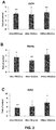
- the inventors unexpectedly found that when detecting the expression of genes related to sternness of cells after nanosecond pulsed electric field stimulation (the experiment was repeated for 5 times, and the cells were collected 0.5 h to 2 h after the electrical stimulation), the expression levels of the genes important for maintaining sternness were increased significantly. As shown in FIG.
- DNA methylation plays an important role on sternness maintenance and differentiation of cells. For example, a cell with higher sternness generally has higher DNA methylation level; in contract, a decrease in DNA methylation is accompanied with differentiation of cell.
- the expression of methylation genes is closely related with the methylation level of cells.
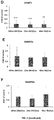
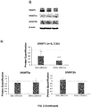
- the expression of the genes related to methylation in cells was detected. The results show that the expression levels of DNMT1 (closely related to methylation level in cells) mRNA and protein were decreased significantly ( FIG. 2D-2H ), but DNMT3a and DNMT3b are not affected, indicating that the effect of the electric shock on methylation is achieved by specifically affecting the expression of DNMT1.
- MethylFlash Global DNA Methylation (5-mC) ELISA Easy Kit was used to detect the global DNA methylation level in cells after stimulation.
- the results show that nanosecond pulsed electric fields stimulation reduced the global DNA methylation level by 40-80% ( FIG. 3 ).
- nanosecond pulsed electric field stimulation could increase sternness of stem cells.
- the present disclosure is the first to show that nanosecond pulsed electric fields can increase the cell sternness.
- DMMB Dimethylmethylene Blue
- the human embryonic stem cells were treated with nanosecond pulses under different combinations of pulse width and field strength. Specifically, six combinations were tested, that was, 10 ns+5 kV/cm, 10 ns+10 kV/cm, 10 ns+20 kV/cm, 100 ns+5 kV/cm, 100 ns+10 kV/cm and 100 ns+20 kV/cm.
- the nanosecond pulsed electric fields are able to increase the expression levels of OCT4, NANOG significantly; and with a pulse width of 10 ns and a field strength of 5 kV/cm, the nanosecond pulsed electric fields also are able to increase the expression level of NANOG and SOX2 genes ( FIG. 7A-7C ) significantly.
- These results demonstrate that after the nanosecond pulsed electric field simulation, the sternness of embryonic stem cells is enhanced effectively.
- Applying nanosecond pulsed electric fields using an electroporation cuvette has the following disadvantages: it only allow single action but not continuous actions at multiple time points; the electric field only acts on a limited amount of cells; and electroporation cuvette is only suitable for suspended single cells, not for cells in pellet or adherent cell, thus the cells need to be digested to single cell, which may affect the cell state; in addition, electroporation cuvette cannot be used for tissues in vivo.
- the inventors has improved the manner of applying electric fields, that is, a conductive film ( FIG. 8 ) is used to replace the electroporation cuvette.
- a conductive film FIG. 8
- the effect of using a conductive film to apply an electric field on porcine bone marrow mesenchymal stem cells was compared with that of using an electroporation cuvette.
- the electric pulse with a parameter of 100 ns and 10 kV/cm was used to simulate the cell suspension in an electroporation cuvette and the porcine bone marrow mesenchymal stem cells seeded on a conductive film, respectively, and the expressions of sternness genes Nanog and OCT 4 were detected. It was found that either applying an electric shock directly to the cells seeded on an conductive film or applying an electric shock to the cell suspension via an electroporation cuvette can enhance the expression of sternness genes effectively, whereas using a conductive film to apply an electric shock can achieve an even higher enhancement, and in particular, the gene expression of Nanog is enhanced more significantly ( FIG.
Landscapes
- Health & Medical Sciences (AREA)
- Life Sciences & Earth Sciences (AREA)
- Engineering & Computer Science (AREA)
- Biomedical Technology (AREA)
- Wood Science & Technology (AREA)
- Organic Chemistry (AREA)
- Chemical & Material Sciences (AREA)
- Zoology (AREA)
- Bioinformatics & Cheminformatics (AREA)
- Genetics & Genomics (AREA)
- General Health & Medical Sciences (AREA)
- Biotechnology (AREA)
- General Engineering & Computer Science (AREA)
- Microbiology (AREA)
- Biochemistry (AREA)
- Developmental Biology & Embryology (AREA)
- Public Health (AREA)
- Veterinary Medicine (AREA)
- Nuclear Medicine, Radiotherapy & Molecular Imaging (AREA)
- Radiology & Medical Imaging (AREA)
- Animal Behavior & Ethology (AREA)
- Cell Biology (AREA)
- Reproductive Health (AREA)
- Gynecology & Obstetrics (AREA)
- Biophysics (AREA)
- Molecular Biology (AREA)
- Rheumatology (AREA)
- Micro-Organisms Or Cultivation Processes Thereof (AREA)
- Medicines Containing Material From Animals Or Micro-Organisms (AREA)
- Manufacture Of Macromolecular Shaped Articles (AREA)
- Materials For Medical Uses (AREA)
Abstract
Description
- This application claims the priority to
Chinese Patent Application No. 201810909901.0 - The present disclosure relates to the technology of using pulsed electric fields for biological treatment, and particularly relates to the technique of using nanosecond pulsed electric fields to treat cells, in particular stem cells.
- Differentiation induction of stem cells is an important research direction in regenerative medicine. The induction efficiency of stem cells needs to be improved to achieve better clinical effects. In order to improve the efficiency of differentiation induction of stem cells, a variety of stimulations has been applied, such as physical stimulations, chemical molecules, and cytokines. Electrical signals, as one of the very important physical stimulations, can facilitate differentiation of stem cells, such as neuronal differentiation, osteogenic differentiation and cardiomyocyte differentiation, by regulating the related signaling pathways, such as calcium ion level and ion channels in cells. However, the biological effect of the electrical signals is weak, thus the electric field need to be applied for a long time ranging from several tens of minutes to several days to take action, which is not favorable to clinical applications.
- Nanosecond pulsed electric fields (nsPEFs) are an advanced physical technology developed in recent years. The pulse width, field strength and instantaneous power of nsPEFs can be accurately controlled, so compared with the conventional electric fields, it gives stronger biological effect in a shorter action time. However, it is still desired to explore more applications of nanosecond pulsed electric fields in this field.
- In the research, the inventors found that the conventional electric fields mostly have a field strength lower than "hundreds of V/cm" and a pulse width higher than microseconds or milliseconds, thereby they only can act at the outside of cell membranes, while nanosecond pulsed electric fields have a pulse width that can reach the nanosecond (ns) level and a field strength that can reach kV/cm level. In additions, nsPEFs can act on organelles within cell membranes. Furthermore, the inventors unexpectedly found that applying nsPEFs on stem cells can significantly enhance the sternness of the stem cells and further enhance its capability to respond to induction, thereby this invention is accomplished.
- In the first aspect, the present disclosure provides use of an electric field in enhancing sternness of a cell, increasing the expression of sternness-related genes in a cell, reducing the expression of methylation-related genes in a cell and/or reducing the methylation level in a cell.
- Preferably, the electric field is a nanosecond pulsed electric field (nsPEF).
- Particularly, the electric field is applied to the cell to give an electric shock to the cell.
- In one embodiment, the cell is a stem cell, preferably a pluripotent stem cell, an unipotent stem cell, an embryonic stem cell, an adult stem cell, an iPS cell or a mesenchymal stem cell.
- In another embodiment, the nanosecond pulsed electric field has a pulse width in a range of 1-300 ns and/or a field strength in a range of 1-30 kV/cm.
- In another embodiment, the pulse width is in a range of 10-150 ns, 20-100 ns, 30-80 ns, 40-70 ns, or 50-60 ns, for example, about 1 ns, 2 ns, 3 ns, 4 ns, 5 ns, 6 ns, 7 ns, 8 ns, 9 ns, 10 ns, 15 ns, 20 ns, 25 ns, 30 ns, 35 ns, 40 ns, 45 ns, 50 ns, 55 ns, 60 ns, 65 ns, 70 ns, 75 ns, 80 ns, 85 ns, 90 ns, 95 ns, 100 ns, 105 ns, 110 ns, 115 ns, 120 ns, 125 ns, 130 ns, 135 ns, 140 ns, 145 ns, 150 ns, 155 ns or 160 ns.
- In another embodiment, the field strength is about 1 kV/cm, 2 kV/cm, 3 kV/cm, 4 kV/cm, 5 kV/cm, 6 kV/cm, 7 kV/cm, 8 kV/cm, 9 kV/cm, 10 kV/cm, 11 kV/cm, 12 kV/cm, 13 kV/cm, 14 kV/cm, 15 kV/cm, 16 kV/cm, 17 kV/cm, 18 kV/cm, 19 kV/cm, 20 kV/cm, 21 kV/cm, 22 kV/cm, 23 kV/cm, 24 kV/cm, 25 kV/cm, 26 kV/cm, 27 kV/cm, 28 kV/cm, 29 kV/cm, or 30 kV/cm.
- In another embodiment, the electric shock is applied 1-1000 times, for example, 10-900 times, 20-800 times, 30-700 times, 40-600 times, 50-500 times, 60-400 times, 70-300 times, 80-200 times, or 90-100 times, and specifically 1, 2, 3, 4, 5, 6, 7, 8, 9, 10, 11, 12, 13, 14, 15, 16, 17, 18, 19, 20, 30, 40, 50, 60, 70, 80, 90 or 100 times. And/or, the electric shock is applied at a frequency of 1-1000 Hz, for example, 10-900 Hz, 20-800 Hz, 30-700 Hz, 40-600 Hz, 50-500 Hz, 60-400 Hz, 70-300 Hz, 80-200 Hz, 90-100 Hz, and specifically about 1, 2, 3, 4, 5, 6, 7, 8, 9, 10, 11, 12, 13, 14, 15, 16, 17, 18, 19, 20, 30, 40, 50, 60, 70, 80, 90, 100 Hz, 200 Hz, 300 Hz, 400 Hz, 500 Hz, 600 Hz, 700 Hz, 800 Hz, 900 Hz, or 1000 Hz.
- In the second aspect, the present disclosure provides a method for enhancing sternness of a cell, increasing the expression of sternness-related genes in a cell, reducing the expression of methylation genes and/or reducing the methylation level in a cell, comprising applying an electric field to the cell to give an electric shock to the cell, wherein the electric field is preferably a nanosecond pulsed electric field;
- The cell is preferably a stem cell, more preferably a pluripotent stem cell, an unipotent stem cell, an embryonic stem cell, an adult stem cell, an iPS cell or a mesenchymal stem cell.
- In one embodiment, the nanosecond pulsed electric field has a pulse width in a range of 1-300 ns and/or a field strength in a range of 1-30 kV/cm.
- Preferably, the pulse width is in a range of 10-150 ns, 20-100 ns, 30-80 ns, 40-70 ns, or 50-60 ns, for example, about 1 ns, 2 ns, 3 ns, 4 ns, 5 ns, 6 ns, 7 ns, 8 ns, 9 ns, 10 ns, 15 ns, 20 ns, 25 ns, 30 ns, 35 ns, 40 ns, 45 ns, 50 ns, 55 ns, 60 ns, 65 ns, 70 ns, 75 ns, 80 ns, 85 ns, 90 ns, 95 ns, 100 ns, 105 ns, 110 ns, 115 ns, 120 ns, 125 ns, 130 ns, 135 ns, 140 ns, 145 ns, 150 ns, 155 ns or 160 ns.
- In addition, a preferred field strength is about 1 kV/cm, 2 kV/cm, 3 kV/cm, 4 kV/cm, 5 kV/cm, 6 kV/cm, 7 kV/cm, 8 kV/cm, 9 kV/cm, 10 kV/cm, 11 kV/cm, 12 kV/cm, 13 kV/cm, 14 kV/cm, 15 kV/cm, 16 kV/cm, 17 kV/cm, 18 kV/cm, 19 kV/cm, 20 kV/cm, 21 kV/cm, 22 kV/cm, 23 kV/cm, 24 kV/cm, 25 kV/cm, 26 kV/cm, 27 kV/cm, 28 kV/cm, 29 kV/cm, or 30 kV/cm.
- In another embodiment, the electric shock is applied 1-1000 times, for example, 10-900 times, 20-800 times, 30-700 times, 40-600 times, 50-500 times, 60-400 times, 70-300 times, 80-200 times, 90-100 times, and specifically 1, 2, 3, 4, 5, 6, 7, 8, 9, 10, 11, 12, 13, 14, 15, 16, 17, 18, 19, 20, 30, 40, 50, 60, 70, 80, 90 or 100 times. And/or, the electric shock is applied at a frequency of 1-1000 Hz, for example, 10-900 Hz, 20-800 Hz, 30-700 Hz, 40-600 Hz, 50-500 Hz, 60-400 Hz, 70-300 Hz, 80-200 Hz and 90-100 Hz, and specifically about 1, 2, 3, 4, 5, 6, 7, 8, 9, 10, 11, 12, 13, 14, 15, 16, 17, 18, 19, 20, 30, 40, 50, 60, 70, 80, 90, 100 Hz, 200 Hz, 300 Hz, 400 Hz, 500 Hz, 600 Hz, 700 Hz, 800 Hz, 900 Hz, or 1000 Hz.
- In another embodiment, the electric field is applied via an electroporation cuvette.
- In another embodiment, the electric field is applied via a conductive film, and preferably, the conductive film is prepared from PLLA (poly-L-lactic acid) and MWCNTs (multi-walled carbon nanotubes) by blend-casting or electro spinning.
- In a specific embodiment, the conductive film is prepared by blend-casting, and specifically by: 1) dissolving the poly-L-lactic acid in a solvent (e.g., chloroform) until fully dissolved; 2) dispersing the multi-walled carbon nanotubes (e.g., carboxylated carbon nanotubes) in the same solvent; 3) performing ultra-sonication (e.g., water bath ultrasound) on the multi-walled carbon nanotubes mixture of step 2), and adding into the poly-L-lactic acid solution of step 1), to allow the final concentration of poly-L-lactic acid is 1-50% w/v; 4) after ultra-sonication, casting the resultant on a plate (e.g., glass plate) and standing (preferably standing overnight); 5) peeling off the film formed on the plate; and 6) placing the film into an oven (e.g., vacuum oven) to fully evaporate the solvent (preferably at a temperature of 35°C), and obtain a conductive film.
- In another specific embodiment, the polylactic acid has a molecular weight in a range of 10,000-1,000,000 Daltons and an intrinsic viscosity of 1-20 dL/g.
- In the third aspect, the present disclosure provides a method for enhancing the capability of a mesenchymal stem cell to differentiate into chondrocyte, osteocyte or adipocyte, comprising applying a nanosecond pulsed electric field to the mesenchymal stem cell to give an electric shock to the mesenchymal stem cell.
- In one embodiment, the nanosecond pulsed electric field has a pulse width in a range of 1-300 ns and/or a field strength in a range of 1-30 kV/cm. Preferably, the pulse width is in a range of 10 -150 ns, 20-100 ns, 30-80 ns, 40-70 ns, 50-60 ns, for example, about 1 ns, 2 ns, 3 ns, 4 ns, 5 ns, 6 ns, 7 ns, 8 ns, 9 ns, 10 ns, 15 ns, 20 ns, 25 ns, 30 ns, 35 ns, 40 ns, 45 ns, 50 ns, 55 ns, 60 ns, 65 ns, 70 ns, 75 ns, 80 ns, 85 ns, 90 ns, 95 ns, 100 ns, 105 ns, 110 ns, 115 ns, 120 ns, 125 ns, 130 ns, 135 ns, 140 ns, 145 ns, 150 ns, 155 ns or 160 ns. Preferably, the field strength is about 1 kV/cm, 2 kV/cm, 3 kV/cm, 4 kV/cm, 5 kV/cm, 6 kV/cm, 7 kV/cm, 8 kV/cm, 9 kV/cm, 10 kV/cm, 11 kV/cm, 12 kV/cm, 13 kV/cm, 14 kV/cm, 15 kV/cm, 16 kV/cm, 17 kV/cm, 18 kV/cm, 19 kV/cm, 20 kV/cm, 21 kV/cm, 22 kV/cm, 23 kV/cm, 24 kV/cm, 25 kV/cm, 26 kV/cm, 27 kV/cm, 28 kV/cm, 29 kV/cm, or 30 kV/cm.
- In another embodiment, the electric shock is applied 1-1000 times, for example 10-900 times, 20-800 times, 30-700 times, 40-600 times, 50-500 times, 60-400 times, 70-300 times, 80-200 times, 90-100 times, and specifically 1, 2, 3, 4, 5, 6, 7, 8, 9, 10, 11, 12, 13, 14, 15, 16, 17, 18, 19, 20, 30, 40, 50, 60, 70, 80, 90 or 100 times. And/or, the electric shock is applied at a frequency of 1-1000 Hz, for example 10-900 Hz, 20-800 Hz, 30-700 Hz, 40-600 Hz, 50-500 Hz, 60-400 Hz, 70-300 Hz, 80-200 Hz, 90-100 Hz, and specifically about 1, 2, 3, 4, 5, 6, 7, 8, 9, 10, 11, 12, 13, 14, 15, 16, 17, 18, 19, 20, 30, 40, 50, 60, 70, 80, 90, 100 Hz, 200 Hz, 300 Hz, 400 Hz, 500 Hz, 600 Hz, 700 Hz, 800 Hz, 900 Hz, or 1000 Hz.
- In another embodiment, the electric field is applied via an electroporation cuvette.
- In yet another embodiment, the electric field is applied via a conductive film. Preferably, the conductive film is prepared from PLLA (poly-L-lactic acid) and MWCNTs (multi-walled carbon nanotubes) by blend-casting or electro spinning.
- In a specific embodiment, the conductive film is prepared by blend-casting, and specifically by: 1) dissolving the poly-L-lactic acid in a solvent (e.g., chloroform) until fully dissolved; 2) dispersing the multi-walled carbon nanotubes (e.g., carboxylated carbon nanotubes) in the same solvent; 3) performing ultrasonication (e.g., water bath ultrasound) on the multi-walled carbon nanotubes mixture of step 2), and adding into the poly-L-lactic acid solution of step 1), to allow the final concentration of poly-L-lactic acid is 1-50% w/v; 4) after ultrasonication, casting the resultant on a plate (e.g., glass plate) and standing (preferably standing overnight); 5) peeling off the film formed on the plate; and 6) placing the film into an oven (e.g., vacuum oven) to fully evaporate the solvent (preferably at a temperature of 35°C), and obtain a conductive film.
- In another specific embodiment, the poly-L-lactic acid has a molecular weight in a range of 10,000-1,000,000 Daltons, and an intrinsic viscosity of 1-20 dL/g.
- In the fourth aspect, the present disclosure provides a cell generated by the method in the second aspect and the third aspect.
- In the fifth aspect, the present disclosure provides a pharmaceutical composition, comprising the cell in the fourth aspect.
- The present disclosure unexpectedly found that nanosecond pulsed electric fields are able to enhance sternness of a stem cell, and the method to enhance sternness is suitable for a various types of stem cells, and the enhancement on sternness of a stem cell will further facilitate its capability of responding to induction and differentiation. Therefore, the technical solutions of the present disclosure are of immeasurable value on improving the activity and differentiation potential of stem cells, and on promoting the stem cells to be used more wildly and effectively in the regenerative medicine.
-
-
FIG. 1A is an image of a nanosecond pulsed electric field device,FIG. 1B is a side view of an electroporation cuvette,FIG. 1C is a top view of an electroporation cuvette,FIG. 1D is a schematic drawing of electroporation cuvettes used in the cell experiments and their field strength simulation, andFIG. 1E is a graph showing the effect on the cell survival status under different electric shock conditions; -
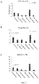
FIG. 2 is graphs showing the detection results of sternness genes and methylation-related genes in porcine bone marrow mesenchymal stem cells stimulated by nanosecond pulsed electric fields; -
FIG. 3 is a graph showing the detection results of the overall methylation level in porcine bone marrow mesenchymal stem cells stimulated by nanosecond pulsed electric fields; -
FIG. 4 is graphs showing the results of alcian blue staining (A), GAG semi-quantification (B), and the gene expression of COLII, ACAN and Sox9 (C), after the cells were stimulated by nanosecond pulsed electric fields and differentiated into chondroblasts; -
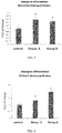
FIG. 5 is a graph showing the results of alizarin red-based semi-quantification, after the cells were stimulated by nanosecond pulsed electric fields and differentiated into osteoblasts; -
FIG. 6 is a graph showing the results of oil red O-based semi-quantification, after the cells were stimulated by nanosecond pulsed electric fields and differentiated into adipoblasts; -
FIG. 7 is graphs showing the detection results of sternness genes of OCT4, NANOG and SOX2 in human embryonic stem cells stimulated by nanosecond pulsed electric fields; -
FIG. 8 is an image of a real conductive film (A) and its scanning electron micrograph (B); and -
FIG. 9 is graphs showing the comparison of the effects on the expression of sternness genes after stimulating mesenchymal stem cells via an electroporation cuvette, and that of via a conductive thin film. - The present disclosure can be further understood by reference to the embodiments. Definitions
- The term "sternness" refers to a capacity of a stem cell to maintain in its undifferentiated state. Stem cells keep a potential capacity to differentiate into one or more cell fate(s) and a potential capacity to be induced to differentiate, while being in continually self-renewal. Sternness of a stem cell can be detected by morphological observation or by detecting sternness genes of the cell. The term "sternness gene", also called as "sternness marker gene" or "sternness-related gene", refers to the genes associated with the status of a stem cell. The expression of such genes is beneficial for maintaining the sternness of a stem cell. The common sternness-related genes include Oct4, Nanog, Sox2 and so on.
- Additionally, sternness of a stem cell may also be characterized by a low methylation level in the cell, and the reduction in methylation level can be achieved by regulating down the expression of methylation genes. Therefore, sternness of a stem cell may be further characterized by the low-expression (reduction in expression) of methylation genes in a cell. In the present disclosure, the term "methylation-related gene", also called as "methylation gene", refers to the genes which facilitate the methylation in a cell to increase the methylation level in the cell, such as methyltransferase genes, and more specifically Dnmt1.
- The term "electric shock" used herein, is also called as "electrical stimulation" or "electrical field stimulation", refers to apply an electric field (or electric pulses) having a certain pulse width and field strength to a subject, such as cells (including stem cells), for a set times at a set frequency. In one embodiment, the electric shock is applied via a specialized device. In one particular embodiment, the electric shock is applied via an electroporation cuvette. In another specific embodiment, the electric shock is applied via a conductive film (or called as a conductive thin film).
- The conductive film may be prepared by using any materials, structures and methods known in the prior art, as long as it is suitable for applying electric fields. For example, a nanofiber and a preparation method disclosed by Zhu S, Jing W, Hu X, Huang Z, Cai Q, Ao Y, Yang X. 2017 (Time-dependent effect of electrical stimulation on osteogenic differentiation of bone mesenchymal stromal cells cultured on conductive nanofibers, J Biomed Mater Res Part A 2017:105A:3369-3383).
- In one embodiment, the conductive film is prepared from poly-L-lactic acid (PLLA) and multi-walled carbon nanotubes (MWCNTs) by blend-casting or electro spinning, and the prepared conductive thin film has a smooth surface and a resistance close to PBS.
- In one embodiment, the cells to be treated are seeded on the conductive film, and receive the electric shock on the conductive film directly without the need to be pre-prepared as a cell suspension.
- In yet another embodiment, the electric shock has a pulse width in a range of 1-300 ns. Preferably, the pulse width is in a range of 10-150 ns, 20-100 ns, 30-80 ns, 40-70 ns, or 50-60 ns, and specifically about 1 ns, 2 ns, 3 ns, 4 ns, 5 ns, 6 ns, 7 ns, 8 ns, 9 ns, 10 ns, 15 ns, 20 ns, 25 ns, 30 ns, 35 ns, 40 ns, 45 ns, 50 ns, 55 ns, 60 ns, 65 ns, 70 ns, 75 ns, 80 ns, 85 ns, 90 ns, 95 ns, 100 ns, 105 ns, 110 ns, 115 ns, 120 ns, 125 ns, 130 ns, 135 ns, 140 ns, 145 ns, 150 ns, 155 ns, or 160 ns.
- In yet another embodiment, the field strength is in the range of 1-30 kV/cm. Preferably, the field strength is about 1 kV/cm, 2 kV/cm, 3 kV/cm, 4 kV/cm, 5 kV/cm, 6 kV/cm, 7 kV/cm, 8 kV/cm, 9 kV/cm, 10 kV/cm, 11 kV/cm, 12 kV/cm, 13 kV/cm, 14 kV/cm, 15 kV/cm, 16 kV/cm, 17 kV/cm, 18 kV/cm, 19 kV/cm, 20 kV/cm, 21 kV/cm, 22 kV/cm, 23 kV/cm, 24 kV/cm, 25 kV/cm, 26 kV/cm, 27 kV/cm, 28 kV/cm, 29 kV/cm, or 30 kV/cm.
- It should be noted that, the values of pulse width and field strength above not only refer to the values themselves, in some embodiments, the applied pulse width may have a certain variation from these values, which may be 1%, 2%, 3%, 4%, 5%, 6%, 7%, 8%, 9% or 10%, resulting from the precision of the device to apply the electric pulse and other reasons, therefore, in these embodiments, the values of pulse width and field strength will also include the variation around these values. Besides, in the present disclosure, such variation may be expressed by using "about".
- In the term "nanosecond pulsed electric field", one nanosecond is equal to one billionth of a second, and a pulse which lasts from a several to tens of nanoseconds is called a nanosecond pulse. Nanosecond pulsed electric fields are mainly characterized by high power but low heat, so there is almost no heat generated during the treatment of tumors. In addition, it can penetrate a cell membrane to act on the organelles in the cell, thereby affecting the biological function of the cell.
- In contrast to the electroporation of cell membrane caused by a longer pulse, a nanosecond pulse generates extremely high power (several billions of watts), extremely short duration (nanoseconds) and high field strength (kV/cm), thus it can penetrate into a cell and act on the subcellular structure before the cell membrane is fully charged. Because the duration of a nanosecond pulse is shorter than the time to charge cell membrane, thus it almost does not affect the cell membrane, and will not cause electroporation of cell membrane.
-
Name of the Instruments Type NO. Manufacturer High Speed Refrigerated Centrifuge 5702R Eppendorf Inverted Phase Contrast Microscope CKX41-RC Olympus Microscope Component for Fluorescence U-RFLT50 Olympus Clean Bench DL-CJ-2ND HDL Electronic Balance BS124S Sartorius Laser Confocal Microscope LSM510 Zeiss High-Pressure Steam Sterilization Pot MLS-3750 SANYO Enzyme Reader 680 Bio-rad Water Purifier Millipore MERCK Complete set for Western Blotting Bio-rad Bio-rad Real-time PCR Instrument Pikoreal 96 Thermo Ordinary PCR Instrument MyCycler Bio-rad Spectrophotometer ND1000 Thermo Chemiluminescence Imaging System 5200 Tanon Ice-maker FM50 Snow Vortex Mixer Vortex-5 Vortex Enzyme Reader Synergy4 Biotek Electroporation Cuvette BTX BTX - Reagents: DMEM (Gibco); DMEM Dulbecco's Modified Eagle Medium (Gibco); Trypsin (Invitrogen); fetal bovine serum (FBS, Gibco); Dexamethasone (Life Technologies); TGF-β3 (Peprotech); sodium pyruvate (Life Technologies); vitamin C (Vc, Sigma); ITS (Insulin Transferrin Selenium premix, Life Technologies); proline (SINOPHARM); sodium glycerophosphate (Sigma); fast green (Amresco); Alkaline Phosphatase (ALP) Assay Kit (NanJing JianCheng Bioengineering Institute); TritonX-100 (Sigma); BD Matrigel™ matrix (BD Biosciences Company, 354277); and mTeSR1 Complete Kit (Stemcell Company, 05850).
- The culture medium for bone marrow mesenchymal stem cells was prepared by: adding 160 mL of DMEM stock solution and 40 mL of melted FBS, and then adding 200 µL of 1000×PS, to a sterilized culture flask using a sample injector. This whole process was operated inside a sterilized Clean Bench in a sterile environment.
- In the experiments, porcine bone marrow mesenchymal stem cells were used. The pigs used in the experiments were purchased from Animal Experiment Department of Beijing Fuwai Hospital. The procedures for use of animals were approved by the Animal Ethical Review in the Beijing Fuwai Hospital, and the animal experiments for all the studies were conducted according to the related regulations of the Ethics Committee of Peking University.
- A pork leg was soaked in 75% alcohol for 20 minutes, and then transferred to the clean hood, and the medullary cavity was open by an electric drill. The bone marrow was washed out with PBS to a petri dish, until the washing solution became clear. The mixture in the petri dish was collected and transferred to a 15 mL centrifuge tube, subjected to centrifugation at 1,000 rpm for 10 min in a centrifuge, and then the cells were collected.
- The precipitated cells were suspended in the culture medium of the bone marrow mesenchymal stem cells and cultured. The culture medium was changed half at the first day, and then changed completely after a rinse with PBS at three-day intervals. During the culture, the cells were observed, photographed by a microscope and recorded.
- When the cells reached 80% confluence, 2 mL of trypsin was added to the cell culture dish, and the dish was put in an incubator to digest for 3 minutes. Once suspension of the bone marrow mesenchymal cells was observed under the microscope, a culture medium of 2-fold volume of trypsin was added into the petri dish, the mixture was transferred into a 15 mL centrifuge tube, then centrifuged at a speed of 1,000 rpm for 5 minutes in a refrigerated centrifuge. The cells were collected, resuspended in the culture medium and counted by using a hemocytometer. The cells were cultured at a density of 5,000 cells/cm2 with the culture medium changed twice every week.
- The subcultured bone marrow mesenchymal cells were digested with trypsin and centrifuged, and the supernatant was discarded. Cell cryopreservation medium was added to the cells and then transferred into a cryogenic vial. The cryogenic vial was successively placed in a programmed cooling box at -20°C for 3 hours, a low-temperature freezer at -80°C overnight, and then a liquid nitrogen tank for long-term preservation.
- To resuscitate the cells, the cryogenic vial was immersed into a water bath at 37°C with quickly shake to let the ice melt. In the subsequent experiments, the fifth-generation primary bone marrow mesenchymal stem cells were used.
- BD Matrigel™ matrix (BD Biosciences Company, 354277) was used for plating cells in a 6-well plate. Matrigel was diluted with pre-cooled DMEM/F-12 according to the instruction and added into a 6-well plate, 1 mL in each well. The plate was shaken slightly to let the Matrigel uniformly covered on the surface of the well, and then incubated at room temperature for 1 h.
- Cells of human embryonic stem cell line H9 were cultured in a culture medium (mTeSR1 Complete Kit, Stemcell, 05850), with the medium changed every day. The growth of cells was assessed and monitored until the next subculture.
- The cells were washed with DMEM/F-12, then digested using a dispase at 37°C for about 4-7 minutes. After removing the dispase by aspiration, the dish was gently washed with DMEM/F-12. The cell colonies were removed using a serological glass pipette or a cell scraper, collected, centrifuged, resuspended and uniformly mixed, and then subcultured at a ratio of 1:6 to 1:10.
- The electric pulse device can be connected and combined in any manner well-known to these skilled in the art. The nanosecond pulsed electric field device used in the examples of the present disclosure comprised a high voltage DC power supply, a coaxial transmission cable, a high-voltage probe and an cell-stimulating electroporation cuvette (any commercially available type of electroporation cuvettes may be used, for example, BTX™ cuvettes). The high voltage DC power supply provided a stimulation electric field, the coaxial transmission cable could compress electrical energy, the high-voltage probe allowed to form a nanosecond pulsed electric field, and the nanosecond level electric field could be applied to the electroporation cuvette. The electric fields constructed by the device of the present disclosure were pulsed electric fields with a pulse width of 100 nanoseconds. The output voltage from the high voltage DC power supply can be read on an oscilloscope. Changing the output voltage can adjust the strength of the nanosecond pulse field inside the electroporation cuvette to different values.
- The cells were counted by a hemocytometer (porcine bone marrow mesenchymal stem cells or human embryonic stem cells). The cells were suspended in PBS to prepare a cell suspension at 100,000-1,500,000 cells/ml. 500-800 µL of the cell suspension was added into the electroporation cuvette. For the test groups, cells were simulated by a nanosecond pulsed electric field with specific parameters. After the completion of the electrical stimulation, the cells were collected by a low-speed centrifugation. Then the porcine bone marrow mesenchymal stem cells were cultured for about two weeks to differentiate into chondroblasts (pellet culture), osteoblasts and lipoblasts by chondrogenic induction, osteogenic induction and adipogenic induction (more details will be described below), respectively. In addition, within 4 h after the cells be electrically stimulated, a part of the electrically stimulated cells was taken out for necessary analysis and detection, such as the sternness gene detection (in the human embryonic stem cells and the porcine bone marrow mesenchymal stem cells) and the global DNA methylation level detection (in the porcine bone marrow mesenchymal stem cells), as shown in the examples.
- The conductive films used in the examples were prepared by the blend-casting technique, using MWCNTs (multi-walled carbon nanotubes) and poly-L-lactic acid with a molecular weight of 100,000 and an intrinsic viscosity of 2.7 dL/g, as raw materials. Specifically, it included the steps: 1) dissolving 1 g of polylactic acid in 5 mL of chloroform until fully dissolved in 8 h; 2) dispersing 30 mg of carboxylated carbon nanotubes in 5 mL of chloroform; 3) performing water bath untrasound for 30 min on the carboxylated carbon nanotubes mixture of step 2), and adding into the poly-L-lactic acid solution of step 1), to allow the final concentration of poly-L-lactic acid was 10% by w/v; 4) after ultra-sonication for 20 min, casting the resultant onto a glass plate and standing overnight; 5) peeling off the film; and 6) placing the film into a vacuum oven at 35°C for one day to fully evaporate the solvent, and obtain a conductive film, having a smooth surface and a resistance similar to that of PBS.
- The nanosecond pulsed electric field device using the conductive film included the following components: a high voltage DC power supply, a coaxial transmission cable, a high-voltage probe, a conductor piece and a cell-simulating conductive film. The high voltage DC power supply provided a simulation electric field, the coaxial transmission cable could compress electrical energy, the high-voltage probe allowed to form a nanosecond pulsed electric field, and the nanosecond level electric field was applied on the conductive film though the conductor pieces at the two side of the conductive film. In this test, the device was used to construct a pulsed electric field having a pulse width of 100 nanoseconds. The output voltage from the high voltage DC power supply could be read on an oscilloscope. Changing the output voltage can adjust the strength of the nanosecond pulse field on the conductive film.
- Using the conductive film to apply electric shocks specifically included the following steps. Porcine bone marrow mesenchymal stem cells were cultured until cell confluence reached 80%. 2 mL of trypsin was added into the cell culture dish, and the cells were digested for 3 minutes in an incubator. Once suspension of the bone marrow mesenchymal cells was observed under the microscope, a cell culture medium containing 2-fold volume of the trypsin was added to the dish. The cells were collected and transferred into a 15 mL centrifuge tube for centrifugation at a speed of 1,000 rpm for 5 minutes in a refrigerated centrifuge. The collected cells were counted by a hemocytometer, and resuspended in a culture medium to a cell density of 500,000-4,000,000 cells/mL. 50 µL of the resulting suspension was dropped uniformly onto the conductive thin film, incubated at room temperature for 3-12 h, and then additional culture medium was added. The culture medium was changed twice a week. When applying electrical stimulation, the culture medium was change to phosphate buffered saline (PBS) and the conductor pieces were connected to the two sides of the conductive film to apply nanosecond pulsed electric field stimulation having specific parameters. After the completion of the electrical stimulation, PBS was replaced back to growth medium for continuing culture. The cells were digested at a predetermined time point for the subsequent detection.
- The culture medium for chondrogenic induction was formulated by: high glucose DMEM (Gibco), 10 ng/mL TGF-β3 (R&D Systems, Minneapolis, MN), 10-7 M dexamethasone (D4902, Sigma), 50 µg/mL ascorbic acid (A5960, Sigma), 1 mM sodium pyruvate (P2256, Sigma), 4 mM proline (P5607, Sigma) and 1% (v/v) ITS (41400045, Gibco).
- The culture was performed by the steps: digesting bone marrow mesenchymal stem cells by trypsin and counting; removing the culture medium, changing to the culture medium for chondrogenic induction to prepare a cell suspension at 2.5×10⁵ cells/mL; transferring 1 mL of the suspension to a 15 mL centrifuge tube to centrifuge at a speed of 500× g for 5 min; culturing the stem cells by pellet culture for 14 days with the culture medium changed every 3 days.
- The culture medium for adipogenic induction was formulated by the solution A and solution B.
- The solution A was a differentiation medium, prepared by 175 mL of a differentiation medium for adipogenic induction of bone marrow mesenchymal stem cells (as a base culture medium of solution A), 20 mL of fetal bovine serum, 2 mL of Penicillin-Streptomycin, 2 mL of Glutamine, 400 µL of Insulin, 200 µL of IBMX, 200 µL of Rosiglitazone, and 200 µL of Dexamethasone.
- The solution B was a maintenance medium, prepared by 175 mL of a differentiation medium for adipogenic induction of bone marrow mesenchymal stem cells (as a base culture medium of solution B), 20 mL of fetal bovine serum, 2 mL of Penicillin-Streptomycin, 2 mL of Glutamine, and 400 µL of Insulin.
- The culture was performed by the following steps.
- Bone marrow mesenchymal stem cells were cultured in an incubator at 37°C with 5% of CO2. When the cell confluence reached 80-90%, the cells were digested by using 0.25% trypsin-0.04% EDTA. The digested mesenchymal stem cells were seeded at a cell density of 2×10⁴ cells/cm2 in a 6-well plate, with 2 mL of complete medium at each well. The cells were cultured in an incubator at 37°C with 5% of CO2, with the medium changed at a three-day interval, until the cell confluence reached 100% or over confluence.
- The complete medium for the mesenchymal stem cells was removed carefully by aspiration, and 2 mL of the solution A (differentiation medium for adipogenic induction of bone marrow mesenchymal stem cells) was added into the 6-well plate. After induction for 3 days, the solution A was removed from the 6-well plate by aspiration, and 2 mL of the solution B (differentiation medium for adipogenic induction of bone marrow mesenchymal stem cells) was added. After 24 h, the solution B was removed by aspiration and replaced with solution A for induction.
- After alternately culturing the cells with the solution A and the solution B for 3-5 rounds (12-20 days), the solution B was used again to maintain the culture for 4-7 days until the lipid droplets became large and round enough. When the cells were maintained in solution B, it was necessary to change to fresh solution B at a 2-3 days' interval.
- The osteogenic induction medium was formulated by DMEM culture medium, containing 10% of FBS, 100 nM dexamethasone, 10 mM β-sodium glycerophosphate, and 50 µg/mL vitamin C.
- The culture was performed by the following steps: digesting bone marrow mesenchymal stem cells by trypsin and counting; diluting the cell suspension to a concentration of 50,000 cells/mL, adding 100 µL of the cell suspension into a 96-well plate; when the cell confluence reached about 70%, removing the culture medium and washing the cells with PBS for 3 times; and adding the osteogenic induction medium to induce the osteogenic differentiation of the bone marrow mesenchymal stem cells, with the culture medium changed every 3 days.
-
- A. A genetic detection was performed to detect gene expression of COL I, COL II, COL X,
SOX 9, ACAN. COL II,SOX 9 and ACAN are the functional genes favorable to chondrogenesis, while COL I and COL X are hypertrophic genes not favorable to chondrogenesis. - B. In the histology detection, the pellets were fixed in 4% paraformaldehyde, embedded in agarose, dehydrated and transparentized, embedded in paraffin, and sliced. Then, alcian blue dye was used to stain the extracellular matrix, and HE-staining was used to show the cell morphology.
- C. Semi-quantitative detection based on glycosaminoglycan (GAG) was performed by: digesting the cell pellets by using proteinase K at 56°C overnight; heating the sample at 90°C for 10 min; using cationic dye, DMMB, to complex with GAG for half-hour; using a decomplexation solution to dissociate the precipitation; detecting absorbance at a wavelength of 656 nm; and calculating the GAG content in the extracellular matrix.
- Alizarin red method was performed as follows: adjusting pH of alizarin red solution to 4.0-4.2; staining the sample for 1 h at room temperature; incubating in PBS for 15 min and removing the supernatant; washing the sample with PBS for 3 times and thoroughly removing the supernatant; adding hexadecyl chloride; and standing at room temperature for 30 min. The supernatant was pipetted out and analyzed by using the ultraviolet spectrophotometer. The absorbance of the sample at a wavelength of 562 nm was measured, and a hexadecyl chloride solution was used as blank control to set zero value. Additionally, the absorbance of the supernatant from a group of the cells without addition of alizarin red were measured. The measurement for each sample was repeated for 3 times to calculate average value and standard deviation.
- This detection was performed by: staining the sample with oil red O for 1 h; washing with PBS for twice; adding isopropyl alcohol for isolation; and measuring at 490 nm for semi-quantitative detection.
- Reagents: TRNzol Total RNA Isolation Kit (DP405), FastQuant RT Kit (with gDNase) (KR106) and SuperReal PreMix Plus (SYBR Green) (FP205) were purchased from Tiangen Biotech (Beijing) co., Ltd. Isopropyl alcohol and chloroform were purchased from Beijing Reagent Factory.
-
- (1) RNA isolation and reverse transcription were performed by: extracting RNA by using DP405 product from Tiangen Company, and subsequently performing reverse transcription by using KR106 kit from Tiangen Company according to the instructions.
- (2) Real-time PCR detection was performed by: carrying out quantitative real-time PCR by using FP205 kit from Tiangen Company to detect the level of gene expression of Oct4, CARM1, DNMT1, DNMT3A, DNMT3B, Mll1, Mll2, Nanog, PRMT5, Sox2 and so on.
- Reagents: MethylFlash Global DNA Methylation (5-mC) ELISA Easy Kit (p-1030) (Epigentek Company) was used.
- Standard curve was plotted according to the instruction of the kit, and DNA methylation levels of samples were detected.
- When using nanosecond pulsed electric fields to stimulate cells, the primary parameters are pulse width and field strength, which need to be controlled at a suitable range to achieve the aim of the experiment and to ensure cell viability (J.C. Weaver, etc.,"A brief overview of electroporation pulse strength-duration space: A region where additional intracellular effects are expected", Bioelectrochemistry, 87(2012)236-243; Tadej Kotnik, etc., Electroporation-based applications in biotechnology, Trends in Biotechnology, 33(8), 2015.8). In this Example, cells were subjected to electric shock via the electroporation cuvette (
FIG. 1 A-D ), with the pulse width being adjusted within the range of 1-300 ns, and the field strength being adjusted within the range of 1-30 kV/cm. The results showed that, within the above ranges, an electric shock with the combination of the pulse width and the field strength did not have obviously adverse effect on the cell survival(FIG. IE). This is consistent with the current understanding for the effect of pulse width and field strength of nanosecond pulsed electric fields on cell survival in this field (J.C.Weaver, etc., "A brief overview of electroporation pulse strength-duration space: A region where additional intracellular effects are expected", Bioelectrochemistry, 87(2012)236-243; Tadej Kotnik, etc., Electroporation-based applications in biotechnology, Trends in Biotechnology, 33(8), 2015.8). - In the subsequent experiments, cells were electrically stimulated by the fields with the combination of the two parameters. In the experiments, as needed, the stimulation was carried out at times ranging from 1 to 1,000 times, at a frequency ranging from 1 to 1,000 Hz, at room temperature or particularly at a suitable temperature ranging from 20 to 37°C. Multiple combinations can produce certain effects.
- As for porcine bone marrow mesenchymal stem cells, the parameters enabling a better effect included three groups (group A, B and C) as shown below. For convenience, in the subsequent examples, the group A, group B and group C might be used to represent parameters described below.
- Group A:
pulse width 10 ns,field strength 20 kV/cm; - Group B:
pulse width 100 ns,field strength 10 kV/cm; - Group C:
pulse width 60 ns,field strength 5 kV/cm; and - As for human embryonic stem cells, the stimulation experiments were also conducted with the pulse width within the range of 1-300 ns and the field strength within the range of 1-30 kV/cm. The cells were stimulated under different combinations of parameters. In Example 4, six combinations: 10 ns+5 kV/cm, 10 ns+10 kV/cm, 10 ns+20 kV/cm, 100 ns+5 kV/cm, 100 ns+10 kV/cm and 100 ns+20 kV/cm, were specifically tested. After the stimulations under these six conditions, the expression of the target genes was detected.
- It should be noted that the specific parameter combinations above merely are part of the preferred embodiments. In facts, within the conditions described above for applying nanosecond pulsed electric fields, there may be a variety of suitable parameter combinations. Furthermore, for the different types of the stem cells, the most suitable conditions may be also different, and need to determine specific parameter combinations depending on the different target cells. However, due to these skilled in the art have already know clearly about the range of the conditions to apply the nanosecond pulse, which is further supported by the present disclosure, it can be understood that these skilled in the art are possible to acquire desired parameter combinations, based on the content of the present disclosure,.
- Nanosecond pulsed electric fields increases the expression of sternness-related genes.
- It is known that electrical signal stimulation may facilitate stem cell differentiation (e.g., mesenchymal stem cell). The reason is generally speculated to be that electrical signals can activate signals or pathways related to cell differentiation, thereby promoting the ability of stem cells to differentiate into a variety of tissue cells. However, because the mechanism of stem cell differentiation is very complicated, the reasons for how the external stimulations enhance the differentiate capability of the stem cells are still poorly understood. The inventors unexpectedly found that when detecting the expression of genes related to sternness of cells after nanosecond pulsed electric field stimulation (the experiment was repeated for 5 times, and the cells were collected 0.5 h to 2 h after the electrical stimulation), the expression levels of the genes important for maintaining sternness were increased significantly. As shown in
FIG. 2A-2C , as compared with the control group without the electric field treatment, the expression level of OCT4 gene increased by nearly 3 times, and the expression level of NANOG and SOX2 also increased significantly. These results indicated that after the mesenchymal stem cells were simulated by nanosecond pulsed electric fields, the sternness of the cells was increased. - It is known that DNA methylation plays an important role on sternness maintenance and differentiation of cells. For example, a cell with higher sternness generally has higher DNA methylation level; in contract, a decrease in DNA methylation is accompanied with differentiation of cell. The expression of methylation genes is closely related with the methylation level of cells. In order to further investigate the increase of the sternness of mesenchymal stem cells after nanosecond pulsed electric field stimulation, the expression of the genes related to methylation in cells was detected. The results show that the expression levels of DNMT1 (closely related to methylation level in cells) mRNA and protein were decreased significantly (
FIG. 2D-2H ), but DNMT3a and DNMT3b are not affected, indicating that the effect of the electric shock on methylation is achieved by specifically affecting the expression of DNMT1. - Subsequently, in order to analyze the DNA methylation state in cells directly, MethylFlash Global DNA Methylation (5-mC) ELISA Easy Kit was used to detect the global DNA methylation level in cells after stimulation. The results show that nanosecond pulsed electric fields stimulation reduced the global DNA methylation level by 40-80% (
FIG. 3 ). - The results above demonstrated that nanosecond pulsed electric field stimulation could increase sternness of stem cells. The present disclosure is the first to show that nanosecond pulsed electric fields can increase the cell sternness.
- In this Example, the changes on the differentiation capability of the mesenchymal stem cells after nanosecond pulsed electric field simulation were detected. For the detection of chondrogenic differentiation, the alcian blue staining was conducted on the 14th day after stimulation. The results showed that after the cells were pretreated with nanosecond pulsed electric fields and then subjected to chondrogenic induction culture, the extracellular matrix secretion is increased obviously (
FIG. 4A ). - Dimethylmethylene Blue (DMMB) assay was used to quantify glycosaminoglycan, and it was found that GAG content was increased to about 1.5 folds (
FIG. 4B ). Further, the expressions of COL II, ACAC andSOX 9 genes, which can significantly facilitate the function of cartilage, were detected. It was found that the expressions of the three genes were increased significantly (FIG. 4C ), while the expressions of COL I, COL X were not affected significantly (results not shown). These results demonstrate that the capability of mesenchymal stems for chondrogenic differentiation is enhanced significantly. - The changes on ability for osteogenic differentiation, after the mesenchymal stem cells was simulated by nanosecond pulsed electric fields, was studied. After the cells were pretreated with nanosecond pulsed electric fields and then subjected to osteogenic induction culture, the efficiency of osteogenic induction was increased to about 1.5 fold, compared with the use of induction medium only. This experiment was repeated for 3 times (n=3) (
FIG. 5 ). - After the cells were pretreated with nanosecond pulsed electric fields and then subjected to adipogenic induction culture, compared with the use of an induction medium only, the efficiency of adipogenic induction was increased significantly. This experiment was repeated for 3 times (n=3) (
FIG. 6 ). - The results above show that the increase of sternness of the mesenchymal stem cells after nanosecond pulsed electric field stimulation leads to direct enhancement on differentiation capability. This also indicates that using nanosecond pulsed electric field to stimulate the stem cells has a broad practical application.
- In order to investigate the effect of nanosecond pulsed electric fields on the sternness of other stem cell types, the human embryonic stem cells were treated with nanosecond pulses under different combinations of pulse width and field strength. Specifically, six combinations were tested, that was, 10 ns+5 kV/cm, 10 ns+10 kV/cm, 10 ns+20 kV/cm, 100 ns+5 kV/cm, 100 ns+10 kV/cm and 100 ns+20 kV/cm. The results show that with a pulse width of 100 ns and a field strength of 10 kV/cm, the nanosecond pulsed electric fields are able to increase the expression levels of OCT4, NANOG significantly; and with a pulse width of 10 ns and a field strength of 5 kV/cm, the nanosecond pulsed electric fields also are able to increase the expression level of NANOG and SOX2 genes (
FIG. 7A-7C ) significantly. These results demonstrate that after the nanosecond pulsed electric field simulation, the sternness of embryonic stem cells is enhanced effectively. - Applying nanosecond pulsed electric fields using an electroporation cuvette has the following disadvantages: it only allow single action but not continuous actions at multiple time points; the electric field only acts on a limited amount of cells; and electroporation cuvette is only suitable for suspended single cells, not for cells in pellet or adherent cell, thus the cells need to be digested to single cell, which may affect the cell state; in addition, electroporation cuvette cannot be used for tissues in vivo.
- Thus, the inventors has improved the manner of applying electric fields, that is, a conductive film (
FIG. 8 ) is used to replace the electroporation cuvette. In this example, the effect of using a conductive film to apply an electric field on porcine bone marrow mesenchymal stem cells was compared with that of using an electroporation cuvette. - The electric pulse with a parameter of 100 ns and 10 kV/cm was used to simulate the cell suspension in an electroporation cuvette and the porcine bone marrow mesenchymal stem cells seeded on a conductive film, respectively, and the expressions of sternness genes Nanog and
OCT 4 were detected. It was found that either applying an electric shock directly to the cells seeded on an conductive film or applying an electric shock to the cell suspension via an electroporation cuvette can enhance the expression of sternness genes effectively, whereas using a conductive film to apply an electric shock can achieve an even higher enhancement, and in particular, the gene expression of Nanog is enhanced more significantly (FIG. 9 ,** represents a very significant difference for p<0.01; and * represents a significant difference for p<0.05). In addition to the advantage on the electric shock effects, using an conductive film to apply an electric shock is further advantaged at that on one hand there is no need to digest cells to prepare suspension, which saves the cost and time for operations, and on other hand cell viability is preserved maximally, which enable the cells after the electric shock to be used in subsequent treatments, for example, the cells adhered on a conductive film after the electric shock can continue to be used in electric shock after a temporary culture, thereby the flexibility of operation is greatly improved. This also indicates that using a conductive film to apply nanosecond pulsed electric fields has good application prospect.
Claims (17)
- Use of an electric field in enhancing sternness of a cell, increasing the expression of sternness-related genes in a cell, reducing the expression of methylation-related genes in a cell and/or reducing the methylation level in a cell; and
preferably, the electric field is a nanosecond pulsed electric field (nsPEF). - The use according to claim 1, wherein the electric field is applied to the cell to give an electric shock to the cell.
- The use according to claim 1 or 2, wherein the cell is a stem cell, preferably a pluripotent stem cell, an unipotent stem cell, an embryonic stem cell, an adult stem cell, an iPS cell or a mesenchymal stem cell.
- The use according to any one of claims 1 to 3, wherein the nanosecond pulsed electric field has a pulse width in a range of 1-300 ns and/or a field strength in a range of 1-30 kV/cm;
preferably, the pulse width is in a range of 10-150 ns, 20-100 ns, 30-80 ns, 40-70 ns, or 50-60 ns, for example, about 1 ns, 2 ns, 3 ns, 4 ns, 5 ns, 6 ns, 7 ns, 8 ns, 9 ns, 10 ns, 15 ns, 20 ns, 25 ns, 30 ns, 35 ns, 40 ns, 45 ns, 50 ns, 55 ns, 60 ns, 65 ns, 70 ns, 75 ns, 80 ns, 85 ns, 90 ns, 95 ns, 100 ns, 105 ns, 110 ns, 115 ns, 120 ns, 125 ns, 130 ns, 135 ns, 140 ns, 145 ns, 150 ns, 155 ns, or 160 ns; and
preferably, the field strength is about 1 kV/cm, 2 kV/cm, 3 kV/cm, 4 kV/cm, 5 kV/cm, 6 kV/cm, 7 kV/cm, 8 kV/cm, 9 kV/cm, 10 kV/cm, 11 kV/cm, 12 kV/cm, 13 kV/cm, 14 kV/cm, 15 kV/cm, 16 kV/cm, 17 kV/cm, 18 kV/cm, 19 kV/cm, 20 kV/cm, 21 kV/cm, 22 kV/cm, 23 kV/cm, 24 kV/cm, 25 kV/cm, 26 kV/cm, 27 kV/cm, 28 kV/cm, 29 kV/cm, or 30 kV/cm. - The use according to any one of claims 1 to 4, wherein the electric shock is applied 1-1000 times, for example, 10-900 times, 20-800 times, 30-700 times, 40-600 times, 50-500 times, 60-400 times, 70-300 times, 80-200 times, or 90-100 times, specifically, 1, 2, 3, 4, 5, 6, 7, 8, 9, 10, 11, 12, 13, 14, 15, 16, 17, 18, 19, 20, 30, 40, 50, 60, 70, 80, 90 or 100 times; and/or
with a frequency of 1-1000 Hz, for example, 10-900 Hz, 20-800 Hz, 30-700 Hz, 40-600 Hz, 50-500 Hz, 60-400 Hz, 70-300 Hz, 80-200 Hz, or 90-100 Hz, specifically, about 1, 2, 3, 4, 5, 6, 7, 8, 9, 10, 11, 12, 13, 14, 15, 16, 17, 18, 19, 20, 30, 40, 50, 60, 70, 80, 90, 100 Hz, 200 Hz, 300 Hz, 400 Hz, 500 Hz, 600 Hz, 700 Hz, 800 Hz, 900 Hz, or 1000 Hz. - A method for enhancing sternness, increasing the expression of sternness-related genes, reducing the expression of methylation genes and/or reducing the methylation level in a cell, comprising applying an electric field to the cell to give an electric shock to the cell;
preferably, the electric field is a nanosecond pulsed electric field; and
preferably, the cell is a stem cell, more preferably a pluripotent stem cell, an unipotent stem cell, an embryonic stem cell, an adult stem cell, an iPS cell or a mesenchymal stem cell. - The method according to claim 6, wherein the nanosecond pulsed electric field has a pulse width in a range of 1-300 ns and/or a field strength in a range of 1-30 kV/cm;
preferably, the pulse width is in a range of 10 -150 ns, 20-100 ns, 30-80 ns, 40-70 ns, or 50-60 ns, for example, about 1 ns, 2 ns, 3 ns, 4 ns, 5 ns, 6 ns, 7 ns, 8 ns, 9 ns, 10 ns, 15 ns, 20 ns, 25 ns, 30 ns, 35 ns, 40 ns, 45 ns, 50 ns, 55 ns, 60 ns, 65 ns, 70 ns, 75 ns, 80 ns, 85 ns, 90 ns, 95 ns, 100 ns, 105 ns, 110 ns, 115 ns, 120 ns, 125 ns, 130 ns, 135 ns, 140 ns, 145 ns, 150 ns, 155 ns, or 160 ns; and
preferably, the field strength is about 1 kV/cm, 2 kV/cm, 3 kV/cm, 4 kV/cm, 5 kV/cm, 6 kV/cm, 7 kV/cm, 8 kV/cm, 9 kV/cm, 10 kV/cm, 11 kV/cm, 12 kV/cm, 13 kV/cm, 14 kV/cm, 15 kV/cm, 16 kV/cm, 17 kV/cm, 18 kV/cm, 19 kV/cm, 20 kV/cm, 21 kV/cm, 22 kV/cm, 23 kV/cm, 24 kV/cm, 25 kV/cm, 26 kV/cm, 27 kV/cm, 28 kV/cm, 29 kV/cm, or 30 kV/cm. - The method according to claim 6 or 7, wherein the electric shock is applied 1-1000 times, for example, 10-900 times, 20-800 times, 30-700 times, 40-600 times, 50-500 times, 60-400 times, 70-300 times, 80-200 times, or 90-100 times, specifically, 1, 2, 3, 4, 5, 6, 7, 8, 9, 10, 11, 12, 13, 14, 15, 16, 17, 18, 19, 20, 30, 40, 50, 60, 70, 80, 90 or 100 times; and/or
frequency of the electric shock is 1-1000 Hz, for example, 10-900 Hz, 20-800 Hz, 30-700 Hz, 40-600 Hz, 50-500 Hz, 60-400 Hz, 70-300 Hz, 80-200 Hz, or 90-100 Hz, specifically, about 1, 2, 3, 4, 5, 6, 7, 8, 9, 10, 11, 12, 13, 14, 15, 16, 17, 18, 19, 20, 30, 40, 50, 60, 70, 80, 90, 100 Hz, 200 Hz, 300 Hz, 400 Hz, 500 Hz, 600 Hz, 700 Hz, 800 Hz, 900 Hz, or 1000 Hz. - A method for enhancing the capability of a mesenchymal stem cell to differentiate into chondrocyte, osteocyte or adipocyte, comprising applying a nanosecond pulsed electric field to the mesenchymal stem cell to give an electric shock to the mesenchymal stem cell.
- The method according to claim 9, wherein the nanosecond pulsed electric field has a pulse width in a range of 1-300 ns and/or a field strength in a range of 1-30 kV/cm;
preferably, the pulse width is in a range of 10-150 ns, 20-100 ns, 30-80 ns, 40-70 ns, or 50-60 ns, for example, about 1 ns, 2 ns, 3 ns, 4 ns, 5 ns, 6 ns, 7 ns, 8 ns, 9 ns, 10 ns, 15 ns, 20 ns, 25 ns, 30 ns, 35 ns, 40 ns, 45 ns, 50 ns, 55 ns, 60 ns, 65 ns, 70 ns, 75 ns, 80 ns, 85 ns, 90 ns, 95 ns, 100 ns, 105 ns, 110 ns, 115 ns, 120 ns, 125 ns, 130 ns, 135 ns, 140 ns, 145 ns, 150 ns, 155 ns, or 160 ns; and
preferably, the field strength is about 1 kV/cm, 2 kV/cm, 3 kV/cm, 4 kV/cm, 5 kV/cm, 6 kV/cm, 7 kV/cm, 8 kV/cm, 9 kV/cm, 10 kV/cm, 11 kV/cm, 12 kV/cm, 13 kV/cm, 14 kV/cm, 15 kV/cm, 16 kV/cm, 17 kV/cm, 18 kV/cm, 19 kV/cm, 20 kV/cm, 21 kV/cm, 22 kV/cm, 23 kV/cm, 24 kV/cm, 25 kV/cm, 26 kV/cm, 27 kV/cm, 28 kV/cm, 29 kV/cm, or 30 kV/cm. - The method according to claim 9 or 10, wherein the electric shock is applied 1-1000 times, for example, 10-900 times, 20-800 times, 30-700 times, 40-600 times, 50-500 times, 60-400 times, 70-300 times, 80-200 times, or 90-100 times, specifically, 1, 2, 3, 4, 5, 6, 7, 8, 9, 10, 11, 12, 13, 14, 15, 16, 17, 18, 19, 20, 30, 40, 50, 60, 70, 80, 90 or 100 times; and/or
frequency of the electric shock is 1-1000 Hz, for example, 10-900 Hz, 20-800 Hz, 30-700 Hz, 40-600 Hz, 50-500 Hz, 60-400 Hz, 70-300 Hz, 80-200 Hz, or 90-100 Hz, specifically, about 1, 2, 3, 4, 5, 6, 7, 8, 9, 10, 11, 12, 13, 14, 15, 16, 17, 18, 19, 20, 30, 40, 50, 60, 70, 80, 90, 100 Hz, 200 Hz, 300 Hz, 400 Hz, 500 Hz, 600 Hz, 700 Hz, 800 Hz, 900 Hz, or 1000 Hz. - The method according to any one of claims 6 to 11, wherein the electric field is applied via an electroporation cuvette.
- The method according to any one of claims 6 to 11, wherein the electric field is applied via a conductive film; and
preferably, the conductive film is prepared from poly-L-lactic acid (PLLA) and multi-walled carbon nanotubes (WCNTs) by blend-casting or electro spinning. - The method according to claim 13, wherein the conductive film is prepared by blend-casting, comprising:1) dissolving the poly-L-lactic acid in a solvent (e.g., chloroform) until fully dissolved;2) dispersing the multi-walled carbon nanotubes (e.g., carboxylated multi-walled carbon nanotubes) in the same solvent;3) performing ultrasonication (e.g., water bath ultrasound) on the multi-walled carbon nanotubes mixture of step 2), and adding into the poly-L-lactic acid solution of step 1), to allow the final concentration of poly-L-lactic acid is 1-50% by w/v;4) after ultrasonication, casting the resultant on a plate (e.g., glass plate) and standing (preferably standing overnight);5) peeling off the film formed on the plate; and6) placing the film into an oven (e.g., vacuum oven) to fully evaporate the solvent (preferably at a temperature of 35°C), and obtain a conductive film.
- The method according to claim 13 or 14, wherein the poly-L-lactic acid has a molecular weight in a range of 10,000-1,000,000 daltons, and an intrinsic viscosity of 1-20 dL/g.
- A cell generated by the method according to any one of claims 6 to 15.
- A pharmaceutical composition comprising the cell of claim 16.
Applications Claiming Priority (2)
| Application Number | Priority Date | Filing Date | Title |
|---|---|---|---|
| CN201810909901.0A CN110819619A (en) | 2018-08-10 | 2018-08-10 | Application of nanosecond pulsed electric field in improving cell stemness |
| PCT/CN2019/099746 WO2020030037A1 (en) | 2018-08-10 | 2019-08-08 | Use of nanosecond pulsed electric field in improving cell stemness |
Publications (3)
| Publication Number | Publication Date |
|---|---|
| EP3851526A1 true EP3851526A1 (en) | 2021-07-21 |
| EP3851526A4 EP3851526A4 (en) | 2022-04-13 |
| EP3851526B1 EP3851526B1 (en) | 2025-11-26 |
Family
ID=69414536
Family Applications (1)
| Application Number | Title | Priority Date | Filing Date |
|---|---|---|---|
| EP19847258.1A Active EP3851526B1 (en) | 2018-08-10 | 2019-08-08 | Use of nanosecond pulsed electric field in improving cell stemness |
Country Status (5)
| Country | Link |
|---|---|
| US (1) | US20210230575A1 (en) |
| EP (1) | EP3851526B1 (en) |
| JP (1) | JP7173640B2 (en) |
| CN (1) | CN110819619A (en) |
| WO (1) | WO2020030037A1 (en) |
Family Cites Families (2)
| Publication number | Priority date | Publication date | Assignee | Title |
|---|---|---|---|---|
| CA2643210C (en) * | 2006-02-24 | 2018-05-01 | Eastern Virginia Medical School | Nanosecond pulsed electric fields cause melanomas to self-destruct |
| CN102485888A (en) * | 2010-12-06 | 2012-06-06 | 吉林康瑞再生医学工程有限公司 | Effect of pulse electromagnetic field on promotion of in vitro osteogenesis and differentiation of human umbilical cord mesenchymal stem cells |
-
2018
- 2018-08-10 CN CN201810909901.0A patent/CN110819619A/en active Pending
-
2019
- 2019-08-08 JP JP2021506337A patent/JP7173640B2/en active Active
- 2019-08-08 EP EP19847258.1A patent/EP3851526B1/en active Active
- 2019-08-08 WO PCT/CN2019/099746 patent/WO2020030037A1/en not_active Ceased
- 2019-08-08 US US17/264,864 patent/US20210230575A1/en not_active Abandoned
Also Published As
| Publication number | Publication date |
|---|---|
| US20210230575A1 (en) | 2021-07-29 |
| CN110819619A (en) | 2020-02-21 |
| JP2022500005A (en) | 2022-01-04 |
| EP3851526A4 (en) | 2022-04-13 |
| EP3851526B1 (en) | 2025-11-26 |
| WO2020030037A1 (en) | 2020-02-13 |
| JP7173640B2 (en) | 2022-11-16 |
Similar Documents
| Publication | Publication Date | Title |
|---|---|---|
| Bunnell et al. | Adipose-derived stem cells: isolation, expansion and differentiation | |
| CN107723271B (en) | A kind of method and the application that promote induction human pluripotent stem cells to be divided into hepatic lineage | |
| Xie et al. | MiR-181d promotes steroid-induced osteonecrosis of the femoral head by targeting SMAD3 to inhibit osteogenic differentiation of hBMSCs. | |
| CN111534483B (en) | Application of insulin-like growth factor binding protein 7 activator in chondrogenic differentiation of human umbilical cord mesenchymal stem cells | |
| CN105331579A (en) | Separation and culture method and application of human testis mesenchymal stem cells | |
| Mantripragada et al. | Improved biological performance of human cartilage-derived progenitors in platelet lysate xenofree media in comparison to fetal bovine serum media | |
| Tseng et al. | Markers of accelerated skeletal muscle regenerative response in murphy roths large mice: characteristics of muscle progenitor cells and circulating factors | |
| Du et al. | Human platelet lysate (hPL) alters the lineage commitment and paracrine functions of human mesenchymal stem cells via mitochondrial metabolism | |
| KR102428120B1 (en) | A novel composition for stem cell culture | |
| Grzesiak et al. | Isolation and morphological characterisation of ovine adipose-derived mesenchymal stem cells in culture | |
| EP3851526B1 (en) | Use of nanosecond pulsed electric field in improving cell stemness | |
| CN111925986B (en) | Umbilical cord mesenchymal stem cell serum-free culture medium and preparation method thereof | |
| CN105797154B (en) | Isolation and application of cartilage stem cells | |
| CN120025970A (en) | A method for rapidly inducing embryonic stem cells to differentiate into cardiomyocytes in vitro | |
| CN115478050B (en) | Method for improving stem cell in vitro amplification capacity | |
| CN103184191A (en) | Extracting method and special culture medium for rat omentum majus adipose source mesenchymal stem cells | |
| CN118028227A (en) | Preparation method and application of universal low-oxygen type mesenchymal stem cells | |
| Xue et al. | Angiotensin II promotes differentiation of mouse c-kit-positive cardiac stem cells into pacemaker-like cells | |
| CN107340277A (en) | A kind of synthetic method and the application of DNA silver nanoclusters and its cell in-situ | |
| CN104120106B (en) | Utilize pig to dedifferente adipose cell and induce the method being differentiated to form Skeletal Muscle Cell | |
| Zhang et al. | Multipotent stem cells with neural crest stem cells characteristics exist in bovine adipose tissue | |
| Wang et al. | Comparison of two representative methods for differentiation of human induced pluripotent stem cells into mesenchymal stromal cells | |
| US12180506B2 (en) | Methods for promoting proliferation and propagation of stem cells | |
| CN120230709B (en) | Serum-free culture medium and application thereof | |
| CN111944761B (en) | Method for promoting differentiation and growth of epidermal stem cells |
Legal Events
| Date | Code | Title | Description |
|---|---|---|---|
| STAA | Information on the status of an ep patent application or granted ep patent |
Free format text: STATUS: THE INTERNATIONAL PUBLICATION HAS BEEN MADE |
|
| PUAI | Public reference made under article 153(3) epc to a published international application that has entered the european phase |
Free format text: ORIGINAL CODE: 0009012 |
|
| STAA | Information on the status of an ep patent application or granted ep patent |
Free format text: STATUS: REQUEST FOR EXAMINATION WAS MADE |
|
| 17P | Request for examination filed |
Effective date: 20210223 |
|
| AK | Designated contracting states |
Kind code of ref document: A1 Designated state(s): AL AT BE BG CH CY CZ DE DK EE ES FI FR GB GR HR HU IE IS IT LI LT LU LV MC MK MT NL NO PL PT RO RS SE SI SK SM TR |
|
| DAV | Request for validation of the european patent (deleted) | ||
| DAX | Request for extension of the european patent (deleted) | ||
| A4 | Supplementary search report drawn up and despatched |
Effective date: 20220315 |
|
| RIC1 | Information provided on ipc code assigned before grant |
Ipc: C12N 5/0775 20100101ALI20220309BHEP Ipc: C12N 5/0735 20100101ALI20220309BHEP Ipc: C12N 13/00 20060101AFI20220309BHEP |
|
| STAA | Information on the status of an ep patent application or granted ep patent |
Free format text: STATUS: EXAMINATION IS IN PROGRESS |
|
| 17Q | First examination report despatched |
Effective date: 20230317 |
|
| GRAP | Despatch of communication of intention to grant a patent |
Free format text: ORIGINAL CODE: EPIDOSNIGR1 |
|
| STAA | Information on the status of an ep patent application or granted ep patent |
Free format text: STATUS: GRANT OF PATENT IS INTENDED |
|
| INTG | Intention to grant announced |
Effective date: 20250626 |
|
| GRAS | Grant fee paid |
Free format text: ORIGINAL CODE: EPIDOSNIGR3 |
|
| GRAA | (expected) grant |
Free format text: ORIGINAL CODE: 0009210 |
|
| STAA | Information on the status of an ep patent application or granted ep patent |
Free format text: STATUS: THE PATENT HAS BEEN GRANTED |
|
| AK | Designated contracting states |
Kind code of ref document: B1 Designated state(s): AL AT BE BG CH CY CZ DE DK EE ES FI FR GB GR HR HU IE IS IT LI LT LU LV MC MK MT NL NO PL PT RO RS SE SI SK SM TR |
|
| REG | Reference to a national code |
Ref country code: CH Ref legal event code: F10 Free format text: ST27 STATUS EVENT CODE: U-0-0-F10-F00 (AS PROVIDED BY THE NATIONAL OFFICE) Effective date: 20251126 Ref country code: GB Ref legal event code: FG4D |
|
| P01 | Opt-out of the competence of the unified patent court (upc) registered |
Free format text: CASE NUMBER: UPC_APP_0012674_3851526/2025 Effective date: 20251108 |
|
| REG | Reference to a national code |
Ref country code: IE Ref legal event code: FG4D |
|
| REG | Reference to a national code |
Ref country code: DE Ref legal event code: R096 Ref document number: 602019078563 Country of ref document: DE |
|
| REG | Reference to a national code |
Ref country code: NL Ref legal event code: MP Effective date: 20251126 |
|
| PG25 | Lapsed in a contracting state [announced via postgrant information from national office to epo] |
Ref country code: ES Free format text: LAPSE BECAUSE OF FAILURE TO SUBMIT A TRANSLATION OF THE DESCRIPTION OR TO PAY THE FEE WITHIN THE PRESCRIBED TIME-LIMIT Effective date: 20251126 |
|
| REG | Reference to a national code |
Ref country code: LT Ref legal event code: MG9D |
|
| PG25 | Lapsed in a contracting state [announced via postgrant information from national office to epo] |
Ref country code: NO Free format text: LAPSE BECAUSE OF FAILURE TO SUBMIT A TRANSLATION OF THE DESCRIPTION OR TO PAY THE FEE WITHIN THE PRESCRIBED TIME-LIMIT Effective date: 20260226 |
|
| PG25 | Lapsed in a contracting state [announced via postgrant information from national office to epo] |
Ref country code: AT Free format text: LAPSE BECAUSE OF FAILURE TO SUBMIT A TRANSLATION OF THE DESCRIPTION OR TO PAY THE FEE WITHIN THE PRESCRIBED TIME-LIMIT Effective date: 20251126 Ref country code: FI Free format text: LAPSE BECAUSE OF FAILURE TO SUBMIT A TRANSLATION OF THE DESCRIPTION OR TO PAY THE FEE WITHIN THE PRESCRIBED TIME-LIMIT Effective date: 20251126 Ref country code: HR Free format text: LAPSE BECAUSE OF FAILURE TO SUBMIT A TRANSLATION OF THE DESCRIPTION OR TO PAY THE FEE WITHIN THE PRESCRIBED TIME-LIMIT Effective date: 20251126 |
|
| REG | Reference to a national code |
Ref country code: AT Ref legal event code: MK05 Ref document number: 1861106 Country of ref document: AT Kind code of ref document: T Effective date: 20251126 |
|
| PG25 | Lapsed in a contracting state [announced via postgrant information from national office to epo] |
Ref country code: NL Free format text: LAPSE BECAUSE OF FAILURE TO SUBMIT A TRANSLATION OF THE DESCRIPTION OR TO PAY THE FEE WITHIN THE PRESCRIBED TIME-LIMIT Effective date: 20251126 |
|
| PG25 | Lapsed in a contracting state [announced via postgrant information from national office to epo] |
Ref country code: RS Free format text: LAPSE BECAUSE OF FAILURE TO SUBMIT A TRANSLATION OF THE DESCRIPTION OR TO PAY THE FEE WITHIN THE PRESCRIBED TIME-LIMIT Effective date: 20260226 |
|
| PG25 | Lapsed in a contracting state [announced via postgrant information from national office to epo] |
Ref country code: IS Free format text: LAPSE BECAUSE OF FAILURE TO SUBMIT A TRANSLATION OF THE DESCRIPTION OR TO PAY THE FEE WITHIN THE PRESCRIBED TIME-LIMIT Effective date: 20260326 |
|
| PG25 | Lapsed in a contracting state [announced via postgrant information from national office to epo] |
Ref country code: PT Free format text: LAPSE BECAUSE OF FAILURE TO SUBMIT A TRANSLATION OF THE DESCRIPTION OR TO PAY THE FEE WITHIN THE PRESCRIBED TIME-LIMIT Effective date: 20260326 |
|
| PG25 | Lapsed in a contracting state [announced via postgrant information from national office to epo] |
Ref country code: PL Free format text: LAPSE BECAUSE OF FAILURE TO SUBMIT A TRANSLATION OF THE DESCRIPTION OR TO PAY THE FEE WITHIN THE PRESCRIBED TIME-LIMIT Effective date: 20251126 |
|
| PG25 | Lapsed in a contracting state [announced via postgrant information from national office to epo] |
Ref country code: LV Free format text: LAPSE BECAUSE OF FAILURE TO SUBMIT A TRANSLATION OF THE DESCRIPTION OR TO PAY THE FEE WITHIN THE PRESCRIBED TIME-LIMIT Effective date: 20251126 |
|
| PG25 | Lapsed in a contracting state [announced via postgrant information from national office to epo] |
Ref country code: BG Free format text: LAPSE BECAUSE OF FAILURE TO SUBMIT A TRANSLATION OF THE DESCRIPTION OR TO PAY THE FEE WITHIN THE PRESCRIBED TIME-LIMIT Effective date: 20251126 |