EP3845138A1 - Anchor to repair a mitral valve prolapse - Google Patents
Anchor to repair a mitral valve prolapse Download PDFInfo
- Publication number
- EP3845138A1 EP3845138A1 EP21020002.8A EP21020002A EP3845138A1 EP 3845138 A1 EP3845138 A1 EP 3845138A1 EP 21020002 A EP21020002 A EP 21020002A EP 3845138 A1 EP3845138 A1 EP 3845138A1
- Authority
- EP
- European Patent Office
- Prior art keywords
- anchor
- thread
- hollow needle
- mitral valve
- elastic sheet
- Prior art date
- Legal status (The legal status is an assumption and is not a legal conclusion. Google has not performed a legal analysis and makes no representation as to the accuracy of the status listed.)
- Withdrawn
Links
- 208000003430 Mitral Valve Prolapse Diseases 0.000 title claims abstract description 6
- 210000004115 mitral valve Anatomy 0.000 claims abstract description 27
- 210000005240 left ventricle Anatomy 0.000 claims abstract description 8
- 238000000034 method Methods 0.000 claims description 9
- 238000005096 rolling process Methods 0.000 claims description 3
- 238000001356 surgical procedure Methods 0.000 description 5
- 210000002837 heart atrium Anatomy 0.000 description 4
- 239000008280 blood Substances 0.000 description 3
- 210000004369 blood Anatomy 0.000 description 3
- 230000001746 atrial effect Effects 0.000 description 2
- 239000011888 foil Substances 0.000 description 2
- 210000003709 heart valve Anatomy 0.000 description 2
- 229920000544 Gore-Tex Polymers 0.000 description 1
- 206010019280 Heart failures Diseases 0.000 description 1
- 208000012287 Prolapse Diseases 0.000 description 1
- 208000006011 Stroke Diseases 0.000 description 1
- 208000007536 Thrombosis Diseases 0.000 description 1
- 210000000709 aorta Anatomy 0.000 description 1
- 238000010009 beating Methods 0.000 description 1
- 210000004763 bicuspid Anatomy 0.000 description 1
- 230000017531 blood circulation Effects 0.000 description 1
- 230000036772 blood pressure Effects 0.000 description 1
- 230000004087 circulation Effects 0.000 description 1
- 208000028831 congenital heart disease Diseases 0.000 description 1
- 201000010099 disease Diseases 0.000 description 1
- 208000037265 diseases, disorders, signs and symptoms Diseases 0.000 description 1
- 206010014665 endocarditis Diseases 0.000 description 1
- 208000018578 heart valve disease Diseases 0.000 description 1
- 210000005246 left atrium Anatomy 0.000 description 1
- 210000004072 lung Anatomy 0.000 description 1
- 239000002184 metal Substances 0.000 description 1
- 208000010125 myocardial infarction Diseases 0.000 description 1
- 230000001105 regulatory effect Effects 0.000 description 1
- 210000005241 right ventricle Anatomy 0.000 description 1
- 210000000591 tricuspid valve Anatomy 0.000 description 1
Images

Classifications
-
- A—HUMAN NECESSITIES
- A61—MEDICAL OR VETERINARY SCIENCE; HYGIENE
- A61B—DIAGNOSIS; SURGERY; IDENTIFICATION
- A61B17/00—Surgical instruments, devices or methods
- A61B17/04—Surgical instruments, devices or methods for suturing wounds; Holders or packages for needles or suture materials
- A61B17/0401—Suture anchors, buttons or pledgets, i.e. means for attaching sutures to bone, cartilage or soft tissue; Instruments for applying or removing suture anchors
-
- A—HUMAN NECESSITIES
- A61—MEDICAL OR VETERINARY SCIENCE; HYGIENE
- A61B—DIAGNOSIS; SURGERY; IDENTIFICATION
- A61B17/00—Surgical instruments, devices or methods
- A61B17/04—Surgical instruments, devices or methods for suturing wounds; Holders or packages for needles or suture materials
- A61B17/06—Needles ; Sutures; Needle-suture combinations; Holders or packages for needles or suture materials
- A61B17/06066—Needles, e.g. needle tip configurations
- A61B17/06109—Big needles, either gripped by hand or connectable to a handle
-
- A—HUMAN NECESSITIES
- A61—MEDICAL OR VETERINARY SCIENCE; HYGIENE
- A61B—DIAGNOSIS; SURGERY; IDENTIFICATION
- A61B17/00—Surgical instruments, devices or methods
- A61B17/04—Surgical instruments, devices or methods for suturing wounds; Holders or packages for needles or suture materials
- A61B17/06—Needles ; Sutures; Needle-suture combinations; Holders or packages for needles or suture materials
- A61B17/06166—Sutures
-
- A—HUMAN NECESSITIES
- A61—MEDICAL OR VETERINARY SCIENCE; HYGIENE
- A61F—FILTERS IMPLANTABLE INTO BLOOD VESSELS; PROSTHESES; DEVICES PROVIDING PATENCY TO, OR PREVENTING COLLAPSING OF, TUBULAR STRUCTURES OF THE BODY, e.g. STENTS; ORTHOPAEDIC, NURSING OR CONTRACEPTIVE DEVICES; FOMENTATION; TREATMENT OR PROTECTION OF EYES OR EARS; BANDAGES, DRESSINGS OR ABSORBENT PADS; FIRST-AID KITS
- A61F2/00—Filters implantable into blood vessels; Prostheses, i.e. artificial substitutes or replacements for parts of the body; Appliances for connecting them with the body; Devices providing patency to, or preventing collapsing of, tubular structures of the body, e.g. stents
- A61F2/02—Prostheses implantable into the body
- A61F2/24—Heart valves ; Vascular valves, e.g. venous valves; Heart implants, e.g. passive devices for improving the function of the native valve or the heart muscle; Transmyocardial revascularisation [TMR] devices; Valves implantable in the body
- A61F2/2442—Annuloplasty rings or inserts for correcting the valve shape; Implants for improving the function of a native heart valve
- A61F2/2454—Means for preventing inversion of the valve leaflets, e.g. chordae tendineae prostheses
- A61F2/2457—Chordae tendineae prostheses
-
- A—HUMAN NECESSITIES
- A61—MEDICAL OR VETERINARY SCIENCE; HYGIENE
- A61F—FILTERS IMPLANTABLE INTO BLOOD VESSELS; PROSTHESES; DEVICES PROVIDING PATENCY TO, OR PREVENTING COLLAPSING OF, TUBULAR STRUCTURES OF THE BODY, e.g. STENTS; ORTHOPAEDIC, NURSING OR CONTRACEPTIVE DEVICES; FOMENTATION; TREATMENT OR PROTECTION OF EYES OR EARS; BANDAGES, DRESSINGS OR ABSORBENT PADS; FIRST-AID KITS
- A61F2/00—Filters implantable into blood vessels; Prostheses, i.e. artificial substitutes or replacements for parts of the body; Appliances for connecting them with the body; Devices providing patency to, or preventing collapsing of, tubular structures of the body, e.g. stents
- A61F2/02—Prostheses implantable into the body
- A61F2/24—Heart valves ; Vascular valves, e.g. venous valves; Heart implants, e.g. passive devices for improving the function of the native valve or the heart muscle; Transmyocardial revascularisation [TMR] devices; Valves implantable in the body
- A61F2/2442—Annuloplasty rings or inserts for correcting the valve shape; Implants for improving the function of a native heart valve
- A61F2/2463—Implants forming part of the valve leaflets
-
- A—HUMAN NECESSITIES
- A61—MEDICAL OR VETERINARY SCIENCE; HYGIENE
- A61F—FILTERS IMPLANTABLE INTO BLOOD VESSELS; PROSTHESES; DEVICES PROVIDING PATENCY TO, OR PREVENTING COLLAPSING OF, TUBULAR STRUCTURES OF THE BODY, e.g. STENTS; ORTHOPAEDIC, NURSING OR CONTRACEPTIVE DEVICES; FOMENTATION; TREATMENT OR PROTECTION OF EYES OR EARS; BANDAGES, DRESSINGS OR ABSORBENT PADS; FIRST-AID KITS
- A61F2/00—Filters implantable into blood vessels; Prostheses, i.e. artificial substitutes or replacements for parts of the body; Appliances for connecting them with the body; Devices providing patency to, or preventing collapsing of, tubular structures of the body, e.g. stents
- A61F2/02—Prostheses implantable into the body
- A61F2/24—Heart valves ; Vascular valves, e.g. venous valves; Heart implants, e.g. passive devices for improving the function of the native valve or the heart muscle; Transmyocardial revascularisation [TMR] devices; Valves implantable in the body
- A61F2/2442—Annuloplasty rings or inserts for correcting the valve shape; Implants for improving the function of a native heart valve
- A61F2/2466—Delivery devices therefor
-
- A—HUMAN NECESSITIES
- A61—MEDICAL OR VETERINARY SCIENCE; HYGIENE
- A61B—DIAGNOSIS; SURGERY; IDENTIFICATION
- A61B17/00—Surgical instruments, devices or methods
- A61B17/00234—Surgical instruments, devices or methods for minimally invasive surgery
- A61B2017/00238—Type of minimally invasive operation
- A61B2017/00243—Type of minimally invasive operation cardiac
-
- A—HUMAN NECESSITIES
- A61—MEDICAL OR VETERINARY SCIENCE; HYGIENE
- A61B—DIAGNOSIS; SURGERY; IDENTIFICATION
- A61B17/00—Surgical instruments, devices or methods
- A61B17/04—Surgical instruments, devices or methods for suturing wounds; Holders or packages for needles or suture materials
- A61B17/0401—Suture anchors, buttons or pledgets, i.e. means for attaching sutures to bone, cartilage or soft tissue; Instruments for applying or removing suture anchors
- A61B2017/0404—Buttons
-
- A—HUMAN NECESSITIES
- A61—MEDICAL OR VETERINARY SCIENCE; HYGIENE
- A61B—DIAGNOSIS; SURGERY; IDENTIFICATION
- A61B17/00—Surgical instruments, devices or methods
- A61B17/04—Surgical instruments, devices or methods for suturing wounds; Holders or packages for needles or suture materials
- A61B17/0401—Suture anchors, buttons or pledgets, i.e. means for attaching sutures to bone, cartilage or soft tissue; Instruments for applying or removing suture anchors
- A61B2017/0406—Pledgets
-
- A—HUMAN NECESSITIES
- A61—MEDICAL OR VETERINARY SCIENCE; HYGIENE
- A61B—DIAGNOSIS; SURGERY; IDENTIFICATION
- A61B17/00—Surgical instruments, devices or methods
- A61B17/04—Surgical instruments, devices or methods for suturing wounds; Holders or packages for needles or suture materials
- A61B17/0401—Suture anchors, buttons or pledgets, i.e. means for attaching sutures to bone, cartilage or soft tissue; Instruments for applying or removing suture anchors
- A61B2017/0409—Instruments for applying suture anchors
-
- A—HUMAN NECESSITIES
- A61—MEDICAL OR VETERINARY SCIENCE; HYGIENE
- A61B—DIAGNOSIS; SURGERY; IDENTIFICATION
- A61B17/00—Surgical instruments, devices or methods
- A61B17/04—Surgical instruments, devices or methods for suturing wounds; Holders or packages for needles or suture materials
- A61B17/0401—Suture anchors, buttons or pledgets, i.e. means for attaching sutures to bone, cartilage or soft tissue; Instruments for applying or removing suture anchors
- A61B2017/0414—Suture anchors, buttons or pledgets, i.e. means for attaching sutures to bone, cartilage or soft tissue; Instruments for applying or removing suture anchors having a suture-receiving opening, e.g. lateral opening
-
- A—HUMAN NECESSITIES
- A61—MEDICAL OR VETERINARY SCIENCE; HYGIENE
- A61B—DIAGNOSIS; SURGERY; IDENTIFICATION
- A61B17/00—Surgical instruments, devices or methods
- A61B17/04—Surgical instruments, devices or methods for suturing wounds; Holders or packages for needles or suture materials
- A61B17/0401—Suture anchors, buttons or pledgets, i.e. means for attaching sutures to bone, cartilage or soft tissue; Instruments for applying or removing suture anchors
- A61B2017/0417—T-fasteners
-
- A—HUMAN NECESSITIES
- A61—MEDICAL OR VETERINARY SCIENCE; HYGIENE
- A61B—DIAGNOSIS; SURGERY; IDENTIFICATION
- A61B17/00—Surgical instruments, devices or methods
- A61B17/04—Surgical instruments, devices or methods for suturing wounds; Holders or packages for needles or suture materials
- A61B17/0401—Suture anchors, buttons or pledgets, i.e. means for attaching sutures to bone, cartilage or soft tissue; Instruments for applying or removing suture anchors
- A61B2017/0464—Suture anchors, buttons or pledgets, i.e. means for attaching sutures to bone, cartilage or soft tissue; Instruments for applying or removing suture anchors for soft tissue
-
- A—HUMAN NECESSITIES
- A61—MEDICAL OR VETERINARY SCIENCE; HYGIENE
- A61B—DIAGNOSIS; SURGERY; IDENTIFICATION
- A61B90/00—Instruments, implements or accessories specially adapted for surgery or diagnosis and not covered by any of the groups A61B1/00 - A61B50/00, e.g. for luxation treatment or for protecting wound edges
- A61B90/36—Image-producing devices or illumination devices not otherwise provided for
- A61B90/37—Surgical systems with images on a monitor during operation
- A61B2090/378—Surgical systems with images on a monitor during operation using ultrasound
-
- A—HUMAN NECESSITIES
- A61—MEDICAL OR VETERINARY SCIENCE; HYGIENE
- A61F—FILTERS IMPLANTABLE INTO BLOOD VESSELS; PROSTHESES; DEVICES PROVIDING PATENCY TO, OR PREVENTING COLLAPSING OF, TUBULAR STRUCTURES OF THE BODY, e.g. STENTS; ORTHOPAEDIC, NURSING OR CONTRACEPTIVE DEVICES; FOMENTATION; TREATMENT OR PROTECTION OF EYES OR EARS; BANDAGES, DRESSINGS OR ABSORBENT PADS; FIRST-AID KITS
- A61F2220/00—Fixations or connections for prostheses classified in groups A61F2/00 - A61F2/26 or A61F2/82 or A61F9/00 or A61F11/00 or subgroups thereof
- A61F2220/0008—Fixation appliances for connecting prostheses to the body
- A61F2220/0016—Fixation appliances for connecting prostheses to the body with sharp anchoring protrusions, e.g. barbs, pins, spikes
Definitions
- the invention relates to the general technical field of medical devices and in particular to an anchor to repair a mitral valve prolapse by fixing mitral valve leaflet in the physiological position and method of use thereof.
- the heart valve disease is a common heart defect that needs to be treated surgically.
- the valve serves as a valve between the atrium and the ventricle to prevent blood from flowing back into the atrium.
- the tricuspid valve is located between the right ventricle and the atrium, the mitral (bicuspid) valve is located between the left ventricle and the atrium, and is responsible for regulating the flow of oxygenated blood from the lungs.
- Oxygenated blood flows from the left atrium through the mitral valve to the left ventricle, the blood pressure from the atrial side opens the mitral valve.
- the mitral valve is then closed by closing the valve leaflets, when blood is expelled from the left ventricle into the aorta.
- MR mitral regurgidation
- insufficiency when the valve leaflets do not close properly.
- the most common cause of MR is prolapse of one or both mitral valve leaflets.
- the mitral valve can be surgically replaced or reconstructed.
- One document (D1) describes use of artificial chordae to correct position of mitral valve leaflets.
- Another document (D2) describes set of 4 artificial chordae loops made from Gore - Tex for fixing the prolapsing leaflet of the mitral valve.
- Another document (D3) describes a surgical device Mitraclip, which is used for catheter procedures of the heart valve surgery.
- EP3263049 discloses a device for repair a heart valve leaflet comprising a catheter assembly provided with a capture mechanism rotatably coupled to a plication mechanism and comprising a clip and a safety tether coupled to both the clip and the catheter assembly.
- the present invention relates to an anchor to repair a mitral valve prolapse.
- the anchor is adapted to pass into the heart through a hollow needle, in order to capture a mitral valve leaflet, transfer it into the physiological position and fix it in this position.
- the anchor according to invention has an elongated shape, which is adapted for longitudinal passage through the hollow needle by means of a guide wire and it is provided with at least one aperture for passing of a thread.
- the elongated shape may be a cylinder, an ellipsoid or a prism. It may be a rectangular prism, a pentagonal prism or more angular prism, trigrammic prism or more-grammic prism.
- the thread is preferably a surgical thread.
- the width (diameter) of the anchor is smaller than the inner diameter of the hollow needle.
- the ends or edges of the anchor may be rounded or angulated. The anchor can be moved into a required position by pulling the thread and fixed in this position by securing the thread to the apex of the heart.
- the anchor having the shape of an elongated cylinder may be formed by a roll of an elastic sheet.
- the roll has the diameter smaller than the inner diameter of the hollow needle.
- the elastic sheet may have a different floor plan, preferably in a rectangle or oval shape. There are two apertures for thread passage in the middle of the elastic sheet.
- the elastic sheet is a plastic foil. The elastic sheet forming the roll is adapted to resume its original flat shape.
- the anchor may have a length in the range from 0.5 to 3 cm, a diameter in the range from 0.05 to 1 cm and the inner diameter of the aperture in the range from 0.005 cm to 1 cm, preferably the length is in the range from 0.5 to 1.5 cm, the diameter is in the range from 0.05 to 0.25 cm and the inner diameter of the aperture is in the range from 0.005 cm to 0.2 cm.
- Invention also provides a method of use of the above described anchor during a mini-invasive surgery, that comprises:
- the method comprises additional steps of: rolling up the elastic sheet into the roll before pushing it through the hollow needle, and unrolling the elastic sheet to resume its flat shape by pulling the thread after passing through the hollow needle.
- the anchor 1 has a shape of an elongated cylinder, which can pass through a hollow needle 5 by means of a guide wire 4.
- the diameter of the elongated cylinder is smaller than the inner diameter of the hollow needle 5, and the ends of the cylinder are rounded.
- the anchor 1 is provided with one aperture 2 in the middle of its length through which a surgical thread 3 passes.
- the surgical thread 3 is used to transfer the anchor and fix it in the required position.
- This exemplary anchor 1 has length of 1 cm, a diameter of 0.2 cm and the inner diameter of the aperture 2 of 0.1 cm.
- the anchor 1 is made from metal.
- the anchor 1 has a shape of an elongated cylinder and it is formed by a roll of an elastic sheet 1a .
- the elastic sheet 1a is a plastic foil having a plan view in a rectangular shape, provided with two apertures 2 in the middle through which a surgical thread 3 passes.
- the surgical thread 3 is used to position the anchor 1 and fix it in the required position.
- the roll of elastic sheet 1a has its diameter smaller then the inner diameter of the hollow needle 5. Its length ranges from 0.5 to 3 cm, its width ranges from 0.05 to 1 cm and the inner diameter of the aperture 2 ranges from 0.005 cm to 1 cm.
- Method of using the anchor 1 according to invention has following steps: The hollow needle 5 is inserted into the left ventricle 8 through a small incision between the ribs and through the apex of the heart to pierce the posterior leaflet 7 and/or the anterior leaflet 9 of the mitral valve.
- the anchor 1 having the thread 3 passing through the aperture 2 is then pushed through the hollow needle 5 by means of a guide wire 4.
- the hollow needle 5 is then pulled back and removed.
- the anchor 1 is pulled towards the apex of the heart using the thread 3, which positions the posterior mitral valve leaflet 7 and/or the anterior mitral valve leaflet 9 in the physiological position.
- the mitral valve leaflet 7 and/or the anterior leaflet 9 is fixed in the physiological position by securing the anchor 1 to the apex of the heart using the thread 3.
- the anchor 1 remains on the atrial side of the mitral valve leaflets. All steps are performed under the echocardiographic control.
- the method comprises additional steps of: rolling up the elastic sheet 1a into the roll before pushing it through the hollow needle 5, and unrolling the elastic sheet 1a by pulling the thread 3 to resume its flat shape after passing through the hollow needle 5 .
Landscapes
- Health & Medical Sciences (AREA)
- Life Sciences & Earth Sciences (AREA)
- Cardiology (AREA)
- Veterinary Medicine (AREA)
- Public Health (AREA)
- Biomedical Technology (AREA)
- Heart & Thoracic Surgery (AREA)
- Engineering & Computer Science (AREA)
- Surgery (AREA)
- Animal Behavior & Ethology (AREA)
- General Health & Medical Sciences (AREA)
- Medical Informatics (AREA)
- Molecular Biology (AREA)
- Nuclear Medicine, Radiotherapy & Molecular Imaging (AREA)
- Oral & Maxillofacial Surgery (AREA)
- Transplantation (AREA)
- Vascular Medicine (AREA)
- Rheumatology (AREA)
- Prostheses (AREA)
Abstract
Anchor (1) for repair of mitral valve prolapse by fixing mitral valve leaflets into the physiological position. The anchor (1) having an elongated shape is provided with at least one aperture (2) for passing of a thread (3). The anchor can have a shape of cylinder, ellipsoid or prism. The anchor (1) can be also made by a roll of an elastic sheet. The anchor (1) provided with the thread (3) is pushed into the left ventricle (8) through the hollow needle (5) using a guide wire (4). After removing the hollow needle (5) the anchor captures the mitral valve leaflet (7) and transfers it into the physiological position and fix it in this position by securing the thread (3) to the apex of the heart.
Description
- The invention relates to the general technical field of medical devices and in particular to an anchor to repair a mitral valve prolapse by fixing mitral valve leaflet in the physiological position and method of use thereof.
- The heart valve disease is a common heart defect that needs to be treated surgically. The valve serves as a valve between the atrium and the ventricle to prevent blood from flowing back into the atrium. The tricuspid valve is located between the right ventricle and the atrium, the mitral (bicuspid) valve is located between the left ventricle and the atrium, and is responsible for regulating the flow of oxygenated blood from the lungs. Oxygenated blood flows from the left atrium through the mitral valve to the left ventricle, the blood pressure from the atrial side opens the mitral valve. The mitral valve is then closed by closing the valve leaflets, when blood is expelled from the left ventricle into the aorta.
- A common disease of the mitral valve is mitral regurgidation (MR) or insufficiency, when the valve leaflets do not close properly. The most common cause of MR is prolapse of one or both mitral valve leaflets. In this case, the mitral valve can be surgically replaced or reconstructed.
- Currently, it is preferred to perform a conservation operation instead of the mitral valve replacement. Patients are not in the risks of thrombosis or endocarditis of the implanted valve.
- One document (D1) describes use of artificial chordae to correct position of mitral valve leaflets.
- Another document (D2) describes set of 4 artificial chordae loops made from Gore - Tex for fixing the prolapsing leaflet of the mitral valve.
- These procedures usually require an open heart surgery, opening the chest, stopping the heart and connecting the patient to the extracorporeal circulation. The open heart surgery can lead to significant complications such as stroke, heart failure or myocardial infarction, with a relatively high mortality, especially in elderly and seriously ill patients.
- Therefore mini-invasive surgeries performed on a beating heart using microscopic video control have been increasingly used during the last decade.
- Another document (D3) describes a surgical device Mitraclip, which is used for catheter procedures of the heart valve surgery.
-
EP3263049 (D4) discloses a device for repair a heart valve leaflet comprising a catheter assembly provided with a capture mechanism rotatably coupled to a plication mechanism and comprising a clip and a safety tether coupled to both the clip and the catheter assembly. - The present invention relates to an anchor to repair a mitral valve prolapse. The anchor is adapted to pass into the heart through a hollow needle, in order to capture a mitral valve leaflet, transfer it into the physiological position and fix it in this position.
- The anchor according to invention has an elongated shape, which is adapted for longitudinal passage through the hollow needle by means of a guide wire and it is provided with at least one aperture for passing of a thread. The elongated shape may be a cylinder, an ellipsoid or a prism. It may be a rectangular prism, a pentagonal prism or more angular prism, trigrammic prism or more-grammic prism. The thread is preferably a surgical thread. The width (diameter) of the anchor is smaller than the inner diameter of the hollow needle. The ends or edges of the anchor may be rounded or angulated. The anchor can be moved into a required position by pulling the thread and fixed in this position by securing the thread to the apex of the heart.
- The anchor having the shape of an elongated cylinder may be formed by a roll of an elastic sheet. The roll has the diameter smaller than the inner diameter of the hollow needle. The elastic sheet may have a different floor plan, preferably in a rectangle or oval shape. There are two apertures for thread passage in the middle of the elastic sheet. The elastic sheet is a plastic foil. The elastic sheet forming the roll is adapted to resume its original flat shape.
- The anchor may have a length in the range from 0.5 to 3 cm, a diameter in the range from 0.05 to 1 cm and the inner diameter of the aperture in the range from 0.005 cm to 1 cm, preferably the length is in the range from 0.5 to 1.5 cm, the diameter is in the range from 0.05 to 0.25 cm and the inner diameter of the aperture is in the range from 0.005 cm to 0.2 cm.
- Invention also provides a method of use of the above described anchor during a mini-invasive surgery, that comprises:
- a) inserting a hollow needle through a small incision between the ribs through the apex of the heart into the left ventricle to pierce the posterior leaflet or the anterior leaflet of the mitral valve,
- b) pushing the anchor provided with the thread through the hollow needle by means of a guide wire,
- c) pulling out the hollow needle,
- d) positioning of the posterior leaflet or the anterior leaflet of mitral valve into the physiological position by moving the anchor by pulling the thread,
- e) fixing of the posterior leaflet or the anterior leaflet in the physiological position by fixing the anchor to the apex of the heart using the thread.
- All steps are performed under the echocardiographic control.
- In case of using the anchor made from an elastic sheet the method comprises additional steps of: rolling up the elastic sheet into the roll before pushing it through the hollow needle, and unrolling the elastic sheet to resume its flat shape by pulling the thread after passing through the hollow needle.
- The invention is further explained using the drawings, where:
-
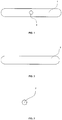
Fig. 1 shows a front view of the anchor according example 1, -
Fig. 2 shows a top view of the anchor according example 1, -
Fig. 3 shows a side view of the anchor according example 1, -
Fig. 4 . shows the anchor according example 1 with a thread, -
Fig. 5 shows a top view of a rectangular elastic sheet according example 2, -
Fig. 6 shows a top view of an oval elastic sheet according example 2 with a thread, -
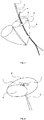
Fig. 7 shows a schematic introduction of the anchor into the mitral valve, -
Fig. 8 shows schematically the positioning of the anchor on the mitral valve and attachment thereof. - The
anchor 1 has a shape of an elongated cylinder, which can pass through ahollow needle 5 by means of a guide wire 4. The diameter of the elongated cylinder is smaller than the inner diameter of thehollow needle 5, and the ends of the cylinder are rounded. Theanchor 1 is provided with oneaperture 2 in the middle of its length through which asurgical thread 3 passes. Thesurgical thread 3 is used to transfer the anchor and fix it in the required position. Thisexemplary anchor 1 has length of 1 cm, a diameter of 0.2 cm and the inner diameter of theaperture 2 of 0.1 cm. Theanchor 1 is made from metal. - The
anchor 1 has a shape of an elongated cylinder and it is formed by a roll of anelastic sheet 1a. Theelastic sheet 1a is a plastic foil having a plan view in a rectangular shape, provided with twoapertures 2 in the middle through which asurgical thread 3 passes. Thesurgical thread 3 is used to position theanchor 1 and fix it in the required position. The roll ofelastic sheet 1a has its diameter smaller then the inner diameter of thehollow needle 5. Its length ranges from 0.5 to 3 cm, its width ranges from 0.05 to 1 cm and the inner diameter of theaperture 2 ranges from 0.005 cm to 1 cm. - Method of using the
anchor 1 according to invention has following steps:
Thehollow needle 5 is inserted into the left ventricle 8 through a small incision between the ribs and through the apex of the heart to pierce theposterior leaflet 7 and/or the anterior leaflet 9 of the mitral valve. - The
anchor 1 having thethread 3 passing through theaperture 2 is then pushed through thehollow needle 5 by means of a guide wire 4. Thehollow needle 5 is then pulled back and removed. - The
anchor 1 is pulled towards the apex of the heart using thethread 3, which positions the posteriormitral valve leaflet 7 and/or the anterior mitral valve leaflet 9 in the physiological position. - The
mitral valve leaflet 7 and/or the anterior leaflet 9 is fixed in the physiological position by securing theanchor 1 to the apex of the heart using thethread 3. Theanchor 1 remains on the atrial side of the mitral valve leaflets. All steps are performed under the echocardiographic control. - In case of using the
anchor 1 according to Example 2 the method comprises additional steps of: rolling up theelastic sheet 1a into the roll before pushing it through thehollow needle 5, and unrolling theelastic sheet 1a by pulling thethread 3 to resume its flat shape after passing through thehollow needle 5. -
- 1
- anchor
- 1a
- elastic sheet
- 2
- aperture
- 3
- thread
- 4
- guide wire
- 5
- hollow needle
- 6
- mitral valve annulus
- 7
- posterior leaflet of mitral valve
- 8
- left ventricle
- 9
- anterior leaflet of mitral valve
Claims (12)
- Anchor (1) to repair a mitral valve prolapse by fixing mitral valve leaflet in the physiological position characterized in that it has an elongated shape, which is adapted for longitudinal passage through a hollow needle (5) by means of a guide wire (4) and it is provided with at least one aperture (2) for passing of a thread (3).
- Anchor (1) according to claim 1, characterized in that the elongated shape is a cylinder, an ellipsoid or a prism, with at least one aperture (2) in the middle or its length.
- Anchor (1) according to claim 2, characterized in that its ends or edges are rounded or angulated.
- Anchor (1) according to claim 2, characterized in that it has the shape of the elongated cylinder made by a roll of an elastic sheet (1a).
- Anchor (1) according to claim 4, characterized in that the elastic sheet (1a) has a floor plan in a rectangle or oval shape.
- Anchor (1) according to claim 5, characterized in that there are two apertures (2) in the middle of the elastic sheet (1 a).
- Anchor (1) according to claim 5, characterized in that the elastic sheet (1a) is adapted to resume its flat shape.
- Anchor (1) according to any of the previous claims, characterized in that it is adapted to be transferred by pulling the thread (3) and fixed in a position by securing the thread (3).
- Anchor (1) according to any of the previous claims, characterized in that its length is in the range from 0.5 to 3 cm, its width is in the range from 0.05 to 1 cm and the inner diameter of the aperture (2) is in the range from 0.005 cm to 1 cm, preferably its length is in the range from 0.5 to 1.5 cm, its width is in the range from 0.05 to 0.25 cm and the inner diameter of the aperture (2) is in the range from 0.005 cm to 0.2 cm.
- Anchor (1) according to any of the previous claims according to claim 1, characterized in that the width of the anchor (1) is smaller than the inner diameter of the hollow needle (5).
- Method of using the anchor (1) according to any of claims 1 to 9 to repair a mitral valve prolapse, characterized in that it comprises:a) inserting the hollow needle (5) through a small incision between the ribs through the apex of the heart into the left ventricle (8) to pierce the posterior leaflet (7) and/or the anterior leaflet (9) of the mitral valve,b) pushing the anchor (1) provided with the thread (3) through the hollow needle (5) by means of a guide wire (4),c) pulling out and removal of the hollow needle (5),d) positioning of the posterior leaflet (7) or the anterior leaflet (9) by moving the anchor (1) towards the apex of the heart by pulling the thread (3),e) fixing of the leaflet(s) (7) and/or (9) in the physiological position by securing the anchor (1) to the apex of the heart using the thread (3),wherein all steps are performed under the echocardiographic control.
- Method according to claim 9, using the anchor (1) according to any of claims 4 -7, characterized in that it further comprises:a step of rolling up the elastic sheet (1a) into the roll before passing through the hollow needle (5), anda step of unrolling the elastic sheet (1 a) to resume its a flat shape after passing through the hollow needle (5).
Applications Claiming Priority (1)
| Application Number | Priority Date | Filing Date | Title |
|---|---|---|---|
| DE202020000044.2U DE202020000044U1 (en) | 2020-01-02 | 2020-01-02 | Anchor for correcting a mitral valve prolapse anchor with central hole for attachment, pulling from the mitral valve sail |
Publications (1)
| Publication Number | Publication Date |
|---|---|
| EP3845138A1 true EP3845138A1 (en) | 2021-07-07 |
Family
ID=70469796
Family Applications (1)
| Application Number | Title | Priority Date | Filing Date |
|---|---|---|---|
| EP21020002.8A Withdrawn EP3845138A1 (en) | 2020-01-02 | 2021-01-01 | Anchor to repair a mitral valve prolapse |
Country Status (2)
| Country | Link |
|---|---|
| EP (1) | EP3845138A1 (en) |
| DE (1) | DE202020000044U1 (en) |
Citations (5)
| Publication number | Priority date | Publication date | Assignee | Title |
|---|---|---|---|---|
| US20130090670A1 (en) * | 2010-04-13 | 2013-04-11 | Neosurgical Limited | Suture delivery system |
| US20170105838A1 (en) * | 2015-02-02 | 2017-04-20 | On-X Life Technologies, Inc. | Rapid Deployment Artificial Chordae Tendinae System |
| EP3263049A1 (en) | 2013-11-12 | 2018-01-03 | St. Jude Medical, Cardiology Division, Inc. | Transfemoral mitral valve repair delivery device |
| US20190328526A1 (en) * | 2016-12-30 | 2019-10-31 | Pipeline Medical Technologies, Inc. | Method of transvascular prosthetic chordae tendinae implantation |
| US20190380835A1 (en) * | 2018-06-05 | 2019-12-19 | Edwards Lifesciences Corporation | Valve repair devices and procedures |
-
2020
- 2020-01-02 DE DE202020000044.2U patent/DE202020000044U1/en not_active Expired - Lifetime
-
2021
- 2021-01-01 EP EP21020002.8A patent/EP3845138A1/en not_active Withdrawn
Patent Citations (5)
| Publication number | Priority date | Publication date | Assignee | Title |
|---|---|---|---|---|
| US20130090670A1 (en) * | 2010-04-13 | 2013-04-11 | Neosurgical Limited | Suture delivery system |
| EP3263049A1 (en) | 2013-11-12 | 2018-01-03 | St. Jude Medical, Cardiology Division, Inc. | Transfemoral mitral valve repair delivery device |
| US20170105838A1 (en) * | 2015-02-02 | 2017-04-20 | On-X Life Technologies, Inc. | Rapid Deployment Artificial Chordae Tendinae System |
| US20190328526A1 (en) * | 2016-12-30 | 2019-10-31 | Pipeline Medical Technologies, Inc. | Method of transvascular prosthetic chordae tendinae implantation |
| US20190380835A1 (en) * | 2018-06-05 | 2019-12-19 | Edwards Lifesciences Corporation | Valve repair devices and procedures |
Also Published As
| Publication number | Publication date |
|---|---|
| DE202020000044U1 (en) | 2020-03-31 |
Similar Documents
| Publication | Publication Date | Title |
|---|---|---|
| US12396720B2 (en) | Devices and methods for locating and implanting tissue anchors at mitral valve commissure | |
| CN113873972B (en) | Heart valve repair | |
| CN111212614B (en) | Axisymmetric adjustable device for treating mitral regurgitation | |
| US10675017B2 (en) | Transcatheter heart valve leaflet plication | |
| US10639024B2 (en) | Method and apparatus for transapical procedures on a mitral valve | |
| JP5198431B2 (en) | Annuloplasty device with helical anchor | |
| CN105852916B (en) | A kind of bicuspid valve flexibility closure plate occluder and method for implantation being implanted into through the apex of the heart | |
| EP3150173B1 (en) | Reabsorbable subaortic ring | |
| CN111200994A (en) | Transcatheter device for treating mitral regurgitation | |
| CN110709029A (en) | Minimally Invasive Implantable Devices and Mitral Valve Implant Systems | |
| US20110270276A1 (en) | Methods and Devices for Cardiac Valve Repair or Replacement | |
| JP2009519784A (en) | System and method for controlling heart valve dimensions | |
| DE102017002974A1 (en) | Heart valve implant, suitable for use in minimally invasive surgery for the repair of a heart valve and / or a heart valve beating on the beating heart | |
| JP2009519783A (en) | Papillary muscle position control device, system and method | |
| JP2011519693A (en) | Suture device and method for suturing anatomic valves | |
| CN115429492B (en) | Valve clamping device and valve clamping system | |
| CN114081670A (en) | Repair system for preventing valve regurgitation | |
| CN114376766B (en) | Adjustable and removable valve clamping device | |
| CN107595437B (en) | Valve clamping system | |
| US11517435B2 (en) | Ring-based prosthetic cardiac valve | |
| CN216124623U (en) | Control release mechanism on valve clamp | |
| US20220304800A1 (en) | Valve Reshaping Device, System, and Related Methods | |
| CN112165904A (en) | Tissue anchors with spacers | |
| CN117618153A (en) | Heart valve repair device and method | |
| WO2021034538A1 (en) | Heart valve leaflet tethering |
Legal Events
| Date | Code | Title | Description |
|---|---|---|---|
| STAA | Information on the status of an ep patent application or granted ep patent |
Free format text: STATUS: REQUEST FOR EXAMINATION WAS MADE |
|
| PUAI | Public reference made under article 153(3) epc to a published international application that has entered the european phase |
Free format text: ORIGINAL CODE: 0009012 |
|
| 17P | Request for examination filed |
Effective date: 20210125 |
|
| AK | Designated contracting states |
Kind code of ref document: A1 Designated state(s): AL AT BE BG CH CY CZ DE DK EE ES FI FR GB GR HR HU IE IS IT LI LT LU LV MC MK MT NL NO PL PT RO RS SE SI SK SM TR |
|
| STAA | Information on the status of an ep patent application or granted ep patent |
Free format text: STATUS: THE APPLICATION IS DEEMED TO BE WITHDRAWN |
|
| 18D | Application deemed to be withdrawn |
Effective date: 20220108 |