EP3845134A1 - Ultrasound diagnosis device and ultrasound diagnosis device control method - Google Patents
Ultrasound diagnosis device and ultrasound diagnosis device control method Download PDFInfo
- Publication number
- EP3845134A1 EP3845134A1 EP19856254.8A EP19856254A EP3845134A1 EP 3845134 A1 EP3845134 A1 EP 3845134A1 EP 19856254 A EP19856254 A EP 19856254A EP 3845134 A1 EP3845134 A1 EP 3845134A1
- Authority
- EP
- European Patent Office
- Prior art keywords
- discrimination
- ultrasound
- unit
- blood vessel
- image
- Prior art date
- Legal status (The legal status is an assumption and is not a legal conclusion. Google has not performed a legal analysis and makes no representation as to the accuracy of the status listed.)
- Granted
Links
Images




Classifications
-
- A—HUMAN NECESSITIES
- A61—MEDICAL OR VETERINARY SCIENCE; HYGIENE
- A61B—DIAGNOSIS; SURGERY; IDENTIFICATION
- A61B8/00—Diagnosis using ultrasonic, sonic or infrasonic waves
- A61B8/06—Measuring blood flow
-
- A—HUMAN NECESSITIES
- A61—MEDICAL OR VETERINARY SCIENCE; HYGIENE
- A61B—DIAGNOSIS; SURGERY; IDENTIFICATION
- A61B8/00—Diagnosis using ultrasonic, sonic or infrasonic waves
- A61B8/42—Details of probe positioning or probe attachment to the patient
- A61B8/4245—Details of probe positioning or probe attachment to the patient involving determining the position of the probe, e.g. with respect to an external reference frame or to the patient
- A61B8/4254—Details of probe positioning or probe attachment to the patient involving determining the position of the probe, e.g. with respect to an external reference frame or to the patient using sensors mounted on the probe
-
- A—HUMAN NECESSITIES
- A61—MEDICAL OR VETERINARY SCIENCE; HYGIENE
- A61B—DIAGNOSIS; SURGERY; IDENTIFICATION
- A61B5/00—Measuring for diagnostic purposes; Identification of persons
- A61B5/48—Other medical applications
- A61B5/4887—Locating particular structures in or on the body
- A61B5/489—Blood vessels
-
- A—HUMAN NECESSITIES
- A61—MEDICAL OR VETERINARY SCIENCE; HYGIENE
- A61B—DIAGNOSIS; SURGERY; IDENTIFICATION
- A61B8/00—Diagnosis using ultrasonic, sonic or infrasonic waves
- A61B8/08—Clinical applications
- A61B8/0891—Clinical applications for diagnosis of blood vessels
-
- A—HUMAN NECESSITIES
- A61—MEDICAL OR VETERINARY SCIENCE; HYGIENE
- A61B—DIAGNOSIS; SURGERY; IDENTIFICATION
- A61B8/00—Diagnosis using ultrasonic, sonic or infrasonic waves
- A61B8/13—Tomography
- A61B8/14—Echo-tomography
-
- A—HUMAN NECESSITIES
- A61—MEDICAL OR VETERINARY SCIENCE; HYGIENE
- A61B—DIAGNOSIS; SURGERY; IDENTIFICATION
- A61B8/00—Diagnosis using ultrasonic, sonic or infrasonic waves
- A61B8/46—Ultrasonic, sonic or infrasonic diagnostic devices with special arrangements for interfacing with the operator or the patient
- A61B8/461—Displaying means of special interest
-
- A—HUMAN NECESSITIES
- A61—MEDICAL OR VETERINARY SCIENCE; HYGIENE
- A61B—DIAGNOSIS; SURGERY; IDENTIFICATION
- A61B8/00—Diagnosis using ultrasonic, sonic or infrasonic waves
- A61B8/48—Diagnostic techniques
- A61B8/488—Diagnostic techniques involving Doppler signals
-
- A—HUMAN NECESSITIES
- A61—MEDICAL OR VETERINARY SCIENCE; HYGIENE
- A61B—DIAGNOSIS; SURGERY; IDENTIFICATION
- A61B8/00—Diagnosis using ultrasonic, sonic or infrasonic waves
- A61B8/52—Devices using data or image processing specially adapted for diagnosis using ultrasonic, sonic or infrasonic waves
- A61B8/5207—Devices using data or image processing specially adapted for diagnosis using ultrasonic, sonic or infrasonic waves involving processing of raw data to produce diagnostic data, e.g. for generating an image
-
- A—HUMAN NECESSITIES
- A61—MEDICAL OR VETERINARY SCIENCE; HYGIENE
- A61B—DIAGNOSIS; SURGERY; IDENTIFICATION
- A61B8/00—Diagnosis using ultrasonic, sonic or infrasonic waves
- A61B8/54—Control of the diagnostic device
-
- A—HUMAN NECESSITIES
- A61—MEDICAL OR VETERINARY SCIENCE; HYGIENE
- A61B—DIAGNOSIS; SURGERY; IDENTIFICATION
- A61B8/00—Diagnosis using ultrasonic, sonic or infrasonic waves
- A61B8/56—Details of data transmission or power supply
-
- A—HUMAN NECESSITIES
- A61—MEDICAL OR VETERINARY SCIENCE; HYGIENE
- A61B—DIAGNOSIS; SURGERY; IDENTIFICATION
- A61B2562/00—Details of sensors; Constructional details of sensor housings or probes; Accessories for sensors
- A61B2562/02—Details of sensors specially adapted for in-vivo measurements
- A61B2562/0219—Inertial sensors, e.g. accelerometers, gyroscopes, tilt switches
Definitions
- the present invention relates to an ultrasound diagnostic apparatus and a method of controlling an ultrasound diagnostic apparatus, and particularly, to an ultrasound diagnostic apparatus and a method of controlling an ultrasound diagnostic apparatus that are used for observing blood vessels of a subject.
- an ultrasound diagnostic apparatus has been known as an apparatus that obtains an image of the inside of a subject.
- the ultrasound diagnostic apparatus comprises an ultrasound probe provided with an oscillator array in which a plurality of elements are arranged.
- an ultrasound beam is transmitted from the oscillator array toward the inside of the subject, ultrasound echoes from the subject are received by the oscillator array, and element data are acquired.
- the ultrasound diagnostic apparatus processes the obtained element data electrically, and generates an ultrasound image for a relevant part of the subj ect.
- a blood vessel of the subject is observed using such an ultrasound diagnostic apparatus.
- the user usually determines whether the blood vessel in the ultrasound image is a vein or an artery by visually observing the ultrasound image obtained by the ultrasound diagnostic apparatus.
- an ultrasound diagnostic apparatus has been developed that can discriminate whether the blood vessel in the ultrasound image is the vein or the artery even in a case where the user does not have specialized knowledge.
- JP2008-272025A discloses an ultrasound diagnostic apparatus that generates a so-called Doppler signal by performing frequency analysis on a received signal generated by receiving an ultrasound echo in a subject, and discriminates a blood vessel in the ultrasound image based on the intensity of the generated Doppler signal.
- the ultrasound diagnostic apparatus disclosed in JP2008-272025A discriminates, among two blood vessels included in the ultrasound image, that the blood vessel having the high intensity of the Doppler signal is the artery, and the blood vessel having the low intensity of the Doppler signal is the vein.
- the observation is often performed while moving the ultrasound probe.
- the ultrasound images representing different tomographic planes of the subject are sequentially generated, but a blood vessel in the ultrasound image is discriminated each time the tomographic plane of the subject represented by the ultrasound image sequentially changes, and thus there is a problem that the power consumption in the ultrasound diagnostic apparatus is large.
- the present invention has been made in order to solve such related-art problems, and an object thereof is to provide an ultrasound diagnostic apparatus and a method of controlling an ultrasound diagnostic apparatus that can reduce the power consumption while discriminating the blood vessel.
- an ultrasound diagnostic apparatus comprises an ultrasound probe, an image acquisition unit that acquires an ultrasound image sequentially by transmitting an ultrasound beam toward a subject from the ultrasound probe, a blood vessel detection unit that detects a blood vessel included in the ultrasound image acquired by the image acquisition unit, a blood vessel discrimination unit that discriminates whether the blood vessel detected by the blood vessel detection unit is a vein or an artery, and a discrimination execution deciding unit that decides whether the blood vessel discrimination unit newly executes discrimination with respect to the ultrasound image of a current frame based on a movement amount of the ultrasound probe or a change amount of the ultrasound image between frames.
- the ultrasound diagnostic apparatus may further comprise a motion sensor that is attached to the ultrasound probe, and a probe movement amount calculating unit that calculates the movement amount of the ultrasound probe based on a value measured by the motion sensor, in which the discrimination execution deciding unit decides whether the blood vessel discrimination unit executes discrimination based on the movement amount of the ultrasound probe calculated by the probe movement amount calculating unit.
- the ultrasound diagnostic apparatus further comprise a discrimination result memory that holds a latest discrimination result by the blood vessel discrimination unit, in which in a case where the movement amount of the ultrasound probe calculated by the probe movement amount calculating unit is equal to or smaller than a predetermined first threshold value, the discrimination execution deciding unit decides that the blood vessel discrimination unit does not newly execute discrimination with respect to the ultrasound image of the current frame, and the blood vessel discrimination unit maintains the latest discrimination result held in the discrimination result memory.
- the blood vessel discrimination unit may track a position of the blood vessel in the ultrasound image from the frame in which the blood vessel discrimination unit executes latest discrimination to the current frame, and maintain the latest discrimination result held in the discrimination result memory.
- the discrimination execution deciding unit may decide that the blood vessel discrimination unit newly executes discrimination with respect to the ultrasound image of the current frame, and the blood vessel discrimination unit may discriminate whether the blood vessel in the ultrasound image of the current frame is a vein or an artery, and update the latest discrimination result held in the discrimination result memory based on the discrimination result.
- the discrimination execution deciding unit may decide that the blood vessel discrimination unit newly executes discrimination with respect to the ultrasound image of the current frame, and the blood vessel discrimination unit may discriminate whether the blood vessel in the ultrasound image of the current frame is a vein or an artery, and update the latest discrimination result held in the discrimination result memory based on the discrimination result.
- the blood vessel discrimination unit may newly execute discrimination on whether the blood vessel in the ultrasound image of the current frame is a vein or an artery after the movement amount of the ultrasound probe calculated by the probe movement amount calculating unit becomes equal to or smaller than a third threshold value that is smaller than the first threshold value.
- the ultrasound diagnostic apparatus further comprise a display unit that displays the ultrasound image acquired by the image acquisition unit and the discrimination result held in the discrimination result memory.
- the movement amount of the ultrasound probe calculated by the probe movement amount calculating unit include at least one of a moving speed of the ultrasound probe in parallel movement, a change amount in a movement direction of the ultrasound probe, or an angular velocity of the ultrasound probe in rotational movement.
- the motion sensor consist of at least one of an acceleration sensor, a gyro sensor, a magnetic sensor, or a position sensor of a global positioning system.
- the ultrasound diagnostic apparatus may further comprise an image change amount calculation unit that calculates the change amount of the ultrasound image between the frames by performing image analysis with respect to the ultrasound image acquired by the image acquisition unit, in which the discrimination execution deciding unit decides whether the blood vessel discrimination unit executes discrimination of the blood vessel based on the change amount of the ultrasound image calculated by the image change amount calculation unit.
- the ultrasound diagnostic apparatus further comprise a discrimination result memory that holds a latest discrimination result of the blood vessel discrimination unit, in which in a case where the change amount of the ultrasound image between the frames calculated by the image change amount calculation unit is equal to or smaller than a predetermined fourth threshold value, the discrimination execution deciding unit decides that the blood vessel discrimination unit does not newly execute discrimination with respect to the ultrasound image of the current frame, and the blood vessel discrimination unit maintains the latest discrimination result held in the discrimination result memory.
- the blood vessel discrimination unit may track a position of the blood vessel in the ultrasound image from the frame in which the blood vessel discrimination unit executes latest discrimination to the current frame, and maintain the latest discrimination result held in the discrimination result memory.
- the discrimination execution deciding unit may decide that the blood vessel discrimination unit newly executes discrimination with respect to the ultrasound image of the current frame, and the blood vessel discrimination unit may discriminate whether the blood vessel in the ultrasound image of the current frame is a vein or an artery, and update the latest discrimination result held in the discrimination result memory based on the discrimination result.
- the discrimination execution deciding unit may decide that the blood vessel discrimination unit newly executes discrimination with respect to the ultrasound image of the current frame, and the blood vessel discrimination unit may discriminate whether the blood vessel in the ultrasound image of the current frame is a vein or an artery, and update the latest discrimination result held in the discrimination result memory based on the discrimination result.
- the blood vessel discrimination unit may newly execute discrimination on whether the blood vessel in the ultrasound image of the current frame is a vein or an artery after the change amount of the ultrasound image between the frames calculated by the image change amount calculation unit becomes equal to or smaller than a sixth threshold value that is smaller than the fourth threshold value.
- the ultrasound diagnostic apparatus further comprise a display unit that displays the ultrasound image acquired by the image acquisition unit and the discrimination result held in the discrimination result memory.
- the image acquisition unit may include a receiving unit that receives an ultrasound echo in the subject by the ultrasound probe and generates a received signal
- the ultrasound diagnostic apparatus may further comprise a Doppler signal generating unit that generates a Doppler signal based on the received signal generated by the receiving unit
- the blood vessel discrimination unit may discriminate whether the blood vessel is a vein or an artery based on the Doppler signal generated by the Doppler signal generating unit.
- a method of controlling an ultrasound diagnostic apparatus comprises acquiring an ultrasound image sequentially by transmitting an ultrasound beam toward a subject from an ultrasound probe, detecting a blood vessel included in the acquired ultrasound image, discriminating whether the detected blood vessel is a vein or an artery, and deciding whether to newly execute discrimination with respect to the ultrasound image of a current frame based on a movement amount of the ultrasound probe or a change amount of the ultrasound image between frames.
- the ultrasound diagnostic apparatus comprises a blood vessel detection unit that detects a blood vessel included in the ultrasound image acquired by the image acquisition unit, a blood vessel discrimination unit that discriminates whether the blood vessel detected by the blood vessel detection unit is a vein or an artery, and a discrimination execution deciding unit that decides whether the blood vessel discrimination unit newly executes discrimination of the blood vessel included in the ultrasound image of a current frame based on a movement amount of the ultrasound probe or a change amount of the ultrasound image between frames, and thus it is possible to reduce the power consumption while discriminating the blood vessel.
- FIG. 1 A configuration of an ultrasound diagnostic apparatus 1 according to Embodiment 1 of the present invention is shown in Fig. 1 .
- the ultrasound diagnostic apparatus 1 comprises an oscillator array 2, and a transmitting unit 3 and a receiving unit 4 are connected to the oscillator array 2.
- An image generation unit 5, a display controller 6, and a display unit 7 are sequentially connected to the receiving unit 4.
- An image acquisition unit 8 is configured by the transmitting unit 3, the receiving unit 4, and the image generation unit 5.
- a Doppler signal generating unit 9 is connected to the receiving unit 4.
- a blood vessel detection unit 10 is connected to the image generation unit 5.
- a blood vessel discrimination unit 11 is connected to the Doppler signal generating unit 9 and the blood vessel detection unit 10, and a discrimination result memory 12 and the display controller 6 are connected to the blood vessel discrimination unit 11.
- the blood vessel discrimination unit 11 and the discrimination result memory 12 are connected to each other so as to be capable of transferring information bidirectionally.
- the oscillator array 2 is included in the ultrasound probe 19, and a motion sensor 13 is attached to the ultrasound probe 19.
- a probe movement amount calculating unit 14 is connected to the motion sensor 13, and a discrimination execution deciding unit 15 is connected to the probe movement amount calculating unit 14.
- the discrimination execution deciding unit 15 is connected to the blood vessel discrimination unit 11.
- the apparatus controller 16 is connected to the display controller 6, the image acquisition unit 8, the Doppler signal generating unit 9, the blood vessel discrimination unit 11, the blood vessel detection unit 10, the probe movement amount calculating unit 14, and the discrimination execution deciding unit 15, and the input unit 17 and the storage unit 18 are connected to the apparatus controller 16.
- the apparatus controller 16 and the storage unit 18 are connected to each other so as to be capable of transferring information bidirectionally.
- the processor 20 is configured by the display controller 6, the image acquisition unit 8, the Doppler signal generating unit 9, the blood vessel discrimination unit 11, the blood vessel detection unit 10, the probe movement amount calculating unit 14, the discrimination execution deciding unit 15, and the apparatus controller 16.
- the oscillator array 2 of the ultrasound probe 19 shown in Fig. 1 has a plurality of oscillators arranged in one dimension or two dimensions. These oscillators transmit ultrasound waves in accordance with drive signals supplied from the transmitting unit 3, respectively, and receive ultrasound echoes from the subject to output the received signals.
- the respective oscillators are configured by, for example, forming electrodes to both ends of piezoelectric material made of a piezoelectric ceramic represented by lead zirconate titanate (PZT), a polymeric piezoelectric element represented by polyvinylidene difluoride (PVDF), and a piezoelectric single crystal represented by a lead magnesium niobate-lead titanate solid solution (PMN-PT).
- PZT lead zirconate titanate
- PVDF polymeric piezoelectric element
- PMN-PT piezoelectric single crystal represented by a lead magnesium niobate-lead titanate solid solution
- the transmitting unit 3 of the image acquisition unit 8 includes, for example, a plurality of pulse generators, and adjust the amounts of delay of the respective drive signals to supply the adjusted drive signals to the plurality of oscillators such that the ultrasound waves transmitted from the plurality of oscillators of the oscillator array 2 form an ultrasound beam, based on a transmission delay pattern selected in accordance with the control signals from the apparatus controller 16.
- a pulsed or consecutive wave-like voltage is applied to electrodes of the plurality of oscillators of the oscillator array 2
- the piezoelectric material expands and contracts
- a pulsed or consecutive wave-like ultrasound wave is generated from the respective oscillators
- the ultrasound beam is formed from a synthetic wave of the ultrasound waves.
- the transmitted ultrasound beam is reflected in, for example, targets such as a part of the subject and is propagated toward the oscillator array 2 of the ultrasound probe 19.
- the ultrasound echoes propagated toward the oscillator array 2 in this way are received by the respective oscillators that constitute the oscillator array 2.
- the respective oscillators that constitute the oscillator array 2 expand and contract by receiving the propagated ultrasound echoes, to generate electrical signals, and output the electrical signals to the receiving unit 4.
- the receiving unit 4 of the image acquisition unit 8 processes the signals output from the oscillator array 2 in accordance with the control signals from the apparatus controller 16. As shown in Fig. 2 , the receiving unit 4 has a configuration that an amplification unit 21 and an analog-digital (AD) conversion unit 22 are serially connected.
- the amplification unit 21 amplifies the signals input from the respective oscillators that constitute the oscillator array 2, and transmits the amplified signals to the AD conversion unit 22.
- the AD conversion unit 22 converts the signals transmitted from the amplification unit 21 into digitalized received signal, and sends the data to the image generation unit 5 of the image acquisition unit 8.
- the image generation unit 5 of the image acquisition unit 8 has a configuration that a signal processing unit 23, a digital scan converter (DSC) 24, and an image processing unit 25 are serially connected.
- the signal processing unit 23 performs reception focus processing that addition (phasing addition) is performed by giving each delay to each data of the received signals, based on a reception delay pattern selected in accordance with the control signals from the apparatus controller 16. With the reception focus processing, sound ray signals in which focal points of the ultrasound echoes are narrowed to one scan line are generated.
- the signal processing unit 23 subjects the generated sound ray signals to the correction of damping resulting from a propagation distance depending on the depth at a position where the ultrasound waves are reflected, and then performs envelope detection processing to generate B-mode image signals indicating a tissue within the subject.
- the B-mode image signals generated in this way are output to the DSC 24.
- the DSC 24 of the image generation unit 5 raster-converts the B-mode image signals into image signals based on a scan mode of normal television signals to generate the ultrasound image.
- the image processing unit 25 of the image generation unit 5 subjects the ultrasound image obtained in the DSC 24 to various kinds of required image processing such as brightness correction, grayscale correction, sharpness correction, and color correction, and then outputs an ultrasound image to the display controller 6, and the blood vessel detection unit 10.
- the Doppler signal generating unit 9 of the processor 20 generates the Doppler signal by a so-called pulse Doppler method.
- the Doppler signal generating unit 9 has a configuration in which an orthogonal detection unit 26, a high-pass filter 27, a fast Fourier transformer 28 are sequentially connected in series.
- the orthogonal detection unit 26 orthogonally detects the received signal and converts the received signal into complex data by mixing the carrier signal of the reference frequency with the received signal generated by the receiving unit 4.
- the high-pass filter 27 functions as a so-called wall filter, and removes frequency components derived from the movement of the body tissue of the subject from the complex data generated by the orthogonal detection unit 26.
- the fast Fourier transformer 28 performs frequency analysis by Fourier transforming the complex data of a plurality of sample points, and generates the Doppler signal representing a so-called spectrum.
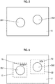
- the blood vessel detection unit 10 of the processor 20 performs image analysis with respect to the ultrasound image acquired by the image acquisition unit 8, and detects the blood vessel included in the ultrasound image. For example, as shown in Fig. 5 , the blood vessel detection unit 10 detects cross sections of blood vessels BV1 and BV2 included in the ultrasound image U.
- the cross section of the blood vessel represents the section of the blood vessel in a case of cutting the blood vessel across a central axis thereof. More specifically, for example, the blood vessel detection unit 10 can detect the cross section of the blood vessel by storing typical pattern data as a template in advance, calculating the similarity to the pattern data while searching the image with the template, and assuming the blood vessel is present in the position where the similarity is equal to or larger than a threshold value and is maximum.
- the motion sensor 13 attached to the ultrasound probe 19 measures the movement of the ultrasound probe 19 moved by the user.
- Examples of the motion sensor 13 include an acceleration sensor, a gyro sensor, a magnetic sensor, or a position sensor of a global positioning system (GPS). Therefore, the motion sensor 13 measures acceleration of the ultrasound probe 19 in parallel movement, angular acceleration or an angular velocity of the ultrasound probe 19 in rotational movement, and position of the ultrasound probe 19 as the movement of the ultrasound probe 19.
- GPS global positioning system
- the probe movement amount calculating unit 14 of the processor 20 calculates the movement amount of the ultrasound probe 19 based on the value measured by the motion sensor 13.
- the probe movement amount calculating unit 14 can calculate a moving speed of the ultrasound probe 19 in parallel movement, an angular velocity of the ultrasound probe 19 in rotational movement, and the change amount in the movement direction of the ultrasound probe 19 as the movement amount of the ultrasound probe 19.
- the blood vessel discrimination unit 11 of the processor 20 discriminates whether the blood vessels BV1 and BV2 detected by the blood vessel detection unit 10 are the vein or the artery.
- the artery beats at a constant cycle
- the intensity of the Doppler signal in the cross section of the blood vessel corresponding to the artery also changes at the same constant cycle as the beat.
- the vein does not beat, and thus the intensity of the Doppler signal in the cross section of the blood vessel corresponding to the vein does not change periodically.
- the blood vessel discrimination unit 11 discriminates that the blood vessel in which the intensity of the Doppler signal generated by the Doppler signal generating unit 9 changes periodically is the artery, and the blood vessel in which the intensity of the Doppler signal does not change periodically is vein.
- the blood vessel be discriminated in a state where the ultrasound probe 19 is substantially stationary such that the blood vessels can be accurately discriminated. Therefore, it is desirable that the blood vessel discrimination unit 11 discriminates the blood vessel after the movement amount of the ultrasound probe 19 calculated by the probe movement amount calculating unit 14 is in a substantially stationary state.
- the blood vessel discrimination unit 11 causes the display unit 7 to display the discrimination results for the blood vessels BV1 and BV2 via the display controller 6.
- the blood vessel discrimination unit 11 can display the discrimination results that the blood vessel BV1 is the artery and the blood vessel BV2 is the vein on the display unit 7 by discriminating that the blood vessel BV1 is the artery and the blood vessel BV2 is the vein, displaying a text T1 indicating that the blood vessel is the artery and a surrounding line E1 surrounding the blood vessel BV1 and the text T1 in the vicinity of the blood vessel BV1 in the ultrasound image U, and displaying a text T2 indicating that the blood vessel is the vein and a surrounding line E2 surrounding the blood vessel BV2 and the text T2 in the vicinity of the blood vessel BV2.
- the blood vessel discrimination unit 11 can display the surrounding lines E1 and E2 with different colors such as red and blue on the display unit 7 such that the user can easily confirm whether the blood vessels BV1 and the BV2 are the vein or the artery.
- the discrimination result memory 12 of the ultrasound diagnostic apparatus 1 stores the latest discrimination result by the blood vessel discrimination unit 11.
- a recording medium such as a hard disc drive (HDD), a solid state drive (SSD), a flexible disc (FD), a magneto-optical (MO) disc, a magnetic tape (MT), a random access memory (RAM), a compact disc (CD), a digital versatile disc (DVD), a secure digital (SD) card, a universal serial bus (USB) memory, or the like, or a server can be used.
- the discrimination result memory 12 is not included in the processor 20, but may be included in the processor 20.
- the discrimination execution deciding unit 15 of the processor 20 decides whether the blood vessel discrimination unit 11 newly executes discrimination of the blood vessel included in the ultrasound image of the current frame acquired by the image acquisition unit 8 based on the movement amount of the ultrasound probe 19 calculated by the probe movement amount calculating unit 14. For example, the discrimination execution deciding unit 15 decides that discrimination of the blood vessel is not newly executed with respect to the ultrasound image of the current frame in a case where the movement amount of the ultrasound probe 19 calculated by the probe movement amount calculating unit 14 is equal to or smaller than a predetermined first threshold value, and decides that discrimination of the blood vessel is newly executed with respect to the ultrasound image of the current frame in a case where the movement amount of the ultrasound probe 19 is larger than the first threshold value. For example, in a case where the movement amount of the ultrasound probe 19 is the moving speed of the ultrasound probe 19 in parallel movement, 20 mm/sec can be set as the first threshold value.
- the apparatus controller 16 of the processor 20 performs control of the respective units of the ultrasound diagnostic apparatus 1 based on the programs that are stored in advance in the storage unit 18 and the like and the operation of the user via the input unit 17.
- the display controller 6 of the processor 20 performs predetermined processing on an ultrasound image generated by the image generation unit 5 of the image acquisition unit 8, and displays the ultrasound image on the display unit 7.
- the display unit 7 of the ultrasound diagnostic apparatus 1 displays the ultrasound image under the control of the display controller 6, and includes display devices such as a liquid crystal display (LCD), and an organic electroluminescence (EL) display.
- display devices such as a liquid crystal display (LCD), and an organic electroluminescence (EL) display.
- the input unit 17 of the ultrasound diagnostic apparatus 1 is a device for the user to perform input operation, and can be configured to comprise a keyboard, a mouse, a trackball, a touchpad, a touch panel, and the like.
- the storage unit 18 stores the operating program of the ultrasound diagnostic apparatus 1, and as the storage unit, a recording medium such as the HDD, the SSD, the FD, the MO disc, the MT, the RAM, the CD, the DVD, the SD card, the USB memory, or the like can be used similarly to the discrimination result memory 12.
- a recording medium such as the HDD, the SSD, the FD, the MO disc, the MT, the RAM, the CD, the DVD, the SD card, the USB memory, or the like can be used similarly to the discrimination result memory 12.
- the processor 20 having the display controller 6, the image acquisition unit 8, the Doppler signal generating unit 9, the blood vessel discrimination unit 11, the blood vessel detection unit 10, the probe movement amount calculating unit 14, the discrimination execution deciding unit 15, and the apparatus controller 16 is configured from a central processing unit (CPU) and control programs for making the CPU perform various kinds of processing.
- the processor 20 may be configured from a field programmable gate array (FGPA), a digital signal processor (DSP), an application specific integrated circuit (ASIC), or other integrated circuits (ICs), or may be configured by combining them.
- FGPA field programmable gate array
- DSP digital signal processor
- ASIC application specific integrated circuit
- ICs integrated circuits
- the configuration can be adopted in which the display controller 6, the image acquisition unit 8, the Doppler signal generating unit 9, the blood vessel discrimination unit 11, the blood vessel detection unit 10, the probe movement amount calculating unit 14, the discrimination execution deciding unit 15, and the apparatus controller 16 of the processor 20 are partially or entirely integrated into one CPU.
- the image acquisition unit 8 of the processor 20 sequentially and consecutively acquires the ultrasound images and displays the ultrasound images on the display unit 7, and the user moves the ultrasound probe 19 as the blood vessel is observed.
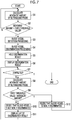
- step S1 the probe movement amount calculating unit 14 calculates the movement amount of the ultrasound probe 19 based on the measured value obtained by the motion sensor 13 attached to the ultrasound probe 19.
- step S2 the blood vessel discrimination unit 11 determines whether the ultrasound probe 19 is in a substantially stationary state, that is, whether the movement amount of the ultrasound probe 19 calculated in step S1 is equal to or smaller than the third threshold value that is smaller than the first threshold value. For example, in a case where the movement amount of the ultrasound probe 19 is the moving speed of the ultrasound probe 19 in parallel movement, 3 mm/sec can be set as the third threshold value.
- step S2 in a case where the determination is made that the movement amount of the ultrasound probe 19 is larger than the third threshold value, the process returns to step S1, the movement amount of the ultrasound probe 19 is newly calculated, and the process proceeds to step S2. As described above, step S1 and step S2 are repeated until the movement amount of the ultrasound probe 19 become equal to or smaller than the third threshold value.
- step S3 the blood vessel detection unit 10 performs image analysis with respect to the ultrasound image of the current frame acquired by the image acquisition unit 8, and performs detection processing of the blood vessels BV1 and BV2 included in the ultrasound image U is performed as shown in Fig. 5 , for example.
- the blood vessel discrimination unit 11 performs processing of discriminating whether the blood vessels BV1 and BV2 are the vein or the artery based on the intensity of the Doppler signal in the blood vessels BV1 and BV2 detected in step S3. For example, it is possible that the Doppler signal generating unit 9 generates the Doppler signal corresponding to entire area of the ultrasound image U, and the blood vessel discrimination unit 11 discriminates that among the blood vessels BV1 and BV2, the blood vessel in which the intensity of the Doppler signal changes periodically is the artery, and the blood vessel in which the intensity of the Doppler signal does not change periodically is vein.
- the oscillator array 2 of the ultrasound probe 19 transmits the ultrasound beam to the cross section of the subject corresponding to the entire area of the ultrasound image U.
- step S5 the blood vessel discrimination unit 11 holds the discrimination result obtained in step S4 in the discrimination result memory 12.
- the blood vessel discrimination unit 11 displays the discrimination result held in the discrimination result memory 12 in step S5 on the display unit 7.
- the blood vessel discrimination unit 11 can display the discrimination results on the display unit 7 by displaying a text T1 indicating that the blood vessel is the artery and a surrounding line E1 surrounding the blood vessel BV1 and the text T1 in the vicinity of the blood vessel BV1, and displaying a text T2 indicating that the blood vessel is the vein and a surrounding line E2 surrounding the blood vessel BV2 and the text T2 in the vicinity of the blood vessel BV2 while being superimposed on the ultrasound image U.
- step S7 it is determined whether the operation of the ultrasound diagnostic apparatus 1 ends. For example, although not shown, in a case where a operation end button for ending the operation of the ultrasound diagnostic apparatus 1 is displayed on the display unit 7, and the user presses the operation end button via the input unit 17, it is determined that the operation of the ultrasound diagnostic apparatus 1 ends, and thus the operation of the ultrasound diagnostic apparatus 1 ends. In a case where the operation end button is not pressed, it is determined that the operation of the ultrasound diagnostic apparatus 1 does not end, and the process proceeds to step S8.
- step S8 the probe movement amount calculating unit 14 newly calculates the movement amount of the ultrasound probe 19 in the same manner as in step S1.
- step S9 the discrimination execution deciding unit 15 determines whether the movement amount of the ultrasound probe 19 calculated in step S8 is larger than the first threshold value. In a case where the determination is made that the movement amount of the ultrasound probe 19 is equal to or smaller than the first threshold value in step S9, the process proceeds to step S10. In step S10, the discrimination execution deciding unit 15 decides that the blood vessel discrimination unit 11 does not newly discriminate the blood vessel in the ultrasound image of the current frame.
- Step S11 the blood vessel discrimination unit 11 displays the latest discrimination result, that is, the discrimination result held in step S5 while being superimposed on the ultrasound image U.
- the detailed operation in step S11 will be described with reference to the flowchart of Fig. 8 .
- Step S11 is processing executed in a case where the determination is made that the movement amount of the ultrasound probe 19 is equal to or smaller than the first threshold value in step S9, and includes three steps of step S13 to step S15 as shown in Fig. 8 .
- step S13 the blood vessel discrimination unit 11 determines whether the movement amount of the ultrasound probe 19 calculated in step S8 is equal to or smaller than a predetermined second threshold value.
- the second threshold value is a value smaller than the first threshold value and equal to or larger than the third threshold value. In a case where the determination is made that the movement amount of the ultrasound probe 19 is equal to or smaller than the second threshold value in step S13, the process proceeds to step S14.
- step S14 the blood vessel discrimination unit 11 determines that the ultrasound probe 19 is in a substantially stationary state, and in step S5, the discrimination result held in the discrimination result memory 12 is maintained. Further, the blood vessel discrimination unit 11 displays the discrimination results for the blood vessels corresponding to the blood vessels BV1 and BV2 shown in Figs. 5 and 6 held in the discrimination result memory 12 in step S5 while being superimposed on the ultrasound image of the current frame. In a case where processing of step S14 is completed, the processing of step S11 ends.
- step S15 the process proceeds to step S15.
- step S15 the blood vessel discrimination unit 11 determines that the ultrasound probe 19 has movement, but has no big movement as to newly perform discrimination processing, tracks the blood vessels BV1 and BV2 included in the ultrasound image U, and maintains the discrimination result held in the discrimination result memory 12 in step S5. Further, the blood vessel discrimination unit 11 performs processing of displaying the discrimination results for the blood vessels corresponding to the blood vessels BV1 and BV2 included in the ultrasound image U held in the discrimination result memory 12.
- the user moves the ultrasound probe 19 by the movement amount that is equal to or smaller than the first threshold value and larger than the second threshold value, and as shown in Fig. 9 , in a case where the blood vessels BV1 and BV2 in the ultrasound image U is moved to blood vessels BV3 and BV4, the blood vessel discrimination unit 11 can display the texts T1 and T2, and the surrounding lines E1 and E2 on the display unit 7 in a state where the texts T1 and T2, and the surrounding lines E1 and E2 are moved in accordance with the movement of the blood vessels BV1 and BV2 in the ultrasound image U, as shown in Fig. 10 .
- the blood vessel discrimination unit 11 tracks the position of the blood vessel, and displays the discrimination results for the blood vessels BV3 and BV4 of the current frame held in step S5 which correspond to the blood vessels BV1 and BV2 of the frame in which the latest discrimination is performed.
- the blood vessel discrimination unit 11 can track the blood vessels BV1 and BV2 by performing matching between the frames adjacent to each other in time series, and a so-called optical flow method.
- step S15 In a case where processing of step S15 is completed, the processing of step S11 ends.
- step S7 the process returns to step S7.
- the determination is made that the operation of the ultrasound diagnostic apparatus 1 ends in step S7
- the process proceeds to step S8, and the movement amount of the ultrasound probe 19 is calculated.
- step S9 the determination is made on whether the movement amount of the ultrasound probe 19 calculated in step S8 is larger than the first threshold value. In a case where the determination is made that the movement amount of the ultrasound probe 19 is equal to or smaller than the first threshold value in step S9, the process proceeds to step S10.
- step S7 the determination is made on whether the operation of the ultrasound diagnostic apparatus 1 ends in step S7 or the determination is made that the movement amount of the ultrasound probe 19 calculated in preceding step S8 is larger than the first threshold value in step S9, the processing items of step S7 to step S11 are repeated.
- step S12 the discrimination execution deciding unit 15 decides that the blood vessel discrimination unit 11 newly execute discrimination with respect to the ultrasound image of the current frame. As described above, in a case where the processing of step S12 is completed, the process returns to step S1, and the probe movement amount calculating unit 14 calculates the movement amount of the ultrasound probe 19.
- step S2 the blood vessel detection unit 10 determines whether the movement amount of the ultrasound probe 19 calculated in step S1 is equal to or smaller than the third threshold value. In a case where the determination is made that the movement amount of the ultrasound probe 19 is larger than the third threshold value in step S2, the process returns to step S1. In a case where the determination is made that the movement amount of the ultrasound probe 19 is equal to or smaller than the third threshold value in step S2, the process proceeds to step S3.
- step S3 the blood vessel detection unit 10 newly detects the blood vessel by performing image analysis with respect to the ultrasound image of the current frame.
- step S4 the blood vessel discrimination unit 11 newly discriminates whether the blood vessel newly detected in step S3 is the vein or the artery.
- step S5 the blood vessel discrimination unit 11 updates the discrimination result held in the discrimination result memory 12 using the new discrimination result obtained in step S4. That is, the blood vessel discrimination unit 11 overwrites the discrimination result previously held in the discrimination result memory 12, and holds the new discrimination result obtained in step S4 in the discrimination result memory 12.
- the blood vessel discrimination unit 11 displays the discrimination result updated in step S5 while being superimposed on the ultrasound image of the current frame.
- step S6 the process proceeds to step S7, and it is determined whether the operation of the ultrasound diagnostic apparatus 1 ends.
- step S7 in a case where the determination is made that the operation of the ultrasound diagnostic apparatus 1 does not end, the process proceeds to step S8, and in a case where the determination is made than the operation of the ultrasound diagnostic apparatus 1 ends, the operation of the ultrasound diagnostic apparatus 1 ends.
- the blood vessel detection unit 10 automatically detects the blood vessels BV1 and BV2 in the ultrasound image U
- the blood vessel discrimination unit 11 discriminates whether the detected blood vessels BV1 and BV2 are the vein or the artery
- the discrimination execution deciding unit 15 decides whether the blood vessel discrimination unit 11 executes discrimination of the blood vessels BV1 and BV2 based on the movement amount of the ultrasound probe 19, and thus by not performing unnecessary discrimination processing with respect to the blood vessels BV1 and BV2 in a case where the movement amount of the ultrasound probe 19 is equal to or smaller than the first threshold value, it is possible to reduce the power consumption of the ultrasound diagnostic apparatus 1 while discriminating the blood vessels BV1 and BV2.
- the rectangular surrounding lines E1 and E2 are displayed while being superimposed on the ultrasound image U, but the shapes of the surrounding lines E1 and E2 are not particularly limited as long as the blood vessel BV1 is surrounded by the surrounding line E1, and the blood vessel BV2 is surrounded by the surrounding line E2.
- the surrounding lines E1 and E2 can each have any closed shape such as a circular shape or a polygonal shape.
- the blood vessel discrimination unit 11 displays the text T1 and the surrounding line E1 in the vicinity of the blood vessel BV1, and the text T2 and the surrounding line E2 in the vicinity of the blood vessel BV2, but also can display only the text T1 and the text T2 while being superimposed on the ultrasound image U.
- the Doppler signal generating unit 9 generates the Doppler signal corresponding to the entire area of the ultrasound image U, but can generate the Doppler signal corresponding to a part of the ultrasound image U including the blood vessels BV1 and BV2.
- the Doppler signals corresponding to a first region of interest and a second region of interest are generated by setting the first region of interest including only the blood vessel BV1 and the second region of interest including the blood vessel BV2, and transmitting the ultrasound beam from the oscillator array 2 of the ultrasound probe 19 with respect to the cross section of the subject corresponding to the first region of interest and the second region of interest of the ultrasound image U.
- the blood vessel discrimination unit 11 discriminates whether the blood vessels BV1 and BV2 are the vein or the artery based on the intensity of the Doppler signal in the blood vessels BV1 and BV2, but the discrimination method of the blood vessel is not particularly limited thereto.
- the diameter of the vein is smaller than the diameter of the artery
- the vascular wall of the vein is thinner than the vascular wall of the artery
- the circularity of the cross section of the vein is lower than the circularity of the cross section of the artery
- the discrimination execution deciding unit 15 decides that the blood vessel discrimination unit 11 newly discriminates blood vessel in step S12, but the discrimination execution deciding unit 15 can also decide that the blood vessel discrimination unit 11 newly executes discrimination, for example, in a case where a state in which the movement amount of the ultrasound probe 19 is equal to or smaller than the first threshold value continues for a predetermined time.
- the operation of the ultrasound diagnostic apparatus 1 in Embodiment 2 will be described using a flowchart shown in Fig. 11 .
- the flowchart of Fig. 11 is a flowchart in which step S16 is added between step S9 and step S10 of the flowchart of Embodiment 1 shown in Fig. 7 .
- step S1 the probe movement amount calculating unit 14 calculates the movement amount of the ultrasound probe 19 based on the measured value measured by the motion sensor 13 attached to the ultrasound probe 19.
- step S2 the blood vessel detection unit 10 determines whether the movement amount of the ultrasound probe 19 calculated in step S1 is equal to or smaller than the third threshold value. In a case where the determination is made that the movement amount of the ultrasound probe 19 is larger than the third threshold value in step S2, the process returns to step S1. In a case where the determination is made that the movement amount of the ultrasound probe 19 is equal to or smaller than the third threshold value in step S2, the process proceeds to step S3.
- step S3 the blood vessel detection unit 10 detects the blood vessels BV1 and BV2 included in the ultrasound image U is performed, for example, as shown in Fig. 5 by performing image analysis with respect to the ultrasound image U of the current frame.
- step S4 the blood vessel discrimination unit 11 discriminates whether the blood vessels BV1 and BV2 detected in step S3 is the vein or the artery.
- step S5 the blood vessel discrimination unit 11 holds the discrimination result obtained in step S4 in the discrimination result memory 12.
- the blood vessel discrimination unit 11 displays the discrimination result held in the discrimination result memory 12 in step S5 while being superimposed on the ultrasound image U of the current frame.
- step S6 the process proceeds to step S7, and it is determined whether the operation of the ultrasound diagnostic apparatus 1 ends. In a case where the determination is made that the operation of the ultrasound diagnostic apparatus 1 ends in step S7, the operation of the ultrasound diagnostic apparatus 1 ends. In a case where the determination is made that the operation of the ultrasound diagnostic apparatus 1 does not end in step S7, the process proceeds to step S8.
- step S8 the probe movement amount calculating unit 14 calculates the movement amount of the ultrasound probe 19 in the same manner as in step S1.
- step S9 the discrimination execution deciding unit 15 determines whether the movement amount of the ultrasound probe 19 calculated in step S8 is larger than the first threshold value. In a case where the determination is made that the movement amount of the ultrasound probe 19 is equal to or smaller than the first threshold value in step S9, the process proceeds to step S16.
- the discrimination execution deciding unit 15 determines whether a predetermined time has elapsed in a state where the movement amount of the ultrasound probe 19 is equal to or smaller than the first threshold value. For example, 10 seconds can be set as a predetermined time. In a case where the determination is made that a predetermined time has not elapsed in a state where the movement amount of the ultrasound probe 19 is equal to or smaller than the first threshold value in step S16, the process proceeds to step S10.
- step S10 the discrimination execution deciding unit 15 decides that the blood vessel discrimination unit 11 newly executes discrimination with respect to the blood vessels BV1 and BV2.
- step S11 the blood vessel discrimination unit 11 displays the latest discrimination result, that is, the discrimination result held in the discrimination result memory 12 in step S5 while being superimposed on the ultrasound image U instead of newly discriminating the blood vessels BV1 and BV2.
- step S9 the processing of step S9 is completed, the process returns to step S7, and it is determined whether the operation of the ultrasound diagnostic apparatus 1 ends.
- step S8 the process proceeds to step S8, and the movement amount of the ultrasound probe 19 is calculated.
- step S9 the determination is made on whether the movement amount of the ultrasound probe 19 calculated in step S8 is larger than the first threshold value. In a case where the determination is made that the movement amount of the ultrasound probe 19 is equal to or smaller than the first threshold value in step S9, the process proceeds to step S16.
- step S7 the determination is made that the movement amount of the ultrasound probe 19 is larger than the first threshold value in step S9, or the determination is made that a predetermined time has elapsed in a state where the movement amount of the ultrasound probe 19 is equal to or smaller than the first threshold value in step S16, the processing items of step S7 to step S9, step S16, step S10, and step S11 are repeated.
- step S9 in a case where the determination is made that the movement amount of the ultrasound probe 19 is larger than the first threshold value, or in a case where by repeating the processing items of step S7 to step S9, step S16, step S10, and step S11, the determination is made that a predetermined time has elapsed in a state where the movement amount of the ultrasound probe 19 is equal to or smaller than the first threshold value in step S16, the process proceeds to step S12.
- step S12 the discrimination execution deciding unit 15 decides that the blood vessel discrimination unit 11 newly execute discrimination. In a case where processing of step S12 is completed, the process returns to step S1.
- the discrimination execution deciding unit 15 decides that the blood vessel discrimination unit 11 newly executes discrimination, and thus the accurate discrimination result can be obtained even in a case where the total movement amount is large as a result of the movements of the ultrasound probe 19 being accumulated in a predetermined time. Since the decision is made that the blood vessel discrimination unit 11 newly executes discrimination with the lapse of time, the accurate discrimination result can be obtained even in a case where, for example, the subject moves instead of the ultrasound probe 19.
- the configuration of an ultrasound diagnostic apparatus 1A according to Embodiment 3 is shown in Fig. 12 .
- the ultrasound diagnostic apparatus 1A of Embodiment 3 comprises an apparatus controller 16A instead of the apparatus controller 16 in the ultrasound diagnostic apparatus 1 of Embodiment 1 shown in Fig. 1 , and further comprises an image change amount calculation unit 29 instead of the motion sensor 13 and the probe movement amount calculating unit 14 which are removed.
- the image change amount calculation unit 29 is connected to the image generation unit 5 of the image acquisition unit 8, and the discrimination execution deciding unit 15 is connected to the image change amount calculation unit 29.
- the apparatus controller 16A is connected to the display controller 6, the image acquisition unit 8, the Doppler signal generating unit 9, the blood vessel detection unit 10, the blood vessel discrimination unit 11, the discrimination execution deciding unit 15, and the image change amount calculation unit 29. Additionally, the input unit 17 and the storage unit 18 are connected to the apparatus controller 16A.
- the processor 20A is configured by the display controller 6, the image acquisition unit 8, the Doppler signal generating unit 9, the blood vessel detection unit 10, the blood vessel discrimination unit 11, the discrimination execution deciding unit 15, the apparatus controller 16A, and the image change amount calculation unit 29.
- the image change amount calculation unit 29 of the processor 20A calculates the change amount of the ultrasound image between the frames adjacent to each other in time series by performing image analysis with respect to the ultrasound image U acquired by the image acquisition unit 8.
- the image change amount calculation unit 29 can calculate a movement distance, a rotational angle, or the like of the ultrasound image between the frames adjacent to each other in time series as the change amount of the ultrasound image.
- the image change amount calculation unit 29 can calculate the change amount of the ultrasound image between the frames adjacent in time series by performing matching between the frames adjacent to each other in time series, and a so-called optical flow method.
- the discrimination execution deciding unit 15 of the processor 20A decides whether the blood vessel discrimination unit 11 newly discriminates the blood vessel based on the change amount of the ultrasound image calculated by the image change amount calculation unit 29. For example, the discrimination execution deciding unit 15 can decide that the blood vessel discrimination unit 11 newly discriminates the blood vessel in a case where the change amount of the ultrasound image calculated by the image change amount calculation unit 29 is larger than a predetermined fourth threshold value, and can decide that the blood vessel discrimination unit 11 does not newly discriminate the blood vessel in a case where the change amount of the ultrasound image is equal to or smaller than the fourth threshold value.
- step S17, step S18, step S19, step S20, and step S21 are provided instead of step S1, step S2, step S8, step S9, and step S11 of the flowchart shown in Fig. 7 , respectively.
- step S17 the image change amount calculation unit 29 calculates the change amount of the ultrasound image between the frames adjacent to each other in time series by performing image analysis with respect to the ultrasound image generated by the image generation unit 5.
- step S18 the blood vessel detection unit 10 determines whether the ultrasound image between the frames adjacent to each other in time series is substantially unchanged, that is, whether the change amount of the ultrasound image calculated in step S17 is equal to or smaller than the sixth threshold value that is smaller than the fourth threshold value. In a case where the change amount of the ultrasound image is larger than the sixth threshold value, the process returns to step S17, and the change amount of the ultrasound image is newly calculated. In this way, the processing items of step S17 to step S18 are repeated until the determination is made that the change amount of the ultrasound image is equal to or smaller than the sixth threshold value in step S18. In a case where the determination is made that the change amount of the ultrasound image calculated in step S17 is equal to or smaller than the sixth threshold value in step S18, the process proceeds to step S3.
- the blood vessel detection unit 10 can detect the blood vessels BV1 and BV2 included in the ultrasound image U as shown in Fig. 5 by performing image analysis with respect to the ultrasound image of the current frame acquired by the image acquisition unit 8.
- step S4 the blood vessel discrimination unit 11 discriminates whether the blood vessels BV1 and BV2 detected in step S3 is the vein or the artery.
- step S5 the blood vessel discrimination unit 11 holds the discrimination result obtained in step S4 in the discrimination result memory 12.
- the blood vessel discrimination unit 11 displays the discrimination result held in the discrimination result memory 12 in step S5 while being superimposed on the ultrasound image U of the current frame.
- step S6 the process proceeds to step S7, and it is determined whether the operation of the ultrasound diagnostic apparatus 1A ends.
- step S7 in a case where the determination is made that the operation of the ultrasound diagnostic apparatus 1A ends, the operation of the ultrasound diagnostic apparatus 1A ends. In a case where the determination is made that the operation of the ultrasound diagnostic apparatus 1A does not end in step S7, the process proceeds to step S19.
- step S19 the image change amount calculation unit 29 calculates the change amount of the ultrasound image U between the frames adjacent to each other in time series in the same manner of step S17.
- step S20 the discrimination execution deciding unit 15 determines whether the change amount of the ultrasound image U calculated in step S19 is larger than the fourth threshold value. In a case where the determination is made that the change amount of the ultrasound image U is equal to or smaller than the fourth threshold value in step S20, the process proceeds to step S10.
- step S10 the discrimination execution deciding unit 15 decides that the blood vessel discrimination unit 11 does not newly execute discrimination with respect to the blood vessels BV1 and BV2.
- step S21 the blood vessel discrimination unit 11 displays the latest discrimination result, that is, the discrimination result held in the discrimination result memory 12 in step S5 on the display unit 7.
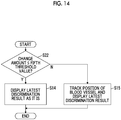
- Step S21 is processing executed in a case where the determination is made that the change amount of the ultrasound image U is equal to or smaller than the fourth threshold value in step S20, and includes three steps of step S22, step S14, and step S15 as shown in the flowchart of Fig. 14 .
- step S22 is added instead of step S13 of the flowchart shown in Fig. 8 .
- step S22 the blood vessel discrimination unit 11 determines whether the change amount of the ultrasound image U calculated in step S19 is equal to or smaller than a predetermined fifth threshold value.
- the fifth threshold value is a value smaller than the fourth threshold value and equal to or larger than the sixth threshold value. In a case where the determination is made that the change amount of the ultrasound image U is equal to or smaller than the fifth threshold value in step S22, the process proceeds to step S14.
- step S14 the blood vessel discrimination unit 11 determines that the ultrasound image U is not substantially changed, and in step S5, the discrimination result held in the discrimination result memory 12 is maintained. Further, the blood vessel discrimination unit 11 displays the discrimination results for the blood vessels corresponding to the blood vessels BV1 and BV2 shown in Figs. 5 and 6 held in the discrimination result memory 12 in step S5 while being superimposed on the ultrasound image of the current frame. In a case where processing of step S14 is completed, the processing of step S21 ends.
- step S15 the process proceeds to step S15.
- step S15 the blood vessel discrimination unit 11 determines that the ultrasound image U changes, but the change is not so large as to newly perform discrimination processing, tracks the blood vessels BV1 and BV2 included in the ultrasound image U, and maintains the discrimination result held in the discrimination result memory 12 in step S5.
- the blood vessel discrimination unit 11 performs processing of displaying the discrimination results for the blood vessels corresponding to the blood vessels BV1 and BV2 included in the ultrasound image U held in the discrimination result memory 12. In this way, in a case where processing of step S15 is completed, the processing of step S21 ends.
- step S21 In a case where the processing of step S21 ends, the process returns to step S7, and it is determined whether the operation of the ultrasound diagnostic apparatus 1A ends. In a case where the determination is made that the operation of the ultrasound diagnostic apparatus 1A does not end in step S7, the process proceeds to step S19.
- step S19 the image change amount calculation unit 29 calculates the change amount of the ultrasound image U between the frames adjacent to each other in time series.
- step S20 the determination is made on whether the change amount of the ultrasound image U calculated in preceding step S19 is larger than the fourth threshold value. As described above, until the determination is made on whether the operation of the ultrasound diagnostic apparatus 1A ends in step S7, or the determination is made that the change amount of the ultrasound image U calculated in step S19 is larger than the fourth threshold value in step S20, the processing items of step S7, step S19, step S20, step S10, and step S21 are repeated.
- step S12 the discrimination execution deciding unit 15 decides that the blood vessel discrimination unit 11 newly executes discrimination with respect to the blood vessels BV1 and BV2. In this way, in a case where processing of step S12 is completed, the process returns to step S17.
- step S17 the image change amount calculation unit 29 calculates the change amount of the ultrasound image U between the frames adjacent to each other in time series.
- step S18 the blood vessel detection unit 10 determines whether the change amount of the ultrasound image U calculated in step S17 is equal to or smaller than the sixth threshold value.
- step S18 in a case where the determination is made that the change amount of the ultrasound image U is larger than the sixth threshold value, the process returns to step S17, and in a case where the determination is made that the change amount of the ultrasound image U is equal to or smaller than the sixth threshold value, the process proceeds to step S3.
- step S3 the blood vessel detection unit 10 performs processing of detecting the blood vessels BV1 and BV2 included in the ultrasound image U is performed by performing image analysis with respect to the ultrasound image U of the current frame.
- step S4 the blood vessel discrimination unit 11 performs processing of discriminating whether the blood vessels BV1 and BV2 detected in step S3 is the vein or the artery.
- step S5 the blood vessel discrimination unit 11 updates the discrimination result held in the discrimination result memory 12 using the discrimination result newly obtained in step S4.
- step S6 the blood vessel discrimination unit 11 displays the discrimination result updated in step S5 on the display unit 7 while being superimposed on the ultrasound image U of the current frame.
- step S7 it is determined whether the operation of the ultrasound diagnostic apparatus 1A ends.
- step S7 in a case where the determination is made that the operation of the ultrasound diagnostic apparatus 1A does not end, the process proceeds to step S19, and in a case where the determination is made than the operation of the ultrasound diagnostic apparatus 1A ends, the operation of the ultrasound diagnostic apparatus 1A ends.
- the ultrasound diagnostic apparatus 1A even in a case where decision is made on whether the blood vessel discrimination unit 11 newly discriminates the blood vessels BV1 and BV2 in the ultrasound image U based on the change amount of the ultrasound images U adjacent to each other in time series, it is possible to reduce the power consumption in the ultrasound diagnostic apparatus 1A while discriminating the blood vessels BV1 and BV2 by not performing unnecessary discrimination of the blood vessels BV1 and BV2 in a case where the change amount of the ultrasound image U is equal to or smaller than the fourth threshold value, in the same manner of aspects of Embodiments 1 and 2.
- Embodiment 3 similar to Embodiment 2, even in a case where a state in which the change amount of the ultrasound image U is equal to or smaller than the fourth threshold value continues for a predetermined time, the discrimination execution deciding unit 15 can decide that the blood vessel discrimination unit 11 newly executes discrimination. That is, in step S20 of Fig. 13 , even in a case where the change amount of the ultrasound image U is equal to or smaller than the fourth threshold value, in a case where a predetermined time has elapsed in a state where the change amount of the ultrasound image U is equal to or smaller than the fourth threshold value, the process proceeds to step S12. In step S12, the discrimination execution deciding unit 15 decides that the blood vessel discrimination unit 11 newly execute discrimination. In a case where processing of step S12 is completed, the process returns to step S1.
- the discrimination execution deciding unit 15 decides that the blood vessel discrimination unit 11 newly executes discrimination, and thus the accurate discrimination result can be obtained even in a case where the total movement amount is large as a result of the change of the ultrasound image U being accumulated in a predetermined time. Since the decision is made that the blood vessel discrimination unit 11 newly executes discrimination with the lapse of time, the accurate discrimination result can be obtained even in a case where, for example, the subject moves instead of the ultrasound probe 19.
- the ultrasound diagnostic apparatus according to the following supplementary notes 1 to 18 can be grasped.
- An ultrasound diagnostic apparatus comprising an ultrasound probe, and a processor, in which the processor acquires an ultrasound image by transmitting an ultrasound beam toward a subject from an ultrasound probe, detects a blood vessel included in the acquired ultrasound image, discriminates whether the detected blood vessel is a vein or an artery, and decides whether discrimination is newly executed with respect to the ultrasound image of a current frame based on a movement amount of the ultrasound probe or a change amount of the ultrasound image between frames.
- the ultrasound diagnostic apparatus further comprising a motion sensor that is attached to the ultrasound probe, in which the processor calculates the movement amount of the ultrasound probe based on a value measured by the motion sensor, decides whether discrimination is executed based on the calculated movement amount of the ultrasound probe.
- the ultrasound diagnostic apparatus further comprising a discrimination result memory that holds a latest discrimination result, in which in a case where the calculated movement amount of the ultrasound probe is equal to or smaller than a predetermined first threshold value, the processor decides that discrimination is not newly executed with respect to the ultrasound image of the current frame, and maintains the latest discrimination result held in the discrimination result memory.
- the ultrasound diagnostic apparatus in which in a case where the calculated movement amount of the ultrasound probe is equal to or smaller than the first threshold value and larger than a second threshold value that is smaller than the first threshold value, the processor tracks a position of the blood vessel in the ultrasound image from the frame in which the latest discrimination is executed to the current frame, and maintains the latest discrimination result held in the discrimination result memory.
- the ultrasound diagnostic apparatus in which in a case where the calculated movement amount of the ultrasound probe is larger than the first threshold value, the processor decides that discrimination is newly executed with respect to the ultrasound image of the current frame, and discriminates whether the blood vessel in the ultrasound image of the current frame is a vein or an artery, and updates the latest discrimination result held in the discrimination result memory based on the discrimination result.
- the ultrasound diagnostic apparatus in which in a case where a state in which the calculated movement amount of the ultrasound probe is equal to or smaller than the first threshold value continues for a predetermined time, the processor decides that discrimination is newly executed with respect to the ultrasound image of the current frame, and discriminates whether the blood vessel in the ultrasound image of the current frame is a vein or an artery, and updates the latest discrimination result held in the discrimination result memory based on the discrimination result.
- the ultrasound diagnostic apparatus in which the processor newly executes discrimination on whether the blood vessel in the ultrasound image of the current frame is a vein or an artery after the calculated movement amount of the ultrasound probe becomes equal to or smaller than a third threshold value that is smaller than the first threshold value.
- the ultrasound diagnostic apparatus according to any one of supplementary notes 3 to 7, further comprising a display device that displays the acquired ultrasound image and the discrimination result held in the discrimination result memory.
- the ultrasound diagnostic apparatus according to any one of supplementary notes 2 to 8, in which the calculated movement amount of the ultrasound probe includes at least one of a moving speed of the ultrasound probe in parallel movement, a change amount in a movement direction of the ultrasound probe, or an angular velocity of the ultrasound probe in rotational movement.
- the ultrasound diagnostic apparatus according to any one of supplementary notes 2 to 9, in which the motion sensor consists of at least one of an acceleration sensor, a gyro sensor, a magnetic sensor, or a position sensor of a global positioning system.
- the ultrasound diagnostic apparatus according to supplementary note 1, in which the processor calculates the change amount of the ultrasound image between the frames by performing image analysis with respect to the acquired ultrasound image, and decides whether discrimination is executed based on the calculated change amount of the ultrasound image.
- the ultrasound diagnostic apparatus further comprising a discrimination result memory that holds a latest discrimination result, in which in a case where the calculated change amount of the ultrasound image between the frames is equal to or smaller than a predetermined fourth threshold value, the processor decides that discrimination is not newly executed with respect to the ultrasound image of the current frame, and maintains the latest discrimination result held in the discrimination result memory.
- the ultrasound diagnostic apparatus in which in a case where the acquired change amount of the ultrasound image between the frames is equal to or smaller than the fourth threshold value and larger than a fifth threshold value that is smaller than the fourth threshold value, the processor tracks a position of the blood vessel in the ultrasound image from the frame in which latest discrimination is executed to the current frame, and maintains the latest discrimination result held in the discrimination result memory.
- the ultrasound diagnostic apparatus in which in a case where the calculated change amount of the ultrasound image between the frames is larger than the fourth threshold value, the processor decides that discrimination is newly executed with respect to the ultrasound image of the current frame, and discriminates whether the blood vessel in the ultrasound image of the current frame is a vein or an artery, and updates the latest discrimination result held in the discrimination result memory based on the discrimination result.
- the ultrasound diagnostic apparatus in which in a case where a state in which the calculated change amount of the ultrasound image between the frames is smaller than the fourth threshold value continues for a predetermined time, the processor decides that discrimination is newly executed with respect to the ultrasound image of the current frame, and discriminates whether the blood vessel in the ultrasound image of the current frame is a vein or an artery, and updates the latest discrimination result held in the discrimination result memory based on the discrimination result.
- the ultrasound diagnostic apparatus in which processor newly executes discrimination on whether the blood vessel in the ultrasound image of the current frame is a vein or an artery after the calculated change amount of the ultrasound image between the frames becomes equal to or smaller than a sixth threshold value that is smaller than the fourth threshold value.
- the ultrasound diagnostic apparatus according to any one of supplementary notes 12 to 16, further comprising a display device that displays the acquired ultrasound image and the discrimination result held in the discrimination result memory.
- the ultrasound diagnostic apparatus according to any one of supplementary notes 1 to 17, further comprising a receiving circuit that receives an ultrasound echo in the subject by the ultrasound probe and generates a received signal, in which the processor generates a Doppler signal based on the received signal generated by the receiving circuit, and discriminates whether the blood vessel is a vein or an artery based on the generated Doppler signal.
Landscapes
- Health & Medical Sciences (AREA)
- Life Sciences & Earth Sciences (AREA)
- Engineering & Computer Science (AREA)
- Heart & Thoracic Surgery (AREA)
- Public Health (AREA)
- Biophysics (AREA)
- Veterinary Medicine (AREA)
- Pathology (AREA)
- Physics & Mathematics (AREA)
- Biomedical Technology (AREA)
- General Health & Medical Sciences (AREA)
- Medical Informatics (AREA)
- Molecular Biology (AREA)
- Surgery (AREA)
- Animal Behavior & Ethology (AREA)
- Radiology & Medical Imaging (AREA)
- Nuclear Medicine, Radiotherapy & Molecular Imaging (AREA)
- Vascular Medicine (AREA)
- Computer Vision & Pattern Recognition (AREA)
- Computer Networks & Wireless Communication (AREA)
- Hematology (AREA)
- Ultra Sonic Daignosis Equipment (AREA)
Abstract
Description
- The present invention relates to an ultrasound diagnostic apparatus and a method of controlling an ultrasound diagnostic apparatus, and particularly, to an ultrasound diagnostic apparatus and a method of controlling an ultrasound diagnostic apparatus that are used for observing blood vessels of a subject.
- In the related art, an ultrasound diagnostic apparatus has been known as an apparatus that obtains an image of the inside of a subject. Generally, the ultrasound diagnostic apparatus comprises an ultrasound probe provided with an oscillator array in which a plurality of elements are arranged. In a state where the ultrasound probe is brought into contact with a body surface of the subject, an ultrasound beam is transmitted from the oscillator array toward the inside of the subject, ultrasound echoes from the subject are received by the oscillator array, and element data are acquired. Moreover, the ultrasound diagnostic apparatus processes the obtained element data electrically, and generates an ultrasound image for a relevant part of the subj ect.
- It is generally performed that a blood vessel of the subject is observed using such an ultrasound diagnostic apparatus. In this case, the user usually determines whether the blood vessel in the ultrasound image is a vein or an artery by visually observing the ultrasound image obtained by the ultrasound diagnostic apparatus. However, in general, it is necessary for the user to have specialized knowledge in order to visually distinguish the vein and the artery in the ultrasound image. Therefore, an ultrasound diagnostic apparatus has been developed that can discriminate whether the blood vessel in the ultrasound image is the vein or the artery even in a case where the user does not have specialized knowledge.
- For example,
JP2008-272025A JP2008-272025A - In a case of actually observing the blood vessel using the ultrasound diagnostic apparatus, the observation is often performed while moving the ultrasound probe.
- In this case, in the ultrasound diagnostic apparatus disclosed in
JP2008-272025A - The present invention has been made in order to solve such related-art problems, and an object thereof is to provide an ultrasound diagnostic apparatus and a method of controlling an ultrasound diagnostic apparatus that can reduce the power consumption while discriminating the blood vessel.
- In order to achieve the above object, an ultrasound diagnostic apparatus according to an aspect of the present invention comprises an ultrasound probe, an image acquisition unit that acquires an ultrasound image sequentially by transmitting an ultrasound beam toward a subject from the ultrasound probe, a blood vessel detection unit that detects a blood vessel included in the ultrasound image acquired by the image acquisition unit, a blood vessel discrimination unit that discriminates whether the blood vessel detected by the blood vessel detection unit is a vein or an artery, and a discrimination execution deciding unit that decides whether the blood vessel discrimination unit newly executes discrimination with respect to the ultrasound image of a current frame based on a movement amount of the ultrasound probe or a change amount of the ultrasound image between frames.
- The ultrasound diagnostic apparatus may further comprise a motion sensor that is attached to the ultrasound probe, and a probe movement amount calculating unit that calculates the movement amount of the ultrasound probe based on a value measured by the motion sensor, in which the discrimination execution deciding unit decides whether the blood vessel discrimination unit executes discrimination based on the movement amount of the ultrasound probe calculated by the probe movement amount calculating unit.
- It is preferable that the ultrasound diagnostic apparatus further comprise a discrimination result memory that holds a latest discrimination result by the blood vessel discrimination unit, in which in a case where the movement amount of the ultrasound probe calculated by the probe movement amount calculating unit is equal to or smaller than a predetermined first threshold value, the discrimination execution deciding unit decides that the blood vessel discrimination unit does not newly execute discrimination with respect to the ultrasound image of the current frame, and the blood vessel discrimination unit maintains the latest discrimination result held in the discrimination result memory.
- In this case, in a case where the movement amount of the ultrasound probe calculated by the probe movement amount calculating unit is equal to or smaller than the first threshold value and larger than a second threshold value that is smaller than the first threshold value, the blood vessel discrimination unit may track a position of the blood vessel in the ultrasound image from the frame in which the blood vessel discrimination unit executes latest discrimination to the current frame, and maintain the latest discrimination result held in the discrimination result memory.
- In a case where the movement amount of the ultrasound probe calculated by the probe movement amount calculating unit is larger than the first threshold value, the discrimination execution deciding unit may decide that the blood vessel discrimination unit newly executes discrimination with respect to the ultrasound image of the current frame, and the blood vessel discrimination unit may discriminate whether the blood vessel in the ultrasound image of the current frame is a vein or an artery, and update the latest discrimination result held in the discrimination result memory based on the discrimination result.
- Alternatively, in a case where a state in which the movement amount of the ultrasound probe calculated by the probe movement amount calculating unit is equal to or smaller than the first threshold value continues for a predetermined time, the discrimination execution deciding unit may decide that the blood vessel discrimination unit newly executes discrimination with respect to the ultrasound image of the current frame, and the blood vessel discrimination unit may discriminate whether the blood vessel in the ultrasound image of the current frame is a vein or an artery, and update the latest discrimination result held in the discrimination result memory based on the discrimination result.
- The blood vessel discrimination unit may newly execute discrimination on whether the blood vessel in the ultrasound image of the current frame is a vein or an artery after the movement amount of the ultrasound probe calculated by the probe movement amount calculating unit becomes equal to or smaller than a third threshold value that is smaller than the first threshold value.
- It is preferable that the ultrasound diagnostic apparatus further comprise a display unit that displays the ultrasound image acquired by the image acquisition unit and the discrimination result held in the discrimination result memory.
- It is preferable that the movement amount of the ultrasound probe calculated by the probe movement amount calculating unit include at least one of a moving speed of the ultrasound probe in parallel movement, a change amount in a movement direction of the ultrasound probe, or an angular velocity of the ultrasound probe in rotational movement.
- It is preferable that the motion sensor consist of at least one of an acceleration sensor, a gyro sensor, a magnetic sensor, or a position sensor of a global positioning system.
- The ultrasound diagnostic apparatus may further comprise an image change amount calculation unit that calculates the change amount of the ultrasound image between the frames by performing image analysis with respect to the ultrasound image acquired by the image acquisition unit, in which the discrimination execution deciding unit decides whether the blood vessel discrimination unit executes discrimination of the blood vessel based on the change amount of the ultrasound image calculated by the image change amount calculation unit.
- It is preferable that the ultrasound diagnostic apparatus further comprise a discrimination result memory that holds a latest discrimination result of the blood vessel discrimination unit, in which in a case where the change amount of the ultrasound image between the frames calculated by the image change amount calculation unit is equal to or smaller than a predetermined fourth threshold value, the discrimination execution deciding unit decides that the blood vessel discrimination unit does not newly execute discrimination with respect to the ultrasound image of the current frame, and the blood vessel discrimination unit maintains the latest discrimination result held in the discrimination result memory.
- In this case, in a case where the change amount of the ultrasound image between the frames acquired by the image change amount calculation unit is equal to or smaller than the fourth threshold value and larger than a fifth threshold value that is smaller than the fourth threshold value, the blood vessel discrimination unit may track a position of the blood vessel in the ultrasound image from the frame in which the blood vessel discrimination unit executes latest discrimination to the current frame, and maintain the latest discrimination result held in the discrimination result memory.
- In a case where the change amount of the ultrasound image between the frames calculated by the image change amount calculation unit is larger than the fourth threshold value, the discrimination execution deciding unit may decide that the blood vessel discrimination unit newly executes discrimination with respect to the ultrasound image of the current frame, and the blood vessel discrimination unit may discriminate whether the blood vessel in the ultrasound image of the current frame is a vein or an artery, and update the latest discrimination result held in the discrimination result memory based on the discrimination result.
- Alternatively, in a case where a state in which the change amount of the ultrasound image between the frames calculated by the image change amount calculation unit is smaller than the fourth threshold value continues for a predetermined time, the discrimination execution deciding unit may decide that the blood vessel discrimination unit newly executes discrimination with respect to the ultrasound image of the current frame, and the blood vessel discrimination unit may discriminate whether the blood vessel in the ultrasound image of the current frame is a vein or an artery, and update the latest discrimination result held in the discrimination result memory based on the discrimination result.
- The blood vessel discrimination unit may newly execute discrimination on whether the blood vessel in the ultrasound image of the current frame is a vein or an artery after the change amount of the ultrasound image between the frames calculated by the image change amount calculation unit becomes equal to or smaller than a sixth threshold value that is smaller than the fourth threshold value.
- It is preferable that the ultrasound diagnostic apparatus further comprise a display unit that displays the ultrasound image acquired by the image acquisition unit and the discrimination result held in the discrimination result memory.
- The image acquisition unit may include a receiving unit that receives an ultrasound echo in the subject by the ultrasound probe and generates a received signal, the ultrasound diagnostic apparatus may further comprise a Doppler signal generating unit that generates a Doppler signal based on the received signal generated by the receiving unit, and the blood vessel discrimination unit may discriminate whether the blood vessel is a vein or an artery based on the Doppler signal generated by the Doppler signal generating unit.
- A method of controlling an ultrasound diagnostic apparatus according to another aspect of the present invention comprises acquiring an ultrasound image sequentially by transmitting an ultrasound beam toward a subject from an ultrasound probe, detecting a blood vessel included in the acquired ultrasound image, discriminating whether the detected blood vessel is a vein or an artery, and deciding whether to newly execute discrimination with respect to the ultrasound image of a current frame based on a movement amount of the ultrasound probe or a change amount of the ultrasound image between frames.
- According to the present invention, the ultrasound diagnostic apparatus comprises a blood vessel detection unit that detects a blood vessel included in the ultrasound image acquired by the image acquisition unit, a blood vessel discrimination unit that discriminates whether the blood vessel detected by the blood vessel detection unit is a vein or an artery, and a discrimination execution deciding unit that decides whether the blood vessel discrimination unit newly executes discrimination of the blood vessel included in the ultrasound image of a current frame based on a movement amount of the ultrasound probe or a change amount of the ultrasound image between frames, and thus it is possible to reduce the power consumption while discriminating the blood vessel.
-
-
Fig. 1 is a block diagram showing a configuration of an ultrasound diagnostic apparatus according to Embodiment 1 of the present invention. -
Fig. 2 is a block diagram showing an internal configuration of a receiving unit according to Embodiment 1 of the present invention. -
Fig. 3 is a block diagram showing an internal configuration of an image generation unit according to Embodiment 1 of the present invention. -
Fig. 4 is a block diagram showing an internal configuration of a Doppler signal generating unit according to Embodiment 1 of the present invention. -
Fig. 5 is a diagram schematically showing an ultrasound image including a cross section of a blood vessel. -
Fig. 6 is a diagram schematically showing a discrimination result of the blood vessel. -
Fig. 7 is a flowchart showing an operation of the ultrasound diagnostic apparatus according to Embodiment 1 of the present invention. -
Fig. 8 is a flowchart showing an operation of displaying the latest discrimination result in Embodiment 1 of the present invention. -
Fig. 9 is a diagram schematically showing a state in which the position of the blood vessel is moved in the ultrasound image. -
Fig. 10 is a diagram schematically showing a state in which the position of blood vessel in the ultrasound image is tracked and the latest discrimination result is displayed. -
Fig. 11 is a flowchart showing an operation of the ultrasound diagnostic apparatus according to Embodiment 2 of the present invention. -
Fig. 12 is a block diagram showing a configuration of an ultrasound diagnostic apparatus according toEmbodiment 3 of the present invention. -
Fig. 13 is a flowchart showing an operation of the ultrasound diagnostic apparatus according toEmbodiment 3 of the present invention. -
Fig. 14 is a flowchart showing an operation of displaying the latest discrimination result inEmbodiment 3 of the present invention. - Hereinafter, embodiments of the present invention will be described based on the accompanying drawings.
- A configuration of an ultrasound diagnostic apparatus 1 according to Embodiment 1 of the present invention is shown in
Fig. 1 . As shown inFig. 1 , the ultrasound diagnostic apparatus 1 comprises an oscillator array 2, and a transmittingunit 3 and areceiving unit 4 are connected to the oscillator array 2. Animage generation unit 5, adisplay controller 6, and a display unit 7 are sequentially connected to thereceiving unit 4. Animage acquisition unit 8 is configured by the transmittingunit 3, thereceiving unit 4, and theimage generation unit 5. A Dopplersignal generating unit 9 is connected to thereceiving unit 4. Additionally, a bloodvessel detection unit 10 is connected to theimage generation unit 5. A bloodvessel discrimination unit 11 is connected to the Dopplersignal generating unit 9 and the bloodvessel detection unit 10, and adiscrimination result memory 12 and thedisplay controller 6 are connected to the bloodvessel discrimination unit 11. The bloodvessel discrimination unit 11 and the discrimination resultmemory 12 are connected to each other so as to be capable of transferring information bidirectionally. The oscillator array 2 is included in theultrasound probe 19, and amotion sensor 13 is attached to theultrasound probe 19. A probe movementamount calculating unit 14 is connected to themotion sensor 13, and a discriminationexecution deciding unit 15 is connected to the probe movementamount calculating unit 14. The discriminationexecution deciding unit 15 is connected to the bloodvessel discrimination unit 11. - The
apparatus controller 16 is connected to thedisplay controller 6, theimage acquisition unit 8, the Dopplersignal generating unit 9, the bloodvessel discrimination unit 11, the bloodvessel detection unit 10, the probe movementamount calculating unit 14, and the discriminationexecution deciding unit 15, and theinput unit 17 and thestorage unit 18 are connected to theapparatus controller 16. Theapparatus controller 16 and thestorage unit 18 are connected to each other so as to be capable of transferring information bidirectionally. - The
processor 20 is configured by thedisplay controller 6, theimage acquisition unit 8, the Dopplersignal generating unit 9, the bloodvessel discrimination unit 11, the bloodvessel detection unit 10, the probe movementamount calculating unit 14, the discriminationexecution deciding unit 15, and theapparatus controller 16. - The oscillator array 2 of the
ultrasound probe 19 shown inFig. 1 has a plurality of oscillators arranged in one dimension or two dimensions. These oscillators transmit ultrasound waves in accordance with drive signals supplied from the transmittingunit 3, respectively, and receive ultrasound echoes from the subject to output the received signals. The respective oscillators are configured by, for example, forming electrodes to both ends of piezoelectric material made of a piezoelectric ceramic represented by lead zirconate titanate (PZT), a polymeric piezoelectric element represented by polyvinylidene difluoride (PVDF), and a piezoelectric single crystal represented by a lead magnesium niobate-lead titanate solid solution (PMN-PT). - The transmitting
unit 3 of theimage acquisition unit 8 includes, for example, a plurality of pulse generators, and adjust the amounts of delay of the respective drive signals to supply the adjusted drive signals to the plurality of oscillators such that the ultrasound waves transmitted from the plurality of oscillators of the oscillator array 2 form an ultrasound beam, based on a transmission delay pattern selected in accordance with the control signals from theapparatus controller 16. In this way, in a case where a pulsed or consecutive wave-like voltage is applied to electrodes of the plurality of oscillators of the oscillator array 2, the piezoelectric material expands and contracts, a pulsed or consecutive wave-like ultrasound wave is generated from the respective oscillators, and the ultrasound beam is formed from a synthetic wave of the ultrasound waves. - The transmitted ultrasound beam is reflected in, for example, targets such as a part of the subject and is propagated toward the oscillator array 2 of the
ultrasound probe 19. The ultrasound echoes propagated toward the oscillator array 2 in this way are received by the respective oscillators that constitute the oscillator array 2. In this case, the respective oscillators that constitute the oscillator array 2 expand and contract by receiving the propagated ultrasound echoes, to generate electrical signals, and output the electrical signals to the receivingunit 4. - The receiving
unit 4 of theimage acquisition unit 8 processes the signals output from the oscillator array 2 in accordance with the control signals from theapparatus controller 16. As shown inFig. 2 , the receivingunit 4 has a configuration that anamplification unit 21 and an analog-digital (AD)conversion unit 22 are serially connected. Theamplification unit 21 amplifies the signals input from the respective oscillators that constitute the oscillator array 2, and transmits the amplified signals to theAD conversion unit 22. TheAD conversion unit 22 converts the signals transmitted from theamplification unit 21 into digitalized received signal, and sends the data to theimage generation unit 5 of theimage acquisition unit 8. - As shown in
Fig. 3 , theimage generation unit 5 of theimage acquisition unit 8 has a configuration that asignal processing unit 23, a digital scan converter (DSC) 24, and animage processing unit 25 are serially connected. Thesignal processing unit 23 performs reception focus processing that addition (phasing addition) is performed by giving each delay to each data of the received signals, based on a reception delay pattern selected in accordance with the control signals from theapparatus controller 16. With the reception focus processing, sound ray signals in which focal points of the ultrasound echoes are narrowed to one scan line are generated. Additionally, thesignal processing unit 23 subjects the generated sound ray signals to the correction of damping resulting from a propagation distance depending on the depth at a position where the ultrasound waves are reflected, and then performs envelope detection processing to generate B-mode image signals indicating a tissue within the subject. The B-mode image signals generated in this way are output to theDSC 24. - The
DSC 24 of theimage generation unit 5 raster-converts the B-mode image signals into image signals based on a scan mode of normal television signals to generate the ultrasound image. Theimage processing unit 25 of theimage generation unit 5 subjects the ultrasound image obtained in theDSC 24 to various kinds of required image processing such as brightness correction, grayscale correction, sharpness correction, and color correction, and then outputs an ultrasound image to thedisplay controller 6, and the bloodvessel detection unit 10. - The Doppler
signal generating unit 9 of theprocessor 20 generates the Doppler signal by a so-called pulse Doppler method. As shown inFig. 4 , the Dopplersignal generating unit 9 has a configuration in which anorthogonal detection unit 26, a high-pass filter 27, afast Fourier transformer 28 are sequentially connected in series. - The
orthogonal detection unit 26 orthogonally detects the received signal and converts the received signal into complex data by mixing the carrier signal of the reference frequency with the received signal generated by the receivingunit 4. - The high-
pass filter 27 functions as a so-called wall filter, and removes frequency components derived from the movement of the body tissue of the subject from the complex data generated by theorthogonal detection unit 26. - The
fast Fourier transformer 28 performs frequency analysis by Fourier transforming the complex data of a plurality of sample points, and generates the Doppler signal representing a so-called spectrum. - The blood
vessel detection unit 10 of theprocessor 20 performs image analysis with respect to the ultrasound image acquired by theimage acquisition unit 8, and detects the blood vessel included in the ultrasound image. For example, as shown inFig. 5 , the bloodvessel detection unit 10 detects cross sections of blood vessels BV1 and BV2 included in the ultrasound image U. - The cross section of the blood vessel represents the section of the blood vessel in a case of cutting the blood vessel across a central axis thereof. More specifically, for example, the blood
vessel detection unit 10 can detect the cross section of the blood vessel by storing typical pattern data as a template in advance, calculating the similarity to the pattern data while searching the image with the template, and assuming the blood vessel is present in the position where the similarity is equal to or larger than a threshold value and is maximum. - A machine learning method described in Csurka et al.: Visual Categorization with Bags of Keypoints, Proc. of ECCV Workshop on Statistical Learning in Computer Vision, pp.59-74 (2004), a general image recognition method using deep learning described in Krizhevsky et al.: Image Net Classification with Deep Convolutional Neural Networks, Advances in Neural ), or the like can be used for the calculation of the similarity in addition to the simple template matching.
- The
motion sensor 13 attached to theultrasound probe 19 measures the movement of theultrasound probe 19 moved by the user. Examples of themotion sensor 13 include an acceleration sensor, a gyro sensor, a magnetic sensor, or a position sensor of a global positioning system (GPS). Therefore, themotion sensor 13 measures acceleration of theultrasound probe 19 in parallel movement, angular acceleration or an angular velocity of theultrasound probe 19 in rotational movement, and position of theultrasound probe 19 as the movement of theultrasound probe 19. - The probe movement
amount calculating unit 14 of theprocessor 20 calculates the movement amount of theultrasound probe 19 based on the value measured by themotion sensor 13. The probe movementamount calculating unit 14 can calculate a moving speed of theultrasound probe 19 in parallel movement, an angular velocity of theultrasound probe 19 in rotational movement, and the change amount in the movement direction of theultrasound probe 19 as the movement amount of theultrasound probe 19. - The blood
vessel discrimination unit 11 of theprocessor 20 discriminates whether the blood vessels BV1 and BV2 detected by the bloodvessel detection unit 10 are the vein or the artery. In general, the artery beats at a constant cycle, the intensity of the Doppler signal in the cross section of the blood vessel corresponding to the artery also changes at the same constant cycle as the beat. On the other hand, the vein does not beat, and thus the intensity of the Doppler signal in the cross section of the blood vessel corresponding to the vein does not change periodically. Therefore, for example, the bloodvessel discrimination unit 11 discriminates that the blood vessel in which the intensity of the Doppler signal generated by the Dopplersignal generating unit 9 changes periodically is the artery, and the blood vessel in which the intensity of the Doppler signal does not change periodically is vein. - It is preferable that the blood vessel be discriminated in a state where the
ultrasound probe 19 is substantially stationary such that the blood vessels can be accurately discriminated. Therefore, it is desirable that the bloodvessel discrimination unit 11 discriminates the blood vessel after the movement amount of theultrasound probe 19 calculated by the probe movementamount calculating unit 14 is in a substantially stationary state. - In addition, the blood
vessel discrimination unit 11 causes the display unit 7 to display the discrimination results for the blood vessels BV1 and BV2 via thedisplay controller 6. For example, as shown inFig. 6 , the bloodvessel discrimination unit 11 can display the discrimination results that the blood vessel BV1 is the artery and the blood vessel BV2 is the vein on the display unit 7 by discriminating that the blood vessel BV1 is the artery and the blood vessel BV2 is the vein, displaying a text T1 indicating that the blood vessel is the artery and a surrounding line E1 surrounding the blood vessel BV1 and the text T1 in the vicinity of the blood vessel BV1 in the ultrasound image U, and displaying a text T2 indicating that the blood vessel is the vein and a surrounding line E2 surrounding the blood vessel BV2 and the text T2 in the vicinity of the blood vessel BV2. In this case, for example, the bloodvessel discrimination unit 11 can display the surrounding lines E1 and E2 with different colors such as red and blue on the display unit 7 such that the user can easily confirm whether the blood vessels BV1 and the BV2 are the vein or the artery. - The
discrimination result memory 12 of the ultrasound diagnostic apparatus 1 stores the latest discrimination result by the bloodvessel discrimination unit 11. As thediscrimination result memory 12, a recording medium such as a hard disc drive (HDD), a solid state drive (SSD), a flexible disc (FD), a magneto-optical (MO) disc, a magnetic tape (MT), a random access memory (RAM), a compact disc (CD), a digital versatile disc (DVD), a secure digital (SD) card, a universal serial bus (USB) memory, or the like, or a server can be used. Thediscrimination result memory 12 is not included in theprocessor 20, but may be included in theprocessor 20. - The discrimination
execution deciding unit 15 of theprocessor 20 decides whether the bloodvessel discrimination unit 11 newly executes discrimination of the blood vessel included in the ultrasound image of the current frame acquired by theimage acquisition unit 8 based on the movement amount of theultrasound probe 19 calculated by the probe movementamount calculating unit 14. For example, the discriminationexecution deciding unit 15 decides that discrimination of the blood vessel is not newly executed with respect to the ultrasound image of the current frame in a case where the movement amount of theultrasound probe 19 calculated by the probe movementamount calculating unit 14 is equal to or smaller than a predetermined first threshold value, and decides that discrimination of the blood vessel is newly executed with respect to the ultrasound image of the current frame in a case where the movement amount of theultrasound probe 19 is larger than the first threshold value. For example, in a case where the movement amount of theultrasound probe 19 is the moving speed of theultrasound probe 19 in parallel movement, 20 mm/sec can be set as the first threshold value. - As a result, it is possible to prevent the blood
vessel discrimination unit 11 from performing unnecessary discrimination processing, reduce the calculation load on the ultrasound diagnostic apparatus 1, and further reduce the power consumption on the ultrasound diagnostic apparatus 1. - The
apparatus controller 16 of theprocessor 20 performs control of the respective units of the ultrasound diagnostic apparatus 1 based on the programs that are stored in advance in thestorage unit 18 and the like and the operation of the user via theinput unit 17. - Under the control of the
apparatus controller 16, thedisplay controller 6 of theprocessor 20 performs predetermined processing on an ultrasound image generated by theimage generation unit 5 of theimage acquisition unit 8, and displays the ultrasound image on the display unit 7. - The display unit 7 of the ultrasound diagnostic apparatus 1 displays the ultrasound image under the control of the
display controller 6, and includes display devices such as a liquid crystal display (LCD), and an organic electroluminescence (EL) display. - The
input unit 17 of the ultrasound diagnostic apparatus 1 is a device for the user to perform input operation, and can be configured to comprise a keyboard, a mouse, a trackball, a touchpad, a touch panel, and the like. - The
storage unit 18 stores the operating program of the ultrasound diagnostic apparatus 1, and as the storage unit, a recording medium such as the HDD, the SSD, the FD, the MO disc, the MT, the RAM, the CD, the DVD, the SD card, the USB memory, or the like can be used similarly to thediscrimination result memory 12. - In addition, the
processor 20 having thedisplay controller 6, theimage acquisition unit 8, the Dopplersignal generating unit 9, the bloodvessel discrimination unit 11, the bloodvessel detection unit 10, the probe movementamount calculating unit 14, the discriminationexecution deciding unit 15, and theapparatus controller 16 is configured from a central processing unit (CPU) and control programs for making the CPU perform various kinds of processing. However, theprocessor 20 may be configured from a field programmable gate array (FGPA), a digital signal processor (DSP), an application specific integrated circuit (ASIC), or other integrated circuits (ICs), or may be configured by combining them. - The configuration can be adopted in which the
display controller 6, theimage acquisition unit 8, the Dopplersignal generating unit 9, the bloodvessel discrimination unit 11, the bloodvessel detection unit 10, the probe movementamount calculating unit 14, the discriminationexecution deciding unit 15, and theapparatus controller 16 of theprocessor 20 are partially or entirely integrated into one CPU. - Next, the operation of the ultrasound diagnostic apparatus 1 in Embodiment 1 will be described in detail using a flowchart shown in
Fig. 7 . In the following operation description, theimage acquisition unit 8 of theprocessor 20 sequentially and consecutively acquires the ultrasound images and displays the ultrasound images on the display unit 7, and the user moves theultrasound probe 19 as the blood vessel is observed. - First, in step S1, the probe movement
amount calculating unit 14 calculates the movement amount of theultrasound probe 19 based on the measured value obtained by themotion sensor 13 attached to theultrasound probe 19. - Next, in step S2, the blood
vessel discrimination unit 11 determines whether theultrasound probe 19 is in a substantially stationary state, that is, whether the movement amount of theultrasound probe 19 calculated in step S1 is equal to or smaller than the third threshold value that is smaller than the first threshold value. For example, in a case where the movement amount of theultrasound probe 19 is the moving speed of theultrasound probe 19 in parallel movement, 3 mm/sec can be set as the third threshold value. In step S2, in a case where the determination is made that the movement amount of theultrasound probe 19 is larger than the third threshold value, the process returns to step S1, the movement amount of theultrasound probe 19 is newly calculated, and the process proceeds to step S2. As described above, step S1 and step S2 are repeated until the movement amount of theultrasound probe 19 become equal to or smaller than the third threshold value. - In a case where the determination is made that the movement amount of the
ultrasound probe 19 is equal to or smaller than the third threshold value in step S2, the process proceeds to step S3. In step S3, the bloodvessel detection unit 10 performs image analysis with respect to the ultrasound image of the current frame acquired by theimage acquisition unit 8, and performs detection processing of the blood vessels BV1 and BV2 included in the ultrasound image U is performed as shown inFig. 5 , for example. - In following step S4, the blood
vessel discrimination unit 11 performs processing of discriminating whether the blood vessels BV1 and BV2 are the vein or the artery based on the intensity of the Doppler signal in the blood vessels BV1 and BV2 detected in step S3. For example, it is possible that the Dopplersignal generating unit 9 generates the Doppler signal corresponding to entire area of the ultrasound image U, and the bloodvessel discrimination unit 11 discriminates that among the blood vessels BV1 and BV2, the blood vessel in which the intensity of the Doppler signal changes periodically is the artery, and the blood vessel in which the intensity of the Doppler signal does not change periodically is vein. In a case where the Doppler signal corresponding to the entire area of the ultrasound image U is obtained by the Dopplersignal generating unit 9, the oscillator array 2 of theultrasound probe 19 transmits the ultrasound beam to the cross section of the subject corresponding to the entire area of the ultrasound image U. - In step S5, the blood
vessel discrimination unit 11 holds the discrimination result obtained in step S4 in thediscrimination result memory 12. - In step S6, the blood
vessel discrimination unit 11 displays the discrimination result held in thediscrimination result memory 12 in step S5 on the display unit 7. For example, as shown inFig. 6 , the bloodvessel discrimination unit 11 can display the discrimination results on the display unit 7 by displaying a text T1 indicating that the blood vessel is the artery and a surrounding line E1 surrounding the blood vessel BV1 and the text T1 in the vicinity of the blood vessel BV1, and displaying a text T2 indicating that the blood vessel is the vein and a surrounding line E2 surrounding the blood vessel BV2 and the text T2 in the vicinity of the blood vessel BV2 while being superimposed on the ultrasound image U. - In the following step S7, it is determined whether the operation of the ultrasound diagnostic apparatus 1 ends. For example, although not shown, in a case where a operation end button for ending the operation of the ultrasound diagnostic apparatus 1 is displayed on the display unit 7, and the user presses the operation end button via the
input unit 17, it is determined that the operation of the ultrasound diagnostic apparatus 1 ends, and thus the operation of the ultrasound diagnostic apparatus 1 ends. In a case where the operation end button is not pressed, it is determined that the operation of the ultrasound diagnostic apparatus 1 does not end, and the process proceeds to step S8. - In step S8, the probe movement
amount calculating unit 14 newly calculates the movement amount of theultrasound probe 19 in the same manner as in step S1. - In following step S9, the discrimination
execution deciding unit 15 determines whether the movement amount of theultrasound probe 19 calculated in step S8 is larger than the first threshold value. In a case where the determination is made that the movement amount of theultrasound probe 19 is equal to or smaller than the first threshold value in step S9, the process proceeds to step S10. In step S10, the discriminationexecution deciding unit 15 decides that the bloodvessel discrimination unit 11 does not newly discriminate the blood vessel in the ultrasound image of the current frame. - In following step S11, the blood
vessel discrimination unit 11 displays the latest discrimination result, that is, the discrimination result held in step S5 while being superimposed on the ultrasound image U. The detailed operation in step S11 will be described with reference to the flowchart ofFig. 8 . Step S11 is processing executed in a case where the determination is made that the movement amount of theultrasound probe 19 is equal to or smaller than the first threshold value in step S9, and includes three steps of step S13 to step S15 as shown inFig. 8 . - First, in step S13, the blood
vessel discrimination unit 11 determines whether the movement amount of theultrasound probe 19 calculated in step S8 is equal to or smaller than a predetermined second threshold value. The second threshold value is a value smaller than the first threshold value and equal to or larger than the third threshold value. In a case where the determination is made that the movement amount of theultrasound probe 19 is equal to or smaller than the second threshold value in step S13, the process proceeds to step S14. - In step S14, the blood
vessel discrimination unit 11 determines that theultrasound probe 19 is in a substantially stationary state, and in step S5, the discrimination result held in thediscrimination result memory 12 is maintained. Further, the bloodvessel discrimination unit 11 displays the discrimination results for the blood vessels corresponding to the blood vessels BV1 and BV2 shown inFigs. 5 and 6 held in thediscrimination result memory 12 in step S5 while being superimposed on the ultrasound image of the current frame. In a case where processing of step S14 is completed, the processing of step S11 ends. - Also, in a case where the determination is made that the movement amount of the
ultrasound probe 19 is larger than the second threshold value in step S13, the process proceeds to step S15. - In step S15, the blood
vessel discrimination unit 11 determines that theultrasound probe 19 has movement, but has no big movement as to newly perform discrimination processing, tracks the blood vessels BV1 and BV2 included in the ultrasound image U, and maintains the discrimination result held in thediscrimination result memory 12 in step S5. Further, the bloodvessel discrimination unit 11 performs processing of displaying the discrimination results for the blood vessels corresponding to the blood vessels BV1 and BV2 included in the ultrasound image U held in thediscrimination result memory 12. - For example, the user moves the
ultrasound probe 19 by the movement amount that is equal to or smaller than the first threshold value and larger than the second threshold value, and as shown inFig. 9 , in a case where the blood vessels BV1 and BV2 in the ultrasound image U is moved to blood vessels BV3 and BV4, the bloodvessel discrimination unit 11 can display the texts T1 and T2, and the surrounding lines E1 and E2 on the display unit 7 in a state where the texts T1 and T2, and the surrounding lines E1 and E2 are moved in accordance with the movement of the blood vessels BV1 and BV2 in the ultrasound image U, as shown inFig. 10 . As described above, the bloodvessel discrimination unit 11 tracks the position of the blood vessel, and displays the discrimination results for the blood vessels BV3 and BV4 of the current frame held in step S5 which correspond to the blood vessels BV1 and BV2 of the frame in which the latest discrimination is performed. For example, the bloodvessel discrimination unit 11 can track the blood vessels BV1 and BV2 by performing matching between the frames adjacent to each other in time series, and a so-called optical flow method. - In a case where processing of step S15 is completed, the processing of step S11 ends.
- In this way, in a case where the latest discrimination result held in step S5 is displayed on the display unit 7 in step S11, the process returns to step S7. In a case where the determination is made that the operation of the ultrasound diagnostic apparatus 1 ends in step S7, the operation of the ultrasound diagnostic apparatus 1 ends. In a case where the determination is made that the operation of the ultrasound diagnostic apparatus 1 does not end in step S7, the process proceeds to step S8, and the movement amount of the
ultrasound probe 19 is calculated. In following step S9, the determination is made on whether the movement amount of theultrasound probe 19 calculated in step S8 is larger than the first threshold value. In a case where the determination is made that the movement amount of theultrasound probe 19 is equal to or smaller than the first threshold value in step S9, the process proceeds to step S10. - As described above, until the determination is made on whether the operation of the ultrasound diagnostic apparatus 1 ends in step S7 or the determination is made that the movement amount of the
ultrasound probe 19 calculated in preceding step S8 is larger than the first threshold value in step S9, the processing items of step S7 to step S11 are repeated. - Also, in a case where the determination is made that the movement amount of the
ultrasound probe 19 calculated in step S8 is larger than the first threshold value in step S9, the process proceeds to step S12. In step S12, the discriminationexecution deciding unit 15 decides that the bloodvessel discrimination unit 11 newly execute discrimination with respect to the ultrasound image of the current frame. As described above, in a case where the processing of step S12 is completed, the process returns to step S1, and the probe movementamount calculating unit 14 calculates the movement amount of theultrasound probe 19. - In following step S2, the blood
vessel detection unit 10 determines whether the movement amount of theultrasound probe 19 calculated in step S1 is equal to or smaller than the third threshold value. In a case where the determination is made that the movement amount of theultrasound probe 19 is larger than the third threshold value in step S2, the process returns to step S1. In a case where the determination is made that the movement amount of theultrasound probe 19 is equal to or smaller than the third threshold value in step S2, the process proceeds to step S3. - In step S3, the blood
vessel detection unit 10 newly detects the blood vessel by performing image analysis with respect to the ultrasound image of the current frame. - In step S4, the blood
vessel discrimination unit 11 newly discriminates whether the blood vessel newly detected in step S3 is the vein or the artery. - In step S5, the blood
vessel discrimination unit 11 updates the discrimination result held in thediscrimination result memory 12 using the new discrimination result obtained in step S4. That is, the bloodvessel discrimination unit 11 overwrites the discrimination result previously held in thediscrimination result memory 12, and holds the new discrimination result obtained in step S4 in thediscrimination result memory 12. - In following step S6, the blood
vessel discrimination unit 11 displays the discrimination result updated in step S5 while being superimposed on the ultrasound image of the current frame. - In a case where the processing of step S6 is completed, the process proceeds to step S7, and it is determined whether the operation of the ultrasound diagnostic apparatus 1 ends. In step S7, in a case where the determination is made that the operation of the ultrasound diagnostic apparatus 1 does not end, the process proceeds to step S8, and in a case where the determination is made than the operation of the ultrasound diagnostic apparatus 1 ends, the operation of the ultrasound diagnostic apparatus 1 ends.
- As described above, in the ultrasound diagnostic apparatus 1 according to Embodiment 1, the blood
vessel detection unit 10 automatically detects the blood vessels BV1 and BV2 in the ultrasound image U, the bloodvessel discrimination unit 11 discriminates whether the detected blood vessels BV1 and BV2 are the vein or the artery, and the discriminationexecution deciding unit 15 decides whether the bloodvessel discrimination unit 11 executes discrimination of the blood vessels BV1 and BV2 based on the movement amount of theultrasound probe 19, and thus by not performing unnecessary discrimination processing with respect to the blood vessels BV1 and BV2 in a case where the movement amount of theultrasound probe 19 is equal to or smaller than the first threshold value, it is possible to reduce the power consumption of the ultrasound diagnostic apparatus 1 while discriminating the blood vessels BV1 and BV2. - In particular, in a portable ultrasound diagnostic apparatus using a battery as a drive power source, it is preferable to reduce the power consumption as much as possible and secure the driving time, and thus it is greatly useful to apply the aspect of Embodiment 1 of the present invention.
- In an example shown in
Fig. 6 of Embodiment 1, the rectangular surrounding lines E1 and E2 are displayed while being superimposed on the ultrasound image U, but the shapes of the surrounding lines E1 and E2 are not particularly limited as long as the blood vessel BV1 is surrounded by the surrounding line E1, and the blood vessel BV2 is surrounded by the surrounding line E2. For example, the surrounding lines E1 and E2 can each have any closed shape such as a circular shape or a polygonal shape. - As shown in
Fig. 6 , the bloodvessel discrimination unit 11 displays the text T1 and the surrounding line E1 in the vicinity of the blood vessel BV1, and the text T2 and the surrounding line E2 in the vicinity of the blood vessel BV2, but also can display only the text T1 and the text T2 while being superimposed on the ultrasound image U. - In Embodiment 1, the Doppler
signal generating unit 9 generates the Doppler signal corresponding to the entire area of the ultrasound image U, but can generate the Doppler signal corresponding to a part of the ultrasound image U including the blood vessels BV1 and BV2. For example, although not shown, the Doppler signals corresponding to a first region of interest and a second region of interest are generated by setting the first region of interest including only the blood vessel BV1 and the second region of interest including the blood vessel BV2, and transmitting the ultrasound beam from the oscillator array 2 of theultrasound probe 19 with respect to the cross section of the subject corresponding to the first region of interest and the second region of interest of the ultrasound image U. As a result, it is possible to reduce the calculation load in the Dopplersignal generating unit 9 in a case of generating the Doppler signal, and further reduce the power consumption of the ultrasound diagnostic apparatus 1. - In Embodiment 1, the blood
vessel discrimination unit 11 discriminates whether the blood vessels BV1 and BV2 are the vein or the artery based on the intensity of the Doppler signal in the blood vessels BV1 and BV2, but the discrimination method of the blood vessel is not particularly limited thereto. In general, the diameter of the vein is smaller than the diameter of the artery, the vascular wall of the vein is thinner than the vascular wall of the artery, and the circularity of the cross section of the vein is lower than the circularity of the cross section of the artery, and thus the bloodvessel discrimination unit 11 can discriminate the vein and the artery based on the diameters of the blood vessels BV1 and BV2 included in the ultrasound image U, the thicknesses of the vascular walls of the blood vessels BV1 and BV2, and the circularities of the cross sections of the blood vessels BV1 and BV2. - In Embodiment 1, only in a case where the determination is made that the movement amount of the
ultrasound probe 19 calculated by the probe movementamount calculating unit 14 is larger than a predetermined first threshold value in step S9 of the flowchart shown inFig. 7 , the discriminationexecution deciding unit 15 decides that the bloodvessel discrimination unit 11 newly discriminates blood vessel in step S12, but the discriminationexecution deciding unit 15 can also decide that the bloodvessel discrimination unit 11 newly executes discrimination, for example, in a case where a state in which the movement amount of theultrasound probe 19 is equal to or smaller than the first threshold value continues for a predetermined time. - The operation of the ultrasound diagnostic apparatus 1 in Embodiment 2 will be described using a flowchart shown in
Fig. 11 . The flowchart ofFig. 11 is a flowchart in which step S16 is added between step S9 and step S10 of the flowchart of Embodiment 1 shown inFig. 7 . - First, in step S1, the probe movement
amount calculating unit 14 calculates the movement amount of theultrasound probe 19 based on the measured value measured by themotion sensor 13 attached to theultrasound probe 19. - In following step S2, the blood
vessel detection unit 10 determines whether the movement amount of theultrasound probe 19 calculated in step S1 is equal to or smaller than the third threshold value. In a case where the determination is made that the movement amount of theultrasound probe 19 is larger than the third threshold value in step S2, the process returns to step S1. In a case where the determination is made that the movement amount of theultrasound probe 19 is equal to or smaller than the third threshold value in step S2, the process proceeds to step S3. - In step S3, the blood
vessel detection unit 10 detects the blood vessels BV1 and BV2 included in the ultrasound image U is performed, for example, as shown inFig. 5 by performing image analysis with respect to the ultrasound image U of the current frame. - In step S4, the blood
vessel discrimination unit 11 discriminates whether the blood vessels BV1 and BV2 detected in step S3 is the vein or the artery. - In step S5, the blood
vessel discrimination unit 11 holds the discrimination result obtained in step S4 in thediscrimination result memory 12. - In following step S6, the blood
vessel discrimination unit 11 displays the discrimination result held in thediscrimination result memory 12 in step S5 while being superimposed on the ultrasound image U of the current frame. - In a case where the processing of step S6 is completed, the process proceeds to step S7, and it is determined whether the operation of the ultrasound diagnostic apparatus 1 ends. In a case where the determination is made that the operation of the ultrasound diagnostic apparatus 1 ends in step S7, the operation of the ultrasound diagnostic apparatus 1 ends. In a case where the determination is made that the operation of the ultrasound diagnostic apparatus 1 does not end in step S7, the process proceeds to step S8.
- In step S8, the probe movement
amount calculating unit 14 calculates the movement amount of theultrasound probe 19 in the same manner as in step S1. - In following step S9, the discrimination
execution deciding unit 15 determines whether the movement amount of theultrasound probe 19 calculated in step S8 is larger than the first threshold value. In a case where the determination is made that the movement amount of theultrasound probe 19 is equal to or smaller than the first threshold value in step S9, the process proceeds to step S16. - Even in a case where the movement amount of the
ultrasound probe 19 is equal to or smaller than the first threshold value, the total movement amount may become large as a result of the movements of theultrasound probe 19 being accumulated in a predetermined time. In step S16, the discriminationexecution deciding unit 15 determines whether a predetermined time has elapsed in a state where the movement amount of theultrasound probe 19 is equal to or smaller than the first threshold value. For example, 10 seconds can be set as a predetermined time. In a case where the determination is made that a predetermined time has not elapsed in a state where the movement amount of theultrasound probe 19 is equal to or smaller than the first threshold value in step S16, the process proceeds to step S10. - In step S10, the discrimination
execution deciding unit 15 decides that the bloodvessel discrimination unit 11 newly executes discrimination with respect to the blood vessels BV1 and BV2. - In following step S11, the blood
vessel discrimination unit 11 displays the latest discrimination result, that is, the discrimination result held in thediscrimination result memory 12 in step S5 while being superimposed on the ultrasound image U instead of newly discriminating the blood vessels BV1 and BV2. In a case where the processing of step S9 is completed, the process returns to step S7, and it is determined whether the operation of the ultrasound diagnostic apparatus 1 ends. - In a case where the determination is made that the operation of the ultrasound diagnostic apparatus 1 does not end in step S7, the process proceeds to step S8, and the movement amount of the
ultrasound probe 19 is calculated. In following step S9, the determination is made on whether the movement amount of theultrasound probe 19 calculated in step S8 is larger than the first threshold value. In a case where the determination is made that the movement amount of theultrasound probe 19 is equal to or smaller than the first threshold value in step S9, the process proceeds to step S16. - In this way, until the determination is made that the operation of the ultrasound diagnostic apparatus 1 ends in step S7, the determination is made that the movement amount of the
ultrasound probe 19 is larger than the first threshold value in step S9, or the determination is made that a predetermined time has elapsed in a state where the movement amount of theultrasound probe 19 is equal to or smaller than the first threshold value in step S16, the processing items of step S7 to step S9, step S16, step S10, and step S11 are repeated. - In step S9, in a case where the determination is made that the movement amount of the
ultrasound probe 19 is larger than the first threshold value, or in a case where by repeating the processing items of step S7 to step S9, step S16, step S10, and step S11, the determination is made that a predetermined time has elapsed in a state where the movement amount of theultrasound probe 19 is equal to or smaller than the first threshold value in step S16, the process proceeds to step S12. In step S12, the discriminationexecution deciding unit 15 decides that the bloodvessel discrimination unit 11 newly execute discrimination. In a case where processing of step S12 is completed, the process returns to step S1. - As described above, in the ultrasound diagnostic apparatus 1 according to Embodiment 2, in a case where a predetermined time has elapsed in a state where the movement amount of the
ultrasound probe 19 is equal to or smaller than the first threshold value, the discriminationexecution deciding unit 15 decides that the bloodvessel discrimination unit 11 newly executes discrimination, and thus the accurate discrimination result can be obtained even in a case where the total movement amount is large as a result of the movements of theultrasound probe 19 being accumulated in a predetermined time. Since the decision is made that the bloodvessel discrimination unit 11 newly executes discrimination with the lapse of time, the accurate discrimination result can be obtained even in a case where, for example, the subject moves instead of theultrasound probe 19. - The configuration of an ultrasound
diagnostic apparatus 1A according toEmbodiment 3 is shown inFig. 12 . The ultrasounddiagnostic apparatus 1A ofEmbodiment 3 comprises anapparatus controller 16A instead of theapparatus controller 16 in the ultrasound diagnostic apparatus 1 of Embodiment 1 shown inFig. 1 , and further comprises an image changeamount calculation unit 29 instead of themotion sensor 13 and the probe movementamount calculating unit 14 which are removed. - As shown in
Fig. 12 , in the ultrasounddiagnostic apparatus 1A, the image changeamount calculation unit 29 is connected to theimage generation unit 5 of theimage acquisition unit 8, and the discriminationexecution deciding unit 15 is connected to the image changeamount calculation unit 29. Theapparatus controller 16A is connected to thedisplay controller 6, theimage acquisition unit 8, the Dopplersignal generating unit 9, the bloodvessel detection unit 10, the bloodvessel discrimination unit 11, the discriminationexecution deciding unit 15, and the image changeamount calculation unit 29. Additionally, theinput unit 17 and thestorage unit 18 are connected to theapparatus controller 16A. Theprocessor 20A is configured by thedisplay controller 6, theimage acquisition unit 8, the Dopplersignal generating unit 9, the bloodvessel detection unit 10, the bloodvessel discrimination unit 11, the discriminationexecution deciding unit 15, theapparatus controller 16A, and the image changeamount calculation unit 29. - The image change
amount calculation unit 29 of theprocessor 20A calculates the change amount of the ultrasound image between the frames adjacent to each other in time series by performing image analysis with respect to the ultrasound image U acquired by theimage acquisition unit 8. The image changeamount calculation unit 29 can calculate a movement distance, a rotational angle, or the like of the ultrasound image between the frames adjacent to each other in time series as the change amount of the ultrasound image. The image changeamount calculation unit 29 can calculate the change amount of the ultrasound image between the frames adjacent in time series by performing matching between the frames adjacent to each other in time series, and a so-called optical flow method. - The discrimination
execution deciding unit 15 of theprocessor 20A decides whether the bloodvessel discrimination unit 11 newly discriminates the blood vessel based on the change amount of the ultrasound image calculated by the image changeamount calculation unit 29. For example, the discriminationexecution deciding unit 15 can decide that the bloodvessel discrimination unit 11 newly discriminates the blood vessel in a case where the change amount of the ultrasound image calculated by the image changeamount calculation unit 29 is larger than a predetermined fourth threshold value, and can decide that the bloodvessel discrimination unit 11 does not newly discriminate the blood vessel in a case where the change amount of the ultrasound image is equal to or smaller than the fourth threshold value. - Next, the operation of the ultrasound
diagnostic apparatus 1A according toEmbodiment 3 will be described using the flowchart shown inFig. 13 . In the flowchart ofFig. 13 , step S17, step S18, step S19, step S20, and step S21 are provided instead of step S1, step S2, step S8, step S9, and step S11 of the flowchart shown inFig. 7 , respectively. - In step S17, the image change
amount calculation unit 29 calculates the change amount of the ultrasound image between the frames adjacent to each other in time series by performing image analysis with respect to the ultrasound image generated by theimage generation unit 5. - In following step S18, the blood
vessel detection unit 10 determines whether the ultrasound image between the frames adjacent to each other in time series is substantially unchanged, that is, whether the change amount of the ultrasound image calculated in step S17 is equal to or smaller than the sixth threshold value that is smaller than the fourth threshold value. In a case where the change amount of the ultrasound image is larger than the sixth threshold value, the process returns to step S17, and the change amount of the ultrasound image is newly calculated. In this way, the processing items of step S17 to step S18 are repeated until the determination is made that the change amount of the ultrasound image is equal to or smaller than the sixth threshold value in step S18. In a case where the determination is made that the change amount of the ultrasound image calculated in step S17 is equal to or smaller than the sixth threshold value in step S18, the process proceeds to step S3. - In step S3, the blood
vessel detection unit 10 can detect the blood vessels BV1 and BV2 included in the ultrasound image U as shown inFig. 5 by performing image analysis with respect to the ultrasound image of the current frame acquired by theimage acquisition unit 8. - In step S4, the blood
vessel discrimination unit 11 discriminates whether the blood vessels BV1 and BV2 detected in step S3 is the vein or the artery. - In step S5, the blood
vessel discrimination unit 11 holds the discrimination result obtained in step S4 in thediscrimination result memory 12. - In following step S6, the blood
vessel discrimination unit 11 displays the discrimination result held in thediscrimination result memory 12 in step S5 while being superimposed on the ultrasound image U of the current frame. - In a case where the processing of step S6 is completed, the process proceeds to step S7, and it is determined whether the operation of the ultrasound
diagnostic apparatus 1A ends. In step S7, in a case where the determination is made that the operation of the ultrasounddiagnostic apparatus 1A ends, the operation of the ultrasounddiagnostic apparatus 1A ends. In a case where the determination is made that the operation of the ultrasounddiagnostic apparatus 1A does not end in step S7, the process proceeds to step S19. - In step S19, the image change
amount calculation unit 29 calculates the change amount of the ultrasound image U between the frames adjacent to each other in time series in the same manner of step S17. - In following step S20, the discrimination
execution deciding unit 15 determines whether the change amount of the ultrasound image U calculated in step S19 is larger than the fourth threshold value. In a case where the determination is made that the change amount of the ultrasound image U is equal to or smaller than the fourth threshold value in step S20, the process proceeds to step S10. - In step S10, the discrimination
execution deciding unit 15 decides that the bloodvessel discrimination unit 11 does not newly execute discrimination with respect to the blood vessels BV1 and BV2. - In following step S21, the blood
vessel discrimination unit 11 displays the latest discrimination result, that is, the discrimination result held in thediscrimination result memory 12 in step S5 on the display unit 7. Step S21 is processing executed in a case where the determination is made that the change amount of the ultrasound image U is equal to or smaller than the fourth threshold value in step S20, and includes three steps of step S22, step S14, and step S15 as shown in the flowchart ofFig. 14 . In the flowchart shown inFig. 14 , step S22 is added instead of step S13 of the flowchart shown inFig. 8 . - First, in step S22, the blood
vessel discrimination unit 11 determines whether the change amount of the ultrasound image U calculated in step S19 is equal to or smaller than a predetermined fifth threshold value. The fifth threshold value is a value smaller than the fourth threshold value and equal to or larger than the sixth threshold value. In a case where the determination is made that the change amount of the ultrasound image U is equal to or smaller than the fifth threshold value in step S22, the process proceeds to step S14. - In step S14, the blood
vessel discrimination unit 11 determines that the ultrasound image U is not substantially changed, and in step S5, the discrimination result held in thediscrimination result memory 12 is maintained. Further, the bloodvessel discrimination unit 11 displays the discrimination results for the blood vessels corresponding to the blood vessels BV1 and BV2 shown inFigs. 5 and 6 held in thediscrimination result memory 12 in step S5 while being superimposed on the ultrasound image of the current frame. In a case where processing of step S14 is completed, the processing of step S21 ends. - Also, in a case where the determination is made that the change amount of the ultrasound image U is larger than the fifth threshold value in step S22, the process proceeds to step S15.
- In step S15, the blood
vessel discrimination unit 11 determines that the ultrasound image U changes, but the change is not so large as to newly perform discrimination processing, tracks the blood vessels BV1 and BV2 included in the ultrasound image U, and maintains the discrimination result held in thediscrimination result memory 12 in step S5. - Further, the blood
vessel discrimination unit 11 performs processing of displaying the discrimination results for the blood vessels corresponding to the blood vessels BV1 and BV2 included in the ultrasound image U held in thediscrimination result memory 12. In this way, in a case where processing of step S15 is completed, the processing of step S21 ends. - In a case where the processing of step S21 ends, the process returns to step S7, and it is determined whether the operation of the ultrasound
diagnostic apparatus 1A ends. In a case where the determination is made that the operation of the ultrasounddiagnostic apparatus 1A does not end in step S7, the process proceeds to step S19. - In step S19, the image change
amount calculation unit 29 calculates the change amount of the ultrasound image U between the frames adjacent to each other in time series. - In following step S20, the determination is made on whether the change amount of the ultrasound image U calculated in preceding step S19 is larger than the fourth threshold value. As described above, until the determination is made on whether the operation of the ultrasound
diagnostic apparatus 1A ends in step S7, or the determination is made that the change amount of the ultrasound image U calculated in step S19 is larger than the fourth threshold value in step S20, the processing items of step S7, step S19, step S20, step S10, and step S21 are repeated. - In a case where the determination is made that the change amount of the ultrasound image U calculated in step S19 is larger than the fourth threshold value in step S20, the process proceeds to step S12. In step S12, the discrimination
execution deciding unit 15 decides that the bloodvessel discrimination unit 11 newly executes discrimination with respect to the blood vessels BV1 and BV2. In this way, in a case where processing of step S12 is completed, the process returns to step S17. - In step S17, the image change
amount calculation unit 29 calculates the change amount of the ultrasound image U between the frames adjacent to each other in time series. - In following step S18, the blood
vessel detection unit 10 determines whether the change amount of the ultrasound image U calculated in step S17 is equal to or smaller than the sixth threshold value. In step S18, in a case where the determination is made that the change amount of the ultrasound image U is larger than the sixth threshold value, the process returns to step S17, and in a case where the determination is made that the change amount of the ultrasound image U is equal to or smaller than the sixth threshold value, the process proceeds to step S3. - In step S3, the blood
vessel detection unit 10 performs processing of detecting the blood vessels BV1 and BV2 included in the ultrasound image U is performed by performing image analysis with respect to the ultrasound image U of the current frame. - In step S4, the blood
vessel discrimination unit 11 performs processing of discriminating whether the blood vessels BV1 and BV2 detected in step S3 is the vein or the artery. - In step S5, the blood
vessel discrimination unit 11 updates the discrimination result held in thediscrimination result memory 12 using the discrimination result newly obtained in step S4. - In step S6, the blood
vessel discrimination unit 11 displays the discrimination result updated in step S5 on the display unit 7 while being superimposed on the ultrasound image U of the current frame. - In the following step S7, it is determined whether the operation of the ultrasound
diagnostic apparatus 1A ends. In step S7, in a case where the determination is made that the operation of the ultrasounddiagnostic apparatus 1A does not end, the process proceeds to step S19, and in a case where the determination is made than the operation of the ultrasounddiagnostic apparatus 1A ends, the operation of the ultrasounddiagnostic apparatus 1A ends. - As described above, in the ultrasound
diagnostic apparatus 1A according toEmbodiment 3, even in a case where decision is made on whether the bloodvessel discrimination unit 11 newly discriminates the blood vessels BV1 and BV2 in the ultrasound image U based on the change amount of the ultrasound images U adjacent to each other in time series, it is possible to reduce the power consumption in the ultrasounddiagnostic apparatus 1A while discriminating the blood vessels BV1 and BV2 by not performing unnecessary discrimination of the blood vessels BV1 and BV2 in a case where the change amount of the ultrasound image U is equal to or smaller than the fourth threshold value, in the same manner of aspects of Embodiments 1 and 2. - In
Embodiment 3, similar to Embodiment 2, even in a case where a state in which the change amount of the ultrasound image U is equal to or smaller than the fourth threshold value continues for a predetermined time, the discriminationexecution deciding unit 15 can decide that the bloodvessel discrimination unit 11 newly executes discrimination. That is, in step S20 ofFig. 13 , even in a case where the change amount of the ultrasound image U is equal to or smaller than the fourth threshold value, in a case where a predetermined time has elapsed in a state where the change amount of the ultrasound image U is equal to or smaller than the fourth threshold value, the process proceeds to step S12. In step S12, the discriminationexecution deciding unit 15 decides that the bloodvessel discrimination unit 11 newly execute discrimination. In a case where processing of step S12 is completed, the process returns to step S1. - Accordingly, with the ultrasound
diagnostic apparatus 1A, in a case where a predetermined time has elapsed in a state where the change amount of the ultrasound image U is equal to or smaller than the fourth threshold value, the discriminationexecution deciding unit 15 decides that the bloodvessel discrimination unit 11 newly executes discrimination, and thus the accurate discrimination result can be obtained even in a case where the total movement amount is large as a result of the change of the ultrasound image U being accumulated in a predetermined time. Since the decision is made that the bloodvessel discrimination unit 11 newly executes discrimination with the lapse of time, the accurate discrimination result can be obtained even in a case where, for example, the subject moves instead of theultrasound probe 19. - From the above description, the ultrasound diagnostic apparatus according to the following supplementary notes 1 to 18 can be grasped.
- An ultrasound diagnostic apparatus comprising an ultrasound probe, and a processor, in which the processor acquires an ultrasound image by transmitting an ultrasound beam toward a subject from an ultrasound probe, detects a blood vessel included in the acquired ultrasound image, discriminates whether the detected blood vessel is a vein or an artery, and decides whether discrimination is newly executed with respect to the ultrasound image of a current frame based on a movement amount of the ultrasound probe or a change amount of the ultrasound image between frames.
- The ultrasound diagnostic apparatus according to supplementary note 1 further comprising a motion sensor that is attached to the ultrasound probe, in which the processor calculates the movement amount of the ultrasound probe based on a value measured by the motion sensor, decides whether discrimination is executed based on the calculated movement amount of the ultrasound probe.
- The ultrasound diagnostic apparatus according to supplementary note 2 further comprising a discrimination result memory that holds a latest discrimination result, in which in a case where the calculated movement amount of the ultrasound probe is equal to or smaller than a predetermined first threshold value, the processor decides that discrimination is not newly executed with respect to the ultrasound image of the current frame, and maintains the latest discrimination result held in the discrimination result memory.
- The ultrasound diagnostic apparatus according to
supplementary note 3, in which in a case where the calculated movement amount of the ultrasound probe is equal to or smaller than the first threshold value and larger than a second threshold value that is smaller than the first threshold value, the processor tracks a position of the blood vessel in the ultrasound image from the frame in which the latest discrimination is executed to the current frame, and maintains the latest discrimination result held in the discrimination result memory. - The ultrasound diagnostic apparatus according to
supplementary note - The ultrasound diagnostic apparatus according to
supplementary note - The ultrasound diagnostic apparatus according to
supplementary note - The ultrasound diagnostic apparatus according to any one of
supplementary notes 3 to 7, further comprising a display device that displays the acquired ultrasound image and the discrimination result held in the discrimination result memory. - The ultrasound diagnostic apparatus according to any one of supplementary notes 2 to 8, in which the calculated movement amount of the ultrasound probe includes at least one of a moving speed of the ultrasound probe in parallel movement, a change amount in a movement direction of the ultrasound probe, or an angular velocity of the ultrasound probe in rotational movement.
- The ultrasound diagnostic apparatus according to any one of supplementary notes 2 to 9, in which the motion sensor consists of at least one of an acceleration sensor, a gyro sensor, a magnetic sensor, or a position sensor of a global positioning system.
- The ultrasound diagnostic apparatus according to supplementary note 1, in which the processor calculates the change amount of the ultrasound image between the frames by performing image analysis with respect to the acquired ultrasound image, and decides whether discrimination is executed based on the calculated change amount of the ultrasound image.
- The ultrasound diagnostic apparatus according to
supplementary note 11 further comprising a discrimination result memory that holds a latest discrimination result, in which in a case where the calculated change amount of the ultrasound image between the frames is equal to or smaller than a predetermined fourth threshold value, the processor decides that discrimination is not newly executed with respect to the ultrasound image of the current frame, and maintains the latest discrimination result held in the discrimination result memory. - The ultrasound diagnostic apparatus according to
supplementary note 12, in which in a case where the acquired change amount of the ultrasound image between the frames is equal to or smaller than the fourth threshold value and larger than a fifth threshold value that is smaller than the fourth threshold value, the processor tracks a position of the blood vessel in the ultrasound image from the frame in which latest discrimination is executed to the current frame, and maintains the latest discrimination result held in the discrimination result memory. - The ultrasound diagnostic apparatus according to
supplementary note - The ultrasound diagnostic apparatus according to
supplementary note - The ultrasound diagnostic apparatus according to
supplementary note - The ultrasound diagnostic apparatus according to any one of
supplementary notes 12 to 16, further comprising a display device that displays the acquired ultrasound image and the discrimination result held in the discrimination result memory. - The ultrasound diagnostic apparatus according to any one of supplementary notes 1 to 17, further comprising a receiving circuit that receives an ultrasound echo in the subject by the ultrasound probe and generates a received signal, in which the processor generates a Doppler signal based on the received signal generated by the receiving circuit, and discriminates whether the blood vessel is a vein or an artery based on the generated Doppler signal.
-
- 1, 1A: ultrasound diagnostic apparatus
- 2: oscillator array
- 3: transmitting unit
- 4: receiving unit
- 5: image generation unit
- 6: display controller
- 7: display unit
- 8: image acquisition unit
- 9: Doppler signal generating unit
- 10: blood vessel detection unit
- 11: blood vessel discrimination unit
- 12: discrimination result memory
- 13: motion sensor
- 14: probe movement amount calculating unit
- 15: discrimination execution deciding unit
- 16, 16A: apparatus controller
- 17: input unit
- 18: storage unit
- 19: ultrasound probe
- 20, 20A: processor
- 21: amplification unit
- 22: AD conversion unit
- 23: signal processing unit
- 24: DSC
- 25: image processing unit
- 26: orthogonal detection unit
- 27: high-pass filter
- 28: fast Fourier transformer
- 29: image change amount calculation unit
- BV1, BV2, BV3, BV4: blood vessel
- E1, E2: surrounding line
- T 1, T2: text
- U: ultrasound image
Claims (19)
- An ultrasound diagnostic apparatus comprising:an ultrasound probe;an image acquisition unit that acquires an ultrasound image sequentially by transmitting an ultrasound beam toward a subject from the ultrasound probe to sequentially acquire ultrasound images;a blood vessel detection unit that detects a blood vessel included in the ultrasound image acquired by the image acquisition unit;a blood vessel discrimination unit that discriminates whether the blood vessel detected by the blood vessel detection unit is a vein or an artery; anda discrimination execution deciding unit that decides whether the blood vessel discrimination unit newly executes discrimination with respect to the ultrasound image of a current frame based on a movement amount of the ultrasound probe or a change amount of the ultrasound image between frames.
- The ultrasound diagnostic apparatus according to claim 1, further comprising:a motion sensor that is attached to the ultrasound probe; anda probe movement amount calculating unit that calculates the movement amount of the ultrasound probe based on a value measured by the motion sensor,wherein the discrimination execution deciding unit decides whether the blood vessel discrimination unit executes discrimination based on the movement amount of the ultrasound probe calculated by the probe movement amount calculating unit.
- The ultrasound diagnostic apparatus according to claim 2, further comprising a discrimination result memory that holds a latest discrimination result by the blood vessel discrimination unit,
wherein in a case where the movement amount of the ultrasound probe calculated by the probe movement amount calculating unit is equal to or smaller than a predetermined first threshold value, the discrimination execution deciding unit decides that the blood vessel discrimination unit does not newly execute discrimination with respect to the ultrasound image of the current frame, and the blood vessel discrimination unit maintains the latest discrimination result held in the discrimination result memory. - The ultrasound diagnostic apparatus according to claim 3,
wherein in a case where the movement amount of the ultrasound probe calculated by the probe movement amount calculating unit is equal to or smaller than the first threshold value and larger than a second threshold value that is smaller than the first threshold value, the blood vessel discrimination unit tracks a position of the blood vessel in the ultrasound image from the frame in which the blood vessel discrimination unit executes latest discrimination to the current frame, and maintains the latest discrimination result held in the discrimination result memory. - The ultrasound diagnostic apparatus according to claim 3 or 4,
wherein in a case where the movement amount of the ultrasound probe calculated by the probe movement amount calculating unit is larger than the first threshold value, the discrimination execution deciding unit decides that the blood vessel discrimination unit newly executes discrimination with respect to the ultrasound image of the current frame, and the blood vessel discrimination unit discriminates whether the blood vessel in the ultrasound image of the current frame is a vein or an artery, and updates the latest discrimination result held in the discrimination result memory based on the discrimination result. - The ultrasound diagnostic apparatus according to claim 3 or 4,
wherein in a case where a state in which the movement amount of the ultrasound probe calculated by the probe movement amount calculating unit is equal to or smaller than the first threshold value continues for a predetermined time, the discrimination execution deciding unit decides that the blood vessel discrimination unit newly executes discrimination with respect to the ultrasound image of the current frame, and the blood vessel discrimination unit discriminates whether the blood vessel in the ultrasound image of the current frame is a vein or an artery, and updates the latest discrimination result held in the discrimination result memory based on the discrimination result. - The ultrasound diagnostic apparatus according to claim 5 or 6,
wherein the blood vessel discrimination unit newly executes discrimination on whether the blood vessel in the ultrasound image of the current frame is a vein or an artery after the movement amount of the ultrasound probe calculated by the probe movement amount calculating unit becomes equal to or smaller than a third threshold value that is smaller than the first threshold value. - The ultrasound diagnostic apparatus according to any one of claims 3 to 7, further comprising a display unit that displays the ultrasound image acquired by the image acquisition unit and the discrimination result held in the discrimination result memory.
- The ultrasound diagnostic apparatus according to any one of claims 2 to 8,
wherein the movement amount of the ultrasound probe calculated by the probe movement amount calculating unit includes at least one of a moving speed of the ultrasound probe in parallel movement, a change amount in a movement direction of the ultrasound probe, or an angular velocity of the ultrasound probe in rotational movement. - The ultrasound diagnostic apparatus according to any one of claims 2 to 9,
wherein the motion sensor consists of at least one of an acceleration sensor, a gyro sensor, a magnetic sensor, or a position sensor of a global positioning system. - The ultrasound diagnostic apparatus according to claim 1, further comprising an image change amount calculation unit that calculates the change amount of the ultrasound image between the frames by performing image analysis with respect to the ultrasound image acquired by the image acquisition unit,
wherein the discrimination execution deciding unit decides whether the blood vessel discrimination unit executes discrimination based on the change amount of the ultrasound image calculated by the image change amount calculation unit. - The ultrasound diagnostic apparatus according to claim 11, further comprising a discrimination result memory that holds a latest discrimination result of the blood vessel discrimination unit,
wherein in a case where the change amount of the ultrasound image between the frames calculated by the image change amount calculation unit is equal to or smaller than a predetermined fourth threshold value, the discrimination execution deciding unit decides that the blood vessel discrimination unit does not newly execute discrimination with respect to the ultrasound image of the current frame, and the blood vessel discrimination unit maintains the latest discrimination result held in the discrimination result memory. - The ultrasound diagnostic apparatus according to claim 12,
wherein in a case where the change amount of the ultrasound image between the frames acquired by the image change amount calculation unit is equal to or smaller than the fourth threshold value and larger than a fifth threshold value that is smaller than the fourth threshold value, the blood vessel discrimination unit tracks a position of the blood vessel in the ultrasound image from the frame in which the blood vessel discrimination unit executes latest discrimination to the current frame, and maintains the latest discrimination result held in the discrimination result memory. - The ultrasound diagnostic apparatus according to claim 12 or 13,
wherein in a case where the change amount of the ultrasound image between the frames calculated by the image change amount calculation unit is larger than the fourth threshold value, the discrimination execution deciding unit decides that the blood vessel discrimination unit newly executes discrimination with respect to the ultrasound image of the current frame, and the blood vessel discrimination unit discriminates whether the blood vessel in the ultrasound image of the current frame is a vein or an artery, and updates the latest discrimination result held in the discrimination result memory based on the discrimination result. - The ultrasound diagnostic apparatus according to claim 12 or 13,
wherein in a case where a state in which the change amount of the ultrasound image between the frames calculated by the image change amount calculation unit is smaller than the fourth threshold value continues for a predetermined time, the discrimination execution deciding unit decides that the blood vessel discrimination unit newly executes discrimination with respect to the ultrasound image of the current frame, and the blood vessel discrimination unit discriminates whether the blood vessel in the ultrasound image of the current frame is a vein or an artery, and updates the latest discrimination result held in the discrimination result memory based on the discrimination result. - The ultrasound diagnostic apparatus according to claim 14 or 15,
wherein the blood vessel discrimination unit newly executes discrimination on whether the blood vessel in the ultrasound image of the current frame is a vein or an artery after the change amount of the ultrasound image between the frames calculated by the image change amount calculation unit becomes equal to or smaller than a sixth threshold value that is smaller than the fourth threshold value. - The ultrasound diagnostic apparatus according to any one of claims 12 to 16, further comprising a display unit that displays the ultrasound image acquired by the image and the discrimination result held in the discrimination result memory.
- The ultrasound diagnostic apparatus according to any one of claims 1 to 17,wherein the image acquisition unit includes a receiving unit that receives an ultrasound echo in the subject by the ultrasound probe and generates a received signal,the ultrasound diagnostic apparatus further comprises a Doppler signal generating unit that generates a Doppler signal based on the received signal generated by the receiving unit, andthe blood vessel discrimination unit discriminates whether the blood vessel is a vein or an artery based on the Doppler signal generated by the Doppler signal generating unit.
- A method of controlling an ultrasound diagnostic apparatus, the method comprising:acquiring an ultrasound image sequentially by transmitting an ultrasound beam toward a subject from an ultrasound probe;detecting a blood vessel included in the acquired ultrasound image;discriminating whether the detected blood vessel is a vein or an artery; anddeciding whether to newly execute discrimination with respect to the ultrasound image of the current frame based on a movement amount of the ultrasound probe or a change amount of the ultrasound image between frames.
Applications Claiming Priority (2)
| Application Number | Priority Date | Filing Date | Title |
|---|---|---|---|
| JP2018158208 | 2018-08-27 | ||
| PCT/JP2019/025851 WO2020044769A1 (en) | 2018-08-27 | 2019-06-28 | Ultrasound diagnosis device and ultrasound diagnosis device control method |
Publications (3)
| Publication Number | Publication Date |
|---|---|
| EP3845134A1 true EP3845134A1 (en) | 2021-07-07 |
| EP3845134A4 EP3845134A4 (en) | 2021-10-27 |
| EP3845134B1 EP3845134B1 (en) | 2022-07-27 |
Family
ID=69644132
Family Applications (1)
| Application Number | Title | Priority Date | Filing Date |
|---|---|---|---|
| EP19856254.8A Active EP3845134B1 (en) | 2018-08-27 | 2019-06-28 | Ultrasound diagnosis device and ultrasound diagnosis device control method |
Country Status (5)
| Country | Link |
|---|---|
| US (1) | US12290400B2 (en) |
| EP (1) | EP3845134B1 (en) |
| JP (1) | JP6924905B2 (en) |
| CN (1) | CN112533544B (en) |
| WO (1) | WO2020044769A1 (en) |
Families Citing this family (38)
| Publication number | Priority date | Publication date | Assignee | Title |
|---|---|---|---|---|
| US10989798B2 (en) * | 2018-07-26 | 2021-04-27 | Fujifilm Sonosite, Inc. | Method and apparatus for selecting power states in an ultrasound imaging system |
| US11759166B2 (en) | 2019-09-20 | 2023-09-19 | Bard Access Systems, Inc. | Automatic vessel detection tools and methods |
| JP7440328B2 (en) * | 2020-04-03 | 2024-02-28 | キヤノンメディカルシステムズ株式会社 | Ultrasound diagnostic equipment and programs |
| EP4157132A1 (en) * | 2020-06-02 | 2023-04-05 | Intuitive Surgical Operations, Inc. | Anatomical scene visualization systems and methods |
| CN113952031B (en) | 2020-07-21 | 2025-10-17 | 巴德阿克塞斯系统股份有限公司 | Magnetic tracking ultrasonic probe and system, method and equipment for generating 3D visualization thereof |
| CN114052905A (en) | 2020-08-04 | 2022-02-18 | 巴德阿克塞斯系统股份有限公司 | System and method for optimized medical component insertion monitoring and imaging enhancement |
| CN217907826U (en) | 2020-08-10 | 2022-11-29 | 巴德阿克塞斯系统股份有限公司 | Medical analysis system |
| CN114129181A (en) | 2020-09-03 | 2022-03-04 | 巴德阿克塞斯系统股份有限公司 | Portable ultrasound probe, system and method |
| CN216135922U (en) | 2020-09-08 | 2022-03-29 | 巴德阿克塞斯系统股份有限公司 | Dynamically adjusting an ultrasound imaging system |
| CN114159098B (en) | 2020-09-10 | 2026-01-02 | 巴德阿克塞斯系统股份有限公司 | Ultrasonic detector with pressure measurement capability |
| US12492953B2 (en) | 2020-09-18 | 2025-12-09 | Bard Access Systems, Inc. | Ultrasound probe with pointer remote control capability |
| CN114246614B (en) | 2020-09-25 | 2025-09-23 | 巴德阿克塞斯系统股份有限公司 | Ultrasound imaging system and minimum catheter length tool |
| WO2022072727A2 (en) | 2020-10-02 | 2022-04-07 | Bard Access Systems, Inc. | Ultrasound systems and methods for sustained spatial attention |
| CN121221162A (en) | 2020-10-15 | 2025-12-30 | 巴德阿克塞斯系统股份有限公司 | Ultrasound imaging system and method for creating three-dimensional ultrasound images of a target region using the system |
| EP4248879B1 (en) * | 2020-11-19 | 2025-01-01 | FUJIFILM Corporation | Information processing device, information processing method, and program |
| WO2022107525A1 (en) * | 2020-11-19 | 2022-05-27 | 富士フイルム株式会社 | Image processing device and method for controlling image processing device |
| EP4248878B1 (en) * | 2020-11-19 | 2025-01-08 | FUJIFILM Corporation | Information processing device, information processing method, and program |
| EP4247267A1 (en) | 2020-11-24 | 2023-09-27 | Bard Access Systems, Inc. | Ultrasound system with target and medical instrument awareness |
| WO2022119856A1 (en) * | 2020-12-01 | 2022-06-09 | Bard Access Systems, Inc. | Ultrasound system with pressure and flow determination capability |
| CN114569155A (en) | 2020-12-01 | 2022-06-03 | 巴德阿克塞斯系统股份有限公司 | Ultrasound imaging system and method for obtaining ultrasound image by the same |
| WO2022132651A1 (en) | 2020-12-14 | 2022-06-23 | Bard Access Systems, Inc. | Securement of hands-free ultrasound probe |
| WO2022168521A1 (en) | 2021-02-02 | 2022-08-11 | 富士フイルム株式会社 | Ultrasonic diagnostic device and method for controlling ultrasonic diagnostic device |
| CN217907828U (en) * | 2021-04-15 | 2022-11-29 | 巴德阿克塞斯系统股份有限公司 | Medical device system configured to detect one or more blood vessels within a target region |
| CN217960146U (en) | 2021-04-15 | 2022-12-06 | 巴德阿克塞斯系统股份有限公司 | Ultrasound imaging system |
| US11896425B2 (en) | 2021-04-23 | 2024-02-13 | Fujifilm Sonosite, Inc. | Guiding instrument insertion |
| US11900593B2 (en) | 2021-04-23 | 2024-02-13 | Fujifilm Sonosite, Inc. | Identifying blood vessels in ultrasound images |
| US12357266B2 (en) * | 2021-04-23 | 2025-07-15 | Fujifilm Sonosite, Inc. | Displaying blood vessels in ultrasound images |
| WO2023274682A1 (en) * | 2021-06-28 | 2023-01-05 | Koninklijke Philips N.V. | Systems, methods, and apparatuses for annotating medical images |
| US12376817B2 (en) | 2021-11-03 | 2025-08-05 | Bard Access Systems, Inc. | Optimized functionality through interoperation of doppler and image based vessel differentiation |
| WO2023081220A1 (en) * | 2021-11-03 | 2023-05-11 | Bard Access Systems, Inc. | Doppler-based vein-artery detection for vascular assessment |
| US12514533B2 (en) | 2022-03-01 | 2026-01-06 | Bard Access Systems, Inc. | Ultrasound imaging system |
| US12514532B2 (en) | 2022-03-01 | 2026-01-06 | Bard Access Systems, Inc. | Ultrasound imaging system |
| EP4489661B1 (en) | 2022-03-16 | 2025-10-29 | Bard Access Systems, Inc. | Ultrasound imaging system |
| KR102732544B1 (en) * | 2022-04-22 | 2024-11-21 | 주식회사 온택트헬스 | Method and device for providing information of m-mode ultrasound images |
| US12102481B2 (en) | 2022-06-03 | 2024-10-01 | Bard Access Systems, Inc. | Ultrasound probe with smart accessory |
| US12137989B2 (en) | 2022-07-08 | 2024-11-12 | Bard Access Systems, Inc. | Systems and methods for intelligent ultrasound probe guidance |
| US12564373B2 (en) | 2022-08-15 | 2026-03-03 | Bard Access Systems, Inc. | Spatially aware medical device configured for performance of insertion pathway approximation |
| KR102641346B1 (en) * | 2022-10-14 | 2024-02-27 | 주식회사 에어스메디컬 | Vessel detection method and computer program performing the same |
Family Cites Families (24)
| Publication number | Priority date | Publication date | Assignee | Title |
|---|---|---|---|---|
| KR100255730B1 (en) * | 1997-12-15 | 2000-05-01 | 이민화 | Ultrasonic color doppler system for displaying artery and vein |
| JP4150603B2 (en) * | 2003-01-29 | 2008-09-17 | アロカ株式会社 | Ultrasonic diagnostic equipment |
| EP1652475B1 (en) * | 2003-08-06 | 2016-08-31 | Hitachi Medical Corporation | Ultrasonographic device and ultrasonographic method |
| JP4615893B2 (en) * | 2004-05-17 | 2011-01-19 | ジーイー・メディカル・システムズ・グローバル・テクノロジー・カンパニー・エルエルシー | Ultrasonic imaging device |
| US20080188752A1 (en) * | 2007-02-05 | 2008-08-07 | Penrith Corporation | Automated movement detection with audio and visual information |
| JP4945300B2 (en) | 2007-04-25 | 2012-06-06 | 株式会社東芝 | Ultrasonic diagnostic equipment |
| US20130041250A1 (en) * | 2011-08-09 | 2013-02-14 | Ultrasonix Medical Corporation | Methods and apparatus for locating arteries and veins using ultrasound |
| WO2013088320A1 (en) * | 2011-12-16 | 2013-06-20 | Koninklijke Philips Electronics N.V. | Automatic blood vessel identification by name |
| CN103458799B (en) * | 2012-01-10 | 2016-12-07 | 柯尼卡美能达株式会社 | Ultrasonic diagnostic device and blood vessel detection method |
| WO2013161277A1 (en) * | 2012-04-23 | 2013-10-31 | パナソニック株式会社 | Ultrasonic diagnosis device and method for controlling same |
| JP6274421B2 (en) * | 2013-03-22 | 2018-02-07 | キヤノンメディカルシステムズ株式会社 | Ultrasonic diagnostic apparatus and control program therefor |
| KR102301228B1 (en) * | 2013-09-03 | 2021-09-13 | 삼성전자주식회사 | Ultrasound probe and operating method thereof |
| JP2015167795A (en) * | 2014-03-10 | 2015-09-28 | セイコーエプソン株式会社 | Ultrasonic measurement device and ultrasonic measurement method |
| JP6545969B2 (en) * | 2015-02-03 | 2019-07-17 | 株式会社日立製作所 | Ultrasonic diagnostic equipment |
| US9763644B2 (en) * | 2015-03-27 | 2017-09-19 | Clarius Mobile Health Corp. | System and method for connecting and controlling wireless ultrasound imaging system from electronic device |
| JP2017046781A (en) * | 2015-08-31 | 2017-03-09 | 株式会社日立製作所 | Ultrasonic diagnostic equipment |
| WO2017096487A1 (en) * | 2015-12-10 | 2017-06-15 | 1929803 Ontario Corp. D/B/A Ke2 Technologies | Systems and methods for automated fluid response measurement |
| JP6621015B2 (en) * | 2016-01-14 | 2019-12-18 | 国立大学法人 名古屋工業大学 | Arterial blood vessel detection device and arterial blood vessel evaluation device |
| US10973422B2 (en) * | 2016-01-22 | 2021-04-13 | Fitbit, Inc. | Photoplethysmography-based pulse wave analysis using a wearable device |
| CA3022043A1 (en) * | 2016-04-26 | 2017-11-02 | EchoNous, Inc. | Ultrasound adaptive power management systems and methods |
| US11660075B2 (en) * | 2016-12-16 | 2023-05-30 | Canon Medical Systems Corporation | Ultrasound diagnosis apparatus and ultrasound probe |
| IT201700006088A1 (en) * | 2017-01-20 | 2018-07-20 | Torino Politecnico | METHOD AND EQUIPMENT FOR NON-INVASIVE DETECTION OF BLOOD VESSEL CONDITIONS |
| US11357415B2 (en) * | 2017-10-27 | 2022-06-14 | Stryker Corporation | Light-based non-invasive blood pressure systems and methods |
| EP3845133B1 (en) * | 2018-08-27 | 2023-01-18 | FUJIFILM Corporation | Ultrasonic diagnostic device and ultrasonic diagnostic device control method |
-
2019
- 2019-06-28 CN CN201980051531.4A patent/CN112533544B/en active Active
- 2019-06-28 WO PCT/JP2019/025851 patent/WO2020044769A1/en not_active Ceased
- 2019-06-28 JP JP2020540107A patent/JP6924905B2/en active Active
- 2019-06-28 EP EP19856254.8A patent/EP3845134B1/en active Active
-
2021
- 2021-01-22 US US17/156,498 patent/US12290400B2/en active Active
Also Published As
| Publication number | Publication date |
|---|---|
| CN112533544B (en) | 2023-11-07 |
| CN112533544A (en) | 2021-03-19 |
| WO2020044769A1 (en) | 2020-03-05 |
| EP3845134A4 (en) | 2021-10-27 |
| JP6924905B2 (en) | 2021-08-25 |
| US20210137492A1 (en) | 2021-05-13 |
| JPWO2020044769A1 (en) | 2021-08-10 |
| US12290400B2 (en) | 2025-05-06 |
| EP3845134B1 (en) | 2022-07-27 |
Similar Documents
| Publication | Publication Date | Title |
|---|---|---|
| EP3845134B1 (en) | Ultrasound diagnosis device and ultrasound diagnosis device control method | |
| US12109064B2 (en) | Ultrasound diagnostic apparatus and method of controlling ultrasound diagnostic apparatus | |
| US20250275743A1 (en) | Ultrasound diagnostic apparatus and control method of ultrasound diagnostic apparatus | |
| EP3960091B1 (en) | Ultrasound diagnostic apparatus, method of controlling ultrasound diagnostic apparatus, and processor for ultrasound diagnostic apparatus | |
| US20210128102A1 (en) | Ultrasound diagnostic apparatus and control method of ultrasound diagnostic apparatus | |
| EP4018936B1 (en) | Ultrasonic diagnostic apparatus and control method for ultrasonic diagnostic apparatus | |
| EP3552554B1 (en) | Ultrasonic diagnosis apparatus and method for controlling ultrasonic diagnosis apparatus | |
| EP3865070B1 (en) | Ultrasound diagnosis device and ultrasound diagnosis device control method | |
| EP3777698A1 (en) | Ultrasonic diagnostic device and method for controlling ultrasonic diagnostic device | |
| EP4458274A2 (en) | Ultrasonic diagnostic apparatus, control method for ultrasonic diagnostic apparatus, and processor for ultrasonic diagnostic apparatus | |
| EP3578108B1 (en) | Ultrasound diagnostic device, ultrasound diagnostic device control method and ultrasound diagnostic device control program | |
| EP3698722A1 (en) | Acoustic wave diagnostic apparatus and method for controlling acoustic wave diagnostic apparatus | |
| EP3845132A1 (en) | Ultrasonic diagnostic device and ultrasonic diagnostic device control method | |
| EP3763295A9 (en) | Ultrasound diagnosis device and method for controlling ultrasound diagnosis device | |
| EP3578109A1 (en) | Ultrasound diagnostic device, ultrasound diagnostic device control method and ultrasound diagnostic device control program | |
| EP4480423B1 (en) | Ultrasound diagnostic apparatus and control method of ultrasound diagnostic apparatus | |
| JP2025103672A (en) | ULTRASONIC DIAGNOSTIC APPARATUS AND METHOD FOR CONTROLLING ULTRASONIC DIAGNOSTIC APPARATUS |
Legal Events
| Date | Code | Title | Description |
|---|---|---|---|
| STAA | Information on the status of an ep patent application or granted ep patent |
Free format text: STATUS: THE INTERNATIONAL PUBLICATION HAS BEEN MADE |
|
| STAA | Information on the status of an ep patent application or granted ep patent |
Free format text: STATUS: REQUEST FOR EXAMINATION WAS MADE |
|
| PUAI | Public reference made under article 153(3) epc to a published international application that has entered the european phase |
Free format text: ORIGINAL CODE: 0009012 |
|
| 17P | Request for examination filed |
Effective date: 20210201 |
|
| AK | Designated contracting states |
Kind code of ref document: A1 Designated state(s): AL AT BE BG CH CY CZ DE DK EE ES FI FR GB GR HR HU IE IS IT LI LT LU LV MC MK MT NL NO PL PT RO RS SE SI SK SM TR |
|
| A4 | Supplementary search report drawn up and despatched |
Effective date: 20210923 |
|
| RIC1 | Information provided on ipc code assigned before grant |
Ipc: A61B 8/14 20060101AFI20210917BHEP |
|
| DAV | Request for validation of the european patent (deleted) | ||
| DAX | Request for extension of the european patent (deleted) | ||
| GRAP | Despatch of communication of intention to grant a patent |
Free format text: ORIGINAL CODE: EPIDOSNIGR1 |
|
| STAA | Information on the status of an ep patent application or granted ep patent |
Free format text: STATUS: GRANT OF PATENT IS INTENDED |
|
| INTG | Intention to grant announced |
Effective date: 20220422 |
|
| GRAS | Grant fee paid |
Free format text: ORIGINAL CODE: EPIDOSNIGR3 |
|
| GRAA | (expected) grant |
Free format text: ORIGINAL CODE: 0009210 |
|
| STAA | Information on the status of an ep patent application or granted ep patent |
Free format text: STATUS: THE PATENT HAS BEEN GRANTED |
|
| AK | Designated contracting states |
Kind code of ref document: B1 Designated state(s): AL AT BE BG CH CY CZ DE DK EE ES FI FR GB GR HR HU IE IS IT LI LT LU LV MC MK MT NL NO PL PT RO RS SE SI SK SM TR |
|
| REG | Reference to a national code |
Ref country code: CH Ref legal event code: EP |
|
| REG | Reference to a national code |
Ref country code: AT Ref legal event code: REF Ref document number: 1506533 Country of ref document: AT Kind code of ref document: T Effective date: 20220815 |
|
| REG | Reference to a national code |
Ref country code: DE Ref legal event code: R096 Ref document number: 602019017619 Country of ref document: DE |
|
| REG | Reference to a national code |
Ref country code: IE Ref legal event code: FG4D |
|
| REG | Reference to a national code |
Ref country code: LT Ref legal event code: MG9D |
|
| REG | Reference to a national code |
Ref country code: NL Ref legal event code: MP Effective date: 20220727 |
|
| PG25 | Lapsed in a contracting state [announced via postgrant information from national office to epo] |
Ref country code: SE Free format text: LAPSE BECAUSE OF FAILURE TO SUBMIT A TRANSLATION OF THE DESCRIPTION OR TO PAY THE FEE WITHIN THE PRESCRIBED TIME-LIMIT Effective date: 20220727 Ref country code: RS Free format text: LAPSE BECAUSE OF FAILURE TO SUBMIT A TRANSLATION OF THE DESCRIPTION OR TO PAY THE FEE WITHIN THE PRESCRIBED TIME-LIMIT Effective date: 20220727 Ref country code: PT Free format text: LAPSE BECAUSE OF FAILURE TO SUBMIT A TRANSLATION OF THE DESCRIPTION OR TO PAY THE FEE WITHIN THE PRESCRIBED TIME-LIMIT Effective date: 20221128 Ref country code: NO Free format text: LAPSE BECAUSE OF FAILURE TO SUBMIT A TRANSLATION OF THE DESCRIPTION OR TO PAY THE FEE WITHIN THE PRESCRIBED TIME-LIMIT Effective date: 20221027 Ref country code: NL Free format text: LAPSE BECAUSE OF FAILURE TO SUBMIT A TRANSLATION OF THE DESCRIPTION OR TO PAY THE FEE WITHIN THE PRESCRIBED TIME-LIMIT Effective date: 20220727 Ref country code: LV Free format text: LAPSE BECAUSE OF FAILURE TO SUBMIT A TRANSLATION OF THE DESCRIPTION OR TO PAY THE FEE WITHIN THE PRESCRIBED TIME-LIMIT Effective date: 20220727 Ref country code: LT Free format text: LAPSE BECAUSE OF FAILURE TO SUBMIT A TRANSLATION OF THE DESCRIPTION OR TO PAY THE FEE WITHIN THE PRESCRIBED TIME-LIMIT Effective date: 20220727 Ref country code: FI Free format text: LAPSE BECAUSE OF FAILURE TO SUBMIT A TRANSLATION OF THE DESCRIPTION OR TO PAY THE FEE WITHIN THE PRESCRIBED TIME-LIMIT Effective date: 20220727 Ref country code: ES Free format text: LAPSE BECAUSE OF FAILURE TO SUBMIT A TRANSLATION OF THE DESCRIPTION OR TO PAY THE FEE WITHIN THE PRESCRIBED TIME-LIMIT Effective date: 20220727 |
|
| REG | Reference to a national code |
Ref country code: AT Ref legal event code: MK05 Ref document number: 1506533 Country of ref document: AT Kind code of ref document: T Effective date: 20220727 |
|
| PG25 | Lapsed in a contracting state [announced via postgrant information from national office to epo] |
Ref country code: PL Free format text: LAPSE BECAUSE OF FAILURE TO SUBMIT A TRANSLATION OF THE DESCRIPTION OR TO PAY THE FEE WITHIN THE PRESCRIBED TIME-LIMIT Effective date: 20220727 Ref country code: IS Free format text: LAPSE BECAUSE OF FAILURE TO SUBMIT A TRANSLATION OF THE DESCRIPTION OR TO PAY THE FEE WITHIN THE PRESCRIBED TIME-LIMIT Effective date: 20221127 Ref country code: HR Free format text: LAPSE BECAUSE OF FAILURE TO SUBMIT A TRANSLATION OF THE DESCRIPTION OR TO PAY THE FEE WITHIN THE PRESCRIBED TIME-LIMIT Effective date: 20220727 Ref country code: GR Free format text: LAPSE BECAUSE OF FAILURE TO SUBMIT A TRANSLATION OF THE DESCRIPTION OR TO PAY THE FEE WITHIN THE PRESCRIBED TIME-LIMIT Effective date: 20221028 |
|
| PG25 | Lapsed in a contracting state [announced via postgrant information from national office to epo] |
Ref country code: SM Free format text: LAPSE BECAUSE OF FAILURE TO SUBMIT A TRANSLATION OF THE DESCRIPTION OR TO PAY THE FEE WITHIN THE PRESCRIBED TIME-LIMIT Effective date: 20220727 Ref country code: RO Free format text: LAPSE BECAUSE OF FAILURE TO SUBMIT A TRANSLATION OF THE DESCRIPTION OR TO PAY THE FEE WITHIN THE PRESCRIBED TIME-LIMIT Effective date: 20220727 Ref country code: DK Free format text: LAPSE BECAUSE OF FAILURE TO SUBMIT A TRANSLATION OF THE DESCRIPTION OR TO PAY THE FEE WITHIN THE PRESCRIBED TIME-LIMIT Effective date: 20220727 Ref country code: CZ Free format text: LAPSE BECAUSE OF FAILURE TO SUBMIT A TRANSLATION OF THE DESCRIPTION OR TO PAY THE FEE WITHIN THE PRESCRIBED TIME-LIMIT Effective date: 20220727 Ref country code: AT Free format text: LAPSE BECAUSE OF FAILURE TO SUBMIT A TRANSLATION OF THE DESCRIPTION OR TO PAY THE FEE WITHIN THE PRESCRIBED TIME-LIMIT Effective date: 20220727 |
|
| REG | Reference to a national code |
Ref country code: DE Ref legal event code: R097 Ref document number: 602019017619 Country of ref document: DE |
|
| PG25 | Lapsed in a contracting state [announced via postgrant information from national office to epo] |
Ref country code: SK Free format text: LAPSE BECAUSE OF FAILURE TO SUBMIT A TRANSLATION OF THE DESCRIPTION OR TO PAY THE FEE WITHIN THE PRESCRIBED TIME-LIMIT Effective date: 20220727 Ref country code: EE Free format text: LAPSE BECAUSE OF FAILURE TO SUBMIT A TRANSLATION OF THE DESCRIPTION OR TO PAY THE FEE WITHIN THE PRESCRIBED TIME-LIMIT Effective date: 20220727 |
|
| PLBE | No opposition filed within time limit |
Free format text: ORIGINAL CODE: 0009261 |
|
| STAA | Information on the status of an ep patent application or granted ep patent |
Free format text: STATUS: NO OPPOSITION FILED WITHIN TIME LIMIT |
|
| P01 | Opt-out of the competence of the unified patent court (upc) registered |
Effective date: 20230515 |
|
| PG25 | Lapsed in a contracting state [announced via postgrant information from national office to epo] |
Ref country code: AL Free format text: LAPSE BECAUSE OF FAILURE TO SUBMIT A TRANSLATION OF THE DESCRIPTION OR TO PAY THE FEE WITHIN THE PRESCRIBED TIME-LIMIT Effective date: 20220727 |
|
| 26N | No opposition filed |
Effective date: 20230502 |
|
| PG25 | Lapsed in a contracting state [announced via postgrant information from national office to epo] |
Ref country code: SI Free format text: LAPSE BECAUSE OF FAILURE TO SUBMIT A TRANSLATION OF THE DESCRIPTION OR TO PAY THE FEE WITHIN THE PRESCRIBED TIME-LIMIT Effective date: 20220727 |
|
| PG25 | Lapsed in a contracting state [announced via postgrant information from national office to epo] |
Ref country code: MC Free format text: LAPSE BECAUSE OF FAILURE TO SUBMIT A TRANSLATION OF THE DESCRIPTION OR TO PAY THE FEE WITHIN THE PRESCRIBED TIME-LIMIT Effective date: 20220727 |
|
| PG25 | Lapsed in a contracting state [announced via postgrant information from national office to epo] |
Ref country code: MC Free format text: LAPSE BECAUSE OF FAILURE TO SUBMIT A TRANSLATION OF THE DESCRIPTION OR TO PAY THE FEE WITHIN THE PRESCRIBED TIME-LIMIT Effective date: 20220727 |
|
| REG | Reference to a national code |
Ref country code: CH Ref legal event code: PL |
|
| REG | Reference to a national code |
Ref country code: BE Ref legal event code: MM Effective date: 20230630 |
|
| PG25 | Lapsed in a contracting state [announced via postgrant information from national office to epo] |
Ref country code: LU Free format text: LAPSE BECAUSE OF NON-PAYMENT OF DUE FEES Effective date: 20230628 |
|
| REG | Reference to a national code |
Ref country code: IE Ref legal event code: MM4A |
|
| PG25 | Lapsed in a contracting state [announced via postgrant information from national office to epo] |
Ref country code: LU Free format text: LAPSE BECAUSE OF NON-PAYMENT OF DUE FEES Effective date: 20230628 |
|
| PG25 | Lapsed in a contracting state [announced via postgrant information from national office to epo] |
Ref country code: IE Free format text: LAPSE BECAUSE OF NON-PAYMENT OF DUE FEES Effective date: 20230628 |
|
| PG25 | Lapsed in a contracting state [announced via postgrant information from national office to epo] |
Ref country code: IE Free format text: LAPSE BECAUSE OF NON-PAYMENT OF DUE FEES Effective date: 20230628 Ref country code: CH Free format text: LAPSE BECAUSE OF NON-PAYMENT OF DUE FEES Effective date: 20230630 |
|
| PG25 | Lapsed in a contracting state [announced via postgrant information from national office to epo] |
Ref country code: IT Free format text: LAPSE BECAUSE OF FAILURE TO SUBMIT A TRANSLATION OF THE DESCRIPTION OR TO PAY THE FEE WITHIN THE PRESCRIBED TIME-LIMIT Effective date: 20220727 Ref country code: BE Free format text: LAPSE BECAUSE OF NON-PAYMENT OF DUE FEES Effective date: 20230630 |
|
| PG25 | Lapsed in a contracting state [announced via postgrant information from national office to epo] |
Ref country code: BG Free format text: LAPSE BECAUSE OF FAILURE TO SUBMIT A TRANSLATION OF THE DESCRIPTION OR TO PAY THE FEE WITHIN THE PRESCRIBED TIME-LIMIT Effective date: 20220727 |
|
| PG25 | Lapsed in a contracting state [announced via postgrant information from national office to epo] |
Ref country code: BG Free format text: LAPSE BECAUSE OF FAILURE TO SUBMIT A TRANSLATION OF THE DESCRIPTION OR TO PAY THE FEE WITHIN THE PRESCRIBED TIME-LIMIT Effective date: 20220727 |
|
| PGFP | Annual fee paid to national office [announced via postgrant information from national office to epo] |
Ref country code: DE Payment date: 20250507 Year of fee payment: 7 |
|
| PGFP | Annual fee paid to national office [announced via postgrant information from national office to epo] |
Ref country code: GB Payment date: 20250508 Year of fee payment: 7 |
|
| PGFP | Annual fee paid to national office [announced via postgrant information from national office to epo] |
Ref country code: FR Payment date: 20250508 Year of fee payment: 7 |
|
| PG25 | Lapsed in a contracting state [announced via postgrant information from national office to epo] |
Ref country code: CY Free format text: LAPSE BECAUSE OF FAILURE TO SUBMIT A TRANSLATION OF THE DESCRIPTION OR TO PAY THE FEE WITHIN THE PRESCRIBED TIME-LIMIT; INVALID AB INITIO Effective date: 20190628 |
|
| PG25 | Lapsed in a contracting state [announced via postgrant information from national office to epo] |
Ref country code: HU Free format text: LAPSE BECAUSE OF FAILURE TO SUBMIT A TRANSLATION OF THE DESCRIPTION OR TO PAY THE FEE WITHIN THE PRESCRIBED TIME-LIMIT; INVALID AB INITIO Effective date: 20190628 |
|
| PG25 | Lapsed in a contracting state [announced via postgrant information from national office to epo] |
Ref country code: TR Free format text: LAPSE BECAUSE OF FAILURE TO SUBMIT A TRANSLATION OF THE DESCRIPTION OR TO PAY THE FEE WITHIN THE PRESCRIBED TIME-LIMIT Effective date: 20220727 |