EP3831944A1 - Anti-ror1 monoclonal antibody, functional fragment thereof, gene, drug delivery composition, and pharmaceutical composition - Google Patents
Anti-ror1 monoclonal antibody, functional fragment thereof, gene, drug delivery composition, and pharmaceutical composition Download PDFInfo
- Publication number
- EP3831944A1 EP3831944A1 EP19845524.8A EP19845524A EP3831944A1 EP 3831944 A1 EP3831944 A1 EP 3831944A1 EP 19845524 A EP19845524 A EP 19845524A EP 3831944 A1 EP3831944 A1 EP 3831944A1
- Authority
- EP
- European Patent Office
- Prior art keywords
- ror1
- amino acid
- seq
- acid sequence
- monoclonal antibody
- Prior art date
- Legal status (The legal status is an assumption and is not a legal conclusion. Google has not performed a legal analysis and makes no representation as to the accuracy of the status listed.)
- Withdrawn
Links
Images





Classifications
-
- C—CHEMISTRY; METALLURGY
- C07—ORGANIC CHEMISTRY
- C07K—PEPTIDES
- C07K16/00—Immunoglobulins [IG], e.g. monoclonal or polyclonal antibodies
- C07K16/18—Immunoglobulins [IG], e.g. monoclonal or polyclonal antibodies against material from animals or humans
- C07K16/28—Immunoglobulins [IG], e.g. monoclonal or polyclonal antibodies against material from animals or humans against receptors, cell surface antigens or cell surface determinants
- C07K16/2803—Immunoglobulins [IG], e.g. monoclonal or polyclonal antibodies against material from animals or humans against receptors, cell surface antigens or cell surface determinants against the immunoglobulin superfamily
-
- A—HUMAN NECESSITIES
- A61—MEDICAL OR VETERINARY SCIENCE; HYGIENE
- A61K—PREPARATIONS FOR MEDICAL, DENTAL OR TOILETRY PURPOSES
- A61K47/00—Medicinal preparations characterised by the non-active ingredients used, e.g. carriers or inert additives; Targeting or modifying agents chemically bound to the active ingredient
- A61K47/50—Medicinal preparations characterised by the non-active ingredients used, e.g. carriers or inert additives; Targeting or modifying agents chemically bound to the active ingredient the non-active ingredient being chemically bound to the active ingredient, e.g. polymer-drug conjugates
- A61K47/51—Medicinal preparations characterised by the non-active ingredients used, e.g. carriers or inert additives; Targeting or modifying agents chemically bound to the active ingredient the non-active ingredient being chemically bound to the active ingredient, e.g. polymer-drug conjugates the non-active ingredient being a modifying agent
- A61K47/68—Medicinal preparations characterised by the non-active ingredients used, e.g. carriers or inert additives; Targeting or modifying agents chemically bound to the active ingredient the non-active ingredient being chemically bound to the active ingredient, e.g. polymer-drug conjugates the non-active ingredient being a modifying agent the modifying agent being an antibody, an immunoglobulin or a fragment thereof, e.g. an Fc-fragment
- A61K47/6801—Drug-antibody or immunoglobulin conjugates defined by the pharmacologically or therapeutically active agent
- A61K47/6803—Drugs conjugated to an antibody or immunoglobulin, e.g. cisplatin-antibody conjugates
- A61K47/68031—Drugs conjugated to an antibody or immunoglobulin, e.g. cisplatin-antibody conjugates the drug being an auristatin
-
- A—HUMAN NECESSITIES
- A61—MEDICAL OR VETERINARY SCIENCE; HYGIENE
- A61K—PREPARATIONS FOR MEDICAL, DENTAL OR TOILETRY PURPOSES
- A61K47/00—Medicinal preparations characterised by the non-active ingredients used, e.g. carriers or inert additives; Targeting or modifying agents chemically bound to the active ingredient
- A61K47/50—Medicinal preparations characterised by the non-active ingredients used, e.g. carriers or inert additives; Targeting or modifying agents chemically bound to the active ingredient the non-active ingredient being chemically bound to the active ingredient, e.g. polymer-drug conjugates
- A61K47/51—Medicinal preparations characterised by the non-active ingredients used, e.g. carriers or inert additives; Targeting or modifying agents chemically bound to the active ingredient the non-active ingredient being chemically bound to the active ingredient, e.g. polymer-drug conjugates the non-active ingredient being a modifying agent
- A61K47/68—Medicinal preparations characterised by the non-active ingredients used, e.g. carriers or inert additives; Targeting or modifying agents chemically bound to the active ingredient the non-active ingredient being chemically bound to the active ingredient, e.g. polymer-drug conjugates the non-active ingredient being a modifying agent the modifying agent being an antibody, an immunoglobulin or a fragment thereof, e.g. an Fc-fragment
- A61K47/6835—Medicinal preparations characterised by the non-active ingredients used, e.g. carriers or inert additives; Targeting or modifying agents chemically bound to the active ingredient the non-active ingredient being chemically bound to the active ingredient, e.g. polymer-drug conjugates the non-active ingredient being a modifying agent the modifying agent being an antibody, an immunoglobulin or a fragment thereof, e.g. an Fc-fragment the modifying agent being an antibody or an immunoglobulin bearing at least one antigen-binding site
- A61K47/6849—Medicinal preparations characterised by the non-active ingredients used, e.g. carriers or inert additives; Targeting or modifying agents chemically bound to the active ingredient the non-active ingredient being chemically bound to the active ingredient, e.g. polymer-drug conjugates the non-active ingredient being a modifying agent the modifying agent being an antibody, an immunoglobulin or a fragment thereof, e.g. an Fc-fragment the modifying agent being an antibody or an immunoglobulin bearing at least one antigen-binding site the antibody targeting a receptor, a cell surface antigen or a cell surface determinant
-
- A—HUMAN NECESSITIES
- A61—MEDICAL OR VETERINARY SCIENCE; HYGIENE
- A61K—PREPARATIONS FOR MEDICAL, DENTAL OR TOILETRY PURPOSES
- A61K47/00—Medicinal preparations characterised by the non-active ingredients used, e.g. carriers or inert additives; Targeting or modifying agents chemically bound to the active ingredient
- A61K47/50—Medicinal preparations characterised by the non-active ingredients used, e.g. carriers or inert additives; Targeting or modifying agents chemically bound to the active ingredient the non-active ingredient being chemically bound to the active ingredient, e.g. polymer-drug conjugates
- A61K47/51—Medicinal preparations characterised by the non-active ingredients used, e.g. carriers or inert additives; Targeting or modifying agents chemically bound to the active ingredient the non-active ingredient being chemically bound to the active ingredient, e.g. polymer-drug conjugates the non-active ingredient being a modifying agent
- A61K47/68—Medicinal preparations characterised by the non-active ingredients used, e.g. carriers or inert additives; Targeting or modifying agents chemically bound to the active ingredient the non-active ingredient being chemically bound to the active ingredient, e.g. polymer-drug conjugates the non-active ingredient being a modifying agent the modifying agent being an antibody, an immunoglobulin or a fragment thereof, e.g. an Fc-fragment
- A61K47/6835—Medicinal preparations characterised by the non-active ingredients used, e.g. carriers or inert additives; Targeting or modifying agents chemically bound to the active ingredient the non-active ingredient being chemically bound to the active ingredient, e.g. polymer-drug conjugates the non-active ingredient being a modifying agent the modifying agent being an antibody, an immunoglobulin or a fragment thereof, e.g. an Fc-fragment the modifying agent being an antibody or an immunoglobulin bearing at least one antigen-binding site
- A61K47/6851—Medicinal preparations characterised by the non-active ingredients used, e.g. carriers or inert additives; Targeting or modifying agents chemically bound to the active ingredient the non-active ingredient being chemically bound to the active ingredient, e.g. polymer-drug conjugates the non-active ingredient being a modifying agent the modifying agent being an antibody, an immunoglobulin or a fragment thereof, e.g. an Fc-fragment the modifying agent being an antibody or an immunoglobulin bearing at least one antigen-binding site the antibody targeting a determinant of a tumour cell
-
- A—HUMAN NECESSITIES
- A61—MEDICAL OR VETERINARY SCIENCE; HYGIENE
- A61P—SPECIFIC THERAPEUTIC ACTIVITY OF CHEMICAL COMPOUNDS OR MEDICINAL PREPARATIONS
- A61P35/00—Antineoplastic agents
-
- A—HUMAN NECESSITIES
- A61—MEDICAL OR VETERINARY SCIENCE; HYGIENE
- A61K—PREPARATIONS FOR MEDICAL, DENTAL OR TOILETRY PURPOSES
- A61K38/00—Medicinal preparations containing peptides
-
- C—CHEMISTRY; METALLURGY
- C07—ORGANIC CHEMISTRY
- C07K—PEPTIDES
- C07K2317/00—Immunoglobulins specific features
- C07K2317/20—Immunoglobulins specific features characterized by taxonomic origin
- C07K2317/24—Immunoglobulins specific features characterized by taxonomic origin containing regions, domains or residues from different species, e.g. chimeric, humanized or veneered
-
- C—CHEMISTRY; METALLURGY
- C07—ORGANIC CHEMISTRY
- C07K—PEPTIDES
- C07K2317/00—Immunoglobulins specific features
- C07K2317/30—Immunoglobulins specific features characterized by aspects of specificity or valency
-
- C—CHEMISTRY; METALLURGY
- C07—ORGANIC CHEMISTRY
- C07K—PEPTIDES
- C07K2317/00—Immunoglobulins specific features
- C07K2317/70—Immunoglobulins specific features characterized by effect upon binding to a cell or to an antigen
- C07K2317/73—Inducing cell death, e.g. apoptosis, necrosis or inhibition of cell proliferation
-
- C—CHEMISTRY; METALLURGY
- C07—ORGANIC CHEMISTRY
- C07K—PEPTIDES
- C07K2317/00—Immunoglobulins specific features
- C07K2317/70—Immunoglobulins specific features characterized by effect upon binding to a cell or to an antigen
- C07K2317/77—Internalization into the cell
Definitions
- the disclosure in the present specification relates to an anti-ROR1 monoclonal antibody and a functional fragment thereof, a gene, a drug delivery composition, and a pharmaceutical composition.
- the disclosure relates to an anti-ROR1 monoclonal antibody and a functional fragment thereof endocytosed in a cancer cell together with an ROR1 during cancer cell endocytosis, a gene having a gene that encodes a variable region of an anti-ROR1 monoclonal antibody as an open reading frame region, a drug delivery composition containing the anti-ROR1 monoclonal antibody or the functional fragment thereof, and a pharmaceutical composition.
- Lung cancer is a primary cause of death of cancer in economically well-developed countries, and among the lung cancer, lung adenocarcinoma is the most frequent histologic subtype.
- lung adenocarcinoma is closely linked to continuous expression of a thyroid transcription factor-1 (TTF-1) that is a lineage-specific transcription factor required for branching morphogenesis formation and a physiological function in a lung (see Non-Patent Literature 1). Further, it is also known that the TTF-1 is a lineage-survival oncogene (see Non-Patent Literatures 2 to 4). Since the TTF-1 is indispensable for production of a surfactant protein and a physiological function in a lung of a healthy adult, identification of a downstream molecule involved in a TTF-1 lineage-specific survival signal is required for development of a novel treatment method.
- TTF-1 thyroid transcription factor-1
- ROR1 receptor tyrosine kinase-like orphan receptor 1
- ROR1 enables not only induction of apoptosis of a cancer cell but also suppression of a pro-survival signal of a cancer cell transferred by receptor tyrosine kinase (hereafter, receptor tyrosine kinase may be referred to as "RTK”) such as EGFR or MET and therefore the ROR1 can be an important target in development of a proliferation inhibitor against a cancer cell (see Patent Literature 2 and Non-Patent Literature 5).
- RTK receptor tyrosine kinase
- kinase activation inhibitors refers to a compound that suppresses activation of RTK related to proliferation and survival of human cancer such as lung cancer. If a kinase activation inhibitor is administered to a patient, however, this may cause mutation that provides resistance to a kinase gene during therapy or occurrence of a bypass path by which a survival signal of a cancer cell depends on another receptor, which causes a problem of cancer cells gaining resistance against the kinase activation inhibitor.
- Patent Literature 2 when one receptor of a cancer cell, such as EGFR, MET, or the like disclosed in Patent Literature 2 described above is a molecule target, a sufficient effect may not be obtained because a signal from another receptor is unable to be blocked, and combined use of inhibitors targeting respective receptors have been attempted. Even with combined use of a plurality of inhibitors, however, there is a problem of likelihood of further occurrence of a bypass path or an increased side effect due to administration of the plurality of inhibitors.
- a normal cell or a cancer cell enables itself to survive by effectively using various survival signals, and these survival signals are transferred via many pieces of RTK existing in cell membranes.
- molecular targeted drugs such as Iressa targeting EGFR
- Iressa targeting EGFR molecular targeted drugs
- a side effect is considered to be a significantly important problem.
- CAV1 Cavin-1 or ROR1 and Caveolin1
- Patent Literature 3 is very effective in searching for a compound that can eliminate activity of various types of RTK by specifically suppressing formation of caveola.
- a cell enables itself to survive by effectively using various survival signals.
- it is necessary to focus on various molecular mechanisms of cancer cells and search for an effective method.
- an anti-ROR1 antibody that recognizes a specific region of an extracellular region of the ROR1 as an epitope is endocytosed in a cancer cell together with the ROR1 during endocytosis and (2) thus, an anti-ROR1 antibody can be used as a delivery that endocytoses a drug in a cell.
- the object of the disclosure in the present specification is to provide an anti-ROR1 monoclonal antibody and a functional fragment thereof endocytosed in a cancer cell together with an ROR1 during cancer cell endocytosis, a gene having a gene that encodes a variable region of an anti-ROR1 monoclonal antibody as an open reading frame region, a drug delivery composition containing the anti-ROR1 monoclonal antibody or the functional fragment thereof, and a pharmaceutical composition.
- the disclosure in the present specification relates to an anti-ROR1 monoclonal antibody and a functional fragment thereof, a gene, a drug delivery composition, and a pharmaceutical composition described below.
- the anti-ROR1 monoclonal antibody disclosed in the present specification is endocytosed in a cancer cell together with an ROR1 that is a protein unique to a cancer cell during cancer cell endocytosis.
- the anti-ROR1 monoclonal antibody disclosed in the present specification can be used as a drug delivery into a cancer cell or a pharmaceutical composition.
- anti-ROR1 monoclonal antibody hereafter, which may be simply referred to as "antibody” and a functional fragment thereof, a gene, a drug delivery composition, and a pharmaceutical composition will be described below in detail.
- the antibody disclosed in the present specification means an antibody that recognizes, as an epitope, positions 156 to 300 in the amino acid sequence of the extracellular region of a human ROR1 and is specifically coupled thereto and that includes a fragment (which may be referred to as a "functional fragment” in the present specification) or a derivative of the antibody exhibiting substantially the same antigen specificity as the original antibody.
- the functional fragment or the derivative of an antibody includes a functional fragment of the antibody, such as a peptide containing Fab, Fab', F(ab') 2 , a single chain antibody (scFv), a disulfide-stabilized V region fragment (dsFv), or CDR, a derivative such as a humanized antibody (for example, a CDR-implant complete human antibody), or the like.
- the derivative can be created by a known method by using a gene recombination technology.
- Amino acid sequences, gene information, and the cDNA sequences of a human ROR1 are well known, and information on the sequence or the like is available from the amino acid sequence deposited in GenBank accession No. NP_005003.2, the gene information on GenBank Gene ID: 4919, and the cDNA deposited in GenBank accession No. NM_005012.1, for example.
- the full length of the amino acid sequence of the human ROR1 is 937, and positions 1 to 403 in the amino acid sequence correspond to the extracellular region.
- positions 156 to 300 in the amino acid sequence of the human ROR1 protein (GenBank accession No. NP_005003.2)
- derivatives thereof are included.
- the term “derivative” as used herein includes one or a plurality of (for example, several (for example, six)) mutated, substituted, deleted, and/or added amino acid residues at positions 156 to 300 in the amino acid sequence and means peptide or polypeptide substantially having the same antigenicity as those of positions 156 to 300 in the amino acid sequence of the human ROR1.
- substantially means that an antibody created from positions 156 to 300 in the amino acid sequence as an epitope is specifically coupled to an ROR1 and has a function of being endocytosed in a cancer cell together with the ROR1 during endocytosis.
- epitope refers to a part of an antigen recognized by an antibody (in the case of the present specification, positions 156 to 300 in the amino acid sequence of the extracellular region of a human ROR1) and means a site on an antigen to which a region including an antibody variable region disclosed in the present specification is coupled.
- the antibody may recognize a linear amino acid sequence and may recognize a three-dimensional spatial structure, and therefore, the epitope can be defined by an amino acid sequence or antigen structure.
- the extracellular region of the amino acid sequence of the human ROR1 includes three regions referred to as Ig (amino acid positions: 66 to 148), cysteine-rich domain (CRD, amino acid positions: 166 to 307), and Kringle (amino acid positions: 311 to 393) (see FIG. 1 ). Therefore, an antibody disclosed in the present specification can be said to be an antibody that substantially recognizes the CRD as an epitope.
- the antibody disclosed in the present specification is specifically coupled to an ROR1 of a human cancer cell and endocytosed in the cell by endocytosis as illustrated in Examples described later.
- the antibody can be used as an antibody for delivering a drug or the like that damage a cancer cell. Therefore, a composition in which the antibodies disclosed in the present specification are dispersed in a medium or the like can be provided as a drug delivery composition.
- a pharmaceutical composition using the antibody disclosed in the present specification by coupling one end of a linker to the antibody disclosed in the present specification and coupling a drug to the other end of the linker.
- a linker Any of cleavage type linkers and non-cleavage type linkers may be used for the linker.
- the drug is not particularly limited as long as it is a drug for cancer therapy such as killing a cancer cell or suppressing proliferation of cancer cells and can be coupled to a linker. Note that, when coupling a linker to an antibody, it is preferable to couple a linker to a site other than the H-chain variable region and the L-chain variable region so as not to inhibit coupling of the antibody to an ROR1.
- the cancer cell is not particularly limited as long as an ROR1 has expressed therein and may be a cell of cancer such as, but not limited thereto, lung cancer (including lung adenocarcinoma, small cell lung cancer, squamous cell lung cancer, and large-cell lung cancer), pancreatic cancer, malignant mesothelioma, breast cancer, stomach cancer, colorectal cancer, oral cancer, esophageal cancer, uterine cancer, kidney cancer, bladder cancer, ovarian cancer, testicular cancer, and the like, for example.
- the biological species from which a cancer cell is derived may be, but not limited to, human, monkey, mouse, rat, guinea pig, pig, cow, sheep, goat, and the like, for example.
- a cDNA sequence (NM_005012.1) of the full length of a human ROR1 was cut out from pCMV6-KL6-ROR1 (OriGene Technologies, Rockville, MDs) and introduced in pCMVpuro to create pCMVpuro-ROR1. Furthermore, animal cells that are controlled by a CMV promotor and cause the full length of a human ROR1 (amino acid positions 1 to 937) or the full length of a human ROR1 extracellular region (amino acid positions 1 to 403) to express at the same time as a puromycin-EGFP fusion protein in the downstream of the IRES sequence were obtained by the method below.
- a PCR product amplified by using 5' Primer including a NotI site (SEQ ID No. 1: 5'-AAT AGCGGCCGC ACCATGCACCGGCCGCGCC-3': the underline indicates the NotI site, and an underline part indicates a site to be cut by a restriction enzyme, though the repeated description will be omitted in the sequences below) and 3' Primer including an XhoI site (SEQ ID No. 2: 5'-GCGG CTCGAG CAGTTCTGCAGAAATCATAGATTCG-3') was digested with restriction enzymes NotI and XhoI, and thereby a fragment was obtained. The obtained fragment was introduced in a NotI-XhoI site of pQCXIPG, and an expression vector pQCXIPG-full was constructed.
- 5' Primer including a NotI site SEQ ID No. 1: 5'-AAT AGCGGCCGC ACCATGCACCGGCCGCGCC-3': the underline indicates the NotI site, and an under
- a PCR product obtained by using 5' Primer including a NotI site (SEQ ID No. 1) and 3' Primer including a BamHI site (SEQ ID No. 3: 5'-CCGC GGATCC TTCCATTTTATTCTTCTCCTTGG-3') was digested with NotI and BamHI, and thereby a fragment was obtained.
- the obtained fragment is introduced in a NotI-BamHI site of pQCXIPG, and an expression vector pQCXIPG-EC was constructed.
- a secretory signal sequence of chicken egg white lysozyme (amino acid sequence: MRSLLILVLCFLPLAALG
- a vector (lyssig-pQCXIPG) that enables forced secretion of a translation product of an introduced gene outside the cell was used for a vector used for causing a partial length of a human ROR1 including the human CDR region and the Kringle region (amino acid positions 156 to 403) and a partial length of a human ROR1 including the human CDR region (amino acid positions 156 to 300) to express in an animal cell.
- a PCR product amplified by using 5' Primer including a NotI site (SEQ ID No. 4: 5'-AATA GCGGCCGC AAGTCCAGGATACTCAGATGA-3') and 3' Primer including a BamHI site (SEQ ID No. 3) was digested with restriction enzymes NotI and BamHI, and thereby a fragment was obtained. The obtained fragment was introduced in a NotI-BamHI site of lyssig-pQCXIPG, and an expression vector pQCXIPG-CK was constructed. Note that pQCXIPG and lyssig-pQCXIPG are vectors modified based on pQCXIP of BD Retro-XTM Q Vectors.
- a PCR product amplified by using 5' Primer (5'-AATA GCGGCCGCA AGTCCAGGATACTCAGATGA-3' (SEQ ID No. 4)) and 3' Primer including a BamHI site (SEQ ID No. 5: 5'-AGCA GGATCC AATCCGGATACAGTTCGCA-3') was digested with restriction enzymes NotI and BamHI, and thereby a fragment was obtained.
- the obtained fragment was introduced in a NotI-BamHI site of lyssig-pQCXIPG, and an expression vector pQCXIPG-CDR was constructed.
- Pantropic Retroviral Expression System (Clontech; K1063-1) was used.
- a GP2-293 (Clontech; K1063-1) in an 80 to 90% confluent state was provided in a collagen-coated 100 mm dish, and 11.2 ⁇ g of an expression vector constructed as described above (pQCXIPG-full, pQCXIPG-EC, pQCXIPG-CK, pQCXIPG-CDR) using Lipofectamine 2000 and 11.2 ⁇ g of pVSV-G (Clontech; K1063-1) are co-transfected.
- DMEM fetal calf serum
- FBS hexadimethrine bromide
- a 293T culture medium provided so as to be in a state of around 40% confluent in a 96-well microplate was replaced with the prepared culture medium containing the virus particles, and thereby pQCXIPG-full, pQCXIPG-EC, pQCXIPG-CK, and pQCXIPG-CDR were genetically introduced.
- the resultant was amplified and cultured with DMEM (SIGMA; D5796)-10% FBS containing 5 ⁇ g/ml of puromycin (SIGMA; P-8833) to establish an antigen expressing cell line (hROR1-full/293T, hROR1-EC/293T, hROR1-CK/293T, hROR1-CDR/293T) .
- a purified protein of a human ROR1 extracellular region (hROR1-EC), a purified protein of a partial length including the human ROR1 CDR-Kringle region (hROR1-CK), or a purified protein of a partial length including the human ROR1 CDR region (hROR1-CDR) were mixed with the same amount of complete adjuvant (SIGMA; F5881) to obtain an emulsion, and BALB/c mice (female) were immunized with the emulsion at 5 to 50 ⁇ g/head for several times every three to seven days.
- SIGMA complete adjuvant
- mice were immunized at 1 ⁇ 10 5 cells/head for several times every three to seven days. Lymphocyte cells were extracted from the mice after three to five days from the final immunization, and cell fusion of the extracted lymphocyte cells with mouse myeloma cells P3U1 (P3-X63Ag8U1) was performed.
- FBS fetal bovine serum
- P3U1 was provided by being cultured with RPMI1640-10% FBS (containing penicillin, streptomycin).
- the extracted mouse lymphocyte cells and the P3U1 were mixed at a ratio of 10:1 to 2:1 and centrifuged.
- 50% polyethylene glycol 4000 (Merck; 1.09727.0100) was gradually added as a fusion accelerating agent to the precipitated cells and gently mixed to perform cell fusion.
- the RPMI1640 was gradually added and gently mixed, the mixture was centrifuged.
- the precipitated fused cells were diluted as appropriate with a HAT culture medium containing 15% FBS (containing RPMI1640, HAT-supplement (Invitrogen; 11067-030), penicillin, streptomycin) and seeded on a 96-well microplate at 200 ⁇ L/well.
- FBS containing RPMI1640, HAT-supplement (Invitrogen; 11067-030), penicillin, streptomycin
- the fused cells were cultured in a CO 2 incubator (5% CO 2 , 37 degrees Celsius), and after a colony was sufficiently formed, a cultured supernatant was sampled and screened.
- a cultured supernatant was sampled and screened.
- hybridomas were sorted with which response was observed by flow cytometry performed on the 293T in which the full length of the ROR1 was forcedly caused to express (hROR1-full/293T).
- hybridoma clones 45 types of hybridoma clones ( FIG. 1 ) were obtained which produce 19 clones from antibodies (a full-length protein of the extracellular region (amino acid positions 1 to 403) as an immunogenicity ( FIG. 1 : 2AM, 2AC, 2CC, 2AA, 2DC series), 13 clones from full-length expressing cells (hROR1-full/293T) as an immunogenicity ( FIG.
- FIG. 1 1AA, 1BA series
- FIG. 1 : 6AM series 5 clones from a partial-length protein including the CDR region (amino acid positions 156 to 300) as an immunogenicity
- FIG. 1 : 70AF series 7 clones from a partial-length protein including the CDR region (amino acid positions 156 to 300) as an immunogenicity
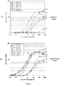
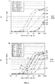
- FIG. 2 to FIG. 4 illustrate the results in a form of graphs.
- the horizontal axis of each graph represents the used antibody concentration (ng/mL), and the vertical axis represents the average fluorescence intensity (MFI) in flow cytometry.
- FIG. 2A is a diagram illustrating the response of anti-human ROR1 antibodies to ROR1 expressing cells of acquired monoclonal antibodies (2AM series and 2AC series).
- FIG. 2B is a diagram illustrating the response of anti-human ROR1 antibodies to ROR1 expressing cells of acquired monoclonal antibodies (2AA series, 2CC series, and 1AA series).
- FIG. 3C is a diagram illustrating the response of anti-human ROR1 antibodies to ROR1 expressing cells of acquired monoclonal antibodies (2DC series and some of 1BA series).
- FIG. 3D is a diagram illustrating the response of anti-human ROR1 antibodies to ROR1 expressing cells of acquired monoclonal antibodies (1BA series).
- FIG. 4E is a diagram illustrating the response of anti-human ROR1 antibodies to ROR1 expressing cells of acquired monoclonal antibodies (6AM series).
- FIG. 4F is a diagram illustrating the response of anti-human ROR1 antibodies to ROR1 expressing cells of acquired monoclonal antibodies (70AF series). Note that, in FIG. 3C and FIG. 3D , reference numbers of some of the acquired monoclonal antibodies are omitted because of the spacing between lines. As illustrated in FIG. 2 to FIG. 4 , it was found that, although having different affinity, all the acquired antibodies are coupled to the human ROR1.
- hybridomas Two types were selected from hybridomas that produce antibodies which recognize the Ig region illustrated in FIG. 1 , one type of hybridoma was selected from hybridomas that produce antibodies which recognize the CDR region illustrated in FIG. 1 , and one type of hybridoma was selected from hybridomas that produce antibodies which recognize the Kringle region illustrated in FIG. 1 . Note that the selected hybridomas are those having relatively high affinity to the ROR1 among antibodies that recognize respective regions.
- the hybridomas that produce the selected antibodies were cultured, and the total RNA was acquired by a general method.
- cDNA was acquired by 5'-RACE method using GeneRacer (registered trademark) kit (Invitrogen).
- the obtained cDNA was used as a template, and a PCR (35 cycles of a cycle [94 degrees Celsius for 30 seconds, 57 degrees Celsius for 30 seconds, and 72 degrees Celsius for 50 seconds]) was performed with Plutinum Tag High Fidelity (Invitrogen) using GeneRacer 5' Primer (SEQ ID No. 48: 5'-CGACTGGAGCACGAGGACACTGA-3') and CH1 (mouse IgG1 constant region 1) 3' Primer (SEQ ID No. 49: 5'-AATTTTCTTGTCCACCTGG-3'). Thereby, the VH gene (cDNA) was isolated.
- the PCR was performed by using GeneRacer 5' Primer and Ck ( ⁇ constant region) 3' Primer (SEQ ID No. 50: 5'-CTAACACTCATTCCTGTTGAAGCTCT-3'), and the VL gene (cDNA) was isolated.
- VH CDR1 SEQ ID No.: amino acid sequence indicated in No.8 VH CDR2: SEQ ID No.: amino acid sequence indicated in No.9 VH CDR3: SEQ ID No.: amino acid sequence indicated in No.10
- VL CDR1 SEQ ID No.: amino acid sequence indicated in No.13
- VL CDR2 SEQ ID No.: amino acid sequence indicated in No.14
- VL CDR3 SEQ ID No.: amino acid sequence indicated in No.15
- VH SEQ ID No.: base sequence indicated in No.6
- VL SEQ ID No.: base sequence indicated in No.11
- ⁇ 6AM_69-2 recognize the CDR region>
- VH CDR1 SEQ ID No.: amino acid sequence indicated in No.18
- VH CDR2 SEQ ID No.: amino acid sequence indicated in No.19
- VH CDR3 SEQ ID No.: amino acid sequence indicated in No.20
- VL CDR1 SEQ ID No.: amino acid sequence indicated in No.23
- VL CDR2 SEQ ID No.: amino acid sequence indicated in No.24
- VL CDR3 SEQ ID No.: amino acid sequence indicated in No.25 VH: SEQ ID No.: amino acid sequence indicated in No.17 VL: SEQ ID No.: amino acid sequence indicated in No.22
- VH SEQ ID No.: base sequence indicated in No.16
- VL SEQ ID No.: base sequence indicated in No.21
- ⁇ 6AM_120-2 recognize the Kringle region>
- VH CDR1 SEQ ID No.: amino acid sequence indicated in No.28 VH CDR2: SEQ ID No.: amino acid sequence indicated in No.29 VH CDR3: SEQ ID No.: amino acid sequence indicated in No.30 VL CDR1: SEQ ID No.: amino acid sequence indicated in No.33 VL CDR2: SEQ ID No.: amino acid sequence indicated in No.34 VL CDR3: SEQ ID No.: amino acid sequence indicated in No.35 VH: SEQ ID No.: amino acid sequence indicated in No.27 VL: SEQ ID No.: amino acid sequence indicated in No.32
- VH SEQ ID No.: base sequence indicated in No.26
- VL SEQ ID No.: base sequence indicated in No.31
- VH CDR1 SEQ ID No.: amino acid sequence indicated in No.38
- VH CDR2 SEQ ID No.: amino acid sequence indicated in No.39
- VH CDR3 SEQ ID No.: amino acid sequence indicated in No.40
- VL CDR1 SEQ ID No.: amino acid sequence indicated in No.43
- VL CDR2 SEQ ID No.: amino acid sequence indicated in No.44
- VL CDR3 SEQ ID No.: amino acid sequence indicated in No.45 VH: SEQ ID No.: amino acid sequence indicated in No.37 VL: SEQ ID No.: amino acid sequence indicated in No.42
- VH SEQ ID No.: base sequence indicated in No.36
- VL SEQ ID No.: base sequence indicated in No.41
- Clone name SEQ ID No. amino acid sequences 2DC_5-2 7 VH 8
- VH CDR1 SYAMS 9
- VL 13 VL
- VL CDR2 RANRLVD 15 VL CDR3 LQYDEFPYT 6AM_69-2 17 VH 18
- VH CDR3 PHYGDYVGY 22 VL 23
- VL CDR2 KVSNRFS 25
- VL CDR3 SQSTHVPFT 6AM_120-2 27
- VH region and the VL region were amplified by a PCR based on the identified gene sequence, and the PCR products were introduced in an antibody-producing vector in which the constant region of human IgG1 is incorporated by a conventional method. Then, 100 ⁇ l at 0.4 ⁇ g/ ⁇ l of these expression vectors digested with a restriction enzyme ScaI and 1 ⁇ 10 7 CHO cells (Chinese hamster ovary) were mixed in a cuvette, and transformed by an electroporation method.
- the mixture was suspended in a Ham F-12 culture medium containing 10% FBS (Thermo Fisher Scientific; 11765054) and dispensed on a 96-well microplate by 100 ⁇ l each, puromycin was added to have the final concentration of 20 ⁇ g/ml, and the resultant was cultured at 37 degrees Celsius for 12 days.
- FBS Thermo Fisher Scientific
- the supernatants were fractionated from the wells forming a single colony, the clones having high activity with a sandwich ELISA method using an anti-human IgG antibody were sorted, the sorted clones were amplified and cultured into a 10 cm dish, the amplified and cultured clones were then acclimatized to an EX-CELL (registered trademark) CD-CHO Fusion culture medium (SAFC; 14365C) to which 4 mM of L-Alanyl-L-Glutamine (Wako; 016-21841) was added, and chimeric antibody-producing cell lines were established.
- SAFC registered trademark CD-CHO Fusion culture medium
- the obtained cell lines were mass-cultured at 37 degrees Celsius with 5% CO 2 for 10 days by using the EX-CELL CD-CHO Fusion culture medium to which 4 mM of L-Alanyl-L-Glutamine was added, and purified antibodies of chimerized antibodies were obtained from the cultured supernatants by using Protein A Sepharose.
- FIG. 5A hereafter, the chimerized antibodies obtained from clones 2DC_5-2, 6AM_69-2, 6AM_120-2, and 2AM_1M17 are denoted as M1, M2, M3, and M4, respectively.
- FIG. 5B illustrates the structure and recognition domains of the created chimerized antibody.
- NCI-H1975 ATCC accession No. CRL-5908
- A427 ATCC accession No. HTB-53
- NCI-H460 ATCC accession No. HTB-177
- the PC-9 cell line RB 4455
- Various cell lines were seeded on 6-well dishes to have 2 ⁇ 10 5 /well, cultured at 37 degrees Celsius for 24 hours, and collected after PBS washing.
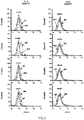
- FIG. 6 illustrates graphs indicating the response of chimerized antibodies to the ROR1 high expressing cell line and the ROR1 low expressing cell line. As illustrated in FIG.
- the chimeric antibodies M1 to M4 were recognized by an acid pH-sensitive dye by using pHrodo (registered trademark) Red Microscale Labeling Kit (Thermo Fisher Scientific; P35363).
- pHrodo registered trademark
- Red Microscale Labeling Kit Thermo Fisher Scientific; P35363
- cells at 1 ⁇ 10 5 /well were seeded on a 6-well dish having a cover glass therein, pHrodo labeled chimeric antibodies at 5 ⁇ g/ml were exposed, reacted at 37 degrees Celsius for 72 hours, and collected after PBS washing, and the fluorescence intensity was measured by flow cytometry.
- FIG. 7 is a diagram illustrating endocytosis of chimerized antibodies to the ROR1 high expressing cell and the ROR1 low expressing cell. As illustrated in FIG. 7 , in the ROR1 high expressing cell line, endocytosis activity due to chimeric antibodies was observed, and when chimeric antibody M2 among others was used, particularly intense endocytosis was observed.
- An antibody that recognizes positions 156 to 300 in the amino acid sequence as an epitope is endocytosed in a cell and thus can be used as a drug delivery antibody or a pharmaceutical composition. Therefore, such an antibody is useful in pharmaceutical industries.
Landscapes
- Health & Medical Sciences (AREA)
- Chemical & Material Sciences (AREA)
- Life Sciences & Earth Sciences (AREA)
- Engineering & Computer Science (AREA)
- Bioinformatics & Cheminformatics (AREA)
- Immunology (AREA)
- General Health & Medical Sciences (AREA)
- Medicinal Chemistry (AREA)
- Pharmacology & Pharmacy (AREA)
- Veterinary Medicine (AREA)
- Public Health (AREA)
- Animal Behavior & Ethology (AREA)
- Organic Chemistry (AREA)
- Epidemiology (AREA)
- Cell Biology (AREA)
- Nuclear Medicine, Radiotherapy & Molecular Imaging (AREA)
- General Chemical & Material Sciences (AREA)
- Chemical Kinetics & Catalysis (AREA)
- Proteomics, Peptides & Aminoacids (AREA)
- Molecular Biology (AREA)
- Genetics & Genomics (AREA)
- Biophysics (AREA)
- Biochemistry (AREA)
- Peptides Or Proteins (AREA)
- Medicines Containing Antibodies Or Antigens For Use As Internal Diagnostic Agents (AREA)
- Preparation Of Compounds By Using Micro-Organisms (AREA)
- Medicinal Preparation (AREA)
- Medicines That Contain Protein Lipid Enzymes And Other Medicines (AREA)
Abstract
Description
- The disclosure in the present specification relates to an anti-ROR1 monoclonal antibody and a functional fragment thereof, a gene, a drug delivery composition, and a pharmaceutical composition. In particular, the disclosure relates to an anti-ROR1 monoclonal antibody and a functional fragment thereof endocytosed in a cancer cell together with an ROR1 during cancer cell endocytosis, a gene having a gene that encodes a variable region of an anti-ROR1 monoclonal antibody as an open reading frame region, a drug delivery composition containing the anti-ROR1 monoclonal antibody or the functional fragment thereof, and a pharmaceutical composition.
- Lung cancer is a primary cause of death of cancer in economically well-developed countries, and among the lung cancer, lung adenocarcinoma is the most frequent histologic subtype.
- It is known that lung adenocarcinoma is closely linked to continuous expression of a thyroid transcription factor-1 (TTF-1) that is a lineage-specific transcription factor required for branching morphogenesis formation and a physiological function in a lung (see Non-Patent Literature 1). Further, it is also known that the TTF-1 is a lineage-survival oncogene (see Non-Patent
Literatures 2 to 4). Since the TTF-1 is indispensable for production of a surfactant protein and a physiological function in a lung of a healthy adult, identification of a downstream molecule involved in a TTF-1 lineage-specific survival signal is required for development of a novel treatment method. - As a downstream molecule induced to express by the TTF-1, a receptor tyrosine kinase-like orphan receptor 1 (ROR1) gene is known. Further, it is also known that it is possible to inhibits proliferation of particular cancer cells by suppressing expression of such an ROR1 gene (see Patent Literature 1).
- Furthermore, it is known that targeting ROR1 enables not only induction of apoptosis of a cancer cell but also suppression of a pro-survival signal of a cancer cell transferred by receptor tyrosine kinase (hereafter, receptor tyrosine kinase may be referred to as "RTK") such as EGFR or MET and therefore the ROR1 can be an important target in development of a proliferation inhibitor against a cancer cell (see
Patent Literature 2 and Non-Patent Literature 5). - As disclosed in
Patent Literature 2 or the like described above, most of molecular targeted drugs that have been developed so far are kinase activation inhibitors. The kinase activation inhibitor refers to a compound that suppresses activation of RTK related to proliferation and survival of human cancer such as lung cancer. If a kinase activation inhibitor is administered to a patient, however, this may cause mutation that provides resistance to a kinase gene during therapy or occurrence of a bypass path by which a survival signal of a cancer cell depends on another receptor, which causes a problem of cancer cells gaining resistance against the kinase activation inhibitor. - Further, when one receptor of a cancer cell, such as EGFR, MET, or the like disclosed in
Patent Literature 2 described above is a molecule target, a sufficient effect may not be obtained because a signal from another receptor is unable to be blocked, and combined use of inhibitors targeting respective receptors have been attempted. Even with combined use of a plurality of inhibitors, however, there is a problem of likelihood of further occurrence of a bypass path or an increased side effect due to administration of the plurality of inhibitors. - For the above reasons, a normal cell or a cancer cell enables itself to survive by effectively using various survival signals, and these survival signals are transferred via many pieces of RTK existing in cell membranes. From the past studies, molecular targeted drugs (such as Iressa targeting EGFR) targeting individual RTK have been developed against cancer cells, and combined use of inhibitors against multiple types of RTK as described above has been attempted. However, since occurrence of a bypass path via further different RTK or these types of RTK have an important role of transferring a survival signal also in a normal cell, a side effect is considered to be a significantly important problem. Thus, it is known that, by suppressing coupling of ROR1 and Cavin-1 or ROR1 and Caveolin1 (hereafter, which may be referred to as "CAV1") rather than targeting individual RTK of a cancer cell, it is possible to suppress formation of caveola in which various proliferation factors are coupled and RTK that transfers a signal into a cell is integrated and it is possible to eliminate the activity of various types of RTK by specifically suppressing formation of caveola (see
Patent Literature 3 and Non-Patent Literature 6). -
- Patent Literature 1: International Publication No.
2010/008069 - Patent Literature 2: International Publication No.
2012/090939 - Patent Literature 3: International Publication No.
2016/063637 -
- Non-Patent Literature 1: Tanaka.H et al.,Cancer Res,2007,Vol.67, p6007-6011
- Non-Patent Literature 2: Kendall.J et al., Proc Natl Acad Sci USA,2007, Vol.104, p16663-16668
- Non-Patent Literature 3: Weir,BA et al., Nature, 2007, Vol.450, p893-898
- Non-Patent Literature 4: Kwei,KA et al., Oncogene,2008, Vol.27, p3635-3640
- Non-Patent Literature 5: Yamaguchi.T et al., Cancer Cell,2012, Vol.21, p348-361
- Non-Patent Literature 6: Yamaguchi.T et al., Nature Commun, 2016, Vol. 7,10060 (doi:10.1038/ncomms10060)
- As described above, the invention disclosed in
Patent Literature 3 is very effective in searching for a compound that can eliminate activity of various types of RTK by specifically suppressing formation of caveola. In this regard, a cell enables itself to survive by effectively using various survival signals. Thus, to develop an effective therapeutic agent against cancer, it is necessary to focus on various molecular mechanisms of cancer cells and search for an effective method. - The disclosure in the present specification is the invention that has been made in order to solve the conventional problems described above, and according to a thorough study, it has been newly found that (1) when an ROR1 that is a protein unique to a cancer cell is focused on, an anti-ROR1 antibody that recognizes a specific region of an extracellular region of the ROR1 as an epitope is endocytosed in a cancer cell together with the ROR1 during endocytosis and (2) thus, an anti-ROR1 antibody can be used as a delivery that endocytoses a drug in a cell.
- That is, the object of the disclosure in the present specification is to provide an anti-ROR1 monoclonal antibody and a functional fragment thereof endocytosed in a cancer cell together with an ROR1 during cancer cell endocytosis, a gene having a gene that encodes a variable region of an anti-ROR1 monoclonal antibody as an open reading frame region, a drug delivery composition containing the anti-ROR1 monoclonal antibody or the functional fragment thereof, and a pharmaceutical composition.
- The disclosure in the present specification relates to an anti-ROR1 monoclonal antibody and a functional fragment thereof, a gene, a drug delivery composition, and a pharmaceutical composition described below.
-
- (1) An anti-ROR1 monoclonal antibody that recognizes
positions 156 to 300 in the amino acid sequence of a human ROR1 protein (GenBank accession No. NP_005003.2) as an epitope. - (2) The anti-ROR1 monoclonal antibody according to (1) above, wherein the anti-ROR1 monoclonal antibody is specifically coupled to an ROR1 of a human cancer cell and endocytosed in a cell by endocytosis.
- (3) The anti-ROR1 monoclonal antibody according to (1) or (2) above,
wherein a heavy-chain variable region includes a region consisting of
a CDR1 H region consisting of an amino acid sequence of SYGIS (SEQ ID No. 18),
a CDR2 H region consisting of an amino acid sequence of EISPRSGYTYYNEKFKG (SEQ ID No. 19), and
a CDR3 H region consisting of an amino acid sequence of PHYGDYVGY (SEQ ID No. 20), and
wherein a light-chain variable region includes a region consisting of
a CDR1 L region consisting of an amino acid sequence of RSSQSLVHSNGNTYLH (SEQ ID No. 23),
a CDR2 L region consisting of an amino acid sequence of KVSNRFS (SEQ ID No. 24), and
a CDR3 L region consisting of an amino acid sequence of SQSTHVPFT (SEQ ID No. 25). - (4) The anti-ROR1 monoclonal antibody according to any one of (1) to (3) above,
wherein the amino acid sequence of an H-chain variable region is of SEQ ID No. 17, and
wherein the amino acid sequence of an L-chain variable region is of SEQ ID No. 22. - (5) A functional fragment of the anti-ROR1 monoclonal antibody according to any one of (1) to (4) above.
- (6) A gene having, as an open reading frame region, a gene that encodes the CDR sequence according to (3) above or the variable region according to (4) above.
- (7) A drug delivery composition including the anti-ROR1 monoclonal antibody according to any one of (1) to (4) above or the functional fragment of the anti-ROR1 monoclonal antibody according to (5) above.
- (8) A pharmaceutical composition including:
- the anti-ROR1 monoclonal antibody according to any one of (1) to (4) above or the functional fragment of the anti-ROR1 monoclonal antibody according to (5) above;
- a linker; and
- a drug,
- wherein one terminal of the linker is coupled to the anti-ROR1 monoclonal antibody or the functional fragment, and the other terminal of the linker is coupled to the drug.
- the anti-ROR1 monoclonal antibody according to any one of (1) to (4) above or the functional fragment of the anti-ROR1 monoclonal antibody according to (5) above;
- The anti-ROR1 monoclonal antibody disclosed in the present specification is endocytosed in a cancer cell together with an ROR1 that is a protein unique to a cancer cell during cancer cell endocytosis. Thus, the anti-ROR1 monoclonal antibody disclosed in the present specification can be used as a drug delivery into a cancer cell or a pharmaceutical composition.
-
- [
FIG. 1] FIG. 1 is a diagram illustrating the structure of an ROR1 molecule and a recognition domain of an acquired monoclonal antibody and a diagram illustrating homology of a mouse ROR1 and a human ROR1. - [
FIG. 2] FIG. 2A is a diagram illustrating response of anti-human ROR1 antibodies to ROR1 expressing cells of acquired monoclonal antibodies (2AM series and 2AC series).FIG. 2B is a diagram illustrating response of anti-human ROR1 antibodies to ROR1 expressing cells of acquired monoclonal antibodies (2AA series, 2CC series, and 1AA series). - [
FIG. 3] FIG. 3C is a diagram illustrating response of anti-human ROR1 antibodies to ROR1 expressing cells of acquired monoclonal antibodies (2DC series and some of 1BA series).FIG. 3D is a diagram illustrating response of anti-human ROR1 antibodies to ROR1 expressing cells of acquired monoclonal antibodies (1BA series). - [
FIG. 4] FIG. 4E is a diagram illustrating response of anti-human ROR1 antibodies to ROR1 expressing cells of acquired monoclonal antibodies (6AM series).FIG. 4F is a diagram illustrating response of anti-human ROR1 antibodies to ROR1 expressing cells of acquired monoclonal antibodies (70AF series). - [
FIG. 5] FIG. 5A is a diagram illustrating the names of origin clones of chimerized antibodies and recognition domains.FIG. 5B is a diagram illustrating structure of chimerized antibody and recognition domains. - [
FIG. 6] FIG. 6 is a diagram illustrating response of chimerized antibodies to ROR1 high expressing cells and ROR1 low expressing cells. - [
FIG. 7] FIG. 7 is a diagram illustrating endocytosis of chimerized antibodies to ROR1 high expressing cells and ROR1 low expressing cells. - [
FIG. 8] FIG. 8A is a diagram illustrating cell proliferation inhibition of the PC-9 by a chimerized antibody and a non-cleavage type MMAF.FIG. 8B is a diagram illustrating cell proliferation inhibition of the PC-9 by a chimerized antibody M2 and a cleavage type MMAF.FIG. 8C is a diagram illustrating cell proliferation inhibition of the A427 by a chimerized antibody M2 and a cleavage type MMAF. - An anti-ROR1 monoclonal antibody (hereafter, which may be simply referred to as "antibody") and a functional fragment thereof, a gene, a drug delivery composition, and a pharmaceutical composition will be described below in detail.
- The antibody disclosed in the present specification means an antibody that recognizes, as an epitope,
positions 156 to 300 in the amino acid sequence of the extracellular region of a human ROR1 and is specifically coupled thereto and that includes a fragment (which may be referred to as a "functional fragment" in the present specification) or a derivative of the antibody exhibiting substantially the same antigen specificity as the original antibody. The functional fragment or the derivative of an antibody includes a functional fragment of the antibody, such as a peptide containing Fab, Fab', F(ab')2, a single chain antibody (scFv), a disulfide-stabilized V region fragment (dsFv), or CDR, a derivative such as a humanized antibody (for example, a CDR-implant complete human antibody), or the like. The derivative can be created by a known method by using a gene recombination technology. - Amino acid sequences, gene information, and the cDNA sequences of a human ROR1 are well known, and information on the sequence or the like is available from the amino acid sequence deposited in GenBank accession No. NP_005003.2, the gene information on GenBank Gene ID: 4919, and the cDNA deposited in GenBank accession No. NM_005012.1, for example. The full length of the amino acid sequence of the human ROR1 is 937, and
positions 1 to 403 in the amino acid sequence correspond to the extracellular region. - Further, in the present specification, when "positions 156 to 300 in the amino acid sequence of the human ROR1 protein (GenBank accession No. NP_005003.2)" is stated, in addition to
positions 156 to 300 in the amino acid sequence, the derivatives thereof are included. The term "derivative" as used herein includes one or a plurality of (for example, several (for example, six)) mutated, substituted, deleted, and/or added amino acid residues atpositions 156 to 300 in the amino acid sequence and means peptide or polypeptide substantially having the same antigenicity as those ofpositions 156 to 300 in the amino acid sequence of the human ROR1. The term "substantially" as used herein means that an antibody created frompositions 156 to 300 in the amino acid sequence as an epitope is specifically coupled to an ROR1 and has a function of being endocytosed in a cancer cell together with the ROR1 during endocytosis. - The term "epitope" refers to a part of an antigen recognized by an antibody (in the case of the present specification,
positions 156 to 300 in the amino acid sequence of the extracellular region of a human ROR1) and means a site on an antigen to which a region including an antibody variable region disclosed in the present specification is coupled. For example, when polypeptide such as an ROR1 is an antigen, the antibody may recognize a linear amino acid sequence and may recognize a three-dimensional spatial structure, and therefore, the epitope can be defined by an amino acid sequence or antigen structure. Note that the extracellular region of the amino acid sequence of the human ROR1 includes three regions referred to as Ig (amino acid positions: 66 to 148), cysteine-rich domain (CRD, amino acid positions: 166 to 307), and Kringle (amino acid positions: 311 to 393) (seeFIG. 1 ). Therefore, an antibody disclosed in the present specification can be said to be an antibody that substantially recognizes the CRD as an epitope. - The antibody disclosed in the present specification is specifically coupled to an ROR1 of a human cancer cell and endocytosed in the cell by endocytosis as illustrated in Examples described later. Thus, the antibody can be used as an antibody for delivering a drug or the like that damage a cancer cell. Therefore, a composition in which the antibodies disclosed in the present specification are dispersed in a medium or the like can be provided as a drug delivery composition.
- Further, it is possible to provide a pharmaceutical composition using the antibody disclosed in the present specification by coupling one end of a linker to the antibody disclosed in the present specification and coupling a drug to the other end of the linker. Any of cleavage type linkers and non-cleavage type linkers may be used for the linker. Further, the drug is not particularly limited as long as it is a drug for cancer therapy such as killing a cancer cell or suppressing proliferation of cancer cells and can be coupled to a linker. Note that, when coupling a linker to an antibody, it is preferable to couple a linker to a site other than the H-chain variable region and the L-chain variable region so as not to inhibit coupling of the antibody to an ROR1.
- The cancer cell is not particularly limited as long as an ROR1 has expressed therein and may be a cell of cancer such as, but not limited thereto, lung cancer (including lung adenocarcinoma, small cell lung cancer, squamous cell lung cancer, and large-cell lung cancer), pancreatic cancer, malignant mesothelioma, breast cancer, stomach cancer, colorectal cancer, oral cancer, esophageal cancer, uterine cancer, kidney cancer, bladder cancer, ovarian cancer, testicular cancer, and the like, for example. The biological species from which a cancer cell is derived may be, but not limited to, human, monkey, mouse, rat, guinea pig, pig, cow, sheep, goat, and the like, for example.
- While Examples will be presented below and embodiments will be specifically described, these Examples and embodiments are not intended to limit or restrict the scope of the invention disclosed in the present specification.
- A cDNA sequence (NM_005012.1) of the full length of a human ROR1 was cut out from pCMV6-KL6-ROR1 (OriGene Technologies, Rockville, MDs) and introduced in pCMVpuro to create pCMVpuro-ROR1. Furthermore, animal cells that are controlled by a CMV promotor and cause the full length of a human ROR1 (
amino acid positions 1 to 937) or the full length of a human ROR1 extracellular region (amino acid positions 1 to 403) to express at the same time as a puromycin-EGFP fusion protein in the downstream of the IRES sequence were obtained by the method below. - A PCR product amplified by using 5' Primer including a NotI site (SEQ ID No. 1: 5'-AATAGCGGCCGCACCATGCACCGGCCGCGCC-3': the underline indicates the NotI site, and an underline part indicates a site to be cut by a restriction enzyme, though the repeated description will be omitted in the sequences below) and 3' Primer including an XhoI site (SEQ ID No. 2: 5'-GCGGCTCGAGCAGTTCTGCAGAAATCATAGATTCG-3') was digested with restriction enzymes NotI and XhoI, and thereby a fragment was obtained. The obtained fragment was introduced in a NotI-XhoI site of pQCXIPG, and an expression vector pQCXIPG-full was constructed.
- A PCR product obtained by using 5' Primer including a NotI site (SEQ ID No. 1) and 3' Primer including a BamHI site (SEQ ID No. 3: 5'-CCGCGGATCCTTCCATTTTATTCTTCTCCTTGG-3') was digested with NotI and BamHI, and thereby a fragment was obtained. The obtained fragment is introduced in a NotI-BamHI site of pQCXIPG, and an expression vector pQCXIPG-EC was constructed.
- On the other hand, a secretory signal sequence of chicken egg white lysozyme (amino acid sequence: MRSLLILVLCFLPLAALG|AAA (SEQ ID No. 46), gene sequence: ATGAGGTCTTTGCTAATCTTGGTGCTTTGCTTCCTGCCCCTGGCTGCTCTGGGG| GCGGCCGCC (SEQ ID No. 47)) was added to the upstream of a multicloning site of the pQCXIPG described above, and a vector (lyssig-pQCXIPG) that enables forced secretion of a translation product of an introduced gene outside the cell was used for a vector used for causing a partial length of a human ROR1 including the human CDR region and the Kringle region (
amino acid positions 156 to 403) and a partial length of a human ROR1 including the human CDR region (amino acid positions 156 to 300) to express in an animal cell. - A PCR product amplified by using 5' Primer including a NotI site (SEQ ID No. 4: 5'-AATAGCGGCCGCAAGTCCAGGATACTCAGATGA-3') and 3' Primer including a BamHI site (SEQ ID No. 3) was digested with restriction enzymes NotI and BamHI, and thereby a fragment was obtained. The obtained fragment was introduced in a NotI-BamHI site of lyssig-pQCXIPG, and an expression vector pQCXIPG-CK was constructed. Note that pQCXIPG and lyssig-pQCXIPG are vectors modified based on pQCXIP of BD Retro-XTM Q Vectors.
- A PCR product amplified by using 5' Primer (5'-AATAGCGGCCGCAAGTCCAGGATACTCAGATGA-3' (SEQ ID No. 4)) and 3' Primer including a BamHI site (SEQ ID No. 5: 5'-AGCAGGATCCAATCCGGATACAGTTCGCA-3') was digested with restriction enzymes NotI and BamHI, and thereby a fragment was obtained. The obtained fragment was introduced in a NotI-BamHI site of lyssig-pQCXIPG, and an expression vector pQCXIPG-CDR was constructed.
- To establish an antigen expressing cell line, Pantropic Retroviral Expression System (Clontech; K1063-1) was used. A GP2-293 (Clontech; K1063-1) in an 80 to 90% confluent state was provided in a collagen-coated 100 mm dish, and 11.2 µg of an expression vector constructed as described above (pQCXIPG-full, pQCXIPG-EC, pQCXIPG-CK, pQCXIPG-CDR) using
Lipofectamine 2000 and 11.2 µg of pVSV-G (Clontech; K1063-1) are co-transfected. After 48 hours, the supernatant containing virus particles was collected, and virus was precipitated by super centrifuge (18,000 rpm, 1,5 hours, 4 degrees Celsius). The precipitate was suspended with 30 µL of TNE (50 mM, Tris-HCl [pH 7.8], 130 mM NaCl, 1mM EDTA), and a retroviral vector concentrated solution was prepared. - Then, 5 µL of the retroviral vector concentrated solution was diluted with 150 µL of DMEM (SIGMA; D5796)-10% FBS containing 8 µg/ml of hexadimethrine bromide (SIGMA; H-9268) to prepare a culture medium containing virus particles. A 293T culture medium provided so as to be in a state of around 40% confluent in a 96-well microplate was replaced with the prepared culture medium containing the virus particles, and thereby pQCXIPG-full, pQCXIPG-EC, pQCXIPG-CK, and pQCXIPG-CDR were genetically introduced. After the genetic introduction, the resultant was amplified and cultured with DMEM (SIGMA; D5796)-10% FBS containing 5 µg/ml of puromycin (SIGMA; P-8833) to establish an antigen expressing cell line (hROR1-full/293T, hROR1-EC/293T, hROR1-CK/293T, hROR1-CDR/293T) .
- Around 1 L of a supernatant obtained by culturing the above established expressing cell line (hROR1-EC/293T, hROR1-CK/293T, hROR1-CDR/293T) with DMEM-10% FBS (5µg/ml puromycin) or CD293 (Invitrogen) was collected, a myc-His tagged recombinant protein was purified from the collected supernatant by using an affinity column filled with carries chemically modified from a myc-tag antibody or a TALON Purification Kit (Clontech; K1253-1) and then dialyzed with PBS, and a purified protein was observed by using SDS-PAGE and western blot. A protein assay kit II (BioRad; 500-0002JA) was used to determine the protein concentration, and the antigen for immunization was prepared.
- A purified protein of a human ROR1 extracellular region (hROR1-EC), a purified protein of a partial length including the human ROR1 CDR-Kringle region (hROR1-CK), or a purified protein of a partial length including the human ROR1 CDR region (hROR1-CDR) were mixed with the same amount of complete adjuvant (SIGMA; F5881) to obtain an emulsion, and BALB/c mice (female) were immunized with the emulsion at 5 to 50 µg/head for several times every three to seven days. Further, in a case of immunization with the cell hROR1-full/293T in which the full length of a human ROR1 expressed, mice were immunized at 1 × 10⁵ cells/head for several times every three to seven days. Lymphocyte cells were extracted from the mice after three to five days from the final immunization, and cell fusion of the extracted lymphocyte cells with mouse myeloma cells P3U1 (P3-X63Ag8U1) was performed.
- Cell fusion was performed based on a general method illustrated below. For the fetal bovine serum (FBS) in all the culture media, a serum inactivated by a process of maintaining the temperature at 56 degrees Celsius for 30 minutes was used. The P3U1 was provided by being cultured with RPMI1640-10% FBS (containing penicillin, streptomycin). The extracted mouse lymphocyte cells and the P3U1 were mixed at a ratio of 10:1 to 2:1 and centrifuged. Then, 50% polyethylene glycol 4000 (Merck; 1.09727.0100) was gradually added as a fusion accelerating agent to the precipitated cells and gently mixed to perform cell fusion. Furthermore, after the RPMI1640 was gradually added and gently mixed, the mixture was centrifuged. The precipitated fused cells were diluted as appropriate with a HAT culture medium containing 15% FBS (containing RPMI1640, HAT-supplement (Invitrogen; 11067-030), penicillin, streptomycin) and seeded on a 96-well microplate at 200 µL/well.
- The fused cells were cultured in a CO2 incubator (5% CO2, 37 degrees Celsius), and after a colony was sufficiently formed, a cultured supernatant was sampled and screened. In the screening, from those found to be positive in ELISA performed on a 96-well plate on which the ROR1 antigen used for immunization was sensitized, hybridomas were sorted with which response was observed by flow cytometry performed on the 293T in which the full length of the ROR1 was forcedly caused to express (hROR1-full/293T). After these hybridomas were amplified and cultured with an HT culture medium containing 15% FBS (containing RPMI1640, HT-supplement (Invitrogen; 21060-017), penicillin, streptomycin) and mono-cloned by a limiting dilution method. In such a way, 45 types of hybridoma clones (
FIG. 1 ) were obtained which produce 19 clones from antibodies (a full-length protein of the extracellular region (amino acid positions 1 to 403) as an immunogenicity (FIG. 1 : 2AM, 2AC, 2CC, 2AA, 2DC series), 13 clones from full-length expressing cells (hROR1-full/293T) as an immunogenicity (FIG. 1 : 1AA, 1BA series), 5 clones from a partial-length protein including the CDR-Kringle region (amino acid positions 156 to 403) as an immunogenicity (FIG. 1 : 6AM series), and 7 clones from a partial-length protein including the CDR region (amino acid positions 156 to 300) as an immunogenicity (FIG. 1 : 70AF series). For these 45 types of antibodies, response to hROR1-EC, hROR1-CK, and hROR1-CDR was observed by the ELISA method, it was analyzed which region was recognized by each antibody to perform epitope grouping, and this results in that 23 types of antibodies recognize the Ig region, 17 types of antibodies recognize the CDR region, and 4 types of antibodies recognize the Kringle region (FIG. 1 ). Further, homology in the amino acid sequences of a mouse and a human is illustrated inFIG. 1 . Although the homology differs for respective regions, the extracellular regions of the ROR1 of a mouse and a human exhibit a significantly high homology. - To inspect response of the antibodies obtained as described above to an endogenous human ROR1, response to an ROR1 high expressing cell NCI-H358 (ATCC accession No. CRL-5807) line was observed as below. The antibodies were purified from the culture supernatant of each hybridoma clone by a general affinity purification method using Protein A-Sepharose, respectively. The obtained purified antibodies were analyzed for the average fluorescence intensity of flow cytometry under the same condition (the same number of NCI-H358 (1 × 10⁵), the same concentration of respective purified antibodies, and the same concentration of secondary (detected) antibodies). Further, data was acquired also for the average fluorescence intensity that depends on the antibody concentration, the detection limit at a low concentration was analyzed, and thereby relative affinity was evaluated.
FIG. 2 to FIG. 4 illustrate the results in a form of graphs. The horizontal axis of each graph represents the used antibody concentration (ng/mL), and the vertical axis represents the average fluorescence intensity (MFI) in flow cytometry.FIG. 2A is a diagram illustrating the response of anti-human ROR1 antibodies to ROR1 expressing cells of acquired monoclonal antibodies (2AM series and 2AC series).FIG. 2B is a diagram illustrating the response of anti-human ROR1 antibodies to ROR1 expressing cells of acquired monoclonal antibodies (2AA series, 2CC series, and 1AA series).FIG. 3C is a diagram illustrating the response of anti-human ROR1 antibodies to ROR1 expressing cells of acquired monoclonal antibodies (2DC series and some of 1BA series).FIG. 3D is a diagram illustrating the response of anti-human ROR1 antibodies to ROR1 expressing cells of acquired monoclonal antibodies (1BA series).FIG. 4E is a diagram illustrating the response of anti-human ROR1 antibodies to ROR1 expressing cells of acquired monoclonal antibodies (6AM series).FIG. 4F is a diagram illustrating the response of anti-human ROR1 antibodies to ROR1 expressing cells of acquired monoclonal antibodies (70AF series). Note that, inFIG. 3C and FIG. 3D , reference numbers of some of the acquired monoclonal antibodies are omitted because of the spacing between lines. As illustrated inFIG. 2 to FIG. 4 , it was found that, although having different affinity, all the acquired antibodies are coupled to the human ROR1. - Next, two types of hybridomas were selected from hybridomas that produce antibodies which recognize the Ig region illustrated in
FIG. 1 , one type of hybridoma was selected from hybridomas that produce antibodies which recognize the CDR region illustrated inFIG. 1 , and one type of hybridoma was selected from hybridomas that produce antibodies which recognize the Kringle region illustrated inFIG. 1 . Note that the selected hybridomas are those having relatively high affinity to the ROR1 among antibodies that recognize respective regions. Next, the hybridomas that produce the selected antibodies were cultured, and the total RNA was acquired by a general method. Next, cDNA was acquired by 5'-RACE method using GeneRacer (registered trademark) kit (Invitrogen). The obtained cDNA was used as a template, and a PCR (35 cycles of a cycle [94 degrees Celsius for 30 seconds, 57 degrees Celsius for 30 seconds, and 72 degrees Celsius for 50 seconds]) was performed with Plutinum Tag High Fidelity (Invitrogen) using GeneRacer 5' Primer (SEQ ID No. 48: 5'-CGACTGGAGCACGAGGACACTGA-3') and CH1 (mouse IgG1 constant region 1) 3' Primer (SEQ ID No. 49: 5'-AATTTTCTTGTCCACCTGG-3'). Thereby, the VH gene (cDNA) was isolated. On the other hand, similarly for the VL gene, the PCR was performed by using GeneRacer 5' Primer and Ck (κ constant region) 3' Primer (SEQ ID No. 50: 5'-CTAACACTCATTCCTGTTGAAGCTCT-3'), and the VL gene (cDNA) was isolated. - After the VH gene and the VL gene were subcloned to a pT7Blue vector, respectively, an auto-sequencer (by Applied Biosystem) or a fluorescent auto-sequencer was used to analyze the sequence. Amino acids encoded by the resultant VH sequence and the resultant VL sequence and each CDR sequence were determined.
- Respective identified sequences are indicated below. The details of the amino acid sequences are indicated in Table 1, and the details of the base sequences are indicated in Table 2 and Table 3.
- VH CDR1: SEQ ID No.: amino acid sequence indicated in No.8
VH CDR2: SEQ ID No.: amino acid sequence indicated in No.9
VH CDR3: SEQ ID No.: amino acid sequence indicated in No.10
VL CDR1: SEQ ID No.: amino acid sequence indicated in No.13
VL CDR2: SEQ ID No.: amino acid sequence indicated in No.14
VL CDR3: SEQ ID No.: amino acid sequence indicated in No.15
VH: SEQ ID No.: amino acid sequence indicated in No.7
VL: SEQ ID No.: amino acid sequence indicated in No.12 - VH: SEQ ID No.: base sequence indicated in No.6
VL: SEQ ID No.: base sequence indicated in No.11 - VH CDR1: SEQ ID No.: amino acid sequence indicated in No.18
VH CDR2: SEQ ID No.: amino acid sequence indicated in No.19
VH CDR3: SEQ ID No.: amino acid sequence indicated in No.20
VL CDR1: SEQ ID No.: amino acid sequence indicated in No.23
VL CDR2: SEQ ID No.: amino acid sequence indicated in No.24
VL CDR3: SEQ ID No.: amino acid sequence indicated in No.25
VH: SEQ ID No.: amino acid sequence indicated in No.17
VL: SEQ ID No.: amino acid sequence indicated in No.22 - VH: SEQ ID No.: base sequence indicated in No.16
VL: SEQ ID No.: base sequence indicated in No.21 - VH CDR1: SEQ ID No.: amino acid sequence indicated in No.28
VH CDR2: SEQ ID No.: amino acid sequence indicated in No.29
VH CDR3: SEQ ID No.: amino acid sequence indicated in No.30
VL CDR1: SEQ ID No.: amino acid sequence indicated in No.33
VL CDR2: SEQ ID No.: amino acid sequence indicated in No.34
VL CDR3: SEQ ID No.: amino acid sequence indicated in No.35
VH: SEQ ID No.: amino acid sequence indicated in No.27
VL: SEQ ID No.: amino acid sequence indicated in No.32 - VH: SEQ ID No.: base sequence indicated in No.26
VL: SEQ ID No.: base sequence indicated in No.31 - VH CDR1: SEQ ID No.: amino acid sequence indicated in No.38
VH CDR2: SEQ ID No.: amino acid sequence indicated in No.39
VH CDR3: SEQ ID No.: amino acid sequence indicated in No.40
VL CDR1: SEQ ID No.: amino acid sequence indicated in No.43
VL CDR2: SEQ ID No.: amino acid sequence indicated in No.44
VL CDR3: SEQ ID No.: amino acid sequence indicated in No.45
VH: SEQ ID No.: amino acid sequence indicated in No.37
VL: SEQ ID No.: amino acid sequence indicated in No.42 - VH: SEQ ID No.: base sequence indicated in No.36
VL: SEQ ID No.: base sequence indicated in No.41[Table 1] Clone name SEQ ID No. amino acid sequences 2DC_5-2 7 VH 
8 VH CDR1 SYAMS 9 VH CDR2 AINTKGGSTYYPDTVKD 10 VH CDR3 HEPVYDYAMDY 12 VL 
13 VL CDR1 KASQDINSYLS 14 VL CDR2 RANRLVD 15 VL CDR3 LQYDEFPYT 6AM_69-2 17 VH 
18 VH CDR1 SYGIS 19 VH CDR2 EISPRSGYTYYNEKFKG 20 VH CDR3 PHYGDYVGY 22 VL 
23 VL CDR1 RSSQSLVHSNGNTYLH 24 VL CDR2 KVSNRFS 25 VL CDR3 SQSTHVPFT 6AM_120-2 27 VH 
28 VH CDR1 GYNMN 29 VH CDR2 NINPYYGNTSYNQKFKG 30 VH CDR3 EGFEYFDY 32 VL 
33 VL CDR1 RASENIYSYLA 34 VL CDR2 NVKTLAE 35 VL CDR3 QHHYGIPYT 2AM_1M17 37 VH 
38 VH CDR1 SYAMS 39 VH CDR2 AINSKGGSTYYPDTVKD 40 VH CDR3 HDERIDYAMDY 42 VL 
43 VL CDR1 KASQDINSYLS 44 VL CDR2 RANRLVD 45 VL CDR3 LQYDEFPYT [Table 2] Clone name SEQ D No base sequences 2DC_5-2 6 VH 
11 VL 
5AM_69-2 16 VH 
21 VL 
[Table 3] Clone name SEQ ID No. base sequences 6AM_120-2 26 VH 
31 VL 
2AM_1M17 36 VH 
41 VL 
- The VH region and the VL region were amplified by a PCR based on the identified gene sequence, and the PCR products were introduced in an antibody-producing vector in which the constant region of human IgG1 is incorporated by a conventional method. Then, 100 µl at 0.4 µg/µl of these expression vectors digested with a restriction enzyme ScaI and 1 × 10⁷ CHO cells (Chinese hamster ovary) were mixed in a cuvette, and transformed by an electroporation method. The mixture was suspended in a Ham F-12 culture medium containing 10% FBS (Thermo Fisher Scientific; 11765054) and dispensed on a 96-well microplate by 100 µl each, puromycin was added to have the final concentration of 20 µg/ml, and the resultant was cultured at 37 degrees Celsius for 12 days. The supernatants were fractionated from the wells forming a single colony, the clones having high activity with a sandwich ELISA method using an anti-human IgG antibody were sorted, the sorted clones were amplified and cultured into a 10 cm dish, the amplified and cultured clones were then acclimatized to an EX-CELL (registered trademark) CD-CHO Fusion culture medium (SAFC; 14365C) to which 4 mM of L-Alanyl-L-Glutamine (Wako; 016-21841) was added, and chimeric antibody-producing cell lines were established. The obtained cell lines were mass-cultured at 37 degrees Celsius with 5% CO2 for 10 days by using the EX-CELL CD-CHO Fusion culture medium to which 4 mM of L-Alanyl-L-Glutamine was added, and purified antibodies of chimerized antibodies were obtained from the cultured supernatants by using Protein A Sepharose. As illustrated in
FIG. 5A , hereafter, the chimerized antibodies obtained from clones 2DC_5-2, 6AM_69-2, 6AM_120-2, and 2AM_1M17 are denoted as M1, M2, M3, and M4, respectively. Further,FIG. 5B illustrates the structure and recognition domains of the created chimerized antibody. - To inspect response of antibodies obtained as described above to an endogenous human ROR1, the response to the ROR1 high expressing cell line (NCI-H1975, PC-9) and the ROR1 low expressing cell line (A427, NCI-H460) was observed as follows. Note that NCI-H1975 (ATCC accession No. CRL-5908), A427 (ATCC accession No. HTB-53), and NCI-H460 (ATCC accession No. HTB-177) were purchased from American Type Culture Collection. The PC-9 cell line (RCB 4455) was obtained from RIKEN cell bank. Various cell lines were seeded on 6-well dishes to have 2 × 10⁵/well, cultured at 37 degrees Celsius for 24 hours, and collected after PBS washing. After chimerized antibodies labeled by using Alexa Fluor (registered trademark) 488 Antibody Labeling Kit (Thermo Fisher Scientific; A20181) were diluted to 1 µg/ml and reacted on ice for one hour, the resultant was washed with a PBS, and the reaction intensity was analyzed by flow cytometry. As a control, isotype control IgG1 against an antigen that does not exist in mammal cells was used.
FIG. 6 illustrates graphs indicating the response of chimerized antibodies to the ROR1 high expressing cell line and the ROR1 low expressing cell line. As illustrated inFIG. 6 , a reaction was observed in the chimeric antibodies M1, M2, M3, and M4 in the ROR1 high expressing cell line, and the reaction was significantly weak in the ROR1 low expressing cell line. From the above results, it was revealed that all the chimeric antibodies M1, M2, M3, and M4 exhibit response to the endogenous human ROR1. - Next, to examine endocytosis activity of chimeric antibodies reacted with the human ROR1 into cells, the chimeric antibodies M1 to M4 were recognized by an acid pH-sensitive dye by using pHrodo (registered trademark) Red Microscale Labeling Kit (Thermo Fisher Scientific; P35363). For the ROR1 high expressing cell line PC-9 and the ROR1 low expressing cell line A427, cells at 1 × 10⁵/well were seeded on a 6-well dish having a cover glass therein, pHrodo labeled chimeric antibodies at 5 µg/ml were exposed, reacted at 37 degrees Celsius for 72 hours, and collected after PBS washing, and the fluorescence intensity was measured by flow cytometry.
FIG. 7 is a diagram illustrating endocytosis of chimerized antibodies to the ROR1 high expressing cell and the ROR1 low expressing cell. As illustrated inFIG. 7 , in the ROR1 high expressing cell line, endocytosis activity due to chimeric antibodies was observed, and when chimeric antibody M2 among others was used, particularly intense endocytosis was observed. -
- (a) To examine the effect of suppressing cell proliferation using endocytosis activity of chimeric antibodies into cells, a suppressing effect caused by a co-process with chimeric antibodies against cell proliferation was evaluated by using a secondary antibody in which monomethyl auristatin F (MMAF) that is a cytotoxic drug is coupled by a non-cleavage type linker. PC-9 lung adenocarcinoma cell lines were seeded on a 96-well dish at 1 × 10³ cells/well, a secondary antibody αHFc-NC-MMAF (Moradec; AH-101AF) in which 0.1 µg/ml or 1.0 µg/ml of the chimeric antibodies M1 to M4 and the MMAF were coupled by a non-cleavage type linker was added in the next day and cultured at 37 degrees Celsius. After five days from the start of the antibody addition process, influence on cell proliferation was studied by using the MTT assay method.
FIG. 8A is a diagram illustrating cell proliferation inhibition of the PC-9 by the chimerized antibody and the non-cleavage type MMAF. As illustrated inFIG. 8A , it was revealed that, while the chimeric antibodies M1 and M4 that recognize the Ig region inhibit cell proliferation when the antibody concentration was high, the chimeric antibody M2 that recognizes the CDR region more intensively inhibits cell proliferation than M1 and M4. Further, it was revealed that all of M1, M2, M4 inhibits cell proliferation in a manner depending on the antibody concentration. On the other hand, the chimeric antibody M3 that recognizes the Kringle region did not at all inhibit cell proliferation. - (b) Next, to further examine the cell proliferation suppression effect of the chimeric antibody M2 that inhibited cell proliferation the most intensively in (a) described above, αHFc-CL-MMAF (Moradec; AH-102AF) in which the cytotoxic drug MMAF is coupled to the secondary antibody by a cleavage type linker was used to perform a co-process of the chimeric antibody M2 and the MMAF-coupled secondary antibody, and influence on the cell proliferation was evaluated. The ROR1 high expressing cell line, namely, the PC-9 cell line and the ROR1 low expressing cell line, namely, the A427 cell line were seeded on a 96-well dish at 1 × 10³ cells/well, and 0.1 µg/ml or 1.0 µg/ml of the chimeric antibody M2 and the αHFc-CL-MMAF were added together in the next day, and cultured at 37 degrees Celsius for five days, and suppression activity against cell proliferation was then measured by an MTT assay method.
FIG. 8B is a diagram illustrating the cell proliferation inhibition of the PC-9 by the chimerized antibody M2 and the cleavage type MMAF.FIG. 8C is a diagram illustrating the cell proliferation inhibition of the A427 by the chimerized antibody M2 and the cleavage type MMAF. - As illustrated in
FIG. 8B , in the PC-9 cell line, while cell proliferation was significantly suppressed when the chimeric antibody M2 and the MMAF-coupled secondary antibody were co-processed, no suppression of cell proliferation was found in the control (only IgG, only M2, IgG-MMAF). Further, it was revealed that the cleavage type linker can more suppress cell proliferation than the non-cleavage type linker. On the other hand, as illustrated inFIG. 8C , in the A427 that is the ROR1 low expressing cell line, even when the chimeric antibody M2 and the MMAF-coupled secondary antibody were co-processed, suppression of cell proliferation was not found at all. - From the above results, it was revealed that:
- (1) Any antibodies that recognize the extracellular region of the human ROR1 are specifically coupled to the human ROR1;
- (2) However, antibodies that recognize
positions 156 to 300 in the amino acid sequence as an epitope exhibit more significant endocytosis into cancer cells than antibodies that recognize other regions as an epitope; and - (3) Therefore, antibodies that recognize
positions 156 to 300 in the amino acid sequence as an epitope can be used as an antibody for delivering a drug into a cancer cell and a pharmaceutical composition when coupled with a drug. - An antibody that recognizes
positions 156 to 300 in the amino acid sequence as an epitope is endocytosed in a cell and thus can be used as a drug delivery antibody or a pharmaceutical composition. Therefore, such an antibody is useful in pharmaceutical industries.


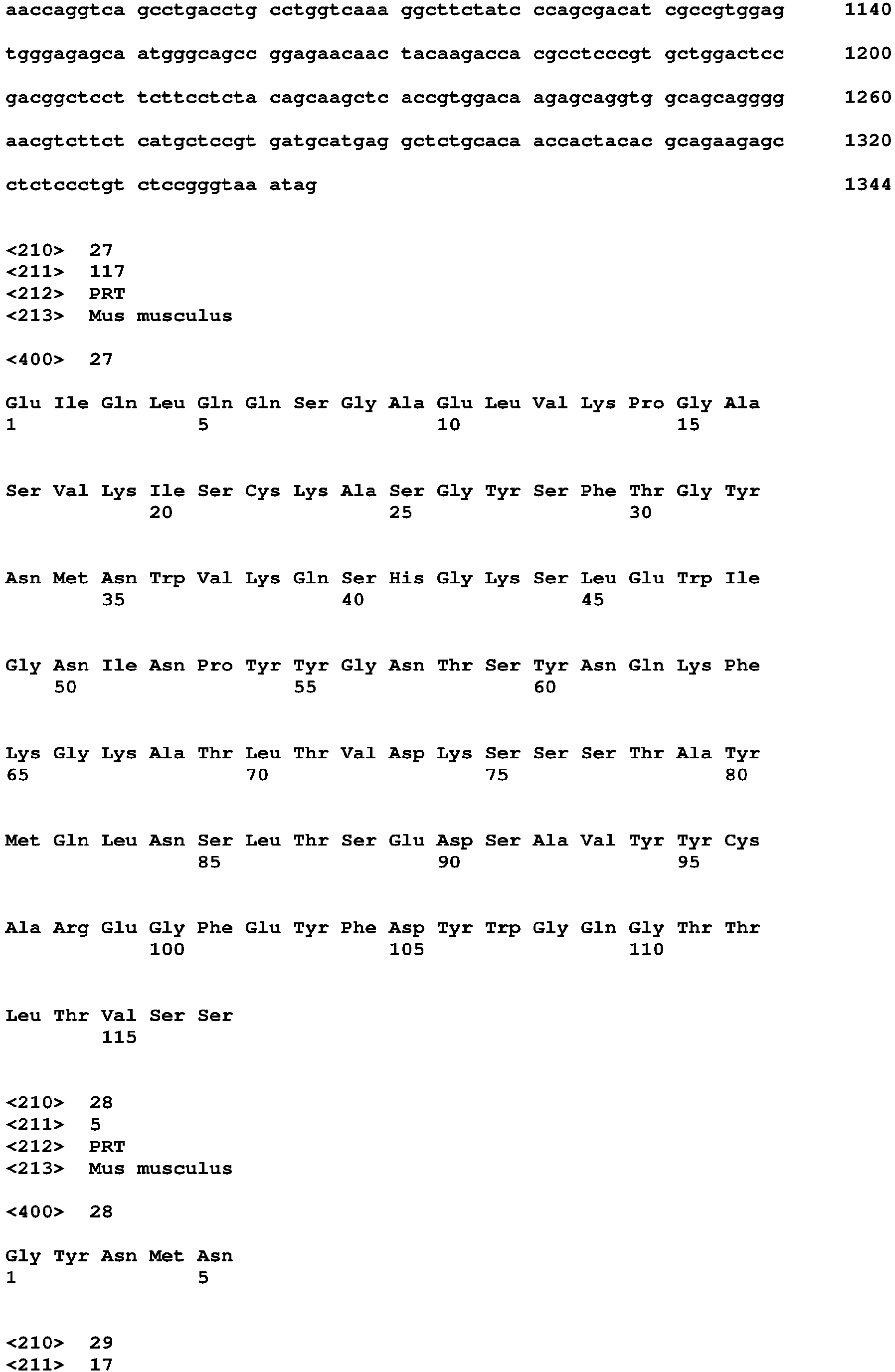


Claims (8)
- An anti-ROR1 monoclonal antibody that recognizes positions 156 to 300 in the amino acid sequence of a human ROR1 protein (GenBank accession No. NP_005003.2) as an epitope.
- The anti-ROR1 monoclonal antibody according to claim 1, wherein the anti-ROR1 monoclonal antibody is specifically coupled to an ROR1 of a human cancer cell and endocytosed in a cell by endocytosis.
- The anti-ROR1 monoclonal antibody according to claim 1 or 2,
wherein a heavy-chain variable region includes a region consisting of
a CDR1 H region consisting of an amino acid sequence of SYGIS (SEQ ID No. 18),
a CDR2 H region consisting of an amino acid sequence of EISPRSGYTYYNEKFKG (SEQ ID No. 19), and
a CDR3 H region consisting of an amino acid sequence of PHYGDYVGY (SEQ ID No. 20), and
wherein a light-chain variable region includes a region consisting of
a CDR1 L region consisting of an amino acid sequence of RSSQSLVHSNGNTYLH (SEQ ID No. 23),
a CDR2 L region consisting of an amino acid sequence of KVSNRFS (SEQ ID No. 24), and
a CDR3 L region consisting of an amino acid sequence of SQSTHVPFT (SEQ ID No. 25). - The anti-ROR1 monoclonal antibody according to any one of claims 1 to 3,
wherein the amino acid sequence of an H-chain variable region is of SEQ ID No. 17, and
wherein the amino acid sequence of an L-chain variable region is of SEQ ID No. 22. - A functional fragment of the anti-ROR1 monoclonal antibody according to any one of claims 1 to 4.
- A gene having, as an open reading frame region, a gene that encodes the CDR sequence according to claim 3 or the variable region according to claim 4.
- A drug delivery composition including the anti-ROR1 monoclonal antibody according to any one of claims 1 to 4 or the functional fragment of the anti-ROR1 monoclonal antibody according to claim 5.
- A pharmaceutical composition including:the anti-ROR1 monoclonal antibody according to any one of claims 1 to 4 or the functional fragment of the anti-ROR1 monoclonal antibody according to claim 5;a linker; anda drug,wherein one terminal of the linker is coupled to the anti-ROR1 monoclonal antibody or the functional fragment, and the other terminal of the linker is coupled to the drug.
Applications Claiming Priority (2)
| Application Number | Priority Date | Filing Date | Title |
|---|---|---|---|
| JP2018145061 | 2018-08-01 | ||
| PCT/JP2019/029481 WO2020026987A1 (en) | 2018-08-01 | 2019-07-26 | Anti-ror1 monoclonal antibody, functional fragment thereof, gene, drug delivery composition, and pharmaceutical composition |
Publications (2)
| Publication Number | Publication Date |
|---|---|
| EP3831944A1 true EP3831944A1 (en) | 2021-06-09 |
| EP3831944A4 EP3831944A4 (en) | 2021-11-10 |
Family
ID=69232451
Family Applications (1)
| Application Number | Title | Priority Date | Filing Date |
|---|---|---|---|
| EP19845524.8A Withdrawn EP3831944A4 (en) | 2018-08-01 | 2019-07-26 | ANTI-MMR1 MONOCLONAL ANTIBODY, ASSOCIATED FUNCTIONAL FRAGMENT, GENE, COMPOSITION OF DRUG ADMINISTRATION AND PHARMACEUTICAL COMPOSITION |
Country Status (4)
| Country | Link |
|---|---|
| US (1) | US20220323598A1 (en) |
| EP (1) | EP3831944A4 (en) |
| JP (1) | JPWO2020026987A1 (en) |
| WO (1) | WO2020026987A1 (en) |
Families Citing this family (10)
| Publication number | Priority date | Publication date | Assignee | Title |
|---|---|---|---|---|
| CA3172530A1 (en) | 2021-02-25 | 2022-09-01 | Spencer PARK | Ror1 targeting chimeric antigen receptor |
| US12545716B2 (en) | 2021-07-19 | 2026-02-10 | Immunome, Inc. | Immunoconjugates and methods |
| US11806405B1 (en) | 2021-07-19 | 2023-11-07 | Zeno Management, Inc. | Immunoconjugates and methods |
| WO2023077026A1 (en) | 2021-10-28 | 2023-05-04 | Lyell Immunopharma, Inc. | Methods for culturing cells expressing ror1-binding protein |
| WO2024064958A1 (en) | 2022-09-23 | 2024-03-28 | Lyell Immunopharma, Inc. | Methods for culturing nr4a-deficient cells |
| WO2024064952A1 (en) | 2022-09-23 | 2024-03-28 | Lyell Immunopharma, Inc. | Methods for culturing nr4a-deficient cells overexpressing c-jun |
| WO2024077174A1 (en) | 2022-10-05 | 2024-04-11 | Lyell Immunopharma, Inc. | Methods for culturing nr4a-deficient cells |
| WO2024213040A1 (en) * | 2023-04-11 | 2024-10-17 | 浙江博锐生物制药有限公司 | Anti-ror1 antibody and drug conjugate thereof |
| WO2025217398A1 (en) | 2024-04-10 | 2025-10-16 | Lyell Immunopharma, Inc. | Methods for culturing cells with improved culture medium |
| CN118063612B (en) * | 2024-04-18 | 2024-07-26 | 上海宏成药业有限公司 | Anti-ROR1 antibodies or antigen-binding fragments thereof and uses thereof |
Family Cites Families (11)
| Publication number | Priority date | Publication date | Assignee | Title |
|---|---|---|---|---|
| US8710022B2 (en) | 2008-07-18 | 2014-04-29 | National University Corporation Nagoya University | Cell proliferation inhibitor |
| AU2010238723A1 (en) * | 2009-04-23 | 2011-11-10 | The United States Of America, As Represented By The Secretary, Department Of Health And Human Services | Anti-human ROR1 antibodies |
| US20120282177A1 (en) * | 2009-11-02 | 2012-11-08 | Christian Rohlff | ROR1 as Therapeutic and Diagnostic Target |
| JP5883396B2 (en) | 2010-12-27 | 2016-03-15 | 国立大学法人名古屋大学 | A method for suppressing cancer cell survival promoting signals mediated by receptor tyrosine kinases |
| CA2858350A1 (en) * | 2011-01-14 | 2013-07-19 | The Regents Of The University Of California | Therapeutic antibodies against ror-1 protein and methods for use of same |
| EP2789630A1 (en) * | 2013-04-09 | 2014-10-15 | EngMab AG | Bispecific antibodies against CD3e and ROR1 |
| JP6701593B2 (en) | 2014-10-24 | 2020-05-27 | 国立大学法人東海国立大学機構 | Method for screening compound for specifically suppressing caveolae formation in cancer cell, screening kit, vector and transformant used in the kit, and method for selecting patient to which molecular target drug is applied |
| WO2016187220A2 (en) * | 2015-05-18 | 2016-11-24 | Five Prime Therapeutics, Inc. | Anti-ror1 antibodies |
| JP2019506158A (en) * | 2016-01-22 | 2019-03-07 | ヤンセン バイオテツク,インコーポレーテツド | Anti-ROR1 antibody, ROR1 × CD3 bispecific antibody, and methods of use thereof |
| US20200338210A1 (en) * | 2016-12-22 | 2020-10-29 | Ardeagen Corporation | Anti-ror1 antibody and conjugates thereof |
| GB201721802D0 (en) * | 2017-12-22 | 2018-02-07 | Almac Discovery Ltd | Ror1-specific antigen binding molecules |
-
2019
- 2019-07-26 WO PCT/JP2019/029481 patent/WO2020026987A1/en not_active Ceased
- 2019-07-26 EP EP19845524.8A patent/EP3831944A4/en not_active Withdrawn
- 2019-07-26 US US17/265,095 patent/US20220323598A1/en not_active Abandoned
- 2019-07-26 JP JP2020533496A patent/JPWO2020026987A1/en active Pending
Also Published As
| Publication number | Publication date |
|---|---|
| US20220323598A1 (en) | 2022-10-13 |
| JPWO2020026987A1 (en) | 2021-08-19 |
| WO2020026987A1 (en) | 2020-02-06 |
| EP3831944A4 (en) | 2021-11-10 |
Similar Documents
| Publication | Publication Date | Title |
|---|---|---|
| US20220323598A1 (en) | Anti-ror1 monoclonal antibody, functional fragment thereof, gene, drug delivery composition, and pharmaceutical composition | |
| JP7345578B2 (en) | Novel anti-PD-L1 antibody | |
| JP7192092B2 (en) | Anti-human claudin18.2 monoclonal antibody and use thereof | |
| AU2012248470B2 (en) | Anti-B7-H3 antibody | |
| TWI844684B (en) | A monoclonal antibody against CEACAM5 and its preparation method and use | |
| CN106536556B (en) | Humanized antibodies that bind LGR5 | |
| CA2669731A1 (en) | Anti-human dlk-1 antibody showing anti-tumor activity in vivo | |
| WO2007142277A1 (en) | Monoclonal antibody capable of binding to heparin-binding epidermal growth factor-like growth factor | |
| KR102149565B1 (en) | Anti-human dlk-1 antibody having anti-tumor activity in vivo | |
| KR20250035542A (en) | Anti-CLDN18.2 antibodies and pharmaceutical compositions and uses thereof | |
| KR102800024B1 (en) | anti-MYL9 antibody | |
| CN113214402A (en) | Isolated antigen binding proteins and uses thereof | |
| WO2017154868A1 (en) | Anti-tmem-180 antibody, anticancer agent, and test method for cancer | |
| CN110325551B (en) | A kind of chimeric antigen receptor and its application | |
| CN112759647A (en) | anti-PD-L1 antibody and pharmaceutical application thereof | |
| WO2018235964A1 (en) | Anti-tgf-beta1 antibodies | |
| JP2015196665A (en) | Anti-LR11 monoclonal antibody having neutralizing activity and pharmaceutical comprising the same | |
| CN114075284B (en) | CD47 binding molecules and uses thereof | |
| EP4703383A1 (en) | Prolactin receptor antibody and use thereof | |
| JP2015199725A (en) | Anti-LR11 monoclonal antibody having neutralizing activity and pharmaceutical comprising the same | |
| CN121464158A (en) | Anti-Claudin18.2 antibodies and methods of making and using the same | |
| JP2024509369A (en) | Anti-PD-L1 antibody and its use | |
| CN117285628A (en) | Anti-VISTA antibodies and their applications | |
| HK40091222A (en) | Her2 targeting agent | |
| JP2015196666A (en) | Anti-LR11 monoclonal antibody having neutralizing activity and pharmaceutical comprising the same |
Legal Events
| Date | Code | Title | Description |
|---|---|---|---|
| STAA | Information on the status of an ep patent application or granted ep patent |
Free format text: STATUS: THE INTERNATIONAL PUBLICATION HAS BEEN MADE |
|
| PUAI | Public reference made under article 153(3) epc to a published international application that has entered the european phase |
Free format text: ORIGINAL CODE: 0009012 |
|
| STAA | Information on the status of an ep patent application or granted ep patent |
Free format text: STATUS: REQUEST FOR EXAMINATION WAS MADE |
|
| 17P | Request for examination filed |
Effective date: 20210226 |
|
| AK | Designated contracting states |
Kind code of ref document: A1 Designated state(s): AL AT BE BG CH CY CZ DE DK EE ES FI FR GB GR HR HU IE IS IT LI LT LU LV MC MK MT NL NO PL PT RO RS SE SI SK SM TR |
|
| REG | Reference to a national code |
Ref country code: DE Ref legal event code: R079 Free format text: PREVIOUS MAIN CLASS: C12N0015130000 Ipc: C07K0016280000 |
|
| A4 | Supplementary search report drawn up and despatched |
Effective date: 20211013 |
|
| DAV | Request for validation of the european patent (deleted) | ||
| DAX | Request for extension of the european patent (deleted) | ||
| RIC1 | Information provided on ipc code assigned before grant |
Ipc: C12P 21/08 20060101ALI20211007BHEP Ipc: A61K 38/08 20190101ALI20211007BHEP Ipc: C12N 15/13 20060101ALI20211007BHEP Ipc: A61K 39/395 20060101ALI20211007BHEP Ipc: A61P 35/00 20060101ALI20211007BHEP Ipc: A61K 47/68 20170101ALI20211007BHEP Ipc: C07K 16/46 20060101ALI20211007BHEP Ipc: C07K 16/28 20060101AFI20211007BHEP |
|
| STAA | Information on the status of an ep patent application or granted ep patent |
Free format text: STATUS: THE APPLICATION IS DEEMED TO BE WITHDRAWN |
|
| 18D | Application deemed to be withdrawn |
Effective date: 20240201 |