EP3792871A1 - Method and data processing system for providing a prediction of a medical target variable - Google Patents
Method and data processing system for providing a prediction of a medical target variable Download PDFInfo
- Publication number
- EP3792871A1 EP3792871A1 EP19197243.9A EP19197243A EP3792871A1 EP 3792871 A1 EP3792871 A1 EP 3792871A1 EP 19197243 A EP19197243 A EP 19197243A EP 3792871 A1 EP3792871 A1 EP 3792871A1
- Authority
- EP
- European Patent Office
- Prior art keywords
- spread
- anatomical structure
- lesions
- prediction
- target variable
- Prior art date
- Legal status (The legal status is an assumption and is not a legal conclusion. Google has not performed a legal analysis and makes no representation as to the accuracy of the status listed.)
- Granted
Links
Images



Classifications
-
- G—PHYSICS
- G16—INFORMATION AND COMMUNICATION TECHNOLOGY [ICT] SPECIALLY ADAPTED FOR SPECIFIC APPLICATION FIELDS
- G16H—HEALTHCARE INFORMATICS, i.e. INFORMATION AND COMMUNICATION TECHNOLOGY [ICT] SPECIALLY ADAPTED FOR THE HANDLING OR PROCESSING OF MEDICAL OR HEALTHCARE DATA
- G16H30/00—ICT specially adapted for the handling or processing of medical images
- G16H30/40—ICT specially adapted for the handling or processing of medical images for processing medical images, e.g. editing
-
- G—PHYSICS
- G06—COMPUTING OR CALCULATING; COUNTING
- G06T—IMAGE DATA PROCESSING OR GENERATION, IN GENERAL
- G06T7/00—Image analysis
- G06T7/0002—Inspection of images, e.g. flaw detection
- G06T7/0012—Biomedical image inspection
-
- G—PHYSICS
- G16—INFORMATION AND COMMUNICATION TECHNOLOGY [ICT] SPECIALLY ADAPTED FOR SPECIFIC APPLICATION FIELDS
- G16H—HEALTHCARE INFORMATICS, i.e. INFORMATION AND COMMUNICATION TECHNOLOGY [ICT] SPECIALLY ADAPTED FOR THE HANDLING OR PROCESSING OF MEDICAL OR HEALTHCARE DATA
- G16H50/00—ICT specially adapted for medical diagnosis, medical simulation or medical data mining; ICT specially adapted for detecting, monitoring or modelling epidemics or pandemics
- G16H50/20—ICT specially adapted for medical diagnosis, medical simulation or medical data mining; ICT specially adapted for detecting, monitoring or modelling epidemics or pandemics for computer-aided diagnosis, e.g. based on medical expert systems
-
- G—PHYSICS
- G16—INFORMATION AND COMMUNICATION TECHNOLOGY [ICT] SPECIALLY ADAPTED FOR SPECIFIC APPLICATION FIELDS
- G16H—HEALTHCARE INFORMATICS, i.e. INFORMATION AND COMMUNICATION TECHNOLOGY [ICT] SPECIALLY ADAPTED FOR THE HANDLING OR PROCESSING OF MEDICAL OR HEALTHCARE DATA
- G16H50/00—ICT specially adapted for medical diagnosis, medical simulation or medical data mining; ICT specially adapted for detecting, monitoring or modelling epidemics or pandemics
- G16H50/30—ICT specially adapted for medical diagnosis, medical simulation or medical data mining; ICT specially adapted for detecting, monitoring or modelling epidemics or pandemics for calculating health indices; for individual health risk assessment
-
- G—PHYSICS
- G06—COMPUTING OR CALCULATING; COUNTING
- G06T—IMAGE DATA PROCESSING OR GENERATION, IN GENERAL
- G06T2207/00—Indexing scheme for image analysis or image enhancement
- G06T2207/10—Image acquisition modality
- G06T2207/10072—Tomographic images
-
- G—PHYSICS
- G06—COMPUTING OR CALCULATING; COUNTING
- G06T—IMAGE DATA PROCESSING OR GENERATION, IN GENERAL
- G06T2207/00—Indexing scheme for image analysis or image enhancement
- G06T2207/20—Special algorithmic details
- G06T2207/20081—Training; Learning
-
- G—PHYSICS
- G06—COMPUTING OR CALCULATING; COUNTING
- G06T—IMAGE DATA PROCESSING OR GENERATION, IN GENERAL
- G06T2207/00—Indexing scheme for image analysis or image enhancement
- G06T2207/20—Special algorithmic details
- G06T2207/20084—Artificial neural networks [ANN]
-
- G—PHYSICS
- G06—COMPUTING OR CALCULATING; COUNTING
- G06T—IMAGE DATA PROCESSING OR GENERATION, IN GENERAL
- G06T2207/00—Indexing scheme for image analysis or image enhancement
- G06T2207/30—Subject of image; Context of image processing
- G06T2207/30004—Biomedical image processing
- G06T2207/30028—Colon; Small intestine
-
- G—PHYSICS
- G06—COMPUTING OR CALCULATING; COUNTING
- G06T—IMAGE DATA PROCESSING OR GENERATION, IN GENERAL
- G06T2207/00—Indexing scheme for image analysis or image enhancement
- G06T2207/30—Subject of image; Context of image processing
- G06T2207/30004—Biomedical image processing
- G06T2207/30056—Liver; Hepatic
-
- G—PHYSICS
- G06—COMPUTING OR CALCULATING; COUNTING
- G06T—IMAGE DATA PROCESSING OR GENERATION, IN GENERAL
- G06T2207/00—Indexing scheme for image analysis or image enhancement
- G06T2207/30—Subject of image; Context of image processing
- G06T2207/30004—Biomedical image processing
- G06T2207/30096—Tumor; Lesion
Definitions
- the invention relates to a computer-implemented method for providing a prediction of a medical target variable.
- the invention relates to a method for providing a trained machine learning algorithm, to a data processing system, to a computer program product and to a computer-readable medium.
- Metastatic colorectal cancer is one of the leading causes of cancer related deaths in modern societies. An adequate and early assessment is systematically correlated with a significantly improved overall patient survival and patient wellbeing. Within tumor therapy, the approximated patient survival itself is an important variable for therapy adjustment, e.g. by adapting the medication, or escalating and deescalating the therapy. Thus, it contributes to an overall better patient care and patient survival. However, currently there is no established and generally accepted technique to quantitatively estimate patient survival, meaning that this estimation largely builds up on radiologic and oncologic experience. The potential of a simple, easily measurable and quantifiable biomarker to estimate the patient survival is therefore of high clinical, as well as business value, as this method could establish a new standard within oncologic therapies.
- the survival of patients can be estimated based on features like volume or maximum diameter of single, or the sum of these values for multiple lesions (Response Evaluation Criteria in Solid Tumors - RECIST).
- DCNNs deep convolutional neural networks
- the underlying technical problem of the invention is to facilitate an improved quantitative assessment of lesions of an anatomical structure in respect of their relevance for a medical target variable. This problem is solved by the subject matter of the independent claims.
- the dependent claims are related to further aspects of the invention.
- the invention relates to a computer-implemented method for providing a prediction of a medical target variable
- each lesion of the plurality of lesions of the anatomical structure is related to a same disease as any other lesion of the plurality of lesions of the anatomical structure.
- each lesion of the plurality of lesions of the anatomical structure is of a same tissue and/or a same type as any other lesion of the plurality of lesions of the anatomical structure.
- the plurality of lesions of the anatomical structure can comprise, for example, two or three or more than three lesions of the anatomical structure.
- the anatomical structure can comprise, for example, at least one further lesion that is not comprised in the plurality of lesions.
- the plurality of lesions can comprise, for example, an entire tumor burden of the anatomical structure.
- the medical imaging data can comprise, for example, a respective representation for each of the lesions of the plurality of lesions.
- the respective representation can be obtained, for example, based on segmentation, in particular automated and/or manual segmentation.
- a respective volume is determined based on the respective representation.
- the prediction of the medical target variable can be further based on a volume parameter, that is calculated based on the respective volumes of the lesions of the plurality of lesions.
- a trained machine learning algorithm is applied onto the medical imaging data, thereby obtaining the spread parameter and/or the prediction of the medical target variable.
- the trained machine learning algorithm can be, for example, a trained deep convolutional neural network.
- the trained machine learning algorithm is based on a first optimization for calculating the spread parameter and/or a second optimization for calculating the prediction of the medical target variable.
- the first optimization for calculating the spread parameter can be based on at least a first part of a plurality of training datasets, in particular on the plurality of training datasets.
- the second optimization for calculating the prediction of the medical target variable can be based on at least a second part of the plurality of training datasets, in particular on the plurality of training datasets.
- the second optimization for calculating the prediction of the medical target variable is separate from and/or subsequent to the first optimization for calculating the spread parameter.
- the trained machine learning algorithm is based on multi-task learning, the multi-task learning comprising the first optimization and the second optimization as related tasks, in particular related tasks that are simultaneously solved.
- the first optimization for calculating the spread parameter can be based on a first loss, for example a reconstruction loss.
- the second optimization for calculating the prediction of the medical target variable can be based on a second loss, for example a discriminative loss.
- the multi-task learning can comprise a weighting, in particular an automated weighting, of the first loss and the second loss relative to each other. The weighting can be based, for example, on a homoscedastic task uncertainty.
- the calculating the spread parameter comprises determining a first spread of the spatial distribution of the plurality of lesions of the anatomical structure with respect to a first direction, determining a second spread of the spatial distribution of the plurality of lesions of the anatomical structure with respect to a second direction, the second direction being orthogonal to the first direction, and determining a third spread of the spatial distribution of the plurality of lesions of the anatomical structure with respect to a third direction, the third direction being orthogonal to the first direction and the second direction.
- the spread parameter can be calculated based on the first spread, the second spread and the third spread.
- the first direction can be, for example, a sagittal direction.
- the second direction can be, for example, a coronal direction.
- the third direction can be, for example, an axial direction.
- a spread of a spatial distribution with respect to a given direction can be, for example, a maximum extension of the spatial distribution along that given direction and/or a maximum extension of a convex envelope of the spatial distribution along that given direction.
- effective extensions are used instead of maximum extensions, for example to smoothen or eliminate the impact of outlier lesions onto the spread parameter.
- an envelope, in particular a convex envelope, of the spatial distribution can be determined.
- the first direction can be, for example, parallel to a first principal axis of the convex envelope.
- the second direction can be, for example, parallel to a second principal axis of the convex envelope.
- the third direction can be, for example, parallel to a third principal axis of the convex envelope.
- the spread parameter can be a function of the first spread, the second spread and the third spread, each of the first spread, the second spread and the third spread being an input of the function.
- the spread parameter can be a function of a ratio of the first spread to the first diameter of the anatomical structure, a ratio of the second spread to the second diameter of the anatomical structure and a ratio of the third spread to the third diameter of the anatomical structure.
- the function can be, for example, a non-linear function, in particular, a random forest-based function and/or a function that is implemented in form of a set of weights of a deep convolutional neural network.
- the spread parameter can be, for example, a real number or a tuple of real numbers.
- the tuple can be, in particular, a 3-tuple comprising a first element based on the first spread, for example the ratio of the first spread to the first diameter of the anatomical structure, a second element based on the second spread, for example the ratio of the second spread to the second diameter of the anatomical structure, and a third element based on the third spread, for example the ratio of the third spread to the third diameter of the anatomical structure.
- the prediction of the medical target variable is a function of the spread parameter, in particular of the spread parameter in form of a tuple, each element of the tuple being an input of the function of the spread parameter.
- the function of the spread parameter can be, for example, a non-linear function of the spread parameter, in particular, a random forest-based function of the spread parameter and/or a function of the spread parameter that is implemented in form of a set of weights of a deep convolutional neural network.
- the spread parameter is calculated based on a generalized mean of the first spread, the second spread and the third spread.
- the generalized mean can be, for example, an arithmetic mean or a quadratic mean.
- the generalized mean can be, for example, a weighted generalized mean or an unweighted generalized mean.
- the spread parameter is calculated based on said generalized mean of the first spread, the second spread and the third spread.
- the spread parameter is a sum of the first spread, the second spread and the third spread and/or a function of the sum of the first spread, the second spread and the third spread
- an function of the first spread, the second spread and the third spread is used that equals a result of multiplying the non-zero constant having the value three and the arithmetic mean of the first spread, the second spread and the third spread, and therefore the spread parameter is calculated based on the arithmetic mean of the first spread, the second spread and the third spread.
- the sum of the first spread, the second spread and the third spread can be understood as an inner function and the function of the sum can be understood as an outer function.
- the spread parameter is calculated based on a sum of the first spread, the second spread and the third spread.
- the spread parameter can be, for example, a sum or a generalized mean, in particular an arithmetic mean or a quadratic mean, of the first spread, the second spread and the third spread.
- a sum or a generalized mean of the first spread, the second spread and the third spread can be used as a label spread parameter for the training of the machine learning algorithm.
- This exemplary feature may be refined in the first optimization of the machine learning algorithm for calculating the spread parameter, for example, in a deep learning procedure.
- each of a first diameter of the anatomical structure with respect to the first direction, a second diameter of the anatomical structure with respect to the second direction and a third diameter of the anatomical structure with respect to the third direction is determined.
- the spread parameter can be calculated based on a ratio of the first spread to the first diameter of the anatomical structure, a ratio of the second spread to the second diameter of the anatomical structure and a ratio of the third spread to the third diameter of the anatomical structure.
- the spread parameter can be, for example, a sum or a generalized mean of a ratio of the first spread to the first diameter of the anatomical structure, a ratio of the second spread to the second diameter of the anatomical structure and a ratio of the third spread to the third diameter of the anatomical structure.
- the spread parameter can be normalized with respect to the size of the anatomical structure.
- a sum or a generalized mean of a ratio of the first spread to the first diameter of the anatomical structure, a ratio of the second spread to the second diameter of the anatomical structure and a ratio of the third spread to the third diameter of the anatomical structure can be used as a label spread parameter for the training of the machine learning algorithm.
- This exemplary feature may be refined in the first optimization of the machine learning algorithm for calculating the spread parameter, for example, in a deep learning procedure.
- the anatomical structure comprises at least one organ, in particular is at least one organ.
- each lesion of the plurality of lesions of the anatomical structure is related to a tumor of the at least one organ.
- the examination area can be, for example, an examination area of a patient.
- the at least one organ can be, for example, at least one organ of the patient.
- the at least one organ is selected from the organ group consisting of a liver, a lung, a brain and a kidney.
- the plurality of lesions of the anatomical structure can be, for example, a plurality of lung nodules of the lung.
- the anatomical structure comprises a lymphatic system, in particular is a lymphatic system.
- the plurality of lesions of the anatomical structure can be, for example, a plurality of enlarged lymph nodes of the lymphatic system.
- the medical target variable is selected from the medical target variable group consisting of a survival estimation, a therapy response estimation and a therapy recommendation.
- the survival estimation can be, for example, a one-year survival estimation and/or a five-year survival estimation.
- the therapy recommendation can be indicative, for example, of a best suited therapy with respect to the anatomical structure.
- the anatomical structure is a liver
- each lesion of the plurality of lesions of the anatomical structure is related to a tumor of the liver
- the medical target variable is a survival estimation
- the invention relates to a computer-implemented method for providing a trained machine learning algorithm, comprising
- the first part of the plurality of training datasets can comprise, for each training dataset of the plurality of training datasets, the respective training medical imaging data and the respective label spread parameter.
- the second part of the plurality of training datasets can comprise, for each training dataset of the plurality of training datasets, the respective label spread parameter and the respective label of the medical target variable.
- the training medical imaging data can comprise, for example, a respective representation for each of the lesions of the respective plurality of lesions.
- the respective representation can be obtained, for example, based on segmentation, in particular automated segmentation and/or manual segmentation.
- the respective training medical imaging data of the examination area is of the same physical dimensions, for example, in millimeters, as the respective training medical imaging data of any other training dataset of the plurality of training datasets. Thereby consistent physical distances among the training datasets can be preserved.
- the respective training medical imaging data can be resized, for example be cut, if necessary, to match a pre-defined physical size.
- 2.5D reformations i.e., slices of a fixed size, which focus on the center-of-mass of the respective plurality of lesions and/or show all lesions of the anatomical structure for example in axial, coronal and/or sagittal orientation can be used as training medical imaging data.
- the machine learning algorithm is trained based on multi-task learning, the multi-task learning comprising the first optimization and the second optimization as related tasks.
- the multi-task learning can comprise, in particular, a simultaneous solving of the first optimization and the second optimization as related tasks.
- the respective label spread parameter is of a same spread feature class as the respective label spread parameter of any other training dataset of the plurality of training datasets.
- the spread feature class can be determined according to one or more aspects disclosed with respect to the spread parameter.
- a search space of the machine learning algorithm can be constraint to focus on the entire spatial distribution of the plurality of lesions and not on single lesion features like shape and texture, in particular to solutions that are similar to a spread parameter.
- a decision-support system based on the trained machine learning algorithm according to one or more of the disclosed aspects might facilitate an easily understandable and interpretable decision.
- a reformation of the medical imaging data for use in the training of the machine learning algorithm for example in form of a deep convolutional neural network, may be adjusted, leading to a machine learning algorithm which uses physical distances and/or tumor spread for prediction.
- the respective label of the medical target variable can be determined according to one or more aspects disclosed with respect to the label of the medical target variable.
- the invention in another aspect relates to a data processing system for providing a prediction of a medical target variable, the data processing system comprising
- the data processing system is configured to implement the method according to one or more of the disclosed aspects.
- the invention relates to a medical imaging device comprising a data processing system for providing a prediction of a medical target variable according to one or more of the disclosed aspects.
- the medical imaging device may be, for example, a computed tomography (CT) device or a magnetic resonance imaging (MRI) device or a combination of different medical imaging modalities, for example, a PET-CT-imaging device.
- CT computed tomography
- MRI magnetic resonance imaging
- the medical imaging data can be acquired, for example, by the medical imaging device.
- the medical imaging data can comprise, for example, computed tomography medical imaging data and/or magnetic resonance medical imaging data.
- the invention in another aspect relates to a computer program product comprising program elements which induce a data processing system to carry out the steps of the method according to one or more of the disclosed aspects, when the program elements are loaded into a memory of the data processing system.
- the invention in another aspect relates to a computer-readable medium on which program elements are stored that can be read and executed by a data processing system, in order to perform the steps of the method according to one or more of the disclosed aspects, when the program elements are executed by the data processing system.
- the computer program product can be, for example, a computer program or comprise another element apart from the computer program.
- This other element can be hardware, for example a memory device, on which the computer program is stored, a hardware key for using the computer program and the like, and/or software, for example, a documentation or a software key for using the computer program.
- a computer-readable medium can be embodied as non-permanent main memory (e.g. randomaccess memory) or as permanent mass storage (e.g. hard disk, USB stick, SD card, solid state disk).
- the data processing system can comprise, for example, at least one of a cloud-computing system, a distributed computing system, a computer network, a computer, a tablet computer, a smartphone or the like.
- the data processing system can comprise hardware and/or software.
- the hardware can be, for example, a processor system, a memory system and combinations thereof.
- the hardware can be configurable by the software and/or be operable by the software. Calculations for performing a step of a method and/or for training a machine learning algorithm may be carried out in a processor.
- Data in particular the medical imaging data and the training datasets, can be received, for example, by receiving a signal that carries the data and/or by reading the data from a computer-readable medium and/or by receiving an input through a user interface.
- Data, in particular, the spread parameter and the prediction of the medical target variable can be provided, for example, by transmitting a signal that carries the data and/or by writing the data into a computer-readable medium and/or by displaying the data on a display.
- any of the algorithms mentioned herein can be based on one or more of the following architectures: convolutional neural network, deep belief network, random forest, deep residual learning, deep reinforcement learning, recurrent neural network, Siamese network, generative adversarial network or auto-encoder.
- the trained machine learning algorithm can be embodied as a deep learning algorithm, in particular as a deep convolutional neural network.
- the expression "based on” can in particular be understood as meaning “using, inter alia”.
- wording according to which a first feature is calculated (or generated, determined etc.) based on a second feature does not preclude the possibility of the first feature being calculated (or generated, determined etc.) based on a third feature.
- the prediction of the medical target variable can be calculated based on the spread parameter and further based on at least one additional feature.
- the at least one additional feature can be related to the plurality of lesions, for example, being indicative of a volume, for example a total combined volume and/or a mean volume, of the lesions of the plurality of lesions.
- the at least one additional feature can be related to patient parameters like age, sex or weight of the patient.
- Fig. 1 shows a medical image of an anatomical structure A with a first example of a plurality of lesions L1, L2 in an axial reformation.
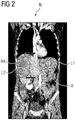
- Fig. 2 shows a medical image of the anatomical structure A with the first example of a plurality of lesions L1, L2 in a coronal reformation.
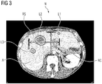
- Fig. 3 shows a medical image of an anatomical structure A with a second example of a plurality of lesions L1, L2, L3 in an axial reformation.
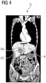
- Fig. 4 shows a medical image of the anatomical structure with the second example of a plurality of lesions L1, L2, L3 in a coronal reformation.
- Fig. 5 shows a medical image of an anatomical structure A with a third example of a plurality of lesions L1, L2, L3 in an axial reformation.
- Fig. 6 shows a medical image of the anatomical structure A with the third example of a plurality of lesions L1, L2, L3 in a coronal reformation,
- the dimension line RS represents a first spread of the spatial distribution of the plurality of lesions L1, L2, L3 of the anatomical structure A with respect to a first direction, the first direction being a sagittal direction.
- the dimension line RC represents a second spread of the spatial distribution of the plurality of lesions L1, L2, L3 of the anatomical structure A with respect to a second direction, the second direction being a coronal direction.
- the dimension line RA represents a third spread of the spatial distribution of the plurality of lesions L1, L2, L3 of the anatomical structure A with respect to a third direction, the third direction being an axial direction.
- the spread parameter can be calculated based on the first spread, the second spread and the third spread. Based on the spread parameter, a prediction of a medical target variable can be calculated, for example to predict the death and/or survival probability of the patient for one year.
- the spread parameter F 3DATS quantifies the maximum tumor spread for the three anatomical orientations axial, sagittal and coronal within an organ. Therefore, the spread parameter F 3DATS could be named "3D anatomical tumor spread". This spread parameter is measured in the physical unit millimeters (mm) .
- the spread parameter F 3DATS can be the sum of the first spread r sagittal , the second spread r coronal and the third spread r axial .
- a deep dispersion feature for a quantification of a spatial distribution of the plurality of lesions can be constructed, which can be pre-trained, for example, by a handcrafted dispersion feature F 3DATS .
- F 3DATS dispersion feature
- a prediction based on F3DATS resulted in an ROC AUC (Area Under the Receiver Operating Characteristics Curve) of 71%
- a prediction based on the RECIST diameter resulted in an ROC AUC of 56%
- a prediction based on the tumor volume resulted in an ROC AUC of 56%
- a prediction based on radiomics features resulted in an ROC AUC of 62%.
- the measured quantities and relevant images can be displayed to a physician on a screen and/or within a graphical user interface, thereby facilitating a quantitative assessment of the lesions of the anatomical structure A in respect of their relevance for the medical target variable.
- the displayed information can comprise, for each of the first spread r sagittal , the second spread r coronal , the third spread r ax-ial and the resulting F 3DATS , a respective numerical value, in particular in millimeters (mm).
- the numerical values can be related, for example, to anatomical distances or to distances measured in the medical images.
- Fig. 7 shows a diagram illustrating a computer-implemented method for providing a prediction of a medical target variable, the method comprising
- Fig. 8 shows a data processing system U for providing a prediction of a medical target variable, and the data processing system U comprising
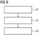
- Fig. 9 shows a diagram illustrating a computer-implemented method for providing a trained machine learning algorithm, the method comprising
Landscapes
- Engineering & Computer Science (AREA)
- Health & Medical Sciences (AREA)
- Medical Informatics (AREA)
- Public Health (AREA)
- General Health & Medical Sciences (AREA)
- Primary Health Care (AREA)
- Biomedical Technology (AREA)
- Epidemiology (AREA)
- Radiology & Medical Imaging (AREA)
- Nuclear Medicine, Radiotherapy & Molecular Imaging (AREA)
- Data Mining & Analysis (AREA)
- Pathology (AREA)
- Databases & Information Systems (AREA)
- Quality & Reliability (AREA)
- Computer Vision & Pattern Recognition (AREA)
- Physics & Mathematics (AREA)
- General Physics & Mathematics (AREA)
- Theoretical Computer Science (AREA)
- Apparatus For Radiation Diagnosis (AREA)
- Measuring And Recording Apparatus For Diagnosis (AREA)
- Image Analysis (AREA)
Abstract
Description
- In one aspect the invention relates to a computer-implemented method for providing a prediction of a medical target variable. In other aspects, the invention relates to a method for providing a trained machine learning algorithm, to a data processing system, to a computer program product and to a computer-readable medium.
- Metastatic colorectal cancer (mCRC) is one of the leading causes of cancer related deaths in modern societies. An adequate and early assessment is systematically correlated with a significantly improved overall patient survival and patient wellbeing. Within tumor therapy, the approximated patient survival itself is an important variable for therapy adjustment, e.g. by adapting the medication, or escalating and deescalating the therapy. Thus, it contributes to an overall better patient care and patient survival. However, currently there is no established and generally accepted technique to quantitatively estimate patient survival, meaning that this estimation largely builds up on radiologic and oncologic experience. The potential of a simple, easily measurable and quantifiable biomarker to estimate the patient survival is therefore of high clinical, as well as business value, as this method could establish a new standard within oncologic therapies.
- The survival of patients can be estimated based on features like volume or maximum diameter of single, or the sum of these values for multiple lesions (Response Evaluation Criteria in Solid Tumors - RECIST). While the currently researched automated inference of features through deep convolutional neural networks (DCNNs) is a generally promising alternative to the above mentioned handcrafted features, a major problem arises from the required network capacity, and therefore parameter amount, to train deep learning models, as there is a lack of large and easily available medical datasets for this purpose.
- The underlying technical problem of the invention is to facilitate an improved quantitative assessment of lesions of an anatomical structure in respect of their relevance for a medical target variable. This problem is solved by the subject matter of the independent claims. The dependent claims are related to further aspects of the invention.
- In one aspect the invention relates to a computer-implemented method for providing a prediction of a medical target variable,
- Receiving medical imaging data of an examination area, the examination area comprising a plurality of lesions of an anatomical structure, wherein each lesion of the plurality of lesions of the anatomical structure spaced apart from any other lesion of the plurality of lesions of the anatomical structure,
- Calculating a spread parameter based on the medical imaging data, the spread parameter being indicative of a spread of a spatial distribution of the plurality of lesions of the anatomical structure,
- Calculating the prediction of the medical target variable based on the spread parameter,
- Providing the prediction of the medical target variable.
- In another aspect, each lesion of the plurality of lesions of the anatomical structure is related to a same disease as any other lesion of the plurality of lesions of the anatomical structure. In another aspect, each lesion of the plurality of lesions of the anatomical structure is of a same tissue and/or a same type as any other lesion of the plurality of lesions of the anatomical structure. The plurality of lesions of the anatomical structure can comprise, for example, two or three or more than three lesions of the anatomical structure. The anatomical structure can comprise, for example, at least one further lesion that is not comprised in the plurality of lesions. The plurality of lesions can comprise, for example, an entire tumor burden of the anatomical structure.
- The medical imaging data can comprise, for example, a respective representation for each of the lesions of the plurality of lesions. For each of the lesions of the plurality of lesions the respective representation can be obtained, for example, based on segmentation, in particular automated and/or manual segmentation. In another aspect, for each lesion of the plurality of lesions a respective volume is determined based on the respective representation. The prediction of the medical target variable can be further based on a volume parameter, that is calculated based on the respective volumes of the lesions of the plurality of lesions.
- In another aspect a trained machine learning algorithm is applied onto the medical imaging data, thereby obtaining the spread parameter and/or the prediction of the medical target variable. The trained machine learning algorithm can be, for example, a trained deep convolutional neural network.
- In another aspect the trained machine learning algorithm is based on a first optimization for calculating the spread parameter and/or a second optimization for calculating the prediction of the medical target variable. The first optimization for calculating the spread parameter can be based on at least a first part of a plurality of training datasets, in particular on the plurality of training datasets. The second optimization for calculating the prediction of the medical target variable can be based on at least a second part of the plurality of training datasets, in particular on the plurality of training datasets.
- In another aspect the second optimization for calculating the prediction of the medical target variable is separate from and/or subsequent to the first optimization for calculating the spread parameter.
- In another aspect the trained machine learning algorithm is based on multi-task learning, the multi-task learning comprising the first optimization and the second optimization as related tasks, in particular related tasks that are simultaneously solved.
- The first optimization for calculating the spread parameter can be based on a first loss, for example a reconstruction loss. The second optimization for calculating the prediction of the medical target variable can be based on a second loss, for example a discriminative loss. The multi-task learning can comprise a weighting, in particular an automated weighting, of the first loss and the second loss relative to each other. The weighting can be based, for example, on a homoscedastic task uncertainty.
- In another aspect the calculating the spread parameter comprises determining a first spread of the spatial distribution of the plurality of lesions of the anatomical structure with respect to a first direction, determining a second spread of the spatial distribution of the plurality of lesions of the anatomical structure with respect to a second direction, the second direction being orthogonal to the first direction, and determining a third spread of the spatial distribution of the plurality of lesions of the anatomical structure with respect to a third direction, the third direction being orthogonal to the first direction and the second direction. The spread parameter can be calculated based on the first spread, the second spread and the third spread.
- The first direction can be, for example, a sagittal direction. The second direction can be, for example, a coronal direction. The third direction can be, for example, an axial direction.
- A spread of a spatial distribution with respect to a given direction can be, for example, a maximum extension of the spatial distribution along that given direction and/or a maximum extension of a convex envelope of the spatial distribution along that given direction. In another aspect effective extensions are used instead of maximum extensions, for example to smoothen or eliminate the impact of outlier lesions onto the spread parameter.
- In another aspect, an envelope, in particular a convex envelope, of the spatial distribution can be determined. The first direction can be, for example, parallel to a first principal axis of the convex envelope. The second direction can be, for example, parallel to a second principal axis of the convex envelope. The third direction can be, for example, parallel to a third principal axis of the convex envelope. Thereby the spread parameter can be obtained in a rotational invariant form.
- In another aspect, the spread parameter can be a function of the first spread, the second spread and the third spread, each of the first spread, the second spread and the third spread being an input of the function. In another aspect, the spread parameter can be a function of a ratio of the first spread to the first diameter of the anatomical structure, a ratio of the second spread to the second diameter of the anatomical structure and a ratio of the third spread to the third diameter of the anatomical structure. The function can be, for example, a non-linear function, in particular, a random forest-based function and/or a function that is implemented in form of a set of weights of a deep convolutional neural network.
- The spread parameter can be, for example, a real number or a tuple of real numbers. The tuple can be, in particular, a 3-tuple comprising a first element based on the first spread, for example the ratio of the first spread to the first diameter of the anatomical structure, a second element based on the second spread, for example the ratio of the second spread to the second diameter of the anatomical structure, and a third element based on the third spread, for example the ratio of the third spread to the third diameter of the anatomical structure.
- In another aspect, the prediction of the medical target variable is a function of the spread parameter, in particular of the spread parameter in form of a tuple, each element of the tuple being an input of the function of the spread parameter. The function of the spread parameter can be, for example, a non-linear function of the spread parameter, in particular, a random forest-based function of the spread parameter and/or a function of the spread parameter that is implemented in form of a set of weights of a deep convolutional neural network.
- In another aspect the spread parameter is calculated based on a generalized mean of the first spread, the second spread and the third spread. The generalized mean can be, for example, an arithmetic mean or a quadratic mean. The generalized mean can be, for example, a weighted generalized mean or an unweighted generalized mean.
- In the context of the present disclosure it is understood that if for calculating the spread parameter a function of the first spread, the second spread and the third spread is used that equals a result of multiplying a non-zero constant and a generalized mean of the first spread, the second spread and the third spread, the spread parameter is calculated based on said generalized mean of the first spread, the second spread and the third spread.
- For example, if the spread parameter is a sum of the first spread, the second spread and the third spread and/or a function of the sum of the first spread, the second spread and the third spread, then for calculating the spread parameter an function of the first spread, the second spread and the third spread is used that equals a result of multiplying the non-zero constant having the value three and the arithmetic mean of the first spread, the second spread and the third spread, and therefore the spread parameter is calculated based on the arithmetic mean of the first spread, the second spread and the third spread. In particular, the sum of the first spread, the second spread and the third spread can be understood as an inner function and the function of the sum can be understood as an outer function.
- In another aspect the spread parameter is calculated based on a sum of the first spread, the second spread and the third spread. The spread parameter can be, for example, a sum or a generalized mean, in particular an arithmetic mean or a quadratic mean, of the first spread, the second spread and the third spread.
- A sum or a generalized mean of the first spread, the second spread and the third spread can be used as a label spread parameter for the training of the machine learning algorithm. This exemplary feature may be refined in the first optimization of the machine learning algorithm for calculating the spread parameter, for example, in a deep learning procedure.
- In another aspect each of a first diameter of the anatomical structure with respect to the first direction, a second diameter of the anatomical structure with respect to the second direction and a third diameter of the anatomical structure with respect to the third direction is determined.
- The spread parameter can be calculated based on a ratio of the first spread to the first diameter of the anatomical structure, a ratio of the second spread to the second diameter of the anatomical structure and a ratio of the third spread to the third diameter of the anatomical structure.
- The spread parameter can be, for example, a sum or a generalized mean of a ratio of the first spread to the first diameter of the anatomical structure, a ratio of the second spread to the second diameter of the anatomical structure and a ratio of the third spread to the third diameter of the anatomical structure. Thereby the spread parameter can be normalized with respect to the size of the anatomical structure.
- A sum or a generalized mean of a ratio of the first spread to the first diameter of the anatomical structure, a ratio of the second spread to the second diameter of the anatomical structure and a ratio of the third spread to the third diameter of the anatomical structure can be used as a label spread parameter for the training of the machine learning algorithm. This exemplary feature may be refined in the first optimization of the machine learning algorithm for calculating the spread parameter, for example, in a deep learning procedure.
- In another aspect the anatomical structure comprises at least one organ, in particular is at least one organ. In another aspect each lesion of the plurality of lesions of the anatomical structure is related to a tumor of the at least one organ. The examination area can be, for example, an examination area of a patient. The at least one organ can be, for example, at least one organ of the patient.
- In another aspect the at least one organ is selected from the organ group consisting of a liver, a lung, a brain and a kidney. The plurality of lesions of the anatomical structure can be, for example, a plurality of lung nodules of the lung.
- In another aspect the anatomical structure comprises a lymphatic system, in particular is a lymphatic system. The plurality of lesions of the anatomical structure can be, for example, a plurality of enlarged lymph nodes of the lymphatic system.
- In another aspect the medical target variable is selected from the medical target variable group consisting of a survival estimation, a therapy response estimation and a therapy recommendation. The survival estimation can be, for example, a one-year survival estimation and/or a five-year survival estimation. The therapy recommendation can be indicative, for example, of a best suited therapy with respect to the anatomical structure.
- In another aspect the anatomical structure is a liver, each lesion of the plurality of lesions of the anatomical structure is related to a tumor of the liver, and the medical target variable is a survival estimation.
- In another aspect the invention relates to a computer-implemented method for providing a trained machine learning algorithm, comprising
- Receiving a plurality of training datasets, each training dataset of the plurality of training datasets comprising
- -- respective training medical imaging data of an examination area, the examination area comprising a respective plurality of lesions of an anatomical structure, wherein each lesion of the respective plurality of lesions of the anatomical structure is spaced apart from any other lesion of the respective plurality of lesions of the anatomical structure,
- -- a respective label spread parameter, the label spread parameter being indicative of a spread of a spatial distribution of the respective plurality of lesions of the anatomical structure,
- -- a respective label of a medical target variable,
- Training a machine learning algorithm based on the plurality of training datasets, the training comprising a first optimization of the machine learning algorithm for calculating a spread parameter and a second optimization of the machine learning algorithm for calculating a prediction of a medical target variable,
- wherein the first optimization for calculating the spread parameter is based on at least a first part of the plurality of training datasets, in particular on the plurality of training datasets,
- wherein the second optimization for calculating the prediction of the medical target variable is based on at least a second part of the plurality of training datasets, in particular on the plurality of training datasets,
- Providing the trained machine learning algorithm.
- The first part of the plurality of training datasets can comprise, for each training dataset of the plurality of training datasets, the respective training medical imaging data and the respective label spread parameter. The second part of the plurality of training datasets can comprise, for each training dataset of the plurality of training datasets, the respective label spread parameter and the respective label of the medical target variable.
- The training medical imaging data can comprise, for example, a respective representation for each of the lesions of the respective plurality of lesions. For each of the lesions of the respective plurality of lesions the respective representation can be obtained, for example, based on segmentation, in particular automated segmentation and/or manual segmentation.
- In another aspect for each training dataset of the plurality of training datasets the respective training medical imaging data of the examination area is of the same physical dimensions, for example, in millimeters, as the respective training medical imaging data of any other training dataset of the plurality of training datasets. Thereby consistent physical distances among the training datasets can be preserved. In particular, for each training dataset of the plurality of training datasets the respective training medical imaging data can be resized, for example be cut, if necessary, to match a pre-defined physical size.
- For the training, 2.5D reformations, i.e., slices of a fixed size, which focus on the center-of-mass of the respective plurality of lesions and/or show all lesions of the anatomical structure for example in axial, coronal and/or sagittal orientation can be used as training medical imaging data.
- In another aspect the machine learning algorithm is trained based on multi-task learning, the multi-task learning comprising the first optimization and the second optimization as related tasks. The multi-task learning can comprise, in particular, a simultaneous solving of the first optimization and the second optimization as related tasks.
- In another aspect for each training dataset of the plurality of training datasets the respective label spread parameter is of a same spread feature class as the respective label spread parameter of any other training dataset of the plurality of training datasets. The spread feature class can be determined according to one or more aspects disclosed with respect to the spread parameter.
- Thereby a search space of the machine learning algorithm can be constraint to focus on the entire spatial distribution of the plurality of lesions and not on single lesion features like shape and texture, in particular to solutions that are similar to a spread parameter.
- A decision-support system based on the trained machine learning algorithm according to one or more of the disclosed aspects might facilitate an easily understandable and interpretable decision. A reformation of the medical imaging data for use in the training of the machine learning algorithm, for example in form of a deep convolutional neural network, may be adjusted, leading to a machine learning algorithm which uses physical distances and/or tumor spread for prediction.
- For each training dataset of the plurality of training datasets the respective label of the medical target variable can be determined according to one or more aspects disclosed with respect to the label of the medical target variable.
- In another aspect the invention relates to a data processing system for providing a prediction of a medical target variable, the data processing system comprising
- a medical imaging data receiver for receiving medical imaging data of an examination area, the examination area comprising a plurality of lesions of an anatomical structure, wherein each lesion of the plurality of lesions of the anatomical structure is spaced apart from any other lesion of the plurality of lesions of the anatomical structure,
- a spread parameter calculator for calculating a spread parameter based on the medical imaging data, the spread parameter being indicative of a spread of a spatial distribution of the plurality of lesions of the anatomical structure,
- a prediction calculator for calculating the prediction of the medical target variable based on the spread parameter,
- a prediction provider for providing the prediction of the medical target variable.
- In another aspect the data processing system is configured to implement the method according to one or more of the disclosed aspects.
- In one aspect the invention relates to a medical imaging device comprising a data processing system for providing a prediction of a medical target variable according to one or more of the disclosed aspects. The medical imaging device may be, for example, a computed tomography (CT) device or a magnetic resonance imaging (MRI) device or a combination of different medical imaging modalities, for example, a PET-CT-imaging device. The medical imaging data can be acquired, for example, by the medical imaging device. The medical imaging data can comprise, for example, computed tomography medical imaging data and/or magnetic resonance medical imaging data.
- In another aspect the invention relates to a computer program product comprising program elements which induce a data processing system to carry out the steps of the method according to one or more of the disclosed aspects, when the program elements are loaded into a memory of the data processing system.
- In another aspect the invention relates to a computer-readable medium on which program elements are stored that can be read and executed by a data processing system, in order to perform the steps of the method according to one or more of the disclosed aspects, when the program elements are executed by the data processing system.
- The computer program product can be, for example, a computer program or comprise another element apart from the computer program. This other element can be hardware, for example a memory device, on which the computer program is stored, a hardware key for using the computer program and the like, and/or software, for example, a documentation or a software key for using the computer program. A computer-readable medium can be embodied as non-permanent main memory (e.g. randomaccess memory) or as permanent mass storage (e.g. hard disk, USB stick, SD card, solid state disk).
- The data processing system can comprise, for example, at least one of a cloud-computing system, a distributed computing system, a computer network, a computer, a tablet computer, a smartphone or the like. The data processing system can comprise hardware and/or software. The hardware can be, for example, a processor system, a memory system and combinations thereof. The hardware can be configurable by the software and/or be operable by the software. Calculations for performing a step of a method and/or for training a machine learning algorithm may be carried out in a processor.
- Data, in particular the medical imaging data and the training datasets, can be received, for example, by receiving a signal that carries the data and/or by reading the data from a computer-readable medium and/or by receiving an input through a user interface. Data, in particular, the spread parameter and the prediction of the medical target variable can be provided, for example, by transmitting a signal that carries the data and/or by writing the data into a computer-readable medium and/or by displaying the data on a display.
- Wherever not already described explicitly, individual embodiments, or their individual aspects and features, can be combined or exchanged with one another without limiting or widening the scope of the described invention, whenever such a combination or exchange is meaningful and in the sense of this invention. Advantages which are described with respect to one embodiment of the present invention are, wherever applicable, also advantageous of other embodiments of the present invention.
- Any of the algorithms mentioned herein can be based on one or more of the following architectures: convolutional neural network, deep belief network, random forest, deep residual learning, deep reinforcement learning, recurrent neural network, Siamese network, generative adversarial network or auto-encoder. In particular, the trained machine learning algorithm can be embodied as a deep learning algorithm, in particular as a deep convolutional neural network.
- In the context of the present invention, the expression "based on" can in particular be understood as meaning "using, inter alia". In particular, wording according to which a first feature is calculated (or generated, determined etc.) based on a second feature does not preclude the possibility of the first feature being calculated (or generated, determined etc.) based on a third feature.
- For example, the prediction of the medical target variable can be calculated based on the spread parameter and further based on at least one additional feature. The at least one additional feature can be related to the plurality of lesions, for example, being indicative of a volume, for example a total combined volume and/or a mean volume, of the lesions of the plurality of lesions. The at least one additional feature can be related to patient parameters like age, sex or weight of the patient.
- Reference is made to the fact that the described methods and the described units are merely preferred example embodiments of the invention and that the invention can be varied by a person skilled in the art, without departing from the scope of the invention as it is specified by the claims.
- The invention will be illustrated below with reference to the accompanying figures using example embodiments. The illustration in the figures is schematic and highly simplified and not necessarily to scale.
-
Fig. 1 shows a medical image of an anatomical structure with a first example of a plurality of lesions in an axial reformation, -
Fig. 2 shows a medical image of the anatomical structure with the first example of a plurality of lesions in a coronal reformation, -
Fig. 3 shows a medical image of an anatomical structure with a second example of a plurality of lesions in an axial reformation, -
Fig. 4 shows a medical image of the anatomical structure with the second example of a plurality of lesions in a coronal reformation, -
Fig. 5 shows a medical image of an anatomical structure with a third example of a plurality of lesions in an axial reformation, -
Fig. 6 shows a medical image of the anatomical structure with the third example of a plurality of lesions in a coronal reformation, -
Fig. 7 shows a diagram illustrating a computer-implemented method for providing a prediction of a medical target variable, -
Fig. 8 shows a data processing system for providing a prediction of a medical target variable, and -
Fig. 9 shows a diagram illustrating a computer-implemented method for providing a trained machine learning algorithm. -
Fig. 1 shows a medical image of an anatomical structure A with a first example of a plurality of lesions L1, L2 in an axial reformation.Fig. 2 shows a medical image of the anatomical structure A with the first example of a plurality of lesions L1, L2 in a coronal reformation. -
Fig. 3 shows a medical image of an anatomical structure A with a second example of a plurality of lesions L1, L2, L3 in an axial reformation.Fig. 4 shows a medical image of the anatomical structure with the second example of a plurality of lesions L1, L2, L3 in a coronal reformation. -
Fig. 5 shows a medical image of an anatomical structure A with a third example of a plurality of lesions L1, L2, L3 in an axial reformation.Fig. 6 shows a medical image of the anatomical structure A with the third example of a plurality of lesions L1, L2, L3 in a coronal reformation, - The dimension line RS represents a first spread of the spatial distribution of the plurality of lesions L1, L2, L3 of the anatomical structure A with respect to a first direction, the first direction being a sagittal direction. The dimension line RC represents a second spread of the spatial distribution of the plurality of lesions L1, L2, L3 of the anatomical structure A with respect to a second direction, the second direction being a coronal direction. The dimension line RA represents a third spread of the spatial distribution of the plurality of lesions L1, L2, L3 of the anatomical structure A with respect to a third direction, the third direction being an axial direction.
- The spread parameter can be calculated based on the first spread, the second spread and the third spread. Based on the spread parameter, a prediction of a medical target variable can be calculated, for example to predict the death and/or survival probability of the patient for one year.
- The spread parameter F3DATS quantifies the maximum tumor spread for the three anatomical orientations axial, sagittal and coronal within an organ. Therefore, the spread parameter F3DATS could be named "3D anatomical tumor spread". This spread parameter is measured in the physical unit millimeters (mm) .
- The spread parameter F3DATS can be the sum of the first spread rsagittal, the second spread rcoronal and the third spread raxial. Then the mathematical formulation for the feature F3DATS can be given by: F3DATS [mm] = (rmax, axial - rmin, axial) + (rmax, sagittal - rmin, sagittal) + (rmax, coronal - rmin, coronal) - raxial + rsagittal + rcoro-nal. In another example, the spread parameter F3DATS NL can be a non-linear function, for example a random forest-based function, of the first spread rsagittal, the second spread rcoronal and the third spread raxial as inputs of the non-linear function, F3DATS_NL [mm] = f(raxial, rsagittal, rcoronal).
- These features are intuitively easy to understand and interpretable. Furthermore, a deep dispersion feature for a quantification of a spatial distribution of the plurality of lesions can be constructed, which can be pre-trained, for example, by a handcrafted dispersion feature F3DATS. When predicting one-year-survival for oncology patients, superior performance can be achieved based on F3DATS when compared to complex radiomics features, the RECIST diameter as well as the overall tumor volume.
- In an exemplary clinical study (103 samples) for one-year-survival prediction based on the entire tumor burden of the liver, a prediction based on F3DATS resulted in an ROC AUC (Area Under the Receiver Operating Characteristics Curve) of 71%, whereas a prediction based on the RECIST diameter resulted in an ROC AUC of 56%, a prediction based on the tumor volume resulted in an ROC AUC of 56% and a prediction based on radiomics features resulted in an ROC AUC of 62%.
- The measured quantities and relevant images can be displayed to a physician on a screen and/or within a graphical user interface, thereby facilitating a quantitative assessment of the lesions of the anatomical structure A in respect of their relevance for the medical target variable.
- The displayed information can comprise, for each of the first spread rsagittal, the second spread rcoronal, the third spread rax-ial and the resulting F3DATS, a respective numerical value, in particular in millimeters (mm). The numerical values can be related, for example, to anatomical distances or to distances measured in the medical images.
- In the case shown in
Fig. 5 andFig. 6 , the following values can be displayed: - 154 mm for the first spread rsagittal,
- 140 mm for the second spread rcoronal,
- 98 mm for the third spread raxial,
- 392 mm for F3DTAS,
- 0.68 for a ratio of the first spread rsagittal to a first diameter of the anatomical structure A with respect to the first direction,
- 0.8 for a ratio of the second spread rcoronal to the second diameter of the anatomical structure A with respect to the second direction,
- 0.59 for a ratio of the third spread raxial to the third diameter of the anatomical structure A with respect to the third direction,
- 0.68 for a mean ratio, and
- 68% for a probability for death of the patient within one year, which is calculated in a manner as explained above.
-
Fig. 7 shows a diagram illustrating a computer-implemented method for providing a prediction of a medical target variable, the method comprising - Receiving RD medical imaging data of an examination area N, the examination area N comprising a plurality of lesions L1, L2, L3 of an anatomical structure A, wherein each lesion of the plurality of lesions L1, L2, L3 of the anatomical structure A is spaced apart from any other lesion of the plurality of lesions L1, L2, L3 of the anatomical structure A,
- Calculating CS a spread parameter based on the medical imaging data, the spread parameter being indicative of a spread of a spatial distribution of the plurality of lesions L1, L2, L3 of the anatomical structure A,
- Calculating CP the prediction of the medical target variable based on the spread parameter,
- Providing PP the prediction of the medical target variable.
-
Fig. 8 shows a data processing system U for providing a prediction of a medical target variable, and the data processing system U comprising - a medical imaging data receiver RD-U for receiving RD medical imaging data of an examination area N, the examination area N comprising a plurality of lesions L1, L2, L3 of an anatomical structure A, wherein each lesion of the plurality of lesions L1, L2, L3 of the anatomical structure A is spaced apart from any other lesion of the plurality of lesions L1, L2, L3 of the anatomical structure A,
- a spread parameter calculator CS-U for calculating CS a spread parameter based on the medical imaging data, the spread parameter being indicative of a spread of a spatial distribution of the plurality of lesions L1, L2, L3 of the anatomical structure A,
- a prediction calculator CP-U for calculating CP the prediction of the medical target variable based on the spread parameter,
- a prediction provider PP-U for providing PP the prediction of the medical target variable.
-
Fig. 9 shows a diagram illustrating a computer-implemented method for providing a trained machine learning algorithm, the method comprising - Receiving RT a plurality of training datasets, each training dataset of the plurality of training datasets comprising
- -- respective training medical imaging data of an examination area N, the examination area N comprising a plurality of lesions L1, L2, L3 of an anatomical structure A, wherein each lesion of the plurality of lesions L1, L2, L3 of the anatomical structure A is spaced apart from any other lesion of the plurality of lesions L1, L2, L3 of the anatomical structure A,
- -- a respective label spread parameter, the label spread parameter being indicative of a spread of a spatial distribution of the plurality of lesions L1, L2, L3 of the anatomical structure A,
- -- a respective label of a medical target variable,
- Training TA a machine learning algorithm based on the plurality of training datasets, the training comprising a first optimization of the machine learning algorithm for calculating a spread parameter and a second optimization of the machine learning algorithm for calculating a prediction of a medical target variable,
- wherein the first optimization for calculating the spread parameter is based on at least a first part of the plurality of training datasets,
- wherein the second optimization for calculating the prediction of the medical target variable is based on at least a second part of the plurality of training datasets,
- Providing PA the trained machine learning algorithm.
Claims (18)
- A computer-implemented method for providing a prediction of a medical target variable, the method comprising- Receiving (RD) medical imaging data of an examination area (N), the examination area (N) comprising a plurality of lesions (L1, L2, L3) of an anatomical structure (A), wherein each lesion of the plurality of lesions (L1, L2, L3) of the anatomical structure (A) is spaced apart from any other lesion of the plurality of lesions (L1, L2, L3) of the anatomical structure (A),- Calculating (CS) a spread parameter based on the medical imaging data, the spread parameter being indicative of a spread of a spatial distribution of the plurality of lesions (L1, L2, L3) of the anatomical structure (A),- Calculating (CP) the prediction of the medical target variable based on the spread parameter,- Providing (PP) the prediction of the medical target variable.
- The method according to claim 1,- wherein a trained machine learning algorithm is applied onto the medical imaging data, thereby obtaining the spread parameter and/or the prediction of the medical target variable.
- The method according to claim 2,- wherein the trained machine learning algorithm is based on a first optimization for calculating the spread parameter and a second optimization for calculating the prediction of the medical target variable,- wherein the first optimization for calculating the spread parameter is based on at least a first part of a plurality of training datasets,- wherein the second optimization for calculating the prediction of the medical target variable is based on at least a second part of the plurality of training datasets.
- The method according to claim 3,- wherein the second optimization is subsequent to the first optimization.
- The method according to claim 3,- wherein the trained machine learning algorithm is based on multi-task learning, the multi-task learning comprising the first optimization and the second optimization as related tasks.
- The method according to one of the claims 1 to 5,- wherein the calculating the spread parameter comprises-- determining a first spread (RS) of the spatial distribution of the plurality of lesions (L1, L2, L3) of the anatomical structure (A) with respect to a first direction,-- determining a second spread (RC) of the spatial distribution of the plurality of lesions (L1, L2, L3) of the anatomical structure (A) with respect to a second direction, the second direction being orthogonal to the first direction, and-- determining a third spread (RA) of the spatial distribution of the plurality of lesions (L1, L2, L3) of the anatomical structure (A) with respect to a third direction, the third direction being orthogonal to the first direction and the second direction, and- wherein the spread parameter is calculated based on the first spread (RS), the second spread (RC) and the third spread (RA).
- The method according to claim 6,- wherein the spread parameter is calculated based on a generalized mean of the first spread (RS), the second spread (RC) and the third spread (RA).
- The method according to claim 6,- wherein each of a first diameter of the anatomical structure (A) with respect to the first direction, a second diameter of the anatomical structure (A) with respect to the second direction and a third diameter of the anatomical structure (A) with respect to the third direction is determined,- wherein the spread parameter is calculated based on a ratio of the first spread (RS) to the first diameter of the anatomical structure (A), a ratio of the second spread (RC) to the second diameter of the anatomical structure (A) and a ratio of the third spread (RA) to the third diameter of the anatomical structure (A).
- The method according to one of the claims 1 to 8,- wherein the anatomical structure (A) comprises at least one organ, and- wherein each lesion of the plurality of lesions (L1, L2, L3) of the anatomical structure (A) is related to a tumor of the at least one organ.
- The method according to claim 9,- wherein the at least one organ is selected from the organ group consisting of a liver, a lung, a brain and a kidney.
- The method according to one of the claims 1 to 8,- wherein the anatomical structure (A) comprises a lymphatic system,- wherein the plurality of lesions (L1, L2, L3) of the anatomical structure (A) is a plurality of enlarged lymph nodes of the lymphatic system.
- The method according to one of the claims 1 to 10,- wherein the medical target variable is selected from the medical target variable group consisting of a survival estimation, a therapy response estimation and a therapy recommendation.
- The method according to one of the claims 1 to 8,- wherein the anatomical structure (A) is a liver,- wherein each lesion of the plurality of lesions (L1, L2, L3) of the anatomical structure (A) is related to a tumor of the liver,- wherein the medical target variable is a survival estimation.
- A computer-implemented method for providing a trained machine learning algorithm, comprising- Receiving (RT) a plurality of training datasets, each training dataset of the plurality of training datasets comprising-- respective training medical imaging data of an examination area (N), the examination area (N) comprising a plurality of lesions (L1, L2, L3) of an anatomical structure (A), wherein each lesion of the plurality of lesions (L1, L2, L3) of the anatomical structure (A) is spaced apart from any other lesion of the plurality of lesions (L1, L2, L3) of the anatomical structure (A),-- a respective label spread parameter, the label spread parameter being indicative of a spread of a spatial distribution of the plurality of lesions (L1, L2, L3) of the anatomical structure (A),-- a respective label of a medical target variable,- Training (TA) a machine learning algorithm based on the plurality of training datasets, the training comprising a first optimization of the machine learning algorithm for calculating a spread parameter and a second optimization of the machine learning algorithm for calculating a prediction of a medical target variable,- wherein the first optimization for calculating the spread parameter is based on at least a first part of the plurality of training datasets,- wherein the second optimization for calculating the prediction of the medical target variable is based on at least a second part of the plurality of training datasets,- Providing (PA) the trained machine learning algorithm.
- A data processing system (U) for providing a prediction of a medical target variable, comprising- a medical imaging data receiver (RD-U) for receiving (RD) medical imaging data of an examination area (N), the examination area (N) comprising a plurality of lesions (L1, L2, L3) of an anatomical structure (A), wherein each lesion of the plurality of lesions (L1, L2, L3) of the anatomical structure (A) is spaced apart from any other lesion of the plurality of lesions (L1, L2, L3) of the anatomical structure (A),- a spread parameter calculator (CS-U) for calculating (CS) a spread parameter based on the medical imaging data, the spread parameter being indicative of a spread of a spatial distribution of the plurality of lesions (L1, L2, L3) of the anatomical structure (A),- a prediction calculator (CP-U) for calculating (CP) the prediction of the medical target variable based on the spread parameter,- a prediction provider (PP-U) for providing (PP) the prediction of the medical target variable.
- The data processing system (U) according to claim 15, configured to implement the method of one of the claims 1 to 13.
- A computer program product comprising program elements which induce a data processing system (U) to carry out the steps of the method according to one of the claims 1 to 13, when the program elements are loaded into a memory of the data processing system (U).
- A computer-readable medium on which program elements are stored that can be read and executed by a data processing system (U), in order to perform the steps of the method according to one of the claims 1 to 13, when the program elements are executed by the data processing system (U).
Priority Applications (2)
| Application Number | Priority Date | Filing Date | Title |
|---|---|---|---|
| EP19197243.9A EP3792871B1 (en) | 2019-09-13 | 2019-09-13 | Method and data processing system for providing a prediction of a medical target variable |
| US17/009,954 US11626203B2 (en) | 2019-09-13 | 2020-09-02 | Method and data processing system for providing a prediction of a medical target variable |
Applications Claiming Priority (1)
| Application Number | Priority Date | Filing Date | Title |
|---|---|---|---|
| EP19197243.9A EP3792871B1 (en) | 2019-09-13 | 2019-09-13 | Method and data processing system for providing a prediction of a medical target variable |
Publications (3)
| Publication Number | Publication Date |
|---|---|
| EP3792871A1 true EP3792871A1 (en) | 2021-03-17 |
| EP3792871C0 EP3792871C0 (en) | 2024-06-12 |
| EP3792871B1 EP3792871B1 (en) | 2024-06-12 |
Family
ID=67956539
Family Applications (1)
| Application Number | Title | Priority Date | Filing Date |
|---|---|---|---|
| EP19197243.9A Active EP3792871B1 (en) | 2019-09-13 | 2019-09-13 | Method and data processing system for providing a prediction of a medical target variable |
Country Status (2)
| Country | Link |
|---|---|
| US (1) | US11626203B2 (en) |
| EP (1) | EP3792871B1 (en) |
Cited By (1)
| Publication number | Priority date | Publication date | Assignee | Title |
|---|---|---|---|---|
| US12002582B2 (en) | 2019-12-12 | 2024-06-04 | Siemens Healthineers Ag | Method for obtaining disease-related clinical information |
Families Citing this family (4)
| Publication number | Priority date | Publication date | Assignee | Title |
|---|---|---|---|---|
| CN113706444B (en) * | 2021-03-25 | 2025-05-27 | 腾讯科技(深圳)有限公司 | Image processing method, device, equipment and storage medium |
| CN113689382B (en) * | 2021-07-26 | 2023-12-01 | 北京知见生命科技有限公司 | Tumor postoperative survival prediction method and system based on medical images and pathological images |
| CN115294191B (en) * | 2022-10-08 | 2022-12-27 | 武汉楚精灵医疗科技有限公司 | Marker size measuring method, device, equipment and medium based on electronic endoscope |
| CN117852361B (en) * | 2024-01-17 | 2024-08-09 | 华中科技大学 | A method and system for predicting damage time of laser-irradiated metal materials |
Citations (1)
| Publication number | Priority date | Publication date | Assignee | Title |
|---|---|---|---|---|
| WO2018222755A1 (en) * | 2017-05-30 | 2018-12-06 | Arterys Inc. | Automated lesion detection, segmentation, and longitudinal identification |
Family Cites Families (3)
| Publication number | Priority date | Publication date | Assignee | Title |
|---|---|---|---|---|
| AU2003227067A1 (en) * | 2002-03-15 | 2003-09-29 | Pharmacia Italia S.P.A. | Method for estimating or predicting the anti-tumor activity of a compound and for estimating or predicting the tumor growth in mammals |
| JP2008520313A (en) * | 2004-11-19 | 2008-06-19 | コーニンクレッカ フィリップス エレクトロニクス エヌ ヴィ | An architecture for collecting field data for computer-aided diagnosis |
| WO2006119340A2 (en) * | 2005-05-04 | 2006-11-09 | Imquant, Inc. | Dynamic tumor diagnostic and treatment system |
-
2019
- 2019-09-13 EP EP19197243.9A patent/EP3792871B1/en active Active
-
2020
- 2020-09-02 US US17/009,954 patent/US11626203B2/en active Active
Patent Citations (1)
| Publication number | Priority date | Publication date | Assignee | Title |
|---|---|---|---|---|
| WO2018222755A1 (en) * | 2017-05-30 | 2018-12-06 | Arterys Inc. | Automated lesion detection, segmentation, and longitudinal identification |
Non-Patent Citations (3)
| Title |
|---|
| ALEXANDER KATZMANN ET AL: "Predicting Lesion Growth and Patient Survival in Colorectal Cancer Patients using Deep Neural Networks", 1ST CONFERENCE ON MEDICAL IMAGING WITH DEEP LEARNING (MIDL 2018), 11 April 2018 (2018-04-11), Amsterdam, The Netherlands, pages 1 - 12, XP055664781, Retrieved from the Internet <URL:https://openreview.net/pdf?id=B1e_uuoif> [retrieved on 20200204] * |
| DABASS MANJU ET AL: "Review of Classification Techniques Using Deep Learning for Colorectal Cancer Imaging Modalities", 2019 6TH INTERNATIONAL CONFERENCE ON SIGNAL PROCESSING AND INTEGRATED NETWORKS (SPIN), IEEE, 7 March 2019 (2019-03-07) - 8 March 2019 (2019-03-08), pages 105 - 110, XP033547916, DOI: 10.1109/SPIN.2019.8711776 * |
| KATZMANN ALEXANDER ET AL: "TumorEncode - Deep Convolutional Autoencoder for Computed Tomography Tumor Treatment Assessment", 2018 INTERNATIONAL JOINT CONFERENCE ON NEURAL NETWORKS (IJCNN), IEEE, 8 July 2018 (2018-07-08), pages 1 - 8, XP033419005, DOI: 10.1109/IJCNN.2018.8489193 * |
Cited By (1)
| Publication number | Priority date | Publication date | Assignee | Title |
|---|---|---|---|---|
| US12002582B2 (en) | 2019-12-12 | 2024-06-04 | Siemens Healthineers Ag | Method for obtaining disease-related clinical information |
Also Published As
| Publication number | Publication date |
|---|---|
| US20210082569A1 (en) | 2021-03-18 |
| EP3792871C0 (en) | 2024-06-12 |
| US11626203B2 (en) | 2023-04-11 |
| EP3792871B1 (en) | 2024-06-12 |
Similar Documents
| Publication | Publication Date | Title |
|---|---|---|
| EP3792871B1 (en) | Method and data processing system for providing a prediction of a medical target variable | |
| US10282588B2 (en) | Image-based tumor phenotyping with machine learning from synthetic data | |
| Ringl et al. | The ribs unfolded-a CT visualization algorithm for fast detection of rib fractures: effect on sensitivity and specificity in trauma patients | |
| EP3043318B1 (en) | Analysis of medical images and creation of a report | |
| CN105938628B (en) | The direct calculating of biological marker from image | |
| JP7645614B2 (en) | DATA PROCESSING APPARATUS AND DATA PROCESSING METHOD | |
| CN107077731B (en) | Visualization of Imaging Uncertainty | |
| CN115719330A (en) | Fully automated assessment of coronary vulnerable plaque in coronary CT images using radiological characterization | |
| JP7058988B2 (en) | Information processing equipment, information processing methods and programs | |
| CN110234270B (en) | Method and apparatus for providing quantitative volume maps of organs or assessment of organ health | |
| US20210296004A1 (en) | Inference Transparency System for Image-Based Clinical Decision Support Systems | |
| Aydin et al. | An evaluation of performance measures for arterial brain vessel segmentation | |
| Hu et al. | Multimodal volume-aware detection and segmentation for brain metastases radiosurgery | |
| Saikia et al. | Lesion detection in women breast’s dynamic contrast-enhanced magnetic resonance imaging using deep learning | |
| JP6982570B2 (en) | Systems and methods for context-aware medical recommendations | |
| Wang et al. | Uncertainty quantification in multi-parametric MRI-based meningioma radiotherapy target segmentation | |
| WO2006119340A2 (en) | Dynamic tumor diagnostic and treatment system | |
| US12062168B2 (en) | Real-time estimation of local cardiac tissue properties and uncertainties based on imaging and electro-anatomical maps | |
| CN114846555A (en) | Measuring in an image | |
| CN120997133B (en) | Methods and systems for analyzing multi-tracer images, storage media, and computer equipment. | |
| Samei et al. | CT-based quantification | |
| Adiraju et al. | Temporal Stability and Prognostic Power of Radiomic Features for Survival Analysis in Lung Adenocarcinoma. | |
| US20260087615A1 (en) | Advanced pulmonary nodule analysis system with malignancy-weighted detection and customized visualization | |
| US20240257947A1 (en) | Opportunistic detection of patient conditions | |
| Woo | Deep Learning Frameworks to Improve Inter-Observer Variability in CT Measurement of Solid Tumor |
Legal Events
| Date | Code | Title | Description |
|---|---|---|---|
| PUAI | Public reference made under article 153(3) epc to a published international application that has entered the european phase |
Free format text: ORIGINAL CODE: 0009012 |
|
| STAA | Information on the status of an ep patent application or granted ep patent |
Free format text: STATUS: THE APPLICATION HAS BEEN PUBLISHED |
|
| AK | Designated contracting states |
Kind code of ref document: A1 Designated state(s): AL AT BE BG CH CY CZ DE DK EE ES FI FR GB GR HR HU IE IS IT LI LT LU LV MC MK MT NL NO PL PT RO RS SE SI SK SM TR |
|
| AX | Request for extension of the european patent |
Extension state: BA ME |
|
| STAA | Information on the status of an ep patent application or granted ep patent |
Free format text: STATUS: REQUEST FOR EXAMINATION WAS MADE |
|
| 17P | Request for examination filed |
Effective date: 20210914 |
|
| RBV | Designated contracting states (corrected) |
Designated state(s): AL AT BE BG CH CY CZ DE DK EE ES FI FR GB GR HR HU IE IS IT LI LT LU LV MC MK MT NL NO PL PT RO RS SE SI SK SM TR |
|
| STAA | Information on the status of an ep patent application or granted ep patent |
Free format text: STATUS: EXAMINATION IS IN PROGRESS |
|
| 17Q | First examination report despatched |
Effective date: 20220712 |
|
| GRAP | Despatch of communication of intention to grant a patent |
Free format text: ORIGINAL CODE: EPIDOSNIGR1 |
|
| STAA | Information on the status of an ep patent application or granted ep patent |
Free format text: STATUS: GRANT OF PATENT IS INTENDED |
|
| RAP1 | Party data changed (applicant data changed or rights of an application transferred) |
Owner name: SIEMENS HEALTHINEERS AG |
|
| INTG | Intention to grant announced |
Effective date: 20240130 |
|
| GRAS | Grant fee paid |
Free format text: ORIGINAL CODE: EPIDOSNIGR3 |
|
| GRAA | (expected) grant |
Free format text: ORIGINAL CODE: 0009210 |
|
| STAA | Information on the status of an ep patent application or granted ep patent |
Free format text: STATUS: THE PATENT HAS BEEN GRANTED |
|
| AK | Designated contracting states |
Kind code of ref document: B1 Designated state(s): AL AT BE BG CH CY CZ DE DK EE ES FI FR GB GR HR HU IE IS IT LI LT LU LV MC MK MT NL NO PL PT RO RS SE SI SK SM TR |
|
| REG | Reference to a national code |
Ref country code: GB Ref legal event code: FG4D |
|
| REG | Reference to a national code |
Ref country code: CH Ref legal event code: EP |
|
| REG | Reference to a national code |
Ref country code: IE Ref legal event code: FG4D |
|
| REG | Reference to a national code |
Ref country code: DE Ref legal event code: R096 Ref document number: 602019053481 Country of ref document: DE |
|
| U01 | Request for unitary effect filed |
Effective date: 20240618 |
|
| U07 | Unitary effect registered |
Designated state(s): AT BE BG DE DK EE FI FR IT LT LU LV MT NL PT RO SE SI Effective date: 20240902 |
|
| PG25 | Lapsed in a contracting state [announced via postgrant information from national office to epo] |
Ref country code: HR Free format text: LAPSE BECAUSE OF FAILURE TO SUBMIT A TRANSLATION OF THE DESCRIPTION OR TO PAY THE FEE WITHIN THE PRESCRIBED TIME-LIMIT Effective date: 20240612 |
|
| PG25 | Lapsed in a contracting state [announced via postgrant information from national office to epo] |
Ref country code: GR Free format text: LAPSE BECAUSE OF FAILURE TO SUBMIT A TRANSLATION OF THE DESCRIPTION OR TO PAY THE FEE WITHIN THE PRESCRIBED TIME-LIMIT Effective date: 20240913 |
|
| PG25 | Lapsed in a contracting state [announced via postgrant information from national office to epo] |
Ref country code: ES Free format text: LAPSE BECAUSE OF FAILURE TO SUBMIT A TRANSLATION OF THE DESCRIPTION OR TO PAY THE FEE WITHIN THE PRESCRIBED TIME-LIMIT Effective date: 20240612 |
|
| U20 | Renewal fee for the european patent with unitary effect paid |
Year of fee payment: 6 Effective date: 20240919 |
|
| PG25 | Lapsed in a contracting state [announced via postgrant information from national office to epo] |
Ref country code: NO Free format text: LAPSE BECAUSE OF FAILURE TO SUBMIT A TRANSLATION OF THE DESCRIPTION OR TO PAY THE FEE WITHIN THE PRESCRIBED TIME-LIMIT Effective date: 20240912 Ref country code: HR Free format text: LAPSE BECAUSE OF FAILURE TO SUBMIT A TRANSLATION OF THE DESCRIPTION OR TO PAY THE FEE WITHIN THE PRESCRIBED TIME-LIMIT Effective date: 20240612 Ref country code: GR Free format text: LAPSE BECAUSE OF FAILURE TO SUBMIT A TRANSLATION OF THE DESCRIPTION OR TO PAY THE FEE WITHIN THE PRESCRIBED TIME-LIMIT Effective date: 20240913 Ref country code: ES Free format text: LAPSE BECAUSE OF FAILURE TO SUBMIT A TRANSLATION OF THE DESCRIPTION OR TO PAY THE FEE WITHIN THE PRESCRIBED TIME-LIMIT Effective date: 20240612 Ref country code: RS Free format text: LAPSE BECAUSE OF FAILURE TO SUBMIT A TRANSLATION OF THE DESCRIPTION OR TO PAY THE FEE WITHIN THE PRESCRIBED TIME-LIMIT Effective date: 20240912 |
|
| PG25 | Lapsed in a contracting state [announced via postgrant information from national office to epo] |
Ref country code: PL Free format text: LAPSE BECAUSE OF FAILURE TO SUBMIT A TRANSLATION OF THE DESCRIPTION OR TO PAY THE FEE WITHIN THE PRESCRIBED TIME-LIMIT Effective date: 20240612 |
|
| PG25 | Lapsed in a contracting state [announced via postgrant information from national office to epo] |
Ref country code: IS Free format text: LAPSE BECAUSE OF FAILURE TO SUBMIT A TRANSLATION OF THE DESCRIPTION OR TO PAY THE FEE WITHIN THE PRESCRIBED TIME-LIMIT Effective date: 20241012 |
|
| PG25 | Lapsed in a contracting state [announced via postgrant information from national office to epo] |
Ref country code: CZ Free format text: LAPSE BECAUSE OF FAILURE TO SUBMIT A TRANSLATION OF THE DESCRIPTION OR TO PAY THE FEE WITHIN THE PRESCRIBED TIME-LIMIT Effective date: 20240612 |
|
| PG25 | Lapsed in a contracting state [announced via postgrant information from national office to epo] |
Ref country code: SK Free format text: LAPSE BECAUSE OF FAILURE TO SUBMIT A TRANSLATION OF THE DESCRIPTION OR TO PAY THE FEE WITHIN THE PRESCRIBED TIME-LIMIT Effective date: 20240612 |
|
| PG25 | Lapsed in a contracting state [announced via postgrant information from national office to epo] |
Ref country code: SM Free format text: LAPSE BECAUSE OF FAILURE TO SUBMIT A TRANSLATION OF THE DESCRIPTION OR TO PAY THE FEE WITHIN THE PRESCRIBED TIME-LIMIT Effective date: 20240612 |
|
| PG25 | Lapsed in a contracting state [announced via postgrant information from national office to epo] |
Ref country code: SM Free format text: LAPSE BECAUSE OF FAILURE TO SUBMIT A TRANSLATION OF THE DESCRIPTION OR TO PAY THE FEE WITHIN THE PRESCRIBED TIME-LIMIT Effective date: 20240612 Ref country code: SK Free format text: LAPSE BECAUSE OF FAILURE TO SUBMIT A TRANSLATION OF THE DESCRIPTION OR TO PAY THE FEE WITHIN THE PRESCRIBED TIME-LIMIT Effective date: 20240612 Ref country code: PL Free format text: LAPSE BECAUSE OF FAILURE TO SUBMIT A TRANSLATION OF THE DESCRIPTION OR TO PAY THE FEE WITHIN THE PRESCRIBED TIME-LIMIT Effective date: 20240612 Ref country code: IS Free format text: LAPSE BECAUSE OF FAILURE TO SUBMIT A TRANSLATION OF THE DESCRIPTION OR TO PAY THE FEE WITHIN THE PRESCRIBED TIME-LIMIT Effective date: 20241012 Ref country code: CZ Free format text: LAPSE BECAUSE OF FAILURE TO SUBMIT A TRANSLATION OF THE DESCRIPTION OR TO PAY THE FEE WITHIN THE PRESCRIBED TIME-LIMIT Effective date: 20240612 |
|
| PLBE | No opposition filed within time limit |
Free format text: ORIGINAL CODE: 0009261 |
|
| STAA | Information on the status of an ep patent application or granted ep patent |
Free format text: STATUS: NO OPPOSITION FILED WITHIN TIME LIMIT |
|
| PG25 | Lapsed in a contracting state [announced via postgrant information from national office to epo] |
Ref country code: MC Free format text: LAPSE BECAUSE OF FAILURE TO SUBMIT A TRANSLATION OF THE DESCRIPTION OR TO PAY THE FEE WITHIN THE PRESCRIBED TIME-LIMIT Effective date: 20240612 |
|
| REG | Reference to a national code |
Ref country code: CH Ref legal event code: PL |
|
| 26N | No opposition filed |
Effective date: 20250313 |
|
| PG25 | Lapsed in a contracting state [announced via postgrant information from national office to epo] |
Ref country code: CH Free format text: LAPSE BECAUSE OF NON-PAYMENT OF DUE FEES Effective date: 20240930 |
|
| PG25 | Lapsed in a contracting state [announced via postgrant information from national office to epo] |
Ref country code: IE Free format text: LAPSE BECAUSE OF NON-PAYMENT OF DUE FEES Effective date: 20240913 |
|
| U20 | Renewal fee for the european patent with unitary effect paid |
Year of fee payment: 7 Effective date: 20250919 |
|
| PGFP | Annual fee paid to national office [announced via postgrant information from national office to epo] |
Ref country code: GB Payment date: 20251002 Year of fee payment: 7 |
|
| PG25 | Lapsed in a contracting state [announced via postgrant information from national office to epo] |
Ref country code: CY Free format text: LAPSE BECAUSE OF FAILURE TO SUBMIT A TRANSLATION OF THE DESCRIPTION OR TO PAY THE FEE WITHIN THE PRESCRIBED TIME-LIMIT; INVALID AB INITIO Effective date: 20190913 |
|
| PG25 | Lapsed in a contracting state [announced via postgrant information from national office to epo] |
Ref country code: HU Free format text: LAPSE BECAUSE OF FAILURE TO SUBMIT A TRANSLATION OF THE DESCRIPTION OR TO PAY THE FEE WITHIN THE PRESCRIBED TIME-LIMIT; INVALID AB INITIO Effective date: 20190913 |