EP3790495B1 - Simulated orthodontic treatment via augmented visualization in real-time - Google Patents
Simulated orthodontic treatment via augmented visualization in real-time Download PDFInfo
- Publication number
- EP3790495B1 EP3790495B1 EP19800352.7A EP19800352A EP3790495B1 EP 3790495 B1 EP3790495 B1 EP 3790495B1 EP 19800352 A EP19800352 A EP 19800352A EP 3790495 B1 EP3790495 B1 EP 3790495B1
- Authority
- EP
- European Patent Office
- Prior art keywords
- user
- image
- face
- virtual
- electronic
- Prior art date
- Legal status (The legal status is an assumption and is not a legal conclusion. Google has not performed a legal analysis and makes no representation as to the accuracy of the status listed.)
- Active
Links
Images


Classifications
-
- A—HUMAN NECESSITIES
- A61—MEDICAL OR VETERINARY SCIENCE; HYGIENE
- A61B—DIAGNOSIS; SURGERY; IDENTIFICATION
- A61B5/00—Measuring for diagnostic purposes; Identification of persons
- A61B5/0059—Measuring for diagnostic purposes; Identification of persons using light, e.g. diagnosis by transillumination, diascopy, fluorescence
- A61B5/0077—Devices for viewing the surface of the body, e.g. camera, magnifying lens
-
- A—HUMAN NECESSITIES
- A61—MEDICAL OR VETERINARY SCIENCE; HYGIENE
- A61B—DIAGNOSIS; SURGERY; IDENTIFICATION
- A61B6/00—Apparatus or devices for radiation diagnosis; Apparatus or devices for radiation diagnosis combined with radiation therapy equipment
- A61B6/50—Apparatus or devices for radiation diagnosis; Apparatus or devices for radiation diagnosis combined with radiation therapy equipment specially adapted for specific body parts; specially adapted for specific clinical applications
- A61B6/51—Apparatus or devices for radiation diagnosis; Apparatus or devices for radiation diagnosis combined with radiation therapy equipment specially adapted for specific body parts; specially adapted for specific clinical applications for dentistry
-
- A—HUMAN NECESSITIES
- A61—MEDICAL OR VETERINARY SCIENCE; HYGIENE
- A61B—DIAGNOSIS; SURGERY; IDENTIFICATION
- A61B6/00—Apparatus or devices for radiation diagnosis; Apparatus or devices for radiation diagnosis combined with radiation therapy equipment
- A61B6/40—Arrangements for generating radiation specially adapted for radiation diagnosis
- A61B6/4064—Arrangements for generating radiation specially adapted for radiation diagnosis specially adapted for producing a particular type of beam
- A61B6/4085—Cone-beams
-
- A—HUMAN NECESSITIES
- A61—MEDICAL OR VETERINARY SCIENCE; HYGIENE
- A61B—DIAGNOSIS; SURGERY; IDENTIFICATION
- A61B6/00—Apparatus or devices for radiation diagnosis; Apparatus or devices for radiation diagnosis combined with radiation therapy equipment
- A61B6/46—Arrangements for interfacing with the operator or the patient
- A61B6/461—Displaying means of special interest
- A61B6/466—Displaying means of special interest adapted to display 3D data
-
- A—HUMAN NECESSITIES
- A61—MEDICAL OR VETERINARY SCIENCE; HYGIENE
- A61C—DENTISTRY; APPARATUS OR METHODS FOR ORAL OR DENTAL HYGIENE
- A61C7/00—Orthodontics, i.e. obtaining or maintaining the desired position of teeth, e.g. by straightening, evening, regulating, separating, or by correcting malocclusions
- A61C7/002—Orthodontic computer assisted systems
-
- G—PHYSICS
- G06—COMPUTING OR CALCULATING; COUNTING
- G06F—ELECTRIC DIGITAL DATA PROCESSING
- G06F3/00—Input arrangements for transferring data to be processed into a form capable of being handled by the computer; Output arrangements for transferring data from processing unit to output unit, e.g. interface arrangements
- G06F3/01—Input arrangements or combined input and output arrangements for interaction between user and computer
- G06F3/011—Arrangements for interaction with the human body, e.g. for user immersion in virtual reality
-
- G—PHYSICS
- G06—COMPUTING OR CALCULATING; COUNTING
- G06F—ELECTRIC DIGITAL DATA PROCESSING
- G06F3/00—Input arrangements for transferring data to be processed into a form capable of being handled by the computer; Output arrangements for transferring data from processing unit to output unit, e.g. interface arrangements
- G06F3/01—Input arrangements or combined input and output arrangements for interaction between user and computer
- G06F3/048—Interaction techniques based on graphical user interfaces [GUI]
-
- A—HUMAN NECESSITIES
- A61—MEDICAL OR VETERINARY SCIENCE; HYGIENE
- A61C—DENTISTRY; APPARATUS OR METHODS FOR ORAL OR DENTAL HYGIENE
- A61C7/00—Orthodontics, i.e. obtaining or maintaining the desired position of teeth, e.g. by straightening, evening, regulating, separating, or by correcting malocclusions
- A61C7/12—Brackets; Arch wires; Combinations thereof; Accessories therefor
-
- A—HUMAN NECESSITIES
- A61—MEDICAL OR VETERINARY SCIENCE; HYGIENE
- A61C—DENTISTRY; APPARATUS OR METHODS FOR ORAL OR DENTAL HYGIENE
- A61C7/00—Orthodontics, i.e. obtaining or maintaining the desired position of teeth, e.g. by straightening, evening, regulating, separating, or by correcting malocclusions
- A61C7/12—Brackets; Arch wires; Combinations thereof; Accessories therefor
- A61C7/14—Brackets; Fixing brackets to teeth
Definitions
- augmented reality face filters such as the SNAPCHAT or INSTAGRAM products
- these filters are mainly used in popular culture today for only entertainment purposes, such as to visually apply renditions of hair, make-up, eyeglasses, facial hair, and hats in real-time to the face or head of the human subject in a video augmentation.
- Kehler Jacob "Nerdy Glasses and Braces Snapchat lens, on the line social media, August 1, 2016 " describes snapchat lenses.
- the present invention is defined in the claims.
- the invention relates to a method and to a system for simulating orthodontic treatment as defined in independent claims 1 and 5, respectively.
- Embodiments of this invention include the use of computer vision and augmented reality technology to superimpose a computer-generated image on a user's view of the real world to provide a view of one, or combination, of the following:
- This technology can be used both as a tool for treatment planning by a medical/dental professional and as a tool for case presentation to a patient.
- Other intended uses include exploration of treatment options by a patient wanting to visualize their facial appearance during or after treatment, possibly switching or transitioning between before and after appearances for comparison.
- a patient may be able to entertain treatment scenarios without first visiting a medical or dental practitioner, using only superficial imagery data.
- the augmentation may require additional data, such as X-ray or cone beam computed tomography (CBCT) scan data, bite registration data, three-dimensional (3D) oral scan data, virtual articulation data, or calibrated measurements in order to generate a treatment plan that provides a more accurate rendition of the patient after treatment.
- CBCT X-ray or cone beam computed tomography
- two or more treatment scenarios may be generated and presented to the patient, thereby giving the patient a somewhat realistic view of treatment outcome. This can serve to help the patient (or doctor) in deciding on a course of action.
- the cost of treatment may be weighed against the quality of outcome. For example, the patient may forego an expensive treatment due to marginal gains in appearance over a less expensive treatment. Similar rules may be applied to the duration of treatment, where time is treated like a cost.
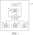
- FIG. 1 is a diagram of a system 10 for simulating orthodontic treatment.
- System 10 includes a processor 16 electronically coupled with a camera 12, a display device 14, an output device 18, a storage device 19, and an input device 20.
- Camera 12 can be implemented with, for example, a digital image sensor providing electronic digital images or video.
- Display device 14 can be implemented with any electronic display, for example a Cathode Ray Tube (CRT), a liquid crystal display (LCD), light emitting diode (LED) display, or organic light emitting diode (OLED) display.
- Output device 18 can be implemented with any device for outputting information, for example a speaker, an LED indicator, or an auxiliary LCD display module.
- Input device 20 can be implemented with any device for entering information or commands, for example a keyboard, microphone, cursor-control device, or touch screen.
- Storage device 19 can be implemented with any electronic device for storing information such as a hard drive, solid state drive, or flash drive. Storage device 19 can optionally be located external to system 10 and accessed via a network connection.
- System 10 can be implemented with, for example, a desktop, notebook, or tablet computer, or a mobile phone.
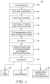
- FIG. 2 is a flow chart of a method 22 for simulating orthodontic treatment.
- This method can be implemented in software or firmware modules for execution by a processor such as processor 16.
- This method is used to visualize a simulation of an orthodontic treatment through augmentation, a virtual overlay onto an image captured of the real world, possibly with manipulation of an estimated 3D representation of a face captured in one or more frames of video in real-time.
- step 24 Facial Detection
- step 26 Full Facial Landmarking
- step 28 Face Pose Estimation
- step 30 Crop Region of Interest Using Facial Landmarks
- step 32 optional 3D Representation of the Face or region of interest
- step 34 optional supplementation of the image or 3D Representation of the Face
- step 36 Visualization
- This step finds a face in a frame of video from camera 12 using a pre-trained model, such as a Viola-Jones Model, an LBPH (Local Binary Pattern Histogram) cascade, or a model trained through deep learning.
- a face context may be created to keep track of this face over multiple frames.
- This face context may contain previous frame data, such as important features used in reprojection, or a point cloud obtained over multiple previous frames.
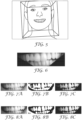
- FIG. 3 is a representation of an image illustrating an example of a detected face as received from camera 12, where the box in the image indicates the area of detection.
- the image in FIG. 3 can optionally be an image of a user with orthodontic appliances in order to virtually remove the appliances rather than virtually adding appliances.
- Landmarks are points of interest describing important structures on the face, and can be found using a shape predictor model which may use an SVM (Support Vector Machine), PCA (Principal Component Analysis), or deep learning and other machine learning algorithms like random forests. These landmarks are used to crop the face or region of interest from the background, and in this example, will be used for rough face pose estimation using a generalized 3D landmark model. They can also be used in the steps below to morph a generalized face mesh to more accurately represent the face detected in the video.
- FIG. 4 is a representation of an image illustrating facial landmarking, where the detected area from the image of FIG. 3 includes dots, or other indicia, for the landmarking.
- an estimated camera extrinsic model can be found by solving the general Perspective N-Point (PNP) problem. Solving the PNP problem assumes the detected landmarks have not been affected by camera lens distortion (radial/tangential lens distortion), or have been undistorted.
- An SfM (Structure from Motion) technique could instead be applied on static (non-moving) parts of the face once the feature matching step below has been completed on multiple sequential frames to obtain a more accurate camera extrinsic model. Knowing the camera extrinsics provides a good 3D point representation of each of the landmarks which can help indicate the pose and further be used for more accurate augmented reality. This allows more accurate cropping of the region of interest for performing augmented reality, but it is not a required step.
- FIG. 5 is a representation of an image illustrating face pose estimation as indicated by the two boxes on the image from FIG. 3 .
- FIG. 6 illustrates a cropped region of interest from the images of FIGS. 3-5 .
- Method 1 The first method that could be employed is to generate a 3D mesh over multiple frames of video, using Reprojection and Mesh reconstruction.
- Reprojection is the process of finding the depth of features in one of the images, turning the features into a 3D point cloud.
- Mesh reconstruction is the technique used to produce a triangle mesh from that point cloud. This assumes more than one image of the same face, where the images have been taken from slightly different angles and locations. The following are the steps of this Method 1.
- Method 2 This method involves a parametric representation of the face or region of interest (e.g., NURBS surface or Bézier surface), or a generic polygonal mesh (i.e., triangles or quadrilaterals), which will be morphed, expanded, or stretched to best fit either a point cloud of the face (see Method 1) or a set of landmarks obtained earlier. If using a generic mesh of a face or region of interest, vertices within the mesh could be assigned weights to landmarks. When the landmarks are found in the image, the vertices of the generic mesh would be pulled towards those landmarks based on their given weights and face pose.
- a parametric representation of the face or region of interest e.g., NURBS surface or Bézier surface
- a generic polygonal mesh i.e., triangles or quadrilaterals
- the topology of the surface could be predefined only in the abstract, such as by graph-theoretical relationships between landmarks or regions outlined by lines between landmarks.
- the NURBS surface or mesh could be generated such that the landmarks coincide with the corners of the surface patches.
- additional points may be necessarily captured in the regions between landmarks, and these points can serve to more accurately define the parameters of the surfaces: control points and polynomial coefficients in the case of NURBS or intermediate vertices in the case of tessellated mesh surfaces.
- Any distinct feature of the face can serve as a landmark or control point in a mesh or parametric model. As such, the accuracy of the model can be improved by increasing the resolution of the video imagery to the extent that these minute features are both visible and recognizable.
- the virtual treatment can be applied, either through augmentation or manipulation of the 3D geometry. This may include pin-pointing locations or areas to apply the simulation.
- augmentation is implemented, where the rendered orthodontic appliance is overlaid onto the region of interest, after estimating the location on the region of interest where the appliance should be placed, or where detected appliances are virtually removed from the region of interest.
- extra steps may need to be taken, such as filling any holes in the image that result from morphing the 3D geometry.
- the rendered orthodontic appliance can be represented by a 2D image or a 3D model.
- various image processing techniques are used on the region of interest to segment and identify the teeth, which include scaling-up the region of interest, noise reduction, applying a segmentation algorithm such as Mean Shift segmentation, histogram equalization, adaptive thresholding on specific channels of multiple color spaces, eroding and dilating (opening/closing), edge detection, and finding contours.
- a segmentation algorithm such as Mean Shift segmentation, histogram equalization, adaptive thresholding on specific channels of multiple color spaces, eroding and dilating (opening/closing), edge detection, and finding contours.
- FIGS. 7A-7C illustrate adaptive thresholding applied to inverted green-red channel of lab color space of region of interest: inverted channel "a" (green-red) of lab color space ( FIG. 7A ), mask after adaptive thresholding applied ( FIG. 7B ), and color image of region of interest with mask applied ( FIG. 7C).
- FIGS. 8A-8C illustrate adaptive thresholding applied to luminance channel of YUV color space of region of interest: channel "Y" (luminance) of YUV color space ( FIG. 8A ), mask after adaptive thresholding applied combined (bitwise and) with mask from Lab color space adaptive threshold ( FIG. 8B ), and final color image of region of interest with combined mask applied ( FIG. 8C ).
- FIGS. 9A and 9B illustrate, respectively, opening and closing of region of interest mask after color space thresholding.
- FIG. 10 illustrates detected edges from canny edge detection.
- Contours can be generated from the detected edges, where individual teeth or groups of teeth can be analyzed or worked with as standalone objects.
- FIG. 11 illustrates closed contours found from detected edges, overlaid onto a color region of interest.
- FIG. 12 illustrates virtual rendering of treatment, in this example brackets and archwires.
- the virtual rendering can involve removing detected appliances such as brackets and archwires.
- the rendered treatment is augmented onto the region of interest.
- Post processing can be done, such as Gaussian blur, to blend the augmented treatment with the original image and make the final image appear more natural, as if it were a part of the original image.
- FIG. 13 illustrates augmentation of treatment, where the virtual brackets and archwires are overlaid on the teeth within the region of interest.
- the augmentation can involve removing detected appliances and displaying the result. Augmentation can be achieved without explicitly segmenting out individual teeth and contours but by doing direct registration of end points of the mouth with end points of teeth. However, identifying each tooth center even approximately provides a more accurate/realistic result.
- Another approach to augmenting images of the face with appliances, restorations, or modified anatomies involves another aspect of 3D modeling.
- the approach described above uses 3D modeling to determine the position, orientation, and scale of the face, which may be mathematically described by a 3D Affine transform in a virtual universe.
- a corresponding transform is then applied to the dental appliances so that they register to the teeth of the patient in somewhat realistic positions and orientations, although 2D image analysis may ultimately be used to find the Facial Axis (FA) points of the teeth at which to place brackets.
- FA Facial Axis
- the video camera may be used as a type of 3D scanner, thereby capturing multiple 2D images of a person's face from different vantage points (i.e., viewpoints and look-at vectors, which together with the image plane form a set of distinct view frustums).
- vantage points i.e., viewpoints and look-at vectors, which together with the image plane form a set of distinct view frustums.
- a 3D model of the person's face (and head) may be generated, originally in the form of a point cloud, then later in the form of a triangular mesh.
- UV mapping or texture mapping
- an image of the 3D model may be rendered onto a 2D plane that is positioned anywhere in the virtual universe according to a view frustum.
- Suitable rendering methods include polygon rendering or ray tracing.
- the color values from the UV map would be applied in the rendering to generate a more realistic, color rendition of the face (as opposed to a monochromatic mesh model shaded only by triangle orientation relative to both the view point and each light source in the scene).
- the 3D model of the face can be combined or integrated (or simply registered) with other 3D models or images that were optionally obtained from different scan sources, such as intraoral scanners, Cone Beam CT (CBCT or 3D X-ray) scanners, MRI scanners, 3D stereoscopic cameras, and the like. Such data would likely have been obtained at earlier dates and used in treatment planning.
- Such other models or images can be stored in and retrieved from, for example, data storage 19.
- the video imagery may serve as the sole source of superficial 3D imagery, thus capturing the soft tissues of the face and whatever dental anatomy may be visible.
- soft tissue data may be captured in CBCT scans (without color information) or in 3D stereoscopic imagery. Regardless, soft tissue data is an essential component in the modeling of certain treatment plans, such as those involving orthognathic surgery, occlusion class correction (via antero-posterior dental movements), significant changes to the anterior proclination of teeth, and cosmetic and plastic surgeries. Embodiments of this invention can thus provide visual feedback to patients on their expected outward appearance resulting from treatment, during or after the treatment, or both during and after.
- a doctor may be able to conscientiously devise one or more treatment plans with a real basis in structural anatomy. These plans may be devised on the doctor's own schedule, without the patient present, and possibly in collaboration with other doctors, technicians, or service providers. Appliances or prostheses may be created and applied to the patient's natural anatomy, and the patient's naturally anatomy can be augmented or modified as part of the treatment plan. 3D virtual models of such appliances and prostheses can be created by, for example, third party labs or service providers and combined with the datasets provided by the doctor. Modified anatomies resulting from treatment can be generated by the doctor, by a third party, or as a collaborative effort using remote software collaboration tools. The treatment plan can be presented back to the patient for review, possible modification, and approval. In some scenarios, the doctor may be the reviewer, and a third party lab or manufacturer is the source of the proposed treatment plan.
- a video camera is used to capture enough frames to generate a 3D model of the patient sufficient to register his/her face to the 3D model comprising the plan, which may be based on other data sources but having at least some anatomical features in common with the newly-captured data.
- a complete re-scan of the patient's face would not be necessary, since a full scan would have already been done at an earlier date in order to facilitate the creation of a detailed treatment plan.
- a partial 3D model of the patient's current anatomy could be created, optionally cropped to remove the portions of anatomy modified by the plan, and best-fit to the plan model.
- the same view frustum may be used to represent a virtual camera, and a 2D rendering of the 3D model may be created on the view plane of this camera. This 2D rendering can then be presented back to the patient (or other observer) in real-time as the patient moves relative to the camera. In effect, the physical and virtual worlds are kept synchronous with one another.
- the same method could be used, except the pixels of these anatomies may need to be colored using previously captured values that were UV mapped onto the mesh of the plan model.
- the color values could be obtained from the current video frame but then transformed to different positions as determined by morphs to the 3D anatomy according to the treatment plan. For example, an orthognathic surgery might prescribe that the mandible be advanced by several millimeters. The color values of the pixels used to render the patient's soft tissues (skin and lips) affected by the advancement would tend not to differ as a result; only their positions in the virtual world and thus the rendering would change.
- the affected pixels in the 2D video image might simply by translated in the image plane according to a 3D Affine transform projected onto the view plane.
- the entire 3D virtual scene may be rendered in real-time and presented back to the patient in sync with their head movements.
- a technique can be employed by which the plan model is continuously augmented or updated while video is being captured for the purpose of presenting the treatment plan to the patient.
- the plan model is continuously augmented or updated while video is being captured for the purpose of presenting the treatment plan to the patient.
- the new images can be used to augment or update the existing model by filling holes, removing or joining islands, and reducing noise. In so doing, the quality of the plan model may be improved in real-time (or after a brief processing delay).
- the dimensional accuracy of the model may be improved by using large, unitary patches of scan data to adjust the positions and orientations of smaller patches that were stitched together with accumulated error.
- This technique could also be used to improve the realism of an avatar-style rendering by updating the texture map to current skin and lighting conditions. Otherwise, the texture map used on the avatar would have come from a previous scan captured at an earlier date and possibly in a different setting.
- the user's facial image or 3D representation can optionally be registered with the image or model of the user's facial anatomy using, for example, PCA or other techniques.
- the images or models are synchronized such that manipulation of one image or representation causes corresponding manipulation of the other image or representation. For example, if the user's facial image is rotated, the registered image or model of the user's facial anatomy is correspondingly rotated.
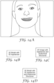
- FIG. 14A illustrates the result of treatment augmentation in a representation of a single image displayed to the user via display device 14 and within a user interface, for example.
- the single image of FIG. 14A can display the result of virtually removing detected appliances worn by the user.
- a 3D representation of the user's face is generated, using the optional step 32, then such 3D representation can be displayed augmented with the virtual treatment (40) as represented by the user interface in FIG. 14B for display on display device 14.
- a supplemented 2D image or a 3D representation is generated, using the optional step 35, then such supplemented 2D image or 3D representation can be displayed augmented with the virtual treatment (42) as represented by the user interfaces in FIGS. 14C and 14D for display on display device 14.
- Method 22 can be executed by processor 16 such that the augmentation is shown to the user on display device 14 in real-time when or shortly after detection of the user's image by camera 12.
- real-time means at a rate of at least one frame per second or, more preferably, at a rate of 15-60 frames per second.
Landscapes
- Engineering & Computer Science (AREA)
- Health & Medical Sciences (AREA)
- Life Sciences & Earth Sciences (AREA)
- Medical Informatics (AREA)
- Physics & Mathematics (AREA)
- General Engineering & Computer Science (AREA)
- Public Health (AREA)
- Veterinary Medicine (AREA)
- General Health & Medical Sciences (AREA)
- Animal Behavior & Ethology (AREA)
- Theoretical Computer Science (AREA)
- Molecular Biology (AREA)
- Surgery (AREA)
- Pathology (AREA)
- Biophysics (AREA)
- Biomedical Technology (AREA)
- Heart & Thoracic Surgery (AREA)
- Human Computer Interaction (AREA)
- High Energy & Nuclear Physics (AREA)
- Optics & Photonics (AREA)
- Nuclear Medicine, Radiotherapy & Molecular Imaging (AREA)
- Radiology & Medical Imaging (AREA)
- General Physics & Mathematics (AREA)
- Oral & Maxillofacial Surgery (AREA)
- Dentistry (AREA)
- Epidemiology (AREA)
- Processing Or Creating Images (AREA)
- Image Processing (AREA)
- Dental Tools And Instruments Or Auxiliary Dental Instruments (AREA)
Description
- The use of augmented reality face filters, such as the SNAPCHAT or INSTAGRAM products, are becoming increasingly popular. However, these filters are mainly used in popular culture today for only entertainment purposes, such as to visually apply renditions of hair, make-up, eyeglasses, facial hair, and hats in real-time to the face or head of the human subject in a video augmentation. A need exists to extend this technology for use in medical or dental applications. Kehler Jacob: "Nerdy Glasses and Braces Snapchat lens, on the line social media, August 1, 2016" describes snapchat lenses.
- The present invention is defined in the claims. The invention relates to a method and to a system for simulating orthodontic treatment as defined in independent claims 1 and 5, respectively.
- The accompanying drawings are incorporated in and constitute a part of this specification and, together with the description, explain the advantages and principles of the invention. In the drawings,
-
FIG. 1 is a diagram of a system for simulating orthodontic treatment; -
FIG. 2 is a flow chart of a method for simulating orthodontic treatment; -
FIG. 3 is a representation of an image illustrating an example of a detected face; -
FIG. 4 is a representation of an image illustrating facial landmarking; -
FIG. 5 is a representation of an image illustrating face pose estimation; -
FIG. 6 illustrates a cropped region of interest; -
FIGS. 7A-7C illustrate adaptive thresholding applied to an inverted channel; -
FIGS. 8A-8C illustrate adaptive thresholding applied to a luminance channel; -
FIGS. 9A and 9B illustrate opening and closing of a region of interest; -
FIG. 10 illustrates detected edges; -
FIG. 11 illustrates closed contours from detected edges; -
FIG. 12 illustrates virtual rendering of treatment; -
FIG. 13 illustrates augmentation of treatment; -
FIG. 14A is a user interface illustrating treatment augmentation in a representation of a single image; -
FIG. 14B is a user interface representing a 3D model augmented with 3D appliances; -
FIG. 14C is a user interface representing a 2D image augmented with appliances and anatomical structures; and -
FIG. 14D is a user interface representing a 3D model augmented with 3D appliances and anatomical structures. - Embodiments of this invention include the use of computer vision and augmented reality technology to superimpose a computer-generated image on a user's view of the real world to provide a view of one, or combination, of the following:
- 1. The addition or subtraction of orthodontic appliance(s), such as lingual brackets, labial brackets, aligners, class correction devices, or retention devices.
- 2. The addition, subtraction, or modification of dental restoration(s), such as crowns, bridges, inlays, onlays, veneers, dentures, or gingiva.
- 3. The addition, subtraction, or modification of natural tooth anatomy, such as by using tooth whitening agents, reducing or building-up cusp tips or incisal edges, or predicting the appearance of teeth after eruption in children (possibly including ectopic eruptions).
- 4. The modification of craniofacial structures via oral or maxillofacial surgery, including dentoalveolar surgery, dental and maxillofacial implants, cosmetic surgery, and orthognathic surgery.
- 5. The addition, subtraction, or modification of maxillofacial, ocular, or craniofacial prosthesis.
- 6. The result of orthodontic and/or restorative treatment - to include the planned positions and shapes of dental anatomical structures, i.e. teeth and gingiva.
- 7. The predicted result of not undergoing orthodontic and/or restorative treatment, possibly showing deleterious consequences, such as malocclusion, tooth wear, gingival recession, gum disease, bone loss, tooth decay, or tooth loss.
- 8. The modification of soft tissues of the face, such as by botulinum toxin injections or more invasive cosmetic and plastic surgeries, such as rhinoplasty, chin enhancement, cheek enhancement, facelift, eyelid lift, neck lift, brow lift, cleft palate repair, burn repair, and scar revision.
- 9. An overlay, of variable opacity, of staged treatment - to compare scheduled versus actual progress of treatment.
- This technology can be used both as a tool for treatment planning by a medical/dental professional and as a tool for case presentation to a patient. Other intended uses include exploration of treatment options by a patient wanting to visualize their facial appearance during or after treatment, possibly switching or transitioning between before and after appearances for comparison. In some cases, a patient may be able to entertain treatment scenarios without first visiting a medical or dental practitioner, using only superficial imagery data. In other cases, the augmentation may require additional data, such as X-ray or cone beam computed tomography (CBCT) scan data, bite registration data, three-dimensional (3D) oral scan data, virtual articulation data, or calibrated measurements in order to generate a treatment plan that provides a more accurate rendition of the patient after treatment. As such, additional time and effort may be needed to clean-up and integrate datasets, explore treatment options, perform measurements and analyses, and plan treatment steps before a credible rendition of treatment outcome can be shown to the patient. In some cases, two or more treatment scenarios may be generated and presented to the patient, thereby giving the patient a somewhat realistic view of treatment outcome. This can serve to help the patient (or doctor) in deciding on a course of action. In some cases, the cost of treatment may be weighed against the quality of outcome. For example, the patient may forego an expensive treatment due to marginal gains in appearance over a less expensive treatment. Similar rules may be applied to the duration of treatment, where time is treated like a cost.
-
FIG. 1 is a diagram of asystem 10 for simulating orthodontic treatment.System 10 includes aprocessor 16 electronically coupled with acamera 12, adisplay device 14, anoutput device 18, astorage device 19, and aninput device 20.Camera 12 can be implemented with, for example, a digital image sensor providing electronic digital images or video.Display device 14 can be implemented with any electronic display, for example a Cathode Ray Tube (CRT), a liquid crystal display (LCD), light emitting diode (LED) display, or organic light emitting diode (OLED) display.Output device 18 can be implemented with any device for outputting information, for example a speaker, an LED indicator, or an auxiliary LCD display module.Input device 20 can be implemented with any device for entering information or commands, for example a keyboard, microphone, cursor-control device, or touch screen.Storage device 19 can be implemented with any electronic device for storing information such as a hard drive, solid state drive, or flash drive.Storage device 19 can optionally be located external tosystem 10 and accessed via a network connection.System 10 can be implemented with, for example, a desktop, notebook, or tablet computer, or a mobile phone. -
FIG. 2 is a flow chart of a method 22 for simulating orthodontic treatment. This method can be implemented in software or firmware modules for execution by a processor such asprocessor 16. This method is used to visualize a simulation of an orthodontic treatment through augmentation, a virtual overlay onto an image captured of the real world, possibly with manipulation of an estimated 3D representation of a face captured in one or more frames of video in real-time. - The following steps of method 22, more fully explained below, are used to produce an exemplary augmented orthodontic appliance simulation, starting with a single frame (image) of video: Facial Detection (step 24); Full Facial Landmarking (step 26); Face Pose Estimation (step 28); Crop Region of Interest Using Facial Landmarks (step 30); optional 3D Representation of the Face or region of interest (step 32); Simulation (step 34); optional supplementation of the image or 3D Representation of the Face (step 35); and Visualization (step 36).
- This step finds a face in a frame of video from
camera 12 using a pre-trained model, such as a Viola-Jones Model, an LBPH (Local Binary Pattern Histogram) cascade, or a model trained through deep learning. Once a face has been detected, a face context may be created to keep track of this face over multiple frames. This face context may contain previous frame data, such as important features used in reprojection, or a point cloud obtained over multiple previous frames.FIG. 3 is a representation of an image illustrating an example of a detected face as received fromcamera 12, where the box in the image indicates the area of detection. The image inFIG. 3 can optionally be an image of a user with orthodontic appliances in order to virtually remove the appliances rather than virtually adding appliances. - Landmarks are points of interest describing important structures on the face, and can be found using a shape predictor model which may use an SVM (Support Vector Machine), PCA (Principal Component Analysis), or deep learning and other machine learning algorithms like random forests. These landmarks are used to crop the face or region of interest from the background, and in this example, will be used for rough face pose estimation using a generalized 3D landmark model. They can also be used in the steps below to morph a generalized face mesh to more accurately represent the face detected in the video.
FIG. 4 is a representation of an image illustrating facial landmarking, where the detected area from the image ofFIG. 3 includes dots, or other indicia, for the landmarking. - Using the detected facial landmarks and a generalized 3D landmark model, an estimated camera extrinsic model (transformation) can be found by solving the general Perspective N-Point (PNP) problem. Solving the PNP problem assumes the detected landmarks have not been affected by camera lens distortion (radial/tangential lens distortion), or have been undistorted. An SfM (Structure from Motion) technique could instead be applied on static (non-moving) parts of the face once the feature matching step below has been completed on multiple sequential frames to obtain a more accurate camera extrinsic model. Knowing the camera extrinsics provides a good 3D point representation of each of the landmarks which can help indicate the pose and further be used for more accurate augmented reality. This allows more accurate cropping of the region of interest for performing augmented reality, but it is not a required step.
FIG. 5 is a representation of an image illustrating face pose estimation as indicated by the two boxes on the image fromFIG. 3 . - This example will crop out the mouth region, where the inside edge of the lips represents the boundaries of the cropped region. This could be the full face or another part of the face if the simulation involves a large or different area of the face, such as the jaw.
FIG. 6 illustrates a cropped region of interest from the images ofFIGS. 3-5 . - This is an optional step and only needed for simulations which require a 3D model of the face or region of the face, such as structural manipulation or improved accuracy for augmentation. Two methods could be used, or a combination of both.
- Method 1. The first method that could be employed is to generate a 3D mesh over multiple frames of video, using Reprojection and Mesh reconstruction. Reprojection is the process of finding the depth of features in one of the images, turning the features into a 3D point cloud. Mesh reconstruction is the technique used to produce a triangle mesh from that point cloud. This assumes more than one image of the same face, where the images have been taken from slightly different angles and locations. The following are the steps of this Method 1.
- i. Feature/Keypoint detection. These are points (corners) on an image that stand out among the rest of the pixels in the image, which are likely to be present in another image at a different angle of the same scene.
- ii. Feature filtering. This is an optional step which may need to be taken if the list of features detected is too large. Various techniques exist to prune the list of features to get a smaller list of the strongest features (most prominent features).
- iii. Feature correspondence. Each frame must have gone through the steps above, and have their own list of features. This is where the features from one image are matched with the features in the other image. Not all features will be matched, and any features which were not matched are discarded. Once the features have been matched, the list of features in the second image should be sorted in the same order as the features from the first image.
- iv. Rectification. Adjust the features in each image so that they are row-aligned on the same plane using the camera extrinsic model. This means that features from one image are on the same x-axis row as the same feature in the other image.
- v. Calculate the disparity map. The disparity map is a list of disparities (distances) on the x-axis between the matching points. The distances are measured in units of pixels.
- vi. Triangulation. Using the geometric relation of the cameras, the method can calculate the epipolar geometry, and the essential and fundamental matrices needed to calculate the depth of each feature in one image.
- vii. Mesh Reconstruction. Turn the 3D point cloud into a 3D mesh for the simulation step. This may also include finding texture coordinates through ray tracing based on camera parameters (e.g., camera location).
- Method 2. This method involves a parametric representation of the face or region of interest (e.g., NURBS surface or Bézier surface), or a generic polygonal mesh (i.e., triangles or quadrilaterals), which will be morphed, expanded, or stretched to best fit either a point cloud of the face (see Method 1) or a set of landmarks obtained earlier. If using a generic mesh of a face or region of interest, vertices within the mesh could be assigned weights to landmarks. When the landmarks are found in the image, the vertices of the generic mesh would be pulled towards those landmarks based on their given weights and face pose. Alternatively, the topology of the surface could be predefined only in the abstract, such as by graph-theoretical relationships between landmarks or regions outlined by lines between landmarks. As points are obtained in 3D via feature recognition (landmark identification) and photogrammetric triangulation, the NURBS surface or mesh could be generated such that the landmarks coincide with the corners of the surface patches. Of course, additional points may be necessarily captured in the regions between landmarks, and these points can serve to more accurately define the parameters of the surfaces: control points and polynomial coefficients in the case of NURBS or intermediate vertices in the case of tessellated mesh surfaces. Any distinct feature of the face can serve as a landmark or control point in a mesh or parametric model. As such, the accuracy of the model can be improved by increasing the resolution of the video imagery to the extent that these minute features are both visible and recognizable.
- Once the steps above are complete, the virtual treatment can be applied, either through augmentation or manipulation of the 3D geometry. This may include pin-pointing locations or areas to apply the simulation. In this example, augmentation is implemented, where the rendered orthodontic appliance is overlaid onto the region of interest, after estimating the location on the region of interest where the appliance should be placed, or where detected appliances are virtually removed from the region of interest. For treatments involving manipulation of 3D geometry, extra steps may need to be taken, such as filling any holes in the image that result from morphing the 3D geometry. The rendered orthodontic appliance can be represented by a 2D image or a 3D model.
- In this example, various image processing techniques are used on the region of interest to segment and identify the teeth, which include scaling-up the region of interest, noise reduction, applying a segmentation algorithm such as Mean Shift segmentation, histogram equalization, adaptive thresholding on specific channels of multiple color spaces, eroding and dilating (opening/closing), edge detection, and finding contours.
- The region of interest is first scaled up and converted to different color spaces, where an adaptive threshold is applied to the channel in which the teeth are most separated from the non-teeth.
FIGS. 7A-7C illustrate adaptive thresholding applied to inverted green-red channel of lab color space of region of interest: inverted channel "a" (green-red) of lab color space (FIG. 7A ), mask after adaptive thresholding applied (FIG. 7B ), and color image of region of interest with mask applied (FIG. 7C). FIGS. 8A-8C illustrate adaptive thresholding applied to luminance channel of YUV color space of region of interest: channel "Y" (luminance) of YUV color space (FIG. 8A ), mask after adaptive thresholding applied combined (bitwise and) with mask from Lab color space adaptive threshold (FIG. 8B ), and final color image of region of interest with combined mask applied (FIG. 8C ). - Once a mask has been created through adaptive thresholding of the color space channels above, opening (eroding, then dilating) and closing (dilating, then eroding) can be used to further segment the teeth and clean up noise.
FIGS. 9A and 9B illustrate, respectively, opening and closing of region of interest mask after color space thresholding. - Edges are detected from the mask, using a technique such as Canny Edge Detection.
FIG. 10 illustrates detected edges from canny edge detection. - Contours can be generated from the detected edges, where individual teeth or groups of teeth can be analyzed or worked with as standalone objects.
FIG. 11 illustrates closed contours found from detected edges, overlaid onto a color region of interest. - From the contour analysis, and general face pose, the treatment is rendered in the correct orientation and scale, ready to be overlaid on the region of interest. The original image could be analyzed to find a general lighting model to apply to rendered treatment, which would make the treatment augmentation fit more naturally in the scene.
FIG. 12 illustrates virtual rendering of treatment, in this example brackets and archwires. Optionally, the virtual rendering can involve removing detected appliances such as brackets and archwires. - The rendered treatment is augmented onto the region of interest. Post processing can be done, such as Gaussian blur, to blend the augmented treatment with the original image and make the final image appear more natural, as if it were a part of the original image.
FIG. 13 illustrates augmentation of treatment, where the virtual brackets and archwires are overlaid on the teeth within the region of interest. Optionally, the augmentation can involve removing detected appliances and displaying the result. Augmentation can be achieved without explicitly segmenting out individual teeth and contours but by doing direct registration of end points of the mouth with end points of teeth. However, identifying each tooth center even approximately provides a more accurate/realistic result. - Another approach to augmenting images of the face with appliances, restorations, or modified anatomies involves another aspect of 3D modeling. The approach described above uses 3D modeling to determine the position, orientation, and scale of the face, which may be mathematically described by a 3D Affine transform in a virtual universe. A corresponding transform is then applied to the dental appliances so that they register to the teeth of the patient in somewhat realistic positions and orientations, although 2D image analysis may ultimately be used to find the Facial Axis (FA) points of the teeth at which to place brackets.
- In this other approach, the video camera may be used as a type of 3D scanner, thereby capturing multiple 2D images of a person's face from different vantage points (i.e., viewpoints and look-at vectors, which together with the image plane form a set of distinct view frustums). Using 3D photogrammetry techniques, a 3D model of the person's face (and head) may be generated, originally in the form of a point cloud, then later in the form of a triangular mesh. Accompanying these techniques is UV mapping (or texture mapping) to apply color values from pixels in the 2D images to 3D vertices or triangles in the mesh. As such, a realistic-looking and reasonably accurate 3D model of the person's face may be generated in the virtual universe. Subsequently, an image of the 3D model may be rendered onto a 2D plane that is positioned anywhere in the virtual universe according to a view frustum. Suitable rendering methods include polygon rendering or ray tracing. The color values from the UV map would be applied in the rendering to generate a more realistic, color rendition of the face (as opposed to a monochromatic mesh model shaded only by triangle orientation relative to both the view point and each light source in the scene).
- At this point, the 3D model of the face can be combined or integrated (or simply registered) with other 3D models or images that were optionally obtained from different scan sources, such as intraoral scanners, Cone Beam CT (CBCT or 3D X-ray) scanners, MRI scanners, 3D stereoscopic cameras, and the like. Such data would likely have been obtained at earlier dates and used in treatment planning. Such other models or images can be stored in and retrieved from, for example,
data storage 19. - In some cases, the video imagery may serve as the sole source of superficial 3D imagery, thus capturing the soft tissues of the face and whatever dental anatomy may be visible. In other cases, soft tissue data may be captured in CBCT scans (without color information) or in 3D stereoscopic imagery. Regardless, soft tissue data is an essential component in the modeling of certain treatment plans, such as those involving orthognathic surgery, occlusion class correction (via antero-posterior dental movements), significant changes to the anterior proclination of teeth, and cosmetic and plastic surgeries. Embodiments of this invention can thus provide visual feedback to patients on their expected outward appearance resulting from treatment, during or after the treatment, or both during and after.
- By capturing multiple 3D data sources of the patient's craniofacial anatomy, a doctor may be able to conscientiously devise one or more treatment plans with a real basis in structural anatomy. These plans may be devised on the doctor's own schedule, without the patient present, and possibly in collaboration with other doctors, technicians, or service providers. Appliances or prostheses may be created and applied to the patient's natural anatomy, and the patient's naturally anatomy can be augmented or modified as part of the treatment plan. 3D virtual models of such appliances and prostheses can be created by, for example, third party labs or service providers and combined with the datasets provided by the doctor. Modified anatomies resulting from treatment can be generated by the doctor, by a third party, or as a collaborative effort using remote software collaboration tools. The treatment plan can be presented back to the patient for review, possible modification, and approval. In some scenarios, the doctor may be the reviewer, and a third party lab or manufacturer is the source of the proposed treatment plan.
- In presenting the treatment plan back to the patient, a video camera is used to capture enough frames to generate a 3D model of the patient sufficient to register his/her face to the 3D model comprising the plan, which may be based on other data sources but having at least some anatomical features in common with the newly-captured data. A complete re-scan of the patient's face would not be necessary, since a full scan would have already been done at an earlier date in order to facilitate the creation of a detailed treatment plan. Thus, with only limited motion relative to the video camera, a partial 3D model of the patient's current anatomy could be created, optionally cropped to remove the portions of anatomy modified by the plan, and best-fit to the plan model. Because the view frustum of each video frame is known via 3D photogrammetry methods, the same view frustum may be used to represent a virtual camera, and a 2D rendering of the 3D model may be created on the view plane of this camera. This 2D rendering can then be presented back to the patient (or other observer) in real-time as the patient moves relative to the camera. In effect, the physical and virtual worlds are kept synchronous with one another.
- There are options in determining what to render and present back to the patient. For example, it might not be necessary to store a texture map in the plan model if the plan is only presented in real-time with the patient present. In this scenario, the registration between individual video frames and the plan model can occur without the use of a texture map, and the color value of each pixel in the video image presented back to the patient can simply pass-through from the value captured by the video camera. In the case of appliances being attached to the patient's teeth (or other parts of the face), the appliances would be rendered in the virtual world, and their 2D renditions would overlay corresponding areas from the video frames, in effect masking the underlying anatomy. In the case of modified anatomies, the same method could be used, except the pixels of these anatomies may need to be colored using previously captured values that were UV mapped onto the mesh of the plan model. Alternatively, the color values could be obtained from the current video frame but then transformed to different positions as determined by morphs to the 3D anatomy according to the treatment plan. For example, an orthognathic surgery might prescribe that the mandible be advanced by several millimeters. The color values of the pixels used to render the patient's soft tissues (skin and lips) affected by the advancement would tend not to differ as a result; only their positions in the virtual world and thus the rendering would change. As such, the affected pixels in the 2D video image might simply by translated in the image plane according to a 3D Affine transform projected onto the view plane. In yet other scenarios, once the registration of the physical video camera is established with the virtual camera, the entire 3D virtual scene may be rendered in real-time and presented back to the patient in sync with their head movements.
- In some of the above scenarios, a technique can be employed by which the plan model is continuously augmented or updated while video is being captured for the purpose of presenting the treatment plan to the patient. Given that in some scenarios only minimal video coverage might have been used to generate a 3D model of the patient, holes, islands, or noisy regions are likely to exist in the mesh comprising the model. As new video images are received, even if the main purpose of capture is to register physical and virtual worlds and render an existing plan model for the patient, the new images can be used to augment or update the existing model by filling holes, removing or joining islands, and reducing noise. In so doing, the quality of the plan model may be improved in real-time (or after a brief processing delay). In some instances, the dimensional accuracy of the model may be improved by using large, unitary patches of scan data to adjust the positions and orientations of smaller patches that were stitched together with accumulated error. This technique could also be used to improve the realism of an avatar-style rendering by updating the texture map to current skin and lighting conditions. Otherwise, the texture map used on the avatar would have come from a previous scan captured at an earlier date and possibly in a different setting.
- For the supplemented image or model, the user's facial image or 3D representation can optionally be registered with the image or model of the user's facial anatomy using, for example, PCA or other techniques. When registered, the images or models are synchronized such that manipulation of one image or representation causes corresponding manipulation of the other image or representation. For example, if the user's facial image is rotated, the registered image or model of the user's facial anatomy is correspondingly rotated.
- This step involves overlaying the region of interest onto the original frame (
FIG. 3 ) and displaying the final image to the user for a 2D image (38).FIG. 14A illustrates the result of treatment augmentation in a representation of a single image displayed to the user viadisplay device 14 and within a user interface, for example. Optionally, the single image ofFIG. 14A can display the result of virtually removing detected appliances worn by the user. - If a 3D representation of the user's face is generated, using the
optional step 32, then such 3D representation can be displayed augmented with the virtual treatment (40) as represented by the user interface inFIG. 14B for display ondisplay device 14. If a supplemented 2D image or a 3D representation is generated, using theoptional step 35, then such supplemented 2D image or 3D representation can be displayed augmented with the virtual treatment (42) as represented by the user interfaces inFIGS. 14C and 14D for display ondisplay device 14. - Method 22 can be executed by
processor 16 such that the augmentation is shown to the user ondisplay device 14 in real-time when or shortly after detection of the user's image bycamera 12. The term "real-time" means at a rate of at least one frame per second or, more preferably, at a rate of 15-60 frames per second.
Claims (8)
- A method for simulating orthodontic treatment, the method comprising:receiving an electronic image of a user's face;detecting the user's face in the electronic image using a model trained through deep learning;detecting facial landmarks using a shape predictor model which uses deep learning or other machine learning algorithms;estimating a rough face pose using a generalized 3D landmark model and the detected facial landmarks;identifying and cropping a region of interest in the image using the detected facial landmarks and the rough face pose, wherein the region of interest includes the user's teeth;placing virtual orthodontic appliances on the user's teeth in the image, wherein a rendered orthodontic appliance is overlaid onto the region of interest; anddisplaying on an electronic display device the user's image with the virtual orthodontic appliances,wherein the receiving, identifying, placing, and displaying steps occur in real-time.
- The method of claim 1, further comprising detecting in a frame of video the electronic image of the user's face.
- The method of claim 1, wherein the placing step comprises placing virtual brackets and a virtual archwire on the user's teeth in the image.
- The method of claim 1, further comprising generating a three-dimensional electronic representation of the user's face and displaying on an electronic display device the three-dimensional representation of the user's face with the virtual orthodontic appliances.
- A system for simulating orthodontic treatment, comprising:a camera (12) providing electronic digital images or video;an electronic display device (14); anda processor (16), electronically coupled to the camera (12) and the electronic display device (14), wherein the processor (16) is configured to:receive from the camera (12) an electronic image of a user's face;detect the user's face in the electronic image using a model trained through deep learning;detect facial landmarks using a shape predictor model which uses deep learning or other machine learning algorithms;estimate a rough face pose using a generalized 3D landmark model and the detected facial landmarks;identify and crop a region of interest in the image using the detected facial landmarks and the rough face pose, wherein the region of interest includes the user's teeth;place virtual orthodontic appliances on the user's teeth in the image, wherein a rendered orthodontic appliance is overlaid onto the region of interest; anddisplay on the electronic display device (14) the user's image with the virtual orthodontic appliances,wherein the receiving, identifying, placing, and displaying occur in real-time.
- The system of claim 5, wherein the processor is further configured to detect in a frame of video from the camera (12) the electronic image of the user's face.
- The system of claim 5, wherein the processor (16) is further configured to place virtual brackets and a virtual archwire on the user's teeth in the image.
- The system of claim 5, wherein the processor (16) is further configured to generate a three-dimensional electronic representation of the user's face and display on an electronic display device the three-dimensional representation of the user's face with the virtual orthodontic appliances.
Applications Claiming Priority (3)
| Application Number | Priority Date | Filing Date | Title |
|---|---|---|---|
| US201862669628P | 2018-05-10 | 2018-05-10 | |
| US201962809806P | 2019-02-25 | 2019-02-25 | |
| PCT/IB2019/053599 WO2019215550A1 (en) | 2018-05-10 | 2019-05-02 | Simulated orthodontic treatment via augmented visualization in real-time |
Publications (3)
| Publication Number | Publication Date |
|---|---|
| EP3790495A1 EP3790495A1 (en) | 2021-03-17 |
| EP3790495A4 EP3790495A4 (en) | 2022-02-09 |
| EP3790495B1 true EP3790495B1 (en) | 2024-07-24 |
Family
ID=68467593
Family Applications (1)
| Application Number | Title | Priority Date | Filing Date |
|---|---|---|---|
| EP19800352.7A Active EP3790495B1 (en) | 2018-05-10 | 2019-05-02 | Simulated orthodontic treatment via augmented visualization in real-time |
Country Status (5)
| Country | Link |
|---|---|
| US (1) | US11517272B2 (en) |
| EP (1) | EP3790495B1 (en) |
| JP (1) | JP7744132B2 (en) |
| CN (1) | CN112087985B (en) |
| WO (1) | WO2019215550A1 (en) |
Families Citing this family (37)
| Publication number | Priority date | Publication date | Assignee | Title |
|---|---|---|---|---|
| EP3545901B1 (en) | 2012-10-30 | 2021-03-03 | University of Southern California | Orthodontic appliance with snap fitted, non-sliding archwire |
| EP3445273B1 (en) | 2016-04-18 | 2023-03-08 | Swift Health Systems Inc. | Orthodontic appliance with non-sliding, tied archwire |
| WO2018144634A1 (en) | 2017-01-31 | 2018-08-09 | Swift Health Systems Inc. | Hybrid orthodontic archwires |
| CN113952052A (en) | 2017-04-21 | 2022-01-21 | 斯威夫特健康系统有限公司 | Indirect-bonded tray, non-slip orthodontic appliance and registration system using the same |
| EP3572030B1 (en) | 2018-05-22 | 2022-11-02 | Dental Monitoring | Method for analysing a dental situation |
| CN113710191A (en) | 2019-03-01 | 2021-11-26 | 斯威夫特健康系统有限公司 | Indirect adhesive tray with integration of occlusal attachments and orthodontic aids |
| EP3952782B1 (en) * | 2019-05-14 | 2023-12-20 | Align Technology, Inc. | Visual presentation of gingival line generated based on 3d tooth model |
| KR20210051141A (en) | 2019-10-30 | 2021-05-10 | 주식회사 스키아 | Method, apparatus and computer program for providing augmented reality based medical information of patient |
| US12053346B2 (en) | 2019-10-31 | 2024-08-06 | Swift Health Systems Inc. | Indirect orthodontic bonding systems and methods |
| US11937996B2 (en) * | 2019-11-05 | 2024-03-26 | Align Technology, Inc. | Face capture and intraoral scanner and methods of use |
| US11842484B2 (en) | 2021-01-04 | 2023-12-12 | James R. Glidewell Dental Ceramics, Inc. | Teeth segmentation using neural networks |
| US11574468B2 (en) * | 2020-03-31 | 2023-02-07 | Toyota Research Institute, Inc. | Simulation-based learning of driver interactions through a vehicle window |
| FR3111066B1 (en) | 2020-06-06 | 2025-07-11 | Olivier Querbes | Method and device for three-dimensional reconstruction of a face with toothed part from a single image |
| CA3186703A1 (en) | 2020-06-11 | 2021-12-16 | Swift Health Systems Inc. | Orthodontic appliance with non-sliding archform |
| JP7599290B2 (en) * | 2020-07-28 | 2024-12-13 | 花王株式会社 | Facial change output method |
| US12131462B2 (en) * | 2021-01-14 | 2024-10-29 | Motahare Amiri Kamalabad | System and method for facial and dental photography, landmark detection and mouth design generation |
| US20220218438A1 (en) * | 2021-01-14 | 2022-07-14 | Orthosnap Corp. | Creating three-dimensional (3d) animation |
| US12268571B2 (en) | 2021-03-12 | 2025-04-08 | Swift Health Systems Inc. | Indirect orthodontic bonding systems and methods |
| CN112967219B (en) * | 2021-03-17 | 2023-12-05 | 复旦大学附属华山医院 | Two-stage tooth point cloud completion method and system based on deep learning network |
| US12136208B2 (en) | 2021-03-31 | 2024-11-05 | James R. Glidewell Dental Ceramics, Inc. | Automatic clean up of jaw scans |
| US12210802B2 (en) | 2021-04-30 | 2025-01-28 | James R. Glidewell Dental Ceramics, Inc. | Neural network margin proposal |
| ES3037947T3 (en) * | 2021-06-01 | 2025-10-08 | Align Technology Inc | Automated management of clinical modifications to treatment plans using three-dimensional controls |
| CN113516781B (en) * | 2021-07-12 | 2025-09-19 | 先临三维科技股份有限公司 | Three-dimensional dynamic tracking method and device, electronic equipment and storage medium |
| CN117677340A (en) * | 2021-07-21 | 2024-03-08 | 高露洁-棕榄公司 | Systems and methods for determining oral health status |
| EP4395687A4 (en) | 2021-09-03 | 2025-07-02 | Swift Health Systems Inc | Orthodontic appliance with non-sliding arch shape |
| WO2023033870A1 (en) | 2021-09-03 | 2023-03-09 | Swift Health Systems Inc. | Method of administering adhesive to bond orthodontic brackets |
| GB2614025B (en) * | 2021-09-07 | 2023-12-27 | Neuronav Ltd | Augmented reality surgical navigation system |
| KR102448169B1 (en) * | 2021-10-05 | 2022-09-28 | 세종대학교산학협력단 | Method and device for predicting orthodontic treatment results based on deep learning |
| US12558163B2 (en) | 2021-10-11 | 2026-02-24 | Hoth Intelligence, Inc. | Method for medical image reconstruction |
| CN114224528B (en) * | 2021-10-26 | 2023-07-18 | 上海交通大学医学院附属第九人民医院 | Oral cavity digital twin model system based on virtual reality interaction and establishment method |
| US20230363857A1 (en) * | 2021-11-17 | 2023-11-16 | Sdc U.S. Smilepay Spv | Systems and methods for generating an interactive visualization of a treatment plan |
| USD1043994S1 (en) | 2022-01-06 | 2024-09-24 | Swift Health Systems Inc. | Archwire |
| US12295806B2 (en) | 2022-01-10 | 2025-05-13 | James R. Glidewell Dental Ceramics, Inc. | Automatic determination of trim-line for aligners |
| JP7600187B2 (en) * | 2022-08-22 | 2024-12-16 | 株式会社モリタ製作所 | DATA GENERATION DEVICE, DATA GENERATION METHOD, AND DATA GENERATION PROGRAM |
| US12511836B2 (en) * | 2022-09-16 | 2025-12-30 | IntegrityWare, Inc. | Object shelling and hollowing |
| US12626404B2 (en) * | 2023-06-01 | 2026-05-12 | International Business Machines Corporation | Training a pose estimation model to determine anatomy keypoints in images |
| CN117237506B (en) * | 2023-11-15 | 2024-02-02 | 中国科学院长春光学精密机械与物理研究所 | Method for generating simulated laser point cloud image by aerial image |
Family Cites Families (21)
| Publication number | Priority date | Publication date | Assignee | Title |
|---|---|---|---|---|
| EP0488987B1 (en) * | 1990-11-26 | 1996-01-31 | Michael Dr. Truppe | Method for representing moving bodies |
| US6632089B2 (en) * | 1999-11-30 | 2003-10-14 | Orametrix, Inc. | Orthodontic treatment planning with user-specified simulation of tooth movement |
| JP2004504077A (en) * | 2000-04-19 | 2004-02-12 | オラメトリックス インコーポレイテッド | Interactive orthodontic care system based on intraoral scanning of teeth |
| US7156655B2 (en) * | 2001-04-13 | 2007-01-02 | Orametrix, Inc. | Method and system for comprehensive evaluation of orthodontic treatment using unified workstation |
| US8142187B2 (en) * | 2005-09-23 | 2012-03-27 | Orametrix, Inc. | Method and apparatus for digitally evaluating insertion quality of customized orthodontic arch wire |
| US7613527B2 (en) | 2006-03-16 | 2009-11-03 | 3M Innovative Properties Company | Orthodontic prescription form, templates, and toolbar for digital orthodontics |
| US9326831B2 (en) | 2006-10-20 | 2016-05-03 | Align Technology, Inc. | System and method for positioning three-dimensional brackets on teeth |
| US8504139B2 (en) * | 2009-03-10 | 2013-08-06 | Medtronic Xomed, Inc. | Navigating a surgical instrument |
| FR2977469B1 (en) * | 2011-07-08 | 2013-08-02 | Francois Duret | THREE-DIMENSIONAL MEASURING DEVICE USED IN THE DENTAL FIELD |
| JP2013128694A (en) | 2011-12-22 | 2013-07-04 | Toshiaki Ko | Program and apparatus for orthodontic simulation |
| US9345553B2 (en) * | 2012-10-31 | 2016-05-24 | Ormco Corporation | Method, system, and computer program product to perform digital orthodontics at one or more sites |
| US9361510B2 (en) * | 2013-12-13 | 2016-06-07 | Intel Corporation | Efficient facial landmark tracking using online shape regression method |
| US9510757B2 (en) * | 2014-05-07 | 2016-12-06 | Align Technology, Inc. | Identification of areas of interest during intraoral scans |
| EP3149653A4 (en) * | 2014-05-29 | 2017-06-14 | Beijing Kuangshi Technology Co., Ltd. | Facial landmark localization using coarse-to-fine cascaded neural networks |
| KR20160004862A (en) | 2014-07-04 | 2016-01-13 | 주식회사 인스바이오 | Patient specific virtual set-up and its simulation and the method manufacturing Orthodontics device or Orthodontics guidance |
| US20160228214A1 (en) * | 2014-10-07 | 2016-08-11 | Orametrix, Inc. | Customized orthodontic interface attachment method and device |
| US10874483B2 (en) * | 2015-07-07 | 2020-12-29 | Align Technology, Inc. | Direct fabrication of attachment templates with adhesive |
| EP4252698B1 (en) * | 2016-07-27 | 2025-04-30 | Align Technology, Inc. | Intraoral scanner with dental diagnostics capabilities |
| US10467815B2 (en) * | 2016-12-16 | 2019-11-05 | Align Technology, Inc. | Augmented reality planning and viewing of dental treatment outcomes |
| EP3462373A1 (en) * | 2017-10-02 | 2019-04-03 | Promaton Holding B.V. | Automated classification and taxonomy of 3d teeth data using deep learning methods |
| US11033361B2 (en) * | 2017-10-19 | 2021-06-15 | Ormco Corporation | Methods for orthodontic treatment planning with augmented visual analysis |
-
2019
- 2019-05-02 JP JP2020563722A patent/JP7744132B2/en active Active
- 2019-05-02 EP EP19800352.7A patent/EP3790495B1/en active Active
- 2019-05-02 WO PCT/IB2019/053599 patent/WO2019215550A1/en not_active Ceased
- 2019-05-02 CN CN201980031042.2A patent/CN112087985B/en active Active
- 2019-05-02 US US17/051,908 patent/US11517272B2/en active Active
Also Published As
| Publication number | Publication date |
|---|---|
| US20210045701A1 (en) | 2021-02-18 |
| JP2021524768A (en) | 2021-09-16 |
| JP7744132B2 (en) | 2025-09-25 |
| WO2019215550A1 (en) | 2019-11-14 |
| CN112087985B (en) | 2023-05-02 |
| EP3790495A4 (en) | 2022-02-09 |
| EP3790495A1 (en) | 2021-03-17 |
| CN112087985A (en) | 2020-12-15 |
| US11517272B2 (en) | 2022-12-06 |
Similar Documents
| Publication | Publication Date | Title |
|---|---|---|
| US11517272B2 (en) | Simulated orthodontic treatment via augmented visualization in real-time | |
| US12070375B2 (en) | Area of interest overlay on dental site using augmented reality | |
| US12079944B2 (en) | System for viewing of dental treatment outcomes | |
| US10980621B2 (en) | Dental design transfer | |
| EP3952782A1 (en) | Visual presentation of gingival line generated based on 3d tooth model | |
| US11083552B2 (en) | Rendering of dental models | |
| KR20140015239A (en) | 2d image arrangement | |
| US12569319B2 (en) | Combined face scanning and intraoral scanning | |
| US20190117337A1 (en) | Methods for orthodontic treatment planning with augmented visual analysis | |
| EP3629336B1 (en) | Dental design transfer | |
| ES3014814T3 (en) | Method for generating a virtual 4d head and teeth | |
| CN110992464B (en) | Rendering tooth model | |
| BR112020023692B1 (en) | METHOD OF SIMULATION OF A VIRTUAL DENTAL SCENE BASED ON A REAL DENTAL SITUATION OF A PATIENT, AND USE OF THE MENTIONED METHOD |
Legal Events
| Date | Code | Title | Description |
|---|---|---|---|
| STAA | Information on the status of an ep patent application or granted ep patent |
Free format text: STATUS: THE INTERNATIONAL PUBLICATION HAS BEEN MADE |
|
| PUAI | Public reference made under article 153(3) epc to a published international application that has entered the european phase |
Free format text: ORIGINAL CODE: 0009012 |
|
| STAA | Information on the status of an ep patent application or granted ep patent |
Free format text: STATUS: REQUEST FOR EXAMINATION WAS MADE |
|
| 17P | Request for examination filed |
Effective date: 20201103 |
|
| AK | Designated contracting states |
Kind code of ref document: A1 Designated state(s): AL AT BE BG CH CY CZ DE DK EE ES FI FR GB GR HR HU IE IS IT LI LT LU LV MC MK MT NL NO PL PT RO RS SE SI SK SM TR |
|
| AX | Request for extension of the european patent |
Extension state: BA ME |
|
| DAV | Request for validation of the european patent (deleted) | ||
| DAX | Request for extension of the european patent (deleted) | ||
| A4 | Supplementary search report drawn up and despatched |
Effective date: 20220110 |
|
| RIC1 | Information provided on ipc code assigned before grant |
Ipc: A61C 7/14 20060101ALI20220103BHEP Ipc: A61B 5/00 20060101ALI20220103BHEP Ipc: A61B 6/14 20060101ALI20220103BHEP Ipc: A61B 6/00 20060101ALI20220103BHEP Ipc: A61C 7/12 20060101ALI20220103BHEP Ipc: A61C 7/00 20060101AFI20220103BHEP |
|
| STAA | Information on the status of an ep patent application or granted ep patent |
Free format text: STATUS: EXAMINATION IS IN PROGRESS |
|
| 17Q | First examination report despatched |
Effective date: 20230418 |
|
| GRAP | Despatch of communication of intention to grant a patent |
Free format text: ORIGINAL CODE: EPIDOSNIGR1 |
|
| STAA | Information on the status of an ep patent application or granted ep patent |
Free format text: STATUS: GRANT OF PATENT IS INTENDED |
|
| RIC1 | Information provided on ipc code assigned before grant |
Ipc: A61C 7/14 20060101ALI20240131BHEP Ipc: A61B 5/00 20060101ALI20240131BHEP Ipc: A61B 6/00 20060101ALI20240131BHEP Ipc: A61C 7/12 20060101ALI20240131BHEP Ipc: A61C 7/00 20060101AFI20240131BHEP |
|
| INTG | Intention to grant announced |
Effective date: 20240213 |
|
| RAP1 | Party data changed (applicant data changed or rights of an application transferred) |
Owner name: SOLVENTUM INTELLECTUAL PROPERTIES COMPANY |
|
| GRAS | Grant fee paid |
Free format text: ORIGINAL CODE: EPIDOSNIGR3 |
|
| GRAA | (expected) grant |
Free format text: ORIGINAL CODE: 0009210 |
|
| STAA | Information on the status of an ep patent application or granted ep patent |
Free format text: STATUS: THE PATENT HAS BEEN GRANTED |
|
| P01 | Opt-out of the competence of the unified patent court (upc) registered |
Free format text: CASE NUMBER: APP_35083/2024 Effective date: 20240611 |
|
| AK | Designated contracting states |
Kind code of ref document: B1 Designated state(s): AL AT BE BG CH CY CZ DE DK EE ES FI FR GB GR HR HU IE IS IT LI LT LU LV MC MK MT NL NO PL PT RO RS SE SI SK SM TR |
|
| REG | Reference to a national code |
Ref country code: GB Ref legal event code: FG4D |
|
| REG | Reference to a national code |
Ref country code: CH Ref legal event code: EP |
|
| REG | Reference to a national code |
Ref country code: IE Ref legal event code: FG4D Ref country code: DE Ref legal event code: R096 Ref document number: 602019055803 Country of ref document: DE |
|
| REG | Reference to a national code |
Ref country code: LT Ref legal event code: MG9D |
|
| REG | Reference to a national code |
Ref country code: NL Ref legal event code: MP Effective date: 20240724 |
|
| PG25 | Lapsed in a contracting state [announced via postgrant information from national office to epo] |
Ref country code: PT Free format text: LAPSE BECAUSE OF FAILURE TO SUBMIT A TRANSLATION OF THE DESCRIPTION OR TO PAY THE FEE WITHIN THE PRESCRIBED TIME-LIMIT Effective date: 20241125 |
|
| REG | Reference to a national code |
Ref country code: AT Ref legal event code: MK05 Ref document number: 1705580 Country of ref document: AT Kind code of ref document: T Effective date: 20240724 |
|
| PG25 | Lapsed in a contracting state [announced via postgrant information from national office to epo] |
Ref country code: NL Free format text: LAPSE BECAUSE OF FAILURE TO SUBMIT A TRANSLATION OF THE DESCRIPTION OR TO PAY THE FEE WITHIN THE PRESCRIBED TIME-LIMIT Effective date: 20240724 |
|
| PG25 | Lapsed in a contracting state [announced via postgrant information from national office to epo] |
Ref country code: PT Free format text: LAPSE BECAUSE OF FAILURE TO SUBMIT A TRANSLATION OF THE DESCRIPTION OR TO PAY THE FEE WITHIN THE PRESCRIBED TIME-LIMIT Effective date: 20241125 Ref country code: NL Free format text: LAPSE BECAUSE OF FAILURE TO SUBMIT A TRANSLATION OF THE DESCRIPTION OR TO PAY THE FEE WITHIN THE PRESCRIBED TIME-LIMIT Effective date: 20240724 |
|
| PG25 | Lapsed in a contracting state [announced via postgrant information from national office to epo] |
Ref country code: NO Free format text: LAPSE BECAUSE OF FAILURE TO SUBMIT A TRANSLATION OF THE DESCRIPTION OR TO PAY THE FEE WITHIN THE PRESCRIBED TIME-LIMIT Effective date: 20241024 |
|
| PG25 | Lapsed in a contracting state [announced via postgrant information from national office to epo] |
Ref country code: GR Free format text: LAPSE BECAUSE OF FAILURE TO SUBMIT A TRANSLATION OF THE DESCRIPTION OR TO PAY THE FEE WITHIN THE PRESCRIBED TIME-LIMIT Effective date: 20241025 Ref country code: FI Free format text: LAPSE BECAUSE OF FAILURE TO SUBMIT A TRANSLATION OF THE DESCRIPTION OR TO PAY THE FEE WITHIN THE PRESCRIBED TIME-LIMIT Effective date: 20240724 Ref country code: PL Free format text: LAPSE BECAUSE OF FAILURE TO SUBMIT A TRANSLATION OF THE DESCRIPTION OR TO PAY THE FEE WITHIN THE PRESCRIBED TIME-LIMIT Effective date: 20240724 |
|
| PG25 | Lapsed in a contracting state [announced via postgrant information from national office to epo] |
Ref country code: BG Free format text: LAPSE BECAUSE OF FAILURE TO SUBMIT A TRANSLATION OF THE DESCRIPTION OR TO PAY THE FEE WITHIN THE PRESCRIBED TIME-LIMIT Effective date: 20240724 |
|
| PG25 | Lapsed in a contracting state [announced via postgrant information from national office to epo] |
Ref country code: LV Free format text: LAPSE BECAUSE OF FAILURE TO SUBMIT A TRANSLATION OF THE DESCRIPTION OR TO PAY THE FEE WITHIN THE PRESCRIBED TIME-LIMIT Effective date: 20240724 |
|
| PG25 | Lapsed in a contracting state [announced via postgrant information from national office to epo] |
Ref country code: IS Free format text: LAPSE BECAUSE OF FAILURE TO SUBMIT A TRANSLATION OF THE DESCRIPTION OR TO PAY THE FEE WITHIN THE PRESCRIBED TIME-LIMIT Effective date: 20241124 Ref country code: AT Free format text: LAPSE BECAUSE OF FAILURE TO SUBMIT A TRANSLATION OF THE DESCRIPTION OR TO PAY THE FEE WITHIN THE PRESCRIBED TIME-LIMIT Effective date: 20240724 |
|
| PG25 | Lapsed in a contracting state [announced via postgrant information from national office to epo] |
Ref country code: HR Free format text: LAPSE BECAUSE OF FAILURE TO SUBMIT A TRANSLATION OF THE DESCRIPTION OR TO PAY THE FEE WITHIN THE PRESCRIBED TIME-LIMIT Effective date: 20240724 |
|
| PG25 | Lapsed in a contracting state [announced via postgrant information from national office to epo] |
Ref country code: ES Free format text: LAPSE BECAUSE OF FAILURE TO SUBMIT A TRANSLATION OF THE DESCRIPTION OR TO PAY THE FEE WITHIN THE PRESCRIBED TIME-LIMIT Effective date: 20240724 |
|
| PG25 | Lapsed in a contracting state [announced via postgrant information from national office to epo] |
Ref country code: PL Free format text: LAPSE BECAUSE OF FAILURE TO SUBMIT A TRANSLATION OF THE DESCRIPTION OR TO PAY THE FEE WITHIN THE PRESCRIBED TIME-LIMIT Effective date: 20240724 Ref country code: NO Free format text: LAPSE BECAUSE OF FAILURE TO SUBMIT A TRANSLATION OF THE DESCRIPTION OR TO PAY THE FEE WITHIN THE PRESCRIBED TIME-LIMIT Effective date: 20241024 Ref country code: LV Free format text: LAPSE BECAUSE OF FAILURE TO SUBMIT A TRANSLATION OF THE DESCRIPTION OR TO PAY THE FEE WITHIN THE PRESCRIBED TIME-LIMIT Effective date: 20240724 Ref country code: IS Free format text: LAPSE BECAUSE OF FAILURE TO SUBMIT A TRANSLATION OF THE DESCRIPTION OR TO PAY THE FEE WITHIN THE PRESCRIBED TIME-LIMIT Effective date: 20241124 Ref country code: HR Free format text: LAPSE BECAUSE OF FAILURE TO SUBMIT A TRANSLATION OF THE DESCRIPTION OR TO PAY THE FEE WITHIN THE PRESCRIBED TIME-LIMIT Effective date: 20240724 Ref country code: GR Free format text: LAPSE BECAUSE OF FAILURE TO SUBMIT A TRANSLATION OF THE DESCRIPTION OR TO PAY THE FEE WITHIN THE PRESCRIBED TIME-LIMIT Effective date: 20241025 Ref country code: FI Free format text: LAPSE BECAUSE OF FAILURE TO SUBMIT A TRANSLATION OF THE DESCRIPTION OR TO PAY THE FEE WITHIN THE PRESCRIBED TIME-LIMIT Effective date: 20240724 Ref country code: ES Free format text: LAPSE BECAUSE OF FAILURE TO SUBMIT A TRANSLATION OF THE DESCRIPTION OR TO PAY THE FEE WITHIN THE PRESCRIBED TIME-LIMIT Effective date: 20240724 Ref country code: BG Free format text: LAPSE BECAUSE OF FAILURE TO SUBMIT A TRANSLATION OF THE DESCRIPTION OR TO PAY THE FEE WITHIN THE PRESCRIBED TIME-LIMIT Effective date: 20240724 Ref country code: AT Free format text: LAPSE BECAUSE OF FAILURE TO SUBMIT A TRANSLATION OF THE DESCRIPTION OR TO PAY THE FEE WITHIN THE PRESCRIBED TIME-LIMIT Effective date: 20240724 |
|
| PG25 | Lapsed in a contracting state [announced via postgrant information from national office to epo] |
Ref country code: SM Free format text: LAPSE BECAUSE OF FAILURE TO SUBMIT A TRANSLATION OF THE DESCRIPTION OR TO PAY THE FEE WITHIN THE PRESCRIBED TIME-LIMIT Effective date: 20240724 Ref country code: DK Free format text: LAPSE BECAUSE OF FAILURE TO SUBMIT A TRANSLATION OF THE DESCRIPTION OR TO PAY THE FEE WITHIN THE PRESCRIBED TIME-LIMIT Effective date: 20240724 Ref country code: RO Free format text: LAPSE BECAUSE OF FAILURE TO SUBMIT A TRANSLATION OF THE DESCRIPTION OR TO PAY THE FEE WITHIN THE PRESCRIBED TIME-LIMIT Effective date: 20240724 |
|
| PG25 | Lapsed in a contracting state [announced via postgrant information from national office to epo] |
Ref country code: EE Free format text: LAPSE BECAUSE OF FAILURE TO SUBMIT A TRANSLATION OF THE DESCRIPTION OR TO PAY THE FEE WITHIN THE PRESCRIBED TIME-LIMIT Effective date: 20240724 |
|
| PG25 | Lapsed in a contracting state [announced via postgrant information from national office to epo] |
Ref country code: CZ Free format text: LAPSE BECAUSE OF FAILURE TO SUBMIT A TRANSLATION OF THE DESCRIPTION OR TO PAY THE FEE WITHIN THE PRESCRIBED TIME-LIMIT Effective date: 20240724 |
|
| REG | Reference to a national code |
Ref country code: DE Ref legal event code: R097 Ref document number: 602019055803 Country of ref document: DE |
|
| PG25 | Lapsed in a contracting state [announced via postgrant information from national office to epo] |
Ref country code: SK Free format text: LAPSE BECAUSE OF FAILURE TO SUBMIT A TRANSLATION OF THE DESCRIPTION OR TO PAY THE FEE WITHIN THE PRESCRIBED TIME-LIMIT Effective date: 20240724 Ref country code: IT Free format text: LAPSE BECAUSE OF FAILURE TO SUBMIT A TRANSLATION OF THE DESCRIPTION OR TO PAY THE FEE WITHIN THE PRESCRIBED TIME-LIMIT Effective date: 20240724 |
|
| PLBE | No opposition filed within time limit |
Free format text: ORIGINAL CODE: 0009261 |
|
| STAA | Information on the status of an ep patent application or granted ep patent |
Free format text: STATUS: NO OPPOSITION FILED WITHIN TIME LIMIT |
|
| 26N | No opposition filed |
Effective date: 20250425 |
|
| PGFP | Annual fee paid to national office [announced via postgrant information from national office to epo] |
Ref country code: DE Payment date: 20250423 Year of fee payment: 7 |
|
| PG25 | Lapsed in a contracting state [announced via postgrant information from national office to epo] |
Ref country code: SE Free format text: LAPSE BECAUSE OF FAILURE TO SUBMIT A TRANSLATION OF THE DESCRIPTION OR TO PAY THE FEE WITHIN THE PRESCRIBED TIME-LIMIT Effective date: 20240724 |
|
| REG | Reference to a national code |
Ref country code: CH Ref legal event code: H13 Free format text: ST27 STATUS EVENT CODE: U-0-0-H10-H13 (AS PROVIDED BY THE NATIONAL OFFICE) Effective date: 20251223 |
|
| PG25 | Lapsed in a contracting state [announced via postgrant information from national office to epo] |
Ref country code: LU Free format text: LAPSE BECAUSE OF NON-PAYMENT OF DUE FEES Effective date: 20250502 |
|
| PG25 | Lapsed in a contracting state [announced via postgrant information from national office to epo] |
Ref country code: CH Free format text: LAPSE BECAUSE OF NON-PAYMENT OF DUE FEES Effective date: 20250531 |
|
| GBPC | Gb: european patent ceased through non-payment of renewal fee |
Effective date: 20250502 |
|
| REG | Reference to a national code |
Ref country code: BE Ref legal event code: MM Effective date: 20250531 |
|
| PG25 | Lapsed in a contracting state [announced via postgrant information from national office to epo] |
Ref country code: MC Free format text: LAPSE BECAUSE OF FAILURE TO SUBMIT A TRANSLATION OF THE DESCRIPTION OR TO PAY THE FEE WITHIN THE PRESCRIBED TIME-LIMIT Effective date: 20240724 |
|
| PG25 | Lapsed in a contracting state [announced via postgrant information from national office to epo] |
Ref country code: GB Free format text: LAPSE BECAUSE OF NON-PAYMENT OF DUE FEES Effective date: 20250502 |
|
| PG25 | Lapsed in a contracting state [announced via postgrant information from national office to epo] |
Ref country code: IE Free format text: LAPSE BECAUSE OF NON-PAYMENT OF DUE FEES Effective date: 20250502 |
|
| PG25 | Lapsed in a contracting state [announced via postgrant information from national office to epo] |
Ref country code: BE Free format text: LAPSE BECAUSE OF NON-PAYMENT OF DUE FEES Effective date: 20250531 |
|
| PG25 | Lapsed in a contracting state [announced via postgrant information from national office to epo] |
Ref country code: FR Free format text: LAPSE BECAUSE OF NON-PAYMENT OF DUE FEES Effective date: 20250531 |