EP3775907B1 - Diagnosing and treatment of a cancer based on the overexpression of the adamtsl5 gene - Google Patents
Diagnosing and treatment of a cancer based on the overexpression of the adamtsl5 gene Download PDFInfo
- Publication number
- EP3775907B1 EP3775907B1 EP19713812.6A EP19713812A EP3775907B1 EP 3775907 B1 EP3775907 B1 EP 3775907B1 EP 19713812 A EP19713812 A EP 19713812A EP 3775907 B1 EP3775907 B1 EP 3775907B1
- Authority
- EP
- European Patent Office
- Prior art keywords
- adamtsl5
- biological sample
- levels
- met
- gene
- Prior art date
- Legal status (The legal status is an assumption and is not a legal conclusion. Google has not performed a legal analysis and makes no representation as to the accuracy of the status listed.)
- Active
Links
Images








Classifications
-
- G—PHYSICS
- G01—MEASURING; TESTING
- G01N—INVESTIGATING OR ANALYSING MATERIALS BY DETERMINING THEIR CHEMICAL OR PHYSICAL PROPERTIES
- G01N33/00—Investigating or analysing materials by specific methods not covered by groups G01N1/00 - G01N31/00
- G01N33/48—Biological material, e.g. blood, urine; Haemocytometers
- G01N33/50—Chemical analysis of biological material, e.g. blood, urine; Testing involving biospecific ligand binding methods; Immunological testing
- G01N33/53—Immunoassay; Biospecific binding assay; Materials therefor
- G01N33/575—Immunoassay; Biospecific binding assay; Materials therefor for cancer
- G01N33/57525—Immunoassay; Biospecific binding assay; Materials therefor for cancer of the liver or pancreas
-
- G—PHYSICS
- G01—MEASURING; TESTING
- G01N—INVESTIGATING OR ANALYSING MATERIALS BY DETERMINING THEIR CHEMICAL OR PHYSICAL PROPERTIES
- G01N33/00—Investigating or analysing materials by specific methods not covered by groups G01N1/00 - G01N31/00
- G01N33/48—Biological material, e.g. blood, urine; Haemocytometers
- G01N33/50—Chemical analysis of biological material, e.g. blood, urine; Testing involving biospecific ligand binding methods; Immunological testing
- G01N33/53—Immunoassay; Biospecific binding assay; Materials therefor
- G01N33/531—Production of immunochemical test materials
- G01N33/532—Production of labelled immunochemicals
- G01N33/533—Production of labelled immunochemicals with fluorescent label
-
- C—CHEMISTRY; METALLURGY
- C12—BIOCHEMISTRY; BEER; SPIRITS; WINE; VINEGAR; MICROBIOLOGY; ENZYMOLOGY; MUTATION OR GENETIC ENGINEERING
- C12Y—ENZYMES
- C12Y111/00—Oxidoreductases acting on a peroxide as acceptor (1.11)
- C12Y111/01—Peroxidases (1.11.1)
- C12Y111/01007—Peroxidase (1.11.1.7), i.e. horseradish-peroxidase
Definitions
- the invention relates to a method of diagnosis, a pharmaceutical composition, a use of a biomarker, and a method of monitoring treatment as defined in the appended claims.
- Cancer is the second leading cause of death in world. Nearly one in six deaths worldwide is due to cancer. For example in 2015, 8.8 million people died of cancer. Therefore, it is important to develop new methods for improving the diagnostic of cancer in patients in need thereof, and notably for predicting their susceptibility to the treatments so as to increase their therapeutic responses, which will results in increased survival expectancies.
- Some biomarkers for Hepatocellular carcinoma are reviewed by Lou et al. in Biomarker in Cancer, pp. 1-9, DOI: 10.1177/1179299X16684640 .
- Epigenetics is the study of changes in gene activity, which does not involve the modifications of the DNA sequence and which can be transmitted in cell divisions. Unlike mutations that affect the DNA sequence, epigenetic modifications are reversible. It is of common knowledge that epigenetic abnormalities lead to the development and progression of human diseases, especially cancers. Epigenetic processes intervene in the regulation of many events such as cell division, differentiation, survival, and mobility. The alteration of these mechanisms promotes the transformation of healthy cells into cancer cells, and so any epigenetic aberration may be involved in tumorigenicity.
- cancer landscape is generally characterized by a diffuse DNA hypomethylation and by focal hypermethylation in CpG-rich regions known as CpG islands (CGI).
- CGI CpG islands
- the invention relates to a method for diagnosing an hepatocellular carcinoma in a mammal in a need thereof.
- the method comprises the following steps:
- the invention relates to a pharmaceutical composition.
- the pharmaceutical composition comprises an agent targeting the ADAMTSL5 gene itself and/or the pathway in which ADAMTSL5 acts, and a pharmaceutically acceptable carrier for use in the treatment of a cancer.
- the invention concerns an in vitro use of an ADAMTSL5 protein as a biomarker of an hepatocellular carcinoma.
- the invention in vitro method for monitoring the response to an anticancer treatment of a mammal suffering from hepatocellular carcinoma comprising determining the ADAMTSL5 level of expression in a biological sample of said mammal at two or more time points during said anticancer treatment, wherein an equal or higher ADAMTSL5 level of expression in a biological sample of the subject at a later time point, compared to a reference value obtained in a biological sample of the subject at an earlier time point, is indicative of a resistance of the subject to said anticancer treatment whereas a lower ADAMTSL5 level is indicative of a response of the subject to said anticancer treatment.
- the method for diagnosing an hepatocellular carcinoma in a mammal in a need thereof is characterized in that: - the biological sample is selected from the group consisting of blood, biopsy tissue, blood serum, blood plasma, urine, stool, sputum, cerebrospinal fluid, or supernatant from cell lysate, preferably the biological sample is tissue biopsy, blood, blood plasma, blood serum, or urine; - the overexpression of ADAMTSL5 gene in the biological sample is determined by measuring the ADAMTSL5 protein levels or mRNA levels in said biological sample; - the ADAMTSL5 protein levels in the biological sample are measured by adding at least one antibody anti-ADAMTSL5 type to said biological sample; - the antibody anti-ADAMTSL5 type is selected from the group consisting of uncoupled or coupled with alkaline phosphatase horse-radish peroxidase, or with fluorescent dyes; - the ADAMTSL5 protein levels in the biological sample are measured by using immunostaining, immunofluor
- the pharmaceutical composition according to the invention is characterized in that: - the agents targeting ADAMTSL5 are selected from the group consisting of blocking antibodies, sh-RNA, si-RNA, and antisense RNA against ADAMTSL5.
- the invention relates to a method for diagnosing and prognosis an hepatocellular carcinoma in a mammal in a need thereof.
- the method comprises a step of providing an obtained biological sample from said mammal, followed by a step of determining, from said biological sample, if the ADAMTSL5 gene/protein is overexpressed, and then, according to a third step, diagnosing/prognosis a cancer from the determination of the overexpression of said gene or protein.
- the mammal is in particular a human.
- all mammals are concerned including even cat, dog, hors or rodents such as mice and rats.
- the biological sample is selected from the group consisting of blood, tissue biopsy, blood serum, blood plasma, urine, stool, sputum, cerebrospinal fluid, and supernatant from cell lysate.
- the biological sample that is in particular used is tissue biopsy, blood, blood plasma, blood serum, or urine.
- the overexpression of ADAMTSL5 gene in the biological sample obtained from the mammals in need thereof is determined by measuring the ADAMTSL5 protein levels or RNA levels in said biological sample.
- up-regulated is used to mean that the expression, activity, or level of a gene, or RNA transcripts or protein products of the gene, is greater than relative to one or more controls, such as, for example, one or more positive and/or negative controls. In particular, increased levels are considered when levels are higher than those in control healthy tissues.
- ADAMTS Disintegrin And Metalloproteinase with ThromboSpondin genes numbered 1 to 20.
- MMPs matrix metalloproteinases
- ADAMs the matrix metalloproteinases
- ADAMTSs belong to the metzincin protease superfamily, named for the conserved methionine residue close to the zinc iondependent metalloproteinase active site.
- Representatives of the ADAMTS family are found in all metazoans, although, to date, they have not been identified in single-cell organisms or in plants.
- ADAMTSs are secreted, extracellular enzymes that have a compound domain organization, comprising, from the amino-terminus: a signal peptide followed by a pro-region of variable length; a metalloproteinase domain; a disintegrin-like domain; a central thrombospondin type 1 sequence repeat (TSR) motif; and a cysteine-rich domain followed by a spacer region.
- ADAMTSL another family of seven ADAMTSlike genes encode proteins that resemble the ancillary domains of ADAMTS, although lack their catalytic domains.
- ADAMTSL proteins which include ADAMTSL 1 to 6 and papilin, may function to modulate the activities of the ADAMTSs.
- ADAMTSL5 is a protein that has been discovered in the late 2000s and is described to bind to fibrillin-1 and to promote fibril formation. The role of ADAMTSL5 in microfibril formation is of considerable interest as a crucial mechanism for growth factor regulation in extracellular matrix.
- ADAMTSL5 is more particularly a secreted protein with a unique domain composition, comprising an N-terminal thrombospondin type 1 repeat, a cystein-rich module, a spacer module, and a C-terminal netrin-like module, which is connected to the spacer by a proline-rich segment.
- ADAMTSL5 is known as already involved in some disease such as psoriasis but, to date, ADAMTSL5 has not been linked to cancer, as potential biomarker or as a target for molecular therapies.
- the ADAMTSL5 protein level is measured by adding at least one antibody to said biological sample.
- the antibody which is in particular used for measuring the ADAMTSL5 protein levels, is of an anti-ADAMTSL5 type.
- the antibody anti-ADAMTSL5 is in particular selected from the group consisting of uncoupled or coupled or conjugated with alkaline phosphatase, horseradish peroxidase (HRP), or with fluorescent dyes.
- the ADAMTSL5 protein levels present in the biological sample of the mammals in need thereof is in particular measured by using immunostaining, immunofluorescence, western blot, or ELISA.
- Immunofluorescence is a technique used for light microscopy with a fluorescence microscope and is used primarily on microbiological samples. This technique uses the specificity of antibodies to their antigen to target fluorescent dyes to specific biomolecule targets within a cell, and therefore allows visualization of the distribution of the target molecule through the sample.
- kits for determining an overexpression of the ADAMTSL5 gene in a biological sample obtained from a mammal comprises at least one antibody anti-ADAMTSL5 type and a container for holding the biological sample.
- the antibodies anti-ADAMTSL5 type used in the kit are the same as those above-described.
- ADAMTSL5 levels present in the biological sample of the mammals in need thereof can be also determined by measuring ADAMTSL5 mRNA levels using, for example, microarray, RNA-seq, in situ hybridization, RNA-scope, as well as regular, semi-quantitative, or quantitative RT-PCRs.
- the invention relates to a pharmaceutical composition.
- the pharmaceutical composition comprises an agent as defined in independent claim 8.
- a demethylating agent is also described, such as for example Decitabine, or agents modulating glycosylation and/or heparin binding.
- a demethylating agent is a compound that leads to genomic DNA hypomethylation by inhibiting the DNA methyltransferase.
- the invention concerns an in vitro use of an ADAMTSL5 protein as a biomarker of an hepatocellular carcinoma.
- the invention in a fourth aspect, relates to an in vitro method for monitoring the response to an anticancer treatment of a mammal suffering from cancer comprising determining the ADAMTSL5 level of expression in a biological sample of said mammal at two or more time points during said anticancer treatment, wherein an equal or higher ADAMTSL5 level of expression in a biological sample of the subject at a later time point, compared to a reference value obtained in a biological sample of the subject at an earlier time point, is indicative of a resistance of the subject to said anticancer treatment whereas a lower ADAMTSL5 level is indicative of a response of the subject to said anticancer treatment.
- mice where used.
- Met a receptor tyrosine kinase (RTK) activated in about 50% of human HCCs
- RTK receptor tyrosine kinase
- Example 1 Hypermethylation in the gene body CGI of ADAMTSL5 and overexpression of mRNA ADAMTSL5 in the Alb- R26 Met tumor model compared with control livers.
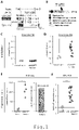
- Fig.1(A) shows a schematic representation of mouse ADAMTSL5 transcripts and CGIs from the UCSC genome browser, and Refseq gene annotations based on the NCBI37/mm9 mouse reference.
- the scheme allows visualization of exons, introns and CGIs of AdamtsL5 , highlighting with a square the gene body CGI found hypermethylated in tumors.
- Fig.1(B) is a schematic representation of the human ADAMTSL5 protein from Badel et al. Matric Biology, 2012 .
- Fig.1(E) illustrates the ADAMTSL5 mRNA expression levels in Alb-R26 Met tumors compared to control livers by RNA-seq.
- Example 2 Overexpression of ADAMTSL5 mRNA and protein in a Alb-R26 Met tumor model.
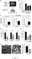
- Fig.2(A) shows representative images of the ADAMTSL5 mRNA expression levels (by RNA-scope) in tumors (dissected from Alb-R26 Met mice) and in control liver (dissected from control mice). Strong staining is observed in Alb-R26 Met tumors, but not in control livers. It is concluded that, similar to data from RNA-seq and RT-qPCR analyses, tumors are characterized by a consistent overexpression of ADAMTSL5 mRNA.
- Fig.2(B) shows representative images of the ADAMTSL5 protein levels by immunofluorescence (left) and immunostaining (right) in tumors (dissected from Alb-R26 Met mice) and in control liver (dissected from control mice). It is noted that ADAMTSL5 is overexpressed in Alb-R26 Met tumors compared to control livers. It is concluded that, tumors are characterized by a consistent overexpression of ADAMTSL5 protein, coherent with high mRNA levels.
- Example 3 In vitro tumorigenic properties of Alb-R26 Met HCC cells, in which ADAMTSL5 is overexpressed. *: P ⁇ 0.05; **: P ⁇ 0.01; ***: P ⁇ 0.001.
- Fig.3(A) is a schematic representation of the establishment of Alb-R26 Met HCC cell lines ( Fan et al. Hepatology, 2017 ) used for molecular and functional studies. The scheme recapitulates how HCC cell lines have been generated for their molecular characterization and for their use in biological assays. Following the protocol, it is established and reported in Fan et al. Hepatology, 2017 , that HCC cell lines were generated from liver tumors dissected from different Alb-R26 Met mice. Briefly, dissected tumors were minced, incubated in a solution containing collagenase and DNAse, and tissue debris were removed using a 100pm sterile filter.
- Fig.3(B) is a graph reporting the ADAMTSL5 expression levels (by RT-qPCR) in three Alb-R26 Met HCC cell lines (HCC3, HCC13, and HCC14) relative to control livers. It is concluded that HCC cells are characterized by a consistent overexpression of ADAMTSL5 mRNA, as shown in tumors.
- Fig.3(C) shows the mRNA expression levels of ADAMTSL5 in Alb-R26 Met HCC cells stably transfected with a plasmid carrying a shRNA targeting sequence (carrying also the puromycin gene for selection of stable clones) versus controls.
- Cells are transfected with the plasmid of interest, then exposed after 48 hours to puromycin for 7 days in order to select cells in which the plasmid has been stably integrated.
- Clones in which the expression levels of ADAMTSL5 are downregulated by the shRNA targeting sequence are identified by RT-qPCR. It is concluded that the shRNA targeting sequence against ADAMTSL5 leads to an efficient downregulation of ADAMTSL5 mRNA levels in HCC cells, which interfere with ADAMTSL5 protein expression levels.
- Fig.3(D) illustrates results of anchorage independent growth assay (soft agar assay) performed using either HCC control cells or HCC cells carrying the shRNA sequence targeting ADAMTSL5. This assay exemplifies the capacity of cells to form colonies in non-adherent condition, therefore revealing their in vitro tumorigenic properties.
- Results show that the number of colonies formed by the Alb-R26 Met HCC cells carrying a shRNA sequence targeting ADAMTSL5 is reduced compared with control cells. It is concluded that high levels of ADAMTSL5 in HCC cells is required for their in vitro tumorigenic properties.
- Fig.3(E) is descriptive of the anchorage independent growth assay (soft agar assay) showing partially rescue of in vitro tumorigenic properties of ADAMTSL5-targeted Alb- R26 Met HCC cells with condition media from control cells.
- the results indicate that extracellular ADAMTSL5 confers tumorigenicity to cells expressing low levels of ADAMTSL5. It is concluded that ADAMTSL5, which is a secreted protein, elicits its function in the extracellular environment and can act as well in a cell non-autonomous manner.
- Fig.3(F) are representative images (left) and quantification (right) of tumor spheres derived from control and ADAMTSL5-targted Alb-R26 Met HCC cells (right). It is noted higher numbers and size of tumor sphere generated from control cells compared with Alb-R26 Met HCC shAdamtsl5 cells (carrying the shRNA sequence targeting ADAMTSL5). It is conclude that high levels of ADAMTSL5 in HCC cells confers self-renewal capabilities.
- Example 4 Loss of in vivo tumorigenic properties of HCC cells following ADAMTSL5 downregulation in Alb-R26 Met HCC cells. *: P ⁇ 0.05; **: P ⁇ 0.01; ***: P ⁇ 0.001.
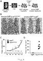
- Fig.4(A) contains images of dissected tumors from xenografts in nude mice injected either with Alb-R26 Met HCC cells (top) or with Alb-R26 Me -shADAMTSL5 HCC cells (bottom).
- Fig.4(A) comprises images of dissected tumors after 8 weeks of xenografts establishment. It is noted that tumors are formed in nude mice wherein the Alb-R26 Met HCC cells were subcutaneous injected, whereas tumors from mice wherein Alb-R26 Met -shADAMTSL5 HCC cells were subcutaneous injected are drastically reduced, illustrating impaired cell tumorigenic properties in vivo.
- Fig.4(B) contains xenograft growth curves reporting the mean tumor volume per group measured every week. It is concluded that downregulation of ADAMTSL5 levels in HCC cells (achieved by stable transfection of a shRNA targeting sequence) interferes with in vivo tumorigenic properties of HCC cells. Collectively, these results show that downregulation of ADAMTSL5 expression levels interfere with tumor establishment/evolution.
- Fig.4(C) it is shown a quantitative analysis of the volume of tumors dissected 8 weeks after cell injection. Each dot corresponds to the volume of each tumor. It is concluded that downregulation of AdamtsL5 levels in HCC cells (achieved by stable transfection of a shRNA targeting sequence) interferes with in vivo tumorigenic properties.
- Example 5 Acquisition of in vivo tumorigenic properties following ADAMTSL5 overexpression by immorto- R26 Met sensitized hepatocytes.
- Fig.5(A) is a schematic representation of the establishment of immorto- R26 Met sensitized hepatocytes (embryonic hepatocytes carrying increased levels of the Met RTK and immortalized with the SV40 large-T antigen). Briefly, cultured hepatocytes from E15.5 mouse R26 Met embryonic livers were infected with a retrovirus carrying the SV40 large-T antigen for immortalization (plus the neomycin gene for selection of stable clones), then subsequently treated for 7 days with a media permissive for hepatocytes to deplete other cell types. The immorto- R26 Met hepatocytes are sensitized because of 3-folds increased of wild-type Met levels, although not tumorigenic as incompetent to form tumors in xenografts.
- Fig.5(B) comprises images of mice after 11 weeks of xenografts establishment. It is noted that nude mice wherein subcutaneous injection of immorto- R26 Met hepa overAdamtsl5 (cells overexpressing ADAMTSL5) develop tumors in both flanks.
- Xenograft studies were performed by subcutaneous injection of immorto- R26 Met control (immorto- R26 Met hepa WT ) or overexpressing ADAMTSL5 (immorto- R26 Met hepa overAdamtsl5 ) hepatocytes (5 ⁇ 10 6 cells) in both flanks of nude mice.
- Fig.5(C) contains xenograft growth curves reporting the mean tumor volume per group measured every week.
- Fig.5(D) it is shown a quantitative analysis of the volume of tumors dissected 11 weeks after cell injection. Each dot corresponds to the volume of each tumor. It is noted that all mice injected with immorto- R26 Met hepa overAdamtsl5 formed tumors in contrast to mice injected with immorto- R26 Met hepa WT . It is concluded that overexpression of ADAMTSL5 levels in immorto-hepatocytes, which are not tumorigenic as incompetent to form tumors in xenografts, is sufficient to confer to them in vivo cell tumorigenic properties.
- Example 6 ADAMTSL5 expression level in Alb-R26 Met tumors at early and latest stages.
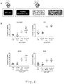
- Fig.6(A) is a schematic representation of the tissue samples used for RT-qPCR analysis of ADAMTSL5 levels.
- the samples used are: control wild-type livers (WT), Alb-R26 Met healthy livers, Alb-R26 Met tumors at early and advanced stages.
- Fig.6(B) the graphs report the expression levels (by RT-qPCR) of ADAMTSL5, AFP (alpha-fetoprotein), GPC3 (Glypican-3) (two HCC markers), and Ki67 (a proliferative marker). It is noted comparable low expression levels of ADAMTSL5 as well as for the other analyzed markers in wild-type and Alb-R26 Met healthy livers. In contrast, transcript levels are already increased in Alb-R26 Met tumors at early stages. It is concluded that ADAMTSL5 transcript levels can be used to discriminate healthy from neoplastic samples already at early tumorigenic state.
- Example 7 High ADAMTSL5 mRNA levels are present in 52% of HCC patients, with a predominance in those associated to alcohol taken.
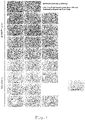
- Fig.7(A) the cohort of HCC patients (371 patients) with ADAMTSL5 mRNA levels is reported on the top: 21% are characterized by ADAMTSL5 downregulation (white, left), 27% by no changes (white with black lines, center), and 52% of HCC patients with upregulation of ADAMTSL5 mRNA levels (black, right).
- Data correspond to RNA-seq studies of a cohort of 371 HCC patients (from TCGA). Note that ADAMTSL5 is overexpressed in 193 out of 371 HCC patients (52%; Log 2 FC>1; FDR ⁇ 0.05). It is concluded that high ADAMTSL5 transcript levels can be detected in more than 50% of HCC patients.
- Fig.7(B) the table reports the presence (black line) or the absence of major HCC risk factors in all 371 analyzed patients. Note that HCC patients with increased ADAMTSL5 levels are significantly associated, although not exclusively, to alcohol taken. It is concluded that high mRNA levels of ADAMTSL5 is present in a large proportion of patients characterized by several risk factors, with a predominance for those associated with alcohol taken.
- Example 8 ADAMTSL5 protein levels are overexpressed in a vast majority of HCC analyzed patients.
- the figure reports immunostaining with anti-ADAMTSL5 antibodies in tumor samples compared to the adjacent liver. Note strong ADAMTSL5 protein levels (dark staining; by Fast Red) in all analyzed HCC patients (except #8 patient) . It is concluded that high ADAMTSL5 protein levels characterize a vast majority of HCC patients.
- Example 9 ADAMTSL5 overexpression in a panel of human cancer cell lines.
- the graph shows the mRNA expression levels of ADAMTSL5 in a panel of human HCC cell lines, in MKN (gastric cancer) cell line, in human breast cell lines, in HELA (human cervix) cancer cell line, and in HEK (human embryonic kidney) cell line. Mean value of three independent experiments. It is concluded that high mRNA levels of ADAMTSL5 are present in cancer cells with different origin.
Landscapes
- Health & Medical Sciences (AREA)
- Life Sciences & Earth Sciences (AREA)
- Immunology (AREA)
- Engineering & Computer Science (AREA)
- Urology & Nephrology (AREA)
- Molecular Biology (AREA)
- Biomedical Technology (AREA)
- Chemical & Material Sciences (AREA)
- Hematology (AREA)
- Biotechnology (AREA)
- Microbiology (AREA)
- Cell Biology (AREA)
- Food Science & Technology (AREA)
- Medicinal Chemistry (AREA)
- Physics & Mathematics (AREA)
- Analytical Chemistry (AREA)
- Biochemistry (AREA)
- General Health & Medical Sciences (AREA)
- General Physics & Mathematics (AREA)
- Pathology (AREA)
- Measuring Or Testing Involving Enzymes Or Micro-Organisms (AREA)
- Gastroenterology & Hepatology (AREA)
Description
- The invention relates to a method of diagnosis, a pharmaceutical composition, a use of a biomarker, and a method of monitoring treatment as defined in the appended claims.
- Cancer is the second leading cause of death in world. Nearly one in six deaths worldwide is due to cancer. For example in 2015, 8.8 million people died of cancer. Therefore, it is important to develop new methods for improving the diagnostic of cancer in patients in need thereof, and notably for predicting their susceptibility to the treatments so as to increase their therapeutic responses, which will results in increased survival expectancies. Some biomarkers for Hepatocellular carcinoma are reviewed by Lou et al. in Biomarker in Cancer, pp. 1-9, DOI: 10.1177/1179299X16684640.
- Epigenetics is the study of changes in gene activity, which does not involve the modifications of the DNA sequence and which can be transmitted in cell divisions. Unlike mutations that affect the DNA sequence, epigenetic modifications are reversible. It is of common knowledge that epigenetic abnormalities lead to the development and progression of human diseases, especially cancers. Epigenetic processes intervene in the regulation of many events such as cell division, differentiation, survival, and mobility. The alteration of these mechanisms promotes the transformation of healthy cells into cancer cells, and so any epigenetic aberration may be involved in tumorigenicity. Among consequences linked to epigenetic abnormalities in cells, there is silencing of tumor suppressor genes or overexpression of oncogenes such as proto-oncogenes, which involve aberrant DNA methylation or mutations of genes encoding enzymes responsible for chromatin modifications. DNA methylation is an essential epigenetic mechanism influencing gene expression levels in cells. These alterations may lead to dramatic biological changes as well as acquisition of malignant properties. The cancer landscape is generally characterized by a diffuse DNA hypomethylation and by focal hypermethylation in CpG-rich regions known as CpG islands (CGI). Generally, CGI hypermethylation at promoters represses transcription of genes acting as tumor suppressors. Nevertheless, a large fraction of DNA methylation is also observed in gene body CGIs, with a positive correlation between methylation and upregulation of gene expression. This implies that DNA hypermethylation in gene body CGIs is linked to the upregulations of genes that would act as positive regulators of tumorigenicity, which is coherent with the reported function of some of them in cancer cells. However, the implication of several others hypermethylated and upregulated genes in cell tumorigenicity still remain unknown.
- These findings have busted the development of drugs targeting epigenetic regulators, which are called epidrugs or epi-medicines. Two main families of compounds have been notably developed until now: (i) compounds that inhibit the DNA methylation, and (ii) compounds that target the histones modifications. However, at present these types of compounds lack of specific action.
- Based on the above, there remains a need for identifying genes that can be used as new specific biomarkers and/or targets to modulate with bioactive agents for cancer treatment. In particular, there remains a need to develop new methods for diagnosis, prognosis, and treatment of cancer.
- In accordance with a first aspect, the invention relates to a method for diagnosing an hepatocellular carcinoma in a mammal in a need thereof. The method comprises the following steps:
- providing a biological sample collected from said mammal;
- determining, from said biological sample, if the ADAMTSL5 gene is overexpressed;
- diagnosing an hepatocellular carcinoma from the determination of the overexpression of said gene.
- According to a second aspect, the invention relates to a pharmaceutical composition. The pharmaceutical composition comprises an agent targeting the ADAMTSL5 gene itself and/or the pathway in which ADAMTSL5 acts, and a pharmaceutically acceptable carrier for use in the treatment of a cancer.
- According to a third aspect, the invention concerns an in vitro use of an ADAMTSL5 protein as a biomarker of an hepatocellular carcinoma.
- In a fourth aspect, the invention relates to an in vitro method for monitoring the response to an anticancer treatment of a mammal suffering from hepatocellular carcinoma comprising determining the ADAMTSL5 level of expression in a biological sample of said mammal at two or more time points during said anticancer treatment, wherein an equal or higher ADAMTSL5 level of expression in a biological sample of the subject at a later time point, compared to a reference value obtained in a biological sample of the subject at an earlier time point, is indicative of a resistance of the subject to said anticancer treatment whereas a lower ADAMTSL5 level is indicative of a response of the subject to said anticancer treatment.
- Advantageously, the method for diagnosing an hepatocellular carcinoma in a mammal in a need thereof according to the invention is characterized in that: - the biological sample is selected from the group consisting of blood, biopsy tissue, blood serum, blood plasma, urine, stool, sputum, cerebrospinal fluid, or supernatant from cell lysate, preferably the biological sample is tissue biopsy, blood, blood plasma, blood serum, or urine; - the overexpression of ADAMTSL5 gene in the biological sample is determined by measuring the ADAMTSL5 protein levels or mRNA levels in said biological sample; - the ADAMTSL5 protein levels in the biological sample are measured by adding at least one antibody anti-ADAMTSL5 type to said biological sample; - the antibody anti-ADAMTSL5 type is selected from the group consisting of uncoupled or coupled with alkaline phosphatase horse-radish peroxidase, or with fluorescent dyes; - the ADAMTSL5 protein levels in the biological sample are measured by using immunostaining, immunofluorescence, western blot, or ELISA; and - the ADAMTSL5 mRNA levels in the biological sample are measured by available methodologies to detect mRNA to said biological sample, including microarray, RNA-seq, in situ hybridization, RNA-scope, as well as regular, semi-quantitative, or quantitative RT-PCRs.
- The pharmaceutical composition according to the invention is characterized in that: - the agents targeting ADAMTSL5 are selected from the group consisting of blocking antibodies, sh-RNA, si-RNA, and antisense RNA against ADAMTSL5.
- Other features and aspects of the present invention will be apparent from the following description and the accompanying drawings, in which:
-
Fig.1 illustrates hypermethylation in the gene body CGI of ADAMTSL5 and overexpression of ADAMTSL5 in a clinically relevant cancer mouse model (Alb-R26Met mice); -
Fig.2 illustrates overexpression of ADAMTSL5 mRNA and protein in a clinically relevant cancer mouse model (Alb-R26Met mice) ; -
Fig.3 illustrates the in vitro tumorigenic properties of hepatocellular carcinoma (HCC) cells established from a clinically relevant cancer mouse model (Alb-R26Met mice) with high expression levels of ADAMTSL5 compared to cells with reduced ADAMTSL5 expression levels; -
Fig.4 illustrates the in vivo loss of tumorigenic properties of HCC cells following ADAMTSL5 downregulation; -
Fig.5 illustrates the in vivo acquisition of tumorigenic properties of immortalized hepatocytes from a clinically relevant mouse model (R26Met mice) following ADAMTSL5 overexpression; -
Fig.6 illustrates ADAMTSL5 expression levels in a clinically relevant cancer mouse model (Alb-R26Met mice) at early and latest stages of tumorigenesis; -
Fig. 7 illustrates high ADAMTSL5 mRNA levels in 52% of HCC patients, with a predominance in those associated to alcohol taken; -
Fig.8 illustrates overexpression of ADAMTSL5 protein levels in 9/10 HCC patients compared to the adjacent liver; and -
Fig. 9 illustrates ADAMTSL5 mRNA levels in human cancer cell lines. - The invention relates to a method for diagnosing and prognosis an hepatocellular carcinoma in a mammal in a need thereof. First of all, the method comprises a step of providing an obtained biological sample from said mammal, followed by a step of determining, from said biological sample, if the ADAMTSL5 gene/protein is overexpressed, and then, according to a third step, diagnosing/prognosis a cancer from the determination of the overexpression of said gene or protein.
- According to the invention, the mammal is in particular a human. However, all mammals are concerned including even cat, dog, hors or rodents such as mice and rats. According to the invention, the biological sample is selected from the group consisting of blood, tissue biopsy, blood serum, blood plasma, urine, stool, sputum, cerebrospinal fluid, and supernatant from cell lysate. The biological sample that is in particular used is tissue biopsy, blood, blood plasma, blood serum, or urine. According to a preferred embodiment of the invention, the overexpression of ADAMTSL5 gene in the biological sample obtained from the mammals in need thereof is determined by measuring the ADAMTSL5 protein levels or RNA levels in said biological sample. The term "up-regulated", "upregulation", "overexpressed", or "overexpression" is used to mean that the expression, activity, or level of a gene, or RNA transcripts or protein products of the gene, is greater than relative to one or more controls, such as, for example, one or more positive and/or negative controls. In particular, increased levels are considered when levels are higher than those in control healthy tissues.
- Practically, mammalian genomes contain 19 ADAMTS (A Disintegrin And Metalloproteinase with ThromboSpondin) genes numbered 1 to 20. Like their relatives, the matrix metalloproteinases (MMPs) and the ADAMs, the ADAMTSs belong to the metzincin protease superfamily, named for the conserved methionine residue close to the zinc iondependent metalloproteinase active site. Representatives of the ADAMTS family are found in all metazoans, although, to date, they have not been identified in single-cell organisms or in plants. All ADAMTSs are secreted, extracellular enzymes that have a compound domain organization, comprising, from the amino-terminus: a signal peptide followed by a pro-region of variable length; a metalloproteinase domain; a disintegrin-like domain; a
central thrombospondin type 1 sequence repeat (TSR) motif; and a cysteine-rich domain followed by a spacer region. Separate from the ADAMTSs, another family of seven ADAMTSlike genes (ADAMTSL) encode proteins that resemble the ancillary domains of ADAMTS, although lack their catalytic domains. These ADAMTSL proteins, which include ADAMTSL 1 to 6 and papilin, may function to modulate the activities of the ADAMTSs. ADAMTSL5 is a protein that has been discovered in the late 2000s and is described to bind to fibrillin-1 and to promote fibril formation. The role of ADAMTSL5 in microfibril formation is of considerable interest as a crucial mechanism for growth factor regulation in extracellular matrix. ADAMTSL5 is more particularly a secreted protein with a unique domain composition, comprising an N-terminal thrombospondin type 1 repeat, a cystein-rich module, a spacer module, and a C-terminal netrin-like module, which is connected to the spacer by a proline-rich segment. ADAMTSL5 is known as already involved in some disease such as psoriasis but, to date, ADAMTSL5 has not been linked to cancer, as potential biomarker or as a target for molecular therapies. - In the context of the present invention and in a preferred embodiment, the ADAMTSL5 protein level is measured by adding at least one antibody to said biological sample. The antibody, which is in particular used for measuring the ADAMTSL5 protein levels, is of an anti-ADAMTSL5 type. The antibody anti-ADAMTSL5 is in particular selected from the group consisting of uncoupled or coupled or conjugated with alkaline phosphatase, horseradish peroxidase (HRP), or with fluorescent dyes.
- According to the invention, the ADAMTSL5 protein levels present in the biological sample of the mammals in need thereof is in particular measured by using immunostaining, immunofluorescence, western blot, or ELISA.
- In biochemistry, immunostaining is known as use of an antibody-based method to detect a specific protein in a biological sample. Immunostaining encompasses a broad range of techniques used in histology, cell biology, and molecular biology, which use antibody-based staining methods. Immunofluorescence is a technique used for light microscopy with a fluorescence microscope and is used primarily on microbiological samples. This technique uses the specificity of antibodies to their antigen to target fluorescent dyes to specific biomolecule targets within a cell, and therefore allows visualization of the distribution of the target molecule through the sample.
- A kit for determining an overexpression of the ADAMTSL5 gene in a biological sample obtained from a mammal is also described. The kit comprises at least one antibody anti-ADAMTSL5 type and a container for holding the biological sample. The antibodies anti-ADAMTSL5 type used in the kit are the same as those above-described.
- According to the invention, ADAMTSL5 levels present in the biological sample of the mammals in need thereof can be also determined by measuring ADAMTSL5 mRNA levels using, for example, microarray, RNA-seq, in situ hybridization, RNA-scope, as well as regular, semi-quantitative, or quantitative RT-PCRs.
- According to a second aspect, the invention relates to a pharmaceutical composition. The pharmaceutical composition comprises an agent as defined in
independent claim 8. In an unclaimed aspect, a demethylating agent is also described, such as for example Decitabine, or agents modulating glycosylation and/or heparin binding. A demethylating agent, according to this description, is a compound that leads to genomic DNA hypomethylation by inhibiting the DNA methyltransferase. - In a third aspect, the invention concerns an in vitro use of an ADAMTSL5 protein as a biomarker of an hepatocellular carcinoma.
- In a fourth aspect, the invention relates to an in vitro method for monitoring the response to an anticancer treatment of a mammal suffering from cancer comprising determining the ADAMTSL5 level of expression in a biological sample of said mammal at two or more time points during said anticancer treatment, wherein an equal or higher ADAMTSL5 level of expression in a biological sample of the subject at a later time point, compared to a reference value obtained in a biological sample of the subject at an earlier time point, is indicative of a resistance of the subject to said anticancer treatment whereas a lower ADAMTSL5 level is indicative of a response of the subject to said anticancer treatment.
- In the following examples, genetically modified mice where used. In these mice, the expression levels of Met, a receptor tyrosine kinase (RTK) activated in about 50% of human HCCs, is slightly enhanced above the endogenous level, based on a genetic approach allowing modulation of gene expression in a tissue/temporal-specific manner (R26stopMet mice; Fan et al. PLoS Genetics 2015; Fan et al. Hepatology 2017). It was recently demonstrated that enhanced methylation (Met) RTK expression levels in the liver perturbs tissue homeostasis, leading to tumor initiation and evolution into HCC (Alb-R26Met). Based on comparisons of Met expression levels in Alb-R26Met (n=32) and human (n=249) liver tumors, it was shown that Met levels in the Alb-R26Met genetic setting (3.16±0.06 versus control livers) correspond to those found in about 20% of HCC patients (48/249). By analyzing 96 different genes in a panel of tumor samples (n=32), it was shown that liver tumorigenesis modelled by the Alb-R26Met mice corresponds to a subset of HCC patients, thus establishing the clinical relevance of the Alb-R26Met HCC mouse model. It was used the Alb-R26Met mouse model to explore the impact of DNA methylation on transcriptional switches associated with tumorigenesis. It was identified a striking enrichment in genes simultaneously hypermethylated in CpG islands (CGIs) and overexpressed. Among them, it is found ADAMTSL5.
- Example 1: Hypermethylation in the gene body CGI of ADAMTSL5 and overexpression of mRNA ADAMTSL5 in the Alb-R26Met tumor model compared with control livers.
-
Fig.1(A) shows a schematic representation of mouse ADAMTSL5 transcripts and CGIs from the UCSC genome browser, and Refseq gene annotations based on the NCBI37/mm9 mouse reference. The scheme allows visualization of exons, introns and CGIs of AdamtsL5 , highlighting with a square the gene body CGI found hypermethylated in tumors. -
Fig.1(B) is a schematic representation of the human ADAMTSL5 protein from Badel et al. Matric Biology, 2012. The different domains present in the ADAMTSL5 protein, which could be involved in the modulation of ADAMTSL5 interactions with other protein and in ADAMTSL5 biological functions, are reported. -
Fig.1(C) and (D) illustrate the methylation levels (β-value) of ADAMTSL5 in the promoter CGI (Fig.1(C) ) and in the gene body CGI (Fig.1(D) ) in control livers (n=3) and Alb-R26Met tumors (n=10). It is noted that the methylation levels of ADAMTSL5 in the promoter CGI is similar in control and tumor samples, whereas the methylation levels in gene body CGI are significantly higher in Alb-R26Met tumors compared to control livers. It is concluded that tumors are characterized by a hypermethylation of ADAMTSL5 in the gene body CGI. -
Fig.1(E) illustrates the ADAMTSL5 mRNA expression levels in Alb-R26Met tumors compared to control livers by RNA-seq. The graphs show the read counts (on the right) and Log2FC (fold-change)(on the left). Significance: *: P<0.05; FDR (p-value adjusted) = 4.31E-15. It is concluded that tumors are characterized by an overexpression of ADAMTSL5 mRNA. -
Fig.1(F) illustrates the ADAMTSL5 expression levels (by RT-qPCR) in Alb-R26Met tumors (n=16) relative to control livers (n=5). It is noted high expression levels of ADAMTSL5 in Alb-R26Met tumors compared to control livers. It is concluded that tumors are characterized by a consistent overexpression of ADAMTSL5 mRNA. - Example 2: Overexpression of ADAMTSL5 mRNA and protein in a Alb-R26Met tumor model.
-
Fig.2(A) shows representative images of the ADAMTSL5 mRNA expression levels (by RNA-scope) in tumors (dissected from Alb-R26Met mice) and in control liver (dissected from control mice). Strong staining is observed in Alb-R26Met tumors, but not in control livers. It is concluded that, similar to data from RNA-seq and RT-qPCR analyses, tumors are characterized by a consistent overexpression of ADAMTSL5 mRNA. -
Fig.2(B) shows representative images of the ADAMTSL5 protein levels by immunofluorescence (left) and immunostaining (right) in tumors (dissected from Alb-R26Met mice) and in control liver (dissected from control mice). It is noted that ADAMTSL5 is overexpressed in Alb-R26Met tumors compared to control livers. It is concluded that, tumors are characterized by a consistent overexpression of ADAMTSL5 protein, coherent with high mRNA levels. - Example 3: In vitro tumorigenic properties of Alb-R26Met HCC cells, in which ADAMTSL5 is overexpressed. *: P<0.05; **: P<0.01; ***: P<0.001.
-
Fig.3(A) is a schematic representation of the establishment of Alb-R26Met HCC cell lines (Fan et al. Hepatology, 2017) used for molecular and functional studies. The scheme recapitulates how HCC cell lines have been generated for their molecular characterization and for their use in biological assays. Following the protocol, it is established and reported in Fan et al. Hepatology, 2017, that HCC cell lines were generated from liver tumors dissected from different Alb-R26Met mice. Briefly, dissected tumors were minced, incubated in a solution containing collagenase and DNAse, and tissue debris were removed using a 100pm sterile filter. Cells were then cultured in William E medium with supplements either directly in adherent conditions or for a week in non-adherent conditions to enrich cells with tumorigenic properties. After expansion of cultured cells, a proportion of them was frozen and kept as stocks, whereas another proportion was characterized molecularly (biochemistry, RT-qPCR) and functionally (for their capacity to form tumors in nude mice). -
Fig.3(B) is a graph reporting the ADAMTSL5 expression levels (by RT-qPCR) in three Alb-R26Met HCC cell lines (HCC3, HCC13, and HCC14) relative to control livers. It is concluded that HCC cells are characterized by a consistent overexpression of ADAMTSL5 mRNA, as shown in tumors. -
Fig.3(C) shows the mRNA expression levels of ADAMTSL5 in Alb-R26Met HCC cells stably transfected with a plasmid carrying a shRNA targeting sequence (carrying also the puromycin gene for selection of stable clones) versus controls. Cells are transfected with the plasmid of interest, then exposed after 48 hours to puromycin for 7 days in order to select cells in which the plasmid has been stably integrated. Clones in which the expression levels of ADAMTSL5 are downregulated by the shRNA targeting sequence are identified by RT-qPCR. It is concluded that the shRNA targeting sequence against ADAMTSL5 leads to an efficient downregulation of ADAMTSL5 mRNA levels in HCC cells, which interfere with ADAMTSL5 protein expression levels. -
Fig.3(D) illustrates results of anchorage independent growth assay (soft agar assay) performed using either HCC control cells or HCC cells carrying the shRNA sequence targeting ADAMTSL5. This assay exemplifies the capacity of cells to form colonies in non-adherent condition, therefore revealing their in vitro tumorigenic properties. - Results show that the number of colonies formed by the Alb-R26Met HCC cells carrying a shRNA sequence targeting ADAMTSL5 is reduced compared with control cells. It is concluded that high levels of ADAMTSL5 in HCC cells is required for their in vitro tumorigenic properties.
-
Fig.3(E) is descriptive of the anchorage independent growth assay (soft agar assay) showing partially rescue of in vitro tumorigenic properties of ADAMTSL5-targeted Alb-R26Met HCC cells with condition media from control cells. The results indicate that extracellular ADAMTSL5 confers tumorigenicity to cells expressing low levels of ADAMTSL5. It is concluded that ADAMTSL5, which is a secreted protein, elicits its function in the extracellular environment and can act as well in a cell non-autonomous manner. -
Fig.3(F) are representative images (left) and quantification (right) of tumor spheres derived from control and ADAMTSL5-targted Alb-R26Met HCC cells (right). It is noted higher numbers and size of tumor sphere generated from control cells compared with Alb-R26Met HCCshAdamtsl5 cells (carrying the shRNA sequence targeting ADAMTSL5). It is conclude that high levels of ADAMTSL5 in HCC cells confers self-renewal capabilities. - Example 4: Loss of in vivo tumorigenic properties of HCC cells following ADAMTSL5 downregulation in Alb-R26Met HCC cells. *: P<0.05; **: P<0.01; ***: P<0.001.
-
Fig.4(A) contains images of dissected tumors from xenografts in nude mice injected either with Alb-R26Met HCC cells (top) or with Alb-R26Me -shADAMTSL5 HCC cells (bottom). - Xenograft studies were performed by subcutaneous injection of Alb-R26Met or Alb-R26Met-shAdamtsL5 HCC cells (5×10⁶ cells) in the flank of nude mice.
Fig.4(A) comprises images of dissected tumors after 8 weeks of xenografts establishment. It is noted that tumors are formed in nude mice wherein the Alb-R26Met HCC cells were subcutaneous injected, whereas tumors from mice wherein Alb-R26Met -shADAMTSL5 HCC cells were subcutaneous injected are drastically reduced, illustrating impaired cell tumorigenic properties in vivo. -
Fig.4(B) contains xenograft growth curves reporting the mean tumor volume per group measured every week. It is concluded that downregulation of ADAMTSL5 levels in HCC cells (achieved by stable transfection of a shRNA targeting sequence) interferes with in vivo tumorigenic properties of HCC cells. Collectively, these results show that downregulation of ADAMTSL5 expression levels interfere with tumor establishment/evolution. - In
Fig.4(C) , it is shown a quantitative analysis of the volume of tumors dissected 8 weeks after cell injection. Each dot corresponds to the volume of each tumor. It is concluded that downregulation of AdamtsL5 levels in HCC cells (achieved by stable transfection of a shRNA targeting sequence) interferes with in vivo tumorigenic properties. - Example 5: Acquisition of in vivo tumorigenic properties following ADAMTSL5 overexpression by immorto-R26Met sensitized hepatocytes.
-
Fig.5(A) is a schematic representation of the establishment of immorto-R26Met sensitized hepatocytes (embryonic hepatocytes carrying increased levels of the Met RTK and immortalized with the SV40 large-T antigen). Briefly, cultured hepatocytes from E15.5 mouse R26Met embryonic livers were infected with a retrovirus carrying the SV40 large-T antigen for immortalization (plus the neomycin gene for selection of stable clones), then subsequently treated for 7 days with a media permissive for hepatocytes to deplete other cell types. The immorto-R26Met hepatocytes are sensitized because of 3-folds increased of wild-type Met levels, although not tumorigenic as incompetent to form tumors in xenografts. -
Fig.5(B) comprises images of mice after 11 weeks of xenografts establishment. It is noted that nude mice wherein subcutaneous injection of immorto-R26Met hepaoverAdamtsl5 (cells overexpressing ADAMTSL5) develop tumors in both flanks. - Xenograft studies were performed by subcutaneous injection of immorto-R26Met control (immorto-R26Met hepaWT) or overexpressing ADAMTSL5 (immorto-R26Met hepaoverAdamtsl5) hepatocytes (5×10⁶ cells) in both flanks of nude mice.
-
Fig.5(C) contains xenograft growth curves reporting the mean tumor volume per group measured every week. - In
Fig.5(D) , it is shown a quantitative analysis of the volume of tumors dissected 11 weeks after cell injection. Each dot corresponds to the volume of each tumor. It is noted that all mice injected with immorto-R26Met hepaoverAdamtsl5 formed tumors in contrast to mice injected with immorto-R26Met hepaWT. It is concluded that overexpression of ADAMTSL5 levels in immorto-hepatocytes, which are not tumorigenic as incompetent to form tumors in xenografts, is sufficient to confer to them in vivo cell tumorigenic properties. - Example 6: ADAMTSL5 expression level in Alb-R26Met tumors at early and latest stages.
-
Fig.6(A) is a schematic representation of the tissue samples used for RT-qPCR analysis of ADAMTSL5 levels. The samples used are: control wild-type livers (WT), Alb-R26Met healthy livers, Alb-R26Met tumors at early and advanced stages. - In
Fig.6(B) , the graphs report the expression levels (by RT-qPCR) of ADAMTSL5, AFP (alpha-fetoprotein), GPC3 (Glypican-3) (two HCC markers), and Ki67 (a proliferative marker). It is noted comparable low expression levels of ADAMTSL5 as well as for the other analyzed markers in wild-type and Alb-R26Met healthy livers. In contrast, transcript levels are already increased in Alb-R26Met tumors at early stages. It is concluded that ADAMTSL5 transcript levels can be used to discriminate healthy from neoplastic samples already at early tumorigenic state. - Example 7: High ADAMTSL5 mRNA levels are present in 52% of HCC patients, with a predominance in those associated to alcohol taken.
- In
Fig.7(A) , the cohort of HCC patients (371 patients) with ADAMTSL5 mRNA levels is reported on the top: 21% are characterized by ADAMTSL5 downregulation (white, left), 27% by no changes (white with black lines, center), and 52% of HCC patients with upregulation of ADAMTSL5 mRNA levels (black, right). Bottom: graph reporting the three subgroup of HCC patients according to low, unchanged, and high ADAMTSL5 expression levels (numbers and percentages are indicated). Data correspond to RNA-seq studies of a cohort of 371 HCC patients (from TCGA). Note that ADAMTSL5 is overexpressed in 193 out of 371 HCC patients (52%; Log2FC>1; FDR<0.05). It is concluded that high ADAMTSL5 transcript levels can be detected in more than 50% of HCC patients. - In
Fig.7(B) , the table reports the presence (black line) or the absence of major HCC risk factors in all 371 analyzed patients. Note that HCC patients with increased ADAMTSL5 levels are significantly associated, although not exclusively, to alcohol taken. It is concluded that high mRNA levels of ADAMTSL5 is present in a large proportion of patients characterized by several risk factors, with a predominance for those associated with alcohol taken. - Example 8: ADAMTSL5 protein levels are overexpressed in a vast majority of HCC analyzed patients.
- The figure reports immunostaining with anti-ADAMTSL5 antibodies in tumor samples compared to the adjacent liver. Note strong ADAMTSL5 protein levels (dark staining; by Fast Red) in all analyzed HCC patients (except #8 patient) . It is concluded that high ADAMTSL5 protein levels characterize a vast majority of HCC patients.
- Example 9: ADAMTSL5 overexpression in a panel of human cancer cell lines.
- The graph shows the mRNA expression levels of ADAMTSL5 in a panel of human HCC cell lines, in MKN (gastric cancer) cell line, in human breast cell lines, in HELA (human cervix) cancer cell line, and in HEK (human embryonic kidney) cell line. Mean value of three independent experiments. It is concluded that high mRNA levels of ADAMTSL5 are present in cancer cells with different origin.
Claims (10)
- A method for diagnosing an hepatocellular carcinoma in a mammal in a need thereof, comprising the following steps:providing a biological sample collected from said mammal;determining, from said biological sample, if the ADAMTSL5 gene is overexpressed;diagnosing an hepatocellular carcinoma from the determination of the overexpression of said gene.
- The method according to claim 1, wherein the biological sample is selected from the group consisting of blood, biopsy tissue, blood serum, blood plasma, urine, stool, sputum, cerebrospinal fluid, or supernatant from cell lysate, preferably the biological sample is tissue biopsy, blood, blood plasma, blood serum, or urine.
- The method according to any one of the preceding claims, wherein the overexpression of ADAMTSL5 gene in the biological sample is determined by measuring the ADAMTSL5 protein levels or mRNA levels in said biological sample.
- The method according to claim 3, wherein the ADAMTSL5 protein levels in the biological sample are measured by adding at least one antibody anti-ADAMTSL5 type to said biological sample.
- The method according to claim 4, wherein the antibody anti-ADAMTSL5 type is selected from the group consisting of uncoupled or coupled with alkaline phosphatase horse-radish peroxidase, or with fluorescent dyes.
- The method according to any one of the claims 3 to 5, wherein the ADAMTSL5 protein levels in the biological sample are measured by using immunostaining, immunofluorescence, western blot, or ELISA.
- The method according to claim 3, wherein the ADAMTSL5 mRNA levels in the biological sample are measured by available methodologies to detect mRNA to said biological sample, including microarray, RNA-seq, in situ hybridization, RNA-scope, as well as regular, semi-quantitative, or quantitative RT-PCRs.
- A pharmaceutical composition comprising an agent targeting ADAMTSL5 and a pharmaceutically acceptable carrier for use in the treatment of an hepatocellular carcinoma, wherein the agents targeting ADAMTSL5 are selected from the group consisting of blocking antibodies, sh-RNA, si-RNA, and antisense RNA against ADAMTSL5.
- An in vitro use of an ADAMTSL5 protein as a biomarker of an hepatocellular carcinoma.
- An in vitro method for monitoring the response to an anticancer treatment of a mammal suffering from hepatocellular carcinoma comprising determining the ADAMTSL5 level of expression in a biological sample of said mammal at two or more time points during said anticancer treatment, wherein an equal or higher ADAMTSL5 level of expression in a biological sample of the subject at a later time point, compared to a reference value obtained in a biological sample of the subject at an earlier time point, is indicative of a resistance of the subject to said anticancer treatment whereas a lower ADAMTSL5 level is indicative of a response of the subject to said anticancer treatment.
Applications Claiming Priority (2)
| Application Number | Priority Date | Filing Date | Title |
|---|---|---|---|
| EP18165345.2A EP3546944A1 (en) | 2018-03-30 | 2018-03-30 | Diagnosis and treatment of a cancer based on the overexpression of the adamtsl5 gene |
| PCT/EP2019/058092 WO2019185919A1 (en) | 2018-03-30 | 2019-03-29 | Method and kit for diagnosing and for treatment of a cancer based on the overexpression of the adamtsl5 gene |
Publications (2)
| Publication Number | Publication Date |
|---|---|
| EP3775907A1 EP3775907A1 (en) | 2021-02-17 |
| EP3775907B1 true EP3775907B1 (en) | 2022-12-14 |
Family
ID=62001949
Family Applications (2)
| Application Number | Title | Priority Date | Filing Date |
|---|---|---|---|
| EP18165345.2A Withdrawn EP3546944A1 (en) | 2018-03-30 | 2018-03-30 | Diagnosis and treatment of a cancer based on the overexpression of the adamtsl5 gene |
| EP19713812.6A Active EP3775907B1 (en) | 2018-03-30 | 2019-03-29 | Diagnosing and treatment of a cancer based on the overexpression of the adamtsl5 gene |
Family Applications Before (1)
| Application Number | Title | Priority Date | Filing Date |
|---|---|---|---|
| EP18165345.2A Withdrawn EP3546944A1 (en) | 2018-03-30 | 2018-03-30 | Diagnosis and treatment of a cancer based on the overexpression of the adamtsl5 gene |
Country Status (7)
| Country | Link |
|---|---|
| US (2) | US20210123916A1 (en) |
| EP (2) | EP3546944A1 (en) |
| JP (1) | JP7353608B2 (en) |
| CN (1) | CN112020649B (en) |
| DK (1) | DK3775907T3 (en) |
| ES (1) | ES2938033T3 (en) |
| WO (1) | WO2019185919A1 (en) |
Family Cites Families (5)
| Publication number | Priority date | Publication date | Assignee | Title |
|---|---|---|---|---|
| EP2010916A2 (en) * | 2006-04-13 | 2009-01-07 | Oncomethylome Sciences SA | Novel tumour suppressor |
| WO2008073919A2 (en) * | 2006-12-08 | 2008-06-19 | Asuragen, Inc. | Mir-20 regulated genes and pathways as targets for therapeutic intervention |
| EP2270510A1 (en) * | 2009-07-02 | 2011-01-05 | EMBL (European Molecular Biology Laboratory) | Diagnostic method for predicting the risk of cancer recurrence based on Histone macroH2A isoforms |
| KR101133243B1 (en) * | 2009-10-29 | 2012-04-06 | 국립암센터 | Use of eIF3m for the Diagnosis and Treatment of Cancer |
| US9689040B2 (en) * | 2011-03-04 | 2017-06-27 | Children's Medical Center Corporation | Selective inhibitors of tumor-initiating cells |
-
2018
- 2018-03-30 EP EP18165345.2A patent/EP3546944A1/en not_active Withdrawn
-
2019
- 2019-03-29 EP EP19713812.6A patent/EP3775907B1/en active Active
- 2019-03-29 CN CN201980024001.0A patent/CN112020649B/en active Active
- 2019-03-29 US US17/042,945 patent/US20210123916A1/en active Pending
- 2019-03-29 WO PCT/EP2019/058092 patent/WO2019185919A1/en not_active Ceased
- 2019-03-29 DK DK19713812.6T patent/DK3775907T3/en active
- 2019-03-29 ES ES19713812T patent/ES2938033T3/en active Active
- 2019-03-29 JP JP2020551947A patent/JP7353608B2/en active Active
-
2025
- 2025-12-09 US US19/413,548 patent/US20260110687A1/en active Pending
Also Published As
| Publication number | Publication date |
|---|---|
| CN112020649B (en) | 2025-03-18 |
| JP7353608B2 (en) | 2023-10-02 |
| WO2019185919A1 (en) | 2019-10-03 |
| ES2938033T3 (en) | 2023-04-04 |
| US20260110687A1 (en) | 2026-04-23 |
| EP3775907A1 (en) | 2021-02-17 |
| US20210123916A1 (en) | 2021-04-29 |
| CN112020649A (en) | 2020-12-01 |
| JP2021519090A (en) | 2021-08-10 |
| EP3546944A1 (en) | 2019-10-02 |
| DK3775907T3 (en) | 2023-02-13 |
Similar Documents
| Publication | Publication Date | Title |
|---|---|---|
| Li et al. | Somatic SF3B1 hotspot mutation in prolactinomas | |
| De Chiara et al. | Tubular cell polyploidy protects from lethal acute kidney injury but promotes consequent chronic kidney disease | |
| Apellaniz‐Ruiz et al. | DICER1‐associated embryonal rhabdomyosarcoma and adenosarcoma of the gynecologic tract: pathology, molecular genetics, and indications for molecular testing | |
| Amodio et al. | Oncogenic role of the E3 ubiquitin ligase NEDD4-1, a PTEN negative regulator, in non-small-cell lung carcinomas | |
| Chakrabarti et al. | Elf5 inhibits the epithelial–mesenchymal transition in mammary gland development and breast cancer metastasis by transcriptionally repressing Snail2 | |
| Rahrmann et al. | Identification of PDE4D as a proliferation promoting factor in prostate cancer using a Sleeping Beauty transposon-based somatic mutagenesis screen | |
| Sjögren et al. | Studies on the molecular pathogenesis of extraskeletal myxoid chondrosarcoma—cytogenetic, molecular genetic, and cDNA microarray analyses | |
| Nashed et al. | Clinical applications of BCR-ABL molecular testing in acute leukemia | |
| Schumacher et al. | Two molecular subgroups of Wilms’ tumors with or without WT1 mutations | |
| Rogg et al. | The WD40-domain containing protein CORO2B is specifically enriched in glomerular podocytes and regulates the ventral actin cytoskeleton | |
| Toubaji et al. | Increased gene copy number of ERG on chromosome 21 but not TMPRSS2–ERG fusion predicts outcome in prostatic adenocarcinomas | |
| US20110177967A1 (en) | Methods and compositions using splicing regulatory proteins involved in tumor suppression | |
| Huertas-Martínez et al. | DNA methylation profiling identifies PTRF/Cavin-1 as a novel tumor suppressor in Ewing sarcoma when co-expressed with caveolin-1 | |
| Wang et al. | Epigenetic silencing of KAZALD1 confers a better prognosis and is associated with malignant transformation/progression in glioma | |
| Chakravarthi et al. | Pseudogene associated recurrent gene fusion in prostate cancer | |
| JP2008514209A (en) | Cancer marker | |
| Love et al. | Androgen regulated genes in human prostate xenografts in mice: relation to BPH and prostate cancer | |
| Miklovicova et al. | Mitochondrial respiratory complex II is altered in renal carcinoma | |
| JP2008517590A (en) | Methods of using slag genes or their replication, transcription or expression products in identifying, diagnosing, preventing or treating the occurrence of cancer expansion and / or metastasis | |
| US20260110687A1 (en) | Method and kit for diagnosing and for treatment of a cancer based on the overexpression of the adamtsl5 gene | |
| US20140011696A1 (en) | Bap1 mutational analysis in determining susceptibility to, prognosis of, and treatment of melanocytic neoplasia | |
| Zhu et al. | circGAPVD1 inhibits the progression of gastric cancer through miR-4424/STK4 axis and encoding GAPVD1-137aa protein | |
| Li et al. | Carbonic anhydrase 10 functions as a tumor suppressor in renal cell carcinoma and its methylation is a risk factor for survival outcome | |
| Yang et al. | Alveolar epithelial type 1 cells serve as a cell of origin for lung adenocarcinoma with distinct molecular and phenotypic presentation | |
| JP2011520456A (en) | Combined method for predicting response to anti-cancer therapy |
Legal Events
| Date | Code | Title | Description |
|---|---|---|---|
| STAA | Information on the status of an ep patent application or granted ep patent |
Free format text: STATUS: UNKNOWN |
|
| STAA | Information on the status of an ep patent application or granted ep patent |
Free format text: STATUS: THE INTERNATIONAL PUBLICATION HAS BEEN MADE |
|
| PUAI | Public reference made under article 153(3) epc to a published international application that has entered the european phase |
Free format text: ORIGINAL CODE: 0009012 |
|
| STAA | Information on the status of an ep patent application or granted ep patent |
Free format text: STATUS: REQUEST FOR EXAMINATION WAS MADE |
|
| 17P | Request for examination filed |
Effective date: 20200803 |
|
| AK | Designated contracting states |
Kind code of ref document: A1 Designated state(s): AL AT BE BG CH CY CZ DE DK EE ES FI FR GB GR HR HU IE IS IT LI LT LU LV MC MK MT NL NO PL PT RO RS SE SI SK SM TR |
|
| AX | Request for extension of the european patent |
Extension state: BA ME |
|
| DAV | Request for validation of the european patent (deleted) | ||
| DAX | Request for extension of the european patent (deleted) | ||
| STAA | Information on the status of an ep patent application or granted ep patent |
Free format text: STATUS: EXAMINATION IS IN PROGRESS |
|
| 17Q | First examination report despatched |
Effective date: 20210712 |
|
| GRAP | Despatch of communication of intention to grant a patent |
Free format text: ORIGINAL CODE: EPIDOSNIGR1 |
|
| STAA | Information on the status of an ep patent application or granted ep patent |
Free format text: STATUS: GRANT OF PATENT IS INTENDED |
|
| INTG | Intention to grant announced |
Effective date: 20220422 |
|
| GRAS | Grant fee paid |
Free format text: ORIGINAL CODE: EPIDOSNIGR3 |
|
| GRAA | (expected) grant |
Free format text: ORIGINAL CODE: 0009210 |
|
| STAA | Information on the status of an ep patent application or granted ep patent |
Free format text: STATUS: THE PATENT HAS BEEN GRANTED |
|
| AK | Designated contracting states |
Kind code of ref document: B1 Designated state(s): AL AT BE BG CH CY CZ DE DK EE ES FI FR GB GR HR HU IE IS IT LI LT LU LV MC MK MT NL NO PL PT RO RS SE SI SK SM TR |
|
| REG | Reference to a national code |
Ref country code: GB Ref legal event code: FG4D |
|
| REG | Reference to a national code |
Ref country code: CH Ref legal event code: EP |
|
| REG | Reference to a national code |
Ref country code: DE Ref legal event code: R096 Ref document number: 602019023147 Country of ref document: DE |
|
| REG | Reference to a national code |
Ref country code: IE Ref legal event code: FG4D |
|
| REG | Reference to a national code |
Ref country code: AT Ref legal event code: REF Ref document number: 1537993 Country of ref document: AT Kind code of ref document: T Effective date: 20230115 |
|
| REG | Reference to a national code |
Ref country code: DK Ref legal event code: T3 Effective date: 20230209 |
|
| REG | Reference to a national code |
Ref country code: NL Ref legal event code: FP |
|
| REG | Reference to a national code |
Ref country code: SE Ref legal event code: TRGR |
|
| REG | Reference to a national code |
Ref country code: ES Ref legal event code: FG2A Ref document number: 2938033 Country of ref document: ES Kind code of ref document: T3 Effective date: 20230404 |
|
| REG | Reference to a national code |
Ref country code: LT Ref legal event code: MG9D |
|
| PG25 | Lapsed in a contracting state [announced via postgrant information from national office to epo] |
Ref country code: NO Free format text: LAPSE BECAUSE OF FAILURE TO SUBMIT A TRANSLATION OF THE DESCRIPTION OR TO PAY THE FEE WITHIN THE PRESCRIBED TIME-LIMIT Effective date: 20230314 Ref country code: LT Free format text: LAPSE BECAUSE OF FAILURE TO SUBMIT A TRANSLATION OF THE DESCRIPTION OR TO PAY THE FEE WITHIN THE PRESCRIBED TIME-LIMIT Effective date: 20221214 Ref country code: FI Free format text: LAPSE BECAUSE OF FAILURE TO SUBMIT A TRANSLATION OF THE DESCRIPTION OR TO PAY THE FEE WITHIN THE PRESCRIBED TIME-LIMIT Effective date: 20221214 |
|
| REG | Reference to a national code |
Ref country code: AT Ref legal event code: MK05 Ref document number: 1537993 Country of ref document: AT Kind code of ref document: T Effective date: 20221214 |
|
| PG25 | Lapsed in a contracting state [announced via postgrant information from national office to epo] |
Ref country code: RS Free format text: LAPSE BECAUSE OF FAILURE TO SUBMIT A TRANSLATION OF THE DESCRIPTION OR TO PAY THE FEE WITHIN THE PRESCRIBED TIME-LIMIT Effective date: 20221214 Ref country code: LV Free format text: LAPSE BECAUSE OF FAILURE TO SUBMIT A TRANSLATION OF THE DESCRIPTION OR TO PAY THE FEE WITHIN THE PRESCRIBED TIME-LIMIT Effective date: 20221214 Ref country code: HR Free format text: LAPSE BECAUSE OF FAILURE TO SUBMIT A TRANSLATION OF THE DESCRIPTION OR TO PAY THE FEE WITHIN THE PRESCRIBED TIME-LIMIT Effective date: 20221214 Ref country code: GR Free format text: LAPSE BECAUSE OF FAILURE TO SUBMIT A TRANSLATION OF THE DESCRIPTION OR TO PAY THE FEE WITHIN THE PRESCRIBED TIME-LIMIT Effective date: 20230315 |
|
| PG25 | Lapsed in a contracting state [announced via postgrant information from national office to epo] |
Ref country code: SM Free format text: LAPSE BECAUSE OF FAILURE TO SUBMIT A TRANSLATION OF THE DESCRIPTION OR TO PAY THE FEE WITHIN THE PRESCRIBED TIME-LIMIT Effective date: 20221214 Ref country code: RO Free format text: LAPSE BECAUSE OF FAILURE TO SUBMIT A TRANSLATION OF THE DESCRIPTION OR TO PAY THE FEE WITHIN THE PRESCRIBED TIME-LIMIT Effective date: 20221214 Ref country code: PT Free format text: LAPSE BECAUSE OF FAILURE TO SUBMIT A TRANSLATION OF THE DESCRIPTION OR TO PAY THE FEE WITHIN THE PRESCRIBED TIME-LIMIT Effective date: 20230414 Ref country code: EE Free format text: LAPSE BECAUSE OF FAILURE TO SUBMIT A TRANSLATION OF THE DESCRIPTION OR TO PAY THE FEE WITHIN THE PRESCRIBED TIME-LIMIT Effective date: 20221214 Ref country code: CZ Free format text: LAPSE BECAUSE OF FAILURE TO SUBMIT A TRANSLATION OF THE DESCRIPTION OR TO PAY THE FEE WITHIN THE PRESCRIBED TIME-LIMIT Effective date: 20221214 Ref country code: AT Free format text: LAPSE BECAUSE OF FAILURE TO SUBMIT A TRANSLATION OF THE DESCRIPTION OR TO PAY THE FEE WITHIN THE PRESCRIBED TIME-LIMIT Effective date: 20221214 |
|
| PG25 | Lapsed in a contracting state [announced via postgrant information from national office to epo] |
Ref country code: SK Free format text: LAPSE BECAUSE OF FAILURE TO SUBMIT A TRANSLATION OF THE DESCRIPTION OR TO PAY THE FEE WITHIN THE PRESCRIBED TIME-LIMIT Effective date: 20221214 Ref country code: PL Free format text: LAPSE BECAUSE OF FAILURE TO SUBMIT A TRANSLATION OF THE DESCRIPTION OR TO PAY THE FEE WITHIN THE PRESCRIBED TIME-LIMIT Effective date: 20221214 Ref country code: IS Free format text: LAPSE BECAUSE OF FAILURE TO SUBMIT A TRANSLATION OF THE DESCRIPTION OR TO PAY THE FEE WITHIN THE PRESCRIBED TIME-LIMIT Effective date: 20230414 Ref country code: AL Free format text: LAPSE BECAUSE OF FAILURE TO SUBMIT A TRANSLATION OF THE DESCRIPTION OR TO PAY THE FEE WITHIN THE PRESCRIBED TIME-LIMIT Effective date: 20221214 |
|
| REG | Reference to a national code |
Ref country code: DE Ref legal event code: R097 Ref document number: 602019023147 Country of ref document: DE |
|
| PLBE | No opposition filed within time limit |
Free format text: ORIGINAL CODE: 0009261 |
|
| STAA | Information on the status of an ep patent application or granted ep patent |
Free format text: STATUS: NO OPPOSITION FILED WITHIN TIME LIMIT |
|
| PG25 | Lapsed in a contracting state [announced via postgrant information from national office to epo] |
Ref country code: MC Free format text: LAPSE BECAUSE OF FAILURE TO SUBMIT A TRANSLATION OF THE DESCRIPTION OR TO PAY THE FEE WITHIN THE PRESCRIBED TIME-LIMIT Effective date: 20221214 |
|
| 26N | No opposition filed |
Effective date: 20230915 |
|
| PG25 | Lapsed in a contracting state [announced via postgrant information from national office to epo] |
Ref country code: SI Free format text: LAPSE BECAUSE OF FAILURE TO SUBMIT A TRANSLATION OF THE DESCRIPTION OR TO PAY THE FEE WITHIN THE PRESCRIBED TIME-LIMIT Effective date: 20221214 |
|
| PG25 | Lapsed in a contracting state [announced via postgrant information from national office to epo] |
Ref country code: BG Free format text: LAPSE BECAUSE OF FAILURE TO SUBMIT A TRANSLATION OF THE DESCRIPTION OR TO PAY THE FEE WITHIN THE PRESCRIBED TIME-LIMIT Effective date: 20221214 |
|
| PG25 | Lapsed in a contracting state [announced via postgrant information from national office to epo] |
Ref country code: BG Free format text: LAPSE BECAUSE OF FAILURE TO SUBMIT A TRANSLATION OF THE DESCRIPTION OR TO PAY THE FEE WITHIN THE PRESCRIBED TIME-LIMIT Effective date: 20221214 |
|
| PGFP | Annual fee paid to national office [announced via postgrant information from national office to epo] |
Ref country code: SE Payment date: 20250328 Year of fee payment: 7 |
|
| PGFP | Annual fee paid to national office [announced via postgrant information from national office to epo] |
Ref country code: DK Payment date: 20250328 Year of fee payment: 7 Ref country code: NL Payment date: 20250328 Year of fee payment: 7 |
|
| PGFP | Annual fee paid to national office [announced via postgrant information from national office to epo] |
Ref country code: LU Payment date: 20250328 Year of fee payment: 7 |
|
| PGFP | Annual fee paid to national office [announced via postgrant information from national office to epo] |
Ref country code: IE Payment date: 20250328 Year of fee payment: 7 |
|
| PGFP | Annual fee paid to national office [announced via postgrant information from national office to epo] |
Ref country code: BE Payment date: 20250328 Year of fee payment: 7 |
|
| PGFP | Annual fee paid to national office [announced via postgrant information from national office to epo] |
Ref country code: DE Payment date: 20250411 Year of fee payment: 7 |
|
| PGFP | Annual fee paid to national office [announced via postgrant information from national office to epo] |
Ref country code: ES Payment date: 20250414 Year of fee payment: 7 |
|
| PGFP | Annual fee paid to national office [announced via postgrant information from national office to epo] |
Ref country code: IT Payment date: 20250328 Year of fee payment: 7 |
|
| PGFP | Annual fee paid to national office [announced via postgrant information from national office to epo] |
Ref country code: CH Payment date: 20250424 Year of fee payment: 7 |
|
| PG25 | Lapsed in a contracting state [announced via postgrant information from national office to epo] |
Ref country code: CY Free format text: LAPSE BECAUSE OF FAILURE TO SUBMIT A TRANSLATION OF THE DESCRIPTION OR TO PAY THE FEE WITHIN THE PRESCRIBED TIME-LIMIT; INVALID AB INITIO Effective date: 20190329 |
|
| PG25 | Lapsed in a contracting state [announced via postgrant information from national office to epo] |
Ref country code: HU Free format text: LAPSE BECAUSE OF FAILURE TO SUBMIT A TRANSLATION OF THE DESCRIPTION OR TO PAY THE FEE WITHIN THE PRESCRIBED TIME-LIMIT; INVALID AB INITIO Effective date: 20190329 |
|
| REG | Reference to a national code |
Ref country code: DE Ref legal event code: R079 Ref document number: 602019023147 Country of ref document: DE Free format text: PREVIOUS MAIN CLASS: G01N0033574000 Ipc: G01N0033575000 |
|
| PG25 | Lapsed in a contracting state [announced via postgrant information from national office to epo] |
Ref country code: TR Free format text: LAPSE BECAUSE OF FAILURE TO SUBMIT A TRANSLATION OF THE DESCRIPTION OR TO PAY THE FEE WITHIN THE PRESCRIBED TIME-LIMIT Effective date: 20221214 |
|
| PGFP | Annual fee paid to national office [announced via postgrant information from national office to epo] |
Ref country code: GB Payment date: 20260330 Year of fee payment: 8 |
|
| PGFP | Annual fee paid to national office [announced via postgrant information from national office to epo] |
Ref country code: FR Payment date: 20260330 Year of fee payment: 8 |