EP3768865B1 - Methods and kits to detect viral particle heterogeneity - Google Patents
Methods and kits to detect viral particle heterogeneity Download PDFInfo
- Publication number
- EP3768865B1 EP3768865B1 EP19716642.4A EP19716642A EP3768865B1 EP 3768865 B1 EP3768865 B1 EP 3768865B1 EP 19716642 A EP19716642 A EP 19716642A EP 3768865 B1 EP3768865 B1 EP 3768865B1
- Authority
- EP
- European Patent Office
- Prior art keywords
- viral
- aav
- dna
- preparation
- capsid
- Prior art date
- Legal status (The legal status is an assumption and is not a legal conclusion. Google has not performed a legal analysis and makes no representation as to the accuracy of the status listed.)
- Active
Links
Images




Classifications
-
- C—CHEMISTRY; METALLURGY
- C12—BIOCHEMISTRY; BEER; SPIRITS; WINE; VINEGAR; MICROBIOLOGY; ENZYMOLOGY; MUTATION OR GENETIC ENGINEERING
- C12Q—MEASURING OR TESTING PROCESSES INVOLVING ENZYMES, NUCLEIC ACIDS OR MICROORGANISMS; COMPOSITIONS OR TEST PAPERS THEREFOR; PROCESSES OF PREPARING SUCH COMPOSITIONS; CONDITION-RESPONSIVE CONTROL IN MICROBIOLOGICAL OR ENZYMOLOGICAL PROCESSES
- C12Q1/00—Measuring or testing processes involving enzymes, nucleic acids or microorganisms; Compositions therefor; Processes of preparing such compositions
- C12Q1/68—Measuring or testing processes involving enzymes, nucleic acids or microorganisms; Compositions therefor; Processes of preparing such compositions involving nucleic acids
-
- C—CHEMISTRY; METALLURGY
- C12—BIOCHEMISTRY; BEER; SPIRITS; WINE; VINEGAR; MICROBIOLOGY; ENZYMOLOGY; MUTATION OR GENETIC ENGINEERING
- C12Q—MEASURING OR TESTING PROCESSES INVOLVING ENZYMES, NUCLEIC ACIDS OR MICROORGANISMS; COMPOSITIONS OR TEST PAPERS THEREFOR; PROCESSES OF PREPARING SUCH COMPOSITIONS; CONDITION-RESPONSIVE CONTROL IN MICROBIOLOGICAL OR ENZYMOLOGICAL PROCESSES
- C12Q1/00—Measuring or testing processes involving enzymes, nucleic acids or microorganisms; Compositions therefor; Processes of preparing such compositions
- C12Q1/68—Measuring or testing processes involving enzymes, nucleic acids or microorganisms; Compositions therefor; Processes of preparing such compositions involving nucleic acids
- C12Q1/6844—Nucleic acid amplification reactions
- C12Q1/686—Polymerase chain reaction [PCR]
-
- C—CHEMISTRY; METALLURGY
- C12—BIOCHEMISTRY; BEER; SPIRITS; WINE; VINEGAR; MICROBIOLOGY; ENZYMOLOGY; MUTATION OR GENETIC ENGINEERING
- C12Q—MEASURING OR TESTING PROCESSES INVOLVING ENZYMES, NUCLEIC ACIDS OR MICROORGANISMS; COMPOSITIONS OR TEST PAPERS THEREFOR; PROCESSES OF PREPARING SUCH COMPOSITIONS; CONDITION-RESPONSIVE CONTROL IN MICROBIOLOGICAL OR ENZYMOLOGICAL PROCESSES
- C12Q1/00—Measuring or testing processes involving enzymes, nucleic acids or microorganisms; Compositions therefor; Processes of preparing such compositions
- C12Q1/70—Measuring or testing processes involving enzymes, nucleic acids or microorganisms; Compositions therefor; Processes of preparing such compositions involving virus or bacteriophage
-
- C—CHEMISTRY; METALLURGY
- C12—BIOCHEMISTRY; BEER; SPIRITS; WINE; VINEGAR; MICROBIOLOGY; ENZYMOLOGY; MUTATION OR GENETIC ENGINEERING
- C12N—MICROORGANISMS OR ENZYMES; COMPOSITIONS THEREOF; PROPAGATING, PRESERVING, OR MAINTAINING MICROORGANISMS; MUTATION OR GENETIC ENGINEERING; CULTURE MEDIA
- C12N15/00—Mutation or genetic engineering; DNA or RNA concerning genetic engineering, vectors, e.g. plasmids, or their isolation, preparation or purification; Use of hosts therefor
- C12N15/09—Recombinant DNA-technology
- C12N15/63—Introduction of foreign genetic material using vectors; Vectors; Use of hosts therefor; Regulation of expression
- C12N15/79—Vectors or expression systems specially adapted for eukaryotic hosts
- C12N15/85—Vectors or expression systems specially adapted for eukaryotic hosts for animal cells
- C12N15/86—Viral vectors
-
- C—CHEMISTRY; METALLURGY
- C12—BIOCHEMISTRY; BEER; SPIRITS; WINE; VINEGAR; MICROBIOLOGY; ENZYMOLOGY; MUTATION OR GENETIC ENGINEERING
- C12N—MICROORGANISMS OR ENZYMES; COMPOSITIONS THEREOF; PROPAGATING, PRESERVING, OR MAINTAINING MICROORGANISMS; MUTATION OR GENETIC ENGINEERING; CULTURE MEDIA
- C12N2750/00—MICROORGANISMS OR ENZYMES; COMPOSITIONS THEREOF; PROPAGATING, PRESERVING, OR MAINTAINING MICROORGANISMS; MUTATION OR GENETIC ENGINEERING; CULTURE MEDIA ssDNA viruses
- C12N2750/00011—Details
- C12N2750/14011—Parvoviridae
- C12N2750/14111—Dependovirus, e.g. adenoassociated viruses
- C12N2750/14141—Use of virus, viral particle or viral elements as a vector
- C12N2750/14143—Use of virus, viral particle or viral elements as a vector viral genome or elements thereof as genetic vector
-
- C—CHEMISTRY; METALLURGY
- C12—BIOCHEMISTRY; BEER; SPIRITS; WINE; VINEGAR; MICROBIOLOGY; ENZYMOLOGY; MUTATION OR GENETIC ENGINEERING
- C12N—MICROORGANISMS OR ENZYMES; COMPOSITIONS THEREOF; PROPAGATING, PRESERVING, OR MAINTAINING MICROORGANISMS; MUTATION OR GENETIC ENGINEERING; CULTURE MEDIA
- C12N2750/00—MICROORGANISMS OR ENZYMES; COMPOSITIONS THEREOF; PROPAGATING, PRESERVING, OR MAINTAINING MICROORGANISMS; MUTATION OR GENETIC ENGINEERING; CULTURE MEDIA ssDNA viruses
- C12N2750/00011—Details
- C12N2750/14011—Parvoviridae
- C12N2750/14111—Dependovirus, e.g. adenoassociated viruses
- C12N2750/14151—Methods of production or purification of viral material
-
- C—CHEMISTRY; METALLURGY
- C12—BIOCHEMISTRY; BEER; SPIRITS; WINE; VINEGAR; MICROBIOLOGY; ENZYMOLOGY; MUTATION OR GENETIC ENGINEERING
- C12Q—MEASURING OR TESTING PROCESSES INVOLVING ENZYMES, NUCLEIC ACIDS OR MICROORGANISMS; COMPOSITIONS OR TEST PAPERS THEREFOR; PROCESSES OF PREPARING SUCH COMPOSITIONS; CONDITION-RESPONSIVE CONTROL IN MICROBIOLOGICAL OR ENZYMOLOGICAL PROCESSES
- C12Q2531/00—Reactions of nucleic acids characterised by
- C12Q2531/10—Reactions of nucleic acids characterised by the purpose being amplify/increase the copy number of target nucleic acid
- C12Q2531/125—Rolling circle
Definitions
- the present disclosure relates to an efficient, accurate and reliable method to determine both protein and nucleic acid amounts in a viral preparation in a single, dual assay.
- Several applications for example, gene therapy, require that the qualitative and/ or quantity attributes of viral preparations be characterized before administration to a subject.
- the potency of a viral preparation consist of two parts: 1) the ability of viral capsid to transduce cells and 2) the ability of DNA in virus to produce the desired product. Inoperable capsids (for e.g., empty) do not effectively transduce the cells. Thus, an accurate tool to measure the dose and ultimately the potency of viral preparations, and to monitor gene therapy vector quality and potency is therefore needed.
- the evaluating step for determining the percentage or ratio of full:empty viral capsids have been by cryogenic transmission electron microscopy (CryoTEM), negative staining TEM, capillary electrophoresis, analytical ultracentrifugation, native agarose gel, alkaline agarose gel, southern blot, dot-blot hybridization, UV spectrophotometry, weak anion exchange chromatography, or mass spectrometry.
- cryogenic transmission electron microscopy CryoTEM
- negative staining TEM capillary electrophoresis
- analytical ultracentrifugation native agarose gel
- alkaline agarose gel alkaline agarose gel
- southern blot southern blot
- dot-blot hybridization UV spectrophotometry
- weak anion exchange chromatography weak anion exchange chromatography
- mass spectrometry weak anion exchange chromatography
- Adeno-Associated Virus AAV which is a commonly used vector of choice in many gene therapies.
- the assay is particularly useful to measure AAV viral full:empty analytics. This novel approach represents a paradigm shift for AAV, and enables the end-user to accelerate their work by obtaining vital information about vector samples with speed, ease, and accuracy.
- TaqMan ® Protein Assays are an adapted form of PLA TM , a proximity ligation assay technology that combines antibody-protein binding with detection of the reporter nucleic acid by real-time PCR. It has been employed for sensitive and specific measurement of protein/ protein expression.
- PLA TM a proximity ligation assay technology that combines antibody-protein binding with detection of the reporter nucleic acid by real-time PCR. It has been employed for sensitive and specific measurement of protein/ protein expression.
- a first step we use an adapted form of the PLA TM technology to obtain a sensitive and specific measurement of viral protein in a capsid and subsequently, by disassembling the viral capsid, in the second step, we release the viral DNA and accurately measure viral DNA by quantitative PCR (qPCR).
- qPCR quantitative PCR
- the disclosure provides a method of quantifying a dose of a viral preparation, comprising:
- the disclosure provides the method as described above, wherein the quantification in a) for viral capsid and in c) for viral DNA is determined from a reference standard curve, or, wherein, the quantification in a) for viral capsid and in c) for viral DNA is determined by digital PCR.
- the disclosure provides the method of any of the claims described above, wherein the viral preparation is selected from the group consisting of parvovirus, adeno-associated virus (AAV), adenovirus, lentivirus, retrovirus, herpesvirus, poxvirus, paramyxovirus, baculovirus, reovirus, alphavirus, flavivirus, and combinations thereof.
- AAV adeno-associated virus
- adenovirus lentivirus
- retrovirus retrovirus
- herpesvirus poxvirus
- paramyxovirus paramyxovirus
- baculovirus baculovirus
- reovirus alphavirus
- flavivirus and combinations thereof.
- the disclosure provides the method of any of the above claims, wherein the steps a) through c) are all performed in a single container.
- the disclosure provides the method of any of the above claims, wherein the heating of step b) is at least at 80°C.
- the disclosure provides the method of any of the above claims, wherein the heating of step b) is up to 100°C.
- the disclosure provides the method of any of the above claims, wherein the method of quantifying viral protein and viral DNA is done within about 4 hours.
- the disclosure provides the method of any of the above claims, wherein the viral preparation comprises one or more of full genome-containing virus particles, partial genome-containing virus particles, self-complementary genome-containing virus particle (double stranded genome), and genome-free virus particles.
- the disclosure provides the method of any of the above claims, further comprising evaluating a percentage or ratio of full versus empty (full:empty) capsids in the viral preparation.
- the disclosure provides the method of any one of the above claims, wherein the percentage of full viral capsids is from about 1% to about 100%.
- the disclosure provides the method of any one of the above claims, wherein the percentage of full viral capsids is about 60% - 100%.
- the disclosure provides a method of measuring the concentration of full AAV capsids in an AAV preparation, comprising:
- the disclosure provides the method of the claim above, wherein the quantification in a) for AAV capsid and in c) for AAV DNA is determined from a reference standard curve, or, wherein, the quantification in a) for AAV capsid and in c) for AAV DNA is determined by digital PCR.
- the disclosure provides the method of above claims, wherein the PLA assay is capable of detecting one or more of thirteen serotypes of the AAV capsid antigen.
- the disclosure provides the method of any of claims above, wherein the steps a) through c) are all performed in a single container.
- the disclosure provides the method of any one of the claims above, further comprising evaluating a percentage or ratio of full versus empty (full:empty) AAV capsids in the AAV preparation.
- the disclosure provides the method of any one of the claims above, wherein the percentage of full AAV capsids is from about 1% to about 100%.
- the disclosure provides the method of any one of the claims above, wherein at least 60- 100% of the AAV capsids are full AAV capsids.
- the disclosure provides the method of any one of claims, wherein the AAV capsid has an antigen that is selected from the group consisting of: AAV1, AAV2, AAV3, AAV4, AAV5, AAV6, AAV7, AAV8, AAV9, AAV10, AAV11, AAV12, AAV13, chimeric AAV, genetically engineered AAV, or chemically modified AAV.
- the AAV capsid has an antigen that is selected from the group consisting of: AAV1, AAV2, AAV3, AAV4, AAV5, AAV6, AAV7, AAV8, AAV9, AAV10, AAV11, AAV12, AAV13, chimeric AAV, genetically engineered AAV, or chemically modified AAV.
- the disclosure provides the method of any one of claims, wherein optionally, the AAV preparation is concentrated and further wherein the preparation is administered at a specific dose to a subject in need thereof.
- the disclosure provides the method of any of the above claims, wherein the heating of step b) is at least at 80°C.
- the disclosure provides the method of any of the above claims, wherein the heating of step b) is up to 100°C.
- the disclosure provides the method of any of the above claims, wherein the method of quantifying viral protein and viral DNA is done within about 4 hours.
- the disclosure provides the method of any of the above claims, wherein the viral preparation comprises one or more of full genome-containing virus particles, partial genome-containing virus particles, self-complementary genome-containing virus particle (double stranded genome), and genome-free virus particles.
- the disclosure provides a kit comprising:
- the disclosure provides the kit described above, wherein the kit measures the concentration of full AAV capsids in an AAV preparation.
- the disclosure provides the kit described above, wherein the AAV capsid has an antigen that is selected from the group consisting of: AAV1, AAV2, AAV3, AAV4, AAV5, AAV6, AAV7, AAV8, AAV9, AAV10, AAV11, AAV12, AAV13, chimeric AAV, genetically engineered AAV, or chemically modified AAV.
- the AAV capsid has an antigen that is selected from the group consisting of: AAV1, AAV2, AAV3, AAV4, AAV5, AAV6, AAV7, AAV8, AAV9, AAV10, AAV11, AAV12, AAV13, chimeric AAV, genetically engineered AAV, or chemically modified AAV.
- the disclosure provides a kit comprising:
- the disclosure provides the kit described above, wherein the kit measures the concentration of full viral capsids in a viral preparation.
- a feature of AAV vector generation in cell culture is the formation of an excess of "empty" capsids, which lack the vector genome.
- the amount of empty capsids is assessed in order to accurately determine the concentration, dose, and/or potency of an AAV preparation. Empty capsids can also be considered as product related impurities, which should be reduced during purification. In certain embodiments, it is important to determine the total amount of empty capsids to reduce an innate and/or adaptive immune response against the capsid antigen.
- the methods described herein can be applied to any viral assay and is not limited to AAV only; for e.g., for the determination of viral vector product quantity (particles/mL) and quality (% full/empty) of viruses including but not limited to: such as parvovirus, adeno-associated virus (AAV), adenovirus, HIV, MMV, lentivirus, retrovirus, herpesvirus, poxvirus, paramyxovirus, baculovirus, reovirus, alphavirus, flavivirus, and combinations thereof.
- viruses including but not limited to: such as parvovirus, adeno-associated virus (AAV), adenovirus, HIV, MMV, lentivirus, retrovirus, herpesvirus, poxvirus, paramyxovirus, baculovirus, reovirus, alphavirus, flavivirus, and combinations thereof.
- the assay may detect one, several or all the viral serotypes of a virus depending on the detection antibody's recognition capacity of the various serotypes.
- the 'detection antibody' may be engineered to recognize one or more serotypes.
- the assay utilizes the AAVX antibody which is a camelid antibody that is found to bind multiple serotypes of AAV, namely one or more of the 13 known AAV serotypes (hence, it is sometimes also called the universal AAV antibody).
- additional anti-AAV antibodies can be included in the assay to detect additional AAV serotypes.
- only one AAV antibody (like AAVX) directed to several of the 13 AAV serotypes may be used in the assay.
- each detection antibody is coupled to an oligonucleotide sequence (3' oligo and 5' oligo respectively).
- at least two detection antibodies are employed, the antibodies detecting at least one serotype.
- multiple sets of 3'-5'- tagged detection antibodies may be used, each set recognizing a different serotype.
- the at least two detection antibodies are capable of detecting multiple serotypes, for e.g., the AAVX antibody that is known to recognize serotypes AAV1 to AAV 13.
- the 3' oligo and 5' oligo are brought into close proximity by a "splint probe".
- the PLA Proximity Ligation Assay
- primers shown as dumbbell shape: the open circle is say the 3' primer and the hatched circle is the 5' primer
- primers specific for the ligated oligo is amplified and detected by FAM fluorescence and quantified.
- primers specific for the viral DNA is amplified and detected by VIC fluorescence and quantified.
- the MasterMix used in qPCR comprises the dye-labelled primers, the polymerase amonst other components necessary for the TaqMan assay.
- the DNA ligase is added separately.
- the capture and/or the detection antibodies are derived from any species, including but not limited to rat, mouse, rabbit, hamster, monkey, chimpanzee, horse, cow, cat, chicken, human, humanized, pig, camelid, chimeric, etc.
- the capture antibody which recognizes one or more serotypes of a virus can be attached to a capture surface via chemical (including but not limited to, sulfhydyl reactive-, carbonyl reactive-, amine reactive-, etc.) means, via affinity means or via physical means.
- the capture antibody is biotinylated which in turn attaches to a streptavidin-activated surface.
- any affinity conjugation ligands can be utilized to achieve a similar result of binding capture antibody to surface.
- capture is limited to magnetic beads alone. In other embodiments, capture can be on any solid surface used to capture viral proteins and/or capsids, including but not limited to: plates, tube, film, membrane, well, glass surface, bead, resin, microfluidic sheet, etc.
- Recombinant viral vector preparations can contain different levels of full or partial genome-containing particles as well as empty virions. Such particle diversity can be attributed to multiple factors such as genome packaging efficiency, production methods, downstream purification techniques and storage conditions.
- therapies employing viral vectors for e.g., gene therapy there exists a potential for vector dose-related immunotoxicity in patients.
- empty virions are inadvertent byproducts of recombinant packaging process, resulting in vector lots with mixtures of full and empty virions at variable ratios.
- Present methods for accurately determining the quantity (particles/mL) of vectors have their limitations. For e.g., current ELISA methods show poor accuracy and precision and can only measure a fraction of the viral serotypes.
- a combination of ELISA and qPCR is available, but it provides a convoluted approach with high margins of error.
- Analytical Ultra-centrifugation (AUC) methods like CsCl gradients are not desirable for the following reasons: they are extremely slow, tedious, low throughput, capital intensive, require large samples of high purity and substantial user training and expertise.
- Electron Microscopy (TEM) similarly involves tedious sample prep, skilled expertise and is very slow and expensive. Besides, neither AUC or TEM are amenable to high-throughput analysis.
- Adeno-Associated Virus is a commonly used vector of choice in gene therapies. It is a preferred vector because it is non-pathogenic, non-toxic, can infect non-dividing and dividing cells, the virus persists, several serotypes are available, has a simple vector design for cost effective manufacturing is highly precise and specific for target cell delivery. Current AAV ELISA assays can only detect 6 of 13 AAV serotypes. Therefore, there is a need for a single, streamlined assay that is accurate and reliable for the determination of viral vector quantity (particles/mL) and quality (% full/empty) for a large number of viral serotypes.
- a qPCR-based dual or duplex assay that quantitates DNA, e.g. viral DNA and protein (viral capsid protein) in a single assay.
- the assay simultaneously determines vector quantity (particle titer) and quality (full & empty) rapidly and in a streamlined workflow producing excellent data quality with accuracy and precision.
- the assay can measure most of the 13 AAV serotypes and the assay can measure crude samples directly with no sample prep.
- the assay may be performed manually as demonstrated in Example 1.
- the assay may use hardware/software for e.g., an automated, magnetic bead processing unit (such as the KingFisher TM Flex instrument), a qPCR instrument ( e .
- the disclosure is directed to a kit comprising the necessary reagents and buffers for the duplex assay for determining full/empty viral vector and/or to quantitate a viral particle titer.
- the disclosure describes an efficient and accurate analytical method for AAV vector titer, especially empty/full ratio determination, is still lacking.
- Existing analytical solutions that offer indirect empty/full determination are extremely slow, tedious, are capital intensive, require large samples of high purity, and high levels of user training and expertise.
- PKA proximity ligation assay
- the methods described herein can be applied to any viral assay and is not limited to AAV only; for e.g., for the determination of viral vector product quantity (particles/mL) and quality (% full/empty) of any virus such as parvovirus, adeno-associated virus (AAV), adenovirus, lentivirus, retrovirus, herpesvirus, poxvirus, paramyxovirus, baculovirus, reovirus, alphavirus, flavivirus, and combinations thereof.
- the assay may detect one, several or all the viral serotypes of a virus depending on the detection antibody's recognition capacity of the various serotypes.
- the detection antibody may be engineered to recognize one or more serotypes.
- protein amounts e.g. viral capsid protein are quantified using a PLA (Proximity Ligation Assay) using two detection antibodies specific to the viral capsid protein, wherein each detection antibody is also coupled to an oligonucleotide sequence (a 3' oligo that is biotin conjugated to the detection antibody, and a 5' oligo also biotin conjugated to the detection antibody).
- the two dye-tagged oligonucleotide sequences are brought together using a 'splint probe', and the nick is ligated using DNA ligase.
- qPCR fluorescence readouts are much more robust, sensitive and much faster than ELISA readouts.
- the viral capsid assembly is disrupted to release the viral DNA.
- the capsid disruption is achieved through heat denaturation (anywhere between 80 to 100°C, depending on the virus); alternatively, the capsid disruption can be done using enzyme digestion e.g., proteinase.
- the capsid disassembly releases the viral DNA which is quantified by a second qPCR assay, this time using oligonucleotide primers specific for the viral DNA and which are tagged with another dye e.g., VIC dye.
- oligonucleotide primers specific for the viral DNA and which are tagged with another dye e.g., VIC dye.
- FIG. 2 shows further details of how viral capsid proteins are captured in the first step for PLA.
- Dynabeads ® MyOne TM Streptavidin T1 magnetic beads, with AAVX capture antibody was used.
- the viral capsid protein is recognized by the AAVX antibody and captured.
- wash step are performed to removed non-specific binding.
- two additional detection antibodies are used which bind the capsid.
- Each detection antibody is also coupled to an oligonucleotide sequence (3' oligo and 5' oligo respectively).
- the 3' oligo and 5' oligo are brought into close proximity by a "splint probe" (In FIG 3b , see the black arc above hatched semicircle.
- the hatched semicircle depicts the 3' and the 5' oligonucleotides that are biotin coupled to the detection antibody).
- the splint probe guides the 3' and the 5' oligonucleotides together next to each other so that the DNA ligase enzyme can ligate the nick and join the oligonucleotides.
- the PLA Proximity Ligation Assay
- the PLA can be performed using primers (shown as dumbbell shape: the open circle is say the 3' primer and the hatched circle is the 5' primer) specific to the ligated DNA which indirectly correspond as "protein probes”.
- primers the ligated DNA is amplified, detected by FAM fluorescence and thus, the viral capsid proteins are quantified.
- Capture is not limited to magnetic beads only: any solid surface can be used to capture viral proteins and/or capsids, including but not limited to: plates, tube, film, membrane, well, glass surface, bead, resin, microfluidic sheet,
- FIG. 3b also shows how the viral capsid assembly is disrupted to release the viral DNA after protein related amplification has been performed.
- the capsid disruption is achieved through heat denaturation (between about 80 to about 100°C, depending on the viral capsid disassembly properties).
- capsid disruption is done using enzyme digestion e.g., proteinase.
- Capsid disassembly releases the viral DNA within, which is quantified by a second qPCR assay, this time using 3' and 5' oligonucleotide primers specific for the viral DNA (see dumbbell next to the wavy lines which depicts viral DNA).
- the 3' and 5' oligonucleotide primers are specific for the viral DNA and comprise another dye e.g., VIC dye, so viral DNA quantification is done on a different fluorescent channel.
- VIC dye e.g., VIC dye
- Example 1 Exemplary Single Streamlined Manual Assay for measuring AAV viral vector
- Described herein is an assay that enables the accurate and reliable determination of AAV vector product quantity (particles/mL) and quality (% full/empty) for several AAV serotypes.
- An AAVX antibody was used in this assay, which can recognize serotypes AAV1 through AAV9 and numerous recombinant and chimeric serotypes (from ThermoFisher Scientific, cat. number 810352201 or 810352210).
- a second AAV antibody referred to as AAVY herein was also used in the assay (from any of ThermoFisher Scientific, cat. numbers 810338001, 810338010, 810333001 and 810333010).
- the antibody reagents used herein is for the purpose of describing particular embodiments only, and is not intended to be limiting.
- the protocol can be performed using any AAV antibody derived from any species, including but not limited to rat, mouse, rabbit, human, camelid, chimeric, etc.
- one or multiple AAV antibodies directed to one or more 13 AAV serotypes can also be used.
- AAV8 vectors were obtained from Virovek, Inc. and University of North Carolina were used as AAV virus DNA standards to confirm DNA copy number, and to evaluate and confirm assay accuracy. Table 1. Shown below are a set of exemplary reagents used in this manual assay.
- AAVX Standard Dilution is done as follows: Prepare serial dilutions in 2 mL Eppendorf tubes as shown in Table 1 below. Add the respective amounts of AAVX Diluent to each tube. Tube NPC (Non-Protein Control) contains the Diluent only and serves as the Non-Protein Control (NPC). For the rest of the Standard tubes, add the appropriate amount of AAVX particles. Mix dilutions well by vortexing and then briefly spin down the tube contents to the bottom. Table 2.
- AAVX Diluent to be Added ( ⁇ L) Total ( ⁇ L) Std 1 1.00E+07 120 0 120 Std 2 1.00E+06 12 ⁇ L of Std 1 108 120 Std 3 1.00E+05 12 ⁇ L of Std 2 108 120 Std 4 1.00E+04 12 ⁇ L of Std 3 108 120 Std 5 1.00E+03 12 ⁇ L of Std 4 108 120 Std 6 1.00E+02 12 ⁇ L of Std 5 108 120 Std 7 1.00E+01 12 ⁇ L of Std 6 108 120 NPC 120 120
- the present protocol is performed on a sample plate.
- the protocol can be applied to any type of container, for e.g., plates, slides, tubes, wells, microfluidic devices, arrays, dishes, films, depressions on any flat surface, etc.
- Sample Plate (PCR-compatible plate). Resuspend AAVX Capture Beads by pipetting or place tube on a rotator for about 10-15 minutes until all the beads are resuspended. Transfer AAVX Capture Beads into a reservoir and dispense 20 ⁇ L into each Standard and Sample wells using a 200 ⁇ L multi-channel pipette (manual or electronic). Dispense 30 ⁇ L of each Standard, starting with NPC, into each of the respective Standard wells. If necessary, dilute unknown samples using the AAVX Diluent Buffer and dispense 30 ⁇ L into the Unknown wells in triplicate.
- Probe mix in 5-ml tubes as shown in Table 3. Adjust based on sample numbers. Mix the Probe solution by inverting the tube 8 times. Transfer Probe solution into a reservoir and dispense 50 ⁇ L to each well of the sample plate using a 200 ⁇ L multi-channel pipette (manual or electronic). Resuspend beads by pipetting up and down 10 times on all four corners of each well (be sure the pipette tips touch all corners of the wall to ensure complete resuspension of beads, using a multi-channel manual pipet and set the volume at 40 ⁇ l). Incubate sample plate at room temperature for 30 mins with mixing 10 times every 15 minutes (total 3 times, at time 0, 15 and 30 minutes). Table 3.
- Probe Solution Reagent Volume/reaction 48 reactions 96 reactions ProteinSEQ Diluent Buffer 56.2 ⁇ L 2700 ⁇ L 5400 ⁇ L AAVX 5' PROBE 1.6 ⁇ L 75 ⁇ L 150 ⁇ L AAVX 3' PROBE 1.6 ⁇ L 75 ⁇ L 150 ⁇ L AAVY 5' PROBE 1.6 ⁇ L 75 ⁇ L 150 ⁇ L AAVY 3' PROBE 1.6 ⁇ L 75 ⁇ L 150 ⁇ L Total 62.5 ⁇ L 3000 ⁇ L 6000 ⁇ L
- Probe detection step briefly spin the plate in a plate centrifuge (when the spin speed reaches 800 rpm, stop the spin). Place sample plate on a magnetic plate until all beads are magnetized ( ⁇ 1 minute, the beads are on the well of the plate not at the bottom). While the plate is on the magnetic stand, remove the liquid from the center of the well using a manual pipette with volume set to 60 ⁇ l). Remove sample plate from the magnet stand and add 100 ⁇ L of Wash Buffer. Resuspend beads by pipetting up and down 10 times on all four corners of each well (set up pipette volume at 90 ⁇ L, see Fig 1 ; be sure the pipette tips touch all corners of the wall to ensure complete resuspension of beads).
- Table 4B below is an alternative when using their other primer and probe for the DNA target (ITR, other GOI, etc.) instead of using the CMV-targeting primer/probe set used in Table 4A above.
- Table 4B. Prepare Mastermix Components Volume/reaction 48 Reactions 96 Reactions 2x FAST master mix 20 ⁇ L 960 ⁇ L 1920 ⁇ L AAVX Ligation and Assay mix 2 ⁇ L 96 ⁇ L 192 ⁇ L other primer and probe mix ⁇ 2 ⁇ L 96 ⁇ L 192 ⁇ L ProteinSEQ Ligase 0.2 ⁇ L 9.6 ⁇ L 19.2 ⁇ L 22.2 ⁇ L 1065.6 ⁇ L 2131.2 ⁇ L
- the probe used should be VIC-labeled, and the primer and probe concentrations are 20X so the final concentrations of primer and probe in qPCR reaction are 1X.
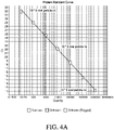
- the run file generates two standard curves: DNA curve ( FIG. 4b ) and Protein curve ( FIG. 4a ).
- the DNA curve flows linear regression and protein curve flows 4PL regression.
- the assay is applicable to a variety of procedures routinely used to detect DNA and protein during vaccine production, including but not limited to: i) viral dose determination; ii) lot-to-lot testing; iii) shelf-life testing; iv) viral serotype identification, etc. It is also useful for the capture/detection/ quantitation of any proteinaceous target for e.g. aptamers. This assay is particularly useful for detection and quantitation in vaccine production.
Landscapes
- Chemical & Material Sciences (AREA)
- Life Sciences & Earth Sciences (AREA)
- Organic Chemistry (AREA)
- Health & Medical Sciences (AREA)
- Engineering & Computer Science (AREA)
- Wood Science & Technology (AREA)
- Zoology (AREA)
- Proteomics, Peptides & Aminoacids (AREA)
- Genetics & Genomics (AREA)
- Immunology (AREA)
- General Engineering & Computer Science (AREA)
- Biotechnology (AREA)
- Bioinformatics & Cheminformatics (AREA)
- Microbiology (AREA)
- Physics & Mathematics (AREA)
- Biochemistry (AREA)
- Molecular Biology (AREA)
- Biophysics (AREA)
- General Health & Medical Sciences (AREA)
- Analytical Chemistry (AREA)
- Virology (AREA)
- Chemical Kinetics & Catalysis (AREA)
- Biomedical Technology (AREA)
- Plant Pathology (AREA)
- Measuring Or Testing Involving Enzymes Or Micro-Organisms (AREA)
Description
- This application claims the benefit under 35 U.S.C. § 119(e) of
U.S. Provisional Application no. 62/647,564, filed 23 March, 2018 - The present disclosure relates to an efficient, accurate and reliable method to determine both protein and nucleic acid amounts in a viral preparation in a single, dual assay.
- Several applications, for example, gene therapy, require that the qualitative and/ or quantity attributes of viral preparations be characterized before administration to a subject. The potency of a viral preparation consist of two parts: 1) the ability of viral capsid to transduce cells and 2) the ability of DNA in virus to produce the desired product. Inoperable capsids (for e.g., empty) do not effectively transduce the cells. Thus, an accurate tool to measure the dose and ultimately the potency of viral preparations, and to monitor gene therapy vector quality and potency is therefore needed. Previously, the evaluating step for determining the percentage or ratio of full:empty viral capsids have been by cryogenic transmission electron microscopy (CryoTEM), negative staining TEM, capillary electrophoresis, analytical ultracentrifugation, native agarose gel, alkaline agarose gel, southern blot, dot-blot hybridization, UV spectrophotometry, weak anion exchange chromatography, or mass spectrometry. These techniques are extremely slow, tedious, are capital intensive, require large samples of high purity, and high levels of user training and expertise. NORDENGRAHN ET AL,VETERINARY MICROBIOLOGY, ELSEVIER BV, NL, vol. 127, no. 3-4, 30 January 2008, p. 227-236, relates to evaluation of a novel proximity ligation assay for the sensitive and rapid detection of foot-and-mouth disease virus.
- Here we describe a dual/ duplex assay that is designed to enable the simultaneous quantitation of vector DNA by quantitative TaqMan assay and viral capsid protein determination by proximity ligation assay (PLA). Both protein and DNA can be quantitated at the same time. The optimized PLA procedure allows the detection of viral proteins in a sensitive manner. Measuring the capsid protein and DNA together leads to the simple, fast, and direct determination of a virus full and empty ratio with a time-to-result of about 4 hours. To validate this approach, we used the Adeno-Associated Virus (AAV) which is a commonly used vector of choice in many gene therapies. In a preferred embodiment, the assay is particularly useful to measure AAV viral full:empty analytics. This novel approach represents a paradigm shift for AAV, and enables the end-user to accelerate their work by obtaining vital information about vector samples with speed, ease, and accuracy.
- TaqMan® Protein Assays are an adapted form of PLA™, a proximity ligation assay technology that combines antibody-protein binding with detection of the reporter nucleic acid by real-time PCR. It has been employed for sensitive and specific measurement of protein/ protein expression. Here, in a first step, we use an adapted form of the PLA™ technology to obtain a sensitive and specific measurement of viral protein in a capsid and subsequently, by disassembling the viral capsid, in the second step, we release the viral DNA and accurately measure viral DNA by quantitative PCR (qPCR). The measurement is possible in a single assay, in a single container, in about 4 hours manually, and can be adapted to semiautomated or fully automated methods as well.
- The present invention is defined in the appended claims.
- In one embodiment, the disclosure provides a method of quantifying a dose of a viral preparation, comprising:
- a) quantifying a total number of viral capsids in the viral preparation via a viral-specific PLA assay;
- b) heating the virus preparation to enable disassembly of the viral capsid, and releasing the viral DNA;
- c) quantifying the total number of viral DNA in the viral preparation via a viral DNA-specific quantitative PCR assay.
- In another embodiment, the disclosure provides the method as described above, wherein the quantification in a) for viral capsid and in c) for viral DNA is determined from a reference standard curve, or, wherein, the quantification in a) for viral capsid and in c) for viral DNA is determined by digital PCR.
- In another embodiment, the disclosure provides the method of any of the claims described above, wherein the viral preparation is selected from the group consisting of parvovirus, adeno-associated virus (AAV), adenovirus, lentivirus, retrovirus, herpesvirus, poxvirus, paramyxovirus, baculovirus, reovirus, alphavirus, flavivirus, and combinations thereof.
- In another embodiment, the disclosure provides the method of any of the above claims, wherein the steps a) through c) are all performed in a single container.
- In one embodiment, the disclosure provides the method of any of the above claims, wherein the heating of step b) is at least at 80°C.
- In another embodiment, the disclosure provides the method of any of the above claims, wherein the heating of step b) is up to 100°C.
- In another embodiment, the disclosure provides the method of any of the above claims, wherein the method of quantifying viral protein and viral DNA is done within about 4 hours.
- In another embodiment, the disclosure provides the method of any of the above claims, wherein the viral preparation comprises one or more of full genome-containing virus particles, partial genome-containing virus particles, self-complementary genome-containing virus particle (double stranded genome), and genome-free virus particles.
- In another embodiment, the disclosure provides the method of any of the above claims, further comprising evaluating a percentage or ratio of full versus empty (full:empty) capsids in the viral preparation.
- In another embodiment, the disclosure provides the method of any one of the above claims, wherein the percentage of full viral capsids is from about 1% to about 100%.
- In another embodiment, the disclosure provides the method of any one of the above claims, wherein the percentage of full viral capsids is about 60% - 100%.
- In an embodiment, the disclosure provides a method of measuring the concentration of full AAV capsids in an AAV preparation, comprising:
- a) quantifying a total number of AAV capsids in the AAV preparation via an AAV-specific PLA assay;
- b) heating the AAV preparation to enable disassembly of the AAV capsid, and releasing the AAV DNA;
- c) quantifying the total number of AAV DNA in the AAV preparation via an AAV DNA-specific quantitative PCR assay; and,
- d) evaluating a percentage of full versus empty AAV capsids in the AAV preparation by calculating the ratio of AAV DNA quantity/ AAV capsid quantity.
- In another embodiment, the disclosure provides the method of the claim above, wherein the quantification in a) for AAV capsid and in c) for AAV DNA is determined from a reference standard curve, or, wherein, the quantification in a) for AAV capsid and in c) for AAV DNA is determined by digital PCR.
- In another embodiment, the disclosure provides the method of above claims, wherein the PLA assay is capable of detecting one or more of thirteen serotypes of the AAV capsid antigen.
- In another embodiment, the disclosure provides the method of any of claims above, wherein the steps a) through c) are all performed in a single container.
- In another embodiment, the disclosure provides the method of any one of the claims above, further comprising evaluating a percentage or ratio of full versus empty (full:empty) AAV capsids in the AAV preparation.
- In some embodiments, the disclosure provides the method of any one of the claims above, wherein the percentage of full AAV capsids is from about 1% to about 100%.
- In another embodiment, the disclosure provides the method of any one of the claims above, wherein at least 60- 100% of the AAV capsids are full AAV capsids.
- In some embodiments, the disclosure provides the method of any one of claims, wherein the AAV capsid has an antigen that is selected from the group consisting of: AAV1, AAV2, AAV3, AAV4, AAV5, AAV6, AAV7, AAV8, AAV9, AAV10, AAV11, AAV12, AAV13, chimeric AAV, genetically engineered AAV, or chemically modified AAV.
- In some embodiments, the disclosure provides the method of any one of claims, wherein optionally, the AAV preparation is concentrated and further wherein the preparation is administered at a specific dose to a subject in need thereof.
- In another embodiment, the disclosure provides the method of any of the above claims, wherein the heating of step b) is at least at 80°C.
- In yet another embodiment, the disclosure provides the method of any of the above claims, wherein the heating of step b) is up to 100°C.
- In some embodiments, the disclosure provides the method of any of the above claims, wherein the method of quantifying viral protein and viral DNA is done within about 4 hours.
- In another embodiment, the disclosure provides the method of any of the above claims, wherein the viral preparation comprises one or more of full genome-containing virus particles, partial genome-containing virus particles, self-complementary genome-containing virus particle (double stranded genome), and genome-free virus particles.
- In one embodiment, the disclosure provides a kit comprising:
- a) wash buffer, diluent buffer, elution buffer, DNA ligase enzyme;
- b) PCR master mix, PCR assay mix and a set of 3' and 5' primers for quantifying AAV DNA;
- c) one or more sets of 3' and 5' probes for detecting and quantifying one or more serotypes of AAV capsid protein, capture beads comprising an antibody that recognizes the AAV capsid, AAV protein standard.
- In some embodiments, the disclosure provides the kit described above, wherein the kit measures the concentration of full AAV capsids in an AAV preparation.
- In some embodiments, the disclosure provides the kit described above, wherein the AAV capsid has an antigen that is selected from the group consisting of: AAV1, AAV2, AAV3, AAV4, AAV5, AAV6, AAV7, AAV8, AAV9, AAV10, AAV11, AAV12, AAV13, chimeric AAV, genetically engineered AAV, or chemically modified AAV.
- In yet another embodiment, the disclosure provides a kit comprising:
- a) wash buffer, diluent buffer, elution buffer, DNA ligase enzyme;
- b) PCR master mix, PCR Assay mix, two sets of viral DNA primers;
- c) one or more sets of 3' and 5' probes for detecting and quantifying one or more serotypes of a virus, capture beads comprising an antibody that recognizes a viral capsid antigen, viral protein standard.
- In yet another embodiment, the disclosure provides the kit described above, wherein the kit measures the concentration of full viral capsids in a viral preparation.
- The novel features of the invention are set forth with particularity in the appended claims. A better understanding of the features and advantages of the present disclosure will be obtained by reference to the following detailed description that sets forth illustrative embodiments, in which the principles of the invention are utilized. The following figures and their descriptions constitute a part of the specification and illustrate the exemplary embodiments that are not to be considered limiting to the scope of the invention.
-
FIG. 1 : This shows the principle of the virus dual/duplex assay. Viral capsid protein is quantified by a PLA (Proximity Ligation Assay) using two specific detection antibodies to the viral capsid protein. Each detection antibody is also coupled to an oligonucleotide sequence (oligo 1 and oligo 2). The oligonucleotide sequences are brought together using a 'splint probe'. The nick is ligated using DNA ligase. The ligated sequence is amplified and quantified by quantitative PCR (qPCR) to give a protein readout. After protein quantification, the viral capsid assembly is disrupted to release the viral DNA, which is quantified by another qPCR assay using nucleic acid primers tagged with another dye e.g., VIC dye. Thus, both protein and DNA can be quantified in the same container one after the other. -
FIG. 2 : This figure shows overall how viral capsid proteins are captured in a separation/ purification workflow. Magnetic beads with capture antibody capture the viral capsids. The viral capsid protein amounts are quantified by PLA (Proximity Ligation Assay) as described above. -
FIG. 3a : This figure is a detailed diagrammatic representation of how a viral capsid protein (smaller cross hatch circle) is captured by a capture antibody on a magnetic bead (larger cross hatch circle). After washes, two detection antibodies coupled with oligonucleotide sequences are shown. The dark black filled arc represents the 'splint probe' that aids to bring the oligonucleotide sequences close together. -
FIG. 3b : The diagram on the left continues from 3a above, where the oligonucleotide sequences are ligated with DNA ligase, quantified by PLA (Proximity Ligation Assay) as described above and is shown in the middle panel. Due to heat denaturation, the viral DNA is released (irregular lines). Moving on to the last panel, the magnetic beads, viral capsid etc. fall apart due to heat denaturation (shown as open circles). Another set of 3' and 5' primers to the viral DNA is added. These primers are tagged with another dye e.g., VIC dye, and is quantified by another qPCR assay to give a viral DNA readout in the same container or in a single assay. -
FIG. 4a : Graph showing protein standard curve. An AAV8 viral preparation was used to generate the protein standard curve. Protein amounts were linear in 10 to 107 viral particle /µl concentration range. qPCR instrument set to measure FAM dye fluorescence. Slope = -3.487, Y-intercept = 39.292, R2= 0.998 (closer to 1 means most points are on or close to the regressed curve), Efficiency% = 93.559. Ct refers to the cycling number for qPCR and indicates the number of cycles for dye detection. The data is based on an average of 3 replicates. Together with the DNA standard curve of 4b, the curves are useful to determine full versus empty viral ratio percentages using Equation 1 (see Example 1). -
FIG. 4b : Graph showing DNA standard curve. An AAV8 vector preparation is used generate a standard DNA curve. DNA was linear 10 - 107 vector copy /µl concentration range. qPCR instrument set to measure VIC dye fluorescence. Slope = -3.344, Y-intercept = 38.403, R2= 0.999, Efficiency % = 99.103. Ct refers to the cycling number for qPCR and indicates the number of cycles for dye detection. The data is based on an average of 3 replicates. In one embodiment, CMV specific sequences are used for AAV expression. Together with the protein standard curve of 4a, the curves are useful to determine full versus empty viral ratio percentages using Equation 1 (see Example 1). -
FIG. 5 : Detection of AAV8 vector full/ empty ratios determined using superimposed protein and DNA standard curves in one reaction. % Full/empty viral vectors can be predicted usingEquation 1 of viral DNA quantitation / capsid protein quantitation multiplied by 100. Measuring the capsid protein and DNA together leads to the simple, fast, and direct determination of a virus, for e.g., AAV virus full and empty ratio with a time-to-result of about 4 hours. - Before the present disclosure is further described, it is to be understood that this disclosure is not limited to particular embodiments described, as such may, of course, vary. It is also to be understood that the terminology used herein is for the purpose of describing particular embodiments only, and is not intended to be limiting, since the scope of the present disclosure will be limited only by the appended claims.
- Unless defined otherwise, all technical and scientific terms used herein have the same meaning as is commonly understood by one of ordinary skill in the art to which this disclosure belongs. If a definition set forth in this section is contrary to or otherwise inconsistent with a definition set forth in a patent, application, or other publication that is herein incorporated by reference, the definition set forth in this section prevails over the definition incorporated herein by reference.
- As used herein and in the appended claims, the singular forms "a," "an," and "the" include plural referents unless the context clearly dictates otherwise. It is further noted that the claims may be drafted to exclude any optional element. As such, this statement is intended to serve as antecedent basis for use of such exclusive terminology as "solely," "only" and the like in connection with the recitation of claim elements, or use of a "negative" limitation.
- As used herein, the terms "including," "containing," and "comprising" are used in their open, non-limiting sense.
- To provide a more concise description, some of the quantitative expressions given herein are not qualified with the term "about". It is understood that, whether the term "about" is used explicitly or not, every quantity given herein is meant to refer to the actual given value, and it is also meant to refer to the approximation to such given value that would reasonably be inferred based on the ordinary skill in the art, including equivalents and approximations due to the experimental and/or measurement conditions for such given value. Whenever a yield is given as a percentage, such yield refers to a mass of the entity for which the yield is given with respect to the maximum amount of the same entity that could be obtained under the particular stoichiometric conditions. Concentrations that are given as percentages refer to mass ratios, unless indicated differently.
- It is appreciated that certain features of the disclosure, which are, for clarity, described in the context of separate embodiments, may also be provided in combination in a single embodiment. Conversely, various features of the disclosure, which are, for brevity, described in the context of a single embodiment, may also be provided separately or in any suitable subcombination. All combinations and embodiments pertaining to the elements of the compositions or methods described herein are specifically enumerated by the present disclosure and are disclosed herein just as if each and every combination was individually and explicitly disclosed; to the extent that such combinations are not in conflict with one another. In addition, all subcombinations of the elements of the compositions or methods described herein are also specifically enumerated by the present disclosure and are disclosed herein just as if each and every sub-combination was individually and explicitly disclosed herein; to the extent that such subcombinations are not in conflict with one another.
- In describing certain embodiments of the present disclosure:
- A feature of AAV vector generation in cell culture is the formation of an excess of "empty" capsids, which lack the vector genome. In certain embodiments, the amount of empty capsids is assessed in order to accurately determine the concentration, dose, and/or potency of an AAV preparation. Empty capsids can also be considered as product related impurities, which should be reduced during purification. In certain embodiments, it is important to determine the total amount of empty capsids to reduce an innate and/or adaptive immune response against the capsid antigen.
- In some embodiments, the methods described herein can be applied to any viral assay and is not limited to AAV only; for e.g., for the determination of viral vector product quantity (particles/mL) and quality (% full/empty) of viruses including but not limited to: such as parvovirus, adeno-associated virus (AAV), adenovirus, HIV, MMV, lentivirus, retrovirus, herpesvirus, poxvirus, paramyxovirus, baculovirus, reovirus, alphavirus, flavivirus, and combinations thereof.
- In some embodiments, the assay may detect one, several or all the viral serotypes of a virus depending on the detection antibody's recognition capacity of the various serotypes. The 'detection antibody' may be engineered to recognize one or more serotypes. In a particular embodiment where AAV full:empty ratios are measured, the assay utilizes the AAVX antibody which is a camelid antibody that is found to bind multiple serotypes of AAV, namely one or more of the 13 known AAV serotypes (hence, it is sometimes also called the universal AAV antibody). In certain assays, additional anti-AAV antibodies can be included in the assay to detect additional AAV serotypes. In some embodiments, only one AAV antibody (like AAVX) directed to several of the 13 AAV serotypes may be used in the assay.
- In order to detect viral capsid protein, in some embodiments, each detection antibody is coupled to an oligonucleotide sequence (3' oligo and 5' oligo respectively). In a preferred embodiment, at least two detection antibodies are employed, the antibodies detecting at least one serotype. In other embodiments, multiple sets of 3'-5'- tagged detection antibodies may be used, each set recognizing a different serotype. In a most preferred embodiment, the at least two detection antibodies are capable of detecting multiple serotypes, for e.g., the AAVX antibody that is known to recognize serotypes AAV1 to
AAV 13. The 3' oligo and 5' oligo are brought into close proximity by a "splint probe". (InFIG 3b , see the black arc above hatched semicircle. The hatched semicircle depicts the 3' and the 5' oligonucleotides (attached to the antibody). The attachment is via biotin coupling. The splint probe guides the 3' and the 5' oligonucleotides next to each other so that the DNA ligase enzyme can ligate the nick and join the oligonucleotides. Now, the PLA (Proximity Ligation Assay) can be performed using primers (shown as dumbbell shape: the open circle is say the 3' primer and the hatched circle is the 5' primer) specific to the ligated DNA which indirectly correspond as "protein probes". In order to detect viral capsid protein, in some embodiments, primers specific for the ligated oligo is amplified and detected by FAM fluorescence and quantified. - Correspondingly, in some embodiments, to detect the released viral DNA, primers specific for the viral DNA is amplified and detected by VIC fluorescence and quantified.
- In some embodiments, the MasterMix used in qPCR comprises the dye-labelled primers, the polymerase amonst other components necessary for the TaqMan assay. The DNA ligase is added separately.
- In some embodiments, the capture and/or the detection antibodies are derived from any species, including but not limited to rat, mouse, rabbit, hamster, monkey, chimpanzee, horse, cow, cat, chicken, human, humanized, pig, camelid, chimeric, etc.
- In some embodiments, the capture antibody which recognizes one or more serotypes of a virus, can be attached to a capture surface via chemical (including but not limited to, sulfhydyl reactive-, carbonyl reactive-, amine reactive-, etc.) means, via affinity means or via physical means. In a preferred embodiment, the capture antibody is biotinylated which in turn attaches to a streptavidin-activated surface. As the skilled artisan would know, alternately, any affinity conjugation ligands can be utilized to achieve a similar result of binding capture antibody to surface.
- In some embodiments, capture is limited to magnetic beads alone. In other embodiments, capture can be on any solid surface used to capture viral proteins and/or capsids, including but not limited to: plates, tube, film, membrane, well, glass surface, bead, resin, microfluidic sheet, etc.
- While preferred embodiments of the present invention have been shown and described herein, it will be obvious to those skilled in the art that such embodiments are provided by way of example only. It should be understood that various alternatives to the embodiments of the invention described herein may be employed in practicing the invention. It is intended that the following claims define the scope of the invention and that methods and structures within the scope of these claims be covered thereby.
- Recombinant viral vector preparations can contain different levels of full or partial genome-containing particles as well as empty virions. Such particle diversity can be attributed to multiple factors such as genome packaging efficiency, production methods, downstream purification techniques and storage conditions. In therapies employing viral vectors for e.g., gene therapy, there exists a potential for vector dose-related immunotoxicity in patients. In viral production, empty virions are inadvertent byproducts of recombinant packaging process, resulting in vector lots with mixtures of full and empty virions at variable ratios. Present methods for accurately determining the quantity (particles/mL) of vectors have their limitations. For e.g., current ELISA methods show poor accuracy and precision and can only measure a fraction of the viral serotypes. A combination of ELISA and qPCR is available, but it provides a convoluted approach with high margins of error. Analytical Ultra-centrifugation (AUC) methods like CsCl gradients are not desirable for the following reasons: they are extremely slow, tedious, low throughput, capital intensive, require large samples of high purity and substantial user training and expertise. Electron Microscopy (TEM) similarly involves tedious sample prep, skilled expertise and is very slow and expensive. Besides, neither AUC or TEM are amenable to high-throughput analysis.
- Adeno-Associated Virus (AAV) is a commonly used vector of choice in gene therapies. It is a preferred vector because it is non-pathogenic, non-toxic, can infect non-dividing and dividing cells, the virus persists, several serotypes are available, has a simple vector design for cost effective manufacturing is highly precise and specific for target cell delivery. Current AAV ELISA assays can only detect 6 of 13 AAV serotypes. Therefore, there is a need for a single, streamlined assay that is accurate and reliable for the determination of viral vector quantity (particles/mL) and quality (% full/empty) for a large number of viral serotypes.
- In some embodiments a qPCR-based dual or duplex assay that quantitates DNA, e.g. viral DNA and protein (viral capsid protein) in a single assay is described. In other words, the assay simultaneously determines vector quantity (particle titer) and quality (full & empty) rapidly and in a streamlined workflow producing excellent data quality with accuracy and precision. The assay can measure most of the 13 AAV serotypes and the assay can measure crude samples directly with no sample prep. The assay may be performed manually as demonstrated in Example 1. Alternatively, the assay may use hardware/software for e.g., an automated, magnetic bead processing unit (such as the KingFisher™ Flex instrument), a qPCR instrument (e.g., 7500 Fast PCR instrument), to achieve an automated or semiautomated hands-off workflow. In one embodiment, the disclosure is directed to a kit comprising the necessary reagents and buffers for the duplex assay for determining full/empty viral vector and/or to quantitate a viral particle titer.
- In some embodiments, the disclosure describes an efficient and accurate analytical method for AAV vector titer, especially empty/full ratio determination, is still lacking. Existing analytical solutions that offer indirect empty/full determination are extremely slow, tedious, are capital intensive, require large samples of high purity, and high levels of user training and expertise. Here we describe a dual/ duplex assay that is designed to enable the simultaneous quantitation of vector DNA by TaqMan assay and viral capsid (protein) determination by proximity ligation assay (PLA). This leads to the simple, fast, and direct determination of AAV full and empty ratio with a time-to-result of about 4 hours. Both protein and DNA can be quantitated at the same time. This novel approach represents a paradigm shift in AAV analytics, and enables the end-user to accelerate their work by obtaining vital information about vector samples with unprecedented speed, ease, and accuracy.
- In other embodiments, the methods described herein can be applied to any viral assay and is not limited to AAV only; for e.g., for the determination of viral vector product quantity (particles/mL) and quality (% full/empty) of any virus such as parvovirus, adeno-associated virus (AAV), adenovirus, lentivirus, retrovirus, herpesvirus, poxvirus, paramyxovirus, baculovirus, reovirus, alphavirus, flavivirus, and combinations thereof. Moreover, the assay may detect one, several or all the viral serotypes of a virus depending on the detection antibody's recognition capacity of the various serotypes. The detection antibody may be engineered to recognize one or more serotypes.
- The principle of this assay is described in
FIG. 1 . In this assay, protein amounts, e.g. viral capsid protein are quantified using a PLA (Proximity Ligation Assay) using two detection antibodies specific to the viral capsid protein, wherein each detection antibody is also coupled to an oligonucleotide sequence (a 3' oligo that is biotin conjugated to the detection antibody, and a 5' oligo also biotin conjugated to the detection antibody). The two dye-tagged oligonucleotide sequences are brought together using a 'splint probe', and the nick is ligated using DNA ligase. Oligonucleotide primers specific for the ligated oligo 3' + oligo 5' sequences (indirectly identifying the viral capsid protein) and which are tagged with a first dye, for e.g., FAM dye, is amplified and quantified by quantitative PCR (qPCR) to give a viral capsid protein readout. qPCR fluorescence readouts are much more robust, sensitive and much faster than ELISA readouts. - Subsequently after protein related amplification, the viral capsid assembly is disrupted to release the viral DNA. In one embodiment, the capsid disruption is achieved through heat denaturation (anywhere between 80 to 100°C, depending on the virus); alternatively, the capsid disruption can be done using enzyme digestion e.g., proteinase. The capsid disassembly releases the viral DNA which is quantified by a second qPCR assay, this time using oligonucleotide primers specific for the viral DNA and which are tagged with another dye e.g., VIC dye. Thus, in this assay, both protein and DNA can be quantified in the same container one after the other.
-
FIG. 2 shows further details of how viral capsid proteins are captured in the first step for PLA. In this experiment, Dynabeads® MyOne™ Streptavidin T1 magnetic beads, with AAVX capture antibody was used. The viral capsid protein is recognized by the AAVX antibody and captured. Then wash step are performed to removed non-specific binding. Then two additional detection antibodies are used which bind the capsid. Each detection antibody is also coupled to an oligonucleotide sequence (3' oligo and 5' oligo respectively). The 3' oligo and 5' oligo are brought into close proximity by a "splint probe" (InFIG 3b , see the black arc above hatched semicircle. The hatched semicircle depicts the 3' and the 5' oligonucleotides that are biotin coupled to the detection antibody). The splint probe guides the 3' and the 5' oligonucleotides together next to each other so that the DNA ligase enzyme can ligate the nick and join the oligonucleotides. Now, the PLA (Proximity Ligation Assay) can be performed using primers (shown as dumbbell shape: the open circle is say the 3' primer and the hatched circle is the 5' primer) specific to the ligated DNA which indirectly correspond as "protein probes". Using the primers, the ligated DNA is amplified, detected by FAM fluorescence and thus, the viral capsid proteins are quantified. Capture is not limited to magnetic beads only: any solid surface can be used to capture viral proteins and/or capsids, including but not limited to: plates, tube, film, membrane, well, glass surface, bead, resin, microfluidic sheet, etc. -
FIG. 3b also shows how the viral capsid assembly is disrupted to release the viral DNA after protein related amplification has been performed. In one embodiment, the capsid disruption is achieved through heat denaturation (between about 80 to about 100°C, depending on the viral capsid disassembly properties). Alternatively, capsid disruption is done using enzyme digestion e.g., proteinase. Capsid disassembly releases the viral DNA within, which is quantified by a second qPCR assay, this time using 3' and 5' oligonucleotide primers specific for the viral DNA (see dumbbell next to the wavy lines which depicts viral DNA). The 3' and 5' oligonucleotide primers (shown as hatched small circles) are specific for the viral DNA and comprise another dye e.g., VIC dye, so viral DNA quantification is done on a different fluorescent channel. Thus, in this assay, both protein and DNA can be quantified in the same container one after the other. - Described herein is an assay that enables the accurate and reliable determination of AAV vector product quantity (particles/mL) and quality (% full/empty) for several AAV serotypes. An AAVX antibody was used in this assay, which can recognize serotypes AAV1 through AAV9 and numerous recombinant and chimeric serotypes (from ThermoFisher Scientific, cat. number 810352201 or 810352210). A second AAV antibody referred to as AAVY herein, was also used in the assay (from any of ThermoFisher Scientific, cat. numbers 810338001, 810338010, 810333001 and 810333010). It is to be understood that the antibody reagents used herein is for the purpose of describing particular embodiments only, and is not intended to be limiting. As the skilled artisan would recognize, the protocol can be performed using any AAV antibody derived from any species, including but not limited to rat, mouse, rabbit, human, camelid, chimeric, etc. In addition, one would also recognize that one or multiple AAV antibodies directed to one or more 13 AAV serotypes can also be used.
- AAV8 vectors were obtained from Virovek, Inc. and University of North Carolina were used as AAV virus DNA standards to confirm DNA copy number, and to evaluate and confirm assay accuracy.
Table 1. Shown below are a set of exemplary reagents used in this manual assay. Table 1: Reagents Quantity Storage AAVX 5' PROBE 1 Tube, 350 µL -15 °C to -25 °C AAVX 3' PROBE 1 Tube, 350 µL -15 °C to -25 °C AAVY 5' PROBE 1 tube, 350 µL -15 °C to -25 °C AAVY 3' PROBE 1 Tube, 350 µL -15 °C to -25 °C AAVX Ligation and Assay Mix 1 Tube, 600 µL -15 °C to -25 ° C DNA Ligase 1 Tube, 55 µL -15 °C to -25 °C AAVX Standard (1 × 10⁷ copies/µL) 1 Tube, 400 µL Shipping -20 °C; Store at; 2 °C to 8 °C after first use AAVX Elution Buffer 1 Tube, 7 ml 2 °C to 8 °C AAVX Capture Beads 1 Bottle, 5.25 ml 2 °C to 8 °C AAVX Diluent Buffer 1 Bottle, 50 ml 2 °C to 8 °C AAVX Wash Buffer 1 Bottle, 100 ml 2 °C to 8 °C TaqMan Fast Master Mix 2X 1 Bottle, 6 ml 2 °C to 8 °C - First, AAVX Standard Dilution is done as follows: Prepare serial dilutions in 2 mL Eppendorf tubes as shown in Table 1 below. Add the respective amounts of AAVX Diluent to each tube. Tube NPC (Non-Protein Control) contains the Diluent only and serves as the Non-Protein Control (NPC). For the rest of the Standard tubes, add the appropriate amount of AAVX particles. Mix dilutions well by vortexing and then briefly spin down the tube contents to the bottom.
Table 2. Serial dilution of AAVX Standard with 3 replicates Table 2 AAVX Particles AAVX (µL) AAVX Diluent to be Added (µL) Total (µL) Std 11.00E+07 120 0 120 Std 21.00E+06 12 µL of Std 1108 120 Std 3 1.00E+05 12 µL of Std 2108 120 Std 4 1.00E+04 12 µL of Std 3 108 120 Std 51.00E+03 12 µL of Std 4 108 120 Std 6 1.00E+02 12 µL of Std 5108 120 Std 7 1.00E+01 12 µL of Std 6 108 120 NPC 120 120 - The present protocol is performed on a sample plate. However, as would be recognized by one of skill in the art, the protocol can be applied to any type of container, for e.g., plates, slides, tubes, wells, microfluidic devices, arrays, dishes, films, depressions on any flat surface, etc.
- Dispense 20 µL AAVX Capture Beads into each Standard and Sample wells. Next, dispense 30 µL of each Standard, starting with NPC, into each of the respective Standard wells. Make several dilutions of your Unknown samples using the AAVX Diluent Buffer and dispense 30 µL of those samples into the Unknown wells in triplicate. Mix really well. Let the plate sit on the bench for 1 hour. Every 15 minutes, pipette up and down and mix well again to re-suspend beads and samples.
- Next, prepare Sample Plate (PCR-compatible plate). Resuspend AAVX Capture Beads by pipetting or place tube on a rotator for about 10-15 minutes until all the beads are resuspended. Transfer AAVX Capture Beads into a reservoir and dispense 20 µL into each Standard and Sample wells using a 200 µL multi-channel pipette (manual or electronic). Dispense 30 µL of each Standard, starting with NPC, into each of the respective Standard wells. If necessary, dilute unknown samples using the AAVX Diluent Buffer and dispense 30 µL into the Unknown wells in triplicate. Using a manual pipette with volume set to 40 µl, pipette each well up and down 10 times to mix, minimize bubbles during mixing. Incubate the sample plate for 1 hour at room temperature. Pipette mix (10 times) every 30 minutes using a manual 200 µL multi-channel pipette (set the volume at 40 µl) for a total of 3 times (at
time 0, 30 and 60 minute during the incubation period). - After capture step, briefly spin the plate in a plate centrifuge, place sample plate on a magnetic plate until all beads are magnetized (~ 1 minute, the beads are on the well of the plate not at the bottom). While the plate is on the magnetic stand, remove the liquid from the center of the well using a manual pipet with volume set to 60 µl. Add 100 µL of Wash Buffer to plate. Resuspend beads by pipetting up and down 10 times on all four corners of each well. Once beads are fully resuspended, place sample plate on the magnetic stand until solution is clear (~ 1 minute) and removes the Wash Buffer while the plate is on the magnetic stand (set the volume to 110 µl). Repeat this process once more. After the second wash step, remove the entire wash buffer then take the sample plate off the magnetic stand.
- Prepare probe mix in 5-ml tubes as shown in Table 3. Adjust based on sample numbers. Mix the Probe solution by inverting the
tube 8 times. Transfer Probe solution into a reservoir and dispense 50 µL to each well of the sample plate using a 200 µL multi-channel pipette (manual or electronic). Resuspend beads by pipetting up and down 10 times on all four corners of each well (be sure the pipette tips touch all corners of the wall to ensure complete resuspension of beads, using a multi-channel manual pipet and set the volume at 40 µl). Incubate sample plate at room temperature for 30 mins with mixing 10 times every 15 minutes (total 3 times, attime Table 3. Probe Solution Reagent Volume/reaction 48 reactions 96 reactions ProteinSEQ Diluent Buffer 56.2 µL 2700 µL 5400 µL AAVX 5' PROBE 1.6 µL 75 µL 150 µL AAVX 3' PROBE 1.6 µL 75 µL 150 µL AAVY 5' PROBE 1.6 µL 75 µL 150 µL AAVY 3' PROBE 1.6 µL 75 µL 150 µL Total 62.5 µL 3000 µL 6000 µL - Repeat 2X Washes (same as before).
- After Probe detection step, briefly spin the plate in a plate centrifuge (when the spin speed reaches 800 rpm, stop the spin). Place sample plate on a magnetic plate until all beads are magnetized (~ 1 minute, the beads are on the well of the plate not at the bottom). While the plate is on the magnetic stand, remove the liquid from the center of the well using a manual pipette with volume set to 60 µl). Remove sample plate from the magnet stand and add 100 µL of Wash Buffer. Resuspend beads by pipetting up and down 10 times on all four corners of each well (set up pipette volume at 90 µL, see
Fig 1 ; be sure the pipette tips touch all corners of the wall to ensure complete resuspension of beads). Once beads are fully resuspended, place sample plate on the magnetic stand until solution is clear (~1 minute) and removes the Wash Buffer while the plate is on the magnetic stand (set the volume to 110 µl). Repeat this process once more. After the second wash step, remove the entire wash buffer then take the sample plate off the magnetic stand. - Transfer Elution Buffer to a plate reservoir, and dispense 15 µL to each well of the plate using a 20 µL or 200 µL multi-channel pipette (manual or electronic). Prepare qPCR mastermix as shown in Table 4. Adjust according to sample number with 20% for overage. Transfer mastermix into a reservoir and dispense 15 µL into each well. Resuspend beads by pipetting up and down 10 times on all 4 corners of each well (be sure the pipette tips touch all corners of the wall to ensure complete resuspension of beads, using a multi-channel manual pipette and set the volume to 25 µl). Seal the plate, and then spin down the plate in a plate centrifuge for 3 seconds at 500 rpm (hold Short button and let go when it reaches 500 rpm). Proceed to the Real-time PCR Instrument.
Table 4A Prepare Mastermix. Components Volume/reaction 48 Reactions 96 Reactions 2x FAST master mix 20 µL 960 µL 1920 µL AAVX Ligation and Assay mix 2 µL 96 µL 192 µL ProteinSEQ Ligase 0.2 µL 9.6 µL 19.2 µL 22.2 µL 1065.6 µL 2131.2 µL - Table 4B below is an alternative when using their other primer and probe for the DNA target (ITR, other GOI, etc.) instead of using the CMV-targeting primer/probe set used in Table 4A above.
Table 4B. Prepare Mastermix Components Volume/reaction 48 Reactions 96 Reactions 2x FAST master mix 20 µL 960 µL 1920 µL AAVX Ligation and Assay mix 2 µL 96 µL 192 µL other primer and probe mix ∗2 µL 96 µL 192 µL ProteinSEQ Ligase 0.2 µL 9.6 µL 19.2 µL 22.2 µL 1065.6 µL 2131.2 µL - ∗ The probe used should be VIC-labeled, and the primer and probe concentrations are 20X so the final concentrations of primer and probe in qPCR reaction are 1X. Load the Sample plate onto the Real-Time PCR instrument. If using AccuSEQ software, proceed with the steps below. If using other Real-time Instruments, follow the Instrument's User Guide. Perform PCR using AccuSEQ Software, the AAV8-Alpha-test-template, AccuSEQ 2.1 software on the computer connected to a 7500 Fast PCR instrument. Use the Define and Set up Standards to set up the standard curve. Click on Define and Set Up Standards, enter Standard Curve Information (7 points, 3 replicates, 1e7 starting quantity and 1:5 dilution). Set thermal cycling conditions as: Hold 37°
C 10 minutes; Hold 95°C 3 minutes; 40 cycles; 95°C 3 seconds; 60°C 30 seconds. Press run. - The run file generates two standard curves: DNA curve (
FIG. 4b ) and Protein curve (FIG. 4a ). The DNA curve flows linear regression and protein curve flows 4PL regression. - On standard curve page, select all reactions in the standard curve, select linear for curve fit (which is for DNA measurement). Click Analysis. Save the file and export the data and name the file as "DNA data".
- On standard curve page, select all reactions in the standard curve, select the 4PL regression for protein analysis. Click Analysis. Save the file and export the data and name the file as "Protein data". To analyze the data, add a new excel sheet to the protein data file, name the new sheet as "analyzed sheet" and copy the following columns from the protein data file first sheet to the new sheet (analyzed sheet), sample name column, target column, quantity column. Then sort the three column data first by target, then by sample name (from A to Z option). Open DNA data file. Add a new excel sheet to the DNA data file, name the new sheet as "analyzed sheet" and copy the following columns from the DNA data file first sheet to the new sheet (analyzed sheet), sample name column, target column, quantity column. Then sort the three column data first by target, then by sample name (from A to Z option).
- Open the AAV8 analysis calculation spreadsheet associated with this protocol (provided in your Alpha Test Package). In the paste sheet, copy all the sample names from protein data file, analysis sheet under the sample name of analysis calculation spreadsheet. Then copy all the DNA quantity data from DNA data file analysis sheet under DNA quantity in the analysis calculation spreadsheet. Then copy all the Protein quantity data from Protein data file analysis sheet under Protein quantity in the analysis calculation spreadsheet. The analysis calculation spreadsheet will calculate all the parameters in the quantity sheet.
- Two standard curves: i) Protein (
FIG. 4a ) and ii) DNA (FIG. 4b ) are generated using different dilutions of viral particle (capsid protein determination) and viral vector (for viral DNA) and the standard curves are plotted. To predict whether viral vector is full or empty, or to calculate the % fullness,Equation 1 is used.
- Spike experiments of known quantities of full/ empty vectors were used to standardize and evaluate the accuracy and sensitivity of the assay. The accuracy of one such experiment is shown below in Table 5,
column 2. The Standard Deviation is shown incolumn 2 and column 3 is a measure of replicate precision CV% = S.D./Average ∗100. This shows that the protein-DNA dual assay is both accurate and sensitive at least over 10 to 107 viral particle /µl protein concentration range, and over 10 - 107 vector copy /µl DNA concentration range.Table 5. Input sample assay readout average (%) assay readout assay readout 10% full viral particle 10.19% 0.0051 5.00 20% full viral particle 21.06% 0.0173 8.23 30% full viral particle 34.40% 0.0189 5.51 40% full viral particle 44.49% 0.0145 3.25 50% full viral particle 54.76% 0.0310 5.67 60% full viral particle 59.95% 0.0526 8.77 70% full viral particle 0.0111 1.62 80% full viral particle 83.05% 0.0757 9.12 90% full viral particle 95.42% 0.0581 6.09 100% full viral particle 106.42% 0.0132 1.24 - Deducing from the standard curves of
FIGs 4a and4b ,Equation 1 is used to calculate % fullness of a virus. For example, if for a given unknown, the protein readout is 107 protein and the corresponding DNA readout is also 107 DNA, using the equation, we get a ratio of 1, which means that the viral capsid is 100% full. In another example, if for a given unknown, the protein readout is 105 protein and the corresponding DNA readout is 103 DNA, fromEquation 1, we get a ratio of 1:100 = 0.01, which means that the viral capsid is 1% full. And in yet another example, if for a given unknown, the protein readout is 106 protein and the corresponding DNA readout is 107 DNA, fromEquation 1 we get a ratio of 10, which means that the viral capsid is 110% full, and so on. - Besides gene therapy applications, the assay is applicable to a variety of procedures routinely used to detect DNA and protein during vaccine production, including but not limited to: i) viral dose determination; ii) lot-to-lot testing; iii) shelf-life testing; iv) viral serotype identification, etc. It is also useful for the capture/detection/ quantitation of any proteinaceous target for e.g. aptamers. This assay is particularly useful for detection and quantitation in vaccine production.
- The examples were intended to illustrate, but not limit certain embodiments of the disclosure. One skilled in the art will understand that various modifications are readily available and can be performed without substantial change in the way the invention works.
Claims (17)
- A method of quantifying a dose of a viral preparation, comprising:a) quantifying a total number of viral capsids in the viral preparation via a viral-specific PLA (proximity ligation assay);b) heating the virus preparation to enable disassembly of the viral capsid, and releasing the viral DNA;c) quantifying the total number of viral DNA in the viral preparation via a viral DNA-specific quantitative PCR assay.
- The method of claim 1, wherein the quantification in a) for viral capsid and in c) for viral DNA is determined from a reference standard curve, or, wherein, the quantification in a) for viral capsid and in c) for viral DNA is determined by digital PCR.
- The method of claims 1-2, wherein the viral preparation is selected from the group consisting of parvovirus, adeno-associated virus (AAV), adenovirus, lentivirus, retrovirus, herpesvirus, poxvirus, paramyxovirus, baculovirus, reovirus, alphavirus, flavivirus, and combinations thereof.
- The method of any of the above claims, wherein the steps a) through c) are all performed in a single container.
- The method of any of the above claims, wherein the heating of step b) is at least at 80°C, and/or is up to 100°C.
- The method of any of the above claims, wherein the method of quantifying viral protein and viral DNA is done within about 4 hours.
- The method of any of the above claims, wherein the viral preparation comprises one or more of full genome-containing virus particles, partial genome-containing virus particles, self-complementary genome-containing virus particle (double stranded genome), and genome-free virus particles.
- The method of any of the above claims, further comprising evaluating a percentage or ratio of full versus empty (full:empty) capsids in the viral preparation.
- The method of any one of the above claims, wherein the percentage of full viral capsids is from about 1%to about 100%, or is about 60% - 100%.
- The method of claim 8, wherein the viral preparation is a preparation of adeno-associated virus (AAV) and quantifying is measuring the concentration of full AAV capsids in an AAV preparation.
- The method of claim 10, wherein the PLA assay is capable of detecting one or more of thirteen serotypes of the AAV capsid antigen.
- The method of any one of claims 10 or 11, wherein the AAV capsid has an antigen that is selected from the group consisting of: AAVl, AAV2, AAV3, AAV4, AAV5, AAV6, AAV7, AAV8, AAV9, AAV10, AAV11, AAV12, AAV 13, chimeric AAV, genetically engineered AAV, or chemically modified AAV.
- The method of any one of claims 10 to 12, wherein optionally, the AAV preparation is concentrated.
- A kit comprising:a) wash buffer, diluent buffer, elution buffer, DNA ligase enzyme;b) PCR master mix, PCR assay mix and a set of 3' and 5' primers for quantifying AAV DNA;c) one or more sets of 3' and 5' probes for detecting and quantifying one or more serotypes of AAV capsid protein, capture beads comprising an antibody that recognizes the AAV capsid, AAV protein standard.
- The kit of claim 14, wherein the AAV capsid has an antigen that is selected from the group consisting of: AAVl, AAV2, AAV3, AAV4, AAV5, AAV6, AAV7, AAV8, AAV9, AAV10, AAV11, AAV12, AAV13, chimeric AAV, genetically engineered AAV, or chemically modified AAV.
- A kit comprising:a) wash buffer, diluent buffer, elution buffer, DNA ligase enzyme;b) PCR master mix, PCR Assay mix, two sets of viral DNA primers;c) one or more sets of 3' and 5' probes for detecting and quantifying one or more serotypes of a virus, capture beads comprising an antibody that recognizes a viral capsid antigen, viral protein standard.
- The kit of any one of claims 14 to 16, wherein the kit measures the concentration of full viral capsids in a viral preparation.
Applications Claiming Priority (2)
| Application Number | Priority Date | Filing Date | Title |
|---|---|---|---|
| US201862647564P | 2018-03-23 | 2018-03-23 | |
| PCT/US2019/023728 WO2019183579A1 (en) | 2018-03-23 | 2019-03-22 | Methods and kits to detect viral particle heterogeneity |
Publications (2)
| Publication Number | Publication Date |
|---|---|
| EP3768865A1 EP3768865A1 (en) | 2021-01-27 |
| EP3768865B1 true EP3768865B1 (en) | 2022-12-14 |
Family
ID=66102219
Family Applications (1)
| Application Number | Title | Priority Date | Filing Date |
|---|---|---|---|
| EP19716642.4A Active EP3768865B1 (en) | 2018-03-23 | 2019-03-22 | Methods and kits to detect viral particle heterogeneity |
Country Status (3)
| Country | Link |
|---|---|
| US (2) | US11649479B2 (en) |
| EP (1) | EP3768865B1 (en) |
| WO (1) | WO2019183579A1 (en) |
Families Citing this family (6)
| Publication number | Priority date | Publication date | Assignee | Title |
|---|---|---|---|---|
| EP4119682A1 (en) * | 2021-07-13 | 2023-01-18 | Genethon | A method for determining the nature of dna impurities inside viral particles |
| EP4511654A4 (en) * | 2022-04-18 | 2026-04-15 | Access Medical Systems Ltd | METHOD FOR DETERMINING DNA CONCENTRATION IN DNA VIRUS |
| US20230392188A1 (en) * | 2022-06-02 | 2023-12-07 | Perkinelmer Health Sciences, Inc. | Electrophoresis-mediated characterization of dna content of adeno-associated virus capsids |
| CN120712363A (en) * | 2023-03-03 | 2025-09-26 | 赛多利斯生物分析仪器公司 | Methods for quantification of viral capsids |
| WO2025166267A1 (en) * | 2024-02-01 | 2025-08-07 | Sartorius Bioanalytical Instruments, Inc. | Methods for determining empty/full ratios of intact viral capsids |
| JP2025143019A (en) * | 2024-03-18 | 2025-10-01 | アイポア株式会社 | Viral vector testing method and machine learning system |
Family Cites Families (4)
| Publication number | Priority date | Publication date | Assignee | Title |
|---|---|---|---|---|
| EP1618201A1 (en) * | 2003-05-01 | 2006-01-25 | University of Florida Research Foundation, Inc. | Vp2 - modified raav vectors and uses thereof |
| EP1957664A1 (en) | 2005-11-14 | 2008-08-20 | Boehringer Ingelheim Vetmedica Gmbh | Method for detecting microorganisms |
| CA2853482C (en) * | 2011-10-28 | 2020-05-19 | Joshua Grieger | Cell line for production of adeno-associated virus |
| CA2988446C (en) | 2015-06-23 | 2020-07-21 | Deutsches Krebsforschungszentrum | Method for large scale production and purification of parvovirus |
-
2019
- 2019-03-22 EP EP19716642.4A patent/EP3768865B1/en active Active
- 2019-03-22 WO PCT/US2019/023728 patent/WO2019183579A1/en not_active Ceased
- 2019-03-22 US US17/040,250 patent/US11649479B2/en active Active
-
2023
- 2023-04-21 US US18/137,956 patent/US20240102077A1/en active Pending
Also Published As
| Publication number | Publication date |
|---|---|
| US20210017608A1 (en) | 2021-01-21 |
| US11649479B2 (en) | 2023-05-16 |
| US20240102077A1 (en) | 2024-03-28 |
| EP3768865A1 (en) | 2021-01-27 |
| WO2019183579A1 (en) | 2019-09-26 |
Similar Documents
| Publication | Publication Date | Title |
|---|---|---|
| US20240102077A1 (en) | Methods and Kits to Detect Viral Particle Heterogeneity | |
| Gimpel et al. | Analytical methods for process and product characterization of recombinant adeno-associated virus-based gene therapies | |
| Veldwijk et al. | Development and optimization of a real-time quantitative PCR-based method for the titration of AAV-2 vector stocks | |
| Kontogiannis et al. | Characterization of AAV vectors: A review of analytical techniques and critical quality attributes | |
| Hitchman et al. | Quantitative real‐time PCR for rapid and accurate titration of recombinant baculovirus particles | |
| Baoutina et al. | Synthetic certified DNA reference material for analysis of human erythropoietin transgene and transcript in gene doping and gene therapy | |
| Troxell et al. | Partial genome content within rAAVs impacts performance in a cell assay-dependent manner | |
| JP7763771B2 (en) | Methods and kits for detecting adeno-associated virus | |
| US12227811B2 (en) | Primer group for simultaneous detection of 15 porcine pathogens through high-throughput targeted amplicon sequencing and use thereof | |
| Soth et al. | Quantification of full and empty particles of adeno-associated virus vectors via a novel dual fluorescence-linked immunosorbent assay | |
| Duong et al. | Three-dimensional linkage analysis with digital PCR for genome integrity and identity of recombinant adeno-associated virus | |
| CN112513291A (en) | Reaction mixtures, methods and kits for performing quantitative real-time PCR | |
| CN118389720B (en) | A primer-probe combination, kit and method for detecting foodborne Clostridium | |
| US20250377334A1 (en) | Adeno-associated virus vectors empty/full ratio analysis using ce-based genome and capsid quantification | |
| CN115896344B (en) | Recombinant adeno-associated virus titer detection primer probe set, kit and application | |
| Wang et al. | Development of high-throughput analytical methods for characterization and quantitation of rAAV genome integrity | |
| Chappleboim et al. | ApharSeq: an extraction-free early-pooling protocol for massively multiplexed SARS-CoV-2 detection | |
| Van den Berghe et al. | Quantification of Adeno-Associated Viral Genomes in Purified Vector Samples by Digital Droplet Polymerase Chain Reaction | |
| US20250376738A1 (en) | Quantitative Immuno-PCR for Simultaneous Determination of AAV Capsid Titer and AAV Genome Copy Titer | |
| Vettermann et al. | Bioanalytical Methods to Study Biodistribution and Shedding of AAV‐Based Gene Therapy Vectors | |
| Sailleau et al. | Real-Time RT-PCR assays for typing of epizootic hemorrhagic disease virus | |
| CN118726618A (en) | qPCR kit for detecting E1A residual DNA | |
| CN120158496A (en) | Adeno-associated virus pretreatment reagent and its application | |
| Soth | Development and application of dual | |
| CN115807064A (en) | Method for detecting free and error packaged DNA in recombinant adeno-associated virus vector product |
Legal Events
| Date | Code | Title | Description |
|---|---|---|---|
| STAA | Information on the status of an ep patent application or granted ep patent |
Free format text: STATUS: UNKNOWN |
|
| STAA | Information on the status of an ep patent application or granted ep patent |
Free format text: STATUS: THE INTERNATIONAL PUBLICATION HAS BEEN MADE |
|
| PUAI | Public reference made under article 153(3) epc to a published international application that has entered the european phase |
Free format text: ORIGINAL CODE: 0009012 |
|
| STAA | Information on the status of an ep patent application or granted ep patent |
Free format text: STATUS: REQUEST FOR EXAMINATION WAS MADE |
|
| 17P | Request for examination filed |
Effective date: 20201023 |
|
| AK | Designated contracting states |
Kind code of ref document: A1 Designated state(s): AL AT BE BG CH CY CZ DE DK EE ES FI FR GB GR HR HU IE IS IT LI LT LU LV MC MK MT NL NO PL PT RO RS SE SI SK SM TR |
|
| AX | Request for extension of the european patent |
Extension state: BA ME |
|
| DAV | Request for validation of the european patent (deleted) | ||
| DAX | Request for extension of the european patent (deleted) | ||
| STAA | Information on the status of an ep patent application or granted ep patent |
Free format text: STATUS: EXAMINATION IS IN PROGRESS |
|
| 17Q | First examination report despatched |
Effective date: 20211015 |
|
| GRAP | Despatch of communication of intention to grant a patent |
Free format text: ORIGINAL CODE: EPIDOSNIGR1 |
|
| STAA | Information on the status of an ep patent application or granted ep patent |
Free format text: STATUS: GRANT OF PATENT IS INTENDED |
|
| INTG | Intention to grant announced |
Effective date: 20220705 |
|
| GRAS | Grant fee paid |
Free format text: ORIGINAL CODE: EPIDOSNIGR3 |
|
| GRAA | (expected) grant |
Free format text: ORIGINAL CODE: 0009210 |
|
| STAA | Information on the status of an ep patent application or granted ep patent |
Free format text: STATUS: THE PATENT HAS BEEN GRANTED |
|
| AK | Designated contracting states |
Kind code of ref document: B1 Designated state(s): AL AT BE BG CH CY CZ DE DK EE ES FI FR GB GR HR HU IE IS IT LI LT LU LV MC MK MT NL NO PL PT RO RS SE SI SK SM TR |
|
| REG | Reference to a national code |
Ref country code: GB Ref legal event code: FG4D |
|
| REG | Reference to a national code |
Ref country code: CH Ref legal event code: EP |
|
| REG | Reference to a national code |
Ref country code: DE Ref legal event code: R096 Ref document number: 602019023153 Country of ref document: DE |
|
| REG | Reference to a national code |
Ref country code: IE Ref legal event code: FG4D |
|
| REG | Reference to a national code |
Ref country code: AT Ref legal event code: REF Ref document number: 1537691 Country of ref document: AT Kind code of ref document: T Effective date: 20230115 |
|
| REG | Reference to a national code |
Ref country code: LT Ref legal event code: MG9D |
|
| REG | Reference to a national code |
Ref country code: NL Ref legal event code: MP Effective date: 20221214 |
|
| PG25 | Lapsed in a contracting state [announced via postgrant information from national office to epo] |
Ref country code: SE Free format text: LAPSE BECAUSE OF FAILURE TO SUBMIT A TRANSLATION OF THE DESCRIPTION OR TO PAY THE FEE WITHIN THE PRESCRIBED TIME-LIMIT Effective date: 20221214 Ref country code: NO Free format text: LAPSE BECAUSE OF FAILURE TO SUBMIT A TRANSLATION OF THE DESCRIPTION OR TO PAY THE FEE WITHIN THE PRESCRIBED TIME-LIMIT Effective date: 20230314 Ref country code: LT Free format text: LAPSE BECAUSE OF FAILURE TO SUBMIT A TRANSLATION OF THE DESCRIPTION OR TO PAY THE FEE WITHIN THE PRESCRIBED TIME-LIMIT Effective date: 20221214 Ref country code: FI Free format text: LAPSE BECAUSE OF FAILURE TO SUBMIT A TRANSLATION OF THE DESCRIPTION OR TO PAY THE FEE WITHIN THE PRESCRIBED TIME-LIMIT Effective date: 20221214 |
|
| REG | Reference to a national code |
Ref country code: AT Ref legal event code: MK05 Ref document number: 1537691 Country of ref document: AT Kind code of ref document: T Effective date: 20221214 |
|
| PG25 | Lapsed in a contracting state [announced via postgrant information from national office to epo] |
Ref country code: RS Free format text: LAPSE BECAUSE OF FAILURE TO SUBMIT A TRANSLATION OF THE DESCRIPTION OR TO PAY THE FEE WITHIN THE PRESCRIBED TIME-LIMIT Effective date: 20221214 Ref country code: LV Free format text: LAPSE BECAUSE OF FAILURE TO SUBMIT A TRANSLATION OF THE DESCRIPTION OR TO PAY THE FEE WITHIN THE PRESCRIBED TIME-LIMIT Effective date: 20221214 Ref country code: HR Free format text: LAPSE BECAUSE OF FAILURE TO SUBMIT A TRANSLATION OF THE DESCRIPTION OR TO PAY THE FEE WITHIN THE PRESCRIBED TIME-LIMIT Effective date: 20221214 Ref country code: GR Free format text: LAPSE BECAUSE OF FAILURE TO SUBMIT A TRANSLATION OF THE DESCRIPTION OR TO PAY THE FEE WITHIN THE PRESCRIBED TIME-LIMIT Effective date: 20230315 |
|
| PG25 | Lapsed in a contracting state [announced via postgrant information from national office to epo] |
Ref country code: NL Free format text: LAPSE BECAUSE OF FAILURE TO SUBMIT A TRANSLATION OF THE DESCRIPTION OR TO PAY THE FEE WITHIN THE PRESCRIBED TIME-LIMIT Effective date: 20221214 |
|
| PG25 | Lapsed in a contracting state [announced via postgrant information from national office to epo] |
Ref country code: SM Free format text: LAPSE BECAUSE OF FAILURE TO SUBMIT A TRANSLATION OF THE DESCRIPTION OR TO PAY THE FEE WITHIN THE PRESCRIBED TIME-LIMIT Effective date: 20221214 Ref country code: RO Free format text: LAPSE BECAUSE OF FAILURE TO SUBMIT A TRANSLATION OF THE DESCRIPTION OR TO PAY THE FEE WITHIN THE PRESCRIBED TIME-LIMIT Effective date: 20221214 Ref country code: PT Free format text: LAPSE BECAUSE OF FAILURE TO SUBMIT A TRANSLATION OF THE DESCRIPTION OR TO PAY THE FEE WITHIN THE PRESCRIBED TIME-LIMIT Effective date: 20230414 Ref country code: ES Free format text: LAPSE BECAUSE OF FAILURE TO SUBMIT A TRANSLATION OF THE DESCRIPTION OR TO PAY THE FEE WITHIN THE PRESCRIBED TIME-LIMIT Effective date: 20221214 Ref country code: EE Free format text: LAPSE BECAUSE OF FAILURE TO SUBMIT A TRANSLATION OF THE DESCRIPTION OR TO PAY THE FEE WITHIN THE PRESCRIBED TIME-LIMIT Effective date: 20221214 Ref country code: CZ Free format text: LAPSE BECAUSE OF FAILURE TO SUBMIT A TRANSLATION OF THE DESCRIPTION OR TO PAY THE FEE WITHIN THE PRESCRIBED TIME-LIMIT Effective date: 20221214 Ref country code: AT Free format text: LAPSE BECAUSE OF FAILURE TO SUBMIT A TRANSLATION OF THE DESCRIPTION OR TO PAY THE FEE WITHIN THE PRESCRIBED TIME-LIMIT Effective date: 20221214 |
|
| PG25 | Lapsed in a contracting state [announced via postgrant information from national office to epo] |
Ref country code: SK Free format text: LAPSE BECAUSE OF FAILURE TO SUBMIT A TRANSLATION OF THE DESCRIPTION OR TO PAY THE FEE WITHIN THE PRESCRIBED TIME-LIMIT Effective date: 20221214 Ref country code: PL Free format text: LAPSE BECAUSE OF FAILURE TO SUBMIT A TRANSLATION OF THE DESCRIPTION OR TO PAY THE FEE WITHIN THE PRESCRIBED TIME-LIMIT Effective date: 20221214 Ref country code: IS Free format text: LAPSE BECAUSE OF FAILURE TO SUBMIT A TRANSLATION OF THE DESCRIPTION OR TO PAY THE FEE WITHIN THE PRESCRIBED TIME-LIMIT Effective date: 20230414 Ref country code: AL Free format text: LAPSE BECAUSE OF FAILURE TO SUBMIT A TRANSLATION OF THE DESCRIPTION OR TO PAY THE FEE WITHIN THE PRESCRIBED TIME-LIMIT Effective date: 20221214 |
|
| REG | Reference to a national code |
Ref country code: DE Ref legal event code: R097 Ref document number: 602019023153 Country of ref document: DE |
|
| PLBE | No opposition filed within time limit |
Free format text: ORIGINAL CODE: 0009261 |
|
| STAA | Information on the status of an ep patent application or granted ep patent |
Free format text: STATUS: NO OPPOSITION FILED WITHIN TIME LIMIT |
|
| PG25 | Lapsed in a contracting state [announced via postgrant information from national office to epo] |
Ref country code: MC Free format text: LAPSE BECAUSE OF FAILURE TO SUBMIT A TRANSLATION OF THE DESCRIPTION OR TO PAY THE FEE WITHIN THE PRESCRIBED TIME-LIMIT Effective date: 20221214 Ref country code: DK Free format text: LAPSE BECAUSE OF FAILURE TO SUBMIT A TRANSLATION OF THE DESCRIPTION OR TO PAY THE FEE WITHIN THE PRESCRIBED TIME-LIMIT Effective date: 20221214 |
|
| 26N | No opposition filed |
Effective date: 20230915 |
|
| PG25 | Lapsed in a contracting state [announced via postgrant information from national office to epo] |
Ref country code: SI Free format text: LAPSE BECAUSE OF FAILURE TO SUBMIT A TRANSLATION OF THE DESCRIPTION OR TO PAY THE FEE WITHIN THE PRESCRIBED TIME-LIMIT Effective date: 20221214 |
|
| REG | Reference to a national code |
Ref country code: BE Ref legal event code: MM Effective date: 20230331 |
|
| PG25 | Lapsed in a contracting state [announced via postgrant information from national office to epo] |
Ref country code: LU Free format text: LAPSE BECAUSE OF NON-PAYMENT OF DUE FEES Effective date: 20230322 |
|
| REG | Reference to a national code |
Ref country code: IE Ref legal event code: MM4A |
|
| PG25 | Lapsed in a contracting state [announced via postgrant information from national office to epo] |
Ref country code: IE Free format text: LAPSE BECAUSE OF NON-PAYMENT OF DUE FEES Effective date: 20230322 |
|
| PG25 | Lapsed in a contracting state [announced via postgrant information from national office to epo] |
Ref country code: BE Free format text: LAPSE BECAUSE OF NON-PAYMENT OF DUE FEES Effective date: 20230331 |
|
| PG25 | Lapsed in a contracting state [announced via postgrant information from national office to epo] |
Ref country code: IT Free format text: LAPSE BECAUSE OF FAILURE TO SUBMIT A TRANSLATION OF THE DESCRIPTION OR TO PAY THE FEE WITHIN THE PRESCRIBED TIME-LIMIT Effective date: 20221214 |
|
| PG25 | Lapsed in a contracting state [announced via postgrant information from national office to epo] |
Ref country code: BG Free format text: LAPSE BECAUSE OF FAILURE TO SUBMIT A TRANSLATION OF THE DESCRIPTION OR TO PAY THE FEE WITHIN THE PRESCRIBED TIME-LIMIT Effective date: 20221214 |
|
| PG25 | Lapsed in a contracting state [announced via postgrant information from national office to epo] |
Ref country code: BG Free format text: LAPSE BECAUSE OF FAILURE TO SUBMIT A TRANSLATION OF THE DESCRIPTION OR TO PAY THE FEE WITHIN THE PRESCRIBED TIME-LIMIT Effective date: 20221214 |
|
| PGFP | Annual fee paid to national office [announced via postgrant information from national office to epo] |
Ref country code: CH Payment date: 20250401 Year of fee payment: 7 |
|
| PG25 | Lapsed in a contracting state [announced via postgrant information from national office to epo] |
Ref country code: CY Free format text: LAPSE BECAUSE OF FAILURE TO SUBMIT A TRANSLATION OF THE DESCRIPTION OR TO PAY THE FEE WITHIN THE PRESCRIBED TIME-LIMIT; INVALID AB INITIO Effective date: 20190322 |
|
| PG25 | Lapsed in a contracting state [announced via postgrant information from national office to epo] |
Ref country code: HU Free format text: LAPSE BECAUSE OF FAILURE TO SUBMIT A TRANSLATION OF THE DESCRIPTION OR TO PAY THE FEE WITHIN THE PRESCRIBED TIME-LIMIT; INVALID AB INITIO Effective date: 20190322 |
|
| PG25 | Lapsed in a contracting state [announced via postgrant information from national office to epo] |
Ref country code: TR Free format text: LAPSE BECAUSE OF FAILURE TO SUBMIT A TRANSLATION OF THE DESCRIPTION OR TO PAY THE FEE WITHIN THE PRESCRIBED TIME-LIMIT Effective date: 20221214 |
|
| REG | Reference to a national code |
Ref country code: CH Ref legal event code: U11 Free format text: ST27 STATUS EVENT CODE: U-0-0-U10-U11 (AS PROVIDED BY THE NATIONAL OFFICE) Effective date: 20260401 |
|
| PGFP | Annual fee paid to national office [announced via postgrant information from national office to epo] |
Ref country code: GB Payment date: 20260226 Year of fee payment: 8 |
|
| PGFP | Annual fee paid to national office [announced via postgrant information from national office to epo] |
Ref country code: DE Payment date: 20260217 Year of fee payment: 8 |
|
| PGFP | Annual fee paid to national office [announced via postgrant information from national office to epo] |
Ref country code: FR Payment date: 20260311 Year of fee payment: 8 |