EP3764365A1 - Improvements in the radiological identification of chronic thromboembolic pulmonary hypertension - Google Patents
Improvements in the radiological identification of chronic thromboembolic pulmonary hypertension Download PDFInfo
- Publication number
- EP3764365A1 EP3764365A1 EP19185178.1A EP19185178A EP3764365A1 EP 3764365 A1 EP3764365 A1 EP 3764365A1 EP 19185178 A EP19185178 A EP 19185178A EP 3764365 A1 EP3764365 A1 EP 3764365A1
- Authority
- EP
- European Patent Office
- Prior art keywords
- interventricular septum
- cteph
- person
- right ventricle
- computed tomography
- Prior art date
- Legal status (The legal status is an assumption and is not a legal conclusion. Google has not performed a legal analysis and makes no representation as to the accuracy of the status listed.)
- Withdrawn
Links
- 208000002815 pulmonary hypertension Diseases 0.000 title claims abstract description 89
- 208000026151 Chronic thromboembolic pulmonary hypertension Diseases 0.000 title claims abstract description 83
- 238000000034 method Methods 0.000 claims abstract description 40
- 238000004590 computer program Methods 0.000 claims abstract description 24
- 238000002591 computed tomography Methods 0.000 claims description 87
- 210000005241 right ventricle Anatomy 0.000 claims description 55
- 210000005240 left ventricle Anatomy 0.000 claims description 45
- 210000000038 chest Anatomy 0.000 claims description 19
- 238000004458 analytical method Methods 0.000 claims description 17
- 230000000747 cardiac effect Effects 0.000 claims description 17
- 230000011218 segmentation Effects 0.000 claims description 10
- 238000012952 Resampling Methods 0.000 claims description 6
- 238000004891 communication Methods 0.000 claims description 6
- 230000001131 transforming effect Effects 0.000 claims description 4
- 238000005352 clarification Methods 0.000 claims description 2
- 210000001147 pulmonary artery Anatomy 0.000 description 7
- 239000002872 contrast media Substances 0.000 description 6
- 238000003745 diagnosis Methods 0.000 description 6
- 238000010191 image analysis Methods 0.000 description 6
- 230000010412 perfusion Effects 0.000 description 6
- 230000006870 function Effects 0.000 description 4
- 239000005337 ground glass Substances 0.000 description 4
- 239000011159 matrix material Substances 0.000 description 4
- 210000000709 aorta Anatomy 0.000 description 3
- 238000013459 approach Methods 0.000 description 3
- 201000010099 disease Diseases 0.000 description 3
- 208000037265 diseases, disorders, signs and symptoms Diseases 0.000 description 3
- 239000003814 drug Substances 0.000 description 3
- 210000002216 heart Anatomy 0.000 description 3
- 238000002560 therapeutic procedure Methods 0.000 description 3
- 210000001519 tissue Anatomy 0.000 description 3
- 208000032544 Cicatrix Diseases 0.000 description 2
- 206010013975 Dyspnoeas Diseases 0.000 description 2
- 208000019693 Lung disease Diseases 0.000 description 2
- 208000031481 Pathologic Constriction Diseases 0.000 description 2
- 210000000988 bone and bone Anatomy 0.000 description 2
- 238000012512 characterization method Methods 0.000 description 2
- 238000013135 deep learning Methods 0.000 description 2
- 229940079593 drug Drugs 0.000 description 2
- 238000002592 echocardiography Methods 0.000 description 2
- 238000011156 evaluation Methods 0.000 description 2
- 238000000605 extraction Methods 0.000 description 2
- 208000019622 heart disease Diseases 0.000 description 2
- 230000002685 pulmonary effect Effects 0.000 description 2
- 231100000241 scar Toxicity 0.000 description 2
- 230000037387 scars Effects 0.000 description 2
- 208000024891 symptom Diseases 0.000 description 2
- 230000002792 vascular Effects 0.000 description 2
- ZCYVEMRRCGMTRW-UHFFFAOYSA-N 7553-56-2 Chemical compound [I] ZCYVEMRRCGMTRW-UHFFFAOYSA-N 0.000 description 1
- 101500014077 Bombina orientalis C-terminal extension peptide Proteins 0.000 description 1
- 101100136092 Drosophila melanogaster peng gene Proteins 0.000 description 1
- 206010061216 Infarction Diseases 0.000 description 1
- 206010064911 Pulmonary arterial hypertension Diseases 0.000 description 1
- 238000010521 absorption reaction Methods 0.000 description 1
- 230000002238 attenuated effect Effects 0.000 description 1
- 230000015572 biosynthetic process Effects 0.000 description 1
- 230000017531 blood circulation Effects 0.000 description 1
- 238000004364 calculation method Methods 0.000 description 1
- 210000005242 cardiac chamber Anatomy 0.000 description 1
- 238000013184 cardiac magnetic resonance imaging Methods 0.000 description 1
- 210000002808 connective tissue Anatomy 0.000 description 1
- GOHCTCOGYKAJLZ-UHFFFAOYSA-N ctep Chemical compound CC=1N(C=2C=CC(OC(F)(F)F)=CC=2)C(C)=NC=1C#CC1=CC=NC(Cl)=C1 GOHCTCOGYKAJLZ-UHFFFAOYSA-N 0.000 description 1
- 238000003066 decision tree Methods 0.000 description 1
- 230000001419 dependent effect Effects 0.000 description 1
- 238000001514 detection method Methods 0.000 description 1
- 238000011161 development Methods 0.000 description 1
- 230000018109 developmental process Effects 0.000 description 1
- 238000002059 diagnostic imaging Methods 0.000 description 1
- 238000002405 diagnostic procedure Methods 0.000 description 1
- 238000003748 differential diagnosis Methods 0.000 description 1
- 230000009977 dual effect Effects 0.000 description 1
- 230000003073 embolic effect Effects 0.000 description 1
- 238000013171 endarterectomy Methods 0.000 description 1
- 238000005516 engineering process Methods 0.000 description 1
- 230000003203 everyday effect Effects 0.000 description 1
- 238000010230 functional analysis Methods 0.000 description 1
- 239000003365 glass fiber Substances 0.000 description 1
- PCHJSUWPFVWCPO-UHFFFAOYSA-N gold Chemical compound [Au] PCHJSUWPFVWCPO-UHFFFAOYSA-N 0.000 description 1
- 230000000004 hemodynamic effect Effects 0.000 description 1
- 238000003384 imaging method Methods 0.000 description 1
- 230000007574 infarction Effects 0.000 description 1
- 230000008595 infiltration Effects 0.000 description 1
- 238000001764 infiltration Methods 0.000 description 1
- 229910052740 iodine Inorganic materials 0.000 description 1
- 239000011630 iodine Substances 0.000 description 1
- 230000001788 irregular Effects 0.000 description 1
- 239000003550 marker Substances 0.000 description 1
- 230000007246 mechanism Effects 0.000 description 1
- 230000000877 morphologic effect Effects 0.000 description 1
- 210000000056 organ Anatomy 0.000 description 1
- 230000002085 persistent effect Effects 0.000 description 1
- 238000010837 poor prognosis Methods 0.000 description 1
- 238000011002 quantification Methods 0.000 description 1
- 230000008707 rearrangement Effects 0.000 description 1
- 230000004895 regional blood flow Effects 0.000 description 1
- 238000012552 review Methods 0.000 description 1
- WXXSNCNJFUAIDG-UHFFFAOYSA-N riociguat Chemical compound N1=C(N)C(N(C)C(=O)OC)=C(N)N=C1C(C1=CC=CN=C11)=NN1CC1=CC=CC=C1F WXXSNCNJFUAIDG-UHFFFAOYSA-N 0.000 description 1
- 229960000529 riociguat Drugs 0.000 description 1
- 210000004872 soft tissue Anatomy 0.000 description 1
- 238000012916 structural analysis Methods 0.000 description 1
- 230000002123 temporal effect Effects 0.000 description 1
- 230000001960 triggered effect Effects 0.000 description 1
- 210000003462 vein Anatomy 0.000 description 1
- 238000009423 ventilation Methods 0.000 description 1
- 230000002861 ventricular Effects 0.000 description 1
Images




Classifications
-
- G—PHYSICS
- G16—INFORMATION AND COMMUNICATION TECHNOLOGY [ICT] SPECIALLY ADAPTED FOR SPECIFIC APPLICATION FIELDS
- G16H—HEALTHCARE INFORMATICS, i.e. INFORMATION AND COMMUNICATION TECHNOLOGY [ICT] SPECIALLY ADAPTED FOR THE HANDLING OR PROCESSING OF MEDICAL OR HEALTHCARE DATA
- G16H30/00—ICT specially adapted for the handling or processing of medical images
- G16H30/40—ICT specially adapted for the handling or processing of medical images for processing medical images, e.g. editing
-
- G—PHYSICS
- G16—INFORMATION AND COMMUNICATION TECHNOLOGY [ICT] SPECIALLY ADAPTED FOR SPECIFIC APPLICATION FIELDS
- G16H—HEALTHCARE INFORMATICS, i.e. INFORMATION AND COMMUNICATION TECHNOLOGY [ICT] SPECIALLY ADAPTED FOR THE HANDLING OR PROCESSING OF MEDICAL OR HEALTHCARE DATA
- G16H50/00—ICT specially adapted for medical diagnosis, medical simulation or medical data mining; ICT specially adapted for detecting, monitoring or modelling epidemics or pandemics
- G16H50/30—ICT specially adapted for medical diagnosis, medical simulation or medical data mining; ICT specially adapted for detecting, monitoring or modelling epidemics or pandemics for calculating health indices; for individual health risk assessment
Definitions
- the present invention is concerned with the radiological identification of chronic thromboembolic pulmonary hypertension (CTEPH).
- CTEPH chronic thromboembolic pulmonary hypertension
- the invention relates to a method, a computer system and a computer program product for automatically identifying signs of the presence of CTEPH in a person.
- Chronic thromboembolic pulmonary hypertension is a special form of pulmonary hypertension (PH). It is characterized by the infiltration of thrombi into the pulmonary arteries. They clog and constrict the vessels in a complete or partial manner and can be converted into connective tissue. In rare cases, there is the development of pulmonary hypertension with a poor prognosis.
- CTEPH chronic myelolism
- a timely diagnosis of CTEPH is also important because there are meanwhile options for the therapy of different forms of manifestation of this disease.
- the preferred CTEPH therapy is operative pulmonary endarterectomy (PEA), by means of which it is possible to heal up to 70% of patients. Meanwhile, perioperative mortality at experienced centres is 2-4%. However, about 30-50% of all CTEPH patients are classified as non-operable. For these patients and for patients with persistent pulmonary hypertension after PEA, a medicament-based therapy was authorized for the first time at the beginning of 2014 in the form of Riociguat.
- PEA operative pulmonary endarterectomy
- the gold standard for diagnosing or ruling out a case of CTEPH is ventilation/perfusion scintigraphy.
- the negative predictive value of perfusion scintigraphy is almost 100%; this means that a proper perfusion distribution rules out a case of CTEPH with a probability bordering on certainty.
- the present invention firstly provides a method for identifying signs of the presence of CTEPH in a person, comprising the following steps:
- the present invention further provides a computer system for identifying signs of the presence of CTEPH in a person, comprising:
- the present invention further provides a computer program product comprising a data carrier on which there is stored a computer program which can be loaded into the memory of a computer system, where it causes the computer system to execute the following steps:
- the present invention focusses on the automated image analysis of computed tomography images of the thorax of the person.
- Computed tomography is an X-ray method which depicts the human body in cross-sectional images (sectional imaging method). Compared to a conventional X-ray image, on which usually only coarse structures and bones are identifiable, CT images also capture in detail soft tissues with small differences in contrast.
- An X-ray tube generates a so-called X-ray fan beam, which penetrates the body and is attenuated to varying degrees within the body owing to the various structures, such as organs and bones.
- the receiving detectors opposite the X-ray emitter receive the signals of varying strength and forward them to a computer, which puts together cross-sectional images of the body from the received data.
- Computed tomography images can be observed in 2D or else in 3D. For better differentiability of structures within the body of the person (e.g. vessels), a contrast agent can be injected into a vein before the generation of CT images.
- Computed tomography is a commonly used method in the diagnosis of heart and lung diseases.
- the CT images are multidetector CT images.
- Multidetector CT refers to the newest generation of computed tomographs, having been available in clinical radiology since 1998.
- Multidetector CT is widely available and distinguished by a high, virtually isotropic resolution (pixel size 0.5-1 mm in each spatial direction), and this makes it possible to view the CT images in any spatial plane.
- the examination time varies between 1 and 10 seconds, giving rise to images virtually free of artefacts, even when there is dyspnoea or lack of patient cooperation.
- the latest MDCT scanners are equipped with "dual-energy" technology, in which two different energies/tube voltages are used simultaneously. Owing to the energy dependence of absorption, it is possible to highlight certain tissue properties, for example the distribution of iodine after administration of contrast agent as surrogate for regional blood circulation.
- a crucial criterion in the present invention is the automation.
- CTEPH is a rare condition which is underdiagnosed. Non-detection of this condition can have fatal consequences for the patient. Therefore, according to the present invention, computed tomography images of the thorax are analysed in an automated manner for signs of the presence of CTEPH. What is meant here by "automated” is that no human intervention at all is required.
- a computer program is thus installed on a computer system that has access to computed tomography images of the thorax, runs as a background process, and analyses the images in an automated manner for signs of the presence of CTEPH.
- a background process refers to a process that does not act directly with the user and hence acts asynchronously to the user interface.
- a computed tomography image of the thorax of a person are received or retrieved.
- a CT image is a data set, by means of which the structures of the thorax of the person can be depicted three-dimensionally.
- a CT image customarily represents the thorax of the person at the time of recording of the computed tomogram.
- Multiple CT images can e.g., represent the heart and/or pulmonary region of the person at different times; on the basis of these multiple CT images, it is possible therefore to identify temporal changes in the tissue structures and thus, for example, to examine a progression of a disease.
- the multiple CT images are CT images representing different regions of the thorax.
- computed tomography is a customary method for the diagnosis of heart and lung diseases. It is therefore conceivable to examine CT images already present in a database for the presence of signs of CTEPH.
- CT images present in one or more databases are retrieved and are analysed for the presence of signs of CTEPH. For example, this can occur at regular intervals. It is, for example, conceivable to carry out at regular intervals, for example every day or every week, a search for new CT images in the databases in which CT images are customarily deposited and to retrieve the new CT images for an image analysis. However, the retrieval can also take place irregularly.
- the retrieval can also be triggered by an event, for example by the storing of a new CT image.
- the retrieval of new CT images is done in an automated manner.
- a CT image generated from the thorax of a person is, after its generation, immediately and automatically subjected to an image analysis according to the invention.
- a computer system oriented to generating a relevant CT image can be configured such that it forwards the CT image to the image analysis according to the invention.
- the components executing the image analysis receive the CT image.
- an automated analysis of the one or more CT image takes place (preferably more than one CT image).
- the analysis is carried out by:
- cardiac segmentation methods are based on deep learning which can be roughly divided into two classes: 2D methods which segment each slice independently and 3D methods, which segment multiple slices together as a volume ( Qiao Zheng et al., 3D consistent robust segmentation of cardiac images by deep learning with spatial propagation, IEEE Trans. On Medical Imaging, 2018, hal-01753086 ).
- segmentation of the cardiac region includes
- Binary segmentation of left and right ventricles of the cardiac region refers in particular to 3D CT images marked by different grey values.
- the left and right ventricles are in particular coded by different grey values.
- Resampling of the binary segmented images to an isotropic spacing is usually required as 3D CT images contain voxels for which the spacing in the three spatial directions is not uniform.
- Resampling refers to process step wherein an isotropic spacing in all direction of the 3D images is achieved.
- a spacing value of equal or below 2.5 mm, more particularly equal or below 2.0 mm is targeted.
- Resampling refers preferably also to the extraction of superfluous tissue such as the interventricular septum e.g., with morphological operators.
- both ventricles are segmented into two new distinct images.
- this step also includes an object analysis step in both images to detect the geometric region enclosing the left and the right ventricles followed by a union of the regions.
- the distance transforming step results in grey level images that look similar to the input images, except that the distance of a voxel from the edge of an object is coded as a grey value: the higher the grey value, the higher the distance.
- the distance-transformed images are intersected, preferably by using a thresholding step to from a thick, intersecting border around the both ventricles. Finally, the intersection volume is extracted defining the new interventricular septum.
- the step b) of the method refers to determining a 2D slice image with the maximum right ventricle diameter by rotating a plane throughout the main axis of an ellipsoid fitted to the interventricular septum.
- a basis for determining the appropriate 2D slice image is the ellipsoid fitting to the interventricular septum and the determination of a preferred direction and a geometrical characterization of the interventricular septum.
- Equation (1) describes an ellipsoid, when the following condition is fulfilled: k det 2 ⁇ det 1 2 > 0
- det 1 a 11 + a 22 + a 33
- det 2 a 11 a 22 + a 22 a 33 + a 11 a 33 ⁇ a 33 a 33 ⁇ a 12 ⁇ a 13 a 13 k ⁇ 4
- the ellipsoid condition (equation 6) is in the majority of cases not fulfilled. In some cases a hyperboloid can be fitted. In order to address this issue a constraint condition can be set to force the fitting of the ellipsoid to the interventricular septum (method of Lagrange multiplier). In a preferred embodiment, however, an ellipsoid is pre-fitted to the left ventricle and the centroid of this ellipsoid is used as a constraint condition for fitting a second ellipsoid to the interventricular septum. The left ventricle is in most cases almost rotation symmetric and the fitted parameters of the surface of second order (equation 1) fulfils the condition of the ellipsoid condition (equation 6).
- the ellipsoid fitted to the interventricular septum is preferably identified by:
- Analysis of the orientation of the ellipsoid fitted to the interventricular septum preferably includes
- the rotation matrix is expressed as a function of the Euler angles ⁇ , ⁇ , and ⁇ .
- R cos ⁇ cos ⁇ sin ⁇ sin ⁇ cos ⁇ ⁇ cos ⁇ sin ⁇ cos ⁇ sin ⁇ cos ⁇ + sin ⁇ sin ⁇ cos ⁇ sin ⁇ sin ⁇ sin ⁇ sin ⁇ + cos ⁇ cos ⁇ cos ⁇ sin ⁇ sin ⁇ ⁇ sin ⁇ cos ⁇ ⁇ sin ⁇ sin ⁇ cos ⁇ cos ⁇ cos ⁇
- the Euler angles are calculated from equation 12 and 13.
- a 3D image is rotated in the rage of [-90, +90] around the main (middle) axis of the ellipsoid fitted to the interventricular septum.
- the method (and/or as part of the computer system, computer program product) relates to the determination of at least one feature selected from the group of ratio of the diameters of the right ventricle and left ventricle, and the degree of curvature of the interventricular septum based on the 2D slice image as determined in the previous step b) which indicate the presence of CTEPH.
- the determination of the ratio of the diameters of the right ventricle and left ventricle preferably includes
- the ratio of the diameters of right ventricle and left ventricle (RV/LV ratio) (see, for example, Gonzales G et al. PLoS ONE 10(5): e0127797 ) is a feature that can indicate the presence of CTEPH.
- a value of 0.9 or more in the RV/LV diameter ratio is an indication of the presence of CTEPH.
- the determination of the degree of curvature of the interventricular septum based on the 2D slice image as determined in step b) preferably includes
- the determination of the degree of curvature of the interventricular septum requires the extraction of the centerline. This is preferably done by applying a skeleton operator. In some cases the skeleton image may contain additional branches. These branches can be excluded with minimum-path, graph based approaches. Then, a polynomial of higher order is fitted to the centerline. It has turned out, that a polynomial of the 3 th leads to best results.
- the degree of curvature of the interventricular septum is determined around the local maxima of the polynomial, these points are identified by computing the first derivative.
- the method (and/or as part of the computer system, computer program product) takes into account at least one additional feature indicative for CTEP selected from the group of volumes of the right ventricle and left ventricle, ratio of the volumes of the right ventricle and the left ventricle, ratio of the diameters of the pulmonary artery and the aorta at the level at which the pulmonary artery branches off, presence and/or severity of stenoses, presence and/or severity of mosaic perfusion, presence and/or severity of ground glass opacity.
- at least one additional feature indicative for CTEP selected from the group of volumes of the right ventricle and left ventricle, ratio of the volumes of the right ventricle and the left ventricle, ratio of the diameters of the pulmonary artery and the aorta at the level at which the pulmonary artery branches off, presence and/or severity of stenoses, presence and/or severity of mosaic perfusion, presence and/or severity of ground glass opacity.
- the ratio of the diameters of the pulmonary artery and the aorta is determined at the point at which the pulmonary artery branches off (see, for example, A.S. Iyer et al.: CT scan-measured pulmonary artery to aorta ratio and echocardiography for detecting pulmonary hypertension in severe COPD, CHEST 2014, Vol. 145(4), 824-832 ).
- a PA:A ratio of 0.7 or greater is a further indication of the presence of CTEPH.
- Characteristic vascular features are, for example, the lack of contrast agent in the distal vessel sections in the case of total obstruction or the formation of rope-ladder thrombi, meshes, stenoses and partial obstructions.
- the CTEPH-specific parenchymal features include scars, mosaic perfusion, ground glass opacity and bronchial anomalies.
- the scars arise as a result of infarctions owing to the occlusion of pulmonary vessels that are usually localized in the lower segments.
- Mosaic perfusion consists of regions of different density resulting from regions of irregular hypo- and hyperperfused regions caused by embolic occlusions, vascular rearrangement of the distal vessels and compensation mechanisms. Hypoperfused regions are observed particularly in the distal direction from the occluded vessels since blood flow and hence the concentration of the contrast agent are reduced in these regions.
- Hyperdense areas are usually apparent in regions that are now hyperperfused owing to the redistribution and stand out as ground glass opacity.
- At least one feature indicating the presence of CTEPH was found, it is possible to store in a database, in relation to the relevant CT image or in relation to the person from whom the CT image was generated, an item of information indicating that at least one CTEPH-specific feature was identified in the CT image.
- a calculation of a probability of the presence of CTEPH is effected on the basis of the characteristic feature(s) ascertained.
- a value of 100% indicates that the patient is suffering from CTEPH; a value of 0% indicates that CTEPH can be ruled out.
- the individual features are weighted with a factor, such that features that are more likely to indicate CTEPH are given a higher value in the probability function than features that are less specific.
- the severity of a feature is determined; where the severity indicates the extent in respect of the presence of CTEPH. The higher the degree (the extent), the clearer the manifestation of the feature and the higher the probability of the presence of CTEPH.
- one or more decision trees or regression trees are run; it may be the case that one feature indicates CTEPH only in combination with another feature. Further methods and combinations of methods of ascertaining the probability are conceivable.
- the threshold value may, for example, be between 20% and 70%. It is preferably above 20% and below 51%.
- a message that the person should be subjected to a further diagnostic procedure in order to confirm CTEPH or to reliably rule it out is then communicated.
- Said message can, for example, be addressed to the person from whom the relevant CT image originates. However, it can also be addressed to his/her physician or hospital carer or to another person who is in contact with the person for whom there are signs of CTEPH.
- the communication of the message can be a text message (e-mail, SMS, etc.) or voice message.
- Figure 1 shows, by way of example, one embodiment for the implementation of the system according to the invention.
- Figure 1 shows a CT system 1 executed as a twin-focus detector system. It has a first x-ray tube 2 with an opposite detector and a second x-ray tube 4 with a further opposite detector 5. Both focus/detector systems 2, 3 and 4, 5 are arranged in a gantry housing 6 on a gantry which rotates about a system axis 9 and is not visualized here.
- the patient 7 is on a longitudinally movable patient bed 8. Before the scanning of the patient 7, to improve the contrast of a CT image reconstructed from the detector output data, a contrast agent is administered to the patient 7 by means of a contrast agent injector 12.
- control and computing unit 10 has a memory 11 which stores, as well as the detector output data measured, computer program data Prg1-Prgn that are executed in operation and essentially assume the function of controlling the system and the evaluation of the data.
- the computer program according to the invention runs as a background process on the control and computing unit 10. It analyses the section images or volume data for the presence of CTEPH indications. If CTEPH indications are identified, and a calculated probability of the presence of CTEPH is above a defined threshold, the computer program according to the invention creates a message on the screen of the control and computing unit 10 that informs the radiologist that there is a suspicion of CTEPH.
- Figure 2 shows a schematic of a further embodiment for the implementation of the system according to the invention.
- the CT system 1 is connected via the connection 14-1 to the control and computing unit 10.
- the control and computing unit 10 controls the CT system 1 and evaluates the detector data and reconstructs the CT image as section images or volume data.
- the section images and volume data are stored in a database 12 to which the control and computing unit 10 is connected via the connection 14-2. It is also conceivable that the database is part of the control and computing unit 10.
- the computer system 13 can also access the database 12 via the connection 14-3.
- the computer program according to the invention is running on the computer system 13. It is configured such that it analyses the CT images of a human thorax stored in the database 12 for indications of the presence of CTEPH. If no indications are identified, corresponding information relating to the CT images is stored. If indications of the presence of CTEPH are identified, the corresponding information relating to the CT images is likewise stored.
- the computer program installed and running on the computer system 13 is configured such that, on the basis of the features ascertained that indicate CTEPH, a probability of the presence of CTEPH is calculated. If this probability is above a defined threshold, the computer program creates a message that CTEPH could be present.
- the computer program installed and running on the computer system 13 may be configured such that it displays the communication as to the presence of CTEPH indications on a screen which is part of the computer system 13. It is also conceivable that the computer program is configured such that it transmits a message of the presence of CTEPH indications via the connection 14-4 to the control and computing unit 10, via which the message is then displayed, for example on a screen. It is also conceivable that the computer system 10 draws the information as to whether CTEPH indications are present directly from the database 12. It is also conceivable that the computer program is configured such that it transmits a message of the presence of CTEPH indications via the connection 14-5 to a further computer system 15, via which the message is then displayed, for example via a screen.
- connection 14-6 draws the information as to whether CTEPH indications are present from the database 12 via the connection 14-6.
- the dotted components in Fig. 2 are optional.
- the connections 14-1, 14-2, 14-3, 14-4, 14-5 and 14-6 may be cable connections, glass fibre-based connections and/or wireless connections (e.g. via radio).
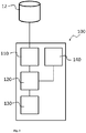
- Fig. 3 shows a schematic view of an embodiment of the computer system 100 according to the invention.
- the computer system 100 is connected to a database 12 on which the computed tomography images of a human thorax are stored. It is also conceivable that the database 12 is part of the computer system 100.
- the computer system 100 comprises a receiver unit 110 with which the computed tomography images can be received or retrieved.
- the computer system 100 comprises a control and computing unit 120 by which the computed tomography images can be analysed and by which features in the computed tomography images that indicate the presence of CTEPH can be recognized.
- the computer system 100 comprises a computing and checking unit 130 by which a probability of the presence of CTEPH can be calculated and by which it can be checked whether the probability is above a defined threshold.
- the computing and checking unit 130 may be part of the control and computing unit 120.
- the computer system 100 comprises an output unit 140 by which a communication as to the result of the analysis can be displayed to a person or transmitted to a person.
- Fig. 4 shows a schematic view of an embodiment of the control and computing unit 120 by which the computed tomography images can be analysed and by which features in the computed tomography images that indicate the presence of CTEPH can be recognized.
- the dotted components in Fig. 4 are preferred embodiments.
- the segmenting (121) of the cardiac region to identify the left ventricle, the right ventricle and the interventricular septum is followed by the determination (122) of a 2D slice image with the maximum right ventricle diameter by rotating a plane throughout the main axis of an ellipsoid fitted to the interventricular septum; and the determination (123) of at least one feature selected from the group of ratio of the diameters of the right ventricle and left ventricle, and degree of curvature of the interventricular septum based on the 2D slice image as determined in step (122) which indicate the presence of CTEPH.
- the segmenting step (121) includes producing (121a) a binary segmentation of left and right ventricles of the cardiac region; resampling (121b) the binary segmented image(s) to a isotropic spacing if required; thresholding (121c) the image(s) to segment the left ventricle and the right ventricle into two separate binary images; distance transforming (121d) each binary image and intersecting the images, and identifying (121e) the left and right ventricle, and the interventricular septum as the intersection volume between the left and the right ventricle.
- the determination step (122) includes fitting (122a) an ellipsoid to the left ventricle and calculating the centroid of the ellipsoid; using (122b) the centroid to fit an ellipsoid to the marginal points of the interventricular septum; analysing (122c) the orientation of the ellipsoid fitted to the interventricular septum.
- the orientation step (122c) preferably includes analysing (122d) the eigenvalues and eigenvectors of the ellipsoid fitted to the interventricular septum to determine the orientation in particular the Euler angles and the main axis of the ellipsoid.
- the determination step (123) includes identifying (123a) the diameters of the left and right ventricle by measuring the distance of the ventricle boundaries perpendicularly to the main axis of the ellipsoid fitted to the interventricular septum in the 2D slice image as determined in step (122).
- the determination step (123) also includes determination of the degree of curvature of the interventricular septum by skeletonizing (123b) the interventricular septum on the 2D slice image; by computing the centerline of the interventricular septum (123c); and by fitting (123d) a polynomial of 3 th order to the centerline and identifying local maxima of the polynomial.
- Fig. 5a shows an exemplary picture of the segmentation of the left ventricle, the right ventricle and the intersecting region after distance-transformation and thresholding (step 121d).
- Fig. 5b shows an exemplary picture of the intersection volume extracted from the intersecting region as shown in Fig. 5a (step 121e). This intersecting volume represents the interventricular septum.
- Fig. 6b shows an exemplary picture of 2D slice image taken for the identification of the centerline of the interventricular septum and the fitting of a polynomial of 3 th order to the centerline in order to determine the degree of curvature of the interventricular septum (steps 123b to 123d).
Landscapes
- Health & Medical Sciences (AREA)
- Medical Informatics (AREA)
- Engineering & Computer Science (AREA)
- Public Health (AREA)
- Epidemiology (AREA)
- General Health & Medical Sciences (AREA)
- Primary Health Care (AREA)
- Radiology & Medical Imaging (AREA)
- Nuclear Medicine, Radiotherapy & Molecular Imaging (AREA)
- Biomedical Technology (AREA)
- Data Mining & Analysis (AREA)
- Databases & Information Systems (AREA)
- Pathology (AREA)
- Apparatus For Radiation Diagnosis (AREA)
Abstract
Description
- The present invention is concerned with the radiological identification of chronic thromboembolic pulmonary hypertension (CTEPH). The invention relates to a method, a computer system and a computer program product for automatically identifying signs of the presence of CTEPH in a person.
- Chronic thromboembolic pulmonary hypertension (CTEPH) is a special form of pulmonary hypertension (PH). It is characterized by the infiltration of thrombi into the pulmonary arteries. They clog and constrict the vessels in a complete or partial manner and can be converted into connective tissue. In rare cases, there is the development of pulmonary hypertension with a poor prognosis.
- The symptoms of CTEPH are non-specific. In the early stage, dyspnoea and fatigue may occur. The duration of the initial symptoms for diagnosis is on average 14 months, and during the course of this some patients are then already in an advanced stage of the disease. This underlines the need for an exact and timely diagnosis.
- A timely diagnosis of CTEPH is also important because there are meanwhile options for the therapy of different forms of manifestation of this disease.
- The preferred CTEPH therapy is operative pulmonary endarterectomy (PEA), by means of which it is possible to heal up to 70% of patients. Meanwhile, perioperative mortality at experienced centres is 2-4%. However, about 30-50% of all CTEPH patients are classified as non-operable. For these patients and for patients with persistent pulmonary hypertension after PEA, a medicament-based therapy was authorized for the first time at the beginning of 2014 in the form of Riociguat.
- The gold standard for diagnosing or ruling out a case of CTEPH is ventilation/perfusion scintigraphy. The negative predictive value of perfusion scintigraphy is almost 100%; this means that a proper perfusion distribution rules out a case of CTEPH with a probability bordering on certainty.
- However, the problem is that CTEPH is comparatively rare. As a result of the rarity and the complex diagnosis and differential diagnosis, CTEPH is underdiagnosed.
- There is therefore a need for an early and uncomplicated identification of signs of the presence of CTEPH.
- According to the invention, this object is achieved by the subjects of
Claims 1, 9 and 14. Preferred embodiments can be found in the dependent claims and in the present description. - The present invention firstly provides a method for identifying signs of the presence of CTEPH in a person, comprising the following steps:
- receiving and/or retrieving one or more computed tomography images of the thorax of the person;
- analysing the one or more computed tomography images by:
- a) segmenting the cardiac region to identify the left ventricle, the right ventricle and the interventricular septum,
- b) determining a 2D slice image with the maximum right ventricle diameter by rotating a plane throughout the main axis of an ellipsoid fitted to the interventricular septum;
- determining at least one feature selected from the group of ratio of the diameters of the right ventricle and left ventricle, and degree of curvature of the interventricular septum based on the 2D slice image as determined in step b) which indicate the presence of CTEPH;
- calculating a probability of the presence of CTEPH on the basis of the at least one feature ascertained;
- communicating a message to the person and/or another person for further assessment of the case if the probability is above a defined threshold;
- The present invention further provides a computer system for identifying signs of the presence of CTEPH in a person, comprising:
- means for the automatic receipt and/or retrieval of one or more computed tomography images of the thorax of the person;
- means for the automated analysis of the one or more computed tomography images by:
- a) segmenting the cardiac region to identify the left ventricle, the right ventricle and the interventricular septum,
- b) determining a 2D slice image with the maximum right ventricle diameter by rotating a plane throughout the main axis of an ellipsoid fitted to the interventricular septum;
- means for the automated identification of at least one feature selected from the group of ratio of the diameters of the right ventricle and left ventricle, and degree of curvature of the interventricular septum based on the 2D slice image as determined in step b) which indicate the presence of CTEPH;
- means for automatically calculating a probability of the presence of CTEPH on the basis of the features ascertained;
- means for the automated communication of a message to the person and/or another person for further clarification of the finding.
- The present invention further provides a computer program product comprising a data carrier on which there is stored a computer program which can be loaded into the memory of a computer system, where it causes the computer system to execute the following steps:
- receiving and/or retrieving one or more computed tomography images of the thorax of the person;
- analysing the one or more computed tomography images by
- a) segmenting the cardiac region to identify the left ventricle, the right ventricle and the interventricular septum,
- b) determining a 2D slice image with the maximum right ventricle diameter by rotating a plane throughout the main axis of an ellipsoid fitted to the interventricular septum;
- determining at least one feature selected from the group of ratio of the diameters of the right ventricle and left ventricle, and the degree of curvature of the interventricular septum based on the 2D slice image as determined in step b) which indicate the presence of CTEPH;
- calculating a probability of the presence of CTEPH on the basis of the features ascertained;
- communicating a message to the person and/or another person for further assessment of the finding if the probability is above a defined threshold
- The individual elements which characterize the method according to the invention, the computer system and the computer program product are explained in more detail below. This explanation does not make a distinction between the individual subjects of the invention (method, computer system, computer program product). Instead, the following descriptions apply analogously to all subjects of the invention, irrespective of their context.
- In the identification of signs of the presence of CTEPH in a person, the present invention focusses on the automated image analysis of computed tomography images of the thorax of the person.
- Computed tomography (CT) is an X-ray method which depicts the human body in cross-sectional images (sectional imaging method). Compared to a conventional X-ray image, on which usually only coarse structures and bones are identifiable, CT images also capture in detail soft tissues with small differences in contrast. An X-ray tube generates a so-called X-ray fan beam, which penetrates the body and is attenuated to varying degrees within the body owing to the various structures, such as organs and bones. The receiving detectors opposite the X-ray emitter receive the signals of varying strength and forward them to a computer, which puts together cross-sectional images of the body from the received data. Computed tomography images (CT images) can be observed in 2D or else in 3D. For better differentiability of structures within the body of the person (e.g. vessels), a contrast agent can be injected into a vein before the generation of CT images.
- Computed tomography is a commonly used method in the diagnosis of heart and lung diseases. Preferably, the CT images are multidetector CT images.
- So-called multidetector CT (MDCT) refers to the newest generation of computed tomographs, having been available in clinical radiology since 1998. Multidetector CT is widely available and distinguished by a high, virtually isotropic resolution (pixel size 0.5-1 mm in each spatial direction), and this makes it possible to view the CT images in any spatial plane. The examination time varies between 1 and 10 seconds, giving rise to images virtually free of artefacts, even when there is dyspnoea or lack of patient cooperation. The latest MDCT scanners are equipped with "dual-energy" technology, in which two different energies/tube voltages are used simultaneously. Owing to the energy dependence of absorption, it is possible to highlight certain tissue properties, for example the distribution of iodine after administration of contrast agent as surrogate for regional blood circulation.
- A crucial criterion in the present invention is the automation. As already set out, CTEPH is a rare condition which is underdiagnosed. Non-detection of this condition can have fatal consequences for the patient. Therefore, according to the present invention, computed tomography images of the thorax are analysed in an automated manner for signs of the presence of CTEPH. What is meant here by "automated" is that no human intervention at all is required. According to the invention, a computer program is thus installed on a computer system that has access to computed tomography images of the thorax, runs as a background process, and analyses the images in an automated manner for signs of the presence of CTEPH. A background process refers to a process that does not act directly with the user and hence acts asynchronously to the user interface.
- In a first step of the method (and/or as part of the computer system, computer program product), one or more computed tomography images of the thorax of a person are received or retrieved. Customarily, a CT image is a data set, by means of which the structures of the thorax of the person can be depicted three-dimensionally. Thus, a CT image customarily represents the thorax of the person at the time of recording of the computed tomogram. Multiple CT images can e.g., represent the heart and/or pulmonary region of the person at different times; on the basis of these multiple CT images, it is possible therefore to identify temporal changes in the tissue structures and thus, for example, to examine a progression of a disease. However, it is also conceivable that the multiple CT images are CT images representing different regions of the thorax.
- As already described, computed tomography is a customary method for the diagnosis of heart and lung diseases. It is therefore conceivable to examine CT images already present in a database for the presence of signs of CTEPH. In one embodiment of the present invention, CT images present in one or more databases are retrieved and are analysed for the presence of signs of CTEPH. For example, this can occur at regular intervals. It is, for example, conceivable to carry out at regular intervals, for example every day or every week, a search for new CT images in the databases in which CT images are customarily deposited and to retrieve the new CT images for an image analysis. However, the retrieval can also take place irregularly. The retrieval can also be triggered by an event, for example by the storing of a new CT image. Preferably, the retrieval of new CT images is done in an automated manner.
- It is also conceivable to carry out the analysis according to the invention for the presence of signs of CTEPH as a kind of standard analysis for each CT image generated from the thorax of a person. Accordingly, in a further preferred embodiment of the present invention, a CT image generated from the thorax of a person is, after its generation, immediately and automatically subjected to an image analysis according to the invention. In such a case, a computer system oriented to generating a relevant CT image can be configured such that it forwards the CT image to the image analysis according to the invention. The components executing the image analysis receive the CT image.
- In a further step of the method (and/or as part of the computer system, computer program product), an automated analysis of the one or more CT image takes place (preferably more than one CT image). The analysis is carried out by:
- a) segmenting the cardiac region to identify the left ventricle, the right ventricle and the interventricular septum,
- b) determining a 2D slice image with the maximum right ventricle diameter by rotating a plane throughout the main axis of an ellipsoid fitted to the interventricular septum.
- Image-based segmentation techniques of the cardiac region consist of thresholding, region-growing, clustering, pixel or voxel classification, and active contour or surface (Peng et al, A review of heart chamber segmentation for structural and functional analysis using cardiac magnetic resonance imaging, 2016, 29:155-195). In recent years, cardiac segmentation methods are based on deep learning which can be roughly divided into two classes: 2D methods which segment each slice independently and 3D methods, which segment multiple slices together as a volume (Qiao Zheng et al., 3D consistent robust segmentation of cardiac images by deep learning with spatial propagation, IEEE Trans. On Medical Imaging, 2018, hal-01753086).
- In particular, segmentation of the cardiac region (referred to above as step a) includes
- producing a binary segmentation of left and right ventricles of the cardiac region,
- resampling the binary segmented image(s) to an isotropic spacing if required,
- thresholding the image(s) to segment the left ventricle and the right ventricle into two separate binary images,
- distance transforming each binary image and intersecting the images,
- identifying the left and right ventricle, and the interventricular septum as the intersection volume between the left and the right ventricle.
- Binary segmentation of left and right ventricles of the cardiac region refers in particular to 3D CT images marked by different grey values. The left and right ventricles are in particular coded by different grey values. Resampling of the binary segmented images to an isotropic spacing is usually required as 3D CT images contain voxels for which the spacing in the three spatial directions is not uniform. Resampling refers to process step wherein an isotropic spacing in all direction of the 3D images is achieved. In particular, a spacing value of equal or below 2.5 mm, more particularly equal or below 2.0 mm is targeted. Resampling refers preferably also to the extraction of superfluous tissue such as the interventricular septum e.g., with morphological operators.
- In the thresholding step both ventricles are segmented into two new distinct images. In particular, this step also includes an object analysis step in both images to detect the geometric region enclosing the left and the right ventricles followed by a union of the regions. The distance transforming step results in grey level images that look similar to the input images, except that the distance of a voxel from the edge of an object is coded as a grey value: the higher the grey value, the higher the distance. The distance-transformed images are intersected, preferably by using a thresholding step to from a thick, intersecting border around the both ventricles. Finally, the intersection volume is extracted defining the new interventricular septum.
- The step b) of the method (and/or as part of the computer system, computer program product) refers to determining a 2D slice image with the maximum right ventricle diameter by rotating a plane throughout the main axis of an ellipsoid fitted to the interventricular septum.
- A basis for determining the appropriate 2D slice image is the ellipsoid fitting to the interventricular septum and the determination of a preferred direction and a geometrical characterization of the interventricular septum.
- The ellipsoid is a special case of a surface of second order. Such a surface is described by the following implicit function:

- Or in matrix notation:

- With:

- Equation (1) describes an ellipsoid, when the following condition is fulfilled:

- Where:


- When fitting the surface of second order (equation 1) to the marginal points of the interventricular septum, the ellipsoid condition (equation 6) is in the majority of cases not fulfilled. In some cases a hyperboloid can be fitted. In order to address this issue a constraint condition can be set to force the fitting of the ellipsoid to the interventricular septum (method of Lagrange multiplier). In a preferred embodiment, however, an ellipsoid is pre-fitted to the left ventricle and the centroid of this ellipsoid is used as a constraint condition for fitting a second ellipsoid to the interventricular septum. The left ventricle is in most cases almost rotation symmetric and the fitted parameters of the surface of second order (equation 1) fulfils the condition of the ellipsoid condition (equation 6).
- The ellipsoid fitted to the interventricular septum is preferably identified by:
- fitting an ellipsoid to the left ventricle and calculating the centroid of the ellipsoid,
- using the centroid to fit an ellipsoid to the marginal points of the interventricular septum,
- analysing the orientation of the ellipsoid fitted to the interventricular septum.
- Analysis of the orientation of the ellipsoid fitted to the interventricular septum preferably includes
- analysing the eigenvalues and eigenvectors of the ellipsoid fitted to the interventricular septum to determine the orientation in particular the Euler angles and the main axis of the ellipsoid.
- The centroid of the ellipsoid fitted to the left ventricle is expressed by the following equation 8:

- Where:

-
Equation 2 then simplifies to the following expression:
- The radius of the ellipsoid and the coefficients of the rotation matrix are obtained through an eigenvalue analysis of Matrix B:


- The rotation matrix is expressed as a function of the Euler angles Φ, ϑ, and ψ.

- The Euler angles are calculated from
equation - In order to determine the 2D slice image with the maximum right ventricle diameter preferably a 3D image is rotated in the rage of [-90, +90] around the main (middle) axis of the ellipsoid fitted to the interventricular septum.
- As a next step the method (and/or as part of the computer system, computer program product) relates to the determination of at least one feature selected from the group of ratio of the diameters of the right ventricle and left ventricle, and the degree of curvature of the interventricular septum based on the 2D slice image as determined in the previous step b) which indicate the presence of CTEPH.
- The determination of the ratio of the diameters of the right ventricle and left ventricle preferably includes
- identifying the diameters of the left and right ventricle by a measuring the distance of the ventricle boundaries perpendicularly to the main axis of the ellipsoid fitted to the interventricular septum in the 2D slice image as determined in step b) and calculating the ratio of the diameters.
- The ratio of the diameters of right ventricle and left ventricle (RV/LV ratio) (see, for example, Gonzales G et al. PLoS ONE 10(5): e0127797) is a feature that can indicate the presence of CTEPH. A value of 0.9 or more in the RV/LV diameter ratio is an indication of the presence of CTEPH.
- The determination of the degree of curvature of the interventricular septum based on the 2D slice image as determined in step b) preferably includes
- skeletonizing the interventricular septum on the 2D slice image,
- computing the centerline of the interventricular septum,
- fitting a polynomial of 3th order to the centerline and identifying local maxima of the polynomial.
- The determination of the degree of curvature of the interventricular septum requires the extraction of the centerline. This is preferably done by applying a skeleton operator. In some cases the skeleton image may contain additional branches. These branches can be excluded with minimum-path, graph based approaches. Then, a polynomial of higher order is fitted to the centerline. It has turned out, that a polynomial of the 3th leads to best results. The degree of curvature of the interventricular septum is determined around the local maxima of the polynomial, these points are identified by computing the first derivative.
- The degree of curvature of the interventricular septum (see, for example, D.A. Moses et al., Quantification of the curvature and shape of the interventricular septum; Magnetic Resonance in Medicine, Vol. 52 (1), 2004, 154-163 and F. Haddad et al.: Septal Curvature is a marker of hemodynamic, anatomical, and electromechanical ventricular interdependence in patients with pulmonary arterial hypertension, Echocardiography Vol. 31(6) 2014, 699-707).
- Preferably, the method (and/or as part of the computer system, computer program product) takes into account at least one additional feature indicative for CTEP selected from the group of volumes of the right ventricle and left ventricle, ratio of the volumes of the right ventricle and the left ventricle, ratio of the diameters of the pulmonary artery and the aorta at the level at which the pulmonary artery branches off, presence and/or severity of stenoses, presence and/or severity of mosaic perfusion, presence and/or severity of ground glass opacity.
- The ratio of the diameters of the pulmonary artery and the aorta (PA:A ratio) is determined at the point at which the pulmonary artery branches off (see, for example, A.S. Iyer et al.: CT scan-measured pulmonary artery to aorta ratio and echocardiography for detecting pulmonary hypertension in severe COPD, CHEST 2014, Vol. 145(4), 824-832). A PA:A ratio of 0.7 or greater is a further indication of the presence of CTEPH. Characteristic vascular features are, for example, the lack of contrast agent in the distal vessel sections in the case of total obstruction or the formation of rope-ladder thrombi, meshes, stenoses and partial obstructions. The CTEPH-specific parenchymal features include scars, mosaic perfusion, ground glass opacity and bronchial anomalies. The scars arise as a result of infarctions owing to the occlusion of pulmonary vessels that are usually localized in the lower segments. Mosaic perfusion consists of regions of different density resulting from regions of irregular hypo- and hyperperfused regions caused by embolic occlusions, vascular rearrangement of the distal vessels and compensation mechanisms. Hypoperfused regions are observed particularly in the distal direction from the occluded vessels since blood flow and hence the concentration of the contrast agent are reduced in these regions. Hyperdense areas are usually apparent in regions that are now hyperperfused owing to the redistribution and stand out as ground glass opacity. The latter and further characteristic features are described in the literature (see, for example, J. E. Leifheit, Characterization of ground glass opacity in patients with chronic thromboembolic pulmonary hypertension compared to pulmonary hypertension of other WHO classification using Dual Energy CT [in German], Inaugural Dissertation for Attaining the Degree of Doctor of Medicine at the Justus Liebig University of Giessen, 2017).
- It is conceivable that, in the image analysis, no at least one CTEPH-specific feature is found in the CT images examined. In such a case, it is possible to store in a database, in relation to the relevant CT image or in relation to the person from whom the CT image was generated, an item of information indicating that no at least one CTEPH-specific feature were identified in the CT image.
- If at least one feature indicating the presence of CTEPH was found, it is possible to store in a database, in relation to the relevant CT image or in relation to the person from whom the CT image was generated, an item of information indicating that at least one CTEPH-specific feature was identified in the CT image.
- In a further step of the method (and/or as part of the computer system, computer program product), a calculation of a probability of the presence of CTEPH is effected on the basis of the characteristic feature(s) ascertained. A value of 100% indicates that the patient is suffering from CTEPH; a value of 0% indicates that CTEPH can be ruled out.
- The probability can be calculated by following many different approaches. It is conceivable, for example, that the ratio of the diameters of the right ventricle and left ventricle and/or degree of curvature of the interventricular septum as ascertained with the analysis described above is/are compared with predefined values. It is also conceivable, for example, that the one or more CT images are examined for the presence of a number of characteristic features. The probability can be calculated from the sum of the number of features found divided by the sum of the number of features tested (P = (sum of features found)/(sum of features tested)). In such an approach, all features are of equal value. Alternatively, it is conceivable that the individual features are weighted with a factor, such that features that are more likely to indicate CTEPH are given a higher value in the probability function than features that are less specific. It is also conceivable that the severity of a feature is determined; where the severity indicates the extent in respect of the presence of CTEPH. The higher the degree (the extent), the clearer the manifestation of the feature and the higher the probability of the presence of CTEPH. It is also conceivable that one or more decision trees or regression trees are run; it may be the case that one feature indicates CTEPH only in combination with another feature. Further methods and combinations of methods of ascertaining the probability are conceivable.
- If the probability is above a defined threshold, a message is generated that gives information that the person who was the subject of the CT images has a defined probability of suffering from CTEPH and therefore further examinations should be undertaken to clarify the finding. The threshold value may, for example, be between 20% and 70%. It is preferably above 20% and below 51%.
- According to the invention, a message that the person should be subjected to a further diagnostic procedure in order to confirm CTEPH or to reliably rule it out is then communicated. Said message can, for example, be addressed to the person from whom the relevant CT image originates. However, it can also be addressed to his/her physician or hospital carer or to another person who is in contact with the person for whom there are signs of CTEPH. The communication of the message can be a text message (e-mail, SMS, etc.) or voice message.
- The invention is described in detail hereinafter with reference to figures and examples, without wishing to restrict the invention to the features or combinations of features described and shown.
-
Figure 1 shows, by way of example, one embodiment for the implementation of the system according to the invention. -
Figure 1 shows aCT system 1 executed as a twin-focus detector system. It has afirst x-ray tube 2 with an opposite detector and a second x-ray tube 4 with a further opposite detector 5. Both focus/detector systems 2, 3 and 4, 5 are arranged in agantry housing 6 on a gantry which rotates about a system axis 9 and is not visualized here. Thepatient 7 is on a longitudinallymovable patient bed 8. Before the scanning of thepatient 7, to improve the contrast of a CT image reconstructed from the detector output data, a contrast agent is administered to thepatient 7 by means of acontrast agent injector 12. The control of the overall CT system and if necessary also the evaluation of the detector data and the reconstruction of the CT image as section images or volume data can be effected by a control andcomputing unit 10. This control andcomputing unit 10 has amemory 11 which stores, as well as the detector output data measured, computer program data Prg1-Prgn that are executed in operation and essentially assume the function of controlling the system and the evaluation of the data. - In one embodiment of the present invention, the computer program according to the invention runs as a background process on the control and
computing unit 10. It analyses the section images or volume data for the presence of CTEPH indications. If CTEPH indications are identified, and a calculated probability of the presence of CTEPH is above a defined threshold, the computer program according to the invention creates a message on the screen of the control andcomputing unit 10 that informs the radiologist that there is a suspicion of CTEPH. -
Figure 2 shows a schematic of a further embodiment for the implementation of the system according to the invention. - The
CT system 1 is connected via the connection 14-1 to the control andcomputing unit 10. The control andcomputing unit 10 controls theCT system 1 and evaluates the detector data and reconstructs the CT image as section images or volume data. The section images and volume data are stored in adatabase 12 to which the control andcomputing unit 10 is connected via the connection 14-2. It is also conceivable that the database is part of the control andcomputing unit 10. Thecomputer system 13 can also access thedatabase 12 via the connection 14-3. The computer program according to the invention is running on thecomputer system 13. It is configured such that it analyses the CT images of a human thorax stored in thedatabase 12 for indications of the presence of CTEPH. If no indications are identified, corresponding information relating to the CT images is stored. If indications of the presence of CTEPH are identified, the corresponding information relating to the CT images is likewise stored. - The computer program installed and running on the
computer system 13 is configured such that, on the basis of the features ascertained that indicate CTEPH, a probability of the presence of CTEPH is calculated. If this probability is above a defined threshold, the computer program creates a message that CTEPH could be present. - The computer program installed and running on the
computer system 13 may be configured such that it displays the communication as to the presence of CTEPH indications on a screen which is part of thecomputer system 13. It is also conceivable that the computer program is configured such that it transmits a message of the presence of CTEPH indications via the connection 14-4 to the control andcomputing unit 10, via which the message is then displayed, for example on a screen. It is also conceivable that thecomputer system 10 draws the information as to whether CTEPH indications are present directly from thedatabase 12. It is also conceivable that the computer program is configured such that it transmits a message of the presence of CTEPH indications via the connection 14-5 to afurther computer system 15, via which the message is then displayed, for example via a screen. It is also conceivable that thecomputer system 15 draws the information as to whether CTEPH indications are present from thedatabase 12 via the connection 14-6. The dotted components inFig. 2 are optional. The connections 14-1, 14-2, 14-3, 14-4, 14-5 and 14-6 may be cable connections, glass fibre-based connections and/or wireless connections (e.g. via radio). -
Fig. 3 shows a schematic view of an embodiment of thecomputer system 100 according to the invention. - The
computer system 100 is connected to adatabase 12 on which the computed tomography images of a human thorax are stored. It is also conceivable that thedatabase 12 is part of thecomputer system 100. Thecomputer system 100 comprises areceiver unit 110 with which the computed tomography images can be received or retrieved. Thecomputer system 100 comprises a control andcomputing unit 120 by which the computed tomography images can be analysed and by which features in the computed tomography images that indicate the presence of CTEPH can be recognized. Thecomputer system 100 comprises a computing and checkingunit 130 by which a probability of the presence of CTEPH can be calculated and by which it can be checked whether the probability is above a defined threshold. The computing and checkingunit 130 may be part of the control andcomputing unit 120. Thecomputer system 100 comprises anoutput unit 140 by which a communication as to the result of the analysis can be displayed to a person or transmitted to a person. -
Fig. 4 shows a schematic view of an embodiment of the control andcomputing unit 120 by which the computed tomography images can be analysed and by which features in the computed tomography images that indicate the presence of CTEPH can be recognized. The dotted components inFig. 4 are preferred embodiments. - The segmenting (121) of the cardiac region to identify the left ventricle, the right ventricle and the interventricular septum is followed by the determination (122) of a 2D slice image with the maximum right ventricle diameter by rotating a plane throughout the main axis of an ellipsoid fitted to the interventricular septum; and the determination (123) of at least one feature selected from the group of ratio of the diameters of the right ventricle and left ventricle, and degree of curvature of the interventricular septum based on the 2D slice image as determined in step (122) which indicate the presence of CTEPH. It is conceivable that the segmenting step (121) includes producing (121a) a binary segmentation of left and right ventricles of the cardiac region; resampling (121b) the binary segmented image(s) to a isotropic spacing if required; thresholding (121c) the image(s) to segment the left ventricle and the right ventricle into two separate binary images; distance transforming (121d) each binary image and intersecting the images, and identifying (121e) the left and right ventricle, and the interventricular septum as the intersection volume between the left and the right ventricle. It is further conceivable that the determination step (122) includes fitting (122a) an ellipsoid to the left ventricle and calculating the centroid of the ellipsoid; using (122b) the centroid to fit an ellipsoid to the marginal points of the interventricular septum; analysing (122c) the orientation of the ellipsoid fitted to the interventricular septum. The orientation step (122c) preferably includes analysing (122d) the eigenvalues and eigenvectors of the ellipsoid fitted to the interventricular septum to determine the orientation in particular the Euler angles and the main axis of the ellipsoid. The determination step (123) includes identifying (123a) the diameters of the left and right ventricle by measuring the distance of the ventricle boundaries perpendicularly to the main axis of the ellipsoid fitted to the interventricular septum in the 2D slice image as determined in step (122). The determination step (123) also includes determination of the degree of curvature of the interventricular septum by skeletonizing (123b) the interventricular septum on the 2D slice image; by computing the centerline of the interventricular septum (123c); and by fitting (123d) a polynomial of 3th order to the centerline and identifying local maxima of the polynomial.
-
Fig. 5a shows an exemplary picture of the segmentation of the left ventricle, the right ventricle and the intersecting region after distance-transformation and thresholding (step 121d). -
Fig. 5b shows an exemplary picture of the intersection volume extracted from the intersecting region as shown inFig. 5a (step 121e). This intersecting volume represents the interventricular septum. -
Fig. 6a shows an exemplary picture of the determination of the 2D slice image with the maximum right ventricle diameter (which is 47.5 mm) by rotating (ψ =0.3) a plane throughout the main axis of an ellipsoid fitted to the interventricular septum (step 122). -
Fig. 6b shows an exemplary picture of 2D slice image taken for the identification of the centerline of the interventricular septum and the fitting of a polynomial of 3th order to the centerline in order to determine the degree of curvature of the interventricular septum (steps 123b to 123d).
Claims (14)
- Method for identifying signs of the presence of CTEPH in a person, comprising the following steps:- receiving and/or retrieving one or more computed tomography images of the thorax of the person;- analysing the one or more computed tomography images by:a) segmenting the cardiac region to identify the left ventricle, the right ventricle and the interventricular septum,b) determining a 2D slice image with the maximum right ventricle diameter by rotating a plane throughout the main axis of an ellipsoid fitted to the interventricular septum;- determining at least one feature selected from the group of ratio of the diameters of the right ventricle and left ventricle, and degree of curvature of the interventricular septum based on the 2D slice image as determined in step b) which indicate the presence of CTEPH;- calculating a probability of the presence of CTEPH on the basis of the at least one feature ascertained;- communicating a message to the person and/or another person for further assessment of the case if the probability is above a defined threshold;where the steps mentioned run automatically as background processes in one or more computer systems.
- Method according to Claim 1, in which step a) includes:- producing a binary segmentation of left and right ventricles of the cardiac region,- resampling the binary segmented image(s) to a isotropic spacing if required,- thresholding the image(s) to segment the left ventricle and the right ventricle into two separate binary images,- distance transforming each binary image and intersecting the images,- identifying the left and right ventricle, and the interventricular septum as the intersection volume between the left and the right ventricle.
- Method according to any one of Claims 1 to 2, in which step b) includes:- fitting an ellipsoid to the left ventricle and calculating the centroid of the ellipsoid,- using the centroid to fit an ellipsoid to the marginal points of the interventricular septum,- analysing the orientation of the ellipsoid fitted to the interventricular septum,
- Method according to Claim 3, in which analysing the orientation of the ellipsoid fitted to the interventricular septum includes- analysing the eigenvalues and eigenvectors of the ellipsoid fitted to the interventricular septum to determine the orientation in particular the Euler angles and the main axis of the ellipsoid.
- Method according to any one of Claims 1 to 4, in which determination of the ratio of the diameters of the right ventricle and left ventricle based on the 2D slice image as determined in step b) includes the- identifying the diameters of the left and right ventricle by measuring the distance of the ventricle boundaries perpendicularly to the main axis of the ellipsoid fitted to the interventricular septum in the 2D slice image as determined in step b).
- Method according to any one of Claims 1 to 5, in which determination of the degree of curvature of the interventricular septum based on the 2D slice image as determined in step b) includes- skeletonizing the interventricular septum on the 2D slice image,- computing the centerline of the interventricular septum,- fitting a polynomial of 3th order to the centerline and identifying local maxima of the polynomial.
- Method according to any one of the Claims 1 to 6, wherein computed tomography images present in one or more databases are automatically retrieved and forwarded to analysis.
- Method according to any one of the Claim 1 to 7, wherein computed tomography images, after their generation, are received from that computer system which generated the computed tomography images and forwarded to analysis.
- Computer system for identifying signs of the presence of CTEPH in a person, comprising:- means for the automatic receipt and/or retrieval of one or more computed tomography images of the thorax of the person;- means for the automated analysis of the one or more computed tomography images by:a) segmenting the cardiac region to identify the left ventricle, the right ventricle and the interventricular septum,b) determining a 2D slice image with the maximum right ventricle diameter by rotating a plane throughout the main axis of an ellipsoid fitted to the interventricular septum;- means for the automated identification of at least one feature selected from the group of ratio of the diameters of the right ventricle and left ventricle, and degree of curvature of the interventricular septum based on the 2D slice image as determined in step b) which indicate the presence of CTEPH;- means for automatically calculating a probability of the presence of CTEPH on the basis of the features ascertained;- means for the automated communication of a message to the person and/or another person for further clarification of the finding.
- Computer system according to Claim 9, comprising
a receiver unit with which the computed tomography images can be received and/or retrieved,
a control and computing unit by which the computed tomography images can be analysed and by which at least one feature in the computed tomography images that indicate the presence of CTEPH can be recognized,
a computing and checking unit 130 by which a probability of the presence of CTEPH can be calculated and by which it can be checked whether the probability is above a defined threshold,
an output unit 140 by which a communication as to the result of the analysis can be displayed to a person or transmitted to a person. - Computer system according to any one of Claims 9 and 10, wherein the computer system is configured such that the following processes run in an automated manner as background processes without human intervention:- receiving and/or retrieving one or more computed tomography images of the thorax of the person from a database;- analysing the one or more computed tomography images bya) segmenting the cardiac region to identify the left ventricle, the right ventricle and the interventricular septum,b) determining a 2D slice image with the maximum right ventricle diameter by rotating a plane throughout the main axis of an ellipsoid fitted to the interventricular septum;- determining at least one feature selected from the group of ratio of the diameters of the right ventricle and left ventricle, and the degree of curvature of the interventricular septum based on the 2D slice image as determined in step b) which indicate the presence of CTEPH;- calculating a probability of the presence of CTEPH on the basis of the at least one feature ascertained;- communicating a message to the person and/or another person for further assessment of the case if the probability is above a defined threshold.
- Computer system according to any one of Claims 9 to 11 which is part of a CT system.
- Computer system according to any of Claims 9 to 11 which is connected to a database on which CT images from a CT system are stored.
- Computer program product comprising a data carrier on which there is stored a computer program which can be loaded into the memory of a computer system, where it causes the computer system to execute the following steps:- receiving and/or retrieving one or more computed tomography images of the thorax of the person;- analysing the one or more computed tomography images bya) segmenting the cardiac region to identify the left ventricle, the right ventricle and the interventricular septum,b) determining a 2D slice image with the maximum right ventricle diameter by rotating a plane throughout the main axis of an ellipsoid fitted to the interventricular septum;- determining at least one feature selected from the group of ratio of the diameters of the right ventricle and left ventricle, and the degree of curvature of the interventricular septum based on the 2D slice image as determined in step b) which indicate the presence of CTEPH;- calculating a probability of the presence of CTEPH on the basis of the features ascertained;- communicating a message to the person and/or another person for further assessment of the finding if the probability is above a defined thresholdwhere the steps mentioned run automatically as background processes on the computer system.
Priority Applications (1)
| Application Number | Priority Date | Filing Date | Title |
|---|---|---|---|
| EP19185178.1A EP3764365A1 (en) | 2019-07-09 | 2019-07-09 | Improvements in the radiological identification of chronic thromboembolic pulmonary hypertension |
Applications Claiming Priority (1)
| Application Number | Priority Date | Filing Date | Title |
|---|---|---|---|
| EP19185178.1A EP3764365A1 (en) | 2019-07-09 | 2019-07-09 | Improvements in the radiological identification of chronic thromboembolic pulmonary hypertension |
Publications (1)
| Publication Number | Publication Date |
|---|---|
| EP3764365A1 true EP3764365A1 (en) | 2021-01-13 |
Family
ID=67226006
Family Applications (1)
| Application Number | Title | Priority Date | Filing Date |
|---|---|---|---|
| EP19185178.1A Withdrawn EP3764365A1 (en) | 2019-07-09 | 2019-07-09 | Improvements in the radiological identification of chronic thromboembolic pulmonary hypertension |
Country Status (1)
| Country | Link |
|---|---|
| EP (1) | EP3764365A1 (en) |
Citations (1)
| Publication number | Priority date | Publication date | Assignee | Title |
|---|---|---|---|---|
| US20160260210A1 (en) * | 2013-11-27 | 2016-09-08 | Universidad Politécnica de Madrid | Method and system for determining the prognosis of a patient suffering from pulmonary embolism |
-
2019
- 2019-07-09 EP EP19185178.1A patent/EP3764365A1/en not_active Withdrawn
Patent Citations (1)
| Publication number | Priority date | Publication date | Assignee | Title |
|---|---|---|---|---|
| US20160260210A1 (en) * | 2013-11-27 | 2016-09-08 | Universidad Politécnica de Madrid | Method and system for determining the prognosis of a patient suffering from pulmonary embolism |
Non-Patent Citations (10)
| Title |
|---|
| A.S. IYER ET AL.: "CT scan-measured pulmonary artery to aorta ratio and echocardiography for detecting pulmonary hypertension in severe COPD", CHEST, vol. 145, no. 4, 2014, pages 824 - 832 |
| BENJAMIN H. FREED ET AL: "MR and CT Imaging for the Evaluation of Pulmonary Hypertension", JACC: CARDIOVASCULAR IMAGING, vol. 9, no. 6, 1 June 2016 (2016-06-01), AMSTERDAM, NL, pages 715 - 732, XP055654791, ISSN: 1936-878X, DOI: 10.1016/j.jcmg.2015.12.015 * |
| D.A. MOSES ET AL.: "Quantification of the curvature and shape of the interventricular septum", MAGNETIC RESONANCE IN MEDICINE, vol. 52, no. 1, 2004, pages 154 - 163, XP002451153 |
| F. HADDAD ET AL.: "Septal Curvature is a marker of hemodynamic, anatomical, and electromechanical ventricular interdependence in patients with pulmonary arterial hypertension", ECHOCARDIOGRAPHY, vol. 31, no. 6, 2014, pages 699 - 707 |
| GERMÁN GONZÁLEZ ET AL: "Automated Axial Right Ventricle to Left Ventricle Diameter Ratio Computation in Computed Tomography Pulmonary Angiography", PLOS ONE, vol. 10, no. 5, 22 May 2015 (2015-05-22), pages e0127797, XP055654784, DOI: 10.1371/journal.pone.0127797 * |
| J. E. LEIFHEIT: "Characterization of ground glass opacity in patients with chronic thromboembolic pulmonary hypertension compared to pulmonary hypertension of other WHO classification using Dual Energy CT [in German", INAUGURAL DISSERTATION FOR ATTAINING THE DEGREE OF DOCTOR OF MEDICINE AT THE JUSTUS LIEBIG UNIVERSITY OF GIESSEN, 2017 |
| KANAKO K. KUMAMARU ET AL: "Implementation and Performance of Automated Software for Computing Right-to-Left Ventricular Diameter Ratio From Computed Tomography Pulmonary Angiography Images :", JOURNAL OF COMPUTER ASSISTED TOMOGRAPHY, vol. 40, no. 3, 1 January 2016 (2016-01-01), US, pages 387 - 392, XP055654787, ISSN: 0363-8715, DOI: 10.1097/RCT.0000000000000375 * |
| PENG ET AL., A REVIEW OF HEART CHAMBER SEGMENTATION FOR STRUCTURAL AND FUNCTIONAL ANALYSIS USING CARDIAC MAGNETIC RESONANCE IMAGING, vol. 29, 2016, pages 155 - 195 |
| QIAO ZHENG ET AL.: "3D consistent robust segmentation of cardiac images by deep learning with spatial propagation", IEEE TRANS. ON MEDICAL IMAGING, 2018 |
| SIMISANI TAKOBANA: "Quantification o f Right Ventricular Function in Pulmonary Hypertension using Cardiac PET Images", CARLETON UNIVERSITY, 1 January 2012 (2012-01-01), XP055655157 * |
Similar Documents
| Publication | Publication Date | Title |
|---|---|---|
| US20200237331A1 (en) | Improvements in the radiological detection of chronic thromboembolic pulmonary hypertension | |
| US7567696B2 (en) | System and method for detecting the aortic valve using a model-based segmentation technique | |
| US8229200B2 (en) | Methods and systems for monitoring tumor burden | |
| US7940970B2 (en) | Method and system for automatic quality control used in computerized analysis of CT angiography | |
| US8630467B2 (en) | Diagnosis assisting system using three dimensional image data, computer readable recording medium having a related diagnosis assisting program recorded thereon, and related diagnosis assisting method | |
| US8620040B2 (en) | Method for determining a 2D contour of a vessel structure imaged in 3D image data | |
| US20190021677A1 (en) | Methods and systems for classification and assessment using machine learning | |
| US20050110791A1 (en) | Systems and methods for segmenting and displaying tubular vessels in volumetric imaging data | |
| CN105934199B (en) | Apparatus and method for processing medical images of body cavities | |
| KR102469907B1 (en) | Medical image reconstruction apparatus and method for screening a plurality of types lung diseases | |
| US20030095692A1 (en) | Method and system for lung disease detection | |
| US20250022133A1 (en) | A method and processor for analyzing the vasculature of a subject | |
| JP2009045436A (en) | Apparatus and method for identifying occlusions | |
| WO2008030192A1 (en) | Detection and localization of vascular occlusion from angiography data | |
| EP4099269A1 (en) | Imaging system and method | |
| EP2619729B1 (en) | Quantification of a characteristic of a lumen of a tubular structure | |
| CN116563256A (en) | Method, device and storage medium for determining vascular stenosis rate and embolism level | |
| Larralde et al. | Evaluation of a 3D segmentation software for the coronary characterization in multi-slice computed tomography | |
| US20250157039A1 (en) | Automatic computation of bpe level | |
| EP3764365A1 (en) | Improvements in the radiological identification of chronic thromboembolic pulmonary hypertension | |
| Kim et al. | Use of cardiac computed tomography for ventricular Volumetry in late postoperative patients with tetralogy of Fallot | |
| US20250054138A1 (en) | Interventional characterization of intra-cerebral thrombus using 3d cone beam computed tomography | |
| Ferreira et al. | Assisted Vascular Analysis (AVA) for Deep Inferior Epigastric Perforators: Pipeline Analysis | |
| JP2025078926A (en) | Information processing system and program | |
| Brathwaite | Characterization of arterial lesions in three-dimensional intravascular ultrasound images |
Legal Events
| Date | Code | Title | Description |
|---|---|---|---|
| PUAI | Public reference made under article 153(3) epc to a published international application that has entered the european phase |
Free format text: ORIGINAL CODE: 0009012 |
|
| STAA | Information on the status of an ep patent application or granted ep patent |
Free format text: STATUS: THE APPLICATION HAS BEEN PUBLISHED |
|
| AK | Designated contracting states |
Kind code of ref document: A1 Designated state(s): AL AT BE BG CH CY CZ DE DK EE ES FI FR GB GR HR HU IE IS IT LI LT LU LV MC MK MT NL NO PL PT RO RS SE SI SK SM TR |
|
| AX | Request for extension of the european patent |
Extension state: BA ME |
|
| STAA | Information on the status of an ep patent application or granted ep patent |
Free format text: STATUS: REQUEST FOR EXAMINATION WAS MADE |
|
| 17P | Request for examination filed |
Effective date: 20210713 |
|
| RBV | Designated contracting states (corrected) |
Designated state(s): AL AT BE BG CH CY CZ DE DK EE ES FI FR GB GR HR HU IE IS IT LI LT LU LV MC MK MT NL NO PL PT RO RS SE SI SK SM TR |
|
| STAA | Information on the status of an ep patent application or granted ep patent |
Free format text: STATUS: THE APPLICATION HAS BEEN WITHDRAWN |
|
| 18W | Application withdrawn |
Effective date: 20230718 |