EP3764101A1 - Tumor marker, and method for collecting and detecting tumor cell in distinction from contaminant cell - Google Patents
Tumor marker, and method for collecting and detecting tumor cell in distinction from contaminant cell Download PDFInfo
- Publication number
- EP3764101A1 EP3764101A1 EP19764529.4A EP19764529A EP3764101A1 EP 3764101 A1 EP3764101 A1 EP 3764101A1 EP 19764529 A EP19764529 A EP 19764529A EP 3764101 A1 EP3764101 A1 EP 3764101A1
- Authority
- EP
- European Patent Office
- Prior art keywords
- amino acid
- seq
- acid sequence
- polypeptide
- sample
- Prior art date
- Legal status (The legal status is an assumption and is not a legal conclusion. Google has not performed a legal analysis and makes no representation as to the accuracy of the status listed.)
- Pending
Links
Images






Classifications
-
- C—CHEMISTRY; METALLURGY
- C07—ORGANIC CHEMISTRY
- C07K—PEPTIDES
- C07K14/00—Peptides having more than 20 amino acids; Gastrins; Somatostatins; Melanotropins; Derivatives thereof
- C07K14/435—Peptides having more than 20 amino acids; Gastrins; Somatostatins; Melanotropins; Derivatives thereof from animals; from humans
- C07K14/46—Peptides having more than 20 amino acids; Gastrins; Somatostatins; Melanotropins; Derivatives thereof from animals; from humans from vertebrates
- C07K14/47—Peptides having more than 20 amino acids; Gastrins; Somatostatins; Melanotropins; Derivatives thereof from animals; from humans from vertebrates from mammals
- C07K14/4701—Peptides having more than 20 amino acids; Gastrins; Somatostatins; Melanotropins; Derivatives thereof from animals; from humans from vertebrates from mammals not used
- C07K14/4748—Tumour specific antigens; Tumour rejection antigen precursors [TRAP], e.g. MAGE
-
- G—PHYSICS
- G01—MEASURING; TESTING
- G01N—INVESTIGATING OR ANALYSING MATERIALS BY DETERMINING THEIR CHEMICAL OR PHYSICAL PROPERTIES
- G01N33/00—Investigating or analysing materials by specific methods not covered by groups G01N1/00 - G01N31/00
- G01N33/48—Biological material, e.g. blood, urine; Haemocytometers
- G01N33/50—Chemical analysis of biological material, e.g. blood, urine; Testing involving biospecific ligand binding methods; Immunological testing
- G01N33/53—Immunoassay; Biospecific binding assay; Materials therefor
- G01N33/575—Immunoassay; Biospecific binding assay; Materials therefor for cancer
- G01N33/5758—Immunoassay; Biospecific binding assay; Materials therefor for cancer involving compounds serving as markers for tumours, cancers or neoplasias, e.g. cellular determinants, receptors, heat shock/stress proteins, A-protein, oligosaccharides or metabolites
- G01N33/5759—Immunoassay; Biospecific binding assay; Materials therefor for cancer involving compounds serving as markers for tumours, cancers or neoplasias, e.g. cellular determinants, receptors, heat shock/stress proteins, A-protein, oligosaccharides or metabolites involving compounds localised on the membrane of tumour or cancer cells
-
- C—CHEMISTRY; METALLURGY
- C07—ORGANIC CHEMISTRY
- C07K—PEPTIDES
- C07K14/00—Peptides having more than 20 amino acids; Gastrins; Somatostatins; Melanotropins; Derivatives thereof
- C07K14/435—Peptides having more than 20 amino acids; Gastrins; Somatostatins; Melanotropins; Derivatives thereof from animals; from humans
- C07K14/46—Peptides having more than 20 amino acids; Gastrins; Somatostatins; Melanotropins; Derivatives thereof from animals; from humans from vertebrates
- C07K14/47—Peptides having more than 20 amino acids; Gastrins; Somatostatins; Melanotropins; Derivatives thereof from animals; from humans from vertebrates from mammals
- C07K14/4701—Peptides having more than 20 amino acids; Gastrins; Somatostatins; Melanotropins; Derivatives thereof from animals; from humans from vertebrates from mammals not used
- C07K14/4725—Proteoglycans, e.g. aggreccan
-
- C—CHEMISTRY; METALLURGY
- C07—ORGANIC CHEMISTRY
- C07K—PEPTIDES
- C07K14/00—Peptides having more than 20 amino acids; Gastrins; Somatostatins; Melanotropins; Derivatives thereof
- C07K14/435—Peptides having more than 20 amino acids; Gastrins; Somatostatins; Melanotropins; Derivatives thereof from animals; from humans
- C07K14/705—Receptors; Cell surface antigens; Cell surface determinants
-
- C—CHEMISTRY; METALLURGY
- C07—ORGANIC CHEMISTRY
- C07K—PEPTIDES
- C07K14/00—Peptides having more than 20 amino acids; Gastrins; Somatostatins; Melanotropins; Derivatives thereof
- C07K14/435—Peptides having more than 20 amino acids; Gastrins; Somatostatins; Melanotropins; Derivatives thereof from animals; from humans
- C07K14/705—Receptors; Cell surface antigens; Cell surface determinants
- C07K14/70546—Integrin superfamily
- C07K14/7055—Integrin beta1-subunit-containing molecules, e.g. CD29, CD49
-
- C—CHEMISTRY; METALLURGY
- C07—ORGANIC CHEMISTRY
- C07K—PEPTIDES
- C07K14/00—Peptides having more than 20 amino acids; Gastrins; Somatostatins; Melanotropins; Derivatives thereof
- C07K14/435—Peptides having more than 20 amino acids; Gastrins; Somatostatins; Melanotropins; Derivatives thereof from animals; from humans
- C07K14/705—Receptors; Cell surface antigens; Cell surface determinants
- C07K14/70578—NGF-receptor/TNF-receptor superfamily, e.g. CD27, CD30, CD40, CD95
-
- C—CHEMISTRY; METALLURGY
- C07—ORGANIC CHEMISTRY
- C07K—PEPTIDES
- C07K14/00—Peptides having more than 20 amino acids; Gastrins; Somatostatins; Melanotropins; Derivatives thereof
- C07K14/435—Peptides having more than 20 amino acids; Gastrins; Somatostatins; Melanotropins; Derivatives thereof from animals; from humans
- C07K14/705—Receptors; Cell surface antigens; Cell surface determinants
- C07K14/70596—Molecules with a "CD"-designation not provided for elsewhere
-
- C—CHEMISTRY; METALLURGY
- C07—ORGANIC CHEMISTRY
- C07K—PEPTIDES
- C07K14/00—Peptides having more than 20 amino acids; Gastrins; Somatostatins; Melanotropins; Derivatives thereof
- C07K14/435—Peptides having more than 20 amino acids; Gastrins; Somatostatins; Melanotropins; Derivatives thereof from animals; from humans
- C07K14/745—Blood coagulation or fibrinolysis factors
-
- C—CHEMISTRY; METALLURGY
- C12—BIOCHEMISTRY; BEER; SPIRITS; WINE; VINEGAR; MICROBIOLOGY; ENZYMOLOGY; MUTATION OR GENETIC ENGINEERING
- C12N—MICROORGANISMS OR ENZYMES; COMPOSITIONS THEREOF; PROPAGATING, PRESERVING, OR MAINTAINING MICROORGANISMS; MUTATION OR GENETIC ENGINEERING; CULTURE MEDIA
- C12N9/00—Enzymes; Proenzymes; Compositions thereof; Processes for preparing, activating, inhibiting, separating or purifying enzymes
- C12N9/10—Transferases (2.)
- C12N9/12—Transferases (2.) transferring phosphorus containing groups, e.g. kinases (2.7)
-
- C—CHEMISTRY; METALLURGY
- C12—BIOCHEMISTRY; BEER; SPIRITS; WINE; VINEGAR; MICROBIOLOGY; ENZYMOLOGY; MUTATION OR GENETIC ENGINEERING
- C12Q—MEASURING OR TESTING PROCESSES INVOLVING ENZYMES, NUCLEIC ACIDS OR MICROORGANISMS; COMPOSITIONS OR TEST PAPERS THEREFOR; PROCESSES OF PREPARING SUCH COMPOSITIONS; CONDITION-RESPONSIVE CONTROL IN MICROBIOLOGICAL OR ENZYMOLOGICAL PROCESSES
- C12Q1/00—Measuring or testing processes involving enzymes, nucleic acids or microorganisms; Compositions therefor; Processes of preparing such compositions
- C12Q1/68—Measuring or testing processes involving enzymes, nucleic acids or microorganisms; Compositions therefor; Processes of preparing such compositions involving nucleic acids
- C12Q1/6876—Nucleic acid products used in the analysis of nucleic acids, e.g. primers or probes
- C12Q1/6883—Nucleic acid products used in the analysis of nucleic acids, e.g. primers or probes for diseases caused by alterations of genetic material
- C12Q1/6886—Nucleic acid products used in the analysis of nucleic acids, e.g. primers or probes for diseases caused by alterations of genetic material for cancer
-
- C—CHEMISTRY; METALLURGY
- C12—BIOCHEMISTRY; BEER; SPIRITS; WINE; VINEGAR; MICROBIOLOGY; ENZYMOLOGY; MUTATION OR GENETIC ENGINEERING
- C12Y—ENZYMES
- C12Y207/00—Transferases transferring phosphorus-containing groups (2.7)
- C12Y207/10—Protein-tyrosine kinases (2.7.10)
- C12Y207/10001—Receptor protein-tyrosine kinase (2.7.10.1)
-
- G—PHYSICS
- G01—MEASURING; TESTING
- G01N—INVESTIGATING OR ANALYSING MATERIALS BY DETERMINING THEIR CHEMICAL OR PHYSICAL PROPERTIES
- G01N33/00—Investigating or analysing materials by specific methods not covered by groups G01N1/00 - G01N31/00
- G01N33/48—Biological material, e.g. blood, urine; Haemocytometers
- G01N33/50—Chemical analysis of biological material, e.g. blood, urine; Testing involving biospecific ligand binding methods; Immunological testing
- G01N33/53—Immunoassay; Biospecific binding assay; Materials therefor
- G01N33/569—Immunoassay; Biospecific binding assay; Materials therefor for microorganisms, e.g. protozoa, bacteria, viruses
- G01N33/56966—Animal cells
-
- G—PHYSICS
- G01—MEASURING; TESTING
- G01N—INVESTIGATING OR ANALYSING MATERIALS BY DETERMINING THEIR CHEMICAL OR PHYSICAL PROPERTIES
- G01N33/00—Investigating or analysing materials by specific methods not covered by groups G01N1/00 - G01N31/00
- G01N33/48—Biological material, e.g. blood, urine; Haemocytometers
- G01N33/50—Chemical analysis of biological material, e.g. blood, urine; Testing involving biospecific ligand binding methods; Immunological testing
- G01N33/53—Immunoassay; Biospecific binding assay; Materials therefor
- G01N33/575—Immunoassay; Biospecific binding assay; Materials therefor for cancer
- G01N33/5758—Immunoassay; Biospecific binding assay; Materials therefor for cancer involving compounds serving as markers for tumours, cancers or neoplasias, e.g. cellular determinants, receptors, heat shock/stress proteins, A-protein, oligosaccharides or metabolites
- G01N33/57585—Immunoassay; Biospecific binding assay; Materials therefor for cancer involving compounds serving as markers for tumours, cancers or neoplasias, e.g. cellular determinants, receptors, heat shock/stress proteins, A-protein, oligosaccharides or metabolites involving compounds identifiable in body fluids
-
- C—CHEMISTRY; METALLURGY
- C12—BIOCHEMISTRY; BEER; SPIRITS; WINE; VINEGAR; MICROBIOLOGY; ENZYMOLOGY; MUTATION OR GENETIC ENGINEERING
- C12N—MICROORGANISMS OR ENZYMES; COMPOSITIONS THEREOF; PROPAGATING, PRESERVING, OR MAINTAINING MICROORGANISMS; MUTATION OR GENETIC ENGINEERING; CULTURE MEDIA
- C12N15/00—Mutation or genetic engineering; DNA or RNA concerning genetic engineering, vectors, e.g. plasmids, or their isolation, preparation or purification; Use of hosts therefor
- C12N15/09—Recombinant DNA-technology
- C12N15/11—DNA or RNA fragments; Modified forms thereof; Non-coding nucleic acids having a biological activity
- C12N15/115—Aptamers, i.e. nucleic acids binding a target molecule specifically and with high affinity without hybridising therewith ; Nucleic acids binding to non-nucleic acids, e.g. aptamers
-
- C—CHEMISTRY; METALLURGY
- C12—BIOCHEMISTRY; BEER; SPIRITS; WINE; VINEGAR; MICROBIOLOGY; ENZYMOLOGY; MUTATION OR GENETIC ENGINEERING
- C12Q—MEASURING OR TESTING PROCESSES INVOLVING ENZYMES, NUCLEIC ACIDS OR MICROORGANISMS; COMPOSITIONS OR TEST PAPERS THEREFOR; PROCESSES OF PREPARING SUCH COMPOSITIONS; CONDITION-RESPONSIVE CONTROL IN MICROBIOLOGICAL OR ENZYMOLOGICAL PROCESSES
- C12Q2600/00—Oligonucleotides characterized by their use
- C12Q2600/158—Expression markers
Definitions
- the present invention relates to a tumor marker, and a method of collecting and detecting a tumor cell contained in a sample.
- the present invention relates to a method which uses a protein or gene expressed by a tumor cell contained in a sample, to collect and detect the tumor cell in distinct from a contaminant cell contained in the sample.
- a tumor cell that has left a primary lesion infiltrates a blood vessel or lymph vessel, and then circulates in blood or lymph fluid, finally invading another organ or tissue to form a metastatic lesion.
- the tumor cell that circulates in blood is also called CTC (Circulating Tumor Cell), and a large number of clinical trials and studies have been carried out therefor.
- CTC Chronic Tumor Cell
- the number of CTCs contained in blood collected from a patient is counted (Patent Document 1), or protein expression, or gene mutation or translocation, in the CTCs is investigated to provide information related to prediction of prognosis or cancer recurrence in the patient.
- CTC detection/analysis requires a technique which enables collection and detection of a very small number of cells in distinction from a large number of cells.
- CTCs have been carried out using a protein (tumor marker) expressed in CTCs such as cytokeratin or EpCAM (Epithelial Cell Adhesion Molecule) (Patent Document 1).
- cytokeratin has the problem of crossing with (non-specific detection of) contaminant cells contained in blood
- EpCAM has the problem that the collection and detection are possible only for part of epithelium-derived CTCs.
- Patent Document 1 Japanese Translated PCT Patent Application Laid-open No. 2008-533487
- An object of the present invention is to provide a tumor marker, and a method of collecting and detecting a tumor cell contained in a sample in distinction from a contaminant cell contained in the sample.
- the present inventors carried out comparative expression analysis using a next-generation sequencer between 10 cancer cell lines and, as samples from healthy individuals, leukocytes, to discover tumor markers with which tumor cells contained in a sample can be collected and detected in distinction from contaminant cells contained in the sample, thereby reaching the present invention.
- the present invention can be exemplified as follows.
- Examples of the sample in the present invention include not only a tissue, whole blood, ascites, or the like collected from a patient, but also samples prepared by diluting the tissue, whole blood, ascites, or the like using an appropriate buffer; components derived from whole blood, such as serum, plasma, cord blood, or blood component fluid; and suspensions prepared by suspending, in an appropriate buffer, a piece of a tissue containing blood such as a liver, lung, spleen, kidney, tumor, or lymph node tissue. Examples of the sample in the present invention also include fractions containing a tumor cell, which fractions are obtained by separation/collection from these samples and suspensions by centrifugation or the like.
- the contaminant cell contained in the sample in the present invention means cell(s) other than tumor cells, and examples of the contaminant cell include erythrocytes, leukocytes, and platelets in cases where the sample is a blood sample such as whole blood, a whole blood dilution, a component derived from whole blood, a suspension of a tissue containing blood, or a fraction containing a tumor cell obtained from these samples or suspensions.
- the method of detecting a tumor cell of the present invention is an excellent method for distinguishing a tumor cell from leukocyte contained in a blood sample, which often led to false-positives in the conventional methods of detecting a tumor cell.
- the method of collecting a tumor cell of the present invention is an excellent method for collecting an EpCAM (Epithelial Cell Adhesion Molecule)-negative tumor cell contained in a blood sample, which often led to false-negatives in the conventional methods of collecting a tumor cell.
- EpCAM Epidermal Cell Adhesion Molecule
- the tumor cell contained in the sample may be directly detected by staining, a probe hybridization method, or the like based on any of the polypeptides (i) to (iii) or the gene encoding the polypeptide which is present in the cell and derived from the cell, or the tumor cell may be indirectly detected by detecting any of the polypeptides (i) to (iii) or the gene encoding the polypeptide which is secreted and derived from the tumor cell contained in the sample.
- the tumor cell is detected based on the expression level of any of the polypeptides (i) to (iii) or the gene encoding the polypeptide derived from the tumor cell.
- the detection may be carried out qualitatively, that is, based on the presence or absence of the tumor cell, or may be carried out quantitatively, that is, based on the number of tumor cells or detected intensity.
- the criterion for the detection is also not limited. For example, a cell may be judged as a tumor cell even when a very low detection signal is found. Alternatively, a cell may be judged as a tumor cell when the detection signal is not weaker than a certain threshold.
- a cell may be judged as a tumor cell when the signal is not weaker than a certain value relative to a signal from a contaminant cell contained in the sample (erythrocytes, leukocytes, and/or the like in cases of a blood sample), or when the signal is not less than 2SD, not less than 3SD, or not less than 4SD from the average of the signal from such contaminant cells.
- a contaminant cell contained in the sample erythrocytes, leukocytes, and/or the like in cases of a blood sample
- the collection of the tumor cell in the present invention may be carried out by detecting any of the polypeptides (i) to (iii) which is present in the cell and derived from the cell, to detect the tumor cell contained in the sample in distinction from the contaminant cell, and then collecting the detected tumor cell using collection means.
- the "any of the polypeptides (i) to (iii)” is a transmembrane polypeptide in all cases.
- the detection and/or collection of the tumor cell contained in the sample is/are carried out using any of the polypeptides (i) to (iii), which is a protein expressed by the tumor cell.
- polypeptide consisting of the amino acid sequence of SEQ ID NO:1 is a protein called TM4SF1 (Transmembrane 4 L six family member 1), L6, M3S1, TAAL6, or H-L6. Its amino acid sequence is presented in GenBank No. NP_055035.1.
- polypeptide consisting of the amino acid sequence of SEQ ID NO:2 is a protein called TNFRSF12A(TNF receptor superfamily member 12A), FN14, TWEAKR, or CD266. Its amino acid sequence is presented in GenBank No. NP_057723.1.
- polypeptide consisting of the amino acid sequence of SEQ ID NO:3 is a protein called SDC1 (Syndecan 1), SDC, SYND1, syndecan, or CD138. Its amino acid sequence is presented in GenBank No. NP_002988.3.
- polypeptide consisting of the amino acid sequence of SEQ ID NO:4 is a protein called F3 (Coagulation factor III, tissue factor), TF, TFA, or CD142. Its amino acid sequence is presented in GenBank No. NP_001984.1.
- polypeptide consisting of the amino acid sequence of SEQ ID NO:5 is a protein called EPHA2 (EPH receptor A2), CTPA, CTPP1, CTRCT6, ECK, or ARCC2. Its amino acid sequence is presented in GenBank No. NP_004422.2.
- polypeptide consisting of the amino acid sequence of SEQ ID NO:6 is a protein called ITGA2 (Integrin subunit alpha 2), CD49B, GPIa, HPA-5, VLA-2, VLAA2, or BR. Its amino acid sequence is presented in GenBank No. NP_002194.2.
- TM4SF1 (SEQ ID NO:1) and TNFRSF12A (SEQ ID NO:2) are highly expressed, irrespective of the cancer type, in lung adenocarcinoma cells, breast adenocarcinoma cells, breast cancer cells, prostate cancer cells, liver cancer cells, and pancreatic cancer cells.
- EpCAM which is a tumor marker used in conventional detection.
- TM4SF1 (SEQ ID NO:1) and TNFRSF12A (SEQ ID NO:2) are transmembrane proteins, a wider range of tumor cells contained in a sample can be collected therewith than with EpCAM, which is a tumor marker used in conventional collection.
- ITGA2 SEQ ID NO:6
- ITGA2 SEQ ID NO:6
- the protein is preferably used in cases where pancreatic cancer cell is to be specifically collected.
- the present invention includes a mode in which one or several amino acid residues in any of the amino acid sequences of SEQ ID NOs: 1 to 6 are substituted with other amino acid residues or deleted, or in which one or several amino acid residues are inserted into any of the amino acid sequences of SEQ ID NOs: 1 to 6, as long as the same activity as that of the protein consisting of the amino acid sequence of any of SEQ ID NOs:1 to 6 is maintained.
- the term “several” means an integer of 20, 19, 18, 17, 16, 15, 14, 13, 12, 11, 10, 9, 8, 7, 6, 5, 4, 3, or 2.
- the term "one or several amino acid residues” preferably means 1 to 10 amino acid residues, more preferably 1 to 5 amino acid residues, still more preferably 1 to 3 amino acid residues, especially preferably 2 or less amino acid residues.
- each polypeptide may be in a mode in which one or more amino acid residues are added to the N-terminus and/or C-terminus of the polypeptide, as long as the function as the protein is maintained.
- the marker used in the method of the present invention may be a protein having a homology of not less than 70% to the entire amino acid sequence of any of SEQ ID NOs:1 to 6, as long as the marker has the same activity as that of the protein consisting of the amino acid sequence of any of SEQ ID NOs:1 to 6.
- the protein has a homology of preferably not less than 80%, more preferably not less than 90%, still more preferably not less than 95%.
- a reaction occurs to remove an intron(s) from a gene (RNA) precursor and to bind the exons before and after the intron(s) to each other. Since the exons remained have diversity, and produce various mature mRNAs, proteins having different activities (splicing variants) are expressed in some cases.
- the polypeptides that may be used for the detection in the present invention also include:
- a dye such as a chemiluminescent dye or fluorescent dye
- a dye which enables specific labeling of any of the polypeptides described above in (i) to (iii)
- the detection may be carried out based on the stained image or staining intensity (such as the luminescence intensity or fluorescence intensity).
- the detection is generally carried out by a method using an antibody (hereinafter also simply referred to as anti-polypeptide antibody) or aptamer against the above-described polypeptide.
- anti-polypeptide antibody an antibody
- aptamer against the above-described polypeptide aptamer against the above-described polypeptide.
- Such a method can be said to be a preferred mode also from the viewpoint of applicability to both the direct detection and the indirect detection.
- polypeptides described in (i) to (iii) are transmembrane proteins, they can also be tumor markers with which a tumor cell contained in a sample can be collected in distinction from leukocyte contained in the sample.
- the antibody when the tumor cell contained in the sample is detected using an anti-polypeptide antibody, the antibody is modified with a labeling substance by a certain method, and the detection is carried out based on the presence or absence, or the amount, of the labeling substance.
- the labeling substance may be appropriately selected from substances normally used in the field of measurement using antigen-antibody reaction. Examples of the labeling substance include fluorescent substances such as fluorescein; enzymes such as alkaline phosphatase; and radioactive substances.
- the binding mode between the antibody and the labeling substance is not limited.
- the mode may be a mode in which the antibody is directly bound to the labeling substance by chemical bonding or the like, or may be a mode in which the antibody is indirectly bound through an antibody against the antibody, to which the labeling substance is bound (labeled secondary antibody).
- the detection format for the tumor cell contained in the sample using the anti-polypeptide antibody is not limited.
- the cell may be manually detected by the ELISA (Enzyme-Linked ImmunoSorbent Assay) method or the Western blotting method, or may be automatically detected using an enzyme immunoassay apparatus such as AIA-900 or AIA-CL2400 (these are manufactured by Tosoh Corporation).
- polypeptides described in (i) to (iii) can be tumor markers with which a tumor cell contained in a sample can be detected in distinction from leukocyte contained in the sample.
- the collection of the tumor cell in distinction from the contaminant cell by the present invention may be carried out by using the polypeptide of any of (i) to (iii) to detect the tumor cell in distinction from the contaminant cell, and then collecting the detected tumor cell using collection means.
- the collection means include means comprising: a substrate provided with a retaining section capable of retaining the tumor cell; and a collecting section for collecting the tumor cell by suction and discharge by a nozzle.
- Specific examples of the collection means include the collection apparatus disclosed in JP 2016-142616 A .
- the gene encoding any of the polypeptides (i) to (iii) is not limited as long as it is a polynucleotide obtained by converting any of the polypeptides (i) to (iii) using codons.
- the gene is preferably a polynucleotide obtained by conversion using human-type codons.
- Examples of the gene encoding a polypeptide comprising at least a polypeptide consisting of the amino acid sequence of any of SEQ ID NOs:1 to 6 include the following sequences:
- the method of detecting the gene described above is not limited.
- a probe may be designed at an appropriate position in the polynucleotide, and then hybridization may be performed to detect the gene.
- a primer set may be designed at appropriate positions in the polynucleotide, and then the polynucleotide may be amplified and detected using the PCR method, RT-PCR method, TRC (Transcription Reverse transcription Concerted) method, NASBA (Nucleic Acid Sequence-Based Amplification) method, TMA (Transcription-Mediated Amplification) method, or the like.
- a sample containing the polynucleotide may be directly subjected to a sequencer to detect the gene.
- the genes encoding the polypeptides described in (i) to (iii) can be tumor markers with which a tumor cell contained in a sample can be detected in distinction from leukocyte contained in the sample.
- detection of tumor cell(s) contained in a sample can be carried out by detecting the polypeptide described in any of the following, or a gene encoding the polypeptide: (i) a polypeptide containing at least any of six amino acid sequences including TM4SF1 (GenBank No. NP_055035.1) and TNFRSF12A(GenBank No. NP_057723.1); (ii) a polypeptide containing an amino acid sequence having a homology of not less than 70% to the amino acid sequence of (i); and (iii) a polypeptide containing at least a splicing variant of the amino acid sequence of (i) or (ii).
- TM4SF1 GenBank No. NP_055035.1
- TNFRSF12A GeneBank No. NP_057723.1
- collection of tumor cell(s) contained in a sample can be carried out by detecting the tumor cell(s) using the polypeptide described in any of (i) to (iii), and collecting the detected tumor cell(s) using collection means.
- tumor cell(s) contained only in a small amount in a sample can be collected in distinction from contaminant cell(s) contained in the sample.
- cancer cell lines the following 10 lines were selected: human lung adenocarcinoma cells (PC9 and PC14), human breast adenocarcinoma cells (MDAMB231), human breast cancer cells (SKBR3), human prostate cancer cells (22Rv1 and PC3), human liver cancer cells (HepG2 and HuH-7), and human pancreatic cancer cells (PANC1 and AsPC-1). Differences in the gene expression level between these cancer cell lines and leukocytes of healthy individuals were analyzed by the following method using a next-generation sequencer.
- PC9 and PC14 human lung adenocarcinoma cells
- MDAMB231 human breast adenocarcinoma cells
- SKBR3 human breast cancer cells
- human prostate cancer cells 22Rv1 and PC3
- human liver cancer cells HepG2 and HuH-7
- PANC1 and AsPC-1 human pancreatic cancer cells
- Table 1 shows the genes whose average expression values (FPKM values) in the cancer cell lines (10 kinds, 48 samples) were not less than 10.00 times higher than the average expression value (FPKM value) in the leukocytes of the healthy individuals (3 specimens, 15 samples), and which encode transmembrane proteins.
- Fig. 1 and Fig. 2 show the results of analysis of each sample regarding the genes encoding TM4SF1 (SEQ ID NO:1) and TNFRSF12A (SEQ ID NO:2), respectively, among the above genes.
- PC9 or PC14 represents a result for human lung adenocarcinoma cells
- MDAMB231 represents a result for human breast adenocarcinoma cells
- SKBR3 represents a result for human breast cancer cells
- HepG2 or HuH-7 represents a result for human liver cancer cells
- AsPC-1 or PANC1 represents a result for human pancreatic cancer cells
- 22Rv1 or PC3 represents a result for human prostate cancer cells
- Leuco1 to Leuco3 represent results for leukocytes of healthy individuals
- 10 ng represents a result for RNA (10 ng) in a plurality of cells.
- the average expression value (FPKM value) of the TM4SF1 gene (SEQ ID NO:7) in the cancer cell lines (10 kinds, 48 samples) is not less than 300 times higher than the average FPKM value in the leukocytes (3 specimens, 15 samples), indicating that the TM4SF1 gene is specifically and highly expressed in the cancer cells (tumor cells) (Table 1). Further, since the TM4SF1 gene is highly expressed, irrespective of the cancer type, in lung adenocarcinoma cells, breast adenocarcinoma cells, breast cancer cells, liver cancer cells, pancreatic cancer cells, and prostate cancer cells ( Fig.
- TM4SF1 protein SEQ ID NO:1
- gene SEQ ID NO:7
- EpCAM a tumor marker used in conventional detection.
- TM4SF1 protein SEQ ID NO:1
- TM4SF1 protein SEQ ID NO:1
- EpCAM a tumor marker used in conventional collection.
- the average expression value (FPKM value) of the TNFRSF12A gene (SEQ ID NO:8) in the cancer cell lines (10 kinds, 48 samples) is not less than 50 times higher than the average FPKM value in the leukocytes (3 specimens, 15 samples), indicating that the TNFRSF 12A gene is specifically and highly expressed in the cancer cells (tumor cells) (Table 1). Further, since the TNFRSF12A gene is highly expressed, irrespective of the cancer type, in lung adenocarcinoma cells, breast adenocarcinoma cells, breast cancer cells, liver cancer cells, pancreatic cancer cells, and prostate cancer cells ( Fig.
- TNFRSF 12A protein SEQ ID NO:2
- gene SEQ ID NO:8
- EpCAM a tumor marker used in conventional detection.
- TNFRSF12A protein SEQ ID NO:2
- EpCAM a tumor marker used in conventional detection
- Table 2 shows the genes whose average expression values (FPKM values) in the lung adenocarcinoma cell lines (2 kinds, 10 samples) were not less than 10.00 times higher than the average expression values (FPKM values) in the leukocytes of the healthy individuals (3 specimens, 15 samples), and which encode transmembrane proteins.
- FPKM values average expression values
- Table 2 Name Protein Gene gene expression values(FPKM+1) GenBank No. SEQ ID NO GenBank No.
- Table 3 shows the genes whose average expression values (FPKM values) in the breast adenocarcinoma or breast cancer cell lines (2 kinds, 10 samples) were not less than 10.00 times higher than the average expression values (FPKM values) in the leukocytes of the healthy individuals (3 specimens, 15 samples), and which encode transmembrane proteins.
- FPKM values average expression values
- Table 3 Name Protein Gene gene expression values(FPKM+1) GenBank No. SEQ ID NO GenBank No.
- SEQ ID NO breast adenocarcinoma or breast cancer cells leukocytes (breast adenocarcinoma or breast cancer cells) /leukocytes TNFRSF12A NP_057723.1 2 NN_016639.2 8 163.64 1.12 146.34 TM4SF1 NP_055035.1 1 NM_014220,2 7 148.41 1.04 143.06 F3 NP_001984.1 4 NM_001993,4 10 64.01 1.03 62.45 SDC1 NP_002988.3 3 NM_001006946.1 9 26.59 1.01 26.41
- Table 4 shows the genes whose average expression values (FPKM values) in the prostate cancer cell lines (2 kinds, 8 samples) were not less than 10.00 times higher than the average expression values (FPKM values) in the leukocytes of the healthy individuals (3 specimens, 15 samples), and which encode transmembrane proteins.
- FPKM values average expression values
- Table 4 Name Protein Gene gene expression values(FPKM+1) GenBank No. SEQ ID NO GenBank No.
- Table 5 shows the genes whose average expression values (FPKM values) in the liver cancer cell lines (2 kinds, 10 samples) were not less than 10.00 times higher than the average expression values (FPKM values) in the leukocytes of the healthy individuals (3 specimens, 15 samples), and which encode transmembrane proteins.
- FPKM+1 GenBank No. SEQ ID NO GenBank No. SEQ ID NO liver cancer cells leukocytes liver cancer cells /leukocytes TM4SF1 NP_055035.1 1 NM_014220.2 7 743.44 1.04 716.61 TNFRSF12A NP_057723.1 2 NM_016639.2 8 41.97 1.12 37.53
- Table 6 shows the genes whose average expression values (FPKM values) in the pancreatic cancer cell lines (2 kinds, 10 samples) were not less than 10.00 times higher than the average expression values (FPKM values) in the leukocytes of the healthy individuals (3 specimens, 15 samples), and which encode transmembrane proteins.
- FPKM values average expression values
- Table 6 Name Protein Gene gene expression values(FPKM+1) GenBank No. SEQ ID NO GenBank No.
- SEQ ID NO pancreatic cancer cells leukocytes pancreatic cancer cells /leukocytes TM4SF1 NP_055035.1 1 M_014220.2 7 866.75 1.04 835.47 TNFRSF12A NP_057723.1 2 NN_016639.2 8 170.47 1.12 152.45 SDC1 NP_002988.3 3 NM_001006946.1 9 35.38 1.01 35.13 EPHA2 NP_004422.2 5 NM_004431.3 11 15.01 1.01 14.79 ITGA2 NP_002194.2 6 NM_002203.3 12 14.20 1.03 13.75
- the genes shown in Table 1 whose average expression values (FPKM values) in the cancer cell lines (10 kinds, 48 samples) were not less than 10.00 times higher than the average expression values (FPKM values) in the leukocytes of the healthy individuals (3 specimens, 15 samples), the genes whose average expression values (FPKM values) were not less than 10.00 times higher in the cells of all cancer types (lung adenocarcinoma, breast adenocarcinoma or breast cancer, prostate cancer, liver cancer, and pancreatic cancer) than in the leukocytes of the healthy individuals (3 specimens, 15 samples) are shown in Table 7.
- pancreatic cancer cells can be specifically detected with the protein and gene shown in this table.
- pancreatic cancer cells can be specifically collected therewith.
- Table 8 Name SEQ ID NO upper: gene expression values(FPKM+1), lower: gene expression ratio to leukocytes Protein Gene leukocytes lung adenocarcinoma cells breast adenocarcinoma or breast cancer cells prostate cancer cells liver cancer cells pancreatic cancer cells ITGA2 6 12 1.03 3.60 5.01 5.29 4.71 14.20 - 3.48 4.85 5.13 4.56 13.75
- Example 1 the TM4SF1 gene was found to be more highly expressed in cancer cells than in leukocytes of healthy individuals. In view of this, whether TM4SF1 can be used for specific detection of tumor cells contained in a blood sample was studied.
- TM4SF1 SEQ ID NO:1
- TM4SF1 SEQ ID NO:1
- Example 1 the TNFRSF 12A gene was found to be more highly expressed in cancer cells than in leukocytes of healthy individuals. In view of this, whether TNFRSF12A can be used for specific detection of tumor cells contained in a blood sample was studied.
- TNFRSF12A SEQ ID NO:2
- TNFRSF12A SEQ ID NO:2
- Example 1 the F3 gene was found to be more highly expressed in cancer cells than in leukocytes of healthy individuals. In view of this, whether F3 can be used for specific detection of tumor cells contained in a blood sample was studied.
- Fig. 7 The results of staining of the human lung adenocarcinoma cells (PC9) are shown in Fig. 7
- Fig. 8 the results of staining of the human breast adenocarcinoma cells (MDAMB231) are shown in Fig. 8 .
- the cells indicated by arrows are spiked cancer cells.
- the cells stained with the antibody against F3 (anti-F3 antibody) (the cells indicated by arrows in Fig. 7(a) and Fig. 8(a) ) were not stained with the antibody against CD45 ( Fig. 7(b) and Fig. 8(b) ).
- F3 SEQ ID NO:4
- Example 1 the EPHA2 gene was found to be more highly expressed in prostate cancer cells than in leukocytes of healthy individuals. In view of this, whether EPHA2 can be used for specific detection of prostate cancer cells contained in a blood sample was studied.
- Fig. 9 The results of staining of the human prostate cancer cells (PC3) are shown in Fig. 9 .
- the cells indicated by arrows are spiked prostate cancer cells.
- the cells stained with the antibody against EPHA2 (anti-EPHA2 antibody) (the cells indicated by arrows in Fig. 9(a) ) were not stained with the antibody against CD45 ( Fig. 9(b) ).
- EPHA2 SEQ ID NO:5
- Example 1 the ITGA2 gene was found to be more highly expressed in pancreatic cancer cells than in leukocytes of healthy individuals. In view of this, whether ITGA2 can be used for specific detection of pancreatic cancer cells contained in a blood sample was studied.
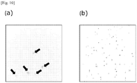
- Fig. 10 The results of staining of the human pancreatic cancer cells PANC1 are shown in Fig. 10
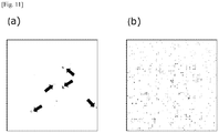
- the results of staining of the human pancreatic cancer cells AsPC-1 are shown in Fig. 11 .
- the cells indicated by arrows are spiked pancreatic cancer cells.
- ITGA2 anti-ITGA2 antibody
- the cells stained with the antibody against ITGA2 (anti-ITGA2 antibody) (the cells indicated by arrows in Fig. 10(a) and Fig. 11(a) ) were not stained with the antibody against CD45 ( Fig. 10(b) and Fig. 11(b) ).
- ITGA2 SEQ ID NO:6
- Example 1 the TM4SF1 gene and the TNFRSF12A gene were found to be more highly expressed in cancer cells than in leukocytes of healthy individuals.
- antibodies that recognize TM4SF1, TNFRSF12A, or an existing tumor marker EpCAM were employed to study whether TM4SF1 protein and TNFRSF12A protein can be used for detection of cancer cells.
- the cancer cell lines (10 lines) used in Example 1 were used as cancer cells.
- each staining result is represented as the intensity of the fluorescence signal (rated on a 5-point scale from “+” to "+++++”) derived from each fluorescently labeled antibody.
- the higher the intensity of the signal (the larger the number of "+” symbols), the higher the expression (signal intensity (expression level): “+” ⁇ "++” ⁇ "+++” ⁇ "++++” ⁇ "+++++”).
- TM4SF1 protein SEQ ID NO:1
- TNFRSF12A protein SEQ ID NO:2
- lung cancer breast adenocarcinoma cells
- breast cancer cells breast cancer cells
- pancreatic cancer pancreatic cancer
- TM4SF1 and/or TNFRSF12A were found also in the cell lines (PC-14, MDAMB231, PC-3, and PANC1) that exhibited low expression (with a single "+” symbol) of the existing tumor marker EpCAM, it is suggested that tumor cells contained in a sample can be accurately detected and collected by combination of TM4SF 1 and/or TNFRSF 12A with EpCAM [Table 9] cancer type cancer cell line name Protein TM4SF1 (SEQ ID NO:1) TNFRSF12A (SEQ ID NO:2) EpCAM lung adenocarcinoma PC-9 + ++++ +++++ PC-14 +++++ + + breast adenocarcinoma MDAMB231 +++ +++++ + breast cancer SKBR3 ++ ++ +++++ prostate cancer PC-3 ++ ++++ + 22Rv1 + + +++++ liver cancer HepG2 +++++ +++ +++ HuH-7 +++++ +++ +++ pancreatic cancer PANC
- TM4SF1 protein (SEQ ID NO:1) and TNFRSF12A protein (SEQ ID NO:2) were found to be highly expressed also in the cell lines (PC-14, MDAMB231, PC-3, and PANC1) that exhibited low expression of the existing tumor marker EpCAM protein.
- TM4SF1 protein SEQ ID NO:1
- TNFRSF12A protein SEQ ID NO:2
- PANC1 human pancreatic cancer cell line
- the "buffer” used in the following procedure is D-PBS(-) (Dulbecco's Phosphate-Buffered Saline, Mg 2+ - and Ca 2+ -free) supplemented with 0.5% BSA and 2 mM EDTA.
- the results of the collection of the cancer cells by using one of, or all of, the anti-TM4SF1 antibody, anti-TNFRSF12A antibody, and anti-EpCAM antibody are shown in Table 10.
- the ratio of the number of collected cells to the number of spiked cells is represented as the cell collection ratio [%].
- TM4SF1 protein and TNFRSF12A protein enable highly efficient collection of cancer cells from a blood sample even when collection of the cancer cells with the existing marker EpCAM protein results in a low collection ratio. Further, it can be seen that combination of TM4SF1 protein and TNFRSF 12A protein with EpCAM protein enables even more efficient collection of cancer cells even when collection of the cancer cells with EpCAM protein results in a low collection ratio.
- Example 1 the SDC1 gene was found to be more highly expressed in cancer cells than in leukocytes of healthy individuals. In view of this, an antibody that recognizes SDC1 was employed to study whether SDC1 can be used for detection of cancer cells. In the present Example, the cancer cell lines (10 lines) used in Example 1 were used as cancer cells.
- each staining result is represented as the intensity of the fluorescence signal (rated on a 5-point scale from “+” to "+++++”) derived from each fluorescently labeled antibody.
- the higher the intensity of the signal (the larger the number of "+” symbols), the higher the expression (signal intensity (expression level): “+” ⁇ "++” ⁇ "+++” ⁇ "++++” ⁇ "+++++”).
- SDC1 protein (SEQ ID NO:3) is expressed, irrespective of the cancer type, in lung adenocarcinoma cells, breast adenocarcinoma cells, breast cancer cells, prostate cancer cells, liver cancer cells, and pancreatic cancer cells. It can thus be seen that a wide range of tumor cells contained in a sample can be detected therewith. The results also suggest that a wide range of tumor cells contained in a sample can be collected with SDC1 protein irrespective of the cancer type (lung cancer, breast adenocarcinoma, breast cancer, prostate cancer, liver cancer, or pancreatic cancer).
- Example 1 the SDC1 gene was found to be more highly expressed in breast cancer or breast adenocarcinoma cells and pancreatic cancer cells than in leukocytes of healthy individuals. In view of this, whether SDC1 can be used for specific detection of breast cancer cells and pancreatic cancer cells contained in a blood sample was studied.
- the results of staining of the human breast cancer cells SKBR3 are shown in Fig. 12
- the results of staining of the human pancreatic cancer cells AsPC-1 are shown in Fig. 13 .
- the cells indicated by arrows are spiked breast cancer cells and pancreatic cancer cells.
- the cells stained with the antibody against SDC1 anti-SDCl antibody
- the cells indicated by arrows in Fig. 12(a) and Fig. 13(a) were not stained with the antibody against CD45 ( Fig. 12(b) and Fig. 13(b) ).
- SDC1 SEQ ID NO:3
- SDC1 SEQ ID NO:3
- Example 7 TM4SF1 protein (SEQ ID NO:1) and TNFRSF12A protein (SEQ ID NO:2) were found, and, in Example 9, SDC1 protein (SEQ ID NO:3) was found, to be highly expressed also in the cell lines (PC-14, MDAMB231, and PC-3) that exhibited low expression of the existing tumor marker EpCAM protein.
- SDC1 protein SEQ ID NO:3 was found, to be highly expressed also in the cell lines (PC-14, MDAMB231, and PC-3) that exhibited low expression of the existing tumor marker EpCAM protein.
- PC-3 human prostate cancer cell line which exhibits low expression of EpCAM protein, was used as cancer cells.
- the "buffer” used in the following procedure is D-PBS(-) (Dulbecco's Phosphate-Buffered Saline, Mg 2+ - and Ca 2+ -free) supplemented with 0.5% BSA and 2 mM EDTA.
- the results of the collection of the cancer cells by using one of, the three other than the anti-EpCAM antibody of, or all four of, the anti-TM4SF1 antibody, anti-TNFRSF12A antibody, anti-SDCl antibody, and anti-EpCAM antibody are shown in Table 12.
- the ratio of the number of collected cells to the number of spiked cells is represented as the cell collection ratio [%].
- TM4SF1 protein, TNFRSF12A protein, and SDC1 protein enable highly efficient collection of cancer cells from a blood sample even when collection of the cancer cells with the existing marker EpCAM protein results in a low collection ratio.
- TM4SF1 protein, TNFRSF12A protein, and SDC1 protein enables even more efficient collection of cancer cells even when collection of the cancer cells with EpCAM protein results in a low collection ratio.
Landscapes
- Health & Medical Sciences (AREA)
- Life Sciences & Earth Sciences (AREA)
- Chemical & Material Sciences (AREA)
- Organic Chemistry (AREA)
- Immunology (AREA)
- Molecular Biology (AREA)
- Biochemistry (AREA)
- General Health & Medical Sciences (AREA)
- Engineering & Computer Science (AREA)
- Zoology (AREA)
- Genetics & Genomics (AREA)
- Medicinal Chemistry (AREA)
- Proteomics, Peptides & Aminoacids (AREA)
- Biophysics (AREA)
- Cell Biology (AREA)
- Toxicology (AREA)
- Gastroenterology & Hepatology (AREA)
- Hematology (AREA)
- Biomedical Technology (AREA)
- Urology & Nephrology (AREA)
- Analytical Chemistry (AREA)
- Pathology (AREA)
- Microbiology (AREA)
- Biotechnology (AREA)
- Wood Science & Technology (AREA)
- Physics & Mathematics (AREA)
- Bioinformatics & Cheminformatics (AREA)
- General Physics & Mathematics (AREA)
- Food Science & Technology (AREA)
- General Engineering & Computer Science (AREA)
- Oncology (AREA)
- Hospice & Palliative Care (AREA)
- Tropical Medicine & Parasitology (AREA)
- Virology (AREA)
- Measuring Or Testing Involving Enzymes Or Micro-Organisms (AREA)
- Investigating Or Analysing Biological Materials (AREA)
- Micro-Organisms Or Cultivation Processes Thereof (AREA)
Abstract
Description
- The present invention relates to a tumor marker, and a method of collecting and detecting a tumor cell contained in a sample. In particular, the present invention relates to a method which uses a protein or gene expressed by a tumor cell contained in a sample, to collect and detect the tumor cell in distinct from a contaminant cell contained in the sample.
- A tumor cell that has left a primary lesion infiltrates a blood vessel or lymph vessel, and then circulates in blood or lymph fluid, finally invading another organ or tissue to form a metastatic lesion. The tumor cell that circulates in blood is also called CTC (Circulating Tumor Cell), and a large number of clinical trials and studies have been carried out therefor. For example, the number of CTCs contained in blood collected from a patient is counted (Patent Document 1), or protein expression, or gene mutation or translocation, in the CTCs is investigated to provide information related to prediction of prognosis or cancer recurrence in the patient. However, since the number of CTCs contained in blood is very small, and various contaminant cells such as erythrocytes and leukocytes are contained in blood, CTC detection/analysis requires a technique which enables collection and detection of a very small number of cells in distinction from a large number of cells.
- Conventionally, detection of CTCs has been carried out using a protein (tumor marker) expressed in CTCs such as cytokeratin or EpCAM (Epithelial Cell Adhesion Molecule) (Patent Document 1). However, cytokeratin has the problem of crossing with (non-specific detection of) contaminant cells contained in blood, and EpCAM has the problem that the collection and detection are possible only for part of epithelium-derived CTCs.
- [Patent Document 1] Japanese Translated PCT Patent Application Laid-open No.
2008-533487 - An object of the present invention is to provide a tumor marker, and a method of collecting and detecting a tumor cell contained in a sample in distinction from a contaminant cell contained in the sample.
- In order to solve the problems described above, the present inventors carried out comparative expression analysis using a next-generation sequencer between 10 cancer cell lines and, as samples from healthy individuals, leukocytes, to discover tumor markers with which tumor cells contained in a sample can be collected and detected in distinction from contaminant cells contained in the sample, thereby reaching the present invention.
- More specifically, the present invention can be exemplified as follows.
- [1] A method of detecting a tumor cell contained in a sample in distinction from a contaminant cell, the method comprising detecting one or more polypeptides selected from the group consisting of the following (i) to (iii) present in the sample:
- (i) a polypeptide comprising at least the amino acid sequence of any of SEQ ID NOs: 1 to 6;
- (ii) a polypeptide comprising an amino acid sequence having a homology of not less than 70% to the amino acid sequence of any of SEQ ID NOs: 1 to 6; and
- (iii) a polypeptide comprising at least a splicing variant of the amino acid sequence of any of SEQ ID NOs: 1 to 6, or a polypeptide comprising at least a splicing variant of a polypeptide having a homology of not less than 70% to the amino acid sequence of any of SEQ ID NOs: 1 to 6.
- [2] The method according to [1], wherein the detection is carried out using an antibody or aptamer that specifically recognizes one or more polypeptides selected from the group consisting of (i) to (iii).
- [3] A method of detecting a tumor cell contained in a sample in distinction from a contaminant cell, the method comprising detecting a gene encoding any of the following polypeptides (i) to (iii) present in the sample:
- (i) a polypeptide comprising at least the amino acid sequence of any of SEQ ID NOs: 1 to 6;
- (ii) a polypeptide comprising an amino acid sequence having a homology of not less than 70% to the amino acid sequence of any of SEQ ID NOs: 1 to 6; and
- (iii) a polypeptide comprising at least a splicing variant of the amino acid sequence of any of SEQ ID NOs: 1 to 6, or a polypeptide comprising at least a splicing variant of a polypeptide having a homology of not less than 70% to the amino acid sequence of any of SEQ ID NOs: 1 to 6.
- [4] A method of collecting a tumor cell, the method comprising: detecting a tumor cell contained in a sample in distinction from a contaminant cell; and collecting the detected tumor cell using collection means; wherein the detection of the tumor cell is carried out by detecting one or more polypeptides selected from the group consisting of the following (i) to (iii) present in the sample:
- (i) a polypeptide comprising at least the amino acid sequence of any of SEQ ID NOs: 1 to 6;
- (ii) a polypeptide comprising at least an amino acid sequence having a homology of not less than 70% to the amino acid sequence of any of SEQ ID NOs: 1 to 6; and
- (iii) a polypeptide comprising at least a splicing variant of the amino acid sequence of any of SEQ ID NOs: 1 to 6, or a polypeptide comprising at least a splicing variant of a polypeptide having a homology of not less than 70% to the amino acid sequence of any of SEQ ID NOs: 1 to 6.
- [5] The method according to [4], wherein the detection of the tumor cell is carried out using an antibody or aptamer that specifically recognizes one or more polypeptides selected from the group consisting of (i) to (iii).
- [6] The method according to any of [1] to [5], wherein the sample is blood, and the contaminant cell contained in the sample is leukocyte.
- [7] A tumor marker comprising any of the following polypeptides (i) to (iii):
- (i) a polypeptide comprising at least the amino acid sequence of any of SEQ ID NOs:1 to 6;
- (ii) a polypeptide comprising at least an amino acid sequence having a homology of not less than 70% to the amino acid sequence of any of SEQ ID NOs: 1 to 6; and
- (iii) a polypeptide comprising at least a splicing variant of the amino acid sequence of any of SEQ ID NOs: 1 to 6, or a polypeptide comprising at least a splicing variant of a polypeptide having a homology of not less than 70% to the amino acid sequence of any of SEQ ID NOs: 1 to 6;
the tumor marker being capable of detecting a tumor cell contained in a sample in distinction from leukocyte contained in the sample.
- [8] A tumor marker comprising a gene encoding any of the following polypeptides (i) to (iii):
- (i) a polypeptide comprising at least the amino acid sequence of any of SEQ ID NOs: 1 to 6;
- (ii) a polypeptide comprising at least an amino acid sequence having a homology of not less than 70% to the amino acid sequence of any of SEQ ID NOs: 1 to 6; and
- (iii) a polypeptide comprising at least a splicing variant of the amino acid sequence of any of SEQ ID NOs: 1 to 6, or a polypeptide comprising at least a splicing variant of a polypeptide having a homology of not less than 70% to the amino acid sequence of any of SEQ ID NOs: 1 to 6;
the tumor marker being capable of detecting a tumor cell contained in a sample in distinction from leukocyte contained in the sample.
- [9] A tumor marker comprising any of the following polypeptides (i) to (iii):
- (i) a polypeptide comprising at least the amino acid sequence of any of SEQ ID NOs: 1 to 6;
- (ii) a polypeptide comprising at least an amino acid sequence having a homology of not less than 70% to the amino acid sequence of any of SEQ ID NOs: 1 to 6; and
- (iii) a polypeptide comprising at least a splicing variant of the amino acid sequence of any of SEQ ID NOs: 1 to 6, or a polypeptide comprising at least a splicing variant of a polypeptide having a homology of not less than 70% to the amino acid sequence of any of SEQ ID NOs: 1 to 6;
which tumor marker enables collection of a tumor cell contained in a sample in distinction from leukocyte contained in the sample.
- The present invention is described below in detail.
- Examples of the sample in the present invention include not only a tissue, whole blood, ascites, or the like collected from a patient, but also samples prepared by diluting the tissue, whole blood, ascites, or the like using an appropriate buffer; components derived from whole blood, such as serum, plasma, cord blood, or blood component fluid; and suspensions prepared by suspending, in an appropriate buffer, a piece of a tissue containing blood such as a liver, lung, spleen, kidney, tumor, or lymph node tissue. Examples of the sample in the present invention also include fractions containing a tumor cell, which fractions are obtained by separation/collection from these samples and suspensions by centrifugation or the like.
- The contaminant cell contained in the sample in the present invention means cell(s) other than tumor cells, and examples of the contaminant cell include erythrocytes, leukocytes, and platelets in cases where the sample is a blood sample such as whole blood, a whole blood dilution, a component derived from whole blood, a suspension of a tissue containing blood, or a fraction containing a tumor cell obtained from these samples or suspensions. In particular, the method of detecting a tumor cell of the present invention is an excellent method for distinguishing a tumor cell from leukocyte contained in a blood sample, which often led to false-positives in the conventional methods of detecting a tumor cell. Further, the method of collecting a tumor cell of the present invention is an excellent method for collecting an EpCAM (Epithelial Cell Adhesion Molecule)-negative tumor cell contained in a blood sample, which often led to false-negatives in the conventional methods of collecting a tumor cell.
- In the detection of a tumor cell in the present invention, the tumor cell contained in the sample may be directly detected by staining, a probe hybridization method, or the like based on any of the polypeptides (i) to (iii) or the gene encoding the polypeptide which is present in the cell and derived from the cell, or the tumor cell may be indirectly detected by detecting any of the polypeptides (i) to (iii) or the gene encoding the polypeptide which is secreted and derived from the tumor cell contained in the sample.
- In the present invention, the tumor cell is detected based on the expression level of any of the polypeptides (i) to (iii) or the gene encoding the polypeptide derived from the tumor cell. The detection may be carried out qualitatively, that is, based on the presence or absence of the tumor cell, or may be carried out quantitatively, that is, based on the number of tumor cells or detected intensity. The criterion for the detection is also not limited. For example, a cell may be judged as a tumor cell even when a very low detection signal is found. Alternatively, a cell may be judged as a tumor cell when the detection signal is not weaker than a certain threshold. Alternatively, a cell may be judged as a tumor cell when the signal is not weaker than a certain value relative to a signal from a contaminant cell contained in the sample (erythrocytes, leukocytes, and/or the like in cases of a blood sample), or when the signal is not less than 2SD, not less than 3SD, or not less than 4SD from the average of the signal from such contaminant cells.
- The collection of the tumor cell in the present invention may be carried out by detecting any of the polypeptides (i) to (iii) which is present in the cell and derived from the cell, to detect the tumor cell contained in the sample in distinction from the contaminant cell, and then collecting the detected tumor cell using collection means. The "any of the polypeptides (i) to (iii)" is a transmembrane polypeptide in all cases.
- In the present invention, the detection and/or collection of the tumor cell contained in the sample is/are carried out using any of the polypeptides (i) to (iii), which is a protein expressed by the tumor cell.
- The polypeptide consisting of the amino acid sequence of SEQ ID NO:1 is a protein called TM4SF1 (Transmembrane 4 L six family member 1), L6, M3S1, TAAL6, or H-L6. Its amino acid sequence is presented in GenBank No. NP_055035.1.
- The polypeptide consisting of the amino acid sequence of SEQ ID NO:2 is a protein called TNFRSF12A(TNF receptor superfamily member 12A), FN14, TWEAKR, or CD266. Its amino acid sequence is presented in GenBank No. NP_057723.1.
- The polypeptide consisting of the amino acid sequence of SEQ ID NO:3 is a protein called SDC1 (Syndecan 1), SDC, SYND1, syndecan, or CD138. Its amino acid sequence is presented in GenBank No. NP_002988.3.
- The polypeptide consisting of the amino acid sequence of SEQ ID NO:4 is a protein called F3 (Coagulation factor III, tissue factor), TF, TFA, or CD142. Its amino acid sequence is presented in GenBank No. NP_001984.1.
- The polypeptide consisting of the amino acid sequence of SEQ ID NO:5 is a protein called EPHA2 (EPH receptor A2), CTPA, CTPP1, CTRCT6, ECK, or ARCC2. Its amino acid sequence is presented in GenBank No. NP_004422.2.
- The polypeptide consisting of the amino acid sequence of SEQ ID NO:6 is a protein called ITGA2 (Integrin subunit alpha 2), CD49B, GPIa, HPA-5, VLA-2, VLAA2, or BR. Its amino acid sequence is presented in GenBank No. NP_002194.2.
- In particular, as shown below in Examples, TM4SF1 (SEQ ID NO:1) and TNFRSF12A (SEQ ID NO:2) are highly expressed, irrespective of the cancer type, in lung adenocarcinoma cells, breast adenocarcinoma cells, breast cancer cells, prostate cancer cells, liver cancer cells, and pancreatic cancer cells. Thus, a wider range of tumor cells contained in a sample can be detected therewith than with EpCAM, which is a tumor marker used in conventional detection. Since TM4SF1 (SEQ ID NO:1) and TNFRSF12A (SEQ ID NO:2) are transmembrane proteins, a wider range of tumor cells contained in a sample can be collected therewith than with EpCAM, which is a tumor marker used in conventional collection.
- In cases where a pancreatic cancer cell is to be specifically detected, it is suggested, based on the result of gene expression analysis shown below in Examples, that ITGA2 (SEQ ID NO:6) is preferably used. Since ITGA2 (SEQ ID NO:6) is a transmembrane protein, it is suggested that the protein is preferably used in cases where pancreatic cancer cell is to be specifically collected.
- The present invention includes a mode in which one or several amino acid residues in any of the amino acid sequences of SEQ ID NOs: 1 to 6 are substituted with other amino acid residues or deleted, or in which one or several amino acid residues are inserted into any of the amino acid sequences of SEQ ID NOs: 1 to 6, as long as the same activity as that of the protein consisting of the amino acid sequence of any of SEQ ID NOs:1 to 6 is maintained. In the present description, the term "several" means an integer of 20, 19, 18, 17, 16, 15, 14, 13, 12, 11, 10, 9, 8, 7, 6, 5, 4, 3, or 2. The term "one or several amino acid residues" preferably means 1 to 10 amino acid residues, more preferably 1 to 5 amino acid residues, still more preferably 1 to 3 amino acid residues, especially preferably 2 or less amino acid residues.
- Similarly, each polypeptide may be in a mode in which one or more amino acid residues are added to the N-terminus and/or C-terminus of the polypeptide, as long as the function as the protein is maintained. The marker used in the method of the present invention may be a protein having a homology of not less than 70% to the entire amino acid sequence of any of SEQ ID NOs:1 to 6, as long as the marker has the same activity as that of the protein consisting of the amino acid sequence of any of SEQ ID NOs:1 to 6. The protein has a homology of preferably not less than 80%, more preferably not less than 90%, still more preferably not less than 95%.
- When a protein is expressed from the gene encoding the protein in a eukaryotic cell, a reaction (splicing) occurs to remove an intron(s) from a gene (RNA) precursor and to bind the exons before and after the intron(s) to each other. Since the exons remained have diversity, and produce various mature mRNAs, proteins having different activities (splicing variants) are expressed in some cases. The polypeptides that may be used for the detection in the present invention also include:
- a polypeptide comprising at least a splicing variant of the amino acid sequence of any of SEQ ID NOs:1 to 6;
- a polypeptide comprising at least a splicing variant of an amino acid sequence which is the same as the amino acid sequence of any of SEQ ID NOs:1 to 6 except that one or several amino acid residues are substituted with other amino acid residues or deleted;
- a polypeptide comprising at least a splicing variant of an amino acid sequence which is the same as the amino acid sequence of any of SEQ ID NOs:1 to 6 except that one or several amino acid residues are inserted; and
- a polypeptide comprising at least a splicing variant of a polypeptide having a homology of not less than 70% to the amino acid sequence of any of SEQ ID NOs: 1 to 6.
- When the detection of the present invention is carried out, a dye (such as a chemiluminescent dye or fluorescent dye) which enables specific labeling of any of the polypeptides described above in (i) to (iii) may be added to perform staining, and then the detection may be carried out based on the stained image or staining intensity (such as the luminescence intensity or fluorescence intensity). However, from the viewpoint of versatility, the detection is generally carried out by a method using an antibody (hereinafter also simply referred to as anti-polypeptide antibody) or aptamer against the above-described polypeptide. Such a method can be said to be a preferred mode also from the viewpoint of applicability to both the direct detection and the indirect detection.
- Since the polypeptides described in (i) to (iii) are transmembrane proteins, they can also be tumor markers with which a tumor cell contained in a sample can be collected in distinction from leukocyte contained in the sample.
- Preferably, when the tumor cell contained in the sample is detected using an anti-polypeptide antibody, the antibody is modified with a labeling substance by a certain method, and the detection is carried out based on the presence or absence, or the amount, of the labeling substance. The labeling substance may be appropriately selected from substances normally used in the field of measurement using antigen-antibody reaction. Examples of the labeling substance include fluorescent substances such as fluorescein; enzymes such as alkaline phosphatase; and radioactive substances. The binding mode between the antibody and the labeling substance is not limited. The mode may be a mode in which the antibody is directly bound to the labeling substance by chemical bonding or the like, or may be a mode in which the antibody is indirectly bound through an antibody against the antibody, to which the labeling substance is bound (labeled secondary antibody). The detection format for the tumor cell contained in the sample using the anti-polypeptide antibody is not limited. The cell may be manually detected by the ELISA (Enzyme-Linked ImmunoSorbent Assay) method or the Western blotting method, or may be automatically detected using an enzyme immunoassay apparatus such as AIA-900 or AIA-CL2400 (these are manufactured by Tosoh Corporation).
- Thus, the polypeptides described in (i) to (iii) can be tumor markers with which a tumor cell contained in a sample can be detected in distinction from leukocyte contained in the sample.
- The collection of the tumor cell in distinction from the contaminant cell by the present invention may be carried out by using the polypeptide of any of (i) to (iii) to detect the tumor cell in distinction from the contaminant cell, and then collecting the detected tumor cell using collection means. Examples of the collection means include means comprising: a substrate provided with a retaining section capable of retaining the tumor cell; and a collecting section for collecting the tumor cell by suction and discharge by a nozzle. Specific examples of the collection means include the collection apparatus disclosed in
JP 2016-142616 A - On the other hand, in the present invention, the gene encoding any of the polypeptides (i) to (iii) is not limited as long as it is a polynucleotide obtained by converting any of the polypeptides (i) to (iii) using codons. The gene is preferably a polynucleotide obtained by conversion using human-type codons.
- Examples of the gene encoding a polypeptide comprising at least a polypeptide consisting of the amino acid sequence of any of SEQ ID NOs:1 to 6 include the following sequences:
- a polynucleotide consisting of nucleotide positions 235 to 840 of the sequence of SEQ ID NO:7 (GenBank No. NM_014220.2), which is obtained by conversion of a polypeptide consisting of the sequence of SEQ ID NO:1;
- a polynucleotide consisting of nucleotide positions 87 to 473 of the sequence of SEQ ID NO:8 (GenBank No. NM_016639.2), which is obtained by conversion of a polypeptide consisting of the sequence of SEQ ID NO:2;
- a polynucleotide consisting of nucleotide positions 392 to 1321 of the sequence of SEQ ID NO:9 (GenBank No. NM_001006946.1), which is obtained by conversion of a polypeptide consisting of the sequence of SEQ ID NO:3;
- a polynucleotide consisting of nucleotide positions 222 to 1106 of the sequence of SEQ ID NO:10 (GenBank No. NM_001993.4), which is obtained by conversion of a polypeptide consisting of the sequence of SEQ ID NO:4;
- a polynucleotide consisting of nucleotide positions 156 to 3083 of the sequence of SEQ ID NO:11 (GenBank No. NM_004431.3), which is obtained by conversion of a polypeptide consisting of the sequence of SEQ ID NO:5; and
- a polynucleotide consisting of nucleotide positions 144 to 3686 of the sequence of SEQ ID NO:12 (GenBank No. NM_002203.3), which is obtained by conversion of a polypeptide consisting of the sequence of SEQ ID NO:6.
- The method of detecting the gene described above is not limited. For example, a probe may be designed at an appropriate position in the polynucleotide, and then hybridization may be performed to detect the gene. Alternatively, a primer set may be designed at appropriate positions in the polynucleotide, and then the polynucleotide may be amplified and detected using the PCR method, RT-PCR method, TRC (Transcription Reverse transcription Concerted) method, NASBA (Nucleic Acid Sequence-Based Amplification) method, TMA (Transcription-Mediated Amplification) method, or the like. Alternatively, a sample containing the polynucleotide may be directly subjected to a sequencer to detect the gene.
- Thus, the genes encoding the polypeptides described in (i) to (iii) can be tumor markers with which a tumor cell contained in a sample can be detected in distinction from leukocyte contained in the sample.
- According to the present invention, detection of tumor cell(s) contained in a sample can be carried out by detecting the polypeptide described in any of the following, or a gene encoding the polypeptide: (i) a polypeptide containing at least any of six amino acid sequences including TM4SF1 (GenBank No. NP_055035.1) and TNFRSF12A(GenBank No. NP_057723.1); (ii) a polypeptide containing an amino acid sequence having a homology of not less than 70% to the amino acid sequence of (i); and (iii) a polypeptide containing at least a splicing variant of the amino acid sequence of (i) or (ii). By the detection method, tumor cell(s) contained only in a small amount in a sample can be detected in distinction from contaminant cell(s) contained in the sample.
- Further, collection of tumor cell(s) contained in a sample can be carried out by detecting the tumor cell(s) using the polypeptide described in any of (i) to (iii), and collecting the detected tumor cell(s) using collection means. By the collection method, tumor cell(s) contained only in a small amount in a sample can be collected in distinction from contaminant cell(s) contained in the sample.
-
-
Fig. 1 is a diagram illustrating the result of TM4SF1 gene expression analysis of leukocytes of healthy individuals and various kinds of cancer cells. -
Fig. 2 is a diagram illustrating the result of TNFRSF 12A gene expression analysis of leukocytes of healthy individuals and various kinds of cancer cells. -
Fig. 3 is a diagram illustrating the result of immunostaining of a blood sample spiked with lung adenocarcinoma cells (PC14), which immunostaining was carried out using (a) an anti-TM4SF1 antibody and (b) an anti-CD45 antibody. -
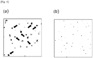
Fig. 4 is a diagram illustrating the result of immunostaining of a blood sample spiked with pancreatic cancer cells (PANC1), which immunostaining was carried out using (a) an anti-TM4SF1 antibody and (b) an anti-CD45 antibody. -
Fig. 5 is a diagram illustrating the result of immunostaining of a blood sample spiked with lung adenocarcinoma cells (PC9), which immunostaining was carried out using (a) an anti-TNFRSF12A (CD266) antibody and (b) an anti-CD45 antibody. -
Fig. 6 is a diagram illustrating the result of immunostaining of a blood sample spiked with breast adenocarcinoma cells (MDAMB231), which immunostaining was carried out using (a) an anti-TNFRSF12A(CD266) antibody and (b) an anti-CD45 antibody. -
Fig. 7 is a diagram illustrating the result of immunostaining of a blood sample spiked with lung adenocarcinoma cells (PC9), which immunostaining was carried out using (a) an anti-CD142 (F3) antibody and (b) an anti-CD45 antibody. -
Fig. 8 is a diagram illustrating the result of immunostaining of a blood sample spiked with breast adenocarcinoma cells (MDAMB231), which immunostaining was carried out using (a) an anti-CD142 (F3) antibody and (b) an anti-CD45 antibody. -
Fig. 9 is a diagram illustrating the result of immunostaining of a blood sample spiked with prostate cancer cells (PC3), which immunostaining was carried out using (a) an anti-EPHA2 antibody and (b) an anti-CD45 antibody. -
Fig. 10 is a diagram illustrating the result of immunostaining of a blood sample spiked with pancreatic cancer cells (PANC1), which immunostaining was carried out using (a) an anti-ITGA2 antibody and (b) an anti-CD45 antibody. -
Fig. 11 is a diagram illustrating the result of immunostaining of a blood sample spiked with pancreatic cancer cells (AsPC-1), which immunostaining was carried out using (a) an anti-ITGA2 antibody and (b) an anti-CD45 antibody. -
Fig. 12 is a diagram illustrating the result of immunostaining of a blood sample spiked with breast cancer cells (SKBR3), which immunostaining was carried out using (a) an anti-SDCl antibody and (b) an anti-CD45 antibody. -
Fig. 13 is a diagram illustrating the result of immunostaining of a blood sample spiked with pancreatic cancer cells (AsPC-1), which immunostaining was carried out using (a) an anti-SDCl antibody and (b) an anti-CD45 antibody. - The present invention is described below in more detail by way of Examples for cases using a blood sample. However, the present invention is not limited to these Examples.
- As cancer cell lines, the following 10 lines were selected: human lung adenocarcinoma cells (PC9 and PC14), human breast adenocarcinoma cells (MDAMB231), human breast cancer cells (SKBR3), human prostate cancer cells (22Rv1 and PC3), human liver cancer cells (HepG2 and HuH-7), and human pancreatic cancer cells (PANC1 and AsPC-1). Differences in the gene expression level between these cancer cell lines and leukocytes of healthy individuals were analyzed by the following method using a next-generation sequencer.
- (1) The respective cancer cell lines were cultured using the following media in an environment at 5% CO2 and 37°C. Thereafter, the cells were detached from the media using 0.25% trypsin/1 mM EDTA, to collect cancer cells as single cells (n = 4). From RNA of each single cell, cDNA synthesis and amplification were carried out using a SMART-Seq v4 Ultra Low Input RNA Kit for Sequencing (Clontech). Similarly, leukocytes collected from blood of three healthy individuals were subjected to cDNA synthesis and amplification from RNA of each single cell (n = 4).
PC9, PC14, and PANC1: DMEM (Dulbecco's Modified Eagle Medium)/Ham's F-12 medium supplemented with 10% (v/v) FBS (fetal bovine serum)
AsPC-1: RPMI-1640 medium supplemented with 10% (v/v) FBS and 1 mM pyruvic acid
MDAMB231: Leibovitz's L-15 medium with L-Glutamine, supplemented with 10% (v/v) FBS
SKBR3: McCoy's 5a medium supplemented with 10% (v/v) FBS
HepG2 and HuH-7: DMEM/high glucose medium supplemented with 10% (v/v) FBS
22Rv1: RPMI0211-1640 medium supplemented with 10% (v/v) FBS
PC3: Ham's F-12K medium supplemented with 10% (v/v) FBS, and kanamycin and streptomycin - (2) A plurality of cells obtained in (1) were collected, and total RNAwas recovered therefrom using an RNeasy Mini Kit (QIAGEN). Thereafter, cDNA synthesis and amplification were carried out from 10 ng of the RNA using a SMART-Seq v4 Ultra Low Input RNA Kit for Sequencing (Clontech). Similarly, RNA was recovered from a plurality of leukocytes collected from blood of three healthy individuals, and cDNA synthesis and amplification were carried out from 10 ng of the RNA.
- (3) Using 1 ng of the cDNA obtained in (1) and (2), library preparation was carried out using a Nextera XT DNA Library Preparation Kit (Illumina) and Nextera XT v2 Index Kit Set A (Illumina). By performing sequencing analysis using Next-seq500 (illumina) under single-end read conditions with a read length of 75 bp, not less than 10,000,000 sequence reads were read for each sample.
- (4) The nucleotide sequences (sequence data) read in (3) were mapped on the human genome sequence using TopHat 2 (Johns Hopkins University) and Bowtie 2 (Johns Hopkins University). The human genome sequence and human gene information were obtained using BUILD GRCh38 published by NCBI (National Center for Biological Information). From the mapped nucleotide sequences, the expression value for each gene was determined in terms of the FPKM (Fragments Per Kilobase of exon per Million reads mapped) unit based on the number of reads of the gene obtained.
- (5) Comparison of expression was carried out for 15 samples from 3 specimens of leukocytes of healthy individuals, and 48 samples from 10 kinds of cancer cell lines.
- Table 1 shows the genes whose average expression values (FPKM values) in the cancer cell lines (10 kinds, 48 samples) were not less than 10.00 times higher than the average expression value (FPKM value) in the leukocytes of the healthy individuals (3 specimens, 15 samples), and which encode transmembrane proteins.
Fig. 1 andFig. 2 show the results of analysis of each sample regarding the genes encoding TM4SF1 (SEQ ID NO:1) and TNFRSF12A (SEQ ID NO:2), respectively, among the above genes. In the figures, PC9 or PC14 represents a result for human lung adenocarcinoma cells; MDAMB231 represents a result for human breast adenocarcinoma cells; SKBR3 represents a result for human breast cancer cells; HepG2 or HuH-7 represents a result for human liver cancer cells; AsPC-1 or PANC1 represents a result for human pancreatic cancer cells; 22Rv1 or PC3 represents a result for human prostate cancer cells; Leuco1 to Leuco3 represent results for leukocytes of healthy individuals; "single" represents a result for RNA in a single cell (n=4); and "10 ng" represents a result for RNA (10 ng) in a plurality of cells.[Table 1] Name Protein Gene gene expression values(FPKM+1) GenBank No. SEQ ID NO GenBank No. SEQ ID NO cancer cells leukocytes cancer cells /leukocytes TM4SF1 NP_055035.1 1 NM_014220.2 7 342.22 1.04 329.87 TNFRSF12A NP_057723.1 2 NM_016639.2 8 94.92 1.12 84.88 SDC1 NP_002988.3 3 NM_001006946.1 9 18.28 1.01 18.15 F3 NP_001984.1 4 NM_001993.4 10 14.72 1.03 14.36 - It can be seen that the average expression value (FPKM value) of the TM4SF1 gene (SEQ ID NO:7) in the cancer cell lines (10 kinds, 48 samples) is not less than 300 times higher than the average FPKM value in the leukocytes (3 specimens, 15 samples), indicating that the TM4SF1 gene is specifically and highly expressed in the cancer cells (tumor cells) (Table 1). Further, since the TM4SF1 gene is highly expressed, irrespective of the cancer type, in lung adenocarcinoma cells, breast adenocarcinoma cells, breast cancer cells, liver cancer cells, pancreatic cancer cells, and prostate cancer cells (
Fig. 1 ), it can be seen that a wider range of tumor cells contained in a sample can be detected with TM4SF1 protein (SEQ ID NO:1) or gene (SEQ ID NO:7) than with EpCAM, which is a tumor marker used in conventional detection. Since TM4SF1 protein (SEQ ID NO:1) is a transmembrane protein, it can be seen that a wider range of tumor cells contained in a sample can be collected therewith than with EpCAM, which is a tumor marker used in conventional collection. - Similarly, it can be seen that the average expression value (FPKM value) of the TNFRSF12A gene (SEQ ID NO:8) in the cancer cell lines (10 kinds, 48 samples) is not less than 50 times higher than the average FPKM value in the leukocytes (3 specimens, 15 samples), indicating that the TNFRSF 12A gene is specifically and highly expressed in the cancer cells (tumor cells) (Table 1). Further, since the TNFRSF12A gene is highly expressed, irrespective of the cancer type, in lung adenocarcinoma cells, breast adenocarcinoma cells, breast cancer cells, liver cancer cells, pancreatic cancer cells, and prostate cancer cells (
Fig. 2 ), it can be seen that a wider range of tumor cells contained in a sample can be detected with TNFRSF 12A protein (SEQ ID NO:2) or gene (SEQ ID NO:8) than with EpCAM, which is a tumor marker used in conventional detection. Since TNFRSF12A protein (SEQ ID NO:2) is a transmembrane protein, it can be seen that a wider range of tumor cells contained in a sample can be collected therewith than with EpCAM, which is a tumor marker used in conventional collection. - Table 2 shows the genes whose average expression values (FPKM values) in the lung adenocarcinoma cell lines (2 kinds, 10 samples) were not less than 10.00 times higher than the average expression values (FPKM values) in the leukocytes of the healthy individuals (3 specimens, 15 samples), and which encode transmembrane proteins.
[Table 2] Name Protein Gene gene expression values(FPKM+1) GenBank No. SEQ ID NO GenBank No. SEQ NO ID lung adenocarcinoma cells leukocytes lung adenocarcinoma cells /leukocytes TM4SF1 NP_055035.1 1 NM_014220.2 7 523.66 1.04 504.76 F3 NP_001984.1 4 NM_0019914 10 154.43 1.03 150.65 TNFRSF12A NP_0577211 2 NM_016639.2 8 135.27 1.12 120.96 SDC1 NP_002988.3 3 NM_001006946.1 9 12.92 1.01 12.83 - Table 3 shows the genes whose average expression values (FPKM values) in the breast adenocarcinoma or breast cancer cell lines (2 kinds, 10 samples) were not less than 10.00 times higher than the average expression values (FPKM values) in the leukocytes of the healthy individuals (3 specimens, 15 samples), and which encode transmembrane proteins.
[Table 3] Name Protein Gene gene expression values(FPKM+1) GenBank No. SEQ ID NO GenBank No. SEQ ID NO breast adenocarcinoma or breast cancer cells leukocytes (breast adenocarcinoma or breast cancer cells) /leukocytes TNFRSF12A NP_057723.1 2 NN_016639.2 8 163.64 1.12 146.34 TM4SF1 NP_055035.1 1 NM_014220,2 7 148.41 1.04 143.06 F3 NP_001984.1 4 NM_001993,4 10 64.01 1.03 62.45 SDC1 NP_002988.3 3 NM_001006946.1 9 26.59 1.01 26.41 - Table 4 shows the genes whose average expression values (FPKM values) in the prostate cancer cell lines (2 kinds, 8 samples) were not less than 10.00 times higher than the average expression values (FPKM values) in the leukocytes of the healthy individuals (3 specimens, 15 samples), and which encode transmembrane proteins.
[Table 4] Name Protein Gene gene expression values(FPKM+1) GenBank No. SEQ ID NO GenBank No. SEQ ID NO prostate cancer cells leukocytes prostate cancer cells /leukocytes TM4SF1 NP_055035.1 1 NM_014220.2 7 67.80 1.04 65.36 TNFRSF12A NP_057723.1 2 NN_016639.2 8 41.18 1.12 36.82 SDC1 NP_002988.3 3 NM_001006946.1 9 22.00 1.01 21.85 EPHA2 NP_004422.2 5 NM_004431.3 11 12.58 1.01 12.40 - Table 5 shows the genes whose average expression values (FPKM values) in the liver cancer cell lines (2 kinds, 10 samples) were not less than 10.00 times higher than the average expression values (FPKM values) in the leukocytes of the healthy individuals (3 specimens, 15 samples), and which encode transmembrane proteins.
[Table 5] Name Protein Gene gene expression values(FPKM+1) GenBank No. SEQ ID NO GenBank No. SEQ ID NO liver cancer cells leukocytes liver cancer cells /leukocytes TM4SF1 NP_055035.1 1 NM_014220.2 7 743.44 1.04 716.61 TNFRSF12A NP_057723.1 2 NM_016639.2 8 41.97 1.12 37.53 - Table 6 shows the genes whose average expression values (FPKM values) in the pancreatic cancer cell lines (2 kinds, 10 samples) were not less than 10.00 times higher than the average expression values (FPKM values) in the leukocytes of the healthy individuals (3 specimens, 15 samples), and which encode transmembrane proteins.
[Table 6] Name Protein Gene gene expression values(FPKM+1) GenBank No. SEQ ID NO GenBank No. SEQ ID NO pancreatic cancer cells leukocytes pancreatic cancer cells /leukocytes TM4SF1 NP_055035.1 1 M_014220.2 7 866.75 1.04 835.47 TNFRSF12A NP_057723.1 2 NN_016639.2 8 170.47 1.12 152.45 SDC1 NP_002988.3 3 NM_001006946.1 9 35.38 1.01 35.13 EPHA2 NP_004422.2 5 NM_004431.3 11 15.01 1.01 14.79 ITGA2 NP_002194.2 6 NM_002203.3 12 14.20 1.03 13.75 - Among the genes shown in Table 1, whose average expression values (FPKM values) in the cancer cell lines (10 kinds, 48 samples) were not less than 10.00 times higher than the average expression values (FPKM values) in the leukocytes of the healthy individuals (3 specimens, 15 samples), the genes whose average expression values (FPKM values) were not less than 10.00 times higher in the cells of all cancer types (lung adenocarcinoma, breast adenocarcinoma or breast cancer, prostate cancer, liver cancer, and pancreatic cancer) than in the leukocytes of the healthy individuals (3 specimens, 15 samples) are shown in Table 7. As described above, it is suggested that a wider range of tumor cells contained in a sample can be detected with the proteins and genes shown in this table than with EpCAM, which is a tumor marker used in conventional detection. Further, since the proteins shown in this table are transmembrane proteins, it is suggested, as described above, that a wider range of tumor cells contained in a sample can be collected therewith than with EpCAM, which is a tumor marker used in conventional collection.
[Table 7] Name SEQ ID NO upper: gene expression values(FPKM+1), lower: gene expression ratio to leukocytes Protein Gene leukocytes lung adenocarcinoma cells breast adenocarcinoma or breast cancer cells prostate cancer cells liver cancer cells pancreatic cancer cells TM4SF1 1 7 1.04 523.66 148.41 67.80 743.44 866.75 - 504.76 143.06 65.36 716.61 835.47 TNFRSF12A 2 8 1.12 135.27 163.64 41.18 41.97 170.47 - 120.96 146.34 36.82 37.53 152.45 - Among the genes shown in Table 6, whose average expression values (FPKM values) in the pancreatic cancer cell lines (2 kinds, 10 samples) were not less than 10.00 times higher than the average expression values (FPKM values) in the leukocytes of the healthy individuals (3 specimens, 15 samples), the gene whose average expression values (FPKM values) in all cell lines other than pancreatic cancer (lung adenocarcinoma, breast adenocarcinoma or breast cancer, prostate cancer, and liver cancer) were less than 10.00 times the average expression value (FPKM values) in the leukocytes of the healthy individuals (3 specimens, 15 samples) is shown in Table 8. It is suggested that pancreatic cancer cells can be specifically detected with the protein and gene shown in this table. Further, since the protein shown in Table is a transmembrane protein, it is suggested that pancreatic cancer cells can be specifically collected therewith.
[Table 8] Name SEQ ID NO upper: gene expression values(FPKM+1), lower: gene expression ratio to leukocytes Protein Gene leukocytes lung adenocarcinoma cells breast adenocarcinoma or breast cancer cells prostate cancer cells liver cancer cells pancreatic cancer cells ITGA2 6 12 1.03 3.60 5.01 5.29 4.71 14.20 - 3.48 4.85 5.13 4.56 13.75 - In Example 1, the TM4SF1 gene was found to be more highly expressed in cancer cells than in leukocytes of healthy individuals. In view of this, whether TM4SF1 can be used for specific detection of tumor cells contained in a blood sample was studied.
- (1) Erythrocytes were removed from blood of a healthy individual by a separation method based on the specific-gravity difference, to provide a blood sample to be used in the present Example. This sample was spiked with human lung adenocarcinoma cells (PC14) or human pancreatic cancer cells (PANC1).
- (2) To 100 µL of the blood sample spiked with the cancer cell line, 10 µL of FcR Blocking Reagent (Miltenyi Biotec) was added, and blocking treatment was carried out at room temperature for 10 minutes.
- (3) Thereafter, 10 µL of a solution of an antibody against TM4SF1 (Human TM4SF1 Phycoerythrin MAb, R&D systems), and 10 µL of a solution of an antibody against CD45 (CD45-FITC, Beckman Coulter) for detection of leukocytes were added, followed by incubation at room temperature for 30 minutes.
- (4) Thereafter, the cells were washed three times with HBSS (Hank's Balanced Salt Solution) supplemented with 1% BSA and 5 mM EDTA, and then mounted on a slide glass using Permount Fisher (Fisher Scientific), followed by observation under a fluorescence microscope.
- The results of staining of the human lung adenocarcinoma cells (PC14) are shown in
Fig. 3 , and the results of staining of the human pancreatic cancer cells (PANC1) are shown inFig. 4 . In the figures, the cells indicated by arrows are spiked cancer cells. According to the results in bothFig. 3 andFig. 4 , the cells stained with the antibody against TM4SF1 (anti-TM4SF1 antibody) (the cells indicated by arrows inFig. 3(a) andFig. 4(a) ) were not stained with the antibody against CD45 (Fig. 3(b) andFig. 4(b) ). Thus, it can be seen that TM4SF1 (SEQ ID NO:1) can be used as a tumor marker for detection of tumor cells contained in a blood sample in distinction from leukocytes contained in the sample. - In Example 1, the TNFRSF 12A gene was found to be more highly expressed in cancer cells than in leukocytes of healthy individuals. In view of this, whether TNFRSF12A can be used for specific detection of tumor cells contained in a blood sample was studied.
- (1) Erythrocytes were removed from blood of a healthy individual by a separation method based on the specific-gravity difference, to provide a blood sample to be used in the present Example. This sample was spiked with human lung adenocarcinoma cells (PC9) or human breast adenocarcinoma cells (MDAMB231).
- (2) To 100 µL of the blood sample spiked with the cancer cell line, 10 µL of FcR Blocking Reagent (Miltenyi Biotec) was added, and blocking treatment was carried out at room temperature for 10 minutes.
- (3) Thereafter, 2.5 µL of a solution of an antibody against TNFRSF12A (PE anti-human CD266 (Fn14, TWEAK R) Antibody, Biolegend), and 10 µL of a solution of an antibody against CD45 (CD45-FITC, Beckman Coulter) for detection of leukocytes were added, followed by incubation at room temperature for 30 minutes.
- (4) Thereafter, the cells were washed three times with HBSS (Hank's Balanced Salt Solution) supplemented with 1% BSA and 5 mM EDTA, and then mounted on a slide glass using Permount Fisher (Fisher Scientific), followed by observation under a fluorescence microscope.
- The results of staining of the human lung adenocarcinoma cells (PC9) are shown in
Fig. 5 , and the results of staining of the human breast adenocarcinoma cells (MDAMB231) are shown inFig. 6 . In the figures, the cells indicated by arrows are spiked cancer cells. According to the results in bothFig. 5 andFig. 6 , the cells stained with the antibody against TNFRSF12A (anti-TNFRSF12A antibody) (the cells indicated by arrows inFig. 5(a) andFig. 6(a) ) were not stained with the antibody against CD45 (Fig. 5(b) andFig. 6(b) ). Thus, it can be seen that TNFRSF12A (SEQ ID NO:2) can be used as a tumor marker for detection of tumor cells contained in a blood sample in distinction from leukocytes contained in the sample. - In Example 1, the F3 gene was found to be more highly expressed in cancer cells than in leukocytes of healthy individuals. In view of this, whether F3 can be used for specific detection of tumor cells contained in a blood sample was studied.
- (1) Erythrocytes were removed from blood of a healthy individual by a separation method based on the specific-gravity difference, to provide a blood sample to be used in the present Example. This sample was spiked with human lung adenocarcinoma cells (PC9) or human breast adenocarcinoma cells (MDAMB231).
- (2) To 100 µL of the blood sample spiked with the cancer cell line, 10 µL of FcR Blocking Reagent (Miltenyi Biotec) was added, and blocking treatment was carried out at room temperature for 10 minutes.
- (3) Thereafter, 10 µL of a solution of an antibody against F3 (CD142-FITC, Miltenyi Biotec), and 7 µL of a solution of an antibody against CD45 (CD45-PE, Beckman Coulter) for detection of leukocytes were added, followed by incubation at room temperature for 30 minutes.
- (4) Thereafter, the cells were washed three times with HBSS (Hank's Balanced Salt Solution) supplemented with 1% BSA and 5 mM EDTA, and then mounted on a slide glass using Permount Fisher (Fisher Scientific), followed by observation under a fluorescence microscope.
- The results of staining of the human lung adenocarcinoma cells (PC9) are shown in
Fig. 7 , and the results of staining of the human breast adenocarcinoma cells (MDAMB231) are shown inFig. 8 . In the figures, the cells indicated by arrows are spiked cancer cells. According to the results in bothFig. 7 andFig. 8 , the cells stained with the antibody against F3 (anti-F3 antibody) (the cells indicated by arrows inFig. 7(a) andFig. 8(a) ) were not stained with the antibody against CD45 (Fig. 7(b) andFig. 8(b) ). Thus, it can be seen that F3 (SEQ ID NO:4) can be used as a tumor marker for detection of tumor cells contained in a blood sample in distinction from leukocytes contained in the sample. - In Example 1, the EPHA2 gene was found to be more highly expressed in prostate cancer cells than in leukocytes of healthy individuals. In view of this, whether EPHA2 can be used for specific detection of prostate cancer cells contained in a blood sample was studied.
- (1) Erythrocytes were removed from blood of a healthy individual by a separation method based on the specific-gravity difference, to provide a blood sample to be used in the present Example. This sample was spiked with human prostate cancer cells (PC3).
- (2) To 100 µL of the blood sample spiked with the cancer cell line, 10 µL of FcR Blocking Reagent (Miltenyi Biotec) was added, and blocking treatment was carried out at room temperature for 10 minutes.
- (3) Thereafter, 5 µL of a solution of an antibody against EPHA2 (PE anti-human EphA2, Biolegend), and 10 µL of a solution of an antibody against CD45 (CD45-FITC, Beckman Coulter) for detection of leukocytes were added, followed by incubation at room temperature for 30 minutes.
- (4) Thereafter, the cells were washed three times with HBSS (Hank's Balanced Salt Solution) supplemented with 1% BSA and 5 mM EDTA, and then mounted on a slide glass using Permount Fisher (Fisher Scientific), followed by observation under a fluorescence microscope.
- The results of staining of the human prostate cancer cells (PC3) are shown in
Fig. 9 . In the figures, the cells indicated by arrows are spiked prostate cancer cells. According to the results inFig. 9 , the cells stained with the antibody against EPHA2 (anti-EPHA2 antibody) (the cells indicated by arrows inFig. 9(a) ) were not stained with the antibody against CD45 (Fig. 9(b) ). Thus, it can be seen that EPHA2 (SEQ ID NO:5) can be used as a tumor marker for detection of prostate cancer cells contained in a blood sample in distinction from leukocytes contained in the sample. - In Example 1, the ITGA2 gene was found to be more highly expressed in pancreatic cancer cells than in leukocytes of healthy individuals. In view of this, whether ITGA2 can be used for specific detection of pancreatic cancer cells contained in a blood sample was studied.
- (1) Erythrocytes were removed from blood of a healthy individual by a separation method based on the specific-gravity difference, to provide a blood sample to be used in the present Example. This sample was spiked with human pancreatic cancer cells (PANC1 or AsPC-1).
- (2) To 100 µL of the blood sample spiked with the cancer cell line, 10 µL of FcR Blocking Reagent (Miltenyi Biotec) was added, and blocking treatment was carried out at room temperature for 10 minutes.
- (3) Thereafter, 5 µL of a solution of an antibody against ITGA2 (FITC-anti-human ITGA2 Antibody, Biolegend), and 20 µL of a solution of an antibody against CD45 (CD45-PE, Beckman Coulter) for detection of leukocytes were added, followed by incubation at room temperature for 40 minutes.
- (4) Thereafter, the cells were washed twice with HBSS (Hank's Balanced Salt Solution) supplemented with 1% BSA, 5 mM EDTA, and 6 µg/mL Tirofiban, and then plated on a slide glass, followed by observation under a fluorescence microscope.
- The results of staining of the human pancreatic cancer cells PANC1 are shown in
Fig. 10 , and the results of staining of the human pancreatic cancer cells AsPC-1 are shown inFig. 11 . In the figures, the cells indicated by arrows are spiked pancreatic cancer cells. According to the results in bothFig. 10 andFig. 11 , the cells stained with the antibody against ITGA2 (anti-ITGA2 antibody) (the cells indicated by arrows inFig. 10(a) andFig. 11(a) ) were not stained with the antibody against CD45 (Fig. 10(b) andFig. 11(b) ). Thus, it can be seen that ITGA2 (SEQ ID NO:6) can be used as a tumor marker for detection of pancreatic cancer cells contained in a blood sample in distinction from leukocytes contained in the sample. - In Example 1, the TM4SF1 gene and the TNFRSF12A gene were found to be more highly expressed in cancer cells than in leukocytes of healthy individuals. In view of this, antibodies that recognize TM4SF1, TNFRSF12A, or an existing tumor marker EpCAM were employed to study whether TM4SF1 protein and TNFRSF12A protein can be used for detection of cancer cells. In the present Example, the cancer cell lines (10 lines) used in Example 1 were used as cancer cells.
- (1) After adding 10 µL of FcR Blocking Reagent (Miltenyi Biotec) to 100 µL of a cancer cell line suspension (2.5 × 10³ cells/100 µL (PC-9 or PC-14) or 1 × 10⁴ cells/100 µL (MDAMB231, SKBR3, PC-3, 22Rv1, HepG2, HuH-7, PANC-1, or AsPC-1)), blocking treatment was carried out at room temperature for 10 minutes.
- (2) Thereafter, 10 µL of an anti-TM4SF1 antibody (PE-anti-human TM4SF1 Antibody, R&D systems), 2.5 µL of an anti-TNFRSF12A antibody (PE-anti-human TNFRSF12A Antibody, Biolegend), or 3 µL of an anti-EpCAM antibody (Alexa Fluoro 488-anti-human EpCAM Antibody, Biolegend) was added, and the resulting mixture was incubated at room temperature for 60 minutes.
- (3) Thereafter, the cells were washed twice with HBSS (Hank's Balanced Salt Solution) supplemented with 1% BSA, 5 mM EDTA, and 6 µg/mL Tirofiban, and then plated on a slide glass, followed by observation under a fluorescence microscope.
- The results of the staining of the cancer cells using the anti-TM4SF1 antibody, anti-TNFRSF12A antibody, or anti-EpCAM antibody are shown in Table 9. In the table, each staining result is represented as the intensity of the fluorescence signal (rated on a 5-point scale from "+" to "+++++") derived from each fluorescently labeled antibody. The higher the intensity of the signal (the larger the number of "+" symbols), the higher the expression (signal intensity (expression level): "+" < "++" < "+++" < "++++" < "+++++"). The results indicate that TM4SF1 protein (SEQ ID NO:1) and TNFRSF12A protein (SEQ ID NO:2) are expressed, irrespective of the cancer type, in lung adenocarcinoma cells, breast adenocarcinoma cells, breast cancer cells, prostate cancer cells, liver cancer cells, and pancreatic cancer cells. It can thus be seen that a wide range of tumor cells contained in a sample can be detected therewith. The results also suggest that a wide range of tumor cells contained in a sample can be collected with TM4SF1 protein or TNFRSF 12A protein irrespective of the cancer type (lung cancer, breast adenocarcinoma, breast cancer, prostate cancer, liver cancer, or pancreatic cancer). Since high expression (with four or more "+" symbols) of TM4SF1 and/or TNFRSF12A was found also in the cell lines (PC-14, MDAMB231, PC-3, and PANC1) that exhibited low expression (with a single "+" symbol) of the existing tumor marker EpCAM, it is suggested that tumor cells contained in a sample can be accurately detected and collected by combination of
TM4SF 1 and/or TNFRSF 12A with EpCAM[Table 9] cancer type cancer cell line name Protein TM4SF1 (SEQ ID NO:1) TNFRSF12A (SEQ ID NO:2) EpCAM lung adenocarcinoma PC-9 + ++++ +++++ PC-14 +++++ + + breast adenocarcinoma MDAMB231 +++ +++++ + breast cancer SKBR3 ++ ++ +++++ prostate cancer PC-3 ++ ++++ + 22Rv1 + + +++++ liver cancer HepG2 +++++ +++ +++ HuH-7 +++++ +++ +++ pancreatic cancer PANC1 +++++ +++++ + AsPC-1 +++++ +++++ +++++ - In Example 7, TM4SF1 protein (SEQ ID NO:1) and TNFRSF12A protein (SEQ ID NO:2) were found to be highly expressed also in the cell lines (PC-14, MDAMB231, PC-3, and PANC1) that exhibited low expression of the existing tumor marker EpCAM protein. In view of this, whether cancer cells contained in a sample can be collected using antibodies that recognize TM4SF1 protein, TNFRSF12A protein, and the existing tumor marker EpCAM protein, respectively, was studied. In the present Example, a human pancreatic cancer cell line PANC1, which exhibits low expression of EpCAM protein, was used as cancer cells. The "buffer" used in the following procedure is D-PBS(-) (Dulbecco's Phosphate-Buffered Saline, Mg2+- and Ca2+-free) supplemented with 0.5% BSA and 2 mM EDTA.
- (1) One milliliter of blood of a healthy individual was spiked with 100 µL (100 cells) of human pancreatic cancer cells (PANC1).
- (2) To the spiked sample of (1), 10 mL of 1 ×BD Pharm Lyse (BD) was added, and the resulting mixture was incubated at room temperature for 10 minutes, followed by washing and suspending the cells in the buffer
- (3) To 100 µL of the cell suspension, 50 µL of FcR Blocking Reagent (Miltenyi Biotec) was added, and blocking treatment was carried out at room temperature for 10 minutes.
- (4) Thereafter, one of, or all three of, 50 µL of an anti-TM4SF1 antibody (PE-anti-human TM4SF1 Antibody, R&D systems), 2.5 µL of an anti-TNFRSF12A antibody (PE-anti-human TNFRSF12A Antibody, Biolegend), and 10 µL of an anti-EpCAM antibody (PE-anti-human EpCAM Antibody, Miltenyi Biotec) was/were added, and the resulting mixture was incubated under refrigeration for 10 minutes, followed by washing and suspending the cells in the buffer.
- (5) To 80 µL of the cell suspension, 20 µL of Anti-PE Microbeads UltraPure (Miltenyi Biotec) was added, and the resulting mixture was incubated under refrigeration for 15 minutes, followed by washing and suspending the cells in the buffer.
- (6) An MS column (Miltenyi Biotec) was mounted on a MACS Separator (Miltenyi Biotec), and washed with the buffer.
- (7) To the column of (6), 500 µL of the cell suspension was added, and the column was washed three times with 500 µL of the buffer.
- (8) The column after the washing was removed from the MACS Separator, and 1 mL of the buffer was added into the column, followed by pushing a plunger into the column, to collect the cells.
- (9) The collected cells were plated on a slide glass, and the number of the cells was counted under the microscope.
- The results of the collection of the cancer cells by using one of, or all of, the anti-TM4SF1 antibody, anti-TNFRSF12A antibody, and anti-EpCAM antibody are shown in Table 10. In this table, the ratio of the number of collected cells to the number of spiked cells is represented as the cell collection ratio [%]. As a result, in the cases where the anti-TM4SF1 antibody or anti-TNFRSF12A antibody was used alone, higher cell collection ratios were found compared to the case where the anti-EpCAM antibody was used alone. Further, in the case where the three antibodies, that is, the anti-TM4SF1 antibody, anti-TNFRSF 12A antibody, and anti-EpCAM antibody, were used together, an even higher cell collection ratio was found compared to the cases where they were used individually. From these results, it can be seen that TM4SF1 protein and TNFRSF12A protein enable highly efficient collection of cancer cells from a blood sample even when collection of the cancer cells with the existing marker EpCAM protein results in a low collection ratio. Further, it can be seen that combination of TM4SF1 protein and TNFRSF 12A protein with EpCAM protein enables even more efficient collection of cancer cells even when collection of the cancer cells with EpCAM protein results in a low collection ratio.
[Table 10] antibody used PANC1 cell collection ratio [%] anti-EpCAM antibody anti-TM4SF1 antibody anti-TNFRSF12A antibody ○ - - 49.0 - ○ - 66.0 - - ○ 74.1 ○ ○ ○ 82.1 - In Example 1, the SDC1 gene was found to be more highly expressed in cancer cells than in leukocytes of healthy individuals. In view of this, an antibody that recognizes SDC1 was employed to study whether SDC1 can be used for detection of cancer cells. In the present Example, the cancer cell lines (10 lines) used in Example 1 were used as cancer cells.
- (1) To 100 µL of a suspension of each cancer cell line (1 × 10⁴ cells/100 µL), 10 µL of FcR Blocking Reagent (Miltenyi Biotec) was added, and blocking treatment was carried out at room temperature for 10 minutes.
- (2) Thereafter, 10 µL of an anti-SDC1 antibody (PE-anti-human SDC1 Antibody, Miltenyi Biotec) was added, and the resulting mixture was incubated at room temperature for 60 minutes.
- (3) Thereafter, the cells were washed twice with HBSS (Hank's Balanced Salt Solution) supplemented with 1% BSA, 5 mM EDTA, and 6 µg/mL Tirofiban, and then plated on a slide glass, followed by observation under a fluorescence microscope.
- The results of the staining of the cancer cells using the anti-SDC1 antibody are shown in Table 11. In the table, each staining result is represented as the intensity of the fluorescence signal (rated on a 5-point scale from "+" to "+++++") derived from each fluorescently labeled antibody. The higher the intensity of the signal (the larger the number of "+" symbols), the higher the expression (signal intensity (expression level): "+" < "++" < "+++" < "++++" < "+++++"). The results indicate that SDC1 protein (SEQ ID NO:3) is expressed, irrespective of the cancer type, in lung adenocarcinoma cells, breast adenocarcinoma cells, breast cancer cells, prostate cancer cells, liver cancer cells, and pancreatic cancer cells. It can thus be seen that a wide range of tumor cells contained in a sample can be detected therewith. The results also suggest that a wide range of tumor cells contained in a sample can be collected with SDC1 protein irrespective of the cancer type (lung cancer, breast adenocarcinoma, breast cancer, prostate cancer, liver cancer, or pancreatic cancer). Since high expression (with four or more "+" symbols) of SDC1 was found also in the cell lines (PC-14, MDAMB231, and PC-3) that exhibited low expression (with a single "+" symbol; see Table 9) of the existing tumor marker EpCAM, it is suggested that tumor cells contained in a sample can be accurately detected and collected by combination of SDC1 with EpCAM
[Table 11] cancer type cancer cell line name Protein SDC1 (SEQ ID NO:3) lung adenocarcinoma PC-9 +++++ PC-14 +++++ breast adenocarcinoma MDAMB231 +++++ breast cancer SKBR3 ++++ prostate cancer PC-3 ++++ 22Rv1 +++ liver cancer HepG2 + HuH-7 ++ pancreatic cancer PANC1 ++ AsPC-1 +++++ - In Example 1, the SDC1 gene was found to be more highly expressed in breast cancer or breast adenocarcinoma cells and pancreatic cancer cells than in leukocytes of healthy individuals. In view of this, whether SDC1 can be used for specific detection of breast cancer cells and pancreatic cancer cells contained in a blood sample was studied.
- (1) Erythrocytes were removed from blood of a healthy individual by a separation method based on the specific-gravity difference, to provide a blood sample to be used in the present Example. This sample was spiked with human breast cancer cells (SKBR3) or human pancreatic cancer cells (AsPC-1).
- (2) To 100 µL of the blood sample spiked with the cancer cell line, 10 µL of FcR Blocking Reagent (Miltenyi Biotec) was added, and blocking treatment was carried out at room temperature for 10 minutes.
- (3) Thereafter, 10 µL of a solution of an antibody against SDC1 (PE-anti-human SDC1 Antibody, Miltenyi Biotec), and 10 µL of a solution of an antibody against CD45 (CD45-FITC, Biolegend) for detection of leukocytes were added, followed by incubation at room temperature for 40 minutes.
- (4) Thereafter, the cells were washed twice with HBSS (Hank's Balanced Salt Solution) supplemented with 1% BSA, 5 mM EDTA, and 6 µg/mL Tirofiban, and then plated on a slide glass, followed by observation under a fluorescence microscope.
- The results of staining of the human breast cancer cells SKBR3 are shown in
Fig. 12 , and the results of staining of the human pancreatic cancer cells AsPC-1 are shown inFig. 13 . In the figures, the cells indicated by arrows are spiked breast cancer cells and pancreatic cancer cells. According to the results in bothFig. 12 andFig. 13 , the cells stained with the antibody against SDC1 (anti-SDCl antibody) (the cells indicated by arrows inFig. 12(a) andFig. 13(a) ) were not stained with the antibody against CD45 (Fig. 12(b) andFig. 13(b) ). Thus, it can be seen that SDC1 (SEQ ID NO:3) can be used as a tumor marker for detection of breast cancer cells and pancreatic cancer cells contained in a blood sample in distinction from leukocytes contained in the sample. - In Example 7, TM4SF1 protein (SEQ ID NO:1) and TNFRSF12A protein (SEQ ID NO:2) were found, and, in Example 9, SDC1 protein (SEQ ID NO:3) was found, to be highly expressed also in the cell lines (PC-14, MDAMB231, and PC-3) that exhibited low expression of the existing tumor marker EpCAM protein. In view of this, whether cancer cells contained in a sample can be collected using antibodies that recognize TM4SF1 protein, TNFRSF12A protein, SDC1 protein, and the existing tumor marker EpCAM protein, respectively, was studied. In the present Example, a human prostate cancer cell line PC-3, which exhibits low expression of EpCAM protein, was used as cancer cells. The "buffer" used in the following procedure is D-PBS(-) (Dulbecco's Phosphate-Buffered Saline, Mg2+- and Ca2+-free) supplemented with 0.5% BSA and 2 mM EDTA.
- (1) One milliliter of blood of a healthy individual was spiked with 100 µL (100 cells) of a human prostate cancer cell line (PC-3).
- (2) To the spiked sample of (1), 10 mL of 1×BD Pharm Lyse (BD) was added, and the resulting mixture was incubated at room temperature for 10 minutes, followed by washing and suspending the cells in the buffer
- (3) To 100 µL of the cell suspension, 50 µL of FcR Blocking Reagent (Miltenyi Biotec) was added, and blocking treatment was carried out at room temperature for 10 minutes.
- (4) Thereafter, one of, the three other than the anti-EpCAM antibody of, or all four of, 50 µL of an anti-TM4SF1 antibody (PE-anti-human TM4SF1 Antibody, R&D systems), 2.5 µL of an anti-TNFRSF12A antibody (PE-anti-human TNFRSF12A Antibody, Biolegend), 5 µL of an anti-SDC1 antibody (PE-anti-human SDC1 Antibody, Biolegend), and 10 µL of an anti-EpCAM antibody (PE-anti-human EpCAM Antibody, Miltenyi Biotec) was/were added, and the resulting mixture was incubated under refrigeration for 10 minutes, followed by washing and suspending the cells in the buffer.
- (5) To 80 µL of the cell suspension, 20 µL of Anti-PE Microbeads UltraPure (Miltenyi Biotec) was added, and the resulting mixture was incubated under refrigeration for 15 minutes, followed by washing and suspending the cells in the buffer.
- (6) An MS column (Miltenyi Biotec) was mounted on a MACS Separator (Miltenyi Biotec), and washed with the buffer.
- (7) To the column of (6), 500 µL of the cell suspension was added, and the column was washed three times with 500 µL of the buffer.
- (8) The column after the washing was removed from the MACS Separator, and 1 mL of the buffer was added into the column, followed by pushing a plunger into the column, to collect the cells.
- (9) The collected cells were plated on a slide glass, and the number of the cells was counted under the microscope.
- The results of the collection of the cancer cells by using one of, the three other than the anti-EpCAM antibody of, or all four of, the anti-TM4SF1 antibody, anti-TNFRSF12A antibody, anti-SDCl antibody, and anti-EpCAM antibody are shown in Table 12. In this table, the ratio of the number of collected cells to the number of spiked cells is represented as the cell collection ratio [%]. As a result, in the cases where the anti-TM4SF1 antibody, anti-TNFRSF12A antibody, or anti-SDCl antibody was used alone, higher cell collection ratios were found compared to the case where the anti-EpCAM antibody was used alone. Further, in the cases where the three other than the anti-EpCAM antibody, or all four antibodies, were used, even higher cell collection ratios were found compared to the cases where they were used individually. Further, in the case where all four antibodies were used, a higher cell collection ratio was found compared to the case where the three other than the anti-EpCAM antibody was used. From these results, it can be seen that TM4SF1 protein, TNFRSF12A protein, and SDC1 protein enable highly efficient collection of cancer cells from a blood sample even when collection of the cancer cells with the existing marker EpCAM protein results in a low collection ratio. Further, it can be seen that combination of TM4SF1 protein, TNFRSF12A protein, and SDC1 protein with EpCAM protein enables even more efficient collection of cancer cells even when collection of the cancer cells with EpCAM protein results in a low collection ratio.
[Table 12] antibody used PC3 cell collection ratio [%] anti-EpCAM antibody anti-TM4SF1 antibody anti-TNFRSF12A antibody anti-SDCl antibody ○ - - - 42.0 - ○ - - 76.0 - - ○ - 48.0 - - - ○ 64.0 - ○ ○ ○ 83.0 ○ ○ ○ ○ 91.0 - The present invention is described above in detail with reference to particular embodiments. However, it is evident to those skilled in the art that the embodiments may be changed or modified in various ways without departing from the spirit and scope of the present invention.
- The descriptions, sequence listings, claims, drawings, and abstracts of Japanese Patent Application No.
2018-042710 filed on March 9, 2018 2018-105966 filed on June 1, 2018 2018-193273 filed on October 12,2018 



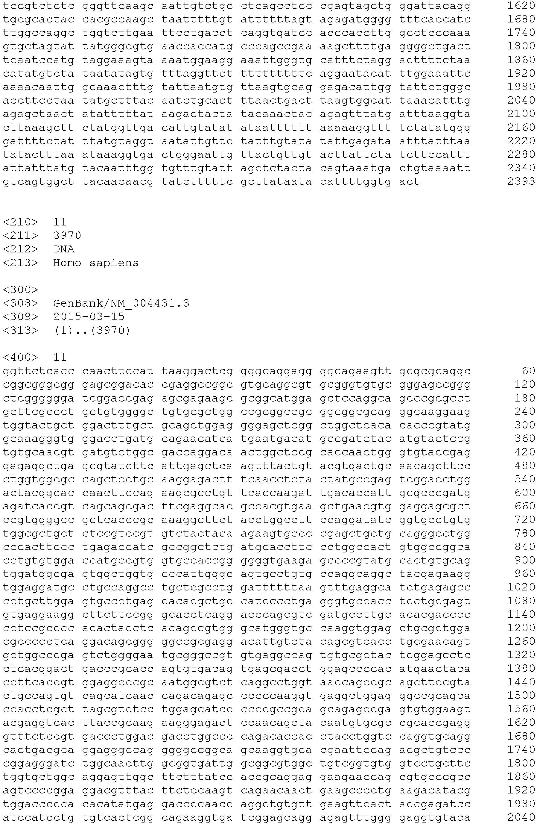


Claims (9)
- A method of detecting a tumor cell contained in a sample in distinction from a contaminant cell, the method comprising detecting one or more polypeptides selected from the group consisting of the following (i) to (iii) present in the sample:(i) a polypeptide comprising at least the amino acid sequence of any of SEQ ID NOs: 1 to 6;(ii) a polypeptide comprising an amino acid sequence having a homology of not less than 70% to the amino acid sequence of any of SEQ ID NOs:1 to 6; and(iii) a polypeptide comprising at least a splicing variant of the amino acid sequence of any of SEQ ID NOs: 1 to 6, or a polypeptide comprising at least a splicing variant of a polypeptide having a homology of not less than 70% to the amino acid sequence of any of SEQ ID NOs: 1 to 6.
- The method according to claim 1, wherein the detection is carried out using an antibody or aptamer that specifically recognizes one or more polypeptides selected from the group consisting of (i) to (iii).
- A method of detecting a tumor cell contained in a sample in distinction from a contaminant cell, the method comprising detecting a gene encoding any of the following polypeptides (i) to (iii) present in the sample:(i) a polypeptide comprising at least the amino acid sequence of any of SEQ ID NOs: 1 to 6;(ii) a polypeptide comprising an amino acid sequence having a homology of not less than 70% to the amino acid sequence of any of SEQ ID NOs:1 to 6; and(iii) a polypeptide comprising at least a splicing variant of the amino acid sequence of any of SEQ ID NOs: 1 to 6, or a polypeptide comprising at least a splicing variant of a polypeptide having a homology of not less than 70% to the amino acid sequence of any of SEQ ID NOs: 1 to 6.
- A method of collecting a tumor cell, the method comprising: detecting a tumor cell contained in a sample in distinction from contaminant cell; and collecting the detected tumor cell using collection means;
wherein the detection of the tumor cell is carried out by detecting one or more polypeptides selected from the group consisting of the following (i) to (iii) present in the sample:(i) a polypeptide comprising at least the amino acid sequence of any of SEQ ID NOs: 1 to 6;(ii) a polypeptide comprising at least an amino acid sequence having a homology of not less than 70% to the amino acid sequence of any of SEQ ID NOs: 1 to 6; and(iii) a polypeptide comprising at least a splicing variant of the amino acid sequence of any of SEQ ID NOs: 1 to 6, or a polypeptide comprising at least a splicing variant of a polypeptide having a homology of not less than 70% to the amino acid sequence of any of SEQ ID NOs: 1 to 6. - The method according to claim 4, wherein the detection of the tumor cell is carried out using an antibody or aptamer that specifically recognizes one or more polypeptides selected from the group consisting of (i) to (iii).
- The method according to any of claims 1 to 5, wherein the sample is blood, and the contaminant cell contained in the sample is leukocyte.
- A tumor marker comprising any of the following polypeptides (i) to (iii):(i) a polypeptide comprising at least the amino acid sequence of any of SEQ ID NOs: 1 to 6;(ii) a polypeptide comprising at least an amino acid sequence having a homology of not less than 70% to the amino acid sequence of any of SEQ ID NOs: 1 to 6; and(iii) a polypeptide comprising at least a splicing variant of the amino acid sequence of any of SEQ ID NOs: 1 to 6, or a polypeptide comprising at least a splicing variant of a polypeptide having a homology of not less than 70% to the amino acid sequence of any of SEQ ID NOs: 1 to 6;
the tumor marker being capable of detecting a tumor cell contained in a sample in distinction from leukocyte contained in the sample. - A tumor marker comprising a gene encoding any of the following polypeptides (i) to (iii):(i) a polypeptide comprising at least the amino acid sequence of any of SEQ ID NOs: 1 to 6;(ii) a polypeptide comprising at least an amino acid sequence having a homology of not less than 70% to the amino acid sequence of any of SEQ ID NOs: 1 to 6; and(iii) a polypeptide comprising at least a splicing variant of the amino acid sequence of any of SEQ ID NOs: 1 to 6, or a polypeptide comprising at least a splicing variant of a polypeptide having a homology of not less than 70% to the amino acid sequence of any of SEQ ID NOs: 1 to 6;
the tumor marker being capable of detecting a tumor cell contained in a sample in distinction from leukocyte contained in the sample. - A tumor marker comprising any of the following polypeptides (i) to (iii):(i) a polypeptide comprising at least the amino acid sequence of any of SEQ ID NOs: 1 to 6;(ii) a polypeptide comprising at least an amino acid sequence having a homology of not less than 70% to the amino acid sequence of any of SEQ ID NOs: 1 to 6; and(iii) a polypeptide comprising at least a splicing variant of the amino acid sequence of any of SEQ ID NOs: 1 to 6, or a polypeptide comprising at least a splicing variant of a polypeptide having a homology of not less than 70% to the amino acid sequence of any of SEQ ID NOs: 1 to 6;
which tumor marker enables collection of a tumor cell contained in a sample in distinction from leukocyte contained in the sample.
Applications Claiming Priority (4)
| Application Number | Priority Date | Filing Date | Title |
|---|---|---|---|
| JP2018042710 | 2018-03-09 | ||
| JP2018105966 | 2018-06-01 | ||
| JP2018193273 | 2018-10-12 | ||
| PCT/JP2019/007470 WO2019172030A1 (en) | 2018-03-09 | 2019-02-27 | Tumor marker, and method for collecting and detecting tumor cell in distinction from contaminant cell |
Publications (2)
| Publication Number | Publication Date |
|---|---|
| EP3764101A1 true EP3764101A1 (en) | 2021-01-13 |
| EP3764101A4 EP3764101A4 (en) | 2022-02-23 |
Family
ID=67846606
Family Applications (1)
| Application Number | Title | Priority Date | Filing Date |
|---|---|---|---|
| EP19764529.4A Pending EP3764101A4 (en) | 2018-03-09 | 2019-02-27 | TUMOR MARKER AND METHOD FOR COLLECTING AND DETECTING A TUMOR CELL FROM A CONTAMINATING CELL |
Country Status (5)
| Country | Link |
|---|---|
| US (1) | US12174194B2 (en) |
| EP (1) | EP3764101A4 (en) |
| JP (1) | JP7326764B2 (en) |
| CN (1) | CN111837039B (en) |
| WO (1) | WO2019172030A1 (en) |
Families Citing this family (7)
| Publication number | Priority date | Publication date | Assignee | Title |
|---|---|---|---|---|
| JP7521750B2 (en) * | 2020-01-14 | 2024-07-24 | 学校法人杏林学園 | Methods for detecting epithelial marker-negative tumor cells |
| WO2022071242A1 (en) * | 2020-09-30 | 2022-04-07 | 国立大学法人 長崎大学 | Method of predicting anticancer drug resistance and prognosis in kidney cancer patient, method of screening anti-kidney cancer substance, and pharmaceutical composition for treating kidney cancer |
| US12435444B2 (en) | 2021-03-09 | 2025-10-07 | Cdr-Life Ag | Rabbit-derived antigen binding protein nucleic acid libraries and methods of making the same |
| AU2022233285A1 (en) | 2021-03-09 | 2023-10-19 | Cdr-Life Ag | Mage-a4 peptide-mhc antigen binding proteins |
| WO2023112980A1 (en) * | 2021-12-16 | 2023-06-22 | 東ソー株式会社 | Tumor cell detection method and cancer testing method |
| CN120202015A (en) | 2022-09-14 | 2025-06-24 | Cdr-生物科技股份有限公司 | MAGE-A4 peptide double T cell adaptors |
| CN116449008A (en) * | 2023-03-07 | 2023-07-18 | 成都普瑞康生物科技有限公司 | Pancreatic cancer circulating tumor cell surface marker and application thereof |
Family Cites Families (32)
| Publication number | Priority date | Publication date | Assignee | Title |
|---|---|---|---|---|
| AU2003253860A1 (en) * | 2002-07-10 | 2004-01-23 | The Regents Of The University Of Michigan | Expression profile of lung cancer |
| AT501014A1 (en) * | 2004-08-19 | 2006-05-15 | Kury & Zeillinger Oeg | METHOD AND KIT FOR THE DIAGNOSIS OF A CANCER DISEASE, METHOD FOR DETERMINING THE APPEAL OF A PATIENT FOR THE TREATMENT OF CANCER DISEASE, THERAPEUTIC AGAINST THE PROPHYLAXIS OR THE TREATMENT OF A CANCER DISEASE |
| US20060134122A1 (en) * | 2004-12-16 | 2006-06-22 | Rapraeger Alan C | Peptides of syndecan-1 for inhibition of cancer |
| KR101363252B1 (en) * | 2005-02-04 | 2014-02-13 | 레이븐 바이오테크놀로지스, 인코퍼레이티드 | Antibodies that bind to epha2 and methods of use thereof |
| EP1861509B1 (en) | 2005-03-14 | 2015-09-09 | Janssen Diagnostics, LLC | A method for predicting progression free and overall survival at each follow-up time point during therapy of metastatic breast cancer patients using circulating tumor cells |
| WO2007127454A2 (en) * | 2006-04-28 | 2007-11-08 | Cythera, Inc. | Hepatocyte lineage cells |
| US20080020979A1 (en) * | 2006-06-09 | 2008-01-24 | Rapraeger Alan C | Peptides of Syndecan-1 For Inhibiting Angiogenesis |
| WO2009064789A2 (en) * | 2007-11-15 | 2009-05-22 | The Johns Hopkins University | Methods for detecting and monitoring circulating cancer stem cells |
| PL2254586T3 (en) * | 2008-02-22 | 2015-09-30 | Agency For Science Tech And Research Astarstar | Mesenchymal stem cell particles |
| KR101040652B1 (en) * | 2008-08-11 | 2011-06-10 | 재단법인서울대학교산학협력재단 | Screening methods for substances that inhibit the cancer metastasis function of TM4SF5 |
| US20160041153A1 (en) | 2008-11-12 | 2016-02-11 | Kirk Brown | Biomarker compositions and markers |
| WO2010073694A1 (en) * | 2008-12-25 | 2010-07-01 | 国立大学法人東京大学 | Diagnosis of treatment of cancer using anti-tm4sf20 antibody |
| EP2309273B1 (en) * | 2009-09-16 | 2016-05-18 | ZEILLINGER, Robert | Novel tumor marker determination |
| CA2788245A1 (en) * | 2010-01-27 | 2011-08-04 | Duke University | Biomarkers for circulating tumor cells |
| WO2011127219A1 (en) | 2010-04-06 | 2011-10-13 | Caris Life Sciences Luxembourg Holdings | Circulating biomarkers for disease |
| SE535084C2 (en) * | 2010-04-16 | 2012-04-10 | Prostasom Handelsbolag | Procedure for cancer diagnosis |
| WO2012024543A1 (en) | 2010-08-18 | 2012-02-23 | Caris Life Sciences Luxembourg Holdings | Circulating biomarkers for disease |
| WO2012115885A1 (en) * | 2011-02-22 | 2012-08-30 | Caris Life Sciences Luxembourg Holdings, S.A.R.L. | Circulating biomarkers |
| CA2827923C (en) * | 2011-02-25 | 2021-11-23 | Chugai Seiyaku Kabushiki Kaisha | Fc.gamma.riib-specific fc antibody |
| WO2013072571A1 (en) | 2011-11-17 | 2013-05-23 | Institut Gustave Roussy | Method for characterising circulating tumour cells and application to diagnostics |
| CN102590531A (en) * | 2012-03-24 | 2012-07-18 | 广西壮族自治区肿瘤防治研究所 | Ovarian cancer related antigen autoantibody repertoire liquid chip detection kit and preparation method thereof |
| US9034827B2 (en) * | 2012-07-09 | 2015-05-19 | Wisconsin Alumni Research Foundation | Syndecan peptides and polypeptides as inhibitors of cancer |
| WO2014198817A1 (en) * | 2013-06-14 | 2014-12-18 | Bayer Pharma Aktiengesellschaft | Anti-tweakr antibodies and uses thereof |
| CA2926087C (en) * | 2013-10-10 | 2023-03-14 | Beth Israel Deaconess Medical Center, Inc. | Tm4sf1 binding proteins and methods of using same |
| US9404927B2 (en) * | 2013-12-05 | 2016-08-02 | The Translational Genomics Research Institute | Systems and methods for diagnosing and treating cancer |
| JP6572547B2 (en) | 2015-02-02 | 2019-09-11 | 東ソー株式会社 | Fine particle recovery device |
| EA037995B1 (en) | 2015-02-24 | 2021-06-21 | Рупрехт-Карлс-Университет Гейдельберг | Biomarker panel for the detection of cancer |
| JP6617516B2 (en) | 2015-10-27 | 2019-12-11 | 東ソー株式会社 | Method for detecting target cells contained in blood sample |
| JP2018042710A (en) | 2016-09-14 | 2018-03-22 | 株式会社新生工業 | Transportation device and game machine |
| JP2018105966A (en) | 2016-12-26 | 2018-07-05 | ソニー株式会社 | Image display device |
| CA3056609A1 (en) * | 2017-03-16 | 2018-09-20 | Seattle Children's Hospital (dba Seattle Children's Research Institute) | Engraftable cell-based immunotherapy for long-term delivery of therapeutic proteins |
| JP6965569B2 (en) | 2017-05-17 | 2021-11-10 | 住友電気工業株式会社 | Glass raw material container |
-
2019
- 2019-02-25 JP JP2019031154A patent/JP7326764B2/en active Active
- 2019-02-27 WO PCT/JP2019/007470 patent/WO2019172030A1/en not_active Ceased
- 2019-02-27 CN CN201980018046.7A patent/CN111837039B/en active Active
- 2019-02-27 US US16/978,887 patent/US12174194B2/en active Active
- 2019-02-27 EP EP19764529.4A patent/EP3764101A4/en active Pending
Also Published As
| Publication number | Publication date |
|---|---|
| EP3764101A4 (en) | 2022-02-23 |
| US20200408769A1 (en) | 2020-12-31 |
| US12174194B2 (en) | 2024-12-24 |
| JP7326764B2 (en) | 2023-08-16 |
| CN111837039A (en) | 2020-10-27 |
| CN111837039B (en) | 2024-06-07 |
| WO2019172030A1 (en) | 2019-09-12 |
| JP2020062004A (en) | 2020-04-23 |
Similar Documents
| Publication | Publication Date | Title |
|---|---|---|
| US12174194B2 (en) | Tumor marker, and method for collecting and detecting tumor cell in distinction from contaminant cell | |
| McDaniel et al. | Phenotypic diversity of circulating tumour cells in patients with metastatic castration‐resistant prostate cancer | |
| EP2742123B1 (en) | Isolation and detection of cancer cells | |
| EP1797431B1 (en) | Detection of elevated levels of her-2/neu protein on circulating cancer cells and treatment | |
| EP3029460A1 (en) | O-cadherin as a biomarker for circulating tumor cells | |
| US20190078153A1 (en) | Method of analyzing genetically abnormal cells | |
| CN111830249A (en) | Use of a method for purifying, isolating and analyzing atypical circulating tumor cells and use of atypical circulating tumor cells | |
| CN105849565B (en) | SHON as a prognostic biomarker for cancer and predicting the efficacy of endocrine therapy for cancer | |
| Carneiro et al. | Minimizing false positives for CTC identification | |
| EP2064552B1 (en) | Method for the detection of cancerous epithelial cells using released cytokeratins as markers for said cells | |
| JP7306618B2 (en) | Factors Involved in Metastasis Formation of Cancer and Method for Predicting Prognosis of Cancer Patients Using Said Factors | |
| JPWO2017033905A1 (en) | Method for detecting OCLN-ARHGAP26 gene | |
| EP3498835A1 (en) | Detection of cldn18-arhgap6 fusion gene or cldn18-arhgap26 fusion gene in pancreatic cancer | |
| JP7599170B2 (en) | Methods for detecting and recovering melanoma cells derived from the yellow race, and reagents and carrier-immobilized molecules used in these methods | |
| JP7521750B2 (en) | Methods for detecting epithelial marker-negative tumor cells | |
| CN108949998A (en) | Predict that patient with breast cancer fights the reactive gene mutation site of HER2 therapeutic agent and its application | |
| KR101649591B1 (en) | Novel biomarker for diagnosing acute leukemia | |
| EP2904107B1 (en) | Methods for vel blood group typing | |
| JP7593584B2 (en) | Tumor cell markers and methods for detecting or harvesting tumor cells - Patents.com | |
| JP2022092366A (en) | Detection method of melanoma cell, recovery method of melanoma cell, and reagent and carrier immobilization molecule used for the methods | |
| Jiang et al. | Functions and clinical significances of LncRNA SChLAP1 and UBE2E3 in prostate cancer | |
| US10619216B2 (en) | Method for detecting RP2-ARHGAP6 gene | |
| Zhang et al. | Plasma Small Extracellular Vesicle-Derived GDF15 Predicts Resistance to PD-1/PD-L1-Based Therapy in Colorectal Cancer: A Pilot Study | |
| CN120891191A (en) | Biomarkers and detection kits, and biochips related to breast cancer prognosis | |
| Prummel et al. | Inflammatory Mesenchymal Stromal Cells and IFN-responsive T cells are key mediators of human bone marrow niche remodeling in CHIP and MDS |
Legal Events
| Date | Code | Title | Description |
|---|---|---|---|
| STAA | Information on the status of an ep patent application or granted ep patent |
Free format text: STATUS: THE INTERNATIONAL PUBLICATION HAS BEEN MADE |
|
| PUAI | Public reference made under article 153(3) epc to a published international application that has entered the european phase |
Free format text: ORIGINAL CODE: 0009012 |
|
| STAA | Information on the status of an ep patent application or granted ep patent |
Free format text: STATUS: REQUEST FOR EXAMINATION WAS MADE |
|
| 17P | Request for examination filed |
Effective date: 20200924 |
|
| AK | Designated contracting states |
Kind code of ref document: A1 Designated state(s): AL AT BE BG CH CY CZ DE DK EE ES FI FR GB GR HR HU IE IS IT LI LT LU LV MC MK MT NL NO PL PT RO RS SE SI SK SM TR |
|
| AX | Request for extension of the european patent |
Extension state: BA ME |
|
| DAV | Request for validation of the european patent (deleted) | ||
| DAX | Request for extension of the european patent (deleted) | ||
| RIC1 | Information provided on ipc code assigned before grant |
Ipc: C12N 9/12 20060101ALI20211018BHEP Ipc: C07K 14/745 20060101ALI20211018BHEP Ipc: C07K 14/705 20060101ALI20211018BHEP Ipc: C07K 14/47 20060101ALI20211018BHEP Ipc: G01N 33/569 20060101ALI20211018BHEP Ipc: C12N 15/115 20100101ALI20211018BHEP Ipc: G01N 33/574 20060101ALI20211018BHEP Ipc: G01N 33/53 20060101ALI20211018BHEP Ipc: C12Q 1/6886 20180101ALI20211018BHEP Ipc: C12Q 1/6813 20180101ALI20211018BHEP Ipc: C12Q 1/04 20060101ALI20211018BHEP Ipc: C12N 15/12 20060101ALI20211018BHEP Ipc: C07K 14/82 20060101ALI20211018BHEP Ipc: G01N 33/68 20060101AFI20211018BHEP |
|
| A4 | Supplementary search report drawn up and despatched |
Effective date: 20220124 |
|
| RIC1 | Information provided on ipc code assigned before grant |
Ipc: C12N 9/12 20060101ALI20220118BHEP Ipc: C07K 14/745 20060101ALI20220118BHEP Ipc: C07K 14/705 20060101ALI20220118BHEP Ipc: C07K 14/47 20060101ALI20220118BHEP Ipc: G01N 33/569 20060101ALI20220118BHEP Ipc: C12N 15/115 20100101ALI20220118BHEP Ipc: G01N 33/574 20060101ALI20220118BHEP Ipc: G01N 33/53 20060101ALI20220118BHEP Ipc: C12Q 1/6886 20180101ALI20220118BHEP Ipc: C12Q 1/6813 20180101ALI20220118BHEP Ipc: C12Q 1/04 20060101ALI20220118BHEP Ipc: C12N 15/12 20060101ALI20220118BHEP Ipc: C07K 14/82 20060101ALI20220118BHEP Ipc: G01N 33/68 20060101AFI20220118BHEP |
|
| STAA | Information on the status of an ep patent application or granted ep patent |
Free format text: STATUS: EXAMINATION IS IN PROGRESS |
|
| 17Q | First examination report despatched |
Effective date: 20230831 |