EP3757511A1 - Method for estimating the in vivo dimensions of a surgical specimen from the ex vivo dimensions thereof - Google Patents
Method for estimating the in vivo dimensions of a surgical specimen from the ex vivo dimensions thereof Download PDFInfo
- Publication number
- EP3757511A1 EP3757511A1 EP19757717.4A EP19757717A EP3757511A1 EP 3757511 A1 EP3757511 A1 EP 3757511A1 EP 19757717 A EP19757717 A EP 19757717A EP 3757511 A1 EP3757511 A1 EP 3757511A1
- Authority
- EP
- European Patent Office
- Prior art keywords
- surgical
- surgical specimen
- specimen
- wcf
- lcf
- Prior art date
- Legal status (The legal status is an assumption and is not a legal conclusion. Google has not performed a legal analysis and makes no representation as to the accuracy of the status listed.)
- Withdrawn
Links
- 238000000034 method Methods 0.000 title claims abstract description 45
- 238000001727 in vivo Methods 0.000 title claims description 56
- 230000007547 defect Effects 0.000 claims abstract description 31
- 230000008961 swelling Effects 0.000 claims abstract description 13
- 230000003902 lesion Effects 0.000 claims description 56
- 208000035346 Margins of Excision Diseases 0.000 claims description 31
- 210000004761 scalp Anatomy 0.000 claims description 16
- 210000001364 upper extremity Anatomy 0.000 claims description 15
- 210000003141 lower extremity Anatomy 0.000 claims description 13
- WSFSSNUMVMOOMR-UHFFFAOYSA-N Formaldehyde Chemical compound O=C WSFSSNUMVMOOMR-UHFFFAOYSA-N 0.000 abstract description 20
- 230000001419 dependent effect Effects 0.000 abstract 1
- 210000003491 skin Anatomy 0.000 description 19
- 238000005259 measurement Methods 0.000 description 8
- 238000011477 surgical intervention Methods 0.000 description 8
- 238000012623 in vivo measurement Methods 0.000 description 4
- 201000001441 melanoma Diseases 0.000 description 4
- 230000002459 sustained effect Effects 0.000 description 4
- 238000001574 biopsy Methods 0.000 description 3
- 238000003745 diagnosis Methods 0.000 description 3
- 210000001519 tissue Anatomy 0.000 description 3
- 210000004027 cell Anatomy 0.000 description 2
- 230000000694 effects Effects 0.000 description 2
- 238000000605 extraction Methods 0.000 description 2
- 230000037081 physical activity Effects 0.000 description 2
- 206010040882 skin lesion Diseases 0.000 description 2
- 231100000444 skin lesion Toxicity 0.000 description 2
- 230000036558 skin tension Effects 0.000 description 2
- 206010064755 Atypical fibroxanthoma Diseases 0.000 description 1
- 206010004146 Basal cell carcinoma Diseases 0.000 description 1
- 201000009030 Carcinoma Diseases 0.000 description 1
- 102000008186 Collagen Human genes 0.000 description 1
- 108010035532 Collagen Proteins 0.000 description 1
- LFQSCWFLJHTTHZ-UHFFFAOYSA-N Ethanol Chemical compound CCO LFQSCWFLJHTTHZ-UHFFFAOYSA-N 0.000 description 1
- 206010027476 Metastases Diseases 0.000 description 1
- 206010028980 Neoplasm Diseases 0.000 description 1
- 206010029098 Neoplasm skin Diseases 0.000 description 1
- 201000004404 Neurofibroma Diseases 0.000 description 1
- 208000009077 Pigmented Nevus Diseases 0.000 description 1
- 208000005214 Poroma Diseases 0.000 description 1
- 208000000453 Skin Neoplasms Diseases 0.000 description 1
- 230000003187 abdominal effect Effects 0.000 description 1
- 201000009431 angiokeratoma Diseases 0.000 description 1
- 208000001119 benign fibrous histiocytoma Diseases 0.000 description 1
- 230000001684 chronic effect Effects 0.000 description 1
- 238000005352 clarification Methods 0.000 description 1
- 229920001436 collagen Polymers 0.000 description 1
- 230000006378 damage Effects 0.000 description 1
- 230000003247 decreasing effect Effects 0.000 description 1
- 230000000593 degrading effect Effects 0.000 description 1
- 210000004207 dermis Anatomy 0.000 description 1
- 230000002542 deteriorative effect Effects 0.000 description 1
- 206010012601 diabetes mellitus Diseases 0.000 description 1
- 238000010586 diagram Methods 0.000 description 1
- 210000004177 elastic tissue Anatomy 0.000 description 1
- 208000017338 epidermoid cysts Diseases 0.000 description 1
- 206010016629 fibroma Diseases 0.000 description 1
- 238000007654 immersion Methods 0.000 description 1
- 230000002757 inflammatory effect Effects 0.000 description 1
- 238000013507 mapping Methods 0.000 description 1
- 239000003550 marker Substances 0.000 description 1
- 201000002699 melanoma in congenital melanocytic nevus Diseases 0.000 description 1
- 230000009401 metastasis Effects 0.000 description 1
- 230000007170 pathology Effects 0.000 description 1
- 238000004321 preservation Methods 0.000 description 1
- 238000002278 reconstructive surgery Methods 0.000 description 1
- 208000014212 sarcomatoid carcinoma Diseases 0.000 description 1
- 230000000276 sedentary effect Effects 0.000 description 1
- 239000000779 smoke Substances 0.000 description 1
- 206010041823 squamous cell carcinoma Diseases 0.000 description 1
- 238000001356 surgical procedure Methods 0.000 description 1
- 230000000699 topical effect Effects 0.000 description 1
Images

Classifications
-
- B—PERFORMING OPERATIONS; TRANSPORTING
- B25—HAND TOOLS; PORTABLE POWER-DRIVEN TOOLS; MANIPULATORS
- B25H—WORKSHOP EQUIPMENT, e.g. FOR MARKING-OUT WORK; STORAGE MEANS FOR WORKSHOPS
- B25H3/00—Storage means or arrangements for workshops facilitating access to, or handling of, work tools or instruments
- B25H3/02—Boxes
-
- A—HUMAN NECESSITIES
- A61—MEDICAL OR VETERINARY SCIENCE; HYGIENE
- A61B—DIAGNOSIS; SURGERY; IDENTIFICATION
- A61B5/00—Measuring for diagnostic purposes; Identification of persons
- A61B5/103—Measuring devices for testing the shape, pattern, colour, size or movement of the body or parts thereof, for diagnostic purposes
- A61B5/107—Measuring physical dimensions, e.g. size of the entire body or parts thereof
- A61B5/1072—Measuring physical dimensions, e.g. size of the entire body or parts thereof measuring distances on the body, e.g. measuring length, height or thickness
-
- G—PHYSICS
- G01—MEASURING; TESTING
- G01B—MEASURING LENGTH, THICKNESS OR SIMILAR LINEAR DIMENSIONS; MEASURING ANGLES; MEASURING AREAS; MEASURING IRREGULARITIES OF SURFACES OR CONTOURS
- G01B21/00—Measuring arrangements or details thereof, where the measuring technique is not covered by the other groups of this subclass, unspecified or not relevant
- G01B21/32—Measuring arrangements or details thereof, where the measuring technique is not covered by the other groups of this subclass, unspecified or not relevant for measuring the deformation in a solid
-
- A—HUMAN NECESSITIES
- A61—MEDICAL OR VETERINARY SCIENCE; HYGIENE
- A61B—DIAGNOSIS; SURGERY; IDENTIFICATION
- A61B2505/00—Evaluating, monitoring or diagnosing in the context of a particular type of medical care
- A61B2505/05—Surgical care
Definitions
- the present invention belongs to the field of dermatology, and more particularly to methods for the surgical excision of a skin lesion, such as a melanoma.
- the object of the present invention is a method which allows the dimensions of a surgical specimen in the in vivo state before excision to be determined, based on the dimensions of said surgical specimen measured in an ex vivo state after excision and after a process of fixing the surgical specimen.
- Another object of the present invention is to determine the dimensions of the surgical defect left in the skin of the patient after excising the surgical specimen due to the swelling of the skin, based on the dimensions of the surgical specimen measured in the in vivo state before excision.
- Another object of the present invention is to determine the surgical margin in the in vivo state before excision, i.e., the distance between the visible outer border of a lesion and the outer border of the surgical specimen, based on the surgical margin measured in an ex vivo state after excision and after a process of fixing the surgical specimen.
- Another object of the present invention is to determine the histological margin in the in vivo state before excision, i.e., the distance between the real outer border of a lesion and the outer border of the surgical specimen, based on the histological margin measured in an ex vivo state after excision and after a process of fixing the surgical specimen.
- the treatment of certain skin lesions comprises excising them by means of a relatively simple surgical intervention.
- a surgical specimen that is essentially planar and has a size larger than the lesion itself is excised, as it is necessary to respect certain surgical margins around same.
- the surgical margins could be around one centimeter or the like.
- the surgical specimen is then subjected to a fixing treatment which prevents it from deteriorating, usually by means of immersion thereof in formalin for a certain time, and sent to a pathologist to perform a biopsy confirming the initial diagnosis.
- a common problem during this procedure consists of the lack of consistency between the different dimensions of the surgical specimen taken into account by the surgeon at the time of the intervention and the same dimensions measured at a later time by the pathologist while performing the biopsy. Indeed, from the very moment the surgical specimen is excised from the body of the patient, a shrinkage effect causing a reduction of the volume thereof is known to occur. As a result, the surgical margins measured by the pathologist are usually smaller than the actual surgical margins the surgeon took into account during the intervention. Histological margins are also reduced.
- Another known problem is related to the expansion of the surgical defect created by excising the surgical specimen due to the swelling of the surrounding skin. Indeed, it is known that the skin around the gap left by the surgical specimen in the skin of the patient after excision tends to shrink, which causes an increase in the size of said gap. If there are substantial differences between the size and shape of the planned wound and the surgical defect after excising the surgical specimen, the closing technique cannot be predicted with certainty before excision. These differences have an important clinical relevance in reconstructive surgery techniques, for example when the decision is made to do a skin flap.
- the present invention solves the aforementioned problems as a result of a method which allows the original in vivo dimensions of a surgical specimen to be determined from the ex vivo dimensions of said surgical specimen hours after excision and already subjected to a process of fixing.
- the pathologist can thereby know the surgical margin the surgeon left during excision and thus be certain that the necessary distances were respected. Furthermore, this also allows the surgeon to demonstrate that he or she complied with the required surgical margins.
- the subscript "IV” will generally be used herein to refer to an in vivo state, prior to the surgical intervention in which the surgical specimen is excised, and the subscript “EV” will generally be used herein to refer to an ex vivo state after excision and a process of fixing the surgical specimen, usually in 10% formalin for 24 hours.
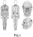
- the biomechanical characteristics of the skin are known to not be uniform in all parts of the body, as is clearly shown by means of the so-called Langer's lines, known today as relaxed skin tension lines (see Figure 1 ). Said tension is primarily induced by the architecture of the collagen framework and of the elastic fibers making up the deep reticular dermis (see the article by Dumas P et al. entitled “Study of skin retraction applied to the treatment of skin tumors. Mapping of the human body", Ann Chir Plast Esthet 2012; 57:118-24 ).
- the mean body mass index was 27.71 kg/m 2 (corresponding to overweight), the mean abdominal girth was 109.38 cm (corresponding to figures above what is recommended both in men, whose normal value is ⁇ 102 cm, and in women, whose normal value normal is ⁇ 88 cm), the most common phototype was III, followed by IV and I and II; 83% of the patients from whom the SS were obtained did not smoke; 67.82% did not drink alcohol; 15.7% suffered diabetes mellitus; 2.2% suffered an inflammatory skin condition; and 2.8% were undergoing treatment with topical or oral corticoids on a regular basis; 72.6% had suffered chronic exposition to the sun; and 70.1% presented actinic damage. 51.1% of the patients from whom the surgical specimens were obtained performed light physical activity; 36.3% maintained a sedentary lifestyle; and 12.6% performed moderate or intense physical activity.
- the location of the surgical specimens was, from more to less common:
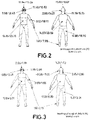
- Figure 2 graphically shows a summary of the data relative to shrinkage of the surgical specimen obtained in the study.
- the inventors of the present application have developed a method for estimating the magnitude of shrinkage of a surgical specimen excised from the skin of a patient.
- the surgical specimen is considered to have an essentially planar shape defined by the length and width dimensions thereof.
- the method comprises the following steps:
- correction factors LCF SS and WCF SS depends on the location in the body of the patient from whom the surgical specimen was excised according to the following table. Furthermore, the value of correction factors LCF SS and WCF SS may vary by 10% with respect to the values of the table: Table 1: Surgical specimen width and length reduction percentages Region of the body WCF SS Region of the body LCF SS Auricle 0.2385 Anterior torso 0.2372 Cervical region 0.1519 Posterior torso 0.2009 Posterior torso 0.1407 Upper limb 0.1911 Scalp 0.1382 Lower limb 0.1876 Anterior torso 0.1335 Cheek, chin, and lip 0.1683 Lower limb 0.1288 Cervical region 0.1625 Frontal region 0.1194 Auricle 0.1427 Cheek, chin, and lip 0.1140 Frontal region 0.1324 Upper limb 0.0953 Nose 0.1243 Nose 0.0708 Scalp 0.1067 Palm or sole 0.0101 Palm or sole 0.0799
- this novel method allows the pathologist to determine what the size of the surgical specimen was right at the time of the excision. To that end, the pathologist must only measure the dimensions of the surgical specimen available, even when it has shrunk after several hours of fixing, for example, in 10% buffered formalin. The application of the preceding formulas will allow the pathologist to estimate the original size in vivo of the surgical specimen in a simple manner.
- Figure 3 graphically shows a summary of the data relative to shrinkage of the surgical specimen obtained in the study.
- the method of the invention further comprises the following steps:
- correction factors LCF SD and WCF SD depends on the location of the surgical specimen in the body of the patient according to the following table. Furthermore, the value of correction factors LCF SD and WCF SD may vary by 10% with respect to the values of the table: Table 2: Surgical defect width and length increase percentages Region of the body WCF SD Region of the body LCF SD Upper limb 0.2189 Posterior torso 0.0680 Posterior torso 0.1733 Anterior torso 0.0571 Anterior torso 0.1283 Cheek, chin, and lip 0.0298 Lower limb 0.0927 Lower limb 0.0234 Scalp 0.0321 Palm or sole 0.0213 Cervical region 0.0285 Upper limb 0.0166 Cheek, chin, and lip 0.0278 Frontal region 0.0159 Palm or sole 0.0161 Scalp 0.0153 Frontal region 0.0099 Cervical region -0.0080 Nose -0.0389 Nose -0.0348 Auricle -0.0809 Auricle -0.0705
- this novel preferred embodiment of the method of the invention allows the surgeon to know beforehand what the real dimensions of the surgical defect that excision of the surgical specimen will cause will be. To that end, the surgeon must simply measure the dimensions of the surgical specimen in vivo, before excision. The application of the preceding formulas will allow the surgeon to estimate the size the surgical defect will have in a simple manner.
- the method of the invention further comprises the following steps:
- the value of the correction factors LCF L and WCF L depends on the location of the surgical specimen in the body of the patient according to the following table. Furthermore, the value of the correction factors LCF L and WCF L may vary by 10% with respect to the values of the table: Table 3: Lesion width and length increase percentages Region of the body WCF L Region of the body LCF L Cervical region 0.2301 Anterior torso 0.2225 Frontal region 0.1968 Frontal region 0.1642 Palm or sole 0.1519 Lower limb 0.1519 Lower limb 0.1385 Nose 0.1501 Anterior torso 0.1349 Posterior torso 0.1418 Cheek, chin, and lip 0.1256 Upper limb 0.1412 Nose 0.1252 Cervical region 0.1329 Posterior torso 0.1210 Palm or sole 0.1262 Auricle 0.0958 Scalp 0.1229 Upper limb 0.0746 Auricle 0.1205 Scalp 0.0507 Cheek, chin, and lip 0.1130
- this novel embodiment of the invention allows the pathologist to determine what the dimensions of the surgical margins were right at the time of the excision. To that end, the pathologist must simply measure the dimensions both of the lesion and of the surgical specimen available, even when it has shrunk after several hours of fixing, for example in 10% buffered formalin. The application of the preceding formulas will allow the pathologist to know what the surgical margins were in the surgical specimen in vivo in a simple manner.
- the shrinkage sustained by the surgical specimen as a whole As a result of the shrinkage sustained by the surgical specimen as a whole, a decrease of the histological margins also takes place. As in the case of the surgical margins, the shrinkage of the histological margins occurs due to the combination of the shrinkage of the surgical specimen and the shrinkage of the lesion.
- the histological margins are the most clinically relevant margins, because they are what really reflect the distance that must be respected between the outer border of the extracted surgical specimen and the real outer border of the lesion.
- the histological margins are not visible in vivo to the naked eye, and there is currently no reliable method for determining them. For that reason, the only available data is that relative to the histological margins ex vivo after the process of fixing which, as mentioned, are shrunken and therefore do not precisely reflect the surgical margins that were really respected during the surgical intervention.
- the method of the invention further comprises the following steps:
- Figure 4 schematically shows a surgical specimen (SS) assumed to be in the in vivo state and in the center of which there is a lesion (L).
- the lesion (L) has a visible apparent border limiting the striped area, and a non-visible real border depicted by means of a discontinuous line outside of the visible border of the lesion (L).
- the distance between the upper border of the surgical specimen (SS) and the visible apparent upper border of the lesion (L) is the width of the surgical margin in vivo, which was estimated as (WSM iv e ) according to formula (6) above.
- the distance between the upper border of the surgical specimen (SS) and the non-visible real upper border of the lesion (L) is the width of the histological margin in vivo (WHM iv e ) to be estimated.
- this novel embodiment of the invention allows the pathologist to determine what the dimensions of the real histological margins were right at the time of the excision. To that end, the pathologist only needs the data already obtained beforehand in addition to the histological margins ex vivo after fixing, which he or she can readily measure.
- Example 1 Estimate of the swelling of a surgical defect
- Example 2 Estimate of the dimensions of a surgical specimen and of the surgical and histological margins
- a pathologist receives a surgical specimen for analysis.
- the surgical specimen consists of a scalp specimen in which there is a melanoma.
- the minimum histological margin must be 1 cm.
- the pathologist measures the histological margins and the width of the histological margin measured turns out to be 0.85 cm. Therefore, doubts arise concerning whether it is necessary to perform another procedure on the patient.
- the pathologist decides to use the formulas described herein to estimate what the dimensions of the specimen, of the lesion, and of the histological margins were in the in vivo state before the surgical intervention.
- the pathologist takes measurements of the specimen, of the lesion, and of the histological margins. The measurements are:
- the pathologist uses the preceding formulas to estimate the dimensions of the surgical specimen, the lesion, and the histological margin in vivo before excision.
- the pathologist applies formulas (1) and (2) to estimate the dimensions of the surgical specimen in vivo :
- LSS IV e LSS EV m ⁇ 1 + LCF SS
- WSS IV e WSS IV m ⁇ 1 + WCF SS
- LSM IV e LSS EV m ⁇ 1 + LCF SS ⁇ LL EV m ⁇ 1 + LCF L / 2
- WSM IV e WSS EV m ⁇ 1 + WCF SS ⁇ WL EV m ⁇ 1 + WCF L / 2
- Example 3 Estimate of the dimensions of a surgical specimen and of the surgical and histological margins
- a patient has suffered metastasis due to an epidermoid carcinoma located in the posterior torso and the patient's family is considering filing a lawsuit against the hospital because excision was performed with a lateral histological margin of 0.435 cm when international protocols establish that an excision with 0.5 cm should be performed.
- the judge has the following measurements from the anatomic pathology report:
- the judge uses the method of the present invention in order to see if the hospital is at all liable and the following in vivo measurements are obtained:
Landscapes
- Health & Medical Sciences (AREA)
- Life Sciences & Earth Sciences (AREA)
- Physics & Mathematics (AREA)
- General Physics & Mathematics (AREA)
- Engineering & Computer Science (AREA)
- Biomedical Technology (AREA)
- Medical Informatics (AREA)
- Dentistry (AREA)
- Biophysics (AREA)
- Pathology (AREA)
- Mechanical Engineering (AREA)
- Heart & Thoracic Surgery (AREA)
- Oral & Maxillofacial Surgery (AREA)
- Molecular Biology (AREA)
- Surgery (AREA)
- Animal Behavior & Ethology (AREA)
- General Health & Medical Sciences (AREA)
- Public Health (AREA)
- Veterinary Medicine (AREA)
- Investigating Or Analysing Biological Materials (AREA)
- Materials For Medical Uses (AREA)
Abstract
Description
- The present invention belongs to the field of dermatology, and more particularly to methods for the surgical excision of a skin lesion, such as a melanoma.
- The object of the present invention is a method which allows the dimensions of a surgical specimen in the in vivo state before excision to be determined, based on the dimensions of said surgical specimen measured in an ex vivo state after excision and after a process of fixing the surgical specimen.
- Another object of the present invention is to determine the dimensions of the surgical defect left in the skin of the patient after excising the surgical specimen due to the swelling of the skin, based on the dimensions of the surgical specimen measured in the in vivo state before excision.
- Another object of the present invention is to determine the surgical margin in the in vivo state before excision, i.e., the distance between the visible outer border of a lesion and the outer border of the surgical specimen, based on the surgical margin measured in an ex vivo state after excision and after a process of fixing the surgical specimen.
- Another object of the present invention is to determine the histological margin in the in vivo state before excision, i.e., the distance between the real outer border of a lesion and the outer border of the surgical specimen, based on the histological margin measured in an ex vivo state after excision and after a process of fixing the surgical specimen.
- The treatment of certain skin lesions, such as discoloring or moles suspected of being cancerous for example, comprises excising them by means of a relatively simple surgical intervention. During said intervention, a surgical specimen that is essentially planar and has a size larger than the lesion itself is excised, as it is necessary to respect certain surgical margins around same. For example, depending on the type of lesion, the surgical margins could be around one centimeter or the like. The surgical specimen is then subjected to a fixing treatment which prevents it from deteriorating, usually by means of immersion thereof in formalin for a certain time, and sent to a pathologist to perform a biopsy confirming the initial diagnosis.
- A common problem during this procedure consists of the lack of consistency between the different dimensions of the surgical specimen taken into account by the surgeon at the time of the intervention and the same dimensions measured at a later time by the pathologist while performing the biopsy. Indeed, from the very moment the surgical specimen is excised from the body of the patient, a shrinkage effect causing a reduction of the volume thereof is known to occur. As a result, the surgical margins measured by the pathologist are usually smaller than the actual surgical margins the surgeon took into account during the intervention. Histological margins are also reduced.
- These discrepancies may cause a number of drawbacks. For example, the decision may be made to perform a second surgical intervention to increase the surgical or histological margins without it actually being necessary. Legal-medical issues with real repercussions in judicial proceedings may also arise. Indeed, in the event of a lawsuit lodged by the patient or his or her family members as a result of a negative progression of the patient after the excision, the surgeon has no way to prove that he or she complied with the required surgical or histological margins. The only available data is often the surgical or histological margins measured hours after the surgical intervention by the pathologist in the shrunken specimen, margins which are logically smaller than those that were respected during the operation.
- Another known problem is related to the expansion of the surgical defect created by excising the surgical specimen due to the swelling of the surrounding skin. Indeed, it is known that the skin around the gap left by the surgical specimen in the skin of the patient after excision tends to shrink, which causes an increase in the size of said gap. If there are substantial differences between the size and shape of the planned wound and the surgical defect after excising the surgical specimen, the closing technique cannot be predicted with certainty before excision. These differences have an important clinical relevance in reconstructive surgery techniques, for example when the decision is made to do a skin flap.
- There is currently no solution for these problems, and surgeons and pathologists must improvise solutions to the problems that are considered.
- The present invention solves the aforementioned problems as a result of a method which allows the original in vivo dimensions of a surgical specimen to be determined from the ex vivo dimensions of said surgical specimen hours after excision and already subjected to a process of fixing. The pathologist can thereby know the surgical margin the surgeon left during excision and thus be certain that the necessary distances were respected. Furthermore, this also allows the surgeon to demonstrate that he or she complied with the required surgical margins.
- Some of the terms used herein are defined below:
- Surgical specimen: Portion of tissue of the patient excised during surgical intervention. The surgical specimen usually adopts an approximately elliptical elongated shape, referred to as "buttonhole", which can be characterized based on the length and width dimensions thereof.
- Lesion: Portion of damaged tissue to be excised from the patient. The lesion is within the surgical specimen, essentially at the center thereof.
- Surgical margin: Minimum distance between the visible outer border of the lesion and the outer border of the surgical specimen for the purpose of ensuring that after excision no injured cells remain in the skin of the patient.
- Histological margin: Minimum distance between the real outer border of the lesion and the outer border of the surgical specimen with the purpose of ensuring that after excision no injured cells remain in the skin of the patient. The real border of the lesion extends beyond the visible border in vivo, and the position thereof can only be determined ex vivo in a laboratory after excision.
- Width and length: Conventionally, surgical specimens excised in this context have a shape referred to as a "buttonhole", i.e., an essentially elliptical planar shape or the like characterized by its length (the longest dimension thereof) and width (the shortest dimension thereof). Both surgical specimens and surgical defects are herein considered to have an essentially elliptical planar shape. Therefore, references herein to the width and length of the different parameters are assumed to refer respectively to the direction of the length of the surgical specimen or lesion (longitudinal direction) and to the direction of the width of the surgical specimen or lesion (transverse direction).
- Surgeon: Medical professional who performs the surgical intervention of excising the surgical specimen from the skin of the patient. Before performing the excision, the surgeon determines through in vivo measurements the dimensions of the surgical specimen ensuring certain established surgical margins.
- Pathologist: Medical professional who performs a subsequent analysis of the surgical specimen to determine the characteristics of the lesion. For example, if the lesion is suspected of being a melanoma, the pathologist performs a biopsy of the surgical specimen to confirm or refute this diagnosis. The pathologist performs his or her analysis hours or days after the extraction of the surgical specimen. Accordingly, after excision by the surgeon, the surgical specimen is subjected to a fixing treatment, usually with formalin, to prevent it from degrading.
- Surgical defect: "Gap" left by the surgical specimen in the skin of the patient after excision.
- The subscript "IV" will generally be used herein to refer to an in vivo state, prior to the surgical intervention in which the surgical specimen is excised, and the subscript "EV" will generally be used herein to refer to an ex vivo state after excision and a process of fixing the surgical specimen, usually in 10% formalin for 24 hours.
- The superscript "e" will generally be used herein to refer to a magnitude estimated according to any of the methods described herein, and the superscript "m" will generally be used herein to refer to a magnitude physically measured by a surgeon or pathologist, whichever is appropriate.
- The following symbols are used:
- LSSIV e
- Estimated length of the surgical specimen in vivo.
- WSSIV e
- Estimated width of the surgical specimen in vivo.
- LSSEV m
- Measured length of the surgical specimen ex vivo (after excision and fixing).
- WSSEV m
- Measured width of the surgical specimen ex vivo (after excision and fixing).
- LCFSS
- Length-correction factor of the surgical specimen.
- WCFSS
- Width-correction factor of the surgical specimen.
- LSDe
- Estimated length of the surgical defect.
- WSDe
- Estimated width of the surgical defect.
- LSSIV m
- Measured length of the surgical specimen in vivo.
- WSSIV m
- Measured width of the surgical specimen in vivo.
- LCFSD
- Length-correction factor of the surgical defect.
- WCFSD
- Width-correction factor of the surgical defect.
- LSMIV e
- Estimated length of the surgical margin in vivo.
- WSMIV e
- Estimated width of the surgical margin in vivo.
- LLEV m
- Measured length of the lesion ex vivo (after excision and fixing).
- WLEV m
- Measured width of the lesion ex vivo (after excision and fixing).
- LCFL
- Length-correction factor of the lesion.
- WCFL
- Width-correction factor of the lesion.
- LHMIV e
- Estimated length of the histological margin in vivo.
- WHMIV e
- Estimated width of the histological margin in vivo.
- LHMIV m
- Measured length of the histological margin ex vivo (after excision and fixing).
- WHMIV m
- Measured width of the histological margin ex vivo (after excision and fixing).
- As mentioned above, surgical specimens of human tissue are known to shrink after excision. This fact has been attributed to the retraction properties of surgical specimens and to the action of the formalin used for the preservation thereof. However, although there are some studies in this regard, the results shed little clarification in terms of the magnitude of shrinkage (see, for example, the article by Zuber TJ entitled "Fusiform excision", Am Fam Physician 2003; 6:1539-44).
- Furthermore, the biomechanical characteristics of the skin are known to not be uniform in all parts of the body, as is clearly shown by means of the so-called Langer's lines, known today as relaxed skin tension lines (see
Figure 1 ). Said tension is primarily induced by the architecture of the collagen framework and of the elastic fibers making up the deep reticular dermis (see the article by Dumas P et al. entitled "Study of skin retraction applied to the treatment of skin tumors. Mapping of the human body", Ann Chir Plast Esthet 2012; 57:118-24). - Having said that, the inventors of the present application have performed a comprehensive study of the magnitude of shrinkage of the surgical specimen based on the original position of said surgical specimen in the skin of the patient. To that end, 433 fusiform excisions of human skin were performed, 244 of which were from men and 189 from women, with a mean age of 63.87 years. In summary, the mean body mass index was 27.71 kg/m2 (corresponding to overweight), the mean abdominal girth was 109.38 cm (corresponding to figures above what is recommended both in men, whose normal value is ≤ 102 cm, and in women, whose normal value normal is ≤ 88 cm), the most common phototype was III, followed by IV and I and II; 83% of the patients from whom the SS were obtained did not smoke; 67.82% did not drink alcohol; 15.7% suffered diabetes mellitus; 2.2% suffered an inflammatory skin condition; and 2.8% were undergoing treatment with topical or oral corticoids on a regular basis; 72.6% had suffered chronic exposition to the sun; and 70.1% presented actinic damage. 51.1% of the patients from whom the surgical specimens were obtained performed light physical activity; 36.3% maintained a sedentary lifestyle; and 12.6% performed moderate or intense physical activity.
- The location of the surgical specimens was, from more to less common:
- 1. Cheek, chin, or lip
- 2. Nose
- 3. Frontal region
- 4. Posterior torso
- 5. Lower limb
- 6. Scalp
- 7. Upper limb
- 8. Anterior torso
- 9. Cervical region
- 10. Soles or palms and auricle
- The most common diagnosis was basal cell carcinoma (55.2%), followed by melanocytic tumors (16.4%), which included melanocytic nevi and melanomas, spindle-cell carcinoma (15%) and other diagnoses (13.4%), being included in this group infundibular cysts, dermatofibromas, neurofibromas, eccrine poromas, angiokeratomas, acquired digital fibromas, and atypical fibroxanthomas.
- It was found that the width and length of the lesion and of the surgical specimens significantly decreased between the in vivo moment, right before excision, and the ex vivo moment, after 24 hours of fixing in 10% buffered formalin. The mean shrinkage of the width was 13.32% for the lesion and 11.60% for the surgical specimen, and mean shrinkage of the length was 14.17% for the lesion and 16.16% for the surgical specimen. 72.75% of the total shrinkage of the width of the lesion and 90.0% of the width of the surgical specimen, as well as 69.02% of the length of the lesion and 90.28% of the length of the surgical specimen were observed between the in vivo measurements, before excision, and ex vivo measurements, right after excision. A significant decrease in the width and length of the lesion and the surgical specimen between the ex vivo measurement, right after excision, and the measurement after 24 hours of fixing in 10% buffered formalin, was furthermore observed.
Figure 2 graphically shows a summary of the data relative to shrinkage of the surgical specimen obtained in the study. - Based on the data of this study relative to the magnitude of shrinkage based on the area of the body of the patient, the inventors of the present application have developed a method for estimating the magnitude of shrinkage of a surgical specimen excised from the skin of a patient. The surgical specimen is considered to have an essentially planar shape defined by the length and width dimensions thereof. The method comprises the following steps:
- 1) Measuring the length and width of the surgical specimen in the ex vivo state (LSSEV m, WSSEV m) excised from a patient after a process of fixing said surgical specimen.
- 2) Estimating the length and width of the surgical specimen in the in vivo state (LSSIV e, WSSIV e) before excision by means of the following formulas:


- LCFSS is a length-correction factor of the specimen.
- WCFSS is a width-correction factor of the specimen.
- The value of correction factors LCFSS and WCFSS depends on the location in the body of the patient from whom the surgical specimen was excised according to the following table. Furthermore, the value of correction factors LCFSS and WCFSS may vary by 10% with respect to the values of the table:
Table 1: Surgical specimen width and length reduction percentages Region of the body WCFSS Region of the body LCFSS Auricle 0.2385 Anterior torso 0.2372 Cervical region 0.1519 Posterior torso 0.2009 Posterior torso 0.1407 Upper limb 0.1911 Scalp 0.1382 Lower limb 0.1876 Anterior torso 0.1335 Cheek, chin, and lip 0.1683 Lower limb 0.1288 Cervical region 0.1625 Frontal region 0.1194 Auricle 0.1427 Cheek, chin, and lip 0.1140 Frontal region 0.1324 Upper limb 0.0953 Nose 0.1243 Nose 0.0708 Scalp 0.1067 Palm or sole 0.0101 Palm or sole 0.0799 - Therefore, this novel method allows the pathologist to determine what the size of the surgical specimen was right at the time of the excision. To that end, the pathologist must only measure the dimensions of the surgical specimen available, even when it has shrunk after several hours of fixing, for example, in 10% buffered formalin. The application of the preceding formulas will allow the pathologist to estimate the original size in vivo of the surgical specimen in a simple manner.
- Surgical defects are also known to shrink after excision. This phenomenon has also been previously studied, although the known papers do not shed any light on the problems described above (see, for example, the article by Hudson-Peacock MJ, et al. entitled "Relation between size of skin excision, wound, and specimen", J AM Acad Dermatol. 1995; 32: 1010-5).
- The study described above also took into account the swelling of the surgical defect. It was determined that, on average, the surgical defect was 5.15% wider and 1.77% longer compared with the in vivo measurement of said surgical defect.
Figure 3 graphically shows a summary of the data relative to shrinkage of the surgical specimen obtained in the study. - Based on the data from the study regarding swelling of the surgical effect in the different areas of the body of the patient, the inventors of the present application have developed a preferred embodiment of the preceding method which further allows the magnitude of the swelling of the cutaneous surgical defect caused by the excision of the surgical specimen to be estimated. In this preferred embodiment, the method of the invention further comprises the following steps:
- 3) Measuring the length and width of the surgical specimen in the in vivo state (LSSIV m, WSSIV m) before being excised from the patient.
- 4) Estimating the length and width of the surgical defect (LSD, WSD) due to the swelling of the skin of the patient after excising the surgical specimen by means of the following formulas:
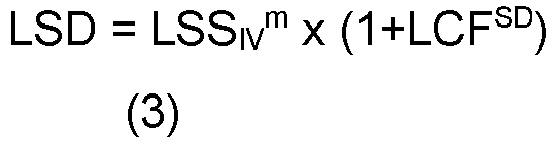

- LCFSD is a length-correction factor of the surgical defect.
- WCFSD is a width-correction factor of the surgical defect.
- The value of correction factors LCFSD and WCFSD depends on the location of the surgical specimen in the body of the patient according to the following table. Furthermore, the value of correction factors LCFSD and WCFSD may vary by 10% with respect to the values of the table:
Table 2: Surgical defect width and length increase percentages Region of the body WCFSD Region of the body LCFSD Upper limb 0.2189 Posterior torso 0.0680 Posterior torso 0.1733 Anterior torso 0.0571 Anterior torso 0.1283 Cheek, chin, and lip 0.0298 Lower limb 0.0927 Lower limb 0.0234 Scalp 0.0321 Palm or sole 0.0213 Cervical region 0.0285 Upper limb 0.0166 Cheek, chin, and lip 0.0278 Frontal region 0.0159 Palm or sole 0.0161 Scalp 0.0153 Frontal region 0.0099 Cervical region -0.0080 Nose -0.0389 Nose -0.0348 Auricle -0.0809 Auricle -0.0705 - Therefore, this novel preferred embodiment of the method of the invention allows the surgeon to know beforehand what the real dimensions of the surgical defect that excision of the surgical specimen will cause will be. To that end, the surgeon must simply measure the dimensions of the surgical specimen in vivo, before excision. The application of the preceding formulas will allow the surgeon to estimate the size the surgical defect will have in a simple manner.
- As a result of the shrinkage sustained by the surgical specimen as a whole, a decrease of the surgical margins takes place. This decrease occurs due to the combination of the shrinkage of the surgical specimen and the shrinkage of the lesion which, as demonstrated by the mentioned study, do not take place to the same extent.
- Based on the data from the study relative to the shrinkage of the lesion in the different areas of the body of the patient, the inventors of the present application have developed a preferred embodiment of the preceding method which further allows the magnitude of the surgical margin respected during excision of said surgical specimen between the outer border of a lesion present in the surgical specimen and the outer border of said surgical specimen to be estimated. In this preferred embodiment, the method of the invention further comprises the following steps:
- 5) Measuring the length and width of the lesion in the ex vivo state (LLEV m, WLEV m) after a process of fixing the surgical specimen in formalin.
- 6) Estimating the length and width of the surgical margin in the in vivo state (LSMIV e, WSMIV e) respected during excision of the surgical specimen by means of the following formulas:


- LCFL is a length-correction factor of the lesion.
- WCFL is a width-correction factor of the lesion.
- The value of the correction factors LCFL and WCFL depends on the location of the surgical specimen in the body of the patient according to the following table. Furthermore, the value of the correction factors LCFL and WCFL may vary by 10% with respect to the values of the table:
Table 3: Lesion width and length increase percentages Region of the body WCFL Region of the body LCFL Cervical region 0.2301 Anterior torso 0.2225 Frontal region 0.1968 Frontal region 0.1642 Palm or sole 0.1519 Lower limb 0.1519 Lower limb 0.1385 Nose 0.1501 Anterior torso 0.1349 Posterior torso 0.1418 Cheek, chin, and lip 0.1256 Upper limb 0.1412 Nose 0.1252 Cervical region 0.1329 Posterior torso 0.1210 Palm or sole 0.1262 Auricle 0.0958 Scalp 0.1229 Upper limb 0.0746 Auricle 0.1205 Scalp 0.0507 Cheek, chin, and lip 0.1130 - Therefore, this novel embodiment of the invention allows the pathologist to determine what the dimensions of the surgical margins were right at the time of the excision. To that end, the pathologist must simply measure the dimensions both of the lesion and of the surgical specimen available, even when it has shrunk after several hours of fixing, for example in 10% buffered formalin. The application of the preceding formulas will allow the pathologist to know what the surgical margins were in the surgical specimen in vivo in a simple manner.
- As a result of the shrinkage sustained by the surgical specimen as a whole, a decrease of the histological margins also takes place. As in the case of the surgical margins, the shrinkage of the histological margins occurs due to the combination of the shrinkage of the surgical specimen and the shrinkage of the lesion.
- The histological margins are the most clinically relevant margins, because they are what really reflect the distance that must be respected between the outer border of the extracted surgical specimen and the real outer border of the lesion. However, the histological margins are not visible in vivo to the naked eye, and there is currently no reliable method for determining them. For that reason, the only available data is that relative to the histological margins ex vivo after the process of fixing which, as mentioned, are shrunken and therefore do not precisely reflect the surgical margins that were really respected during the surgical intervention.
- The inventors of the present application have developed a preferred embodiment of the preceding method which further allows the magnitude of the histological margin respected during excision of said surgical specimen between the real outer border of the lesion present in the surgical specimen and the outer border of said surgical specimen to be estimated. In this preferred embodiment, the method of the invention further comprises the following steps:
- 7) Estimating the length and width of the histological margin in the in vivo state (LHMIV e, WHMIV e) respected during excision of the surgical specimen by means of the following formulas:


- WHMev m is the measured length of the histological margin ex vivo.
- LHMev m is the measured width of the histological margin ex vivo.
- WLev m is the measured width of the lesion ex vivo.
- LLev m is the measured length of the lesion ex vivo.
- The underlying geometric justification of these formulas for estimating histological margins is briefly described below. This brief justification will be given taking into account the dimension relative to the width of the histological margin, although it is evident that it would be done in the same way for the dimension relative to the length of the histological margin.
-
Figure 4 schematically shows a surgical specimen (SS) assumed to be in the in vivo state and in the center of which there is a lesion (L). The lesion (L) has a visible apparent border limiting the striped area, and a non-visible real border depicted by means of a discontinuous line outside of the visible border of the lesion (L). The distance between the upper border of the surgical specimen (SS) and the visible apparent upper border of the lesion (L) is the width of the surgical margin in vivo, which was estimated as (WSMiv e) according to formula (6) above. The distance between the upper border of the surgical specimen (SS) and the non-visible real upper border of the lesion (L) is the width of the histological margin in vivo (WHMiv e) to be estimated. - Therefore, to estimate the histological margins in vivo it is herein assumed that the magnitude of shrinkage sustained by the histological margins from the value thereof in vivo to the value thereof ex vivo after the process of fixing is the same as the shrinkage sustained by the surgical margins from the value thereof in vivo to the value thereof ex vivo after the process of fixing. This can mathematically be expressed as:

- It is therefore deduced that:

- The width of the surgical margin ex vivo after fixing can simply be measured by the pathologist. Alternatively, this parameter can be deduced from the respective measurements of the surgical specimen and of the lesion in the ex vivo state after fixing, because it is evident that the width of the surgical margin will be half of the difference between the width of the surgical specimen and the width of the lesion:

- Finally, by introducing equation (11) in equation (10), equation (7) mentioned above is obtained:

- Therefore, this novel embodiment of the invention allows the pathologist to determine what the dimensions of the real histological margins were right at the time of the excision. To that end, the pathologist only needs the data already obtained beforehand in addition to the histological margins ex vivo after fixing, which he or she can readily measure.
-
-
Figure 1 schematically shows the position of the relaxed skin tension lines in a human body. -
Figure 2 shows a body map of the mean overall shrinkage of the width and length of the surgical specimen based on the location. -
Figure 3 shows a body map of the mean swelling of the width and length of the surgical defect based on the location. -
Figure 4 shows an illustrative schematic diagram of obtaining the formula for estimating the histological margins in vivo prior to excision. - Some particular examples of the application of the method of the present invention are described below.
- The surgeon finely marks with a skin marker an oval-shaped surgical specimen 2.5 cm wide by 7.5 cm long on an upper limb of the patient. Before performing the excision, the surgeon uses formulas (3) and (4) to estimate what the dimensions of the surgical defect that will remain after extraction will be:


- The surgeon knows the measured length of the surgical specimen in vivo (LSSIV m = 7.5 cm) and the measured width of the surgical specimen in vivo (WSSIV m = 2.5 cm). Looking at Table 3, the surgeon also obtains the value of correction factors relative to the swelling of a surgical defect for an upper limb: WCFSD = 0.2189 and LCFSD = 0.0680. By entering these values in formulas (3) and (4), the surgeon determines that: LSD = 7.62 cm and WSD = 3.05 cm. Knowing these measurements will help the surgeon to better plan closing the surgical defect after excising the surgical specimen.
- A pathologist receives a surgical specimen for analysis. The surgical specimen consists of a scalp specimen in which there is a melanoma. According to the applicable international protocols, the minimum histological margin must be 1 cm. However, the pathologist measures the histological margins and the width of the histological margin measured turns out to be 0.85 cm. Therefore, doubts arise concerning whether it is necessary to perform another procedure on the patient.
- In view of this situation, the pathologist decides to use the formulas described herein to estimate what the dimensions of the specimen, of the lesion, and of the histological margins were in the in vivo state before the surgical intervention. To that end, starting from the surgical specimen available, which is in the ex vivo state and has already been subjected to the fixing method, the pathologist takes measurements of the specimen, of the lesion, and of the histological margins. The measurements are:
- Measured width of the surgical specimen ex vivo (WSSev m) = 2.6 cm
- Measured length of the surgical specimen ex vivo (LSSev m) = 8.1 cm
- Measured width of the lesion ex vivo (WLev m) = 0.95 cm
- Measured length of the lesion ex vivo (LLev m) = 1.8 cm
- Measured width of the histological margin ex vivo (WHMev m) = 0.85 cm
- Measured length of the histological margin ex vivo (LHMev m) = 2.1 cm
- The pathologist then uses the preceding formulas to estimate the dimensions of the surgical specimen, the lesion, and the histological margin in vivo before excision. First, the pathologist applies formulas (1) and (2) to estimate the dimensions of the surgical specimen in vivo:


- Table 1 indicates the value of correction factors of the dimensions of the surgical specimen for the scalp: (LCFSS = 0.1067; WCFSS = 0.1382). By applying the formulas, an estimate of the length of the surgical specimen in vivo (LSSIV e = 8.96 cm) and an estimate of the width of the surgical specimen in vivo (WSSIV e = 2.96 cm) are obtained.
- Then the pathologist applies formulas (5) and (6) to estimate the dimensions of the surgical margins in vivo:


- Table 3 indicates the value of correction factors of the lesion for the scalp: (LCFL = 0.1229; WCFL = 0.0507). By applying the formulas, an estimate of the length of the surgical margin in vivo (LSMIV e = 3.47 cm) and an estimate of the width of the surgical margin in vivo (WSMIV e = 0.98 cm) are obtained.
- Lastly, the pathologist applies formulas (7) and (8) to estimate the dimensions of the histological margins in vivo:


- By applying these formulas, an estimate of the length of the histological margin in vivo (WHMiv e = 1.01 cm) and an estimate of the width of the histological margin in vivo (LHMiv e = 2.31 cm) are obtained.
- It is thus determined that the surgeon respected the surgical histological margins in vivo required under the protocols, and it is therefore not necessary to perform another procedure.
- A patient has suffered metastasis due to an epidermoid carcinoma located in the posterior torso and the patient's family is considering filing a lawsuit against the hospital because excision was performed with a lateral histological margin of 0.435 cm when international protocols establish that an excision with 0.5 cm should be performed. The judge has the following measurements from the anatomic pathology report:
- Measured width of the surgical specimen ex vivo (WSSev m) = 1.45 cm
- Measured length of the surgical specimen ex vivo (LSSev m) = 4 cm
- Measured width of the lesion ex vivo (WLev m) = 0.45 cm
- Measured length of the lesion ex vivo (LLev m) = 0.9 cm
- Measured width of the histological margin ex vivo (WHMev m) = 0.435 cm
- Measured length of the histological margin ex vivo (LHMev m) = 1.45 cm
- The judge uses the method of the present invention in order to see if the hospital is at all liable and the following in vivo measurements are obtained:
- Estimated width of the surgical specimen in vivo (WSSiv e) = 1.65 cm
- Estimated length of the surgical specimen in vivo (LSSiv e) = 4.8 cm
- Estimated width of the surgical margin in vivo (WSMiv e) = 0.57 cm
- Estimated length of the surgical margin in vivo (LSMiv e) = 1.89 cm
- Estimated width of the histological margin in vivo (WHMiv e) = 0.50 cm
- Estimated length of the histological margin in vivo (LHMiv e) = 1.77 cm
- Therefore, it is determined that 0.5 cm minor lateral histological margin had been achieved and the center where the surgery took place cannot be held liable.
Claims (4)
- A method for estimating the magnitude of shrinkage of a surgical specimen excised from the skin of a patient, wherein the surgical specimen adopts an essentially planar shape defined by the length and width dimensions thereof, characterized in that it comprises the following steps:- measuring the length and width of the surgical specimen in the ex vivo state (LSSEV m, WSSEV m) excised from a patient after a process of fixing said surgical specimen; and- estimating the length and width of the surgical specimen in the in vivo state (LSSIV e, WSSIV e) before excision by means of the following formulas:


and wherein the value of correction factors LCFSS and WCFSS depends on the location in the body of the patient from whom the surgical specimen was excised according to the following table, wherein the value of correction factors LCFSS and WCFSS may vary by 10% with respect to the values of the table:Region of the body WCFSS Region of the body LCFSS Auricle 0.2385 Anterior torso 0.2372 Cervical region 0.1519 Posterior torso 0.2009 Posterior torso 0.1407 Upper limb 0.1911 Scalp 0.1382 Lower limb 0.1876 Anterior torso 0.1335 Cheek, chin, and lip 0.1683 Lower limb 0.1288 Cervical region 0.1625 Frontal region 0.1194 Auricle 0.1427 Cheek, chin, and lip 0.1140 Frontal region 0.1324 Upper limb 0.0953 Nose 0.1243 Nose 0.0708 Scalp 0.1067 Palm or sole 0.0101 Palm or sole 0.0799 - The method according to claim 1, which further allows the magnitude of the swelling of the cutaneous surgical defect caused by excising said surgical specimen to be estimated by means of the following additional steps:- measuring the length and width of the surgical specimen in the in vivo state (LSSIV m, WSSIV m) before being excised from the patient; and- estimating the length and width of the surgical defect (LSD, WSD) due to the swelling of the skin of the patient after excising the surgical specimen by means of the following formulas:


and wherein the value of correction factors LCFSD and WCFSD depends on the location of the surgical specimen in the body of the patient according to the following table, wherein the value of correction factors LCFSD and WCFSD may vary by 10% with respect to the values of the table:Region of the body WCFSD Region of the body LCFSD Upper limb 0.2189 Posterior torso 0.0680 Posterior torso 0.1733 Anterior torso 0.0571 Anterior torso 0.1283 Cheek, chin, and lip 0.0298 Lower limb 0.0927 Lower limb 0.0234 Scalp 0.0321 Palm or sole 0.0213 Cervical region 0.0285 Upper limb 0.0166 Cheek, chin, and lip 0.0278 Frontal region 0.0159 Palm or sole 0.0161 Scalp 0.0153 Frontal region 0.0099 Cervical region -0.0080 Nose -0.0389 Nose -0.0348 Auricle -0.0809 Auricle -0.0705 - The method according to any of the preceding claims, which further allows the magnitude of the surgical margin respected during excision of said surgical specimen between the outer border of a lesion present in the surgical specimen and the outer border of said surgical specimen to be estimated by means of the following additional steps:- measuring the length and width of the lesion in the ex vivo state (LLEV m, WLEV m) after a process of fixing the surgical specimen; and- estimating the length and width of the surgical margin in the in vivo state (LSMIV e, WSMIV e) respected during excision of the surgical specimen by means of the following formulas:


and wherein the value of correction factors LCFL and WCFL depends on the location of the surgical specimen in the body of the patient according to the following table, wherein the value of correction factors LCFL and WCFL may vary by 10% with respect to the values of the table:Region of the body WCFL Region of the body LCFL Cervical region 0.2301 Anterior torso 0.2225 Frontal region 0.1968 Frontal region 0.1642 Palm or sole 0.1519 Lower limb 0.1519 Lower limb 0.1385 Nose 0.1501 Anterior torso 0.1349 Posterior torso 0.1418 Cheek, chin, and lip 0.1256 Upper limb 0.1412 Nose 0.1252 Cervical region 0.1329 Posterior torso 0.1210 Palm or sole 0.1262 Auricle 0.0958 Scalp 0.1229 Upper limb 0.0746 Auricle 0.1205 Scalp 0.0507 Cheek, chin, and lip 0.1130 - The method according to any of the preceding claims, which further allows the magnitude of the histological margin respected during excision of said surgical specimen between the real outer border of the lesion present in the surgical specimen and the outer border of said surgical specimen to be estimated by means of the following additional steps:- measuring the length and width of the histological margin in the ex vivo state (LHMev m, WHMev m) after the process of fixing the surgical specimen; and- estimating the length and width of the histological margin in the in vivo state (LHMIV e, WHMIV e) respected during excision of the surgical specimen by means of the following formulas:


Applications Claiming Priority (2)
| Application Number | Priority Date | Filing Date | Title |
|---|---|---|---|
| ES201830152A ES2723448A1 (en) | 2018-02-21 | 2018-02-21 | Procedure to estimate the in vivo dimensions of a surgical piece from its ex vivo dimensions |
| PCT/ES2019/070093 WO2019162549A1 (en) | 2018-02-21 | 2019-02-20 | Method for estimating the in vivo dimensions of a surgical specimen from the ex vivo dimensions thereof |
Publications (2)
| Publication Number | Publication Date |
|---|---|
| EP3757511A1 true EP3757511A1 (en) | 2020-12-30 |
| EP3757511A4 EP3757511A4 (en) | 2021-11-17 |
Family
ID=65365779
Family Applications (1)
| Application Number | Title | Priority Date | Filing Date |
|---|---|---|---|
| EP19757717.4A Withdrawn EP3757511A4 (en) | 2018-02-21 | 2019-02-20 | PROCEDURE FOR DETERMINING THE IN-VIVO DIMENSIONS OF A SURGICAL SAMPLE FROM ITS EX-VIVO DIMENSIONS |
Country Status (3)
| Country | Link |
|---|---|
| EP (1) | EP3757511A4 (en) |
| ES (1) | ES2723448A1 (en) |
| WO (1) | WO2019162549A1 (en) |
-
2018
- 2018-02-21 ES ES201830152A patent/ES2723448A1/en not_active Withdrawn
-
2019
- 2019-02-20 WO PCT/ES2019/070093 patent/WO2019162549A1/en not_active Ceased
- 2019-02-20 EP EP19757717.4A patent/EP3757511A4/en not_active Withdrawn
Also Published As
| Publication number | Publication date |
|---|---|
| WO2019162549A1 (en) | 2019-08-29 |
| EP3757511A4 (en) | 2021-11-17 |
| ES2723448A1 (en) | 2019-08-27 |
Similar Documents
| Publication | Publication Date | Title |
|---|---|---|
| McCausland et al. | Depth of endometrial penetration in adenomyosis helps determine outcome of rollerball ablation | |
| Stedenfeldt et al. | Episiotomy characteristics and risks for obstetric anal sphincter injuries: a case‐control study | |
| Kottner et al. | Pressure ulcers: a critical review of definitions and classifications | |
| Guo et al. | Relation between a first branchial cleft anomaly and the facial nerve | |
| Cho et al. | Clinical significance of contralateral reactive lesion in vocal fold polyp and cyst | |
| Nix | Skin and wound inspection and assessment | |
| Wolloscheck et al. | Inguinal hernia: measurement of the biomechanics of the lower abdominal wall and the inguinal canal | |
| Hayashida et al. | The clinical, structural, and biological features of neovaginas: a comparison of the Frank and the McIndoe techniques | |
| Casadio et al. | Comparison of hysteroscopic cesarean scar defect repair with 26 Fr resectoscope and 16 Fr mini-resectoscope: a prospective pilot study | |
| Ibeanu et al. | Hypertrophic cervical elongation: clinical and histological correlations | |
| Mühlberger et al. | An anatomical study of femoral vein valves near the saphenofemoral junction | |
| Christensen et al. | Histopathologic assessment of depth of follicular invasion of squamous cell carcinoma (SCC) in situ (SCCis): implications for treatment approach | |
| EP3757511A1 (en) | Method for estimating the in vivo dimensions of a surgical specimen from the ex vivo dimensions thereof | |
| Chan et al. | Optical coherence tomography and its role in Mohs micrographic surgery: a case report | |
| Frank | The colposcopic examination | |
| Leithauser et al. | Hereditary mucoepithelial dysplasia: unique histopathological findings in skin lesions | |
| Harni et al. | Three rings vulvoscopy": a new approach to the vulva | |
| Winter et al. | Vulvoscopic evaluation of vulvodynia | |
| AlHassan et al. | Deepening fornix technique using central split-medium thickness skin graft to treat contracted anophthalmic sockets | |
| Pupić-Bakrač et al. | Two-centre experience in the treatment of thyroglossal duct remnants using the modified muscle-sparing Sistrunk technique | |
| Chuh et al. | Outcomes of dermoscope-guided surgical procedures in primary care: case-control study | |
| Sahai et al. | Comparison of punch biopsy and loop biopsy for the management of severe cervical intraepithelial neoplasia (CIN) | |
| RU2638623C1 (en) | Method for estimation of efficiency of reconstructive surgery in orbit | |
| Choi et al. | Changes in the length and area of congenital nevi involving the face and extremities after two serial excisions | |
| Xylouri et al. | Case report: A Large Fibroepithelial Polyp of the Vulva |
Legal Events
| Date | Code | Title | Description |
|---|---|---|---|
| STAA | Information on the status of an ep patent application or granted ep patent |
Free format text: STATUS: THE INTERNATIONAL PUBLICATION HAS BEEN MADE |
|
| PUAI | Public reference made under article 153(3) epc to a published international application that has entered the european phase |
Free format text: ORIGINAL CODE: 0009012 |
|
| STAA | Information on the status of an ep patent application or granted ep patent |
Free format text: STATUS: REQUEST FOR EXAMINATION WAS MADE |
|
| 17P | Request for examination filed |
Effective date: 20200921 |
|
| AK | Designated contracting states |
Kind code of ref document: A1 Designated state(s): AL AT BE BG CH CY CZ DE DK EE ES FI FR GB GR HR HU IE IS IT LI LT LU LV MC MK MT NL NO PL PT RO RS SE SI SK SM TR |
|
| AX | Request for extension of the european patent |
Extension state: BA ME |
|
| DAV | Request for validation of the european patent (deleted) | ||
| DAX | Request for extension of the european patent (deleted) | ||
| A4 | Supplementary search report drawn up and despatched |
Effective date: 20211015 |
|
| RIC1 | Information provided on ipc code assigned before grant |
Ipc: G01B 21/32 20060101ALI20211011BHEP Ipc: A61B 5/107 20060101AFI20211011BHEP |
|
| STAA | Information on the status of an ep patent application or granted ep patent |
Free format text: STATUS: THE APPLICATION IS DEEMED TO BE WITHDRAWN |
|
| 18D | Application deemed to be withdrawn |
Effective date: 20220514 |