EP3754600A1 - A method and system for machine learning classification based on structure or material segmentation in an image - Google Patents
A method and system for machine learning classification based on structure or material segmentation in an image Download PDFInfo
- Publication number
- EP3754600A1 EP3754600A1 EP20177226.6A EP20177226A EP3754600A1 EP 3754600 A1 EP3754600 A1 EP 3754600A1 EP 20177226 A EP20177226 A EP 20177226A EP 3754600 A1 EP3754600 A1 EP 3754600A1
- Authority
- EP
- European Patent Office
- Prior art keywords
- machine learning
- learning model
- segmentation
- image
- classification machine
- Prior art date
- Legal status (The legal status is an assumption and is not a legal conclusion. Google has not performed a legal analysis and makes no representation as to the accuracy of the status listed.)
- Granted
Links
Images

Classifications
-
- G—PHYSICS
- G06—COMPUTING OR CALCULATING; COUNTING
- G06T—IMAGE DATA PROCESSING OR GENERATION, IN GENERAL
- G06T7/00—Image analysis
- G06T7/0002—Inspection of images, e.g. flaw detection
- G06T7/0012—Biomedical image inspection
-
- G—PHYSICS
- G06—COMPUTING OR CALCULATING; COUNTING
- G06T—IMAGE DATA PROCESSING OR GENERATION, IN GENERAL
- G06T7/00—Image analysis
- G06T7/10—Segmentation; Edge detection
- G06T7/12—Edge-based segmentation
-
- A—HUMAN NECESSITIES
- A61—MEDICAL OR VETERINARY SCIENCE; HYGIENE
- A61B—DIAGNOSIS; SURGERY; IDENTIFICATION
- A61B5/00—Measuring for diagnostic purposes; Identification of persons
- A61B5/45—For evaluating or diagnosing the musculoskeletal system or teeth
- A61B5/4504—Bones
- A61B5/4509—Bone density determination
-
- A—HUMAN NECESSITIES
- A61—MEDICAL OR VETERINARY SCIENCE; HYGIENE
- A61B—DIAGNOSIS; SURGERY; IDENTIFICATION
- A61B5/00—Measuring for diagnostic purposes; Identification of persons
- A61B5/48—Other medical applications
- A61B5/4842—Monitoring progression or stage of a disease
-
- A—HUMAN NECESSITIES
- A61—MEDICAL OR VETERINARY SCIENCE; HYGIENE
- A61B—DIAGNOSIS; SURGERY; IDENTIFICATION
- A61B5/00—Measuring for diagnostic purposes; Identification of persons
- A61B5/72—Signal processing specially adapted for physiological signals or for diagnostic purposes
- A61B5/7235—Details of waveform analysis
- A61B5/7264—Classification of physiological signals or data, e.g. using neural networks, statistical classifiers, expert systems or fuzzy systems
- A61B5/7267—Classification of physiological signals or data, e.g. using neural networks, statistical classifiers, expert systems or fuzzy systems involving training the classification device
-
- A—HUMAN NECESSITIES
- A61—MEDICAL OR VETERINARY SCIENCE; HYGIENE
- A61B—DIAGNOSIS; SURGERY; IDENTIFICATION
- A61B5/00—Measuring for diagnostic purposes; Identification of persons
- A61B5/72—Signal processing specially adapted for physiological signals or for diagnostic purposes
- A61B5/7271—Specific aspects of physiological measurement analysis
- A61B5/7275—Determining trends in physiological measurement data; Predicting development of a medical condition based on physiological measurements, e.g. determining a risk factor
-
- G—PHYSICS
- G06—COMPUTING OR CALCULATING; COUNTING
- G06F—ELECTRIC DIGITAL DATA PROCESSING
- G06F18/00—Pattern recognition
- G06F18/20—Analysing
- G06F18/21—Design or setup of recognition systems or techniques; Extraction of features in feature space; Blind source separation
- G06F18/214—Generating training patterns; Bootstrap methods, e.g. bagging or boosting
-
- G—PHYSICS
- G06—COMPUTING OR CALCULATING; COUNTING
- G06F—ELECTRIC DIGITAL DATA PROCESSING
- G06F18/00—Pattern recognition
- G06F18/20—Analysing
- G06F18/21—Design or setup of recognition systems or techniques; Extraction of features in feature space; Blind source separation
- G06F18/214—Generating training patterns; Bootstrap methods, e.g. bagging or boosting
- G06F18/2148—Generating training patterns; Bootstrap methods, e.g. bagging or boosting characterised by the process organisation or structure, e.g. boosting cascade
-
- G—PHYSICS
- G06—COMPUTING OR CALCULATING; COUNTING
- G06F—ELECTRIC DIGITAL DATA PROCESSING
- G06F18/00—Pattern recognition
- G06F18/20—Analysing
- G06F18/21—Design or setup of recognition systems or techniques; Extraction of features in feature space; Blind source separation
- G06F18/217—Validation; Performance evaluation; Active pattern learning techniques
-
- G—PHYSICS
- G06—COMPUTING OR CALCULATING; COUNTING
- G06F—ELECTRIC DIGITAL DATA PROCESSING
- G06F18/00—Pattern recognition
- G06F18/20—Analysing
- G06F18/24—Classification techniques
-
- G—PHYSICS
- G06—COMPUTING OR CALCULATING; COUNTING
- G06F—ELECTRIC DIGITAL DATA PROCESSING
- G06F18/00—Pattern recognition
- G06F18/20—Analysing
- G06F18/24—Classification techniques
- G06F18/241—Classification techniques relating to the classification model, e.g. parametric or non-parametric approaches
- G06F18/2411—Classification techniques relating to the classification model, e.g. parametric or non-parametric approaches based on the proximity to a decision surface, e.g. support vector machines
-
- G—PHYSICS
- G06—COMPUTING OR CALCULATING; COUNTING
- G06F—ELECTRIC DIGITAL DATA PROCESSING
- G06F18/00—Pattern recognition
- G06F18/20—Analysing
- G06F18/24—Classification techniques
- G06F18/243—Classification techniques relating to the number of classes
- G06F18/2431—Multiple classes
-
- G—PHYSICS
- G06—COMPUTING OR CALCULATING; COUNTING
- G06T—IMAGE DATA PROCESSING OR GENERATION, IN GENERAL
- G06T7/00—Image analysis
- G06T7/10—Segmentation; Edge detection
- G06T7/11—Region-based segmentation
-
- G—PHYSICS
- G06—COMPUTING OR CALCULATING; COUNTING
- G06F—ELECTRIC DIGITAL DATA PROCESSING
- G06F18/00—Pattern recognition
- G06F18/20—Analysing
- G06F18/24—Classification techniques
- G06F18/243—Classification techniques relating to the number of classes
- G06F18/24323—Tree-organised classifiers
-
- G—PHYSICS
- G06—COMPUTING OR CALCULATING; COUNTING
- G06T—IMAGE DATA PROCESSING OR GENERATION, IN GENERAL
- G06T2207/00—Indexing scheme for image analysis or image enhancement
- G06T2207/10—Image acquisition modality
- G06T2207/10072—Tomographic images
- G06T2207/10081—Computed x-ray tomography [CT]
-
- G—PHYSICS
- G06—COMPUTING OR CALCULATING; COUNTING
- G06T—IMAGE DATA PROCESSING OR GENERATION, IN GENERAL
- G06T2207/00—Indexing scheme for image analysis or image enhancement
- G06T2207/10—Image acquisition modality
- G06T2207/10072—Tomographic images
- G06T2207/10088—Magnetic resonance imaging [MRI]
-
- G—PHYSICS
- G06—COMPUTING OR CALCULATING; COUNTING
- G06T—IMAGE DATA PROCESSING OR GENERATION, IN GENERAL
- G06T2207/00—Indexing scheme for image analysis or image enhancement
- G06T2207/10—Image acquisition modality
- G06T2207/10132—Ultrasound image
-
- G—PHYSICS
- G06—COMPUTING OR CALCULATING; COUNTING
- G06T—IMAGE DATA PROCESSING OR GENERATION, IN GENERAL
- G06T2207/00—Indexing scheme for image analysis or image enhancement
- G06T2207/20—Special algorithmic details
- G06T2207/20076—Probabilistic image processing
-
- G—PHYSICS
- G06—COMPUTING OR CALCULATING; COUNTING
- G06T—IMAGE DATA PROCESSING OR GENERATION, IN GENERAL
- G06T2207/00—Indexing scheme for image analysis or image enhancement
- G06T2207/20—Special algorithmic details
- G06T2207/20081—Training; Learning
-
- G—PHYSICS
- G06—COMPUTING OR CALCULATING; COUNTING
- G06T—IMAGE DATA PROCESSING OR GENERATION, IN GENERAL
- G06T2207/00—Indexing scheme for image analysis or image enhancement
- G06T2207/20—Special algorithmic details
- G06T2207/20084—Artificial neural networks [ANN]
-
- G—PHYSICS
- G06—COMPUTING OR CALCULATING; COUNTING
- G06T—IMAGE DATA PROCESSING OR GENERATION, IN GENERAL
- G06T2207/00—Indexing scheme for image analysis or image enhancement
- G06T2207/30—Subject of image; Context of image processing
- G06T2207/30004—Biomedical image processing
- G06T2207/30008—Bone
-
- G—PHYSICS
- G06—COMPUTING OR CALCULATING; COUNTING
- G06T—IMAGE DATA PROCESSING OR GENERATION, IN GENERAL
- G06T2207/00—Indexing scheme for image analysis or image enhancement
- G06T2207/30—Subject of image; Context of image processing
- G06T2207/30004—Biomedical image processing
- G06T2207/30096—Tumor; Lesion
-
- G—PHYSICS
- G06—COMPUTING OR CALCULATING; COUNTING
- G06V—IMAGE OR VIDEO RECOGNITION OR UNDERSTANDING
- G06V2201/00—Indexing scheme relating to image or video recognition or understanding
- G06V2201/03—Recognition of patterns in medical or anatomical images
Definitions
- the present invention relates to a computer vision system and method, employing machine learning and in particular deep neural networks, for classifying (and monitoring changes in) an image (such as a medical image), based on structural or material segmentation.
- Possible medical imaging applications include Computed Tomography (CT), Magnetic Resonance (MR), Ultrasound, HRpQCT (High-Resolution peripheral Quantitative Computed Tomography), and Pathology Scanner imaging.
- Segmentation is the process of segmenting (or differentiating) structures or objects in an image, such as a medical image, from one another by differentiating pixels (in 2D image) or voxels (in 3D image) in the image.
- quantitative features such as volume, shape, and density are calculated. For example, lesion size and shape are calculated after the lesion has been segmented in a brain CT or MRI scan; the bone mineral density may be calculated after the femoral neck is segmented in a hip DXA (dual-energy x-ray absorptiometry) scan.
- a doctor may make a diagnosis or treatment decision after he or she has compared such calculated values with healthy reference data.
- a T-score is the standard score of a patient's bone mineral density compared to the young normal reference mean.
- the WHO World Health Organization
- osteoporosis as a T-score of -2.5 or lower, that is, a bone density that is two and a half standard deviations or more below the mean of a 30-year-old healthy man/ woman.
- the segmentation may be achieved manually, semi-manually, or automatically.
- a user operates a computer to move a rectangular box over a hip DXA scan and thereby select the region of the femoral neck.
- Semi-manual segmentation may be performed by an image processing program employing a user's initialisation or input. For example, a user may operate a computer to draw an approximate bone boundary on a wrist CT scan; the program then adjusts the approximate boundary into a contour that segments bone from the surrounding tissues.
- Automatic segmentation may be performed by utilizing the features of the object of interest, such as intensity values, edges and shapes.
- a voxel-value based thresholding method is used to segment bone from the surrounding tissues in CT scans.
- Some other programs use machine learning algorithms to train a classifier to segment abnormal tissues in medical images.
- a feature-based machine learning algorithm such as a support vector machine and a decision tree, may be used as a classifier by using tumour images and normal images as training data. The trained classifier slides through the new image "window” by "window” to segment any image regions of tumour tissues.
- a transitional zone exists at any sample composed of both cortical and trabecular bones.
- the transitional zone comprises the inner cortex adjacent to the medullary canal and trabeculae abutting against the cortex contiguous with the endocortical surface.
- the transitional zone is a site of vigorous bone remodelling. It is important to identify and segment this region in bone microstructure assessment but, owing to limitations in image resolution, it is essentially impossible for an expert to annotate this region both accurately and consistently. Without annotated images as training data, the segmentation model cannot be trained.
- the network comprises input layer, hidden layers, and an output layer.
- An image is fed into the network through the input layer.
- the image is sampled and applied with convolutional operations to generate hidden layers.
- the output of each layer is used as input to the next layer in the network.
- the output layer is fully connected at the end that will output a classification result.
- Training data are images with classification labels.
- the training process obtains the parameters of the neural network. After the training is finished, a new image will be processed by the neural network with the obtained parameters to generate a classification result.
- a deep neural network algorithm may be used to train a model to determine the condition (for example, no, mild, moderate, severe) of diabetic retinopathy from OCT (Optical Coherence Tomography) images.
- OCT Optical Coherence Tomography
- the end-to-end solution brings two problems in clinical practices.
- the end-to-end solution is a black box: the input is the medical image, and the output the classification of diseases or conditions. It is difficult to interpret the process whereby the neural network makes its decision-so it is difficult for the user to assess the reliability of the classification results.
- this solution requires a substantial amount of training data. As discussed above, in medical applications annotating or labelling the training data is a tedious and time-consuming process. Collecting enough training data for each category of each type of classification result thus poses a significant challenge.
- a system for classifying a structure or material in an image of a subject comprising:
- the classifier generates the one or more classifications based on the segmentations maps and non-image data pertaining to the subject.
- the system may be configured to train the classification machine learning model.
- the segmenter comprises:
- the structure segmenter is configured to employ a structure segmentation machine learning model to generate the structure segmentation maps
- the material segmenter is configured to employ a material segmentation machine learning model to generate the material segmentation maps
- the abnormality segmenter is configured to employ an abnormality segmentation model to generate the abnormality segmentation maps.
- the structure segmenter may be configured to train the structure segmentation machine learning model, the material segmenter to train the material segmentation machine learning model, and/or the abnormality segmenter to train the abnormality segmentation model.
- system further comprises a segmentation map processor configured to process the segmentation maps before the segmentation maps are input by the classifier.
- the segmentation map processor is configured to down-sample the segmentation maps.
- the classification machine learning model comprises a neural network, a support vector machine, a decision tree, or a combination thereof.
- the classification machine learning model may comprise a neural network that includes convolutional neural network layers and fully-connected neural network layers.
- the image is a medical image
- the classifications correspond to probabilities that the structure or material, or the subject, will sustain a specified condition or symptom in respective timeframes.
- the timeframes include a shorter-term timeframe, a longer-term timeframe, and at least one intermediate-term timeframe intermediate the shorter-term timeframe and the longer-term timeframe.
- the condition or symptom is bone fracture.
- the image is a medical image
- the classifications correspond to probabilities that the structure or material, or the subject, will sustain respective conditions or symptoms.
- the conditions or symptoms are bone conditions.
- the image is a medical image
- the classifications correspond to probabilities of respective rates of disease or pathology progression.
- the classifications may comprise classifications corresponding any one or more of: stable, modest deterioration, and accelerated deterioration.
- the image is a medical image
- the classifications correspond to probabilities of efficacy of respective treatment options.
- the treatment options may include an antiresorptive treatment and/or an anabolic treatment.
- the image is a medical image
- the classifications correspond to respective medical conditions.
- the medical conditions may include any one or more of: osteomalacia, tumour, osteonecrosis and infection.
- the classification machine learning model is a model trained with image data and non-image data relating to training subjects, and generates the respective scores based on image data (typically constituting one or more images) and non-image data relating to the subject.
- a computer-implemented method for classifying a structure or material in an image of a subject comprising:
- the classification machine learning model is used to generate the one or more classifications based on the segmentations maps and non-image data pertaining to the subject.
- the method may include training the classification machine learning model.
- forming the one or more segmentations comprises:
- the method may include employing a structure segmentation machine learning model to generate the structure segmentation maps, a material segmentation machine learning model to generate the material segmentation maps, and an abnormality segmentation model to generate the abnormality segmentation maps.
- the method may include training the structure segmentation machine learning model, the material segmentation machine learning model, and/or the abnormality segmentation model.
- a computer-implemented diagnostic method comprising the method of the second aspect.
- a computer-implemented method for training a classification machine learning model for classifying a structure or material in an image of a subject comprising:
- This aspect may be used in conjunction or in combination with (or as a part of) the second aspect, such as to train the classification machine learning model of the second aspect.
- the method may include segmenting the one or more images (such as in the course of generating the annotated segmentation maps).
- the method includes, when the performance is found in step (d) not to be satisfactory, receiving and using more image and non-image data for training the classification machine learning model.
- the classification model can be trained by various machine learning algorithms, so may comprise-for example-a neural network, a support vector machine, a decision tree, or a combination thereof.
- the classification machine learning model comprises a neural network having a plurality of layers comprising artificial neurons, wherein the parameters comprise layer numbers, neuron numbers, neuron weights, and neuron function parameters; and testing the classification machine learning model includes testing the classification machine learning model on the testing data.
- updating the parameters includes determining a gradient of a loss function.
- the images are medical images and the non-image data comprise clinical records.
- the method includes dividing the annotated segmentation maps and the annotated non-image data into the training set, a development set and the testing set, and using the development data to investigate the learning procedure and to tune the parameters (and, when the classification machine learning model comprises a neural network, tune the layers).
- a system for training a classification machine learning model for classifying a structure or material in an image of a subject comprising a processor configured to:
- a computer program comprising program code configured, when executed by one of more computing devices, to implemented the method of any one or more of the second to fourth aspects.
- a computer-readable medium comprising such a computer program.
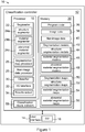
- Figure 1 is a schematic view of a classification system 10 for classifying a structure or material in a medical image (based on structural and material segmentation), according to an embodiment of the present invention.
- system 10 comprises a classification controller 12 and a user interface 14 (including a GUI 16).
- User interface 14 is provided for representing information to a user and for receiving input (including feedback) from a user; it typically comprises one or more displays (on one or more of which may be displayed the GUI 16), a web browser, a keyboard and a mouse, and optionally a printer.
- Classification controller 12 includes at least one processor 18 and a memory 20.
- System 10 may be implemented, for example, as a combination of software and hardware on a computer (such as a personal computer or mobile computing device), or as a dedicated image segmentation system.
- System 10 may optionally be distributed; for example, some or all of the components of memory 20 may be located remotely from processor 18; user interface 14 may be located remotely from memory 20 and/or from processor 18.
- system 10 may be implemented in a service-oriented architecture, with its components communicating with each other over a communication network such as a LAN (local area network), WAN (wide area network) or the internet.
- System 10 may be deployed in the cloud, and its use shared by users at different locations.
- system 10 is implemented as a standalone system (of software and hardware) or as standalone software executable by a computer, and deployed in one location; for example, system 10 may be deployed in a hospital, medical clinic or other clinical setting.
- Memory 20 is in data communication with processor 18, and typically comprises both volatile and non-volatile memory (and may include more than one of each type of memory), including RAM (Random Access Memory), ROM and one or more mass storage devices.
- RAM Random Access Memory
- ROM Read Only Memory
- processor 18 includes a segmenter 22 (which includes a structure segmenter 24a, a material segmenter 24b, and an abnormal segmenter in the form of an abnormal material segmenter 24c), a segmentation map processor 26, and a non-image data processor 28.
- processor 18 also includes a classifier 30, an I/O interface 32 and a results output 34.
- Memory 20 includes program code 36, image data 38, non-image data 40, segmentation models 42 (including, in this example, structure segmentation models 44, material segmentation models 46, and abnormality segmentation models in the form of abnormal material segmentation models 48), segmentation maps 50 (including, in this example, structure segmentation maps 52, material segmentation maps 54, and abnormality segmentation maps in the form of abnormal material segmentation maps 56).
- segmentation models 42 including, in this example, structure segmentation models 44, material segmentation models 46, and abnormality segmentation models in the form of abnormal material segmentation models 48
- segmentation maps 50 including, in this example, structure segmentation maps 52, material segmentation maps 54, and abnormality segmentation maps in the form of abnormal material segmentation maps 56.
- Structure segmenter 24a, material segmenter 24b and abnormal material segmenter 24c train the respective segmentation models 44, 46, 48, and use segmentation models 44, 46, 48 to perform segmentation on incoming images and generate structure segmentation maps 52, material segmentation maps 54, and abnormal material segmentation maps 56, respectively.
- Memory 20 also includes a classification machine learning model in the form of a classification neural network 58, which is trained and used by classifier 30 to perform classification by using segmentation maps 50 and non-image data 40.
- Classification controller 12 is implemented, at least in part (and in some embodiments entirely), by processor 18 executing program code 36 from memory 20.
- abnormal material segmenter 24c may also be referred to as an abnormal tissue segmenter
- abnormal material segmentation maps 56 may also be referred to as abnormal tissue segmentation maps.
- I/O interface 32 is configured to read or receive medical image data and non-image data pertaining to a subject, and to store these data as image data 38 and non-image data 40 of memory 20 for processing.
- Image data 38 is typically in the form, in this embodiment, of a medical image of-for example-a region of the body of a subject.
- Non-image data 40 typically includes subject or patient information from various structured and non-structured data sources, collected throughout a subject's medical consultations, treatment and follow-up consultations.
- Subject structured data may include basic subject information such as sex, age, weight, height; laboratory test results such as blood test results and DNA test results; treatment data such as the types of medication and dosage; and questionnaire data such as smoking and drinking habits and fracture history.
- Subject unstructured data may include text documents of laboratory results, doctors' notes, and radiological reports.
- Non-image data 40 may in a variety of formats, such as numerical data, text, voice, and video.
- Segmenter 22 processes image data 38 (constituting one or more medical images) and uses structure segmentation models 44, material segmentation models 46 and abnormal material segmentation models 48 to generate-from image data 38-structure segmentation maps 52, material segmentation maps 54 and abnormal material segmentation maps 56, respectively, which characterize image data 38 in different ways.
- Classifier 30 then inputs the resulting segmentation maps 50 and non-image data 40, and generates therefrom results in the form of a classification output.
- the classification output is, in this embodiment, presented to users or used for further analysis via I/O interface 32 and at either results output 34 and/or user interface 14.
- the classification output of classifier 30 (in this embodiment, generated using classification neural network 58) comprises a respective condition score for each of one or more classifications (and preferably for each of a plurality of possible classifications). Each score represents a predicted likelihood that the subject falls into the corresponding classification.
- the classifications are "negligible fracture risk”, “imminent fracture risk”, “intermediate-term fracture risk”, and "long-term fracture risk”. The classification output is described in more detail below.
- the classifier outputs a respective disease progression score for each of one or more condition progression states.
- Each score represents a predicted likelihood that a current condition will progress to another condition.
- the disease progressions may include "stable”, “modest deterioration”, and "accelerated deterioration”.
- the classifier outputs a respective treatment score for each of multiple treatment options.
- Each score represents a predicted likelihood that the treatment is the most efficient for the patient.
- the treatment options may include "antiresorptive”, “anabolic”, and “antiresorptive + anabolic”.
- the classification output comprises a score for each of one or more possible classifications corresponding to known medical conditions or pathologies. For example, in a bone fragility assessment, these classifications could be "osteomalacia”, “tumour”, “osteonecrosis” and “infection”.
- the resulting scores represent the degree to which the (e.g. bone) sample of the subject conforms to that classification/condition. If only one classification has a significant score, or one classification has a score that is significantly greater than all other scores, that classification may be regarded as a diagnosis, or suggested diagnosis, of the corresponding condition or pathology.
- the classification output comprises two or more sets of such scores (selected from the aforementioned examples or otherwise).
- image data 38-constituting one or more medial images comprises data generated by one or more of a variety of medical image modalities (such as HRpQCT, or High-Resolution peripheral Quantitative Computed Tomography) implemented by one or more medical imaging devices (such as a HRpQCT scanner). Each of these devices scans a sample (whether in vivo or in vitro) and creates a visual representation, generally of a portion of the interior of a subject's body.
- the medical images may depict, for example, a part of a body or a whole body of a subject (e.g. the brain, the hip or the wrist).
- the medical images might be acquired by scanning the same sample or body part using different imaging modalities, as different imaging modalities may reveal different characteristics of the same sample or body part.
- the medical images might be acquired by scanning different body parts using the same image modalities, as different body parts of the same patients might provide different insights towards a better diagnosis of diseases or conditions. For example, in bone fragility assessment, both the wrist and the leg of a patient may be scanned by an HRpQCT scanner (or indeed acquired by scanning the different samples or body parts using different imaging modalities) to provide information for use in assessing a subject's bone quality.
- the image data 38 may constitute a 2D (two-dimensional) image that may be represented as a 2D array of pixels, or a 3D (three-dimensional) image that may be represented as a 3D array of voxels.
- a 2D (two-dimensional) image that may be represented as a 2D array of pixels
- a 3D (three-dimensional) image that may be represented as a 3D array of voxels.
- the medical images described below are 3D images that may be represented as a 3D array of voxels.
- the one or more received medical images, stored in image data 38, are segmented by segmenter 22, using trained segmentation models 42, into respective segmentation maps 50.
- Each segmentation map 52, 54, 56 characterizes the respective medical image differently.
- a structure segmentation map 52 represents the medical image as one or more different anatomical structures from a predefined set of structures.
- a wrist CT scan may be segmented into compact cortex, transitional zone, and trabecular region.
- Material segmentation map represents the medical image into multiple different materials from a predefined set of materials.
- a wrist CT scan might be segmented into mineralized material, fully mineralized material, red marrow in the trabecular region, and yellow marrow in the trabecular region.
- An abnormal material segmentation map 56 represents the medical image as normal material and abnormal material (or, in this example, normal tissue and abnormal tissue). For example, a tumour or fracture might be segmented from a wrist CT scan and represented in an abnormal material segmentation map 56 as 'abnormal'.
- Segmentation maps 50 are inputted into classifier 30, in combination with non-image data 40.
- Classifier 30 generates one or more classification outputs based on segmentation maps 50 and the non-image data 40.
- Input data of classifier 30 is generally multi-dimensional, so classifier 30 is implemented with machine learning algorithms, such as a neural network, support vector machine, decision tree, or a combination thereof.
- classifier 30 employs or is implemented as classification neural network 58 (though in other embodiments, other machine learning algorithms may be acceptable), including-in this example-convolutional neural network layers and fully-connected neural network layers. Classification neural network 58 is trained with training data, as is described below.
- the ultimate classification output is outputted by system 10 to a user via results output 34 or user interface 14.
- the classification output may optionally include a visual presentation of one or more of the corresponding segmentation maps 50. Segmentation maps 50 may be presented in case they can assist a user in interpreting the classification output, such as in assessing the reliability of the results.
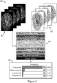
- FIG 2 is a high-level schematic diagram 50 illustrating the operation of system 10 in calculating bone fracture risk of a subject from a medical imaging scan, in this example a wrist HRpQCT scan 62 (also shown in negative at 62').
- system 10 receives the wrist HRpQCT scan 62 comprising a plurality of slices.
- an HRpQCT scan can comprise 100 or more slices, but four slices are depicted in the figure for simplicity.
- Segmenter 22 segments scan 62 into a structure segmentation map 52 in which the scan is segmented into compact cortex, outer transitional zone, inner transitional zone, and trabecular region. Segmenter 22 segments scan 62 into material segmentation map 54, in which scan 62 is segmented into surrounding muscle, surrounding fat, yellow marrow adipose, and red marrow adipose.
- Data 64 comprising segmentation maps 52, 54, abnormal material segmentation maps 56 and non-image data 40 (e.g. clinical factors including sex and age) are processed by trained classifier 30 to generate classification outputs.
- the classification outputs include segmentation maps 52, 54 and a table or report 66.
- Table or report 66 includes, in numerical and/or graphical form, fracture probabilities in each category of fracture risk: imminent fracture risk 68a (fracture within two years: t ⁇ 2 y), intermediate-term fracture risk 68b (fracture within two to five years: 2 ⁇ t ⁇ 5 y), long-term fracture risk 68c (fracture in five to ten years, 5 ⁇ t ⁇ 10 y), and negligible fracture risk 68d.
- the probability that the wrist is at risk of fracture within two years is 95.6%
- that the wrist is at a risk of fracture in two to five years 2.4%
- that the wrist is at a risk of fracture in five to 10 years 1.6%
- the wrist is at negligible risk of fracture is 0.3%.
- the probability that the subject will not have a wrist fracture in the next five years is only 4.4%.
- Table or report 66 does not include a diagnosis (e.g. that the subject has osteoporosis), but it will be appreciated that these probabilities may be of great value, including-for example-to prompt the subject to pursue a diagnosis, such as by undergoing medical examination or consultation.
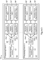
- Figure 3 is a schematic view at 70 of the operation of segmenter 22.
- Segmenter 22 is configured to receive input includes one or more medical images (from image data 38) and to process the images so as to generate segmentation maps 50.
- the medical images might be acquired using the same imaging modality by scanning different body parts of a patient. For example, in some applications of assessing bone quality, both wrist and leg of a patient might be scanned by an HRpQCT scanner for the assessment.
- the medical images might be acquired using different imaging modalities by scanning the same or different body parts of a patient.
- both the wrist HRpQCT scan and the hip DXA scan of a patient are acquired for the assessment (though again bearing in mind that the medical images may be acquired by scanning the different samples or body parts using other imaging modalities).
- segmenter 22 implements one or more processing branches 1 to n, (labelled 72 1 ,..., 72 n in the figure) corresponding to medical images 1 to n of the subject (labelled 38 1 ,..., 38 n ).
- medical images 38 1 ,...,38 n may be due to-for example-different imaging modalities (labelled 74 1 ,..., 74 n ), as is the case in the illustrated example, different body parts, different scans of a single body part, or a combination two or more of these.
- Respective segmentation branches 72 1 ,..., 72 n are configured to receive an image, to segment the image according to image type (such as with different program code 36), and to generate the branch output (comprising the segmentation maps 50 of the input image).
- segmenter 22 is configured first to select a processing branch of processing branches 1 to n according to the type of the input image. Segmenter 22 ascertains the type of image according to the sample (e.g. scanned body part) and imaging modality, information that can be determined from the respective image, including from metadata stored in a header file of the medical image and/or from the file type. For example, the information of scanned body part and imaging modality may be accessed from the metadata.
- the sample e.g. scanned body part
- imaging modality information that can be determined from the respective image, including from metadata stored in a header file of the medical image and/or from the file type. For example, the information of scanned body part and imaging modality may be accessed from the metadata.
- Each input medical image 1 to n is processed by one or more of three segmentation programs ( viz. structure segmenter 24a, material segmenter 24b, and abnormal material segmenter 24c) into the corresponding segmentation maps 52, 54, 56.
- Segmenter 22 thus employs up to n instances each of segmenters 24a, 24b, 24c (labelled 24a 1 ,..., 24a n , 24b 1 ,..., 24b n , and 24c 1 ,..., 24c n , respectively), either in parallel or sequentially, though the number of such instances of each segmenter 24a, 24b, 24c (being from 0 to n in each case) may differ.
- Structure segmenter 24a, material segmenter 24b, and abnormal material segmenter 24c may generate respective segmentation maps in each processing branch 72 1 ,..., 72 n .
- structure segmenter 24a, material segmenter 24b, and abnormal material segmenter 24c generate respective segmentation maps corresponding to medical imaging modalities 1 to n ; the resulting structure segmentation maps, material segmentation maps and abnormal tissue segmentation maps are correspondingly labelled structure segmentation maps 52 1 ,..., 52 n , the material segmentation maps 54 1 ,..., 54 n and the abnormal tissue segmentation maps 56 1 ,..., 56 n .
- Structure segmenter 24a, material segmenter 24b, and abnormal material segmenter 24c assign to each voxel of these segmentation maps 50 one or more 'types' (or 'categories') from respective predetermined sets of types (or categories).
- structure segmenter 24a assigns a respective structure type (from a predefined set of structure types) to each voxel in the scan. For example, a wrist HRpQCT scan is segmented into a structure segmentation map 52 in which each voxel in the scan is assigned a structure type (or category) from the set of "surrounding tissues", "compact cortex”, “transitional zone", and "trabecular region".
- Material segmenter 24b assigns a respective material type (from a predefined set of material types) to each voxel. For example, in this embodiment, material segmenter 24b segments a wrist HRpQCT scan into a material segmentation map 54 in which each voxel in the scan is assigned a material type from the set of "mineralised material”, “fully mineralised material”, “red marrow adipose”, and “yellow marrow adipose”.
- Abnormal material segmenter 24c assigns a respective abnormality or normality type (from a predefined set of abnormalities or normality types, such as a set comprising "normal” and "abnormal") to each voxel. For example, in this embodiment, abnormal material segmenter 24c segments a wrist HRpQCT scan into an abnormal tissue segmentation map 54 in which each voxel in the scan is assigned either "normal” or "abnormal".
- abnormal material segmenter 24c can distinguish different types of abnormality, and the predefined set of abnormality or normality types may include-in addition to "normal”-and one or more specific abnormalities particular to the sample type under examination; if the sample is bone, these may include, for example, "fracture crack” or "bone tumour".
- the set may optionally include "abnormal” for cases in which abnormal material segmenter 24c cannot determine a specific type of abnormality.
- structure segmenter 24a, material segmenter 24b, and abnormal material segmenter 24c assign respective types with confidence limits or probabilities to each voxel in the medical image.
- structure segmenter 24a, material segmenter 24b, and abnormal material segmenter 24c may assign a plurality of types (each optionally with a confidence limit or probability) to each voxel in the medical image. For example, structure segmenter 24a-when segmenting a wrist HRpQCT scan-may assign both "transitional zone" and "trabecular region” to ambiguous voxels, but with respective (and typically different) confidence limits or probabilities.
- Segmenter 22 generates segmentation maps 50 by using the trained segmentation models 42 (including structure segmentation models, material segmentation models and abnormal material segmentation models).
- the segmentation models are trained using machine learning algorithms (such as a neural network, a support vector machine, a decision tree, or a combination thereof).
- the segmentation models are trained using deep neural networks that comprises multiple layers include convolutional neural network layers, fully connected neural network layers, normalisation layers, and multiplicative layers.
- Segmenter 22 may also (or alternatively) perform segmentation using non-machine learning based methods, such as a method based on the location of edges, corners, and transitional slopes, or on global features such as histogram and intensity values of the image.
- non-machine learning based methods such as a method based on the location of edges, corners, and transitional slopes, or on global features such as histogram and intensity values of the image.
- U.S. Patent Application Publication No. 2012/0232375 A1 discloses a method for segmenting the transitional zone between the compact cortical and trabecular region from a wrist CT scan, based on local and global features of a bone: in many applications, it would be suitable to implement this method in segmenter 22.
- FIG. 4 is a schematic illustration of the operation 80 of classifier 30.
- Classifier 30 is configured to receive input that includes segmentation maps 50 (generated by segmenter 22) and non-image subject data 40, and to process that input so as to generate one or more classification results.
- segmentation maps 50 and non-image subject data 40 are processed before being passed to classifier 30 by segmentation map processor 26 and non-image data processor 28, respectively.
- segmentation map processor 26 sets the type of any voxels (in a particular segmentation map) that have been assigned more than one type (though typically with different confidence limits or probabilities), such as by assigning to the voxel the type that has the higher or highest probability.
- Non-image data may include structured and unstructured data.
- Non-image data processor 28 is configured to employ a variety of techniques to process any structured data by extracting features from it, in each case according to the structure and form of the data.
- structured data are typically stored and maintained in structured data storage such as database tables, .json files, .xml files and .csv files.
- Non-image data processor 28 extracts features from structured data by querying the required parameters and attributes from the data's respective sources.
- Non-image data processor 28 processes unstructured data in two steps: firstly by converting it into structured data, then by extracting features from the converted data.
- the conversion method employed by non-image data processor 28 is specific to each source. For example, to convert a doctor's notes into structured data, non-image data processor 28 employs a trained model of optical character recognition (OCR) to convert the notes into text recognisable by a computer.
- OCR optical character recognition
- Non-image data processor 28 then parses the converted text using keywords such as, in this example, "fractures”, “pain”, “fall”, etc. Once the unstructured data has been converted into structured data, non-image data processor 28 then extracts features from the now structured data.
- the processed non-image data 40 and segmentation maps 22 are passed to classifier 30, which uses these inputs to generate a classification outputs comprising classification score (such as disease condition scores 82, disease progression scores 84, and/or treatment scores 86).
- classification score such as disease condition scores 82, disease progression scores 84, and/or treatment scores 86.
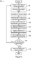
- Figure 5 is a flow diagram 90 of the training of classification neural network 58 of classifier 30 with a deep neural network, according to an embodiment of the present invention.
- data-including medical image data and non-image data- are collected or input for training and testing.
- the collected images are segmented by segmenter 22 so as to generate segmentation maps (as described above).
- the image data 38 and non-image data 40 are annotated with labels provided by qualified experts with domain knowledge.
- the training classification outputs may be determined based on subject clinical records. For example, if the classification output is to include a fracture probability score, then the training output is the timing (post-scan) of a fracture, ascertained from the subject's medical history-and, where no fracture is apparent, recorded as "negligible risk" (or some equivalent designation). If the classification output is to include a score for categories that correspond to known medical conditions, then the training output is the actual medical condition of the subject, also ascertained from the subject's clinical records.
- the data (comprising segmentation maps 50 and non-image data 40) is split into a training set, a development or 'dev' set (which may be omitted in some implementations), and a testing set, each for a different use.
- the training set is the data on which the learning algorithm is to be run;
- the dev set is the data used to tune the parameters;
- the testing set is the data to be used to evaluate the performance of the trained model.
- each layer consists of artificial neurons.
- An artificial neuron is a mathematical function that receives one or more inputs and sums them to produce an output. Usually, each input is separately weighted, and the sum is passed through a non-linear function. As the neural network learns, the weights of the model are adjusted in response to the error (the difference between the network output and the annotations) it produces until the error cannot be reduced further.
- the parameters of classification neural network 58- including layer numbers, neuron numbers, neuron weights, and neuron function parameters, etc.-are initialized.
- the learning algorithm runs on the training data set to update the parameters of classification neural network 58.
- the parameters might be updated by determining a gradient of a loss function.
- the loss function is calculated by the labelled classification output and output generated by classification neural network 58.
- the dev data may be used to investigate the learning procedure and tune the layers and parameters.
- step 106 classifier 30-provided with classification neural network 58-is tested on the testing data.
- step 108 an evaluation is made as to whether the performance of classifier 30 is satisfactory. If the performance is unsatisfactory, processing returns to step 92 where more training data is collected.
- step 108 If, at step 108, the performance is found to be satisfactory, processing continues at step 110, where the trained classifier 30 is outputted or flagged for deployment, or released for use. Processing then ends.
Landscapes
- Engineering & Computer Science (AREA)
- Life Sciences & Earth Sciences (AREA)
- Health & Medical Sciences (AREA)
- Theoretical Computer Science (AREA)
- Physics & Mathematics (AREA)
- Computer Vision & Pattern Recognition (AREA)
- Data Mining & Analysis (AREA)
- General Physics & Mathematics (AREA)
- Artificial Intelligence (AREA)
- Evolutionary Computation (AREA)
- General Health & Medical Sciences (AREA)
- Medical Informatics (AREA)
- General Engineering & Computer Science (AREA)
- Evolutionary Biology (AREA)
- Bioinformatics & Computational Biology (AREA)
- Bioinformatics & Cheminformatics (AREA)
- Pathology (AREA)
- Veterinary Medicine (AREA)
- Biophysics (AREA)
- Biomedical Technology (AREA)
- Heart & Thoracic Surgery (AREA)
- Public Health (AREA)
- Molecular Biology (AREA)
- Surgery (AREA)
- Animal Behavior & Ethology (AREA)
- Nuclear Medicine, Radiotherapy & Molecular Imaging (AREA)
- Quality & Reliability (AREA)
- Radiology & Medical Imaging (AREA)
- Psychiatry (AREA)
- Signal Processing (AREA)
- Physiology (AREA)
- Fuzzy Systems (AREA)
- Orthopedic Medicine & Surgery (AREA)
- Rheumatology (AREA)
- Oral & Maxillofacial Surgery (AREA)
- Dentistry (AREA)
- Mathematical Physics (AREA)
- Image Analysis (AREA)
- Apparatus For Radiation Diagnosis (AREA)
Abstract
Description
- This application is based on and claims priority from
US patent application no. 16/448,474 filed 21 June 2019 - The present invention relates to a computer vision system and method, employing machine learning and in particular deep neural networks, for classifying (and monitoring changes in) an image (such as a medical image), based on structural or material segmentation. Possible medical imaging applications include Computed Tomography (CT), Magnetic Resonance (MR), Ultrasound, HRpQCT (High-Resolution peripheral Quantitative Computed Tomography), and Pathology Scanner imaging.
- Computer vision and image processing techniques have been applied to medical image analysis. Some computer-aided systems achieve the analysis with two steps: segmentation and quantitative calculation. Segmentation is the process of segmenting (or differentiating) structures or objects in an image, such as a medical image, from one another by differentiating pixels (in 2D image) or voxels (in 3D image) in the image. Based on the segmentation, quantitative features such as volume, shape, and density are calculated. For example, lesion size and shape are calculated after the lesion has been segmented in a brain CT or MRI scan; the bone mineral density may be calculated after the femoral neck is segmented in a hip DXA (dual-energy x-ray absorptiometry) scan. A doctor may make a diagnosis or treatment decision after he or she has compared such calculated values with healthy reference data.
- For example, a T-score is the standard score of a patient's bone mineral density compared to the young normal reference mean. The WHO (World Health Organization) defines osteoporosis as a T-score of -2.5 or lower, that is, a bone density that is two and a half standard deviations or more below the mean of a 30-year-old healthy man/woman.
- The segmentation may be achieved manually, semi-manually, or automatically. In an example of manual segmentation, a user operates a computer to move a rectangular box over a hip DXA scan and thereby select the region of the femoral neck.
- Semi-manual segmentation may be performed by an image processing program employing a user's initialisation or input. For example, a user may operate a computer to draw an approximate bone boundary on a wrist CT scan; the program then adjusts the approximate boundary into a contour that segments bone from the surrounding tissues.
- Automatic segmentation may be performed by utilizing the features of the object of interest, such as intensity values, edges and shapes. In one existing example, a voxel-value based thresholding method is used to segment bone from the surrounding tissues in CT scans. Some other programs use machine learning algorithms to train a classifier to segment abnormal tissues in medical images. For example, a feature-based machine learning algorithm, such as a support vector machine and a decision tree, may be used as a classifier by using tumour images and normal images as training data. The trained classifier slides through the new image "window" by "window" to segment any image regions of tumour tissues.
- Machine learning algorithms have shown promising accuracy and efficiency in this field. However, it is a significant challenge to both collect sufficient training data and to annotate the training data. The training images must be annotated by experts, which is a tedious and time-consuming process. Moreover, in some applications, it may be very difficult or nearly impossible to accurately annotate the training images, even for experts. For example, in bone quality assessment, a transitional zone exists at any sample composed of both cortical and trabecular bones. The transitional zone comprises the inner cortex adjacent to the medullary canal and trabeculae abutting against the cortex contiguous with the endocortical surface. The transitional zone is a site of vigorous bone remodelling. It is important to identify and segment this region in bone microstructure assessment but, owing to limitations in image resolution, it is essentially impossible for an expert to annotate this region both accurately and consistently. Without annotated images as training data, the segmentation model cannot be trained.
- In the last few years, deep learning or deep neural networks have outperformed human in many visual recognition tasks such as natural image classification. In an exemplary CNN (Convolutional Neural Network) implementation, the network comprises input layer, hidden layers, and an output layer. An image is fed into the network through the input layer. The image is sampled and applied with convolutional operations to generate hidden layers. The output of each layer is used as input to the next layer in the network. The output layer is fully connected at the end that will output a classification result. Training data are images with classification labels. The training process obtains the parameters of the neural network. After the training is finished, a new image will be processed by the neural network with the obtained parameters to generate a classification result. For example, a deep neural network algorithm may be used to train a model to determine the condition (for example, no, mild, moderate, severe) of diabetic retinopathy from OCT (Optical Coherence Tomography) images.
- However, this end-to-end solution brings two problems in clinical practices. First, the end-to-end solution is a black box: the input is the medical image, and the output the classification of diseases or conditions. It is difficult to interpret the process whereby the neural network makes its decision-so it is difficult for the user to assess the reliability of the classification results. Secondly, this solution requires a substantial amount of training data. As discussed above, in medical applications annotating or labelling the training data is a tedious and time-consuming process. Collecting enough training data for each category of each type of classification result thus poses a significant challenge.
- According to a first aspect of the invention, there is provided a system for classifying a structure or material in an image of a subject, comprising:
- a segmenter configured to form one or more segmentations of a structure or material in an image (comprising, for example, a medical image) and generate from the segmentations one or more segmentation maps of the image including categorizations of pixels or voxels of the segmentation maps assigned from one or more respective predefined sets of categories;
- a classifier that implements a trained classification machine learning model configured to generate, based on the segmentations maps, one or more classifications and to assign to the classifications respective scores indicative of a likelihood that the structure or material, or the subject, falls into the respective classifications; and
- an output for outputting a result indicative of the classifications and scores.
- In an embodiment, the classifier generates the one or more classifications based on the segmentations maps and non-image data pertaining to the subject.
- The system may be configured to train the classification machine learning model.
- In an embodiment, the segmenter comprises:
- i) a structure segmenter configured to generate structure segmentation maps including categorizations of the pixels or voxels assigned from a predefined set of structure categories,
- ii) a material segmenter configured to generate material segmentation maps including categorizations of the pixels or voxels assigned from a predefined set of material categories, and/or
- iii) an abnormality segmenter configured to generate abnormality segmentation maps including categorizations of the pixels or voxels assigned from a predefined set of abnormality or normality categories.
- In an example, the structure segmenter is configured to employ a structure segmentation machine learning model to generate the structure segmentation maps, the material segmenter is configured to employ a material segmentation machine learning model to generate the material segmentation maps, and the abnormality segmenter is configured to employ an abnormality segmentation model to generate the abnormality segmentation maps. The structure segmenter may be configured to train the structure segmentation machine learning model, the material segmenter to train the material segmentation machine learning model, and/or the abnormality segmenter to train the abnormality segmentation model.
- In an embodiment, the system further comprises a segmentation map processor configured to process the segmentation maps before the segmentation maps are input by the classifier. In an example, the segmentation map processor is configured to down-sample the segmentation maps.
- In an embodiment, the classification machine learning model comprises a neural network, a support vector machine, a decision tree, or a combination thereof. For example, the classification machine learning model may comprise a neural network that includes convolutional neural network layers and fully-connected neural network layers.
- In an embodiment, the image is a medical image, and the classifications correspond to probabilities that the structure or material, or the subject, will sustain a specified condition or symptom in respective timeframes. On an example, the timeframes include a shorter-term timeframe, a longer-term timeframe, and at least one intermediate-term timeframe intermediate the shorter-term timeframe and the longer-term timeframe. In another example, the condition or symptom is bone fracture.
- In an embodiment, the image is a medical image, and the classifications correspond to probabilities that the structure or material, or the subject, will sustain respective conditions or symptoms. In an example, the conditions or symptoms are bone conditions.
- In an embodiment, the image is a medical image, and the classifications correspond to probabilities of respective rates of disease or pathology progression. For example, the classifications may comprise classifications corresponding any one or more of: stable, modest deterioration, and accelerated deterioration.
- In an embodiment, the image is a medical image, and the classifications correspond to probabilities of efficacy of respective treatment options. For example, the treatment options may include an antiresorptive treatment and/or an anabolic treatment.
- In an embodiment, the image is a medical image, and the classifications correspond to respective medical conditions. For example, the medical conditions may include any one or more of: osteomalacia, tumour, osteonecrosis and infection.
- In an embodiment, the classification machine learning model is a model trained with image data and non-image data relating to training subjects, and generates the respective scores based on image data (typically constituting one or more images) and non-image data relating to the subject.
- According to a second aspect of the invention, there is provided a computer-implemented method for classifying a structure or material in an image of a subject, comprising:
- forming one or more segmentations of a structure or material in an image;
- generating from the segmentations one or more segmentation maps of the image including categorizations of pixels or voxels of the segmentation maps assigned from respective predefined sets of categories of the structure or material;
- using a trained classification machine learning model to generate, based on the segmentations maps, one or more classifications and to assign to the classifications respective scores indicative of a likelihood that the structure or material, or the subject, falls into the respective classifications; and
- outputting a result indicative of the classifications and scores.
- In an embodiment, the classification machine learning model is used to generate the one or more classifications based on the segmentations maps and non-image data pertaining to the subject.
- The method may include training the classification machine learning model.
- In an embodiment, forming the one or more segmentations comprises:
- i) generating structure segmentation maps including categorizations of the pixels or voxels assigned from a predefined set of structure categories,
- ii) generating material segmentation maps including categorizations of the pixels or voxels assigned from a predefined set of material categories, and/or
- iii) generating abnormality segmentation maps including categorizations of the pixels or voxels assigned from a predefined set of abnormality or normality categories.
- For example, the method may include employing a structure segmentation machine learning model to generate the structure segmentation maps, a material segmentation machine learning model to generate the material segmentation maps, and an abnormality segmentation model to generate the abnormality segmentation maps. In particular, the method may include training the structure segmentation machine learning model, the material segmentation machine learning model, and/or the abnormality segmentation model.
- According to a third aspect of the invention, there is provided a computer-implemented diagnostic method, comprising the method of the second aspect.
- According to a fourth aspect of the invention, there is provided a computer-implemented method for training a classification machine learning model for classifying a structure or material in an image of a subject, the method comprising:
- dividing annotated segmentation maps and annotated non-image data into a training set and a testing set (such that, as a result, the training set and the testing each include some annotated segmentation maps and some annotated non-image data), the annotated segmentation maps obtained by segmenting one or more images;
- (a) implementing a classification machine learning model, including initializing parameters of the classification machine learning model;
- (b) updating the parameters of the classification machine learning model by running a learning algorithm on the training data;
- (c) testing the classification machine learning model on the testing data;
- (d) evaluating whether the classification machine learning model has satisfactory performance;
- repeating steps (a) to (d) when the performance is found in step (d) not to be satisfactory; and
- outputting the classification machine learning model for deployment as a trained classification machine learning model, or flagging the classification machine learning model as a trained classification machine learning model.
- This aspect may be used in conjunction or in combination with (or as a part of) the second aspect, such as to train the classification machine learning model of the second aspect.
- The method may include segmenting the one or more images (such as in the course of generating the annotated segmentation maps).
- In an embodiment, the method includes, when the performance is found in step (d) not to be satisfactory, receiving and using more image and non-image data for training the classification machine learning model.
- The classification model can be trained by various machine learning algorithms, so may comprise-for example-a neural network, a support vector machine, a decision tree, or a combination thereof.
- Thus, in one embodiment, the classification machine learning model comprises a neural network having a plurality of layers comprising artificial neurons, wherein the parameters comprise layer numbers, neuron numbers, neuron weights, and neuron function parameters; and testing the classification machine learning model includes testing the classification machine learning model on the testing data.
- In an embodiment, updating the parameters includes determining a gradient of a loss function.
- In an embodiment, the images are medical images and the non-image data comprise clinical records.
- In an embodiment, the method includes dividing the annotated segmentation maps and the annotated non-image data into the training set, a development set and the testing set, and using the development data to investigate the learning procedure and to tune the parameters (and, when the classification machine learning model comprises a neural network, tune the layers).
- According to this aspect, there is also provided a system for training a classification machine learning model for classifying a structure or material in an image of a subject, the system comprising a processor configured to:
- divide annotated segmentation maps and annotated non-image data into a training set and a testing set, the annotated segmentation maps obtained by segmenting one or more images;
- (a) implement a classification machine learning model, including initializing parameters of the classification machine learning model;
- (b) update the parameters of the classification machine learning model by running a learning algorithm on the training data;
- (c) test the classification machine learning model on the testing data;
- (d) evaluate whether the classification machine learning model has satisfactory performance;
- repeat steps (a) to (d) when the performance is found in step (d) not to be satisfactory; and output the classification machine learning model for deployment as a trained classification machine learning model, or flag the classification machine learning model as a trained classification machine learning model.
- According to a fifth aspect of the invention, there is provided a computer program comprising program code configured, when executed by one of more computing devices, to implemented the method of any one or more of the second to fourth aspects. According to this aspect, there is also provided a computer-readable medium, comprising such a computer program.
- It should be noted that any of the various individual features of each of the above aspects of the invention, and any of the various individual features of the embodiments described herein including in the claims, can be combined as suitable and desired.
- In order that the invention may be more clearly ascertained, embodiments will now be described by way of example with reference to the following drawing, in which:
-
Figure 1 is a schematic view of a classification system according to an embodiment of the present invention; -
Figure 2 is a high-level schematic diagram illustrating the operation of the classification system ofFigure 1 , in calculating bone fracture risk of a subject from a medical imaging scan; -
Figure 3 is a schematic view of the operation of the segmenter of the classification system ofFigure 1 ; -
Figure 4 is a schematic illustration of the operation of classifier of the classification system ofFigure 1 ; -
Figure 5 is a flow diagram of the training of the classification neural network of the classification system ofFigure 1 . -
Figure 1 is a schematic view of aclassification system 10 for classifying a structure or material in a medical image (based on structural and material segmentation), according to an embodiment of the present invention. - Referring to
Figure 1 ,system 10 comprises aclassification controller 12 and a user interface 14 (including a GUI 16).User interface 14 is provided for representing information to a user and for receiving input (including feedback) from a user; it typically comprises one or more displays (on one or more of which may be displayed the GUI 16), a web browser, a keyboard and a mouse, and optionally a printer.Classification controller 12 includes at least oneprocessor 18 and amemory 20.System 10 may be implemented, for example, as a combination of software and hardware on a computer (such as a personal computer or mobile computing device), or as a dedicated image segmentation system.System 10 may optionally be distributed; for example, some or all of the components ofmemory 20 may be located remotely fromprocessor 18;user interface 14 may be located remotely frommemory 20 and/or fromprocessor 18. For example,system 10 may be implemented in a service-oriented architecture, with its components communicating with each other over a communication network such as a LAN (local area network), WAN (wide area network) or the internet.System 10 may be deployed in the cloud, and its use shared by users at different locations. - In certain other embodiments,
system 10 is implemented as a standalone system (of software and hardware) or as standalone software executable by a computer, and deployed in one location; for example,system 10 may be deployed in a hospital, medical clinic or other clinical setting. -
Memory 20 is in data communication withprocessor 18, and typically comprises both volatile and non-volatile memory (and may include more than one of each type of memory), including RAM (Random Access Memory), ROM and one or more mass storage devices. - As is discussed in greater detail below,
processor 18 includes a segmenter 22 (which includes astructure segmenter 24a, amaterial segmenter 24b, and an abnormal segmenter in the form of anabnormal material segmenter 24c), asegmentation map processor 26, and anon-image data processor 28.Processor 18 also includes aclassifier 30, an I/O interface 32 and aresults output 34. -
Memory 20 includesprogram code 36,image data 38,non-image data 40, segmentation models 42 (including, in this example,structure segmentation models 44,material segmentation models 46, and abnormality segmentation models in the form of abnormal material segmentation models 48), segmentation maps 50 (including, in this example, structure segmentation maps 52, material segmentation maps 54, and abnormality segmentation maps in the form of abnormal material segmentation maps 56).Structure segmenter 24a,material segmenter 24b andabnormal material segmenter 24c train therespective segmentation models segmentation models -
Memory 20 also includes a classification machine learning model in the form of a classificationneural network 58, which is trained and used byclassifier 30 to perform classification by using segmentation maps 50 andnon-image data 40.Classification controller 12 is implemented, at least in part (and in some embodiments entirely), byprocessor 18 executingprogram code 36 frommemory 20. - It should be noted that, as the present embodiment relates to the classifying of structures and/or materials in a medical image,
abnormal material segmenter 24c may also be referred to as an abnormal tissue segmenter, and abnormal material segmentation maps 56 may also be referred to as abnormal tissue segmentation maps. - In broad terms, I/
O interface 32 is configured to read or receive medical image data and non-image data pertaining to a subject, and to store these data asimage data 38 andnon-image data 40 ofmemory 20 for processing.Image data 38 is typically in the form, in this embodiment, of a medical image of-for example-a region of the body of a subject.Non-image data 40 typically includes subject or patient information from various structured and non-structured data sources, collected throughout a subject's medical consultations, treatment and follow-up consultations. Subject structured data may include basic subject information such as sex, age, weight, height; laboratory test results such as blood test results and DNA test results; treatment data such as the types of medication and dosage; and questionnaire data such as smoking and drinking habits and fracture history. Subject unstructured data may include text documents of laboratory results, doctors' notes, and radiological reports.Non-image data 40 may in a variety of formats, such as numerical data, text, voice, and video. -
Segmenter 22 processes image data 38 (constituting one or more medical images) and usesstructure segmentation models 44,material segmentation models 46 and abnormalmaterial segmentation models 48 to generate-from image data 38-structure segmentation maps 52, material segmentation maps 54 and abnormal material segmentation maps 56, respectively, which characterizeimage data 38 in different ways.Classifier 30 then inputs the resulting segmentation maps 50 andnon-image data 40, and generates therefrom results in the form of a classification output. The classification output is, in this embodiment, presented to users or used for further analysis via I/O interface 32 and at eitherresults output 34 and/oruser interface 14. - The classification output of classifier 30 (in this embodiment, generated using classification neural network 58) comprises a respective condition score for each of one or more classifications (and preferably for each of a plurality of possible classifications). Each score represents a predicted likelihood that the subject falls into the corresponding classification. In the present example of bone fragility assessment, the classifications are "negligible fracture risk", "imminent fracture risk", "intermediate-term fracture risk", and "long-term fracture risk". The classification output is described in more detail below.
- In an alternative embodiment, the classifier outputs a respective disease progression score for each of one or more condition progression states. Each score represents a predicted likelihood that a current condition will progress to another condition. For example, in bone fragility assessment, the disease progressions may include "stable", "modest deterioration", and "accelerated deterioration".
- In still another embodiment, the classifier outputs a respective treatment score for each of multiple treatment options. Each score represents a predicted likelihood that the treatment is the most efficient for the patient. For example, in a bone fragility assessment, the treatment options may include "antiresorptive", "anabolic", and "antiresorptive + anabolic".
- In a further embodiment, the classification output comprises a score for each of one or more possible classifications corresponding to known medical conditions or pathologies. For example, in a bone fragility assessment, these classifications could be "osteomalacia", "tumour", "osteonecrosis" and "infection". In such an embodiment, the resulting scores represent the degree to which the (e.g. bone) sample of the subject conforms to that classification/condition. If only one classification has a significant score, or one classification has a score that is significantly greater than all other scores, that classification may be regarded as a diagnosis, or suggested diagnosis, of the corresponding condition or pathology.
- In certain embodiments, the classification output comprises two or more sets of such scores (selected from the aforementioned examples or otherwise).
- Returning to
Figure 1 , as will be appreciated by the skilled person in this art, image data 38-constituting one or more medial images-comprises data generated by one or more of a variety of medical image modalities (such as HRpQCT, or High-Resolution peripheral Quantitative Computed Tomography) implemented by one or more medical imaging devices (such as a HRpQCT scanner). Each of these devices scans a sample (whether in vivo or in vitro) and creates a visual representation, generally of a portion of the interior of a subject's body. The medical images may depict, for example, a part of a body or a whole body of a subject (e.g. the brain, the hip or the wrist). The medical images might be acquired by scanning the same sample or body part using different imaging modalities, as different imaging modalities may reveal different characteristics of the same sample or body part. The medical images might be acquired by scanning different body parts using the same image modalities, as different body parts of the same patients might provide different insights towards a better diagnosis of diseases or conditions. For example, in bone fragility assessment, both the wrist and the leg of a patient may be scanned by an HRpQCT scanner (or indeed acquired by scanning the different samples or body parts using different imaging modalities) to provide information for use in assessing a subject's bone quality. - The
image data 38 may constitute a 2D (two-dimensional) image that may be represented as a 2D array of pixels, or a 3D (three-dimensional) image that may be represented as a 3D array of voxels. For convenience, the medical images described below are 3D images that may be represented as a 3D array of voxels. - As mentioned above, the one or more received medical images, stored in
image data 38, are segmented bysegmenter 22, using trainedsegmentation models 42, into respective segmentation maps 50. Eachsegmentation map structure segmentation map 52 represents the medical image as one or more different anatomical structures from a predefined set of structures. For example, a wrist CT scan may be segmented into compact cortex, transitional zone, and trabecular region. Material segmentation map represents the medical image into multiple different materials from a predefined set of materials. For example, a wrist CT scan might be segmented into mineralized material, fully mineralized material, red marrow in the trabecular region, and yellow marrow in the trabecular region. An abnormalmaterial segmentation map 56 represents the medical image as normal material and abnormal material (or, in this example, normal tissue and abnormal tissue). For example, a tumour or fracture might be segmented from a wrist CT scan and represented in an abnormalmaterial segmentation map 56 as 'abnormal'. - Segmentation maps 50 are inputted into
classifier 30, in combination withnon-image data 40.Classifier 30 generates one or more classification outputs based on segmentation maps 50 and thenon-image data 40. Input data ofclassifier 30 is generally multi-dimensional, soclassifier 30 is implemented with machine learning algorithms, such as a neural network, support vector machine, decision tree, or a combination thereof. - In this embodiment,
classifier 30 employs or is implemented as classification neural network 58 (though in other embodiments, other machine learning algorithms may be acceptable), including-in this example-convolutional neural network layers and fully-connected neural network layers. Classificationneural network 58 is trained with training data, as is described below. - As mentioned above, the ultimate classification output is outputted by
system 10 to a user viaresults output 34 oruser interface 14. The classification output may optionally include a visual presentation of one or more of the corresponding segmentation maps 50. Segmentation maps 50 may be presented in case they can assist a user in interpreting the classification output, such as in assessing the reliability of the results. -
Figure 2 is a high-level schematic diagram 50 illustrating the operation ofsystem 10 in calculating bone fracture risk of a subject from a medical imaging scan, in this example a wrist HRpQCT scan 62 (also shown in negative at 62'). As shown inFigure 2 ,system 10 receives thewrist HRpQCT scan 62 comprising a plurality of slices. (As will be appreciated by the skilled person, an HRpQCT scan can comprise 100 or more slices, but four slices are depicted in the figure for simplicity.) -
Segmenter 22 segments scan 62 into astructure segmentation map 52 in which the scan is segmented into compact cortex, outer transitional zone, inner transitional zone, and trabecular region.Segmenter 22 segments scan 62 intomaterial segmentation map 54, in which scan 62 is segmented into surrounding muscle, surrounding fat, yellow marrow adipose, and red marrow adipose.Data 64 comprising segmentation maps 52, 54, abnormal material segmentation maps 56 and non-image data 40 (e.g. clinical factors including sex and age) are processed by trainedclassifier 30 to generate classification outputs. The classification outputs include segmentation maps 52, 54 and a table orreport 66. Table or report 66 includes, in numerical and/or graphical form, fracture probabilities in each category of fracture risk:imminent fracture risk 68a (fracture within two years: t < 2 y), intermediate-term fracture risk 68b (fracture within two to five years: 2 ≤ t < 5 y), long-term fracture risk 68c (fracture in five to ten years, 5 ≤ t ≤ 10 y), andnegligible fracture risk 68d. In the illustrated example, the probability that the wrist is at risk of fracture within two years is 95.6%, that the wrist is at a risk of fracture in two to five years 2.4%, that the wrist is at a risk of fracture in five to 10 years 1.6%, and that the wrist is at negligible risk of fracture is 0.3%. In other words, the probability that the subject will not have a wrist fracture in the next five years (either because the wrist has negligible risk of fracture or because there is only a long-term fracture risk) is only 4.4%. Table or report 66 does not include a diagnosis (e.g. that the subject has osteoporosis), but it will be appreciated that these probabilities may be of great value, including-for example-to prompt the subject to pursue a diagnosis, such as by undergoing medical examination or consultation. -
Figure 3 is a schematic view at 70 of the operation ofsegmenter 22.Segmenter 22 is configured to receive input includes one or more medical images (from image data 38) and to process the images so as to generatesegmentation maps 50. The medical images might be acquired using the same imaging modality by scanning different body parts of a patient. For example, in some applications of assessing bone quality, both wrist and leg of a patient might be scanned by an HRpQCT scanner for the assessment. The medical images might be acquired using different imaging modalities by scanning the same or different body parts of a patient. For example, in some other applications of assessing bone quality, both the wrist HRpQCT scan and the hip DXA scan of a patient are acquired for the assessment (though again bearing in mind that the medical images may be acquired by scanning the different samples or body parts using other imaging modalities). - Referring to
Figure 3 ,segmenter 22 implements one ormore processing branches 1 to n, (labelled 721,..., 72n in the figure) corresponding tomedical images 1 to n of the subject (labelled 381,..., 38n). In the case of plural processing branches,medical images 381,...,38n may be due to-for example-different imaging modalities (labelled 741,..., 74n), as is the case in the illustrated example, different body parts, different scans of a single body part, or a combination two or more of these. Respective segmentation branches 721,..., 72n are configured to receive an image, to segment the image according to image type (such as with different program code 36), and to generate the branch output (comprising the segmentation maps 50 of the input image). - To process a respective input medical image,
segmenter 22 is configured first to select a processing branch ofprocessing branches 1 to n according to the type of the input image.Segmenter 22 ascertains the type of image according to the sample (e.g. scanned body part) and imaging modality, information that can be determined from the respective image, including from metadata stored in a header file of the medical image and/or from the file type. For example, the information of scanned body part and imaging modality may be accessed from the metadata. - Each input
medical image 1 to n is processed by one or more of three segmentation programs (viz.structure segmenter 24a,material segmenter 24b, andabnormal material segmenter 24c) into the corresponding segmentation maps 52, 54, 56.Segmenter 22 thus employs up to n instances each ofsegmenters segmenter -
Structure segmenter 24a,material segmenter 24b, andabnormal material segmenter 24c may generate respective segmentation maps in each processing branch 721,..., 72n. InFigure 3 , for example,structure segmenter 24a,material segmenter 24b, andabnormal material segmenter 24c generate respective segmentation maps corresponding tomedical imaging modalities 1 to n; the resulting structure segmentation maps, material segmentation maps and abnormal tissue segmentation maps are correspondingly labelled structure segmentation maps 521,..., 52n, the material segmentation maps 541,..., 54n and the abnormal tissue segmentation maps 561,..., 56n. It should be noted, however, that in some applications it may not be possible or desirable to generate all three types of segmentation map. This may be due, for example, to the limitations of the images, of the imaging modalities, or ofsegmenters -
Structure segmenter 24a,material segmenter 24b, andabnormal material segmenter 24c assign to each voxel of these segmentation maps 50 one or more 'types' (or 'categories') from respective predetermined sets of types (or categories). Thus, in thisembodiment structure segmenter 24a assigns a respective structure type (from a predefined set of structure types) to each voxel in the scan. For example, a wrist HRpQCT scan is segmented into astructure segmentation map 52 in which each voxel in the scan is assigned a structure type (or category) from the set of "surrounding tissues", "compact cortex", "transitional zone", and "trabecular region". -
Material segmenter 24b assigns a respective material type (from a predefined set of material types) to each voxel. For example, in this embodiment,material segmenter 24b segments a wrist HRpQCT scan into amaterial segmentation map 54 in which each voxel in the scan is assigned a material type from the set of "mineralised material", "fully mineralised material", "red marrow adipose", and "yellow marrow adipose". -
Abnormal material segmenter 24c assigns a respective abnormality or normality type (from a predefined set of abnormalities or normality types, such as a set comprising "normal" and "abnormal") to each voxel. For example, in this embodiment,abnormal material segmenter 24c segments a wrist HRpQCT scan into an abnormaltissue segmentation map 54 in which each voxel in the scan is assigned either "normal" or "abnormal". Optionally, in certain other embodiments,abnormal material segmenter 24c can distinguish different types of abnormality, and the predefined set of abnormality or normality types may include-in addition to "normal"-and one or more specific abnormalities particular to the sample type under examination; if the sample is bone, these may include, for example, "fracture crack" or "bone tumour". In such an embodiment, the set may optionally include "abnormal" for cases in whichabnormal material segmenter 24c cannot determine a specific type of abnormality. - In some implementations,
structure segmenter 24a,material segmenter 24b, andabnormal material segmenter 24c assign respective types with confidence limits or probabilities to each voxel in the medical image. In some other implementations,structure segmenter 24a,material segmenter 24b, andabnormal material segmenter 24c may assign a plurality of types (each optionally with a confidence limit or probability) to each voxel in the medical image. For example,structure segmenter 24a-when segmenting a wrist HRpQCT scan-may assign both "transitional zone" and "trabecular region" to ambiguous voxels, but with respective (and typically different) confidence limits or probabilities. -
Segmenter 22 generates segmentation maps 50 by using the trained segmentation models 42 (including structure segmentation models, material segmentation models and abnormal material segmentation models). The segmentation models are trained using machine learning algorithms (such as a neural network, a support vector machine, a decision tree, or a combination thereof). In this embodiments, the segmentation models are trained using deep neural networks that comprises multiple layers include convolutional neural network layers, fully connected neural network layers, normalisation layers, and multiplicative layers. -
Segmenter 22 may also (or alternatively) perform segmentation using non-machine learning based methods, such as a method based on the location of edges, corners, and transitional slopes, or on global features such as histogram and intensity values of the image. For example,U.S. Patent Application Publication No. 2012/0232375 A1 ("Method and System for Image Analysis of Selected Tissue Structures") discloses a method for segmenting the transitional zone between the compact cortical and trabecular region from a wrist CT scan, based on local and global features of a bone: in many applications, it would be suitable to implement this method insegmenter 22. -
Figure 4 is a schematic illustration of theoperation 80 ofclassifier 30.Classifier 30 is configured to receive input that includes segmentation maps 50 (generated by segmenter 22) and non-imagesubject data 40, and to process that input so as to generate one or more classification results. - However, both the segmentation maps 50 and non-image
subject data 40 are processed before being passed toclassifier 30 bysegmentation map processor 26 andnon-image data processor 28, respectively. For example, in some implementations, it may be expedient to down-sample segmentation maps 50 into lower resolution maps, such as to allow faster image processing byclassifier 30; such processing, if desired or required, is performed bysegmentation map processor 26. In some implementations,segmentation map processor 26 sets the type of any voxels (in a particular segmentation map) that have been assigned more than one type (though typically with different confidence limits or probabilities), such as by assigning to the voxel the type that has the higher or highest probability. - Non-image data may include structured and unstructured data.
Non-image data processor 28 is configured to employ a variety of techniques to process any structured data by extracting features from it, in each case according to the structure and form of the data. For example, structured data are typically stored and maintained in structured data storage such as database tables, .json files, .xml files and .csv files.Non-image data processor 28 extracts features from structured data by querying the required parameters and attributes from the data's respective sources. -
Non-image data processor 28 processes unstructured data in two steps: firstly by converting it into structured data, then by extracting features from the converted data. The conversion method employed bynon-image data processor 28 is specific to each source. For example, to convert a doctor's notes into structured data,non-image data processor 28 employs a trained model of optical character recognition (OCR) to convert the notes into text recognisable by a computer.Non-image data processor 28 then parses the converted text using keywords such as, in this example, "fractures", "pain", "fall", etc. Once the unstructured data has been converted into structured data,non-image data processor 28 then extracts features from the now structured data. - The processed
non-image data 40 and segmentation maps 22 are passed toclassifier 30, which uses these inputs to generate a classification outputs comprising classification score (such as disease condition scores 82, disease progression scores 84, and/or treatment scores 86). -
Figure 5 is a flow diagram 90 of the training of classificationneural network 58 ofclassifier 30 with a deep neural network, according to an embodiment of the present invention. As shown in the figure, atstep 92, data-including medical image data and non-image data-are collected or input for training and testing. Atstep 94, the collected images are segmented bysegmenter 22 so as to generate segmentation maps (as described above). - At
step 96, theimage data 38 andnon-image data 40 are annotated with labels provided by qualified experts with domain knowledge. In a medical application, the training classification outputs may be determined based on subject clinical records. For example, if the classification output is to include a fracture probability score, then the training output is the timing (post-scan) of a fracture, ascertained from the subject's medical history-and, where no fracture is apparent, recorded as "negligible risk" (or some equivalent designation). If the classification output is to include a score for categories that correspond to known medical conditions, then the training output is the actual medical condition of the subject, also ascertained from the subject's clinical records. - At
step 98, the data (comprising segmentation maps 50 and non-image data 40) is split into a training set, a development or 'dev' set (which may be omitted in some implementations), and a testing set, each for a different use. The training set is the data on which the learning algorithm is to be run; the dev set is the data used to tune the parameters; the testing set is the data to be used to evaluate the performance of the trained model. - At
step 100, the layers of the deep neural network are implemented. Each layer consists of artificial neurons. An artificial neuron is a mathematical function that receives one or more inputs and sums them to produce an output. Usually, each input is separately weighted, and the sum is passed through a non-linear function. As the neural network learns, the weights of the model are adjusted in response to the error (the difference between the network output and the annotations) it produces until the error cannot be reduced further. - At
step 102, the parameters of classification neural network 58- including layer numbers, neuron numbers, neuron weights, and neuron function parameters, etc.-are initialized. Atstep 104, the learning algorithm runs on the training data set to update the parameters of classificationneural network 58. For example, the parameters might be updated by determining a gradient of a loss function. The loss function is calculated by the labelled classification output and output generated by classificationneural network 58. The dev data may be used to investigate the learning procedure and tune the layers and parameters. - At
step 106, classifier 30-provided with classification neural network 58-is tested on the testing data. Atstep 108, an evaluation is made as to whether the performance ofclassifier 30 is satisfactory. If the performance is unsatisfactory, processing returns to step 92 where more training data is collected. - If, at
step 108, the performance is found to be satisfactory, processing continues atstep 110, where the trainedclassifier 30 is outputted or flagged for deployment, or released for use. Processing then ends. - It will be understood to persons skilled in the art of the invention that many modifications may be made without departing from the scope of the invention, in particular it will be apparent that certain features of embodiments of the invention can be employed to form further embodiments.
- It is to be understood that, if any prior art is referred to herein, such reference does not constitute an admission that the prior art forms a part of the common general knowledge in the art in any country.
- In the claims which follow and in the preceding description of the invention, except where the context requires otherwise due to express language or necessary implication, the word "comprise" or variations such as "comprises" or "comprising" is used in an inclusive sense, i.e. to specify the presence of the stated features but not to preclude the presence or addition of further features in various embodiments of the invention.
Claims (17)
- A computer-implemented method for training a classification machine learning model for classifying a structure or material in an image of a subject, the method comprising:dividing annotated segmentation maps and annotated non-image data into a training set and a testing set, the annotated segmentation maps obtained by segmenting one or more images;(a) implementing a classification machine learning model, including initializing parameters of the classification machine learning model;(b) updating the parameters of the classification machine learning model by running a learning algorithm on the training data;(c) testing the classification machine learning model on the testing data;(d) evaluating whether the classification machine learning model has satisfactory performance;repeating steps (a) to (d) when the performance is found in step (d) not to be satisfactory; andoutputting the classification machine learning model for deployment as a trained classification machine learning model, or flagging the classification machine learning model as a trained classification machine learning model.
- A method as clamed in claim 1, including, when the performance is found to be unsatisfactory, collecting more image and non-image data for training the classification machine learning model.
- A method as clamed in either claim 1 or 2, wherein the classification machine learning model comprises a neural network having a plurality of layers comprising artificial neurons, wherein
the parameters comprise layer numbers, neuron numbers, neuron weights, and neuron function parameters; and
testing the classification machine learning model includes testing the classification machine learning model on the testing data. - A classification machine learning model trained according to any one of claims 1 to 3.
- A system for training a classification machine learning model for classifying a structure or material in an image of a subject, the system comprising a processor configured to:divide annotated segmentation maps and annotated non-image data into a training set and a testing set, the annotated segmentation maps obtained by segmenting one or more images;(a) implement a classification machine learning model, including initializing parameters of the classification machine learning model;(b) update the parameters of the classification machine learning model by running a learning algorithm on the training data;(c) test the classification machine learning model on the testing data;(d) evaluate whether the classification machine learning model has satisfactory performance;repeat steps (a) to (d) when the performance is found in step (d) not to be satisfactory; andoutput the classification machine learning model for deployment as a trained classification machine learning model, or flag the classification machine learning model as a trained classification machine learning model.
- A system for classifying a structure or material in an image of a subject, comprising:a segmenter configured to form one or more segmentations of a structure or material in an image and generate from the segmentations one or more segmentation maps of the image including categorizations of pixels or voxels of the segmentation maps assigned from one or more respective predefined sets of categories;a classifier that implements a trained classification machine learning model configured to generate, based on the segmentations maps, one or more classifications and to assign to the classifications respective scores indicative of a likelihood that the structure or material, or the subject, falls into the respective classifications; andan output for outputting a result indicative of the classifications and scores.
- A system as claimed in claim 6, wherein the classifier generates the one or more classifications based on the segmentations maps and non-image data pertaining to the subject.
- A system as claimed in either claim 6 or 7, wherein the segmenter comprises:i) a structure segmenter configured to generate structure segmentation maps including categorizations of the pixels or voxels assigned from a predefined set of structure categories, such as with a structure segmentation machine learning model;ii) a material segmenter configured to generate material segmentation maps including categorizations of the pixels or voxels assigned from a predefined set of material categories, such as with a material segmentation machine learning model; and/oriii) an abnormality segmenter configured to generate abnormality segmentation maps including categorizations of the pixels or voxels assigned from a predefined set of abnormality or normality categories, such as with an abnormality segmentation model.
- A system as claimed in any one of claims 6 to 8, further comprising a segmentation map processor configured to down-sample or otherwise process the segmentation maps before the segmentation maps are input by the classifier.
- A system as claimed in any one of claims 6 to 9, wherein the image is a medical image, and the classifications correspond to(i) probabilities that the structure or material, or the subject, will sustain a specified condition or symptom in respective timeframes;(ii) probabilities that the structure or material, or the subject, will sustain a specified condition or symptom in respective timeframes that include a shorter-term timeframe, a longer-term timeframe, and at least one intermediate-term timeframe intermediate the shorter-term timeframe and the longer-term timeframe;(iii) probabilities that the structure or material, or the subject, will sustain respective conditions or symptoms;(iv) probabilities of respective rates of disease or pathology progression;(v) probabilities of respective rates of disease or pathology progression, the classifications comprising classifications corresponding any one or more of: stable, modest deterioration, and accelerated deterioration;(vi) probabilities of efficacy of respective treatment options;(vii) probabilities of efficacy of respective treatment options, the treatment options including an antiresorptive treatment and/or an anabolic treatment;(viii) respective medical conditions; and/or(ix) respective medical conditions that include any one or more of: osteomalacia, tumour, osteonecrosis and infection.
- A system as claimed in any one of claims 6 to 10, the trained classification machine learning model is a model trained by the method of any one of claims 1 to 3.
- A computer-implemented method for classifying a structure or material in an image of a subject, comprising:forming one or more segmentations of a structure or material in an image;generating from the segmentations one or more segmentation maps of the image including categorizations of pixels or voxels of the segmentation maps assigned from respective predefined sets of categories of the structure or material;using a trained classification machine learning model to generate, based on the segmentations maps, one or more classifications and to assign to the classifications respective scores indicative of a likelihood that the structure or material, or the subject, falls into the respective classifications; andoutputting a result indicative of the classifications and scores.
- A method as claimed in claim 12, wherein the trained classification machine learning model generates (i) the one or more classifications based on the segmentation maps and non-image data pertaining to the subject, and/or (ii) the respective scores based on image data and non-image data relating to the subject having been trained with image data and non-image data relating to training subjects.
- A method as claimed in either claim 12 or 13, wherein forming the one or more segmentations comprises:i) generating structure segmentation maps including categorizations of the pixels or voxels assigned from a predefined set of structure categories, such as with a structure segmentation machine learning model;ii) generating material segmentation maps including categorizations of the pixels or voxels assigned from a predefined set of material categories, such as with a material segmentation machine learning model; and/oriii) generating abnormality segmentation maps including categorizations of the pixels or voxels assigned from a predefined set of abnormality or normality categories, such as with an abnormality segmentation model.
- A method as claimed in any one of claims 12 to 14, the trained classification machine learning model is a model trained by the method of any one of claims 1 to 3.
- A classification of a structure or material in an image of a subject, generated according to the method of any one of claims 12 to 15.
- A computer-readable medium, comprising a computer program code configured, when executed by one of more computing devices, to implemented the method of any one of claims 1 to 3 and 12 to 15.
Priority Applications (1)
| Application Number | Priority Date | Filing Date | Title |
|---|---|---|---|
| EP25174877.8A EP4592952A3 (en) | 2019-06-21 | 2020-05-28 | A method and system for machine learning classification based on structure or material segmentation in an image |
Applications Claiming Priority (1)
| Application Number | Priority Date | Filing Date | Title |
|---|---|---|---|
| US16/448,474 US11263497B2 (en) | 2019-06-21 | 2019-06-21 | Method and system for machine learning classification based on structure or material segmentation in an image |
Related Child Applications (1)
| Application Number | Title | Priority Date | Filing Date |
|---|---|---|---|
| EP25174877.8A Division EP4592952A3 (en) | 2019-06-21 | 2020-05-28 | A method and system for machine learning classification based on structure or material segmentation in an image |
Publications (3)
| Publication Number | Publication Date |
|---|---|
| EP3754600A1 true EP3754600A1 (en) | 2020-12-23 |
| EP3754600C0 EP3754600C0 (en) | 2025-05-14 |
| EP3754600B1 EP3754600B1 (en) | 2025-05-14 |
Family
ID=70921817
Family Applications (2)
| Application Number | Title | Priority Date | Filing Date |
|---|---|---|---|
| EP20177226.6A Active EP3754600B1 (en) | 2019-06-21 | 2020-05-28 | A method and system for machine learning classification based on structure or material segmentation in an image |
| EP25174877.8A Pending EP4592952A3 (en) | 2019-06-21 | 2020-05-28 | A method and system for machine learning classification based on structure or material segmentation in an image |
Family Applications After (1)
| Application Number | Title | Priority Date | Filing Date |
|---|---|---|---|
| EP25174877.8A Pending EP4592952A3 (en) | 2019-06-21 | 2020-05-28 | A method and system for machine learning classification based on structure or material segmentation in an image |
Country Status (5)
| Country | Link |
|---|---|
| US (2) | US11263497B2 (en) |
| EP (2) | EP3754600B1 (en) |
| JP (2) | JP7623795B2 (en) |
| CN (1) | CN112116609A (en) |
| ES (1) | ES3030740T3 (en) |
Cited By (3)
| Publication number | Priority date | Publication date | Assignee | Title |
|---|---|---|---|---|
| EP3850635A1 (en) * | 2018-09-13 | 2021-07-21 | Koninklijke Philips N.V. | Calculating boundary conditions for virtual ffr and ifr calculation based on myocardial blush characteristics |
| WO2023078897A1 (en) * | 2021-11-03 | 2023-05-11 | Medimaps Group Sa | Method for analyzing a texture of a bone from a digitized image |
| CN116369959A (en) * | 2023-06-05 | 2023-07-04 | 杭州医策科技有限公司 | Method and device for preoperative positioning of parathyroid glands based on dual-mode CT |
Families Citing this family (27)
| Publication number | Priority date | Publication date | Assignee | Title |
|---|---|---|---|---|
| WO2018209057A1 (en) | 2017-05-11 | 2018-11-15 | The Research Foundation For The State University Of New York | System and method associated with predicting segmentation quality of objects in analysis of copious image data |
| US11263497B2 (en) * | 2019-06-21 | 2022-03-01 | StraxCorp Pty. Ltd. | Method and system for machine learning classification based on structure or material segmentation in an image |
| EP3889894B1 (en) * | 2020-04-01 | 2025-10-29 | Siemens Healthineers AG | Improved abnormality detection in medical imaging data |
| US20220061919A1 (en) * | 2020-09-02 | 2022-03-03 | Abys Medical | Method And Integrated System For Assisting In Setting Up A Personalized Therapeutic Approach For Patients Subject To Medical And Surgical Care |
| US11436724B2 (en) * | 2020-10-30 | 2022-09-06 | International Business Machines Corporation | Lesion detection artificial intelligence pipeline computing system |
| US11694329B2 (en) | 2020-10-30 | 2023-07-04 | International Business Machines Corporation | Logistic model to determine 3D z-wise lesion connectivity |
| US11587236B2 (en) | 2020-10-30 | 2023-02-21 | International Business Machines Corporation | Refining lesion contours with combined active contour and inpainting |
| US11688063B2 (en) | 2020-10-30 | 2023-06-27 | Guerbet | Ensemble machine learning model architecture for lesion detection |
| US11749401B2 (en) | 2020-10-30 | 2023-09-05 | Guerbet | Seed relabeling for seed-based segmentation of a medical image |
| US11688517B2 (en) | 2020-10-30 | 2023-06-27 | Guerbet | Multiple operating point false positive removal for lesion identification |
| GB2606255B (en) | 2021-02-01 | 2023-06-28 | 2692873 Ontario Inc | System and method for classifying dermatological images using machine learning |
| KR102559616B1 (en) * | 2021-02-10 | 2023-07-27 | 주식회사 빔웍스 | Method and system for breast ultrasonic image diagnosis using weakly-supervised deep learning artificial intelligence |
| CN113873196A (en) * | 2021-03-08 | 2021-12-31 | 南通市第一人民医院 | A method and system for improving the quality of infection prevention and control management |
| CN112991370B (en) * | 2021-04-08 | 2022-11-25 | 重庆大学 | Rock core CT crack identification and segmentation method |
| KR102603525B1 (en) * | 2021-06-28 | 2023-11-16 | 전남대학교산학협력단 | Knee bone tumor detection system and method using x-ray image |
| US20230083935A1 (en) * | 2021-09-08 | 2023-03-16 | Canon Medical Systems Corporation | Method and apparatus for partial volume identification from photon-counting macro-pixel measurements |
| WO2023054287A1 (en) * | 2021-10-01 | 2023-04-06 | 富士フイルム株式会社 | Bone disease prediction device, method, and program, learning device, method, and program, and trained neural network |
| CN115168408B (en) * | 2022-08-16 | 2024-05-28 | 北京永洪商智科技有限公司 | Query optimization method, device, equipment and storage medium based on reinforcement learning |
| JP7479648B1 (en) * | 2022-11-14 | 2024-05-09 | 京セラ株式会社 | Prediction device, prediction system, prediction method, control program, and recording medium |
| US12505654B2 (en) * | 2023-01-17 | 2025-12-23 | Adobe Inc. | Material selection from images |
| CN117437459B (en) * | 2023-10-08 | 2024-03-22 | 昆山市第一人民医院 | Method for realizing user knee joint patella softening state analysis based on decision network |
| US12361555B2 (en) * | 2023-11-03 | 2025-07-15 | Qure.Ai Technologies Private Limited | System and method for classifying type of a fracture on musculoskeletal X-ray image |
| WO2025101973A1 (en) * | 2023-11-09 | 2025-05-15 | Cedars-Sinai Medical Center | Systems and methods for disease classification and treatment recommendation using large language models |
| WO2025206282A1 (en) * | 2024-03-29 | 2025-10-02 | 京セラ株式会社 | Information processing system, prediction device, information processing method, control program, and recording medium |
| TWI917981B (en) * | 2024-06-14 | 2026-03-11 | 廣達電腦股份有限公司 | Health status evalutaion method and scanner using the same |
| CN118918249B (en) * | 2024-07-15 | 2025-04-29 | 首都医科大学附属北京积水潭医院 | Construction method of three-substance model based on double-layer spectrum CT and quantitative analysis system for bone density and bone marrow fat content |
| WO2026018690A1 (en) * | 2024-07-16 | 2026-01-22 | ソニーセミコンダクタソリューションズ株式会社 | Information processing device, information processing method, and program |
Citations (2)
| Publication number | Priority date | Publication date | Assignee | Title |
|---|---|---|---|---|
| US20120232375A1 (en) | 2009-09-11 | 2012-09-13 | StraxCorp Pty Ltd. | Method and system for image analysis of selected tissue structures |
| WO2019106061A1 (en) * | 2017-12-01 | 2019-06-06 | Ucb Biopharma Sprl | Three-dimensional medical image analysis method and system for identification of vertebral fractures |
Family Cites Families (30)
| Publication number | Priority date | Publication date | Assignee | Title |
|---|---|---|---|---|
| WO1996012187A1 (en) * | 1994-10-13 | 1996-04-25 | Horus Therapeutics, Inc. | Computer assisted methods for diagnosing diseases |
| CA2529929A1 (en) * | 2003-06-25 | 2005-01-06 | Siemens Medical Solutions Usa, Inc. | Systems and methods for automated diagnosis and decision support for breast imaging |
| US7912528B2 (en) | 2003-06-25 | 2011-03-22 | Siemens Medical Solutions Usa, Inc. | Systems and methods for automated diagnosis and decision support for heart related diseases and conditions |
| US8331637B2 (en) | 2006-03-03 | 2012-12-11 | Medic Vision-Brain Technologies Ltd. | System and method of automatic prioritization and analysis of medical images |
| WO2010115885A1 (en) | 2009-04-03 | 2010-10-14 | Oslo Universitetssykehus Hf | Predictive classifier score for cancer patient outcome |
| JP5762008B2 (en) | 2011-01-19 | 2015-08-12 | 株式会社東芝 | Medical image processing apparatus and medical image processing program |
| US9122950B2 (en) * | 2013-03-01 | 2015-09-01 | Impac Medical Systems, Inc. | Method and apparatus for learning-enhanced atlas-based auto-segmentation |
| US9760990B2 (en) * | 2014-12-14 | 2017-09-12 | International Business Machines Corporation | Cloud-based infrastructure for feedback-driven training and image recognition |
| US9858665B2 (en) * | 2015-04-03 | 2018-01-02 | Regents Of The University Of Minnesota | Medical imaging device rendering predictive prostate cancer visualizations using quantitative multiparametric MRI models |
| US10169871B2 (en) * | 2016-01-21 | 2019-01-01 | Elekta, Inc. | Systems and methods for segmentation of intra-patient medical images |
| US10282588B2 (en) * | 2016-06-09 | 2019-05-07 | Siemens Healthcare Gmbh | Image-based tumor phenotyping with machine learning from synthetic data |
| CN106156793A (en) * | 2016-06-27 | 2016-11-23 | 西北工业大学 | Extract in conjunction with further feature and the classification method of medical image of shallow-layer feature extraction |
| US10657364B2 (en) | 2016-09-23 | 2020-05-19 | Samsung Electronics Co., Ltd | System and method for deep network fusion for fast and robust object detection |
| EP3392832A1 (en) | 2017-04-21 | 2018-10-24 | General Electric Company | Automated organ risk segmentation machine learning methods and systems |
| EP3596697B1 (en) * | 2017-06-28 | 2021-03-17 | Deepmind Technologies Limited | Generalizable medical image analysis using segmentation and classification neural networks |
| IL271743B2 (en) * | 2017-06-30 | 2024-08-01 | Promaton Holding Bv | Classification and 3D model of dental structures - 3D maxilla using deep learning methods |
| JP7005191B2 (en) * | 2017-06-30 | 2022-01-21 | キヤノンメディカルシステムズ株式会社 | Image processing equipment, medical diagnostic imaging equipment, and programs |
| JP6824845B2 (en) * | 2017-08-09 | 2021-02-03 | キヤノン株式会社 | Image processing systems, equipment, methods and programs |
| CA3074505A1 (en) | 2017-08-31 | 2019-03-07 | Omniearth, Inc. | Systems and methods for automatic estimation of object characteristics from digital images |
| CN109410188B (en) * | 2017-10-13 | 2021-06-04 | 科亚医疗科技股份有限公司 | System and method for segmenting medical images |
| KR20200106028A (en) | 2017-10-30 | 2020-09-10 | 고에키자이단호진 간겐큐카이 | Image diagnosis support device, data collection method, image diagnosis support method, and image diagnosis support program |
| CN108596200A (en) * | 2018-01-03 | 2018-09-28 | 深圳北航新兴产业技术研究院 | The method and apparatus of Medical Images Classification |
| EP3511866A1 (en) * | 2018-01-16 | 2019-07-17 | Koninklijke Philips N.V. | Tissue classification using image intensities and anatomical positions |
| JPWO2019176806A1 (en) * | 2018-03-16 | 2021-04-08 | 富士フイルム株式会社 | Machine learning equipment and methods |
| KR102243644B1 (en) * | 2018-12-07 | 2021-04-23 | 서울대학교 산학협력단 | Apparatus and Method for Generating Medical Image Segmentation Deep-Learning Model, Medical Image Segmentation Deep-Learning Model Generated Therefrom |
| WO2020133354A1 (en) * | 2018-12-29 | 2020-07-02 | Shenzhen Malong Technologies Co., Ltd. | 3d segmentation network and 3d refinement module |
| JP7568628B2 (en) * | 2019-01-07 | 2024-10-16 | エクシーニ ディアグノスティクス アーべー | System and method for platform independent whole body image segmentation - Patents.com |
| CN109902672B (en) * | 2019-01-17 | 2024-07-19 | 平安科技(深圳)有限公司 | Image labeling method and device, storage medium and computer equipment |
| US11263497B2 (en) * | 2019-06-21 | 2022-03-01 | StraxCorp Pty. Ltd. | Method and system for machine learning classification based on structure or material segmentation in an image |
| US11545266B2 (en) * | 2019-09-30 | 2023-01-03 | GE Precision Healthcare LLC | Medical imaging stroke model |
-
2019
- 2019-06-21 US US16/448,474 patent/US11263497B2/en active Active
-
2020
- 2020-05-28 EP EP20177226.6A patent/EP3754600B1/en active Active
- 2020-05-28 ES ES20177226T patent/ES3030740T3/en active Active
- 2020-05-28 EP EP25174877.8A patent/EP4592952A3/en active Pending
- 2020-06-10 JP JP2020101138A patent/JP7623795B2/en active Active
- 2020-06-19 CN CN202010563984.XA patent/CN112116609A/en active Pending
-
2022
- 2022-01-26 US US17/584,752 patent/US12217418B2/en active Active
-
2024
- 2024-10-01 JP JP2024172513A patent/JP2025013801A/en active Pending
Patent Citations (2)
| Publication number | Priority date | Publication date | Assignee | Title |
|---|---|---|---|---|
| US20120232375A1 (en) | 2009-09-11 | 2012-09-13 | StraxCorp Pty Ltd. | Method and system for image analysis of selected tissue structures |
| WO2019106061A1 (en) * | 2017-12-01 | 2019-06-06 | Ucb Biopharma Sprl | Three-dimensional medical image analysis method and system for identification of vertebral fractures |
Non-Patent Citations (3)
| Title |
|---|
| CEM M DENIZ ET AL: "Segmentation of the Proximal Femur from MR Images using Deep Convolutional Neural Networks", ARXIV.ORG, CORNELL UNIVERSITY LIBRARY, 201 OLIN LIBRARY CORNELL UNIVERSITY ITHACA, NY 14853, 20 April 2017 (2017-04-20), XP081408431, DOI: 10.1038/S41598-018-34817-6 * |
| KRISHNARAJ ARUN ET AL: "Simulating Dual-Energy X-Ray Absorptiometry in CT Using Deep-Learning Segmentation Cascade", JOURNAL OF THE AMERICAN COLLEGE OF RADIOLOGY, ELSEVIER, AMSTERDAM, NL, vol. 16, no. 10, 12 April 2019 (2019-04-12), pages 1473 - 1479, XP085846821, ISSN: 1546-1440, [retrieved on 20190412], DOI: 10.1016/J.JACR.2019.02.033 * |
| LI CHEN ET AL: "Measuring Bone Density Connectivity Using Dual Energy X-Ray Absorptiometry Images", THE INTERNATIONAL JOURNAL OF FUZZY LOGIC AND INTELLIGENT SYSTEMS, vol. 17, no. 4, 31 December 2017 (2017-12-31), pages 235 - 244, XP055734561, ISSN: 1598-2645, DOI: 10.5391/IJFIS.2017.17.4.235 * |
Cited By (5)
| Publication number | Priority date | Publication date | Assignee | Title |
|---|---|---|---|---|
| EP3850635A1 (en) * | 2018-09-13 | 2021-07-21 | Koninklijke Philips N.V. | Calculating boundary conditions for virtual ffr and ifr calculation based on myocardial blush characteristics |
| WO2023078897A1 (en) * | 2021-11-03 | 2023-05-11 | Medimaps Group Sa | Method for analyzing a texture of a bone from a digitized image |
| WO2023078545A1 (en) * | 2021-11-03 | 2023-05-11 | Medimaps Group Sa | Method for analyzing a texture of a bone from a digitized image |
| CN116369959A (en) * | 2023-06-05 | 2023-07-04 | 杭州医策科技有限公司 | Method and device for preoperative positioning of parathyroid glands based on dual-mode CT |
| CN116369959B (en) * | 2023-06-05 | 2023-08-11 | 杭州医策科技有限公司 | Parathyroid preoperative positioning method and device based on bimodal CT |
Also Published As
| Publication number | Publication date |
|---|---|
| US11263497B2 (en) | 2022-03-01 |
| EP4592952A2 (en) | 2025-07-30 |
| EP3754600C0 (en) | 2025-05-14 |
| CN112116609A (en) | 2020-12-22 |
| EP3754600B1 (en) | 2025-05-14 |
| US12217418B2 (en) | 2025-02-04 |
| US20200401843A1 (en) | 2020-12-24 |
| EP4592952A3 (en) | 2025-09-24 |
| JP2025013801A (en) | 2025-01-28 |
| US20220147757A1 (en) | 2022-05-12 |
| JP7623795B2 (en) | 2025-01-29 |
| JP2021002339A (en) | 2021-01-07 |
| ES3030740T3 (en) | 2025-07-01 |
Similar Documents
| Publication | Publication Date | Title |
|---|---|---|
| US12217418B2 (en) | Method and system for machine learning classification based on structure or material segmentation in an image | |
| US20230351588A1 (en) | Image analysis method and system | |
| Hennessey et al. | Artificial intelligence in veterinary diagnostic imaging: A literature review | |
| AU2023270223B2 (en) | Image Analysis Method and System | |
| Kang et al. | Prediction of bone mineral density in CT using deep learning with explainability | |
| US20250173860A1 (en) | Systems, devices, and methods for spine analysis | |
| Pradhan et al. | Transforming view of medical images using deep learning | |
| US12299888B2 (en) | Similarity determination apparatus, similarity determination method, and similarity determination program | |
| EP3624054A1 (en) | Analysis of skeletal trauma using deep learning | |
| AU2019204380C1 (en) | A Method and System for Machine Learning Classification Based on Structure or Material Segmentation in an Image | |
| Ruikar et al. | DNN-based knee OA severity prediction system: pathologically robust feature engineering approach | |
| US20230096522A1 (en) | Method and system for annotation of medical images | |
| Derrick et al. | The Computer-Assisted Decedent Identification Method of Computer-Assisted Radiographic Identification | |
| Ramos | Analysis of medical images to support decision-making in the musculoskeletal field | |
| US20250086784A1 (en) | Biological Age and Survival Risk Determination from Imaging Biomarkers | |
| Hemalatha | HEAL-Net: Hybrid Deep Learning for Tooth Abnormality Detection | |
| Sais et al. | Robust 3D reconstruction of rib cage bones in computed tomography images, by combining knowledge from radiological, machine learning, and innovative graph guidance | |
| de Oliveira et al. | MRI LUMBAR SPINE SEMANTIC SEGMENTATION USING YOLOv8 |
Legal Events
| Date | Code | Title | Description |
|---|---|---|---|
| PUAI | Public reference made under article 153(3) epc to a published international application that has entered the european phase |
Free format text: ORIGINAL CODE: 0009012 |
|
| STAA | Information on the status of an ep patent application or granted ep patent |
Free format text: STATUS: THE APPLICATION HAS BEEN PUBLISHED |
|
| AK | Designated contracting states |
Kind code of ref document: A1 Designated state(s): AL AT BE BG CH CY CZ DE DK EE ES FI FR GB GR HR HU IE IS IT LI LT LU LV MC MK MT NL NO PL PT RO RS SE SI SK SM TR |
|
| AX | Request for extension of the european patent |
Extension state: BA ME |
|
| STAA | Information on the status of an ep patent application or granted ep patent |
Free format text: STATUS: REQUEST FOR EXAMINATION WAS MADE |
|
| STAA | Information on the status of an ep patent application or granted ep patent |
Free format text: STATUS: EXAMINATION IS IN PROGRESS |
|
| 17P | Request for examination filed |
Effective date: 20210618 |
|
| RBV | Designated contracting states (corrected) |
Designated state(s): AL AT BE BG CH CY CZ DE DK EE ES FI FR GB GR HR HU IE IS IT LI LT LU LV MC MK MT NL NO PL PT RO RS SE SI SK SM TR |
|
| 17Q | First examination report despatched |
Effective date: 20210720 |
|
| RAP3 | Party data changed (applicant data changed or rights of an application transferred) |
Owner name: CURVEBEAM AL LIMITED |
|
| RAP3 | Party data changed (applicant data changed or rights of an application transferred) |
Owner name: CURVEBEAM AI LIMITED |
|
| GRAP | Despatch of communication of intention to grant a patent |
Free format text: ORIGINAL CODE: EPIDOSNIGR1 |
|
| STAA | Information on the status of an ep patent application or granted ep patent |
Free format text: STATUS: GRANT OF PATENT IS INTENDED |
|
| INTG | Intention to grant announced |
Effective date: 20250107 |
|
| GRAS | Grant fee paid |
Free format text: ORIGINAL CODE: EPIDOSNIGR3 |
|
| GRAA | (expected) grant |
Free format text: ORIGINAL CODE: 0009210 |
|
| STAA | Information on the status of an ep patent application or granted ep patent |
Free format text: STATUS: THE PATENT HAS BEEN GRANTED |
|
| AK | Designated contracting states |
Kind code of ref document: B1 Designated state(s): AL AT BE BG CH CY CZ DE DK EE ES FI FR GB GR HR HU IE IS IT LI LT LU LV MC MK MT NL NO PL PT RO RS SE SI SK SM TR |
|
| REG | Reference to a national code |
Ref country code: GB Ref legal event code: FG4D |
|
| REG | Reference to a national code |
Ref country code: CH Ref legal event code: EP |
|
| REG | Reference to a national code |
Ref country code: DE Ref legal event code: R096 Ref document number: 602020051163 Country of ref document: DE |
|
| REG | Reference to a national code |
Ref country code: IE Ref legal event code: FG4D |
|
| REG | Reference to a national code |
Ref country code: ES Ref legal event code: FG2A Ref document number: 3030740 Country of ref document: ES Kind code of ref document: T3 Effective date: 20250701 |
|
| RAP4 | Party data changed (patent owner data changed or rights of a patent transferred) |
Owner name: CURVEBEAM AI LIMITED |
|
| U01 | Request for unitary effect filed |
Effective date: 20250526 |
|
| U07 | Unitary effect registered |
Designated state(s): AT BE BG DE DK EE FI FR IT LT LU LV MT NL PT RO SE SI Effective date: 20250603 |
|
| U20 | Renewal fee for the european patent with unitary effect paid |
Year of fee payment: 6 Effective date: 20250728 |
|
| PGFP | Annual fee paid to national office [announced via postgrant information from national office to epo] |
Ref country code: ES Payment date: 20250630 Year of fee payment: 6 |
|
| PG25 | Lapsed in a contracting state [announced via postgrant information from national office to epo] |
Ref country code: GR Free format text: LAPSE BECAUSE OF FAILURE TO SUBMIT A TRANSLATION OF THE DESCRIPTION OR TO PAY THE FEE WITHIN THE PRESCRIBED TIME-LIMIT Effective date: 20250815 Ref country code: NO Free format text: LAPSE BECAUSE OF FAILURE TO SUBMIT A TRANSLATION OF THE DESCRIPTION OR TO PAY THE FEE WITHIN THE PRESCRIBED TIME-LIMIT Effective date: 20250814 |
|
| PG25 | Lapsed in a contracting state [announced via postgrant information from national office to epo] |
Ref country code: PL Free format text: LAPSE BECAUSE OF FAILURE TO SUBMIT A TRANSLATION OF THE DESCRIPTION OR TO PAY THE FEE WITHIN THE PRESCRIBED TIME-LIMIT Effective date: 20250514 |
|
| PG25 | Lapsed in a contracting state [announced via postgrant information from national office to epo] |
Ref country code: HR Free format text: LAPSE BECAUSE OF FAILURE TO SUBMIT A TRANSLATION OF THE DESCRIPTION OR TO PAY THE FEE WITHIN THE PRESCRIBED TIME-LIMIT Effective date: 20250514 |
|
| PG25 | Lapsed in a contracting state [announced via postgrant information from national office to epo] |
Ref country code: RS Free format text: LAPSE BECAUSE OF FAILURE TO SUBMIT A TRANSLATION OF THE DESCRIPTION OR TO PAY THE FEE WITHIN THE PRESCRIBED TIME-LIMIT Effective date: 20250814 |
|
| PG25 | Lapsed in a contracting state [announced via postgrant information from national office to epo] |
Ref country code: IS Free format text: LAPSE BECAUSE OF FAILURE TO SUBMIT A TRANSLATION OF THE DESCRIPTION OR TO PAY THE FEE WITHIN THE PRESCRIBED TIME-LIMIT Effective date: 20250914 |
|
| REG | Reference to a national code |
Ref country code: CH Ref legal event code: H13 Free format text: ST27 STATUS EVENT CODE: U-0-0-H10-H13 (AS PROVIDED BY THE NATIONAL OFFICE) Effective date: 20251223 |
|
| PG25 | Lapsed in a contracting state [announced via postgrant information from national office to epo] |
Ref country code: SM Free format text: LAPSE BECAUSE OF FAILURE TO SUBMIT A TRANSLATION OF THE DESCRIPTION OR TO PAY THE FEE WITHIN THE PRESCRIBED TIME-LIMIT Effective date: 20250514 |
|
| PG25 | Lapsed in a contracting state [announced via postgrant information from national office to epo] |
Ref country code: CH Free format text: LAPSE BECAUSE OF NON-PAYMENT OF DUE FEES Effective date: 20250531 |
|
| PG25 | Lapsed in a contracting state [announced via postgrant information from national office to epo] |
Ref country code: CZ Free format text: LAPSE BECAUSE OF FAILURE TO SUBMIT A TRANSLATION OF THE DESCRIPTION OR TO PAY THE FEE WITHIN THE PRESCRIBED TIME-LIMIT Effective date: 20250514 |
|
| PG25 | Lapsed in a contracting state [announced via postgrant information from national office to epo] |
Ref country code: SK Free format text: LAPSE BECAUSE OF FAILURE TO SUBMIT A TRANSLATION OF THE DESCRIPTION OR TO PAY THE FEE WITHIN THE PRESCRIBED TIME-LIMIT Effective date: 20250514 |
|
| PG25 | Lapsed in a contracting state [announced via postgrant information from national office to epo] |
Ref country code: MC Free format text: LAPSE BECAUSE OF FAILURE TO SUBMIT A TRANSLATION OF THE DESCRIPTION OR TO PAY THE FEE WITHIN THE PRESCRIBED TIME-LIMIT Effective date: 20250514 |
|
| PLBE | No opposition filed within time limit |
Free format text: ORIGINAL CODE: 0009261 |
|
| STAA | Information on the status of an ep patent application or granted ep patent |
Free format text: STATUS: NO OPPOSITION FILED WITHIN TIME LIMIT |
|
| REG | Reference to a national code |
Ref country code: CH Ref legal event code: L10 Free format text: ST27 STATUS EVENT CODE: U-0-0-L10-L00 (AS PROVIDED BY THE NATIONAL OFFICE) Effective date: 20260325 |
|
| PG25 | Lapsed in a contracting state [announced via postgrant information from national office to epo] |
Ref country code: IE Free format text: LAPSE BECAUSE OF NON-PAYMENT OF DUE FEES Effective date: 20250528 |
|
| 26N | No opposition filed |
Effective date: 20260217 |
|
| U20 | Renewal fee for the european patent with unitary effect paid |
Year of fee payment: 7 Effective date: 20260527 |
|
| PGFP | Annual fee paid to national office [announced via postgrant information from national office to epo] |
Ref country code: GB Payment date: 20260522 Year of fee payment: 7 |
|
| PG25 | Lapsed in a contracting state [announced via postgrant information from national office to epo] |
Ref country code: CY Free format text: LAPSE BECAUSE OF FAILURE TO SUBMIT A TRANSLATION OF THE DESCRIPTION OR TO PAY THE FEE WITHIN THE PRESCRIBED TIME-LIMIT Effective date: 20250514 |