EP3730046A1 - Magnetic resonance temperature imaging method and device - Google Patents
Magnetic resonance temperature imaging method and device Download PDFInfo
- Publication number
- EP3730046A1 EP3730046A1 EP17935744.7A EP17935744A EP3730046A1 EP 3730046 A1 EP3730046 A1 EP 3730046A1 EP 17935744 A EP17935744 A EP 17935744A EP 3730046 A1 EP3730046 A1 EP 3730046A1
- Authority
- EP
- European Patent Office
- Prior art keywords
- signal
- water
- fat
- strength
- transverse relaxation
- Prior art date
- Legal status (The legal status is an assumption and is not a legal conclusion. Google has not performed a legal analysis and makes no representation as to the accuracy of the status listed.)
- Withdrawn
Links
Images

Classifications
-
- G—PHYSICS
- G01—MEASURING; TESTING
- G01R—MEASURING ELECTRIC VARIABLES; MEASURING MAGNETIC VARIABLES
- G01R33/00—Arrangements or instruments for measuring magnetic variables
- G01R33/20—Arrangements or instruments for measuring magnetic variables involving magnetic resonance
- G01R33/44—Arrangements or instruments for measuring magnetic variables involving magnetic resonance using nuclear magnetic resonance [NMR]
- G01R33/48—NMR imaging systems
- G01R33/4804—Spatially selective measurement of temperature or pH
-
- A—HUMAN NECESSITIES
- A61—MEDICAL OR VETERINARY SCIENCE; HYGIENE
- A61B—DIAGNOSIS; SURGERY; IDENTIFICATION
- A61B5/00—Measuring for diagnostic purposes; Identification of persons
- A61B5/01—Measuring temperature of body parts ; Diagnostic temperature sensing, e.g. for malignant or inflamed tissue
- A61B5/015—By temperature mapping of body part
-
- A—HUMAN NECESSITIES
- A61—MEDICAL OR VETERINARY SCIENCE; HYGIENE
- A61B—DIAGNOSIS; SURGERY; IDENTIFICATION
- A61B5/00—Measuring for diagnostic purposes; Identification of persons
- A61B5/05—Detecting, measuring or recording for diagnosis by means of electric currents or magnetic fields; Measuring using microwaves or radio waves
- A61B5/055—Detecting, measuring or recording for diagnosis by means of electric currents or magnetic fields; Measuring using microwaves or radio waves involving electronic [EMR] or nuclear [NMR] magnetic resonance, e.g. magnetic resonance imaging
-
- G—PHYSICS
- G01—MEASURING; TESTING
- G01R—MEASURING ELECTRIC VARIABLES; MEASURING MAGNETIC VARIABLES
- G01R33/00—Arrangements or instruments for measuring magnetic variables
- G01R33/20—Arrangements or instruments for measuring magnetic variables involving magnetic resonance
- G01R33/44—Arrangements or instruments for measuring magnetic variables involving magnetic resonance using nuclear magnetic resonance [NMR]
- G01R33/48—NMR imaging systems
- G01R33/50—NMR imaging systems based on the determination of relaxation times, e.g. T1 measurement by IR sequences; T2 measurement by multiple-echo sequences
-
- A—HUMAN NECESSITIES
- A61—MEDICAL OR VETERINARY SCIENCE; HYGIENE
- A61B—DIAGNOSIS; SURGERY; IDENTIFICATION
- A61B5/00—Measuring for diagnostic purposes; Identification of persons
- A61B5/01—Measuring temperature of body parts ; Diagnostic temperature sensing, e.g. for malignant or inflamed tissue
-
- G—PHYSICS
- G01—MEASURING; TESTING
- G01R—MEASURING ELECTRIC VARIABLES; MEASURING MAGNETIC VARIABLES
- G01R33/00—Arrangements or instruments for measuring magnetic variables
- G01R33/20—Arrangements or instruments for measuring magnetic variables involving magnetic resonance
- G01R33/44—Arrangements or instruments for measuring magnetic variables involving magnetic resonance using nuclear magnetic resonance [NMR]
- G01R33/48—NMR imaging systems
- G01R33/4828—Resolving the MR signals of different chemical species, e.g. water-fat imaging
Definitions
- the present invention relates to the field of magnetic resonance, and specifically, to a magnetic resonance temperature imaging method and apparatus.
- Magnetic resonance temperature imaging can be used to monitor temperature distribution and changes of a tested object in a non-invasive, real time, and in vivo manner.
- Magnetic resonance can be used to monitor temperatures based on different temperature sensitive parameters, and common parameters include proton density (PD), a relaxation time, a diffusion coefficient, and a proton resonance frequency shift (PRFS).
- PD proton density
- PRFS proton resonance frequency shift
- this technology cannot be applied to fat-containing tissue because hydrogen protons in fat are temperature-insensitive.
- embodiments of the present invention are intended to provide a magnetic resonance temperature imaging method and apparatus, to alleviate the foregoing problem.
- an embodiment of the present invention provides a magnetic resonance temperature imaging method, where the magnetic resonance temperature imaging method includes:
- an embodiment of the present invention further provides a magnetic resonance temperature imaging apparatus, where the magnetic resonance temperature imaging apparatus includes:
- a first calculation unit configured to obtain a first strength amplitude value of a water signal, a first phase value of the water signal, a first strength amplitude value of a fat signal, a first phase value of the fat signal, a first transverse relaxation time of water, a first transverse relaxation time of fat, and a first field drift caused by a non-uniform main magnetic field, based on a preset magnetic resonance signal model, a pre-assigned initial water-fat tissue temperature image, and a water-fat separation algorithm;
- a second calculation unit configured to obtain a second strength amplitude value of the water signal, a second phase value of the water signal, a second strength amplitude value of the fat signal, a second phase value of the fat signal, a second transverse relaxation time of the water, a second transverse relaxation time of the fat, a second field drift caused by the non-uniform main magnetic field, and a second water-fat tissue temperature image that minimize a difference between signal strength after the first time of fitting and signal strength before the fitting, by fitting the preset magnetic resonance signal model for the first time based on the initial water-fat tissue temperature image, the first strength amplitude value of the water signal, the first phase value of the water signal, the first strength amplitude value of the fat signal, the first phase value of the fat signal, the first transverse relaxation time of the water, the first transverse relaxation time of the fat, and the first field drift caused by the non-uniform main magnetic field;
- a third calculation unit configured to obtain a third strength amplitude value of the water signal, a third phase value of the water signal, a third strength amplitude value of the fat signal, a third phase value of the fat signal, a third transverse relaxation time of the water, a third transverse relaxation time of the fat, and a third field drift caused by the non-uniform main magnetic field that minimize a difference between signal strength after the second time of fitting and signal strength before the fitting, by fitting the preset magnetic resonance signal model for the second time by keeping the second water-fat tissue temperature image unchanged and based on the second water-fat tissue temperature image, the second strength amplitude value of the water signal, the second phase value of the water signal, the second strength amplitude value of the fat signal, the second phase value of the fat signal, the second transverse relaxation time of the water, the second transverse relaxation time of the fat, and the second field drift caused by the non-uniform main magnetic field; and
- a fourth calculation unit configured to obtain a fourth strength amplitude value of the water signal, a fourth strength amplitude value of the fat signal, a fourth field drift caused by the non-uniform main magnetic field, and a water-fat tissue temperature image at a current moment that minimize a difference between signal strength after the third time of fitting and signal strength before the fitting, by fitting the preset magnetic resonance signal model for the third time by keeping the third phase value of the water signal, the third phase value of the fat signal, the third transverse relaxation time of the water, and the third transverse relaxation time of the fat unchanged and based on the second water-fat tissue temperature image, the third strength amplitude value of the water signal, the third phase value of the water signal, the third strength amplitude value of the fat signal, the third phase value of the fat signal, the third transverse relaxation time of the water, the third transverse relaxation time of the fat, and the third field drift caused by the non-uniform main magnetic field.
- the magnetic resonance signal model includes multiple fat peaks, and the fourth strength amplitude value of the water signal, the fourth strength amplitude value of the fat signal, the fourth field drift caused by the non-uniform main magnetic field, and the water-fat tissue temperature image at the current moment that minimize the difference between the signal strength and the signal strength before the fitting are estimated. In this way, it is ensured that a temperature result is unbiased.
- Reference numerals 100-Magnetic resonance temperature imaging apparatus; 200-Server; 101-Memory; 102-Storage controller; 103-Processor; 104-Peripheral interface; 301-First calculation unit; 302-Second calculation unit; 303-Third calculation unit; 304-Filtering unit; 305-Fourth calculation unit.
- FIG. 1 is a structural block diagram of a server 200 according to an embodiment of the present invention.
- the server 200 includes a magnetic resonance temperature imaging apparatus 100, a memory 101, a storage controller 102, one or more processors (only one processor is shown in the figure) 103, a peripheral interface 104, and the like. These components communicate with each other by using one or more communications buses/signal lines.
- the magnetic resonance temperature imaging apparatus 100 includes at least one software function module that may be stored in the memory 101 in the form of software or firmware or that may be built into an operating system (OS) of the server 200.
- OS operating system
- the memory 101 may be configured to store a software program and a module, for example, a program instruction/module corresponding to the magnetic resonance temperature image apparatus and method in the embodiments of the present invention.
- the processor 103 executes various functional applications and data processing, for example, the magnetic resonance temperature imaging method provided in the embodiments of the present invention, by running the software program and the module stored in the memory 101.
- the memory 101 may include a high speed random access memory, and may further include a nonvolatile memory, for example, one or more magnetic storage apparatuses or flash memories, or other nonvolatile solid state memories. Access to the memory 101 by the processor 103 and other possible components may be under the control of the storage controller 102.
- the peripheral interface 104 couples various input/output apparatuses to the processor 103 and the memory 101.
- the peripheral interface 104, the processor 103, and the storage controller 102 may be implemented in a single chip. In other examples, the peripheral interface 104, the processor 103, and the storage controller 102 each may be implemented by a separate chip.
- the structure shown in FIG. 1 is merely an example, and the server 200 may alternatively include more or fewer components than those shown in FIG. 1 , or have a configuration different from that shown in FIG. 1 .
- the components shown in FIG. 1 may be implemented by hardware, software, or a combination thereof.
- an embodiment of the present invention provides a magnetic resonance temperature imaging method.
- the magnetic resonance temperature imaging method includes the following steps.
- Step S201 Obtain a first strength amplitude value of a water signal, a first phase value of the water signal, a first strength amplitude value of a fat signal, a first phase value of the fat signal, a first transverse relaxation time of water, a first transverse relaxation time of fat, and a first field drift caused by a non-uniform main magnetic field, based on a preset magnetic resonance signal model, a pre-assigned initial water-fat tissue temperature image, and a water-fat separation algorithm.
- Step S202 Obtain a second strength amplitude value of the water signal, a second phase value of the water signal, a second strength amplitude value of the fat signal, a second phase value of the fat signal, a second transverse relaxation time of the water, a second transverse relaxation time of the fat, a second field drift caused by the non-uniform main magnetic field, and a second water-fat tissue temperature image that minimize a difference between signal strength after the first time of fitting and signal strength before the fitting, by fitting the preset magnetic resonance signal model for the first time based on the initial water-fat tissue temperature image, the first strength amplitude value of the water signal, the first phase value of the water signal, the first strength amplitude value of the fat signal, the first phase value of the fat signal, the first transverse relaxation time of the water, the first transverse relaxation time of the fat, and the first field drift caused by the non-uniform main magnetic field.
- Step S203 Smooth the second water-fat tissue temperature image by using a low-pass filter, to obtain a smoothed second water-fat tissue temperature image.
- Step S204 Obtain a third strength amplitude value of the water signal, a third phase value of the water signal, a third strength amplitude value of the fat signal, a third phase value of the fat signal, a third transverse relaxation time of the water, a third transverse relaxation time of the fat, and a third field drift caused by the non-uniform main magnetic field that minimize a difference between signal strength after the second time of fitting and signal strength before the fitting, by fitting the preset magnetic resonance signal model for the second time by keeping the smoothed second water-fat tissue temperature image unchanged and based on the second water-fat tissue temperature image, the second strength amplitude value of the water signal, the second phase value of the water signal, the second strength amplitude value of the fat signal, the second phase value of the fat signal, the second transverse relaxation time of the water, the second transverse relaxation time of the fat, and the second field drift caused by the non-uniform main magnetic field.
- the preset magnetic resonance signal model is fitted for the second time based on an equation
- Step S205 Obtain a fourth strength amplitude value of the water signal, a fourth strength amplitude value of the fat signal, a fourth field drift caused by the non-uniform main magnetic field, and a water-fat tissue temperature image at a current moment that minimize a difference between signal strength after the third time of fitting and signal strength before the fitting, by fitting the preset magnetic resonance signal model for the third time by keeping the third phase value of the water signal, the third phase value of the fat signal, the third transverse relaxation time of the water, and the third transverse relaxation time of the fat unchanged and based on the second water-fat tissue temperature image, the third strength amplitude value of the water signal, the third phase value of the water signal, the third strength amplitude value of the fat signal, the third phase value of the fat signal, the third transverse relaxation time of the water, the third transverse relaxation time of the fat, and the third field drift caused by the non-uniform main magnetic field.
- the preset magnetic resonance signal model is fitted for the third time based on an equation
- the preset magnetic resonance signal model is fitted to obtain the second strength amplitude value of the water signal, the second phase value of the water signal, the second strength amplitude value of the water signal, the second phase value of the water signal, the second transverse relaxation time of the water, the second transverse relaxation time of the fat, the second field drift caused by the non-uniform main magnetic field, and the second water-fat tissue temperature image that minimize the difference between the signal strength and the signal strength before the fitting, to ensure that temperature estimation is unbiased, in other words, ensure accuracy of the second water-fat tissue temperature image; then, the second water-fat tissue temperature image is smoothed, and the third strength amplitude value of the water signal, the third phase value of the water signal, the third strength amplitude value of the fat signal, the third phase value of the fat signal, the third transverse relaxation time of the water, the third transverse relaxation time of the fat, and the third field drift caused by the non-uniform main magnetic field that minimize the difference between the signal strength after the second
- the water-fat tissue temperature image at the current moment may be used as an initial value of the next cycle, and steps S201 to S205 are repeated to obtain a water-fat tissue temperature image at the next moment.
- an embodiment of the present invention further provides a magnetic resonance temperature imaging apparatus 100.
- the magnetic resonance temperature imaging apparatus 100 includes a first calculation unit 301, a second calculation unit 302, a third calculation unit 303, a filtering unit 304, and a fourth calculation unit 305.
- the first calculation unit 301 is configured to obtain a first strength amplitude value of a water signal, a first phase value of the water signal, a first strength amplitude value of a fat signal, a first phase value of the fat signal, a first transverse relaxation time of water, a first transverse relaxation time of fat, and a first field drift caused by a non-uniform main magnetic field, based on a preset magnetic resonance signal model, a pre-assigned initial water-fat tissue temperature image, and a water-fat separation algorithm.
- the first calculation unit 301 can perform step S201.
- the second calculation unit 302 is configured to obtain a second strength amplitude value of the water signal, a second phase value of the water signal, a second strength amplitude value of the water signal, a second phase value of the water signal, a second transverse relaxation time of the water, a second transverse relaxation time of the fat, a second field drift caused by the non-uniform main magnetic field, and a second water-fat tissue temperature image that minimize a difference between signal strength after the first time of fitting and signal strength before the fitting, by fitting the preset magnetic resonance signal model for the first time based on the initial water-fat tissue temperature image, the first strength amplitude value of the water signal, the first phase value of the water signal, the first strength amplitude value of the fat signal, the first phase value of the fat signal, the first transverse relaxation time of the water, the first transverse relaxation time of the fat, and the first field drift caused by the non-uniform main magnetic field.
- the second calculation unit 302 can perform step S202.
- the filtering unit 304 is configured to smooth the second water-fat tissue temperature image by using a low-pass filter, to obtain a smoothed second water-fat tissue temperature image.
- the filtering unit 304 can perform step S203.
- the third calculation unit 303 is configured to obtain a third strength amplitude value of the water signal, a third phase value of the water signal, a third strength amplitude value of the fat signal, a third phase value of the fat signal, a third transverse relaxation time of the water, a third transverse relaxation time of the fat, and a third field drift caused by the non-uniform main magnetic field that minimize a difference between signal strength after the second time of fitting and signal strength before the fitting, by fitting the preset magnetic resonance signal model for the second time by keeping the second water-fat tissue temperature image unchanged and based on the second water-fat tissue temperature image, the second strength amplitude value of the water signal, the second phase value of the water signal, the second strength amplitude value of the fat signal, the second phase value of the fat signal, the second transverse relaxation time of the water, the second transverse relaxation time of the fat, and the second field drift caused by the non-uniform main magnetic field.
- the third calculation unit 303 is configured to fit the preset magnetic resonance signal model for the second time based on an equation
- the third calculation unit 303 can perform step S204.
- the fourth calculation unit 305 is configured to obtain a fourth strength amplitude value of the water signal, a fourth strength amplitude value of the fat signal, a fourth field drift caused by the non-uniform main magnetic field, and a water-fat tissue temperature image at a current moment that minimize a difference between signal strength after the third time of fitting and signal strength before the fitting, by fitting the preset magnetic resonance signal model for the third time by keeping the third phase value of the water signal, the third phase value of the fat signal, the third transverse relaxation time of the water, and the third transverse relaxation time of the fat unchanged and based on the second water-fat tissue temperature image, the third strength amplitude value of the water signal, the third phase value of the water signal, the third strength amplitude value of the water signal, the third phase value of the water signal, the third transverse relaxation time of the water, the third transverse relaxation time of the fat, and the third field drift caused by the non-uniform main magnetic field.
- the fourth calculation unit 305 is configured to fit the preset magnetic resonance signal model for the third time based on an equation
- the fourth calculation unit 305 can perform step S205.
- the magnetic resonance signal model includes multiple fat peaks, and the fourth strength amplitude value of the water signal, the fourth strength amplitude value of the fat signal, the fourth field drift caused by the non-uniform main magnetic field, and the water-fat tissue temperature image at the current moment that minimize the difference between the signal strength and the signal strength before the fitting are estimated. In this way, it is ensured that a temperature result is unbiased.
- each block in the flowchart or block diagram may represent a part of one module, program segment, or code.
- the part of the module, program segment, or code includes one or more executable instructions for implementing a specified logical function.
- the functions noted in the blocks may be executed in an order different from that noted in the accompanying drawings.
- each block in the block diagrams and/or flowcharts and a combination of blocks in the block diagrams and/or flowcharts may be implemented by using a dedicated hardware-based system for executing a specified function or action, or may be implemented by using a combination of dedicated hardware and a computer instruction.
- each module in the various embodiments of the present invention may be integrated together to form a separate part, or each module may exist alone, or two or more modules may be integrated to form a separate part.
- the functions may be stored in a computer-readable storage medium.
- the computer software product is stored in a storage medium, and includes several instructions for enabling a computer device (which may be a personal computer, a server, a network device, or the like) to perform all or some of the steps of the method described in the embodiments of the present invention.
- the storage medium includes various media that can store program code, for example, a USB flash drive, a removable hard disk, a read-only memory (ROM), a random access memory (RAM), a magnetic disk, and an optical disk.
- program code for example, a USB flash drive, a removable hard disk, a read-only memory (ROM), a random access memory (RAM), a magnetic disk, and an optical disk.
- the term "include”, “comprise”, or their any other variant is intended to cover a non-exclusive inclusion, so that a process, a method, an article, or a device that includes a series of elements not only includes these elements, but also includes other elements that are not expressly listed, or further includes elements inherent to such process, method, article, or device.
- An element preceded by "includes a " further includes, without more constraints, additional identical elements in the process, method, article, or device that includes the element.
Landscapes
- Physics & Mathematics (AREA)
- Health & Medical Sciences (AREA)
- Life Sciences & Earth Sciences (AREA)
- High Energy & Nuclear Physics (AREA)
- General Physics & Mathematics (AREA)
- Condensed Matter Physics & Semiconductors (AREA)
- Nuclear Medicine, Radiotherapy & Molecular Imaging (AREA)
- Heart & Thoracic Surgery (AREA)
- Animal Behavior & Ethology (AREA)
- Biomedical Technology (AREA)
- Pathology (AREA)
- Medical Informatics (AREA)
- Molecular Biology (AREA)
- Surgery (AREA)
- Engineering & Computer Science (AREA)
- General Health & Medical Sciences (AREA)
- Public Health (AREA)
- Veterinary Medicine (AREA)
- Biophysics (AREA)
- Radiology & Medical Imaging (AREA)
- Magnetic Resonance Imaging Apparatus (AREA)
Abstract
Description
- This application claims priority to Chinese Patent Application No.
201711387804.1, filed on December 20, 2017 - The present invention relates to the field of magnetic resonance, and specifically, to a magnetic resonance temperature imaging method and apparatus.
- Magnetic resonance temperature imaging can be used to monitor temperature distribution and changes of a tested object in a non-invasive, real time, and in vivo manner. Magnetic resonance can be used to monitor temperatures based on different temperature sensitive parameters, and common parameters include proton density (PD), a relaxation time, a diffusion coefficient, and a proton resonance frequency shift (PRFS). In water, there is a linear relationship between a proton resonance frequency and a temperature, and the linear relationship is tissue-independent, and therefore a PRFS-based magnetic resonance temperature imaging technology is most widely applied. However, this technology cannot be applied to fat-containing tissue because hydrogen protons in fat are temperature-insensitive. In this case, there is a non-linear relationship between the fat-containing tissue and a temperature change, and the non-linear relationship is related to a ratio of water to the fat in the tissue. To implement temperature imaging for the fat-containing tissue, impact of the fat needs to be eliminated or corrected.
- In view of this, embodiments of the present invention are intended to provide a magnetic resonance temperature imaging method and apparatus, to alleviate the foregoing problem.
- According to a first aspect, an embodiment of the present invention provides a magnetic resonance temperature imaging method, where the magnetic resonance temperature imaging method includes:
- Obtaining a first strength amplitude value of a water signal, a first phase value of the water signal, a first strength amplitude value of a fat signal, a first phase value of the fat signal, a first transverse relaxation time of water, a first transverse relaxation time of fat, and a first field drift caused by a non-uniform main magnetic field, based on a preset magnetic resonance signal model, a pre-assigned initial water-fat tissue temperature image, and a water-fat separation algorithm; obtaining a second strength amplitude value of the water signal, a second phase value of the water signal, a second strength amplitude value of the fat signal, a second phase value of the fat signal, a second transverse relaxation time of the water, a second transverse relaxation time of the fat, a second field drift caused by the non-uniform main magnetic field, and a second water-fat tissue temperature image that minimize a difference between signal strength after the first time of fitting and signal strength before the fitting, by fitting the preset magnetic resonance signal model for the first time based on the initial water-fat tissue temperature image, the first strength amplitude value of the water signal, the first phase value of the water signal, the first strength amplitude value of the fat signal, the first phase value of the fat signal, the first transverse relaxation time of the water, the first transverse relaxation time of the fat, and the first field drift caused by the non-uniform main magnetic field;
- Obtaining a third strength amplitude value of the water signal, a third phase value of the water signal, a third strength amplitude value of the fat signal, a third phase value of the fat signal, a third transverse relaxation time of the water, a third transverse relaxation time of the fat, and a third field drift caused by the non-uniform main magnetic field that minimize a difference between signal strength after the second time of fitting and signal strength before the fitting, by fitting the preset magnetic resonance signal model for the second time by keeping the second water-fat tissue temperature image unchanged and based on the second water-fat tissue temperature image, the second strength amplitude value of the water signal, the second phase value of the water signal, the second strength amplitude value of the fat signal, the second phase value of the fat signal, the second transverse relaxation time of the water, the second transverse relaxation time of the fat, and the second field drift caused by the non-uniform main magnetic field; and
- Obtaining a fourth strength amplitude value of the water signal, a fourth strength amplitude value of the fat signal, a fourth field drift caused by the non-uniform main magnetic field, and a water-fat tissue temperature image at a current moment that minimize a difference between signal strength after the third time of fitting and signal strength before the fitting, by fitting the preset magnetic resonance signal model for the third time by keeping the third phase value of the water signal, the third phase value of the fat signal, the third transverse relaxation time of the water, and the third transverse relaxation time of the fat unchanged and based on the second water-fat tissue temperature image, the third strength amplitude value of the water signal, the third phase value of the water signal, the third strength amplitude value of the fat signal, the third phase value of the fat signal, the third transverse relaxation time of the water, the third transverse relaxation time of the fat, and the third field drift caused by the non-uniform main magnetic field.
- According to a second aspect, an embodiment of the present invention further provides a magnetic resonance temperature imaging apparatus, where the magnetic resonance temperature imaging apparatus includes:
- A first calculation unit, configured to obtain a first strength amplitude value of a water signal, a first phase value of the water signal, a first strength amplitude value of a fat signal, a first phase value of the fat signal, a first transverse relaxation time of water, a first transverse relaxation time of fat, and a first field drift caused by a non-uniform main magnetic field, based on a preset magnetic resonance signal model, a pre-assigned initial water-fat tissue temperature image, and a water-fat separation algorithm;
- A second calculation unit, configured to obtain a second strength amplitude value of the water signal, a second phase value of the water signal, a second strength amplitude value of the fat signal, a second phase value of the fat signal, a second transverse relaxation time of the water, a second transverse relaxation time of the fat, a second field drift caused by the non-uniform main magnetic field, and a second water-fat tissue temperature image that minimize a difference between signal strength after the first time of fitting and signal strength before the fitting, by fitting the preset magnetic resonance signal model for the first time based on the initial water-fat tissue temperature image, the first strength amplitude value of the water signal, the first phase value of the water signal, the first strength amplitude value of the fat signal, the first phase value of the fat signal, the first transverse relaxation time of the water, the first transverse relaxation time of the fat, and the first field drift caused by the non-uniform main magnetic field;
- A third calculation unit, configured to obtain a third strength amplitude value of the water signal, a third phase value of the water signal, a third strength amplitude value of the fat signal, a third phase value of the fat signal, a third transverse relaxation time of the water, a third transverse relaxation time of the fat, and a third field drift caused by the non-uniform main magnetic field that minimize a difference between signal strength after the second time of fitting and signal strength before the fitting, by fitting the preset magnetic resonance signal model for the second time by keeping the second water-fat tissue temperature image unchanged and based on the second water-fat tissue temperature image, the second strength amplitude value of the water signal, the second phase value of the water signal, the second strength amplitude value of the fat signal, the second phase value of the fat signal, the second transverse relaxation time of the water, the second transverse relaxation time of the fat, and the second field drift caused by the non-uniform main magnetic field; and
- A fourth calculation unit, configured to obtain a fourth strength amplitude value of the water signal, a fourth strength amplitude value of the fat signal, a fourth field drift caused by the non-uniform main magnetic field, and a water-fat tissue temperature image at a current moment that minimize a difference between signal strength after the third time of fitting and signal strength before the fitting, by fitting the preset magnetic resonance signal model for the third time by keeping the third phase value of the water signal, the third phase value of the fat signal, the third transverse relaxation time of the water, and the third transverse relaxation time of the fat unchanged and based on the second water-fat tissue temperature image, the third strength amplitude value of the water signal, the third phase value of the water signal, the third strength amplitude value of the fat signal, the third phase value of the fat signal, the third transverse relaxation time of the water, the third transverse relaxation time of the fat, and the third field drift caused by the non-uniform main magnetic field.
- According to the magnetic resonance temperature imaging method and apparatus provided in the present invention, accuracy and precision of the water-fat tissue temperature image at the current moment are improved by using a two-step iterative temperature estimation algorithm in comparison with the prior art, the magnetic resonance signal model includes multiple fat peaks, and the fourth strength amplitude value of the water signal, the fourth strength amplitude value of the fat signal, the fourth field drift caused by the non-uniform main magnetic field, and the water-fat tissue temperature image at the current moment that minimize the difference between the signal strength and the signal strength before the fitting are estimated. In this way, it is ensured that a temperature result is unbiased.
- To make the foregoing objectives, features, and advantages of the present invention clearer and easier to understand, preferred embodiments are described in detail below with reference to the accompanying drawings.
-
-
FIG. 1 is a structural block diagram of a server according to the present invention; -
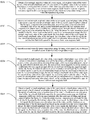
FIG. 2 is a flowchart of a magnetic resonance temperature imaging method according to the present invention; and -
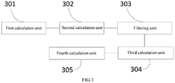
FIG. 3 is a functional and structural block diagram of a magnetic resonance temperature imaging apparatus according to the present invention. - Reference numerals: 100-Magnetic resonance temperature imaging apparatus; 200-Server; 101-Memory; 102-Storage controller; 103-Processor; 104-Peripheral interface; 301-First calculation unit; 302-Second calculation unit; 303-Third calculation unit; 304-Filtering unit; 305-Fourth calculation unit.
- The following clearly and comprehensively describes the technical solutions in the embodiments of the present invention with reference to the accompanying drawings in the embodiments of the present invention. Clearly, the described embodiments are merely some but not all of the embodiments of the present invention. Components in the embodiments of the present invention described and shown in the accompanying drawings usually can be arranged and designed in various different configurations. Therefore, the following detailed description of the embodiments of the present invention provided in the accompanying drawings is not intended to limit the protection scope of the present invention, but is merely intended to represent the selected embodiments of the present invention. All other embodiments obtained by a person skilled in the art based on the embodiments of the present invention without creative efforts shall fall within the protection scope of the present invention.
- A magnetic resonance temperature imaging method and apparatus provided in the preferred embodiments of the present invention can be applied to a
server 200.FIG. 1 is a structural block diagram of aserver 200 according to an embodiment of the present invention. As shown inFIG. 1 , theserver 200 includes a magnetic resonancetemperature imaging apparatus 100, amemory 101, astorage controller 102, one or more processors (only one processor is shown in the figure) 103, aperipheral interface 104, and the like. These components communicate with each other by using one or more communications buses/signal lines. The magnetic resonancetemperature imaging apparatus 100 includes at least one software function module that may be stored in thememory 101 in the form of software or firmware or that may be built into an operating system (OS) of theserver 200. - The
memory 101 may be configured to store a software program and a module, for example, a program instruction/module corresponding to the magnetic resonance temperature image apparatus and method in the embodiments of the present invention. Theprocessor 103 executes various functional applications and data processing, for example, the magnetic resonance temperature imaging method provided in the embodiments of the present invention, by running the software program and the module stored in thememory 101. Thememory 101 may include a high speed random access memory, and may further include a nonvolatile memory, for example, one or more magnetic storage apparatuses or flash memories, or other nonvolatile solid state memories. Access to thememory 101 by theprocessor 103 and other possible components may be under the control of thestorage controller 102. - The
peripheral interface 104 couples various input/output apparatuses to theprocessor 103 and thememory 101. In some embodiments, theperipheral interface 104, theprocessor 103, and thestorage controller 102 may be implemented in a single chip. In other examples, theperipheral interface 104, theprocessor 103, and thestorage controller 102 each may be implemented by a separate chip. - It can be understood that the structure shown in
FIG. 1 is merely an example, and theserver 200 may alternatively include more or fewer components than those shown inFIG. 1 , or have a configuration different from that shown inFIG. 1 . The components shown inFIG. 1 may be implemented by hardware, software, or a combination thereof. - Referring to
FIG. 2 , an embodiment of the present invention provides a magnetic resonance temperature imaging method. The magnetic resonance temperature imaging method includes the following steps. - Step S201: Obtain a first strength amplitude value of a water signal, a first phase value of the water signal, a first strength amplitude value of a fat signal, a first phase value of the fat signal, a first transverse relaxation time of water, a first transverse relaxation time of fat, and a first field drift caused by a non-uniform main magnetic field, based on a preset magnetic resonance signal model, a pre-assigned initial water-fat tissue temperature image, and a water-fat separation algorithm.
- Specifically, the preset magnetic resonance signal model is


- Step S202: Obtain a second strength amplitude value of the water signal, a second phase value of the water signal, a second strength amplitude value of the fat signal, a second phase value of the fat signal, a second transverse relaxation time of the water, a second transverse relaxation time of the fat, a second field drift caused by the non-uniform main magnetic field, and a second water-fat tissue temperature image that minimize a difference between signal strength after the first time of fitting and signal strength before the fitting, by fitting the preset magnetic resonance signal model for the first time based on the initial water-fat tissue temperature image, the first strength amplitude value of the water signal, the first phase value of the water signal, the first strength amplitude value of the fat signal, the first phase value of the fat signal, the first transverse relaxation time of the water, the first transverse relaxation time of the fat, and the first field drift caused by the non-uniform main magnetic field.
- Specifically, the preset magnetic resonance signal model is fitted for the first time based on an equation

- Step S203: Smooth the second water-fat tissue temperature image by using a low-pass filter, to obtain a smoothed second water-fat tissue temperature image.
- Specifically, the second water-fat tissue temperature image is smoothed by using the low-pass filter based on an equation ΔT̃i =(1-µ)ΔTi +µΔ
T , to obtain the smoothed second water-fat tissue temperature image, where ΔTi represents a current temperature estimation value of the ith pixel, ΔT represents an average value of temperature values of all pixels, and ΔT̃i represents a temperature value obtained after the ith pixel is smoothed. - Step S204: Obtain a third strength amplitude value of the water signal, a third phase value of the water signal, a third strength amplitude value of the fat signal, a third phase value of the fat signal, a third transverse relaxation time of the water, a third transverse relaxation time of the fat, and a third field drift caused by the non-uniform main magnetic field that minimize a difference between signal strength after the second time of fitting and signal strength before the fitting, by fitting the preset magnetic resonance signal model for the second time by keeping the smoothed second water-fat tissue temperature image unchanged and based on the second water-fat tissue temperature image, the second strength amplitude value of the water signal, the second phase value of the water signal, the second strength amplitude value of the fat signal, the second phase value of the fat signal, the second transverse relaxation time of the water, the second transverse relaxation time of the fat, and the second field drift caused by the non-uniform main magnetic field.
- Specifically, the preset magnetic resonance signal model is fitted for the second time based on an equation

- Step S205: Obtain a fourth strength amplitude value of the water signal, a fourth strength amplitude value of the fat signal, a fourth field drift caused by the non-uniform main magnetic field, and a water-fat tissue temperature image at a current moment that minimize a difference between signal strength after the third time of fitting and signal strength before the fitting, by fitting the preset magnetic resonance signal model for the third time by keeping the third phase value of the water signal, the third phase value of the fat signal, the third transverse relaxation time of the water, and the third transverse relaxation time of the fat unchanged and based on the second water-fat tissue temperature image, the third strength amplitude value of the water signal, the third phase value of the water signal, the third strength amplitude value of the fat signal, the third phase value of the fat signal, the third transverse relaxation time of the water, the third transverse relaxation time of the fat, and the third field drift caused by the non-uniform main magnetic field.
- Specifically, the preset magnetic resonance signal model is fitted for the third time based on an equation

- According to the magnetic resonance temperature imaging method, first, the preset magnetic resonance signal model is fitted to obtain the second strength amplitude value of the water signal, the second phase value of the water signal, the second strength amplitude value of the water signal, the second phase value of the water signal, the second transverse relaxation time of the water, the second transverse relaxation time of the fat, the second field drift caused by the non-uniform main magnetic field, and the second water-fat tissue temperature image that minimize the difference between the signal strength and the signal strength before the fitting, to ensure that temperature estimation is unbiased, in other words, ensure accuracy of the second water-fat tissue temperature image; then, the second water-fat tissue temperature image is smoothed, and the third strength amplitude value of the water signal, the third phase value of the water signal, the third strength amplitude value of the fat signal, the third phase value of the fat signal, the third transverse relaxation time of the water, the third transverse relaxation time of the fat, and the third field drift caused by the non-uniform main magnetic field that minimize the difference between the signal strength after the second time of fitting and the signal strength before the fitting are estimated, where in this case, the obtained third phase value of the water signal, third phase value of the fat signal, third transverse relaxation time of the water, and third transverse relaxation time of the fat are accurate; and finally, the fourth strength amplitude value of the water signal, the fourth strength amplitude value of the fat signal, the fourth field drift caused by the non-uniform main magnetic field, and the water-fat tissue temperature image at the current moment are obtained by fitting the signal model again by keeping the third phase value of the water signal, the third phase value of the fat signal, the third transverse relaxation time of the water, and the third transverse relaxation time of the fat unchanged, where in this case, precision of the obtained preset magnetic resonance signal model is improved because of a decreased number of free variables in the preset magnetic resonance signal model.
- In addition, the water-fat tissue temperature image at the current moment may be used as an initial value of the next cycle, and steps S201 to S205 are repeated to obtain a water-fat tissue temperature image at the next moment.
- Referring to
FIG. 3 , an embodiment of the present invention further provides a magnetic resonancetemperature imaging apparatus 100. It should be noted that basic principles and technical effects of the magnetic resonancetemperature imaging apparatus 100 provided in this embodiment of the present invention are the same as those in the foregoing embodiment. For brevity, refer to the corresponding content in the foregoing embodiment for content not mentioned in this embodiment of the present invention. The magnetic resonancetemperature imaging apparatus 100 includes afirst calculation unit 301, asecond calculation unit 302, athird calculation unit 303, afiltering unit 304, and afourth calculation unit 305. - The
first calculation unit 301 is configured to obtain a first strength amplitude value of a water signal, a first phase value of the water signal, a first strength amplitude value of a fat signal, a first phase value of the fat signal, a first transverse relaxation time of water, a first transverse relaxation time of fat, and a first field drift caused by a non-uniform main magnetic field, based on a preset magnetic resonance signal model, a pre-assigned initial water-fat tissue temperature image, and a water-fat separation algorithm. - The preset magnetic resonance signal model is



- It can be understood that the
first calculation unit 301 can perform step S201. - The
second calculation unit 302 is configured to obtain a second strength amplitude value of the water signal, a second phase value of the water signal, a second strength amplitude value of the water signal, a second phase value of the water signal, a second transverse relaxation time of the water, a second transverse relaxation time of the fat, a second field drift caused by the non-uniform main magnetic field, and a second water-fat tissue temperature image that minimize a difference between signal strength after the first time of fitting and signal strength before the fitting, by fitting the preset magnetic resonance signal model for the first time based on the initial water-fat tissue temperature image, the first strength amplitude value of the water signal, the first phase value of the water signal, the first strength amplitude value of the fat signal, the first phase value of the fat signal, the first transverse relaxation time of the water, the first transverse relaxation time of the fat, and the first field drift caused by the non-uniform main magnetic field. - The
second calculation unit 302 is configured to fit the preset magnetic resonance signal model for the first time based on an equation
- It can be understood that the
second calculation unit 302 can perform step S202. - The
filtering unit 304 is configured to smooth the second water-fat tissue temperature image by using a low-pass filter, to obtain a smoothed second water-fat tissue temperature image. - It can be understood that the
filtering unit 304 can perform step S203. - The
filtering unit 304 is configured to smooth the second water-fat tissue temperature image by using the low-pass filter based on an equation ΔT̃i =(1-µ)ΔT i+µΔT , to obtain the smoothed second water-fat tissue temperature image, where ΔTi represents a current temperature estimation value of the ith pixel, ΔT represents an average value of temperature values of all pixels, and ΔT̋i represents a temperature value obtained after the ith pixel is smoothed. - The
third calculation unit 303 is configured to obtain a third strength amplitude value of the water signal, a third phase value of the water signal, a third strength amplitude value of the fat signal, a third phase value of the fat signal, a third transverse relaxation time of the water, a third transverse relaxation time of the fat, and a third field drift caused by the non-uniform main magnetic field that minimize a difference between signal strength after the second time of fitting and signal strength before the fitting, by fitting the preset magnetic resonance signal model for the second time by keeping the second water-fat tissue temperature image unchanged and based on the second water-fat tissue temperature image, the second strength amplitude value of the water signal, the second phase value of the water signal, the second strength amplitude value of the fat signal, the second phase value of the fat signal, the second transverse relaxation time of the water, the second transverse relaxation time of the fat, and the second field drift caused by the non-uniform main magnetic field. - The
third calculation unit 303 is configured to fit the preset magnetic resonance signal
- It can be understood that the
third calculation unit 303 can perform step S204. - The
fourth calculation unit 305 is configured to obtain a fourth strength amplitude value of the water signal, a fourth strength amplitude value of the fat signal, a fourth field drift caused by the non-uniform main magnetic field, and a water-fat tissue temperature image at a current moment that minimize a difference between signal strength after the third time of fitting and signal strength before the fitting, by fitting the preset magnetic resonance signal model for the third time by keeping the third phase value of the water signal, the third phase value of the fat signal, the third transverse relaxation time of the water, and the third transverse relaxation time of the fat unchanged and based on the second water-fat tissue temperature image, the third strength amplitude value of the water signal, the third phase value of the water signal, the third strength amplitude value of the water signal, the third phase value of the water signal, the third transverse relaxation time of the water, the third transverse relaxation time of the fat, and the third field drift caused by the non-uniform main magnetic field. - Specifically, the
fourth calculation unit 305 is configured to fit the preset magnetic resonance signal model for the third time based on an equation
- It can be understood that the
fourth calculation unit 305 can perform step S205. - In conclusion, according to the magnetic resonance temperature imaging method and apparatus, accuracy and precision of the water-fat tissue temperature image at the current moment are improved by using a two-step iterative temperature estimation algorithm, the magnetic resonance signal model includes multiple fat peaks, and the fourth strength amplitude value of the water signal, the fourth strength amplitude value of the fat signal, the fourth field drift caused by the non-uniform main magnetic field, and the water-fat tissue temperature image at the current moment that minimize the difference between the signal strength and the signal strength before the fitting are estimated. In this way, it is ensured that a temperature result is unbiased.
- In the several embodiments provided in this application, it should be understood that the disclosed apparatus and method may be implemented in other manners. The apparatus embodiment described above is merely an example. For example, flowcharts and block diagrams in the accompanying drawings show possible implementation architectures, functions, and operations of the apparatus, the method, and the computer program product according to the multiple embodiments of the present invention. In this regard, each block in the flowchart or block diagram may represent a part of one module, program segment, or code. The part of the module, program segment, or code includes one or more executable instructions for implementing a specified logical function. It should also be noted that in some alternative implementations, the functions noted in the blocks may be executed in an order different from that noted in the accompanying drawings. For example, two consecutive blocks may actually be executed substantially in parallel, or two consecutive blocks may sometimes be executed in a reverse order, and this depends on functions involved. It should also be noted that each block in the block diagrams and/or flowcharts and a combination of blocks in the block diagrams and/or flowcharts may be implemented by using a dedicated hardware-based system for executing a specified function or action, or may be implemented by using a combination of dedicated hardware and a computer instruction.
- In addition, the functional modules in the various embodiments of the present invention may be integrated together to form a separate part, or each module may exist alone, or two or more modules may be integrated to form a separate part.
- If the functions are implemented in the form of software functional modules and sold or used as separate products, the functions may be stored in a computer-readable storage medium. Based on such an understanding, the technical solutions of the present invention, essentially or the part contributing to the prior art, may be embodied in the form of a software product. The computer software product is stored in a storage medium, and includes several instructions for enabling a computer device (which may be a personal computer, a server, a network device, or the like) to perform all or some of the steps of the method described in the embodiments of the present invention. The storage medium includes various media that can store program code, for example, a USB flash drive, a removable hard disk, a read-only memory (ROM), a random access memory (RAM), a magnetic disk, and an optical disk. It should be noted that in this specification, relational terms such as first and second are used only to distinguish one entity or operation from another entity or operation, without necessarily requiring or implying any such actual relationship or order between such entities or operations. In addition, the term "include", "comprise", or their any other variant is intended to cover a non-exclusive inclusion, so that a process, a method, an article, or a device that includes a series of elements not only includes these elements, but also includes other elements that are not expressly listed, or further includes elements inherent to such process, method, article, or device. An element preceded by "includes a ..." further includes, without more constraints, additional identical elements in the process, method, article, or device that includes the element.
- The foregoing descriptions are merely preferred embodiments of the present invention, and are not intended to limit the present invention. A person skilled in the art can make various modifications and changes to the present invention. Any modifications, equivalent replacements, and improvements made within the spirit and the principle of the present invention shall fall within the protection scope of the present invention. It should be noted that similar reference numerals and letters represent similar terms in the following accompanying drawings. Therefore, once an item is defined in one accompanying drawing, the item does not need to be further defined and explained in subsequent accompanying drawings.
- The foregoing descriptions are merely specific implementations of the present invention, but the protection scope of the present invention is not limited thereto. Variations or replacements that can be readily figured out by any person skilled in the art within the technical scope of the present invention shall fall within the protection scope of the present invention. Therefore, the protection scope of the present invention should be subject to the protection scope of the claims.
- It should be noted that in this specification, relational terms such as first and second are used only to distinguish one entity or operation from another entity or operation, without necessarily requiring or implying any such actual relationship or order between such entities or operations. In addition, the term "include", "comprise", or their any other variant is intended to cover a non-exclusive inclusion, so that a process, a method, an article, or a device that includes a series of elements not only includes these elements, but also includes other elements that are not expressly listed, or further includes elements inherent to such process, method, article, or device. An element preceded by "includes a ..." further includes, without more constraints, additional identical elements in the process, method, article, or device that includes the element.
Claims (10)
- A magnetic resonance temperature imaging method, wherein the magnetic resonance temperature imaging method comprises:obtaining a first strength amplitude value of a water signal, a first phase value of the water signal, a first strength amplitude value of a fat signal, a first phase value of the fat signal, a first transverse relaxation time of water, a first transverse relaxation time of fat, and a first field drift caused by a non-uniform main magnetic field, based on a preset magnetic resonance signal model, a pre-assigned initial water-fat tissue temperature image, and a water-fat separation algorithm;obtaining a second strength amplitude value of the water signal, a second phase value of the water signal, a second strength amplitude value of the fat signal, a second phase value of the fat signal, a second transverse relaxation time of the water, a second transverse relaxation time of the fat, a second field drift caused by the non-uniform main magnetic field, and a second water-fat tissue temperature image that minimize a difference between signal strength after the first time of fitting and signal strength before the fitting, by fitting the preset magnetic resonance signal model for the first time based on the initial water-fat tissue temperature image, the first strength amplitude value of the water signal, the first phase value of the water signal, the first strength amplitude value of the fat signal, the first phase value of the fat signal, the first transverse relaxation time of the water, the first transverse relaxation time of the fat, and the first field drift caused by the non-uniform main magnetic field;obtaining a third strength amplitude value of the water signal, a third phase value of the water signal, a third strength amplitude value of the fat signal, a third phase value of the fat signal, a third transverse relaxation time of the water, a third transverse relaxation time of the fat, and a third field drift caused by the non-uniform main magnetic field that minimize a difference between signal strength after the second time of fitting and signal strength before the fitting, by fitting the preset magnetic resonance signal model for the second time by keeping the second water-fat tissue temperature image unchanged and based on the second water-fat tissue temperature image, the second strength amplitude value of the water signal, the second phase value of the water signal, the second strength amplitude value of the fat signal, the second phase value of the fat signal, the second transverse relaxation time of the water, the second transverse relaxation time of the fat, and the second field drift caused by the non-uniform main magnetic field; andobtaining a fourth strength amplitude value of the water signal, a fourth strength amplitude value of the fat signal, a fourth field drift caused by the non-uniform main magnetic field, and a water-fat tissue temperature image at a current moment that minimize a difference between signal strength after the third time of fitting and signal strength before the fitting, by fitting the preset magnetic resonance signal model for the third time by keeping the third phase value of the water signal, the third phase value of the fat signal, the third transverse relaxation time of the water, and the third transverse relaxation time of the fat unchanged and based on the second water-fat tissue temperature image, the third strength amplitude value of the water signal, the third phase value of the water signal, the third strength amplitude value of the fat signal, the third phase value of the fat signal, the third transverse relaxation time of the water, the third transverse relaxation time of the fat, and the third field drift caused by the non-uniform main magnetic field.
- The magnetic resonance temperature imaging method according to claim 1, wherein before the step of obtaining a third strength amplitude value of the water signal, a third phase value of the water signal, a third strength amplitude value of the fat signal, a third phase value of the fat signal, a third transverse relaxation time of the water, a third transverse relaxation time of the fat, and a third field drift caused by the non-uniform main magnetic field that minimize a difference between signal strength after the second time of fitting and signal strength before the fitting, by fitting the preset magnetic resonance signal model for the second time by keeping the second water-fat tissue temperature image unchanged and based on the second water-fat tissue temperature image, the second strength amplitude value of the water signal, the second phase value of the water signal, the second strength amplitude value of the fat signal, the second phase value of the fat signal, the second transverse relaxation time of the water, the second transverse relaxation time of the fat, and the second field drift caused by the non-uniform main magnetic field, the magnetic resonance temperature imaging method comprises:
smoothing the second water-fat tissue temperature image by using a low-pass filter, to obtain a smoothed second water-fat tissue temperature image. - The magnetic resonance temperature imaging method according to claim 2, wherein the second water-fat tissue temperature image is smoothed by using the low-pass filter based on an equation ΔT̃i =(1-µ)ΔTi +µΔ
T , to obtain the smoothed second water-fat tissue temperature image, wherein ΔTi represents a current temperature estimation value of the ith pixel, ΔT represents an average value of temperature values of all pixels, and ΔT i represents a temperature value obtained after the ith pixel is smoothed. - The magnetic resonance temperature imaging method according to claim 1, wherein the preset magnetic resonance signal model is


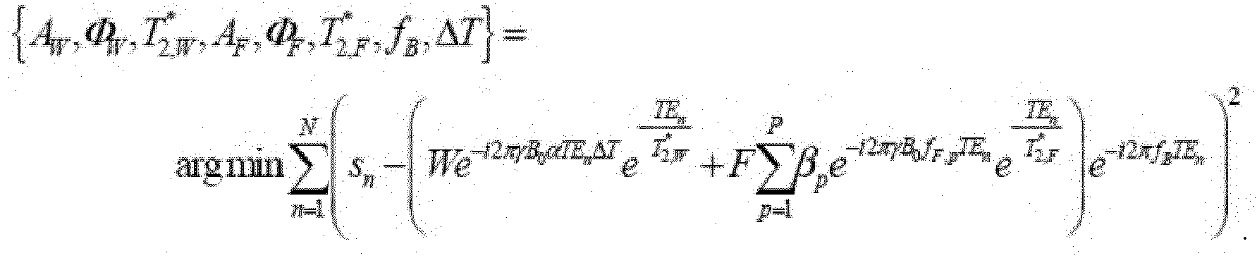
- The magnetic resonance temperature imaging method according to claim 4, wherein the preset magnetic resonance signal model is fitted for the first time based on an equation
- A magnetic resonance temperature imaging apparatus, wherein the magnetic resonance temperature imaging apparatus comprises:a first calculation unit, configured to obtain a first strength amplitude value of a water signal, a first phase value of the water signal, a first strength amplitude value of a fat signal, a first phase value of the fat signal, a first transverse relaxation time of water, a first transverse relaxation time of fat, and a first field drift caused by a non-uniform main magnetic field, based on a preset magnetic resonance signal model, a pre-assigned initial water-fat tissue temperature image, and a water-fat separation algorithm;a second calculation unit, configured to obtain a second strength amplitude value of the water signal, a second phase value of the water signal, a second strength amplitude value of the fat signal, a second phase value of the fat signal, a second transverse relaxation time of the water, a second transverse relaxation time of the fat, a second field drift caused by the non-uniform main magnetic field, and a second water-fat tissue temperature image that minimize a difference between signal strength after the first time of fitting and signal strength before the fitting, by fitting the preset magnetic resonance signal model for the first time based on the initial water-fat tissue temperature image, the first strength amplitude value of the water signal, the first phase value of the water signal, the first strength amplitude value of the fat signal, the first phase value of the fat signal, the first transverse relaxation time of the water, the first transverse relaxation time of the fat, and the first field drift caused by the non-uniform main magnetic field;a third calculation unit, configured to obtain a third strength amplitude value of the water signal, a third phase value of the water signal, a third strength amplitude value of the fat signal, a third phase value of the fat signal, a third transverse relaxation time of the water, a third transverse relaxation time of the fat, and a third field drift caused by the non-uniform main magnetic field that minimize a difference between signal strength after the second time of fitting and signal strength before the fitting, by fitting the preset magnetic resonance signal model for the second time by keeping the second water-fat tissue temperature image unchanged and based on the second water-fat tissue temperature image, the second strength amplitude value of the water signal, the second phase value of the water signal, the second strength amplitude value of the fat signal, the second phase value of the fat signal, the second transverse relaxation time of the water, the second transverse relaxation time of the fat, and the second field drift caused by the non-uniform main magnetic field; anda fourth calculation unit, configured to obtain a fourth strength amplitude value of the water signal, a fourth strength amplitude value of the fat signal, a fourth field drift caused by the non-uniform main magnetic field, and a water-fat tissue temperature image at a current moment that minimize a difference between signal strength after the third time of fitting and signal strength before the fitting, by fitting the preset magnetic resonance signal model for the third time by keeping the third phase value of the water signal, the third phase value of the fat signal, the third transverse relaxation time of the water, and the third transverse relaxation time of the fat unchanged and based on the second water-fat tissue temperature image, the third strength amplitude value of the water signal, the third phase value of the water signal, the third strength amplitude value of the fat signal, the third phase value of the fat signal, the third transverse relaxation time of the water, the third transverse relaxation time of the fat, and the third field drift caused by the non-uniform main magnetic field.
- The magnetic resonance temperature imaging apparatus according to claim 6, wherein the magnetic resonance temperature imaging apparatus further comprises:
a filtering unit, configured to smooth the second water-fat tissue temperature image by using a low-pass filter, to obtain a smoothed second water-fat tissue temperature image. - The magnetic resonance temperature imaging apparatus according to claim 7, wherein the filtering unit is configured to smooth the second water-fat tissue temperature image by using the low-pass filter based on an equation ΔT̃i =(1-µ)ΔT i+µΔ
T , to obtain the smoothed second water-fat tissue temperature image, wherein ΔTi represents a current temperature estimation value of the ith pixel, ΔT represents an average value of temperature values of all pixels, and ΔT̃i represents a temperature value obtained after the ith pixel is smoothed. - The magnetic resonance temperature imaging apparatus according to claim 6, wherein the preset magnetic resonance signal model is


- The magnetic resonance temperature imaging apparatus according to claim 9, wherein the second calculation unit is configured to fit the preset magnetic resonance signal model for the first time based on an equation

Applications Claiming Priority (2)
| Application Number | Priority Date | Filing Date | Title |
|---|---|---|---|
| CN201711387804.1A CN109938704B (en) | 2017-12-20 | 2017-12-20 | Magnetic resonance temperature imaging method and device |
| PCT/CN2017/119483 WO2019119506A1 (en) | 2017-12-20 | 2017-12-28 | Magnetic resonance temperature imaging method and device |
Publications (2)
| Publication Number | Publication Date |
|---|---|
| EP3730046A1 true EP3730046A1 (en) | 2020-10-28 |
| EP3730046A4 EP3730046A4 (en) | 2022-02-23 |
Family
ID=66992463
Family Applications (1)
| Application Number | Title | Priority Date | Filing Date |
|---|---|---|---|
| EP17935744.7A Withdrawn EP3730046A4 (en) | 2017-12-20 | 2017-12-28 | METHOD AND DEVICE FOR TEMPERATURE MAGNETIC RESONANCE IMAGING |
Country Status (4)
| Country | Link |
|---|---|
| US (1) | US20200292651A1 (en) |
| EP (1) | EP3730046A4 (en) |
| CN (1) | CN109938704B (en) |
| WO (1) | WO2019119506A1 (en) |
Families Citing this family (4)
| Publication number | Priority date | Publication date | Assignee | Title |
|---|---|---|---|---|
| CN110766661B (en) * | 2019-09-26 | 2022-10-28 | 上海联影医疗科技股份有限公司 | Water-fat separation method for magnetic resonance imaging, magnetic resonance imaging method and equipment |
| CN110742589B (en) * | 2019-11-05 | 2023-09-22 | 广州互云医院管理有限公司 | Fat reference magnetic resonance temperature imaging method |
| CN112824921B (en) * | 2019-11-20 | 2022-12-20 | 上海联影医疗科技股份有限公司 | Magnetic resonance field drift compensation method, adjustment method, field drift compensation system and magnetic resonance system |
| CN115270468B (en) * | 2022-07-29 | 2026-02-17 | 安徽医科大学 | Magnetic resonance signal simulation method for fatty liver tissue |
Family Cites Families (7)
| Publication number | Priority date | Publication date | Assignee | Title |
|---|---|---|---|---|
| JP2000300536A (en) * | 1999-04-22 | 2000-10-31 | Hitachi Medical Corp | Three-dimensional temperature-measuring method using mri apparatus |
| CN101507603B (en) * | 2008-10-14 | 2012-11-14 | 清华大学 | A method and device for magnetic resonance temperature measurement |
| CN102116856B (en) * | 2010-12-30 | 2013-01-16 | 中国科学院深圳先进技术研究院 | Transverse relaxation time measuring method and system |
| CN104739409B (en) * | 2013-12-31 | 2018-02-13 | 西门子(深圳)磁共振有限公司 | MR imaging method and device |
| CN104382597A (en) * | 2014-11-11 | 2015-03-04 | 奥泰医疗系统有限责任公司 | Dixon water-grease separation and discriminating method and system in magnetic resonance imaging |
| CN105796065B (en) * | 2014-12-29 | 2019-02-15 | 中国科学院深圳先进技术研究院 | Reference-free temperature measurement method and system based on water-fat separation |
| CN106896334B (en) * | 2017-04-18 | 2019-11-29 | 清华大学 | The method and MRI system of active implantation material surrounding tissue temperature under MR are assessed in advance |
-
2017
- 2017-12-20 CN CN201711387804.1A patent/CN109938704B/en active Active
- 2017-12-28 WO PCT/CN2017/119483 patent/WO2019119506A1/en not_active Ceased
- 2017-12-28 EP EP17935744.7A patent/EP3730046A4/en not_active Withdrawn
-
2020
- 2020-06-01 US US16/888,882 patent/US20200292651A1/en not_active Abandoned
Also Published As
| Publication number | Publication date |
|---|---|
| EP3730046A4 (en) | 2022-02-23 |
| US20200292651A1 (en) | 2020-09-17 |
| CN109938704B (en) | 2020-05-19 |
| WO2019119506A1 (en) | 2019-06-27 |
| CN109938704A (en) | 2019-06-28 |
Similar Documents
| Publication | Publication Date | Title |
|---|---|---|
| US20200292651A1 (en) | Magnetic resonance temperature imaging method and apparatus | |
| Haldar et al. | Anatomically constrained reconstruction from noisy data | |
| CN108896943B (en) | Magnetic resonance quantitative imaging method and device | |
| Ye et al. | Semiparametric analysis of correlated recurrent and terminal events | |
| Ma et al. | On estimation efficiency of the central mean subspace | |
| Somasundaram et al. | Brain segmentation in magnetic resonance human head scans using multi-seeded region growing | |
| Simms et al. | A rapid high-performance semi-automated tool to measure total kidney volume from MRI in autosomal dominant polycystic kidney disease | |
| EP4265179A1 (en) | Electrical impedance imaging method, system, storage medium, and electronic device | |
| Weinmann et al. | Iterative Potts and Blake–Zisserman minimization for the recovery of functions with discontinuities from indirect measurements | |
| Bertrand et al. | Flexible parametric approach to classical measurement error variance estimation without auxiliary data | |
| US8498461B2 (en) | Method and magnetic resonance system to determine system-dependent phase information | |
| CN103649782B (en) | It is catalyzed many phase of echos method of deploying | |
| CN104107045A (en) | Magnetic resonance imaging method and device | |
| Tavallali et al. | On the convergence and accuracy of the cardiovascular intrinsic frequency method | |
| Prince et al. | GLMsingle: a toolbox for improving single-trial fMRI response estimates | |
| CN109917314B (en) | Magnetic resonance field map determination method, device, electronic device and storage medium | |
| JP6641678B2 (en) | Fault diagnosis device | |
| US9679378B2 (en) | Risk prediction of tissue infarction | |
| US12455335B2 (en) | Method and device for water-fat separation of image, computer apparatus, and computer-readable storage medium | |
| Prat-Carrabin et al. | Distributed range adaptation in human parietal encoding of numbers | |
| Patel et al. | Individual variation in brain microstructural-cognition relationships in aging | |
| CN110766661B (en) | Water-fat separation method for magnetic resonance imaging, magnetic resonance imaging method and equipment | |
| Kim et al. | Pattern-based Granger causality mapping in fMRI | |
| Colli et al. | Longtime behavior for a generalized Cahn-Hilliard system with fractional operators | |
| US10120051B2 (en) | Method for estimating a convolution product |
Legal Events
| Date | Code | Title | Description |
|---|---|---|---|
| STAA | Information on the status of an ep patent application or granted ep patent |
Free format text: STATUS: THE INTERNATIONAL PUBLICATION HAS BEEN MADE |
|
| PUAI | Public reference made under article 153(3) epc to a published international application that has entered the european phase |
Free format text: ORIGINAL CODE: 0009012 |
|
| STAA | Information on the status of an ep patent application or granted ep patent |
Free format text: STATUS: REQUEST FOR EXAMINATION WAS MADE |
|
| 17P | Request for examination filed |
Effective date: 20200708 |
|
| AK | Designated contracting states |
Kind code of ref document: A1 Designated state(s): AL AT BE BG CH CY CZ DE DK EE ES FI FR GB GR HR HU IE IS IT LI LT LU LV MC MK MT NL NO PL PT RO RS SE SI SK SM TR |
|
| AX | Request for extension of the european patent |
Extension state: BA ME |
|
| DAV | Request for validation of the european patent (deleted) | ||
| DAX | Request for extension of the european patent (deleted) | ||
| A4 | Supplementary search report drawn up and despatched |
Effective date: 20220125 |
|
| RIC1 | Information provided on ipc code assigned before grant |
Ipc: G01R 33/48 20060101ALI20220119BHEP Ipc: A61B 5/055 20060101ALI20220119BHEP Ipc: A61B 5/01 20060101AFI20220119BHEP |
|
| STAA | Information on the status of an ep patent application or granted ep patent |
Free format text: STATUS: THE APPLICATION IS DEEMED TO BE WITHDRAWN |
|
| 18D | Application deemed to be withdrawn |
Effective date: 20220701 |