EP3707492B1 - Lipid-preserving refractive index matching for prolonged imaging depth for tranparent tissue sample and composition - Google Patents
Lipid-preserving refractive index matching for prolonged imaging depth for tranparent tissue sample and composition Download PDFInfo
- Publication number
- EP3707492B1 EP3707492B1 EP18807834.9A EP18807834A EP3707492B1 EP 3707492 B1 EP3707492 B1 EP 3707492B1 EP 18807834 A EP18807834 A EP 18807834A EP 3707492 B1 EP3707492 B1 EP 3707492B1
- Authority
- EP
- European Patent Office
- Prior art keywords
- optical
- weight percent
- refractive index
- clearing solution
- clearing
- Prior art date
- Legal status (The legal status is an assumption and is not a legal conclusion. Google has not performed a legal analysis and makes no representation as to the accuracy of the status listed.)
- Active
Links
Images




Classifications
-
- G—PHYSICS
- G01—MEASURING; TESTING
- G01N—INVESTIGATING OR ANALYSING MATERIALS BY DETERMINING THEIR CHEMICAL OR PHYSICAL PROPERTIES
- G01N1/00—Sampling; Preparing specimens for investigation
- G01N1/28—Preparing specimens for investigation including physical details of (bio-)chemical methods covered elsewhere, e.g. G01N33/50, C12Q
- G01N1/30—Staining; Impregnating ; Fixation; Dehydration; Multistep processes for preparing samples of tissue, cell or nucleic acid material and the like for analysis
-
- G—PHYSICS
- G01—MEASURING; TESTING
- G01N—INVESTIGATING OR ANALYSING MATERIALS BY DETERMINING THEIR CHEMICAL OR PHYSICAL PROPERTIES
- G01N33/00—Investigating or analysing materials by specific methods not covered by groups G01N1/00 - G01N31/00
- G01N33/48—Biological material, e.g. blood, urine; Haemocytometers
- G01N33/50—Chemical analysis of biological material, e.g. blood, urine; Testing involving biospecific ligand binding methods; Immunological testing
- G01N33/5005—Chemical analysis of biological material, e.g. blood, urine; Testing involving biospecific ligand binding methods; Immunological testing involving human or animal cells
- G01N33/5008—Chemical analysis of biological material, e.g. blood, urine; Testing involving biospecific ligand binding methods; Immunological testing involving human or animal cells for testing or evaluating the effect of chemical or biological compounds, e.g. drugs, cosmetics
- G01N33/5082—Supracellular entities, e.g. tissue, organisms
Definitions
- the present disclosure relates generally to optical clearing and, more specifically, to an optical clearing solution and an optical clearing method called lipid-preserving index matching for prolonged imaging depth (LIMPID).
- LIMPID lipid-preserving index matching for prolonged imaging depth
- Imaging techniques like three-dimensional (3-D) microscopy and optical coherence tomography often suffer from restricted imaging depth in biological tissue, which appears opaque.
- the opacity of the biological tissue is due to light scattering on interfaces between lipids and other tissue compounds with different refractive indices.
- Optical clearing is a process that makes biological tissues more optically transparent, while keeping internal structure intact.
- optical clearing techniques include organic solvent-based clearing techniques, lipid extraction after hydrogel embedding techniques, and aqueous solution based techniques. These optical clearing techniques suffer from one or more limitations, including requiring a long time, requiring fancy instruments, suffering from limited dye compatibility, and requiring extra work.
- the present invention relates to an optical clearing method called lipid-preserving index matching for prolonged imaging depth (LIMPID).
- the method of the present invention is defined in claim 8.
- the optical clearing solution of the present invention includes water, iodixanol, a polyhydric alcohol and urea.
- the optical clearing solution of the present invention is defined in claim 1. Iodixanol and urea work together in a synergistic manner to strengthen the clearing capability of the optical clearing solution.
- the optical clearing solution has an adjustable refractive index and osmolality, which improves the image quality by matching the refractive index to optical hardware and eliminating tissue distortions.
- first should not limit the elements being described by these terms. These terms are only used to distinguish one element from another. Thus, a “first” element discussed below could also be termed a “second” element without departing from the teachings of the present disclosure.
- sequence of operations is not limited to the order presented in the claims or figures unless specifically indicated otherwise.
- imaging can refer to the act of creating a visual representation of an interior of at least a portion of a sample for analysis.
- 3D three-dimensional
- a volumetric visual representation of the interior of at least the portion of the sample is created.
- Example imaging modalities can include, but are not limited to, confocal microscopy, light-sheet microscopy, two-photon microscopy, optical coherence tomography, and the like.
- lipid-preserving index matching for prolonged imaging depth can refer to an optical clearing method employing a clearing agent that includes at least water; an iodine-containing, non-ionic radiocontrast agent; and urea at varying concentrations.
- the clearing agent can further include a refractive index adjustment agent, an osmotic pressure adjustment agent, and/or a detergent.
- optical clearing and “clearing” can refer to the act of minimizing light scatter from a sample by exposing the sample to a clearing agent.
- sample can refer to a biological specimen taken from a subject.
- the sample can be a tissue sample excised from a subject.
- weight percent refers to a number of grams of solute in 100 mL of solution.
- the term "subject" can refer to any warm-blooded organism including, but not limited to, a human being, a pig, a rat, a mouse, a bird, a dog, a cat, a goat, a sheep, a horse, a monkey, an ape, a rabbit, a cow, etc.
- the present invention includes an optical clearing method called lipid-preserving index matching for prolonged imaging depth (LIMPID - a synonym for clear). Clearing a tissue sample with LIMPID can increase imaging depth when imaging with confocal microscopy, light-sheet microscopy, two-photon microscopy, optical coherence tomography, or the like, allowing investigation of the three-dimensional (3-D) organization of many complex structures.
- a significant benefit of LIMPID over other clearing methods is it simplicity: the clearing solution is easy to make and the clearing process only requires a single step (immersion of the sample, which can be labeled or preprocessed, in the clearing agent before imaging).
- LIMPID allows for speedy clearing that is compatible with many different instruments and sample preparation techniques, and increases both the imaging depth and the signal to noise ratio (SNR).
- the LIMPID clearing agent includes water, an iodine-containing, non-ionic radiocontrast agent, and urea. Water is the solvent for the other ingredients. However, the LIMPID clearing agent has a higher clearing capability than most existing water-soluble optical clearing agents.
- the iodine-containing, non-ionic radiocontrast agent e.g., iodixanol, which can be in solution with a polyhydric alcohol
- Urea is a protein denaturation and super-hydration agent that reduces scattering from proteins and aids in antigen retrieval.
- the LIMPID clearing agent can also include a refractive index adjustment agent and/or an osmotic pressure adjustment agent, which can help to improve image quality.
- the LIMPID clearing agent can also include a detergent to help permeabilization of cells within the sample.
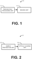
- One aspect of the present disclosure can include a system 10 ( FIG. 1 ) that can be used to perform an optical clearing method called lipid-preserving index matching for prolonged imaging depth (LIMPID).
- LIMPID lipid-preserving index matching for prolonged imaging depth
- a significant benefit of LIMPID over other clearing methods is its simplicity.
- the clearing solution is easy to make and the clearing process only requires one step (immersion of the labeled sample in the clearing agent before imaging).
- the system 10 includes a preparation / processing unit 12 and an imaging unit 14.
- One or more of the preparation / processing unit 12 and the imaging unit 14 can perform computer-aided actions, which are stored in one or more non-transitory memory units and executed by one or more processors.
- the preparation / processing unit 12 and the imaging unit 14 unique entitles (e.g., located at different places).
- functionalities of one or more of the preparation / processing unit 12 and the imaging unit 14 can be combined.
- the preparation / processing unit 12 can perform optical clearing on a prepared sample using LIMPID.
- LIMPID provides a simple and cost effective way to evaluate properties (e.g., morphology, 3D organization, etc.) of a sample.
- the imaging unit 14 can record images of the optically cleared prepared sample that can be used in the evaluation of the sample.
- the imaging unit 14 can include hardware for an imaging modality, like microscopy (e.g., confocal microscopy, light-sheet microscopy, two-photon microscopy, etc.), optical coherence tomography (OCT), or any other imaging modality that would benefit from the increased image depth achieved through optical clearing.
- OCT optical coherence tomography
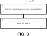
- the preparation / processing unit 12 can include an optical clearing unit 24 that can apply the LIMPID clearing agent to the sample.
- the sample can be immersed in a solution of the LIMPID clearing agent.
- the sample can be immersed for a time sufficient for the clearing to occur in the sample (the time is variable based on the size and depth of the sample) before the sample is imaged.
- the preparation / processing unit 12 can include a sample preparation unit 22 that can prepare the sample for clearing and/or imaging.
- the sample preparation unit 22 can facilitate the performance of labeling, staining, adding contrast agent, or any other pre-imaging task necessary for clearing and/or imaging.
- the clearing agent solution includes at least water, an iodine-containing, non-ionic radiocontrast agent, and urea.
- the water is a solvent for all other ingredients.
- the iodine-containing, non-ionic radiocontrast agent and the urea when used together, can provide a synergistic effect of decreasing the speed required to clear the sample and increasing the clearing capability (e.g., increasing the depth of the sample that is cleared in the time).
- the iodine-containing, non-ionic radiocontrast agent is added to the water to reduce scattering from lipids, as well as provides index matching.
- the iodine-containing, non-ionic radiocontrast agent includes 40 to 80 weight percent iodixanol in solution with 0 to 20 weight percent polyhydric alcohol (such as glycerol, poly(ethylene glycol), triethanolamine, and quadrol (N,N,N', N-Tetrakis(2-Hydroxypropyl)ethylenediamine).
- polyhydric alcohol such as glycerol, poly(ethylene glycol), triethanolamine, and quadrol (N,N,N', N-Tetrakis(2-Hydroxypropyl)ethylenediamine.
- the iodine-containing, non-ionic radiocontrast agent can include 50 to 70 weight percent iodixanol.
- the iodine-containing, non-ionic radiocontrast agent can include 57 to 64 weight percent iodixanol. In further instances, the iodine-containing, non-ionic radiocontrast agent can include about 60 weight percent iodixanol.
- the urea is added to the water as a protein denaturation and super-hydration agent that reduces scattering from proteins.
- the urea is added to the water at a concentration of 10 to 50 weight percent.
- the urea can be added to the water at a concentration of 25 to 45 weight percent.
- the urea can be added to the water at a concentration of 30 to 50 weight percent.
- the urea can be added to the water at a concentration of 35 to 45 weight percent.
- the clearing agent solution can have an adjustable refractive index and/or an adjustable osmolality to match the refractive index of optical hardware and/or intrinsic osmotic pressure of the tissue to eliminate image and/or tissue distortions.
- the refractive index adjustment agent and/or the osmotic pressure adjustment agent comprises at least one of iohexol, thiodiglycol, glycerol, poly(ethylene glycol), triethanolamine, and quadrol (N,N,N',N-Tetrakis(2-Hydroxypropyl)ethylenediamine.
- the adjustable refractive index and/or the adjustable osmolality can be provided by a refractive index adjustment agent and/or an osmotic pressure adjustment agent.
- each of the refractive index adjustment agent and the osmotic pressure adjustment agent can be added to the water at a concentration of 0 to 20 weight percent. In some instances, each of the refractive index adjustment agent and the osmotic pressure adjustment agent can be added to the water at a concentration of 0 to 10 weight percent. In other instances, each of the refractive index adjustment agent and the osmotic pressure adjustment agent can be added to the water at a concentration of 0 to 5 weight percent. In further instances, each of the refractive index adjustment agent and the osmotic pressure adjustment agent can be added to the water at a concentration of 10 to 20 weight percent. In still further instances, each of the refractive index adjustment agent and the osmotic pressure adjustment agent can be added to the water at a concentration of 15 to 20 weight percent.
- the clearing agent may also include a detergent to help permeabilization of cells in the sample.
- the detergent can include triton X-100, digitonin, saponin, or the like.
- the detergent can have a concentration of between 0 and 2 weight percent. However, in some instances, the detergent can have a concentration between 0 and 0.5 weight percent. In other instances, the detergent can have a concentration between 0.5 and 2 weight percent. In still other instances, the detergent can have a concentration between 0.25 and 0.75 weight percent.
- the imaging unit 14 can record raw data corresponding to one or more images of the sample after the sample emerges from the preparation / processing unit.
- the tissue sample can remain immersed in the clearing agent during imaging; however, in some instances, the tissue sample can be removed from the clearing agent before imaging.
- the imaging unit 14 can also be linked to an image processing unit to transfer the raw data for analysis.
- the image processing unit can process the raw data for analysis and/or display one or more images based on the raw data.
- the method of the present invention includes the optical clearing method 30 as described in FIG. 3 and called lipid-preserving index matching for prolonged imaging depth (LIMPID).
- the method 30 can be executed using at least a portion of the system 10 shown in FIGS. 1 and 2 and described above.
- a sample can be immersed in an optical clearing agent.
- LIMPID uses a clearing agent that combines the use of an iodine-containing, non-ionic radiocontrast agent and urea to strengthen its clearing capability with both the iodine-containing, non-ionic radiocontrast agent and the urea working together to provide a strong synergistic effect on speed and clearing capability.
- the clearing agent has an adjustable refractive index and osmolality, which improves the image quality by matching the refractive index to optical hardware to eliminate tissue distortions.
- the sample can be imaged. In some instances, the sample can be within the clearing solution as it is imaged.
- LIMPID lipid-preserving index matching for prolonged imaging depth
- samples including an E9 quail body, an E6 quail body, an E9 quail brain and an E9 quail heart, were labeled, as shown in the "before clearing" row of images.
- the samples were placed in the LIMPID clearing agent (including at least water, an iodine-containing, non-ionic radiocontrast agent, and urea) and cleared overnight.
- the after clearing row of images show the effect of the clearing agent on the samples.
- LIMPID is a simple procedure, that works at a high speed and high transparency with a wide range refractive index, flexible imaging setup, minimum toxicity, mild to the lipid bilayer, with low size changes, low viscosity, and low cost.
- FIGS. 5 and 6 show a comparison between different clearing methods - LIMPID, ScaleA2, CUBIC, ClearT2, SeeDB2, ScaleSQ, and 47% TDE.
- Tg(tie1: H2B-eYFP) quail brains at the same embryo stage (E9) were processed with clearing protocols according to the corresponding original published journal articles.
- the cleared samples were imaged with the same inverted fluorescent confocal microscope.
- the same optical setup (10x air objective), the same laser power (-20 mW), the same detector gain (100%) and the same software configuration (e.g., line average, accumulation, etc.) were used. Volumetric image datasets were taken for each cleared sample.
- FIG. 5 for each type of cleared sample, a volumetric image dataset was rendered in maximum intensity projection (at the XZ plane) for visual comparison. Each pixel on each panel represents the maximum pixel value (normalized from 0 to 1) at the corresponding pixel location from the 512 XZ images from each volumetric dataset.
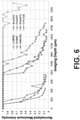
- FIG. 6 shows a plot of the maximum pixel value of 262,144 pixels at different imaging depths for each type of cleared sample. As shown in both FIGS. 5 and 6 , LIMPID provides the best imaging depth.
- FIGS. 7 and 8 Experiments showing the images obtained with LIMPID cleared samples are shown in FIGS. 7 and 8 .
- a sample stage 35 quail embryo heart
- fluorescent conjugated antibody can be imaged using fluorescent confocal microscopy after LIMPID clearing.
- LIMPID preserved the fluorescent probe and antibody binding through the optical clearing process.
- a sample adult mouse brain slice
- a lipophilic fluorescent dye DiO
- a lipophilic fluorescent dye can bond and diffuse through phospholipid bilayers in biological structures. LIMPID can preserve the phospholipid bilayer during the optical clearing process and enables successful labeling and imaging of the related structures.
Landscapes
- Health & Medical Sciences (AREA)
- Life Sciences & Earth Sciences (AREA)
- Engineering & Computer Science (AREA)
- Biomedical Technology (AREA)
- Immunology (AREA)
- Chemical & Material Sciences (AREA)
- Molecular Biology (AREA)
- Analytical Chemistry (AREA)
- Physics & Mathematics (AREA)
- Pathology (AREA)
- General Physics & Mathematics (AREA)
- General Health & Medical Sciences (AREA)
- Biochemistry (AREA)
- Hematology (AREA)
- Urology & Nephrology (AREA)
- Cell Biology (AREA)
- Toxicology (AREA)
- Medicinal Chemistry (AREA)
- Food Science & Technology (AREA)
- Bioinformatics & Cheminformatics (AREA)
- Tropical Medicine & Parasitology (AREA)
- Microbiology (AREA)
- Biotechnology (AREA)
- Investigating Or Analysing Biological Materials (AREA)
- Investigating, Analyzing Materials By Fluorescence Or Luminescence (AREA)
- Sampling And Sample Adjustment (AREA)
Description
- The present disclosure relates generally to optical clearing and, more specifically, to an optical clearing solution and an optical clearing method called lipid-preserving index matching for prolonged imaging depth (LIMPID).
- Imaging techniques like three-dimensional (3-D) microscopy and optical coherence tomography often suffer from restricted imaging depth in biological tissue, which appears opaque. The opacity of the biological tissue is due to light scattering on interfaces between lipids and other tissue compounds with different refractive indices. Optical clearing is a process that makes biological tissues more optically transparent, while keeping internal structure intact. Several optical clearing techniques have been developed to reduce such scattering and, thereby, increase imaging depth, enabling imaging of large regions of interest. These optical clearing techniques include organic solvent-based clearing techniques, lipid extraction after hydrogel embedding techniques, and aqueous solution based techniques. These optical clearing techniques suffer from one or more limitations, including requiring a long time, requiring fancy instruments, suffering from limited dye compatibility, and requiring extra work.
- In an aspect, the present invention relates to an optical clearing method called lipid-preserving index matching for prolonged imaging depth (LIMPID). The method of the present invention is defined in claim 8. The optical clearing solution of the present invention includes water, iodixanol, a polyhydric alcohol and urea. The optical clearing solution of the present invention is defined in claim 1. Iodixanol and urea work together in a synergistic manner to strengthen the clearing capability of the optical clearing solution. The optical clearing solution has an adjustable refractive index and osmolality, which improves the image quality by matching the refractive index to optical hardware and eliminating tissue distortions.
- The foregoing and other features of the present invention will become apparent to those skilled in the art upon reading the following description with reference to the accompanying drawings, in which:
-
FIG. 1 is a diagram showing a system that can be used to perform an optical clearing method called lipid-preserving index matching for prolonged imaging depth (LIMPID) in accordance with an aspect of the present disclosure; -
FIG. 2 is a diagram showing an example configuration of the preparation / processing unit inFIG. 1 ; -
FIG. 3 is a process flow diagram illustrating an optical clearing method called LIMPID according to another aspect of the present disclosure; -
FIG. 4 shows examples of LIMPID clearing in different samples; -
FIG. 5 shows a maximum intensity projection of 512 slices (1160 µm width × 3560 µm depth) of same age quail brains treated with different clearing agents; -
FIG. 6 shows a plot of the maximum pixel intensity at different imaging depths within the 512 slices (1160 µm width × 3560 µm depth) of same age quail brains treated with different clearing agents; -
FIG. 7 shows that a stage 35 quail embryo heart previously stained with a fluorescent conjugated antibody can be imaged using fluorescent confocal microscopy after LIMPID clearing; and -
FIG. 8 shows that an adult male brain slice stained with lipophilic fluorescent dye can be imaged using fluorescent confocal microscopy after LIMPID clearing. - Unless otherwise defined, all technical terms used herein have the same meaning as commonly understood by one of ordinary skill in the art to which the present disclosure pertains.
- As used herein, the singular forms "a," "an" and "the" can also include the plural forms, unless the context clearly indicates otherwise.
- As used herein, the terms "comprises" and/or "comprising," can specify the presence of stated features, steps, operations, elements, and/or components, but do not preclude the presence or addition of one or more other features, steps, operations, elements, components, and/or groups.
- As used herein, the term "and/or" can include any and all combinations of one or more of the associated listed items.
- As used herein, the terms "first," "second," etc. should not limit the elements being described by these terms. These terms are only used to distinguish one element from another. Thus, a "first" element discussed below could also be termed a "second" element without departing from the teachings of the present disclosure. The sequence of operations (or acts/steps) is not limited to the order presented in the claims or figures unless specifically indicated otherwise.
- As used herein, the term "imaging" can refer to the act of creating a visual representation of an interior of at least a portion of a sample for analysis. When the imaging is referred to as being "three-dimensional (3D)", a volumetric visual representation of the interior of at least the portion of the sample is created. Example imaging modalities can include, but are not limited to, confocal microscopy, light-sheet microscopy, two-photon microscopy, optical coherence tomography, and the like.
- As used herein, the term "lipid-preserving index matching for prolonged imaging depth (LIMPID)" can refer to an optical clearing method employing a clearing agent that includes at least water; an iodine-containing, non-ionic radiocontrast agent; and urea at varying concentrations. The clearing agent can further include a refractive index adjustment agent, an osmotic pressure adjustment agent, and/or a detergent.
- As used herein, the terms "optical clearing" and "clearing" can refer to the act of minimizing light scatter from a sample by exposing the sample to a clearing agent.
- As used herein, the term "sample" can refer to a biological specimen taken from a subject. For example, the sample can be a tissue sample excised from a subject.
- As used herein, the term "weight percent" refers to a number of grams of solute in 100 mL of solution.
- As used herein, the term "subject" can refer to any warm-blooded organism including, but not limited to, a human being, a pig, a rat, a mouse, a bird, a dog, a cat, a goat, a sheep, a horse, a monkey, an ape, a rabbit, a cow, etc.
- The present invention includes an optical clearing method called lipid-preserving index matching for prolonged imaging depth (LIMPID - a synonym for clear). Clearing a tissue sample with LIMPID can increase imaging depth when imaging with confocal microscopy, light-sheet microscopy, two-photon microscopy, optical coherence tomography, or the like, allowing investigation of the three-dimensional (3-D) organization of many complex structures. A significant benefit of LIMPID over other clearing methods is it simplicity: the clearing solution is easy to make and the clearing process only requires a single step (immersion of the sample, which can be labeled or preprocessed, in the clearing agent before imaging). LIMPID allows for speedy clearing that is compatible with many different instruments and sample preparation techniques, and increases both the imaging depth and the signal to noise ratio (SNR).
- The LIMPID clearing agent includes water, an iodine-containing, non-ionic radiocontrast agent, and urea. Water is the solvent for the other ingredients. However, the LIMPID clearing agent has a higher clearing capability than most existing water-soluble optical clearing agents. The iodine-containing, non-ionic radiocontrast agent (e.g., iodixanol, which can be in solution with a polyhydric alcohol) can be used for index matching and reducing scattering from lipids within the sample. Urea is a protein denaturation and super-hydration agent that reduces scattering from proteins and aids in antigen retrieval. Use of the iodine-containing, non-ionic radiocontrast agent together with the urea does not damage lipid content or reduce the speed of diffusion, but instead produces a synergistic effect on speed and clearing capability. The LIMPID clearing agent can also include a refractive index adjustment agent and/or an osmotic pressure adjustment agent, which can help to improve image quality. In some instances, the LIMPID clearing agent can also include a detergent to help permeabilization of cells within the sample.
- One aspect of the present disclosure can include a system 10 (
FIG. 1 ) that can be used to perform an optical clearing method called lipid-preserving index matching for prolonged imaging depth (LIMPID). A significant benefit of LIMPID over other clearing methods is its simplicity. The clearing solution is easy to make and the clearing process only requires one step (immersion of the labeled sample in the clearing agent before imaging). - The
system 10 includes a preparation /processing unit 12 and animaging unit 14. One or more of the preparation /processing unit 12 and theimaging unit 14 can perform computer-aided actions, which are stored in one or more non-transitory memory units and executed by one or more processors. In some instances, the preparation /processing unit 12 and theimaging unit 14 unique entitles (e.g., located at different places). However, in other instances, functionalities of one or more of the preparation /processing unit 12 and theimaging unit 14 can be combined. - The preparation /
processing unit 12 can perform optical clearing on a prepared sample using LIMPID. LIMPID provides a simple and cost effective way to evaluate properties (e.g., morphology, 3D organization, etc.) of a sample. Theimaging unit 14 can record images of the optically cleared prepared sample that can be used in the evaluation of the sample. Theimaging unit 14 can include hardware for an imaging modality, like microscopy (e.g., confocal microscopy, light-sheet microscopy, two-photon microscopy, etc.), optical coherence tomography (OCT), or any other imaging modality that would benefit from the increased image depth achieved through optical clearing. - As shown in
FIG. 2 , the preparation /processing unit 12 can include anoptical clearing unit 24 that can apply the LIMPID clearing agent to the sample. As an example, the sample can be immersed in a solution of the LIMPID clearing agent. The sample can be immersed for a time sufficient for the clearing to occur in the sample (the time is variable based on the size and depth of the sample) before the sample is imaged. In some instances, the preparation /processing unit 12 can include a sample preparation unit 22 that can prepare the sample for clearing and/or imaging. For example, the sample preparation unit 22 can facilitate the performance of labeling, staining, adding contrast agent, or any other pre-imaging task necessary for clearing and/or imaging. - The clearing agent solution includes at least water, an iodine-containing, non-ionic radiocontrast agent, and urea. The water is a solvent for all other ingredients. The iodine-containing, non-ionic radiocontrast agent and the urea, when used together, can provide a synergistic effect of decreasing the speed required to clear the sample and increasing the clearing capability (e.g., increasing the depth of the sample that is cleared in the time).
- The iodine-containing, non-ionic radiocontrast agent is added to the water to reduce scattering from lipids, as well as provides index matching. According to the present invention, the iodine-containing, non-ionic radiocontrast agent includes 40 to 80 weight percent iodixanol in solution with 0 to 20 weight percent polyhydric alcohol (such as glycerol, poly(ethylene glycol), triethanolamine, and quadrol (N,N,N', N-Tetrakis(2-Hydroxypropyl)ethylenediamine). In some instances, the iodine-containing, non-ionic radiocontrast agent can include 50 to 70 weight percent iodixanol. In other instances, the iodine-containing, non-ionic radiocontrast agent can include 57 to 64 weight percent iodixanol. In further instances, the iodine-containing, non-ionic radiocontrast agent can include about 60 weight percent iodixanol.
- The urea is added to the water as a protein denaturation and super-hydration agent that reduces scattering from proteins. According to the present invention the urea is added to the water at a concentration of 10 to 50 weight percent. In some instances, the urea can be added to the water at a concentration of 25 to 45 weight percent. In other instances, the urea can be added to the water at a concentration of 30 to 50 weight percent. In still other instances, the urea can be added to the water at a concentration of 35 to 45 weight percent.
- In some instances, the clearing agent solution can have an adjustable refractive index and/or an adjustable osmolality to match the refractive index of optical hardware and/or intrinsic osmotic pressure of the tissue to eliminate image and/or tissue distortions. The refractive index adjustment agent and/or the osmotic pressure adjustment agent comprises at least one of iohexol, thiodiglycol, glycerol, poly(ethylene glycol), triethanolamine, and quadrol (N,N,N',N-Tetrakis(2-Hydroxypropyl)ethylenediamine. The adjustable refractive index and/or the adjustable osmolality can be provided by a refractive index adjustment agent and/or an osmotic pressure adjustment agent. Each of the refractive index adjustment agent and the osmotic pressure adjustment agent can be added to the water at a concentration of 0 to 20 weight percent. In some instances, each of the refractive index adjustment agent and the osmotic pressure adjustment agent can be added to the water at a concentration of 0 to 10 weight percent. In other instances, each of the refractive index adjustment agent and the osmotic pressure adjustment agent can be added to the water at a concentration of 0 to 5 weight percent. In further instances, each of the refractive index adjustment agent and the osmotic pressure adjustment agent can be added to the water at a concentration of 10 to 20 weight percent. In still further instances, each of the refractive index adjustment agent and the osmotic pressure adjustment agent can be added to the water at a concentration of 15 to 20 weight percent.
- The clearing agent may also include a detergent to help permeabilization of cells in the sample. Examples of the detergent can include triton X-100, digitonin, saponin, or the like. The detergent can have a concentration of between 0 and 2 weight percent. However, in some instances, the detergent can have a concentration between 0 and 0.5 weight percent. In other instances, the detergent can have a concentration between 0.5 and 2 weight percent. In still other instances, the detergent can have a concentration between 0.25 and 0.75 weight percent.
- Referring again to
FIG. 1 , theimaging unit 14 can record raw data corresponding to one or more images of the sample after the sample emerges from the preparation / processing unit. The tissue sample can remain immersed in the clearing agent during imaging; however, in some instances, the tissue sample can be removed from the clearing agent before imaging. Theimaging unit 14 can also be linked to an image processing unit to transfer the raw data for analysis. The image processing unit can process the raw data for analysis and/or display one or more images based on the raw data. - In one embodiment the method of the present invention includes the
optical clearing method 30 as described inFIG. 3 and called lipid-preserving index matching for prolonged imaging depth (LIMPID). Themethod 30 can be executed using at least a portion of thesystem 10 shown inFIGS. 1 and 2 and described above. - At
step 32, a sample can be immersed in an optical clearing agent. LIMPID uses a clearing agent that combines the use of an iodine-containing, non-ionic radiocontrast agent and urea to strengthen its clearing capability with both the iodine-containing, non-ionic radiocontrast agent and the urea working together to provide a strong synergistic effect on speed and clearing capability. Additionally, the clearing agent has an adjustable refractive index and osmolality, which improves the image quality by matching the refractive index to optical hardware to eliminate tissue distortions. Atstep 34, the sample can be imaged. In some instances, the sample can be within the clearing solution as it is imaged. - The following experiment shows an optical clearing method called lipid-preserving index matching for prolonged imaging depth (LIMPID). LIMPID provides simplicity, speed, instrument compatibility, and imaging performance, including imaging depth and signal to noise ratio. The following experimental results are shown for the purpose of illustration only and are not intended to limit the scope of the appended claims.
- As shown in
FIG. 4 , samples, including an E9 quail body, an E6 quail body, an E9 quail brain and an E9 quail heart, were labeled, as shown in the "before clearing" row of images. The samples were placed in the LIMPID clearing agent (including at least water, an iodine-containing, non-ionic radiocontrast agent, and urea) and cleared overnight. The after clearing row of images show the effect of the clearing agent on the samples. LIMPID is a simple procedure, that works at a high speed and high transparency with a wide range refractive index, flexible imaging setup, minimum toxicity, mild to the lipid bilayer, with low size changes, low viscosity, and low cost. -
FIGS. 5 and6 show a comparison between different clearing methods - LIMPID, ScaleA2, CUBIC, ClearT2, SeeDB2, ScaleSQ, and 47% TDE. With all clearing agents, Tg(tie1: H2B-eYFP) quail brains at the same embryo stage (E9) were processed with clearing protocols according to the corresponding original published journal articles. The cleared samples were imaged with the same inverted fluorescent confocal microscope. For all confocal imaging tasks, the same optical setup (10x air objective), the same laser power (-20 mW), the same detector gain (100%) and the same software configuration (e.g., line average, accumulation, etc.) were used. Volumetric image datasets were taken for each cleared sample. Starting by focusing at the outermost surface of the sample, a 512x512 pixel (1160x1160 mm) 2D confocal scan was taken at the microscope objective focal plane. Then, the focal plane was moved 75 mm deeper into the sample repeatedly and a 512x512 pixel (1160x1160 mm) 2D confocal scan was taken after each movement until the maximum pixel value on the confocal scan dropped below the noise flour. InFIG. 5 , for each type of cleared sample, a volumetric image dataset was rendered in maximum intensity projection (at the XZ plane) for visual comparison. Each pixel on each panel represents the maximum pixel value (normalized from 0 to 1) at the corresponding pixel location from the 512 XZ images from each volumetric dataset.FIG. 6 shows a plot of the maximum pixel value of 262,144 pixels at different imaging depths for each type of cleared sample. As shown in bothFIGS. 5 and6 , LIMPID provides the best imaging depth. - Experiments showing the images obtained with LIMPID cleared samples are shown in
FIGS. 7 and8 . As shown inFIG. 7 , a sample (stage 35 quail embryo heart) previously being stained with fluorescent conjugated antibody can be imaged using fluorescent confocal microscopy after LIMPID clearing. LIMPID preserved the fluorescent probe and antibody binding through the optical clearing process. As shown inFIG. 8 , a sample (adult mouse brain slice) stained with a lipophilic fluorescent dye (DiO) can be imaged using fluorescent confocal microscopy after LIMPID clearing. A lipophilic fluorescent dye can bond and diffuse through phospholipid bilayers in biological structures. LIMPID can preserve the phospholipid bilayer during the optical clearing process and enables successful labeling and imaging of the related structures. - From the above description, those skilled in the art will perceive improvements, changes and modifications. Such improvements, changes and modifications are within the skill of one in the art and are intended to be covered by the appended claims.
Claims (13)
- An optical clearing solution comprising:water;an iodine-containing, non-ionic radiocontrast agent comprising 40 to 80 weight percent iodixanol in solution with 0 to 20 weight percent polyhydric alcohol; andurea, wherein the urea has a concentration of 10 to 50 weight percent.
- The optical clearing solution of claim 1, wherein the polyhydric alcohol comprises at least one of glycerol, poly(ethylene glycol), triethanolamine, and quadrol (N,N,N',N-Tetrakis(2-Hydroxypropyl)ethylenediamine.
- The optical clearing solution of claim 1, further comprising 0 to 20 weight percent of a refractive index adjustment agent and/or an osmotic pressure adjustment agent.
- The optical clearing solution of claim 3, wherein the refractive index adjustment agent and/or the osmotic pressure adjustment agent comprises at least one of iohexol, thiodiglycol, glycerol, poly(ethylene glycol), triethanolamine, and quadrol N,N,N',N-Tetrakis(2-Hydroxypropyl)ethylenediamine.
- The optical clearing solution of claim 1, wherein a refractive index of the optical clearing solution matches a refractive index of optical hardware to eliminate tissue distortion.
- The optical clearing solution of claim 1, further comprising 0 to 1 weight percent of a detergent to help permeabilization of cells in a tissue.
- The optical clearing solution of claim 6, wherein the detergent is triton X-100, digitonin or saponin.
- A method comprising:
immersing the labeled sample in an optical clearing solution before imaging, wherein the optical clearing solution comprises:water;an iodine-containing non-ionic radiocontrast agent comprising 40 to 80 weight percent iodixanol in solution with 0 to 20 weight percent polyhydric alcohol; andurea, wherein the urea has a concentration of 10 to 50 weight percent. - The method of claim 8, wherein the imaging is fluorescent microscopy, confocal microscopy, light-sheet microscopy, two-photon microscopy, structured illumination microscopy, light-field microscopy, or optical coherence tomography.
- The method of claim 8, wherein the polyhydric alcohol comprises at least one of glycerol, poly(ethylene glycol), triethanolamine, and quadrol N,N,N',N-Tetrakis(2-Hydroxypropyl)ethylenediamine.
- The method of claim 8, wherein the optical clearing solution further comprises 0 to 20 weight percent refractive index adjustment agent and/or an osmotic pressure adjustment agent.
- The method of claim 8, wherein the optical clearing solution matches a refractive index of optical hardware of an imaging modality to eliminate optical distortion.
- The method of claim 8, wherein optical clearing solution further comprises 0 to 1 weight percent of a detergent to help permeabilization of cells in a tissue.
Applications Claiming Priority (2)
| Application Number | Priority Date | Filing Date | Title |
|---|---|---|---|
| US201762583649P | 2017-11-09 | 2017-11-09 | |
| PCT/US2018/060078 WO2019094753A1 (en) | 2017-11-09 | 2018-11-09 | Lipid-preserving refractive index matching for prolonged imaging depth for transparent tissue sample and composition |
Publications (2)
| Publication Number | Publication Date |
|---|---|
| EP3707492A1 EP3707492A1 (en) | 2020-09-16 |
| EP3707492B1 true EP3707492B1 (en) | 2022-02-02 |
Family
ID=64453646
Family Applications (1)
| Application Number | Title | Priority Date | Filing Date |
|---|---|---|---|
| EP18807834.9A Active EP3707492B1 (en) | 2017-11-09 | 2018-11-09 | Lipid-preserving refractive index matching for prolonged imaging depth for tranparent tissue sample and composition |
Country Status (5)
| Country | Link |
|---|---|
| US (1) | US12181386B2 (en) |
| EP (1) | EP3707492B1 (en) |
| AU (1) | AU2018364670B2 (en) |
| CA (1) | CA3080149C (en) |
| WO (1) | WO2019094753A1 (en) |
Families Citing this family (3)
| Publication number | Priority date | Publication date | Assignee | Title |
|---|---|---|---|---|
| WO2021023289A1 (en) * | 2019-08-07 | 2021-02-11 | JelloX Biotech Inc. | Composition and method for rendering biological material |
| CN110579442B (en) * | 2019-08-26 | 2021-02-12 | 华中科技大学 | Refractive index matching method adaptive to microfluidic-optical sheet imaging and application thereof |
| PL247511B1 (en) * | 2022-12-14 | 2025-07-21 | Univ Warszawski Medyczny | A solution for optically cleansing and expanding tissues, its use and a method for cleansing and expanding tissues. |
Family Cites Families (9)
| Publication number | Priority date | Publication date | Assignee | Title |
|---|---|---|---|---|
| US6472216B1 (en) | 2001-07-24 | 2002-10-29 | Ann-Shyn Chiang | Aqueous tissue clearing solution |
| CN108061677B (en) | 2010-03-12 | 2021-10-29 | 国立研究开发法人理化学研究所 | Clarifying reagents for biological materials and their uses |
| CN103492852B (en) | 2011-04-28 | 2016-08-17 | 独立行政法人理化学研究所 | The transparence method of biomaterial and utilization thereof |
| EP2711682B1 (en) | 2011-05-20 | 2019-07-10 | Riken | Clarifying reagent for biological materials and use thereof |
| WO2014115206A1 (en) | 2013-01-28 | 2014-07-31 | 独立行政法人科学技術振興機構 | Method for rendering tissue transparent, reagent for rendering tissue transparent, and tissue observation method |
| US10794802B2 (en) | 2013-09-20 | 2020-10-06 | California Institute Of Technology | Whole-body tissue stabilization and selective extractions via tissue-hydrogel hybrids for high resolution intact circuit mapping and phenotyping |
| WO2015041755A1 (en) * | 2013-09-20 | 2015-03-26 | California Institute Of Technology | Methods for phenotyping of intact whole tissues |
| US20180031452A1 (en) | 2015-03-18 | 2018-02-01 | Riken | Method for observing biological material and clearing method |
| US11130931B2 (en) * | 2016-12-22 | 2021-09-28 | The University Of Hong Kong | Compositions and methods for clearing tissue |
-
2018
- 2018-11-09 EP EP18807834.9A patent/EP3707492B1/en active Active
- 2018-11-09 US US16/760,652 patent/US12181386B2/en active Active
- 2018-11-09 CA CA3080149A patent/CA3080149C/en active Active
- 2018-11-09 WO PCT/US2018/060078 patent/WO2019094753A1/en not_active Ceased
- 2018-11-09 AU AU2018364670A patent/AU2018364670B2/en active Active
Non-Patent Citations (1)
| Title |
|---|
| None * |
Also Published As
| Publication number | Publication date |
|---|---|
| CA3080149A1 (en) | 2019-05-16 |
| AU2018364670B2 (en) | 2020-06-11 |
| EP3707492A1 (en) | 2020-09-16 |
| CA3080149C (en) | 2021-07-06 |
| AU2018364670A1 (en) | 2020-05-28 |
| US12181386B2 (en) | 2024-12-31 |
| WO2019094753A1 (en) | 2019-05-16 |
| US20210372890A1 (en) | 2021-12-02 |
Similar Documents
| Publication | Publication Date | Title |
|---|---|---|
| US20240142352A1 (en) | Biological-specimen transparentizing agent, system, and use therefor | |
| Kolesová et al. | Comparison of different tissue clearing methods and 3D imaging techniques for visualization of GFP-expressing mouse embryos and embryonic hearts | |
| Pawlina et al. | Histology: a text and atlas: with correlated cell and molecular biology | |
| Treweek et al. | Whole-body tissue stabilization and selective extractions via tissue-hydrogel hybrids for high-resolution intact circuit mapping and phenotyping | |
| Fereidouni et al. | Microscopy with ultraviolet surface excitation for rapid slide-free histology | |
| US20170199104A1 (en) | Whole-body tissue stabilization and selective extractions via tissue-hydrogel hybrids for high resolution intact circuit mapping and phenotyping | |
| TW202102832A (en) | Method for analyzing tissue specimens | |
| Gómez-Gaviro et al. | Optimized CUBIC protocol for three-dimensional imaging of chicken embryos at single-cell resolution | |
| Gonzalez-Bellido et al. | Labeling and confocal imaging of neurons in thick invertebrate tissue samples | |
| CA3080149C (en) | Lipid-preserving index matching for prolonged imaging depth (limpid) | |
| Singh et al. | Quantifying three-dimensional rodent retina vascular development using optical tissue clearing and light-sheet microscopy | |
| US20170108414A1 (en) | High-resolution three-dimensional imaging of mammalian hearts | |
| Ding et al. | Light-sheet fluorescence microscopy for the study of the murine heart | |
| Ishii et al. | Correlative microscopy and block-face imaging (CoMBI) method for both paraffin-embedded and frozen specimens | |
| Konno et al. | Pathological application of carbocyanine dye-based multicolour imaging of vasculature and associated structures | |
| Garcia-Vasquez et al. | Development of a light microscopy stain for the sclerites of Gyrodactylus von Nordmann, 1832 (Monogenea) and related genera | |
| Walsh et al. | Multifluorescence high‐resolution episcopic microscopy for 3D imaging of adult murine organs | |
| CN114096826A (en) | Clarification compositions, kits and methods for transparentizing and further staining biological materials | |
| CN114402060B (en) | Compositions and methods for tissue clearing | |
| Parfitt | Immunofluorescence tomography: high-resolution 3-D reconstruction by serial-sectioning of methacrylate embedded tissues and alignment of 2-D immunofluorescence images | |
| Zhao et al. | Cellular and molecular probing of intact transparent human organs | |
| TWI761016B (en) | Method for preparation of tissue sections | |
| Nischik et al. | Evaluation of standard imaging techniques and volumetric preservation of nervous tissue in genetically identical offspring of the crayfish Procambarus fallax cf. virginalis (Marmorkrebs) | |
| Savio et al. | A framework for analyzing confocal images of transversal tubules in cardiomyocytes | |
| US12152969B2 (en) | Method for preparation of tissue sections |
Legal Events
| Date | Code | Title | Description |
|---|---|---|---|
| STAA | Information on the status of an ep patent application or granted ep patent |
Free format text: STATUS: UNKNOWN |
|
| STAA | Information on the status of an ep patent application or granted ep patent |
Free format text: STATUS: THE INTERNATIONAL PUBLICATION HAS BEEN MADE |
|
| PUAI | Public reference made under article 153(3) epc to a published international application that has entered the european phase |
Free format text: ORIGINAL CODE: 0009012 |
|
| STAA | Information on the status of an ep patent application or granted ep patent |
Free format text: STATUS: REQUEST FOR EXAMINATION WAS MADE |
|
| 17P | Request for examination filed |
Effective date: 20200529 |
|
| AK | Designated contracting states |
Kind code of ref document: A1 Designated state(s): AL AT BE BG CH CY CZ DE DK EE ES FI FR GB GR HR HU IE IS IT LI LT LU LV MC MK MT NL NO PL PT RO RS SE SI SK SM TR |
|
| AX | Request for extension of the european patent |
Extension state: BA ME |
|
| GRAP | Despatch of communication of intention to grant a patent |
Free format text: ORIGINAL CODE: EPIDOSNIGR1 |
|
| STAA | Information on the status of an ep patent application or granted ep patent |
Free format text: STATUS: GRANT OF PATENT IS INTENDED |
|
| DAV | Request for validation of the european patent (deleted) | ||
| DAX | Request for extension of the european patent (deleted) | ||
| INTG | Intention to grant announced |
Effective date: 20201203 |
|
| GRAJ | Information related to disapproval of communication of intention to grant by the applicant or resumption of examination proceedings by the epo deleted |
Free format text: ORIGINAL CODE: EPIDOSDIGR1 |
|
| STAA | Information on the status of an ep patent application or granted ep patent |
Free format text: STATUS: REQUEST FOR EXAMINATION WAS MADE |
|
| INTC | Intention to grant announced (deleted) | ||
| GRAS | Grant fee paid |
Free format text: ORIGINAL CODE: EPIDOSNIGR3 |
|
| STAA | Information on the status of an ep patent application or granted ep patent |
Free format text: STATUS: GRANT OF PATENT IS INTENDED |
|
| GRAP | Despatch of communication of intention to grant a patent |
Free format text: ORIGINAL CODE: EPIDOSNIGR1 |
|
| INTG | Intention to grant announced |
Effective date: 20210621 |
|
| GRAA | (expected) grant |
Free format text: ORIGINAL CODE: 0009210 |
|
| STAA | Information on the status of an ep patent application or granted ep patent |
Free format text: STATUS: THE PATENT HAS BEEN GRANTED |
|
| AK | Designated contracting states |
Kind code of ref document: B1 Designated state(s): AL AT BE BG CH CY CZ DE DK EE ES FI FR GB GR HR HU IE IS IT LI LT LU LV MC MK MT NL NO PL PT RO RS SE SI SK SM TR |
|
| REG | Reference to a national code |
Ref country code: GB Ref legal event code: FG4D |
|
| REG | Reference to a national code |
Ref country code: CH Ref legal event code: EP Ref country code: AT Ref legal event code: REF Ref document number: 1466707 Country of ref document: AT Kind code of ref document: T Effective date: 20220215 |
|
| REG | Reference to a national code |
Ref country code: DE Ref legal event code: R096 Ref document number: 602018030398 Country of ref document: DE |
|
| REG | Reference to a national code |
Ref country code: IE Ref legal event code: FG4D |
|
| REG | Reference to a national code |
Ref country code: LT Ref legal event code: MG9D |
|
| REG | Reference to a national code |
Ref country code: NL Ref legal event code: MP Effective date: 20220202 |
|
| REG | Reference to a national code |
Ref country code: AT Ref legal event code: MK05 Ref document number: 1466707 Country of ref document: AT Kind code of ref document: T Effective date: 20220202 |
|
| PG25 | Lapsed in a contracting state [announced via postgrant information from national office to epo] |
Ref country code: SE Free format text: LAPSE BECAUSE OF FAILURE TO SUBMIT A TRANSLATION OF THE DESCRIPTION OR TO PAY THE FEE WITHIN THE PRESCRIBED TIME-LIMIT Effective date: 20220202 Ref country code: RS Free format text: LAPSE BECAUSE OF FAILURE TO SUBMIT A TRANSLATION OF THE DESCRIPTION OR TO PAY THE FEE WITHIN THE PRESCRIBED TIME-LIMIT Effective date: 20220202 Ref country code: PT Free format text: LAPSE BECAUSE OF FAILURE TO SUBMIT A TRANSLATION OF THE DESCRIPTION OR TO PAY THE FEE WITHIN THE PRESCRIBED TIME-LIMIT Effective date: 20220602 Ref country code: NO Free format text: LAPSE BECAUSE OF FAILURE TO SUBMIT A TRANSLATION OF THE DESCRIPTION OR TO PAY THE FEE WITHIN THE PRESCRIBED TIME-LIMIT Effective date: 20220502 Ref country code: NL Free format text: LAPSE BECAUSE OF FAILURE TO SUBMIT A TRANSLATION OF THE DESCRIPTION OR TO PAY THE FEE WITHIN THE PRESCRIBED TIME-LIMIT Effective date: 20220202 Ref country code: LT Free format text: LAPSE BECAUSE OF FAILURE TO SUBMIT A TRANSLATION OF THE DESCRIPTION OR TO PAY THE FEE WITHIN THE PRESCRIBED TIME-LIMIT Effective date: 20220202 Ref country code: HR Free format text: LAPSE BECAUSE OF FAILURE TO SUBMIT A TRANSLATION OF THE DESCRIPTION OR TO PAY THE FEE WITHIN THE PRESCRIBED TIME-LIMIT Effective date: 20220202 Ref country code: ES Free format text: LAPSE BECAUSE OF FAILURE TO SUBMIT A TRANSLATION OF THE DESCRIPTION OR TO PAY THE FEE WITHIN THE PRESCRIBED TIME-LIMIT Effective date: 20220202 Ref country code: BG Free format text: LAPSE BECAUSE OF FAILURE TO SUBMIT A TRANSLATION OF THE DESCRIPTION OR TO PAY THE FEE WITHIN THE PRESCRIBED TIME-LIMIT Effective date: 20220502 |
|
| PG25 | Lapsed in a contracting state [announced via postgrant information from national office to epo] |
Ref country code: PL Free format text: LAPSE BECAUSE OF FAILURE TO SUBMIT A TRANSLATION OF THE DESCRIPTION OR TO PAY THE FEE WITHIN THE PRESCRIBED TIME-LIMIT Effective date: 20220202 Ref country code: LV Free format text: LAPSE BECAUSE OF FAILURE TO SUBMIT A TRANSLATION OF THE DESCRIPTION OR TO PAY THE FEE WITHIN THE PRESCRIBED TIME-LIMIT Effective date: 20220202 Ref country code: GR Free format text: LAPSE BECAUSE OF FAILURE TO SUBMIT A TRANSLATION OF THE DESCRIPTION OR TO PAY THE FEE WITHIN THE PRESCRIBED TIME-LIMIT Effective date: 20220503 Ref country code: FI Free format text: LAPSE BECAUSE OF FAILURE TO SUBMIT A TRANSLATION OF THE DESCRIPTION OR TO PAY THE FEE WITHIN THE PRESCRIBED TIME-LIMIT Effective date: 20220202 Ref country code: AT Free format text: LAPSE BECAUSE OF FAILURE TO SUBMIT A TRANSLATION OF THE DESCRIPTION OR TO PAY THE FEE WITHIN THE PRESCRIBED TIME-LIMIT Effective date: 20220202 |
|
| PG25 | Lapsed in a contracting state [announced via postgrant information from national office to epo] |
Ref country code: IS Free format text: LAPSE BECAUSE OF FAILURE TO SUBMIT A TRANSLATION OF THE DESCRIPTION OR TO PAY THE FEE WITHIN THE PRESCRIBED TIME-LIMIT Effective date: 20220602 |
|
| PG25 | Lapsed in a contracting state [announced via postgrant information from national office to epo] |
Ref country code: SM Free format text: LAPSE BECAUSE OF FAILURE TO SUBMIT A TRANSLATION OF THE DESCRIPTION OR TO PAY THE FEE WITHIN THE PRESCRIBED TIME-LIMIT Effective date: 20220202 Ref country code: SK Free format text: LAPSE BECAUSE OF FAILURE TO SUBMIT A TRANSLATION OF THE DESCRIPTION OR TO PAY THE FEE WITHIN THE PRESCRIBED TIME-LIMIT Effective date: 20220202 Ref country code: RO Free format text: LAPSE BECAUSE OF FAILURE TO SUBMIT A TRANSLATION OF THE DESCRIPTION OR TO PAY THE FEE WITHIN THE PRESCRIBED TIME-LIMIT Effective date: 20220202 Ref country code: EE Free format text: LAPSE BECAUSE OF FAILURE TO SUBMIT A TRANSLATION OF THE DESCRIPTION OR TO PAY THE FEE WITHIN THE PRESCRIBED TIME-LIMIT Effective date: 20220202 Ref country code: DK Free format text: LAPSE BECAUSE OF FAILURE TO SUBMIT A TRANSLATION OF THE DESCRIPTION OR TO PAY THE FEE WITHIN THE PRESCRIBED TIME-LIMIT Effective date: 20220202 Ref country code: CZ Free format text: LAPSE BECAUSE OF FAILURE TO SUBMIT A TRANSLATION OF THE DESCRIPTION OR TO PAY THE FEE WITHIN THE PRESCRIBED TIME-LIMIT Effective date: 20220202 |
|
| REG | Reference to a national code |
Ref country code: DE Ref legal event code: R097 Ref document number: 602018030398 Country of ref document: DE |
|
| PG25 | Lapsed in a contracting state [announced via postgrant information from national office to epo] |
Ref country code: AL Free format text: LAPSE BECAUSE OF FAILURE TO SUBMIT A TRANSLATION OF THE DESCRIPTION OR TO PAY THE FEE WITHIN THE PRESCRIBED TIME-LIMIT Effective date: 20220202 |
|
| PLBE | No opposition filed within time limit |
Free format text: ORIGINAL CODE: 0009261 |
|
| STAA | Information on the status of an ep patent application or granted ep patent |
Free format text: STATUS: NO OPPOSITION FILED WITHIN TIME LIMIT |
|
| 26N | No opposition filed |
Effective date: 20221103 |
|
| PG25 | Lapsed in a contracting state [announced via postgrant information from national office to epo] |
Ref country code: SI Free format text: LAPSE BECAUSE OF FAILURE TO SUBMIT A TRANSLATION OF THE DESCRIPTION OR TO PAY THE FEE WITHIN THE PRESCRIBED TIME-LIMIT Effective date: 20220202 |
|
| PG25 | Lapsed in a contracting state [announced via postgrant information from national office to epo] |
Ref country code: MC Free format text: LAPSE BECAUSE OF FAILURE TO SUBMIT A TRANSLATION OF THE DESCRIPTION OR TO PAY THE FEE WITHIN THE PRESCRIBED TIME-LIMIT Effective date: 20220202 |
|
| REG | Reference to a national code |
Ref country code: CH Ref legal event code: PL |
|
| P01 | Opt-out of the competence of the unified patent court (upc) registered |
Effective date: 20230528 |
|
| REG | Reference to a national code |
Ref country code: BE Ref legal event code: MM Effective date: 20221130 |
|
| PG25 | Lapsed in a contracting state [announced via postgrant information from national office to epo] |
Ref country code: LI Free format text: LAPSE BECAUSE OF NON-PAYMENT OF DUE FEES Effective date: 20221130 Ref country code: IT Free format text: LAPSE BECAUSE OF FAILURE TO SUBMIT A TRANSLATION OF THE DESCRIPTION OR TO PAY THE FEE WITHIN THE PRESCRIBED TIME-LIMIT Effective date: 20220202 Ref country code: CH Free format text: LAPSE BECAUSE OF NON-PAYMENT OF DUE FEES Effective date: 20221130 |
|
| PG25 | Lapsed in a contracting state [announced via postgrant information from national office to epo] |
Ref country code: LU Free format text: LAPSE BECAUSE OF NON-PAYMENT OF DUE FEES Effective date: 20221109 |
|
| PG25 | Lapsed in a contracting state [announced via postgrant information from national office to epo] |
Ref country code: IE Free format text: LAPSE BECAUSE OF NON-PAYMENT OF DUE FEES Effective date: 20221109 |
|
| PG25 | Lapsed in a contracting state [announced via postgrant information from national office to epo] |
Ref country code: BE Free format text: LAPSE BECAUSE OF NON-PAYMENT OF DUE FEES Effective date: 20221130 |
|
| PG25 | Lapsed in a contracting state [announced via postgrant information from national office to epo] |
Ref country code: CY Free format text: LAPSE BECAUSE OF FAILURE TO SUBMIT A TRANSLATION OF THE DESCRIPTION OR TO PAY THE FEE WITHIN THE PRESCRIBED TIME-LIMIT Effective date: 20220202 |
|
| PG25 | Lapsed in a contracting state [announced via postgrant information from national office to epo] |
Ref country code: MK Free format text: LAPSE BECAUSE OF FAILURE TO SUBMIT A TRANSLATION OF THE DESCRIPTION OR TO PAY THE FEE WITHIN THE PRESCRIBED TIME-LIMIT Effective date: 20220202 Ref country code: HU Free format text: LAPSE BECAUSE OF FAILURE TO SUBMIT A TRANSLATION OF THE DESCRIPTION OR TO PAY THE FEE WITHIN THE PRESCRIBED TIME-LIMIT; INVALID AB INITIO Effective date: 20181109 |
|
| PG25 | Lapsed in a contracting state [announced via postgrant information from national office to epo] |
Ref country code: MT Free format text: LAPSE BECAUSE OF FAILURE TO SUBMIT A TRANSLATION OF THE DESCRIPTION OR TO PAY THE FEE WITHIN THE PRESCRIBED TIME-LIMIT Effective date: 20220202 |
|
| PGFP | Annual fee paid to national office [announced via postgrant information from national office to epo] |
Ref country code: DE Payment date: 20241127 Year of fee payment: 7 |
|
| PGFP | Annual fee paid to national office [announced via postgrant information from national office to epo] |
Ref country code: GB Payment date: 20241127 Year of fee payment: 7 |
|
| PGFP | Annual fee paid to national office [announced via postgrant information from national office to epo] |
Ref country code: FR Payment date: 20241128 Year of fee payment: 7 |
|
| PG25 | Lapsed in a contracting state [announced via postgrant information from national office to epo] |
Ref country code: TR Free format text: LAPSE BECAUSE OF FAILURE TO SUBMIT A TRANSLATION OF THE DESCRIPTION OR TO PAY THE FEE WITHIN THE PRESCRIBED TIME-LIMIT Effective date: 20220202 |