EP3705141A1 - Sheet-like decellularized material and artificial blood vessel employing said material - Google Patents
Sheet-like decellularized material and artificial blood vessel employing said material Download PDFInfo
- Publication number
- EP3705141A1 EP3705141A1 EP18872781.2A EP18872781A EP3705141A1 EP 3705141 A1 EP3705141 A1 EP 3705141A1 EP 18872781 A EP18872781 A EP 18872781A EP 3705141 A1 EP3705141 A1 EP 3705141A1
- Authority
- EP
- European Patent Office
- Prior art keywords
- blood vessel
- sheet
- artificial blood
- biomaterial
- decellularized
- Prior art date
- Legal status (The legal status is an assumption and is not a legal conclusion. Google has not performed a legal analysis and makes no representation as to the accuracy of the status listed.)
- Granted
Links
Images
Classifications
-
- A—HUMAN NECESSITIES
- A61—MEDICAL OR VETERINARY SCIENCE; HYGIENE
- A61L—METHODS OR APPARATUS FOR STERILISING MATERIALS OR OBJECTS IN GENERAL; DISINFECTION, STERILISATION OR DEODORISATION OF AIR; CHEMICAL ASPECTS OF BANDAGES, DRESSINGS, ABSORBENT PADS OR SURGICAL ARTICLES; MATERIALS FOR BANDAGES, DRESSINGS, ABSORBENT PADS OR SURGICAL ARTICLES
- A61L27/00—Materials for grafts or prostheses or for coating grafts or prostheses
- A61L27/36—Materials for grafts or prostheses or for coating grafts or prostheses containing ingredients of undetermined constitution or reaction products thereof, e.g. transplant tissue, natural bone, extracellular matrix
- A61L27/3604—Materials for grafts or prostheses or for coating grafts or prostheses containing ingredients of undetermined constitution or reaction products thereof, e.g. transplant tissue, natural bone, extracellular matrix characterised by the human or animal origin of the biological material, e.g. hair, fascia, fish scales, silk, shellac, pericardium, pleura, renal tissue, amniotic membrane, parenchymal tissue, fetal tissue, muscle tissue, fat tissue, enamel
-
- A—HUMAN NECESSITIES
- A61—MEDICAL OR VETERINARY SCIENCE; HYGIENE
- A61F—FILTERS IMPLANTABLE INTO BLOOD VESSELS; PROSTHESES; DEVICES PROVIDING PATENCY TO, OR PREVENTING COLLAPSING OF, TUBULAR STRUCTURES OF THE BODY, e.g. STENTS; ORTHOPAEDIC, NURSING OR CONTRACEPTIVE DEVICES; FOMENTATION; TREATMENT OR PROTECTION OF EYES OR EARS; BANDAGES, DRESSINGS OR ABSORBENT PADS; FIRST-AID KITS
- A61F2/00—Filters implantable into blood vessels; Prostheses, i.e. artificial substitutes or replacements for parts of the body; Appliances for connecting them with the body; Devices providing patency to, or preventing collapsing of, tubular structures of the body, e.g. stents
- A61F2/02—Prostheses implantable into the body
- A61F2/04—Hollow or tubular parts of organs, e.g. bladders, tracheae, bronchi or bile ducts
- A61F2/06—Blood vessels
-
- A—HUMAN NECESSITIES
- A61—MEDICAL OR VETERINARY SCIENCE; HYGIENE
- A61L—METHODS OR APPARATUS FOR STERILISING MATERIALS OR OBJECTS IN GENERAL; DISINFECTION, STERILISATION OR DEODORISATION OF AIR; CHEMICAL ASPECTS OF BANDAGES, DRESSINGS, ABSORBENT PADS OR SURGICAL ARTICLES; MATERIALS FOR BANDAGES, DRESSINGS, ABSORBENT PADS OR SURGICAL ARTICLES
- A61L27/00—Materials for grafts or prostheses or for coating grafts or prostheses
- A61L27/36—Materials for grafts or prostheses or for coating grafts or prostheses containing ingredients of undetermined constitution or reaction products thereof, e.g. transplant tissue, natural bone, extracellular matrix
- A61L27/3604—Materials for grafts or prostheses or for coating grafts or prostheses containing ingredients of undetermined constitution or reaction products thereof, e.g. transplant tissue, natural bone, extracellular matrix characterised by the human or animal origin of the biological material, e.g. hair, fascia, fish scales, silk, shellac, pericardium, pleura, renal tissue, amniotic membrane, parenchymal tissue, fetal tissue, muscle tissue, fat tissue, enamel
- A61L27/362—Skin, e.g. dermal papillae
-
- A—HUMAN NECESSITIES
- A61—MEDICAL OR VETERINARY SCIENCE; HYGIENE
- A61L—METHODS OR APPARATUS FOR STERILISING MATERIALS OR OBJECTS IN GENERAL; DISINFECTION, STERILISATION OR DEODORISATION OF AIR; CHEMICAL ASPECTS OF BANDAGES, DRESSINGS, ABSORBENT PADS OR SURGICAL ARTICLES; MATERIALS FOR BANDAGES, DRESSINGS, ABSORBENT PADS OR SURGICAL ARTICLES
- A61L27/00—Materials for grafts or prostheses or for coating grafts or prostheses
- A61L27/36—Materials for grafts or prostheses or for coating grafts or prostheses containing ingredients of undetermined constitution or reaction products thereof, e.g. transplant tissue, natural bone, extracellular matrix
- A61L27/3604—Materials for grafts or prostheses or for coating grafts or prostheses containing ingredients of undetermined constitution or reaction products thereof, e.g. transplant tissue, natural bone, extracellular matrix characterised by the human or animal origin of the biological material, e.g. hair, fascia, fish scales, silk, shellac, pericardium, pleura, renal tissue, amniotic membrane, parenchymal tissue, fetal tissue, muscle tissue, fat tissue, enamel
- A61L27/3625—Vascular tissue, e.g. heart valves
-
- A—HUMAN NECESSITIES
- A61—MEDICAL OR VETERINARY SCIENCE; HYGIENE
- A61L—METHODS OR APPARATUS FOR STERILISING MATERIALS OR OBJECTS IN GENERAL; DISINFECTION, STERILISATION OR DEODORISATION OF AIR; CHEMICAL ASPECTS OF BANDAGES, DRESSINGS, ABSORBENT PADS OR SURGICAL ARTICLES; MATERIALS FOR BANDAGES, DRESSINGS, ABSORBENT PADS OR SURGICAL ARTICLES
- A61L27/00—Materials for grafts or prostheses or for coating grafts or prostheses
- A61L27/36—Materials for grafts or prostheses or for coating grafts or prostheses containing ingredients of undetermined constitution or reaction products thereof, e.g. transplant tissue, natural bone, extracellular matrix
- A61L27/3683—Materials for grafts or prostheses or for coating grafts or prostheses containing ingredients of undetermined constitution or reaction products thereof, e.g. transplant tissue, natural bone, extracellular matrix subjected to a specific treatment prior to implantation, e.g. decellularising, demineralising, grinding, cellular disruption/non-collagenous protein removal, anti-calcification, crosslinking, supercritical fluid extraction, enzyme treatment
-
- A—HUMAN NECESSITIES
- A61—MEDICAL OR VETERINARY SCIENCE; HYGIENE
- A61L—METHODS OR APPARATUS FOR STERILISING MATERIALS OR OBJECTS IN GENERAL; DISINFECTION, STERILISATION OR DEODORISATION OF AIR; CHEMICAL ASPECTS OF BANDAGES, DRESSINGS, ABSORBENT PADS OR SURGICAL ARTICLES; MATERIALS FOR BANDAGES, DRESSINGS, ABSORBENT PADS OR SURGICAL ARTICLES
- A61L27/00—Materials for grafts or prostheses or for coating grafts or prostheses
- A61L27/36—Materials for grafts or prostheses or for coating grafts or prostheses containing ingredients of undetermined constitution or reaction products thereof, e.g. transplant tissue, natural bone, extracellular matrix
- A61L27/3683—Materials for grafts or prostheses or for coating grafts or prostheses containing ingredients of undetermined constitution or reaction products thereof, e.g. transplant tissue, natural bone, extracellular matrix subjected to a specific treatment prior to implantation, e.g. decellularising, demineralising, grinding, cellular disruption/non-collagenous protein removal, anti-calcification, crosslinking, supercritical fluid extraction, enzyme treatment
- A61L27/3691—Materials for grafts or prostheses or for coating grafts or prostheses containing ingredients of undetermined constitution or reaction products thereof, e.g. transplant tissue, natural bone, extracellular matrix subjected to a specific treatment prior to implantation, e.g. decellularising, demineralising, grinding, cellular disruption/non-collagenous protein removal, anti-calcification, crosslinking, supercritical fluid extraction, enzyme treatment characterised by physical conditions of the treatment, e.g. applying a compressive force to the composition, pressure cycles, ultrasonic/sonication or microwave treatment, lyophilisation
-
- A—HUMAN NECESSITIES
- A61—MEDICAL OR VETERINARY SCIENCE; HYGIENE
- A61L—METHODS OR APPARATUS FOR STERILISING MATERIALS OR OBJECTS IN GENERAL; DISINFECTION, STERILISATION OR DEODORISATION OF AIR; CHEMICAL ASPECTS OF BANDAGES, DRESSINGS, ABSORBENT PADS OR SURGICAL ARTICLES; MATERIALS FOR BANDAGES, DRESSINGS, ABSORBENT PADS OR SURGICAL ARTICLES
- A61L27/00—Materials for grafts or prostheses or for coating grafts or prostheses
- A61L27/50—Materials characterised by their function or physical properties, e.g. injectable or lubricating compositions, shape-memory materials, surface modified materials
-
- A—HUMAN NECESSITIES
- A61—MEDICAL OR VETERINARY SCIENCE; HYGIENE
- A61L—METHODS OR APPARATUS FOR STERILISING MATERIALS OR OBJECTS IN GENERAL; DISINFECTION, STERILISATION OR DEODORISATION OF AIR; CHEMICAL ASPECTS OF BANDAGES, DRESSINGS, ABSORBENT PADS OR SURGICAL ARTICLES; MATERIALS FOR BANDAGES, DRESSINGS, ABSORBENT PADS OR SURGICAL ARTICLES
- A61L27/00—Materials for grafts or prostheses or for coating grafts or prostheses
- A61L27/50—Materials characterised by their function or physical properties, e.g. injectable or lubricating compositions, shape-memory materials, surface modified materials
- A61L27/507—Materials characterised by their function or physical properties, e.g. injectable or lubricating compositions, shape-memory materials, surface modified materials for artificial blood vessels
-
- A—HUMAN NECESSITIES
- A61—MEDICAL OR VETERINARY SCIENCE; HYGIENE
- A61L—METHODS OR APPARATUS FOR STERILISING MATERIALS OR OBJECTS IN GENERAL; DISINFECTION, STERILISATION OR DEODORISATION OF AIR; CHEMICAL ASPECTS OF BANDAGES, DRESSINGS, ABSORBENT PADS OR SURGICAL ARTICLES; MATERIALS FOR BANDAGES, DRESSINGS, ABSORBENT PADS OR SURGICAL ARTICLES
- A61L2430/00—Materials or treatment for tissue regeneration
- A61L2430/40—Preparation and treatment of biological tissue for implantation, e.g. decellularisation, cross-linking
Definitions
- the present invention relates to a sheet-like decellularized material and an artificial blood vessel using the material.
- Blood vessel grafts are used in the construction of blood vessels for bypass surgery, and in repair or replacement of damaged or morbid blood vessels.
- patients' own blood vessels are preferable replacement grafts for affected areas having a diameter less than 5 mm, and patient's own internal thoracic artery, radial artery, saphenous vein, etc. are used.
- invasive collection cannot be avoided when using patient's own blood vessel, which accordingly translates into a significant burden on the patient's body and into inevitable variability in the length and quality of the blood vessel depending on individuals or cases.
- An artificial blood vessel made from a synthetic resin such as polyester and polytetrafluoroethylene is used for revascularization of limb peripheral arteries and the like.
- an artificial blood vessel made from such synthetic resin cannot be used because of early thrombus formation and intima thickening, in a case where the artificial blood vessel is used in a small-diameter blood vessel such as the coronary arteries.
- the lumen of the artificial blood vessel is covered with the patient's own vascular endothelial cells by means of tissue engineering techniques.
- the bone marrow must be collected from the patient, and be cultured and allowed to graft onto the artificial blood vessel, prior to surgery, which requires preparations in advance. The usefulness of this approach is low in surgery that must be performed in an emergency.
- there is also a problem that the vascular endothelial cells covering the lumen of the blood vessel are easily peeled off, which may cause thrombus formation.
- Patent Literature 1 proposes an artificial blood vessel in which a sheet of decellularized biomaterial is formed into a tubular shape in order to prevent rejection.
- the artificial blood vessel is formed by rolling a sheet prepared from a porcine aorta into a tubular shape without any modification.
- the edge portion of the sheet juts into the lumen of the tube, in the luminal cross-section, by the extent of the thickness of the sheet, and the cross-section of the tube does not become circular or elliptical (see: FIG. 8 in Patent Literature 2).
- Patent Literature 2 describes a sheet of biological tissue, the sheet including on at least one side tapered edge portion thinning down in the thickness direction towards an end thereof, and a tubular structure using the sheet.
- a porcine aorta is used for preparing sheets, etc. in the examples, as in Patent Literature 1.
- sheets prepared from a porcine aorta are specifically used as a sheet of the biological tissue in Patent Literatures 1 and 2. Therefore, there has been a demand for a material which has more improved pressure resistance than a sheet of biological tissue derived from a porcine aorta, and which can maintain excellent pressure resistance when used as an artificial blood vessel or for repair of a blood vessel.
- the problem to be solved by the present invention is to provide a sheet-like material capable of maintaining excellent pressure resistance when used as an artificial blood vessel or for repair of a blood vessel.
- an artificial blood vessel having a much higher pressure resistance than one derived from a porcine aorta which was conventionally considered to be optimum, can be prepared by preparing an artificial blood vessel using a biomaterial-derived sheet-like decellularized material having a tensile strength and an elongation rate in a specific range.
- the present inventors have completed the present invention.
- the present invention is as follows.
- the biomaterial-derived sheet-like decellularized material of the present invention can provide an artificial blood vessel having a much higher pressure resistance strength than one derived from a porcine aorta, which was conventionally considered to be optimum. Therefore, when the biomaterial-derived sheet-like decellularized material of the present invention is used as an artificial blood vessel or for repair of a blood vessel, excellent pressure resistance comparable to that of a blood vessel itself can be maintained.
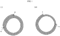
- FIG. 1 (i) is a diagram illustrating a front cross-section of an artificial blood vessel in one embodiment of the present invention.
- FIG. 1(ii) is a diagram illustrating a front cross-section of an artificial blood vessel in another embodiment of the present invention.
- the biomaterial-derived sheet-like decellularized material of the present invention is a biomaterial-derived sheet-like decellularized material having a maximum value of tensile strength in four directions of 4 MPa or more and an elongation rate in the direction exhibiting the maximum tensile strength of 50% to 300%.
- a biomaterial used for the biomaterial-derived sheet-like decellularized material of the present invention is an animal-derived material.
- the animal is preferably a vertebrate or the like, and more preferably a mammal, a bird or the like due to milder rejection reactions.
- a mammalian livestock, avian livestock, human or the like is more preferably used because of easy availability. Examples of the mammalian livestock include a cow, pig, sheep, horse, goat, deer, dog, cat, rabbit, hamster, guinea pig, rat, mouse, camel, llama, donkey, yak, alpaca, raccoon dog, weasel, fox, squirrel, raccoon and the like.
- avian livestock examples include a chicken, duck, turkey, goose, guinea fowl, pheasant, ostrich, quail, parakeet, parrot and the like.
- Preferable animals include a pig, cow, horse, human and the like, and more preferably a pig, cow and the like in terms of availability and safety.
- a site of animal tissue used as a biomaterial may be a site having extracellular matrix structure.
- the site include a heart, pericardium, heart valve, fascia, skin, dermis, blood vessel, liver, kidney, ureter, bladder, urethra, tongue, tonsil, esophagus, stomach, small intestine, colon, anus, pancreas, spleen, lung, brain, bone, spinal cord, cartilage, testis, uterus, fallopian tube, ovary, placenta, cornea, skeletal muscle, tendon, nerve, dura mater, umbilical cord, amniotic membrane, intestinal tract, small intestine submucosa, other collagen containing tissue, etc.
- a heart, pericardium, heart valve, fascia, skin, blood vessel and the like are preferable among the above-mentioned sites.
- a heart, pericardium, heart valve, fascia, skin and the like are more preferable, because excellent pressure resistance can be exhibited.
- a pericardium is particularly preferable, because it exhibits excellent pressure resistance comparable to that of a blood vessel itself.
- the biomaterial is subsequently subjected to decellularization and sheeting to prepare a biomaterial-derived sheet-like decellularized material.
- Decellularization may be performed before or after sheeting, or may be performed after rolling into an artificial blood vessel. However, in view of processability and easiness of decellularization, decellularization is preferably performed before rolling, and is preferably performed after forming into a sheet shape.
- Decellularization is performed to remove antigenic components such as cells and nucleic acid components from biomaterials collected from an animal. By performing decellularization, it is possible to suppress the rejection that occurs when used as a transplant tissue in a living body.
- the decellularization method is not particularly limited in the present invention, and a conventional known method may be used.
- Examples of decellularization include a physical agitation, ultrasonic processing, freeze-thaw method, high hydrostatic pressure method, hypertonic solution/hypotonic solution method, a treatment using a surfactant such as an anionic surfactant and nonionic surfactant, an enzymatic treatment by a proteolytic enzyme, nucleolytic enzyme and the like, as well as a treatment using an alcohol solvent.
- a surfactant such as an anionic surfactant and nonionic surfactant
- an enzymatic treatment by a proteolytic enzyme, nucleolytic enzyme and the like as well as a treatment using an alcohol solvent.
- the foregoing may be implemented in combination of two or more thereof.
- a method including a high hydrostatic pressure method is preferably used in the present invention from the viewpoint of efficient decellularization while maintaining the mechanical strength of the structural protein and from the viewpoint of blood compatibility.
- the shape of the sheet is not limited, but is preferably rectangular (oblong) or substantially rectangular, from the viewpoint of the pressure resistance of the rolled artificial blood vessel, handleability and processability.
- the size of the sheet may be appropriately selected depending on the size of the desired artificial blood vessel.
- the length of one side of the sheet in the length direction (“long side") is usually 10 to 400 mm, preferably 20 to 300 mm, from the viewpoint of the pressure resistance and handleability when rolled.
- the length of one side of the sheet in the width direction (“short side”) is usually 1.5 to 200 mm, preferably 3 to 70 mm, and more preferably 6 to 40 mm.
- the maximum value of the tensile strength in four directions is 4 MPa or more, preferably 5 MPa or more.
- an artificial blood vessel prepared by rolling the same has extremely high pressure resistance strength.
- the maximum values of the tensile strength in four directions are respectively 3.1 MPa and 3.9 MPa, so that the pressure resistance strengths of the artificial blood vessels prepared by rolling the same are inferior.
- the elongation rate in the direction exhibiting the maximum tensile strength is 50% to 300%, preferably 100 to 250%, and more preferably 150 to 220%.
- the artificial blood vessel prepared by rolling the biomaterial-derived sheet-like decellularized material has extremely high pressure resistance strength.
- the elongation rates are respectively 304% and 332%, so that the pressure resistance strength of the artificial blood vessels prepared by rolling the same are inferior.
- the biomaterial-derived sheet-like decellularized material having an anisotropy in the tensile strength and a maximum stress ratio of 1.5 to 5, is also preferable in the present invention.
- an artificial blood vessel prepared by rolling the biomaterial-derived sheet-like decellularized material having a maximum value of tensile strength in four directions of 4 MPa or more and an elongation rate in the direction exhibiting the maximum tensile strength of 50% to 300% of the present invention has an extremely higher pressure resistance strength such as 400 mmHg or more compared with one prepared from a porcine aorta-derived material which was conventionally considered to be optimum, as described in Test Examples 3 and 4.
- the pressure resistance strength of the artificial blood vessel of the present invention is preferably 400 mmHg or more, more preferably 600 mmHg or more, still more preferably 800 mmHg or more, and most preferably 1,000 mmHg or more.
- An artificial blood vessel can be prepared by using the biomaterial-derived sheet-like decellularized material of the present invention.
- a shape of the front cross-section of the artificial blood vessel of the present invention includes, for example, a circular shape, a substantially circular shape, an elliptical shape, a substantially elliptical shape, and the like.
- the shape is highly flexible, and may be deformed in accordance with the intended use.
- the inner circumference is preferably 1.5 to 200 mm, more preferably 3 to 70 mm, and still more preferably 6 to 40 mm.
- a part of the wall portion forming the artificial blood vessel has a two-layer structure, from the viewpoint of pressure resistance.
- Embodiments where a part of the wall portion forming the artificial blood vessel has a two-layer structure include an artificial blood vessel having a two-layer structure and a three-layer structure as the wall portion ( FIG. 1 (i) ), an artificial blood vessel in which the entire wall portion is a two-layer structure, and an artificial blood vessel having a single-layer structure and a two-layer structure as the wall portion ( FIG. 1(ii) ).
- the length of the two-layer structure portion in the artificial blood vessel having a two-layer structure and a three-layer structure as the wall portion is preferably 0% or more, more preferably 50% or more, and still more preferably 80% or more of the length of the outer circumference.
- the length of the outer circumference denotes herein the length of the outer wall portion of the artificial blood vessel, taking one specific point of the artificial blood vessel as the starting point and end point.
- the length of the outer circumference in FIG. 1 (i) denotes the length of the outer wall portion of the artificial blood vessel, taking point g as the starting point and end point.
- the length of the two-layer structure portion in the artificial blood vessel having a single-layer structure and a two-layer structure as a wall portion is preferably 10% or more, more preferably 50% or more, and still more preferably 80% or more of the length of the outer circumference.
- the length of the outer circumference denotes herein the length of the outer wall portion of the artificial blood vessel, taking one specific point of the artificial blood vessel as the starting point and end point.
- the length of the outer circumference in FIG. 1(ii) denotes the length of the outer wall portion of the artificial blood vessel, taking point j as the starting point and end point.
- a biomaterial-derived sheet-like decellularized material having a shape (“taper") in which the thickness of at least one side of the edge portion decreases toward the end of the edge portion as described in Patent Literature 2.
- both ends or one end of the cross-section of the biomaterial-derived sheet-like decellularized material have a tapered shape.
- the cross-section of the biomaterial-derived sheet-like decellularized material need not be processed linearly.
- the tapered portion may be provided at the edge portions on all four sides of the biomaterial-derived sheet-like decellularized material.
- the tapered portion may be provided at the edge portions on three sides, two sides or one side of the biomaterial-derived sheet-like decellularized material.
- the biomaterial-derived sheet-like decellularized material is rolled so that the tapered portion is on the inner lumen side of the artificial blood vessel, to form an artificial blood vessel.
- the edge portions on at least two sides of the biomaterial-derived sheet-like decellularized material are tapered, and more preferably the edge portions on two sides of the biomaterial-derived sheet-like decellularized material in the length direction are tapered.
- the edge portion on one side of the biomaterial-derived sheet-like decellularized material is tapered, and most preferably, the edge portion on one side in the length direction is tapered.
- the artificial blood vessel can be formed by rolling the biomaterial-derived sheet-like decellularized material on a core member.
- various types of core members can be selected, depending on the intended inner circumference and the length of the artificial blood vessel, and the quality of the material does not matter.
- the outer circumference of the core member to be used corresponds substantially to the inner circumference of the artificial blood vessel, and the core member may be appropriately selected in accordance with the intended inner circumference of the artificial blood vessel.
- the core material is not particularly limited, and examples thereof include a tube or cylindrical bar made of polytetrafluoroethylene (PTFE), polyurethane (PU) or a stainless steel material (SUS).
- the artificial blood vessel of the present invention can be formed by stitching a part of the biomaterial-derived sheet-like decellularized material, or by bonding a part of the biomaterial-derived sheet-like decellularized material using an adhesive, or by resorting to both of the foregoing. Bonding with use of an adhesive is preferable from the viewpoint of processability. Therefore, the tapered portion of the biomaterial-derived sheet-like decellularized material may be fixed to the inner wall of the artificial blood vessel by means of, for example, stitching or an adhesive.
- a conventionally used adhesive for biological tissue may be used herein as the adhesive to be used.
- examples thereof include a fibrin glue, cyanoacrylate-based polymerizable adhesive, and gelatin glue resulting from cross-linking of gelatin and resorcinol by formalin, and the like, and a fibrin glue is preferable from the viewpoint of pressure resistance.
- a fibrin glue denotes herein a formulation in which pasty clots formed through the action of the enzyme thrombin on fibrinogen are utilized, for example, in tissue closure, adhesion in organ damage, hemostasis and the like.
- the site at which the adhesive is applied is not particularly limited, as long as the adhesive allows bonding of the biomaterial-derived sheet-like decellularized material so as to form the artificial blood vessel.
- the adhesive is preferably applied so that no adhesive is present on the inner wall surface of the artificial blood vessel. This is because the adhesive may give rise to some adverse effects when coming into contact with substances passing through the inside (lumen) of the artificial blood vessel.
- the artificial blood vessel of the present invention is used as an artificial blood vessel, but may also be used as a graft of a tubular biological tissue such as an ureter, trachea, and lymph duct.
- the artificial blood vessel of the present invention has a much higher pressure resistance strength than one derived from a porcine aorta, which was conventionally considered to be optimum.
- the pressure resistance strength of the artificial blood vessel of the present invention is preferably 600 mmHg or more, more preferably 800 mmHg or more, and still more preferably 1,000 mmHg or more.
- the artificial blood vessel of the present invention exhibits excellent handleability during surgery or the like.
- the biomaterial-derived sheet-like decellularized material of the present invention may also be used to repair a blood vessel as a blood vessel repairing material.
- the biomaterial-derived sheet-like decellularized material of the present invention may be used for treatment such as application to a damaged part of a blood vessel.
- An adventitia of a porcine aorta was completely stripped off, and was cut open to obtain a sheet-like aorta.
- the obtained sheet in a polyethylene zippered bag was subjected to a high hydrostatic pressure treatment for 15 minutes at 100 MPa in a high pressure processing device for research and development (Dr. CHEF, Kobe Steel, Ltd.), using a physiological saline as a medium.
- the treated sheet was shaken for 96 hours at 4 °C in a physiological saline containing 20 ppm of the nucleolytic enzyme DNase, followed by a treatment for 72 hours at 4 °C in 80% ethanol, and was lastly washed for 2 hours at 4 °C in a physiological saline, to obtain a porcine aortic sheet-like decellularized material.
- the porcine aortic sheet-like decellularized material prepared in Example 1 was cut and shaped to 24 mm ⁇ 100 mm.
- a biological adhesive fibrin glue was applied to the surface on the medial tissue side of the porcine aortic sheet-like decellularized material, and the porcine aortic sheet-like decellularized material was rolled twice on a core member of a PTFE tube having an outer diameter of 3.0 mm in such a manner that the intimal tissue was inside after formation of the artificial blood vessel, and the porcine aortic sheet-like decellularized material was pressed and shaped for 5 minutes.
- a collected porcine pericardial sheet in a polyethylene zippered bag was subjected to a high hydrostatic pressure treatment for 15 minutes at 100 MPa in a high pressure processing device for research and development (Dr. CHEF, Kobe Steel, Ltd.), using a physiological saline as a medium.
- the treated sheet was shaken for 96 hours at 4 °C in a saline containing 20 ppm of the nucleolytic enzyme DNase, followed by a treatment for 72 hours at 4 °C in 80% ethanol, and was lastly washed for 2 hours at 4 °C in a physiological saline, to obtain a porcine pericardial sheet-like decellularized material.
- a dermal layer was separated from a porcine skin to obtain a sheet-like dermis.
- the sheet-like dermis and a physiological saline as a medium for a high hydrostatic pressure treatment were added to a polyethylene zippered bag.
- the mixture was pressurized at 100 MPa of a hydrostatic pressure for 15 minutes using a high pressure processing device for research and development (Dr. CHEF, Kobe Steel, Ltd.).
- the sheet-like dermis subjected by a high hydrostatic pressure treatment was shaken and washed for 96 hours at 4 °C in a physiological saline containing 20 ppm of the nucleolytic enzyme DNase, followed by a treatment for 72 hours at 4 °C in 80% ethanol, and was lastly washed for 2 hours at 4 °C in a physiological saline, to obtain a porcine dermal sheet-like decellularized material.
- dumbbell test pieces of No. 8 described in ISO 37 were collected from the rectangular sheet-like decellularized materials prepared in Examples 1, 3 and 4 (porcine aorta, porcine pericardium, and porcine dermis). In order to evaluate the anisotropy of the tensile strength in the sheet, dumbbell test pieces were prepared from one sheet in the direction of 0°, 30°, 60° and 90°, assuming the direction is 0° in the case where a dumbbell test piece is prepared in the direction parallel to the long side.
- the thickness of the parallel portion of the dumbbell test piece was measured using One-Shot 3D Measuring Macroscope (VR-3200, Keyence Corporation). The length (40 mm) between the cutting end faces of the punching blade of the parallel portion was used as the width (mm) of the test piece.
- a cross-sectional area A (mm 2 ) of the test piece was calculated from the thickness and width of the test piece by the following equation.
- A t ⁇ w (A: cross sectional area of test piece (mm 2 ); t: thickness of test piece (mm); w: width of test piece (mm))
- the tensile test was performed in accordance with ISO 37, as follows.
- the test piece was attached to Table Top Universal Testing Machine (MCT-2150, A&D Company, Limited) so that both ends of the test piece were held symmetrically in order to uniformly distribute the tensile force to the cross section.
- the testing machine was operated to continuously observe changes in the distance between the marked lines and changes in the force, and the maximum load Fmax (N) and the distance between the marked lines at the breakage Lb (mm) were measured.
- the speed of the holding tool was set to 200 mm/min. Data of the test pieces broken outside of the marked lines were discarded.
- the test was repeated on additional test pieces until the measurements of the test pieces punched in four directions were made twice correctly for each direction.
- the tensile strength in four directions, the anisotropy of the tensile strength, and the elongation rate were calculated from the measured values by the following equations. The results are shown in Table 2.
- the "tensile strength in four directions” and the “stress ratio” were compared among the four directions based on the average value in each direction.
- ⁇ Fmax / A ( ⁇ : tensile strength (MPa); Fmax: maximum load (N); A: cross-sectional area of test piece (mm 2 ))
- ⁇ Lb ⁇ L 0 / L 0 ⁇ 100 (Lb: distance between the marked lines at the breakage (mm); L0: initial distance between the marked lines (mm))
- Anisotropy in sheet was treated as a stress ratio S calculated by the following equation.
- S ⁇ max / ⁇ min ( ⁇ max: the maximum tensile strength (MPa) in the tensile tests in four directions; ⁇ min: the minimum tensile strength (MPa) in the tensile tests in four directions)
- the sheet-like decellularized materials prepared in Examples 3 and 4 were cut and shaped to 24 mm ⁇ 100 mm.
- the surface water was wiped off.
- a biological adhesive fibrin glue was applied thereto, and the sheet-like decellularized materials were rolled twice on a core member of a PTFE tube having an outer diameter of 3.0 mm, and the sheet-like decellularized materials were pressed and shaped for 5 minutes. They were immersed in a physiological saline, and the PTFE tube of the core member was removed, and both ends were cut to prepare artificial blood vessels of 100 mm ⁇ 3 mm ⁇ . Rolling and shaping were performed so that the tensile strength in the circumferential direction of the artificial blood vessels increased.
- a bovine aortic sheet-like decellularized material, and a bovine pericardial sheet-like decellularized material, and a bovine aortic decellularized artificial blood vessel and a bovine pericardial decellularized artificial blood vessel were prepared in the same manner as in Examples 1 to 5 except that a bovine aorta was used in place of a porcine aorta and a bovine pericardium was used in place of a porcine pericardium.
- the biomaterial-derived sheet-like decellularized material of the present invention can provide an artificial blood vessel having a much higher pressure resistance strength than one derived from a porcine aorta, which was conventionally considered to be optimum. Therefore, when the biomaterial-derived sheet-like decellularized material of the present invention is used as an artificial blood vessel or for repair of a blood vessel, excellent pressure resistance comparable to that of a blood vessel itself can be maintained.
Landscapes
- Health & Medical Sciences (AREA)
- Life Sciences & Earth Sciences (AREA)
- Chemical & Material Sciences (AREA)
- Biomedical Technology (AREA)
- Engineering & Computer Science (AREA)
- Veterinary Medicine (AREA)
- Oral & Maxillofacial Surgery (AREA)
- Transplantation (AREA)
- Animal Behavior & Ethology (AREA)
- General Health & Medical Sciences (AREA)
- Public Health (AREA)
- Epidemiology (AREA)
- Medicinal Chemistry (AREA)
- Dermatology (AREA)
- Chemical Kinetics & Catalysis (AREA)
- Botany (AREA)
- Molecular Biology (AREA)
- Vascular Medicine (AREA)
- Urology & Nephrology (AREA)
- Zoology (AREA)
- Heart & Thoracic Surgery (AREA)
- Cardiology (AREA)
- Gastroenterology & Hepatology (AREA)
- Pulmonology (AREA)
- Prostheses (AREA)
- Materials For Medical Uses (AREA)
- Micro-Organisms Or Cultivation Processes Thereof (AREA)
Abstract
Description
- The present invention relates to a sheet-like decellularized material and an artificial blood vessel using the material.
- Blood vessel grafts are used in the construction of blood vessels for bypass surgery, and in repair or replacement of damaged or morbid blood vessels. For example, in the treatment of atherosclerosis of coronary arteries or peripheral blood vessels, patients' own blood vessels are preferable replacement grafts for affected areas having a diameter less than 5 mm, and patient's own internal thoracic artery, radial artery, saphenous vein, etc. are used. However, invasive collection cannot be avoided when using patient's own blood vessel, which accordingly translates into a significant burden on the patient's body and into inevitable variability in the length and quality of the blood vessel depending on individuals or cases. Furthermore, there is a problem that in cases of reoperation, it is impossible to obtain again patients' own blood vessels as they are already in use.
- An artificial blood vessel made from a synthetic resin such as polyester and polytetrafluoroethylene is used for revascularization of limb peripheral arteries and the like. However, an artificial blood vessel made from such synthetic resin cannot be used because of early thrombus formation and intima thickening, in a case where the artificial blood vessel is used in a small-diameter blood vessel such as the coronary arteries. In order to prevent blood clotting in an artificial blood vessel made from such synthetic resin, the lumen of the artificial blood vessel is covered with the patient's own vascular endothelial cells by means of tissue engineering techniques. However, the bone marrow must be collected from the patient, and be cultured and allowed to graft onto the artificial blood vessel, prior to surgery, which requires preparations in advance. The usefulness of this approach is low in surgery that must be performed in an emergency. In addition, there is also a problem that the vascular endothelial cells covering the lumen of the blood vessel are easily peeled off, which may cause thrombus formation.
- Among them, Patent Literature 1 proposes an artificial blood vessel in which a sheet of decellularized biomaterial is formed into a tubular shape in order to prevent rejection. Specifically, the artificial blood vessel is formed by rolling a sheet prepared from a porcine aorta into a tubular shape without any modification. However, the edge portion of the sheet juts into the lumen of the tube, in the luminal cross-section, by the extent of the thickness of the sheet, and the cross-section of the tube does not become circular or elliptical (see: FIG. 8 in Patent Literature 2). When the cross-section of the tubular structure is in such a shape, localized pressure acts on the portion jutting out when blood flows therethrough, and as a result, peel-off may occur, and thus the pressure resistance of the blood vessel is insufficient. In addition, there arises a problem that the handleability becomes worse during surgery, if the sheet is made thicker in order to secure pressure resistance. Moreover, it is also considered that platelets and the like more easily adhere to the portion jutting out, and thrombus are more easily generated.
- The invention described in Patent Literature 2 by the present applicant, is one which solves the above problem of Patent Literature 1. Patent Literature 2 describes a sheet of biological tissue, the sheet including on at least one side tapered edge portion thinning down in the thickness direction towards an end thereof, and a tubular structure using the sheet. A porcine aorta is used for preparing sheets, etc. in the examples, as in Patent Literature 1.
- As mentioned above, sheets prepared from a porcine aorta are specifically used as a sheet of the biological tissue in Patent Literatures 1 and 2. Therefore, there has been a demand for a material which has more improved pressure resistance than a sheet of biological tissue derived from a porcine aorta, and which can maintain excellent pressure resistance when used as an artificial blood vessel or for repair of a blood vessel.
-
- Patent Literature 1:
WO 2014/109185 - Patent Literature 2:
WO 2016/194895 - The problem to be solved by the present invention is to provide a sheet-like material capable of maintaining excellent pressure resistance when used as an artificial blood vessel or for repair of a blood vessel.
- As a result of intensive studies to solve the above problems, the present inventors have surprisingly found that an artificial blood vessel having a much higher pressure resistance than one derived from a porcine aorta, which was conventionally considered to be optimum, can be prepared by preparing an artificial blood vessel using a biomaterial-derived sheet-like decellularized material having a tensile strength and an elongation rate in a specific range. Thus, the present inventors have completed the present invention.
- Namely, the present invention is as follows.
- (1) A biomaterial-derived sheet-like decellularized material having a maximum value of tensile strength in four directions of 4 MPa or more and an elongation rate in the direction exhibiting the maximum tensile strength of 50% to 300%.
- (2) The biomaterial-derived sheet-like decellularized material according to (1), wherein a pressure resistance strength of an artificial blood vessel formed by rolling the biomaterial-derived sheet-like decellularized material is 400 mmHg or more.
- (3) The biomaterial-derived sheet-like decellularized material according to (1) or (2), wherein the tensile strength is anisotropic and a maximum stress ratio is 1.5 to 5.
- (4) The biomaterial-derived sheet-like decellularized material according to any one of (1) to (3), which is derived from pericardium.
- (5) The biomaterial-derived sheet-like decellularized material according to any one of (1) to (4), which is for an artificial blood vessel or for repair of a blood vessel.
- (6) An artificial blood vessel comprising the biomaterial-derived sheet-like decellularized material according to any one of (1) to (4).
- The biomaterial-derived sheet-like decellularized material of the present invention can provide an artificial blood vessel having a much higher pressure resistance strength than one derived from a porcine aorta, which was conventionally considered to be optimum. Therefore, when the biomaterial-derived sheet-like decellularized material of the present invention is used as an artificial blood vessel or for repair of a blood vessel, excellent pressure resistance comparable to that of a blood vessel itself can be maintained.
-
FIG. 1 (i) is a diagram illustrating a front cross-section of an artificial blood vessel in one embodiment of the present invention.FIG. 1(ii) is a diagram illustrating a front cross-section of an artificial blood vessel in another embodiment of the present invention. - The biomaterial-derived sheet-like decellularized material of the present invention is a biomaterial-derived sheet-like decellularized material having a maximum value of tensile strength in four directions of 4 MPa or more and an elongation rate in the direction exhibiting the maximum tensile strength of 50% to 300%.
- A biomaterial used for the biomaterial-derived sheet-like decellularized material of the present invention is an animal-derived material. The animal is preferably a vertebrate or the like, and more preferably a mammal, a bird or the like due to milder rejection reactions. A mammalian livestock, avian livestock, human or the like is more preferably used because of easy availability. Examples of the mammalian livestock include a cow, pig, sheep, horse, goat, deer, dog, cat, rabbit, hamster, guinea pig, rat, mouse, camel, llama, donkey, yak, alpaca, raccoon dog, weasel, fox, squirrel, raccoon and the like. Examples of the avian livestock include a chicken, duck, turkey, goose, guinea fowl, pheasant, ostrich, quail, parakeet, parrot and the like. Preferable animals include a pig, cow, horse, human and the like, and more preferably a pig, cow and the like in terms of availability and safety.
- A site of animal tissue used as a biomaterial may be a site having extracellular matrix structure. Examples of the site include a heart, pericardium, heart valve, fascia, skin, dermis, blood vessel, liver, kidney, ureter, bladder, urethra, tongue, tonsil, esophagus, stomach, small intestine, colon, anus, pancreas, spleen, lung, brain, bone, spinal cord, cartilage, testis, uterus, fallopian tube, ovary, placenta, cornea, skeletal muscle, tendon, nerve, dura mater, umbilical cord, amniotic membrane, intestinal tract, small intestine submucosa, other collagen containing tissue, etc. When a skin, dermis, or the like is used, the thickness of the material is large so that the handleability is poor. When a part derived from pores remains, platelets and the like easily adhere to the part and there is a problem that thrombus are easily generated. In consideration of these issues, and from the viewpoints of easiness and availability of decellularization and pressure resistance and handleability when rolled, a heart, pericardium, heart valve, fascia, skin, blood vessel and the like are preferable among the above-mentioned sites. In addition, a heart, pericardium, heart valve, fascia, skin and the like are more preferable, because excellent pressure resistance can be exhibited. Further, a pericardium is particularly preferable, because it exhibits excellent pressure resistance comparable to that of a blood vessel itself.
- The biomaterial is subsequently subjected to decellularization and sheeting to prepare a biomaterial-derived sheet-like decellularized material. Decellularization may be performed before or after sheeting, or may be performed after rolling into an artificial blood vessel. However, in view of processability and easiness of decellularization, decellularization is preferably performed before rolling, and is preferably performed after forming into a sheet shape.
- Decellularization is performed to remove antigenic components such as cells and nucleic acid components from biomaterials collected from an animal. By performing decellularization, it is possible to suppress the rejection that occurs when used as a transplant tissue in a living body.
- The decellularization method is not particularly limited in the present invention, and a conventional known method may be used. Examples of decellularization include a physical agitation, ultrasonic processing, freeze-thaw method, high hydrostatic pressure method, hypertonic solution/hypotonic solution method, a treatment using a surfactant such as an anionic surfactant and nonionic surfactant, an enzymatic treatment by a proteolytic enzyme, nucleolytic enzyme and the like, as well as a treatment using an alcohol solvent. The foregoing may be implemented in combination of two or more thereof.
- A method including a high hydrostatic pressure method is preferably used in the present invention from the viewpoint of efficient decellularization while maintaining the mechanical strength of the structural protein and from the viewpoint of blood compatibility.
- In forming into a sheet, the shape of the sheet is not limited, but is preferably rectangular (oblong) or substantially rectangular, from the viewpoint of the pressure resistance of the rolled artificial blood vessel, handleability and processability. In the case of a rectangular or substantially rectangular sheet, the size of the sheet may be appropriately selected depending on the size of the desired artificial blood vessel. The length of one side of the sheet in the length direction ("long side") is usually 10 to 400 mm, preferably 20 to 300 mm, from the viewpoint of the pressure resistance and handleability when rolled. The length of one side of the sheet in the width direction ("short side") is usually 1.5 to 200 mm, preferably 3 to 70 mm, and more preferably 6 to 40 mm.
- In the biomaterial-derived sheet-like decellularized material of the present invention, the maximum value of the tensile strength in four directions is 4 MPa or more, preferably 5 MPa or more. By using the biomaterial-derived sheet-like decellularized material of the present invention having a maximum value of tensile strength in four directions of 4 MPa or more, an artificial blood vessel prepared by rolling the same has extremely high pressure resistance strength. On the other hand, as understood from Test Examples 3 and 4, in the biomaterial-derived sheet-like decellularized material prepared using a porcine aorta or bovine aorta, the maximum values of the tensile strength in four directions are respectively 3.1 MPa and 3.9 MPa, so that the pressure resistance strengths of the artificial blood vessels prepared by rolling the same are inferior.
- In the biomaterial-derived sheet-like decellularized material of the present invention, the elongation rate in the direction exhibiting the maximum tensile strength is 50% to 300%, preferably 100 to 250%, and more preferably 150 to 220%. By setting the elongation rate in this range, the artificial blood vessel prepared by rolling the biomaterial-derived sheet-like decellularized material has extremely high pressure resistance strength. On the other hand, as understood from Test Examples 3 and 4, in the biomaterial-derived sheet-like decellularized material prepared using a porcine aorta or bovine aorta, the elongation rates are respectively 304% and 332%, so that the pressure resistance strength of the artificial blood vessels prepared by rolling the same are inferior.
- The biomaterial-derived sheet-like decellularized material having an anisotropy in the tensile strength and a maximum stress ratio of 1.5 to 5, is also preferable in the present invention. In that case, it is preferable to roll the biomaterial-derived sheet-like decellularized material of the present invention to prepare an artificial blood vessel, so as to increase the tensile strength in the circumferential direction of the artificial blood vessel.
- An artificial blood vessel prepared by rolling a sheet-like decellularized material prepared using a porcine aorta or bovine aorta, which was conventionally considered to be optimum, has a low pressure resistance strength such as 120 mmHg and 121 mmHg as described in Test Examples 3 and 4. In contrast, an artificial blood vessel prepared by rolling the biomaterial-derived sheet-like decellularized material having a maximum value of tensile strength in four directions of 4 MPa or more and an elongation rate in the direction exhibiting the maximum tensile strength of 50% to 300% of the present invention, has an extremely higher pressure resistance strength such as 400 mmHg or more compared with one prepared from a porcine aorta-derived material which was conventionally considered to be optimum, as described in Test Examples 3 and 4. Namely, the pressure resistance strength of the artificial blood vessel of the present invention is preferably 400 mmHg or more, more preferably 600 mmHg or more, still more preferably 800 mmHg or more, and most preferably 1,000 mmHg or more.
- An artificial blood vessel can be prepared by using the biomaterial-derived sheet-like decellularized material of the present invention. A shape of the front cross-section of the artificial blood vessel of the present invention includes, for example, a circular shape, a substantially circular shape, an elliptical shape, a substantially elliptical shape, and the like. The shape is highly flexible, and may be deformed in accordance with the intended use. Generally, the inner circumference is preferably 1.5 to 200 mm, more preferably 3 to 70 mm, and still more preferably 6 to 40 mm.
- In the artificial blood vessel of the present invention, it is preferable that a part of the wall portion forming the artificial blood vessel has a two-layer structure, from the viewpoint of pressure resistance. Embodiments where a part of the wall portion forming the artificial blood vessel has a two-layer structure include an artificial blood vessel having a two-layer structure and a three-layer structure as the wall portion (
FIG. 1 (i) ), an artificial blood vessel in which the entire wall portion is a two-layer structure, and an artificial blood vessel having a single-layer structure and a two-layer structure as the wall portion (FIG. 1(ii) ). - The length of the two-layer structure portion in the artificial blood vessel having a two-layer structure and a three-layer structure as the wall portion (the distance on the outer wall from point g on the outer wall clockwise up to point h, in
FIG. 1 (i) ) is preferably 0% or more, more preferably 50% or more, and still more preferably 80% or more of the length of the outer circumference. The length of the outer circumference denotes herein the length of the outer wall portion of the artificial blood vessel, taking one specific point of the artificial blood vessel as the starting point and end point. For example, the length of the outer circumference inFIG. 1 (i) denotes the length of the outer wall portion of the artificial blood vessel, taking point g as the starting point and end point. - The length of the two-layer structure portion in the artificial blood vessel having a single-layer structure and a two-layer structure as a wall portion (the distance on the outer wall from point j on the outer wall anticlockwise up to point k, in
FIG. 1(ii) ) is preferably 10% or more, more preferably 50% or more, and still more preferably 80% or more of the length of the outer circumference. The length of the outer circumference denotes herein the length of the outer wall portion of the artificial blood vessel, taking one specific point of the artificial blood vessel as the starting point and end point. For example, the length of the outer circumference inFIG. 1(ii) denotes the length of the outer wall portion of the artificial blood vessel, taking point j as the starting point and end point. - In the artificial blood vessel of the present invention, it is preferable to use a biomaterial-derived sheet-like decellularized material having a shape ("taper") in which the thickness of at least one side of the edge portion decreases toward the end of the edge portion as described in Patent Literature 2. Namely, both ends or one end of the cross-section of the biomaterial-derived sheet-like decellularized material have a tapered shape. The cross-section of the biomaterial-derived sheet-like decellularized material need not be processed linearly. The tapered portion may be provided at the edge portions on all four sides of the biomaterial-derived sheet-like decellularized material. The tapered portion may be provided at the edge portions on three sides, two sides or one side of the biomaterial-derived sheet-like decellularized material. The biomaterial-derived sheet-like decellularized material is rolled so that the tapered portion is on the inner lumen side of the artificial blood vessel, to form an artificial blood vessel. Preferably, the edge portions on at least two sides of the biomaterial-derived sheet-like decellularized material are tapered, and more preferably the edge portions on two sides of the biomaterial-derived sheet-like decellularized material in the length direction are tapered. Still more preferably, the edge portion on one side of the biomaterial-derived sheet-like decellularized material is tapered, and most preferably, the edge portion on one side in the length direction is tapered.
- In one embodiment of preparation of the artificial blood vessel, the artificial blood vessel can be formed by rolling the biomaterial-derived sheet-like decellularized material on a core member. Concerning the outer circumference and the length of the core member, various types of core members can be selected, depending on the intended inner circumference and the length of the artificial blood vessel, and the quality of the material does not matter. The outer circumference of the core member to be used corresponds substantially to the inner circumference of the artificial blood vessel, and the core member may be appropriately selected in accordance with the intended inner circumference of the artificial blood vessel. The core material is not particularly limited, and examples thereof include a tube or cylindrical bar made of polytetrafluoroethylene (PTFE), polyurethane (PU) or a stainless steel material (SUS).
- The artificial blood vessel of the present invention can be formed by stitching a part of the biomaterial-derived sheet-like decellularized material, or by bonding a part of the biomaterial-derived sheet-like decellularized material using an adhesive, or by resorting to both of the foregoing. Bonding with use of an adhesive is preferable from the viewpoint of processability. Therefore, the tapered portion of the biomaterial-derived sheet-like decellularized material may be fixed to the inner wall of the artificial blood vessel by means of, for example, stitching or an adhesive.
- A conventionally used adhesive for biological tissue may be used herein as the adhesive to be used. Examples thereof include a fibrin glue, cyanoacrylate-based polymerizable adhesive, and gelatin glue resulting from cross-linking of gelatin and resorcinol by formalin, and the like, and a fibrin glue is preferable from the viewpoint of pressure resistance. A fibrin glue denotes herein a formulation in which pasty clots formed through the action of the enzyme thrombin on fibrinogen are utilized, for example, in tissue closure, adhesion in organ damage, hemostasis and the like.
- The site at which the adhesive is applied is not particularly limited, as long as the adhesive allows bonding of the biomaterial-derived sheet-like decellularized material so as to form the artificial blood vessel. However, the adhesive is preferably applied so that no adhesive is present on the inner wall surface of the artificial blood vessel. This is because the adhesive may give rise to some adverse effects when coming into contact with substances passing through the inside (lumen) of the artificial blood vessel. Further, it is necessary to sufficiently apply the adhesive to the tapered portion and bond it so that the cross-section of the artificial blood vessel is circular, substantially circular, elliptical or substantially elliptical, as shown in
FIGS. 1 (i) and (ii). This is because when bonding of the tapered portion is insufficient, pressure resistance at the affected portion may be impaired and the desired artificial blood vessel may not be obtained. - The artificial blood vessel of the present invention is used as an artificial blood vessel, but may also be used as a graft of a tubular biological tissue such as an ureter, trachea, and lymph duct.
- The artificial blood vessel of the present invention has a much higher pressure resistance strength than one derived from a porcine aorta, which was conventionally considered to be optimum. The pressure resistance strength of the artificial blood vessel of the present invention is preferably 600 mmHg or more, more preferably 800 mmHg or more, and still more preferably 1,000 mmHg or more. In addition, the artificial blood vessel of the present invention exhibits excellent handleability during surgery or the like.
- The biomaterial-derived sheet-like decellularized material of the present invention may also be used to repair a blood vessel as a blood vessel repairing material. For example, the biomaterial-derived sheet-like decellularized material of the present invention may be used for treatment such as application to a damaged part of a blood vessel.
- Hereinafter, the present invention is explained in more detail with reference to examples, but the present invention is not limited only to these examples.
- An adventitia of a porcine aorta was completely stripped off, and was cut open to obtain a sheet-like aorta. The obtained sheet in a polyethylene zippered bag was subjected to a high hydrostatic pressure treatment for 15 minutes at 100 MPa in a high pressure processing device for research and development (Dr. CHEF, Kobe Steel, Ltd.), using a physiological saline as a medium. The treated sheet was shaken for 96 hours at 4 °C in a physiological saline containing 20 ppm of the nucleolytic enzyme DNase, followed by a treatment for 72 hours at 4 °C in 80% ethanol, and was lastly washed for 2 hours at 4 °C in a physiological saline, to obtain a porcine aortic sheet-like decellularized material.
- The porcine aortic sheet-like decellularized material prepared in Example 1 was cut and shaped to 24 mm × 100 mm. A biological adhesive fibrin glue was applied to the surface on the medial tissue side of the porcine aortic sheet-like decellularized material, and the porcine aortic sheet-like decellularized material was rolled twice on a core member of a PTFE tube having an outer diameter of 3.0 mm in such a manner that the intimal tissue was inside after formation of the artificial blood vessel, and the porcine aortic sheet-like decellularized material was pressed and shaped for 5 minutes. It was immersed in a physiological saline, and the PTFE tube of the core member was removed, and both ends were cut to prepare an artificial blood vessel of 100 mm × 3 mmΦ. Rolling and shaping were performed so that the flow path was formed in the same direction as the flow path of the aorta.
- One end of a porcine aorta was clamped with forceps, and the opposite end was cannulated and ligated. A syringe and a manometer were connected to the cannula. A physiological saline in the syringe was injected into the porcine aorta, and the pressure at the burst of the porcine aorta was measured as a pressure resistance strength. The results are shown in Table 1.
- One end of the artificial blood vessel prepared in Example 2 was clamped with forceps, and the opposite end was cannulated and ligated. A syringe and a manometer were connected to the cannula. A physiological saline in the syringe was injected into the artificial blood vessel, and the pressure at the burst of the artificial blood vessel was measured as a pressure resistance strength. The results are shown in Table 1.
[Table 1] Material Pressure resistance strength (mmHg) Porcine aorta > 1,000 Porcine aortic decellularized rolled blood vessel 120 - A collected porcine pericardial sheet in a polyethylene zippered bag was subjected to a high hydrostatic pressure treatment for 15 minutes at 100 MPa in a high pressure processing device for research and development (Dr. CHEF, Kobe Steel, Ltd.), using a physiological saline as a medium. The treated sheet was shaken for 96 hours at 4 °C in a saline containing 20 ppm of the nucleolytic enzyme DNase, followed by a treatment for 72 hours at 4 °C in 80% ethanol, and was lastly washed for 2 hours at 4 °C in a physiological saline, to obtain a porcine pericardial sheet-like decellularized material.
- A dermal layer was separated from a porcine skin to obtain a sheet-like dermis. The sheet-like dermis and a physiological saline as a medium for a high hydrostatic pressure treatment were added to a polyethylene zippered bag. The mixture was pressurized at 100 MPa of a hydrostatic pressure for 15 minutes using a high pressure processing device for research and development (Dr. CHEF, Kobe Steel, Ltd.). The sheet-like dermis subjected by a high hydrostatic pressure treatment was shaken and washed for 96 hours at 4 °C in a physiological saline containing 20 ppm of the nucleolytic enzyme DNase, followed by a treatment for 72 hours at 4 °C in 80% ethanol, and was lastly washed for 2 hours at 4 °C in a physiological saline, to obtain a porcine dermal sheet-like decellularized material.
- Dumbbell test pieces of No. 8 described in ISO 37 were collected from the rectangular sheet-like decellularized materials prepared in Examples 1, 3 and 4 (porcine aorta, porcine pericardium, and porcine dermis). In order to evaluate the anisotropy of the tensile strength in the sheet, dumbbell test pieces were prepared from one sheet in the direction of 0°, 30°, 60° and 90°, assuming the direction is 0° in the case where a dumbbell test piece is prepared in the direction parallel to the long side.
- The thickness of the parallel portion of the dumbbell test piece was measured using One-Shot 3D Measuring Macroscope (VR-3200, Keyence Corporation). The length (40 mm) between the cutting end faces of the punching blade of the parallel portion was used as the width (mm) of the test piece.
- A cross-sectional area A (mm2) of the test piece was calculated from the thickness and width of the test piece by the following equation.

- The tensile test was performed in accordance with ISO 37, as follows. The test piece was attached to Table Top Universal Testing Machine (MCT-2150, A&D Company, Limited) so that both ends of the test piece were held symmetrically in order to uniformly distribute the tensile force to the cross section. The testing machine was operated to continuously observe changes in the distance between the marked lines and changes in the force, and the maximum load Fmax (N) and the distance between the marked lines at the breakage Lb (mm) were measured. The speed of the holding tool was set to 200 mm/min. Data of the test pieces broken outside of the marked lines were discarded. The test was repeated on additional test pieces until the measurements of the test pieces punched in four directions were made twice correctly for each direction. The tensile strength in four directions, the anisotropy of the tensile strength, and the elongation rate were calculated from the measured values by the following equations. The results are shown in Table 2.
- The "tensile strength in four directions" and the "stress ratio" were compared among the four directions based on the average value in each direction.
- σ (MPa (N/mm2)) was calculated by the following equation.

- ε (%) was calculated by the following equation.

- Anisotropy in sheet was treated as a stress ratio S calculated by the following equation.

- The sheet-like decellularized materials prepared in Examples 3 and 4 were cut and shaped to 24 mm × 100 mm. The surface water was wiped off. A biological adhesive fibrin glue was applied thereto, and the sheet-like decellularized materials were rolled twice on a core member of a PTFE tube having an outer diameter of 3.0 mm, and the sheet-like decellularized materials were pressed and shaped for 5 minutes. They were immersed in a physiological saline, and the PTFE tube of the core member was removed, and both ends were cut to prepare artificial blood vessels of 100 mm × 3 mmΦ. Rolling and shaping were performed so that the tensile strength in the circumferential direction of the artificial blood vessels increased.
- One ends of three artificial blood vessels prepared in Examples 2 and 5 were clamped with forceps, and the opposite ends were cannulated and ligated. A syringe and a manometer were connected to the cannula. A physiological saline in the syringe was injected into the artificial blood vessel, and the pressure at the burst of the artificial blood vessel was measured as a pressure resistance strength. The results are shown in Table 2.
[Table 2] Before rolling After rolling Thickness *1 (µm) maximum value of tensile strength in four directions *2 (MPa) S (stress ratio) Elongation rate (%) Pressure resistance strength (mmHg) Whole average In direction exhibiting maxium tensile sterength Porcine aorta 585 3.1 7.3 304 305 120 Porcine dermis 1,120 8.7 1.4 173 144 626 Porcine pericardium 349 5.9 2.1 158 170 1,347 *1 Thickness is average of thickness of test pieces in four directions.
*2 Maimum value of tensile strength is the maximum value in all tensile strengths mesured in four directions. - A bovine aortic sheet-like decellularized material, and a bovine pericardial sheet-like decellularized material, and a bovine aortic decellularized artificial blood vessel and a bovine pericardial decellularized artificial blood vessel were prepared in the same manner as in Examples 1 to 5 except that a bovine aorta was used in place of a porcine aorta and a bovine pericardium was used in place of a porcine pericardium.
- The tensile test and the pressure resistance test were conducted in the same manner as in Test example 3, using the bovine-derived sheet-like decellularized materials and the bovine-derived decellularized artificial blood vessel prepared in Example 6. The results are shown in Table 3.
[Table 3] Before rolling After rolling Thickness *1 (µm) maximum value of tensile strength in four directions *2 (MPa) S (stress ratio) Elongation rate (%) Pressure resistance strength (mmHg) Whole average In direction exhibiting maxium tensile sterength Bovine aorta 910 3.9 10.7 350 332 121 Bovine pericardium 648 12.7 2.0 184 206 1,063 *1 Thickness is average of thickness of test pieces in four directions.
*2 Maimum value of tensile strength is the maximum value in all tensile strengths mesured in four directions. - The embodiments and examples disclosed herein are illustrative and non-restrictive in every aspect. The scope of the present invention is indicated by the claims and not by the above description. All modifications within the meaning and scope equivalent to the claims are included in the present invention.
- The biomaterial-derived sheet-like decellularized material of the present invention can provide an artificial blood vessel having a much higher pressure resistance strength than one derived from a porcine aorta, which was conventionally considered to be optimum. Therefore, when the biomaterial-derived sheet-like decellularized material of the present invention is used as an artificial blood vessel or for repair of a blood vessel, excellent pressure resistance comparable to that of a blood vessel itself can be maintained.
Claims (6)
- A biomaterial-derived sheet-like decellularized material having a maximum value of tensile strength in four directions of 4 MPa or more and an elongation rate in the direction exhibiting the maximum tensile strength of 50% to 300%.
- The biomaterial-derived sheet-like decellularized material according to claim 1, wherein a pressure resistance strength of an artificial blood vessel formed by rolling the biomaterial-derived sheet-like decellularized material is 400 mmHg or more.
- The biomaterial-derived sheet-like decellularized material according to claim 1 or 2, wherein the tensile strength is anisotropic and a maximum stress ratio is 1.5 to 5.
- The biomaterial-derived sheet-like decellularized material according to any one of claims 1 to 3, which is derived from pericardium.
- The biomaterial-derived sheet-like decellularized material according to any one of claims 1 to 4, which is for an artificial blood vessel or for repair of a blood vessel.
- An artificial blood vessel comprising the biomaterial-derived sheet-like decellularized material according to any one of claims 1 to 4.
Applications Claiming Priority (2)
| Application Number | Priority Date | Filing Date | Title |
|---|---|---|---|
| JP2017210576 | 2017-10-31 | ||
| PCT/JP2018/039426 WO2019087880A1 (en) | 2017-10-31 | 2018-10-24 | Sheet-like decellularized material and artificial blood vessel employing said material |
Publications (3)
| Publication Number | Publication Date |
|---|---|
| EP3705141A1 true EP3705141A1 (en) | 2020-09-09 |
| EP3705141A4 EP3705141A4 (en) | 2021-08-18 |
| EP3705141B1 EP3705141B1 (en) | 2025-10-01 |
Family
ID=66331704
Family Applications (1)
| Application Number | Title | Priority Date | Filing Date |
|---|---|---|---|
| EP18872781.2A Active EP3705141B1 (en) | 2017-10-31 | 2018-10-24 | Sheet-like decellularized material and artificial blood vessel employing said material |
Country Status (7)
| Country | Link |
|---|---|
| US (1) | US20200254146A1 (en) |
| EP (1) | EP3705141B1 (en) |
| JP (1) | JP7412174B2 (en) |
| KR (1) | KR102700693B1 (en) |
| CN (1) | CN111050814A (en) |
| AU (1) | AU2018358273B2 (en) |
| WO (1) | WO2019087880A1 (en) |
Families Citing this family (3)
| Publication number | Priority date | Publication date | Assignee | Title |
|---|---|---|---|---|
| TW202233828A (en) * | 2020-11-12 | 2022-09-01 | 日商Adeka股份有限公司 | Decellularized tissue composition |
| JP2025004711A (en) * | 2023-06-26 | 2025-01-15 | 国立大学法人 東京大学 | Method for producing decellularized tissue or organ by hydrostatic pressure |
| CN119367612A (en) * | 2023-12-26 | 2025-01-28 | 成都奇璞生物科技有限公司 | Artificial blood vessel basal layer and preparation method thereof |
Family Cites Families (12)
| Publication number | Priority date | Publication date | Assignee | Title |
|---|---|---|---|---|
| WO2005063314A1 (en) * | 2003-12-26 | 2005-07-14 | Cardio Incorporated | Decellularized tissue and method of preparing the same |
| JPWO2005063316A1 (en) * | 2003-12-26 | 2007-12-20 | 公立大学法人大阪府立大学 | Implantable biomaterial and method for producing the same |
| WO2009134991A2 (en) * | 2008-04-30 | 2009-11-05 | Ethicon, Inc. | Tissue engineered blood vessel |
| US8295301B2 (en) * | 2008-06-16 | 2012-10-23 | Qualcomm Atheros, Inc. | Managing coexistence among signaling protocols on a shared medium |
| WO2014034759A1 (en) * | 2012-08-31 | 2014-03-06 | 国立大学法人京都大学 | Method for decellularizing tissue employing pressure treatment |
| US20150100115A1 (en) * | 2012-10-08 | 2015-04-09 | Cormatrix Cardiovascular, Inc. | Extracellular Matrix Prostheses for Treating Damaged Biological Tissue |
| KR101941941B1 (en) * | 2013-01-08 | 2019-01-24 | 케이엠 바이올로직스 가부시키가이샤 | Artificial blood vessel using decellularized blood vessel sheet |
| KR20140109185A (en) | 2013-03-05 | 2014-09-15 | 삼성전자주식회사 | Image processing apparatus and method |
| HK1245159A1 (en) * | 2015-02-27 | 2018-08-24 | Adeka株式会社 | Decellularized tissue |
| CN104700355A (en) | 2015-03-31 | 2015-06-10 | 百度在线网络技术(北京)有限公司 | Generation method, device and system for indoor two-dimension plan |
| US20180296728A1 (en) * | 2015-06-02 | 2018-10-18 | Adeka Corporation | Sheet of biological tissue, tubular structure obtained from said sheet, and artificial blood vessel comprising said tubular structure |
| CN106075582A (en) * | 2016-06-27 | 2016-11-09 | 暨南大学 | A kind of engineering blood vessel support and construction method thereof |
-
2018
- 2018-10-24 CN CN201880055547.8A patent/CN111050814A/en active Pending
- 2018-10-24 JP JP2019551177A patent/JP7412174B2/en active Active
- 2018-10-24 US US16/755,948 patent/US20200254146A1/en active Pending
- 2018-10-24 WO PCT/JP2018/039426 patent/WO2019087880A1/en not_active Ceased
- 2018-10-24 AU AU2018358273A patent/AU2018358273B2/en active Active
- 2018-10-24 KR KR1020207004826A patent/KR102700693B1/en active Active
- 2018-10-24 EP EP18872781.2A patent/EP3705141B1/en active Active
Also Published As
| Publication number | Publication date |
|---|---|
| CN111050814A (en) | 2020-04-21 |
| EP3705141A4 (en) | 2021-08-18 |
| KR102700693B1 (en) | 2024-08-28 |
| WO2019087880A1 (en) | 2019-05-09 |
| JP7412174B2 (en) | 2024-01-12 |
| CA3084334A1 (en) | 2019-05-09 |
| AU2018358273B2 (en) | 2023-11-16 |
| EP3705141B1 (en) | 2025-10-01 |
| KR20200081355A (en) | 2020-07-07 |
| US20200254146A1 (en) | 2020-08-13 |
| JPWO2019087880A1 (en) | 2020-09-24 |
| AU2018358273A1 (en) | 2020-05-07 |
Similar Documents
| Publication | Publication Date | Title |
|---|---|---|
| DE69724827T2 (en) | TUBULAR TRANSPLANT CONSTRUCTIONS MADE OF SUBMUCOSAL TISSUE | |
| AU2018358273B2 (en) | Sheet-like decellularized material and artificial blood vessel employing said material | |
| US20200114044A1 (en) | Decellularized tissue | |
| JP2023076668A (en) | Method for producing decellularized material for transplantation and graft composition comprising biocompatible material containing said material | |
| US20220323647A1 (en) | Sheet of biological tissue, tubular structure obtained from said sheet, and artificial blood vessel comprising said tubular structure | |
| Roudier et al. | Effects of weaving parameters on the properties of completely biological tissue-engineered vascular grafts | |
| HK40036680A (en) | Sheet-like decellularized material and artificial blood vessel employing said material | |
| HK40036680B (en) | Sheet-like decellularized material and artificial blood vessel employing said material | |
| RU2829524C2 (en) | Tissue fibrosis inhibitor using biocompatible polymer | |
| EP4008368A1 (en) | Tissue fibrosis inhibitor in which biocompatible polymer is used | |
| HK1247077B (en) | Sheet of biological tissue, tubular structure produced from said sheet, and artificial blood vessel comprising said tubular structure | |
| Schleifenbaum et al. | Acellularization-induced changes in tensile properties are organ specific |
Legal Events
| Date | Code | Title | Description |
|---|---|---|---|
| STAA | Information on the status of an ep patent application or granted ep patent |
Free format text: STATUS: THE INTERNATIONAL PUBLICATION HAS BEEN MADE |
|
| PUAI | Public reference made under article 153(3) epc to a published international application that has entered the european phase |
Free format text: ORIGINAL CODE: 0009012 |
|
| STAA | Information on the status of an ep patent application or granted ep patent |
Free format text: STATUS: REQUEST FOR EXAMINATION WAS MADE |
|
| 17P | Request for examination filed |
Effective date: 20200409 |
|
| AK | Designated contracting states |
Kind code of ref document: A1 Designated state(s): AL AT BE BG CH CY CZ DE DK EE ES FI FR GB GR HR HU IE IS IT LI LT LU LV MC MK MT NL NO PL PT RO RS SE SI SK SM TR |
|
| AX | Request for extension of the european patent |
Extension state: BA ME |
|
| DAV | Request for validation of the european patent (deleted) | ||
| DAX | Request for extension of the european patent (deleted) | ||
| REG | Reference to a national code |
Ref country code: HK Ref legal event code: DE Ref document number: 40036680 Country of ref document: HK |
|
| A4 | Supplementary search report drawn up and despatched |
Effective date: 20210719 |
|
| RIC1 | Information provided on ipc code assigned before grant |
Ipc: A61L 27/36 20060101AFI20210713BHEP Ipc: A61L 27/50 20060101ALI20210713BHEP Ipc: C07K 14/78 20060101ALI20210713BHEP |
|
| STAA | Information on the status of an ep patent application or granted ep patent |
Free format text: STATUS: EXAMINATION IS IN PROGRESS |
|
| 17Q | First examination report despatched |
Effective date: 20230313 |
|
| GRAP | Despatch of communication of intention to grant a patent |
Free format text: ORIGINAL CODE: EPIDOSNIGR1 |
|
| STAA | Information on the status of an ep patent application or granted ep patent |
Free format text: STATUS: GRANT OF PATENT IS INTENDED |
|
| INTG | Intention to grant announced |
Effective date: 20250509 |
|
| GRAS | Grant fee paid |
Free format text: ORIGINAL CODE: EPIDOSNIGR3 |
|
| GRAA | (expected) grant |
Free format text: ORIGINAL CODE: 0009210 |
|
| STAA | Information on the status of an ep patent application or granted ep patent |
Free format text: STATUS: THE PATENT HAS BEEN GRANTED |
|
| AK | Designated contracting states |
Kind code of ref document: B1 Designated state(s): AL AT BE BG CH CY CZ DE DK EE ES FI FR GB GR HR HU IE IS IT LI LT LU LV MC MK MT NL NO PL PT RO RS SE SI SK SM TR |
|
| REG | Reference to a national code |
Ref country code: GB Ref legal event code: FG4D Ref country code: CH Ref legal event code: F10 Free format text: ST27 STATUS EVENT CODE: U-0-0-F10-F00 (AS PROVIDED BY THE NATIONAL OFFICE) Effective date: 20251001 |
|
| REG | Reference to a national code |
Ref country code: DE Ref legal event code: R096 Ref document number: 602018086102 Country of ref document: DE |
|
| REG | Reference to a national code |
Ref country code: IE Ref legal event code: FG4D |
|
| P01 | Opt-out of the competence of the unified patent court (upc) registered |
Free format text: CASE NUMBER: UPC_APP_7557_3705141/2025 Effective date: 20250923 |
|
| PGFP | Annual fee paid to national office [announced via postgrant information from national office to epo] |
Ref country code: DE Payment date: 20251021 Year of fee payment: 8 |
|
| PGFP | Annual fee paid to national office [announced via postgrant information from national office to epo] |
Ref country code: GB Payment date: 20251121 Year of fee payment: 8 |
|
| PGFP | Annual fee paid to national office [announced via postgrant information from national office to epo] |
Ref country code: FR Payment date: 20251128 Year of fee payment: 8 |
|
| REG | Reference to a national code |
Ref country code: NL Ref legal event code: MP Effective date: 20251001 |
|
| REG | Reference to a national code |
Ref country code: AT Ref legal event code: MK05 Ref document number: 1841892 Country of ref document: AT Kind code of ref document: T Effective date: 20251001 |
|
| PG25 | Lapsed in a contracting state [announced via postgrant information from national office to epo] |
Ref country code: NL Free format text: LAPSE BECAUSE OF FAILURE TO SUBMIT A TRANSLATION OF THE DESCRIPTION OR TO PAY THE FEE WITHIN THE PRESCRIBED TIME-LIMIT Effective date: 20251001 |
|
| PG25 | Lapsed in a contracting state [announced via postgrant information from national office to epo] |
Ref country code: ES Free format text: LAPSE BECAUSE OF FAILURE TO SUBMIT A TRANSLATION OF THE DESCRIPTION OR TO PAY THE FEE WITHIN THE PRESCRIBED TIME-LIMIT Effective date: 20251001 |
|
| REG | Reference to a national code |
Ref country code: LT Ref legal event code: MG9D |
|
| PG25 | Lapsed in a contracting state [announced via postgrant information from national office to epo] |
Ref country code: NO Free format text: LAPSE BECAUSE OF FAILURE TO SUBMIT A TRANSLATION OF THE DESCRIPTION OR TO PAY THE FEE WITHIN THE PRESCRIBED TIME-LIMIT Effective date: 20260101 |
|
| PG25 | Lapsed in a contracting state [announced via postgrant information from national office to epo] |
Ref country code: HR Free format text: LAPSE BECAUSE OF FAILURE TO SUBMIT A TRANSLATION OF THE DESCRIPTION OR TO PAY THE FEE WITHIN THE PRESCRIBED TIME-LIMIT Effective date: 20251001 Ref country code: AT Free format text: LAPSE BECAUSE OF FAILURE TO SUBMIT A TRANSLATION OF THE DESCRIPTION OR TO PAY THE FEE WITHIN THE PRESCRIBED TIME-LIMIT Effective date: 20251001 Ref country code: FI Free format text: LAPSE BECAUSE OF FAILURE TO SUBMIT A TRANSLATION OF THE DESCRIPTION OR TO PAY THE FEE WITHIN THE PRESCRIBED TIME-LIMIT Effective date: 20251001 |
|
| PGFP | Annual fee paid to national office [announced via postgrant information from national office to epo] |
Ref country code: IT Payment date: 20260130 Year of fee payment: 8 |
|
| PG25 | Lapsed in a contracting state [announced via postgrant information from national office to epo] |
Ref country code: RS Free format text: LAPSE BECAUSE OF FAILURE TO SUBMIT A TRANSLATION OF THE DESCRIPTION OR TO PAY THE FEE WITHIN THE PRESCRIBED TIME-LIMIT Effective date: 20260101 |
|
| PG25 | Lapsed in a contracting state [announced via postgrant information from national office to epo] |
Ref country code: IS Free format text: LAPSE BECAUSE OF FAILURE TO SUBMIT A TRANSLATION OF THE DESCRIPTION OR TO PAY THE FEE WITHIN THE PRESCRIBED TIME-LIMIT Effective date: 20260201 |
|
| PG25 | Lapsed in a contracting state [announced via postgrant information from national office to epo] |
Ref country code: CZ Free format text: LAPSE BECAUSE OF FAILURE TO SUBMIT A TRANSLATION OF THE DESCRIPTION OR TO PAY THE FEE WITHIN THE PRESCRIBED TIME-LIMIT Effective date: 20251001 Ref country code: PT Free format text: LAPSE BECAUSE OF FAILURE TO SUBMIT A TRANSLATION OF THE DESCRIPTION OR TO PAY THE FEE WITHIN THE PRESCRIBED TIME-LIMIT Effective date: 20260202 |
|
| PG25 | Lapsed in a contracting state [announced via postgrant information from national office to epo] |
Ref country code: PL Free format text: LAPSE BECAUSE OF FAILURE TO SUBMIT A TRANSLATION OF THE DESCRIPTION OR TO PAY THE FEE WITHIN THE PRESCRIBED TIME-LIMIT Effective date: 20251001 |
|
| PG25 | Lapsed in a contracting state [announced via postgrant information from national office to epo] |
Ref country code: LV Free format text: LAPSE BECAUSE OF FAILURE TO SUBMIT A TRANSLATION OF THE DESCRIPTION OR TO PAY THE FEE WITHIN THE PRESCRIBED TIME-LIMIT Effective date: 20251001 |
|
| PG25 | Lapsed in a contracting state [announced via postgrant information from national office to epo] |
Ref country code: BG Free format text: LAPSE BECAUSE OF FAILURE TO SUBMIT A TRANSLATION OF THE DESCRIPTION OR TO PAY THE FEE WITHIN THE PRESCRIBED TIME-LIMIT Effective date: 20251001 |
|
| REG | Reference to a national code |
Ref country code: CH Ref legal event code: H13 Free format text: ST27 STATUS EVENT CODE: U-0-0-H10-H13 (AS PROVIDED BY THE NATIONAL OFFICE) Effective date: 20260527 |
|
| PG25 | Lapsed in a contracting state [announced via postgrant information from national office to epo] |
Ref country code: RO Free format text: LAPSE BECAUSE OF FAILURE TO SUBMIT A TRANSLATION OF THE DESCRIPTION OR TO PAY THE FEE WITHIN THE PRESCRIBED TIME-LIMIT Effective date: 20251001 |
|
| PG25 | Lapsed in a contracting state [announced via postgrant information from national office to epo] |
Ref country code: LU Free format text: LAPSE BECAUSE OF NON-PAYMENT OF DUE FEES Effective date: 20251024 |
|
| REG | Reference to a national code |
Ref country code: BE Ref legal event code: MM Effective date: 20251031 |
|
| PG25 | Lapsed in a contracting state [announced via postgrant information from national office to epo] |
Ref country code: LT Free format text: LAPSE BECAUSE OF FAILURE TO SUBMIT A TRANSLATION OF THE DESCRIPTION OR TO PAY THE FEE WITHIN THE PRESCRIBED TIME-LIMIT Effective date: 20251001 |
|
| PG25 | Lapsed in a contracting state [announced via postgrant information from national office to epo] |
Ref country code: MC Free format text: LAPSE BECAUSE OF FAILURE TO SUBMIT A TRANSLATION OF THE DESCRIPTION OR TO PAY THE FEE WITHIN THE PRESCRIBED TIME-LIMIT Effective date: 20251001 |
|
| PG25 | Lapsed in a contracting state [announced via postgrant information from national office to epo] |
Ref country code: SM Free format text: LAPSE BECAUSE OF FAILURE TO SUBMIT A TRANSLATION OF THE DESCRIPTION OR TO PAY THE FEE WITHIN THE PRESCRIBED TIME-LIMIT Effective date: 20251001 |
|
| PG25 | Lapsed in a contracting state [announced via postgrant information from national office to epo] |
Ref country code: DK Free format text: LAPSE BECAUSE OF FAILURE TO SUBMIT A TRANSLATION OF THE DESCRIPTION OR TO PAY THE FEE WITHIN THE PRESCRIBED TIME-LIMIT Effective date: 20251001 |
|
| PG25 | Lapsed in a contracting state [announced via postgrant information from national office to epo] |
Ref country code: CH Free format text: LAPSE BECAUSE OF NON-PAYMENT OF DUE FEES Effective date: 20251031 Ref country code: BE Free format text: LAPSE BECAUSE OF NON-PAYMENT OF DUE FEES Effective date: 20251031 |
|
| PG25 | Lapsed in a contracting state [announced via postgrant information from national office to epo] |
Ref country code: EE Free format text: LAPSE BECAUSE OF FAILURE TO SUBMIT A TRANSLATION OF THE DESCRIPTION OR TO PAY THE FEE WITHIN THE PRESCRIBED TIME-LIMIT Effective date: 20251001 |
|
| PG25 | Lapsed in a contracting state [announced via postgrant information from national office to epo] |
Ref country code: SK Free format text: LAPSE BECAUSE OF FAILURE TO SUBMIT A TRANSLATION OF THE DESCRIPTION OR TO PAY THE FEE WITHIN THE PRESCRIBED TIME-LIMIT Effective date: 20251001 |
|
| PG25 | Lapsed in a contracting state [announced via postgrant information from national office to epo] |
Ref country code: SE Free format text: LAPSE BECAUSE OF FAILURE TO SUBMIT A TRANSLATION OF THE DESCRIPTION OR TO PAY THE FEE WITHIN THE PRESCRIBED TIME-LIMIT Effective date: 20251001 |
|
| PLBE | No opposition filed within time limit |
Free format text: ORIGINAL CODE: 0009261 |
|
| STAA | Information on the status of an ep patent application or granted ep patent |
Free format text: STATUS: NO OPPOSITION FILED WITHIN TIME LIMIT |
|
| REG | Reference to a national code |
Ref country code: CH Ref legal event code: L10 Free format text: ST27 STATUS EVENT CODE: U-0-0-L10-L00 (AS PROVIDED BY THE NATIONAL OFFICE) Effective date: 20260812 |