EP3699299A1 - Process for classification of glioma - Google Patents
Process for classification of glioma Download PDFInfo
- Publication number
- EP3699299A1 EP3699299A1 EP19305215.6A EP19305215A EP3699299A1 EP 3699299 A1 EP3699299 A1 EP 3699299A1 EP 19305215 A EP19305215 A EP 19305215A EP 3699299 A1 EP3699299 A1 EP 3699299A1
- Authority
- EP
- European Patent Office
- Prior art keywords
- glioma
- status
- alt
- telomere
- idhwt
- Prior art date
- Legal status (The legal status is an assumption and is not a legal conclusion. Google has not performed a legal analysis and makes no representation as to the accuracy of the status listed.)
- Withdrawn
Links
- 206010018338 Glioma Diseases 0.000 title claims abstract description 192
- 208000032612 Glial tumor Diseases 0.000 title claims abstract description 137
- 238000000034 method Methods 0.000 title claims description 113
- 230000008569 process Effects 0.000 title claims description 95
- 108091035539 telomere Proteins 0.000 claims abstract description 75
- 102000055501 telomere Human genes 0.000 claims abstract description 75
- 210000003411 telomere Anatomy 0.000 claims abstract description 73
- 208000005017 glioblastoma Diseases 0.000 claims abstract description 70
- 201000010133 Oligodendroglioma Diseases 0.000 claims abstract description 51
- 230000035772 mutation Effects 0.000 claims abstract description 30
- 208000022080 low-grade astrocytoma Diseases 0.000 claims abstract description 21
- 108010075869 Isocitrate Dehydrogenase Proteins 0.000 claims abstract description 9
- 206010003571 Astrocytoma Diseases 0.000 claims description 47
- 108020004414 DNA Proteins 0.000 claims description 46
- 102100039905 Isocitrate dehydrogenase [NADP] cytoplasmic Human genes 0.000 claims description 39
- 101710102690 Isocitrate dehydrogenase [NADP] cytoplasmic Proteins 0.000 claims description 37
- 101710175291 Isocitrate dehydrogenase [NADP], mitochondrial Proteins 0.000 claims description 37
- 210000004027 cell Anatomy 0.000 claims description 35
- 230000004083 survival effect Effects 0.000 claims description 28
- 238000000338 in vitro Methods 0.000 claims description 24
- 230000001225 therapeutic effect Effects 0.000 claims description 23
- 238000003556 assay Methods 0.000 claims description 16
- 102100037845 Isocitrate dehydrogenase [NADP], mitochondrial Human genes 0.000 claims description 13
- 230000007246 mechanism Effects 0.000 claims description 12
- 101000960234 Homo sapiens Isocitrate dehydrogenase [NADP] cytoplasmic Proteins 0.000 claims description 9
- 101001042041 Bos taurus Isocitrate dehydrogenase [NAD] subunit beta, mitochondrial Proteins 0.000 claims description 8
- 101000599886 Homo sapiens Isocitrate dehydrogenase [NADP], mitochondrial Proteins 0.000 claims description 7
- 210000004369 blood Anatomy 0.000 claims description 7
- 239000008280 blood Substances 0.000 claims description 7
- 210000001175 cerebrospinal fluid Anatomy 0.000 claims description 7
- 108090000623 proteins and genes Proteins 0.000 claims description 7
- 230000033863 telomere maintenance Effects 0.000 claims description 7
- 238000011282 treatment Methods 0.000 claims description 7
- 239000003153 chemical reaction reagent Substances 0.000 claims description 6
- 239000003112 inhibitor Substances 0.000 claims description 5
- 239000012188 paraffin wax Substances 0.000 claims description 5
- 238000004590 computer program Methods 0.000 claims description 3
- 238000002991 immunohistochemical analysis Methods 0.000 claims description 3
- 238000012163 sequencing technique Methods 0.000 claims description 2
- 206010028980 Neoplasm Diseases 0.000 description 46
- 239000000523 sample Substances 0.000 description 23
- 238000004422 calculation algorithm Methods 0.000 description 19
- 238000011529 RT qPCR Methods 0.000 description 16
- 230000003321 amplification Effects 0.000 description 15
- 238000003199 nucleic acid amplification method Methods 0.000 description 15
- 238000003752 polymerase chain reaction Methods 0.000 description 15
- 108010017842 Telomerase Proteins 0.000 description 12
- 210000000349 chromosome Anatomy 0.000 description 11
- 239000000090 biomarker Substances 0.000 description 10
- 238000012217 deletion Methods 0.000 description 10
- 238000006243 chemical reaction Methods 0.000 description 9
- 230000006870 function Effects 0.000 description 7
- 238000002844 melting Methods 0.000 description 6
- 230000008018 melting Effects 0.000 description 6
- 206010002224 anaplastic astrocytoma Diseases 0.000 description 5
- 210000004556 brain Anatomy 0.000 description 5
- 210000003169 central nervous system Anatomy 0.000 description 5
- 230000037430 deletion Effects 0.000 description 5
- 201000010099 disease Diseases 0.000 description 5
- 208000037265 diseases, disorders, signs and symptoms Diseases 0.000 description 5
- 230000002055 immunohistochemical effect Effects 0.000 description 5
- 238000011002 quantification Methods 0.000 description 5
- 208000021994 Diffuse astrocytoma Diseases 0.000 description 4
- 108010033040 Histones Proteins 0.000 description 4
- 238000000137 annealing Methods 0.000 description 4
- 238000004925 denaturation Methods 0.000 description 4
- 230000036425 denaturation Effects 0.000 description 4
- 208000037846 diffuse midline glioma Diseases 0.000 description 4
- 238000002474 experimental method Methods 0.000 description 4
- 206010073131 oligoastrocytoma Diseases 0.000 description 4
- 102000004169 proteins and genes Human genes 0.000 description 4
- 238000000926 separation method Methods 0.000 description 4
- 208000003174 Brain Neoplasms Diseases 0.000 description 3
- 208000005443 Circulating Neoplastic Cells Diseases 0.000 description 3
- 102000004190 Enzymes Human genes 0.000 description 3
- 108090000790 Enzymes Proteins 0.000 description 3
- 238000013459 approach Methods 0.000 description 3
- 238000004364 calculation method Methods 0.000 description 3
- 230000001419 dependent effect Effects 0.000 description 3
- 238000001514 detection method Methods 0.000 description 3
- 201000001169 fibrillary astrocytoma Diseases 0.000 description 3
- 230000036541 health Effects 0.000 description 3
- 208000030883 malignant astrocytoma Diseases 0.000 description 3
- 230000017074 necrotic cell death Effects 0.000 description 3
- 210000004498 neuroglial cell Anatomy 0.000 description 3
- 238000011533 pre-incubation Methods 0.000 description 3
- 238000004393 prognosis Methods 0.000 description 3
- 230000007420 reactivation Effects 0.000 description 3
- 238000003753 real-time PCR Methods 0.000 description 3
- 230000004044 response Effects 0.000 description 3
- 238000001356 surgical procedure Methods 0.000 description 3
- 238000002560 therapeutic procedure Methods 0.000 description 3
- 210000004881 tumor cell Anatomy 0.000 description 3
- FWMNVWWHGCHHJJ-SKKKGAJSSA-N 4-amino-1-[(2r)-6-amino-2-[[(2r)-2-[[(2r)-2-[[(2r)-2-amino-3-phenylpropanoyl]amino]-3-phenylpropanoyl]amino]-4-methylpentanoyl]amino]hexanoyl]piperidine-4-carboxylic acid Chemical compound C([C@H](C(=O)N[C@H](CC(C)C)C(=O)N[C@H](CCCCN)C(=O)N1CCC(N)(CC1)C(O)=O)NC(=O)[C@H](N)CC=1C=CC=CC=1)C1=CC=CC=C1 FWMNVWWHGCHHJJ-SKKKGAJSSA-N 0.000 description 2
- 102000013701 Cyclin-Dependent Kinase 4 Human genes 0.000 description 2
- 108010025464 Cyclin-Dependent Kinase 4 Proteins 0.000 description 2
- 108010009392 Cyclin-Dependent Kinase Inhibitor p16 Proteins 0.000 description 2
- 108010014303 DNA-directed DNA polymerase Proteins 0.000 description 2
- 102000016928 DNA-directed DNA polymerase Human genes 0.000 description 2
- 108700039887 Essential Genes Proteins 0.000 description 2
- 102000012011 Isocitrate Dehydrogenase Human genes 0.000 description 2
- 102000007530 Neurofibromin 1 Human genes 0.000 description 2
- 108010085793 Neurofibromin 1 Proteins 0.000 description 2
- 208000012247 Oligodendroglial tumor Diseases 0.000 description 2
- 108091034117 Oligonucleotide Proteins 0.000 description 2
- 102100033254 Tumor suppressor ARF Human genes 0.000 description 2
- JLCPHMBAVCMARE-UHFFFAOYSA-N [3-[[3-[[3-[[3-[[3-[[3-[[3-[[3-[[3-[[3-[[3-[[5-(2-amino-6-oxo-1H-purin-9-yl)-3-[[3-[[3-[[3-[[3-[[3-[[5-(2-amino-6-oxo-1H-purin-9-yl)-3-[[5-(2-amino-6-oxo-1H-purin-9-yl)-3-hydroxyoxolan-2-yl]methoxy-hydroxyphosphoryl]oxyoxolan-2-yl]methoxy-hydroxyphosphoryl]oxy-5-(5-methyl-2,4-dioxopyrimidin-1-yl)oxolan-2-yl]methoxy-hydroxyphosphoryl]oxy-5-(6-aminopurin-9-yl)oxolan-2-yl]methoxy-hydroxyphosphoryl]oxy-5-(6-aminopurin-9-yl)oxolan-2-yl]methoxy-hydroxyphosphoryl]oxy-5-(6-aminopurin-9-yl)oxolan-2-yl]methoxy-hydroxyphosphoryl]oxy-5-(6-aminopurin-9-yl)oxolan-2-yl]methoxy-hydroxyphosphoryl]oxyoxolan-2-yl]methoxy-hydroxyphosphoryl]oxy-5-(5-methyl-2,4-dioxopyrimidin-1-yl)oxolan-2-yl]methoxy-hydroxyphosphoryl]oxy-5-(4-amino-2-oxopyrimidin-1-yl)oxolan-2-yl]methoxy-hydroxyphosphoryl]oxy-5-(5-methyl-2,4-dioxopyrimidin-1-yl)oxolan-2-yl]methoxy-hydroxyphosphoryl]oxy-5-(5-methyl-2,4-dioxopyrimidin-1-yl)oxolan-2-yl]methoxy-hydroxyphosphoryl]oxy-5-(6-aminopurin-9-yl)oxolan-2-yl]methoxy-hydroxyphosphoryl]oxy-5-(6-aminopurin-9-yl)oxolan-2-yl]methoxy-hydroxyphosphoryl]oxy-5-(4-amino-2-oxopyrimidin-1-yl)oxolan-2-yl]methoxy-hydroxyphosphoryl]oxy-5-(4-amino-2-oxopyrimidin-1-yl)oxolan-2-yl]methoxy-hydroxyphosphoryl]oxy-5-(4-amino-2-oxopyrimidin-1-yl)oxolan-2-yl]methoxy-hydroxyphosphoryl]oxy-5-(6-aminopurin-9-yl)oxolan-2-yl]methoxy-hydroxyphosphoryl]oxy-5-(4-amino-2-oxopyrimidin-1-yl)oxolan-2-yl]methyl [5-(6-aminopurin-9-yl)-2-(hydroxymethyl)oxolan-3-yl] hydrogen phosphate Polymers Cc1cn(C2CC(OP(O)(=O)OCC3OC(CC3OP(O)(=O)OCC3OC(CC3O)n3cnc4c3nc(N)[nH]c4=O)n3cnc4c3nc(N)[nH]c4=O)C(COP(O)(=O)OC3CC(OC3COP(O)(=O)OC3CC(OC3COP(O)(=O)OC3CC(OC3COP(O)(=O)OC3CC(OC3COP(O)(=O)OC3CC(OC3COP(O)(=O)OC3CC(OC3COP(O)(=O)OC3CC(OC3COP(O)(=O)OC3CC(OC3COP(O)(=O)OC3CC(OC3COP(O)(=O)OC3CC(OC3COP(O)(=O)OC3CC(OC3COP(O)(=O)OC3CC(OC3COP(O)(=O)OC3CC(OC3COP(O)(=O)OC3CC(OC3COP(O)(=O)OC3CC(OC3COP(O)(=O)OC3CC(OC3COP(O)(=O)OC3CC(OC3CO)n3cnc4c(N)ncnc34)n3ccc(N)nc3=O)n3cnc4c(N)ncnc34)n3ccc(N)nc3=O)n3ccc(N)nc3=O)n3ccc(N)nc3=O)n3cnc4c(N)ncnc34)n3cnc4c(N)ncnc34)n3cc(C)c(=O)[nH]c3=O)n3cc(C)c(=O)[nH]c3=O)n3ccc(N)nc3=O)n3cc(C)c(=O)[nH]c3=O)n3cnc4c3nc(N)[nH]c4=O)n3cnc4c(N)ncnc34)n3cnc4c(N)ncnc34)n3cnc4c(N)ncnc34)n3cnc4c(N)ncnc34)O2)c(=O)[nH]c1=O JLCPHMBAVCMARE-UHFFFAOYSA-N 0.000 description 2
- 238000004458 analytical method Methods 0.000 description 2
- 230000003140 astrocytic effect Effects 0.000 description 2
- 230000008901 benefit Effects 0.000 description 2
- 238000001574 biopsy Methods 0.000 description 2
- 201000011510 cancer Diseases 0.000 description 2
- 238000001816 cooling Methods 0.000 description 2
- 230000000875 corresponding effect Effects 0.000 description 2
- 238000003745 diagnosis Methods 0.000 description 2
- VHJLVAABSRFDPM-QWWZWVQMSA-N dithiothreitol Chemical compound SC[C@@H](O)[C@H](O)CS VHJLVAABSRFDPM-QWWZWVQMSA-N 0.000 description 2
- 102000052116 epidermal growth factor receptor activity proteins Human genes 0.000 description 2
- 108700015053 epidermal growth factor receptor activity proteins Proteins 0.000 description 2
- 210000003527 eukaryotic cell Anatomy 0.000 description 2
- 238000000684 flow cytometry Methods 0.000 description 2
- 238000009396 hybridization Methods 0.000 description 2
- 238000005259 measurement Methods 0.000 description 2
- 230000011278 mitosis Effects 0.000 description 2
- 230000000877 morphologic effect Effects 0.000 description 2
- YOHYSYJDKVYCJI-UHFFFAOYSA-N n-[3-[[6-[3-(trifluoromethyl)anilino]pyrimidin-4-yl]amino]phenyl]cyclopropanecarboxamide Chemical compound FC(F)(F)C1=CC=CC(NC=2N=CN=C(NC=3C=C(NC(=O)C4CC4)C=CC=3)C=2)=C1 YOHYSYJDKVYCJI-UHFFFAOYSA-N 0.000 description 2
- 230000035755 proliferation Effects 0.000 description 2
- 238000001959 radiotherapy Methods 0.000 description 2
- 230000003362 replicative effect Effects 0.000 description 2
- 238000011160 research Methods 0.000 description 2
- 230000002441 reversible effect Effects 0.000 description 2
- 238000004904 shortening Methods 0.000 description 2
- 239000007787 solid Substances 0.000 description 2
- 210000001519 tissue Anatomy 0.000 description 2
- KKVYYGGCHJGEFJ-UHFFFAOYSA-N 1-n-(4-chlorophenyl)-6-methyl-5-n-[3-(7h-purin-6-yl)pyridin-2-yl]isoquinoline-1,5-diamine Chemical compound N=1C=CC2=C(NC=3C(=CC=CN=3)C=3C=4N=CNC=4N=CN=3)C(C)=CC=C2C=1NC1=CC=C(Cl)C=C1 KKVYYGGCHJGEFJ-UHFFFAOYSA-N 0.000 description 1
- 208000006274 Brain Stem Neoplasms Diseases 0.000 description 1
- 206010006187 Breast cancer Diseases 0.000 description 1
- 208000026310 Breast neoplasm Diseases 0.000 description 1
- 102100025064 Cellular tumor antigen p53 Human genes 0.000 description 1
- 208000004139 Choroid Plexus Neoplasms Diseases 0.000 description 1
- 206010008805 Chromosomal abnormalities Diseases 0.000 description 1
- 208000031404 Chromosome Aberrations Diseases 0.000 description 1
- 108020004638 Circular DNA Proteins 0.000 description 1
- 230000004536 DNA copy number loss Effects 0.000 description 1
- 230000007067 DNA methylation Effects 0.000 description 1
- 230000004543 DNA replication Effects 0.000 description 1
- 230000004568 DNA-binding Effects 0.000 description 1
- 101000960235 Dictyostelium discoideum Isocitrate dehydrogenase [NADP] cytoplasmic Proteins 0.000 description 1
- 101000599885 Dictyostelium discoideum Isocitrate dehydrogenase [NADP], mitochondrial Proteins 0.000 description 1
- 102000001301 EGF receptor Human genes 0.000 description 1
- 108060006698 EGF receptor Proteins 0.000 description 1
- 108091092566 Extrachromosomal DNA Proteins 0.000 description 1
- 206010064571 Gene mutation Diseases 0.000 description 1
- 102100039289 Glial fibrillary acidic protein Human genes 0.000 description 1
- 101710193519 Glial fibrillary acidic protein Proteins 0.000 description 1
- 102000006947 Histones Human genes 0.000 description 1
- 101000741885 Homo sapiens Protection of telomeres protein 1 Proteins 0.000 description 1
- 101000800312 Homo sapiens TERF1-interacting nuclear factor 2 Proteins 0.000 description 1
- 102100034343 Integrase Human genes 0.000 description 1
- GQYIWUVLTXOXAJ-UHFFFAOYSA-N Lomustine Chemical compound ClCCN(N=O)C(=O)NC1CCCCC1 GQYIWUVLTXOXAJ-UHFFFAOYSA-N 0.000 description 1
- 208000000172 Medulloblastoma Diseases 0.000 description 1
- 101600097262 Monodelphis domestica Cyclin-dependent kinase inhibitor 2A (isoform 1) Proteins 0.000 description 1
- 241001529936 Murinae Species 0.000 description 1
- 101100381978 Mus musculus Braf gene Proteins 0.000 description 1
- 206010029260 Neuroblastoma Diseases 0.000 description 1
- 102100038745 Protection of telomeres protein 1 Human genes 0.000 description 1
- 108010092799 RNA-directed DNA polymerase Proteins 0.000 description 1
- 102100022851 Rab5 GDP/GTP exchange factor Human genes 0.000 description 1
- 101710203837 Replication-associated protein Proteins 0.000 description 1
- CGNLCCVKSWNSDG-UHFFFAOYSA-N SYBR Green I Chemical compound CN(C)CCCN(CCC)C1=CC(C=C2N(C3=CC=CC=C3S2)C)=C2C=CC=CC2=[N+]1C1=CC=CC=C1 CGNLCCVKSWNSDG-UHFFFAOYSA-N 0.000 description 1
- 238000002105 Southern blotting Methods 0.000 description 1
- 102100033085 TERF1-interacting nuclear factor 2 Human genes 0.000 description 1
- 102100032938 Telomerase reverse transcriptase Human genes 0.000 description 1
- 102000007315 Telomeric Repeat Binding Protein 1 Human genes 0.000 description 1
- 108010033711 Telomeric Repeat Binding Protein 1 Proteins 0.000 description 1
- 102000007316 Telomeric Repeat Binding Protein 2 Human genes 0.000 description 1
- 108010033710 Telomeric Repeat Binding Protein 2 Proteins 0.000 description 1
- BPEGJWRSRHCHSN-UHFFFAOYSA-N Temozolomide Chemical compound O=C1N(C)N=NC2=C(C(N)=O)N=CN21 BPEGJWRSRHCHSN-UHFFFAOYSA-N 0.000 description 1
- 102000015098 Tumor Suppressor Protein p53 Human genes 0.000 description 1
- 108010078814 Tumor Suppressor Protein p53 Proteins 0.000 description 1
- 241000251539 Vertebrata <Metazoa> Species 0.000 description 1
- 102000056014 X-linked Nuclear Human genes 0.000 description 1
- 238000011226 adjuvant chemotherapy Methods 0.000 description 1
- 230000002411 adverse Effects 0.000 description 1
- 239000002246 antineoplastic agent Substances 0.000 description 1
- 210000001130 astrocyte Anatomy 0.000 description 1
- 201000007989 benign glioma Diseases 0.000 description 1
- 239000012620 biological material Substances 0.000 description 1
- 230000017531 blood circulation Effects 0.000 description 1
- 210000004958 brain cell Anatomy 0.000 description 1
- 230000003197 catalytic effect Effects 0.000 description 1
- 230000004663 cell proliferation Effects 0.000 description 1
- 230000006364 cellular survival Effects 0.000 description 1
- 201000007455 central nervous system cancer Diseases 0.000 description 1
- 208000025997 central nervous system neoplasm Diseases 0.000 description 1
- 238000012512 characterization method Methods 0.000 description 1
- 239000003795 chemical substances by application Substances 0.000 description 1
- 238000011284 combination treatment Methods 0.000 description 1
- 238000011970 concomitant therapy Methods 0.000 description 1
- 230000002596 correlated effect Effects 0.000 description 1
- 238000009109 curative therapy Methods 0.000 description 1
- 229940127089 cytotoxic agent Drugs 0.000 description 1
- 238000007405 data analysis Methods 0.000 description 1
- 238000010790 dilution Methods 0.000 description 1
- 239000012895 dilution Substances 0.000 description 1
- 208000014616 embryonal neoplasm Diseases 0.000 description 1
- 208000023437 ependymal tumor Diseases 0.000 description 1
- 238000000605 extraction Methods 0.000 description 1
- 239000012634 fragment Substances 0.000 description 1
- 239000012520 frozen sample Substances 0.000 description 1
- 230000002068 genetic effect Effects 0.000 description 1
- 230000002518 glial effect Effects 0.000 description 1
- 210000005046 glial fibrillary acidic protein Anatomy 0.000 description 1
- 208000027671 high grade ependymoma Diseases 0.000 description 1
- 238000007489 histopathology method Methods 0.000 description 1
- 102000055869 human IDH1 Human genes 0.000 description 1
- 102000012084 human isocitrate dehydrogenase 2 Human genes 0.000 description 1
- 108010036396 human isocitrate dehydrogenase 2 Proteins 0.000 description 1
- 238000011534 incubation Methods 0.000 description 1
- 238000002372 labelling Methods 0.000 description 1
- 230000003902 lesion Effects 0.000 description 1
- 229960002247 lomustine Drugs 0.000 description 1
- 208000030173 low grade glioma Diseases 0.000 description 1
- 239000003550 marker Substances 0.000 description 1
- 201000004058 mixed glioma Diseases 0.000 description 1
- 230000004048 modification Effects 0.000 description 1
- 238000012986 modification Methods 0.000 description 1
- 239000003147 molecular marker Substances 0.000 description 1
- 230000001459 mortal effect Effects 0.000 description 1
- 210000005036 nerve Anatomy 0.000 description 1
- 230000001537 neural effect Effects 0.000 description 1
- 208000014500 neuronal tumor Diseases 0.000 description 1
- 238000007481 next generation sequencing Methods 0.000 description 1
- 238000010606 normalization Methods 0.000 description 1
- 210000004248 oligodendroglia Anatomy 0.000 description 1
- 230000008520 organization Effects 0.000 description 1
- 201000008968 osteosarcoma Diseases 0.000 description 1
- 229920000136 polysorbate Polymers 0.000 description 1
- 239000002243 precursor Substances 0.000 description 1
- CPTBDICYNRMXFX-UHFFFAOYSA-N procarbazine Chemical compound CNNCC1=CC=C(C(=O)NC(C)C)C=C1 CPTBDICYNRMXFX-UHFFFAOYSA-N 0.000 description 1
- 229960000624 procarbazine Drugs 0.000 description 1
- 230000002035 prolonged effect Effects 0.000 description 1
- 230000007115 recruitment Effects 0.000 description 1
- 230000010076 replication Effects 0.000 description 1
- 238000005096 rolling process Methods 0.000 description 1
- 102200044941 rs121913399 Human genes 0.000 description 1
- 102200069690 rs121913500 Human genes 0.000 description 1
- 230000009758 senescence Effects 0.000 description 1
- 241000894007 species Species 0.000 description 1
- 230000008685 targeting Effects 0.000 description 1
- 108010057210 telomerase RNA Proteins 0.000 description 1
- 239000003277 telomerase inhibitor Substances 0.000 description 1
- 229960004964 temozolomide Drugs 0.000 description 1
- 230000002792 vascular Effects 0.000 description 1
- OGWKCGZFUXNPDA-XQKSVPLYSA-N vincristine Chemical compound C([N@]1C[C@@H](C[C@]2(C(=O)OC)C=3C(=CC4=C([C@]56[C@H]([C@@]([C@H](OC(C)=O)[C@]7(CC)C=CCN([C@H]67)CC5)(O)C(=O)OC)N4C=O)C=3)OC)C[C@@](C1)(O)CC)CC1=C2NC2=CC=CC=C12 OGWKCGZFUXNPDA-XQKSVPLYSA-N 0.000 description 1
- 229960004528 vincristine Drugs 0.000 description 1
- OGWKCGZFUXNPDA-UHFFFAOYSA-N vincristine Natural products C1C(CC)(O)CC(CC2(C(=O)OC)C=3C(=CC4=C(C56C(C(C(OC(C)=O)C7(CC)C=CCN(C67)CC5)(O)C(=O)OC)N4C=O)C=3)OC)CN1CCC1=C2NC2=CC=CC=C12 OGWKCGZFUXNPDA-UHFFFAOYSA-N 0.000 description 1
- XLYOFNOQVPJJNP-UHFFFAOYSA-N water Substances O XLYOFNOQVPJJNP-UHFFFAOYSA-N 0.000 description 1
Images



Classifications
-
- C—CHEMISTRY; METALLURGY
- C12—BIOCHEMISTRY; BEER; SPIRITS; WINE; VINEGAR; MICROBIOLOGY; ENZYMOLOGY; MUTATION OR GENETIC ENGINEERING
- C12Q—MEASURING OR TESTING PROCESSES INVOLVING ENZYMES, NUCLEIC ACIDS OR MICROORGANISMS; COMPOSITIONS OR TEST PAPERS THEREFOR; PROCESSES OF PREPARING SUCH COMPOSITIONS; CONDITION-RESPONSIVE CONTROL IN MICROBIOLOGICAL OR ENZYMOLOGICAL PROCESSES
- C12Q1/00—Measuring or testing processes involving enzymes, nucleic acids or microorganisms; Compositions therefor; Processes of preparing such compositions
- C12Q1/68—Measuring or testing processes involving enzymes, nucleic acids or microorganisms; Compositions therefor; Processes of preparing such compositions involving nucleic acids
- C12Q1/6876—Nucleic acid products used in the analysis of nucleic acids, e.g. primers or probes
- C12Q1/6883—Nucleic acid products used in the analysis of nucleic acids, e.g. primers or probes for diseases caused by alterations of genetic material
- C12Q1/6886—Nucleic acid products used in the analysis of nucleic acids, e.g. primers or probes for diseases caused by alterations of genetic material for cancer
-
- C—CHEMISTRY; METALLURGY
- C12—BIOCHEMISTRY; BEER; SPIRITS; WINE; VINEGAR; MICROBIOLOGY; ENZYMOLOGY; MUTATION OR GENETIC ENGINEERING
- C12Q—MEASURING OR TESTING PROCESSES INVOLVING ENZYMES, NUCLEIC ACIDS OR MICROORGANISMS; COMPOSITIONS OR TEST PAPERS THEREFOR; PROCESSES OF PREPARING SUCH COMPOSITIONS; CONDITION-RESPONSIVE CONTROL IN MICROBIOLOGICAL OR ENZYMOLOGICAL PROCESSES
- C12Q2600/00—Oligonucleotides characterized by their use
- C12Q2600/112—Disease subtyping, staging or classification
-
- C—CHEMISTRY; METALLURGY
- C12—BIOCHEMISTRY; BEER; SPIRITS; WINE; VINEGAR; MICROBIOLOGY; ENZYMOLOGY; MUTATION OR GENETIC ENGINEERING
- C12Q—MEASURING OR TESTING PROCESSES INVOLVING ENZYMES, NUCLEIC ACIDS OR MICROORGANISMS; COMPOSITIONS OR TEST PAPERS THEREFOR; PROCESSES OF PREPARING SUCH COMPOSITIONS; CONDITION-RESPONSIVE CONTROL IN MICROBIOLOGICAL OR ENZYMOLOGICAL PROCESSES
- C12Q2600/00—Oligonucleotides characterized by their use
- C12Q2600/118—Prognosis of disease development
-
- C—CHEMISTRY; METALLURGY
- C12—BIOCHEMISTRY; BEER; SPIRITS; WINE; VINEGAR; MICROBIOLOGY; ENZYMOLOGY; MUTATION OR GENETIC ENGINEERING
- C12Q—MEASURING OR TESTING PROCESSES INVOLVING ENZYMES, NUCLEIC ACIDS OR MICROORGANISMS; COMPOSITIONS OR TEST PAPERS THEREFOR; PROCESSES OF PREPARING SUCH COMPOSITIONS; CONDITION-RESPONSIVE CONTROL IN MICROBIOLOGICAL OR ENZYMOLOGICAL PROCESSES
- C12Q2600/00—Oligonucleotides characterized by their use
- C12Q2600/158—Expression markers
Definitions
- the present invention concerns the classification of tumor brains and the choice of therapeutic options useful for treating patients with tumor brains, based on said classification.
- a glioma is a type of tumor deriving from the glial cells of the brain or the spine. Gliomas represent about 30 % of all brain and central nervous system tumors, and 80 % of the malignant brain tumors.
- Malignant gliomas are graded from grade II to grade IV, although benign gliomas are designated as gliomas of grade I.
- Gliomas are defined as tumors of the category "diffuse astrocytic and oligodendroglial tumors". In this 4 th edition of the WHO classification, gliomas are defined with molecular biomarkers, and then subclassed into the following subclasses: the oligodendroglioma (OD), the astrocytoma (A), the glioblastoma (GBM), the diffuse midline glioma and the oligoastrocytoma.
- OD oligodendroglioma
- A astrocytoma
- GBM glioblastoma
- diffuse midline glioma the oligoastrocytoma
- glioma When no molecular marker is available, glioma are classified as being “NOS” (for Not Otherwise Specified). In this circumstance, glioma is designated with the term "oligoastrocytoma”.
- Histological criteria such as vascularization, necrosis or proliferation further refine each subclass into different grades: diffuse (grade II) or anaplasic (grade III) glioma.
- Grade IV the most advanced stage, defines glioblastoma.
- grade II to IV gliomas cannot be completely resected and are not curable by surgical excision.
- Each subclass of glioma is defined by a median time of overall survival (OS) of the patients affected by said glioma.
- OS overall survival
- glioblastoma clinicians distinguish primary glioblastoma that have an IDHwt status, and secondary glioblastoma that present a mutation in IDH 1 and/or IDH 2 (IDHmt) and that are supposed to derive from low-grade, IDHmt astrocytoma.
- IDHmt IDH 1 and/or IDH 2
- Diffuse astrocytoma IDHmt (grade II), anaplasic astrocytoma IDHmt (grade III), glioblastoma IDHmt (grade IV), glioblastoma IDHwt (grade IV) are mainly reported in adults.
- Histopathological classification is the basis of the World Health Organization (WHO) classification; however, it suffers from a high variability of interpretation from one practician to another. This is also due to the fact that tumors from the same category are highly heterogeneous. Consequently, therapeutic strategies may be wrongly chosen, if the glioma is incorrectly classified.
- WHO World Health Organization
- astrocytoma IDHwt subclass is particularly heterogeneous in terms of response to treatment and overall survival, with a much more severe prognosis than the IDH mutated astrocytoma. No guideline is available for their treatment.
- TMMs telomere maintenance mechanisms
- telomeres DNA-protein complexes present at the end of chromosomes in eukaryotic cells, which play a crucial role in cellular survival. Indeed, in healthy cells, a gradual shortening of telomere happens at each replicative cycle. When telomeres reach a critical size, cells stop to proliferate and enter in senescence. Most cancer cells maintain the length of their telomeres by the reactivation of telomerase, or through the telomerase-independent alternative lengthening of telomeres (ALT) mechanism.
- ALT telomerase-independent alternative lengthening of telomeres
- the international application WO 2017/127803 discloses a glioma classification method based on multiple molecular features, comprising:
- This method is based on the measure of at least six parameters, including molecular parameters.
- this classification process would not be feasible in an usual clinical practice. Moreover, the timing for choosing a therapeutic strategy would be too long in regard to the duration of this diagnosis process.
- a relevant classification of gliomas is the key for improving the therapeutic strategies and hopefully the clinical outcomes.
- the present invention discloses a process of classification of gliomas that present all the advantages listed above.
- the present invention relates to an in vitro process for classifying a glioma, comprising the following steps:
- gliomas of low grades may be further refined as being of grade II or III, as a function of the histological grading determined in step (c).
- the parameters (a.i) and (a.ii) are measured, and then each parameter is compared to at least one threshold value.
- the ALT status in (a.i) is measured by performing a C-circle assay coupled to a telomere-specific PCR, thereby obtaining a C-circle value and selecting the ALT status of the glioma after comparison of said C-circle value to threshold values, determining four classes : "ALT -", “ALT intermediate”, "ALT +”, and "ALT++".
- the telomere length status in (a.ii) is measured by quantifying the telomeric DNA with a telomere-specific PCR, thereby obtaining a T-length value and selecting the telomere length status of the glioma after comparison of said T-length value with threshold values determining the 3 following classes: "short", “intermediate” and "long”.
- the present invention also concerns an in vitro process for reclassifying a glioma of the group "astrocytoma IDHwt", according to the WHO's classification, comprising the following steps:
- gliomas of low grades may be further refined as being of grade II or III as a function of the histological grading.
- the present invention also concerns a process for choosing a therapeutic strategy for treating a glioma, comprising the steps of:
- the present invention also concerns a process for adapting a therapeutic strategy for treating a glioma, comprising the steps of:
- the present invention also concerns a computer program product comprising code instructions for implementing a process as defined above, for classifying a glioma.
- the present invention also concerns a kit for the implementation of the processes as described above, comprising:
- the present invention also concerns an inhibitor of the telomere maintenance mechanism for its use in the treatment of a glioma, wherein said glioma has been previously classified according to the process as described above.
- the overall survival (OS, upper curves) and the disease free survival (DFS, bottom curves) have been followed for each patient.
- 210 patients were analyzed (Standard classification: 74 low-grade astrocytoma, 24 glioblastoma IDHwt, 57 glioblastoma IDHmt, 55 oligodendroglioma).
- 29 have discordant molecular parameters as regard with the immuno-histological classification.
- the algorithm of the invention allows a separation into five subclasses: oligodendroglioma-like, glioblastoma IDHwt-like, glioblastoma IDHmt-like, low-grade astrocytoma -like (A_IDHmt like), and other gliomas.
- OD oligodendroglioma
- A_IDHmt Astrocytoma_IDHmt
- GBM_IDHmt secondary glioblastoma (also called glioblastoma IDHmt)
- GBM_IDHwt primary glioblastoma (also called glioblastoma IDHwt).
- Glioma is identified on the basis of histopathological analysis. Classification as a “glioma” is given by a health practician on the basis of:
- the present invention relates to an in vitro process for classifying a glioma, comprising the following steps:
- This classification process allows the distinction of classes of gliomas, different from the 2016 WHO standard classification of tumors of the central nervous system.
- the subclasses proposed by the present invention are the following:
- low-grade astrocytoma designates astrocytoma of grade II (diffuse astrocytoma) and of grade III (anaplastic astrocytoma).
- Each group of patients having glioma is defined by a median time of overall survival of said patients. For example, a patient whose glioma is classified in the class "glioblastoma IDHwt-like" can expect an overall survival time comprised between 7 and 15 months.
- this classification process allows the re-classification of some gliomas that had been previously wrongly characterized, and consequently an incorrect expectation of median time of disease free survival and/or of overall survival had been announced to the patient. Furthermore, a non-adapted therapeutic strategy might be used for incorrectly classified patients.
- the algorithm of the invention would allow to inform the patient that his/her median overall survival time would be 7 to 15 months (GBM IDHwt-like), instead of 15 years for an oligodendroglioma of grade II.
- the process of the invention allows a classification with a prognostic value at least equivalent to the standard (WHO's) classification.
- this classification process is easy to implement in a clinical environment, since it necessitates only DNA extracted from said glioma sample, for the measure of parameters (a.i), (a.ii) and optionally (b).
- the grading of the tumor is measured on the basis of immuno-histochemical analyses of the glioma sample.
- the process according to the invention comprises at least two steps (a) and (d) but might also comprise other supplementary steps, well known by the man skilled in the art, such as the optional steps (b) and (c), and also the detection of an EGFR amplification, the detection of a CDKN2A copy number loss, and/or the detection of chromosomal abnormalities like the gain in chromosome 7p correlated with loss of chromosome 10q (Inda et al., 2003).
- the process of the invention comprises both steps (a) and (d).
- the process of the invention comprises the three steps (a), (c) and (d).
- the process of the invention comprises the three steps (a), (b) and (d).
- the process of the invention comprises the four steps (a), (b), (c) and (d).
- the process of the invention consists in both steps (a) and (d).
- the process of the invention consists in three steps (a), (b) and (d).
- the process of the invention consists in three steps (a), (c) and (d).
- the process of the invention consists in the four steps (a) to (d).
- a stereotaxic biopsy may be realized to obtain a tumor sample. From this tumor sample, DNA is extracted by any technique well known by the man skilled in the art.
- the parameters (a.i), (a.ii) and (b) are measured on the basis of DNA extracted from a sample of said glioma, that has been recovered by any technique known by the man skilled in the art.
- glioma samples are not analyzed directly but are conserved before extraction of DNA.
- tissue samples are frozen or are processed as paraffin blocks.
- these tumor samples are annotated with clinical information on the patient.
- the parameters (a.i) (a.ii) and (b) of tumor samples are measured on the basis of DNA extracted from a glioma sample that has been conserved in paraffin or frozen.
- the techniques for measuring parameters (a.i) (a.ii) and (b) of tumor samples can be realised on the basis of DNA extracted from a glioma sample that has been conserved in paraffin or frozen, even if said DNA is of poor quality and/or in a small amount.
- the parameters (a.i) (a.ii) and (b) of tumor samples are measured on the basis of DNA of glioma cells, extracted from a blood sample or a cerebrospinal fluid sample from a patient having said glioma.
- Glioma cells have a tendency to necrose and cell debris and/or free DNA circulate in blood and cerebrospinal fluid of patients affected by glioma. Moreover, cells designated as Circulating tumor cells (CTCs) issued from a primary tumor are able to circulate around the body in the blood circulation. From these cell debris, circulating DNA (circDNA) and/or CTCs, tumor-specific DNA can be purified and analyzed in order to assess the three parameters (a.i) (a.ii) and (b).
- CTCs Circulating tumor cells
- glioma sample designates the DNA from the glioma cells, that is obtained from the blood or from the cerebrospinal fluid of the patient.
- This implementation of the process is advantageous since, in the preliminary steps not included in the process, the invasive step of biopsy for obtaining a tumor sample is avoided.
- the in vitro process for classifying a glioma comprises the following steps:
- the process of the invention comprises both steps (a) and (c).
- the process of the invention comprises the three steps (a), (b) and (c).
- the process of the invention consists in both steps (a) and (c).
- the process of the invention consists in three steps (a), (b) and (c).
- the values of the parameters (a.i) and (a.ii), i.e. the ALT status and the telomere length status, are measured and then compared each one to at least one threshold value.
- threshold value is intended to mean a value determined with a group of glioma samples that are well characterized, and whose ALT status and telomere length status have been previously determined.
- threshold values for ALT status and telomere length that are defined below.
- step (a.i) is compared to at least one ALT threshold value, in order to determine the ALT status of the glioma; and the value obtained in step (a.ii) is compared to at least one T-length threshold value, in order to determine the telomere length status of the glioma.
- TMM Telomere maintenance mechanism
- chromosome extremities designed as telomeres, consist of:
- Telomerase is a ribonucleic reverse transcriptase enzyme, able to add the telomere repeat sequence to the 3' end of telomeres. It consists of a catalytic subunit called TERT (for Telomerase Reverse Transcriptase), an essential RNA component TERC that functions as the RNA template for the addition of the telomeric repeats, and a series of auxiliary components.
- TERT for Telomerase Reverse Transcriptase
- TERC essential RNA component that functions as the RNA template for the addition of the telomeric repeats
- auxiliary components a series of auxiliary components.
- tumour cells In most tumour cells, the TMM is linked to the reactivation of telomerase. Nevertheless, about 10% of tumour cells acquire immortality through the telomerase-independent alternative lengthening of telomeres (ALT) mechanism.
- ALT telomeres
- Glioma can therefore be classified as "ALT-dependent tumors", when the telomerase-independent mechanism is in place, and "ALT-independent tumors" when a telomerase-dependant mechanism is activated in the cell.
- ALT intermediate ALT+ and ALT ++.
- the ALT-independent tumors define the ALT- subgroup, wherein no C circle is detected.
- Any suitable assay for determining the ALT status of cells can be used for the implementation of the process.
- CCCTAA partially single-stranded telomeric
- ECT extrachromosomal DNA
- C-circles is a specific and sensitive marker of the ALT process.
- a C-circle is composed of a circular DNA of C 3 TA 2 sequence, partially double stranded with a short T 2 AG 3 sequence.
- the technique of Rolling Circle Amplification (RCA) has been used to detect telomeric circles in ALT+ cells.
- the "CC-assay” involves the use of ⁇ 29, a highly processive DNA polymerase that is auto-primed by the partial G-strand (TTAGGG)n.
- telomere positive cells telomerase positive cells
- ALT-dependant cells telomerase positive cells
- the amplified telomeric DNA can be quantified by all techniques well known by the man skilled in the art, such as hybridization and flow cytometry methods.
- the quantification of the amplified telomeric DNA circles is performed as described in (Gil & Coetzer, 2004) with a real-time polymerase chain reaction (PCR) - based method, and particularly with a telomere-specific PCR.
- PCR polymerase chain reaction
- C-circle value expressed in arbitrary units, equal to the following ratio: Quantity of total telomeric DNA (including RCA-amplifiedC-circles) / Quantity of total telomeric DNA (without amplification of C-circle content) is measured by the means described above for each glioma sample.
- This value is then compared to at least three thresholds values, previously defined according to the general knowledge of the man skilled in the art.
- the ALT status in (a.i) is determined by performing a C-circle assay coupled to a telomere-specific PCR, thereby obtaining a C-circle value and selecting the ALT status of the glioma from:
- the in vitro process for classifying a glioma comprises the following steps:
- telomere length is the second essential parameter of the process, measured in step (a.ii) of the process of the invention.
- telomere length was determined by Southern hybridization to determine the mean terminal restriction fragment length.
- This technique was labor-intensive, time-consuming and necessitates high amount of DNA (over 1 ⁇ g), not available in standard clinical use.
- Various alternative techniques have since been proposed, including slot blots, next generation sequencing, hybridization protection assays and flow cytometry.
- telomere length status in (a.ii) is determined by quantifying the telomeric DNA with a telomere-specific PCR.
- the quantification technique comprises the following steps:
- telomere content value representative of the mean quantity of telomeric sequences in cells for each sample is obtained. This value is next normalized by internal controls obtained from DNA extracted from ALT+ and ALT- cell lines, as it is well known by the man skilled in the art. The resulting value is designated as "T-length" value.
- telomere length status of the glioma can be determined as follow:
- the in vitro process for classifying a glioama comprises the following steps:
- the ALT status and the T-length status are measured concomitantly with the same experimentation, comprising:
- Figure 3 presents an example of standard curves that can be used for the calculation of the qPCR efficiency, and that are useful for determining both the C-circle value and the telomere length.
- the parameter of IDH status has previously been described for the classification of gliomas.
- the IDH status in optional step (b) is determined by sequencing both genes encoding proteins IDH1 and IDH2.
- Wild-type sequence of human IDH1 is represented in SEQ ID NO:1, and wild type sequence of human IDH2 is represented in SEQ ID NO: 2, as presented in table 1 below: SEQ ID NO: 1 UniProtKB - O75874, OMIM 147700 SEQ ID NO: 2 UniProtKB - P48735, OMIM 147650
- the IDH status of the glioma cells is determined as follows:
- Step (b) of the process according to the invention is optional. Indeed, and as illustrated in figure 1 , determination of the IDH status is particularly relevant in the following case: when the intermediate classification (after step (a)) is "GbmOD" i.e. an oligodendroglioma or a glioblastoma; indeed, a glioma presenting the following features:
- the tumor grading is measured in step (c) on the basis of immuno-histochemical analyses of pictures of said tumor, thereby selecting the tumor grade of the glioma from stage II, III or IV.
- the grade of the glioma will advantageously be determined by a man skilled in the art such as a neuro-specialized physician.
- the histological grading of the tumor is not determined.
- these gliomas are not actually conventional "astrocytomas" as defined by the WHO classification.
- the invention concerns an in vitro process for reclassifying a glioma of the class "astrocytoma IDHwt" according to the WHO's classification, comprising the following steps:
- the IDH status of the glioma is known before the implementation of the process and therefore is not assessed again.
- the histological grade of the "astrocytoma IDHwt” is usually known before the implementation of the process. However, optionally, the histological grade of the "astrocytoma IDHwt" might be determined during the process in step (b).
- the IDH status of this kind of glioma is IDHwt
- the glioma might be re-classified in a class such as "glioblastoma_IDHmt-like" or "low-grade astrocytoma -like", although tumors in this class present an IDHmt status.
- the glioma previously classified as "IDHwt astrocytoma” keeps its status of IDHwt, but is categorized in this astrocytoma class because the patient affected with said glioma would present the same median overall survival than those affected with such astrocytoma.
- the present invention also relates to a process for choosing a therapeutic strategy for treating a glioma, comprising the steps of:
- These therapies can be the actual "gold standard” therapies: radiotherapy with a concomitant or adjuvant chemotherapy, with chemotherapeutic agents such as Temozolomide or PCV, a combination treatment for brain tumors including procarbazine, lomustine, and vincristine.
- the present invention also relates to a process for adapting a therapeutic strategy for treating a glioma, comprising the steps of:
- telomere maintenance mechanism A promising therapeutic approach for the treatment of gliomas is the administration of inhibitors of any telomere maintenance mechanism.
- telomerase inhibitors increase the response to radiotherapy in a murine orthotopic model of human gliobastoma (Ferrandon et al., 2015).
- the present invention relates to an inhibitor of at least one telomere maintenance mechanism (TMM) for its use in the treatment of a glioma, wherein said glioma has been previously classified according to the process of the invention.
- TMM telomere maintenance mechanism
- the determination of the ALT status of the glioma allows the practician to choose between TMM inhibitors specific of the ALT mechanism, or specific of the telomerase reactivation mechanism.
- steps (a) (b) and (c) are recorded on a computer device, into a software program that is configured to memorize said data and to execute steps to classify the glioma in function of said parameters data.
- the present invention also concerns a computer program product comprising code instructions for implementing a process as described above, for classifying a glioma.
- the present invention also concerns a kit for the implementation of the processes as described, comprising:
- telomere- cells such as breast cancer-derived cells
- ALT+ cells such as osteosarcoma cell lines
- Rolling circle amplification of C-circle is performed as described in (Henson et al., 2009) and (Henson et al., 2014). Briefly, 3.2 ⁇ l of total genomic DNA (5ng/ ⁇ L) were incubated for 18h at 30°C with 3,75 units of ⁇ i29 DNA polymerase (New England Biolabs) (0.375 ⁇ L of 10U/ ⁇ L), in 0.2 ⁇ g/ ⁇ L of BSA, 0.1% Tween, 4 ⁇ M DTT (Dithiothreitol), 1mM dNTP, 1 ⁇ L of 10X NEB buffer. Enzyme is heat-inactivated at 65°C for 20 min. The same reaction is performed without the enzyme ⁇ 29 ( ⁇ -).
- TA and ALT correspond to total genomic DNA extracted from HeLa (ALT-) and U2OS (ALT+) cell lines respectively.
- the 10 ⁇ L of ⁇ - and cp+ reactions are then diluted by adding 30 ⁇ L of water (molecular biology grade), 5 ⁇ L are used to performed each qPCR reaction.
- Oligonucleotides for the qPCR reaction have been previously described in (Gil et al ., 2004) and (Lau et al ., 2013).
- oligonucleotides used in the qPCR are presented in table 2: SEQ ID NO: 3 5'-CGGTTTGTTTGGGTTTGGGTTTGGGTTTGGGTT-3' forward Tel1a SEQ ID NO: 4 5'-GGCTTGCCTTACCCTTACCCTTACCCTTACCCTTACCCTTACCCT-3' reverse primer tel2B SEQ ID NO: 5 5'-CAGCAAGTGGGAAGGTGTAATCC-3' RPL0/36B 4 forward primer SEQ ID NO: 6 5'-CCATTCTATCATCAACGGGTACAA-3' RPL0/36B 4 reverse primer
- Telo-PCR and qPCR against 36B4 are run in duplicate for each condition ⁇ - and ⁇ +, on a 480 Light Cycler Thermocycler (Roche, Houwald, Germany), in 1X final LightCycler®
- the fluorescence in logarithmic scale is analyzed as a function of PCR cycle, the threshold is set as half the high of the linear fraction of the amplification curves (all experiments). Intersection between the threshold of amplification curve gives the CT for each reaction.
- ⁇ - correspond to the telomere length (T-Length)
- C r correspond to the Circle score and is calculated as follow : ⁇ + / ⁇ -
- the first step (a) of the algorithm allows the classification of the tumors into 4 main categories:
- GbmOD category is then subclassified in step (b) according to the IDH status:
- glioma are then classified according to their grade in step (c).
- tumors of the instance "A_GBM_IDHmt” will be subclassified, according to their grade, into AII-IDHmt like (AD), AIII_IDHmt like (AA) or GBM_IDHmt like (GBMII).
- gliomas have been classified according to the standard algorithm according to the WHO 2016 classification (upper line), or with the algorithm of the invention (bottom line) and are presented in figure 4A .
- the algorithm of the invention allows a separation into five subclasses: "oligodendroglioma-like”, “glioblastoma IDHwt-like”, “glioblastoma IDHmt-like", “low-grade astrocytoma -like” and “other” (the code is depicted in the legend).
- Figure 4B shows the obtained results when considering only the 29 gliomas with discordant molecular biomarkers and immuno-histochemical classification.
- the classification obtained with the algorithm of the invention is in agreement with the standard process of classification for 72% of gliomas.
- the figure 5 presents a comparison of the curves of median overall survival (OS) and median disease-free survival (DFS) obtained after standard classification and classification according to the process of the invention
- OS overall survival
- DFS disease free survival
- 29 have discordant molecular parameters as regard with the immuno-histological classification.
- the algorithm of the invention allows a separation into five subclasses: oligodendroglioma-like, glioblastoma IDHwt-like, glioblastoma IDHmt-like, low-grade astrocytoma -like (A_IDHmt like), and other gliomas.
Landscapes
- Chemical & Material Sciences (AREA)
- Life Sciences & Earth Sciences (AREA)
- Health & Medical Sciences (AREA)
- Organic Chemistry (AREA)
- Proteomics, Peptides & Aminoacids (AREA)
- Engineering & Computer Science (AREA)
- Immunology (AREA)
- Pathology (AREA)
- Analytical Chemistry (AREA)
- Zoology (AREA)
- Genetics & Genomics (AREA)
- Wood Science & Technology (AREA)
- Physics & Mathematics (AREA)
- Biotechnology (AREA)
- Microbiology (AREA)
- Molecular Biology (AREA)
- Hospice & Palliative Care (AREA)
- Biophysics (AREA)
- Oncology (AREA)
- Biochemistry (AREA)
- Bioinformatics & Cheminformatics (AREA)
- General Engineering & Computer Science (AREA)
- General Health & Medical Sciences (AREA)
- Measuring Or Testing Involving Enzymes Or Micro-Organisms (AREA)
Abstract
The invention relates to anin vitroprocess for classifying a glioma, comprising the following steps:a. Measuring, from a glioma sample, the following two parameters:i. The Alternative Lengthening of Telomeres (ALT) status ; andii. The telomere length status;b. Optionally, determining the isocitrate dehydrogenase genes mutation status (IDH status) of said glioma;c. Optionnally, determining the histological grade of said glioma;d. Based on the data obtained in steps (a) (b) and (c), classifying said glioma in one of the five following classes: oligodendroglioma -like, glioblastoma IDHwt -like, glioblastoma IDHmt -like, low-grade astrocytoma -like, and other gliomas.
Description
- The present invention concerns the classification of tumor brains and the choice of therapeutic options useful for treating patients with tumor brains, based on said classification.
- A glioma is a type of tumor deriving from the glial cells of the brain or the spine. Gliomas represent about 30 % of all brain and central nervous system tumors, and 80 % of the malignant brain tumors.
- Malignant gliomas are graded from grade II to grade IV, although benign gliomas are designated as gliomas of grade I.
- According to the 2016 WHO classification (4th Edition) of tumors of the central nervous system (CNS), summarized in (Louis et al., 2016), malignant tumors of the CNS are classified according to immuno-histological and molecular criteria into different categories:
- diffuse astrocytic and oligodendroglial tumors;
- others astrocytic tumors;
- ependymal tumors;
- choroid plexus tumors;
- neuronal and mixed neuro-glial tumors;
- tumors of the pineal region;
- embryonal tumors (including medulloblastoma, CNS neuroblastoma);
- tumors of the cranial and paraspinal nerves; and
- "other" gliomas.
- Gliomas are defined as tumors of the category "diffuse astrocytic and oligodendroglial tumors". In this 4th edition of the WHO classification, gliomas are defined with molecular biomarkers, and then subclassed into the following subclasses: the oligodendroglioma (OD), the astrocytoma (A), the glioblastoma (GBM), the diffuse midline glioma and the oligoastrocytoma.
- Molecular features define theses subclasses:
- Complete deletion of both the short arm of chromosome 1 (1p) and the long arm of chromosome 19 (19q), designated as 1p/19q co-deletion, is the molecular genetic signature of oligodendrogliomas;
- Histone mutations (H3.3 or H3.1 K27M mutation and G34R/V H3.3 mutation) have been identified in diffuse midline glioma; presence of a histone mutation was recognized to portend an adverse prognosis, regardless of the histological grade of the lesion;
-
Isocitrate dehydrogenase 1 or 2 mutations are observed in low-grade astrocytoma (anaplasic astrocytoma (AA) or diffuse astrocytoma (AD)), oligodendroglioma and secondary GBM, also named IDH mutated GBM (IDHmt). Primary GBM do not show mutation of IDH, and are classified as IDHwt GBM. - When no molecular marker is available, glioma are classified as being "NOS" (for Not Otherwise Specified). In this circumstance, glioma is designated with the term "oligoastrocytoma".
- Histological criteria such as vascularization, necrosis or proliferation further refine each subclass into different grades: diffuse (grade II) or anaplasic (grade III) glioma. Grade IV, the most advanced stage, defines glioblastoma.
- Because of their diffusely infiltrating nature, grade II to IV gliomas cannot be completely resected and are not curable by surgical excision.
- Each subclass of glioma is defined by a median time of overall survival (OS) of the patients affected by said glioma. A brief summary of features of each subclass is presented below.
- 1. Oligodendrogliomas are believed to originate from the oligodendrocytes of the brain or from a glial precursor cell. They occur primarily in adults (median age at onset is 45 years) but are also found in children. Oligodendrogliomas are slowly growing, and therefore patients have a prolonged survival compared to other glioma. They benefit from less aggressive therapeutic approach. Median survival times for patients affected with oligodendroglioma are of about 15 years for grade II, and 3.5 years for grade III.
- 2. Astrocytomas (grade II and III) can occur in most parts of the brain. They originate in a particular kind of glial cells, star-shaped brain cells called astrocytes. People can develop astrocytomas at different ages. For grade II to IV astrocytomas, despite decades of therapeutic research, curative treatment is still non-existent. The median overall survival is of about 5 to 8 years for grade II astrocytoma, and about 3 years for grade III astrocytoma.
- 3. Grade IV astrocytoma is designed as "multiform glioblastoma" (GBM). It is the most frequent adult brain tumor, and one of the most aggressive tumors among all human cancers. The extremely infiltrative nature of this tumor makes complete surgical removal impossible.
- Among glioblastoma, clinicians distinguish primary glioblastoma that have an IDHwt status, and secondary glioblastoma that present a mutation in IDH 1 and/or IDH 2 (IDHmt) and that are supposed to derive from low-grade, IDHmt astrocytoma.
- Patients affected with a secondary glioblastoma present a better median overall survival than those affected with primary glioblastoma: the overall survival of patients with primary glioblastoma ranges from 7 to 15 months, whereas the overall survival with secondary glioblastoma is about 2 years and a half.
- Diffuse astrocytoma IDHmt (grade II), anaplasic astrocytoma IDHmt (grade III), glioblastoma IDHmt (grade IV), glioblastoma IDHwt (grade IV) are mainly reported in adults.
- 4. Diffuse midline glioma are grade IV tumors that tend to occur in children and young adult; they are characterized with the presence of a histone mutation. Diffuse midline glioma tends to spread out and invade neighboring tissue. These tumors are associated with an overall dismal prognosis, with a median overall survival of less than 12 months.
- 5. Oligoastrocytoma is a subclass of gliomas that present with an appearance of mixed glial cell origin, astrocytoma and oligodendroglioma. This class is considered to be obsolete by several scientifics, since it designates morphologically ambiguous tumors that should actually be classified into astrocytomas or oligodendrogliomas. With the identification of the molecular biomarker "co-deletion of 1p/19q", these gliomas can now be classified into one the following category:
- a. Oligodendroglioma if the co-deletion is present;
- b. Astrocytoma if no co-deletion is identified.
- Histopathological classification is the basis of the World Health Organization (WHO) classification; however, it suffers from a high variability of interpretation from one practician to another. This is also due to the fact that tumors from the same category are highly heterogeneous. Consequently, therapeutic strategies may be wrongly chosen, if the glioma is incorrectly classified.
- Another group of patients, not referenced in the current WHO's classification, consists in grade II and III glioma with astrocytoma features (based on morphological and immuno-histological parameters) but without any IDH mutation, this last feature being not expected according to the WHO's classification.
- This "astrocytoma IDHwt" subclass is particularly heterogeneous in terms of response to treatment and overall survival, with a much more severe prognosis than the IDH mutated astrocytoma. No guideline is available for their treatment.
- Beside histological features and specific molecular biormarkers, an additional defining feature for gliomas is the characterization of the telomere maintenance mechanisms (TMMs).
- Telomeres are DNA-protein complexes present at the end of chromosomes in eukaryotic cells, which play a crucial role in cellular survival. Indeed, in healthy cells, a gradual shortening of telomere happens at each replicative cycle. When telomeres reach a critical size, cells stop to proliferate and enter in senescence. Most cancer cells maintain the length of their telomeres by the reactivation of telomerase, or through the telomerase-independent alternative lengthening of telomeres (ALT) mechanism.
- The international application
WO 2017/127803 discloses a glioma classification method based on multiple molecular features, comprising: - i) IDH mutation status,
- ii) DNA methylation cluster,
- iii) RNA cluster,
- iv) Telomere length,
- v) Telomere maintenance, and
- vi) At least one biomarker, in particular selected among the following biomarkers:
an amplification of Epidermal Growth Factor Receptor, a mutation in the protein p53, an IDH mutation, the co-deletion 1p19q, a chromosome 7 amplification coupled with a chromosome 10 deletion, a Cyclin-dependent kinase 4 (CDK4) amplification coupled with a Cyclin Dependent Kinase Inhibitor 2A (CDKN2A) deletion, a chromosome 19 (chrI9) amplification coupled with a chromosome 20 (chr20) amplification, a B-raf gene mutation coupled with a Neurofibromin 1 (NF1) mutation. - This method is based on the measure of at least six parameters, including molecular parameters. In view of (i) the quantity of biological material, (ii) the high cost and (iii) the time-consuming techniques that are necessary for measuring these parameters, this classification process would not be feasible in an usual clinical practice. Moreover, the timing for choosing a therapeutic strategy would be too long in regard to the duration of this diagnosis process.
- A relevant classification of gliomas is the key for improving the therapeutic strategies and hopefully the clinical outcomes.
- Currently, hospital services classify gliomas according to histological features and on the basis of available molecular biomarkers (such as mutation of TERT, mutation of histones, mutation of IDH, amplification of EGFR, deletion of CDKN2A). This classification process is fastidious, expensive, and variable from one hospital to another, since highly dependent on the pictures interpretation of the pathologist.
- Since the current classification process is not satisfying, active research is currently conducted in order to identify and classify as precisely as possible gliomas, in a clinical environment, for helping health practitioners to choose the best therapeutic approach for patients.
- Ideally, an efficient classification process would answer to the following criteria:
- It would be based on reproducible and reliable techniques;
- It would give homogeneous classification when performed by different services or hospitals;
- It could be realized with a few amount of DNA and, if necessary, with a poor quality sample of DNA;
- It would allow the classification of outsiders tumors, with no specific features, currently classified as "oligoastrocytoma" or "IDHwt astrocytoma"; and
- It would be easy, cheap and rapid, to give a quick answer to the clinician.
- The present invention discloses a process of classification of gliomas that present all the advantages listed above.
- The present invention relates to an in vitro process for classifying a glioma, comprising the following steps:
- a. Measuring, from a glioma sample, the following two parameters:
- i. The Alternative Lengthening of Telomeres (ALT) status ; and
- ii. The telomere length status;
- b. Optionally, determining the isocitrate dehydrogenase genes mutation status (IDH status) of said glioma;
- c. Optionally, determining the histological grade of said glioma;
- d. Based on the data obtained in steps (a) (b) and (c), classifying said glioma in one of the five following classes: oligodendroglioma-like, glioblastoma IDHwt-like, glioblastoma IDHmt-like, low-grade astrocytoma-like and other gliomas.
- Among said five classes, gliomas of low grades may be further refined as being of grade II or III, as a function of the histological grading determined in step (c).
- In a specific implementation of this process, the parameters (a.i) and (a.ii) are measured, and then each parameter is compared to at least one threshold value.
- In another specific implementation of this process, the ALT status in (a.i) is measured by performing a C-circle assay coupled to a telomere-specific PCR, thereby obtaining a C-circle value and selecting the ALT status of the glioma after comparison of said C-circle value to threshold values, determining four classes : "ALT -", "ALT intermediate", "ALT +", and "ALT++".
- In another specific implementation of this process, the telomere length status in (a.ii) is measured by quantifying the telomeric DNA with a telomere-specific PCR, thereby obtaining a T-length value and selecting the telomere length status of the glioma after comparison of said T-length value with threshold values determining the 3 following classes: "short", "intermediate" and "long".
- The present invention also concerns an in vitro process for reclassifying a glioma of the group "astrocytoma IDHwt", according to the WHO's classification, comprising the following steps:
- a. Measuring, for said astrocytoma IDHwt, the following two parameters :
- i. The Alternative Lengthening of Telomeres (ALT) status ; and
- ii. The telomere length status;
- b. Optionally, determining the histological grade of said glioma;
- c. Based on the data obtained in steps (a) and (b), classifying said "astrocytoma IDHwt-like" in one of the following classes:
- oligodendroglioma-like, glioblastoma IDHwt-like, glioblastoma IDHmt-like,
- low-grade astrocytoma-like, and other gliomas.
- Among said five classes, gliomas of low grades may be further refined as being of grade II or III as a function of the histological grading.
- The present invention also concerns a process for choosing a therapeutic strategy for treating a glioma, comprising the steps of:
- a) Implementing the in vitro process for classifying said glioma as described above; and
- b) Based on said classification, choosing the more adapted therapeutic strategy for the patient affected by said glioma.
- The present invention also concerns a process for adapting a therapeutic strategy for treating a glioma, comprising the steps of:
- a) Implementing the in vitro process for classifying said glioma as described above; and
- b) Based on said classification, adapting the therapeutic strategy for the patient affected by said glioma.
- The present invention also concerns a computer program product comprising code instructions for implementing a process as defined above, for classifying a glioma.
- The present invention also concerns a kit for the implementation of the processes as described above, comprising:
- Reagents suitable for performing a C-circle assay;
- Reagents suitable for performing a Telomere-specific PCR; and
- Adequate internal controls comprising genomic DNA from ALT+ cells and from ALT- cells.
- The present invention also concerns an inhibitor of the telomere maintenance mechanism for its use in the treatment of a glioma, wherein said glioma has been previously classified according to the process as described above.
-
-
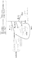
Figure 1 . Schematic representation of the classification steps
The following abbreviations are used:- TL: T-length value; long, int. and short designate the three possible TL status;
- C: C-circle value; int.: intermediate; ALT++, ALT+, ALT int. and ALT- designate the four possible ALT status;
- IDH: IDH status is chosen among IDHwt (no mutation) or IDHmt (at least one mutation in IDH1 and/or IDH2)
- Classification of gliomas "like" according to the algorithm of the invention: OD : oligodendroglioma; OD II: oligodendroglioma grade II; OD III: oligodendroglioma grade III; GbmOD: GBM or OD; GBM_IDHwt : primary glioblastoma; GBM_IDHmt: secondary glioblastoma; A_GBM_IDHmt: astrocytoma or secondary glioblastoma (mutated for IDH); A_IDHmt: astrocytomas; AIII_IDHmt : astrocytoma grade II; AIII_IDHmt : astrocytoma grade III.
-
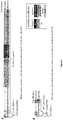
Figure 2 . Schematic representation of the classification steps for an astrocytoma IDHwt
The same abbreviations than infigure 1 are used. -
Figure 3 . Standard curve for calculation of qPCR efficiency
qPCR were led using DNA from an ALT positive tumor (left : TeloPCR, right : qPCR targeting the reference housekeeping gene 36B4).- (A) representative amplification curves are shown with (+) or without (-) pre-incubation with the enzyme φ29, as shown by the corresponding arrows. Telomere length is determined in the φ29-, and the c-circle value is calculated by using +φ29 and - φ29 values. Note that the same amount of genomic DNA is used in the two conditions (same curves for 36B4).
- (B) DNA from an ALT positive tumor was diluted and the CT value (y) is plotted against the log-transformed dilution factor (x) (TeloPCR on the left, and qPCR against 36B4 on the right). Note that the correlation coefficient (R2) is nearly 1 for both qPCR.
-
Figure 4 . Comparison of the diagnosis value of the two classifications
Gliomas (with the exception of anaplastic astrocytoma IDHwt) have been classified according to the standard algorithm according to the WHO 2016 classification (upper line), or with the algorithm of the invention (bottom line).
The algorithm of the invention allows a separation into five subclasses: "oligodendroglioma-like", "glioblastoma IDHwt-like", "glioblastoma IDHmt-like", "low-grade astrocytoma -like" and "other" (the code is depicted in the legend).- (A) Only patients with molecular biomarkers in agreement with the immuno-histochemical classification are analyzed (N=180), the two classifications are concordant for 93.5% of gliomas, are different (noted misclassified) for 3.8%, and 2.7% of gliomas are classified as "others".
- (B) Considering only the gliomas with discordant molecular biomarkers and immuno-histochemical classification (N=29), the algorithm is in agreement with histological classification for 72% of gliomas.
-
Figure 5 . Comparison of the results curves of median overall survival and median disease-free survival obtained after standard classification and classification according to the process of the invention
Gliomas (with the exception of anaplastic astrocytoma IDHwt) have been classified according to the standard algorithm following the WHO 2016 classification (left curves), or with the algorithm of the invention (rigth curves). - The overall survival (OS, upper curves) and the disease free survival (DFS, bottom curves) have been followed for each patient. 210 patients were analyzed (Standard classification: 74 low-grade astrocytoma, 24 glioblastoma IDHwt, 57 glioblastoma IDHmt, 55 oligodendroglioma). Among these patients, 29 have discordant molecular parameters as regard with the immuno-histological classification.
- The algorithm of the invention allows a separation into five subclasses: oligodendroglioma-like, glioblastoma IDHwt-like, glioblastoma IDHmt-like, low-grade astrocytoma -like (A_IDHmt like), and other gliomas.
- The following abbreviations are used:
OD: oligodendroglioma; A_IDHmt: Astrocytoma_IDHmt; GBM_IDHmt: secondary glioblastoma (also called glioblastoma IDHmt); GBM_IDHwt: primary glioblastoma (also called glioblastoma IDHwt). - Note that the curves are highly similar regarding the overall survival, and that the algorithm is even more discriminant that the standard classification considering the low grade astrocytoma and the oligodendroglioma.
- "Glioma" is identified on the basis of histopathological analysis. Classification as a "glioma" is given by a health practician on the basis of:
- morphological criteria, such as cell shape and density, nuclear atypias, vascularisation, necrosis and mitosis number (determined on hematoxilin-eosin-labelled sections);
- immuno-histochemical labelling of different epitopes, such as GFAP, OLIG2, IDH1-R132H, ATRX, TP53, Ki67, and
- if available, molecular biomarker such as mutations of IDH1 and 2, and/or 1p/19q deletion (according to the WHO classification).
- The present invention relates to an in vitro process for classifying a glioma, comprising the following steps:
- a. Measuring, from a glioma sample, the following two parameters:
- i. The Alternative Lengthening of Telomeres (ALT) status ; and
- ii. The telomere length status;
- b. Optionally, determining the isocitrate dehydrogenase genes mutation status (IDH status) of said glioma;
- c. Optionally, determining the histological grade of said glioma;
- d. Based on the data obtained in steps (a) (b) and (c), classifying said glioma in one of the five following classes: oligodendroglioma-like, glioblastoma IDHwt-like, glioblastoma IDHmt-like, low-grade astrocytoma like, and other gliomas.
- This classification process allows the distinction of classes of gliomas, different from the 2016 WHO standard classification of tumors of the central nervous system.
- The subclasses proposed by the present invention are the following:
- ∘ oligodendroglioma-like,
- ∘ glioblastoma IDHwt-like (also designated as primary glioblastoma),
- ∘ glioblastoma IDHmt-like (also designated as secondary glioblastoma),
- ∘ low-grade astrocytoma like, and
- ∘ other gliomas.
- The phrase "low-grade astrocytoma" designates astrocytoma of grade II (diffuse astrocytoma) and of grade III (anaplastic astrocytoma).
- The term "like" is used to mean that the patient in the group is expected to have similar response to treatment and overall survival as the group's name, but not necessarily have the corresponding immuno-histological or molecular parameters.
- Each group of patients having glioma is defined by a median time of overall survival of said patients. For example, a patient whose glioma is classified in the class "glioblastoma IDHwt-like" can expect an overall survival time comprised between 7 and 15 months.
- Advantageously, this classification process allows the re-classification of some gliomas that had been previously wrongly characterized, and consequently an incorrect expectation of median time of disease free survival and/or of overall survival had been announced to the patient. Furthermore, a non-adapted therapeutic strategy might be used for incorrectly classified patients.
- In this case, the algorithm of the invention would allow to inform the patient that his/her median overall survival time would be 7 to 15 months (GBM IDHwt-like), instead of 15 years for an oligodendroglioma of grade II.
- Also, the therapeutic strategies will not be the same, depending on the classification conclusions.
- As presented in
figure 4 , the process of the invention allows a classification with a prognostic value at least equivalent to the standard (WHO's) classification. Advantageously, this classification process is easy to implement in a clinical environment, since it necessitates only DNA extracted from said glioma sample, for the measure of parameters (a.i), (a.ii) and optionally (b). - Optionally, the grading of the tumor is measured on the basis of immuno-histochemical analyses of the glioma sample.
- It is understood that the process according to the invention comprises at least two steps (a) and (d) but might also comprise other supplementary steps, well known by the man skilled in the art, such as the optional steps (b) and (c), and also the detection of an EGFR amplification, the detection of a CDKN2A copy number loss, and/or the detection of chromosomal abnormalities like the gain in chromosome 7p correlated with loss of chromosome 10q (Inda et al., 2003).
- In a specific embodiment of the invention, the process of the invention comprises both steps (a) and (d).
- In a specific embodiment of the invention, the process of the invention comprises the three steps (a), (c) and (d).
- In a specific embodiment of the invention, the process of the invention comprises the three steps (a), (b) and (d).
- In a specific embodiment of the invention, the process of the invention comprises the four steps (a), (b), (c) and (d).
- In a specific embodiment of the invention, the process of the invention consists in both steps (a) and (d).
- In a specific embodiment of the invention, the process of the invention consists in three steps (a), (b) and (d).
- In a specific embodiment of the invention, the process of the invention consists in three steps (a), (c) and (d).
- In another specific embodiment of the invention, the process of the invention consists in the four steps (a) to (d).
- Surgery to remove as much of the tumor as possible is usually the first step in treating gliomas. After this procedure, a sample of the tumor is further analyzed.
- If no surgery is performed, a stereotaxic biopsy may be realized to obtain a tumor sample. From this tumor sample, DNA is extracted by any technique well known by the man skilled in the art.
- In an embodiment of the invention, the parameters (a.i), (a.ii) and (b) are measured on the basis of DNA extracted from a sample of said glioma, that has been recovered by any technique known by the man skilled in the art.
- In most cases, glioma samples are not analyzed directly but are conserved before extraction of DNA. Typically such collected tissue samples are frozen or are processed as paraffin blocks. Advantageously, these tumor samples are annotated with clinical information on the patient.
- In an embodiment of the invention, the parameters (a.i) (a.ii) and (b) of tumor samples are measured on the basis of DNA extracted from a glioma sample that has been conserved in paraffin or frozen.
- Advantageously, the techniques for measuring parameters (a.i) (a.ii) and (b) of tumor samples can be realised on the basis of DNA extracted from a glioma sample that has been conserved in paraffin or frozen, even if said DNA is of poor quality and/or in a small amount.
- In an aspect of the invention, the parameters (a.i) (a.ii) and (b) of tumor samples are measured on the basis of DNA of glioma cells, extracted from a blood sample or a cerebrospinal fluid sample from a patient having said glioma.
- Glioma cells have a tendency to necrose and cell debris and/or free DNA circulate in blood and cerebrospinal fluid of patients affected by glioma. Moreover, cells designated as Circulating tumor cells (CTCs) issued from a primary tumor are able to circulate around the body in the blood circulation. From these cell debris, circulating DNA (circDNA) and/or CTCs, tumor-specific DNA can be purified and analyzed in order to assess the three parameters (a.i) (a.ii) and (b).
- In this aspect of the invention, the term "glioma sample" designates the DNA from the glioma cells, that is obtained from the blood or from the cerebrospinal fluid of the patient.
- This implementation of the process is advantageous since, in the preliminary steps not included in the process, the invasive step of biopsy for obtaining a tumor sample is avoided.
- In this embodiment, the in vitro process for classifying a glioma comprises the following steps:
- a. Measuring, from a glioma sample, i.e. from the DNA of glioma cells that has been obtained from the blood or from the cerebrospinal fluid of the patient, the following two parameters:
- i. The Alternative Lengthening of Telomeres (ALT) status ; and
- ii. The telomere length status;
- b. Optionally, determining the isocitrate dehydrogenase genes mutation status (IDH status) of said glioma;
- c. Based on the data obtained in steps (a) and (b), classifying said glioma in one of the five following classes: oligodendroglioma -like, glioblastoma IDHwt -like, glioblastoma IDHmt -like, low-grade astrocytoma -like, and other gliomas.
- In this embodiment, no solid glioma sample is available. Therefore, the histological grading of the tumor cannot be determined. The process is therefore limited to the classification in five classes, without any further sub-classification of the gliomas according to their histological grade.
- In a specific embodiment of the invention, the process of the invention comprises both steps (a) and (c).
- In a specific embodiment of the invention, the process of the invention comprises the three steps (a), (b) and (c).
- In a specific embodiment of the invention, the process of the invention consists in both steps (a) and (c).
- In a specific embodiment of the invention, the process of the invention consists in three steps (a), (b) and (c).
- In a specific embodiment of the invention, the values of the parameters (a.i) and (a.ii), i.e. the ALT status and the telomere length status, are measured and then compared each one to at least one threshold value.
- For the purposes of carrying out the above process, the term "threshold value" is intended to mean a value determined with a group of glioma samples that are well characterized, and whose ALT status and telomere length status have been previously determined.
- Based on the results obtained for these groups of specific gliomas, inventors have defined threshold values for ALT status and telomere length that are defined below.
- Other threshold values can be easily determined by those skilled in the art by means of their general knowledge.
- Therefore, the value obtained in step (a.i) is compared to at least one ALT threshold value, in order to determine the ALT status of the glioma; and the value obtained in step (a.ii) is compared to at least one T-length threshold value, in order to determine the telomere length status of the glioma.
- In vertebrates, chromosome extremities, designed as telomeres, consist of:
- a repeated motif of sequence 5'-(TTAGGG)n-3', that is 10-15 kilobases long in human at birth, and a 3' G-rich single-stranded tail of 150 to 300 bases;
- six associated proteins which comprise the sheltering complex that promotes telomere protection, by ensuring stability and assisting specialized replication machinery for accurate extension of chromosome ends and recruitment of telomerase. The six proteins comprise three DNA-binding poteins (TRF1, TRF2, POT1) interconnected by three additional proteins (TIN2, ACD, RAP1).
- Telomerase is a ribonucleic reverse transcriptase enzyme, able to add the telomere repeat sequence to the 3' end of telomeres. It consists of a catalytic subunit called TERT (for Telomerase Reverse Transcriptase), an essential RNA component TERC that functions as the RNA template for the addition of the telomeric repeats, and a series of auxiliary components.
- In most tumour cells, the TMM is linked to the reactivation of telomerase. Nevertheless, about 10% of tumour cells acquire immortality through the telomerase-independent alternative lengthening of telomeres (ALT) mechanism.
- Glioma can therefore be classified as "ALT-dependent tumors", when the telomerase-independent mechanism is in place, and "ALT-independent tumors" when a telomerase-dependant mechanism is activated in the cell.
- Within the ALT-dependent group, different subgroup can be identified: ALT intermediate, ALT+ and ALT ++.
- The ALT-independent tumors define the ALT- subgroup, wherein no C circle is detected.
- Any suitable assay for determining the ALT status of cells can be used for the implementation of the process.
- Henson et al. (2009) have disclosed an assay based on partially single-stranded telomeric (CCCTAA)n DNA circles that allows a specific measurement of the ALT status. This assay is known as "the C-circle assay" or "CC assay".
- Briefly, this C-circle assay can be summarized as follow:
Telomeric extrachromosomal DNA (ECT) is particularly enriched in ALT-dependant cells versus telomerase-positive or mortal cells. Among different species of ECT, C-circles is a specific and sensitive marker of the ALT process. A C-circle is composed of a circular DNA of C3TA2 sequence, partially double stranded with a short T2AG3 sequence. The technique of Rolling Circle Amplification (RCA) has been used to detect telomeric circles in ALT+ cells. The "CC-assay" involves the use of φ29, a highly processive DNA polymerase that is auto-primed by the partial G-strand (TTAGGG)n. - The amplified telomeric DNA is then quantified together with the genomic telomeric DNA after and before incubation with φ29. A ratio between these two quantities of DNA is calculated, and compared to the same ratio obtained in telomerase positive cells and in ALT-dependant cells. (Henson et al., 2017).
- The amplified telomeric DNA can be quantified by all techniques well known by the man skilled in the art, such as hybridization and flow cytometry methods.
- In a preferred embodiment of the invention, the quantification of the amplified telomeric DNA circles is performed as described in (Gil & Coetzer, 2004) with a real-time polymerase chain reaction (PCR) - based method, and particularly with a telomere-specific PCR.
- A "C-circle" value, expressed in arbitrary units, equal to the following ratio:
Quantity of total telomeric DNA (including RCA-amplifiedC-circles) / Quantity of total telomeric DNA (without amplification of C-circle content)
is measured by the means described above for each glioma sample. - This value is then compared to at least three thresholds values, previously defined according to the general knowledge of the man skilled in the art.
- In particular, the inventors have defined four thresholds as follow:
- A threshold said "high";
- A threshold said "intermediate"
- A threshold said "positive"
- A threshold said "negative".
- In a specific embodiment of the invention, in the in vitro process, the ALT status in (a.i) is determined by performing a C-circle assay coupled to a telomere-specific PCR, thereby obtaining a C-circle value and selecting the ALT status of the glioma from:
- ALT++, when the C-circle value is superior to the threshold value "high";
- ALT+, when the C-circle value is superior to the threshold value "positive";
- ALT intermediate, when the C-circle value is inferior to the threshold value "intermediate", and
- ALT-, when the C-circle value is inferior to the threshold value "negative".
- In a specific embodiment of the invention, the in vitro process for classifying a glioma comprises the following steps:
- a. Measuring, from a glioma sample, the following two parameters:
- i. The Alternative Lengthening of Telomeres (ALT) status ; and
- ii. The telomere length status;
- b. Optionally, determining the isocitrate dehydrogenase genes mutation status (IDH status) of said glioma;
- c. Optionally, determining the histological grade of said glioma;
- d. Based on the data obtained in steps (a) (b) and (c), classifying said glioma in one of the five following classes: oligodendroglioma -like, glioblastoma IDHwt -like, glioblastoma IDHmt -like, low-grade astrocytoma -like, and other gliomas,
- Measurement of telomere length is the second essential parameter of the process, measured in step (a.ii) of the process of the invention.
- The original method for determining telomere length employed Southern hybridization to determine the mean terminal restriction fragment length. However this technique was labor-intensive, time-consuming and necessitates high amount of DNA (over 1µg), not available in standard clinical use. Various alternative techniques have since been proposed, including slot blots, next generation sequencing, hybridization protection assays and flow cytometry.
- Although any technique for determining the telomere length status might be employed in the implementation of the process of the invention, in a preferred embodiment, telomere length status in (a.ii) is determined by quantifying the telomeric DNA with a telomere-specific PCR.
- In particular, the real-time quantitative PCR described in (Gil & Coetzer, 2004) can be used for this quantification.
- According to this embodiment, the quantification technique comprises the following steps:
- Quantitative PCR reactions are initiated with addition of primers specifics for telomeres (TeloPCR) and for the used housekeeping gene(s) such as 36B4 (36B4 PCR) to samples of total DNA;
- The efficiency of TeloPCR (ETelo) and 36B4 (E36B4) PCR is determined on the basis of standard curves generated from two reference DNA samples, one ALT+ and one ALT-; for example, a DNA from previously characterized ALT+ tumors and ALT- tumors, either extracted from paraffin block or frozen sample;
- Products of the qPCR are quantified as well-known by the man skilled in the art, following the ETelo - CT(telo) / E36B4 - CT(36B4) method, where the threshold cycle (CT) is the cycle at which the fluorescence level reaches a certain amount (the threshold) for each qPCR (CT(Telo) and CT36B4), and E stands for PCR efficiency and is determined for each gene according to the standard curves.
- The telomere content value representative of the mean quantity of telomeric sequences in cells for each sample is obtained. This value is next normalized by internal controls obtained from DNA extracted from ALT+ and ALT- cell lines, as it is well known by the man skilled in the art. The resulting value is designated as "T-length" value.
- Four threshold have been defined as a function of increasing TL : short, middle-long, long and very long. With these thresholds, the telomere length status of the glioma can be determined as follow:
- Long telomere, when the T-length value is superior to the threshold value "very long";
- Intermediate telomere, when the T-length value is comprised between the threshold values "middle-long" and "long"; and
- Short telomere, when the T-length value is inferior to the threshold value "short
- In a specific embodiment of the invention, the in vitro process for classifying a glioama comprises the following steps:
- a. Measuring, from a glioma sample, the following two parameters:
- i. The Alternative Lengthening of Telomeres (ALT) status ; and
- ii. The telomere length status;
- b. Optionally, determining the isocitrate dehydrogenase genes mutation status (IDH status) of said glioma;
- c. Optionally, determining the histological grade of said glioma;
- d. Based on the data obtained in steps (a) (b) and (c), classifying said glioma in one of the five following classes: oligodendroglioma -like, glioblastoma IDHwt -like, glioblastoma IDHmt -like, low-grade astrocytoma -like, and other gliomas,
- In a specific embodiment of the invention, the ALT status and the T-length status are measured concomitantly with the same experimentation, comprising:
- the C-circle assay, and
- a telomere specific quantitative PCR.
-
Figure 3 presents an example of standard curves that can be used for the calculation of the qPCR efficiency, and that are useful for determining both the C-circle value and the telomere length. - The parameter of IDH status has previously been described for the classification of gliomas.
- In a specific embodiment of the invention, the IDH status in optional step (b) is determined by sequencing both genes encoding proteins IDH1 and IDH2.
- Sequences of human
isocitrate dehydrogenase proteins 1 and 2 are well known of the man skilled in the art, and can be found in proteins database such as GeneBank and OMIM. - Wild-type sequence of human IDH1 is represented in SEQ ID NO:1, and wild type sequence of human IDH2 is represented in SEQ ID NO: 2, as presented in table 1 below:
SEQ ID NO: 1 
UniProtKB - O75874, OMIM 147700 SEQ ID NO: 2 
UniProtKB - P48735, OMIM 147650 - The IDH status of the glioma cells is determined as follows:
- The glioma is said of "IDH mutated (IDHmt) status" in case of the identification of a point mutation of residue R132 in IDH1 protein, and/or of a point mutation of residue R172 in IDH2 protein, and
- The glioma is said of "IDH wild-type (IDHwt) status" in case of each of both proteins IDH1 and IDH2 present a wild-type sequence at the respective residues R132 and R172.
- Step (b) of the process according to the invention is optional. Indeed, and as illustrated in
figure 1 , determination of the IDH status is particularly relevant in the following case: when the intermediate classification (after step (a)) is "GbmOD" i.e. an oligodendroglioma or a glioblastoma; indeed, a glioma presenting the following features: - ∘ ALT status = ALT-; and
- ∘ T-length = intermediate ;
- In this case, a step (b) of determination of the IDH status is necessary for the final classification, where:
- ∘ If IDH status = IDHwt, then the glioma is a primary glioblastoma (GBM_IDHwt or GBMI);
- ∘ If IDH status = IDHmt, the glioma is an oligodendroglioma (OD), with a much better prognostic.
- Optionally, the tumor grading is measured in step (c) on the basis of immuno-histochemical analyses of pictures of said tumor, thereby selecting the tumor grade of the glioma from stage II, III or IV.
- These grades are defined in the actual WHO classification of gliomas, and are based on the presence of the following criteria of the tumor cells: mitosis, vascular proliferation and necrosis.
- The grade of the glioma will advantageously be determined by a man skilled in the art such as a neuro-specialized physician.
- When no solid glioma sample is available, for example in the case of the implementation of the process on glioma DNA extracted from blood or cerebrospinal fluid of the patient, the histological grading of the tumor is not determined.
- As previously presented, the subclass of "astrocytoma IDHwt" as determined by WHO's classification is particularly heterogeneous and difficult to classify.
- Often, these gliomas are not actually conventional "astrocytomas" as defined by the WHO classification.
- Therefore, the median overall survival is highly variable with a median of 2.5 years, which is much more severe than for classical astrocytoma. Moreover, therapeutic options are not defined by guidelines and might be wrongly chosen.
- Therefore, there is a need for reclassifying these specific gliomas with a novel combination of parameters representative of the nature of said glioma.
- In a particular aspect, the invention concerns an in vitro process for reclassifying a glioma of the class "astrocytoma IDHwt" according to the WHO's classification, comprising the following steps:
- a. Measuring, for said astrocytoma IDHwt, the following two parameters :
- i. The Alternative Lengthening of Telomeres (ALT) status ; and
- ii. The telomere length status;
- b. Optionally, determining the histological grade of said glioma;
- c. Based on the data obtained in steps (a) and (b), classifying said "astrocytoma IDHwt -like" in one of the following classes: oligodendroglioma-like, glioblastoma IDHwt-like, glioblastoma IDHmt-like, ow-grade astrocytoma-like, and other gliomas.
- In this embodiment, the IDH status of the glioma is known before the implementation of the process and therefore is not assessed again.
- In this process, the histological grade of the "astrocytoma IDHwt" is usually known before the implementation of the process. However, optionally, the histological grade of the "astrocytoma IDHwt" might be determined during the process in step (b).
- Although the IDH status of this kind of glioma is IDHwt, the glioma might be re-classified in a class such as "glioblastoma_IDHmt-like" or "low-grade astrocytoma -like", although tumors in this class present an IDHmt status. Nevertheless, it is understood that in this case, the glioma previously classified as "IDHwt astrocytoma" keeps its status of IDHwt, but is categorized in this astrocytoma class because the patient affected with said glioma would present the same median overall survival than those affected with such astrocytoma.
- In this implementation of the process, further steps could be included such as determination of the status of TERT (mutated or wild-type) (Vinagre et al., 2013) and determination of the status of ATRX (present or absent) (Nandakumar et al., 2017).
- The present invention also relates to a process for choosing a therapeutic strategy for treating a glioma, comprising the steps of:
- a) Implementing the in vitro process for classifying said glioma as presented above, and
- b) Based on said classification, choosing the more adapted therapeutic strategy for the patient affected by said glioma.
- These therapies can be the actual "gold standard" therapies: radiotherapy with a concomitant or adjuvant chemotherapy, with chemotherapeutic agents such as Temozolomide or PCV, a combination treatment for brain tumors including procarbazine, lomustine, and vincristine.
- The present invention also relates to a process for adapting a therapeutic strategy for treating a glioma, comprising the steps of:
- a) Implementing the in vitro process for classifying said glioma as presented above, and
- b) Based on said classification, adapting the therapeutic strategy for the patient affected by said glioma.
- A promising therapeutic approach for the treatment of gliomas is the administration of inhibitors of any telomere maintenance mechanism. In particular, it has been shown that telomerase inhibitors increase the response to radiotherapy in a murine orthotopic model of human gliobastoma (Ferrandon et al., 2015).
- Thus, the present invention relates to an inhibitor of at least one telomere maintenance mechanism (TMM) for its use in the treatment of a glioma, wherein said glioma has been previously classified according to the process of the invention.
- Advantageously, the determination of the ALT status of the glioma allows the practician to choose between TMM inhibitors specific of the ALT mechanism, or specific of the telomerase reactivation mechanism.
- In a specific implementation of these processes, data obtained in steps (a) (b) and (c) are recorded on a computer device, into a software program that is configured to memorize said data and to execute steps to classify the glioma in function of said parameters data.
- Accordingly, the present invention also concerns a computer program product comprising code instructions for implementing a process as described above, for classifying a glioma.
- The present invention also concerns a kit for the implementation of the processes as described, comprising:
- Reagents suitable for performing a C-circle assay;
- Reagents suitable for performing a Telomere-specific PCR; and
- Adequate internal controls comprising genomic DNA from ALT+ cells and from ALT- cells.
- In particular, internal controls are chosen among cell lines expressing the telomerase (ALT- cells), such as breast cancer-derived cells, and among cell lines using the process of "Alternative lengthening of telomere" (ALT+ cells), such as osteosarcoma cell lines.
- Other useful internal controls can be also present in said kit for the implementation of the processes of the invention.
- Although the present invention herein has been described with reference to particular embodiments, it is to be understood that these embodiments are merely illustrative of the principles and applications of the present invention. It is therefore to be understood that numerous modifications may be made to the illustrative embodiments and that other arrangements may be devised without departing from the spirit and scope of the present invention as defined by the appended claims.
- Rolling circle amplification of C-circle is performed as described in (Henson et al., 2009) and (Henson et al., 2014). Briefly, 3.2µl of total genomic DNA (5ng/µL) were incubated for 18h at 30°C with 3,75 units of ϕi29 DNA polymerase (New England Biolabs) (0.375µL of 10U/µL), in 0.2µg/µL of BSA, 0.1% Tween, 4µM DTT (Dithiothreitol), 1mM dNTP, 1µL of 10X NEB buffer. Enzyme is heat-inactivated at 65°C for 20 min. The same reaction is performed without the enzyme ϕ29 (ϕ-).
- For each experiment, two internal controls are added, namely TA and ALT. TA and ALT correspond to total genomic DNA extracted from HeLa (ALT-) and U2OS (ALT+) cell lines respectively.
- The 10µL of ϕ- and cp+ reactions are then diluted by adding 30µL of water (molecular biology grade), 5µL are used to performed each qPCR reaction.
- Oligonucleotides for the qPCR reaction have been previously described in (Gil et al., 2004) and (Lau et al., 2013).
- The sequence for oligonucleotides used in the qPCR are presented in table 2:
SEQ ID NO: 3 5'-CGGTTTGTTTGGGTTTGGGTTTGGGTTTGGGTTTGGGTT-3' forward Tel1a SEQ ID NO: 4 5'-GGCTTGCCTTACCCTTACCCTTACCCTTACCCTTACCCT-3' reverse primer tel2B SEQ ID NO: 5 5'-CAGCAAGTGGGAAGGTGTAATCC-3' RPL0/ 36B 4 forward primerSEQ ID NO: 6 5'-CCATTCTATCATCAACGGGTACAA-3' RPL0/ 36B 4 reverse primer - Telo-PCR and qPCR against 36B4 are run in duplicate for each condition ϕ- and ϕ+, on a 480 Light Cycler Thermocycler (Roche, Houwald, Luxembourg), in 1X final LightCycler®
- FastStart DNA Master SYBR Green I (10|jL), 200nM final of TeloPCR-specific primers or 300 nMm final of 36B4-specific primers. Details of thermocycling conditions are detailed below
- For each qPCR, the exact conditions are summarized in tables 3 and 4 below:
Table 3. TeloPCR Analysis Mode Cycles Segment Target Temperature Hold time Acquisition Mode Pre Incub ation None 1 95°C 10 min none Amplification Quantification 32 Denaturation 95°C 5 s none Annealing 56 °C Rate = 4°C/s 10 s none Extension 72°C 60 s single Melting Curve Melting Curves 1 Denaturation 95 °C 0 s none Annealing 65 °C 15 s none Melting 95 °C Ramp Rate = 0,1 °C/s 0 s continuous Cooling none 1 40 °C 30 s none Table 4. PCR36B4 Analysis Mode Cycles Segment Target Température Hold time Acquisition Mode Pre Incubation None 1 95°C 10 min none Amplification Quantification 36 Denaturation 95°C 5s none Annealing 58 °C 10 s none Extension 72°C 40 s single Melting Curve Melting Curves 1 Denaturation 95°C 0 s none Annealing 65 °C 15 s none Melting 95 °C Ramp Rate = 0,1 °C/s 0 s continuous Cooling none 1 40 °C 30 s none - The fluorescence in logarithmic scale is analyzed as a function of PCR cycle, the threshold is set as half the high of the linear fraction of the amplification curves (all experiments). Intersection between the threshold of amplification curve gives the CT for each reaction.
- Efficiency of TeloPCR (Etelo) and 36B4 (E36B4) PCR are respectively 1.70111 and 1.9672.
- For each reaction, the following value is calculated : Etelo -CT/ E36B4 -CT, and annotated as ϕ+ and ϕ- as a function of the initial circle reaction.
- ϕ- correspond to the telomere length (T-Length)
Cr correspond to the Circle score and is calculated as follow : ϕ+ / ϕ- - The difference of T-Length between the two internal controls Δ is calculated and correspond to x units (determined for each batch of controls) with Δ= ϕ-HeLa - ϕ-U2OS
TLnorm is then calculated for each sample as follow : ϕ-/(Δ/x)
A standard curve for calculation of qPCR efficiency is presented infigure 3 . - As shown in
figure 1 , the first step (a) of the algorithm allows the classification of the tumors into 4 main categories: - OD (oligodendroglioma),
- GbmOD (glioblastoma (GBM_IDHwt in
figure 1 ) or OD), - A_GBM_IDHmt (AD (AII_IDHmt in
figure 1 ), AA (AIII_IDHmt infigure 1 ), or GBMII (GBM_IDHmt infigure 1 ), and - others.
- GbmOD category is then subclassified in step (b) according to the IDH status:
- If the tumor is IDHwt : GBM_IDHwt or
- If the tumor is IDHmt: OD.
- All glioma are then classified according to their grade in step (c). In particular, tumors of the categorie "A_GBM_IDHmt" will be subclassified, according to their grade, into AII-IDHmt like (AD), AIII_IDHmt like (AA) or GBM_IDHmt like (GBMII).
- An example of equations is given below; however, the indicated numbers have been defined for this experiment only, and are not limitating the scope of the claimed invention.
- A=(SI(TLnorm>4;"A_GBMII";SI(ET(Cr>1,5;TLnorm>0,8);"A_GBMII";SI(ET(TLnorm<0,66; Cr<1,9);"OD";SI(ET(Cr <1,28;ET(TLnorm>0,66; TLnorm<3));"GbmOD";SI(Cr >2;"A_GBMII";"other"))))))
- B=SI(ET(A="GbmOD";IDH="MT");"OD";(SI(ET(A="GbmOD";IDH="WT");"GBM";A)))
- C=SI(ET(B="A_GBMII";G="III");"AA";SI(ET(B="A_GBMII";G="II");"AD";SI(ET(B="A_GBMII ";G="IV");"GBMII";B)
- First, only 180 patients with molecular biomarkers in agreement with the immuno-histochemical classification (i.e. with the exception of anaplastic astrocytoma IDHwt) were analyzed.
- These gliomas have been classified according to the standard algorithm according to the WHO 2016 classification (upper line), or with the algorithm of the invention (bottom line) and are presented in
figure 4A . - The algorithm of the invention allows a separation into five subclasses: "oligodendroglioma-like", "glioblastoma IDHwt-like", "glioblastoma IDHmt-like", "low-grade astrocytoma -like" and "other" (the code is depicted in the legend).
- Both processes of classification are concordant for 93.5% of gliomas. However for 3.8% of gliomas, the classfications are different (noted misclassified). With the algorithm of the invention, 2.7% of gliomas are classified as "others".
- In conclusion, with the classification process according to the invention:
- ∘ For 94% of the gliomas, the classification was the same with the standard algorithm than with the process of the invention;
- ∘ 3.8% of the gliomas were differently classified (noted "misclassified") - their re-classification was relevant with the OS and DFS curves presented in
figure 5 ; - ∘ 2.7% of the gliomas are classified in the category "others".
-
Figure 4B shows the obtained results when considering only the 29 gliomas with discordant molecular biomarkers and immuno-histochemical classification. - In this case, the classification obtained with the algorithm of the invention is in agreement with the standard process of classification for 72% of gliomas.
- The
figure 5 presents a comparison of the curves of median overall survival (OS) and median disease-free survival (DFS) obtained after standard classification and classification according to the process of the invention - The overall survival (OS, upper curves) and the disease free survival (DFS, bottom curves) have been followed for each patient.
- 210 patients were analyzed, classified as follow according to the standard classification: 74 low-grade astrocytoma, 24 glioblastoma IDHwt, 57 glioblastoma IDHmt, and 55 oligodendroglioma.
- Among these patients, 29 have discordant molecular parameters as regard with the immuno-histological classification.
- The algorithm of the invention allows a separation into five subclasses: oligodendroglioma-like, glioblastoma IDHwt-like, glioblastoma IDHmt-like, low-grade astrocytoma -like (A_IDHmt like), and other gliomas.
- Note that the curves are highly similar regarding the overall survival, and that the algorithm is even more discriminant that the standard classification considering the low grade astrocytoma and the oligodendroglioma.
-
- Louis DN, Perry A, Reifenberger G, von Deimling A, Figarella-Branger D, Cavenee WK, Ohgaki H, Wiestler OD, Kleihues P, Ellison DW. The 2016 World Health Organization Classification of Tumors of the Central Nervous System: a summary. Acta Neuropathol. 2016 Jun;131(6):803-20. doi: 10.1007/s00401-016-1545-1. Epub 2016 May 9. Review.
- Henson JD, Cao Y, Huschtscha LI, et al. DNA C-circles are specific and quantifiable markers of alternative-lengthening-of-telomeres activity. Nat. Biotechnol. 2009;27(12):1181-1185.
- Henson JD, Lau LM, Koch S, et al. The C-Circle Assay for alternative-lengthening-of-telomeres activity. Methods San Diego Calif. 2017;114:74-84.
- Gil ME, Coetzer TL. Real-time quantitative PCR of telomere length. Mol. Biotechnol. 2004;27(2): 169-172.
- Inda MM, Fan X, Muñoz J, Perot C, Fauvet D, Danglot G, Palacio A, Madero P,Zazpe I, Portillo E, Tuñón T, Martínez-Peñuela JM, Alfaro J, Eiras J, Bernheim A,Castresana JS. Chromosomal abnormalities in human glioblastomas: gain in chromosome 7p correlating with loss in chromosome 10q. Mol Carcinog. 2003 Jan;36(1):6-14. PubMed PMID: 12503074.
- Vinagre J, Almeida A, Pópulo H, Batista R, Lyra J, Pinto V, Coelho R, Celestino R, Prazeres H, Lima L, Melo M, da Rocha AG, Preto A, Castro P, Castro L, Pardal F, Lopes JM, Santos LL, Reis RM, Cameselle-Teijeiro J, Sobrinho-Simões M, Lima J, Máximo V, Soares P. Frequency of TERT promoter mutations in human cancers. Nat Commun. 2013;4:2185.
- Nandakumar, P., Mansouri, A., & Das, S. The Role of ATRX in Glioma Biology. Frontiers in oncology, 7, 236 (2017).
- Ferrandon S, Malleval C, El Hamdani B, et al. Telomerase inhibition improves tumor response to radiotherapy in a murine orthotopic model of human glioblastoma. Mol Cancer. 2015;
- Lau LM, Dagg RA, Henson JD, Au AY, Royds JA, Reddel RR. Detection of alternative lengthening of telomeres by telomere quantitative PCR. Nucleic Acids Res. 2013 Jan;41 (2):e34.


Claims (15)
- An in vitro process for classifying a glioma, comprising the following steps :a. Measuring, from a glioma sample, the following two parameters:i. The Alternative Lengthening of Telomeres (ALT) status ; andii. The telomere length status;b. Optionally, determining the isocitrate dehydrogenase genes mutation status (IDH status) of said glioma;c. Optionally, determining the histological grade of said glioma;d. Based on the data obtained in steps (a) (b) and (c), classifying said glioma in one of the five following classes: oligodendroglioma -like, glioblastoma IDHwt -like, glioblastoma IDHmt -like, low-grade astrocytoma -like, and other gliomas.
- The in vitro process according to claim 1, wherein each class of glioma is defined by a median time of overall survival of the patients affected by said glioma.
- The in vitro process according to claim 1 or 2, wherein the steps (a) and (b) are realized on the basis of DNA extracted from a glioma sample that has been conserved in paraffin or frozen.
- The in vitro process according to anyone of claims 1 to 3, wherein the steps (a) and (b) are realized on the basis of DNA extracted from a blood sample or from a cerebrospinal fluid sample of a patient having said glioma.
- The in vitro process according to anyone of claims 1 to 4, wherein the value of each one of the parameters (a.i) and (a.ii) is compared to at least one threshold value.
- The in vitro process according to anyone of claims 1 to 5, wherein the ALT status in (a.i) is measured by performing a C-circle assay coupled to a telomere-specific PCR, thereby obtaining a C-circle value and selecting the ALT status of the glioma from:- ALT++, when the C-circle value is superior to the threshold value "high";- ALT+, when the C-circle value is superior to the threshold "positive";- ALT intermediate, when the C-circle value is inferior to threshold values "intermediate", and- ALT-, when the C-circle value is inferior to the threshold value "negative".
- The in vitro process according to anyone of claims 1 to 6, wherein the telomere length status in (a.ii) is measured by quantifying the telomeric DNA with a telomere-specific PCR, thereby obtaining a T-length value and selecting the telomere length status of the glioma from:- Long telomere, when the T-length value is superior to the threshold value "very long";- Intermediate telomere, when the T-length value is comprised between the threshold values "long" and "middle-long"; and- Short telomere, when the T-length value is inferior to the threshold value "short".
- The in vitro process according to anyone of claims 1 to 7, wherein the IDH status in step (b) is determined by sequencing both genes encoding proteins IDH1, whose wild-type sequence is represented in SEQ ID NO.1, and IDH2, whose wild-type sequence is represented in SEQ ID NO. 2, thereby selecting the IDH status of the glioma from:- IDH mutated (IDHmt) status in case of a mutation of residue R132 in IDH1 protein, and/or a mutation of residue R172 in IDH2 protein, or- IDH wild-type (IDHwt) status in case of both proteins IDH1 and IDH2 present a wild-type sequence at the respective residues R132 and R172.
- The in vitro process according to anyone of claims 1 to 8, wherein the histological grade in step (c) is determined on the basis of immuno-histochemical analyses of said glioma sample, thereby selecting the histological grade of said glioma from stage II, III or IV.
- An in vitro process for reclassifying a glioma of the class "astrocytoma" with a IDH status "IDHwt", comprising the following steps :a. Measuring, for said astrocytoma IDHwt, the following two parameters:i. The Alternative Lengthening of Telomeres (ALT) status ; andii. The telomere length status;b. Optionally, determining the histological grade of said glioma;c. Based on the data obtained in steps (a) and (b), classifying said "astrocytoma IDHwt-like" in one of the following classes: oligodendroglioma-like, glioblastoma IDHwt-like, glioblastoma IDHmt-like, low-grade astrocytoma -like and other gliomas.
- A process for choosing a therapeutic strategy for treating a glioma, comprising the steps of :a) Implementing the in vitro process for classifying said glioma according to anyone of claims 1 to 10; andb) Based on said classification, choosing the more adapted therapeutic strategy for the patient affected by said glioma.
- A process for adapting a therapeutic strategy for treating a glioma, comprising the steps of :a) Implementing the in vitro process for classifying said glioma according to anyone of claims 1 to 10; andb) Based on said classification, adapting the therapeutic strategy for the patient affected by said glioma.
- A computer program product comprising code instructions for implementing a process according to anyone of claims 1 to 10, for classifying a glioma.
- A kit for the implementation of the processes according to anyone of claims 1 to 12, comprising:- Reagents suitable for performing a C-circle assay;- Reagents suitable for performing a Telomere-specific PCR; and- Adequate internal controls comprising genomic DNA from ALT+ cells and from ALT- cells.
- Inhibitor of at least one telomere maintenance mechanism for its use in the treatment of a glioma, wherein said glioma has been previously classified according to the process according to anyone of claims 1 to 10.
Priority Applications (4)
| Application Number | Priority Date | Filing Date | Title |
|---|---|---|---|
| EP19305215.6A EP3699299A1 (en) | 2019-02-22 | 2019-02-22 | Process for classification of glioma |
| US17/432,813 US20220119893A1 (en) | 2019-02-22 | 2020-02-19 | Process for classification of glioma |
| PCT/EP2020/054346 WO2020169660A1 (en) | 2019-02-22 | 2020-02-19 | Process for classification of glioma |
| EP20704562.6A EP3927848A1 (en) | 2019-02-22 | 2020-02-19 | Process for classification of glioma |
Applications Claiming Priority (1)
| Application Number | Priority Date | Filing Date | Title |
|---|---|---|---|
| EP19305215.6A EP3699299A1 (en) | 2019-02-22 | 2019-02-22 | Process for classification of glioma |
Publications (1)
| Publication Number | Publication Date |
|---|---|
| EP3699299A1 true EP3699299A1 (en) | 2020-08-26 |
Family
ID=65802022
Family Applications (2)
| Application Number | Title | Priority Date | Filing Date |
|---|---|---|---|
| EP19305215.6A Withdrawn EP3699299A1 (en) | 2019-02-22 | 2019-02-22 | Process for classification of glioma |
| EP20704562.6A Pending EP3927848A1 (en) | 2019-02-22 | 2020-02-19 | Process for classification of glioma |
Family Applications After (1)
| Application Number | Title | Priority Date | Filing Date |
|---|---|---|---|
| EP20704562.6A Pending EP3927848A1 (en) | 2019-02-22 | 2020-02-19 | Process for classification of glioma |
Country Status (3)
| Country | Link |
|---|---|
| US (1) | US20220119893A1 (en) |
| EP (2) | EP3699299A1 (en) |
| WO (1) | WO2020169660A1 (en) |
Families Citing this family (2)
| Publication number | Priority date | Publication date | Assignee | Title |
|---|---|---|---|---|
| CN113308545A (en) * | 2021-06-11 | 2021-08-27 | 南京医科大学 | DNA methylation-based invasive glioma classification device |
| CN117987544A (en) * | 2023-12-27 | 2024-05-07 | 江苏阔然生物医药科技有限公司 | Probe primer set and detection kit for rapid typing and identification of pediatric brain glioma during surgery |
Citations (1)
| Publication number | Priority date | Publication date | Assignee | Title |
|---|---|---|---|---|
| WO2017127803A1 (en) | 2016-01-22 | 2017-07-27 | The Trustees Of Columbia University In The City Of New York | Methods for classification of glioma |
-
2019
- 2019-02-22 EP EP19305215.6A patent/EP3699299A1/en not_active Withdrawn
-
2020
- 2020-02-19 EP EP20704562.6A patent/EP3927848A1/en active Pending
- 2020-02-19 US US17/432,813 patent/US20220119893A1/en active Pending
- 2020-02-19 WO PCT/EP2020/054346 patent/WO2020169660A1/en not_active Ceased
Patent Citations (1)
| Publication number | Priority date | Publication date | Assignee | Title |
|---|---|---|---|---|
| WO2017127803A1 (en) | 2016-01-22 | 2017-07-27 | The Trustees Of Columbia University In The City Of New York | Methods for classification of glioma |
Non-Patent Citations (22)
| Title |
|---|
| "WHO classification" |
| "WHO classification", 2016 |
| DATABASE EMBASE [online] ELSEVIER SCIENCE PUBLISHERS, AMSTERDAM, NL; 1 November 2018 (2018-11-01), NOOR H ET AL: "A novel C-circle assay for detecting alternative lengthening of telomeres (ALT) mechanisms", XP002793653, Database accession no. EMB-628633441 * |
| FERRANDON S; MALLEVAL C; EL HAMDANI B ET AL.: "Telomerase inhibition improves tumor response to radiotherapy in a murine orthotopic model of human glioblastoma", MOL CANCER, 2015 |
| FOGLI ANNE ET AL: "Detection of the alternative lengthening of telomeres pathway in malignant gliomas for improved molecular diagnosis", JOURNAL OF NEURO-ONCOLOGY, KLUWER, BOSTON, US, vol. 135, no. 2, 28 July 2017 (2017-07-28), pages 381 - 390, XP036351458, ISSN: 0167-594X, [retrieved on 20170728], DOI: 10.1007/S11060-017-2585-7 * |
| GIL ME; COETZER TL: "Real-time quantitative PCR of telomere length", MOL. BIOTECHNOL., vol. 27, no. 2, 2004, pages 169 - 172 |
| HAKIN-SMITH V ET AL: "Alternative lengthening of telomeres and survival in patients with glioblastoma multiforme", LANCET, ELSEVIER, AMSTERDAM, NL, vol. 361, no. 9360, 8 March 2003 (2003-03-08), pages 836 - 838, XP004783597, ISSN: 0140-6736, DOI: 10.1016/S0140-6736(03)12681-5 * |
| HENSON JD; CAO Y; HUSCHTSCHA LI ET AL.: "DNA C-circles are specific and quantifiable markers of alternative-lengthening-of-telomeres activity", NAT. BIOTECHNOL., vol. 27, no. 12, 2009, pages 1181 - 1185, XP055048134, DOI: doi:10.1038/nbt.1587 |
| HENSON JD; LAU LM; KOCH S ET AL.: "The C-Circle Assay for alternative-lengthening-of-telomeres activity", METHODS SAN DIEGO CALIF., vol. 114, 2017, pages 74 - 84, XP029886623, DOI: doi:10.1016/j.ymeth.2016.08.016 |
| INDA MM; FAN X; MUNOZ J; PEROT C; FAUVET D; DANGLOT G; PALACIO A; MADERO P; ZAZPE I; PORTILLO E: "Chromosomal abnormalities in human glioblastomas: gain in chromosome 7p correlating with loss in chromosome 10q", MOL CARCINOG., vol. 36, no. 1, January 2003 (2003-01-01), pages 6 - 14 |
| JEITANY M ET AL: "A preclinical mouse model of glioma with an alternative mechanism of telomere maintenance (ALT)", INTERNATIONAL JOURNAL OF CANCER 20150401 WILEY-LISS INC. USA, vol. 136, no. 7, 1 April 2015 (2015-04-01), pages 1546 - 1558, XP002793654, ISSN: 0020-7136 * |
| JOERG BALSS J ET AL: "Analysis of the IDH1 codon 132 mutation in brain tumors", ACTA NEUROPATHOLOGICA, SPRINGER, BERLIN, DE, vol. 116, no. 6, 5 November 2008 (2008-11-05), pages 597 - 602, XP019657078, ISSN: 1432-0533, DOI: 10.1007/S00401-008-0455-2 * |
| LAU LM; DAGG RA; HENSON JD; AU AY; ROYDS JA; REDDEL RR: "Detection of alternative lengthening of telomeres by telomere quantitative PCR", NUCLEIC ACIDS RES., vol. 41, no. 2, January 2013 (2013-01-01), pages e34, XP055240849, DOI: doi:10.1093/nar/gks781 |
| LOUIS DAVID N ET AL: "The 2016 World Health Organization Classification of Tumors of the Central Nervous System: a summary", ACTA NEUROPATHOLOGICA, SPRINGER VERLAG, BERLIN, DE, vol. 131, no. 6, 9 May 2016 (2016-05-09), pages 803 - 820, XP035683913, ISSN: 0001-6322, [retrieved on 20160509], DOI: 10.1007/S00401-016-1545-1 * |
| LOUIS DN; PERRY A; REIFENBERGER G; VON DEIMLING A; FIGARELLA-BRANGER D; CAVENEE WK; OHGAKI H; WIESTLER OD; KLEIHUES P; ELLISON DW: "The 2016 World Health Organization Classification of Tumors of the Central Nervous System: a summary", ACTA NEUROPATHOL., vol. 131, no. 6, 9 May 2016 (2016-05-09), pages 803 - 20, XP035878676, DOI: doi:10.1007/s00401-016-1545-1 |
| MANGEREL J ET AL: "Alternative lengthening of telomeres is enriched in, and impacts survival of TP53 mutant pediatric malignant brain tumors", ACTA NEUROPATHOLOGICA 2014 SPRINGER VERLAG DEU, vol. 128, no. 6, 2014, pages 853 - 862, XP002793651, ISSN: 0001-6322 * |
| MCDONALD KERRIE L ET AL: "Presence of Alternative Lengthening of Telomeres Mechanism in Patients With Glioblastoma Identifies a Less Aggressive Tumor Type With Longer Survival", JOURNAL OF NEUROPATHOLOGY & EXPERIMENTAL NEUROLOGY, vol. 69, no. 7, July 2010 (2010-07-01), pages 729 - 736, XP002793650, ISSN: 0022-3069 * |
| NANDAKUMAR, P.; MANSOURI, A.; DAS, S.: "The Role of ATRX in Glioma Biology", FRONTIERS IN ONCOLOGY, vol. 7, 2017, pages 236 |
| NGUYEN D N ET AL: "Molecular and morphologic correlates of the alternative lengthening of telomeres phenotype in high-grade astrocytomas", BRAIN PATHOLOGY 2013 BLACKWELL PUBLISHING LTD GBR, vol. 23, no. 3, May 2013 (2013-05-01), pages 237 - 243, XP002793652, ISSN: 1015-6305 * |
| NOOR H ET AL: "A novel C-circle assay for detecting alternative lengthening of telomeres (ALT) mechanisms", NEURO-ONCOLOGY 20181101 OXFORD UNIVERSITY PRESS NLD, vol. 20, no. Supplement 6, 1 November 2018 (2018-11-01), pages vi42 CONF 20181115 to 20181118 New Orleans, LA - 23rd Annu, ISSN: 1523-5866 * |
| VINAGRE J; ALMEIDA A; POPULO H; BATISTA R; LYRA J; PINTO V; COELHO R; CELESTINO R; PRAZERES H; LIMA L: "Frequency of TERT promoter mutations in human cancers", NAT COMMUN., vol. 4, 2013, pages 2185 |
| YAN HAI ET AL: "IDH1 and IDH2 mutations in gliomas", NEW ENGLAND JOURNAL OF MEDICINE, THE - NEJM, MASSACHUSETTS MEDICAL SOCIETY, US, vol. 360, no. 8, 19 February 2009 (2009-02-19), pages 765 - 773, XP002538459, ISSN: 1533-4406, DOI: 10.1056/NEJMOA0808710 * |
Also Published As
| Publication number | Publication date |
|---|---|
| EP3927848A1 (en) | 2021-12-29 |
| WO2020169660A1 (en) | 2020-08-27 |
| US20220119893A1 (en) | 2022-04-21 |
Similar Documents
| Publication | Publication Date | Title |
|---|---|---|
| KR102622305B1 (en) | Detection method using chromosomal interaction sites | |
| CN116850285A (en) | Immunotherapy approaches for patients whose tumors carry a high burden of passenger gene mutations | |
| AU2019372440A1 (en) | Methods to diagnose and treat cancer using non-human nucleic acids | |
| Takeda et al. | Clinical outcome for EML4-ALK-positive patients with advanced non-small-cell lung cancer treated with first-line platinum-based chemotherapy | |
| Li et al. | Recurrently mutated genes differ between leptomeningeal and solid lung cancer brain metastases | |
| US20220119893A1 (en) | Process for classification of glioma | |
| Sanson et al. | Analysis of loss of chromosome 10q, DMBT1 homozygous deletions, and PTEN mutations in oligodendrogliomas | |
| Lee et al. | A polycythemia vera JAK2 mutation masquerading as a duodenal cancer mutation | |
| JP7722675B2 (en) | Method for predicting therapeutic response to EGFR tyrosine kinase inhibitors in EGFR-mutated non-small cell lung cancer | |
| Othman et al. | Molecular study of Nucleophosmin 1 (NPM1) gene in acute myeloid leukemia in Kurdish population | |
| Sonmezler et al. | Integration of Liquid Biopsies into Clinical Laboratory Applications via NGS in Cancer Diagnostics. | |
| Sun et al. | The existence and role of microchimerism after microtransplantion | |
| CN111154883B (en) | Breast cancer related gene PIK3CA site g.179220986A & gtT mutant and application thereof | |
| JP2003501058A (en) | Method and marker for determining prognosis of efficacy of anticancer drug | |
| KR101869931B1 (en) | Method of predicting sensitivity for molecular targeted anticancer drugs | |
| Ottaiano et al. | Clinical and genetic drivers of oligo-metastatic disease in colon cancer | |
| Delfau-Larue et al. | CDKN2A AND TP53 DELETIONS PREDICT ADVERSE OUTCOME IN YOUNGER MANTLE CELL LYMPHOMA PATIENTS, INDEPENDENT OF TREATMENT AND | |
| WO2017106365A1 (en) | Methods for measuring mutation load | |
| KR20100037639A (en) | Predictive markers for egfr inhibitors treatment | |
| Ahmed | 140P Retrospective review of actionable IDH mutations and MGMT promoter methylation among CNS tumors | |
| Nozaki et al. | Prognostic impact of telomerase activity in patients with neuroblastoma. | |
| CN109913481A (en) | PIK3CA gene g.1792224821G>A mutation and its application in auxiliary diagnosis of breast cancer | |
| CN111607646A (en) | A nucleotide sequence set for detecting BRAF gene mutation and its application | |
| US20220334121A1 (en) | Liquid biopsy to detect cancer early and sensitively in patients with neurofibromatosis type 1 | |
| CN112941187A (en) | Breast cancer related gene PIK3CA-Q928H mutant and specific primer thereof |
Legal Events
| Date | Code | Title | Description |
|---|---|---|---|
| PUAI | Public reference made under article 153(3) epc to a published international application that has entered the european phase |
Free format text: ORIGINAL CODE: 0009012 |
|
| STAA | Information on the status of an ep patent application or granted ep patent |
Free format text: STATUS: THE APPLICATION HAS BEEN PUBLISHED |
|
| AK | Designated contracting states |
Kind code of ref document: A1 Designated state(s): AL AT BE BG CH CY CZ DE DK EE ES FI FR GB GR HR HU IE IS IT LI LT LU LV MC MK MT NL NO PL PT RO RS SE SI SK SM TR |
|
| AX | Request for extension of the european patent |
Extension state: BA ME |
|
| STAA | Information on the status of an ep patent application or granted ep patent |
Free format text: STATUS: THE APPLICATION IS DEEMED TO BE WITHDRAWN |
|
| 18D | Application deemed to be withdrawn |
Effective date: 20210227 |